Differential Expression of Markers and Activities in a Group of PC12 Nerve Cell Clones
10
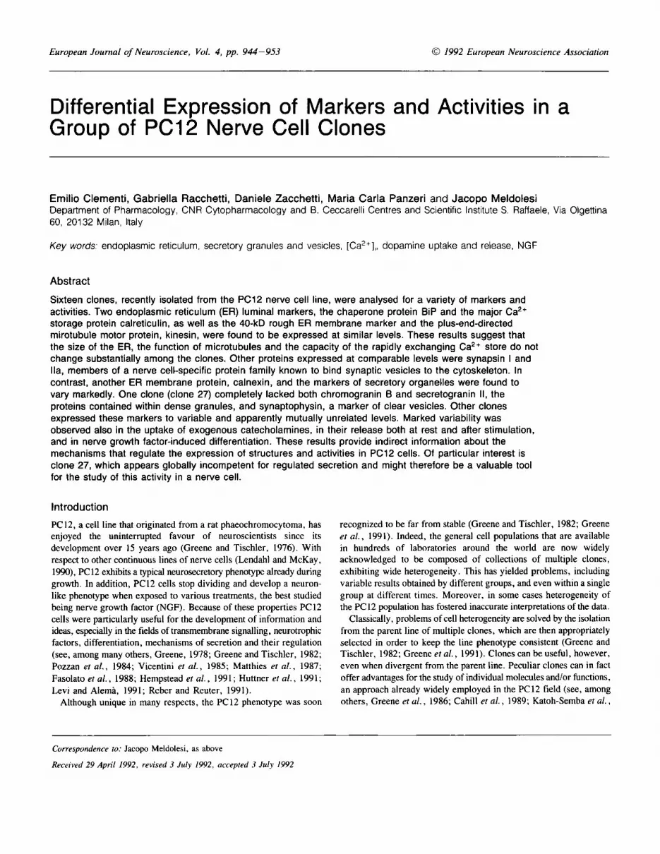
European Journal of Neuroscience, Vol. 4, pp. 944-953 @ 1992 European Neuroscience Association Differential Expression of Markers and Activities in a Group of PC12 Nerve Cell Clones Ernilio Clernenti, Gabriella Racchetti, Daniele Zacchetti, Maria Carla Panzeri and Jacopo Meldolesi Department of Pharmacology, CNR Cytopharmacology and B. Ceccarelli Centres and Scientific Institute S. Raffaele, Via Olgettina 60, 20132 Milan, Italy Key words: endoplasmic reticulum, secretory granules and vesicles, [Ca2+],, dopamine uptake and release, NGF Abstract Sixteen clones, recently isolated from the PC12 nerve cell line, were analysed for a variety of markers and activities. Two endoplasmic reticulum (ER) luminal markers, the chaperone protein BiP and the major Ca2+ storage protein calreticulin, as well as the 40-kD rough ER membrane marker and the plus-end-directed mirotubule motor protein, kinesin, were found to be expressed at similar levels. These results suggest that the size of the ER, the function of microtubules and the capacity of the rapidly exchanging Ca2+ store do not change substantially among the clones. Other proteins expressed at comparable levels were synapsin I and Ha, members of a nerve cell-specific protein family known to bind synaptic vesicles to the cytoskeleton. In contrast, another ER membrane protein, calnexin, and the markers of secretory organelles were found to vary markedly. One clone (clone 27) completely lacked both chromogranin B and secretogranin II, the proteins contained within dense granules, and synaptophysin, a marker of clear vesicles. Other clones expressed these markers to variable and apparently mutually unrelated levels. Marked variability was observed also in the uptake of exogenous catecholamines, in their release both at rest and after stimulation, and in nerve growth factor-induced differentiation. These results provide indirect information about the mechanisms that regulate the expression of structures and activities in PC12 cells. Of particular interest is clone 27, which appears globally incompetent for regulated secretion and might therefore be a valuable tool for the study of this activity in a nerve cell. Introduction PC12, a cell line that originated from a rat phaeochromocytoma, has enjoyed the uninterrupted favour of neuroscientists since its development over 15 years ago (Greene and Tischler, 1976). With respect to other continuous lines of nerve cells (Lendahl and McKay, 1990), PC12 exhibits a typical neurosecretory phenotype already during growth. In addition, PC12 cells stop dividing and develop a neuron- like phenotype when exposed to various treatments, the best studied being nerve growth factor (NGF). Because of these properties PC12 cells were particularly useful for the development of information and ideas, especially in the fields of transmembrane signalling, neurotrophic factors, differentiation, mechanisms of secretion and their regulation (see, among many others, Greene, 1978; Greene and Tischler, 1982; Pozzan et al., 1984; Vicentini er al., 1985; Matthies et a / . , 1987; Fasolato et al., 1988; Hempstead et al., 1991 ; Huttner et al., 1991; Levi and Alema, 1991; Reber and Reuter, 1991). Although unique in many respects, the PC12 phenotype was soon recognized to be far from stable (Greene and Tischler, 1982; Greene et al., 1991). Indeed, the general cell populations that are available in hundreds of laboratories around the world are now widely acknowledged to be composed of collections of multiple clones, exhibiting wide heterogeneity. This has yielded problems, including variable results obtained by different groups, and even within a single group at different times. Moreover, in some cases heterogeneity of the PC 12 population has fostered inaccurate interpretations of the data. Classically, problems of cell heterogeneity are solved by the isolation from the parent line of multiple clones, which are then appropriately selected in order to keep the line phenotype consistent (Greene and Tischler, 1982; Greene et al., 1991). Clones can be useful, however, even when divergent from the parent line. Peculiar clones can in fact offer advantages for the study of individual molecules and/or functions, an approach already widely employed in the PC12 field (see, among others, Greene et al., 1986; Cahill et al., 1989; Katoh-Semba et a/. , Correspondence to: Jacopo Meldolesi, as above Received 29 April 1992, revised 3 July 1992, accepted 3 July I992
Transcript of Differential Expression of Markers and Activities in a Group of PC12 Nerve Cell Clones
European Journal of Neuroscience, Vol. 4, pp. 944-953 @ 1992 European Neuroscience Association
Differential Expression of Markers and Activities in a Group of PC12 Nerve Cell Clones
Ernilio Clernenti, Gabriella Racchetti, Daniele Zacchetti, Maria Carla Panzeri and Jacopo Meldolesi Department of Pharmacology, CNR Cytopharmacology and B. Ceccarelli Centres and Scientific Institute S. Raffaele, Via Olgettina 60, 20132 Milan, Italy
Key words: endoplasmic reticulum, secretory granules and vesicles, [Ca2+],, dopamine uptake and release, NGF
Abstract
Sixteen clones, recently isolated from the PC12 nerve cell line, were analysed for a variety of markers and activities. Two endoplasmic reticulum (ER) luminal markers, the chaperone protein BiP and the major Ca2+ storage protein calreticulin, as well as the 40-kD rough ER membrane marker and the plus-end-directed mirotubule motor protein, kinesin, were found to be expressed at similar levels. These results suggest that the size of the ER, the function of microtubules and the capacity of the rapidly exchanging Ca2+ store do not change substantially among the clones. Other proteins expressed at comparable levels were synapsin I and Ha, members of a nerve cell-specific protein family known to bind synaptic vesicles to the cytoskeleton. In contrast, another ER membrane protein, calnexin, and the markers of secretory organelles were found to vary markedly. One clone (clone 27) completely lacked both chromogranin B and secretogranin II, the proteins contained within dense granules, and synaptophysin, a marker of clear vesicles. Other clones expressed these markers to variable and apparently mutually unrelated levels. Marked variability was observed also in the uptake of exogenous catecholamines, in their release both at rest and after stimulation, and in nerve growth factor-induced differentiation. These results provide indirect information about the mechanisms that regulate the expression of structures and activities in PC12 cells. Of particular interest is clone 27, which appears globally incompetent for regulated secretion and might therefore be a valuable tool for the study of this activity in a nerve cell.
Introduction
PC12, a cell line that originated from a rat phaeochromocytoma, has enjoyed the uninterrupted favour of neuroscientists since its development over 15 years ago (Greene and Tischler, 1976). With respect to other continuous lines of nerve cells (Lendahl and McKay, 1990), PC12 exhibits a typical neurosecretory phenotype already during growth. In addition, PC12 cells stop dividing and develop a neuron- like phenotype when exposed to various treatments, the best studied being nerve growth factor (NGF). Because of these properties PC12 cells were particularly useful for the development of information and ideas, especially in the fields of transmembrane signalling, neurotrophic factors, differentiation, mechanisms of secretion and their regulation (see, among many others, Greene, 1978; Greene and Tischler, 1982; Pozzan et al., 1984; Vicentini er al., 1985; Matthies et a / . , 1987; Fasolato et al., 1988; Hempstead et al . , 1991 ; Huttner et al . , 1991; Levi and Alema, 1991; Reber and Reuter, 1991).
Although unique in many respects, the PC12 phenotype was soon
recognized to be far from stable (Greene and Tischler, 1982; Greene et al., 1991). Indeed, the general cell populations that are available in hundreds of laboratories around the world are now widely acknowledged to be composed of collections of multiple clones, exhibiting wide heterogeneity. This has yielded problems, including variable results obtained by different groups, and even within a single group at different times. Moreover, in some cases heterogeneity of the PC 12 population has fostered inaccurate interpretations of the data.
Classically, problems of cell heterogeneity are solved by the isolation from the parent line of multiple clones, which are then appropriately selected in order to keep the line phenotype consistent (Greene and Tischler, 1982; Greene et al., 1991). Clones can be useful, however, even when divergent from the parent line. Peculiar clones can in fact offer advantages for the study of individual molecules and/or functions, an approach already widely employed in the PC12 field (see, among others, Greene et al . , 1986; Cahill et al . , 1989; Katoh-Semba et a/ . ,
Correspondence to: Jacopo Meldolesi, as above
Received 29 April 1992, revised 3 July 1992, accepted 3 July I992

Markers and activities in PC12 clones 945
1989; Ariga et al., 1989; Andersen et al . , 1990; Altin et al. , 1991; Baetge and Hammang, 1991; Loeb et al., 1991; Oohira et al., 1991; Zacchetti er al. , 1991; Clementi et al . , 1992). In addition, the parallel characterization of numerous clones can reveal information about complex problems, ranging from the coordinated expression of different molecules to the appreciation of their physiological role. So far, the application of this last approach to PC12 cells has been restricted to a single problem, the mechanism of action of NGF (Bothwell et al., 1980; Green et al., 1986). In the present work we have asked a group of 16 randomly selected clones a series of questions concerning primarily their cell biology and physiology. The results that we have obtained illustrated, and in some cases clarify, important issues that up until now have not been specifically investigated in this and other nerve cell lines.
Materials and methods Materials The antibodies (Abs) employed have been described elsewhere: anti- BiP, a rat monoclonal Ab (mAb) (Bole et al . , 1986); anti-calreticulin, a rabbit polyclonal Ab (pAb) (Perrin et al., 1991); anti40 kD, an affinity-purified rabbit pAb (Dunn, 1990); anti-kinesin, an affinity- purified rabbit pAb (Navone et al., 1992); anti-ER membrane proteins, affinity-purified rabbit pAbs (Louvard er al., 1982); anti-synaptophysin, a rabbit serum ((3111) (Valtorta et al., 1988); anti-synapsin I, a rabbit serum (G178) also recognizing synapsin I1 (Torri Tarelli et al., 1990); anti-synapsin IIa and IIb, two anti-peptide affinity-purified rabbit pAbs (Siidhof et al., 1989); anti-chromogranin A and anti-secretogranin 11, two rabbit pAbs (Rosa et al., 1985); anti-chromogranin B, a mouse mAb (Rosa et al., 1989). The anti-tyrosine-0-hydroxylase Ab was purchased from Eugene Tech, Eugene, OR, USA. [3H]dopamine (DA) and [ '251]protein A were purchased from Amersham, UK; NGF was the kind gift of Dr G. Toffano, Fidia Research Laboratories. All other chemicals employed were obtained from the sources specified elsewhere (Zacchetti et al., 1991; Villa et al., 1992).
Cell cultures The isolation of some of the clones from the PC12 parent population after transfection with the neomycin resistance gene and selection by culturing with G418 has been described elsewhere (Grohovaz et al . , 1991; Zacchetti et al., 1991). The other clones were isolated in parallel experiments from the same parent cell population. The isolated clones were employed during the tenth passage. Cultured monolayers were grown as previously described (Pozzan et al., 1984). Differentiation was induced by adding NGF (50 ng/ml) to the culture medium for 7 days. At the beginning of the biochemical experiments the culture medium was removed and the cells were detached from the dishes by a gentle flow of incubation medium containing (mmol/l): NaCI, 125; KCI, 5; KH,P04 and MgSO,, 1.2; CaCI,, 2; glucose, 6 ; HEPES- NaOH buffer, pH 7.4,25; they were then employed for either sodium dodecyl sulphate - polyacrylamide gel electrophoresis (SDS - PAGE) or for [3H]DA uptake and release experiments.
[3H]dopamine uptake and release Uptake and release of [3H]DA was studied as described elsewhere (Pozzan et al., 1984). In brief, cell suspensions from the various clones ( - 106/ml) in the incubation medium were first kept at 37°C with [3H]DA (3 pCi/ml) and ascorbic acid (0.1 mglml) for 30 min. At the end of this labelling incubation, triple aliquots (0.2 ml) of the carefully
mixed suspensions were quickly centrifuged (10 s, 10 OOO r.p.m.) through l-ml layers of oil of appropriate density prepared in standard Eppendorftubes. The water layer remaining above the oil was discarded and the surface washed with water, then the oil layer was removed and the remaining pellets were resuspended and counted. The remaining suspension of [3H]DA-labelled cells was separated from the medium by rapid centrifugation (5 s, 10 OOO r.p.m.), washed twice with fresh medium, resuspended and divided into six aliquots, to three of which was added ionomycin [OS pM, added 100-fold more concentrated in dimethyl sulphoxide (DMSO)]; the others received DMSO alone. After an 8-min incubation, two-thirds of the mixed suspensions were collected and centrifuged through the oil as described above. Both the water layer above the oil and the pellets were counted, and the percentage release of the tracer estimated was from the data. The experiments were repeated twice, with highly consistent results.
Homogenization and subcellular fractionation All operations were carried out at 4°C. Washed cell pellets were homogenized by 40 strokes of a Teflon-glass homogenizer in a medium containing 10 mM TrislHC1, 1 mM EGTA, 0.1 mM phenylmethyl-sulphonyl fluoride, pH 7.4. The homogenates were centrifuged at 500 g for 2 min to eliminate nuclei and cell debris, then frozen in small aliquots. Total microsomal fractions were prepared as described by Alderson and Volpe (1989).
SDS - PAGE and Western blotting SDS -slab gel electrophoresis was carried out as described elsewhere (Villa et al., 1992) on minigels. Polyacrylamide concentration varied between 6.5 and 12% depending on the molecular weight of the proteins investigated. Some of the gels were stained with Coomassie blue, others were used for blotting. High transfer of proteins onto nitrocellulose membranes was carried out at 200 mA for 18 h in a buffer containing 25 mM Tris, 192 mM glycine, pH 8.3, and 20% methanol. After transfer, both the gels and the blots were routinely stained with Ponceau red. Blots were processed at room temperature, first with phosphate- buffered saline (PBS) containing 3% bovine serum albumin for 1 h, then for 2 h with appropriate concentrations of the specific Abs in the same buffer. After washing five times for 5 min with PBS containing 0.2% Tween-20, the blots were incubated for 60 min with appropriate secondary Abs conjugated with alkaline phosphatase that were then revealed by staining with 5-bromo,4-chloro-3-indoyl phosphate and nitroblue tetrazolium. Alternatively, the bound rabbit Abs were revealed with [1251]protein A. In this case the procedure was as above, but the washing solution was Tris-HC1, 50 mM, pH 7.4, containing 150 mM NaCI, 0.05% Tween-20 and 5% powdered milk. The dried nitrocellulose strips were finally autoradiographed at - 80°C for variable periods of time. Densitometry of the relevant bands of immunoblots was carried out using an LKB chromatoscanner CS-380. The images shown are representative of two to four experiments.
Light and electron microscopy The procedures for the in siru fixation and sample preparation for electron microscopy were described by Pozzan et al. (1984).
Results Figure 1 shows examples of Coomassie blue-stained SDS- polyacrylamide gels of total homogenates (A) and microsomal fractions (B) obtained from PC12 cell clones. Although a few differences can

946 Markers and activities in PC12 clones
A A
B 200 - 1 1 6 - 9 7 - 6 6-
4 2 1
3 1 -
14 19 21 27 4 3 FIG. 1. SDS-PAGE of homogenates (A) and total microsomal fractions (B) prepared from PC12 clones. In these gels (polyacrylamide concentration - 10%) and in the blots shown in Figures 2-5 the protein loaded/slot was 80 pg. The bars to the left mark the position of standards. From top to bottom, myosin heavy chain (200 kD), galactosidase ( I 16 kD), phosphorylase b (97 kD), bovine serum albumin (66 kD), ovalbumin (45 kD) and carbonic anhydrase (31 kD). In this and the following figures the numbers below the panels indicate the PC12 clones employed.
be noticed even at this level of resolution, most of the bands appear to be similarly represented, and therefore the general protein patterns look similar. More interesting results were obtained when the SDS-PAGE bands were transferred to nitrocellulose and some of them revealed by Western blotting, using specific Abs against markers of intracellular structures. Figure 2 shows that some of these markers are expressed at similar levels by all clones (differences among homogenates revealed by microdensitometry being less than 3-fold). This was found to be the case with two microsomal luminal proteins, BiP and calreticulin, i.e. the chaperone adenosine triphosphatase (ATPase) (Bole er af., 1986; Flynn er af., 1991) and the major low affinity-high capacity Ca2+ binding protein (Fliegel er af., 1989; Treves er af., 1990; Nguyen Van et al . , 1989), respectively (Fig. 2A, B). Similar results were obtained with a rough endoplasmic reticulum 40-kD protein marker, recognized by affinity-purified anti-microsomal membrane Abs (DuM, 1990; Fig. 2C). In some of the clones (especially 5A and 29) these latter Abs decorated additional bands missing in the other clones, with apparent relative molecular masses (M,) of -43 and 38 kD, respectively (Fig. 2C). With all the Abs mentioned so far, Western blot analyses were also carried out on total microsome fractions, with results similar to those obtained with the corresponding cell homogenates (data not shown). Blot D in Figure 2 illustrates the results obtained with Abs against the microtubule-based motor protein, kinesin (Allan et af., 1991). In this case also the expression was found to vary only moderately among the clones, with values distinctly higher than average (+60%) in clone 8 and lower (-50%) than average in clones 19, 37 and 64.
An additional group of ER membrane proteins was investigated by the use of the Abs raised by Louvard er al. (1982) against rough
C
D
1 2 3 4 SA 8 14 15 16A 19 21 27 29 31 38 64
FIG. 2. Western blot demonstration of BiP (A), calreticulin (B), the 40-kD ER membrane marker (C) and kinesin (D) in homogenates obtained from PC12 clones. Blots (from 10% polyacrylamide gels) were revealed by the alkaline phosphatase procedure.
-9 1
-6 6 -5 8
-2 9
3 4 5A 8 14 15 16A 19 21 27 29 37 38 64
FIG. 3. Western blot demonstration of ER membrane markers recognized by the Ab raised by Louvard ef al. (1982) in PC12 clone homogenates. Polyacrylamide concentration = 9%. The position of the four ma'or bands is
binding. marked to the right (91 = calnexin). Blots were revealed by [ I2 s' []protein A
microsomal vesicles stripped of their bound ribosomes. In the original study on kidney cells these Abs were shown to recognize four major ER membrane proteins, with apparent M, of 91, 66, 58 and 29 kD, as well as a few additional minor bands (Louvard er af., 1982). In our PC12 cell preparations the four major bands were easily recognized; however, the expression of some of them varied greatly and independently among the clones (Fig. 3). The 29-kD protein was expressed by all clones in low and roughly similar amounts. Also expressed by all clones were the 58-kD (the largest immunodecorated band o f Fig. 3) and 66-kD proteins, the latter with a distinctly greater degree of variability. The most variable among the major proteins revealed by the anti-ER membrane Abs was, however, the 91-kD protein, which has recently been cloned and named calnexin (Wada et af., 1991). As shown in Figure 3, calnexin was prominent in clones 14, 15, 27 and 64, very low in 5A, 16A, 29 and 38, and almost undetectable in 21. Additional bands labelled by the Ab were distinct in various clones, i.e. bands running slightly faster than calnexin in clones 3, 14, 15, 19, 29 and 64, a band at -82 kD in clones 15 and 64, and another at -74 kD in clones 8, 15, 27, 37 and 64.
Secretory organelles The two classical neurotransmitters released by PC 12 cells, acetylcholine and catecholamines, have been suggested to be stored within separate organelles, i.e. clear vesicles positive for various synaptic vesicle markers and typical dense chromaffin-like granules, respectively (Cutler and Cramer, 1990; Clift-O'Grady er al., 1990; De Camilli and Jahn, 1990; Regnier-Vigouroux et af., 1991). Results

Markers and activities in PC12 clones 947
obtained with synaptophysin, an integral membrane protein highly concentrated in the clear vesicles, are shown in Fig. 4A. The typical, thick band of the protein appeared prominent in various clones (1, 2, 3, 21, 38) and variously represented in the others, except for clone 27, which remained completely negative even after long-term exposure of the [ '251]protein A-labelled blots. The blot in Figure 4B illustrates the expression of synapsins, a class of peripheral membrane proteins that in neurons mediate the interaction of vesicle with the actin cytoskeleton within presynaptic terminals (De Camilli et al., 1990; Valtorta ef al., 1992). Low, but easily detectable, levels of two proteins of this family, the minor of -80 and the major of -70 kD, were revealed in all clones, including the synaptophysin-negative clone 27. Labelling of the blots with Abs specific for the different synapsins revealed the 80-kD protein to be of type I and the 70-kD protein to be the IIa isoform (not shown). Among the clones, expression of these two synapsins varied only moderately and roughly in parallel, with no apparent correlation with the expression of synaptophysin (Fig. 4B).
The next proteins investigated were the granins (Huttner et al., 1991). Like all typical secretory proteins, granins are synthesized in the rough ER and transported across the Golgi complex. Because of their concentration within dense granules they can, however, be used as markers of the latter organelles. Previous results on the parent PC12 line (Rosa ef al., 1985) had revealed very low, if any, expression of the major granin of bovine chromaffin cells, chromogranin A, unless the cells were pretreated with glucocorticoids (Rausch et al., 1988; see however Gowda et al . , 1990). In contrast, PC12 cells are known to express two other members of the family, chromogranin B and secretogranin I1 (Rosa et al. , 1985). In agreement with the previous results, chromogranin A could not be detected in any of our clones (not shown), whereas most of the clones exhibited both chromogranin B and secretogranin I1 (Fig. 5A, B). These proteins are known to undergo various post-translational modifications during intracellular transport, including 0-linked glycosylation, phosphorylation, sulphation and proteolytic processing (Huttner et al., 1991). With anti- chromogranin B Abs, eight clones exhibited multiple (up to five) closely apposed bands, possibly due to various degrees of glycosylation, ranging in apparent M, from - 113 to - 105 kD, and six clones exhibited the lower M, band only. The overall levels of expression of the protein varied considerably and independently of the band pattern, from high (clones 2, 3, 4, 19, 38) to very low (clones 16A, 29, 37). Two clones, 64 and 27, remained completely negative.
With anti-secretogranin I1 Abs (Fig. 5B) the typical 86-84-kD doublet was accompanied by another doublet at - 64 kD and by a few additional lower M, bands, probably the result of the intragranular partial proteolysis of the mature protein (Huttner et al., 1991). By far the richest in secretogranin I1 was clone 19. The others varied, some (e.g. clones 5A, 8, 15, 21, 37 and 38) in apparent rough correlation with, others (especially clone 3) independently of the levels of chromogranin B. Of the two clones negative for the latter protein, 64 was weak and 27 apparently negative also for secretogranin 11.
The lack of expression of one or both granins, observed in clones 64 and 27, respectively, raised the question of the formation of dense secretory granules. This problem was investigated by studying conventional thin-section electron microscope images obtained from the two granin-deficient and one granin-rich clone (clone 19). Figure 6A shows that the cells of clone 27 maintained many of the features typical of PC12 cells. In particular, the rough ER was well developed and sometimes moderately dilated, whereas the Golgi complex was distinctly smaller and more compact than in the other clones (compare
A -Sph
B -Syn I -Syn IIa
1 2 3 4 5 A 1 14 1 5 1 6 A 1 0 2 1 2 7 2 9 3 7 3 8 6 4
FIG. 4. Western blot demonstration of clear vesicle markers, synaptophysin (Sph, panel A) and synapsins (Syn) I and IIa (panel B) in homogenates of PC 12 clones. The two panels come from the same blots (from 10% polyacrylamide gels) labelled with ['251]protein A. Plates were exposed to the blots for 2.5 h in the case of synaptophysin and for 2 days in the case of synapsins.
A
B
-ChB
-Sgll -6 4
1 2 J 4 I A I 1 4 IS 1 6 A l O 2 1 2 7 ?o 3 7 3 0 6 4
FIG. 5. Western blot demonstration of granins, chromogranin B (ChB, panel A) and secretogranin I1 (SgII, panel B) in homogenates of PC12 clones. The blots shown come from gels run in parallel (polyacrylamide concentration = 9% for ChB, 10% for SGII), and were revealed by the alkaline phosphatase procedure.
Fig. 6A and B). No typical, very dense secretion granules were ever observed in clone 27. Lighter bodies, heterogeneous in both size and shape, which were often observed arranged in small clusters (arrowheads in Fig. 6A), were found to be lysosomes inasmuch as they were positive for the specific membrane marker lamp-1 (originally named lgp120; Lewis et al . , 1985) and negative for secretogranin I1 (S. Hashimoto and J. Meldolesi, unpublished observations). In contrast, the cells of the chromogranin Bdeficient clone 64 (not shown) contained granules, although their number and cumulative volume was distinctly lower than in the granin-rich clone 19 (Fig. 6B). Morphometric analyses of parallel sets of images of the clones revealed the fraction of the cytoplasmic area occupied by granules in clones 64 and 19 to be (average&SD) 0.21 &0.12 and 0.38*0.13%, respectively (number of analysed cell profiles was 30 for each clone).
Uptake and release of catecholamines Uptake of exogenously administered [3H]DA and release of the radioactivity from cells kept at rest or exposed to the Ca2+ ionophore ionomycin (0.5 pM) were next investigated in suspensions prepared from the various clones (Pozzan et al., 1984). Four examples of the results obtained are given in Figure 7. Uptake varied considerably (over 6-fold) from clone 1, which was the most active, to the granule-free clone 27, which was the least active. The granin-poor clones, in particular 37 (not shown) and 64 (Fig. 7), were also low in uptake. Release was variable, both under resting conditions and during stimulation with ionomycin. Six clones (including the granin-poor clone 64, Fig. 7) exhibited the behaviour previously described for the mixed PC12 cell population (Pozzan et al., 1984): 5-8% unstimulated release during the 8-min incubation, with a 3-4-fold increase induced by ionomycin. In five clones [ 1 (Fig. 7), 3, 15 and also 2 and 16A (not shown)] basal release was similar, but ionomycin stimulation was larger

948 Markers and activities in PC12 clones
FIG. 6. Thin section electron microscopy of PC12 cell clones 27 (A) and 19 (B). The general structure of the cells in the two clones is similar; however, those of clone 27 exhibit a distinctly smaller Golgi complex (GC) and no secretion granules. In contrast, the granules (identified by their very dense contents, surrounded in most cases by a clear juxtamembrane space) are numerous in clone 19 (arrows in panel B). The heterogeneous structures marked in the two panels by arrowheads are lysosornes. ER, rough endoplasmic reticulum cisternae; N, nucleus; M, mitochondrion; L, lipid droplet. 21 OOOx.
or massive. In clones 21 (Fig. 7), 37 and 38 (not shown) release was high even under basal conditions, and ionomycin stimulation was moderate. Clone 4 was intermediate between clones 1 and 2 1, i.e. basal and stimulated releases were both high. Finally, in the secretion granule- free clone 27, basal release was marginal and no stimulation was detected with ionomycin (Fig. 7).
NGF-induced differentiation
The last property investigated in the PC12 clones was the ability to modify their phenotype in response to NGF treatment (7 days, 50 ng/ml). Examples of the results obtained are shown in Figure 8. In various clones, especially 8 and 38 (Fig. 8D), as well as 21, 64 and 29 (Fig. 8C), the changes were marked; most cells exhibited an increase in size together with the development of several (3 -5) neurites, often of considerable length. Cells of clone 19 also responded well,
but only when isolated, whereas they maintained largely the neurosecretory phenotype when closely clustered (Fig. 8B). In eight additional clones (1 -5A, 14- 16A) the NGF-induced changes, although clearly evident, were of moderate extent, while in the last two clones [27 (Fig. 8A) and 37, characterized by some neurites also independently of NGF] the phenotype remained essentially unchanged after treatment.
Discussion In the present work, a group of 16 PC12 cell clones developed in the laboratory was investigated systematically for the expression of various proteins and functional activities. Although indirect, this kind of approach can provide information on several important aspects of cell biology and physiology. In particular, it can help to distinguish elements

Markers and activities in PC12 clones 949
Uptake (cpm) 9857 6626 2613 1607
FIG. 7. [3H]DA uptake into and release from intact cells of PC12 clones. Histograms illustrate release from the clones identified immediately below the columns, incubated for 8 min under resting conditions (basal) and together with 0.5 p M ionomycin (stimulated). Results are expressed as percentage release of the radioactivity accumulated by uptake. Actual uptake values in the four clones are indicated at the bottom. Results are representative of two consistent experiments carried out in triplicate.
FIG. 8. Clear-field morphology of PC12 clones differentiated by 1 week of exposure to NGF (50 ng/ml). The images shown refer to clones 27 (A), 19 (B), 29 (C) and 38 (D), respectively. 200x.
(proteins, structures, activities) that are essential for the cell, and are therefore maintained in all clones, from those that are not essential, and can therefore be dropped completely or greatly reduced; and to identify, among physiologically or structurally interconnected elements, those that are expressed coordinately from those expressed independently of each other. The summary of the results provided in
Table 1 refers to clones investigated during the tenth passage (10 weeks of culture). So far, a systematic study of the phenotype stability has not been carried out in all the clones. With some of them, however, progressive disappearance of their peculiarities and the emergence of a phenotype similar to that of the parent line (presumably due to the coexistence of multiple clones) was observed after the 25th passage.
ER markers and kinesin Some of the investigated proteins that we have found to be expressed at similar levels in all clones are known to be located in the ER. BiP, a well known heat shock protein, is the resident ATPase of the ER lumen, and is known to interact with both secretory and membrane proteins during their growth, and to assist them in the establishment of their three-dimensional structure. Moreover, BiP remains associated with misfolded proteins and thus prevents their transport along the exocytic pathway. Because of these activities, BiP is now recognized as the ER lumen chaperone (Flynn et al., 1991; Ellis and Van der Vies, 1991). Calreticulin, on the other hand, is a major low affinity-high capacity Ca2+ binding protein (Macer and Koch, 1988; Fliegel et al., 1989; Treves et al., 1990). In some cells calreticulin has been reported to be concentrated within specialized portions of the ER, where it may contribute to the storage of the rapidly exchanging Caz+ pool and to its release to the cytosol upon appropriate stimulation (activation of the receptors for either inositol 1,4,5-trisphosphate or ryanodin) (Treves et al., 1990; Arber et al., 1992). Because of their fundamental physiological roles and their ubiquity in eukaryotic cells, the expression of these two proteins by all PC12 clones was not unexpected. The fact that the relative levels of these two proteins remained approximately the same when referred to total protein of both cell homogenates and microsomes indicates in addition that all our clones, in spite of their diversity in other structures, such as secretion granules and vesicles, possess ERs of approximately the same size, and rapidly exchanging Caz+ stores of similar capacity. Of the other two proteins distributed almost evenly among the clones, one, of 40 kD, is a rough ER membrane protein of unknown function. Its wide distribution among cells (Dunn, 1990; Villa et al., 1992) suggests its involvement in fundamental activities (transmembrane transport of the proteins synthesized by bound ribosomes?); the other, kinesin, has a well known, ubiquitous role in the transport of organelles along microtubules towards their plus end (Allan et al., 1991).
ER membranes were investigated also by the Abs raised by Louvard et al. (1982) against ribosome-stripped rough microsome vesicles. The high specificity of these Abs (initially believed to be restricted to the rough ER, and now known to recognize smooth elements as well), was demonstrated in a series of classical studies (see among others Tougard et al., 1984; Lippincot-Schwartz et al., 1990; Wada et al., 1991; Villa et al., 1992). The corresponding antigens are therefore widely considered as a group of typical ER markers. A property of the antigen group that had not been realized until recently is its variability in different cells. In the original study (Louvard et al., 1982), the Abs were reported to recognize four main bands in cultured kidney cells, with apparent M, of 91, 66, 58 and 29 kD, whereas in cerebellar microsomes (Villa et al., 1992) the 91-kD was found to largely predominate and the 58-kD to be completely lacking. The three lower M, proteins have now been found to be expressed by all PC12 clones: the 29-kD protein at approximately the same, minor level, and the 58- and also the 66-kD proteins at much higher levels; in addition the 66-kD protein was found to be variable. In the case of the 91-kD

950 Markers and activities in PC12 clones
TABLE 1. Markers and activities in PC12 clones
Clone
Marker
BiP Calreticulin 4okD Kinesin Calnexin Synaptophysin Synapsins* Chromogranin B Secretogranin I1 [3H]DA uptake Resting t3H]DA release Ca2+-induced [3H]DA release NGF-induced differentiation
14 15 16A 19 21 2 1 29 37 38 64
- Means around average; increases, moderate and marked, are + and f; decreases = *Synapsins I and IIa are considered together.
protein variability was such that one clone (21) appeared completely negative. With additional bands, also recognized by the Abs raised by Louvard et al. (1982), the negative clones were numerous. The result with the 91-kD band may be of importance because this protein, now named calnexin, has been recently cloned and recognized to be a Ca2+ binding protein that protrudes from the membrane into the ER lumen (Wada et al . , 1991; see also Villa et al., 1992). Because of these properties, the protein was suggested to participate in either the Ca2+-dependent anchoring or the chaperoning of luminal proteins (Wada et al., 1991; Ahluwalia et al., 1992), two typical housekeeping activities that appear hardly compatible with the variable expression among PC12 clones that we have now demonstrated.
Secretory organelles As is now increasingly recognized in nerve cells, PC12 cells appear to possess two types of regulated secretion organelles, dense granules and clear vesicles, characterized by largely distinct markers: the luminal granins in one case and various membrane proteins, such as synaptophysin, in the other (Cutler and Cramer, 1990; De Camilli and Jahn, 1990; Huttner et al., 1991; Regnier-Vigoroux et al., 1991).
None of our clones was found to express chromogranin A, the major granin of bovine chromaffin cells already reported to be low or absent in the PC12 parent line (Rosa et al., 1985; Rausch et al., 1988). In contrast, twb other granins, chromogranin B and secretogranin 11, were present, but they were variable in terms of both quantity and molecular heterogeneity. In the case of secretogranin 11, heterogeneity appears to be due primarily to proteolytic processing by so far only partially characterized intragranular proteases, and it might be of physiological importance for the generation of active peptides; in the case of chromogranin B, it may be due to different degrees of glycosylation (Huttner et al., 1991). Since the immunoblot patterns of the two granins were obviously different among the clones, these clones were probably differently endowed with the corresponding enzymes. Moreover, in the case of the three clones investigated by ultrastructural morphometry, the amounts of granin expressed appeared to correlate, at least roughly, with the dense granule mass within the cell. From this point of view, the two most interesting clones were 64 and 27. In the first clone, dense granules were observed in spite of the lack of expression of chromogranin B. This means that a single granin, in this case
and -. respectively; 0. mean not detected.
secretogranin 11, is enough to support the organelle biogenesis (see also Rosa et al. , 1992). When, however, both granins were absent, as in clone 27, dense granules failed to appear. Interestingly, 27 was the only clone apparently free of the clear vesicle marker, the membrane protein synaptophysin (De Camilli and Jahn, 1990), which in contrast was well represented in five clones and clearly detectable in all the others. Taken together, these results suggest that regulation of the secretory organelle biogenesis is complex. In fact, expression of granins was not always parallel to that of synaptophysin, as shown (among others) by clones 19 and 21, which were rich in one and average or below in the other. Expression of the two classes of organelles might thus be regulated independently. On the other hand, the concomitant absence of the two types of markers suggests that in clone 27 the defect concerns the entire regulated secretory activity. Surprisingly, clone 27 resembled the other clones in the expression of synapsins (I and IIa). Within synaptic terminals the latter proteins, which belong to a nerve cell-specific family, are known to link clear vesicles to the cytoskeleton in a phosphorylation dependent fashion, and thus to participate in the regulation of their discharge (De Camilli et al., 1990; Valtorta et al., 1992). Expression of synapsin I had already been reported in PC12 cells (Romano er al., 1987) while synapsin IIa had been observed in various neuroendocrine cells (De Camilli et al., 1990). Regulation of secretion seems to be only part of the function of synapsins, which recent evidence implicates also in the differentiation of nerve cells, in particular in the establishment of neurites and vesicle- rich varicosities (Han er al., 1991). Because of these properties, we did not expect expression of synapsins to be dissociable from that of clear vesicles, as we demonstrate here (to our knowledge for the first time) in our clone 27. Future work will establish whether in this clone synapsins are still bound to membranes or remain soluble in the cytosol because of lack of their specific membrane binding sites.
Catecholamine uptake and release In the case of uptake, measured following administration of t3H]DA, some correlation with the granin content was observed. Thus, not only was clone 27 the lowest, but the granin-poor clones 64 and 37 were also weak. On the other hand, clone 1, which contains average levels of granins, was the strongest, higher than granin-rich clones such as 19 and 38. Preliminary Western blot experiments, carried out with

Markers and activities in PC12 clones 951
an Ab against the limiting enzyme, tyrosine hydroxylase, revealed that catecholamine biosynthesis was also variable among our clones. Interestingly, expression of the enzyme was apparently dissociable from catecholamine uptake, as it was low or undetectable in clones (e.g. clones 1 and 15) shown to be high in the latter activity (not shown).
The complexity of the catecholamine control also emerged from the release experiments. Only half of the clones behaved as expected from the results with the parent line: low unstimulated release and moderate (3-4-fold) increase elicited by ionomycin (Pozzan et al., 1984). Other clones showed unexpectedly high levels of secretion, in some cases even without Stimulation, in others as a response to the ionophore. In separate experiments (Zacchetti et al., 1991, and unpublished results) the concentration of free cytosolic Ca2+ ([ca2+],) of the various clones was found to be similar, both under resting conditions and after stimulation. Thus, the strong unstimulated catecholamine release of clones 2 I , 37 and 38 and the massive release of clones 1, 3 and 15 cannot be due to a basic defect in Ca2+ homeostasis. The specific mechanisms involved (hypothetically a defect in catecholamine storage within the granules and a higher sensitivity of granule discharge to [Ca2+li, respectively) need to be specifically investigated.
NGF-induced differentiation The results of this group of experiments confirm the variable responsiveness of PC12 clones to NGF, presumably due to the different levels of expression of the specific high-affinity receptor (Green et al., 1986). Interestingly, one of the two clones that failed to modify its phenotype after NGF was the granule-free clone 27. The latter, however, did not resemble some of the previously described NGF- unresponsive clones, characterized by a flat, undifferentiated appearance (Andersen et al . , 1990), but exhibited in contrast a few neurites independently of NGF treatment.
Conclusion Together with those of previous studies specifically focused on the nature and functioning of intracellular Ca2+ stores (Zacchetti et al., 1991), the present results clarify a number of important properties of the set of PC12 clones available in our laboratory, and thus provide some new information on the parent cell line, an invaluable research tool in neurobiology. A few of the conclusions, i.e. the consistency of the ER among the clones in terms of both size and content of some (but not all) components, and the much larger variability of secretory granules, appear well documented. However, the underlying mechanisms and the functional consequences remain largely to be investigated. Also unexplained are the differences observed in the release processes, under resting and Ca2+ ionophore-stimulated conditions. Clearly, to tackle these problems specifically it will be necessary to widen the research approach in terms of both techniques employed (not only Western but also Northern blots, protein synthesis, etc.) and experimental design. A special place in the development of the work should be attributed to clone 27. This clone on the one hand maintains typical properties of PC12 cells (general phenotype; peculiar markers, such as synapsins, muscarinic and bradykinin B, receptors; see Zacchetti et al., 1991) and on the other hand appears totally incompetent for regulated secretion. If indeed its defect consists in the lack of expression of this differentiated function, clone 27 might ultimately turn out to be a precious tool to establish how function is determined and regulated in professional eukaryotic secretors.
Acknowledgements The generous gift of the antibodies employed for Western blotting is gratefully acknowledged: for anti-synapsins and anti-granins, Drs F. Valtorta and P. Rosa, respectively (Department of Pharmacology, University of Milan, Italy); for anti- kinesin, Dr F. Navone (same Department) and Dr R. Vale (University of San Francisco, USA); for anti-BiP, Dr D. Bole (Northwestern University, Ann Arbor, USA); for anti-calreticulin, Dr H. D. Soling (University of Gottingen, Germany); for anti-40 kD, Dr W. Dunn (University of Florida, Gainsville, USA); for anti-ER membrane proteins, Dr D. Louvard (Institut Pasteur, Pans, France). NGF was the kind gift of Dr G. Toffano (FIDIA Research Laboratories, Abano Terme, Italy). We also thank Drs F. Valtorta, P. Rosa and F. Navone for critically reading the text and L. Di Giorgio for secretarial assistance. Supported by grants from CNR (Target Project on Biotechnology and Bio- instruments and Special Project on the Biology and Pathology of Ca2') and the S. Romanello Foundation.
Abbreviations Ab ATPase DA DMSO EGTA ER HEPES mAb NGF
PBS PAb
antibody adenosine triphosphatase dopamine dimethyl sulphoxide ethylene glycol-bis(fl-aminwthy1 ether)N,N,N',N'-tetraacetic acid endoplasmic reticulum N-(2-hydroxyethyl)piperazine-N'-(2-ethanesulphonic acid) monoclonal antibody nerve growth factor polyclonal antibody phosphate-buffered saline
References Ahluwalia, N., Bergeron, J. J. M., Wada, I., Degen, E. and Williams, D. B.
(1992) The p88 molecular chaperon is identical to the endoplasmic reticulum membrane protein, calnexin. J . B i d . Chem., 267, 10914- 10918.
Alderson, B. H. and Volpe, P. (1989) Distribution of endoplasmic reticulum and claciosome markers in membrane fractions isolated from different regions of the canine brain. Arch. Biochem. Biophys., 272, 162-174.
Allan, V. I., Vale, R. D. and Navone, F. (1991) Microtubule-based organelle transport in neurons. In Burgoyne, R. D. (ed.), 7he Neuronal Cytoskeleton. Alan Liss, New York, pp. 257-282.
Altin, J. G., Kujubu, D. A,, Raffioni, S., Eveleth, D. D., Henchman, H. R. and Bradshaw, R. A. (1991) Differential induction of primary-response (TIS) genes in PC12 pheochromocytoma cells and the unresponsive variant PClZnnr5. J. Biol. Chem., 266, 5401-5406.
Andersen, J. K., Zhang, M. B., Zhong, X. H., Rozenberg, Y. Y. and Howard, B. D. (1990) I-Methyl-4-phenyl-I ,2,3,6-tetrahydropyridine- resistant, flat-cell pC12 variant having a partial loss of transformed phenotype. J. Neurochem., 35, 559-567.
Arber, S. A., Krause, K.-H. and Caroni, P. (1992) S-cyclophilin is an intracellularly retained protein that colocalizes with the Ca2+-regulatory proteins calreticulin and Ca2+-ATPase in a smooth ER compartment. J . Cell
Ariga, T., Suzuki, M., Yu, R. K., Kuroda, Y., Shimada, I., Inagaki, F. and Miyatake, T. (1989) Accumulation of unique globseries glycolipids in PC12h pheochromocytoma cells. J. B i d . Chem., 264, 1516- 1521.
Baetge, E. E. and Hammang, J. P. (1991) Neurite outgrowth in PC12 cells deficient in GAP-43. Neuron, 6, 21-30.
Bole, D. G., Hendershot, L. M. and Kearney, J. F. (1986) Posttranslational association of immunoglobulin heavy chain binding protein with nascent heavy chains in nonsecreting hybridomas. J. Cell Biol., 102, 1558- 1566.
Bothwell, M. A., Schechter, A. L. and Vaughn, K. M. (1980) Clonal variants of PC12 pheochromocytoma cells with altered response to nerve growth factor. Cell, 21, 857-866.
Cahill, A. L., Horwitz, J. and Perlman, R. L. (1989) Phosphorylation of tyrosine hydroxylase in protein kinase C-deficient PC 12 cells. Neuroscience, 30,
Clementi, E., Scheer, H., Zacchetti, D., Fasolato, C., Pozzan, T. and
Biol., 116, 113-125.
811-818.

952 Markers and activities in PC12 clones
Meldolesi, J. (1992) Receptor-activated Ca2+ influx. Two independently regulated mechanisms of influx stimulation coexist in neurosecretory PC 12 cells. J. Biol. Chem., 267, 2164-2172.
Clift-O’Grady, F., Linstedt, A. D., Lowe, A. W., Grote, E. and Kelly, R. B. (1990) Biogenesis of synaptic vesicle-like structures in a pheochromocytoma cell line PC12. J . Cell Biol.. 110, 1693-1703.
Cutler, D. F. and Cramer, L. P. (1990) Sorting during transport to the surface of PC12 cells: divergence of synaptic vesicle and secretory granule proteins. J. Cell Biol., 110, 721-730.
De Camilli, P. and Jahn, R. (1990) Pathways to regulated exocytosis in neurons. Annu. Rev. Physiol., 52, 625 -685.
De Camilli, P., Benfenati, F., Valtorta, F. and Greengard, P. (1990) The synapsins. Annu. Rev. Cell Biol., 6 , 433 -460.
Dunn, W. A., Jr (1990) Studies on the mechanisms of autophagy: formation of the autophagic vacuole. J. Cell Biol., 110, 1923-1933.
Ellis, R. J. and Van der Vies, S. M. (1991) Molecular chaperones. Annu. Rev. Biochem., 60, 321 -347.
Fasolato, C., Pandiella, A., Meldolesi, J. and Pozzan, T. (1988) Generation of inositolphosphates, cytosolic Ca2+ and ionic fluxes in PC12 cells treated with bradykinin. J . Biol. Chem., 264, 17350- 17359.
Fliegel, L., Bums, K., Opas, M. and Michalak, M. (1989) The high-affinity calcium binding protein of a sarcoplasmic reticulum. Tissue distribution, and homology with calregulin. Biochim. Biophys. Acra, 982, 1-8.
Flynn, G. C., Pohl, J., Flocco, M. T. and Rothman, J . E. (1991) Peptide- binding specificity of the molecular chaperone BiP. Nature, 353,726-732.
Gowda, D. C., Hogue-Lanceletti, R., Margolis, R. K. and Margolis, R. U. (1990) Chromaffin granule and PC12 cell chondroitin sulfate proteoglycans and their relation to chromogranin A. Arch. Biochem. Biophys., 281,
Green, S. H., Rydel, R. E., Connolly, J. L. and Greene, L. A. (1986) PC12 cell mutants that possess low- but not high-affinity nerve growth factor receptors neither respond to nor internalize nerve growth factor. J . Cell Biol.,
Greene, L. A. (1978) Nerve growth factor prevents death and stimulates neuronal differentiation of clonal PC12 pheochromocytoma cells in serum-free medium. J. Cell Biol., 78, 747 -755.
Greene, L. A. and Tischler, A. S. (1976) Establishment of a noradrenergic clonal line of rat adrenal pheochromocytoma cells which respond to nerve growth factor. Proc. Narl. Acud. Sci. USA, 73, 2424-2428.
Greene, L. A. and Tischler, A. S. (1982) PC12 pheochromocytoma cultures in neurobiological research. Adv. Cell Neurobiol., 3, 373 -414.
Greene, L. A,, Sobeih, M. M. and Teng, K. K. (1991) Methodologies for the culture and experimental use of the PC12 rat pheochromocytoma cell line. In Banker, G. and G o s h , K. (eds), Culruring Nerve Cells. MIT Press, Cambridge, MA, pp. 178-212.
Grohovaz, F., Zacchetti, D., Clementi, E., Lorenzon, P., Meldolesi, J. and Fumagalli, G. (1991) [Ca2+Ii imaging in PC12 cells: multiple response patterns to receptor activation reveal new aspects of transmembrane signalling. J . Cell Biol., 113, 1341 - 1350.
Han, H. Q., Nichols, R. A., Rubin, M. R., Bahler, M. and Greengard, P. (1991) Induction of formation of presynaptic terminals in neuroblastoma cells by synapsin 1%. Nature, 349, 697-700.
Hempstead, B. L., Martin-Zanca, D., Kaplan, D. R. andChao, M. V. (1991) High affinity NGF binding requires co-expression of the trk proto-oncogene and the low affinity NGF receptor. Nature, 350, 678-683.
Huttner, W. B., Gerdes, H.-H. and Rosa, P. (1991) The granin (chromogranin/secretogranin) family. Trendr Biochem. Sci., 16, 27 - 30.
Katoh-Semba, R., Oohira, A., Sano, M., Watanabe, K., Kitajima, S. and Kashiwamata, S. (1989) Glycosaminoglycan composition of PC 12 pheochromocytoma cells: a comparison with PC12D cells, a new subline of PC12 cells. J. Neurochem., 52, 889-895.
Lendahl, U. and McKay, R. G. C. (1990) The use of cell lines in neurobiology. Trends Neurosci., 4, 132- 137.
Levi, A. and Alemh, S. (1991) The mechanism of action of nerve growth factor. Annu. Rev. Phurmacol. Toxicol., 31, 205-228.
Lewis, V., Green, S. A., Marsh, M., Vihko, P., Helenius, A. and Mellman, I. (1985) Glycoproteins of the lysosomal membrane. J. Cell Biol., 100, 1839-1847.
Lippincott-Schwartz, J., Donaldson, J . G . , Schweizer, A., Berger, E. G . , Hauri, H. P., Yan, L. C. and Klausner, R. D. (1990) Microtubuledependent retrograde transport of proteins into the ER in the presence of brefeldin A suggests an ER recycling pathway. Cell, 60, 821 -836.
2 19 -224.
102, 830-843.
Loeb, D. M., Maragos, J. , Martin-Zanca, D., Chao, M. V., Parada, L. F. and Greene, L. A. (1991) The trk proto-oncogene rescues NGF responsiveness in mutant NGF-nonresponsive PC12 cell lines Cell, 66, 961-966.
Louvard, D., Reggio, H. and Warren, G. (1982) Antibodies to the Golgi complex and rough endoplasmic reticulum. J. Cell Biol., 92, 92 - 107.
Macer, D. R. J. and Koch, G. L. E. (1988) Identification of a set of calcium- binding proteins in reticuloplasm, the luminal content of the endoplasmic reticulum. J. Cell Sci., 91, 61 -70.
Matthies, H. J. G., Palfrey, H. C., Hirning, L. D. and Miller, R. J. (1987) Down regulation of protein kinase C in neuronal cells: effects on neurotransmitter release. J. Neurosci., 7 , 1198 - 1206.
Navone, F., Niclas, J., Hom-Booher, N., Sparks, L., Bernstein, H. D., McCaffrey, G. and Vale, R. D. (1992) Cloning and expression of the human kinesin heavy chain gene: interaction of the C-terminal domain with cytoplasmic microtubules in transfected CY 1 cells. J. Cell Biol., 117,
Nguyen Van, P. N., Peter, F. and Soling, H.-D. (1989) Four intracisternal calcium-binding glycoproteins from rat liver microsomes with high affinity for calcium. J. Biol. Chem., 264, 17494- 17501.
Oohira, A,, Matsui, F. and Katoh-Semba, R. (1991) Inhibitory effects of brain chondroitin sulfate proteoglycans on neurite outgrowth from PC 12D cells. J. Neurosci., 11, 822-827.
Perrin, D., Sonnichsen, B., Soling, H. D. and Nguyen-Van, P. (1991) Purkinje cells of rat and chicken cerebellum contain calreticulin (CaBP3). FEBSLerr.,
Pozzan, T., Gatti, G., Dozio, N., Vicentini, L. M. and Meldolesi, J. (1984) Ca’+-dependent and -independent release of neurotransmitters from PC 12 cells: a role for protein kinase C activation? J . Cell Biol., 99, 628-638.
Rausch, D. M., Iacangelo, A. L. and Eiden, L. E. (1988) Glucocorticoid- and nerve growth factor-induced changes in chromgranin A expression define two different neuronal phenotypes in PC12 cells. Mol. Endocrinol., 2,
Reber, B. F. X. and Reuter, H. (1991) Dependence of cytosolic calcium in differentiating rat pheochromocytoma cells on calcium channels and intracellular stores. J. Physiol. (London), 435, 145- 162.
Regnier-Vigouroux, A,, Tooze, S. A. and Huttner, W. B. (1991) Newly synthesized synaptophysin is transported to synaptic-like microvesicles via constitutive secretory vesicles and the plasma membrane. EMBO J . , 10, 3589-3601.
Romano, C., Nichols, R. A. and Greengard, P. (1987) Synapsin I in PC12 cells. 11. Evidence for regulation by NGF of phosphorylation at a novel site. J . Neurosci., 7, 1300-1306.
Rosa, P., Hille, A., Lee, R. W. H., Zanini, A,, De Camilli, P. and Huttner, W. B. (1985) Secretogranins I and II: two tyrosine-sulfated secretory proteins common to a variety of cells secreting peptides by the regulated pathway. J. Cell Biol., 101, 1999-2011.
Rosa, P., Weiss, U., Pepperkok, R., Ansorge, W., Neihrs, C., Stelzer, E. H. K. and Huttner, W. B. (1989) An antibody against secretogranin I (chromogranin B) is packaged into secretory granules. J . Cell Biol., 109, 17-34.
Rosa, P., Bassetti, M., Weiss, U. and Huttner, W. B. (1992) Widespread occurrence of chromogranins/secretogranins in the matrix of secretory granules of endocrinologically silent pituitary adenomas. J. Hisrochem. Cyrochem.,
Rothman, J. E. (1989) Polypeptide chain binding proteins: catalysts of protein folding and related processes in cells. Cell, 59, 591 -601.
Siidhof, T. C., Czernik, A. J., Kao, H. T., Takei, K., Johnston, P. A., Horiuchi, A, , Kanazir, S. D., Wagner, M. A,, Perin, M. S., De Camilli, P. and Greengard, P. (1989) Synapsins: mosaics of shared and individual domains in a family of synaptic vesicle phosphoproteins. Science, 245, 1474- 1480.
Torri Tarelli, F., Villa, A., Valtorta, F., De Camilli, P., Greengard, P. and Ceccarelli, B. (1990) Redistribution of synaptophysin and synapsin I during a-latrotoxin-induced release of neurotransmitter at the neuromuscular junction. J. Cell Biol., 110, 449-459.
Tougard, C., Louvard, D., Picart, R. and Tixier-Vidal, A. (1984) The rough endoplasmic reticulum and the Golgi apparatus visualized using specific antibodies in normal and tumoral prolactin cells in culture. J. Cell Biol.,
Treves, S., De Mattei, M., Lanfredi, M., Villa, A., Green, M. N., MacLennan, D. H., Meldolesi, J. and Pozzan, T. (1990) Calreticulin is a candidate for a calsequestrin-like function in Ca” storage compartments (calciosomes) of liver and brain. Biochem. J . , 271, 473-480.
1263- 1275.
294, 47-50.
921 -927.
40, 523-533.
96, 1197- 1207.

Markers and activities in PC12 clones 953
Valtorta, F., Jahn, R., Fesce, R., Greengard, P. and Ceccarelli, B. (1988) Synaptophysin (p38) at the frog neuromuscular junction: its incorporation into the axolemma and recycling after intense quanta1 secretion. J. Cell B i d ,
Valtorta, F., Benfenati, F. and Greengard, P. (1992) Structures and function of the synapsins. J. Cell Biol., in press.
Vicentini, L. M., Ambrosini, A,, Di Virgilio, F., Pozzan, T. and Meldolesi, J. ( 1985) Muscarinic receptor-induced phosphoinositide hydrolysis at resting cytosolic Ca2+ in PC12 cells. J. Cell Biol., 100, 1330-1333.
Villa, A,, Sharp, A. H., Racchetti, G., Podini, P., Bole, D. G., Dunn, W. A., Pozzan, T., Snyder, S. H. and Meldolessi, J. (1992) The endoplasmic
107, 2717-2727.
reticulum of Purkinje neuron body and dendrites: molecular identity and specialization for Ca2+ transport. Neuroscience, 49, 467 -477.
Wada, I., Rindress, I., Cameron, P. H., Ou, W. J., Doherty, J. J., Louvard, D., Bell, A. W., Dignard, D., Thomas, D. Y. and Bergeron, J. J. M. (1991) SSR and associated calnexin are major calcium binding proteins of the endoplasmic reticulum membrane. J. Biol. Chem., 266, 19599- 19610.
Zacchetti, D., Clementi, E., Fasolato, C., Lorenzon, P., Zottini, M., Grohovaz, F., Fumagalli, G., Pozzan, T. and Meldolesi, J. (1991) Intracellular Ca2+ pools in PC12 cells. A unique, rapidly exchanging pool is sensitive to both inositol 1,4,5-trisphosphate and caffeine-ryanodine. J. Biol. Chem., 266, 20152-20158.