High performance cDNA sequence analysis using grid technology
Upload
independentCategory
view
2download
0
Volume 12 Number 10 1984
Isolation of cDNA and genomic DNA clones encoding type II collagen
Marian F.Young', Gabriel Vogeli2, Anne Marie Nunez', M.Pilar Fernandez', Marjorie Sullivan3 andMark E.Sobell*
'Laboratory of Developmental Biology and Anomalies, National Institute of Dental Research,2National Eye Institute, National Institutes of Health, Bethesda, MD 20205 and 3Genex, Rockville,MD, USA
Received 13 January 1984; Revised and Accepted 18 April 1984
ABSTRACTA cDNA library constructed from total chick embryo RNA was screened with
an enriched fraction of type II collagen mRNA. Two overlapping cDNA cloneswere characterized and shown to encode the COOH propeptide of type IIcollagen. In addition, a type II collagen clone was isolated from a Charon 4Alibrary of chick genomic fragments. Definitive identification of the cloneswas based on DNA sequence analysis. The 3' end of the type II collagen geneappears to be similar to that of other interstitial collagen genes. Northernhybridization data indicates that there is a marked decrease in type IIcollagen mRNA levels in chondrocytes treated with the dedifferentiating agent5-bromodeoxyuridine. The major type II collagen mRNA species is 5300 baseslong, similar to that of other interstitial collagen RNAs.
INTRODUCTIONThe collagen family of proteins are differentially expressed in various
connective tissues (1). A subset of this family, the interstitial collagens(types I, II, and III), are composed of three alpha chains which form a stabletriple helical molecule, characterized by repeating triplets of amino acidswith the sequence gly-X-Y (2,3). Procollagen molecules are precursor forms
which contain NH2 terminal and COOH terminal propeptides in addition to the
helical portion of the protein. The interstitial collagen genes of severalspecies have been shown to be remarkably complex, yet similar, in their exon-
intron structures (4-8). The chick a2(I) gene, for example, is 38 kilobases
(kb) in length and its coding sequences are interrupted by approximately 50
introns (4,5). There is a clear delineation of functional domains in theinterstitial collagen genes. Exons encoding the helical portion of the
collagen molecule can be distinguished from those encoding the propeptides.The former exons are most commonly 54 base pairs in length, corresponding to 6
multiples of 9 base pair units which code for gly-X-Y triplets (4-10).Type II collagen is a specific phenotypic marker of cartilage. When
chondrocytes are cultured in vitro, either for prolonged periods or in the
presence of a variety of agents, the cartilage phenotype is altered and the
© IR L Press Limited, Oxford, England.
Nucleic Acids Research
4207

Nucleic Acids Research
cells appear to "dedifferentiate" (12-15). In particular, it has beendemonstrated that 5-bromodeoxyuridine-treated chondrocytes cease expression oftype II collagen and instead synthesize type I collagen. This change in geneexpression is correlated with altered mRNA levels for type I and type IIcollagens(15-17). Changes in mRNA levels have also been reported in Roussarcoma virus-transformed chondrocytes (18).
To investigate the molecular basis for changes in type II collagen geneexpression, we are in the process of isolating cDNA and genomic clonesencoding chick type II collagen. We report a novel protocol for theconstruction and isolation of type II collagen cDNA plasmids using totalembryo RNA as a source of cDNA template, followed by counterscreens with RNAsfrom different tissues expressing the various collagen genes. Further, wereport the isolation of a chick genomic fragment containing the 3' end of thetype II collagen gene. Preliminary analysis suggests that the type IIcollagen gene is similar in structure to the other interstitial collagens.Our results agree with and extend recent reports of other type II collagenrecombinant clones which appeared while this work was in progress (17,19,20).
MATERIALS AND METHODSPreparation of RNA.
Total cellular RNA was extracted from ten day old chick embryos and from16 day old chick embryo sterna, crops, calvaria and fibroblasts by anadaptation of the guanidine hydrochloride procedure described previously (21).Poly(A)-containing RNA was isolated by chromatography on oligo (dT) cellulose(Type T3, Collaborative Research) as described (22). Fractionation of poly(A)containing sterna RNA according to molecular weight was carried out by densitycentrifugation on 10%-36% linear sucrose gradients in an SW28 Beckman rotorfor 21 hours at 26,000 rpm as described (23). Gradient fractions wereanalyzed by cell free translation in a micrococcal nuclease treatedreticulocyte lysate (Bethesda Research Laboratories) as described (24) and thefractions containing type II collagen RNA were pooled.Construction of recombinant plasmids.
Poly(A) RNA from ten day old chick embryos was used as template tosynthesize double stranded cDNA as described previously (25). The doublestranded cDNA was digested with S1 nuclease as described and tailed with dCresidues using alpha [32P]-labeled dCTP tracer (26). The reaction productswere electrophoresed in a 0.8% low melting agarose gel in E buffer (27) and
4208

Nucleic Acids Research
all tailed cDNAs greater than 1000 bp in length were eluted from gel slices by
heating at 650 in an equal volume of lM NaCl, 40 mM Tris pH 7.5, 10 mM EDTA pH7.5. After phenol extraction, the tailed double stranded cDNA was ethanolprecipitated, dissolved in TEN buffer (300 mM NaCl, 1 mM EDTA, 10 mM Tris pH
7.5) and annealed with PstI-cleaved pBR322 that had previously been tailedwith dG residues as described (26). The resulting recombinant plasmids wererecovered by transformation of Escherichia coli strain N38 (C600 r-m-).Colony hybridization.
Bacterial colonies were transferred to nitrocellulose filters (Schleicherand Schuell) and the filters were placed face up for 10 minutes onto Whatman3MM paper which had been soaked in 0.5 N NaOH. Filters were subsequently
placed for ten minutes onto 3MM paper soaked in lM Tris, pH 8.0, 0.6M NaCl,and for 30 minutes onto paper soaked in lM Tris pH 8.0, 0.6M NaCl, 40 jgproteinase K. The filters were then dipped into chloroform and placed facedown on 3MM paper, covered with Whatman paper and pressed to leave bacterialdebris on the bottom filter paper. The nitrocellulose filters containing thedenatured DNA were then air dried and baked at 800 in vacuo for 2 hours. Forrelaxed hybridization conditions filters were hybridized in 10% Dextran
sulphate, 0.6 M NaCl, 50% formamide at 300 overnight after which filters werewashed in 2X SSC at 250. For stringent conditions, filters were hybridized at410 overnight in 10% Dextran sulphate, 40% formamide, 5X SSC, 0.25%Denhardt's, 100 ig/ml denatured salmon sperm DNA. After hybridization,filters were washed at 250 in 2XSSC, 0.1% SDS and at 520 in O.lxSSC, 0.1%
SDS. Filters were blotted dry and placed against preflashed X-Ray film.
Hybridization probes were prepared by isolation of inserts fromrecombinant plasmids pCg45 and pCg54 (obtained from H. Boedtker, Harvard U.)Plasmid pCg54 contains an 1100 bp HindIII-KpnI insert encoding chick al(I)procollagen (28). Plasmid pCg45 contains a 2500 bp insert in the HindIII siteof pBR322 encoding chick a2(I) procollagen (29). After restriction, plasmidDNA was electrophoresed on 1.5% agarose gels, electrophoresed onto DEAE
membrane (Schleicher and Schuell) using a Bio Rad Trans Blot Apparatus. The
collagen cDNA inserts were extracted from the DEAE by incubation in 6.5M urea,lM NaCl for 10 minutes at 650 and ethanol precipitated. Purified inserts were
nick translated with 32P-deoxynucleotides as described (30), and denatured byboiling for 5 minutes before addition to the colony hybridization filters.
To prepare labeled RNA probes, 2.0 jg sucrose gradient purified RNA were
boiled in 50mM Tris pH 9.5 for 30 minutes after which half of the sample was
4209

Nucleic Acids Research
boiled for an additional 30 minutes. The two fractions were pooled and
kinased with polynucleotide kinase as described (31). The 32P-labeled RNAfragments were chromatographed on Sephadex G-50 (fine) and void volumefractions were pooled and used directly for stringent hybridization.
Plasmid DNA Isolation.
Supercoiled plasmid DNA was recovered from E. coli supernatant after
equilibrium centrifugation in ethidium bromide-cesium chloride as previously
described (25).Northern Hybridization.
Total cellular RNA was extracted from a variety of tissues as described
above and 5 iig of each RNA preparation were electrophoresed on a 1% agarosehorizontal slab gel containing 6mM methylmercuric hydroxide and transferred to
diazobenzyloxymethyl paper (Schleicher and Schuell) as described previously
(30). To ensure that equal amounts of RNA were loaded and transferred in each
lane, the gel was stained with ethidium bromide and quantities of 18S and 28SrRNA were assessed. The filter was hybridized to 32P-labeled insert from pCslat 450C for 18 hour in 5 X SSC, 50% formamide, 0.1% SDS, 1% glycine, lxDenhardt's without bovine serum albumin, and 1 mg/ml carrier yeast RNA as
described (30). The filter was washed and autoradiographed as described
previously (30).El ectronmicroscopic Analysis.
To form cDNA-R loops, pCs2 DNA was converted to a linear molecule withHind III and hybridized to poly(A) RNA from chick embryo sterna in 70%
formamide, lOOmM Tris pH 8.0, 0.5M NaCl, lOmM EDTA at 520 for 3 hours as
described (25). The concentration of DNA was 10 jig/ml and of RNA was100 jg/ml.
To analyze cDNA-cDNA heteroduplexes, equimolar quantities of linearizedpCsl and pCs2 were denatured at 850 in 10 mM Tris pH 7.5, 1 mM EDTA, 50%
formamide, hybridized at 450 for 2 hours and then at 250 overnight.To analyze exon-intron structure of XCs7, R loops were prepared as
described above. Heteroduplexes between the DNA of recombinant phage XCs7 andXCharon 4A were formed in 50% formamide, O.lM tricine, pH 8.0 and 0.5M NaCl
for 2 hours at 520, and then RNA was hybridized to the duplex at 520 for 3
hours in 70% formamide, 0.5M NaCl, 0.1 M tricine pH 8.0. Samples were mountedfor electron microscopy by the method described by Tilghman et al (31).Double stranded circular SV40 DNA was included as internal standard. The
4210

Nucleic Acids Research
preparation of the sample for microscopy, photography and analysis of data was
performed as described (32).Screening of Chick Genomic Library.
A library of chick genomic fragments in XCharon 4A (33), provided by D.Engel (Northwestern U.), was screened as previously described (34). A 300
base pair AluI-PstI fragment was isolated from the 5' end of pCs2, labeled by
nick translation (30), and used as a hybridization probe. Hybridization was
performed at 680 in 6 X SSC, 10 X Denhardt's and 0.1% SDS. DNA filters werewashed three times with 1 X SSC and 0.1% SDS at room temperature.
DNA Sequencing.
DNA fragments were isolated, labeled with gamna 32P-labeled ATP bypolynucleotide kinase, and sequenced as described (31).
RESULTS AND DISCUSSIONConstruction and screening of a chick embryo cDNA library.
The stage 36 (10 day) chick embryo expresses most known types of collagen(35). To construct a cDNA library containing a representative sample of
collagen sequences, we prepared RNA from whole ten day old chick embryo and
synthesized cDNA molecules by established procedures. We enriched forcollagen cDNA molecules by selecting double stranded cDNAs greater than 1000
bp in length. The cDNA molecules were inserted into the PstI site of pBR322by homopolymer tailing and recombinant plasmids were transformed into E. coli.
Four thousand plasmids were screened for type II collagen cDNA sequences
by colony hybridization. Sucrose gradient purified [32P]-labeled chick
sternal mRNA known to be enriched in type II collagen mRNA was used as a probeand forty positive colonies were obtained. [32P]-labeled lens RNA was used to
counterscreen the filters since lens does not produce type II collagen butinstead expresses other collagen types. Of the forty clones selected by theinitial sternal RNA screen, only six appeared to contain cartilage-specificDNA sequences since they did not hybridize to the crude mixture of lens RNAs.
The colony hybridization filters were also screened using purified [32p]_labeled cDNA inserts from plasmids pCg54 (28) and pCg45 (29), which code for
pro al(I) and pro a2(I) collagen, respectively. Relaxed hybridizationconditions were used to allow for cross-hybridization to any collagen-like DNA
sequences. Initially, three clones were identified using relaxed washingconditions but only two of these clones were visualized on the radioautographsof the filters after stringent washes. These same two clones, named pCsl and
4211

Nucleic Acids Research
TISSUE SPECIFICITY OF pCsl
A. B.1 2 3 4 5 1 2
9800- 6600
5300 sw 40-4500
Figure 1. Northern hybridization. Total cellular RNA was extracted from avariety of chick embryo tissues, separated on methylmercury agarose gelsand transferred to diazobenzyj2xymethyl paper as described in Methods.The filter was hybridized to P-labeled insert from pCsl. The length ofthe hybridized mRNA species was determined by comparison with the knownsizes of ADNA restricted with HindIII. (A) Lane 1, sterna; lane 2,fibroblasts; lane 3, Rous sarcoma virus-transformed fibroblasts; lane 4,crops; lane 5, calvaria. (B) Lane 1, chondrocytes cultured in vitro for 5days; lane 2, parallel culture of chondrocytes grown in the presence of 5-bromodeoxyuri di ne.
pCs2, were of the original six clones which hybridized to sternal RNA but not
lens RNA. It thus appeared that pCsl and pCs2 contained collagen-like
sequences which were homologous but not identical to type I collagen DNA.
Tissue specificity of pCsl and pCs2.
Since type II collagen is found in cartilage but not in other tissuessuch as skin, bone, and muscle, we performed a Northern blotting experiment.(Figure lA). [32P]-labeled insert from pCsl hybridized to a mRNA species
approximately 5300 bases long in chick sterna (lane 1) but not in fibroblasts
(lane 2), crops (lane 4), or calvaria (lane 5). There was also no
hybridization to RNA from Rous sarcoma virus-transformed chick embryofibroblasts (lane 3), which produce low levels of type I collagen (29). The
same RNA preparations in lanes 2 and 5 hybridized to type I collagen cDNA andthe crop RNA used in lane 4 hybridized to type III collagen genomic clones
4212

Nucleic Acids Research
(not shown). A similar experiment showed the same tissue specificity profilewhen pCs2 insert was used as the hybridization probe. Two minor cartilage RNA
species, approximately 7200 and 6000 bases long, were identified in the sternaRNA after extended exposure of the radioautograph (not shown). Polymorphismin the lengths of mRNAs appears to be a general feaure of the collagen genefamily. The mechanism appears to be variability in the length of the 3'untranslated region of the mRNA due to the presence of multiple poly (A)attachment sites (8,11).
Previous studies have demonstrated that chondrocytes cultured in vitroexpress type II collagen (12-14). However, when treated with the thymidineanalog 5-bromodeoxyuridine, the cells no longer synthesize type II collagenand have decreased levels of translatable type II collagen RNA (15-17). Wetherefore performed a Northern blotting experiment in which [32P1-labeledinsert from pCsl was hybridized to a filter containing RNA from control
chondrocytes (Figure 1B, lane 1) and from 5-bromodeoxyuridine-treated cells(Figure 1B, lane 2). As would be expected using a type II collagen probe,there was a marked reduction in the amount of hybridization of the pCsl cDNAinsert to the RNA from the treated chondrocytes.Electromicroscopic analysis of pCsl and pCs2.
We prepared a Southern blot to determine if pCsl and pCs2 containoverlapping DNA sequences (Figure 2 insert). pCsl and pCs2 DNAs were digestedwith PstI and then resolved by electrophoresis through a bed of 1% agarose.DNA was then transferred to nitrocellulose and hybridized to [32P]-labeledpurified insert from pCs2. The pCs2 insert hybridized to itself (lane 2) andalso to the pCsl insert (lane 1). The sizes of the inserts of pCsl and pCs2,determined by comparison to known molecular weight standards, were 780 and1250 base pairs, respectively.
Preliminary restriction digests of plasmids pCsl and pCs2 suggested thatthe cDNA inserts were oriented in the same direction in pBR322 (data not
shown). It was therefore possible to determine the precise extent of homologybetween pCsl and pCs2 by electronmicroscopic heteroduplex analysis (Figure2). Equimolar quantities of linearized pCsl and pCs2 were denatured, allowedto reanneal, and were then visualized by electronmicroscopy. Analysis of 13
hybrid molecules revealed a loop of single stranded DNA of 474 + 85 bases.The short arm of the double stranded hybrid molecules was measured to be 1331+ 210 base pairs. Taking into consideration the known distance between thePstI site and the EcoRI restriction site used to linearize the plasmids (752base pairs), we determined that pCsl and pCs2 inserts contained 759 + 210 base
4213

Nucleic Acids Research
Figure 2. Cross hybridization of pCsl and pCs2. (Top) Electron micrograph ofa representative heteroduplex structure of pCsl and pCs2 DNA.Heteroduplexes were formed between pCsl and pCs2 and were analyzed asdescribed in Methods. The bar below the photograph represents 1
kilobase. (Bottom) Schematic diagram of micrograph. (Inset) Southernhybridization. PstI-digested DNAs from pCsl (lane 1) and pCs2 (lane 2)were electrophoresed and transferred to nitrocellulose piper by thetechnique of Southern (41). The blot was hybridized to P-labeledinserts from pCsl and pCs2. Sizes of inserts were determined bycomparison to the known lengths of fragments of lambda DNA restricted withHindIII.
pairs in common. In some electronmicrographs, an additional loop (of lessthan 100 bases) was visualized approximately 600 base pairs from the large D
loop, at a distance consistent with the junction of the long arm of pBR322 and
the cDNA inserts. This indicated that there might be a small difference in
length between the pCsl and pCs2 inserts at one end, in addition to a
4214
C.) C)U UCL a
1250 _
780- "

Nucleic Acids Research
-.'',, W's '' .f';N '*1,'L''.''f.,~~~~~~~~~~~~~NI
RNA tail
.. .............
DNA-RNAssDNhybrid
ds DNA
Figure 3. Electronmicrograph of R loop between pCs2 and sterna RNA.Linearized pCs2 DNA was hybridized to poly(A) RNA from chick embryo sternaas described in Methods. In the schematic drawing of the micrograph, thestippled line represents the mRNA. The bar underneath the micrographrepresents 1 kilobase.
relatively large difference at the other.To determine the extent of homology of pCs2 insert with type II collagen
mRNA, cDNA-R loops were analyzed. Figure 3 shows a representative R loop inwhich linearized pCs2 DNA was hybridized to sucrose gradient purified sternaRNA enriched for type II collagen mRNA molecules. A single stranded loop ofthe expected size (1137 + 146 bases) was visualized in the 9 moleculesstudied, indicating that pCs2 insert contained faithful complementarysequences to type II collagen mRNA. The large tail of unhybridized mRNAvisualized at the junction of the R loop and the short arm of the plasmid most
likely represents 5' mRNA sequences. The absence of a 3' tail suggested that
4215

Nucleic Acids Research
Am
M Eq 13 W. m m El
%-0>s > coE XUm c > > n
c . .cCOE< < <: m xII 1 < <1I I IIIa a a a a a a A A I III
a .
' c
pcsl0-
a ba ' I I a I a
200 400 600 800 1000 1200 1400
B 193AGG ACC TGC CGC GAC ATC AAA CTC TGC CAT CCC GAG TGG MG AGC GGA GAT TAC TGG ATT
al(Ii) arg thr cys arg asp ile lys leu cys his pro glu trp lys ser gly asp tyr trp ilea(MIII) asn ieu phe leu glual(I) leu met giy asp g9ua2(I) leu arg ser ser phe
253GAC CCG MC CAG GGC TGC ACC TTG GAC GCC ATC AAA GTA TTC TGC MC ATG GAG ACA GGC
al(II) asp pro asn gin gly cys thr leu asp ala ile lys val phe cys asn met glu thr glyal(III) lys met tyral(I) asn tyra2(I) ala arg ala tyr asp phe ala
313GAG ACC TGC GTC TAC CCG ACC CCC AGC AGC ATC CCC AGG MG MC TGG TGG ACC AGC MG
al(II) glu thr cys val tyr pro thr pro ser ser ile pro arg lys asn trp trp thr ser lysa(MIII) leu ser ala asn ala thr val thr glual(I) gin ala thr ala gin tyr leua2(I) ile his ala ser leu glu asp thr thr tyr val
373ACG *** AAA GAC AAG MG CAC GTC TGG TTT GCA GAG ACC ATC AAC GGC GGT TTC CAC TTC
al(II) thr lys asp lys lys his val trp phe ala glu thr ile asn gly gly phe his pheal(III) ser ser gly gly ser met lys ginai(I) asn pro g9u gly met ser asp gina2(I) asn pro ile gly thr gin
433AGC TAC GGC GAT GAG MC CTG TCC CCC MC ACC GCC AGC ATC CAG ATG ACC TTC CTG CGC
al(iI) ser tyr gly asp glu asn leu ser pro asn thr ala ser ile gin met thr phe leu argal(III) pro asp pro glu asp val ser glu val leu alaa(iCI) glu gly gly ser asn ala asp val ala leua2(I) glu asn gly gly val thr thr lys asp met ala thr leu ala met
493CTC CTG TCC ACC GAG GGC TCC CAG AAC GTC ACC TAC CAC TGC MG MC AGC ATC GCC TAC
al(II) leu leu ser thr glu gly ser gin asn val thr tyr his cys lys asn ser ile ala tyral(iII) ile ser arg ala ileal (I) met ala thr vala2(I) ala asn his ala ser ile
553ATG
al (II) metal (I II)al (I)a2(I)
724ACC TCG CGC CTG CCC ATT GTA GAT ATT GCA CCT ATG GAC ATT GGC GGA GCC GAT CAG GAG
al(II) thr ser arg leu pro ile val asp ile ala pro met asp ile gly gly ala asp gin glual(III) met val ile proal (I) ile leu val ala proa2(I) pro leu leu leu
784TTT GGC GTG GAT ATT GGC CCA GTC TGC TTC TTG
al(II) phe gly val asp ile gly pro val cys phe leuail(III) valal(I) ilea2(I) leu his lys
4216
Nucleic Acids Research
pCs2 might contain cDNA sequences close to the 3' terminus of the type II
collagen mRNA molecule.Physical mapping of pCsl and pCs2.
We constructed a restriction map of the cDNA clones by a combination of
partial restriction endonuclease digestions and by restriction digestion ofisolated fragments of pCsl and pCs2 (Fig 4A). There were no restriction sites
for EcoRI, HindIII, TaqI, KpnI, ClaI, SalI or EcoRV. pCsl and pCs2 have
nearly identical 5' termini. As predicted by the heteroduplex (Fig 2), pCsl
and pCs2 have 700 base pairs in common. pCs2 extends approximately 500 bp 3'
to pCsl, corresponding to the D-loop of 474 + 85 bases seen in theheteroduplex. There was a striking similarity with other sternal cDNA clones,
pCarl and pCar2, previously described by Vuorio et al. (19). pCs2 appears to
contain all the sequences of both pCarl and pCar2 with an additional 100 base
pairs at the 5' end. pCs2 contains a single BamHI site, confirming the
suggestion by Vuorio et al. (19) that the BamHI site at the 3' end of pCarIwas artificially generated during homopolymer tailing.Sequence Analysis.
We sequenced three regions of pCsl and pCs2 to determine if they encode
type II procollagen (fragments a, b, and c, in Fig. 4). The DNA sequences
were compared to the known sequences of other interstitial chick procollagengenes. An optimal alignment of the sequenced regions of pCsl and pCs2 with
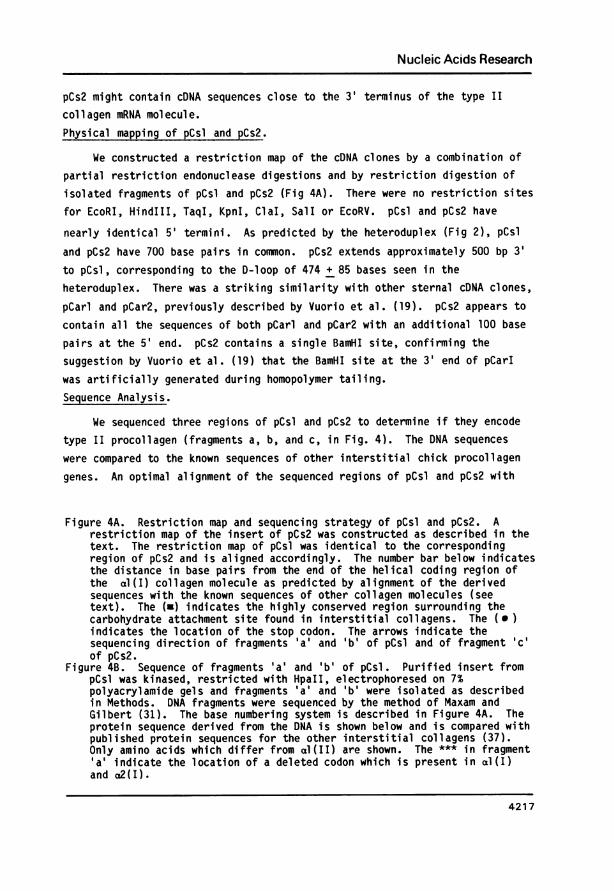
Figure 4A. Restriction map and sequencing strategy of pCsl and pCs2. Arestriction map of the insert of pCs2 was constructed as described in thetext. The restriction map of pCsl was identical to the correspondingregion of pCs2 and is aligned accordingly. The number bar below indicatesthe distance in base pairs from the end of the helical coding region ofthe al(I) collagen molecule as predicted by alignment of the derivedsequences with the known sequences of other collagen molecules (seetext). The (a) indicates the highly conserved region surrounding thecarbohydrate attachment site found in interstitial collagens. The ( )indicates the location of the stop codon. The arrows indicate thesequencing direction of fragments 'a' and 'b' of pCsl and of fragment 'c'of pCs2.
Figure 4B. Sequence of fragments 'a' and 'b' of pCsl. Purified insert frompCsl was kinased, restricted with HpaII, electrophoresed on 7%polyacrylamide gels and fragments 'a' and 'b' were isolated as describedin Methods. DNA fragments were sequenced by the method of Maxam andGilbert (31). The base numbering system is described in Figure 4A. Theprotein sequence derived from the DNA is shown below and is compared withpublished protein sequences for the other interstitial collagens (37).Only amino acids which differ from al(II) are shown. The *** in fragment'a' indicate the location of a deleted codon which is present in al(I)and a2(I).
4217

Nucleic Acids Research
al(I), a2(I), and al(III) procollagens indicates that pCsl and pCs2 contain
sequences encoding the COOH-terminal propeptide of a procollagen chain. Thetranslated pCsl sequence is 72%, 63% and 70% homologous with the amino acidsequences of the COOH terminal propeptide of al(I), a2(I) and al(III),respectively, and the respective nucleotide sequence homologies are 67%, 61%,and 64%.
The sense strand DNA sequence of pCsl and pCs2 fragments and theircorresponding translation products are presented in Fig. 4B. In the basenumbering system used in Fig. 4, COOH terminal collagen sequences are numberedfrom the end of the helical-coding region (coordinate 0) and correspond to thenumbering system used by Fuller and Boedtker (36) for the al(I) procollagengene. We aligned the most 5' sequences of pCsl (fragment a, Fig 4) with basenumber 193 of the al(I) COOH terminal propeptide coding region. Fragment "a"contains sequences from bases 193 to 555. It should be noted that bases 376-378 (coding for proline) of the al(I) COOH terminal coding region are notpresent in the corresponding region of pCsl. These three bases have also beenshown to be missing in aI(III) (6,37).
By analogy with the known exon-intron splice sites of the a2(I) andal(III) collagen genes, in which exons are numbered progressively from the 3'end of the gene, fragment "a" of pCsl contains part of exon 4 (ending at basenumber 238 within the gly codon), all of exon 3 (from bases 239-429), and the5' end of exon 2.
Fragment "a" contains 5 regions (corresponding to bases 193-213, 235-270,277-321, 385-414, 508-555) of the COOH propeptide which have been previouslyidentified as showing homologies in their amino acid sequence between al(I),a2(I) and al(III) collagens (37). The amino acid sequence of these segmentsis also highly conserved in pCsl. The fifth segment (bases 508-555) has alsobeen identified as being highly conserved not only in primary proteinstructure but also in nucleotide sequence (37). In this segment, 42nucleotide positions contain the identical base sequence in pCsl and the other3 collagen genes described above. Furthermore 3 of the 6 nucleotide positionsthat differ among these 4 genes occur at base positions 509, 511 and 513.This suggests an even tighter conservation of nucleotide sequence betweenbases 514-555, coding for a 14 amino acid long region of the COOH-propeptidethat surrounds the carbohydrate attachment site (bases 517-525).
By analogy with the exon-intron structure of the al(III) gene (6),fragment "b" contains part of exon 1 of an interstitial collagen gene. Anochre termination codon (TM) was identified in fragment "b" at bases 817-
4218

Nucleic Acids Research
819. The 3' flanking sequence appears to be relatively long since the 3' end
of fragment "c" is approximately 600 bp from the stop codon. Although R-
looping data (see Fig. 3) suggests that pCs2 contains cDNA sequences close to
the 3' terminus of the type II collagen mRNA molecule, it is unlikely that the
3' end of pCs2 actually represents the end of the noncoding portion of the
mRNA. The canonical pAATAAA sequence that is usually present 10-20
nucleotides 5' to the poly(A) addition site of eukaryotic mRNA is not
discernible in fragment "c" (data not shown).Isolation of a Genomic Clone.
Sequence analysis of pCsl and pCs2 demonstrated that the recombinant
plasmids encoded an interstitial collagen molecule different from al(I),a2(I), and al(III). Furthermore, the cDNA clones hybridized specifically to
RNA from sterna, suggesting that they encoded type II collagen. Amino acid
sequence of the type II collagen COOH propeptide is not available. Therefore,we screened a Charon 4A phage library of chick genomic fragments to obtain
cloned DNA likely to encode the helical region of type II collagen, for whichthe sequence was available (Dr. W. Butler, U. Alabama). 5 X 10⁵ plaques were
screened using the more 5' PstI-AluI fragment of the pCs2 insert (see Fig 4)as a cross hybridization probe. One clone, XCs7, was isolated andcharacterized.
Restriction endonuclease mapping and Southern blot hybridization withdifferent subfragments of pCsl and pCs2 defined the region of XCs7corresponding to the 3' end of the gene (see Fig 5).
We used two types of heteroduplex analysis to study the exon-intron
structure of the 3' end of the gene cloned in XCs7. Heteroduplexes formed
between the DNA of recombinant phage XCs7 and the DNA of Charon 4A were
hybridized to sucrose gradient fractions of chick sternal RNA enriched for
type II collagen mRNA. A representative structure is depicted in Fig. 6. At
least 10 exons are found interspersed within a 4.5 kb DNA segment.
The size of exons and introns in XCs7 was measured by electronmicroscopicscanning and is shown in Table I. An interesting feature of the gene is that
exons within the carboxy propeptide encoding region are larger than those in
the helical region. A similar observation has been noted for the chick a2(I)and al(III) collagen genes (37). Another interesting feature of the al(II)gene is that compared to the c2(I) and al(III) collagen genes, the total
length of intron (1-9) is substantially (50%) smaller. Because the resolutionof electron microscope analysis does not allow detection of introns smaller
than 100 bp, it is possible that the region of the al(II) gene contained
4219

Nucleic Acids Research
A.
LONG ARM k\ GENOMIC DNA SHORT ARM
H HB E B B jE E B B B0 5/25I,, a I a I I I 40
0 5 j~~20 25 "s 30 35 40
"
B. E S SS TT T ST S B S B
1 - 120 21 22 23 24 25
C.1ser al a phe ala gly leu gly gln thr gl uTCT GCT TTT GCT GGA CTG GGT CAG ACG GAG
19lys gly pro asp pro ile arg tyr metAAG GGC CCC GAC CCC ATC CGC TAC ATG
Figure 5A. Restriction map of xCs7. The restriction map of xCs7 was derivedfrom a series of single and double digestions of the recombinant clone byEcoRI (E), BamHI (B), and 1ndIII (H). A southern blot of the restrictiondigests was hybridized to P-labeled PstI-AluI fragments of pCs2 insertto determine the orientation of genomic DNA within xCs7. The numbersbelow the map refer to kilobases from the end of the long arm of thephage. The heavy bar between 19.5 and 25.4 kb delineates the regionof xCs7 that hybridized to pCs2 DNA.
Figure 5B. Restriction map of the 5.9 kb genomic fragment. A restriction mapwas derived from a series of single and double digestions by SmaI (S),PvuII (P), and TaqI (T).
Figure 5C. Nucleotide sequence of CB 14 of al(II) procollagen. The TaqI-SmaIfragment delineated by the bar in Figure 5B was kinased and restrictedwith HinfI which cleaves close to the 3' SmaI site. A portion of thesequence of the TaqI-HinfI fragment corresponding to cyanogen bromidepeptide 14 of al(II) collagen (commencing several codons after the end ofthe helical coding region and ending two codons before thecarboxypeptidase cleavage site) is shown. Derived amino acid residues arenumbered starting with the first codon of CB 14.
within xCs7 may contain more than 10 introns and exons. Confirmation of theprecise intron-exon structure of xCs7 must await complete DNA sequenceanalysis.
If the chick genomic sequences in xCs7 are complementary to the cDNAclones pCsl and pCs2, then heteroduplexes between xCs7 and pCsl should
4220
iI
IIIIIIIII

Nucleic Acids Research
.;~~~74t+ R - f.re v W
IL4_ee4r~~~~~~~~~~re
< -i 2 > O ~ ~~~~~FmCh4
/ r r 3' ~~~~~FLANKING/zJ ~~~~~~SEQUENCES
Figure 6. Electronmicrograph of heteroduplex between xCs7 and sterna RNA.Heteroduplexes between recombinant phage xCs7 and Charon 4A were formed asdescribed in Methods, and poly(A) RNA from chick embryo sterna washybridized to the duplexes. The bar beneath the micrograph designates 1kilobase. The numbers in the schematic diagram below correspond to exonsbeginning at the 3' end of the gene.
specifically indicate the exon-intron structure of the 3' end of thepresumptive type II collagen gene. A representative molecule of such a duplexis shown in Fig. 7. DNA sequence analysis of pCsl (Fig. 4B) indicated that it
4221

Nucleic Acids Research
Table I: Sizes of exons and introns in ACs7
Exona Exon length Intron length(bp) (bp)
l 764 + 59b 97 + 41
2 316 + 61 634 + 95
3 186 + 45 299 + 73
4 299 + 81 133 + 44
5 104 + 50 83+ 31
6 121 + 44 109 + 39
7 131 + 53 283 + 47
8 125 + 52 124 + 58
9 97+ 40 175 + 119
10 108+55
a Exons are numbered from the 3' end of the gene.b Standard deviationThe 3' flanking region downstream from exon 1 which extends to the short
arm of XCharon 4A is about 9kB. The 5' end of the DNA insert of XCs7 is anintervening sequence containing part or all of intron 10 of the gene.
coded for sequences homologous to exons 1-4 of the other interstitial collagengenes. The three single stranded DNA loops visualized in theelectronmicrograph in Fig. 7 most likely represent introns which delineateexons 1, 2, 3, and 4 of the gene. Moreover the sizes of the DNA loopscorrespond well to those of the introns separating exons 1-4 in the R loop inFig. 6. The size of exons 2 and 3 in both forms of heteroduplex analysis alsocorrespond well. Exons 1 and 4 in the cDNA genomic heteroduplex, however, areshorter than those analyzed for the genomic R loop seen in Fig. 6. This isconsistent with DNA sequence analysis of pCsl, which showed that its 3' endincludes some, but not all, of the untranslated region of the 3' end of itscorresponding mRNA. In addition, the 5' end of pCsl encodes only a smallportion of the 3' end of exon 4.Sequence of the COOH telopeptide of the type II collagen gene.
The heteroduplex analysis (Fig. 7) suggested that XCs7 encoded genomic
4222
Nucleic Acids Research
XCs7
pOsi
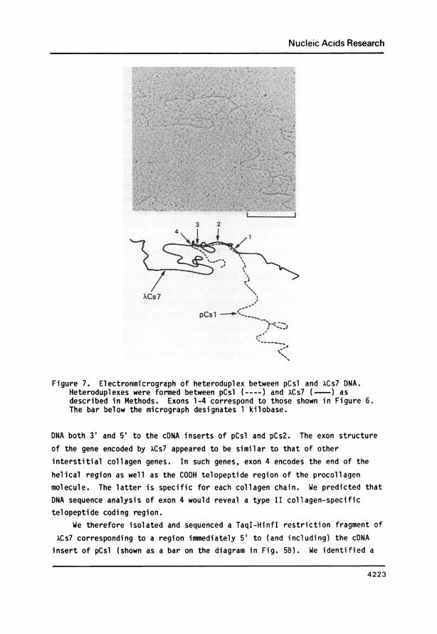
Figure 7. Electronmicrograph of heteroduplex between pCsl and xCs7 DNA.Heteroduplexes were formed between pCsl (---- ) and XCs7 (--) asdescribed in Methods. Exons 1-4 correspond to those shown in Figure 6.The bar below the micrograph designates 1 kilobase.
DNA both 3' and 5' to the cONA inserts of pCsl and pCs2. The exon structureof the gene encoded by xCs7 appeared to be similar to that of otherinterstitial collagen genes. In such genes, exon 4 encodes the end of the
helical region as well as the COOH telopeptide region of the procollagenmolecule. The latter is specific for each collagen chain. We predicted thatDNA sequence analysis of exon 4 would reveal a type II collagen-specifictelopeptide coding region.
We therefore isolated and sequenced a TaqI-HinfI restriction fragment of
xCs7 corresponding to a region immediately 5' to (and including) the cDNAinsert of pCsl (shown as a bar on the diagram in Fig. SB). We identified a
4223
Nucleic Acids Research
region approximately 175 base pairs downstream from the TaqI site whichcorresponded exactly with the 5' sequence of fragment 'a' of pCsl (see Fig.4B). This confirmed the heteroduplex analysis (Fig. 7) which indicated thatXCs7 contains sequences including, and 5' to, pCsl. In addition, the twelvebases upstream from fragment 'a' of pCsl (corresponding in our cDNA basenumbering system to bases 181-192) coded for 4 amino acid residues which were
100% homologous with al(I), a2(I), and al(III) procollagen protein sequences.The residues encoded by bases 181-192 are lys-asn-pro-ala corresponding to
resudues 76-79 of exon 4 of the al(III) collagen gene (37). This region was
previously identified as an area of amino acid homology in collagen genes byYamada et al (37). As an additional aid in aligning the sequences of theTaqI-HinfI fragment with other collagen molecules, we identified 8 other aminoacid residues with 100% homology to other interstitial procollagen chains
(37). These were residues 42, 53, 59, 64, 65, 66, 71 and 73 of exon 4 ofthe al(III) collagen gene (37) encoding asp, glu, lys, gln, ile, glu, pro, and
gly respectively.If XCs7 is a genomic type II collagen clone, then the 21 codons
immediately upstream from the asp residue (first amino acid of the COOHpropeptide) should encode cyanogen bromide peptide fragments 14 and 15 (CB14and CB15) of the type II collagen chain (38). This is indeed the case. CB15is a dipeptide of arg and ala located between CB14 and the carboxypeptidasecleavage site (38) . The nucleotide-derived sequence of the two residuesimmediately prior to the first asp codon of the COOH propeptide of XCs7 ispAGGGCA, coding for arg-ala.
The sense strand DNA sequence and translated amino acid sequence of CB14is presented in Fig. 5C. The deduced amino acid composition of CB14 shown inFig 5C is 100% in agreement with the known amino acid composition of CB14(38). Residue 18 is tyr, which is also found 4 residues upstream from thecarboxypeptidase cleavage site in the other three interstitial collagen chains(37). The wobble position of this codon, however, is a T in the al(III)gene. No other amino acid residue in CB14 is completely homologous with allthe other collagen chains. Overall amino acid homology between the type IIcollagen sequence and al(I), a2(I), and al(III) collagens was 32%, 11%, and42%, respectively. This is consistent with the expected uniqueness of thetelopeptide. This region of the al(II) chain contains one more amino acidresidue than do the al(I) and al(III) collagens.
We also compared the translated amino acid sequence shown in Fig. 5C witha protein sequence provided to us by Dr. W. Butler (U. Alabama) which aligned
4224

Nucleic Acids Research
with the first 12 residues of CB14. We found two discrepancies between the
protein sequence and the nucleotide-derived sequence. Whereas the firsttranslated residue in Fig. 5C is ser, the protein sequence predicted glu inthis position. Possibly, this reflects an allelic difference. We areconvinced that our DNA sequence is correct since our gels are unambiguous andthe amino acid composition of CB14 (38) predicts 1 ser and cannot account fora glu in residue 1 in addition to the glu and gln at residues 8 and 10.
Furthermore, Miller (38) found that at a noticeable frequency, CB14 was notcleaved from the CB7 fragment (more NH2 terminal to it). The presence of a
ser at residue 1 is consistent with inefficient cleavage of met-ser peptidebonds which has been reported by others (39). There was also a discrepancy at
residue 9, where a pro was predicted by the available protein sequence. Theamino acid composition of CB14 (38) predicted 1 thr and 2 pro residues,consistent with our DNA-derived sequence. Sandell et al (20) who recentlyidentified a type II collagen genomic clone, also found a thr in this
posi tion.
SUMMARYIn conclusion, we constructed and isolated two recombinant cDNA clones
and a genomic clone containing the 3' end of the al(II) collagen gene. It hasbeen widely assumed that, because of the secondary structure of the collagenmessage, long cDNA transcripts could best be obtained by using highly enrichedsources of collagen RNA. We have demonstrated that at least in the case ofthe type II collagen gene, it is possible to obtain reasonable sizetranscripts and to clone the collagen gene from a highly heterogeneous message
source, the whole chick embryo. However, the success of our approach was
dependent upon counterscreening with a variety of other collagen probes in
which relaxed and stringent hybridization conditions were used in tandem.Conclusive identification of the clones was complicated by the limited
availability of amino acid sequence data. We therefore depended uponsequencing a specific telopeptide region of a genomic DNA clone. Colinearityof the genomic clone with the cDNA clones was demonstrated by a combinationof heteroduplex mapping, Southern hybridization, and DNA sequencing.
The cDNA clones pCsl and pCs2 appear to overlap with the cartilage-specific cDNA clones described by Vuorio et al (19) and by Lukens et al(16). In the latter case, the clones were identified as encoding type II
collagen by the method of hybrid-selected translation. The clones of Vuorioet al (18) were identified by the method used in the present report, i.e. DNA
4225

Nucleic Acids Research
sequencing of the telopeptide region of a genomic clone. It should be noted
that pCsl and pCs2 and the clones of Vuorio et al (19) and Lukens et al (17)are quite different in restriction fragment analysis and DNA sequence from a
presumptive al(II) collagen cDNA clone described by Duchene et al (40). In
our hands, Northern hybridization experiments show that the latter clone cross
hybridizes with type I collagen mRNA even under the most stringent conditions
(data not shown). We conclude that the clone of Duchene et al (40) is in fact
not a type II collagen DNA probe.
In the Northern hybridization experiments described in Figure 1, we used
stringent hybridization conditions to achieve specific hybridization of our
recombinant cDNA probes with sternal RNA. We confirmed our previous results
and the results of others (15-17) that show that type II collagen mRNA levels
are markedly decreased when the cartilage phenotype is altered by BUdR
treatment of chondrocytes. No cross hybridization of pCsl and pCs2 was
demonstrated with the considerable amounts of type I collagen mRNA present in
dedifferentiated (BUdR-treated) chondrocytes. We are currently in the process
of using the cDNA and genomic clones to determine the molecular mechanisms
involved in chondrocyte dedifferentiation.
ACKNOWLEDGEMENTS
We would like to thank Dr. Kwang-Sam Koh, Kenneth Galen and Frances
Cannon for their technical assistance, William Upholt and Linda Sandell for
preliminary sequence information of their type II chick cDNA clones and Bill
Butler for protein sequence data. We are also grateful to George Martin and
Yoshihiko Yamada for useful discussions and careful review of this
manuscript. We also thank Helga Boedtker for permission to use cDNA probes
devel oped i n her 1 aboratory.
*Current address: Laboratory of Pathology, National Cancer Institute, National Institutes ofHealth, Bethesda, MD 20205, USA
REFERENCES1. Prockop, D.J and Champe, F.C., Eds (1980) Gene Families of Collagen and
Other Proteins, Elservier/North Holland, New York.2. Bornstein, P. and Sage, H. (1980) Annu. Rev. Biochem. 49, 959-1003.3. Galloway, D. (1982) in Collagen in Health and Disease, Weiss, J.B. and
Jayson, M.I.V. Eds, pp. 528-558, Churchill-Livingstone, Edinburgh.4. Ohkubo, H., Vogeli, G., Mudryj, M., Avvedimento, V.E., Sullivan, M.,
Pastan, I. and de Crombrugghe, B. (1980) Proc Natl. Acad. Sci. USA 78,7057-7063.
4226

Nucleic Acids Research
5. Wozney, J., Hanahan, D., Tate, V., Boedtker, H. and Doty, P. (1981) Nature294, 129-135.
6. Yamada, Y., Mudryj, M., Sullivan, M. and de Crombrugghe, B. (1983) J.Biol. Chem. 258, 2758-2761.
7. Monsen, J. and McCarthy, B.J. (1981) DNA 1, 59-69.8. Meyers, J.C., Dickson, L.A., de Wet, W.J., Bernard, M.P., Chu, M-L, Di
Liberto, M., Pepe, G., Sangiorgi, F.O. and Ramirez, F. (1983) J. Biol.Chem. 258, 10128-10135.
9. Vogeli, G., Ohkubo, H., Avvedimento, V.E., Sullivan, M., Yamada, Y.,Mudryj, M., Pastan, I., and de Crombrugghe, B. (1981) Cold Spring HarborSymposia on Quantitative Biology. Volume XLV, pp 777-783.
10. Yamada, Y., Avvedimento, V., Mudryj, M.,Ohkubo, H., Vogeli, G., Irani, M.,Pastan, I., and de Crombrugghe, B. (1980) Cell 22, 887-892.
11. Aho, S., Tate, V., and Boedtker, H. (1983) Nucl. Acids. Res. 11, 5443-5450.
12. Schiltz, J.R., Mayne, R.M. and Holtzer, H. (1973) Differentiation 1, 97-108.
13. Abbott, J. and Holtzer, H. (1968) Proc. Natl. Acad. Sci. USA 59, 1144-1151.
14. Mayne, R., Vail, M.S., and Miller, E.J. (1975) Proc. Natl. Acad. Sci. USA72, 4511-4515.
15. Kaul, R., Hewitt, A.T., Varner, H., Somerman, M., Martin, G.R. and Sobel,M.E. (1981) Fed. Proc. 40, 1626.
16. Pawlowski, P.J., Brierley, G.T. and Lukens, L.N. (1981) J. Biol. Chem.256, 7695-7698.
17. Lukens, L.N., Frischauf, A.M., Pawlowski, P.J., Brierley, G.T. andLehrach, H. (1983) Nucl. Acids Res. 11, 6021-6039.
18. Adams, S.L., Boettiger, D., Focht, R.J., Holtzer, H. and Pacifici, M.(1982) Cell 30, 373-384.
19. Vuorio, E., Sandell, L., Kravis, D., Sheffield, V.C., Vuorio, T., Dorfman,A., Upholt, W.B. (1982) Nucl. Acids Res. 10, 1175-1192.
20. Sandell, L.J., Yamada, Y., Dorfman, A. and Upholt, W.B. (1983) J. Biol.Chem. 258, 11617-11621.
21. Gottesman, M.M. and Sobel, M.E. (1980) Cell 19, 449-455.22. Aviv, H. and Leder, P. (1972) Proc. Natl. Acad. Sci. USA 69, 1408-1412.23. Rosen, J.M., Woo, S.L.C. and Comstock, J.P. (1975) J. Biochem. 14, 2895-
2903.24. Sobel, M.E., Dion, L.D., Vuust, J., Colburn, N.H. (1983) Mol. and Cell
Biol. 3, 1527-1532.25. Sobel, M.E., Yamamoto, T., Adams, S.L., Dilauro, R., Avvedimento, V.E., de
Crombugghe, B. and Pastan, I. (1978) Proc. Natl. Acad. Sci. USA 75, 5846-5850.
26. Nelson, T. and Brutlag, D. (1979) Methods in Enzymology 68, 41-49.27. Loening, U.E. (1969) Biochem. J. 102, 251-257.28. Lehrach, H., Frishauf, A.M., Hanahan, D., Wozney, J., Fuller, F., and
Boedtker, H. (1979) Biochemistry 18, 3146-3152.29. Lehrach, H., Frischauf, A.M., Hanahan, D., Wozney, J., Fuller, F.,
Crkvenjakov, R., Boedtker, H., and Doty, P. (1978) Proc. Natl. Acad. Sci.75, 5417-5421.
30. Sobel, M.E., Yamamoto, T., de Crombrugghe, B. and Pastan, I. (1981)Biochemistry 20, 2678-2684.
31. Gilbert, W. and Maxam, A. (1973) Proc. Natl. Acad. Sci. USA 70, 3581-3584.32. Tilghman, S.M., Curtis, P.J., Tiemeier, D.C., Leder, P., and Weissman, C.
(1978) Proc. Nat. Acad. Sci. USA 75 1309-1313.33. Dodgson, J.B., Stroniner, J., and Engel, J.D. (1979) Cell 17, 879-887.34. Maniatis, T., Hardison, R.C., Lacy, E., Lauer, J., O'Connell, C. and Quon,
E. (1978) Cell 15, 687-701.
4227

Nucleic Acids Research
35. Merlino, G.T., McKeon, C., de Crombrugghe, B., and Pastan, I. (1983) J.Biol. Chem. 258, 10041-10048.
36. Fuller, F. and Boedtker, H. (1981) Biochemistry 20, 996-1006.37. Yamada, Y., Kuhn, K. and de Crombrugghe, B. (1983) Nucl. Acids Res. 11,
2733-2744.38. Miller, E.J. (1972) Biochemistry 11, 4903-4909.39. Schroeder, W.A., Shelton, J.B. and Shelton, J.R. (1969) Arch. Biochem.
Biophys. 130, 551.40. Duchene, M., Sobel, M.E., and Muller, P.K. (1982) Exp. Cell Res. 142, 317-
324.41. Southern, E. (1979) Methods in Enzymology 68, 152-176.
4228
Copyright © 2022 FDOKUMEN