Thoracic rhizotomy versus thoracic paravertebral nerve ...
7
ORIGINAL ARTICLE Open Access Thoracic rhizotomy versus thoracic paravertebral nerve radiofrequency in thoracic cancer pain Mohamed Adly Elramely 1 and Wael Abdelmoneim Mohammad Abdelwahab 2* Abstract Background: Lung cancer is the most frequent occurring malignancy and mostly presenting with pain. Interventional procedures reduce medications and their side effects. Rhizotomy is another modality for control of chest wall pain due to tumor invasion or somatic and neural structures. We compared the degree of pain relief in thoracic rhizotomy versus thoracic paravertebral nerve radiofrequency. Results: Six hours postoperatively, radiofrequency (RF) ablation of dorsal root ganglia resulted in reduction of required dose of narcotics in 12 patients (80%) compared to 6 patients (40%) treated with RF ablation of TPN (P value = 0.025). After 4 weeks reduction in required narcotic dose was recorded in 80.0% of DRG patients compared to 33.3% of TPN patients (P value = 0.010). Patients’ activity improved in 11 patients (73.3%) in DRG group compared to 5 patients (33.3%) in TPN group (P value = 0.028). No significant difference in frequency of sensory loss (P value = 1.000), burning sensation (P value = 0.128) and allodynia (P value = 0.139). Conclusion: RF ablation of DRG is superior to RF ablation of TPN in relieving thoracic pain with more improvement of patients’ activity and similar adverse outcomes. Background Lung cancer is the most frequent occurring malignancy. There is 1.61 million new case diagnosed yearly (Siegel et al. 2014). Pain is the most common presentation in cancer patients (Caraceni and Portenoy 1999). Pain may complicate with other symptoms as depression, and fa- tigue (Laird et al. 2011a) and may affect daily activity (Laird et al. 2011b). Early intervention, including symp- tomatic management, improves quality of life (Temel et al. 2010). Cancer may result in several types of pain with dif- ferent manifestations (Portenoy and Lesage 1999). Neuropathic pain is burning or shooting in character. It results from peripheral or central nervous system injury and may complicate with sensory loss and variable response to opioids (Caraceni and Portenoy 1999; Stute et al. 2003). Pain in lung cancer may be due to pleural invasion, chest wall invasion, or costopleural syndrome. Cancer ir- ritates peripheral nerve endings of the C and A-delta primary afferent fibers. Stimulation of these fibers results in the recruitment of quiescent nociceptors, and the ac- tivation of NMDA-receptor-channel complex leading to dorsal horn sensitization. This process results in persist- ence of pain (Portenoy and Lesage 1999). Typically, interventional procedures of cancer pain have an additive effect for other modalities for treatment aiming at pain control. Furthermore, interventional pro- cedures reduce medications and their side effects. Inter- ventional procedures in the form of the interruption of nerve conduction aiming at diminishing pain from the target area have been applied (Wong et al. 2007). Inter- costal neurolysis is a quiet easy technique that lasts for 3 to 8 weeks. However, studies reported occurrence of © The Author(s). 2021 Open Access This article is licensed under a Creative Commons Attribution 4.0 International License, which permits use, sharing, adaptation, distribution and reproduction in any medium or format, as long as you give appropriate credit to the original author(s) and the source, provide a link to the Creative Commons licence, and indicate if changes were made. The images or other third party material in this article are included in the article's Creative Commons licence, unless indicated otherwise in a credit line to the material. If material is not included in the article's Creative Commons licence and your intended use is not permitted by statutory regulation or exceeds the permitted use, you will need to obtain permission directly from the copyright holder. To view a copy of this licence, visit http://creativecommons.org/licenses/by/4.0/. * Correspondence: [email protected] 2 Faculty of Medicine, Ain shams University, 48 Mohammad Helmy street, Hadaek Elkobba, P.O.11755, Cairo, Egypt Full list of author information is available at the end of the article Ain-Shams Journal of Anesthesiology Elramely and Abdelwahab Ain-Shams Journal of Anesthesiology (2021) 13:11 https://doi.org/10.1186/s42077-020-00125-3
-
Upload
khangminh22 -
Category
Documents
-
view
4 -
download
0
Transcript of Thoracic rhizotomy versus thoracic paravertebral nerve ...

ORIGINAL ARTICLE Open Access
Thoracic rhizotomy versus thoracicparavertebral nerve radiofrequency inthoracic cancer painMohamed Adly Elramely1 and Wael Abdelmoneim Mohammad Abdelwahab2*
Abstract
Background: Lung cancer is the most frequent occurring malignancy and mostly presenting with pain.Interventional procedures reduce medications and their side effects. Rhizotomy is another modality for control ofchest wall pain due to tumor invasion or somatic and neural structures. We compared the degree of pain relief inthoracic rhizotomy versus thoracic paravertebral nerve radiofrequency.
Results: Six hours postoperatively, radiofrequency (RF) ablation of dorsal root ganglia resulted in reduction ofrequired dose of narcotics in 12 patients (80%) compared to 6 patients (40%) treated with RF ablation of TPN (Pvalue = 0.025). After 4 weeks reduction in required narcotic dose was recorded in 80.0% of DRG patients comparedto 33.3% of TPN patients (P value = 0.010). Patients’ activity improved in 11 patients (73.3%) in DRG groupcompared to 5 patients (33.3%) in TPN group (P value = 0.028). No significant difference in frequency of sensoryloss (P value = 1.000), burning sensation (P value = 0.128) and allodynia (P value = 0.139).
Conclusion: RF ablation of DRG is superior to RF ablation of TPN in relieving thoracic pain with more improvementof patients’ activity and similar adverse outcomes.
BackgroundLung cancer is the most frequent occurring malignancy.There is 1.61 million new case diagnosed yearly (Siegelet al. 2014). Pain is the most common presentation incancer patients (Caraceni and Portenoy 1999). Pain maycomplicate with other symptoms as depression, and fa-tigue (Laird et al. 2011a) and may affect daily activity(Laird et al. 2011b). Early intervention, including symp-tomatic management, improves quality of life (Temelet al. 2010).Cancer may result in several types of pain with dif-
ferent manifestations (Portenoy and Lesage 1999).Neuropathic pain is burning or shooting in character.It results from peripheral or central nervous systeminjury and may complicate with sensory loss and
variable response to opioids (Caraceni and Portenoy1999; Stute et al. 2003).Pain in lung cancer may be due to pleural invasion,
chest wall invasion, or costopleural syndrome. Cancer ir-ritates peripheral nerve endings of the C and A-deltaprimary afferent fibers. Stimulation of these fibers resultsin the recruitment of quiescent nociceptors, and the ac-tivation of NMDA-receptor-channel complex leading todorsal horn sensitization. This process results in persist-ence of pain (Portenoy and Lesage 1999).Typically, interventional procedures of cancer pain
have an additive effect for other modalities for treatmentaiming at pain control. Furthermore, interventional pro-cedures reduce medications and their side effects. Inter-ventional procedures in the form of the interruption ofnerve conduction aiming at diminishing pain from thetarget area have been applied (Wong et al. 2007). Inter-costal neurolysis is a quiet easy technique that lasts for 3to 8 weeks. However, studies reported occurrence of
© The Author(s). 2021 Open Access This article is licensed under a Creative Commons Attribution 4.0 International License,which permits use, sharing, adaptation, distribution and reproduction in any medium or format, as long as you giveappropriate credit to the original author(s) and the source, provide a link to the Creative Commons licence, and indicate ifchanges were made. The images or other third party material in this article are included in the article's Creative Commonslicence, unless indicated otherwise in a credit line to the material. If material is not included in the article's Creative Commonslicence and your intended use is not permitted by statutory regulation or exceeds the permitted use, you will need to obtainpermission directly from the copyright holder. To view a copy of this licence, visit http://creativecommons.org/licenses/by/4.0/.
* Correspondence: [email protected] of Medicine, Ain shams University, 48 Mohammad Helmy street,Hadaek Elkobba, P.O.11755, Cairo, EgyptFull list of author information is available at the end of the article
Ain-Shams Journalof Anesthesiology
Elramely and Abdelwahab Ain-Shams Journal of Anesthesiology (2021) 13:11 https://doi.org/10.1186/s42077-020-00125-3
neuritis and advised that neurolytic agents should belimited to those with a short life expectancy (Swarmet al. 2005).Dorsal root ganglion (DRG) ablation or thoracic rhi-
zotomy is another modality for chest wall pain due totumor invasion or somatic and neural structures. It in-volves segmental or multi-segmental destruction of thedorsal sensory roots. Rhizotomy is either surgically doneor achieved by chemical neurolysis or radiofrequency ab-lation. It is an effective method of pain control especiallywith refractory localized pain syndromes (Hogan et al.1991).Diagnostic block with local anesthetic is initially ap-
plied prior to a planned neurolytic block to predict thelikely outcome (Silvestri et al. 2002). Paravertebral blockscan be used in case of chest pain (Schneider et al. 1993).Radiofrequency ablation is attained by the passage of
low-energy, high-frequency alternating current (100,000–500,000 Hz) that causes oscillations of tissue ions.This oscillation results in heating of charged macromol-ecules, especially proteins (Organ 1976-1977). RF heat-ing to 45 °C causes many cells to die rapidly.Neuroablation targets either the dorsal root ganglion(DRG) or a peripheral nerve. Above 55 °C, there is an in-discriminate destruction of both small- and large-diameter myelinated fibers. Histologically, there are focalnecrosis, hemorrhages, extensive edema, and features ofWallerian degeneration (Smith et al. 1981).The mode of action of RF was initially attributed to
the thermocoagulation of nerve fibers. However, contra-dictory observations as transient sensory loss in the asso-ciated dermatome, whereas the pain relief may last forlonger periods suggest that temperature is not the onlymechanism responsible for the decrease in pain trans-mission (Racz and Ruiz-Lopez 2006).The use of RF for the management of neuropathic
pain is controversial for the fear of development of neur-itis and deafferentation pain. Consequently, RF ablationcould be only applied in somatic pain (De Louw et al.2001).The aim of this study is to compare the reduction in
the narcotic requirement after thoracic rhizotomy versusthoracic paravertebral nerve radiofrequency in thoraciccancer pain management. The secondary outcome wasthe degree of improvement in daily activity.
MethodsThis study was approved by the ethical committee. Allpatients were asked for a signed and informed consentafter declaration of the technique and its possible bene-fits, risks, and side effects while for patients under 21years old, an informed consent from their guardians wassigned. We enrolled 30 patients with somatic chest paindue to underlying intra-thoracic malignancy. The
inclusion criteria were age 18 years to 60 years, no inter-spinal extension of the tumor that was confirmed byMRI prior to the intervention, duration of pain wasmore than 3 months, and visual analogue scale (VAS)was more than 5 on 0–10 scale despite medical treat-ment or intolerability to medical treatment. Preoperativeexclusion criteria were patient refusal and lack of under-standing by the patient of the purpose of the study, co-agulopathy, i.e., INR > 1.4 or platelet count < 80000 orlocal infection. Intraoperative exclusion criteria werefailure either to localize or to get the anterior epiduralspace or the paravertebral space by image guidance anddye confirmation.The preoperative evaluation included physical examin-
ation. Patients underwent complete blood count and co-agulation profile. The primary outcome variable was theincidence of more than 50% reduction in the narcotic re-quirement after intervention at two assessment intervals6 h after the procedure and 4 weeks later. The secondaryoutcome was improved daily activity as described by thepatient to be improved or not after 4 weeks.A standardized protocol was used. It included standard
monitoring (ECG, non-invasive arterial pressure, pulseoximetry). All patients fulfilling the inclusion criteria gota diagnostic block with 5 ml, 0.25% bupivacaine thoracicparavertebral nerve block for each affected level. The af-fected dermatome determined the level of the nerveblock (NB) (Uchida 2009).All patients were put in prone position, and in both
groups, we got anteroposterior view to determine theintended levels for injection and then we got to an ob-lique view about 10 to 15° to the contralateral side.Then, in the dorsal root ganglia block group, we
intended the foramen just below the pedicle (Fig. 1a),and then, the needle was introduced in a gun barrelfashion (Fig. 1b) after that we confirmed the position ofthe needle in a lateral view (Fig. 1c) after that weinjected contrast to confirmed delineation of the con-trast at the anterior epidural space (Fig. 1d, e).While in the paravertebral nerve block group after
turning to the oblique view about 10 to 15° to thecontralateral side, we entered just lateral to the vertebralbody, the needle hit the lateral side of vertebral body, itrotated laterally, and then, the position was confirmed inlateral position by injecting contrast and saw its spreadwithin the paravertebral space (Fig. 2a).Following the diagnostic testing, patients were ran-
domized by a computer-generated numbers’ techniqueinto two groups. Group 1 got dorsal root ganglia abla-tion at 67 °C for 60 s. The generator (PMG-115- TD,V2.0A, Baylis Medical) with a 10-cm electrode and a 5-mm active tip had been used after sensory stimulationbetween 0.2 and 0.7 V while group 2 got thoracic para-vertebral nerve (TPN) ablation with the same protocol.
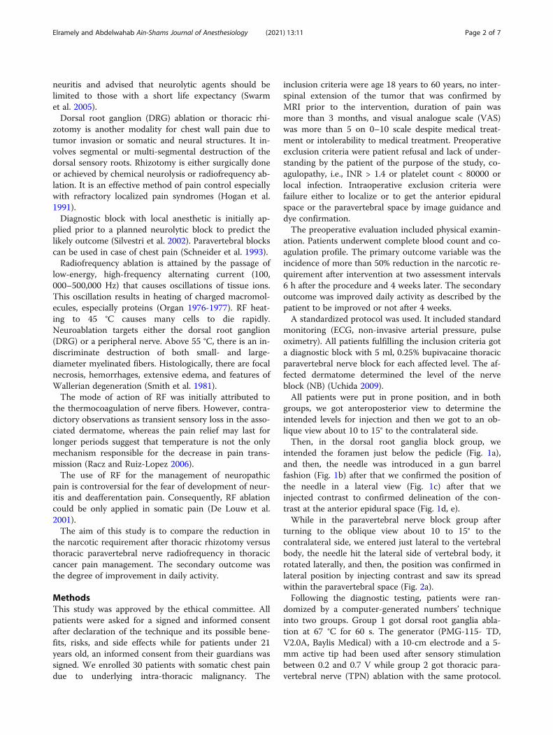
Elramely and Abdelwahab Ain-Shams Journal of Anesthesiology (2021) 13:11 Page 2 of 7

Before ablation, 1.5 ml of 2% mepivacaine and 2 mg ofbetamethasone were administered (Cohen et al. 2004).
Measured parametersThe two groups were compared regarding the reductionof narcotics, activity improvement, sensory loss, burningsensation, and back pain.
Sample size calculationA previous study (Cohen et al. 2006) reported more than50% pain relief in 61.5% of patients treated by dorsalroot ganglia pulsed radiofrequency compared to 21.4%of those treated with radiofrequency of the intercostalnerves. Based on these results, a sample size of 15 cases
in each group will be satisfactory to elicit the differenceat an alpha level of 0.05 and a power of the test of 95%.
Statistical methodsIBM SPSS Advanced Statistics Version 20.0 was usedfor data analysis using (SPSS Inc., Chicago, IL). Nor-mally distributed numerical data are presented asmean ± SD, and differences between groups werecompared using the independent Student’s t test, cat-egorical variables were analyzed using the χ2 test orFisher’s exact test and are presented as number (%).All p values are two-sided. p < 0.05 is considered sta-tistically significant.
Fig. 1 a Oblique view showing entry point just caudal to the pedicle. b Needle as the end on view through the foramen. c Lateral view showingthe needle tip within the foramen. d Contrast delineating the nerve root and epidural spread. e Contrast within the foramen in lateral view
Fig. 2 a Contrast spread at the paravertebral space
Elramely and Abdelwahab Ain-Shams Journal of Anesthesiology (2021) 13:11 Page 3 of 7

ResultsThe study was conducted in the period from 28th ofSeptember 2014 to 30th of June 2015 at the NationalCancer Institute and included thirty patients with thor-acic cancer pain. Patients fulfilling inclusion criteria wererandomized to receive RF ablation of DRG (group 1, n =15) or of TPN (group 2, n = 15) to treat somatic chestpain. Patients included (8 and 9) males and (7 and 6) fe-males in group 1 and group 2, respectively, with themean age 43.866 ± 10.58 and 44.8 ± 9.45 years. No sig-nificant difference (p value = 1.000) in the type of pa-tients between the two groups regarding previoustreatment and procedures they got (Table 1). Table 2shows the intended levels for ablation as indicated bythe diagnostic intercostal block prior to intervention.Table 3 shows the actually performed thoracic segments.Six hours postoperatively, RF ablation of DRG resulted
in reduction of required dose of narcotics in 12 patients(80%) compared to 6 patients (40%) treated with RF ab-lation of TPN (p value = 0.025). After 4 weeks, reductionwas recorded in 80.0% compared to 33.3% of patients inthe two groups, respectively (p value = 0.010). Patients’activity (assessed by yes or no scale) improved in 11 pa-tients (73.3%) in the DRG group compared to 5 patients(33.3%) in the TPN group (p value = 0.028). The twogroups were comparable in frequency of sensory loss (12in groups 1 and 11 in group 2) (p value = 1.000), burn-ing sensation (3 in groups 1 and 8 in group 2) (p value =0.128), and allodynia (4 in groups 1 and 9 in group 2) (pvalue = 0.139).
DiscussionConsidering improved cancer survival rates, the recogni-tion and treatment of malignant chest pain has becomea major challenge. There are no randomized clinical tri-als compared with radiofrequency lesioning of the dorsalroot ganglion and thoracic paravertebral nerve. Ourstudy aims at comparing the outcome of treatment withradiofrequency (RF) of the dorsal root ganglia (DRG)
(Fig. 1a–e) versus RF of the thoracic paravertebral nerves(TPN) (Fig. 2a, b).Thoracic DRG targeting is much more challenging
than the paravertebral thoracic nerve as 3 patients in theDRG group (Table 3) got less number of level thanintended. In addition, the transforaminal approach car-ries the risk of injury to the single radicular artery tohigher thoracic levels. It may also lead to injury of theunpaired artery of Adamkiewicz which exclusively sup-plies the spinal cord (White and El-Khoury 2002), add-ing to consideration the finding of Hamann et al. (2006)who studied the delivery of pulsed RF to the L4 anteriorprimary ramus just lateral to the intervertebral foramenin rats. Fourteen days later, there was an upregulation inactivating transcription factor 3, indicating cellular stressin DRG cell bodies. Consequently, this finding raised thequestion if targeting the TPN may result in comparableresults to lesioning the DRG without the inherited riskof the later. Moreover, the contradictory observations ofshort-lived with long lasting pain relief suggest that painrelief is not temperature-dependent (Racz and Ruiz-Lopez 2006).In our study, there was a reduction of narcotic re-
quirement (in 80% of patients after 6 h and 4 weeks).
Table 1 Procedure group
Group Total
Group 1 (DRG group) Group 2 (TPN group)
Procedure Surgery Count 6 6 12
% within group 40.0% 40.0% 40.0%
Surgery + chemotherapy Count 6 7 13
% within group 40.0% 46.7% 43.3%
Biopsy Count 3 2 5
% within group 20.0% 13.3% 16.7%
Total Count 15 15 30
% within group 100.0% 100.0% 100.0%
Chi-square tests: Fisher’s exact test, Value .400, p value 1.000
Table 2 Levels intended group
Group Total
Group 1(DRG group)
Group 2(TPN group)
Levelsintended
3 Count 12 13 25
% withingroup
80.0% 86.7% 83.3%
4 Count 3 2 5
% withingroup
20.0% 13.3% 16.7%
Total Count 15 15 30
% withingroup
100.0% 100.0% 100.0%
Chi-square tests: Fisher’s exact test, p value 1.000
Elramely and Abdelwahab Ain-Shams Journal of Anesthesiology (2021) 13:11 Page 4 of 7

These findings are comparable to that of Stolker et al.(1994), and they evaluated 45 patients with thoracic ra-dicular pain treated with RF. There was a significant re-duction of pain in more than 70% of patients 13 to 46months after treatment. Van Kleff and Spans (1995) per-formed the same study and found that 52% had a signifi-cant pain reduction for 9–39 months. The effect wassmaller when several segments were targeted. No studiescompared RF with pulsed radiofrequency (PRF). Onlyone retrospective study compared pulsed radiofrequencyPRF of the intercostal nerve with DRG. DRG PRF re-sulted in more success, and the effect of RF is better andlasts longer (Cohen et al. 2006).Cohen et al. (2006) compared pharmacotherapy,
pulsed RF of the intercostal nerves (ICN), and pulsedRF of the dorsal root ganglia (DRG) in chronic post-surgical thoracic pain (CPTP). It is a retrospectivestudy, and the investigators concluded that pulsedRF of the DRG was associated with the best pain re-lief. They recommended the performance of pro-spective studies to confirm these results and identifythe best candidates for this treatment (Cohen et al.2006).Interestingly, patients in the DRG treatment group
showed that they experienced symptoms for a longerduration of time than patients in the other two groups.However, previous studies evaluating procedural inter-ventions for pain control have shown the duration ofsymptoms to correlate negatively with success rates(Quigley et al. 1998; Perez et al. 2003; Tanaka et al.2002). In our study, burning sensation and allodynia hada higher incidence in the thoracic nerve interventiongroup with no statistical significance. This differencewith our finding can be explained by different impliedtechniques as RF is different from PRF in bothtemperature and type of energy used. Moreover, RF hasbeen proved to have a different effect in terms of cellularstructure in animal trials.
There are several limitations of this study of Stevenet al. First, this was a retrospective study. The patientswere not randomized. Second, outcomes are reported interms of percent of pain relief. In clinical practice, a pa-tient’s reported percent reduction in pain does not al-ways correspond precisely with their change innumerical pain rating.Animal studies provided conflicting data about the ef-
fect of different types of radiofrequency with no reliableprovision regarding mechanism of action. In a study byHiguchi et al. (2002), the investigators exposed rat DRGto continuous RF and pulsed RF. In both groups, atemperature of 38 °C for 2 min was targeted. When theanimals were humanely killed 3 h after lesioning, the au-thors found increased c-Fos expression in the dorsalhorn after pulsed, but not continuous RF application.Apparently, it seems mode-dependent rather thantemperature effect. On the contrary, Van Zundert et al.(2005) performed continuous RF at 67 °C for 1 min, orpulsed RF for either 2 min or 8 min on 19 rats whounderwent cervical laminectomies. The authors of thisstudy found increased numbers of c-Fos immunoreactivecells in the dorsal horn of subject of the three groups,with no differences noted between groups.These conflicting data suggests the need for a clinical
trial that brings the radiofrequency technology out ofthe theory to a more definite action in terms of indica-tion and effect.There have been no prospective controlled trials on
terminal radiofrequency (TRF) of the thoracic DRG.Stolker et al. conducted a prospective uncontrolled trial.They investigated the effect of RF on the thoracic DRGat 67 °C, and they treated 45 patients afflicted with thor-acic segmental pain. They reported pain relief at 2months up to 13 to 46 months (Stolker et al. 1994).In our study design, it seemed unreasonable to assess
the long-term response because of the nature of cancerpain with entitled plasticity and continuous change intype and source of pain.Our study involved only pain of somatic pain and ex-
cluded neuropathic pain. The potential hazard of neuraldestruction caused by RF might intensify symptoms byinducing deafferentation pain.It is based on Van Kleef and Spans (1995) suggestion
that TRF-DRG was not suitable for neuropathic painsyndromes with sensory loss and should be only reservedfor purely nociceptive pain syndromes.The therapeutic effect of TRF is achieved by a partial
nerve lesion to nociceptive afferents (Bogduk 2006). Onthe other hand, minor nerve injury can sometimes pro-duce devastating pain, whereas modest or diffuse de-afferentation does not (Moore et al. 2002). Clearly, thedefinitive ablation at DRG, at the current study, resulted
Table 3 Performed levels
Group Total
Group 1(DRG group)
Group 2(TPN group)
Performedlevels
N Count 3 0 3
% withingroup
20.0% 0.0% 10.0%
Y Count 12 15 27
% withingroup
80.0% 100.0% 90.0%
Total Count 15 15 30
% withingroup
100.0% 100.0% 100.0%
Chi-square tests: Fisher’s exact test, p value .224
Elramely and Abdelwahab Ain-Shams Journal of Anesthesiology (2021) 13:11 Page 5 of 7

in lower incidence, statistically insignificant, of allodyniaand burning sensation.The provision of adding steroid can be explained by
the fact that pro-inflammatory cytokines secreted at thesite of nerve injury are involved in the development andmaintenance of central sensitization and neuropathicpain (Romundstad and andStubhaug 2007). Conse-quently, this explained the low incidence of burning painin our candidates. These findings go with Van Kleef andSpans (1995) observations. The investigators evaluatedthe effectiveness of TRF-DRG (67 °C, 60 s) in patientspresenting with chronic thoracic pain and reported sig-nificantly better short-term and long-term pain relief.However, in their report, 14 (33%) out of 43 patients ex-perienced a mild burning pain in the treated dermatomefor some days following treatment.Patient activity is determined by a direct question that
is used to assess patient satisfaction and change in func-tion. It is not validated, and future studies should in-clude validated outcome measures assessing not onlypain, but the mood, function, and quality of life.Finally, taken in context, our findings suggest that RF
of the DRG is superior to RF of the TPN. However,given the inherent risk of performing interventionalthoracic procedures, we cannot recommend it as a first-line treatment based on the results of one study. We be-lieve it should be reserved for those patients refractoryto pharmacotherapy and should be implemented withrehabilitation and psychological counseling, as indicated.
ConclusionRF ablation of DRG is superior to RF ablation of TPN inrelieving thoracic pain with more improvement ofpatients’ activity and similar adverse outcomes.
AbbreviationsRF: Radiofrequency; DRG: Dorsal root ganglia; TPN: Thoracic paravertebralnerve; MRI: Magnetic resonance imaging; INR: International normalizing ratio
AcknowledgementsNot applicable
Authors’ contributionsBoth the authors MA Elramely and W. Abdelmoneim participated in caseselection, editing, and performing thoracic radiofrequency rhizotomy andthoracic paravertebral nerve radiofrequency techniques. MA Elramelycollected the data as hemodynamics, pain assessment, and complicationincidence. W. Abdelmoneim achieved statistics. The authors read andapproved the final manuscript.
FundingNot applicable
Availability of data and materialsThe datasets used and/or analyzed during the current study are availablefrom the corresponding author on a reasonable request.
Ethics approval and consent to participateThe Institutional Review Board (IRB) approval number is 201001415034.2 on July7, 2015, and the clinical trial registration number is PACTR201511001257724. A
written consent from the participants was taken individually. All patientswere asked for a signed and informed consent after declaration of thetechnique and its possible benefits, risks, and side effects while forpatients under 21 years old, an informed consent from their guardianswas signed.
Consent for publicationConsent for publication was obtained from the participants.
Competing interestsThe authors declare that they have no competing interests.
Author details1National Cancer Institute, Cairo University, 21 Zizinia city, New Cairo,P.O.11853, Cairo, Egypt. 2Faculty of Medicine, Ain shams University, 48Mohammad Helmy street, Hadaek Elkobba, P.O.11755, Cairo, Egypt.
Received: 21 January 2020 Accepted: 29 December 2020
ReferencesBogduk N (2006) Pulsed radiofrequency. Pain Med 7(5):396–407Caraceni A, Portenoy RK (1999) An international survey of cancer pain
characteristics and syndromes. IASP Task Force on Cancer Pain. Pain. 82(32):263–274
Cohen SP, Larkin TM, Chang AS, Stojanovic MP (2004) The causes of false-positivemedial branch (facet joint) blocks in soldiers and retirees. Mil Med 169:781–786
Cohen SP, Sireci A, Wu CL, Larkin TM, Williams KA, Hurley RW (2006) Pulsedradiofrequency of the dorsal root ganglia is superior to pharmacotherapy orpulsed radiofrequency of the intercostal nerves in the treatment of chronicpostsurgical thoracic pain. Pain Phys 9:227–236
De Louw AJ, Vles HS, Freling G, Herpers MJ, Arends JW, Kleef M (2001) Themorphological effects of a radio frequency lesion adjacent to the dorsalrootganglion (RF-DRG)-an experimental study in the goat. Eur J Pain 5(2):169–174
Hamann W, Abou-Sherif S, Thompson S, Hall S (2006) Pulsed radiofrequencyapplied to dorsal root ganglia causes a selective increase in ATF3 in smallneurons. Eur J Pain 10:171–176
Higuchi Y, Nashold BS, Sluijter M, Cosman E, Pearlstein RD (2002) Exposure of thedorsal root ganglion in rats to pulsed radiofrequency currents activatesdorsal dorsal horn lamina I and II neurons. Neurosurgery 50:850–856
Hogan Q, Haddox JD, Abram S, Weissman D, Taylor ML, Janjan N (1991) Epiduralopiates and local anesthetics for the management of cancer pain. Pain. 46(3):271–279
Laird BJ, Scott AC, Colvin LA, McKeon AL, Murray GD, Fearon KC, Fallon MT(2011a) Pain, depression, and fatigue as a symptom cluster in advancedcancer. J Pain Symptom Manag 42(1):1–11
Laird BJ, Walley J, Murray GD, Clausen E, Colvin LA, Fallon MT (2011b)Characterization of cancer-induced bone pain: an exploratory study. SupportCare Cancer 19(9):1393–1401
Moore KA, Kohno T, Karchewski LA, Scholz J, Baba H, Woolf CJ (2002) Partialperipheral nerve injury promotes a selective loss of GABAergic inhibition inthe superficial dorsal horn of the spinal cord. J Neurosci 22(15):6724–6731
Organ LW (1976-1977) Electrophysiologic principles of radiofrequency lesionmaking. Appl Neurophysiol 39(2):69–76
Perez RS, Zuurmond WW, Bezemer PD, Kuik DJ, van Loenen AC, de Lange JJ,Zuidhof AJ (2003) The treatment of complex regional pain syndrome type Iwith free radical scavengers: a randomized controlled study. Pain 102:297–307
Portenoy RK, Lesage P (1999) Management of cancer pain. Lancet. 353(9165):1695–1700
Quigley MR, Bost J, Maroon JC, Elrifai A, Panahandeh M (1998) Outcome aftermicrodiscectomy: results of a prospective single institutional study. SurgNeurol 49:263–267
Racz GB, Ruiz-Lopez R (2006) Radiofrequency procedures. Pain Pract 6(1):46–50Romundstad L, andStubhaug A (2007) Glucocorticoids for acute and persistent
postoperative neuropathic pain: what is the evidence? Anesthesiology.107(3):371–373
Schneider RF, Villamena PC, Harvey J, Surick BG, Surick IW, Beattie EJ (1993) Lackof efficacy of intrapleural bupivacaine for postoperative analgesia followingthoracotomy. Chest. 103(2):414–416
Elramely and Abdelwahab Ain-Shams Journal of Anesthesiology (2021) 13:11 Page 6 of 7

Siegel R, Ma J, Zou Z, Jemal A (2014) Cancer statistics. CA Cancer J Clin 64:9–29Silvestri GA, Sherman C, Williams T, Leong SS, Flume P, Turrisi A (2002) Caring for
the dying patient with lung cancer. Chest. 122(3):1028–1036Smith HP, McWhorter JM, Challa VR (1981) Radiofrequency neurolysis in a clinical
model neuropathological correlation. J Neurosurg 55(2):246–253Stolker RJ, Vervest Ac M, Groen GJ (1994) The treatment of chronic thoracic
segmental pain by radiofrequency percutaneons partial rhizotomy. J NeuroSurg 80:986–992
Stute P, Soukup J, Menzel M, Sabatowski R, Grond S (2003) Analysis andtreatment of different types of neuropathic cancer pain. J Pain SymptomManag 26(6):1123–1131
Swarm RA, Karanikolas M, Cousins MJ (2005) Anaesthetic techniques for paincontrol. In: Doyle DD, Hanks G, Cherny NI, Calman SK (eds) Oxford textbookof palliativemedicine, 3rd edn. Oxford University Press, NewYork, pp 378–396
Tanaka N, Sakahashi H, Sato E, Hirose K, Ishima T, Ishii S (2002) Intra-articularinjection of high molecular weight hyaluronan after arthrocentesis astreatment for rheumatoid knees with joint effusion. Rheumatol Int 22:151–154
Temel JS, Greer JA, Muzikansky A, Gallagher ER, Admane S, Jackson VA, DahlinCM, Blinderman CD, Jacobsen J, Pirl WF, Billings JA, Lynch TJ (2010) Earlypalliative care for patients with metastatic non-small-cell lung cancer. N EnglJ Med 363(8):733–742
Uchida K (2009) Radiofrequency treatment of the thoracic paravertebral nervecombined with glucocorticoid for refractory neuropathic pain followingbreast cancer surgery. Pain Phys 12(4):E277–E283
Van Kleef M, Spans F (1995) The effects of producing a radiofrequency lesionadjacent to the dorsal root ganglion in patients with thoracic segmental painby radiofrequency percutaneous partial rhizotomy. Clin J Pain 11:325–332
Van Zundert J, de Louw AJ, Joosten EA, Kessels AG, Honig W, Dederen PJ,Veening JG, Vles JS, van Kleef M (2005) Pulsed and continuousradiofrequency current adjacent to the cervical dorsal root ganglion of therat induces late cellular activity in the dorsal horn. Anesthesiology 102:125–131
White ML, El-Khoury GY (2002) Neurovascular injuries of the spinal cord. Eur JRadiol 42:117–126
Wong FC, Lee TW, Yuen KK, Lo SH, Sze WK, Tung SY (2007) Intercostal nerveblockade for cancer pain: effectiveness and selection of patients. Hong KongMed J 13(4):266–270
Publisher’s NoteSpringer Nature remains neutral with regard to jurisdictional claims inpublished maps and institutional affiliations.
Elramely and Abdelwahab Ain-Shams Journal of Anesthesiology (2021) 13:11 Page 7 of 7