Inactivating mutations of caspase-8 gene in colorectal carcinomas
Upload
independentCategory
view
2download
0
TOXICOLOGICAL SCIENCES 105(1), 142–152 (2008)
doi:10.1093/toxsci/kfn110
Advance Access publication June 4, 2008
Satratoxin G–Induced Apoptosis in PC-12 Neuronal Cells is Mediatedby PKR and Caspase Independent
Zahidul Islam,*,†,‡ Colleen C. Hegg,*,§ Hee Kyong Bae, and James J. Pestka*,†,‡,1
*Center for Integrative Toxicology; †Department of Microbiology and Molecular Genetics; ‡Department of Food Science and Human Nutrition; and
§Department of Pharmacology and Toxicology, Michigan State University, East Lansing, Michigan 48824-1224
Received April 11, 2008; accepted May 25, 2008
Satratoxin G (SG) is a macrocyclic trichothecene mycotoxin
produced by Stachybotrys chartarum, a mold suggested to play an
etiologic role in damp building-related illnesses. Acute intranasal
exposure of mice to SG specifically induces apoptosis in olfactory
sensory neurons of the nose. The PC-12 rat pheochromocytoma
cell model was used to elucidate potential mechanisms of SG-
induced neuronal cell death. Agarose gel electrophoresis revealed
that exposure to SG at 10 ng/ml or higher for 48-h induced DNA
fragmentation characteristic of apoptosis in PC-12 cells. SG-
induced apoptosis was confirmed by microscopic morphology,
hypodiploid fluorescence and annexin V-fluorescein isothiocya-
nate (FITC) uptake. Messenger RNA expression of the proapop-
totic genes p53, double-stranded RNA–activated protein kinase
(PKR), BAX, and caspase-activated DNAse was significantly
elevated from 6 to 48 h after SG treatment. SG also induced
apoptosis and proapoptotic gene expression in neural growth
factor-differentiated PC-12 cells. Although SG-induced caspase-3
activation, caspase inhibition did not impair apoptosis. Moreover,
SG induced nuclear translocation of apoptosis-inducing factor
(AIF), a known contributor to caspase-independent neuronal cell
death. SG-induced apoptosis was not affected by inhibitors of
oxidative stress or mitogen-activated protein kinases but was
suppressed by the PKR inhibitor C16 and by PKR siRNA
transfection. PKR inhibition also blocked SG-induced apoptotic
gene expression and AIF translocation but not caspase-3
activation. Taken together, SG-induced apoptosis in PC-12
neuronal cells is mediated by PKR via a caspase-independent
pathway possibly involving AIF translocation.
Key Words: apoptosis; PC-12; PKR; C16; Satratoxin G;
Stachybotrys; cell culture; cytotoxicity; RT-PCR; natural products.
The black mold Stachybotrys chartarum grows on cellulose-
containing building materials such as gypsum board, ceiling
tiles and wood following water damage and is detectable in
indoor air samples taken during air quality investigations
(Pestka et al., 2007). Chronic indoor exposures to S. chartarumin water-damaged homes and workplaces following water
damage have been postulated to contribute to debilitating
respiratory (Croft et al., 2002; Hodgson et al., 1998; Johanning
et al., 1996) and nonrespiratory symptoms involving immune
and neurological impairment (Gordon et al., 2004; Johanning
et al., 1996). Although in vitro and in vivo research on
S. chartarum and its mycotoxins suggests that adverse effects
in humans are biologically plausible, establishing an etiologic
role in building-related illnesses requires further investigation
of mechanisms of action and dose response relationships as
well as accurate measurement of exposure in water-damaged
buildings (Institute of Medicine, 2004).
The trichothecenes are a family of over 200 fungal
sesquiterpenoid metabolites that are extremely potent trans-
lational inhibitors (Grove, 2007). Trichothecenes can further
activate mitogen-activated protein kinases (MAPKs) and initiate
both inflammatory gene expression and apoptosis in vitro and
in vivo in a process known as the ribotoxic stress response
(Pestka et al., 2004). Because Stachybotrys conidiospores and
submicron mycelial fragments contain satratoxin G (SG) and
other macrocyclic trichothecenes (Brasel et al., 2005; Gregory
et al., 2004), understanding the cellular targets and mechanisms
of these toxins is of critical importance.
We have recently observed that single intranasal instillations
with SG or two other related macrocyclic trichothecenes,
isosatratoxin F, and roridin A, cause apoptosis in the murine
nose after 24 h with olfactory sensory neurons (OSNs) and the
olfactory bulb (OB) being prominent targets (Islam et al., 2006,
2007). The onset of OSN apoptosis and atrophy correspond
with increases of proapoptotic gene expression in the nasal
turbinates, but the upstream mechanisms remain unclear.
Double-stranded RNA (dsRNA)–activated protein kinase
(PKR) is a widely expressed dual specificity (serine/theonine
and tyrosine) protein kinase that is activated by dsRNA,
interferon, trichothecene mycotoxins and other agents
(Williams, 2001; Zhou et al., 2003). PKR associates with the
ribosome (Wu et al., 1998) and can selectively shut down
translation via phosphorylation of eukaryotic initiation factor
2a as well as activate nuclear factor kappa B (NF-jB) (Garcia
et al., 2007). Furthermore, PKR has been demonstrated to
mediate apoptosis induced by dsRNA, lipopolysaccharide
(LPS), and tumor necrosis factor (TNF)-a (Der et al., 1997;
1 To whom correspondence should be addressed at 234 G.M. Trout
Building, Michigan State University, East Lansing, MI 48824. Fax: (517)
353-8963. E-mail: [email protected].
� The Author 2008. Published by Oxford University Press on behalf of the Society of Toxicology. All rights reserved.For permissions, please email: [email protected]
by guest on February 24, 2014http://toxsci.oxfordjournals.org/
Dow
nloaded from

Gil and Esteban, 2000; Yeung and Lau, 1998; Yeung et al.,1996). Our laboratory has previously shown that MAPK
activation and apoptosis induction by trichothecene deoxyni-
valenol (DON) and other translational inhibitors is down-
regulated in monocyte and macrophage cultures treated with
PKR inhibitors as well as in PKR-deficient monocyte cultures
(Zhou et al., 2003). These findings imply that a potential
critical role exists for this kinase in trichothecene-induced
apoptosis. Interestingly, following intranasal instillation of
mice to SG (Islam et al., 2006) or roridin A (Islam et al., 2007),
PKR messenger RNA (mRNA) concentrations in nasal
turbinates are upregulated in parallel with OSN apoptosis.
The observation that macrocyclic trichothecenes can selec-
tively target OSN and OB is of particular interest because
diminution of olfactory function has been associated with early
stages of neurodegenerative illnesses such as Parkinson’s and
Alzheimer’s diseases (Demarquay et al., 2007; Hawkes, 2003;
Takeda et al., 2007). Several investigations of SG’s effects in
vitro have been conducted in leukocytes (Chung et al., 2003;
Gregory et al., 2004; Sorenson et al., 1987; Yang et al., 2000),
however, understanding the mechanisms for induction of OSN
death by macrocyclic trichothecenes requires study of their
direct effects in neuronal cell cultures.
In general, primary cell neuronal cultures present major
problems relative to their inability to maintain cell division and
heterogeneity (Slotkin et al., 2007). Although there have been
several reports of cloned OSN cell cultures (Coronas et al.,1997; Illing et al., 2002; Lakard et al., 2007), these are as yet
not generally available and their robustness for mechanistic
toxicology studies has not been established. The neural crest-
derived pheochromocytoma cell line (PC-12) (Greene and
Tischler, 1976) is well-characterized and is widely employed to
model undifferentiated, dividing neuronal cultures and in
differentiated cells with phenotypic and functional character-
istics of sympathetic and sensory neuronal cells (Aykin-Burns
and Ercal, 2006; Hegg and Miletic, 1998; Henck et al., 2001;
Westerink and Ewing, 2008). PC-12 cells have been useful in
studies of neurotoxic chemicals (Brenneman et al., 2000;
Walkinshaw and Waters, 1994) and recently, Nusuetrong et al.(2005) reported that satratoxin H (SH)–induced apoptosis
occurs in PC-12 cells.
The purpose of this study was to characterize mechanisms of
SG-induced apoptosis in PC-12 cultures relative to gene
expression and intracellular signaling. The results strongly
suggest that SG-induced neuronal cell death is mediated by
PKR via a caspase-independent pathway.
MATERIALS AND METHODS
Cells and reagents. PC-12 cells were obtained from American Type
Culture Collection (Manassas, VA). All chemicals were purchased from Sigma
Chemical Co. (St Louis, MO) unless otherwise noted. SG was purified from
S. chartarum cultures as previously described (Hinkley and Jarvis, 2001) and
identity confirmed by electrospray ionization/collision-induced dissociation
tandem mass spectroscopy (Tuomi et al., 1998). Inhibitors of oxidative stress
(L-N-acetylcysteine (NAC), L-N-nitro-arginine methyl ester (NAME), quercetin),
MAPKs (SB203580, p38; PD98059, extracellular-signal regulated kinases
(ERK); C-jun N-terminal kinases (JNK) inhibitor I, JNK), PKR (C16 and PKR
inhibitor negative control), and caspase-3 (cell-permeable, DEVD-CHO) were
obtained from Calbiochem (San Diego, CA).
Experimental design
PC-12 cells were cultured in 100 mm 3 20 mm dishes (Corning, NY) with
10 ml of F-12K medium (ATCC) supplemented with 2.5% (vol/vol) fetal
bovine serum (Atlanta Biologicals, Lawrenceville, GA), 15% (vol/vol) horse
serum (Atlanta Biologicals), 100 U/ml penicillin and 100 lg/ml streptomycin
(Gibco-BRL, Rockville, MD) at 37�C with 6% CO2. Cells were harvested by
centrifugation at 250 3 g for 10 min and resuspended in 10 ml of culture
medium in a 50-ml culture tube (Corning, NY). The cell suspensions were
passaged through a 22-gauge 1.5-inch needle four to five times to yield single
cells and these were then reconstituted in culture medium to 5 3 105 cells/ml.
For experiments with naı̈ve (undifferentiated) PC-12 cells, single cell
suspensions (5 3 105/ml) were transferred to six-well (2 ml/well) or 12-well
(1 ml per well) collagen-coated plates (BD Biosciences Pharmingen, San
Diego, CA) where they grew adherently. Prior to toxin treatment, cells were
cultured for 24 h to minimize background stress activation. SG was dissolved in
pyrogen-free water (Sigma Chemical Co.) and added to supplemented F-12K
medium for addition to cell cultures. For inhibition studies, cells were
preincubated with various pharmacologic inhibitors for 30 min prior to SG
treatment. For some experiments, PC-12 cells were differentiated to the
neuronal phenotype by incubation with 100 ng/ml of nerve growth factor
(NGF) for 5 days prior to treatment with SG and/or inhibitors (Levi et al.,
1985).
DNA fragmentation analysis by agarose gel electrophoresis. PC-12 cells
were extracted and analyzed for DNA fragmentation as described previously
(Islam et al., 2002). In brief, cells were harvested by scraping with a disposable
cell lifter (Fisher brand), suspended in phosphate-buffered saline (PBS),
centrifuged for 10 min (250 3 g) at 4�C and the pellet suspended in 0.1 ml of
hypotonic lysing buffer (10mM Tris, pH 7.4, 10mM ethylenediaminetetraacetic
acid [EDTA], pH 8.0, 0.5% [vol/vol] Triton X-100). Cells were incubated for
10 min at 4�C and the resultant lysate was centrifuged for 30 min (13,000 3 g)
at 4�C. The supernatant, which contained fragmented DNA, was digested for
1 h at 37�C with 0.4 lg/ml of RNase A (Roche, Indianapolis, IN) and then
incubated 1 h at 37�C with 0.4 lg/ml of proteinase K (Roche). DNA was
precipitated with 50% (vol/vol) isopropanol in 0.5M NaCl at �20�C overnight.
The precipitate was centrifuged at 13,000 3 g for 30 min at 4�C. The resultant
pellet was air dried and resuspended in 10mM Tris (pH 7.4), 1mM EDTA (pH
8.0). An aliquot equivalent to 1 3 106 cells was electrophoresed at 70 V for 2 h
in 2% (wt/vol) agarose gel in 90mM Tris–borate buffer containing 2mM EDTA
(pH 8.0). After electrophoresis, the gel was stained with ethidium bromide
(0.5 lg/ml), and the nucleic acids were visualized with a ultraviolet trans-
illuminator. A 100-bp DNA ladder (GIBCO-BRL) was used for molecular sizing.
Light microscopy. Cells were fixed with 4% (vol/vol) formaldehyde in
Dulbecco’s phosphate buffer (PBS) (Sigma) for microscopic visualization of
apoptotic morphology. Light microscopic photographs were obtained using
a Nikon epifluorescent microscope equipped with a SPOT-RT digital camera
(Diagnostic Instruments, Detroit, MI).
Flow cytometry. Apoptotic cells were assayed by flow cytometric
measurement of hypodiploid cell fluorescence following propidium iodide
(PI) staining (Islam et al., 2002). Culture media was repeatedly pipetted onto
plate surface to release cells and cells centrifuged for 10 min (250 3 g) at 4�C.
Cells (1 3 106) were resuspended in 0.2 ml of PBS, mixed with 0.2 ml of heat-
inactivated fetal bovine serum, and fixed immediately by dropwise addition of
1.2 ml of ice-cold 70% (vol/vol) ethanol with gentle mixing. Cells were held at
4�C overnight, washed and incubated in 1 ml PI DNA staining reagent (PBS
containing 50 lg/ml PI, 50 lg/ml RNase A, 0.1mM EDTA disodium, and 0.1%
SATRATOXIN G–INDUCED APOPTOSIS IN PC-12 NEURONAL CELLS 143
by guest on February 24, 2014http://toxsci.oxfordjournals.org/
Dow
nloaded from

[vol/vol] Triton X-100) on ice until analysis. Cell cycle distribution for single
cells was measured with a Becton Dickinson FACS Vantage (San Jose, CA).
Data from 5000 cells were collected in list mode. The 488 line of an argon laser
was used to excite PI and fluorescence was detected at 615–645 nm. The cell
cycle of individual cells was performed using doublet discrimination gating to
eliminate doublet and cell aggregate based on DNA fluorescence. A gate was
selected to include hypofluorescent cells. Cells in the DNA histogram with
hypofluorescent DNA were designated apoptotic. All other cells distributed in
a normal cell cycle profile.
Apoptosis was further assessed by a second flow cytometric method
employing an annexin V-fluorescein isothiocyanate (FITC) Apoptosis Detection Kit
(BD-Pharmingen, San Diego, CA) which detects early redistribution of phospha-
tidylserine from the inner to the outer layer of the cell membrane of apoptotic cells.
Briefly, treated cells were released from the plate surface, washed twice with ice-cold
PBS and cells (1 3 105) then resuspended in 100 ll of binding buffer. Annexin
V-FITC (5 ll) and PI (5 ll) solutions were added, and the cell suspension vortexed
gently. The cell suspension was held on ice for 10 min, and then mixed with 400ll of
binding buffer. Stained cell suspensions were immediately analyzed by flow
cytometry using the Becton Dickinson FACS Vantage.
Real-time PCR. Total RNA was isolated from PC-12 cells using RNeasy
Protect Mini kit (Qiagen Inc. Valencia, CA). Real-time PCR for apoptosis-
related genes (Caspase-3, p53, PKR, BAX, CAD) was performed on an ABI
PRISM 7900HT Sequence Detection System using Taqman One-Step RT-PCR
Master Mix and Assays-on-Demand primer/probe gene expression products
according to the manufacturer’s protocols (Applied Biosystems, Foster City,
NY). Relative quantification of apoptotic and cytokine gene expression was
carried out using an 18S RNA control and an arithmetic formula method
(Audige et al., 2003).
Caspase-3 assay. PC-12 cells were suspended in 200 ll of 3-[(3-
cholamidopropyl)dimethylammonio]-1-propanesulfonate (CHAPS) buffer
(100mM 4-(2-hydroxyethyl)-1-piperazineethanesulfonic acid [pH 7.5] contain-
ing 10% [vol/vol] sucrose, 0.5% [wt/vol] CHAPS, 1mM EDTA, 10mM
dithiotreitol, and 1% [vol/vol] protease inhibitor cocktail [Sigma]) and
subjected to four repeated freeze-thaw cycles. Cell lysates were centrifuged
at 10,000 3 g for 10 min at 4�C. Following protein assay using a DC Protein
Quantitation Kit (Bio-Rad), lysates were adjusted to 50 lg/100 ll CHAPS
buffer and then incubated at 37�C for 30 min with an equal volume of
fluorogenic substrate consisting of 25lM DEVD-AMC (Calbiochem, San
Diego, CA) dissolved in CHAPS buffer. Substrate cleavage was measured
using a Cyto Fluor II microplate fluorescence reader (Biosearch, Bedford, MA)
at excitation and emission wavelengths of 360 and 460 nm, respectively.
Confocal microscopic analysis of apoptosis-inducing factor
translocation. PC-12 cells were grown to approximately 80% confluency
on collagen-coated eight-well slide (BD Biosciences Pharmingen, San Diego,
CA) containing 400 ll of medium. Cells were then incubated with PKR C16
inhibitor, PKR negative inhibitor or DMSO vehicle. SG (10 ng/ml) was added
30 min later and the culture incubated for 12–24 h. Cells were washed twice
with PBS and fixed with 3% (vol/vol) formaldehyde in PBS for 15 min at room
temperature. Cells were washed twice with PBS and permeabilized by
incubation with 400 ll of Triton (2% Triton X-100 in PBS) per well for 15 min
at 25�C. The cells were blocked with 400 ll of Odyssey blocking buffer
(Li-Cor Biosciences, Lincoln, NE) per well for 1 h. For immunostaining, slides
were incubated with rabbit AIF polyclonal antibody (eBioscience Inc., San
Diego, CA) diluted (1:100) in Odyssey blocking buffer (100 ll per well) at
25�C for 1 h. Wells were washed four times with PBS and then incubated with
100 ll per well FITC-conjugated goat anti-rabbit antibody (Sigma) diluted
(1:100) in Odyssey blocking buffer for 1 h at 25�C in the dark. Wells were
washed four times with PBS and then incubated with 100 ll of 4#,6-diamidino-
2-phenylindole (DAPI) (0.5 lg/ml), a marker for cell nuclei, for 5 min.
Unbound DAPI was washed away and slides were mounted in Vectashield
mounting media for fluorescence (Vector labs, Burlingame, CA). Control slides
included omission of the primary antibody or omission of the secondary
antibody. PC-12 cells were visualized on a multichannel confocal laser-
scanning microscope (Olympus FluoView 1000 LSCM; LUMPlanFl 3 40W
water-immersion objective; NA 0.8) equipped with imaging analysis software
(FV10-ASW 1.5; Olympus). FITC dye was excited at 488 nm and band pass
filtered from 505 to 605 nm, and DAPI was excited at 405 nm and band pass
filtered from 430 to 470 nm. Images were collected sequentially to minimize
fluorescent bleedthrough between emission channels.
PKR siRNA transfection. Rat PKR and control siRNA cocktails (PKR
ON-TARGETplus SMARTpool or ON-TARGETplus siCONTROL Nontarget-
ing Pool) were purchased from Dharmacon RNA Technologies (Chicago, IL).
The four target sequences for the PKR pool were (1) sense-GAUGGAAAUC-
CUCGAACAAUU and antisense 5#-PUUGUUCGAGGAUUUCCAUCUU,
(2) sense-GGAUUAUAQUACACUCGAAAUU and antisense-5#-PUUUCGA-
GUGUAUAUAAUCCUU, (3) sense-GAACAAAAGUCAUCGUUAGUU and
antisense-5#-PCUAACGAUGACUUUUGUUCUU, (4) sense-GGAAAAGA-
GAAAUCGGAGUUU and antisense-5#-PACUCCGAUUUCUCUUUUC-
CUU. Cells were cultured on 100 mm 3 20 mm dishes with 10 ml of F-12K
medium as described above until approximately 80% confluency was reached
and then collected, centrifuged (250 3 g) for 10 min at 25�C and resuspended
in 10 ml of culture medium. Cell suspensions were passed through 22G 1.5-inch
needle four to five times and then diluted in culture medium (2 3 106 cells/ml)
for nucleofection. Cells were centrifuged and resuspended in 100 ll of
Nucleofector solution, mixed with siRNAs and electroporated using program
U-029 and the Amaxa Nucleofector (Amaxa Biosystems, Gaithersburg, MD).
Transfected cells were cultured for 48 h on collagen-coated plates as described
above prior to SG treatment.
Detection of PKR protein. Cells were collected by centrifugation, washed
with PBS, lysed in 50 ll of hot lysis buffer (1% [wt/vol] sodium dodecylsulfate,
1mM sodium ortho-vanadate and 10mM Tris, pH 7.4) and then boiled for
5 min. The lysate was vortexed and centrifuged at 12,000 3 g for 15 min at
4�C. Total cellular proteins were resolved by 12% (wt/vol) acrylamide gel and
transferred to a polyvinylidene difluoride membrane (Amersham, Arlington
Heights, IL). Blots were incubated in Odyssey blocking buffer (LI-COR
Biosciences) for 1 h at room temperature with gentle shaking. The membrane
was then incubated for another 1 h with primary mouse anti-rat PKR
monoclonal antibody (B-10; Santa Cruz Biotechnology Inc., Santa Cruz, CA)
and mouse anti-rat b-actin monoclonal antibody (Sigma) diluted in Odyssey
blocking buffer (1:1000 and 1:10,000, respectively). The blot was washed four
times for 5 min each at 25�C in 0.1% Tween-20 in PBS and then incubated for
1 h with IRdye 800CW-labeled secondary goat polyclonal anti-mouse IgG
(LI-COR Biosciences). The membrane was washed four times for 5 min each at
25�C in 0.1% Tween-20 in PBS, rinsed with PBS to remove residual Tween-20
and then scanned with an Odyssey Infrared Imaging System (LI-COR
Biosciences). Anti-PKR and anti-actin antibodies binding evoked fluorescent
bands that resolved at 68 and 42 kDa, respectively.
Statistics. Data were statistically analyzed with SigmaStat v 3.1 (Jandel
Scientific, San Rafael, CA) with the criterion for significance set at p < 0.05.
Morphometric and RT-PCR data were compared using one-way ANOVA with
Student-Newman-Keuls post-test.
RESULTS
SG Induces Apoptosis in Undifferentiated PC-12 Cells
The capacity of SG to induce apoptosis in undifferentiated
PC-12 cells was first assessed by monitoring DNA fragmen-
tation. SG concentrations of 10 ng/ml (18.4nM) or higher of
SG after 48 h induced DNA fragmentation into 200-kb
fragments (Fig. 1A). The characteristic morphological features
of apoptosis were detectable microscopically 48 h after SG
treatment (Fig. 1B). When frequencies of hypodiploid
fluorescent apoptotic cells were quantitated following PI
144 ISLAM ET AL.
by guest on February 24, 2014http://toxsci.oxfordjournals.org/
Dow
nloaded from

staining of DNA, apoptotic cell percentages were also found to
be significantly increased after 48 h incubation with SG at 10
ng/ml or higher (Fig. 1C). Annexin V-FITC/PI staining of live
cells revealed that the number of annexin V-FITCþ/PI� cells
increased (lower right quadrant, Fig. 1D) by 10-fold following
SG treatment compared with control cells, thus suggesting the
presence of the apoptotic marker phosphoserine. Taken
together, the resultant data from these four approaches
suggested that SG induced characteristic features of apoptosis
in undifferentiated PC-12 neuronal cells.
SG Induces Apoptotic Gene Expression in UndifferentiatedPC-12 Cells
Expression of mRNAs for the proapoptotic genes caspase-3,
p53, PKR, BAX, and CAD were measured by real-time PCR in
control and SG-treated cells at several time intervals (Fig. 2).
The tumor suppressor gene p53, which is involved in cell cycle
arrest after DNA damage, was significantly upregulated from 6
to 48 h after SG treatment, as was CAD, which targets and
damages DNA, and PKR. Expression of BAX, which induces
mitochondrial-related proteins with proapoptotic activity, was
upregulated at 18 and 48 h. mRNA expression for caspase-3,
which activates CAD, was not significantly affected by the
treatment at any time during the 48 h period.
SG Induces Apoptosis and Proapoptotic Gene Expression inNGF-Differentiated PC-12 Cells
The ability of SG to evoke apoptosis in NGF-differentiated
PC-12 cells was also determined. SG induced DNA fragmen-
tation in naı̈ve PC-12 cells at 5, 10, and 25 ng/ml and NGF-
differentiated cells at 10 and 25 ng/ml (Fig. 3A). SG
upregulated expression of two representative proapoptotic
genes, p53 and CAD similarly in naı̈ve and differentiated
PC-12 cells (Fig. 3B). Thus, NGF-differentiated PC-12 cells
were also susceptible to SG-induced apoptosis and proapop-
totic gene expression. Based on these findings, undifferentiated
cells were used for remaining experiments unless otherwise
noted.
SG-Induced Apoptosis is Caspase-3 Independent
The role of caspase-3 in SG-induced apoptosis was assessed
in PC-12 cells. Incubation with 10 ng/ml SG markedly induced
caspase-3 activity after 18 h and thereafter (Fig. 4A). However,
although incubation with the caspase inhibitor DEVD-CHO
effectively blocked caspase-3 activation (Fig. 4B), SG-induced
apoptosis was unaffected (Fig. 4C). These results suggest that
SG-induced apoptosis was caspase-3-independent.
FIG. 1. SG induces apoptosis in undifferentiated PC-12 cells. Cells were
grown on collagen-coated plates, treated with SG for 48 h and assessed for
apoptosis by four methods. Panels demonstrate: (A) concentration-dependent
induction of DNA fragmentation; (B) SG (10 ng/ml) induction of vesicles
morphologically consistent with apoptosis; (C) concentration-dependent induction
of hypofluorescent DNA in PI-stained cells. Data are mean ± SEM (n ¼ 3).
Bars marked with different letters in C, differ (p < 0.05); and (D) SG (10 ng/ml)
induction of FITC-annexin-V uptake. Results are representative of three
independent experiments.
FIG. 2. SG induces apoptotic gene expression in undifferentiated PC-12
cells were incubated with SG (10 ng/ml) for various time intervals and analyzed
for apoptotic gene expression by real-time PCR. Data are mean ± SEM (n ¼ 3).
Asterisks indicate significant differences from control group (p < 0.05). Results
are representative of three independent experiments.
SATRATOXIN G–INDUCED APOPTOSIS IN PC-12 NEURONAL CELLS 145
by guest on February 24, 2014http://toxsci.oxfordjournals.org/
Dow
nloaded from

SG Induces AIF Translocation
Nuclear translocation of AIF reportedly contributes to
caspase-independent apoptosis in PC-12 neuronal cells (Liou
et al., 2005) as well as in mouse neurodegeneration models
(Wang et al., 2003). Immunofluorescence detection of AIF in
conjunction with confocal microscopy revealed markedly
increased AIF immunoreactivity in the nuclei of PC-12 cells
12 h after SG addition as compared with vehicle-treatment
(Fig. 5). Elevated AIF immunoreactivity in SG-treated cells
was still detectable after 24 h but at markedly lower levels than
at 12 h. These data indicate that SG induced AIF translocation
into the nucleus by 12 h and to a lesser extent after 24 h.
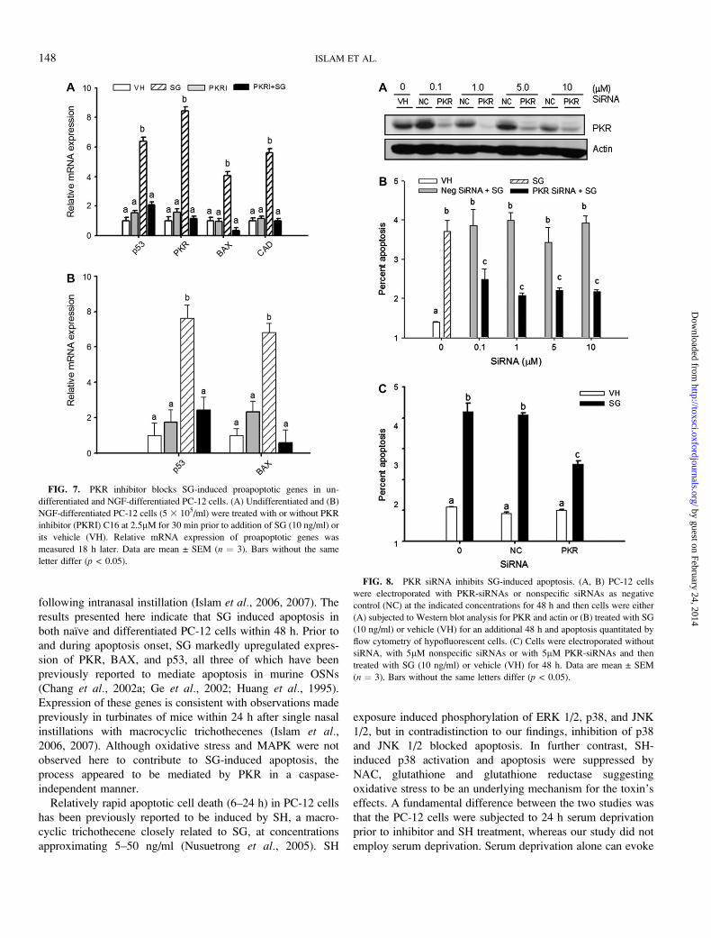
SG-Induced Apoptosis and Proapoptotic Gene Expression isSuppressed by PKR Inhibition
PC-12 cells were pretreated with a panel of pharmacologic
inhibitors to assess potential upstream roles of oxidative stress,
MAPKs and PKR in SG-induced apoptosis. Cells were
harvested 48 h after SG treatment and hypodiploid fluorescent
cells measured by flow cytometry (Fig. 6A). Neither inhibitors
of oxidative stress, (L-NAC, L-NAME, and quercetin) nor
inhibitors of p38, ERK, and JNK modulated SG-induced
apoptosis. In contrast, SG-induced apoptosis was completely
inhibited by the PKR inhibitor C16 (Jammi et al., 2003). When
a structural analog of the PKR inhibitor was used as a negative
FIG. 3. SG induces apoptosis and proapoptotic gene expression in NGF-
differentiated PC-12 cells. Cells were differentiated by incubating with 100 ng/ml
of NGF for 5 days. Panels show: (A) concentration-dependent DNA frag-
mentation in differentiated and undifferentiated PC-12 cells treated for 48 h with
or without SG (10 ng/ml) and (B) p53 and BAX mRNA expression in dif-
ferentiated and undifferentiated PC-12 cells treated with SG (10 ng/ml) or its
vehicle (VH) for 18 h. Data are mean ± SEM (n ¼ 3). Bars without the same
letter within a group differ (p < 0.05).
FIG. 4. SG-induced apoptosis is caspase-3 independent. (A) PC-12 cells
were cultured with SG (10 ng/ml) and analyzed at intervals for caspase-3
activity. (B, C) Cells were treated with cell-permeable caspase-3 inhibitor I
(DEVD-CHO) for 30 min prior to SG (10 ng/ml) treatment. Cells were
harvested after 48 h and analyzed for (B) caspase-3 activity or (C) apoptosis by
PI uptake. Data are mean ± SEM (n ¼ 3). Asterisks indicate significant
difference from vehicle control (p < 0.05). Bars without same letter differ (p <
0.05). Results are representative of three independent experiments.
146 ISLAM ET AL.
by guest on February 24, 2014http://toxsci.oxfordjournals.org/
Dow
nloaded from

control, SG-induced apoptosis was not affected (Fig. 6B).
Consistent with the aforementioned findings, C16 suppressed
expression of the proapoptotic genes p53, PKR, BAX, and
CAD at 18 h (Fig. 7A). SG-induced proapoptotic gene
expression in NGF-differentiated PC-12 cells was similarly
suppressed by the PKR inhibitor (Fig. 7B).
PKR siRNA Knockdown Inhibits SG-Induced Apoptosis
The role of PKR in SG-induced PC-12 cell apoptosis was
confirmed by knockdown with PKR siRNAs. Electroporation
with a PKR siRNA cocktail at 0.1, 1, 5, and 10lM followed by
a 48 h incubation markedly suppressed expression of PKR
protein as compared with that for negative control siRNA
(Fig. 8A). SG-induced apoptosis was significantly reduced in
cells treated with PKR-siRNA at all concentrations (Figs. 8B and
8C). SG similarly induced apoptosis in cells electroporated in the
presence and absence of negative control siRNA suggesting the
absence of off-target effects (Fig. 8C). Collectively, the siRNA
data indicated that SG-induced apoptosis in PC-12 cells is likely
to be a PKR-dependent process.
PKR Mediates SG-Induced AIF Translocation but notCaspase-3 Activation
Preincubation with C16 did not inhibit SG-induced caspase-3
activity indicating that activation of this enzyme was a PKR-
independent effect (Fig. 9). However, PKR inhibition blocked AIF
translocation to the nucleus (Fig. 10) at 12 h, whereas the PKR
negative inhibitor control or vehicle alone had no effect. PKR-
dependent SG-induced apoptosis in PC-12 thus appeared to be
linked to AIF nuclear translocation but not caspase-3 activation.
DISCUSSION
The capacity of macrocyclic trichothecenes to selectively
target neurons in the nose and in the brain of mice following
intranasal instillation is of fundamental importance because
olfactory function loss often occurs with early stages
of neurodegenerative illnesses such as Parkinson’s and
Alzheimer’s diseases (Demarquay et al., 2007; Hawkes, 2003;
Takeda et al., 2007). Understanding how SG causes apoptosis
in PC-12 cells can provide insight how this and
other macrocyclic trichothecenes induce apoptosis in OSNs
FIG. 5. SG induces AIF translocation. PC-12 cells were grown on eight-
well culture slides and treated with or without SG (10 ng/ml) for 12 and 24 h.
Cell nuclei were detected with DAPI and subjected to immunohistochemistry
with anti-AIF antibody followed by FITC-conjugated secondary antibody.
FIG. 6. SG-induced apoptosis is blocked by inhibition of PKR but not
oxidative stress or MAPK. (A) PC-12 cells were treated with inhibitor for 30
min before SG (10 ng/ml) treatment. Inhibitors included L-NAC at 1mM,
L-NAME at 5mM, quercetin at 10lM, PKR inhibitor (PKRI) C16 at 2.5lM,
p38 inhibitor SB203580 (p38I) at 2.5lM, ERK inhibitor PD98059 (ERKI) at
50lM and JNK inhibitor I (JNKI) at 2.5lM. Cells were harvested 48 h after SG
treatment and apoptosis assessed by PI uptake. Arrow indicates that SG-
induced apoptosis was completely inhibited by PKR inhibitor. Several
concentrations of each inhibitor were used but only the highest dose is shown.
(B) Cells were treated with PKRI at 2.5lM and nonfunctional PKR inhibitor
negative control (PKRNI) at the same concentration 30 min before SG (10 ng/
ml) treatment. Apoptosis was measured after 48 h. Data are mean ± SEM (n ¼ 3).
Bars without the same letter differ (p < 0.05). Results are representative of two
independent experiments.
SATRATOXIN G–INDUCED APOPTOSIS IN PC-12 NEURONAL CELLS 147
by guest on February 24, 2014http://toxsci.oxfordjournals.org/
Dow
nloaded from
following intranasal instillation (Islam et al., 2006, 2007). The
results presented here indicate that SG induced apoptosis in
both naı̈ve and differentiated PC-12 cells within 48 h. Prior to
and during apoptosis onset, SG markedly upregulated expres-
sion of PKR, BAX, and p53, all three of which have been
previously reported to mediate apoptosis in murine OSNs
(Chang et al., 2002a; Ge et al., 2002; Huang et al., 1995).
Expression of these genes is consistent with observations made
previously in turbinates of mice within 24 h after single nasal
instillations with macrocyclic trichothecenes (Islam et al.,2006, 2007). Although oxidative stress and MAPK were not
observed here to contribute to SG-induced apoptosis, the
process appeared to be mediated by PKR in a caspase-
independent manner.
Relatively rapid apoptotic cell death (6–24 h) in PC-12 cells
has been previously reported to be induced by SH, a macro-
cyclic trichothecene closely related to SG, at concentrations
approximating 5–50 ng/ml (Nusuetrong et al., 2005). SH
exposure induced phosphorylation of ERK 1/2, p38, and JNK
1/2, but in contradistinction to our findings, inhibition of p38
and JNK 1/2 blocked apoptosis. In further contrast, SH-
induced p38 activation and apoptosis were suppressed by
NAC, glutathione and glutathione reductase suggesting
oxidative stress to be an underlying mechanism for the toxin’s
effects. A fundamental difference between the two studies was
that the PC-12 cells were subjected to 24 h serum deprivation
prior to inhibitor and SH treatment, whereas our study did not
employ serum deprivation. Serum deprivation alone can evoke
FIG. 7. PKR inhibitor blocks SG-induced proapoptotic genes in un-
differentiated and NGF-differentiated PC-12 cells. (A) Undifferentiated and (B)
NGF-differentiated PC-12 cells (5 3 105/ml) were treated with or without PKR
inhibitor (PKRI) C16 at 2.5lM for 30 min prior to addition of SG (10 ng/ml) or
its vehicle (VH). Relative mRNA expression of proapoptotic genes was
measured 18 h later. Data are mean ± SEM (n ¼ 3). Bars without the same
letter differ (p < 0.05).
FIG. 8. PKR siRNA inhibits SG-induced apoptosis. (A, B) PC-12 cells
were electroporated with PKR-siRNAs or nonspecific siRNAs as negative
control (NC) at the indicated concentrations for 48 h and then cells were either
(A) subjected to Western blot analysis for PKR and actin or (B) treated with SG
(10 ng/ml) or vehicle (VH) for an additional 48 h and apoptosis quantitated by
flow cytometry of hypofluorescent cells. (C) Cells were electroporated without
siRNA, with 5lM nonspecific siRNAs or with 5lM PKR-siRNAs and then
treated with SG (10 ng/ml) or vehicle (VH) for 48 h. Data are mean ± SEM
(n ¼ 3). Bars without the same letters differ (p < 0.05).
148 ISLAM ET AL.
by guest on February 24, 2014http://toxsci.oxfordjournals.org/
Dow
nloaded from

PC-12 apoptosis and this is mediated by oxidative stress
(Ferrari et al., 1995; Liu et al., 2003; Maroto and Perez-Polo,
1997). Thus, oxidative stress might have exacerbated SH
effects, making it difficult to discern the toxin’s underlying
mechanisms. Nevertheless, the aforementioned findings are
important because they demonstrate that macrocyclic tricho-
thecenes have the potential to interact with other stressors to
evoke neuronal death.
Consistent with SG’s effects on PC-12 cells, our laboratory
has previously demonstrated in cloned macrophages that
induction of apoptosis by the simple trichothecene DON is
regulated by PKR (Pestka et al., 2004; Zhou et al., 2003).
Although the role of PKR in mediating apoptosis in virus-
infected cells is well-established (Garcia et al., 2007), PKR can
be activated in the absence of viruses by the PKR-activating
protein PACT and by its mouse homologue RAX (Bennett
et al., 2004; Ito et al., 1999; Patel and Sen, 1998). PKR also
mediates apoptosis induction by dsRNA, TNF-a, LPS,
tunicamycin, or serum starvation (Cole, 2007).
Of particular significance to the present study, PKR has been
linked to neuronal apoptosis. Chang et al. (2002a) demon-
strated that PKR and eIF2a phosphorylation play a significant
role in apoptosis of neuroblastoma cells and primary neuronal
cultures induced by b-amyloid peptides, calcium ionophores
and flavanoids. PKR activation has been associated with
degenerating neurons in Alzheimer’s (Chang et al., 2002b;
Onuki et al., 2004; Peel, 2004), Parkinson’s and Huntington’s
(Bando et al., 2005) diseases as well as amyotrophic lateral
sclerosis (Garcia et al., 2006; Hu et al., 2003), HIV-associated
dementia (Alirezaei et al., 2007), and retinal ganglion cell
death (Shimazawa et al., 2007).
There are several possible downstream mechanisms by
which PKR might mediate apoptosis. First, phosphorylation of
eIF2a with accompanying translational inhibition have been
proposed to be sufficient for PKR-induced apoptotic responses
simply by inhibiting synthesis of anti-apoptotic factors such as
inhibitor of protein synthesis (IAP) (Scheuner et al., 2006).
Second, PKR-mediated phosphorylation has been demon-
strated to induce the eIF2a/ATF4/CHOP pathway which drives
expression of apoptotic genes (Lee et al., 2007). Finally, NF-
jB activation by PKR has also been suggested to mediate
apoptosis (Gil and Esteban, 2000). Proapoptotic genes
regulated by NF-jB include p53, caspase 1, IRF-1, Fas L,
and Fas (Garcia et al., 2006). Understanding involvement of
NF-jB in the PC-12 model described here is complicated by
this transcription factor’s capacity to regulate expression of
both proapoptotic and anti-apoptotic genes. Overall, clarifica-
tion is needed on the mechanisms by which PKR mediates SG-
induced apoptosis in neuronal cells.
Conventional apoptosis is generally considered to involve
initiation of execution resulting from activation of caspases
(Krantic et al., 2007). Caspase-3 is widely recognized to be
a critical central mediator of apoptosis in many types of cells
(Turk and Stoka, 2007) and has been specifically shown to
mediate apoptosis in OSNs (Cowan and Roskams, 2004).
Although caspase-3 is inactive in adult neuronal cells, it is
reactivated upon injury and can contribute to normal apoptotic
death (Stoka et al., 2006; Yakovlev et al., 2001). Caspase-3
and CAD can be activated by binding of death ligands to TNF-
receptor family or in response to stress-induced mitochondrial
proteins such as BAX (Green, 2000). Although SG did not
induce caspase-3 gene expression, it did activate caspase-3.
Thus, it was surprising that caspase-3 inhibition by DEVD-
CHO did not inhibit SG-induced apoptosis. Relatedly, it was ob-
served that the PKR inhibitor C16 did not inhibit SG-induced
caspase-3 activity, even though it suppressed SG-induced
apoptosis. Because DEVD-CHO also blocks caspase-6, caspase-7,
caspase-8, and caspase-10 activity, caspases per se do not
appear to be integral to SG-induced apoptosis.
FIG. 9. SG-induced caspase-3 activation is PKR-independent. PC-12 cells
were cultured with 2.5lM of PKRI C16 or vehicle for 30 min prior to
incubation with SG (10 ng/ml) or vehicle. After 48 h, cells were harvested and
analyzed for caspase-3 activity. Bars without the same letters differ (p < 0.05).
FIG. 10. SG-induced AIF translocation is PKR-dependent. PC-12 cells
were grown in eight-well culture slides and incubated with 2.5lM PKRI C16 or
PKRI negative control (PKRN) for 30 min and then treated with SG (10 ng/ml)
for 12 h. AIF immunoreactivity and DAPI staining of cell nuclei were detected
with immunofluorescence microscopy.
SATRATOXIN G–INDUCED APOPTOSIS IN PC-12 NEURONAL CELLS 149
by guest on February 24, 2014http://toxsci.oxfordjournals.org/
Dow
nloaded from

Both caspase-dependent and caspase-independent neuronal
cell death pathways have been described (Stefanis, 2005). The
mitochondrial flavoprotein AIF is a primary mediator of
caspase-independent apoptosis-like programmed cell death
and has been linked to neurodegeneration (Cao et al., 2007;
Krantic et al., 2007). Translocation of AIF from mitochondria
to the nucleus acts as a proapoptotic trigger possibly by binding
to DNA, stimulating DNAse activity and evoking DNA
fragmentation (Cande et al., 2002; Martinou and Green,
2001; Ye et al., 2002). Both p53 and BAX have been shown
to have upstream roles in AIF translocation (Cheung et al.,2005; Stambolsky et al., 2006). The possibility therefore exists
that SG-induced p53 and BAX gene expression drives AIF
translocation in PC-12 cells.
Taken together, the results presented herein indicate that SG
induces PKR-dependent cell death in PC-12 cells that has the
characteristics of apoptosis but does not appear to require
caspases. Consistent with these observations, SG induces
nuclear translocation of AIF, a known mediator of caspase-
independent apoptosis-like programmed cell death. There is
a need for improved understanding of both the mechanisms by
which PKR drives SG-induced cell death and the role of AIF in
this process. The relevance to these findings to previously
observed SG-induced OSN cell death in mouse nasal turbinates
will require further exploration using specific in vitro and
in vivo models for this neuronal cell-type.
FUNDING
Michigan State University Foundation Strategic Partnership
Grant and Public Health Service Grant (ES03358) to J.J.P.
from the National Institute for Environmental Health Sciences.
ACKNOWLEDGMENTS
We thank Dr Jack Harkema for advice as well as Sherry Shi,
Sarah Godbehere, and Mary Rosner for technical support.
REFERENCES
Alirezaei, M., Watry, D. D., Flynn, C. F., Kiosses, W. B., Masliah, E.,
Williams, B. R., Kaul, M., Lipton, S. A., and Fox, H. S. (2007). Human
immunodeficiency virus-1/surface glycoprotein 120 induces apoptosis
through RNA-activated protein kinase signaling in neurons. J. Neurosci.
27, 11047–11055.
Audige, A., Yu, Z. R., Frey, B. M., Uehlinger, D. E., Frey, F. J., and Vogt, B.
(2003). Epithelial sodium channel (ENaC) subunit mRNA and protein
expression in rats with puromycin aminonucleoside-induced nephrotic
syndrome. Clin. Sci. (Lond) 104, 389–395.
Aykin-Burns, N., and Ercal, N. (2006). Effects of selenocystine on lead-
exposed Chinese hamster ovary (CHO) and PC-12 cells. Toxicol. Appl.
Pharmacol. 214, 136–143.
Bando, Y., Onuki, R., Katayama, T., Manabe, T., Kudo, T., Taira, K., and
Tohyama, M. (2005). Double-strand RNA dependent protein kinase (PKR) is
involved in the extrastriatal degeneration in Parkinson’s disease and
Huntington’s disease. Neurochem. Int. 46, 11–18.
Bennett, R. L., Blalock, W. L., and May, W. S. (2004). Serine 18
phosphorylation of RAX, the PKR activator, is required for PKR activation
and consequent translation inhibition. J Biol. Chem. 279, 42687–42693.
Brasel, T. L., Douglas, D. R., Wilson, S. C., and Straus, D. C. (2005). Detection
of airborne Stachybotrys chartarum macrocyclic trichothecene
mycotoxins on particulates smaller than conidia. Appl. Environ. Microbiol.
71, 114–122.
Brenneman, K. A., Wong, B. A., Buccellato, M. A., Costa, E. R., Gross, E. A.,
and Dorman, D. C. (2000). Direct olfactory transport of inhaled manganese
((54)MnCl(2))? to the rat brain: Toxicokinetic investigations in a unilateral
nasal occlusion model. Toxicol Appl Pharmacol 169, 238–48.
Cande, C., Cecconi, F., Dessen, P., and Kroemer, G. (2002). Apoptosis-
inducing factor (AIF): Key to the conserved caspase-independent pathways
of cell death? J. Cell Sci. 115, 4727–4734.
Cao, G., Xing, J., Xiao, X., Liou, A. K., Gao, Y., Yin, X. M., Clark, R. S.,
Graham, S. H., and Chen, J. (2007). Critical role of calpain I in
mitochondrial release of apoptosis-inducing factor in ischemic neuronal
injury. J. Neurosci. 27, 9278–9293.
Chang, R. C., Suen, K. C., Ma, C. H., Elyaman, W., Ng, H. K., and Hugon, J.
(2002a). Involvement of double-stranded RNA-dependent protein kinase and
phosphorylation of eukaryotic initiation factor-2alpha in neuronal de-
generation. J. Neurochem. 83, 1215–1225.
Chang, R. C., Wong, A. K., Ng, H. K., and Hugon, J. (2002b). Phosphorylation
of eukaryotic initiation factor-2a (eIF2a) is associated with neuronal
degeneration in Alzheimer’s disease. Neuroreport 13, 2429–2432.
Cheung, E. C., Melanson-Drapeau, L., Cregan, S. P., Vanderluit, J. L.,
Ferguson, K. L., McIntosh, W. C., Park, D. S., Bennett, S. A., and
Slack, R. S. (2005). Apoptosis-inducing factor is a key factor in neuronal
cell death propagated by BAX-dependent and BAX-independent mecha-
nisms. J. Neurosci. 25, 1324–1334.
Chung, Y. J., Jarvis, B., and Pestka, J. (2003). Modulation of lipopolysaccha-
ride-induced proinflammatory cytokine production by satratoxins and other
macrocyclic trichothecenes in the murine macrophage. J. Toxicol. Environ.
Health A 66, 379–391.
Cole, J. L. (2007). Activation of PKR: An open and shut case? Trends
Biochem. Sci. 32, 57–62.
Coronas, V., Feron, F., Hen, R., Sicard, G., Jourdan, F., and Moyse, E. (1997).
In vitro induction of apoptosis or differentiation by dopamine in an
immortalized olfactory neuronal cell line. J. Neurochem. 69, 1870–1881.
Cowan, C. M., and Roskams, A. J. (2004). Caspase-3 and caspase-9 mediate
developmental apoptosis in the mouse olfactory system. J. Comp Neurol.
474, 136–148.
Croft, W. A., Jastromski, B. M., Croft, A. L., and Peters, H. A. (2002). Clinical
confirmation of trichothecene mycotoxicosis in patient urine. J. Environ.
Biol. 23, 301–320.
Demarquay, G., Ryviln, P., and Royet, J. P. (2007). Olfaction and neurological
diseases: A review of the literature. Revue Neurologique 163, 155–167.
Der, S. D., Yang, Y. L., Weissmann, C., and Williams, B. R. (1997). A double-
stranded RNA-activated protein kinase-dependent pathway mediating stress-
induced apoptosis. Proc. Natl. Acad. Sci. U. S. A. 94, 3279–3283.
Ferrari, G., Yan, C. Y., and Greene, L. A. (1995). N-acetylcysteine (D- and
L-stereoisomers) prevents apoptotic death of neuronal cells. J. Neurosci. 15,
2857–2866.
Garcia, M. A., Gil, J., Ventoso, I., Guerra, S., Domingo, E., Rivas, C., and
Esteban, M. (2006). Impact of protein kinase PKR in cell biology: From
antiviral to antiproliferative action. Microbiol. Mol. Biol. Rev. 70,
1032–1060.
150 ISLAM ET AL.
by guest on February 24, 2014http://toxsci.oxfordjournals.org/
Dow
nloaded from

Garcia, M. A., Meurs, E. F., and Esteban, M. (2007). The dsRNA protein
kinase PKR: Virus and cell control. Biochimie 89, 799–811.
Ge, Y., Tsukatani, T., Nishimura, T., Furukawa, M., and Miwa, T. (2002). Cell
death of olfactory receptor neurons in a rat with nasosinusitis infected
artificially with Staphylococcus. Chem Senses 27, 521–7.
Gil, J., and Esteban, M. (2000). Induction of apoptosis by the dsRNA-dependent
protein kinase (PKR): Mechanism of action. Apoptosis. 5, 107–114.
Gordon, W. A., Cantor, J. B., Johanning, E., Charatz, H. J., Ashman, T. A.,
Breeze, J. L., Haddad, L., and Abramowitz, S. (2004). Cognitive impairment
associated with toxigenic fungal exposure: A replication and extension of
previous findings. Appl. Neuropsychol. 11, 65–74.
Green, D. R. (2000). Apoptotic pathways: Paper wraps stone blunts scissors.
Cell 102, 1–4.
Greene, L. A., and Tischler, A. S. (1976). Establishment of a noradrenergic
clonal line of rat adrenal pheochromocytoma cells which respond to nerve
growth factor. Proc. Natl. Acad. Sci. U. S. A 73, 2424–2428.
Gregory, L., Pestka, J. J., Dearborn, D. G., and Rand, T. G. (2004).
Localization of satratoxin-G in Stachybotrys chartarum spores and spore-
impacted mouse lung using immunocytochemistry. Toxicol. Pathol. 32,
26–34.
Grove, J. F. (2007). The trichothecenes and their biosynthesis. Fortschr. Chem.
Org. Naturst. 88, 63–130.
Hawkes, C. (2003). Olfaction in neurodegenerative disorder. Movement
Disorders 18, 364–372.
Hegg, C. C., and Miletic, V. (1998). Diminished blocking effect of acute lead
exposure on high-threshold voltage-gated calcium currents in PC12 cells
chronically exposed to the heavy metal. Neurotoxicology 19, 413–420.
Henck, J. W., Reindel, J. F., and Anderson, J. A. (2001). Growth and
development in rats given recombinant human epidermal growth factor
(1-48) as neonates. Toxicol Sci 62, 80–91.
Hinkley, S. F., and Jarvis, B. B. (2001). Chromatographic method for
Stachybotrys toxins. Methods Mol. Biol. 157, 173–194.
Hodgson, M. J., Morey, P., Leung, W. Y., Morrow, L., Miller, D., Jarvis, B. B.,
Robbins, H., Halsey, J. F., and Storey, E. (1998). Building-associated
pulmonary disease from exposure to Stachybotrys chartarum and Aspergillus
versicolor. J. Occup. Environ. Med. 40, 241–249.
Hu, J. H., Zhang, H., Wagey, R., Krieger, C., and Pelech, S. L. (2003). Protein
kinase and protein phosphatase expression in amyotrophic lateral sclerosis
spinal cord. J Neurochem. 85, 432–442.
Huang, C. C., Chen, K., and Huang, T. Y. (1995). Immunohistochemical
studies of sensory neurons in rat olfactory epithelium. Eur. Arch.
Otorhinolaryngol. 252, 86–91.
Illing, N., Boolay, S., Siwoski, J. S., Casper, D., Lucero, M. T., and
Roskams, A. J. (2002). Conditionally immortalized clonal cell lines from the
mouse olfactory placode differentiate into olfactory receptor neurons.
Molecular and Cellular Neuroscience 20, 225–243.
Institute of Medicine. (2004). Damp Indoor Spaces and Health, pp. 1–355.
National Academies Press, Washington, D.C..
Islam, Z., Amuzie, C. J., Harkema, J. R., and Pestka, J. J. (2007). Neurotoxicity
and inflammation in the nasal airways of mice exposed to the macrocyclic
trichothecene mycotoxin roridin A: Kinetics and potentiation by bacterial
lipopolysaccharide co-exposure. Toxicol. Sci. 98, 526–541.
Islam, Z., Harkema, J. R., and Pestka, J. J. (2006). Satratoxin G from the black
mold Stachybotrys chartarum evokes olfactory sensory neuron loss and
inflammation in the murine nose and brain. Environ. Health Perspect. 114,
1099–1107.
Islam, Z., Moon, Y. S., Zhou, H. R., King, L. E., Fraker, P. J., and Pestka, J. J.
(2002). Endotoxin potentiation of trichothecene-induced lymphocyte apo-
ptosis is mediated by up-regulation of glucocorticoids. Toxicol. Appl.
Pharmacol. 180, 43–55.
Ito, T., Yang, M., and May, W. S. (1999). RAX, a cellular activator for double-
stranded RNA-dependent protein kinase during stress signaling. J Biol.
Chem. 274, 15427–15432.
Jammi, N. V., Whitby, L. R., and Beal, P. A. (2003). Small molecule inhibitors
of the RNA-dependent protein kinase. Biochem. Biophys. Res. Commun.
308, 50–57.
Johanning, E., Biagini, R., Hull, D., Morey, P., Jarvis, B., and Landsbergis, P.
(1996). Health and immunology study following exposure to toxigenic fungi
(Stachybotrys chartarum) in a water-damaged office environment. Int. Arch.
Occup. Environ. Health 68, 207–218.
Krantic, S., Mechawar, N., Reix, S., and Quirion, R. (2007). Apoptosis-inducing
factor: A matter of neuron life and death. Prog. Neurobiol. 81, 179–196.
Lakard, S., Lesniewska, E., Michel, G., Lakard, B., Morrand-Villeneuve, N.,
and Versaux-Botteri, C. (2007). In vitro induction of differentiation by
retinoic acid in an immortalized olfactory neuronal cell line. Acta Histochem.
109(2), 111–121.
Lee, E. S., Yoon, C. H., Kim, Y. S., and Bae, Y. S. (2007). The double-strand
RNA-dependent protein kinase PKR plays a significant role in a sustained
ER stress-induced apoptosis. FEBS Lett. 581, 4325–4332.
Levi, A., Eldridge, J. D., and Paterson, B. M. (1985). Molecular cloning of
a gene sequence regulated by nerve growth factor. Science 229, 393–395.
Liou, A. K., Zhou, Z., Pei, W., Lim, T. M., Yin, X. M., and Chen, J. (2005).
BimEL up-regulation potentiates AIF translocation and cell death in response
to MPTP. FASEB J. 19, 1350–1352.
Liu, H., Nowak, R., Chao, W., and Bloch, K. D. (2003). Nerve growth factor
induces anti-apoptotic heme oxygenase-1 in rat pheochromocytoma PC12
cells. J. Neurochem. 86, 1553–1563.
Maroto, R., and Perez-Polo, J. R. (1997). BCL-2-related protein expression
in apoptosis: Oxidative stress versus serum deprivation in PC12 cells.
J. Neurochem. 69, 514–523.
Martinou, J. C., and Green, D. R. (2001). Breaking the mitochondrial barrier.
Nat. Rev. Mol. Cell Biol. 2, 63–67.
Nusuetrong, P., Yoshida, M., Tanitsu, M. A., Kikuchi, H., Mizugaki, M.,
Shimazu, K., Pengsuparp, T., Meksuriyen, D., Oshima, Y., and Nakahata, N.
(2005). Involvement of reactive oxygen species and stress-activated
MAPKs in satratoxin H-induced apoptosis. Eur. J Pharmacol. 507, 239–246.
Onuki, R., Bando, Y., Suyama, E., Katayama, T., Kawasaki, H., Baba, T.,
Tohyama, M., and Taira, K. (2004). An RNA-dependent protein kinase is
involved in tunicamycin-induced apoptosis and Alzheimer’s disease. EMBO
J. 23, 959–968.
Patel, R. C., and Sen, G. C. (1998). PACT, a protein activator of the interferon-
induced protein kinase, PKR. EMBO J. 17, 4379–4390.
Peel, A. L. (2004). PKR activation in neurodegenerative disease.
J. Neuropathol. Exp. Neurol. 63, 97–105.
Pestka, J. J., Yike, I., Dearborn, D. G., Ward, M. D., and Harkema, J. R.
(2007). Stachybotrys chartarum, trichothecene mycotoxins, and damp
building-related illness: New insights into a public health enigma. Toxicol.
Sci. [Epub ahead of print]
Pestka, J. J., Zhou, H. R., Moon, Y., and Chung, Y. J. (2004). Cellular and
molecular mechanisms for immune modulation by deoxynivalenol and other
trichothecenes: Unraveling a paradox. Toxicol. Lett. 153, 61–73.
Scheuner, D., Patel, R., Wang, F., Lee, K., Kumar, K., Wu, J., Nilsson, A.,
Karin, M., and Kaufman, R. J. (2006). Double-stranded RNA-dependent
protein kinase phosphorylation of the alpha-subunit of eukaryotic translation
initiation factor 2 mediates apoptosis. J. Biol. Chem. 281, 21458–21468.
Shimazawa, M., Ito, Y., Inokuchi, Y., and Hara, H. (2007). Involvement of
double-stranded RNA-dependent protein kinase in ER stress-induced retinal
neuron damage. Invest Ophthalmol. Vis. Sci. 48, 3729–3736.
Slotkin, T. A., MacKillop, E. A., Ryde, I. T., Tate, C. A., and Seidler, F. J.
(2007). Screening for developmental neurotoxicity using PC12 cells:
SATRATOXIN G–INDUCED APOPTOSIS IN PC-12 NEURONAL CELLS 151
by guest on February 24, 2014http://toxsci.oxfordjournals.org/
Dow
nloaded from

Comparisons of organophosphates with a carbamate, an organochlorine, and
divalent nickel. Environ. Health Perspect. 115, 93–101.
Sorenson, W. G., Frazer, D. G., Jarvis, B. B., Simpson, J., and Robinson, V. A.
(1987). Trichothecene mycotoxins in aerosolized conidia of Stachybotrys
atra. Appl. Environ. Microbiol. 53, 1370–1375.
Stambolsky, P., Weisz, L., Shats, I., Klein, Y., Goldfinger, N., Oren, M., and
Rotter, V. (2006). Regulation of AIF expression by p53. Cell Death. Differ.
13, 2140–2149.
Stefanis, L. (2005). Caspase-dependent and -independent neuronal death: Two
distinct pathways to neuronal injury. Neuroscientist 11, 50–62.
Stoka, V., Turk, V., and Bredesen, D. E. (2006). Differential regulation of the
intrinsic pathway of apoptosis in brain and liver during ageing. FEBS Lett.
580, 3739–3745.
Takeda, A., Kikuchi, A., Matsuzaki-Kobayashi, M., Sugeno, N., and
Itoyama, Y. (2007). Olfactory dysfunction in Parkinson’s disease. Journalof Neurology 254, 2–7.
Tuomi, T., Saarinen, L., and Reijula, K. (1998). Detection of polar and
macrocyclic trichothecenes from indoor environments. Analyst 9, 1835–1841.
Turk, B., and Stoka, V. (2007). Protease signalling in cell death: Caspases
versus cysteine cathepsins. FEBS Lett. 581, 2761–2767.
Walkinshaw, G., and Waters, C. M. (1994). Neurotoxin-induced cell death in
neuronal PC12 cells is mediated by induction of apoptosis. Neuroscience 63,
975–987.
Wang, H., Shimoji, M., Yu, S. W., Dawson, T. M., and Dawson, V. L. (2003).
Apoptosis inducing factor and PARP-mediated injury in the MPTP mouse
model of Parkinson’s disease. Ann. N. Y. Acad. Sci. 991, 132–139.
Westerink, R. H., and Ewing, A. G. (2008). The PC12 cell as model for
neurosecretion. Acta Physiol (Oxf) 192, 273–285.
Williams, B. R. (2001). Signal integration via PKR. Sci. STKE. 2001, RE2.
Wu, S., Kumar, K. U., and Kaufman, R. J. (1998). Identification and
requirement of three ribosome binding domains in dsRNA-dependent protein
kinase (PKR). Biochemistry 37, 13816–13826.
Yakovlev, A. G., Ota, K., Wang, G., Movsesyan, V., Bao, W. L.,
Yoshihara, K., and Faden, A. I. (2001). Differential expression of apoptotic
protease-activating factor-1 and caspase-3 genes and susceptibility to
apoptosis during brain development and after traumatic brain injury.
J. Neurosci. 21, 7439–7446.
Yang, G. H., Jarvis, B. B., Chung, Y. J., and Pestka, J. J. (2000). Apoptosis
induction by the satratoxins and other trichothecene mycotoxins: Relation-
ship to ERK, p38 MAPK, and SAPK/JNK activation. Toxicol. Appl.
Pharmacol. 164, 149–160.
Ye, H., Cande, C., Stephanou, N. C., Jiang, S., Gurbuxani, S., Larochette, N.,
Daugas, E., Garrido, C., Kroemer, G., and Wu, H. (2002). DNA binding is
required for the apoptogenic action of apoptosis inducing factor. Nat. Struct.
Biol. 9, 680–684.
Yeung, M. C., and Lau, A. S. (1998). Tumor suppressor p53 as a component of
the tumor necrosis factor-induced, protein kinase PKR-mediated apoptotic
pathway in human promonocytic U937 cells. J. Biol. Chem. 273,
25198–25202.
Yeung, M. C., Liu, J., and Lau, A. S. (1996). An essential role for the
interferon-inducible, double-stranded RNA-activated protein kinase PKR in
the tumor necrosis factor-induced apoptosis in U937 cells. Proc. Natl. Acad.
Sci. U. S. A 93, 12451–12455.
Zhou, H. R., Lau, A. S., and Pestka, J. J. (2003). Role of double-stranded RNA-
activated protein kinase R (PKR) in deoxynivalenol-induced ribotoxic stress
response. Toxicol. Sci. 74, 335–344.
152 ISLAM ET AL.
by guest on February 24, 2014http://toxsci.oxfordjournals.org/
Dow
nloaded from
Copyright © 2022 FDOKUMEN