Nanogold-functionalized magnetic beads with redox activity for sensitive electrochemical immunoassay...
7
Analytica Chimica Acta 711 (2012) 17–23 Contents lists available at SciVerse ScienceDirect Analytica Chimica Acta j ourna l ho me page: www.elsevier.com/locate/aca Nanogold-functionalized magnetic beads with redox activity for sensitive electrochemical immunoassay of thyroid-stimulating hormone Bing Zhang, Dianping Tang ∗ , Bingqian Liu, Yuling Cui, Huafeng Chen, Guonan Chen ∗ Ministry of Education Key Laboratory of Analysis and Detection for Food Safety, Fujian Provincial Key Laboratory of Analysis and Detection Technology for Food Safety, Department of Chemistry, Fuzhou University, Fuzhou 350108, PR China a r t i c l e i n f o Article history: Received 15 September 2011 Received in revised form 24 October 2011 Accepted 25 October 2011 Available online 4 November 2011 Keywords: Electrochemical immunosensor Gold–graphene nanocomposites Nanolabels Redox-active GoldMag nanostructures Thyroid-stimulating hormone a b s t r a c t A new electrochemical immunosensor for sensitive determination of thyroid-stimulating hormone (TSH) was designed by using redox-active nanogold-functionalized magnetic beads (GoldMag) as signal tags on the nanogold–graphene interface. To construct such GoldMag nanostructures, polyethyleneimine- functionalized magnetic beads (PEI-MBs) were initially prepared by using a wet chemical method, and the electroactive thionine molecules and gold nanoparticles were then alternately immobilized on the surface of PEI-MBs by using an opposite-charged adsorption technique and an in situ synthesis method, respec- tively. The synthesized GoldMag nanostructures were utilized as signal tags for the label of horseradish peroxidase-anti-TSH conjugates (HRP-anti-TSH). With a sandwich-type immunoassay format, the conju- gated signal tags on the transducer were increased with the increasing TSH concentration in the sample, thus enhancing the signal of the electrochemical immunosensor due to the labeled HRP toward the cat- alytic reduction of H 2 O 2 . Under optimal conditions, the current was proportional to the logarithm of TSH concentration ranging from 0.01 to 20 IU mL −1 in pH 6.0 HAc–NaAc containing 6 mM H 2 O 2 . The detection limit (LOD) was 0.005 IU mL −1 TSH at 3s B . The immunosensor displayed an acceptable reproducibility, stability and selectivity. In addition, the methodology was evaluated with human serum specimens, receiving good correlation with results from commercially available electrochemiluminescent analyzer. © 2011 Elsevier B.V. All rights reserved. 1. Introduction Thyroid-stimulating hormone (TSH), a glycoprotein hormone produced by thyrotrope cells in the anterior pituitary gland, reg- ulates the endocrine function of the thyroid gland by stimulating the production and release of thyroid hormones [1]. The level of thyroid hormone in the blood has an effect on the pituitary release of TSH, and tested in the blood of patients suspected of suffering from excess (hyperthyroidism), or deficiency (hypothyroidism) of thyroid hormone. Usually, a reference range for TSH test in human serum is between 0.4 and 5.0 IU mL −1 for adults, while the ther- apeutic target range is between 0.3 and 3.0 IU mL −1 for patients on treatment [2]. Thus, accuracy detection of TSH level in human serum is absolutely necessary. Immunoassay, based on the specific reaction of the antigen–antibody recognition, has been one of the most widely used biomedical methods and gained increasing attention in clinic diagnosis, food safety, and environmental monitoring [3]. Recently, various strategies have been reported for detection of TSH [4–8]. ∗ Corresponding authors. Tel.: +86 591 2286 6125; fax: +86 591 2286 6135. E-mail addresses: [email protected], [email protected] (D. Tang), [email protected] (G. Chen). The Mello group reported a fast assay protocol for detection of TSH in human serum without chemical preprocessing by using infrared spectroscopy and least squares support vector machines [4]. Georges et al. developed a case of levothyroxin overdose because of gross overestimation of TSH by chemiluminescent and IRMA assays [5]. Van Deventer et al. utilized direct analog immunoassay and tandem mass spectrometry to evaluate inverse log-linear relationship between TSH and free thyroxine [6]. Despite many advances in this field, it is still a challenge to explore new protocols and strategies for further improving the simplicity, selectivity, and sensitivity of the immunoassays. Electrochemical immunoassay has become an important analytical tool in clinical and biochemical analyses, because of its high sensitivity, low cost, low power requirements, and high compatibility with advanced micromachining technologies [9]. For the successful development of electrochemical immunoas- says, biomolecular immobilization on the transducers is crucial. The present methods mainly involve adsorption, entrapment, and covalent binding/cross-linking technique [10]. A major limitation of entrapment technique is the additional diffusion barrier resulting from the entrapped materials [11]. In contrast, one of the problems commonly associated with covalent binding is the decrease of pro- tein bioactivity when the proteins are exposed to reactive groups and harsh reaction conditions [12]. The rapidly emerging research 0003-2670/$ – see front matter © 2011 Elsevier B.V. All rights reserved. doi:10.1016/j.aca.2011.10.049
-
Upload
independent -
Category
Documents
-
view
9 -
download
0
Transcript of Nanogold-functionalized magnetic beads with redox activity for sensitive electrochemical immunoassay...

Ne
BMo
a
ARRAA
KEGNRT
1
puttoftsaos
audv
(
0d
Analytica Chimica Acta 711 (2012) 17– 23
Contents lists available at SciVerse ScienceDirect
Analytica Chimica Acta
j ourna l ho me page: www.elsev ier .com/ locate /aca
anogold-functionalized magnetic beads with redox activity for sensitivelectrochemical immunoassay of thyroid-stimulating hormone
ing Zhang, Dianping Tang ∗, Bingqian Liu, Yuling Cui, Huafeng Chen, Guonan Chen ∗
inistry of Education Key Laboratory of Analysis and Detection for Food Safety, Fujian Provincial Key Laboratory of Analysis and Detection Technology for Food Safety, Departmentf Chemistry, Fuzhou University, Fuzhou 350108, PR China
r t i c l e i n f o
rticle history:eceived 15 September 2011eceived in revised form 24 October 2011ccepted 25 October 2011vailable online 4 November 2011
eywords:lectrochemical immunosensorold–graphene nanocompositesanolabels
a b s t r a c t
A new electrochemical immunosensor for sensitive determination of thyroid-stimulating hormone (TSH)was designed by using redox-active nanogold-functionalized magnetic beads (GoldMag) as signal tagson the nanogold–graphene interface. To construct such GoldMag nanostructures, polyethyleneimine-functionalized magnetic beads (PEI-MBs) were initially prepared by using a wet chemical method, and theelectroactive thionine molecules and gold nanoparticles were then alternately immobilized on the surfaceof PEI-MBs by using an opposite-charged adsorption technique and an in situ synthesis method, respec-tively. The synthesized GoldMag nanostructures were utilized as signal tags for the label of horseradishperoxidase-anti-TSH conjugates (HRP-anti-TSH). With a sandwich-type immunoassay format, the conju-gated signal tags on the transducer were increased with the increasing TSH concentration in the sample,
edox-active GoldMag nanostructureshyroid-stimulating hormone
thus enhancing the signal of the electrochemical immunosensor due to the labeled HRP toward the cat-alytic reduction of H2O2. Under optimal conditions, the current was proportional to the logarithm of TSHconcentration ranging from 0.01 to 20 �IU mL−1 in pH 6.0 HAc–NaAc containing 6 mM H2O2. The detectionlimit (LOD) was 0.005 �IU mL−1 TSH at 3sB. The immunosensor displayed an acceptable reproducibility,stability and selectivity. In addition, the methodology was evaluated with human serum specimens,receiving good correlation with results from commercially available electrochemiluminescent analyzer.
. Introduction
Thyroid-stimulating hormone (TSH), a glycoprotein hormoneroduced by thyrotrope cells in the anterior pituitary gland, reg-lates the endocrine function of the thyroid gland by stimulatinghe production and release of thyroid hormones [1]. The level ofhyroid hormone in the blood has an effect on the pituitary releasef TSH, and tested in the blood of patients suspected of sufferingrom excess (hyperthyroidism), or deficiency (hypothyroidism) ofhyroid hormone. Usually, a reference range for TSH test in humanerum is between 0.4 and 5.0 �IU mL−1 for adults, while the ther-peutic target range is between 0.3 and 3.0 �IU mL−1 for patientsn treatment [2]. Thus, accuracy detection of TSH level in humanerum is absolutely necessary.
Immunoassay, based on the specific reaction of thentigen–antibody recognition, has been one of the most widely
sed biomedical methods and gained increasing attention in cliniciagnosis, food safety, and environmental monitoring [3]. Recently,arious strategies have been reported for detection of TSH [4–8].∗ Corresponding authors. Tel.: +86 591 2286 6125; fax: +86 591 2286 6135.E-mail addresses: [email protected], [email protected]
D. Tang), [email protected] (G. Chen).
003-2670/$ – see front matter © 2011 Elsevier B.V. All rights reserved.oi:10.1016/j.aca.2011.10.049
© 2011 Elsevier B.V. All rights reserved.
The Mello group reported a fast assay protocol for detection ofTSH in human serum without chemical preprocessing by usinginfrared spectroscopy and least squares support vector machines[4]. Georges et al. developed a case of levothyroxin overdosebecause of gross overestimation of TSH by chemiluminescentand IRMA assays [5]. Van Deventer et al. utilized direct analogimmunoassay and tandem mass spectrometry to evaluate inverselog-linear relationship between TSH and free thyroxine [6]. Despitemany advances in this field, it is still a challenge to explore newprotocols and strategies for further improving the simplicity,selectivity, and sensitivity of the immunoassays. Electrochemicalimmunoassay has become an important analytical tool in clinicaland biochemical analyses, because of its high sensitivity, low cost,low power requirements, and high compatibility with advancedmicromachining technologies [9].
For the successful development of electrochemical immunoas-says, biomolecular immobilization on the transducers is crucial.The present methods mainly involve adsorption, entrapment, andcovalent binding/cross-linking technique [10]. A major limitation ofentrapment technique is the additional diffusion barrier resulting
from the entrapped materials [11]. In contrast, one of the problemscommonly associated with covalent binding is the decrease of pro-tein bioactivity when the proteins are exposed to reactive groupsand harsh reaction conditions [12]. The rapidly emerging research
1 Chimic
fiatGthitaavitshw
suturnambamatafmacmapsfmsi
2
2
mdB(acco9≥(w([wl
8 B. Zhang et al. / Analytica
eld of nanotechnology provides excitingly new possibilities fordvanced development of new analytical tools and instrumenta-ion for bioanalytical and biotechnological applications [13,14].raphene has triumphantly attracted great interests for the fabrica-
ion of electrochemical immunosensors due to high conductivity,igh surface-to-volume ratio, high elasticity and electromechan-
cal modulation [15,16]. Significantly, some scientists found thathe graphene hybrid nanostructures, such as gold, silver, coppernd cobalt, usually display unique characteristics [17]. Antigens orntibodies were usually immobilized on the graphene nanosheetsia covalent conjugation or �-stacking interaction. However, themmobilized amount is limited. To improve this issue, we syn-hesized a new nanomaterial by directly reducing Au(III) onto theurface of graphene nanosheets to construct a nanogold–grapheneybrid nanostructures for the immobilized of biomolecules in thisork.
An important concern of an electrochemical immunoassay isignal amplification. Recently, various nanomaterials have beensed, e.g. gold nanoparticles, silver nanoparticles, silica nanopar-icles, and carbon nanotubes [18–20]. Magnetic nanoparticles, asnique biomolecular carriers, have been attractive because it iseadily separated from complex system with an external mag-et [21]. However, most magnetic beads without modificationre easily aggregated into clusters in the solution due to theagnetic dipolar attraction, which limits their utilization in the
iological analysis. To improve this shortcoming, we synthesized novel organic–inorganic redox-active nanogold-functionalizedagnetic beads (GoldMag) nanostructure for the label of secondary
ntibodies, and the as-prepared bionanolabels were used for elec-rochemical detection of thyroid-stimulating hormone, as a modelnalyte, on the nanogold-functionalized graphene sensing plat-orm in this paper. Initially, polyethyleneimine (PEI)-functionalized
agnetic beads were synthesized by using a wet chemical method,nd then the electroactive thionine molecules and gold nanoparti-les were alternately immobilized on the surface of PEI-modifiedagnetic beads by using an opposite-charged adsorption technique
nd an in situ synthesis method, respectively. Finally, horseradisheroxidase-labeled anti-TSH antibodies were immobilized on theurface of gold nanoparticles, which were utilized as signal tagsor determination of TSH with a sandwich-type immunoassay for-
at. The aim of this work is to explore a novel nanolabels-basedignal-amplified protocol for detection of biomarkers in clinicalmmunoassays.
. Experimental
.1. Materials
Mouse monoclonal anti-TSH antibody (anti-TSH), HRP-labeledonoclonal anti-TSH antibodies (HRP-anti-TSH) and TSH stan-
ards with 0, 0.5, 2, 5, 10 and 20 �IU mL−1 were purchased fromiocell Biotechnol. Co. Ltd. (Zhengzhou, China). Graphene oxideGO) nanosheets with the lateral dimensions between 300 nmnd 12 �m with an average size of ∼3 �m were prepared andharacterized in our laboratory as described recently [19]. 16-nmolloidal gold particles were synthesized according to our previ-us report [21]. Polyethyleneimine (PEI, branched, MW 10,000,9 wt%), HAuCl4·4H2O, and thionine acetate salt (Th, dye content85%) were purchased from Alfa Aesar®. Bovine serum albumin
BSA, 96%) was purchased from Merck-Schuchardt (Germany). Theell-dispersive nanogold particles on graphene oxide nanosheets
AuNP-GOs) were synthesized [22], and characterized as described23]. All other reagents were of analytical grade and were usedithout further purification. Ultrapure water obtained from a Mil-
ipore water purification system (≥18 M�, Milli-Q, Millipore) was
a Acta 711 (2012) 17– 23
used in all runs. 0.1 M acetic acid-buffered saline (ABS) solutionswith various pH values were prepared by mixing stock solutionsof 0.1 M HAc and 0.1 M NaAc, and 0.1 M KCl was added as the sup-porting electrolyte. Clinical serum samples were made available byFujian Provincial Hospital, China.
2.2. Instrumentation
All electrochemical measurements were carried out with aCHI 430A Electrochemical Workstation (Chenhua, Shanghai, China)with a conventional three-electrode system using a modifiedgold electrode as working electrode, a platinum wire as auxiliaryelectrode, and a saturated calomel electrode (SCE) as referenceelectrode. Electrochemical impedance spectroscopy (EIS) was per-formed on a CHI 604D Electrochemical Workstation (ShanghaiCH Instruments Inc., China). Ultraviolet–vis absorption (UV–vis)spectra were recorded with an 1102 UV–vis spectrophotometer(Techcomp, China). The size of nanostructures was character-ized using Tecnai G2 F20 transmission electron microscopy (USA).Scanning electron microscopic (SEM) images were obtained usinga Philips XL30E scanning electron microscope (Philips-FEI, TheNetherlands). Clinical serum samples were assayed using Roche2010 Electrochemiluminescent Automatic Analyzer (Switzerland).
2.3. Procedures
2.3.1. Preparation of PEI-functionalized magnetic beads(PEI-MBs)
PEI-functionalized magnetic beads (PEI-MBs) were synthe-sized by using a precipitation method. Initially, FeSO4·7H2O(3.74 × 10⁻⁴ mol) was dissolved in KNO3 aqueous solution (1 mL,2 M) under a nitrogen atmosphere, and then NaOH aqueous solu-tion (1 mL, 1 M) and PEI (8 mL, 5 mg mL−1) were added quicklyin the mixture. Following that, the mixture was heated to 90 ◦Cwith stirring until the solution completely became black. Finally,the resulting PEI-MB nanocomposites were washed, separated andpurified by using an external magnet, and the obtained precipitatewas suspended into ultrapure water (5 mL).
2.3.2. In-situ synthesis and bioconjugation of organic–inorganicredox-active GoldMag nanostructures (GoldMags)
5 mL of the prepared PEI-MB colloids was initially added into90 mL of 1.0 wt% colloidal gold, and then the mixture was mechan-ically stirred for 1.5 h. During this process, gold nanoparticleswere immobilized on the surface of PEI-MBs due to the interac-tion between –NH2 and gold nanoparticles. Following that, themixture was mechanically separated to remove dissociated goldnanoparticles by using an external magnet, and the precipitate wasresuspended into 10 mL of ultrapure water. 1 mL of thionine aque-ous solution (1.0 mM) was added in the mixture with slight stirringto make thionine adsorb on the surface of nanogold-modified PEI-MBs. Afterwards, the obtained precipitate by centrifugation wasadded to 5 mL HAuCl4 aqueous solution (1.0%, w/w). With the pos-itively charged thionine, the [AuCl4]− ions were adsorbed on themagnetic beads. Subsequently, the Au(III) was reduced to zerovalent Au0 upon addition of NH2OH· HCl. Finally, the resultingorganic–inorganic redox-active GoldMag nanostructures (desig-nated as GoldMag) were separated and purified by using an externalmagnet.
Next, GoldMag nanostructures were employed for the labelof HRP-anti-TSH. Prior to experiment, 20 mg of GoldMag nanos-tructures was dissolved in 2 mL of distilled water. Following that,
500 �L of HRP-anti-TSH (1.0 mg mL−1) was added into the mixture,and gently stirred for 12 h at 150 rpm at 4 ◦C. The formed bionanola-bels were achieved by centrifugation at 8000 rpm for 10 min at4 ◦C, and dissolved in 2 mL pH 7.4 PBS. The HRP-anti-TSH-labeled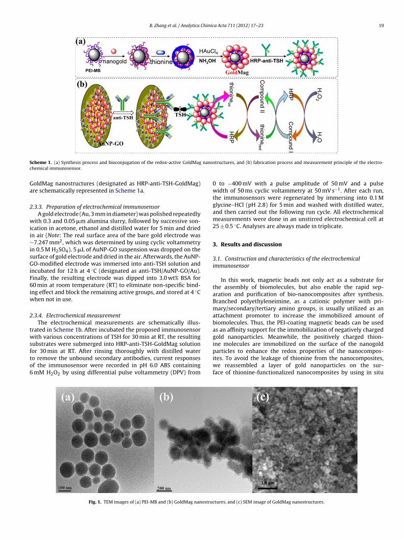
B. Zhang et al. / Analytica Chimica Acta 711 (2012) 17– 23 19
S nanoc
Ga
2
wii∼isGiF6iw
2
twsfto6
cheme 1. (a) Synthesis process and bioconjugation of the redox-active GoldMaghemical immunosensor.
oldMag nanostructures (designated as HRP-anti-TSH-GoldMag)re schematically represented in Scheme 1a.
.3.3. Preparation of electrochemical immunosensorA gold electrode (Au, 3 mm in diameter) was polished repeatedly
ith 0.3 and 0.05 �m alumina slurry, followed by successive son-cation in acetone, ethanol and distilled water for 5 min and driedn air (Note: The real surface area of the bare gold electrode was7.247 mm2, which was determined by using cyclic voltammetry
n 0.5 M H2SO4). 5 �L of AuNP-GO suspension was dropped on theurface of gold electrode and dried in the air. Afterwards, the AuNP-O-modified electrode was immersed into anti-TSH solution and
ncubated for 12 h at 4 ◦C (designated as anti-TSH/AuNP-GO/Au).inally, the resulting electrode was dipped into 3.0 wt% BSA for0 min at room temperature (RT) to eliminate non-specific bind-
ng effect and block the remaining active groups, and stored at 4 ◦Chen not in use.
.3.4. Electrochemical measurementThe electrochemical measurements are schematically illus-
rated in Scheme 1b. After incubated the proposed immunosensorith various concentrations of TSH for 30 min at RT, the resulting
ubstrates were submerged into HRP-anti-TSH-GoldMag solution
or 30 min at RT. After rinsing thoroughly with distilled watero remove the unbound secondary antibodies, current responsesf the immunosensor were recorded in pH 6.0 ABS containingmM H2O2 by using differential pulse voltammetry (DPV) from
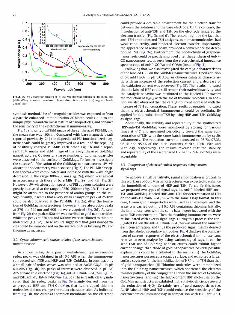
Fig. 1. TEM images of (a) PEI-MB and (b) GoldMag nanostruc
structures, and (b) fabrication process and measurement principle of the electro-
0 to −400 mV with a pulse amplitude of 50 mV and a pulsewidth of 50 ms cyclic voltammetry at 50 mV s−1. After each run,the immunosensors were regenerated by immersing into 0.1 Mglycine–HCl (pH 2.8) for 5 min and washed with distilled water,and then carried out the following run cycle. All electrochemicalmeasurements were done in an unstirred electrochemical cell at25 ± 0.5 ◦C. Analyses are always made in triplicate.
3. Results and discussion
3.1. Construction and characteristics of the electrochemicalimmunosensor
In this work, magnetic beads not only act as a substrate forthe assembly of biomolecules, but also enable the rapid sep-aration and purification of bio-nanocomposites after synthesis.Branched polyethyleneimine, as a cationic polymer with pri-mary/secondary/tertiary amino groups, is usually utilized as anattachment promoter to increase the immobilized amount ofbiomolecules. Thus, the PEI-coating magnetic beads can be usedas an affinity support for the immobilization of negatively chargedgold nanoparticles. Meanwhile, the positively charged thion-ine molecules are immobilized on the surface of the nanogold
particles to enhance the redox properties of the nanocompos-ites. To avoid the leakage of thionine from the nanocomposites,we reassembled a layer of gold nanoparticles on the sur-face of thionine-functionalized nanocomposites by using in situtures, and (c) SEM image of GoldMag nanostructures.
20 B. Zhang et al. / Analytica Chimic
F(a
saut
trnosnwtatdiHgmSctafwmct
3i
rsa6Aacamf
ig. 2. UV–vis absorption spectra of (a) PEI-MB, (b) gold colloids, (c) thionine, andd) GoldMag nanostructures (Inset: UV–vis absorption spectra of (e) magnetic beadsnd (f) PEI).
ynthesis method. Use of nanogold particles was expected to favor particle-enhanced immobilization of biomolecules due to thenique physical and chemical feature of nanoparticles, and enhancehe sensitivity of the electrochemical immunoassay.
Fig. 1a shows typical TEM image of the synthesized PEI-MB, andhe mean size was 100 nm. Compared with bare magnetic beadseported previously [24], the dispersion of PEI-functionalized mag-etic beads could be greatly improved as a result of the repellingf positively charged PEI-MBs each other. Fig. 1b and c repre-ent TEM image and SEM image of the as-synthesized GoldMaganostructures. Obviously, a large number of gold nanoparticlesere attached to the surface of GoldMags. To further investigate
he successful fabrication of the GoldMag nanostructures, UV–visbsorption spectrometry was also used (Fig. 2). The PEI-MB absorp-ion spectra were complicated, and increased with the wavelengthecreased in the range 800–200 nm (Fig. 2a), which was almost
n accordance with those of bare MBs (Fig. 2e) and PEI (Fig. 2f).owever, UV–vis absorption spectra of PEI aqueous solution werereatly increased at the range of 250–200 nm (Fig. 2f). The reasonight be attributed to the presence of amino groups on the PEI.
ignificantly, it seems that a very weak absorption peak at 250 nmould be also observed at the PEI-MBs (Fig. 2a). After the forma-ion of GoldMag nanostructures, however, three absorption peakst 270 nm, 520 nm and 600 nm were acquired (Fig. 2d). As seenrom Fig. 2b, the peak at 520 nm was ascribed to gold nanoparticles,hile the peaks at 270 nm and 600 nm were attributed to thionineolecules (Fig. 2c). These results suggested that gold nanoparti-
les could be immobilized on the surface of MBs by using PEI andhionine as matrices.
.2. Cyclic voltammetric characteristics of the electrochemicalmmunosensor
As shown in Fig. 3e, a pair of well-defined, quasi-reversibleedox peaks was obtained in pH 6.0 ABS when the immunosen-or reacted with TSH and HRP-anti-TSH-GoldMag. In contrast, only
small pair of redox waves was obtained at AuNP-GO/Au in pH.0 ABS (Fig. 3b). No peaks of interest were observed in pH 6.0BS at bare gold electrode (Fig. 3a), anti-TSH/AuNP-GO/Au (Fig. 3c)nd TSH/anti-TSH/AuNP-GO/Au (Fig. 3d). These results clearly indi-
ated that the redox peaks in Fig. 3e mainly derived from thes-prepared HRP-anti-TSH-GoldMag, that is, the doped thionineolecules did not change the redox characteristics. As indicatedrom Fig. 3b, the AuNP-GO complex membrane on the electrode
a Acta 711 (2012) 17– 23
could provide a desirable environment for the electron transferbetween the solution and the base electrode. On the contrary, theintroduction of anti-TSH and TSH on the electrode hindered theelectron transfer (Fig. 3c and d). The reason might be the fact thatanti-TSH antibodies and TSH antigens, as biomacromolecules, hadweak conductivity, and hindered electron transfer. Importantly,the appearance of redox peaks provided a convenience for detec-tion of TSH (Fig. 3e). Furthermore, the conductivity of graphenenanosheets could be greatly improved after the synthesis of AuNP-GO nanocomposites, as seen from the electrochemical impedancespectroscope of AuNP-GO/Au and GO/Au (inset of Fig. 3).
Following that, we also investigated the catalytic characteristicsof the labeled HRP on the GoldMag nanostructures. Upon additionof 6.0 mM H2O2 in pH 6.0 ABS, an obvious catalytic characteris-tic with an increase of the reduction current and a decrease ofthe oxidation current was observed (Fig. 3f). The results indicatedthat the labeled HRP could still remain their native bioactivity, andthe catalytic behavior was attributed to the labeled HRP towardthe reduction of H2O2 with the aid of thionine molecules. In addi-tion, we also observed that the catalytic current increased with theincrease of TSH concentration. These results adequately indicatedthat the electrochemical immunosensor could be preliminarilyapplied for determination of TSH by using HRP-anti-TSH-GoldMagas signal tags.
Additionally, the stability and repeatability of the synthesizedHRP-anti-TSH-GoldMag were monitored by storing for varioustimes at 4 ◦C, and measured periodically toward the same con-centration of TSH with the same-batch immunosensors by cyclicvoltammetry. The reduction currents decreased to 98.7%, 97.3%,96.1% and 95.4% of the initial currents at 5th, 10th, 15th and20th day, respectively. The results revealed that the stabilityand repeatability of the as-prepared HRP-anti-TSH-GoldMag wereacceptable.
3.3. Comparison of electrochemical responses using varioussignal tags
To achieve a high sensitivity, signal amplification is crucial. Inthis work, use of GoldMag nanostructures was expected to enhancethe immobilized amount of HRP-anti-TSH. To clarify this issue,we prepared two types of signal tags, i.e. AuNP-labeled HRP-anti-TSH and GoldMag-labeled HRP-anti-TSH, for determination of TSHon the anti-TSH/AuNP-GO/Au with the same assay format. In thiscase, 16-nm gold nanoparticles were used as an example, and theassay was carried out in pH 6.0 ABS containing thionine. Initially,the immunosensors with the same batch were incubated with thesame TSH concentration. Then the resulting immunosensors werere-incubated with excess signal tags. During this process, the con-jugated TSH on the anti-TSH/AuNP-GO/Au was almost the same foreach concentration, and thus the produced signal mainly derivedfrom the labeled secondary antibodies. Fig. 4 displays the compar-ison of current responses of the electrochemical immunosensorsrelative to zero analyte by using various signal tags. It can beseen that use of GoldMag nanostructures could exhibit highercurrent change than those of gold nanoparticles. Several possibleexplanations could be attributed to the results: (i) The GoldMagnanostructures possessed a scraggy surface, and exhibited a largersurface coverage for the immobilization of HRP-anti-TSH than thatof gold nanoparticles; (ii) Thionine molecules were immobilizedinto the GoldMag nanostructures, which shortened the electrontransfer pathway of the conjugated HRP on the surface of GoldMagnanostructures; and (iii) The high-content HRP molecules on the
GoldMag nanostructures exhibited high catalytic efficiency towardthe reduction of H2O2. Certainly, use of gold nanoparticles (i.e.AuNP-labeled HRP-anti-TSH) could enhance the sensitivity of theelectrochemical immunoassay in comparison with HRP-anti-TSH,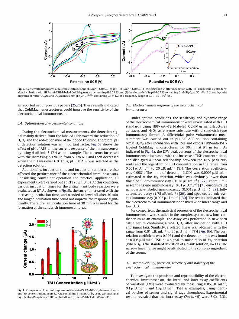
B. Zhang et al. / Analytica Chimica Acta 711 (2012) 17– 23 21
F SH/Aa and (d at a f
ate
3
nHoebwwd
aCeveiaif
Fot
ig. 3. Cyclic voltammograms of (a) gold electrode (Au), (b) AuNP-GO/Au, (c) anti-Tfter incubation with HRP-anti-TSH-labeled GoldMag nanostructures in pH 6.0 ABS,iagrams of AuNP-GO/Au and GO/Au in 5.0 mM [Fe(CN)6]4−/3− containing 0.1 M KCl
s reported in our previous papers [25,26]. These results indicatedhat GoldMag nanostructures could improve the sensitivity of thelectrochemical immunosensor.
.4. Optimization of experimental conditions
During the electrochemical measurements, the detection sig-al mainly derived from the labeled HRP toward the reduction of2O2 and the redox behavior of the doped thionine. Therefore, pHf detection solution was an important factor. Fig. 5a shows theffect of pH of ABS on the current response of the immunosensory using 5 �IU mL−1 TSH as an example. The currents increasedith the increasing pH value from 5.0 to 6.0, and then decreasedhen the pH was over 6.0. Thus, pH 6.0 ABS was selected as theetection solution.
Additionally, incubation time and incubation temperature alsoffected the performance of the electrochemical immunosensors.onsidering convenient operation and practical application, allxperiments were carried out at RT (25 ± 1.0 ◦C). At this condition,arious incubation times for the antigen–antibody reaction werevaluated at RT. As shown in Fig. 5b, the current increased with the
ncreasing incubation time, and trended to level off after 30 min,nd longer incubation time could not improve the response signif-cantly. Therefore, an incubation time of 30 min was used for theormation of the sandwich immunocomplex.ig. 4. Comparison of current responses of the anti-TSH/AuNP-GO/Au toward vari-us TSH concentrations in pH 6.0 ABS containing 6 mM H2O2 by using various signalags: (a) GoldMag-labeled HRP-anti-TSH and (b) AuNP-labeled HRP-anti-TSH.
uNP-GO/Au, (d) the electrode ‘c’ after incubation with TSH and (e) the electrode ‘d’f) the electrode ‘e’ in pH 6.0 ABS containing 6 mM H2O2 at 50 mV s−1 (Inset: Nyquistrequency range of 0.01–1.0 × 10⁵ Hz).
3.5. Electrochemical response of the electrochemicalimmunosensor
Under optimal conditions, the sensitivity and dynamic rangeof the electrochemical immunosensor were investigated with TSHstandards using HRP-anti-TSH-labeled GoldMag nanostructuresas traces and H2O2 as enzyme substrate with a sandwich-typeimmunoassay format. A differential pulse voltammetric mea-surement was carried out in pH 6.0 ABS solution containing6 mM H2O2 after incubation with TSH and excess HRP-anti-TSH-labeled GoldMag nanostructures for 30 min at RT in turn. Asindicated in Fig. 6a, the DPV peak currents of the electrochemicalimmunosensor increased with the increase of TSH concentrations,and displayed a linear relationship between the DPV peak cur-rents and the logarithm of TSH concentration in the range from0.001 �IU mL−1 to 20 �IU mL−1 TSH. The correlation coefficientwas 0.9981. The limit of detection (LOD) was 0.0005 �IU mL−1
estimated at the 3sB criterion, which was obviously lower thanthose of fluoroimmunoassay (0.028 �IU mL−1) [27], chemilumi-nescent enzyme immunoassay (0.01 �IU mL−1) [7], europium(III)nanoparticle-labeled immunoassay (0.0012 �IU mL−1) [28], fullyautomated assay (1.75 �IU mL−1) [29], and spot-coated microw-ells immunoassay (0.003 �IU mL−1) [30]. The results indicated thatthe electrochemical immunosensor enabled wide linear range andlow LOD.
For comparison, the analytical properties of the electrochemicalimmunosensor were studied in the complex system, new born cat-tle serum as an example. The assay was performed in new borncattle serum containing 6 mM H2O2 after incubation with TSHand signal tags. Similarly, a related linear was obtained with therange from 0.01 �IU mL−1 to 20 �IU mL−1 TSH (Fig. 6b). The cor-relation coefficient was 0.9961 and the detection limit was foundat 0.005 �IU mL−1 TSH at a signal-to-noise ratio of 3sB criterion(where sB is the standard deviation of a blank solution, n = 11). Thenarrow linear range might be attributed to the complex ingredientof the serum.
3.6. Reproducibility, precision, selectivity and stability of theelectrochemical immunosensor
To investigate the precision and reproducibility of the electro-chemical immunosensor, the intra- and inter-assay coefficients
of variation (CVs) were evaluated by measuring 0.05 �IU mL−1,0.1 �IU mL−1, and 10 �IU mL−1 TSH as examples, using identi-cal batches of sensor and signal tags throughout. Experimentalresults revealed that the intra-assay CVs (n = 5) were 5.9%, 7.3%,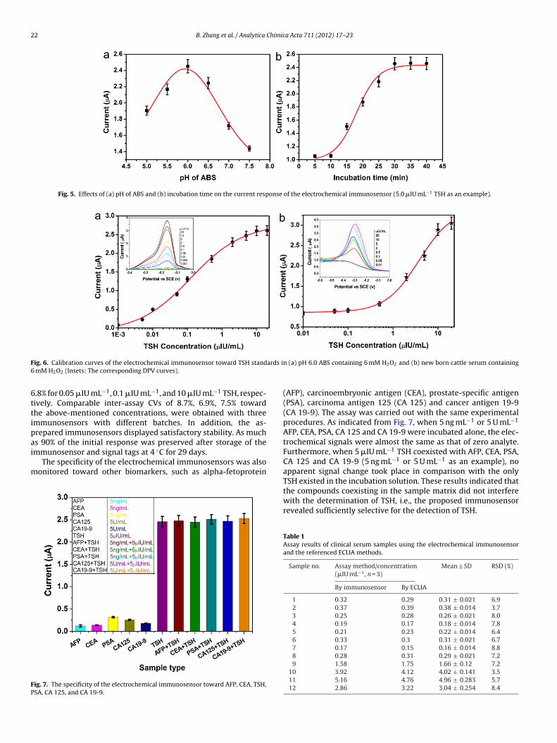
22 B. Zhang et al. / Analytica Chimica Acta 711 (2012) 17– 23
Fig. 5. Effects of (a) pH of ABS and (b) incubation time on the current response of the electrochemical immunosensor (5.0 �IU mL−1 TSH as an example).
F ards i6
6ttipai
m
FP
ig. 6. Calibration curves of the electrochemical immunosensor toward TSH stand mM H2O2 (Insets: The corresponding DPV curves).
.8% for 0.05 �IU mL−1, 0.1 �IU mL−1, and 10 �IU mL−1 TSH, respec-ively. Comparable inter-assay CVs of 8.7%, 6.9%, 7.5% towardhe above-mentioned concentrations, were obtained with threemmunosensors with different batches. In addition, the as-repared immunosensors displayed satisfactory stability. As much
s 90% of the initial response was preserved after storage of themmunosensor and signal tags at 4 ◦C for 29 days.The specificity of the electrochemical immunosensors was alsoonitored toward other biomarkers, such as alpha-fetoprotein
ig. 7. The specificity of the electrochemical immunosensor toward AFP, CEA, TSH,SA, CA 125, and CA 19-9.
n (a) pH 6.0 ABS containing 6 mM H2O2 and (b) new born cattle serum containing
(AFP), carcinoembryonic antigen (CEA), prostate-specific antigen(PSA), carcinoma antigen 125 (CA 125) and cancer antigen 19-9(CA 19-9). The assay was carried out with the same experimentalprocedures. As indicated from Fig. 7, when 5 ng mL−1 or 5 U mL−1
AFP, CEA, PSA, CA 125 and CA 19-9 were incubated alone, the elec-trochemical signals were almost the same as that of zero analyte.Furthermore, when 5 �IU mL−1 TSH coexisted with AFP, CEA, PSA,CA 125 and CA 19-9 (5 ng mL−1 or 5 U mL−1 as an example), noapparent signal change took place in comparison with the only
TSH existed in the incubation solution. These results indicated thatthe compounds coexisting in the sample matrix did not interferewith the determination of TSH, i.e., the proposed immunosensorrevealed sufficiently selective for the detection of TSH.Table 1Assay results of clinical serum samples using the electrochemical immunosensorand the referenced ECLIA methods.
Sample no. Assay method/concentration(�IU mL−1, n = 3)
Mean ± SD RSD (%)
By immunosensor By ECLIA
1 0.32 0.29 0.31 ± 0.021 6.92 0.37 0.39 0.38 ± 0.014 3.73 0.25 0.28 0.26 ± 0.021 8.04 0.19 0.17 0.18 ± 0.014 7.85 0.21 0.23 0.22 ± 0.014 6.46 0.33 0.3 0.31 ± 0.021 6.77 0.17 0.15 0.16 ± 0.014 8.88 0.28 0.31 0.29 ± 0.021 7.29 1.58 1.75 1.66 ± 0.12 7.2
10 3.92 4.12 4.02 ± 0.141 3.511 5.16 4.76 4.96 ± 0.283 5.712 2.86 3.22 3.04 ± 0.254 8.4
Chimic
3
pocoildoma
4
ibAEGonsthgtriacdattb
A
CF
[[[[[
[
[[[[
[
[
[
[
[[[[[
[
B. Zhang et al. / Analytica
.7. Analysis of clinical serum samples
To further elucidate the analytical reliability and applicableotential of the developed immunosensor, the assayed resultsf TSH in human serum specimens using the electrochemi-al immunosensor were compared with the referenced valuesbtained from the commercialized electrochemiluminescencemmunoassay (ECLIA) after appropriate dilution. The results areisted in Table 1. As indicated from Table 1, the relative standardeviations (RSDs) for all the samples were below 9.0%. More-ver, these data show no significant difference between the twoethods. Therefore, the electrochemical immunosensor provided
possible application for detection of TSH in clinical diagnostics.
. Conclusion
This study reports a sensitive and versatile electrochemicalmmunosensor for detection of TSH in clinical immunoassaysy using redox-active GoldMag nanostructures as trace tags anduNP-dispersed graphene nanosheets as immunosensing platform.xperimental results indicated that use of highly conductive AuNP-O nanocomposites greatly decreased the signal-to-noise ratiof the electrochemical immunoassay. The redox-active GoldMaganostructures, conjugated with HRP molecules, enhanced theensitivity of the electrochemical immunoassay. Ultrahigh sensi-ivity was achieved via the amplification of enzymatic signal withigh-content HRP. In addition, AuNPs were directly attached onraphene nanosheets without the need for surface modification,hus decreasing the assay cost. Importantly, GoldMag nanomate-ials not only provide a biocompatible microenvironment for themmobilization of biomolecules, but also facilitate the fast sep-ration and purification after the synthesis. Compared with theonventional methods, e.g. addition of electron mediator in theetection solution, the electroactive GoldMag nanostructures couldvoid the contamination and interference of mediators. Althoughhe present assay system is focused on the determination of thearget hormone, it can be easily extend to the detection of otheriomolecules in clinical diagnostics.
cknowledgements
Support by the National Natural Science Foundation ofhina (nos. 21075019, 41176079, and 20735002), the Researchund for the Doctoral Program of Higher Education of China
[
a Acta 711 (2012) 17– 23 23
(no. 20103514120003), the National Science Foundation of FujianProvince (no. 2011J06003), and the “973” National Basic ResearchProgram of China (no. 2010CB732403) is gratefully acknowledged.Mr. J. Huang (Fujian Provincial Hospital, China) is thanked for ECLIAanalysis of real samples for the method-comparison study.
References
[1] D. Bauer, B. Ettinger, M. Nevitt, K. Stone, Ann. Intern. Med. 134 (2001)561–568.
[2] http://en.wikipedia.org/wiki/Thyroid-stimulating hormone.[3] N. Kobayashi, H. Oyamama, Analyst 136 (2011) 642–651.[4] C. Mello, A. Marangoni, R. Poppi, I. Noda, Anal. Chim. Acta 696 (2011) 47–52.[5] A. Georges, A. Charrie, S. Raynaud, C. Lombard, J. Corcuff, Clin. Chem. Lab. Med.
49 (2011) 873–875.[6] H. Van Deventer, D. Mendu, A. Remaley, S. Slodin, Clin. Chem. 57 (2011)
122–127.[7] Z. Lin, X. Wang, Z. Li, S. Ren, G. Chen, X. Ying, J. Lin, Talanta 75 (2008) 965–972.[8] F. Wu, S. Han, T. Xu, Y. He, Anal. Biochem. 314 (2003) 87–96.[9] S. Laschi, S. Centi, M. Mascini, Bioanal. Rev. 3 (2011) 11–25.10] D. Tang, R. Yuan, Y. Chai, J. Phys. Chem. B 110 (2006) 11640–11646.11] S. D’Souza, Biosens. Bioelectron. 16 (2001) 337–353.12] D. Du, S. Liu, J. Chen, H. Ju, H. Lian, J. Li, Biomaterials 26 (2005) 6487–6495.13] D. Knopp, D. Tang, R. Niessner, Anal. Chim. Acta 647 (2009) 14–30.14] D. Du, Z. Zou, Y. Shin, J. Wang, H. Wu, M. Engelhard, J. Liu, I. Aksay, Y. Lin, Anal.
Chem. 82 (2010) 2989–2995.15] C. Rao, A. Sood, K. Subrahmanyam, A. Govindarai, Angew. Chem. Int. Ed. 48
(2009) 7752–7777.16] P. Avouris, Nano Lett. 10 (2010) 4285–4294.17] M. Pumera, Chem. Soc. Rev. 39 (2010) 4146–4157.18] Y. Li, R. Yuan, Y. Chai, Z. Song, Electrochim. Acta 56 (2011) 6715–6721.19] J. Tang, D. Tang, B. Su, Q. Li, B. Qiu, G. Chen, Electrochim. Acta 56 (2011)
3773–3780.20] Y. Yang, S. Dong, T. Shen, C. Jian, H. Chang, Y. Li, J. Zhou, Electrochim. Acta 56
(2011) 6021–6025.21] M. Szymanski, R. Porter, G. Dep, Y. Wang, B. Haggett, Phys. Chem. Chem. Phys.
13 (2011) 5383–5387;J. Tang, D. Tang, B. Su, J. Huang, B. Qiu, G. Chen, Biosens. Bioelectron. 26 (2011)3219–3226.
22] X. Chen, G. Wu, J. Chen, X. Chen, Z. Xie, X. Wang, J. Am. Chem. Soc. 133 (2011)3693–3695.
23] B. Zhang, D. Tang, B. Liu, H. Chen, Y. Cui, G. Chen, Biosens. Bioelectron. 28 (2011)174–180.
24] D. Tang, B. Su, J. Tang, J. Ren, G. Chen, Anal. Chem. 82 (2010) 1527–1534.25] D. Tang, R. Yuan, Y. Chai, Anal. Chem. 80 (2008) 1582–1588.26] D. Tang, J. Ren, Anal. Chem. 80 (2008) 8064–8070.27] F. Wu, Sh. Han, T. Xu, Y. He, Anal. Biochem. Acta 314 (2003) 87–96.28] A. Pelkkikangas, S. Jaakohuhta, T. Lövgren, H. Härmä, Anal. Chim. Acta 517
(2004) 169–176.29] D. Hermsen, M. Broecker-Preuss, M. Casati, J. Mas, A. Eckstein, D. Gassner, J.
van Helden, K. Inomata, J. Jarausch, J. Kratzsch, K. Mann, N. Miyazaki, M. Angel,N. Moreno, T. Murakami, H. Roth, J. Noh, W. Scherbaum, M. Schott, Clin. Chim.Acta 401 (2009) 84–89.
30] J. Ylikotila, L. Valimaa, M. Vehniainen, H. Takalo, T. Lovgren, K. Pettersson, J.Immunol. Methods 306 (2005) 104–114.