Diagnosis of Oropouche Virus Infection Using a Recombinant Nucleocapsid Protein-Based Enzyme...
8
JOURNAL OF CLINICAL MICROBIOLOGY, 0095-1137/01/$04.0010 DOI: 10.1128/JCM.39.7.2445–2452.2001 July 2001, p. 2445–2452 Vol. 39, No. 7 Copyright © 2001, American Society for Microbiology. All Rights Reserved. Diagnosis of Oropouche Virus Infection Using a Recombinant Nucleocapsid Protein-Based Enzyme Immunoassay MOHAMMAD F. SAEED, 1,2 MARCIO NUNES, 3 PEDRO F. VASCONCELOS, 3 AMELIA P. A. TRAVASSOS DA ROSA, 3 DOUGLAS M. WATTS, 4 KEVIN RUSSELL, 4 ROBERT E. SHOPE, 1,2,5 ROBERT B. TESH, 1,2,5 AND ALAN D. T. BARRETT 1,2,5 * Departments of Microbiology and Immunology 1 and Pathology 5 and Center for Tropical Diseases, 2 The University of Texas Medical Branch, Galveston, Texas 77555; Instituto Evandro Chagas, Belem, Brazil 3 ; and U.S. Naval Medical Research Center Detachment, Lima, Peru 4 Received 19 October 2000/Returned for modification 30 January 2001/Accepted 11 April 2001 Oropouche (ORO) virus is an emerging infectious agent that has caused numerous outbreaks of an acute febrile (dengue-like) illness among humans in Brazil, Peru, and Panama. Diagnosis of ORO virus infection is based mainly on serology. Two different antigens, hamster serum antigen (HSA) and Vero cell lysate antigen (VCLA), are currently used in enzyme immunoassays (EIAs) in Brazil and Peru, respectively, to investigate the epidemiology of ORO virus infection. Both antigens involve use of infectious virus, and for this reason their use is restricted. Consequently, the frequency and distribution of ORO virus infection are largely unexplored in other countries of South America. This report describes the use of a bacterially expressed recombinant nucleocapsid (rN) protein of ORO virus in EIAs for the diagnosis of ORO virus infection. The data revealed that the purified rN protein is comparable to the authentic viral N protein in its antigenic characteristics and is highly sensitive and specific in EIAs. Among 183 serum samples tested, a high degree of concordance was found between rN protein-based EIA and HSA- and VCLA-based EIAs for the detection of both ORO virus-specific immunoglobulin M (IgM) and IgG antibodies. The high sensitivity, specificity, and safety of the rN protein-based EIA make it a useful diagnostic technique that can be widely used to detect ORO virus infection in South America. Oropouche (ORO) virus, a member of the Simbu serogroup of the genus Bunyavirus, family Bunyaviridae, is an emerging human pathogen that is transmitted mainly by the biting midge Culicoides paraensis. ORO virus is the etiologic agent of ORO fever, a dengue-like acute febrile illness that is a significant public health problem in tropical South America. Over the past 39 years there have been at least 30 reported outbreaks of ORO fever, involving more than half a million people in trop- ical South America (14). Seroepidemiological investigations of ORO fever in Brazil indicate that, while the prevalence of ORO antibodies is relatively low (0 to 2%) in areas where epidemic transmission has not been reported (13), during ep- idemics an incidence rate of 30% is common and may reach as high as 60% (10). The disease has also been reported in Pan- ama and is endemic in humid tropical regions of Peru, with a fairly high (18 to 46%) seroprevalence rate (20). Due to the debilitating nature and duration (5 to 6 days) of symptoms and the high incidence rates during epidemics, ORO fever clearly has the potential for substantial social and economic impact. The clinical diagnosis of ORO fever is difficult because of the nonspecific nature of symptoms; it is easily confused with dengue fever and a number of other arbovirus illnesses that are common in tropical South America. Laboratory diagnosis of the disease is usually based on serological methods including the plaque reduction neutralization test, complement fixation test, hemagglutination inhibition (HI) test, and enzyme immu- noassay (EIA). However, all of these techniques require the handling of infectious virus at some part of the process; since ORO virus is a biosafety level 3 agent, this limits the labora- tories that can work with it. Consequently, only a few labora- tories in South America currently conduct serological testing for ORO fever, and the frequency and distribution of the disease are not well known. To investigate the epidemiology of ORO fever, we have developed an EIA for the laboratory diagnosis of ORO virus infection. This assay is based on the bacterially expressed re- combinant nucleocapsid (rN) protein, and its advantages in- clude simplicity, safety of use, and the possibility of standard- ization, in addition to good sensitivity and specificity. Thus, our rN protein-based EIA can potentially be used to investigate the epidemiology of ORO virus infection and possibly to ob- tain a more accurate estimate of the impact of ORO fever on human health in tropical South America. MATERIALS AND METHODS Virus and cell culture. Vero-E6 cells were cultivated in Eagle’s minimal es- sential medium (EMEM) supplemented with 10% fetal bovine serum (FBS) and penicillin (100 U/ml)-streptomycin (100 mg/ml). ORO virus strain MD023 was used; this virus was originally isolated from the blood of a human with ORO fever in Peru and was propagated in Vero cells in EMEM supplemented with 2% FBS and antibiotics. When 75 to 80% of the cellular monolayer had cytopathic effect, the culture supernatant was collected and centrifuged at 2,000 3 g for 5 to 10 min to remove cellular debris. The supernatant was aliquoted in 1-ml volumes and frozen at 270°C as stock virus. Serum samples and immunological reagents. Of the 183 human serum sam- ples used in this study, 108 were obtained from individuals during ORO fever epidemiological investigations in Brazil and 75 were obtained from individuals as part of previous ORO surveillance studies in Iquitos and Santa Clara, Peru. The negative-control human sera were obtained from the World Arbovirus Refer- * Corresponding author. Mailing address: Department of Pathology, University of Texas Medical Branch, Galveston, TX 77555-0609. Phone: (409) 772-6662. Fax: (409) 747-2415. E-mail: abarrett@utmb .edu. 2445 on August 1, 2016 by guest http://jcm.asm.org/ Downloaded from
-
Upload
independent -
Category
Documents
-
view
2 -
download
0
Transcript of Diagnosis of Oropouche Virus Infection Using a Recombinant Nucleocapsid Protein-Based Enzyme...

JOURNAL OF CLINICAL MICROBIOLOGY,0095-1137/01/$04.0010 DOI: 10.1128/JCM.39.7.2445–2452.2001
July 2001, p. 2445–2452 Vol. 39, No. 7
Copyright © 2001, American Society for Microbiology. All Rights Reserved.
Diagnosis of Oropouche Virus Infection Using a RecombinantNucleocapsid Protein-Based Enzyme Immunoassay
MOHAMMAD F. SAEED,1,2 MARCIO NUNES,3 PEDRO F. VASCONCELOS,3
AMELIA P. A. TRAVASSOS DA ROSA,3 DOUGLAS M. WATTS,4 KEVIN RUSSELL,4
ROBERT E. SHOPE,1,2,5 ROBERT B. TESH,1,2,5 AND ALAN D. T. BARRETT1,2,5*
Departments of Microbiology and Immunology1 and Pathology5 and Center for Tropical Diseases,2
The University of Texas Medical Branch, Galveston, Texas 77555; Instituto Evandro Chagas,Belem, Brazil3; and U.S. Naval Medical Research Center Detachment, Lima, Peru4
Received 19 October 2000/Returned for modification 30 January 2001/Accepted 11 April 2001
Oropouche (ORO) virus is an emerging infectious agent that has caused numerous outbreaks of an acutefebrile (dengue-like) illness among humans in Brazil, Peru, and Panama. Diagnosis of ORO virus infection isbased mainly on serology. Two different antigens, hamster serum antigen (HSA) and Vero cell lysate antigen(VCLA), are currently used in enzyme immunoassays (EIAs) in Brazil and Peru, respectively, to investigate theepidemiology of ORO virus infection. Both antigens involve use of infectious virus, and for this reason their useis restricted. Consequently, the frequency and distribution of ORO virus infection are largely unexplored inother countries of South America. This report describes the use of a bacterially expressed recombinantnucleocapsid (rN) protein of ORO virus in EIAs for the diagnosis of ORO virus infection. The data revealedthat the purified rN protein is comparable to the authentic viral N protein in its antigenic characteristics andis highly sensitive and specific in EIAs. Among 183 serum samples tested, a high degree of concordance wasfound between rN protein-based EIA and HSA- and VCLA-based EIAs for the detection of both OROvirus-specific immunoglobulin M (IgM) and IgG antibodies. The high sensitivity, specificity, and safety of therN protein-based EIA make it a useful diagnostic technique that can be widely used to detect ORO virusinfection in South America.
Oropouche (ORO) virus, a member of the Simbu serogroupof the genus Bunyavirus, family Bunyaviridae, is an emerginghuman pathogen that is transmitted mainly by the biting midgeCulicoides paraensis. ORO virus is the etiologic agent of OROfever, a dengue-like acute febrile illness that is a significantpublic health problem in tropical South America. Over the past39 years there have been at least 30 reported outbreaks ofORO fever, involving more than half a million people in trop-ical South America (14). Seroepidemiological investigations ofORO fever in Brazil indicate that, while the prevalence ofORO antibodies is relatively low (0 to 2%) in areas whereepidemic transmission has not been reported (13), during ep-idemics an incidence rate of 30% is common and may reach ashigh as 60% (10). The disease has also been reported in Pan-ama and is endemic in humid tropical regions of Peru, with afairly high (18 to 46%) seroprevalence rate (20). Due to thedebilitating nature and duration (5 to 6 days) of symptoms andthe high incidence rates during epidemics, ORO fever clearlyhas the potential for substantial social and economic impact.
The clinical diagnosis of ORO fever is difficult because ofthe nonspecific nature of symptoms; it is easily confused withdengue fever and a number of other arbovirus illnesses that arecommon in tropical South America. Laboratory diagnosis ofthe disease is usually based on serological methods includingthe plaque reduction neutralization test, complement fixationtest, hemagglutination inhibition (HI) test, and enzyme immu-
noassay (EIA). However, all of these techniques require thehandling of infectious virus at some part of the process; sinceORO virus is a biosafety level 3 agent, this limits the labora-tories that can work with it. Consequently, only a few labora-tories in South America currently conduct serological testingfor ORO fever, and the frequency and distribution of thedisease are not well known.
To investigate the epidemiology of ORO fever, we havedeveloped an EIA for the laboratory diagnosis of ORO virusinfection. This assay is based on the bacterially expressed re-combinant nucleocapsid (rN) protein, and its advantages in-clude simplicity, safety of use, and the possibility of standard-ization, in addition to good sensitivity and specificity. Thus, ourrN protein-based EIA can potentially be used to investigatethe epidemiology of ORO virus infection and possibly to ob-tain a more accurate estimate of the impact of ORO fever onhuman health in tropical South America.
MATERIALS AND METHODS
Virus and cell culture. Vero-E6 cells were cultivated in Eagle’s minimal es-sential medium (EMEM) supplemented with 10% fetal bovine serum (FBS) andpenicillin (100 U/ml)-streptomycin (100 mg/ml). ORO virus strain MD023 wasused; this virus was originally isolated from the blood of a human with OROfever in Peru and was propagated in Vero cells in EMEM supplemented with 2%FBS and antibiotics. When 75 to 80% of the cellular monolayer had cytopathiceffect, the culture supernatant was collected and centrifuged at 2,000 3 g for 5to 10 min to remove cellular debris. The supernatant was aliquoted in 1-mlvolumes and frozen at 270°C as stock virus.
Serum samples and immunological reagents. Of the 183 human serum sam-ples used in this study, 108 were obtained from individuals during ORO feverepidemiological investigations in Brazil and 75 were obtained from individuals aspart of previous ORO surveillance studies in Iquitos and Santa Clara, Peru. Thenegative-control human sera were obtained from the World Arbovirus Refer-
* Corresponding author. Mailing address: Department of Pathology,University of Texas Medical Branch, Galveston, TX 77555-0609.Phone: (409) 772-6662. Fax: (409) 747-2415. E-mail: [email protected].
2445
on August 1, 2016 by guest
http://jcm.asm
.org/D
ownloaded from

ence Center at the University of Texas Medical Branch (Galveston, Tex.). Thesesera were collected from healthy individuals residing in the United States whowere known to have never visited South America.
Hyperimmune mouse ascitic fluids (HIMAF) used in Western blotting andEIA were obtained from the World Arbovirus Reference Center. Mice receivedfour weekly intraperitoneal injections of 10%-infected newborn mouse brainmixed with Freund’s adjuvant, followed by sarcoma-180 cells, as described byTikasingh et al. (18).
Anti-Xpress antibody, used in a Western blot assay to identify the full-lengthrecombinant N protein containing a His tag leader peptide, was purchased fromInvitrogen (Carlsbad, Calif.). It is a mouse monoclonal antibody that specificallyreacts with a His tag leader peptide encoded by the pTrcHis expression vectorused in these studies.
Amplification and cloning of nucleocapsid gene. Viral RNA was extractedfrom 0.5 ml of stock virus, as described previously (12). To amplify the nu-cleocapsid gene, the RNA was subjected to reverse transcription-PCR usingprimers ORON5 (59-AAAGAGGATCCAATAATGTCAGAGTTCATTT-39)and ORON3 (59-GTGAATTCCACTATATGCCAATTCCGAATT-39), as de-scribed previously (15). Primers ORON5 and ORON3 were designed so as togenerate BamHI and EcoRI sites, respectively. The amplicon was first clonedinto the TA cloning vector (pCR2.1; Invitrogen), amplified in bacteria, anddigested with BamHI and EcoRI restriction enzymes. The insert DNA was thenrecovered and cloned into the BamHI and EcoRI sites of pTrcHisB expressionvector (Invitrogen). The recombinant expression vector was transformed intocells, which were then selected on Luria-Bertani (LB) agar containing ampicillin(100 mg/ml). Following overnight incubation in LB broth containing ampicillin,plasmid DNA was extracted and analyzed for the presence of insert DNA bydigestion with BamHI and EcoRI restriction enzymes.
Purification of ORO virus rN protein. Purification of the recombinant proteinwas carried out using the pTrcHis Xpress kit (Invitrogen) according to themanufacturer’s instructions. Briefly, bacteria containing the recombinant expres-sion vector were grown in LB broth supplemented with ampicillin. When theoptical density at 600 nm (OD600) of the culture reached 0.6, protein expressionwas induced by the addition of 5 mM isopropyl-b-D-thiogalactopyranoside(IPTG). After 5 to 6 h of induction, bacteria were pelleted and resuspended inbuffer (50 mM sodium phosphate and 300 mM NaCl, pH 7.8) containing ly-sozyme (100 mg/ml) and incubated on ice for 30 min. Thereafter, cells weresonicated and centrifuged at 10,000 3 g for 15 min. The supernatant was loadedon a ProBond histidine-binding column, preequilibrated with buffer containing50 mM sodium phosphate and 300 mM NaCl, pH 7.8. Subsequent to rinsing withthe washing buffer (50 mM sodium phosphate and 300 mM NaCl, pH 6.0), therecombinant protein was eluted with a concentration gradient (0 to 1.0 M) ofimidazole. Each of the eluted fractions was analyzed by electrophoresis on asodium dodecyl sulfate (SDS)–12% polyacrylamide gel. The identity of theexpressed protein was confirmed by Western blot analysis using ORO virus-specific HIMAF and human serum from an ORO virus-infected individual.
Preparation of hamster serum antigen (HSA). One hundred microliters ofbrain homogenate of newborn mice infected with ORO virus (strain BeAn19991) was inoculated intraperitoneally into 4- to 5-week-old female Syriangolden hamsters (Mesocricetus auratus). When the hamsters first showed signs ofillness (36 to 48 h postinoculation), blood was collected by cardiac puncture andallowed to clot at 4°C. Subsequently, the serum was removed and diluted 10-foldwith isotonic saline solution. The diluted serum was expressed into 20 volumes ofdry, chilled acetone with an 18-gauge needle and extracted for 5 min withintermittent shaking. The sample was then centrifuged at 500 3 g for 5 min at4°C; the supernatant was discarded, and the sediment was resuspended in 20volumes of chilled acetone by vigorous shaking. After incubation for 1 h at 4°C,the sample was centrifuged at 500 3 g for 10 to 15 min and the sediment wasdried under vacuum at room temperature for 1 h. Finally, the dried sediment wasresuspended in a sufficient volume of borate-saline solution (0.12 M NaCl, 0.05M H3BO3, 0.024 N NaOH, pH 9.0) to make a 1:10 dilution based on the originalvolume of serum and stored at 270°C in 1- to 2-ml aliquots.
The use of animals in this study was in accordance with a University of TexasMedical Branch protocol for the use of animals in biomedical research.
Preparation of VCLA. Vero cell lysate antigen (VCLA) was prepared essen-tially as described by Beaty et al. (1). Briefly, Vero cells were infected with OROvirus (strain MD023). At the time when cytopathic effects began to appear(approximately 20 to 25% cell death), cells were harvested, centrifuged at10,000 3 g for 10 min at 4°C, and washed once with 0.1 M borate-saline solution(pH 9.0). Thereafter, the cells were resuspended in borate-saline containing 1%Triton X-100, sonicated, and centrifuged at 10,000 3 g for 5 min at 4°C. Thesupernatant was collected, aliquoted, and stored at 4°C.
EIA. (i)IgG EIA. Wells of microtiter plates were coated with antigen (purifiedORO virus rN protein or HSA or VCLA) and diluted in carbonate-bicarbonatebuffer, pH 9.6, and the plates were incubated at 4°C. Subsequently, the plateswere washed five times with phosphate-buffered saline (PBS), pH 7.4 (Gibco-BRL), containing 0.05% Tween 20 (Sigma Chemical Co., St. Louis, Mo.) fol-lowed by the addition of 250 ml of blocking buffer (4% bovine serum albumin inPBS) to each well. After incubation for 15 to 20 min at 37°C, the blocking bufferwas aspirated and 100-ml portions of serum samples (diluted 1:400 in blockingbuffer) were added to the wells and the plates were incubated at 37°C for 1 h.Thereafter, the plates were washed five times as described above, and 100 ml ofperoxidase-conjugated goat anti-human immunoglobulin G (IgG) (Kirkegaard &Perry Laboratories, Gaithersburg, Md.), diluted 1:2,000 in blocking buffer, wasadded to each well, followed by incubation for 1 h at 37°C. The plates were thenwashed as described above and 100 ml of peroxidase substrate, 2.29-azino-di[3-ethyl-benzthiazoline sulfonate (6)] (Kirkegaard & Perry Laboratories), wasadded to each well. Plates were then incubated at room temperature for 15 to 30min, and OD405 was measured in a microplate reader. At least one positive-control serum and three negative-control sera were included in each assay. Thecutoff was the mean OD405 of negative samples plus three standard deviations.Test sera having an OD405 greater than the cutoff were considered positive.
(ii)IgM EIA. An IgM capture method was used as follows. Wells of microtiterplates were coated with 100 ml of anti-human IgM (Kirkegaard & Perry Labo-ratories) diluted 1:200 in carbonate-bicarbonate buffer, pH 9.6, and the plateswere incubated at 4°C for at least 16 h. Subsequently, the plates were washed fivetimes with wash buffer (PBS–0.05% Tween 20), and 250 ml of blocking buffer(4% bovine serum albumin in PBS) was added to each well. After incubation for15 to 20 min, the blocking buffer was aspirated and 50 ml of test sera (diluted1:400 in blocking buffer) was added to wells and the plates were incubated at37°C for 1 h. The plates were then washed as described above, and 50 ml ofappropriately diluted antigen (rN protein or HSA) was added to each well,followed by incubation for 1 h at 37°C. The plates were again washed, and 50 mlof ORO virus-specific HIMAF, diluted 1:200 in blocking buffer, was added toeach well. After a 1-h incubation at 37°C, the plates were washed, 50 ml ofperoxidase-conjugated sheep anti-mouse Ig (diluted 1:2,000; Amersham) wasadded to each well, and the plates were incubated at 37°C for 1 h. Finally, afterthe washing, 100 ml of peroxidase substrate was added and the plates wereincubated at room temperature for 15 to 30 min. OD was measured in a micro-plate reader at 405 nm. The cutoff was calculated as described above.
Neutralization assay. Serum samples, diluted 10-fold in PBS, were heat-inac-tivated at 56°C for 30 min, mixed with an equal volume of ORO virus strainMD023 (diluted in PBS to give 40 to 50 PFU/0.1 ml), and incubated at 37°C for1 h. Subsequently, 0.2 ml of virus-serum mixture was inoculated into confluentmonolayer cultures of Vero cells grown in six-well tissue culture plates. Afterincubation for 30 min at room temperature, each well was overlaid with 5 ml of1% agar (Sigma Chemical Co.) containing EMEM supplemented with 2% FBS.Thereafter, plates were incubated at 37°C. After 4 to 5 days, monolayers werestained with neutral red and the plaques in each well were counted. At least threenegative-control serum samples were included. Serum samples that showed an80% or greater reduction in number of plaques compared to negative-controlsera were considered neutralizing, and those that showed less than 80% reduc-tion at 1:20 serum dilution were considered to be negative.
HI assays. The HI tests were performed according to the methods of Clarkeand Casals (3) using modifications for microtiter plates (17). For these assays, theHSA described above was used as the antigen. Prior to the HI assay, the test serawere extracted with acetone and adsorbed with goose red blood cells as describedby Beaty et al. (1).
SDS-polyacrylamide gel electrophoresis (PAGE) and Western blotting. Pro-tein samples were resolved on an SDS–12% polyacrylamide gel by electrophore-sis, using the method of Laemmli (9). The resolved samples were then trans-ferred to a polyvinylidene difluoride membrane (Bio-Rad) by electroblotting ina buffer containing Tris-HCl (25 mM), glycine (192 mM), and methanol (20%[vol/vol]). Subsequently, the membrane was incubated with 5% nonfat milk (inPBS) for 1 h at room temperature to block nonspecific protein binding sites.Thereafter, the membrane was incubated with the primary antibody (OROvirus-specific HIMAF diluted 1:200 or ORO virus-specific human serum diluted1:50 or anti-Xpress diluted 1:5,000 in 5% nonfat milk) for 1 h at room temper-ature, followed by washing with PBS and incubation with a secondary antibody(alkaline phosphatase-conjugated goat anti-mouse or anti-human IgG; SigmaChemical Co.). After 1 h at room temperature, the membrane was washed andthe antigen-antibody complex on the membrane was detected by the addition ofa small volume of BCIP (5-bromo-4-chloro-3-indolylphosphate)-nitroblue tetra-zolium (Sigma Chemical Co.), the substrate for alkaline phosphatase.
2446 SAEED ET AL. J. CLIN. MICROBIOL.
on August 1, 2016 by guest
http://jcm.asm
.org/D
ownloaded from

RESULTS
Amplification and cloning of the ORO virus N cDNA. Thegenomic RNA of ORO virus (strain MD023) was extractedand subjected to reverse transcription-PCR using primers de-scribed above to amplify the N gene. The nucleotide sequenceof the amplicon was confirmed by sequencing as describedelsewhere (15). Subsequently, the amplified cDNA was clonedinto the pTrcHisB expression vector (Invitrogen) between theBamHI and EcoRI sites in frame with the start codon of theHis tag leader peptide. The recombinant construct was trans-formed into competent Escherichia coli cells, and plasmidDNA extraction and digestion with BamHI and EcoRI restric-tion enzymes confirmed the presence of the construct in trans-formed bacteria (data not shown). A single colony of the trans-formed bacteria was grown in LB broth and treated with IPTGto induce the expression of rN protein, which was then ana-lyzed by PAGE and Western blotting, using HIMAF raisedagainst ORO virus. The results (Fig. 1a) show the presence oftwo anti-ORO virus antibody-reactive bands in extracts of bac-teria transformed with the recombinant plasmid (lane 2) butnot in extracts of bacteria transformed with vector alone (lane1). This suggested that the ORO virus rN protein was ex-pressed in bacteria; however there were two forms of theprotein. From the molecular weight estimates, the slower-mi-grating band corresponded to the full-length recombinant fu-sion protein, while the faster-migrating band represented thetruncated version. SDS-PAGE revealed that the faster-migrat-ing band corresponded in size to the authentic viral N proteinexpressed in ORO virus-infected Vero cells (Fig. 1a; comparelanes 2 and 3), suggesting that the truncated version consists ofthe entire N protein lacking the His tag leader peptide. Thiswas confirmed by Western blot analysis using antibodiesagainst the His tag fusion peptide (Invitrogen). The resultsshow that, while both forms reacted with anti-ORO virus an-
tibodies (Fig. 1a, lane 2), only the slower-migrating form wasreactive to the anti-His tag antibodies (Fig. 1b, lane 2).
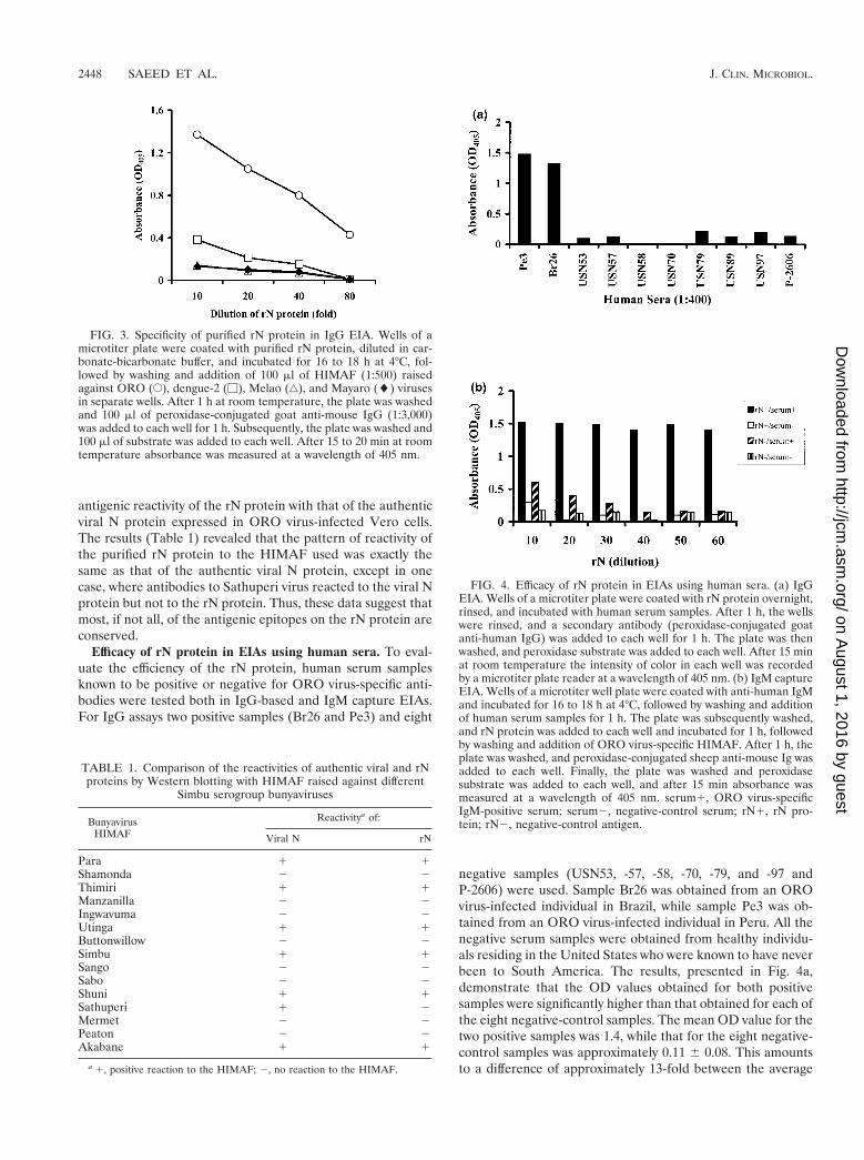
Purification of ORO virus rN protein. The ORO virus rNprotein, expressed in E. coli, was purified by affinity chroma-tography. The recombinant protein was eluted with a gradientof 0 to 1.0 M imidazole. Lysate from bacteria transformed withvector alone was used as a negative control. Each fraction wasanalyzed by SDS-PAGE and immunoblotting. The resultsshow that only two bands, representing the two forms of the rNprotein, were present in fractions eluted with 0.5 M imidazole(Fig. 2a). Both bands were absent in corresponding fractionseluted from control bacterial lysate (data not shown). Westernblot analyses revealed that both bands were reactive to OROvirus-specific mouse immune ascitic fluid (Fig. 2b) and humanserum from an ORO virus-infected individual (data notshown). These data, therefore, attest that the rN protein waspurified from the bacterial lysate to homogeneity. Use of theeluted fraction, containing both bands, in EIA using HIMAFraised against ORO, dengue-2, Mayaro, and Melao viruses (allof them prevalent in South America) showed that the rN pro-tein reacted specifically to HIMAF raised against ORO virusand that there was no cross-reactivity to HIMAF raised againstthe other viruses examined (Fig. 3). These data, therefore,suggest that, despite the presence of two forms, the purified rNprotein is highly specific in EIAs for detecting antibodies toORO virus.
Antigenic characterization of the rN protein. Serologicaltests have indicated that there is extensive cross-reactivityamong the N proteins of many Simbu serogroup viruses (7). Todetermine if the antigenic epitopes were conserved on the rNprotein, HIMAF raised against several Simbu serogroup vi-ruses (Table 1) were obtained from World Arbovirus Refer-ence Center and used in Western blot analyses to compare the
FIG. 1. Reactivity of recombinant and virally expressed N proteinsof ORO virus with anti-ORO virus and anti-His tag antibodies. Proteinsamples were resolved on an SDS-polyacrylamide gel and transferredto polyvinylidene difluoride membranes by electroblotting. The mem-branes were then probed with ORO virus-specific HIMAF (1:200) (a)or anti-Xpess (anti-His tag leader peptide; 1:5,000; Invitrogen) (b)antibody. Lane M, kaleidoscope molecular weight marker (Bio-Rad);lane 1, lysate from IPTG-induced control bacteria (bacteria trans-formed with vector alone); lane 2, lysate from IPTG-induced bacteriatransformed with a recombinant plasmid (pTrcHisB-ORO18N); lane3, lysate from ORO virus-infected Vero cells; lane 4, nickel chelateaffinity-purified rN protein.
FIG. 2. Purification of ORO virus rN protein. Following IPTGinduction bacteria containing the recombinant plasmid were lysed andthe rN protein was purified by metal chelate affinity chromatography.The bound protein was eluted from the column by a concentrationgradient (0 to 0.5 M) of imidazole. The eluted fractions were analyzedby SDS-PAGE followed by Coomassie blue staining of the gel (a) andimmunoblotting using ORO virus-specific HIMAF (1:200) (b). LaneM, kaleidoscope molecular weight marker (Bio-Rad); lane 1, bacteriallysate before purification; lane 2, fraction from 0.05 M imidazoleelution; lane 3, fraction from 0.2 M imidazole elution; lanes 4 and 5,fractions from 0.35 M imidazole elution; lanes 6 and 7, fractions from0.5 M imidazole elution. Arrows indicate the positions of two OROantibody-reactive bands.
VOL. 39, 2001 RECOMBINANT ORO VIRUS N PROTEIN-BASED EIA 2447
on August 1, 2016 by guest
http://jcm.asm
.org/D
ownloaded from
antigenic reactivity of the rN protein with that of the authenticviral N protein expressed in ORO virus-infected Vero cells.The results (Table 1) revealed that the pattern of reactivity ofthe purified rN protein to the HIMAF used was exactly thesame as that of the authentic viral N protein, except in onecase, where antibodies to Sathuperi virus reacted to the viral Nprotein but not to the rN protein. Thus, these data suggest thatmost, if not all, of the antigenic epitopes on the rN protein areconserved.
Efficacy of rN protein in EIAs using human sera. To eval-uate the efficiency of the rN protein, human serum samplesknown to be positive or negative for ORO virus-specific anti-bodies were tested both in IgG-based and IgM capture EIAs.For IgG assays two positive samples (Br26 and Pe3) and eight
negative samples (USN53, -57, -58, -70, -79, and -97 andP-2606) were used. Sample Br26 was obtained from an OROvirus-infected individual in Brazil, while sample Pe3 was ob-tained from an ORO virus-infected individual in Peru. All thenegative serum samples were obtained from healthy individu-als residing in the United States who were known to have neverbeen to South America. The results, presented in Fig. 4a,demonstrate that the OD values obtained for both positivesamples were significantly higher than that obtained for each ofthe eight negative-control samples. The mean OD value for thetwo positive samples was 1.4, while that for the eight negative-control samples was approximately 0.11 6 0.08. This amountsto a difference of approximately 13-fold between the average
FIG. 3. Specificity of purified rN protein in IgG EIA. Wells of amicrotiter plate were coated with purified rN protein, diluted in car-bonate-bicarbonate buffer, and incubated for 16 to 18 h at 4°C, fol-lowed by washing and addition of 100 ml of HIMAF (1:500) raisedagainst ORO (E), dengue-2 (M), Melao (‚), and Mayaro (l) virusesin separate wells. After 1 h at room temperature, the plate was washedand 100 ml of peroxidase-conjugated goat anti-mouse IgG (1:3,000)was added to each well for 1 h. Subsequently, the plate was washed and100 ml of substrate was added to each well. After 15 to 20 min at roomtemperature absorbance was measured at a wavelength of 405 nm.
FIG. 4. Efficacy of rN protein in EIAs using human sera. (a) IgGEIA. Wells of a microtiter plate were coated with rN protein overnight,rinsed, and incubated with human serum samples. After 1 h, the wellswere rinsed, and a secondary antibody (peroxidase-conjugated goatanti-human IgG) was added to each well for 1 h. The plate was thenwashed, and peroxidase substrate was added to each well. After 15 minat room temperature the intensity of color in each well was recordedby a microtiter plate reader at a wavelength of 405 nm. (b) IgM captureEIA. Wells of a microtiter well plate were coated with anti-human IgMand incubated for 16 to 18 h at 4°C, followed by washing and additionof human serum samples for 1 h. The plate was subsequently washed,and rN protein was added to each well and incubated for 1 h, followedby washing and addition of ORO virus-specific HIMAF. After 1 h, theplate was washed, and peroxidase-conjugated sheep anti-mouse Ig wasadded to each well. Finally, the plate was washed and peroxidasesubstrate was added to each well, and after 15 min absorbance wasmeasured at a wavelength of 405 nm. serum1, ORO virus-specificIgM-positive serum; serum2, negative-control serum; rN1, rN pro-tein; rN2, negative-control antigen.
TABLE 1. Comparison of the reactivities of authentic viral and rNproteins by Western blotting with HIMAF raised against different
Simbu serogroup bunyaviruses
BunyavirusHIMAF
Reactivitya of:
Viral N rN
Para 1 1Shamonda 2 2Thimiri 1 1Manzanilla 2 2Ingwavuma 2 2Utinga 1 1Buttonwillow 2 2Simbu 1 1Sango 2 2Sabo 2 2Shuni 1 1Sathuperi 1 2Mermet 2 2Peaton 2 2Akabane 1 1
a 1, positive reaction to the HIMAF; 2, no reaction to the HIMAF.
2448 SAEED ET AL. J. CLIN. MICROBIOL.
on August 1, 2016 by guest
http://jcm.asm
.org/D
ownloaded from

OD values for positive- and negative-control samples, suggest-ing that the rN protein specifically reacts to ORO virus-specificIgG antibodies in human sera with no or very little nonspecificreaction. Similarly, in the IgM capture EIA, the OD valueobtained for the positive-control sample was significantlyhigher than that for the negative-control sample (Fig. 4b),suggesting a highly specific reaction of the rN protein to OROvirus-specific IgM antibodies. Thus, these data suggest that therN protein is quite efficient at specifically detecting both IgGand IgM antibodies to ORO virus in human sera.
Diagnostic potential of purified rN protein by EIA. Sensi-tivity and specificity are important issues for a diagnostic re-agent. Statistically, sensitivity refers to the probability that thediagnostic reagent gives a positive result when the individualtested actually has the disease, while specificity is the proba-bility that the diagnostic reagent gives a negative result whenthe individual tested does not have the disease. In other words,the sensitivity of a diagnostic reagent is high if it has no, or avery low frequency of, false negatives. Similarly, the specificityis high when the frequency of false positives is zero or very low.To evaluate the sensitivity and specificity of the rN protein ina diagnostic EIA, the purified-rN protein-based EIA was com-pared with VCLA-based and HSA-based EIAs using humanserum samples.
Comparison with HSA for the detection of IgG antibodies.Initially, 108 human serum samples from Brazil were tested forIgG by rN protein- and HSA-based EIAs. Of the 108 serumsamples tested, 71 were positive and 25 were negative for OROvirus-specific IgG with both antigens (Table 2). This corre-sponds to a concordance of approximately 89% between theresults obtained using the two antigens. Seven samples whichtested positive in the rN protein-based EIA were negative inthe HSA-based EIA, while five samples which tested positivein the HSA-based EIA were negative in the rN protein-basedEIA. To investigate the discordance in more detail, neutral-ization and HI assays were performed on discordant samplesplus control samples of positive and negative concordant sam-
ples. ORO virus strain MD023 was used for neutralizationassays. The results (Table 3) show that six (Br21, Br44, Br69,Br85, Br89, and Br90) of the seven discordant samples thattested positive by rN protein-based EIA but negative by HSA-based EIA were positive for anti-ORO virus antibodies, bothby neutralization (.80% plaque inhibition at 1:20 dilution)and HI (each with HI titer of $160) tests. The remainingsample (Br 40) tested negative both by neutralization (#80%plaque inhibition at 1:20 dilution) and HI (titer of #20) tests.Similarly, four (Br30, Br32, Br36, and Br71) of the five discor-dant sera that tested positive by HSA-based EIA but negativeby rN protein-based EIA tested positive by neutralization(.80% plaque inhibition at 1:20 dilution) and HI tests (HItiter of $80), while one sample (Br12) tested negative by thetwo assays (#80% plaque inhibition at 1:20 dilution and HItiter of #20) (Table 3). Together, these data correspond tofour false-negative results and one false-positive result by rNprotein-based EIA compared to six false-negative results andone false-positive result by HSA-based EIA. Thus, based on108 Brazilian samples tested, the error frequency for the rNprotein is equivalent to, if not lower than, that for HSA, andtherefore the rN protein was comparable to HSA in its sensi-tivity and specificity for detecting ORO virus-specific IgG an-tibodies in human sera.
Comparison with HSA for the detection IgM antibodies.The rN protein and HSA were compared using a total of 127serum samples (52 from Brazil and 75 from Peru) in IgMcapture EIAs. Of the 52 Brazilian samples tested, 25 werepositive and 19 were negative, while all of the Peruvian sampleswere negative for ORO virus-specific IgM antibodies with bothantigens (Table 2). This corresponds to a concordance of ap-proximately 94% between the results obtained by rN protein-and HSA-based EIAs. The eight discordant samples (Br8, -11,-17, -19, -30, -41, -43, and -44) represented those that testedpositive by HSA-based EIA but negative by rN protein-basedEIA. All of the discordant samples were positive in neutral-ization tests (.80% plaque inhibition at 1:20 dilution; Table
TABLE 2. Comparison of rN protein-based EIA with HSA-basedEIA to detect ORO virus-specific IgG and IgM and VCLA-basedEIA to detect ORO virus-specific IgG antibodies in human seraa
HSA- or VCLA-basedEIA result
No. of samples withindicated result in rN
protein-based EIA Concordance (%)
Positive Negative
HSAIgG EIA 88.9
Positive 71 5Negative 7 25
IgM EIA 93.7Positive 25 8Negative 0 94
VCLAIgG EIA 86.7
Positive 36 9Negative 1 29
a Positive and Negative, absorbance values above and below the cutoff, respec-tively. The cutoff was equal to the mean plus three standard deviations of theabsorbance values obtained with three negative-control sera run alongside ineach experiment.
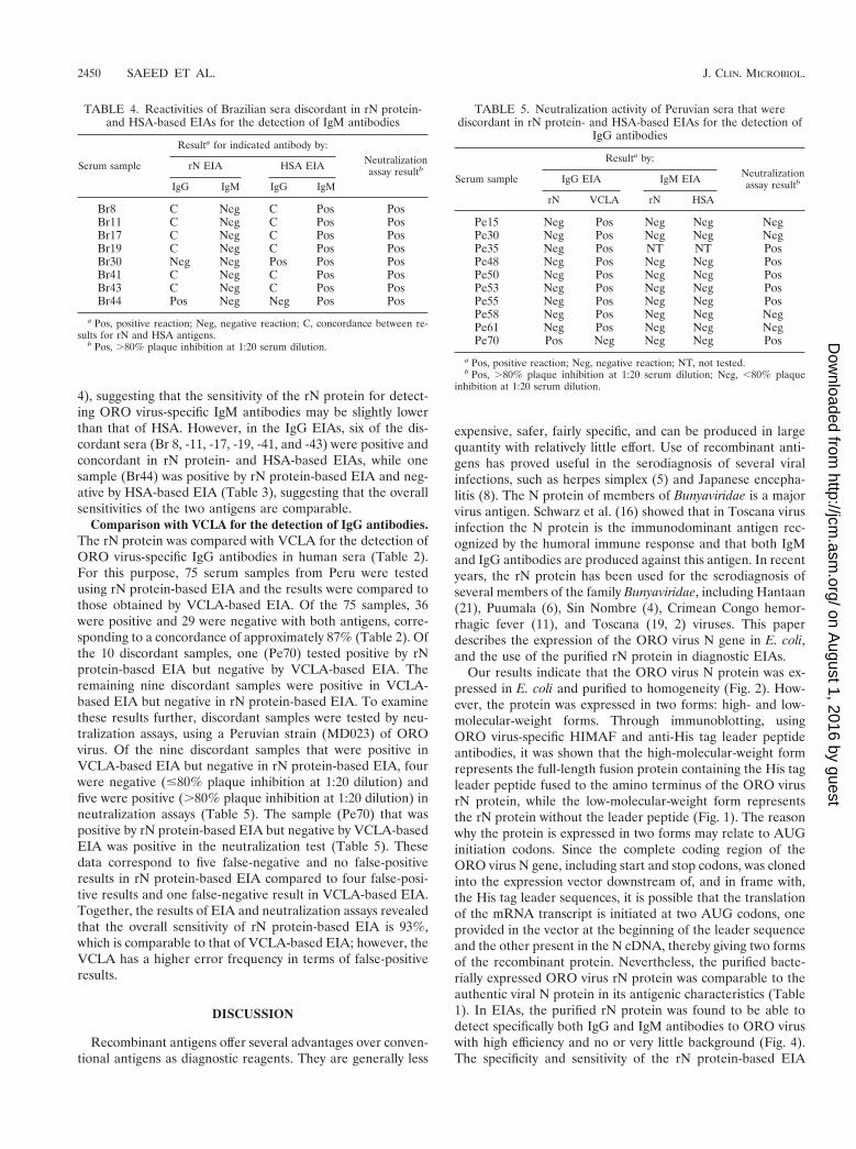
TABLE 3. Reactivities of Brazilian sera discordant in rN protein-and HSA-based EIAs for the detection of IgM antibodies
Serumsample
Resulta for indicatedantibody by:
Neutralizationassay resultb
HI assayresultcrN EIA HSA EIA
IgG IgM IgG IgM
Br12 Neg C Pos C Neg NegBr21 Pos C Neg C Pos PosBr30 Neg Neg Pos Pos Pos PosBr32 Neg C Pos C Pos PosBr36 Neg C Pos C Pos PosBr40 Pos C Neg C Neg NegBr44 Pos Neg Neg Pos Pos PosBr69 Pos NT Neg NT Pos PosBr71 Neg NT Pos NT Pos PosBr85 Pos NT Neg NT Pos PosBr89 Pos NT Neg NT Pos PosBr90 Pos NT Neg NT Pos Pos
a Pos, positive reaction; Neg, negative reaction; C, concordance between re-sults for rN and HSA antigens; NT, not tested.
b Pos, .80% plaque inhibition at 1:20 serum dilution; Neg, ,80% plaqueinhibition at 1:20 dilution.
c Pos, HI titer, .160; Neg, HI titer, ,20.
VOL. 39, 2001 RECOMBINANT ORO VIRUS N PROTEIN-BASED EIA 2449
on August 1, 2016 by guest
http://jcm.asm
.org/D
ownloaded from
4), suggesting that the sensitivity of the rN protein for detect-ing ORO virus-specific IgM antibodies may be slightly lowerthan that of HSA. However, in the IgG EIAs, six of the dis-cordant sera (Br 8, -11, -17, -19, -41, and -43) were positive andconcordant in rN protein- and HSA-based EIAs, while onesample (Br44) was positive by rN protein-based EIA and neg-ative by HSA-based EIA (Table 3), suggesting that the overallsensitivities of the two antigens are comparable.
Comparison with VCLA for the detection of IgG antibodies.The rN protein was compared with VCLA for the detection ofORO virus-specific IgG antibodies in human sera (Table 2).For this purpose, 75 serum samples from Peru were testedusing rN protein-based EIA and the results were compared tothose obtained by VCLA-based EIA. Of the 75 samples, 36were positive and 29 were negative with both antigens, corre-sponding to a concordance of approximately 87% (Table 2). Ofthe 10 discordant samples, one (Pe70) tested positive by rNprotein-based EIA but negative by VCLA-based EIA. Theremaining nine discordant samples were positive in VCLA-based EIA but negative in rN protein-based EIA. To examinethese results further, discordant samples were tested by neu-tralization assays, using a Peruvian strain (MD023) of OROvirus. Of the nine discordant samples that were positive inVCLA-based EIA but negative in rN protein-based EIA, fourwere negative (#80% plaque inhibition at 1:20 dilution) andfive were positive (.80% plaque inhibition at 1:20 dilution) inneutralization assays (Table 5). The sample (Pe70) that waspositive by rN protein-based EIA but negative by VCLA-basedEIA was positive in the neutralization test (Table 5). Thesedata correspond to five false-negative and no false-positiveresults in rN protein-based EIA compared to four false-posi-tive results and one false-negative result in VCLA-based EIA.Together, the results of EIA and neutralization assays revealedthat the overall sensitivity of rN protein-based EIA is 93%,which is comparable to that of VCLA-based EIA; however, theVCLA has a higher error frequency in terms of false-positiveresults.
DISCUSSION
Recombinant antigens offer several advantages over conven-tional antigens as diagnostic reagents. They are generally less
expensive, safer, fairly specific, and can be produced in largequantity with relatively little effort. Use of recombinant anti-gens has proved useful in the serodiagnosis of several viralinfections, such as herpes simplex (5) and Japanese encepha-litis (8). The N protein of members of Bunyaviridae is a majorvirus antigen. Schwarz et al. (16) showed that in Toscana virusinfection the N protein is the immunodominant antigen rec-ognized by the humoral immune response and that both IgMand IgG antibodies are produced against this antigen. In recentyears, the rN protein has been used for the serodiagnosis ofseveral members of the family Bunyaviridae, including Hantaan(21), Puumala (6), Sin Nombre (4), Crimean Congo hemor-rhagic fever (11), and Toscana (19, 2) viruses. This paperdescribes the expression of the ORO virus N gene in E. coli,and the use of the purified rN protein in diagnostic EIAs.
Our results indicate that the ORO virus N protein was ex-pressed in E. coli and purified to homogeneity (Fig. 2). How-ever, the protein was expressed in two forms: high- and low-molecular-weight forms. Through immunoblotting, usingORO virus-specific HIMAF and anti-His tag leader peptideantibodies, it was shown that the high-molecular-weight formrepresents the full-length fusion protein containing the His tagleader peptide fused to the amino terminus of the ORO virusrN protein, while the low-molecular-weight form representsthe rN protein without the leader peptide (Fig. 1). The reasonwhy the protein is expressed in two forms may relate to AUGinitiation codons. Since the complete coding region of theORO virus N gene, including start and stop codons, was clonedinto the expression vector downstream of, and in frame with,the His tag leader sequences, it is possible that the translationof the mRNA transcript is initiated at two AUG codons, oneprovided in the vector at the beginning of the leader sequenceand the other present in the N cDNA, thereby giving two formsof the recombinant protein. Nevertheless, the purified bacte-rially expressed ORO virus rN protein was comparable to theauthentic viral N protein in its antigenic characteristics (Table1). In EIAs, the purified rN protein was found to be able todetect specifically both IgG and IgM antibodies to ORO viruswith high efficiency and no or very little background (Fig. 4).The specificity and sensitivity of the rN protein-based EIA
TABLE 4. Reactivities of Brazilian sera discordant in rN protein-and HSA-based EIAs for the detection of IgM antibodies
Serum sample
Resulta for indicated antibody by:Neutralizationassay resultbrN EIA HSA EIA
IgG IgM IgG IgM
Br8 C Neg C Pos PosBr11 C Neg C Pos PosBr17 C Neg C Pos PosBr19 C Neg C Pos PosBr30 Neg Neg Pos Pos PosBr41 C Neg C Pos PosBr43 C Neg C Pos PosBr44 Pos Neg Neg Pos Pos
a Pos, positive reaction; Neg, negative reaction; C, concordance between re-sults for rN and HSA antigens.
b Pos, .80% plaque inhibition at 1:20 serum dilution.
TABLE 5. Neutralization activity of Peruvian sera that werediscordant in rN protein- and HSA-based EIAs for the detection of
IgG antibodies
Serum sample
Resulta by:Neutralizationassay resultbIgG EIA IgM EIA
rN VCLA rN HSA
Pe15 Neg Pos Neg Neg NegPe30 Neg Pos Neg Neg NegPe35 Neg Pos NT NT PosPe48 Neg Pos Neg Neg PosPe50 Neg Pos Neg Neg PosPe53 Neg Pos Neg Neg PosPe55 Neg Pos Neg Neg PosPe58 Neg Pos Neg Neg NegPe61 Neg Pos Neg Neg NegPe70 Pos Neg Neg Neg Pos
a Pos, positive reaction; Neg, negative reaction; NT, not tested.b Pos, .80% plaque inhibition at 1:20 serum dilution; Neg, ,80% plaque
inhibition at 1:20 serum dilution.
2450 SAEED ET AL. J. CLIN. MICROBIOL.
on August 1, 2016 by guest
http://jcm.asm
.org/D
ownloaded from

were compared with those of HSA- and VCLA-based EIAs,the two assays currently used for the serodiagnosis of OROvirus infection in Brazil and Peru, respectively. The rN proteinwas found to be highly sensitive and specific in detecting IgGand IgM antibodies to ORO virus in human sera. A total of 183human serum samples (108 from Brazil and 75 from Peru)were tested for the presence of ORO virus-specific IgG anti-bodies, and 127 serum samples (52 from Brazil and 75 fromPeru) were tested for the presence of ORO virus-specific IgMantibodies. A high degree of concordance was found betweenthe results obtained with the rN protein and with either of theother two antigens (89% with HSA and 87% with VCLA) forthe detection of ORO virus-specific IgG (Table 2). Examina-tion of the discordant samples by neutralization and/or HIassays revealed that, out of 183 serum samples tested by rNprotein-based EIA, only 1 (0.5%) was a false positive and 9(4.9%) were false negatives, which raises the overall sensitivityand specificity of the rN protein-based EIA to approximately95 and 99.5%, respectively. In contrast, of the 108 serum sam-ples tested by HSA-based EIA, one (0.9%) was a false positiveand six (5.5%) were false negatives, which equates to an overallsensitivity of approximately 93% and specificity of 99% forHSA-based EIA. Similarly, of the 75 samples tested by VCLA-based EIA, four (5.3%) were false positives and one (1.3%)was a false negative, which equates to an overall sensitivity of98% and specificity of approximately 95% for VCLA-basedEIA. Thus, these data suggest that the sensitivity of the rNprotein for the detection of ORO virus-specific IgG antibodiesin human sera is comparable to those of the other two antigenscurrently used for the diagnosis of ORO virus infection; how-ever, in terms of specificity the rN protein is better than VCLA.In IgM capture assays, the frequency of false-negative sampleswas relatively higher for the rN protein (approximately 6%)than for HSA, suggesting that HSA has a relatively highersensitivity than the rN protein for the detection of ORO virus-specific IgM antibodies. However, the overall concordance be-tween the two antigens was approximately 95% (Table 2).
Because of its high sensitivity (95%) and specificity (99.5%),the rN protein may prove very useful in obtaining a moreaccurate estimate of the seroprevalence of ORO virus infec-tion in tropical regions of South America; it may also help inobtaining an estimate of the prevalence of infection by otherORO-like viruses and reassortants. Although the percentageof false-negative results for IgG antibodies detected by the rNprotein-based EIA (4.9%) is comparable to that of the HSA-based EIA (5. 5%), it is still higher than that of the VCLA-based EIA (1.3%). There may be several explanations for this.First, it is possible that the samples that gave false-negativeresults may contain IgG antibodies to only the viral surfaceglycoproteins. Presumably, during ORO virus infection IgGantibodies to viral surface glycoproteins appear earlier thanthe IgG antibodies to the N protein. Thus, if a serum is col-lected at a stage when IgG antibodies to the N protein areundetectable or below detectable levels, the rN protein-basedEIA would not score that serum as positive, while HSA andVCLA, being whole-virus antigens, would be able to detectIgG antibodies to viral surface glycoproteins. Similarly, beingdirected towards surface glycoproteins, neutralization and HIassays would also score such a serum as positive. An alternateexplanation may be infection by a reassortant virus having the
medium (M) RNA segment of ORO virus and the small (S)RNA segment of a different bunyavirus. In such a situation,neutralization and HI assays and VCLA-based and HSA-basedEIAs, since they are able to detect viral glycoproteins (prod-ucts of the M RNA segment), would score the serum sample aspositive. On the other hand the rN protein-based EIA, whichdetects antibodies to the N protein (a product of the S RNAsegment), would score the serum sample as negative. This issueneeds to be further investigated, especially in view of the needfor more-accurate epidemiological investigations, to define thedistribution and prevalence of ORO infection in South Amer-ica.
In conclusion, the rN protein-based EIA is a potentiallygood assay for the detection of both IgG and IgM antibodies toORO virus with high sensitivity and specificity. Thus, the rNprotein-based EIA appears to be a simple and readily accessi-ble assay for diagnosing ORO virus infection. These charac-teristics make the rN protein a useful diagnostic reagent fordiagnostic and epidemiological purposes in tropical SouthAmerica.
ACKNOWLEDGMENTS
We are thankful to David Beasley of our laboratory for help withgraphics and to Stuart Nichol, Mike Bowen, Pierre Rollin, and C. J.Peters of the National Centers for Disease Control and Prevention,Atlanta, Ga., for helpful discussions.
This work was supported in part by NIH grants AI 43336 and AI10984. Mohammad Saeed was supported in part by the James W.McLaughlin fellowship fund.
REFERENCES
1. Beaty, B. J., C. H. Calisher, and R. E. Shope. 1995. Arboviruses, p. 189–212.In E. H. Lennette, D. A. Lennette, and E. T. Lennette (ed.), Diagnosticprocedures for viral, rickettsial and chlamydial infections. American PublicHealth Association, Washington, D.C.
2. Ciufolini, M. G., C. Fiorentini, P. Di Bonito, S. Mochi, and C. Giorgi. 1999.Detection of Toscana virus-specific immunoglobulins G and M by an en-zyme-linked immunosorbent assay based on recombinant viral nucleopro-tein. J. Clin. Microbiol. 37:2010–2012.
3. Clarke, D. H., and J. Casals. 1958. Techniques for haemagglutination andhaemagglutination inhibition with arthropod-borne viruses. Am. J. Trop.Med. Hyg. 7:561–573.
4. Feldmann, H., A. Sanchez, S. Morzunov, C. F. Spiropoulou, P. E. Rollin,T. G. Ksiazek, C. J. Peters, and S. T. Nichols. 1993. Utilization of autopsyRNA for the synthesis of the nucleocapsid antigen of a newly recognizedvirus associated with hantavirus pulmonary syndrome. Virus Res. 30:351–367.
5. Kakkanas, A., H. Papadogeorgaki, R. Manservigi, V. Miriagou, U. Georgo-poulou, and P. Mavromara. 1995. Escherichia coli expressed herpes simplexvirus gG1 and gG2 proteins in ELISA and immunoblotting assays. Intervi-rology 38:346–351.
6. Kallio-Kokko, H., O. Vapalahit, A. Lundkvist, and A. Vaheri. 1998. Evalu-ation of Puumala virus IgG and IgM enzyme immunoassays based on re-combinant baculovirus-expressed nucleocapsid protein for early nephro-pathia epidemica diagnosis. Clin. Diagn. Virol. 10:83–90.
7. Kinney, R. M., and C. H. Calisher. 1981. Antigenic relationship amongSimbu serogroup (Bunyaviridae) viruses. Am. J. Trop. Med. Hyg. 30:1307–1318.
8. Konishi, E., P. W. Mason, and R. E. Shope. 1996. Enzyme-linked immu-nosorbent assay using recombinant antigens for serodiagnosis of Japaneseencephalitis. J. Med. Virol. 48:76–79.
9. Laemmli, U. K. 1970. Cleavage of structural proteins during the assembly ofthe head of bacteriophage T4. Nature 227:680–685.
10. Leduc, J. W., and F. P. Pinheiro. 1989. Oropouche fever, p. 1–15. In T. P.Monath (ed.), The arboviruses: epidemiology and ecology, vol. 4. CRC Press,Boca Raton, Fla.
11. Marriot, A. C., T. Polyzoni, A. Antoniadis, and P. A. Nuttall. 1994. Detectionof human antibodies to Crimean-Congo hemorrhagic fever virus using ex-pressed viral nucleocapsid protein. J. Gen. Virol. 75:2157–2161.
12. Ni, H., and A. D. T. Barrett. 1995. Nucleotide and amino acid differences ofthe structural protein genes of Japanese encephalitis virus from differentgeographical locations. J. Gen. Virol. 76:401–407.
VOL. 39, 2001 RECOMBINANT ORO VIRUS N PROTEIN-BASED EIA 2451
on August 1, 2016 by guest
http://jcm.asm
.org/D
ownloaded from
13. Pinheiro, F. P., A. P. A. Travassos da Rosa, R. Ishak, R. B. Freitas, M. L.Gomez, J. W. LeDuc, and O. F. Olivia. 1981. Oropouche virus: a review ofclinical, epidemiological and ecological findings. Am. J. Trop. Med. Hyg.30:149–160.
14. Pinheiro, F. P., A. P. A. Travassos da Rosa, and P. F. C. Vasconcelos. 1998.An overview of Oropouche fever epidemics in Brazil and neighbouringcountries, p. 186–192. In A. P. A. Travassos da Rosa, P. F. C. Vasconcelos,and J. F. S. Travassos da Rosa (ed.), An overview of arbovirology in Braziland neighbouring countries. The Evandro Chagas Institute, Belem, Brazil.
15. Saeed, M. F., H. Wang, M. Nunes, P. Vasconcelos, S. C. Weaver, R. E. Shope,D. M. Watts, R. B. Tesh, and A. D. T. Barrett. 2000. Nucleotide sequencesand phylogeny of the nucleocapsid gene of Oropouche virus. J. Gen. Virol.81:743–748.
16. Schwarz, T. F., S. Gilch, C. Pauli, and G. Jager. 1996. Immunoblot detectionof antibodies to Toscana virus. J. Med. Virol. 49:83–86.
17. Sever, J. H., A. C. Ley, F. Walman, B. M. Caplan, P. W. Crockett, and H. C.
Turner. 1964. Utilization of disposable plastic plates with a serologic micro-technique. Am. J. Clin. Pathol. 41:167–170.
18. Tikasingh, E. S., L. Spence, and W. G. Downs. 1966. The use of adjuvantsand sarcoma 180 cells in the production of mouse hyperimmune ascitic fluidsto arboviruses. Am. J. Trop. Med. Hyg. 15:219–226.
19. Valassina, M., D. Soldateschi, G. Maria dal Maso, L. Santini, S. Bianchi,P. E. Valensin, and M. G. Cusi. 1998. Diagnostic potential of Toscana virusN protein expressed in Escherichia coli. J. Clin. Microbiol. 36:3170–3172.
20. Watts, D. M., I. Phillips, J. D. Callahan, W. Greibenow, K. C. Hyams, andC. G. Hayes. 1997. Oropouche virus transmission in Amazon river basin ofPeru. Am. J. Trop. Med. Hyg. 56:148–152.
21. Zoller, L., S. Yang, P. Gott, E. K. Bautz, and G. Darai. 1993. Use recombi-nant nucleocapsid proteins of the Hantaan and nephropathia epidemicaserotypes of Hantaviruses as immunodiagnostic antigens. J. Med. Virol.39:200–207.
2452 SAEED ET AL. J. CLIN. MICROBIOL.
on August 1, 2016 by guest
http://jcm.asm
.org/D
ownloaded from