Development and Characterization of Monoclonal Antibodies ...
Upload
khangminh22Category
view
2download
0
HAL Id: tel-00922980https://tel.archives-ouvertes.fr/tel-00922980
Submitted on 1 Jan 2014
HAL is a multi-disciplinary open accessarchive for the deposit and dissemination of sci-entific research documents, whether they are pub-lished or not. The documents may come fromteaching and research institutions in France orabroad, or from public or private research centers.
L’archive ouverte pluridisciplinaire HAL, estdestinée au dépôt et à la diffusion de documentsscientifiques de niveau recherche, publiés ou non,émanant des établissements d’enseignement et derecherche français ou étrangers, des laboratoirespublics ou privés.
Design and Optimization of Recombinant AntibodiesDirected Against Platelet Glycoprotein VI with
Therapeutic and Diagnostic PotentialsMuhammad Zahid
To cite this version:Muhammad Zahid. Design and Optimization of Recombinant Antibodies Directed Against PlateletGlycoprotein VI with Therapeutic and Diagnostic Potentials. Human health and pathology. UniversitéParis Sud - Paris XI, 2011. English. �NNT : 2011PA114835�. �tel-00922980�

UNIVERSITÉ PARIS-SUD 11
ECOLE DOCTORALE :
INNOVATION THÉRAPEUTIQUE : DU FONDAMENTAL A L’APPLIQUÉ
PÔLE : PHYSIOPATHOLOGIE MOLECULAIRE ET CELLULAIRE
DISCIPLINE :
Physiopathologie cellulaire et moléculaire
ANNÉE 2011 - 2012 SÉRIE DOCTORAT N°
THÈSE DE DOCTORAT
soutenue le 24 novembre 2011
par
Muhammad ZAHID
Design and Optimization of Recombinant Antibodies Directed Against Platelet Glycoprotein VI with Therapeutic and Diagnostic
Potentials
Directeur de thèse : Professeur Philippe BILLIALD, EA4530, Université Paris-Sud 11
Co-directeur de thèse : Docteur Martine JANDROT-PERRUS INSERM U698
Composition du jury :
Président du jury : Professeur Dominique PORQUET, Doyen de la Faculté de Pharmacie, Université Paris-Sud 11
Rapporteurs : Docteur Richard LE NAOUR, EA 4303, Université de Reims, Champagne-Ardenne
Professeur Philippe NGUYEN, EA 3801, Université de Reims, Champagne-Ardenne
Examinateurs : Professeur Philippe BILLIALD, Faculté de Pharmacie, Université Paris-Sud 11
Professeur Delphine BORGEL, EA4531, Faculté de Pharmacie, Université Paris-Sud 11
Docteur Martine JANDROT-PERRUS, Inserm U698, Hôpital Bichat, Paris


ACKNOWLEDGEMENT
I would like to begin by thanking Professor Philippe BILLIALD for his super-human
patience and valuable advice. Thank you for standing by me at all tough times and for being
part of the great moments of my PhD. Thank you, for your vision and for teaching me the
intricacies and the beauty of the recombinant antibody technology. Once again a very warm
‘thank you’ to gave a naive pakistani boy a chance to begin his journey through molecular
biology, for introducing me to the world of science and for instilling in me the spirit of the
free scientist.
I have no appropriate words to manifest my feeling of respect, gratefulness and
obligations for Dr. Martine JANDROT-PERRUS for providing constant guidance, useful and
timely suggestions and encouragement throughout my research work. She really took great
interest in my whole research work. Thanks, for your advice on a million things, for teaching
me how to be pragmatic in research, for innumerable interesting discussions… I could go on.
I am still waiting for my quiche!
I thank Professor Dominique PORQUET and Professor Delphine BORGEL for
accepting to be part of my jury. I would like to express my utmost gratitude to Professor
Philippe NGUYEN and Dr. Richard LE NAOUR for their valuable comments and discussions
on my thesis.
I am thankful to Professor Jean-Baptiste MICHEL, Professor Cécile BERNARD and
Professor Christian POÜS who opened their labs to me and who taught me that hard work
does pay off!
I am also greatly thankful to Professor Max GOYFFON for his valuable and thought
provoking advices.
Thank you Professor Isabelle DIMIER-POISSON for providing all the possible
facilities in your lab during my master and Dr. Nicolas AUBREY for your help throughout
my research work for especially recombinant antibody fragments purification.
A successful thesis in biology is never accomplished alone. I have been extremely
lucky to be surrounded by many bright and caring people. Stéphane LOYAU, you are one in
a million. Thank you for holding my hand during my work in the lab, for teaching me the
basics of laboratory life and for listening to my cribbing!

Maxime BOUABDELLI, we started this long journey together, and now we are each
heading in separate directions. Thanks for always staying beside me, through the good times
and the worst! Thanks for helping me out in many experiments.
Thanks Rana IFTIKHAR for many great moments, for being thoughtful and thanks for
listening to my million cribs and for being an excellent roommate! Good luck for a great PhD.
Thank you Marie Christine BOUTON, Véronique AROCAS, Véronique OLLIVER
Laurence VENISSE, Claude YEPREMIAN and Magali NOIRAY for your advice on
everything.
Finally, I thank my loving family without whom I would not be here today. Lastly I
would like to mention the name of my son Khizar ZAHID, whose sweet memories always
remain in my heart.
I thank everyone involved in the work presented in this thesis.

I
Table of Contents
PART A: STATE OF THE ART
1 Historical Development of Therapeutic Antibodies ...........................................1
1.1 Antibodies Engineering...............................................................................6
1.1.1 Structure of Ig Immunoglobulin ..........................................................6
1.1.2 Antibodies in Bodies............................................................................7
1.1.3 Murine Monoclonal Antibodies Production ........................................8
1.1.4 Chimerization.....................................................................................10
!"!"# Humanization.....................................................................................12
1.1.6 Alternatives ........................................................................................16
1.2 Fully Human Antibodies ...........................................................................17
1.2.1 Human Monoclonal Antibodies .........................................................17
1.2.2 Different Approaches of Human Monoclonal Antibodies Prod. .......18
1.2.3 Human B Cell Hybridoma .................................................................18
1.3 Antibodies Selecting Methods ..................................................................20
1.3.1 Exposure on the Surface of Phage (Phage Display) ..........................21
1.3.2 Exposure on the Surface of a Ribosome (Ribosome Display)...........23
1.3.3 Exposure on the Yeast Surface (Yeast Display) ................................24
1.4 Design of Novel Antibody Formats with Antigen-binding Activity.........24
1.4.1 Monovalent Antibody Formats ..........................................................24
1.4.2 Polyvalent Antibody Fragments ........................................................34
1.4.3 Armed Antibodies (Cancer/Immunoconjugates) ...............................38
1.4.4 Antibody Fragment Expression System.............................................39
2 Platelets, Thrombosis and Anti-Thrombotics ...................................................49
2.1 Hemostasis and Thrombosis......................................................................49
2.1.1 Major Types of Thrombosis...............................................................51
2.1.2 Different Phases of Thrombus Formation..........................................52
2.1.3 Stabilization Phase or Perpetuation ...................................................53
2.1.4 Actors Involved in Thrombus Formation ..........................................53
2.2 Platelets .....................................................................................................58
$"$"! Platelet Structure................................................................................59

II
2.2.2 Platelet Plug Formation......................................................................65
2.2.3 Other Functions of Platelets besides Hemostasis...............................70
2.3 Platelet Receptors: Structure and Function ...............................................71
2.3.1 The GPIb/V/IX Complex and vWF ...................................................71
2.3.2 Integrins .............................................................................................74
2.3.3 Collagen Receptors ............................................................................76
2.3.4 G-Protein-Coupled Receptors (GPCRs) ............................................78
2.3.5 Recently Identified Receptors Stabilizing Thrombosis .....................82
2.4 Antithrombotic Drugs ...............................................................................84
2.4.1 Anticoagulants ...................................................................................84
2.4.2 Antiplatelet Agents ............................................................................86
2.4.3 New Class of Antiplatelet Agents in Clinical Trials..........................97
2.5 Adhesion : Target of the Future ?..............................................................99
2.5.1 Inhibiting Platelet Adhesion ..............................................................99
2.5.2 GPIb-VWF Axis ..............................................................................100
2.5.3 Integrin !2"1 and GPVI...................................................................105
2.6 GPVI, a Major Receptor of Collagen......................................................106
2.6.1 Nucleotide Sequence, Genomic Structure of GPVI and Polymorphisms ..................................................................................................107
2.6.2 GPVI Ligands ..................................................................................109
2.6.3 Structure of GPVI ............................................................................111
2.6.4 GPVI Coupled Signaling Pathway...................................................116
2.6.5 GPVI Down-regulation....................................................................118
2.6.6 GPVI Deficiencies ...........................................................................120
2.6.7 GPVI in Haemostasis and Thrombosis ............................................123
2.6.8 GPVI and Inflammation...................................................................125
2.6.9 Strategies for Therapeutic Targeting of GPVI.................................125
PART B: EXPERIMENTAL APPROACHES
3 Aim of the study .............................................................................................131
3.1 Design of Anti-GPVI scFv 9O12 with Diagnostic Potential ..................133
3.2 Design of Anti-GPVI Recombinant Antibody Fragments with Therapeutic Potential .................................................................................................................135

III
3.2.1 Redesigning of a humanized Single-chain Fv Directed Against Human Platelets GPVI.......................................................................................137
3.2.2 Recombinant Antibody Fab Fragments ...........................................138
PART C: GENERAL DISCUSSION AND PERSPECTIVES
4 General Discussion and Perspectives .............................................................143
REFERENCES 146
ANNEXES 179
Annexe 1:
Scorpion antivenoms: Progresses and challenges.
M. ZAHID, S. ADI-BESSALEM, J. MUZARD, M. JUSTE, M.F. MARTIN-EAUCLAIRE, N. AUBREY, F. LARABA-DJEBARI, P. BILLIALD.
In J. Barbier, E. Benoit, P. Marchot, C. Mattei, D. Servent (Eds), E-Book Rencontres en
Toxinologie 18 « Advances and new technologies in Toxinology ». Société française pour l'Etude des Toxines. 2010, pp 243-252.
Annexe 2:
Design and humanization of a murine scFv that blocks human platelt
glycoprotein VI in vitro.
J. MUZARD, M. BOUABDELI, M. ZAHID, V. OLLIVIER, J.J. LACAPERE, M. JANDROT-
PERRUS, P. BILLIALD.
FEBS Journal. 2009, 276, 4207-4222.

!
!
!
!
!
!
!
!
!
!
!
!
!
!
!
!
!
!
!"#$%&%
%
'$"$(%)*%$+(%&#$%

1
1 Historical Development of Therapeutic Antibodies
Among the marvels of scientific research which have distinguished our century, no
achievements are more remarkable, nor of greater moment to the welfare of mankind,
than those pertaining to the field of biology, pathology and therapeutic investigation.
A glance at the history of therapeutic procedure in the prophylactic treatment of
infectious diseases shows that the general principle underlying all later discoveries
had been, though crudely, divined at a much earlier period than we are wont to
suppose. It is known that the ancient Hindus and Persians, as well as the nomad tribes
and caravans of farther Asia, practised inoculation of equine virus or horse-pox - the
mammary pustule developed during early lactation in the horse, camel, cow and even
in woman. The inoculation of human virus is of immemorial origin, probably coeval
with the importation of variola from Asia into Africa by the Saracens. It is certain that
as early as the tenth century the Arabs and Chinese adopted the custom of
variolization (inoculation of small-pox).
It was Jenner (1776), who started the systematic and exhaustive study of the subject
destined to prove inestimably beneficial to mankind and then Pasteur who, in 1880,
announced to the world the issue of his labors, touching the protective inoculation of
animals and thus was the broken thread of pathogenic research taken up a new and the
task of solving its mysteries resumed - be it said with more profound acumen and far
more complete appliances than ever before.
The French savant demonstrated that cultures of the bacilli of chicken-cholera,
when thoroughly dried and long exposed to the air, lost their virulence and that fowls
inoculated with the attenuated virus were rendered insensible to the attacks of more
energetic micro-organisms. It was, mutatis mutandis, a modification or development
of the Jennerian principle: "The history of vaccination constitutes the first step in a
long series of labours inspired by the admirable discoveries originating in the genius
of Pasteur. The principle is always the same - to diminish the strength of the virus and
inject it into the animal which we wish to render immune "(Bernheim). Salmon and
Smith, in 1886-87, showed conclusively that animals may be rendered immune
against certain infectious diseases by inoculating them with filtered cultures
containing the toxic products of pathogenic micro-organisms entirely free from the

2
living bacteria to which they owe their origin. By this process immunity against the
bacillus of hog-cholera was attained in pigeons.
Roux (1888), employing similar sterilized cultures, succeeded in protecting
susceptible animals against the anthrax bacillus (Roux and Yersin, 1888); and then
Behring and Kitasato (1890) have proved that immunity against the action of the
tetanus bacillus may be conferred by the use of toxic products in solution free from
the presence of active germs ( von Behring and Kitasato, 1890, von Behring and
Kitasato 1991b). The significance of this discovery could hardly be over-estimated.
By it the entire theory of causal phenomena- the protective force in which the
immunizing property was supposed to reside - became modified. If not a living
organism, but a chemical substance, proved to be the immunizing agent, then
resistance to toxic influences must proceed from some source other than bacterial
metabolism - some organic force inherent in the inoculated system. To ascertain the
nature and operation of this bactericidal power and determine the rationale of
acquired immunity now engaged the earnest attention of savants throughout the
world.
Despite use of the antitoxin, death rates from diphtheria were still high in the early
1900s and the need for a vaccine was clear. In 1913, von Behring had produced long
lasting immunity in guinea pigs, monkeys and asses using a carefully balanced
mixture of toxin and antitoxin ( Von Behring and Kitasato, 1913). This was used in
the first vaccination studies on humans. A widespread immunization program
followed the development of formalin-inactivated toxin by A. T. Glenny and Barbara
Hopkins (Glenny and Hopkins, 1923) and Gaston Ramon in the early 1920s. The
antitoxin was first used to treat a seriously ill girl in 1891, who subsequently
recovered.
Production of the antitoxin on a large scale was achieved in horses, with both the
diphtheria serum and the antiserum being standardised using guinea pigs. Widespread
use of the antitoxin followed and studies in rabbits showed that it had to be
administered soon after infection to be effective (Zinsser, 1931).
Anti-infectious serotherapy was used for the first time in humans in 1891, followed
by the development of antivenom by Phisalix and Calmette (1894). The term antibody
(antikörfs) was coined by Pfeiffer in 1898 later ‘magic bullet’ by Paul Ehrlich (1908).
3
Thus, active immunity, passive immunity and transfer of immunity were discovered,
understood and used in human medicine in a record time. At the same time,
supporters of humoral confronted with supporters of cellular immunity: Metchnikoff
discovered phagocytosis in 1883. Later, Wright made the concept of opsonization and
described opsonins. Antitoxins or antibodies are the body's soldiers in the fight
against a disease-causing organism (Bockemuhl, 1994).
It was the begining of using serum for anti-infective and anti-venomous
immunotherapy. As all antibodies preparations are derived from the serum of
immunized animals or immune human donors, hence this form of therapy is known as
“serum therapy”. Serum therapy was effective, but administration of large amounts of
xenogenic proteins was often associated with side effects ranging from
hypersensitivity reactions to serum sickness (a form of antibody complex disease).
Later on (1930s), improved antibody purification methods reduced the magnitude of
toxicity and the serum therapy became effective mainly in infectious diseases. In the
1940s, the discovery of antibiotics has contributed to the decline of anti-infective
immunotherapy, due mainly to their greater ease of use (oral administration), their
broader spectrum of action, but also soon after in terms of cost. Thus, the use of
serum therapy declined due to the use of antimicrobial chemotherapy but antibody-
based therapies retained a niche as a treatment for envenomations, intoxications and
certain viral infections.
In the second half of the 20th century, the inability to treat certain viral diseases
divert the attention to the development of antibodies preparations derived from
immunized human donors for the prophylaxis and treatment of hepatitis A and B,
rabies, and pneumonia caused by respiratory syncytial virus (RSV) (Casadevall et al.,
2004).
In contrast, in the envenomation domain, the xenogenic serum therapy always
remained the only specific treatment. Even today, the antibodies of animal origin
(from immunized horses) have been used for the treatment of envenomations. The
only improvement is the fragmentation of these molecules into Fab and Fab'2 and an
improvement in their purification. Recombinant antibody technology may provide
significant advantages in the near futur as discussed in our review (Zahid et al; 2010).
The polyclonal serum therapy, carried out using sera of animal origin is also used in
very limited domains. This is the case of preventive therapy in transplant rejection by

4
using rabbit antibodies (Thymoglobulin®; anti-human thymocytes IgG), or the
curative treatment of digitalis intoxications (DigiFab®; sheep polyclonal Fab
fragments). Polyclonal antibodies, from human donors, vaccinated or not, are better
tolerated and administered in some patients as serum therapy in tetanus or
immunodeficiencies. Traditional Serotherapy can present a number of drawbacks
such as, the low efficiency of the preparations, because only a small fraction of
antibodies are directed against the target of interest.
In 1975, the recipients of the Nobel Prize (1980), Köhler and Milstein developed a
revolutionary method for the production of highly specific monoclonal antibody. The
technology is to merge an antibody producing cell with an immortal cell line
(myeloma), resulting in the possibility of isolating a single generation of B
lymphocytes producing antibodies with the desired characteristics. They offer the
opportunity to keep an unlimited amount of antibody with unique specificity and
reproducible affinity and well defined, the two important properties in many
experimental or clinical situations (Kohler and Milstein, 1975). Thus, hundreds of
hybridomas have been described. The first hybridoma derived murine monoclonal
anti-CD3 was introduced into clinical practice in the mid-1980s to prevent organ
rejection and this give rise to the hope for the rapid development of many therapeutic
applications. However, this hope faced earlier failure. In fact, the administration of
heterologous proteins in particular (murine monoclonal antibodies) to humans is
generally associated with side effects, mainly immunological (HAMA: human anti-
mouse antibody). Thus, modulation of immune responses has been studied by two
approaches in order to reduce the anti-mouse antibody (HAMA) responses. The first
one is to produce chimeric antibodies less immunogenic, where constant domains of a
murine monoclonal antibody are replaced by constant domains of human origin. The
second approach is to produce humanized or entire human antibody fragments even
less immunogenic. It took a decade for the first chimeric monoclonal antibody,
Abciximab® for hemostasis, to be approved by FDA in 1994 (Faulds and Sorkin,
1994). While the first humanized monoclonal antibody; Daclizumab (Zenapax®) for
kidney transplant rejection, was approved for clinical use by Food and Drug
Administration (FDA) in 1997 (Vincenti et al., 1998). Humanization alleviated the
HAMA response to various degrees, but many other drawbacks became evident. For
example, the humanization process is technology demanding and the process may

5
result in reduced antigen binding affinity and decreased efficacy. Two major
approaches were developed in order to avoid the human immune response to murine-
derived mAbs and to overcome the technical challenges associated with humanizing
murine mabs. The first one is to express human antibody fragments on bacteriophage
surfaces (Vaughan et al., 1996). Adalimumomab (Humira®), the first fully human
mAb derived from a bacteriophage display antibody library, was approved by FDA in
2002 for the treatment of rheumatoid arthritis (Weinblatt et al., 2003). The second
approach was the use of transgenic mice to produce fully human antibodies (Russell
et al., 2000). Panitimumab (Vectibix®), an anti-EGFR antibody approved for
colorectal cancer therapy in 2006, was the first fully human therapeutic antibody
derived from a transgenic mouse system (Chu, 2006).
Thus, to date, twenty five antibodies or antibody fragments, have received
authorization for marketing by the FDA for therapeutic use and hundreds more in
development (An, 2010). It is likely that this number will significantly increase in
coming years. Indeed, antibodies are now the leading source of recombinant proteins
being in clinical trails with targets and with varied medical applications: hundreds of
molecules are reportedly in clinical trials with potential applications in oncology,
transplantation, infection and rheumatology.
In the cardiovascular domain, a chimeric Fab fragment (Abciximab), has proved
efficient for the treatment of certain acute phase of coronary syndromes, in
combination with other molecules. However, the use of Abciximab is largely
associated with adverse bleeding effects, risks of thrombocytopenia and
hypersensitivity reactions due to residual immunogenicity. Thus, research and
development of safer and more efficient biodrugs is needed (Figure1). For a detailed
progress in antivenoms, refer to the following article published on line
(http://www.sfet.asso.fr) (Annex 1).

6
1.1 Antibodies Engineering
Antibody belongs to the glycoprotein family of immunoglobulins. It is
synthesized during the immune response triggered by the introduction of a foreign
agent, to neutralize the antigen and then eliminated. The naive immunocompetent
cells of B cell lines (specific immune-mediated humoral response) are selected and
activated by the pathogenic agents. They differenciate into clones of plasma cells
which are then able to secrete a specific type of antibody capable to recognize and
bind to the antigen that has induced it. The binding of the antibody paratope and the
epitope of the antigen allows the formation of antigen-antibody complex. It gives the
signal to the effector cells of the immune system, telling them that the time has come
to destroy the antigen. Antibodies have the advantages of greater specificity, less risk
of off-target toxicity and a faster and surer pathway through the clinic. It binds to the
target and direct the immune system to attack it through two powerful mechanisms (i)
Antibody-dependent cellular cytotoxicity (ADCC) and (ii) complement-dependent
cytotoxicity (CDC). Antibodies have physiological properties and activities that have
not been duplicated by small molecule drugs and these include toxin neutralization,
complement activation, microbial opsonization and antibody-directed cellular
cytotoxicity (Casadevall, 1999).
1.1.1 Structure of Ig Immunoglobulin
Antibodies serve a dual purpose, both specifically and stably binding antigens and
also attracting effector cells. Antibodies may neutralize a pathogen such as a virus, by
simply blocking its surface functions itself (Burton, 2002) or by labeling it for
destruction by innate killer cells. These two functions reflect the Y-shaped structure
of the antibody, consisting of two homologous arms with specificity-determining
function and one opposite arm designed for effector functions. Sequence-wise, this bi-
functionality is reflected by one sequentially variable and one sequentially constant
domain of the molecule (figure 2). Structurally, the antibody is a globular tetramer,
consisting of two longer heavy (H) chains (50 kDa, about 450 residues) and two
shorter light (L) chains (25 kDa, about 220 residues), connected together by disulfide
bridges and stabilized by noncovalent bonds (Figure 2). Each heavy and light chain
has constant domains that contribute to the binding of effector molecules on host cells


7
and variable domains that recognizes the target antigen. The enzymatic cleavage can
isolate different fragments: The Fab portion, which participates in antigen binding and
the Fc portion which is the support of the biological properties of immunoglobulin.
There are five functionally different major subclasses of heavy chain fragments (IgA,
IgD, IgE, IgG, IgM), but only two variants of the light chain subclasses (kappa and
lambda) without any known functional differences (Bengten et al., 2000). The
majority of antibodies found in the serum belong to the IgG class and most structural
information has been derived from this class of antibody. Being heavily stabilized by
cysteine bonds, antibodies subjected to cysteine protease papain digestion, form three
characteristic fragments frequently referred to; two identical antigen binding
fragments (Fab) and one crystallizable (Fc) fragment. The binding site is formed by
the convergence of six hypervariable peptide loops known as complementarity
determining regions (CDRs). It is primarily the variation in amino acids sequence in
these regions that produces antibodies of different specificities. Three CDRs are
provided by each heavy (H)- and ligh (L)-chain protein (Kabat and Wu, 1971, Wu
and Kabat., 1970). In vivo, diversity in antibody combining sites is produced by
multiple variable (V), diversity (D) and joining (J) gene segments and somatic
mutation. CDRs 1 and 2 of H and L chains are encoded by V regions. Light-chain
CDR 3(LCDR3) is produced by the combination of V and J regions, where as heavy-
chain CDR3 (HCDR3) is formed by the combination of V, D and J regions. The
HCDR3 have the potential to generate more than 1014 peptides in this region, which
differ both in length and sequence (Sanz, 1991).
1.1.2 Antibodies in Bodies
The ability of antibody to recognize antigen is due to an advanced combinatorial
process involving fusion of different sets of gene segments (V, D and J) each existing
in multiple different copies. These gene segments, separated by thousands of base
pairs in the genome, are brought together by a stepwise recombination process and
fused into functional heavy and light chain genes during the differentiation from a
hematopoietic stem cell to a mature naïve B cell (Tonegawa, 1983). In addition to the
great number of different gene segment copies, the actual variability is further
increased by imperfections in the recombination and segment joining process. As
mature antibody presenting B cells circulating the body, the antibody affinity is
relatively low (10-5 – 10-6 M) (Foote and Eisen, 1995), so upon antigen encounter the
8
B-cell migrates to secondary lymphoid organs (e.g. lymph nodes) for further binding
improvements of the antibody. In broad terms two major changes appear. Firstly class
switching, which leads to a change in constant region of the expressed antibody
enabling a choice of innate effector functions while still retaining the same antigen
specificity (Harriman et al., 1993). The second change is a somatic hypermutation,
which greatly contributes to diversifying the information encoded by the germline
gene segments. This process, typically involving a change between zero and 25 amino
acids, is mainly achieved by mutations all over the variable region, especially in
mutational hotspots that are more susceptible to replacement mutations (Neuberger
and Milstein, 1995, Wagner et al., 1995). B-cells with improved binding affinity will
proliferate and outcompete other cells in the fight for limited amount of antigen
displayed on the follicular dendritic cells and they will thus get the T-cell signal
necessary for survival (Kosco-Vilbois, 2003). In a study the affinities found in
matured antibody populations after immunization with tetanus toxoid varied
substantially ranging from micromolar to picomolar values (Poulsen et al., 2007).
1.1.3 Murine Monoclonal Antibodies Production
Monoclonal antibodies represent a single B lymphocyte generating antibodies to
one specific epitope. B-cells can be isolated easily from the spleen and lymph nodes
of immunized animals; but, these cells have a limited life span, and can only divide a
limited number of times. For an antibody to be useful in research or industry, it must
be readily available in large quantities. Due to the limited life span, this would not be
possible using B-cells cultured in vitro as they would eventually stop dividing and the
population would die out.
Consequently, the discovery of hybridoma technology by Kohler and Milstein
(1975) aroused great enthusiasm about the use of monoclonal antibodies in the field
of immuno-detection, as well as in human immunotherapy. This technology is based
on fusion of lymphoid cells, secreting a single population of antibodies (monoclonal)
of known specificity, with myeloma cells, thus conferring immortality to hybrid cells
known as hybridomas (Kohler and Milstein, 1975) (Figure 3).
To generate hybridomas, B cells, usually from the spleen of mice immunized against
an antigen (polyclonal response), are fused with a non-secretory myeloma cell line
and deficient in HGPRT (hypoxanthine guanine phosphoribosyl transferase), an


9
enzyme of the purine salvage pathway. The technology usually allows a small
proportion of the cells to fuse. The mixture is cultured in a medium containing
hypoxanthine, aminopterin which blocks de novo synthesis of purines and thymidine
(HAT medium) which provides the substrate for salvage path way. Both non-fused
myeloma and the B cell die. Each hybrid cell capable of dividing indefinitely, is then
isolated, often by limiting dilution method, and then selected for its ability to
synthesize immunoglobulins directed against the antigen of interest. Clones from a
single cell that is both immortal and producing a particular antibody (monoclonal) are
selected and thus isolated.
The majority of hybridomas have been obtained by fusing the immunized mouse
lymphocytes with murine myeloma cells. It poses few problems of genetic instability,
the fact remains that the effectiveness of fusion may be low and thus constitute a
limiting factor for a rich and varied repertoire of monoclonal antibodies. Clones of
interest in the immune repertoire are weakly represented. For example, Bahraoui et al.
(1988) have obtained two murine monoclonal antibodies directed against scorpion
AahII toxin and only one had high affinity and neutralize the toxin. Despite numerous
attempts, since no other neutralizing antibody of the toxin could be isolated (Clot-
Faybesse et al., 1999) and this irrespective of the immunogen used (toxin, toxin
coupled to a tetanus carrier protein).
Although the interest of monoclonal antibodies from murine hybridoma is perfectly
demonstrated in diagnosis, their therapeutic use is limited or impossible due to their
intrinsic characteristics. The first point is an immune response when administered
repeatedly to the human. Indeed, the murine monoclonal antibodies, once injected to
humans induce an immune response and production of human anti-mouse
immunoglobulin (HAMA for Human Anti-Mouse Antibody) (Bruggemann et al.,
1989). Another major drawback is the inability of the Fc fragment of murine
antibodies to interact efficiently with the effector cells of the immune system in
humans. Fc fragment also contributes to the strong immunogenicity of murine
antibodies.
So in 35 years, only three (tositumomab, muromonab-CD3 and ibritumomab tiuxetan)
murine monoclonal antibodies have received FDA approval for therapeutic use under
strict conditions of supervision which is only 17% of the approved therapeutic
antibodies: the first mAb approved was a murine anti-CD3 (Orthoclone® OKT3;

10
Johnson and Johnson) used as a treatment to reverse transplant rejection in
immunosuppressed patients. Moreover, two radiolabeled murine anti-CD20 mAbs,
ibritumomab (Zevalin®; Biogen Idec) and Tositumomab (Bexar®; GlaxoSmithKline)
are used for the treatment of non-Hodgkin lymphoma. Bexxar® is a iodine
radiolabeled antibody (Laffly and Sodoyer, 2006) while Zevalin® is radiolabelled
either with Yttrium-90 or indium-111 (Milenic et al., 2004). Overall, the approval
success rate calculated for murine products is only 3%. The concerns associated with
murine mAbs have been their: (1) low overall tissue uptake (slow diffusion of the
antibodies from the blood into the tissue), (2) slow clearance from blood because of
their large size, and (3) their immunogenicity, which shortens their efficiency during
therapy and may cause immunity related side effects (Illidge and Brock, 2000). Today
different kinds of recombinant antibody constructs have been engineered in order to
reduce their immunogenicity, to increase their tissue penetration ratios and to connect
them to cytotoxic agents for better therapeutic index (Figure 4)
1.1.4 Chimerization
The development of chimeric antibodies was found necessary when clinical studies
with murine antibodies had failed because of the development of immune responses
(HAMA response). An important application of antibody engineering is the
possibility to create chimeric antibodies. The binding activity of an immunoglobulin
(IgG) is generated by the variable domains. Chimeric antibodies comprise the variable
domains (VH and VL) of a murine monoclonal antibody fused to constant regions of
heavy and light chains of human immunoglobulin (Figure 5). The framework regions
of natural antibody is conserved, which is favourable in maintaining the three-
dimensional conformation of the hypervariable regions essential for antigen
interaction. Such antibodies are 75% of human origin and less immunogenic than
murine antibodies (Hwang et al., 2005), because interspecies epitopes are mainly
located at the CH2 and CH3 domains (Bose et al., 2005). In contrast, immunogenic
reactions directed against the framework regions of murine variable domains remain
possible (Bruggemann et al., 1989) leading to the development of a new generation of
therapeutic antibody (Reichert et al., 2005).
The facility with which murine monoclonal antibodies can be produced using
hybridoma technology and the serious constraints these antibodies impose upon their
use in the clinical setting led to the development of new approaches. Techniques have


11
been developed to modify, at least partly, the readily available rodent monoclonal
antibodies into antibodies with predominantly human immunoglobulin chains,
preserving those parts of the murine antibody which correspond to the antigen binding
sites. Initially, the Fc portion of the antibody molecule, which dictates the functions of
the antibody, was chemically exchanged with a human constant portion, giving rise to
chimeric monoclonal antibodies (Boulianne et al., 1984). The antigen specificity of
the chimeric antibodies is identical to that of the initial mouse monoclonal antibody,
whereas the effector functions are determined solely by the human Fc domain. In
comparison to the original mouse monoclonal antibodies, the chimeric molecules are
less “murine” and they therefore induce a significantly decreased HAMA response in
human recipients. However, the remaining immunogenicity renders even these
antibodies non-tolerable.
In designing chimeric mAbs, genetic engineering techniques have to be used to
replace the murine Fc region with one of the human sequence (Morrison et al., 1984).
Surprisingly, the chimeric mAbs have proved to be superior to the early human mAbs
because replacement of the murine Fc was sufficient for improving efficacy and
reducing HAMA response for at least some products. Following administration of a
chimeric antibody to human, the frequency of secretion of human antibodies directed
against a chimeric antibody (HACA: Human Anti-Chimeric Antibodies, 30% of
recipients) can be halved compared to the frequency of secretion of antibodies
directed against parental murine monoclonal antibody (HAMA Human Anti-Mouse
Antibodies; 60% recipients) (Khazaeli et al., 1994). In another study, a chimeric Fab
(c7E3) can induce a HACA type response in 1% of subjects, compared to 20% for
parental murine Fab administered under the same conditions (Knight et al., 1995).
Differences observed between these studies could be explained by differences in
existing structures between these antibodies (or antibodies fragments) and human
antibodies (or antibody fragments). In some cases, when antibodies are detected
against a chimeric antibody, their concentration can be on average 10 to 50 times
lower than the concentration of antibodies directed against the unmodified parental
antibody (Khazaeli et al., 1991). Finally, only four therapeutic antibodies of chimeric
nature are currently available. Basiliximab (Simulect®; IgG1 anti-CD25) and
cetuximab (Ertuximab®; IgG anti-EGFR). Abciximab (c7E3 Fab)(Reopro®) targeting
integrin GPIIbIIIa (Faulds and Sorkin, 1994) is the only chimeric antibody fragment

12
(Fab) used in therapy (Knight et al., 1995). Infliximab (Remicade®; anti-TNF!) is
used to treat autoimmune diseases (Knight et al., 1993). Although chimeric antibodies
are less “foreign” than murine monoclonal antibodies and also less immunogenic, but
human anti-chimeric antibody responses (HACA) have been observed (Baert et al.,
2003).
!"!"# Humanization%
It is possible to reduce further the immunogenicity by performing a more complete
transformation, resulting in humanized antibodies, by exchanging the hypervariable
loops (CDRs) of a human immunoglobulin by those of a murine immunoglobulin-
specific to an antigen of interest (Figure 5). The humanized antibody so obtained is
85-90% of human origin (Chames et al., 2009). Verhoeyen et al. (1988) have
demonstrated the feasibility of this method by conferring specificity of a mouse anti-
lysozyme antibody to a human antibody. This method can be very efficient (Gorman
and Clark, 1990, Presta et al., 1997, Riechmann et al., 1988). However, it is often
necessary to retain the amino acid residues located in the framework regions. These
amino acid residues play a key role in the conformation of the hypervariable loops
and are thus retained to preserve better affinity of the antibody for the target antigen
(Caldas et al., 2003). Residues located at the CDR-FR junction are important in
particular, as that of the VH-VL interface (Hurle and Gross, 1994). In some cases,
identification of amino acids located in the framework regions, are important, either
to maintain CDR conformation, or for the interaction with the epitope, and permited
to restore the activity and the affinity of the antibody 6.7, anti-CD18 (Caldas et al.,
2003).
The molecular methods developed and improved in the past two decades and the
greater comprehension of the structure and function of the different antibody domains
led to novel revolutionary approaches to the production of monoclonal antibodies.
While the hybridoma and Epstein Barr virus (EBV) methods facilitate
immortalization of specific antibody-secreting B cells, the molecular techniques focus
on immortalization of genes corresponding to specific antibodies (see details later).
Molecular techniques have been used to further eliminate those portions in the murine
immunoglobulin chains that are not involved in the binding of antigen and to replace
them with the corresponding human sequences. Complementarity determining regions
(CDR’s) within the heavy and light chains variable regions, play a prominent role in


13
the binding specificity of the antibody. DNA fragments corresponding to the CDR’s
were grafted into the framework of human immunoglobulin genes (Jones et al., 1986).
Further more, replacement of some amino acid residues in the frame work regions of
the “recipient” human immunoglobulin with the corresponding amino acids of the
mouse “parental” monoclonal antibody proved advantageous (Carter, 2006). Thus,
humanized antibodies retain the specificity and binding affinity of the “parental”
murine monoclonal antibody, they exhibit reduced immunogenicity in humans and
they acquire biological functions of choice.
Different humanization approaches can be considered, depending on one hand on
the nature of the human frame work scaffold, the sequence of murine variable
domains (donor CDR), and on the other, on human sequences used to support the
murine hypervariable loops. The latter may be derived from antibodies whose
structure was solved by crystallography (Riechmann et al., 1988), germline sequences
(Tan et al., 2002) or even a consensus sequence (Carter et al., 1992). However, CDRs
grafting is more technically demanding than a mere fusion, and directed mutagenesis
approches are often needed to restore the parental murine antibody affinity. To this
would be added other approaches, such as resurfacing, which are designed to change
only the murine amino acids exposed on the surface of the antibody (Padlan, 1991).
The first strategy to produce humanized antibodies is to choose a sequence of
human origin as close as possible among those whose three dimensional structure is
known in order to preserve the maximum environment for folding of hypervariable
loops. Using a crystalographic structure as support to the murine hypervariable loops
may allow identification of residues involved in antigen binding (Yazaki et al., 2004).
A second approach is to identify the human germline sequence more close to that of
the murine antibody in order to serve as receiver of murine hypervariable loops
(CDRs) (Gonzales et al., 2004). The use of germline sequences combined with the
construction of combinatorial libraries of antibody was also used. This approach is
uncommon and has been used in particular for the humanization of the BR96 Fab, and
have allowed to obtain up to five active humanized Fab which retained a high affinity
for the antigen expressed on the surface of many carcinomas in humans (Rosok et al.,
1996).

14
''Consensus'' strategy on the other hand, utilizes variable light (VL) and variable
heavy (VH) domain frameworks derived from the most common amino acid found at
each position within a given human sub-group. What ever the method, CDR grafting
may not result in the complete retention of antigen binding properties because some
frame work residues can interact directly with antigen (Mian et al., 1991), or affect
the conformation of CDRs loops (Foote and Winter, 1992). In this case the antibody
must be engineered to restore high affinity. On the other hand, humanization of a
xenogenic antibody may still have immunogenicity and immune response can be
induced against its xenogenic CDRs. Not all residues within the CDRs of an antibody
are essential for antigen binding. About 20-30% of the residues are involved in
antigen binding (Padlan, 1994). These residues are designated as specificity
determing residues (SDRs) (Padlan et al., 1995). Thus, a murine antibody can be
humanized through grafting SDRs onto human template and therefore, minimizing the
immunogenicity (Gonzales et al., 2004). It overcomes some somatic mutations
creating a potentially immunogenic site on the humanized antibody (Presta et al.,
1993). This technique has been used successfully for antibody HPC4 directed against
human protein C (O'Connor et al., 1998).
Another one is resurfacing strategy, which was proposed for the first time by Padlan
(Padlan, 1991). This approach involves the replacement of solvent exposed murine
framework residues in the variable regions with human residues (Roguska et al.,
1994).
This strategy rests on the assumption that only exposed residues are likely to trigger
an immune response in human. The number of residues to substitute are relatively low
(Roguska et al., 1994). Whatever the strategy, a humanization does not generally
recover the original affinity of the murine antibody, knowing that some structural
residues can interact directly with antigen or have an effect on remote CDR
conformation (Caldas et al., 2000, Foote and Winter, 1992). Indeed, it is generally
necessary to introduce additional mutations in framework regions or even within the
CDR to find an affinity similar to the original antibody for its target. Thus, it was
possible to restore the affinity of humanized antibodies who had lost their affinity
when the CDR of mouse antibody were grafted onto a structure of human origin
(Caldas et al., 2003, Tempest et al., 1991). Thus, CAMPATH-1 antibody, directed
against CD52 and proposed for the treatment of non-Hodgkin B lymphoma was the

15
first humanized antibody tested clinically in human. A mutation in the framework
regions of the heavy chain and three mutations in framework regions of the light
chain were needed to restore a close affinity (2 to 3 times lower) than that of the
parental antibody (Vaughan et al., 1998). Although these different approaches can
lead to a molecule relatively close to a human antibody, there are still very few
therapeutic antibodies from these technologies which concluded the superiority of one
approach over another in terms of immunogenicity of the humanized antibody.
Finally, ‘Guided selection’ is a process that transfers the specificity of a murine
monoclonal antibody to novel human monoclonal antibody by creating a hybrid
library of the murine heavy chain and random human light chains, then the selection
for binding antibodies and repeating the process with the human heavy chains.
Adalimumab (Humira® ) was the first phage display derived human antibody and was
generated by “Guided selection” starting from a murine monoclonal antibody (Baca et
al., 1997, Jespers et al., 1994).
Although humanization allows virtually to eliminate the murine antibody sequences,
some clinical observations indicate that the residual immunogenicity of the antibody
can be varied from negligible to intolerable. It is known as Humanized Anti-Human
Antibodies type (HAHA) immune responses (Hwang et al., 2005). It is difficult to
imagine that a total lack of immunogenicity can be achieved even with antibodies of
human origin, considering the contribution of anti-idiotype and anti-allotype
responses (Clark, 2000, Stephens et al., 1995). However, it appears that the
humanized antibodies are less immunogenic than chimeric antibodies, which are
themselves much less immunogenic than unmodified murine antibodies (Baert et al.,
2003, Bruggemann et al., 1989, Carter, 2006, Hwang et al., 2005). Moreover, other
factors besides the number of amino acids of murine origin and the intrinsic properties
of the therapeutic antibody can help to induce secondary immune responses, including
the patient's immune status, route of administration, the dose used, as well as co-
therapy (Chirino et al., 2004, Clark, 2000).
Advancement in genetic engineering technology have made it possible to design
human mAbs and have also opened the door to the study of a variety of mAbs
fragments. Increased control over the design of these fragments might increase the
rate of success in future (Figure 5).

16
1.1.6 Alternatives
The focus for protein therapeutics is the generation of clinically safe and effective
molecules which are tolerated by the human immune system. So in addition to already
described methods the following alternative approaches may also be used.
1.1.6.1 Deimmunisation
As an alternative, deimmunisation has been developed by Biovation (UK,
www.biovation.co.uk) which consist of the identification and removal of T helper cell
epitopes from the antibody (Mateo et al., 2000, Roque-Navarro et al., 2003). Two
products of deimmunisation are actually under clinicla trails; J591, a modified
antibody binding to prostate specific membrane antigen (PSMA) and the radiolabelled
antibody Tromboview. A phase 1 clinical study have shown that J591 is well
tolerated, non-immunogenic and can be injected in multiple doses (Bander et al.,
2005).
1.1.6.2 Primatization
Another method for obtaining antibodies that are closely related to human
antibodies involves the use of non-human primates. Primate antibodies are more
similar in sequence to human antibodies than that of murine antibodies and are less
susceptible to be immunogenic in humans. Indeed, the gene segments of macaques
are as closely related to human immunoglobulin genes that human genes to each other
(Lewis et al., 1993, Newman et al., 1992). The most straightforward approach to
isolate human antibodies with high affinities, is the construction and screening of
human immune libraries, but humans cannot be immunized with all antigens of
interest, for ethical and practical reasons. This difficulty can be circumvented by
utilizing non-human primates (NHP) instead of humans. Antibody fragments with
high affinities (from 3 nM to 50 pM) and neutralizing properties have been obtained.
The framework regions (FR)- the regions most implicated in tolerance - of these NHP
antibody fragments were shown to be very similar to FR encoded by human germline
genes. The isolation of NHP antibody fragments from immune libraries, followed by
the "super-humanization" process, opens a new and highly efficient approach for the
production of high-quality recombinant antibodies for therapeutic uses (Pelat and
Thullier, 2009).

17
Three primatized antibodies directed against human antigens are currently in
clinical trails (IDEC C9.1, IDEC 114, and IDEC 151) and the preliminary results do
not show any anti-primate antibody response (Bugelski et al., 2000). In addition,
Biogen Idec is developing Lumiliximab, a primatized anti-CD23 macaca/human
chimeric antibody that inhibits the production of IgE antibody, for potential treatment
of allergic conditions (Reichert, 2004).
Preclinical data demonstrated an enhanced antitumor effect when lumiliximab, an
anti-CD23 monoclonal antibody, is combined with fludarabine or rituximab. Clinical
data from a phase 1 trial with lumiliximab demonstrated an acceptable toxicity profile
in patients with relapsed or refractory chronic lymphocytic leukemia (CLL) (Byrd et
al., 2010).
1.2 Fully Human Antibodies
Monoclonal antibodies of human origin (third generation of antibodies) may be of
utmost importance in therapeutics utilization. Therefore, several procedures are in use
for the generation of human mAb (Humab) such as selection from human hybridoma ,
transgenic mice or from in vitro libraries. Technologies are in use essential because (i)
human hybridomas are not stable and (ii) human immunization with some antigen is
not feasible for ethical reasons.
1.2.1 Human Monoclonal Antibodies
Human monoclonal antibodies are virtually indispensable for immunotherapy of
infectious diseases, autoimmune diseases, to prevent rejection of organ transplantation
and for the treatment of cancer. The hybridoma technique has been proved to be the
most and highly producible method for the generation of murine monoclonal
antibodies but it does not work well using human B lymphocytes. While murine
monoclonal antibodies are immunogenic when administered in human bodies. Grace
of biotechnological development, human monoclonal antibodies have been
manufactured with higher efficiency (Ohnuma and Morimoto, 2010). Although the
original hybridoma technique has proved to be extremely reproducible for murine
monoclonal antibodies, new strategies were introduced to improve the production of
monoclonal antibodies in general and of human monoclonal antibodies in particular.
Alternative techniques have also been developed to create native and even non-native,
newly composed antibodies. Because of the enormous clinical potential initially

18
ascribed to monoclonal antibodies, there have been continuous attempts to construct
human therapeutic monoclonal antibodies. The pharmacological industries are
intensely involved in these developments. The current approaches for the production
of human monoclonal antibodies are still inadequate and continuous attempts have
been made to improve the techniques. In addition, the methods offered now a days to
test the in vivo efficacy and effectiveness of human monoclonal antibodies are
unsatisfactory. Finally, the fact that today only very few completely-humanized
monoclonal antibodies are used in the clinical setting indicates that the field is in its
initial phase (Steinitz, 2009).
It is possible to perform hybridizations of human lymphocyte cells with myeloma of
other species (mouse and rat) or human myeloma. However, the lines obtained are
unstable, frequently lose human chromosomes and their ability to secrete antibodies
(Carson and Freimark, 1986). Cellular technology, still used, consist generally to
immortalize B cells from a donor with the Epstein Barr virus (EBV) (Nakamura et al.,
1988). The use of this method remains limited due to a lack of efficient
immortalization process, but also for ethical reasons. Indeed, it is difficult to consider
the immunization of an individual with toxic antigens or pathogens. The only human
lymphocyte cells that may to be immortalized are specific vaccine antigens, bacteria
or viruses from naturally infected patients.
1.2.2 Different Approaches for Human Monoclonal Antibodies
Production
Attempts to obtain human monoclonal antibodies by the conventional methods of
cell fusion have so far been mostly confronted with failures. Several approaches have
helped to overcome these difficulties.
1.2.3 Human B Cell Hybridoma
It is very difficult to create a human B cell hybridoma for the production of high
affinity IgG monoclonal antibody. Encouraging results have been obtained by using a
heteromyelomas (Mouse x human hybrid myeloma) as fusioner partners. Problem
again in heteromyelomas is that the human lymphocytes are often unstable.
Alternative, is the immortalization of human antibody secreting cells by Epstein-Barr
Virus (EBV) infection. But, it is difficult to clone EBV infected cells and usually
produces a very low amounts of IgG, however, now the addition of a polyclonal B

19
cell activator (CPG) has ameliorated the situation (Karpas et al., 2001, Traggiai et al.,
2004). As the human antibdoy does not have specificity to “self” and is the major
problem in human antibody therapeutics.
1.2.3.1 Transgenic Mice
Human monoclonal antibodies can also be isolated using transgenic mice which is
able to secrete human antibodies in response to immunization (Bruggemann and
Neuberger, 1996).
Transgenic ‘humanized’ mice were created by replacing the entire mouse IgG
repertoire with a human repertoire (Lonberg, 2008). Upon immunization, these
humanized mice produce human IgGs and conventional hybridoma techniques can be
used to clone human antibodies with the required properties. This approach has the
advantage of yielding in vivo maturated antibodies, circumventing the need for
additional affinity maturation. Moreover, they directly lead to full length IgG, which
is often the preferred format for therapy. However, humanized mice cannot be used
effectively when the immunogen is toxic or when the targetted antigen shares a high
degree of homology with its murine ortholog. The later problem represents a real
limitation. The transgenic mouse is ideally suited for the generation of antibodies
against multispanning membrane proteins or protein-antigen complexes which are
difficult to produce and purify in functional form. Moreover, transgenic mice may
facilitate the production of high affinity antibodies which does not need further
affinity maturation (Tabrizi et al., 2009). An alternative approach, is the introduction
into heavy and light chain gene-deficient knockout mice the human
minichromosomes (derived from human chromosomes 2 and 14) containing the
complete germline clusters for heavy and k light chains and the mice is known as
transchromo mice. Upon vaccination, these knockout/knockin mice produced human
antibodies and their spleens were used to make human monoclonal antibodies,
applying the conventional hybridoma technique (Ishida et al., 2002a, Ishida et al.,
2002b, Tomizuka et al., 2000). At the moment some problems related to the
technology remain unresolved. First, the human immunoglobulin genes transferred
into the knockout mice are incomplete. Second, the Ig-“humanized” transgenic mice
possess murine T cells and therefore their humoral response is not purely human-
specific. Third, in this system glycosylation of the human antibodies is mouse-
specific. Thus, if the antibodies are intended for immunotherapy they will be

20
recognized by anti-Gal! 1-3Gal antibodies normally present in human serum
(Borrebaeck et al., 1993). Fourth, the durability of the foreign human chromosomal
material is of major concern. A disturbing drawback is that biological industries are
the proprietors of the knockout/knockin mice, which are, therefore, not freely
available to the scientific community. Many companies have transgenic rodents
platforms such as Medarex, Kirin, Abgenix (Amgen), Alexin, Roche, Boehringer etc.
Human monoclonal antibodies can be produced by transplanting a functional human
immune system into immunodeficient mouse strains. Several transgenic mice
expressing human Ig genes are available (HuMab Mouse, XenoMouse, Taranschromo
Mouse etc) which are producing human mAbs (Davis et al., 2004). For example,
transplanting a functional human immune system into immunodeficient mouse
(SCID), SCID-bg, Trimera. Severe combined immunodeficient (SCID) mice lacks
mature T and B cells and are devoid of endogenous serum immunoglubulins, can be
successfully reconstituted with human peripheral blood lymphocytes. Such mice
reconstituted with a competent human immune system would represent an invaluable
tool for producing large amounts of human antibodies, after immunization with
antigen. However, the limitations of such mice is their short life spans, spontaneous
production of functional lymphocytes with aging and residual immunity leading to
variable levels of engraftment. Natural killer (NK) functions in particular would be
detrimental to engraftment of human lymphoid cells (Goldman et al., 1998). The
Trimera mouse has been developed in three step process. Firstly, normal mice is
rendered immuno-incompetent by a lethal split-dose total body irradiation. Secondly,
the myeloid and erythroid lineages are reconstituted by transplantation of bone
marrow cells from a genetically immune-deficient mouse donor. Thirdly, the resulting
preconditioned mouse is transplanted with human cells that can be maintained in the
host (Ilan et al., 2002).
1.3 Antibodies Selecting Methods
In recent years, the use of display vectors and in vitro selection technologies has
transformed the way in which antibodies specific for a given target can be generated.
Using these new emerging technologies, it is now possible to design repertoires of
ligands from scratch and use the power of libraries panning to select those antibody
fragments having the desired properties. With phage, ribosome or yeast display,

21
tailor-made antibodies may be synthesized and selected to acquire the desired affinity
of binding and specificity for in vitro and in vivo diagnosis, or for immunotherapy of
human diseases.
1.3.1 Exposure on the Surface of Phage (Phage Display)
This technique is used for producing antibody fragments (Fabs, scFvs). Gene
segments encoding the antibody fragments are fused to the gene encoding one of the
coat protein of a bacteriophage. Bacteriophage containing such gene fusions are used
to infect bacteria, and the resulting phage particles have coats that express the
antibody-like fusion protein, with the antigen-binding domain displayed on the
outside of the bateriophage (Figure 6).
A collection of recombinant phage, each displaying a different antigen-binding
domain on its surface, is known as a phage display library. In a similar way that
antibodies specific for a particular antigen can be isolated from a complex mixture by
affinity chromatography, phage expressing antigen-binding domains specific for a
particular antigen can be isolated by selecting the phage in the library for binding to
that antigen. The phage particles that bind are recovered and used to infect fresh
bacteria. Each phage isolated in this way will produce a monoclonal antigen-binding
particle analogous to a monoclonal antibody.
The genes encoding the antigen binding site, which are unique to each phage, can
then be recovered from the phage DNA and used to construct genes for a complete
antibody molecule by joining them to parts of immunoglobulin genes that encode the
invariant parts of the antibody. When these reconstructed antibody genes are
introduced into a suitable host cell line, the transfected cells can secrete antibodies
with all the desirable characteristics of monoclonal antibodies of the suitable host cell
line.
The antibody fragment gene can be fused to the gene of any of the filamentous
phage capsid proteins. Protein III is present in 3 to 5 copies only on the surface of
phage. It is generally tolerant for fusion with large protein at its N-terminus, while
pVIII is a small protein of 50 amino acid residues, which poorly tolerates the insertion
of proteins. However, to maintain proper capsid assembly of the fusions with protein
VIII (pVIII) should not represent more than 20% of the pVIII capsid (Adams and
Schier, 1999, Holliger and Riechmann, 1997, Kretzschmar et al., 1995). If the


22
molecule is immunopurified by binding to the antigen of interest, its gene is available,
allowing sequencing and further multiplication to the specific clone. It made it
possible to rapidly and efficiently select fully human antibody fragments against
virtually any antigen by using “universal” large non-immunized libraries
(Hoogenboom, 2005, Nejatollahi et al., 2002). By repeating rounds of selection /
amplification by infecting E. coli bacteria, a rare clone may be isolated from many
non-specific clones (Winter et al., 1994).
All reported naı#ve libraries have been constructed in phagemid vectors as fusions to
pIII, partially due to the higher transformation efficiency of phagemid vectors
compared to phage vectors. The phagemids are plasmids in which the encapsidation
signal and the replication origin of phage M13 were incorporated. These vectors have
the ability to maintain and replicate in any plasmid in E. coli. Both systems, phages or
phagemids were used to express antibody fragments fused to pIII (Hoogenboom et
al., 1991). In phagemid systems, helper phage is provided in trans to supply the other
phage genes and gene products for phage particle generation (Marks et al., 1992). As
a result, wild-type pIII competes with antibody fragment–pIII fusion for incorporation
into phage. The resulting phage population consists of phage bearing between none
and five copies of antibody fragment per phage. In fact, the majority of phage bear no
antibody fragment, with the next most frequent phage bearing a single copy of
antibody fragment. Such monovalent display has the potential advantage of allowing
more efficient selection of higher-affinity antibodies compared to multivalent display
as occurs with phage vectors. With multivalent display, the presence of multiple
antibodies per phage permits avidity and a higher functional affinity when the antigen
is multivalent, as occurs with solid-phase immobilization of antigen.
During the screening procedure generally termed as “biopanning” phages that
display a relevant antibody will be retained on a surface coated with antigen while
non-specific phages will be washed away. Multiple rounds of biopanning makes
possible the selection of best binder. Each system has its advantages and
disadvantages. The phage system does not require superinfection with helper phage
and cultures are simplified. All pIII are fused with the antibody fragment and a greater
avidity of the resulting phage particle is expected. It thus promotes the selection of
antibodies with low affinity. However, phages are genetically unstable (the inserted
fragment is often deleted) and large-scale transformations necessary for the

23
construction of large banks are weak. Phagemids have, in turn, the characteristics of
plasmids for everything concerning the DNA preparations and transformations.
Nevertheless, the phage particles obtained are heterogeneous at the genome level
(helper phage genome or phagemid incorporated into the capsid) and protein (only
1% of the particles will incorporate a fusion pIII-antibody fragment as the helper
phage leads to a pIII wild incorporated more easily). A low prevalence of
recombinant phages is an advantage when one wish to select a high-affinity
antibodies without being bothered by the effects of avidity (Souriau et al., 2004).
Anti-TNF! antibody, Adalimumab (Humira®), derived from phage display is the
only human antibody approved by the FDA to treat rheumatoid arthritis.
1.3.2 Exposure on the Surface of a Ribosome (Ribosome Display)
A less common route to generate human antibodies is ribosome display (Hanes and
Pluckthun, 1997). The principle is based on the in vitro expression of an antibody
fragment and stopping of translation, while the protein is still attached to the ribosome
(Hanes and Pluckthun, 1997, Irving et al., 2001). In this case, mRNA is transcribed
from antibody cDNA libraries and subsequently translated in vitro to produce
complexes where ribosomes are still connected both to mRNA and nascent
polypeptide. The antibody-ribosome-mRNA complex being selected by binding to an
immobilized target antigen. Then, isolated messenger RNA serves as template for
reverse transcriptase to obtain the cDNA which is then amplified by PCR reaction to
generate a population of selected DNA, as a starting point for further rounds of
selection. A new in vitro transcription/translation can recreate the
RNA/protein/ribosome complex and to make further rounds of selection (Levin and
Weiss, 2006). This system does not suffer from limitations caused by the cloning step
and transformation in vivo. Great diversity can also be achieved (Hanes et al., 2000).
Although still uncommon, the selection on ribosome surface can form rich banks (>
1012), but also to obtain mutants with higher affinities, 30 to 40 times higher affinity
than that of the original antibody fragments (Hanes et al., 2000). An affinity
maturated ribosomal display antibody has been developed, CAT-354, which is a
human anti-IL-3 monoclonal antibody for potential treatment of severe asthma
(Blanchard et al., 2005) (FIGURE 7).

24
1.3.3 Exposure on the Yeast Surface (Yeast Display)
This method allows the expression of antibody fragments fused to a membraneous
protein on the surface of yeast (Kieke et al., 1997). This method has two advantages.
First, the selection of mutants expressed on the surface can be performed using a cell
sorter (FACS) (Feldhaus and Siegel, 2004). Then, the eukaryotic expression system
promotes protein folding similar to that performed in mammalian cells (Cho et al.,
1998). This system could be ideal for expressing antibodies on the surface of a cell
(Levin and Weiss, 2006).
Recently a novel adapter-directed yeast display system with modular features was
developed. This display system consists of two modules, a display vector and a helper
vector, and is capable of displaying proteins of interest on the surface of
Saccharomyces cerevisiae through the interaction of two small adapters that are
expressed from the display and helper vectors. An anti-VEGF scFv antibody gene was
cloned into the display vector and introduced alone into yeast S. cerevisiae cells. This
led to the expression and secretion of a scFv antibody that was fused in-frame with
the coiled-coil adapter GR1. For display purposes, a helper vector was constructed to
express the second coiled-coil adapter GR2 that was fused with the outer wall protein
Cwp2, and this was genetically integrated into the yeast genome. Co-expression of the
scFv-GR1 and GR2-Cwp2 fusions in the yeast cells resulted in the functional display
of anti-VEGF scFv antibodies on the yeast cell surfaces through pairwise interaction
between the GR1 and GR2 adapters. When the adapter-directed phage and yeast
display modules are combined, it is possible to expand the adapter-directed display to
a novel cross-species display that can shuttle between phage and yeast systems (Wang
et al., 2010) (Figure 8).
1.4 Design of Novel Antibody Formats with Antigen-
binding Activity
1.4.1 Monovalent Antibody Formats
Since the early 1990s, it is possible to create small sized recombinant proteins,
retaining the recognition function of an antigen carried by the parental
immunoglobulin (Laffly and Sodoyer, 2005). The first fragment created was a
monovalent Fv fragment, consisting of non-covalent association of VH and VL


25
domains of an antibody (Pluckthun and Skerra, 1989). ScFv fragments are much more
stable, in which the variable domains of heavy and light chains associated via a
peptide bond were then constructed. They usually preserve the functional properties
(antigen recognition) of the parent antibody. They can also be used as a basic module
to design more complex antibody fragments, multivalent, single-or multispecificity
(Aubrey et al., 2001, Holliger and Hudson, 2005, Holliger et al., 1993, Kipriyanov
and Little, 1999, Kipriyanov et al., 1996, Pluckthun and Pack, 1997).
1.4.1.1 ScFvs
Single-chain variable fragments (scFvs) antibodies are the smallest fragments of an
antibody molecule (25-30 kDa) which still retain full antigen binding properties
(Figure 9). The first scFv molecules were independently developed by Huston et al.
and Bird et al. (1988) (Bird et al., 1988, Huston et al., 1988). Since then many scFvs
and scFv-fusion proteins have been constructed. These molecules consist of VH and
VL variable domains of an antibody which are held together and stabilized covalently
by a peptide linker. They can be arranged in the VL-VH (Bird et al., 1988) or VH-VL
orientation (Huston et al., 1988), the C-terminal of one domain is connected to the N-
terminal of the other domain. The peptide linker maintains the cohesion of the two
VH and VL variable domains without disturbing the inter-domain contacts. It must be
flexible, hydrophilic and its length exceeds 12 residues in response to the structural
features association of VH and VL. Peptide linker of same length, however, creates
even more constraints when the orientation is VL-VH (Huston et al., 1993). Indeed,
structural analysis of Fab fragments show that the distance between the C-terminus of
VL and the N-terminus of VH is greater than that between the C-terminus of VH and
the N-terminus of VL (39-43 Å vs 30-34 Å) (Harris et al., 1998). Huston et al. (1988)
for the first time used the peptide linker (Gly4Ser)3 to fused the heavy and light
variable domains, small Gly residues conferring flexibility and polar Ser residues
contributing to greater solubility. This linker has no defined secondary structure and
generally not interfering with the variable domains. It still remains the most
commonly used, although many others have been studied with a view to further
improving the recognition activity and stability of scFv (Huston et al., 1988, Huston
et al., 1993). scFv banks in which the peptide bond has a random composition, have
also been constructed (Hennecke et al., 1998, Turner et al., 1997). Tang et al. (1996)
analyzed the sequences of 22 linkers, corresponding to the most active scFv and

26
produced in soluble form. The analysis reveals the importance of the hydrophilic
nature of the linker with an abundance of arginine residues. They noted the presence
of a proline residue (forming an elbow) in the peptide bond of many isolated active
individual scFv. Moreover, Hennecke et al. (1998) have not identified a link that
allows improved binding and stability properties of an scFv constructed with the
linker (Gly4Ser)3. Various linker lengths have been designed to provide flexibility and
enhance solubility with the most used linkers, a (Gly4Ser)3 peptide (Huston et al.,
1991).
scFv molecules, derived from genes isolated from murine hybridoma cell lines, are
capable to specifically bind to their target antigens with affinities ranging up to those
of their parent mAb (Adams and Schier, 1999). ScFv represent potentially very useful
molecules for the targeted delivery of drugs, toxins or radionuclides to a tumor site
(Milenic et al., 1991). Due to their small size, these targeting molecules are cleared
more rapidly from the blood, penetrate faster and deeper into the tissue, offer
enhanced accumulation in target tissues, display reduced immunogenic properties, but
they also have a reduced tissue retention time, which makes them more suitable for
therapeutic candidate (Yokota et al., 1992).
The scFv can be multimerized mainly into dimers and trimers (Holliger et al., 1993,
Kortt et al., 1994). A balance exists between the oligomeric forms, but the monomeric
form is thermodynamically the most stable (Aubrey et al., 2004). This balance
depends on many factors which are the main force of interaction between the variable
domains, high stability of the VH/VL leading to a higher proportion of dimeric and
multimeric scFv (Kipriyanov et al., 2003). In solution, 18HD37 scFv directed against
the molecule CD19, with a linker of 18 amino acid residues, was in the form of
monomer, dimer and multimer while 18OKT3 scFv, anti-CD3 was only isolated as a
monomer (90 %) (Kipriyanov et al., 1998).
The construction and production of such recombinant antibody fragments is not as
simple as it might seem. The solubility, stability and immunoreactivity are the
properties of these fragments which require careful considerations and much work to
deal with.
One major problem with the use of scFvs is their tendency to aggregate following
purification, particularly if they are derived from hybridoma cells (Kipriyanov et al.,

27
1997a). This is due to circumstances which include the presence of a disulfide bridge
in the VH and VL domain, exposed hydrophobic patches at the variable/constant
domain interfaces, ionic strength, pH, linker length and at last but not the least, the
primary sequence, which is the most causative factor in aggregation tendency
(Pluckthun and Pack, 1997, Ramm et al., 1999).
Furthermore, the extent of interconnection between the scFv (fragments) increases
the risk for aggregation. The stability of a particular scFv is dependent primarily on
the nature and strenght of the VH-VL interface interactions and the type of linker
used (Huston et al., 1991, Raag and Whitlow, 1995). Several strategies have been
developed to overcome the aggregation and instability problem of scFv during
expression and purification, including use of different detergents (Arndt et al., 1998),
design of inter-domain disulfide bridges (Brinkmann et al., 1997), use of different
soluble tag-fusion proteins (LaVallie et al., 1993, Smith and Johnson, 1988) or
expression hosts (Baneyx, 1999), removal, addition or substitution of some amino
acids in the framework region (Brinkmann et al., 1997). Another obstacle associated
with the use of scFvs is their short retention time in the tissue. Due to the rapid
clearance of these small 25-30 kDa proteins from the blood pool (with a half-life
ranging from less than 15 min and up to hours), the absolute amount of scFv uptake
by the tissue is limited (Pavlinkova et al., 1999).
Recombinant antibody fragments can also be used as selective carriers for
delivering toxins or cytotoxic agents to the malignant cell population to induce
growth arrest, cell death or apoptosis (Maloney et al., 1999). Such fusion proteins,
immunotoxins, can selectively bind to cell surface receptors/antigens, block the
binding site for growth factors, or kill cells by either catalytic inhibition of protein
synthesis or induction of apoptosis depending on the toxin moiety.
The scFv has different pharmacokinetic properties from those of other recombinant
antibody formats. The plasma half-life and elimination of B6.2 scFv (anti-tumor) is
lower than that of his counterpart Fab. The scFv and Fab have half-lives of 2.4 min to
2.8 h and 14.8 min to 7.5 h respectively (Colcher et al., 1990). This phenomenon
seems to be general, and it is accepted that the scFv have a higher diffusivity in the
blood and tissues than other antibody formats, but their removal from circulation by
the kidney is also much faster (Kipriyanov et al., 1997a, Kipriyanov et al., 1997b).
Thus, the half-lives of distribution and elimination are very short, respectively, from

28
2-12 min and 1.5-4 h (Adams and Schier, 1999). ScFvs are able to penetrate into solid
tumors, but their rapid renal elimination limit the accumulation (Colcher et al., 1990).
scFv as diagnostic, can be manipulated in vitro at the gene level to introduce linker
groups for immobilization and coupling to nanoparticles. For example, the
introduction of a cysteine residue at the C-terminus of an scFv construction allows
covalent attachement to gold-coated sensor interfaces in directed orientation
(Backmann et al., 2005). Shen et al, (2005) (Shen et al., 2005a, Shen et al., 2005b)
used an additional cysteine or two additional histidine within the linker peptide of the
scFv to immobilise the antibody to a gold piezoimmunosenser surface. All scFvs
retained antigen-binding activity, and coupled at high surface concentration. These
results suggest that the location on the linker sequence, in which the amino acids were
incorporated, was well tolerated and did not interfere with the scFv binding activity.
The same group further improved this approach by generating a polycationic arginine
peptide linker absorbed to anionic charged template surfaces. These immunosensors
have the potential applications as highly sensitive detection assays (Hagemeyer et al.,
2009, Shen et al., 2008).
scFv as therapeutic agents, Pexelizumoab (Alexion Pharmaceuticals), a recombinant
humanizd scFv to C5, was the first scFv to enter clinical trial (Evans et al., 1995,
Fitch et al., 1999) and is used to inhibit compement-mediated tissue damage
associated with reperfusion injury and inflammation that occurs during open heart
surgery. It give satisfactory results in clinical trails and can also be used as adjunctive
therapy to fibrinolysis and primary percutaneous coronary intervention (Mahaffey et
al., 2006) (Figure 9).
1.4.1.2 Fab Fragment
A recombinant Fab fragment is a heterodimer consisting of the light chain (VL-CL)
and the Fd portion of heavy chains (VH-CH1) of the antibodies (Figure 2). The
association is made by forming disulfide bridge between CH1 and CL domains. Fab,
whether recombinant (rFab) or derived from papain proteolysis (pFab), is the
monovalent antibody fragment which, according to numerous studies, have the best
affinity for the antigen, often very close to that of the parental antibody. The Fab
molecules are more stable than scFv due to the presence of constant domains. Indeed,
in a study where different monovalent formats (scFv, Fv, Fab) of


29
antiphosphorylcholine MCP603 antibody were produced, the Fab appeared less
susceptible to denaturation by urea, than scFv and Fv formats (Arndt et al., 2001).
In contrast, production of recombinant Fab is often difficult and sometimes
production yield is very low (Laffly and Sodoyer, 2005). The proportion of active
molecule is sometimes also very low. Indeed, it was found that only 6% of rFab
derived from 9C2 antibody (anti-toxin AahI) was active against its antigen (Aubrey et
al., 2004). A small proportion of correctly folded Fab (30%) was also obtained in
yeast (P. pastoris) (Lange et al., 2001). To improve expression of recombinant Fab,
Humphreys et al. (2002) have modified the nucleotide sequence of signal peptides in
order to balance the output of two chains co-expressed in E. coli. A unique
periplasmic expression yield of 580mg/L of culture in a fermenter has been obtained
using the constructed vector (Humphreys et al., 2002). Finally, the recombinant Fab
sometimes tend to dimerize and aggregate (Cyster and Williams, 1992).
The pharmacokinetic characteristics of Fab is different from those of whole
antibody, particularly well illustrated by the numerous studies carried out on
antivenoms (Chippaux and Goyffon, 1998). The plasma half-life and elimination in
rats are from 6h to 104h for IgG and from 30 minutes to 23 hours for the Fab format
respectively (Weir et al., 2002). The murine Fab have a high degree of
immunogenicity and are thus not adapted to therapeutic requirements. The
immunogenicity can be reduced by chimerization or humanization (Kipriyanov and
Little, 1999). The chimerization which involves replacing the murine constant
domains with human constant domains is better tolerated, preserves the affinity of the
parental murine Fab and improves the half-life in blood. A humanized Fab can also be
obtained through recombinant technology. It contains only the hypervariable loops of
murine monoclonal antibodies, and the rest of the structure being of human origin
(Carter, 2006) (Figure 9).
1.4.1.3 Single Domain Antibodies (Specific antibodies) : VHH, VNAR and Human
Domain Antibody (dAbs)
1.4.1.3.1 Camelids (Camel, llama)
These antibodies naturally devoid of light chains (Figure 9). These are called heavy
chain antibodies (HcAbs) in camelids. These antibodies have a single variable domain
(called VHH for camelids), which generates high affinity towards a large spectrum of

30
antigens. These small domains (13-16kDa) can be easily produced in bacteria or
yeast. In addition, these single-chain structures, limited to the heavy chain antibodies,
have no CH1domain, constant region is therefore consists only of CH2 and CH3
domains. The variable domain, or VHH has a common CDR3 very long (up to 24
residues for the camel or llama antibodies). This length may explain the adaptation to
the setting of molecular cavities (Koch-Nolte et al., 2007). This characteristic seems
to be necessary to provide the antigen contact area sufficient for proper interaction
and compensate the absence of the VL domain. Moreover, the primary sequence of
the FR regions of these antibodies and that of FR of VH domains of human antibodies
is more than 80% homologous. Their three-dimensional structures are very close, and
often superposable (Harmsen and De Haard, 2007, Holliger and Hudson, 2005). The
VHH are particularly well suited for expression in the E. coli or yeast systems.
Technologies such as phage or ribosome display, to isolate the highly VHH specific
for an antigen of interest from immune, non-immune or semi-synthetic banks, have
already been used (Harmsen and De Haard, 2007).
Ablynx oriented for several years on nanobodies technology (nanobodies) derived
from llama antibodies. The Nanobody ALX-0081 give positive results in phase I
clinical trial. This molecule directed against the von Willebrand factor (vWF) would
be able to reduce the risk of thrombosis in patients with acute coronary syndromes
without inducing side effects. The VHH represent ideal tools to target other antigens.
Thus, a Nanobody specifically recognizing an epitope expressed consistently by
different subspecies of the parasite Trypanosoma brucei, the causative agent of
African trypanosomiasis, was obtained after immunization of camels and selection by
phage display. This process allowed obtaining the Nanobodies NbAn33, which then
produced as a fusion protein (NbAn33-APOL1), proved capable of durable cure of
experimental trypanosomiasis in vivo in mice. Finally, the company developed a
portfolio of Nanobody other candidates in preclinical trials in a variety of
applications, including inflammation, oncology, pulmonology, and Alzheimer's
disease (source: ablynx.com).
1.4.1.3.2 IgNAR
Other species of mammals have antibodies that are structurally particular, as is the
case for sharks which also lack light chain and are called IgNAR. Thus, the structure

31
of an immunoglobulin isolated from serum of shark, have been determined (Ignar,
Novel Antigen Receptor) (Figure 9). This is a heavy chain homodimer, stabilized by
disulfide bonds, and naturally devoid of light chain. In contrast to camelid antibodies,
this molecule is composed of five constant domains and a single variable domain
comprising two regions similar to the conventional hypervariable loops (Nuttall et al.,
2003).
Antibodies that recognize a parasitic antigen or cholera toxin, have been isolated
from naive antibody variable regions libraries of sharks (Liu et al., 2007). In addition,
strategies to modulate the characteristics of the IgNAR have already been proposed,
particularly for producing bivalent forms of variable domains, and their high avidity
for their target (Nuttall et al., 2003). Walsh et al. (2011) described a novel reagent, a
single variable domain (V(NAR)) of the shark immunoglobulin new antigen receptor
(IgNAR) antibodies. The V(NAR) domain displayed biologically useful affinity for
recombinant and native hepatitis B e antigen (HbeAg), and recognised a unique
conformational epitope (Walsh et al., 2011).
1.4.1.3.3 Domain Antibody
A domain antibody (dAb) is either the variable domain of an antibody heavy chain
(VH domain) (Ward et al., 1989). or the variable domain of an antibody light chain
(VL domain) (Pereira et al., 1998). Each dAb therefore contains three of the six
naturally occurring complementarity determining regions (CDRs) from an antibody,
which are the highly diversified loop regions that bind to the target antigen. Although
it might seem surprising that three CDR regions are sufficient to confer antigen-
binding specificity and high affinity, Darwinian evolution has itself arrived at that
very solution in camelids, which produce antibodies comprising only a heavy chain.
The antigen-binding site of these antibodies consists of a single unpaired variable
domain. dAbs are the smallest functional antigen-binding fragments of an antibody.
Their small size and ability to be isolated and affinity-matured by using in vitro
selection such as phage display means that very specific, high-affinity dAbs can be
generated in a matter of weeks from vast ‘single pot’ libraries
In comparison with other antibody formats, domain antibodies (dAbs) technology
has several advantages. Firstly, dAbs consist of only a single immunoglobulin
variable domain and therefore have a particularly short serum half-life (Holliger and

32
Hudson, 2005). Secondly, they can also be expressed as soluble protein in bacterial
culture with no requirement for mammalian tissue culture expression system and
thirdly, it can be reasoned that larger antibody constructs pose a risk for receptor
multimerization and systemic activation of platelets. In summary dAbs, with their
fully human scaffolds, potentially provide a novel format for the development of anti-
platelet therapeutics (Walker et al., 2009).
Gay et al, (2010) reported preclinical data for CEP-37247, the first human
framework domain antibody construct to enter the clinic, having high selectivity and
affinity for target antigen TNF! (Gay et al., 2010). Thus, the small size of these
molecules, their complete solubility and stability could soon be exploited for
diagnostic purposes or therapy.
1.4.1.4 Other Molecules of Interest
1.4.1.4.1 Ankyrin
Totally natural molecules other than antibodies are capable to recognize and
neutralize a target. The immune system of lower vertebrates, such as eel, is based on
repeated protein motifs (leucine-rich), which constitute a class of proteins endowed
with binding activity and abundant in nature. As vertebrates generate immune
repertoire from recombinations gene, eels rearrange the gene segments of their
repeated proteins to generate a diverse repertoire. These repeated domains proteins
play a major role in the immune system in humans, animals, plants, insects, bacteria.
The ankyrin molecule is composed of a compact framework regions of variable size,
which make it possible to merge peptide loops variability (Binz et al., 2005, Binz et
al., 2003). Thus, certain structural repeat motifs (ankyrin repeat) of ankyrin can
permit to use this protein to build paratopes banks. The structures, are monomeric and
small size (17kDa), fully soluble, and without disulfide bond, which facilitates their
production in the form of recombinant protein in prokaryotic organisms (E. coli).
Their pharmacokinetics properties can be modulated by crosslinking polyethylene
glycol (PEG). This strategy is currently used by the Molecular Partner company,
which generates the Designed Ankyrin Repeat Protein or DARPins. For example, one
of these target molecules with high affinity, the same epitope as trastuzumab, a
therapeutic anti-Her2 antibody. Moreover, this DARPin is capable of binding Her2 in
33
vivo. The potential diagnostic imaging (in vivo) and treatment of these molecules
should now be confirmed in clinical studies (source: molecularpartners.com).
1.4.1.4.2 Anticalin
The molecules developed by the Pieris-Proteolab company (Anticalins) are based on
the use of compact structures, derived lipocalin, a protein family naturally expressed
in humans and endowed with binding properties (apolipoprotein D, carrier of retinol).
The lipocalin are molecules composed of a single polypeptide chain of 160 to 180
amino acids, which has a combination of four flexible hypervariable regions
(Schlehuber and Skerra, 2002). Thus, the lipocalin cloning and the creation of a banks
of hypervariable loops allowed to generate Anticalins, whose main characteristics are
their stability and solubility. The affinity of these molecules is usually equivalent to
that of murine monoclonal antibodies, but their human origin is an advantage with a
view to use it in man. The small size of Anticalins (20 kDa) and their stability appear
to be essential properties to consider it for targeting an intratissular antigen. In
addition, the marking of these molecules with a radioactive agent is usually easy. The
fusion of two anticalins molecules has also been developed for obtaining bivalents
duocalins recognizing two different targets (Schlehuber and Skerra, 2001). Thus,
anticalins and duocalins may be particularly suited for imaging in oncology in vivo,
but also in therapy. In this area, two anticalins (PR-050, PR-055), anti-VEGF have
already been produced. One of them could be a substitute for ranibizumab
(Lucentis®) a humanized Fab used in patients with the exudative of age-related
macular degeneration (source: Pier-ag.com).
1.4.1.4.3 Microbodies
Microbodies are novel pharmacophoric entities derived from naturally occuring
cysteine-knot microproteins, which provide stable scaffold and can be engineered to
high affinity binding proteins (Hu, 2009). The microbodies (microproteins),
developed by the Nascacell company. They exhibit various biological functions:
inhibition of signal transduction, cellular regulation. In plants, these microproteins
have a role in defense against parasites, while in insects, they act as toxins to block a
target. These molecules are small, less than 50 amino acids. The structure of the
microproteins, perfectly stable, is based on the presence of 3 to 4 disulfide bridges.
The microbodies are highly stable and resistant to temperature, pH and proteolytic

34
degradation. These molecules can be synthesized or produced recombinantly. Their
potential applications are varied, the validation of new therapeutic targets, through the
stabilization of peptides or inhibitors of target therapeutic potential until administered
orally (Source: nascacell.com).
1.4.1.4.4 Aptamers
RNA aptamers are short nucleic acid sequences that have the capacity to bind a
target with high affinity and in a specific manner. Some aptamers are potent inhibitors
of protein function. They are conventionally isolated from combinatorial
oligonucleotide banks (1015 different sequences) using in vitro selection methods.
Aptamers have many advantages over monoclonal antibodies. They can be selected
quickly by a simple in vitro process and effective for many targets, even those that are
toxic or weakly immunogenic. They are stable and resistant to nucleases, physical or
chemical denaturation, without loss of activity. Finally, the development of
chemically stabilized aptamers allowed us to target and inhibit growth factors,
receptors, coagulation factors and intracytoplasmic proteins such as thrombin or
kinases. The Archemix company has several therapeutic aptamers in advanced
clinical phase. Some target proteins or coagulation enzymes, such as Factor (Regado
Biosciences, www.regadobio.com), von Willebrand factor (vWF) or thrombin
(NU172, phase II). Other aptamers, target proteins involved in development of certain
cancers and chronic autoimmune diseases (Sources: archemix.com, America Heart
Association, www.heart.org)
1.4.2 Polyvalent Antibody Fragments
There are several possibilities for the production of multivalent scFvs. Some of the
strategies described exploit the multimerization behavior of scFv when the variable
domains of one scFv molecule pair with the complementary domains of another one
(Whitlow et al., 1993). The production level of dimers and higher multimers are
linker dependent and can be increased under certain conditions (Desplancq et al.,
1994, Whitlow et al., 1994). The stabilities of such multivalent Fv, however, differ
significantly depending on the specific variable domains from which they are
derived/constructed (Whitlow et al., 1994). Polyvalent antibody fragments evolution
were aimed to enhance affinity for the antigen, in addition to stability and expression
levels (Holliger and Hudson, 2005).

35
1.4.2.1 Monospecific Antibody Fragments
Antibody that can bind to a single antigen epitope are known as monospecific
antibodies. The dimeric (diabody), trimeric (triabody) and tetrameric (tetrabody)
forms of scFvs can be produced using short peptide linkers to link the two variable
domains (Figure 9) (Hudson and Kortt, 1999, Kortt et al., 2001, Kortt et al., 1997). A
short peptide linker (11 amino acid residues or less) induces multimerization of
fragment because of steric constraints occurance and prevent the association of
variable heavy and light domains of a single subunit between them. Several subunits
may then assemble the variable domain of a heavy subunit pairing with the variable
light domain of another subunit. The nature and number of amino acid residues of the
arm and also the arrangement of the two variable domains, influence valency, stability
and the proportions of different oligomers (Kortt et al., 2001). It was observed that a
peptide linker of 3 to 12 amino acids residues promotes diabody formation (Kortt et
al., 1994). Reducing the linker to less than three residues leads to triabody or
tetrabody (Dolezal et al., 2000). However, this is not a general phenomenon (Le Gall
et al., 2004).
The presence of several antigenic recognition sites can usually increase the apparent
affinity (avidity) of the fragment to its antigen (Kortt et al., 1997, Pluckthun and
Skerra, 1989). However the gain of avidity is not always significant and depends on
the type of antigen (soluble or membraneous) and the presence or absence of multiple
copies of the epitope on the target antigen. Simply changing the size of the peptide
linker thus leads to a panel of antibody fragments, 25 to 100 kDa, with different
pharmacokinetic characteristics. Le Gall et al. (1999) produced the scFv HD37 anti-
human CD19, in the VH-VL orientation, as a diabody (10 residues linker), a triabody
(no linker) and tetrabody (1 residue linker) (Le Gall et al., 1999). Their results also
demonstrated that different formats are not only based on the size of the peptide linker
between the variable domains, but also dependent on the nature of amino acid
residues located in the vicinity of their proximity.
1.4.2.2 Bispecific Antibody Fragments (bsAb)
Antibody that can bind to two different antigen epitopes is known as bispecific.
Although bispecific antibodies do not occur naturally, they can be generated through a
number of strategies. They can be designed to bind either two adjacent epitopes on a
36
single antigen, or to bind two different antigens for numerous applications, but
particularly for recruitment of cytotoxic T- and natural killer cells (NK) or retargeting
of toxins, radionuclides or cytotoxic drugs for cancer treatment. Bispecific antibodies
can be produced by fusion of two hybridomas into a single ‘quadroma’ by chemical
cross-linking or genetic fusion of two different Fab or scFv molecules.
Several recombinant fragments have been developed. The most studied recombinant
formats include
1.4.2.2.1 Tandem scFv
Tandem scFv, the bispecific single-chain Fv antibodies comprise four covalently
linked immunoglobulin variable domains (VH and VL) of two different specificities.
The variable domains of each antibody is fused with the other by a peptide linker of
less than 12 amino acid residues and also associated in the form of scFv. The length
and composition of the peptide linker used between the two scFv as a general rule
have no influence on the two binding activities, however they affect the flexibility of
the molecule. The distance between the two paratopes of the molecule varies from 50
Å (5 amino acid residue linker) to 100-120Å (linker of 20 residues) (Kipriyanov et
al., 2003). For a good stability a short linker is of importance. Concerning the
orientations of variable domains, many different construction are possible (Brandao et
al., 2003, Yoshida et al., 2003). Kipriyanov et al, 2003 described tandem anti-CD3
(OKT3) and anti-CD19 (HD37) scFv fragments in which only anti-CD19 activity was
observed, regardless of the domains sequence. The dual activity, however, were found
in the heterodimeric diabody and single-chain diabody formats (Figure 9).
1.4.2.2.2 Bispecific Diabodies
Bispecific diabodies, two subunits of 25kDa constitute a heterodimeric diabody.
The first subunit constitute the heavy variable domain of an antibody fused to the
light variable domain of the second antibody by a short peptide linker. The second
subunit have the same structure but the origin of domains is inverted (reversed).
The compact structure of the diabodies give them attractive properties such as good
tissue penetration, expression and solubility as well as enhanced stability. Several
preclinical studies have shown the efficacy of these molecules but no clinical trials
have yet been reported (Fisher, 2001). The disadvantages of the heterodimeric

37
diabody are (i) need a bicistronic expression vector to express it simultaneously and
the same proportion of the two subunits and (ii) the formation of an inactive
homodimer is always possible, under a weak interaction between the complementary
variable domains. Kipriyanov et al, (2003) elaborated heterodimeric bifunctional
diabodies of anti-CD19 and anti-CD3. They observed the formation of aggregates
(VHCD3-VLCD19) and soluble inactive homodimers (Figure. 9).
1.4.2.2.3 Single–chain Diabody (scDb)
Single–chain diabody (scDb), in which the two chains of the fragment are linked via
an additional linker, which combines all domains in a single polypeptide. The VH and
VL variable domains of the same antibody is arranged in the centre of the molecule
and the variable domains of the other antibody is situated at the extremity. The
possible arrangement is therefore, VHa-VLb-VHb-VLa or VHb-VLa-VHa-VLb. The
choice of the linker peptide located in the center of the molecule between the two
variable domains of one antibody is crucial. It is responsible for the proper folding of
the molecule. So it must be long (more than 15 residues) and flexible (Nettelbeck et
al., 2001, Nettelbeck et al., 2004). The linkers between the other variable domains are
generally identical and shorter in order to prevent the association of neighbours
variable domains (Kipriyanov et al., 2003). The presence of the central peptide linker
participate in increasing the stability of the recombinant molecule and prevents the
formation of inactive homodimers (Kipriyanov et al., 2003).
Design a bifunctional scDb remains still delicate. This requires a fine structural
analysis and modeling to determine which arrangement of different variable domains
is the most suitable one. It may also be determined which peptide linker, is critical for
the proper folding and the stability of the molecule (Volkel et al., 2001). This single
chain diabody format has also shown potency in preclinical trials (Muller et al.,
2007).
All these bispecific formats rely on the use of flexible peptide linkers and although
these linkers have obvious advantages in term of antibody engineering but also have
some disadvantages due to their foreign nature which may lead to an unwanted
immune response. Moreover, the flexible nature make linkers prone to proteolytic
cleavage in serum, potentially leading to production issues, poor antibody stability,
aggregation and increased immunogenicity (Fisher and DeLisa, 2009).

38
Bispecific antibodies have been used in various ways, the most exciting application
being the possibility to simultaneously target the effector cells of the immune system
and stimulate them through the interaction with an activating receptor in order to
achieve an efficient lysis of tumour cells. One of these molecules, MT103 has given
very encouraging results in clinical trials, including complete remission, for the
treatment of non-Hodgkin’s lymphoma. Another bispecific T-cell engager format,
MT101 (anti-CD3 x EpCAM) is also being tested at the moment against colorectal
cancers (Amann et al., 2009). Catumaxomab (Removab ®; an anti-EpCAM x CD3,
has demonstrated efficacy and safety in phase II/III clinical trail against malignant
ascites (Shen et al., 2008). Hagemeyer et al, (2004) have developed an scFv-TAP
which have both fibrin targeting and anti-FXa activity (Hagemeyer et al., 2004)
(Figure 9).
1.4.3 Armed Antibodies (Cancer/Immunoconjugates)
Antibody engineering have made it possible to increase the effector function of
monoclonal antibodies by arming them with either radionuclide drugs or potent toxins
or by engineering recombinant bispecific antibodies that simultaneously bind the
target and activate receptors or immune effector cells such as CD3 and FcR
(Kreitman, 1999, Kreitman et al., 2000, Wu and Senter, 2005).
Development of mAb for imaging or for therapeutic application is far from being
optimal. Intact antibody (150kDa) penetrates solid tumors very slowly and with slow
blood clearance. Therefore, researchers developed smaller antibody fragment with
shorter half-life and rapid blood clearance and therefore with reduced background in
imaging or alternatively with limited unwanted exposure to normal tissues. Preclinical
imaging studies were carried out using antibody fragments targeting CEA and
HER2/Neu (Wu and Senter, 2005).
Another approach to increase the effector function of mAb is by antibody-directed
enzyme prodrug therapy (ADEPT), which uses mAb to specifically deliver an enzyme
that activates a subsequently administered prodrug. Phase 1 trial of ADEPT reported
using murine F(ab’)2 anti-CEA fragment linked to carboxypeptidase G2 followed by
prodrug bis-iodo-phenol mustard (ZD276P) in patients with advanced colorectal
carcinoma, but the main problem with trial was the immunogenicity of the murine
F(ab’)2 and the conjugated enzyme (Francis et al., 2002). FDA has approved three

39
mAb conjugate for cancers, out of which two are murine radiolabeled mAb to treat B
cell lymphoma, a CD-20-specific IgG2Ik radiolabeled with Y90 (Ibritumomab)
Tiuxetan) (Jacene et al., 2007) and a CD-20 IgG2aI radiolabeled with I131
(Tositumomab) (Borghaei et al., 2004). While the third mAb conjugate is a
humanized CD-33 specific IgG4k mAb chemically conjugated to a calicheamicin
derivative that induces double strand breaks (Gemtuzumab Ozogamicin) for the
treatment of leukemia (Linenberger, 2005).
1.4.4 Antibody Fragment Expression System
Various expression systems can be used for the production of whole antibodies and
antibody fragments. These includes bacterial or mammalian cell culture and
transgenic animals or plants. The antibodies are the products of highly differentiated
cells, which offers difficulty to produce them in a host cell other than B lymphocytes
and that does not have an equivalent complexity. The choice of expression system, a
host and a vector is based on different criteria: (i) the predicted final use of
recombinant antibodies (therapeutic or diagnostic in vitro or in vivo), (ii) the
necessary presence or absence of carbohydrate residues, (iii) the desired production
yields, this yield affects the cost of goods, which is compromised principally of two
factors: the cost of the upstream process that generates the antibody, and the cost of
the down stream process, that is, purification and final fill of the active
pharmaceutical ingredient. (iv) the technologies available in each laboratory. The
amplified gene of the variable domains of the antibody is inserted into a vector
suitable for expression in the system of the selected host. In this manner, the antibody
fragments can be produced in prokaryotic systems (Bird et al., 1988, Huston et al.,
1988, Skerra and Pluckthun, 1988), in yeast (Davis et al., 1991), in insects cells (Poul
et al., 1995), in mammalian cells (Riechmann et al., 1988) and in plant cells (Owen et
al., 1992).
1.4.4.1 E. coli
The first expression of a complete antibody gene in E. coli was reported by Cabilly
et al. (1984) and Boss et al. (1984). They produced the heavy and light chains of the
antibody in the form of insoluble inclusion bodies and without specific activity. A
solubilization of the aggregates, denaturation, renaturation and then purification were
found necessary for the active antibody fragments. The production of antibody
40
fragment (scFv, Fv and Fab) in soluble and active form were first shown by different
researchers (Glockshuber et al., 1992, Skerra and Pluckthun, 1988). E. coli have
many advantages as a host for the production of antibody fragments such as rapid
production, inexpensive, requires very simple materials, the periplasm favours the
disulfide bonds formation necessary for antibody fragments activity.
While disadvantages are that there is no glycosylation of proteins in E. coli. Level
of expression may also be a problem. It is often very difficult to find correct
agreement between, (i) a mass production within the bacterial cytoplasm as inclusion
bodies, which must be solubilized and purified, (ii) a reduced production in the form
of native protein exported into the periplasm or into the culture medium or, finally,
(ii) a production in the form of fusion protein directly into the periplasm, on the
bacterial surface, or on the surface of bacteriophage envelope.
1.4.4.1.1 Cytoplasmic Expression
The antibody fragments have also been produced in insoluble form in the cytoplasm
of E. coli (Bird et al., 1988, Huston et al., 1988). After extraction of the inclusion
bodies, denaturation, and renaturation, a small fraction of the recombinant protein
were recovered in native active conformation. The in vitro refolding of the protein
and the arrangement of disulfide bridges can be heterogeneous, even after purification
and hence lead to errors in the analysis of functional parameters of the protein, for
example, to calculate its affinity (Huston et al., 1991). There are multiple causes of
the aggregation of recombinant antibodies in the bacterial cytoplasm. Maturation of
the overproduced recombinant protein may be incomplete because of the inability of
the cellular machinery to process large amounts of heterologous protein. The
chemical environement of the cytoplasm is incapable for the formation of disulfide
bonds, the antibody fragments aggregates in the subform of polypeptide leads to their
precipitation (Pluckthun, 1994).
The newly synthesized proteins, according to their final location, are supported by
different proteins: the chaperones (Ellis, 1990). The chaperone proteins are bacterial
stress proteins produced in large amounts when the bacteria are subjected to
aggression on the physiological state such as the sudden rise in temperature or
abnormal overproduction of an exogenous protein (Ellis, 1990). Early work
highlighted the GroE complex formation, composed of two proteins GroEL and

41
GroES, which promotes the polymerization of ribulose bisphosphate carboxylase
(Rubiscos) in vivo (Goloubinoff et al., 1989). In addition, the GroE complex
preserves the aggregation of bacterial polypeptides promoting the acquisition of the
native structure of the cytoplasmic proteins and maintain proteins in a state held
responsible for the translocation to the periplasmic membrane (Martin et al., 1991).
Another complex DnaJ/DnaK proteins are associated with proteins in synthesis stage
and which is still attached to ribosomes, which showed that the two complexes would
act at different stages of proteins maturation. Skerra and Pluckthun (1991) discussed
the importance of these chaperones in the folding mechanism of newly synthesized
proteins.
Both prokaryotes and eukaryotes have reducing cytoplasm while the periplasm of
prokaryotes is oxidized which promote disulfide bonding and correct folding of
proteins. In the cytoplasm, an enzyme thioredoxin reductase, coded by trxB gene
destroy the disulfide bond formed. Thus, those proteins which do not require the
formation of disulfide bonds for reaching its native structure can be produced in a
functional form in the cytoplasm. However, Beckwith and co-workers mutated the
thioredoxin reductase by silencing its activity and were able to produce a correctly
folded alkaline phosphatase in the bacterial cytoplasm (Derman et al., 1993). The
preliminary model indicated that NADPH was the source of reducing potential used
by thioredoxin reductase to reduce oxidized thioredoxin but later it was found that
there was a second thioredoxin and a glutathione oxidoreductases, all are responsible
for the reducing cytoplasmic cysteines (Stewart et al., 1998). It was shown that
thioredoxin acts as a reductase only when it remains constantly reduced by the
thioredoxin reductase activity. Also glutaredoxin 3 can catalyze disulfide bond
formation in the periplasm, but its activity depends on oxidized glutathione
availability rather than disulfide bond formation protein B (DsbB) (Eser et al., 2009).
Therefore, it is possible to obtain an oxidizing cytoplasm in a cellular system in which
glutaredoxin activity is abrogated by gor- mutation, and both thioredoxin 1 and 2 are
kept oxidized as a consequence of thioredoxin reductase mutations. Reduced
glutathione, necessary to preserve cell viability, is produced by disulfide reductase
activity of mutated peroxiredoxin AhpC (Faulkner et al., 2008, Yamamoto et al.,
2008). Interestingly, AhpC reductase activity does not significantly influence the
oxidizing conditions of the cytoplasm in trxB-, gor- cells (Proba et al., 1995). So now
42
single (trxB-) and double (trxB-, gor-) mutant strains are commercially available with
the names of AD494 and Origami (Novagen), respectively for the cytoplasmic
expression of recombinant proteins with multiple disulfide bonds in their native
structure.
1.4.4.1.2 Periplasmic Expression
In gram negative bacteria, the periplasm, a compartment between the external and
internal membrane, provide a favourable environement for the production of active
heterologous proteins (Figure 10). The periplasmic proteins are generally soluble,
correctly folded with disulfide bonds because of the presence of periplasmic ‘disulfide
binding isomerases (Wulfing and Pluckthun, 1994) and a favourable oxydoreductible
environement. Their proteolytic degradation is habitually limited and further
purification is facilitated because the recombinant protein represents the majority
protein fraction of the bacterial compartment which is equivalent to 30% of the
bacterial volume (Van Wielink and Duine, 1990). The strategy generally used for the
production of the variable domains of the antibody in the periplasm consist of fusing
the protein to the C-terminus of a signal peptide which transport the protein towards
the periplasm. The signal peptide is cleaved at the membrane level and liberate the
recombinant proteins in the periplasm. The translocation of the new-synthesized
protein across the internal membrane requires a signal peptide and a proteinic
machinery for the maturation (Hockney, 1994).
Considering the potential danger of having an over crowed cytoplasm, some authors
proposed that protein secretion could be effectively modulated at the translational
level by modifying the shine-dalgarno sequence (Mavrangelos et al., 2001), while
other found that the crucial turning region starts upstream of the shine-dalgarno
region and spans approximately twenty nucleotides downstream of the initiation
codon (Simmons and Yansura, 1996). The other parameters influencing the
expression rate have been evaluated in relationship with the production of disulfide-
bond dependent proteins, such as growth medium, plasmid origin of replication, and
expression promoters. For instance, the expression features of tac, uspB, T7, trc,
lacUV5, malK, pm/xylS have been investigated to achieve high product yields by
preventing protein precipitation and cell lysis (Jeong and Lee, 2000, Paal et al., 2009).
The choice of a suitable leader peptide sequence also play an important role in protein


43
expression level. The signal peptide which consist of about dozens residues, have a
very conserved architectures (Von Heijne, 1985). The N-terminal extremity of signal
peptide is hydrophilic (2-14 residues) in which three are positively charged, followed
by a long hydrophobic region (more than 8 residues) rich in alanine and leucine
residues but lacking polar or charged residues, which give it !-helical secondary
structure in the apolar environement. The C-terminus of the signal peptide contain a
recognition site for the signal peptidase for cleavage of the precursor proteins
(Pugsley, 1993). In the absence of an N-terminal signal peptide for periplasmic
secretion, recombinant protein expressed in bacteria accumulate in the cytoplasm. The
fusion of suitable leader sequence allows for the translocation of unfolded precursors
into the periplasm by either the sec (relatively slow, post-translational translocation)
or the SRP (fast, co-translational translocation) system (Luirink and Sinning, 2004).
The search for optimal leader peptides to use in the combination with recombinant
proteins has been initially undertaken by comparing the efficiency of natural signal
sequences identified in the precursors of bacterial periplasmic proteins, including the
leader peptide from spA, phoA, ribose binding protein, pelB, ompA, ompT, dsbA,
torA, torT, and tolT. Moreover , synthetic and phage pIII leader peptide were also
used (Mergulhao et al., 2003, Thie et al., 2008). Initially, the approach was not
systematic and no clear preference for any among them was apparant, although ompT
resulted preferable when coupled to overexpression of chaperones involved in the
stabilization of intermediates translocated through the sec export machinary (Ignatova
et al., 2003). However, there also exist a strong correlation between hydrophobicity of
the leader sequence and the export mechanism (Huber et al., 2005). Apparantly,
cotranslational translocation by SRP needs the presence of highly hydrophobic leader
sequences, even though further unknown biophysical features may be critical. The
physiological necessity of the SRP pathway as an alternative to the post-translational
secretion mediated by the sec route is required to avoid premature folding of the
proteins in the cytoplasm. The biotechnological implication of these conclusions is
that poor periplasmic accumulation of rapidly folding recombinant proteins may be
the consequence of their non-productive cytoplasmic misfolding that prevents
efficient translocation and correct periplasmic folding. Therefore, the choice of the
leader peptide may make the difference in terms of secretion efficiency, as
demonstrated for thermodynamic stable proteins (Steiner et al., 2008).

44
Another important point is the mechanism of protein oxidation in the periplasm.
Spontaneous protein oxidation in periplasm is extremly slow and is incompatible with
cell activity. Therefore, it is necessary that the disulfide bond formation is
enzymatically catalyzed. Periplasmic protein oxidation is regulated by the five
members of the Dsb protein system (DsbA, B, C, D, G) (Messens and Collet, 2006).
With the exception of DsbB, these proteins belong to the thioredoxin protein
superfamily and are involved in both disulfide bond formation and rearrangement.
Co-expression of periplasmic chaperons and foldases are also important for the
correct protein folding in the E. coli periplasm. These chaperons and foldases belong
to several classes and often have partially overlapping functions. There are peptidyl-
proplyl isomerases and chaperons such as SurA, FkpA, PpiA, PpiD, and Skp and
chaperone/proteases such as DegP (Dartigalongue and Raina, 1998, Narayanan and
Chou, 2008). The prolyl-cis/trans isomerase activity of Ppi apparantly catalyzes the
rate limiting step of the recombinant antibody folding represented by the
isomerization of proline 95 (Kabat numbering) (Jager and Pluckthun, 1997). Its
coexpression increased the solubility of scFvs and Fab fragments (Lin et al., 2008).
Similarly, fusions to FkpA resulted in increased yields of functional recombinant
antibodies (Padiolleau-Lefevre et al., 2006, Zhang et al., 2003). Similarly the
cytoplasmic chaperons also affects the accumulation of periplasmic proteins.
It was also found that the level of accumulation of a given protein was medium
composition-dependent. A simple screening technique was developed to rapidly
identify the best available medium for any given recombinant protein. The screen was
tested on different recombinant proteins and shown to be an effective means of
empirically determining the best medium. It was further observed that the medium
composition affected the relative level of soluble protein that accumulated. These
results suggested that a medium screen should be employed as a routine part of
developing bacterial strains which produce recombinant proteins. The application of
such a screen could aid in improving the recovery of recombinant proteins (Baneyx,
1999)
Analysis of products approved till 2010 confirm that system based on mammalian
cells and Escherichia coli remain the workhorses of biopharmaceutical production.
Bacterial system are suitable for the expression of scFvs and Fabs, because bacteria
lack the cellular machinary to glycosylate proteins, only aglycosyl-antibody

45
fragments are produced from bacterial fermentation. Recombinant fragments can be
localized intracellularly or within the periplasm. The yield of scFvs in bacterial
expression system is usually low, primarily as a result of incorrectly folded protein
retained either at the inner cell membrane or as an insoluble aggregate in the
periplasm. Although the insoluble periplasmic protein can be extracted and refolded, a
high periplasmic expression can cause toxicity and cell death (Sanchez et al., 1999).
2 out of 3 Fab fragments approved by the FDA for therapeutic applications are
produced in the periplasm of bacteria. Ranibizumab (Lucentis®) for the treatment of
Age-related Macular Degeneration (AMD) and certolizumab (Cimzia®) which is a
pegylated Fab targeting TNF! using for the treatment of Crohn’s disease.
Bacterial expression systems are cost-effective for the production of antibody
fragments where no effector function or extended pharmacokinetic activity is
required. Because the assembly and glycosylation of full-length antibodies is not
possible in bacterial systems, mammalian cell culture or transgenic organisms are
better suited for production of these molecules (Figure. 10).
1.4.4.2 Baculovirus Expression System
The baculovirus system has been used for the production of functionally active
antibodies (Guttieri and Liang, 2004). In addition, cassette of baculovirus vectors with
human Fc regions have also been engineered to allow the transfer of specific variable
regions selected by phage display and their subsequent expression as a complete
antibody (Liang et al., 2001). A disadvantage of the baculovirus expression system is
the catalytic properties of infectious baculovirus that make it unsuitable for the
optimal antibody production.
1.4.4.3 Mammalian Cells
Any human antibody IgG subclass can be generated in mammalian cell culture
systems using either Chinese hamster ovary (CHO) cells, or NSO cells or Human
embryo kidney (HEK-293) (Grunberg et al., 2003). Mammalian cell culture has
emerged as the method of choice for the production of most of the currently
commercialized monoclonal antibodies, but the cost of production using this system is
very high. In general, productivity in mammalian cell culture continue to increase
because of improved recombinant expression vectors, the identification of ‘hot
integration spots’ within the genome and the enhancement of biomass accumulation

46
which has resulted in cell-culture processes achieving yields of 1-2g/L of unpurified
antibody (Zhou et al., 1997). Furthermore, the increase in the productivity of
mammalian cell culture could be achieved by enhancing cell growth and the inherent
specific productivity of the manufacturing cell line (Chadd and Chamow, 2001). Only
one Fab out of the three FDA approved are produced in sp2/0 abciximab (Reopro®)
used for the treatment of angioplasty.
1.4.4.4 Yeast
The ability of yeast to grow in chemically defined medium in the absence of animal-
derived growth factors and to secrete large amounts of recombinant protein have
made it a system widely used to produce proteins that can not be functionally
expressed in E. coli due to folding problem or absence of glycosylation in E. coli.
Several antibody fragments have been produced at a high yield in Pichia pastoris
such as scFv or Fab (Fischer et al., 1999, Ning et al., 2003),. The methylotrophic
yeast Pichia pastoris is as easy as E. coli in genetical manipulation and is the
eukaryotic protein synthesis pathway (Fischer et al., 1999). Moreover, antibody
fragments can be produced in Pichia under fully validated conditions, which are
essential for the production of therapeutic antibodies. Yeast can express recombinant
proteins in a define medium which are secreted into the medium, enabling purification
from a starting material that does not contain a great number of contaminants.
1.4.4.5 Transgenic Plants
Antibody DNA is introduced into the plant using either Agrobacterium infection or
gene bombardment in the presence of a carrier molecule and gold particles. In the
case of corn, antibody is produced in the seed while in tobacco plants, antibody is
produced in the leaf. Antibodies extracted from plants (called plant antibodies) have
particular advantages in term of production cost, ease of scaling up or down
production to meet market demand, and freedom from animal-derived pathogens
including prions and viruses (Schillberg et al., 2003, Stoger et al., 2002) The first
functional antibody was expressed in plants by Hiat et al, 1989 and since then plant
antibodies have been produced in different transgenic plants such as in tobacco,
soybean, alfalfa and other plants (Khoudi et al., 1999, Stoger et al., 2005). Plants have
proven to be effective systems for the production of functional therapeutic
monoclonal antibodies which are capable to confer passive protection against

47
bacterial and viral pathogens in different animals (Ma et al., 2005, Stoger et al.,
2002). The generation of transgenic plants for preclinical phase 1 studies takes
approximately 20 months, although now advancement of inducible promoters make it
possible to control the expression only in harvested plant tissue so that antibody is
generated in a good manufacturing facilities (GMP) facility and not out in the field
(Doran, 2000, Shih and Doran, 2009).
The only disadvantage, is that the carbohydrate structures generated in plants are
quite distinct in comparison and structure from human glycoproteins. To over come
this problem, aglycosyl-antibodies are typically produced in transgenic plants. For
IgG1 subclasses requiring effectory functions, this type of expression has obvious
limitations, but, an aglycosyl mutein of IgG2 or IgG4, both of which lack significant
effector function, might be successful. The antibody huNR-LU-10mAb generated in
corn has been genetically engineered to knockout the glycosylation site and also
another humanized aglycosylated IgG1 has been produced which treat herpes simplex
virus 2, produced in soyabean (Zeitlin et al., 1998). The first anti-HIV antibody
P2G12 produced in genetically modified plants has been approved by EU (Home
Media Latest news, 20 Jully, 2011).
1.4.4.6 Transgenic Animals
Transgenic animals are also used for the large scale production of antibodies. The
generation of a transgenic goat and cow herd able to produce antibody for phase 1
clinical trails takes up to two years. Antibody DNA fused to a milk–specific
regulatory element is inserted into a single cell embryo by microinjection and then the
transgenic animals produces the antibody in the milk (Limonta et al., 1995). The first
milk from farm transgenic mammals which contain a human antithrombin III protein,
received agreement from EMEA (European Agency for the Evaluation of Medicinal
Products) was to put in the market in 2006 (Houdebine, 2009).
Despite limitation by the relatively long interval from birth to first lactation
encountered with domestic livestock, the discontinous nature of the lactation cycle
and substantial material investments required to produce transgenic dairy animals
(Dyck et al., 2003).
It has been reported that the production of antibodies in the egg white take about 18
months. This expression system has the potential to supply large quatities of material

48
for clinical trials relatively inexpensively. The commercial cost of chicken eggs
(produced under conditions that do not comply with current good manufacturing
practice (cGMP)) is currently about 5 cent per egg or about 50 cent per gram (Dollars)
(Chadd and Chamow, 2001). Two monoclonal antibodies and human interferon-"1a
could be recovered from chicken egg white (Houdebine, 2009).

49
2 Platelets, Thrombosis and Anti-Thrombotics
2.1 Hemostasis and Thrombosis
Hemostasis is the physiological process aimed to limit blood loss at sites of injury.
It involves complex interactions between the vessel wall, blood cells (mainly
platelets) and blood proteins (mainly coagulation factors and inhibitors). The clot
plugs the injured vessels and provide large amounts of repairing factors required for
tissue healing.
Thrombosis as a pathological entity was recognized in the early 19th century.
Virchow postulated that thrombosis is initiated by blood stasis, vascular wall damage
and blood cell activation (Wu, 1984). Platelets were identified in the late 19th century
as crucial blood cells that are involved in thrombosis (Bizzozero, 1882) and
fibrinogen was identified as a plasma protein involved in clot formation
(Hammarsten, 1879). Knowledge of platelet physiology and coagulation cascade
advanced through the first half of the 20th century and laid the foundation of the
discovery for the molecular mechanisms of thrombosis. The second half of the 20th
century witnessed a rapid increase in knowledge of the molecules that mediate
platelet function. Major advances have been made in understanding the mechanisms
of platelet activation and the biochemistry of the cascade of coagulation reactions
(Born, 1962, Zucker and Nachmias, 1985).
Platelet activation, coagulation, and resulting thrombus formation are crucial to
limit blood loss after tissue trauma. However, in diseased arteries, these processes
may lead to thrombotic vessel occlusion with obstruction of blood flow and
subsequent tissue damage, as in myocardial infarction and ischemic stroke (Ruggeri,
2002). Therefore, progress in understanding the mechanisms of platelet adhesion,
activation, and aggregation is essential to identify new therapeutic targets for the
treatment of these two leading causes of mortality and severe disability in the
developed world (Stegner and Nieswandt, 2010).
Hemostasis is the set of physiological mechanisms aimed at monitoring and
maintaining vascular integrity by preventing blood loss. The main objective of this
process is, to arrest bleeding but also to prevent thrombosis. Three principal actors

50
actively participate in hemostasis: the vessel wall, platelets and coagulation factors.
Under physiological conditions, endothelial cells have a non-thrombogenic surface
that prevents adhesion of blood cells and activation of coagulation. When the
endothelium is damaged, the secretion of anti-thrombotic molecules is disturbed and
prothrombotic molecules are exposed. The subendothelial matrix components have a
high prothrombotic potential, particularly the collagen. The most important substance
for platelets binding is collagen. Collagen binds to von Willebrand factor which
interacts with the GPIb/IX/V complex. It also directly interacts with the glycoprotein
VI and with the integrin !2"1 (Ruggeri, 2002). Adhesion initiates platelet activation
through key processes: shape change of the platelet surface membrane, which results
in membrane protrusions to facilitate tethering to other platelets (Jackson, 2007), and
the activation of complex intracellular pathways that ultimately results in the release
of platelet agonists such as ADP, thromboxane A2 (Meadows and Bhatt, 2007). Such
agonists subsequently activate other platelets. The last step in this process,
aggregation results from a modification of the GPIIbIIIa complex on the surface of
activated platelets. This receptor is an integrin that undergo a shape change with
activation, allowing it to bind fibrinogen and von Willebrand factor. Since fibrinogen
and von Willebrand factor present several motif for binding to GPIIbIIIa, they could
thereby effecting the cross-linking of platelet to platelet, ultimately leading to the
formation of a platelet thrombus (Davi and Patrono, 2007). This allows the clogging
of the vascular gap.
Coagulation consolidates the thrombus, forming a network of fibrin and stabilizes
the surface pressure. The activation process is triggered by exposure of tissue factor.
Thrombin, an enzyme produced at the end of the coagulation cascade converts
fibrinogen into solid soluble fibrin clot. The rate of thrombin formation is critical for
the effective thrombus formation. It depends on the coordinated action of several
factors: zymogens enzymes, or cofactors on a anionic phospholipid surface provided
by activated platelets. The formation and activity of thrombin inhibitors are limited
acting at different levels of the cascade [antithrombin, !2-macroglobulin complex
protein S, tissue factor pathway inhibitor (TFP1), heparin co-factor or HC II].
Fibrinolysis allows destruction and limits the extension of clots. This step involves
in particular the zymogen plasminogen, which circulates in an inactive form in
plasma. Bound to fibrin it is cleaved by tissue type plasminogen activator (tPA) or

51
urokinase and is transformed into plasmin, a very powerful proteolytic enzyme
capable of degrading the fibrin clot but also destroy fibrinogen. Fibrinolysis normally
occur at the clot level. The fibrinolytic system is regulated by two types of inhibitors:
plasmin inhibitors (!2-anti-plasmin !2-macroglobulin) and plasminogen activator
inhibitor (PAR-1).
2.1.1 Major Types of Thrombosis
Human thrombosis is a common disorder that may occur in diverse blood vessels
but coronary arterial, cerebrovascular and deep venous thrombosis are the most
important manifestations.
Collectively, arterial and venous thrombosis have the highest morbidity and
mortality. The mechanisms leading to arterial and venous thrombosis are different.
Arterial thrombosis occurs in conditions of high shear rates and platelets are critical in
the initiation of arterial thrombosis. Venous thrombosis occurs in conditions of low
shear rates (stasis) and coagulation in critical.
2.1.1.1 Arterial Thrombosis
The formation of a thrombus within an artery is known as arterial thrombosis. In
most cases, arterial thrombosis follows rupture of atheroma plaques (accumulation
and swelling in artery walls), and is therefore referred to as atherothrombosis.
Atherothrombosis could affect different areterial territories: coronary artery
thrombosis results in acute coronary syndromes (myocardial infarction) while cerebral
arteries thrombosis results in stroke. One of the major clinical problems in the
developed countries is arterial thrombosis caused by erosion of an atherosclerotic
plaque.
2.1.1.2 Venous Thrombosis
Venous thrombosis is the formation of a thrombus within a vein. There are several
diseases which can be classified under this category. (i) Deep vein thrombosis (DVT)
is the formation of a blood clot within a deep vein. It most commonly affects leg veins
(the femoral vein). The triggering mechanism for deep venous thrombosis has not
been clearly elucidated. Three important factors in the formation of a blood clot
within a deep vein are, the rate of blood flow, the blood thickness and quality of the
vessel wall. Classical signs of DVT include swelling, pain and redness of the affected

52
area. (ii) Portal vein thrombosis is a form of venous thrombosis affecting the hepatic
portal vein, which can lead to portal hypertension and reduction of the blood supply to
the liver. It usually has a pathological cause such as pancreatitis, cirrhosis,
diverticulitis or cholangiocarcinoma. (iii) Renal vein thrombosis is the obstruction of
the renal vein by a thrombus which lead to reduced drainage from the kidney. (iv)
jugular vein thrombosis is a condition that may occur due to infection, intravenous
drug use or malignancy (Webster et al., 2005).
Venous thrombosis is initiated at the junction of valves and the vascular wall. It was
proposed that local blood stasis could be triggering event. This proposal was based on
a series of in vitro experiments, although this proposal has not been proven in vivo,
but several pieces of evidences strongly support the notion that venous thrombosis is
initiated by activation of the cascade of coagulation reactions. (i) Morphological
analysis of venous clots reveals the nidus of such a clot to be composed exclusively of
fibrin. (ii) Elevation of coagulation factors, mainly factor VIII, increases the risk of
deep venous thrombosis (Koster et al., 1995). (iii) Mutation of factor V(Factor V
Leiden), which is resistent to digestion by activated protein C, a powerfull inhibitor of
coagulation, results in increased risk of deep venous thrombosis (Dahlback, 1997).
(iv) Finally, deep venous thrombosis responds to anticoagulants but not to platelet
inhibitors. Deep venous thrombosis may lead to life-threatening pulmonary
embolization and infarction.
2.1.2 Different Phases of Thrombus Formation
Formation of platelet plugs at the sites of vascular damage require a co-ordinated
series of events leading to the following phases
2.1.2.1 Initiation Phase
(platelet arrest onto the exposed subendothelium creating a monolayer of activated
cells). Upon vascular injury extracellular matrix components are exposed to blood,
particularly VWF, collagen, fibronectin, thrombospondin, and laminin. The
rheological conditions largely influence the adhesive interaction of platelets. Blood
flow has a greater velocity in the centre of the vessel than near the wall, thereby
generating shear forces between adjacent layers of fluid that become maximal at the
wall. These shear forces are ranging from less than 500s-1 in venules and large veins,
upto 5000s-1 in small arterioles and upto 40000s-1 in stenosed arteries (Bluestein et al.,

53
1997). Thus at low shear rate (as in veins and large arteries), platelet adhesion to the
vessel wall primarily involves binding to fibrilar collagen, fibronectin and laminin
while at conditions of elevated shear stress, such as those encountered in the
microvasculature or in stenotic arteries, platelet tethering to the damaged
subendothelium is critically dependent on their interaction with subendothelial bound
VWF (Jackson et al., 2003, Savage et al., 1998).
2.1.2.2 Extension Phase
(Recruitment and activation of additional platelets through the local release of
major platelet agonists). After deposition of a platelet monolayer over the exposed
VWF and collagen, the next step in thrombus formation is the recruitment of
additional platelets from the flowing blood, which upon activation acquire the ability
to stick to each other in a process commonly referred to as platelet aggregation. This
is made possible by the local accumulation of soluble agonists and metabolites that
are produced by adherent-activated platelets, including ADP, TXA2, serotonin, and
epinephrine. The final step is activation of the integrin !IIb"3 (GPIIbIIIa), causing a
conformational change that enables it to bind fibrinogen and VWF, allowing stable
bridges between platelets. The great number of !IIb"3 copies on the platelet surface,
(40000-80000), allows the assembly of large aggregates at the site of vascular injury
(Petrich et al., 2007).
2.1.3 Stabilization Phase or Perpetuation
Stabilization of the platelet plug prevents premature disaggregation until wound
healing occurs. Currently, the existing static model of thrombus formation has been
visualized as a dynamic model of thrombus build-up and stabilization in which
continous signaling is needed to stabilize thrombi and prevent their dissolution
(Cosemans et al., 2006).
Arterial thrombosis is schemed in Figures 11 and 12.
2.1.4 Actors Involved in Thrombus Formation
2.1.4.1 The Vessel Wall
Structure of the vessel of the vessel wall:


54
The vascular wall consists of three layers:
2.1.4.1.1 Tunica Adventitia
The strong outer covering of arteries and veins. It consists of a connective tissue,
poorly organized, rich in collagen and elastic fibers and containing fibroblasts and
adipocytes. These fibers allow the arteries and veins to stretch to prevent
overexpansion due to the pressure that is exerted on the walls by blood flow. It forms
an envelope that ensures the anchoring of the arteries to surrounding structures. It is
irrigated by vasa vasorum which have a nurturing role for adventitia itself and for the
outer part of the media. A network of unmyelinated vasomotor nerves joined the
smooth muscle of the media. It is sometimes traversed by longitudinal smooth muscle
fibers (Figure 13).
2.1.4.1.2 Tunica Media
The middle layer of the walls of arteries and veins. This layer is thicker in arteries
than in veins.
The media, consisting of smooth muscle cells and elastic fibers, is the thickest and
the main constituent of the artery. It consists mainly of smooth muscle cells, stacked
concentrically layered called lamellar units. The number of layers depends on the type
of artery (a layer for arterioles and several layers in the elastic arteries). Each lamellar
unit consists of smooth muscle cells layers surrounded by an extracellular matrix
composed of fibrous, elastic proteins (collagen and elastin) and muco-
polysaccharides. Except in the thoracic aorta, the media is avascular, and receiving
irrigation at its external part from the vasa vasorum of the adventitia. A strip of
elastin, the external elastic lamina separates the media from adventitia in the vessels
of high caliber (Figure 13).
2.1.4.1.3 Tunica Intima
The inner layer of arteries and veins. In arteries this layer is composed of an elastic
membrane lining and smooth endothelium (special type of epithelial tissue) that is
covered by elastic tissues (Figure 13). It is in direct contact with sub-endothelium and
blood flow. It consists of a- single layer of endothelial cells, joined, forming a blanket
seal between blood flow and subendothelium. The endothelium is a full organ with a
specific differentiation of organ interacting with its environment. The vascular


55
endothelium has different and important properties in the defense against thrombosis
and inflammation and control of the vascular tone. Endothelial cells respond
differently depending on the mechanical and biochemical agents, which are applied.
Indeed, the pulsatile blood flow generates different types of hemodynamic forces:
hydrostatic pressures, cyclic stretches and shear forces or shear stress. Endothelial
cells are sensitive to oxidative stress caused by the increased amounts of oxygen
derivatives, such as superoxide ions, hydrogen peroxide and the hydrolyse radicals,
overflowing the antioxidant system. This stress leads to the acquisition by the cell of a
proinflammatory phenotype and a change of interactions between the endothelium,
leukocytes and platelets.
Regulating role of the endothelium:
The endothelium, forming the luminal surface of vessels is physiologically non-
thrombogenic surface. Natural inhibitors of platelet aggregation are expressed by
endothelial cells: prostacyclin (PGI2), nitric oxide (NO) increase repectively the
intracellular concentration of cAMP and cGMP which are potent inhibitory signals.
CD39 is an ecto-ADPase, which degrade ADP and prevent it from activating
platelets.
The endothelium also expresses an important anticoagulant activity.
Thrombomodulin (TM) is a transmembrane protein expressed constitutively on
endothelial surface. TM has a large extracellular domain, which contains binding sites
for thrombin and protein C (Sadler, 1997). The conformation of thrombin is altered
after binding to TM leading to the loss of its catalytic activity on converting
fibrinogen to fibrin. On the other hand, thrombin acquires the capacity to convert
protein C bound to the endothelial cell proteins C receptor (EPCR) to activated
protein C (APC). APC binds to protein S on the platelet surface where it degrades
factor Va and VIIIa, thereby downregulating coagulation reactions (Dahlback, 2004).
Furhtermore, glycosaminoglycans present at the surface of endothelial cells, provide
sites for the binding of coagulation inhibitors, mainly antithrombin and catalyse their
activity.
Endothelium also plays a role in the lysis of fibrin clots by a well-coordinated
program.

56
When it is damaged, the endothelium loses its protective role and acquires
deleterious properties: it exposes adhesive proteins favouring the recruitment of
inflammatory cells and of platelets.
Subendothelial layer of connective tissue is the site where the atherosclerotic plaque
forms. Functionally intact endothelial cells are markedly reduced in atherosclerotic
lesions. Thus, production of PGI2, NO and CD39 is greatly compromised. As a result,
platelet aggregation proceeds with little check and balance in the atheromatous
plaque.
2.1.4.2 Platelets
Platelets are the principal effectors of cellular hemostasis in humans and other
mammals. They adhere to exposed subendothelial matrix and aggregate in response to
prothrombotic signals which contribute to the formation of platelet-fibrin clots that
are critical for sealing vascular disruptions and for ultimate vessel and wound repair
(White, 2000). In addition, platelets have roles in acute and chronic inflammation,
including release of proinflammatory mediators, display of surface molecules that
have inflammatory function, and interactions with leukocytes and endothelial cells
(McIntyre et al., 2003, Senturk, 2010).
Platelets are in the first line in physiological hemostasis and in arterial thrombosis.
Patients presenting severe quantitative or qualitative platelet deficiencies suffer from
hemorraghic syndromes. As a corollary, patients treated with antiplatelet agents
which block the main platelet functions have an increased risk of bleeding and there is
a constant search for treatments capable to limit pathologic platelet activation while
keeping a minimal level of physiologic responses.
The properties of platelets are developed in chapter 3.
2.1.4.3 The Coagulation System
At the site of tissue injury, the coagulation cascade is triggered by the exposure of
tissue factor (TF). TF is a transmembrane glycoprotein that becomes exposed to blood
after injury and can also be expressed by macrophages in particular during sepsis
(Taubman et al., 1997). Clotting is initiated by the binding of FVII/FVIIa to TF
(extrinsic pathway). Free factor VIIa in blood has a weak catalytic activity that is
enhanced by 3 to 4 times when it binds to cell surface TF (Losche, 2005). The

57
TF/FVIIa complex initiates blood coagulation by activating both FX and FIX. Factor
X appears to be a preferred substrate and is rapidly converted to factor Xa, which
results in the generation of a small amount of thrombin. The FVIIIa/FIXa complex
provides an alternative route to activate FX. Factor Xa generated by VIIa/TF, is
rapidly inactivated by tissue factor pathway inhibitor (TFPI), a natural anticoagulant
produced by vascular endothelial cells (Bajaj et al., 2001, Dahlback, 2000). Tissue
factor pathway inhibitor (TFPI) binds tightly to the TF/VIIa/Xa complex, thereby
completly blocking this pathway. Thus, the TF/VIIa/Xa pathway has been named the
initiation phase of coagulation. While factor IXa generated by VIIa/TF is not inhibited
by TFPI and is the predominant pathway for generating a large quantity of thrombin
and fibrin. Thus VIIa/TF/IXa pathway is called the propagation phase of coagulation
(Wu and Matijevic-Aleksic, 2005).
Coagulation is also triggered by the contact of blood with negatively charged
molecules such as polyphosphates released from activated platelets or nucleic acids
released by apoptitic or necrotic tissues. FXII is then activated, FXIIa in turns activate
FXI, FXIa finally activates FIX. The contact activation is closely linked to the
activation of the kininogen/kinin pathway and to inflammation.
The coagulation cascade ends in the activation of prothrombin in thrombin by FXa.
The reactions of the coagulation cascade are catalyzed by electronegative surfaces
provided by activated platelets. The plasma membrane of activated platelets, altered
during activation, exposes procoagulant anionic phospholipids in particular
phosphatidylserine (PS). In the presence of Ca2+, the exposure of PS by activated
platelets provide support to the assembly of the complexes of activation of factors X
and II (tenease and prothrombinase).
Thrombin amplify its own production by upregulating loops including the activation
of the cofactors FVIII and FV, activation of FX and platelets.
Thrombin selectively cleaves two peptidic bonds on fibrinogen producing a fibrin
monomer and fibrinopeptides A and B. Conformational changes in fibrin monomers
expose polymerization sites allowing fibrin to non covalently assembles as thicken
fibrils. FXIII activated by thrombin stabilizes fibrin by forming covalent bonds. The
platelets contained in the clot allow it to retract via their contractile cytoskeleton.

58
Growth factors, cytokines and chemokines released by the clot allow cell
recolonization and contribute to tissue repair.
Inhibitors finally tune the coagulation cascade in order to limit the process in the
space and in the time. Inhibitors include TFPI and the serpins antithrombin and
heparin cofactor II. Antithrombin is the main inhibitor of thrombin and factor Xa. It
behaves as a suicide substrate and forms a covalent complex with its targets. The rate
of inactivation is considerably increased in the presence of glycosaminoglycans and in
particular of heparin which is as a consequence a very commonly used anticoagulant.
Another regulating system, the protein C pathway is supported by the endothelium:
thrombin is captured by TM and activates protein C; Activated Protein C (APC)
proteolitically inactivates FVa and FVIIIa, stopping the coagulation cascade.
2.1.4.4 The Fibrinolytic System
Fibrinolysis weakens the clot and allows its elimination.
Thrombin serves as a bridging molecule to signal the activation of the fibrolytic
programme, primarily by inducing the production and release of tissue plasminogen
activator (tPA) from endothelial cells (van Hinsbergh, 1988). When tPA and its
substrate, plasminogen, bind to fibrin, tPA converts plasminogen to plasmin, which
then degrades fibrin step-wise into multiple degradation products (Lucas et al., 1983).
Co-localization of tPA and plasminogen to fibrin ensures the targeting of plasmin to
fibrin. Plasmin that leaks into circulating blood is rapidly neutralized by a specific
inhibitor, !2-antiplasmin. Further more, excess of free tPA is bound by plasminogen
activator inhibitor-1 (PAI-1) and is removed. It has been speculated that fibrinolysis is
impaired in atheromatous plaque, which contributes to mass accumulation of
thrombus. Administration of tPA has been shown to dissolve coronary or
cerebrovascular thrombi and thus alleviate myocardial infarction or cerebral infarction
(Chalela et al., 2004, Nordt and Bode, 2003).
2.2 Platelets
Bizzozero (1881) coined the term “ blut plattchen” (blood platelets) to describe tiny
blood elements that clumped at the site of vascular puncture (Jackson, 2007). Platelets
are small anucleate cell fragments, originating from megakaryocytes in the bone

59
marrow that circulate in the blood stream for about 7-12 days (Figure 14 and 15).
Each mature megakaryocyte produces 1000 to 3000 platelets (Bluteau et al., 2009).
But recently, some researchers assumed that platelets are derived from the existing
platelets and are of the views that platelets have the capacity for independent
replication (Schwertz et al., 2010).
Their numbers in blood varies from 150 to 350x109 /L (George, 2000). This is much
more than required to ensure physiologic hemostasis, a bleeding risk appearing for a
platelet count lower than 5.109/L.
Platelets are critical effectors of hemostasis, blood clotting, and wound repair.
Platelets monitor the integrity of the vascular system and most platelets never undergo
firm adhesion and are finally cleared by macrophages in spleen and liver (Stegner and
Nieswandt, 2010).
$"$"! Platelet Structure%
Platlelet circulate in a discoid form of about 2$m in diameter, but change shape to
become spherical after activation. They have the ability to spread when joining,
forming lamellipodia, and produce extensions, filopodia and pseudopodia.
Platelets have a complex ultrastructure:
!"!"#"# Peripheral Zone$
It consists of the membrane and its associated structures (Figure 16). The
membrane is a phospholipid bilayer in which many glycoproteins are embedded,
some of which are receptors involved in platelet activation. Beneath the membrane
and bound to it, is the skeleton membrane, which is associated with proteins involved
in intracellular signal transduction. Specialized microdomains of the plasma
membrane (rafts or lipid rafts) are enriched in proteins anchored to the membrane,
playing an important role in platelet signaling.
2.2.1.1.1 The Platelet Membrane
The Platelet membrane constitutes the exchange surface with the environment; its
components are involved in all platelet fuctions and are therefore main targets of a
pharmacological action.


60
2.2.1.1.1.1 Lipids
The membrane is a typical trilaminar structure, with two outer and inner lipid layers
of different phospholipids (PL) composition. Phosphatidylcholine and sphingomyelin
are mainly located in the outer leaflet (monolayer), while phosphatidylethanolamine,
phosphatidylserine (approximately 13%) and phosphatidylinositol (less than 5%)
predominate in the inner leaflet. Upon activation, the negatively charged
phospholipids migrate to the outer leaflet, an important step that will promote
coagulation.
Formation of a pore for platelet granule secretion necessitates the fusion of two lipid
membranes. The fusion of opposing lipid bilayers in an aqueous environment requires
sufficient energy to overcome electrostatic repulsive and hydration forces between the
two membranes (Reed et al., 2000). Relatively little is known about the specific lipids
required for platelet membrane fusion. The exact role of PL in potentiating membrane
fusion has not been defined. However, altering membrane curvature, serving as
protein attachment sites, and signaling are proposed functions for PL in membrane
fusion (Jones et al., 1999, Vitale et al., 2001).
Cholesterol is a stabilizer of the membrane. Platelet membrane cholestrol
concentration reflects the plasma cholestrol concentration and is higher in
hypercholesterolemic individuals (Carvalho et al., 1974, Shattil et al., 1977). Platelets
are incapable of synthesizing cholestrol and the contents of the cholesterol in platelet
membrane is probably established during their formation from megakaryocytes and
may also acquire cholesterol through exchange with plasma lipoprotein (Schick and
Schick, 1985). The repitation of choslesterol is not homogenous and membrane
microdomains enriched in cholesterol (rafts) have been identified.
The platelet membrane is particularly rich in arachidonic acid released by
phospholipase A2 upon platelet activation. A cyclooxygenase will induce the
formation of endoperoxides and thromboxane synthetase will lead to the formation of
thromboxane A2, which is a platelet activator. This pathway is the target of aspirin,
antithrombotic drug most commonly used in primary and secondary prevention of
cardiovascular diseases (Patrono and Rocca, 2009).
In platelets, phosphatidylinositol 4, 5-biphosphate (PIP2) is synthesized in an
activation-dependent manner by both type I and type II phosphatidylinositol

61
phosphate kinases (PIPKs) (Rozenvayn and Flaumenhaft, 2001, Tolias et al., 2000)
http://atvb.ahajournals.org/cgi/content/full/23/7/1152 - R25-130349). A role for PIP2
in platelet granule secretion is evidenced by several observations in permeabilized
platelets. When phosphatidylinositol-specific phospholipase-C is infused into
permeabilized platelets, it cleaves PIP2 and inhibits !-granule secretion (Rozenvayn
and Flaumenhaft, 2001). Exogenously added PIP2 also inhibits platelet !-granule
secretion, presumably by competing with endogenous PIP2 localized in platelet
membranes. Antibodies directed at type II PIPKs inhibit PIP2 synthesis and interfere
with !-granule secretion mediated by Ca2+, a thrombin receptor agonist peptide
(TRAP), or the PKC agonist PMA (Rozenvayn and Flaumenhaft, 2001, Tolias et al.,
2000). Furthermore, recombinant type IIß PIPK augments TRAP- or PMA-induced !-
granule secretion (Rozenvayn and Flaumenhaft, 2003). These experiments
demonstrate that PIP2 synthesis resulting from the activity of type II PIPK contributes
to agonist-induced granule secretion.
At the inner leaflet of the membrane, posphoinisitides metabolism is playing a
critical role in the cascade of activating signals. A complex network of lipid kinases
and phosphatases tightly regulates the equilibrium between the different metabolites.
Briefly, hydrolysis of phosphatidyl inositol 4,5-bisphosphate (PIP2) by phospholipase
C constitutes a key step in platelet activation. Its products, diacyl glycerol (DAG) and
inositol triphosphate (IP3) are responsible of calcium mobilisation and protein kinase
C activation. On the other hand, the production of PI3,4,5P3 by PI-3 kinase is also a
key point allowing the recruitment of signaling proteins and the building up of
signaling complexes (Gratacap et al., 2009).
2.2.1.1.2 Membrane Cytoskeleton
The discoid shape of resting platelet is achieved by an internal coil formed from
microtubules that sits just beneath the plasma membrane. Microtubules are rigid,
hollow polymers assembled from subunits. The dominant tubulin isoform expressed
in platelets and megakaryocytes is beta1.
A dense membrane cytoskeleton laminates the cytoplasmic side of the plasma
membrane. The main components of this network are spectrin, adduscin, filamin, and
the vWF receptor, GPIb. This membrane skeleton is closely associated to the


62
scaffolding actin cytosolic skeleton which maintains platelet integrity (Hartwig,
2006).
The cytoskeletal rearrangements that occur following platelet activation begin by
the loss of discoid shape: platelets first become sphorocytic and then rapidly extent
filopodia. Following platelet activation, the membrane cytoskeleton disassembles, the
cortical actin filmant network is disrupted and new actin filament assembly is initiated
from the ends of actin filmants that about the plasma membrane. When activation
occurs on a surface, platelets form lamellae, broad actin-filament rich sheets, and
spread (Figure 17). Filopodia, long thin processes with actin filament bundle cores,
are prominent in platlets at early times following activation.
2.2.1.1.2.1 Glycoproteins
The platelet membrane is rich in glycoproteins, which act as sensors for external
signals. Most are integral membrane proteins and few are GPI-coupled proteins.
Platelet membrane glycoproteins have been classified initially according to their
electrophoretic mobility and subsequently according to function. The development of
monoclonal antibodies enabled functional analysis and purification of receptors.
Analysis of these proteins brought the observation that platelet membrane
glycoprotein receptors belong to receptor ‘superfamilies’ expressed by many cell
types and fundamental to biological processes including haemostasis, inflammation,
wound healing, foetal maturation and morphogenesis, and cancer growth and
metastasis. These include cell surface adhesion receptors, receptors of the
immunoglobulin superfamily, seventransmembrane domain G-protein coupled
receptors, tetraspanins, leucine-rich glycoproteins. The expression and functionality
of cell surface adhesion receptors is dynamic and depends on the cell's environment
and state of activation. As example, the partition of these proteins in membrane
microdomains may vary during activation with modification of raft composition and
tethered sites of adhesive receptors become exposed when platelets are activated.
Platelet receptors will be described later on (chapter 4).
2.2.1.2 Cytoplasm
The cytoplasm is rich in organelles.

63
2.2.1.2.1 Tubular Systems
A tubular system connected to the surface canalicular system (open canalicular
system, OCS) forms deep invaginations constituting a large membrane surface in
contact with the outside and which facilitates the spreading of platelets (Figure 16). A
membrane system not connected to the surface, the dense tubular system (DTS), is
similar to smooth endoplasmic reticulum residual of megakaryocyte. It contains
enzymes for lipid metabolism, and is a reservoir of Ca2+, which plays a critical role in
platelet activation and enzymes regulating its intracellular transport.
2.2.1.2.2 Cytoskeleton
Platelet integrity is maintained by an elaborate internal actin filament cytoskeleton
including actin. Of the actin contained within a resting platelet, 40% is assembled into
2,000 to 5,000 filaments. Actin forms a spacefilling network within the platelet
cytoplasm; filaments run from the cell center toward the plasma membrane, where
they turn and run parallel to it. Before new actin assembly is used to construct
filopodia and lamellae, the actin-based cytoskeleton of the resting platelet is
reorganized. Reorganization is mediated by the activation of two proteins, gelsolin
and cofilin, which combine to actin filaments fragment and promote disassembly of
F-actin net. Calcium is important for actin network reorganization. Two convergent
pathways then are used to initiate actin assembly: fragmentation/uncapping of barbed
filament ends, and activation of the actin-related protein (Arp) 2/3 complex, which
nucleates filaments. Gelsolin inactivation provides actin fragments onto which the
actin-related protein (Arp) 2/3 complex binds and nucleates a second filament. The
acitn organization in filopodia versus lamellipodia is drastically different .
2.2.1.2.3 Granules
The cytoplasm is rich in: alpha and dense granules and lysosome (Figure 16). Some
mitochondria are present. Each human platelet contains 50 to 80 !-granules, 3 to 8
dense granules, and a few lysosomes
The alpha granules contain a wide variety of proteins (more than 300 releasable
proteins identified by proteomic analysis) including adhesion molecules such as
fibrinogen, von Willebrand factor (vWF), thrombospondin, chemokines and cytokines
(PF4) fibrinolytic regulators, immunologic modulators, and an assortment of

64
coagulation, complement, growth (PDGF), and proangiogenic and antiangiogenic
factors. Some of these proteins are synthesized by megakaryocytes, the precursor of
platelets (i.e; thrombomodulin or PF4) whereas others are incorporated by an
endocytosis process (i.e. fibrinogen; albumin, immunoglobulins). The membrane of
alpha granules also express adhesion receptors such as P-selectin, the GPIb-V-IX and
integrin !IIb"3 (GPIIbIIIa) that are exposed during exocytosis by fusion of the
granule membrane with the plasma membrane.
The dense granules mostly contain small molecules, including calcium, magnesium,
polyphosphate, ATP, ADP, GTP, GDP, and serotonin (critical for platelet activation
and vasoconstriction).
In the resting state, platelet !-granules and dense granules are distributed in an
apparently random fashion throughout the platelet. On platelet activation, however,
platelets undergo a dramatic shape change. The !-granules coalesce in the center of
the platelet and fuse with the open canalicular system (OCS) and also with one
another (Ginsberg et al., 1980, Stenberg et al., 1984). Contraction of cyto skeleton
causes the release of granules (dense and alpha granules from the platelets) and their
contents into the extracellular milieu (Rendu and Brohard-Bohn, 2001). Granule
contents are released into the OCS and diffuse out into the extracellular environment
(http://atvb.ahajournals.org/cgi/content/full/23/7/1152 - R64-130349).
Exocytosis via fusion with plasmalemma has also been observed and described
(Morgenstern , 1987). The prominent morphologic changes that occur in the platelet
after activation result from rapid microtubule reorganization and actin polymerization.
Centralization of granules occurs concurrently with activation-induced shape change
and preceeds granule release. Thus, it is possible that the morphologic changes
induced by cytoskeletal reorganization contribute to granule secretion.
Platelet granule release is important for hemostasis, because patients with inherited
granule defects have bleeding problems. !- granules are absent in the gray platelet and
ARC syndromes (Lo, 2005), while deficient dense granules are observed in isolation,
in combination with !-granules deficiency, or as part of a syndrome in the
Hermansky-Pudlak, Chediak-Higashi, and Griscelli syndromes (Nurden and Nurden,
2008). The molecular mechanisms involved in platelet granule secretion are complex
and incompletely understood (Coppinger et al., 2004, Flaumenhaft, 2003, Reed, 2004,

65
Ren et al., 2008). Italiano and colleagues have shown that !-granules can be
distinguished based on the pro- or anti-angiogenic factors that they contain (Italiano et
al., 2008).
Platelets also release lysosomal enzymes such as cathepsins and hexosaminidase
which may play a role in clot remodeling or in further platelet activation (Anitua et
al., 2004). Platelets also release fibrinolytic agents such as plasminogen activator
inhibitor-1 (PAI-1) and thrombin- activator fibrinolysis inhibitor (TAFI) which are
important for clot remodeling. Perhaps the most diverse category is the mitogens such
as PDGF, IGF-1, VEGF, and bFGF. These proteins are thought to promote wound
healing and angiogenesis through their stimulation of chemotaxis, cell proliferation,
and maturation of the cells surrounding the wound site. Finally, platelets release a
number of chemokines and cytokines such as RANTES, IL-8, and MIP1!, which
promote activation of passing leukocytes and lead to a range of immune responses.
This releasate catalogue suggests that platelet secretion is pivotal in establishing the
microenvironment at the wound site.
Therefore, controlling release from the granule stores, either specifically or globally,
may prove to be an effective strategy to manipulate this microenvironment (Italiano et
al., 2008).
2.2.2 Platelet Plug Formation
Platelet plug formation results from the coordinated action of different agonists,
receptors and signaling pathways.
2.2.2.1 Initiation Phase
The initiation phase is assumed to results from the contact of platelets with the
injured vessel wall. Extracellular matrix exposed at the surface of a ruptured vessel is
enriched in platelet-activating substances among which collagen is the best
characterized one (Bornstein and Sage, 1980). Collagen attracts platelets by several
mechanisms. When platelets come into contact with the sub-endothelium, they adhere
to matrix proteins, principally von Willebrand factor (VWF) and collagen (Ruggeri
and Mendolicchio, 2007, Varga-Szabo et al., 2008). The first interaction is claimed to
be formed between vWF and the GPIb-V-IX complex leading to platelet adhesion
(tethering) (Pareti et al., 1986). Interaction between vWF and platelet GPIb/IX/V

66
results in the attachment of a single layer of platelets to the plaque surface (Ruggeri,
2002). Platelet adhesion is essential for the subsequent accumulation of platelets.
Platelets roll and are slowed-down allowing the direct contact between collagen and
its receptor GPVI and GPIaIIa (integrin !2"1). It has been reported that collagen
binds to these two classes of receptors in a sequential manner (Mangin et al., 2003,
Nieswandt and Watson, 2003). Binding of collagen to platelets results in platelet
activation.
2.2.2.2 Progression Phase
Activation signals are emitted leading to secretion of granules content and synthesis
of thromboxane A2 (TXA2). This is the progression phase. Secreted ADP and TXA2
are soluble agonists that allow the recruitment of circulating platelets (Cattaneo and
Gachet, 2001, Murugappan et al., 2004). ADP recruits circulating platelets and
induces activation of the newly recruited platelets, release of additional ADP,
generation of TXA2 via arachnidonic acid metabolism and conformational changes of
GPIIbIIIa. Thus, ADP is an important endogenous mediator of platelet aggregation
and plays a key role in the expansion of platelet aggregates induced by collagen. The
action of ADP is receptor-mediated. It binds to purinergic receptor isoforms P2Y1
and P2Y12 on platelet surface (Dorsam and Kunapuli, 2004).
Very rapidly, Phospholipase A2 (PLA2) catalyzes the release of arachidonic acid
from the sn-2 position of membrane phospholipids, notably phosphatidylcholine
(Kudo and Murakami, 2002, Murakami and Kudo, 2003). Cyclooxygenase-1(COX-1)
and thromboxane synthase (TXAS) are constitutively expressed in human platelets.
COX-1 converts arachidonic acid to prostaglandin H2 (PGH2) in turn converted into
TXA2 by TXAS; TXA2 is released into the extracellular milieu; it has a short half-
life and is rapidly degraded to an inactive and stable metabolite, TXB2. TXA2 binds
to a specific receptor on the platelet surface and induces platelet release reaction and
aggregation (So, 2003). TXA2 receptor is G-coupled seven-transmembrane domain
receptor. It causes contraction of smooth muscle cells and is an important mediator of
coronary arterial vasospasm (Calvete, 1999).
2.2.2.3 Aggregation
The final step is the aggregation of platelets: activated platelets expose at their
surface a receptor for fibrinogen that is the activated integrin !IIb"3 (GPIIbIIIa).

67
GPIIbIIIa is a constitutively expressed on the external surface of platelets (Naik and
Parise, 1997). It weakly binds to fibrinogen when platelets are at resting state. When
platelets are activated by collagen, thrombin, ADP or TXA2, the GPIIbIIIa
conformation is altered, and a high affinity binding site for fibrinogen is formed
(Fullard, 2004). Fibrinogen possesses several sites for binding to GPIIbIIIa and forms
bridges between adjacent platelets. This stage is the target of GPIIbIIIa antagonists
(Coller and Shattil, 2008).
2.2.2.4 Platelet Procoagulant Activity
Apart from forming platelet plugs, activated platelets have an important
procoagulant role. During activation their plasma membrane undergoes remodeling
with exposure of PS and emission of procoagulant microparticles that allow catalyzes
of thrombin formation. Collagen and thrombin are the most efficient agonists
inducing PS exposure (Edouard M. BEVERS, 1982, Lenta, 2003). The major role of
PS (negatively charged) is to bind factor VIIa, the zymogens factor X and II and the
co-factors IX. These factors are vitamin K dependent that means that they are gamma-
carboxylated by a vitamin K dependent carboxylase and thus are negatively charged.
Vitamin K dependent factors bind to PS via a Ca2+ bridge. Release of FV by activated
platelets also contributes to thrombin generation. Factor V and VIII also bind to
lipids. Thus, PS allows localization of the coagulation cascade and increases the
efficiency of the activation of factor X and II.
Thrombin in turn is a very potent platelet agonist, at least based on results from in
vitro experiments. It binds to specific receptors and amplifies platelet activation. It
induces the formation of fibrin which trapes the platelet aggregates in a stable clot.
Coagulation factors VIIa (FVIIa) and Xa (FXa) are proteases that act upstream of
thrombin in the coagulation cascade and require cofactors to interact with their
substrates. These proteases elicit cellular responses. Co-expression of tissue factor
(TF), the cellular cofactor for FVIIa, together with PAR1, PAR2, PAR3, or PAR4
conferred TF-dependent FVIIa activation of PAR2 and, to lesser degree, PAR1. The
TF/FVIIa complex converts the inactive zymogen Factor X (FX) to FXa. Strikingly,
when FX was present, low picomolar concentrations of FVIIa caused robust signaling
in cells expressing TF and PAR2 (Camerer, 2000). And it cleaves a peptide from the
receptor; the peptide serves as an active ligand to activate platelets. The first thrombin

68
receptor that has been identified is the prototype of the protease activated receptors
(PAR). These receptors belong to the 7 transmembrane domains receptors family but
they are characterized by a particular mechanism of action. Cleavage of a peptide
bond in their N-terminal extracellular domain unmark a tetrahead peptide motif that
forms intramolecular interactions leading to receptor activation. PAR-1 and PAR-4
are thrombin receptors on human platelets while PAR-3 and PAR-4 are thrombin
receptors on mouse platelet (Camerer, 2000). Thus, thrombin plays a key role in
enlarging the platelet aggregate mass.
The platelet clot is also stabilized by numerous interactions involving soluble
proteins such as thrombospondin or interactions between surface receptors (Eph-
Ephrine) and receptors for unknown ligands. CLEC-2, CD40L, SLAM (signaling
lymphocyte activation molecules), Gas6 (growth arrest-specific gene 6) have been
proposed to participate to platelet-platelet interactions. Possible candidates for platelet
plug stability are JAM-A, JAM-C, CD226 and CD84. Molecules that limit thrombus
growth have also been identified: ESAM (endothelial cell specific adhesion molecule)
or PECAM-1 (platelet endothelial cell adhesion molecules, CD31). The development
of transgenic mice deficient for specific proteins has considerably increased the
number of partners suspected to be involved in thrombus formation or stabilization.
However the part they play in physiological hemostasis or in thrombosis in human is
not yet established (Figure 18).
2.2.2.5 Platelet Activation Pathways
Thus, schematically, platelets are activated by insoluble agonists (proteins of the
extracellular matrix such as collagen and vWF) and by soluble agonists such as
thrombin, ADP and thromboxane A2 (TxA2).
The receptors of these agonists on the surface of platelets, are of the following
different types:
- 7-transmembrane receptors coupled to G proteins (PAR1 and 4: thrombin receptor,
P2Y1 and P2Y12: the ADP receptor; TP! : receptor of TxA2).
-glycoproteins of the superfamily of leucine-rich proteins (GPIb, GPV, GPIX)
-integrins (!2"1, !IIb"3, !6"1)
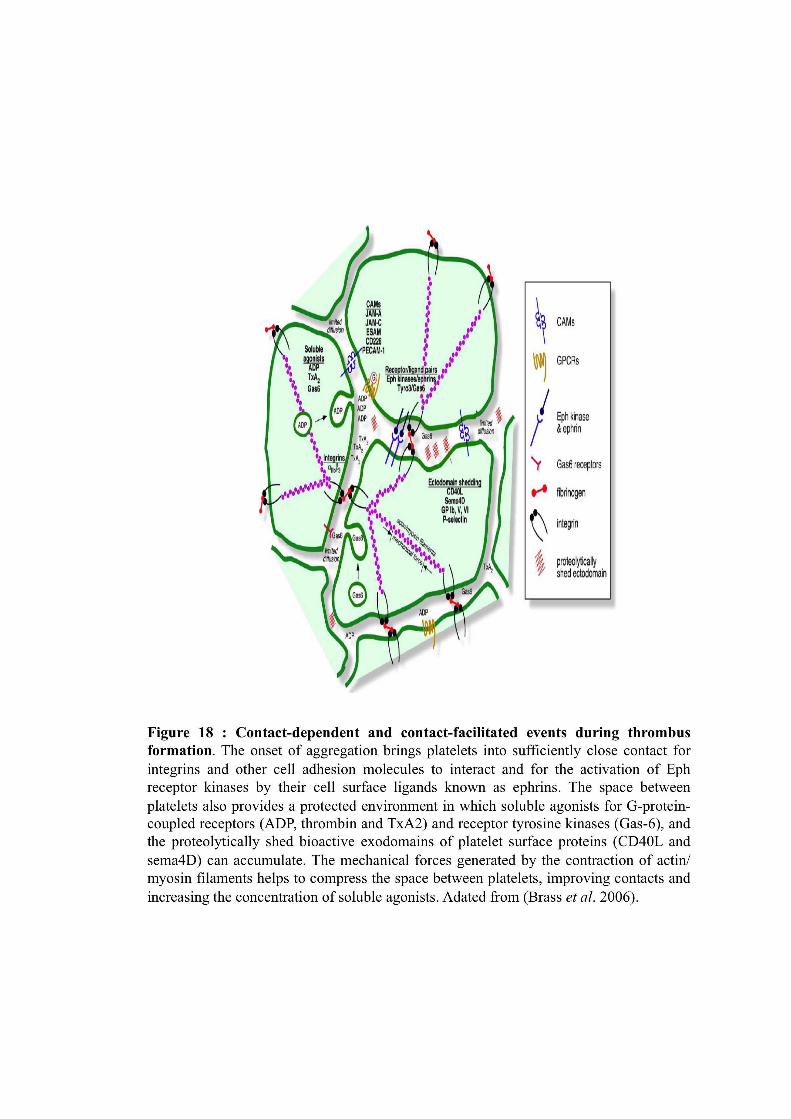

69
-Immunoglobulin like superfamily receptors (Fc%RIIa: Low affinity receptor of the
IgG constant domain, GPVI: major collagen receptor)
-calcium channel (P2X1, ATP receptor)
-Tetraspanin (CD63
and CD9, domain 4 transmembrane receptor).
The main platelet receptors and their properties as targets for antiplatelet agents are
described in chapter 4.
These receptors are coupled to various signaling pathways. As a consequence,
receptor engagement induces the sequential activation of a network of complementary
signals ending in mobilization of calcium. A cross-talk between these pathways
allows optimal responses. Conversely blockade of the knots of this network impair
more or less severely and specifically platelet responses. Soluble agonists (thrombin,
ADP, and TxA2) stimulate receptors that couple to heterotrimeric G proteins and
induce distinct downstream signaling pathways (Offermanns, 2006). Gq proteins
activate PLC" leading to calcium mobilization (via inositol-1,4,5-trisphosphate, IP3)
and activation of protein kinase (PK) C (via DAG) (Offermanns et al., 1997).
G12/G13 proteins regulate multiple pathways, of which the Rho/Rho-kinase pathway,
leading to myosine light chain phosphorylation and platelet shape change, has been
well explored (Hart et al., 1998). The !-subunit of Gi family proteins inhibits
adenylyl cyclase, while its "% complexes can regulate several channels and enzymes,
most notably PI3Ks (Cantley, 2002) (Figures 19 and 20).
Adhesive proteins activate different pathways. Those coupled to the GPIb-V-IX
complex are still a matter of dabate.
Receptors belonging to the immunoreceptor superfamily could be activating when
presenting an immunotyrosine based activation motif (ITAM) or inhibitory when
presenting an immunotyrosine based inhibitory motif (ITIM). GPVI, Fc%RIIa and C-
type lectin receptor 2 (CLEC-2) belong to the first category and are coupled to the
activation of tyrosine phosphorylation cascades downstream of the ITAM, or
hemITAM, respectively, leading to full platelet activation. PECAM-1 and G6Bb
belongs to the second category and are coupled to the activation of protein tyrosine
phosphatases downstream of their ITIM.


70
Integrins are also coupled to an outside –in signaling pathway that involves Tyr-
kinases as the ITAM-triggered signals.
2.2.3 Other Functions of Platelets besides Hemostasis
Platelets have also other functions that are now recognized in the tissue repair,
angiogenesis, inflammation, however, there is a growing appreciation for their role in
inflammation. Platelets promote leukocyte recruitment and produce microparticles
which are vectors to spread inflammation (Smyth et al., 2009). Platelets release a
broad range of inflammatory mediators in chronic inflammatory diseases that support
endothelial cells activation, leukocytes adhesion and transmigration, monocyte
maturation, and elaboration of cytokines and reactive oxygen species (Davi and
Patrono, 2007). Platelet is a storehouse for a host of inflammatory mediators and
growth factors that play a role in inflammation (atherothrombosis). These include
adhesion proteins (fibrinogen, fibronectin, vWF, thrombospondin, vitronectin, p-
selectin, integrin !IIb"3), growth factors (platelet-derived growth factors,
transforming growth factor-beta, epidermal growth factor, basic fibroblast growth
factor), chemokines (eg, PF-4, regulated on activation, normal T-cell expressed and
secreted, epithelial neutrophil-activating protein 78, macrophage inflammatory
protein-1alpha), cytokines and cytokine-like factors (eg, interleukin-1beta, CD40
ligand, thromboglobulin-beta), and coagulation factors (eg, factor V, factor XI,
plasminogen activator inhibitor type 1, plasminogen, protein S). All these substances
act in concert to mediate a wide range of functions, including cell adhesion, cell
activation, chemotaxis, cell survival, proliferation coagulation and proteolysis
(Jennings, 2009).
As previously suspected that platelets play a role in rheumatoid arthritis: due to their
accumulation in the joints of affected patients; platelet thrombi were observed in
synovial vessels of patients with rheumatoid arthritis, and increased numbers of
platelets in synovial fluid and of microparticles in blood were associated with the
condition (Diaz-Gonzalez, 2010, Yaron et al., 1978).
Boilard et al. (2008) did not detect intact platelets in the synovial fluid of the
rheumatoid arthritis patient. Then how did the incendiary microparticles gain access
to the joints?. A possible explanation is that platelets adhere to activated
polymorphonuclear leukocytes and monocytes in the circulation in many

71
inflammatory conditions (Weyrich and Zimmerman, 2004) including rheumatoid
arthritis (Weissmuller et al., 2008). Jerry Ware, a hematologist at the university of
Arkansas for Medical sciences in Little Rock concluded that platelets do not instigate
rheumatoid arthritis but just worsen it.
Besides all these, platelets are chock-full of biologically influential molecules-not
just ingredients for blood clotting but a wealth of growth factors, immune system
messengers, enzymes, and other compounds. Researchers have identified more than
1100 kinds of proteins inside platelets or on their surface (Leslie, 2010). Platelets also
appear to guard against microbial invasion because their Toll-Like receptors (TLRs)
recognizes molecular features of microbes. Semple and colleagues have found that
when pathogens trip a platelet’s TLRs, there’s a surge in TNF-!, a compound that
fuels inflammation, one of the body’s most potent protections against infection
(Aslam et al., 2006).
2.3 Platelet Receptors: Structure and Function
2.3.1 The GPIb/V/IX Complex and vWF
VWF permits the interaction of platelets with the sub-endothelial matrix of the
vessels under high shear rates. It is responsible for the initial adhesion of platelets to
the collagen.
2.3.1.1 GPIb/V/IX Complex
The GPIb/V/IX complex is a highly abundant (25000 copies per platelet) and
structurally unique receptor complex exclusively expressed in platelets and
megakaryocytes. Four different genes encodes the receptor complex, namely the
alpha and beta subunits of GPIb, GPIX and GPV (Cauwenberghs et al., 2000, George
et al., 1984), all of which belong to the leucine-rich repeat protein superfamily and are
characterized by the presence of one or more leucine-rich repeats (Berndt et al.,
2001). GPIb! (150kDa) and GPIb" (27kDa) are associated by a disulfide bridge and
are also associated by non-covalent bonds with GPIX (22kDa) and GPV (82 kDa)
with a stoechiometry of 2:2:1. GPIX and GPV are considered to serve as a scaffold
for GPIb, which harbors binding sites for vWF (Figure 21).


72
The binding site for VWF is situated on GPIb!, on the N-terminus in first 282 amino
acid residues. This region contains six leucine rich domains, one domain of sulfated
tyrosine and loop formed by a disulfide bridge. These three elements participates in
interaction with VWF (Berndt et al., 2001, Cauwenberghs et al., 2000).
A thrombin binding site is also identified on this region. A growing number of
proteins have been shown to bind to this extracellular domain of GPIb! : this include
other coagulation factors such as FXI, FXII and high molecular weight kininogen ;
GPIb-V-IX is also recognized as a counter receptor for P-selectin and this could
contribute to platelet interactions with inflammed endothelial cells or with leukocytes.
The integrin Mac-1 is proposed to contribute to platelet clearance by macrophages via
its binding to clustered GPIb-V-IX. GPIb can initiate adhesion by interacting with
other ligands. Thrombospondin-1 has also been shown to interact with GPIb under
high shear flow conditions in vitro (Jurk et al., 2003).
The receptor mediates the initial contact of platelets with the exposed Extracellular
matrix (ECM) that function irrespectively of cellular activation and thereby facilitates
rapid interaction that resist shear forces acting on the cells (Sakariassen et al., 1986).
At the high shear rates found in small arteries and arterioles, the instantaneous onset
of the interaction between GPIb! and the A1 domain of vWF immobilized on
collagen or on the surface of the activated platelets is crucial for the initial tethering or
capture of flowing platelets. This interaction is, however, characterized by a rapid
dissociation rate and is insufficient for stable adhesion. This can be illustrated by the
rolling of GPIb-IX-expressing cells or platelets on a vWF monolayer at high shear
(Savage et al., 1998). Thus, the principal function of GPIb/V/IX is to recruit platelets
to the site of injury and to reduce their velocity to enable the interaction of other
receptors with the thrombogenic surface.
The intracellular signal triggered by vWF binding to GPIb-V-IX is complex and not
yet well understood: ITAM containing receptors, phospholipase C, phospholipase A2,
the cytoskeleton, interactions with the 14.3.3 protein and activation of PI3-kinase
have all been reported to be involved (Canobbio et al., 2004). In conditions of high
shear flow, vWF/GPIb interaction allows the formation a a single layer of adherent
platelets but downstream signal are not sufficient by themselves to permit thrombus
growth (Pugh et al.).

73
In human the lack or dysfunction of GPIb/IX/V is associated with the Bernard-
Soulier syndrome (BSS), a congenital bleeding disorder characterized by mild
thrombocytopenia, giant platelets and inability of the cells to aggregate in response to
ristocetin (Lopez et al., 1998).
2.3.1.2 Von Willebrand Factor
The Von willebrand factor (vWF) is a glycoprotein synthesized by endothelial cells
(stored in Weible Palade bodies) and megakaryocytes. Stored in !-granules, it is
released upon platelet activation. vWF is a multimeric adhesive glycoprotein and is
the central mediator in platelet adhesion. The monomers (about 220kDa) assemble
into multimers (up to 20 x 103 kDa) in the Golgi complex. vWF is found in the
Weibel-palade bodies of endothelial cells, in the alpha granules of platelets and in the
plasma at a concentration of about 10$g/mL in human (Berndt et al., 2001). The high
molecular weight multimers of vWF are specifically processed by a protease
ADAMTS13 (A Disintegrin And Metalloprotease with ThromboSpondin type 1
motif, member 13) repeats causing VWF size reduction. This proteolytic cleavage is
essential to reduce the size of ultra large VWF polymers, which, when exposed to
high shear stress in the microcirculation, are prone to form platelets clumps.
The biological functions of vonWillebrand factor are exerted through specific
domains that interact with extracellular matrix components and cell membrane
receptors to promote the initial tethering and adhesion of platelets to sub- endothelial
surfaces, as well as platelet aggregation. It contains binding sites for collagen as well
as for the two major platelet receptors (GPIb! and integrin !IIb"3). Moreover, it
binds the procoagulant co-enzyme, factor VIII, contributing to its stability and,
indirectly, to its function in the generation of fibrin. Factor VIII, which is a necessary
co-factor for the rapid generation of factor Xa at sites of injury (Ruggeri, 2001).
The mature subunit of vWF consists of 2050 amino acids and is composed of four
different repeating domains (A-D) (Shelton-Inloes et al., 1986). The three
homologous A domains span residues 497 to 1111 and regulate interaction with
different receptors and prothrombotic ligands of the subendothelial matrix. The A1
domain exclusively binds collagen type VI, whereas collagen I and III are bound via
the A3 domain (Hoylaerts et al., 1997). The C1 domain contains the sequence Arg-
Gly-Asp (RGD), which represensts a binding motif for both of the platelet Beta3-
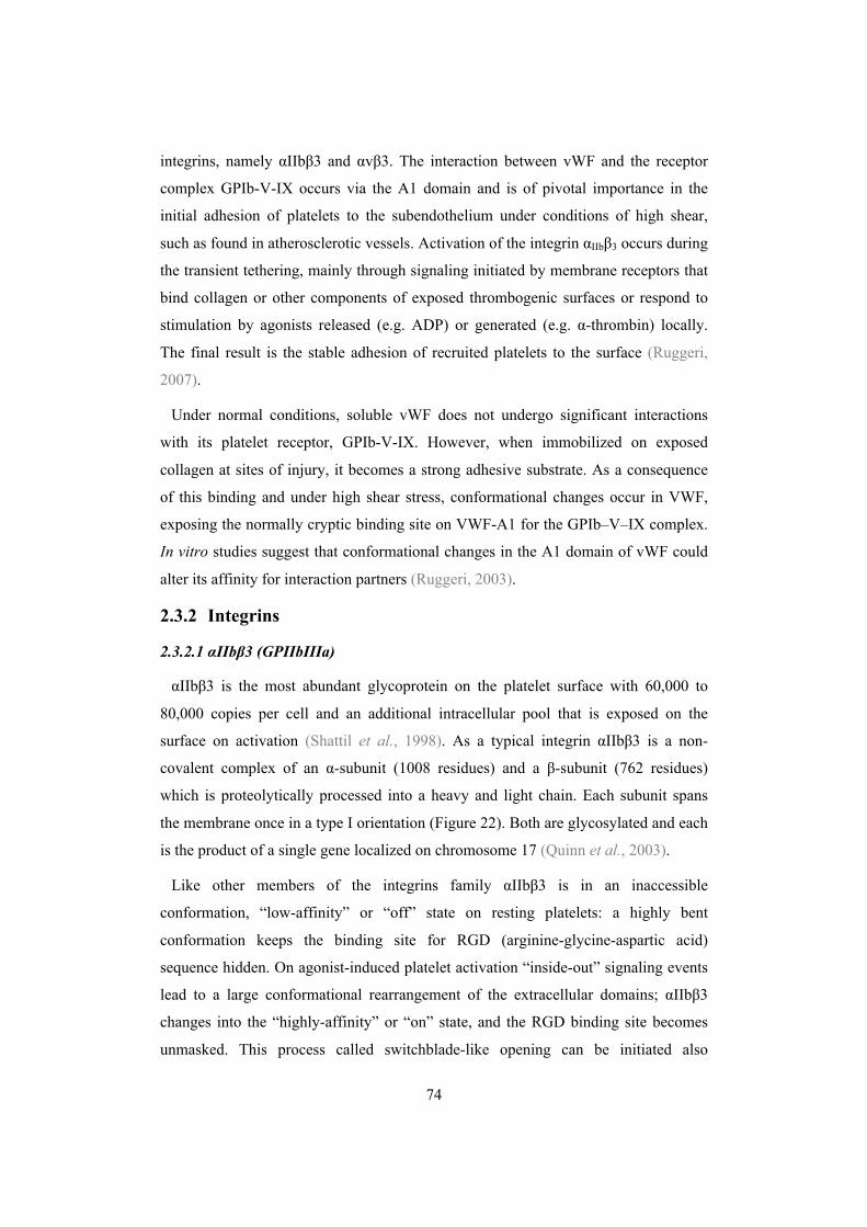
74
integrins, namely !IIb"3 and !v"3. The interaction between vWF and the receptor
complex GPIb-V-IX occurs via the A1 domain and is of pivotal importance in the
initial adhesion of platelets to the subendothelium under conditions of high shear,
such as found in atherosclerotic vessels. Activation of the integrin !IIb"3 occurs during
the transient tethering, mainly through signaling initiated by membrane receptors that
bind collagen or other components of exposed thrombogenic surfaces or respond to
stimulation by agonists released (e.g. ADP) or generated (e.g. !-thrombin) locally.
The final result is the stable adhesion of recruited platelets to the surface (Ruggeri,
2007).
Under normal conditions, soluble vWF does not undergo significant interactions
with its platelet receptor, GPIb-V-IX. However, when immobilized on exposed
collagen at sites of injury, it becomes a strong adhesive substrate. As a consequence
of this binding and under high shear stress, conformational changes occur in VWF,
exposing the normally cryptic binding site on VWF-A1 for the GPIb–V–IX complex.
In vitro studies suggest that conformational changes in the A1 domain of vWF could
alter its affinity for interaction partners (Ruggeri, 2003).
2.3.2 Integrins
2.3.2.1 !IIb"3 (GPIIbIIIa)
!IIb"3 is the most abundant glycoprotein on the platelet surface with 60,000 to
80,000 copies per cell and an additional intracellular pool that is exposed on the
surface on activation (Shattil et al., 1998). As a typical integrin !IIb"3 is a non-
covalent complex of an !-subunit (1008 residues) and a "-subunit (762 residues)
which is proteolytically processed into a heavy and light chain. Each subunit spans
the membrane once in a type I orientation (Figure 22). Both are glycosylated and each
is the product of a single gene localized on chromosome 17 (Quinn et al., 2003).
Like other members of the integrins family !IIb"3 is in an inaccessible
conformation, “low-affinity” or “off” state on resting platelets: a highly bent
conformation keeps the binding site for RGD (arginine-glycine-aspartic acid)
sequence hidden. On agonist-induced platelet activation “inside-out” signaling events
lead to a large conformational rearrangement of the extracellular domains; !IIb"3
changes into the “highly-affinity” or “on” state, and the RGD binding site becomes
unmasked. This process called switchblade-like opening can be initiated also


75
passively by manganese ions (Smith et al., 1994). The “inside-out” activation of
!IIb"3 seems to be regulated through a mechanism that essentially involves the
cytoplasmic tails of the two subunits. Studies on mice have established a requirement
for talin and talin binding to "3 in inside-out !IIb"3 signaling. Another identified
partner is kindlin-3.
The main role of !IIb"3 after activation is binding with fibrinogen in the manner
dependent on Ca2+. Fibrinogen is an heterodimer (A!, B", %)2 and due to its
multivalency it supports platelet aggregation. Fibrinogen concentration is 4$g/L and
is the most represented !IIb"3 ligand. Fibrinogen binding to !IIb"3 involves the RGD
motif on !-chain of fibrinogen and a dodecapeptide at the C-terminus of the
fibrinogen -chain. !IIb"3 also binds several other ligands each containing the RGD
sequence. Although fibrinogen is the predominant ligand in plasma, the role of von
Willebrand factor (VWF) in conditions of high shear should be emphasized, while
fibronectin, vitronectin and CD40L may also participate (Naik and Parise, 1997,
Ruggeri, 2002, Shattil et al., 1998).
Once bound to immobilized ligands, !IIb"3 induces processes such as spreading. It
is also necessary for clot retraction that is due its interaction with the platelet
cytoskeletton. Binding of its ligand !IIb"3 triggers an outside-in signaling pathway
that reinforce the level of activation signals in platelets and is required for aggregates
stabilization (Shattil et al., 1998). The network of proteins involved in outside-in
!IIb"3 signaling is very large (Coller and Shattil, 2008). Many parallels are apparent
between this process and signaling triggered by immunoreceptors, including
dependence on Src family kinases (SFKs) and Syk protein tyrosine kinase. The
cytoplasmic domain of !IIb"3 may serve a nucleating function for some of these
proteins.
Quantitative and/or qualitative abnormalities of !IIb"3 (Glanzmann
thrombasthenia) is characterized by a major abnormality of platelets aggregation in
response to all agonists and the clinical manifestation is an hemorrhagic syndrome
which may be very severe (Nurden, 2009, Nurden and Nurden, 2008, Seligsohn,
2002). In man, expression of the !IIb gene (ITGA2B) (and therefore of !IIb"3
integrin) is restricted to cells of the megakaryocytic cell lineage. Expression of the "3
gene (ITGB3) is more widespread, with the vitronectin receptor (!v"3) being
expressed in many cell types, including endothelial cells, osteoblasts, smooth muscle

76
cells, and leukocytes. The inability to bind adhesive proteins when stimulated
explains the platelet phenotype in GT.
Due the importance of GPIIbIIIa in platelet aggregation it has become an attractive
pharmacological target for the preventation of cardiovascular events.
2.3.2.2 Integrins !v"3, !5"1( or VLA-5), !6"1(or VLA-6)
These integrins are also expressed on platelet surface. These may bind to different
proteins of the extracellular matrix such as !v"3 that binds to vitronectin and can also
bind to fibronectin and osteoponin, while !5"1 is the receptor for fibronectin and
!6"1 is a receptor for laminin on platelet surface (Kamata et al., 2005, Sonnenberg et
al., 1988). Various in vitro and in vivo studies have demonstrated that these receptors
may modulate platelet responses and that their relative importance to the overall
processes of adhesion, activation and spreading is determined by the nature of
extracellular matrix, which differ in different parts of an individual’s body (Inoue et
al., 2006).
2.3.3 Collagen Receptors
In addition to the indirect interaction of platelets with collagen via the vWF/GPIb-
V-IX axis, direct interactions are also made with the collagen receptors present on
platelet surface (Figure 23). Collagen triggers the initiation of platelet aggregation
using two classes of collagen receptors: GPVI and GPIaIIa (integrin !2"1). It has
been reported that collagen binds to these two classes of receptors in a sequential
manner (Mangin et al., 2003, Nieswandt and Watson, 2003).
2.3.3.1 Integrin !2"1
The direct collagen adhesion receptor integrin !2"1 (GPIaIIa or VLA-2 or
CD49b/CD29), is the first collagen receptor that has been identified (Santoro, 1986).
It is next to !IIb"3 the second most important platelet integrin with approximately
2000–4000 copies per platelet. The expression of integrin !2"1 is not restricted to the
platelet lineage and it is present on many different cell types. Patients with inherited
reduced expression of !2"1 or acquired deficient activity show a mild bleeding
tendency and impaired platelet responses to collagen. This receptor recognizes a
GFOGER (O stands hydroxyproline) motif present in the sequence of collagen type I

77
and IV (Emsley et al., 2000). Integrin !2"1 can bind to collagen type I monomers and
polymers. Both integrin !- and integrin "-subunits fold into a N-terminal extracellular
globular head that creates the ligand binding surface sitting on two membrane-
spanning legs. All ! -subunits of integrins contain at the N-terminal of ! seven-bladed
"-propeller structure. The !2 extracellular domain furthermore contains a unique
inserted (I) domain, with a conserved cation-binding site known as the metal ion-
dependent adhesion site (MIDAS) with a clear preference for Mg2+/Mn2+. The integrin
!2"1 is assumed to have a primarily adhesive role. An early model of platelet-
collagen interactions, the so called “ 2-step, 2-site” model incorporated the idea that
platelets in a first step firmly bind to collagen via high affinity !2"1 allowing
subsequent interaction of a second receptor leading to platelet activation (Santoro et
al., 1991). This second, low affinity receptor was later proposed to be GPVI.
However, the idea that !2"1 integrin is active on unstimulated platelets was
challenged by Moroi and coworkers who demonstrated that the affinity of the integrin
for soluble collagen increases on cellular stimulation (Jung and Moroi, 2000). Upon
activation the integrin shifts into a high-affinity state, with the extracellular domains
extended upwards. This rapid conformational switch is usually triggered by inside-out
signaling. The mechanism of !2"1 activation is believed to be similar to that of other
integrins. Both talin and kindlin-3 that bind to the cytoplasmic tail of the integrin
subunits are required and cooperate to regulate integrin activation. Inside-out
activation of GPIaIIa can be induced through ligation of GPVI as demonstrated by
defective adhesion of GPVI-deficient or –inhibited platelets to the matrix proteins.
Human platelet deposition on collagen depends on the concerted interplay of several
receptors: GPIb in synergy with integrin !2"1 mediating primary adhesion. However,
even in combination, these 2 axes provide an insufficient stimulus for full thrombus.
Engagement of GPVI is required to ensure platelet activation (Lecut et al., 2005,
Nieswandt et al., 2001b, Siljander et al., 2004b). The relative contribution of each of
these receptors very much depends on the experimental conditions but it is now
generally accepted that they act synergistically in the process of platelet adhesion,
activation and expression of procoagulant activity on the extracellular matrix. Binding
of integrin !2"1 to collagen contributes to cellular activation indirectly by reinforcing
GPVI-collagen interactions and directly by a series of intracellular signaling events
summarized as “outside-in” signaling. These are very similar to those induced by
GPVI, including Src, Syk, SLP-76 and PLC (Li et al.). Thus, although structurally

78
unrelated, the two major collagen receptors share important signaling molecules and
act in a co-operative manner, reinforcing each other’s activity.
2.3.3.2 GPVI
GPVI is thus the major receptor for platelet activation by collagen. As the target of
this work, it will be described in more details in the next chapter.
Other proposed collagen receptors
-P65, specific receptor for collagen type III and P47, TIIICBP, specific receptors for
collagen type III whose nature and role is still not well understood. GPIV is also
considered as collagen receptor but GPIV (CD36) deficient patient (5% in Japanies)
have no abnormal collagen response.
2.3.4 G-Protein-Coupled Receptors (GPCRs)
2.3.4.1 ADP Receptors (P2Y1 and P2Y12) and ATP Receptor (P2X1)
By itself ADP is a weak agonist of platelet aggregation inducing only reversible
responses as compared to strong agonists such as thrombin or collagen. However, due
to its presence in large amounts in the platelet dense granules and its release upon
activation at sites of vascular injury, ADP is an important so-called secondary agonist,
which amplifies most of the platelet responses and contributes to the stabilization of
the thrombus. The receptors for extracellular nucleotides belong to the P2 family
which consists of two classes of membrane receptors: P2X ligand-gated cation
channels (P2X1–7) and G protein-coupled P2Y receptors (P2Y1,2,4,6,11,12,13,14)
(for a review see (Gachet, 2008).
Platelets express two ADP receptors, P2Y1 and P2Y12 (Figure 24). Co-activation of
the P2Y1 and P2Y12 receptors is necessary for normal ADP-induced platelet
aggregation since separate inhibition of either of them with selective antagonists
results in a dramatic decrease in aggregation. However, the two receptors have
different functions since, except in the case of collagen-induced activation, P2Y1
plays a minor role when other agonists induce platelet aggregation whereas P2Y12
supports amplification of these responses.
The P2Y1 receptor is responsible for ADP-induced calcium mobilization required
for platelet shape change and aggregation (Hechler et al., 1998, Leon et al., 1999).
P2Y1 is expressed at a very low level on the platelet memebrane (aroud 150 sites per

79
platelet). P2Y1 is coupled to a Gq protein and activates phospholipase C and,
downstream, protein kinase C (PKC) and mobilization of Ca+2. Lack of P2Y1 leads to
reduced aggregation to all major agonists, resistance collagen/epinephrine and ADP-
induced thromboembolism, and moderately increased bleeding times (Leon et al.,
1999). Conversly, mice overexpressing P2Y1 in their platelets have shorter tail
bleeding times and are susceptible to thromboembolism and thrombosis (Hechler et
al., 2003). The P2Y1 receptor should be regarded as an attractive target for
antiplatelet compounds. It is worthy of note that inhibition of the P2Y1 receptor
results in only moderate prolongation of the bleeding time, which could be
advantageous in terms of safety.
P2Y12 is expressed at a higher level (in the order of 1500 copies per platelet). Its
tissue distribution is limited although not entirely restricted to platelets. The P2Y12
receptor strongly coupled to Gi2 leading to inhibition of adenylate cyclase and
lowering cAMP, thereby enabling amplification of platelet activation via P2Y1 and
also by other agonists which would otherwise be hindered (Cattaneo and Gachet,
2001, Cattaneo et al., 2003, Hollopeter et al., 2001, Kahner et al., 2006). It also
activates the phosphoinositide 3-kinase (PI3K) pathway. Patients lacking P2Y12
receptor display selective defects of ADP-induced platelet aggregation. Mice lacking
P2Y12 form only small and unstable thrombi and show marked protection from
arterial occlusion and prolonged bleeding times (Andre et al., 2002). Owing to its
central role in the growth and stabilization of a thrombus, the P2Y12 receptor is an
established target of antithrombotic drugs
P2Y1 and P2Y12 have been shown to display gene sequence variations, which have
been proposed to be associated with variable platelet responsiveness to ADP.
The third component of the platelet P2 receptors is P2X1, a ligand-gated cation
channel responsible for a fast calcium entry induced by ATP. Although unable to
trigger platelet aggregation by itself, the P2X1 receptor induces transient shape
change and participates in collagen- and shear-induced aggregation. One hallmark of
this receptor is that it requires high shear conditions to fully play its role in the
formation of a thrombus. Since the P2X1 receptor plays an important role in thrombus
formation only under high-shear conditions, it might represent the ideal target for an
antithrombotic drug. The P2X1 receptor may also be considered as a potential target
for antiplatelet strategies, with the interesting feature that P2X1 antagonists should be
80
effective only at sites of severe stenosis where shear forces are very high, without
having a deleterious effect on normal haemostasis.
2.3.4.2 Thrombin Receptors (PAR1 and PAR4)
Thrombin is the most potent platelet activator at least in vitro. Its receptors called
Protease Activated Receptors (PARs) belong to the seven transmembrane domain
GPCR superfamily but present a unique mechanism of activation (Varga-Szabo et al.,
2008). The activation of a PAR is a 2-step process. First, the cryptic ligand is
unmasked by proteolytic cleavage of the receptor N-terminal domain; then, an
intramolecular rearrangement allows the ligand and the receptor moieties to interact.
On human platelets, thrombin activates PAR1 and PAR4 (Figure 25). PAR1 is a high-
affinity receptor for thrombin by virtue of a hirudin (Hir)-like sequence that resides in
its N-terminal extracellular domain. The Hir sequence allows PAR1 to compete with
the much more plentiful fibrinogen, and, as a result, PAR1 is activated by thrombin at
even subnanomolar concentrations. PAR4 has evolved a different strategy for
interacting with thrombin. Bereft of a high-affinity Hir-like sequence, PAR4 has
instead optimized its interactions with the active site of thrombin and uses a
negatively charged cluster of amino acid residues to slow dissociation from the
positively charged thrombin molecule. PAR4 is activated at a high thrombin
concentrations; it is cleaved and signals more slowly but, despite its slower response,
generates the majority of the intracellular calcium flux and does not require additional
input from the P2Y12 ADP receptor to form stable platelet–platelet aggregates (Kahn
et al., 1998). Mouse platelets express PAR3 and PAR4 (Nakanishi-Matsui et al.,
2000).
In the case of murine platelets, PAR4 binds to the active site in a conformation that
leaves exosite I free for interaction with cofactors like PAR3.
PAR1 and PAR4 are coupled to a heterotrimeric G proteins Gq, which activate the
phosphilipase C (PLC), protein kinases C (PKCs) and the mobilization of Ca2+. These
signaling systems are critical in the platelet and bring numerous factors in to action
such as Ca2+ calmodulin dependent kinases, MAP kinases or integrin activation
modulators and to the dynamics of the cytoskeleton and the secretion.


81
PAR1 and PAR4 are also coupled to G12/13, a heterotrimeric G protein can lead to
the activation of RhoA-ROCK pathway, a regulator of phosphatase of myosin light
chain (Becker et al., 2009, Macaulay et al., 2010).
PAR1 signaling is strengthened by additional inputs from Gi-coupled signals evoked
by stimulation of P2Y12 by ADP and leading to a decrease in the platelet
concentration of cAMP whereas PAR4 signaling does not require additional input
(Offermanns, 2006) (Figure 25).
Patients with arterial thrombosis receive in hospital anticoagulant treatments aimed
to inhibit thrombin formation and or thrombin activity. However, inhibition of PAR1
could be of interest in the secondary prevention of arterial thrombosis and specific
antagonists have been developed.
2.3.4.3 Thromboxane A2 Receptor (TP!)
Like ADP, TXA2 functions as a positive-feedback mediator during platelet
activation. The action of TXA2 is locally restricted because of its short half-life. The
TXA2 receptor identified on platelets (TP!). It is also activated by the prostaglandin
endoperoxides PGG2 and PGH2, couples to Gq and G12/G13 and to PLC (Brass et
al., 1987, Shenker et al., 1991).
The role of TP as the platelet TXA2 receptor has been demonstrated in studies using
platelets from TP deficient mice, which become unresponsive to TXA2. TP-deficient
mice have prolonged bleeding times and are unable to form stable thrombi (Thomas et
al., 1998). A reduced activation of TP-deficient platelets has been suggested to
contribute to a reduced injury induced vascular proliferation as well as to a reduced
progression of atherosclerosis observed in mice lacking TP. Some studies using the
radiolabeled TXA2 analogues as ligands demonstrated two classes of binding sites in
platelets, and indicated that they might represent the receptor subtypes (Dorn, 1989,
Hedberg et al., 1988, Morinelli et al., 1987). They further suggested that the two
putative subtypes of the receptor may independently mediate shape change and
aggregation (Dorn, 1989, Dorn and DeJesus, 1991). This suggestion has been
supported by the reports that platelet shape change and aggregation can be
differentiated by several TXA2 analogues. For example, in some species, the TXA2
agonist U46619 induces platelet shape change and an increase in calcium without
aggregation (Nakano et al., 1987). In addition, the TXA2 analogue S-145 prevents

82
aggregation and secretion by U46619 but itself induces shape change (Hanasaki and
Arita, 1988). One model consistent with these observations is that the TXA2 receptor
mediated signaling consists of two separate receptor-effector systems mediating
distinct functional responses; one linked to PLC activation resulting in platelet
aggregation and secretion, and the other mediating an increase in cytosolic calcium
and platelet shape change (Takahara et al., 1990).
2.3.5 Recently Identified Receptors Stabilizing Thrombosis
Many other receptors involved are gradually implicated in the growth of the clot and
its stabilization. Their function is mainly known through the study of genetically
modified mice, but their role in human diseases and the possibility whether they may
be good therapeutic targets is not yet established.
The stabilization of a newly formed thrombus is essential to arrest bleeding at sites
of vascular injury. The final thrombus is embedded in a fibrin network to withstand
the shear forces generated by the flowing blood. In addition, outside-in signaling
through ligand-occupied integrin !IIb"3 plays a central role in thrombus stabilization
and beside CLEC-2, whose involvement in thrombus perpetuation is currently under
discussion (see below), other proteins including CD40L (Andre et al., 2002), SLAM
(signaling lymphocyte activation molecule), Gas6 (growth arrest-specific gene 6)
(Cosemans et al., 2010) and semaphorin 4D (Zhu et al., 2007) have been established
to participate in platelet–platelet interaction. It has also been reported that blockade of
Eph kinases/ephrins interactions resulted in a decreased thrombus volume in vitro
under flow conditions, suggesting that this interaction contributes to the stabilization
of platelet–platelet contacts (Prevost et al., 2005). It can be anticipated that other
platelet receptors will be identified to be involved in promoting platelet plug stability
and possible candidates include JAM-A, JAM-C, CD226 and CD84 (Brass et al.,
2008). TREM-like transcript-1 has recently been reported to protects against
inflammation-mediated hemorrhage and to facilitate platelet aggregation in mice and
human (Brass et al., 2008, Stalker et al., 2009, Washington et al., 2009).
However, molecules that limit thrombus growth were also identified. Mice lacking
the endothelial cell specific adhesion molecule (ESAM) (Stalker et al., 2009) or
platelet endothelial cell adhesion molecule-1 (PECAM-1) (Falati et al., 2006)
developed larger thrombi in vivo as compared to control mice, indicating that these

83
molecules serve as negative regulators of thrombus formation, although the effects
may be limited.
2.3.5.1 CLEC-2
CLEC-2 is a C-type lectin-like type II transmembrane receptor identified to
mediate the potent platelet activation responses to snake venom toxin, rhodocytin
(Suzuki-Inoue et al., 2006). CLEC-2 lack a signal sequence but show a putative
hydrophobic transmembrane region. The carbohydrate recognition domain (CRD)
conserved six cysteine residues and these cysteine residues generate three interchain
disulfide bonds that are typical of C-type lectins (Weis and Drickamer, 1996). CLEC
have a tyrosine residue in their predicted cytoplasmic domain and also contain several
serine and threonine residues which are potential sites of phosphorylation (Colonna et
al., 2000). CLEC-2 uses a similar signaling pathway to GPVI, initiated by tyrosine
phosphorylation of a single ITAM motif, termed hem ITAM, within its cytoplasmic
tail (Suzuki-Inoue et al., 2006, Watson et al., 2010). The only identified physiological
ligand of CLEC-2 is podoplanin (which is a transmembrane protein), an endothelial
sialomucin glycoprotein. Podoplanin is expressed in the lymphatic sac, and it was
shown that the CLEC-2-podoplanin interaction is essential for the separation of the
lymphatic from the blood vascular system during embroynic development (Bertozzi et
al., 2010, Uhrin et al., 2010). Mice pretreated with a specific antibody to CLEC-2
exhibit a selective loss of the C-type lectin receptor and impaired platelet activation
on collagen at high shear in vitro or in vivo (May et al., 2009).
CLEC-2 knockout mice displayed reduced thrombus stability under flow in vitro
and in vivo leading to prolonged tail bleeding times and protection from arterial
thrombus formation (May et al., 2009, Suzuki-Inoue et al., 2010).
2.3.5.2 Fc#RIIA
Human platelets express on their surface the low affinity receptor for IgG Fc%RIIA.
This receptor belongs to the immunoreceptor superfamily and shares its signaling
mechanism with GPVI. Fc%RIIA is involved in heparin-induced thrombocytopenia,
the immune complexes triggering platelet activation. The molecules targeting Tyr-
kinase below the Fc%RIIA could be useful in the treatment of Heparin-induced
thrombocytopenia (HIT) by inhibiting, on one hand platelet activation and, on the
other side, by controling their elimination by macrophage (Worth et al., 2006).

84
2.3.5.3 Inhibitory Receptors
Although the inhibitory receptors CD31 (PECAM-1), G6Bb and CAECAM seem to
raise the threshold of doses of agonists needed to induce platelet aggregation, it is still
too early to assume interest as therapeutic targets.
2.4 Antithrombotic Drugs
There are considerable differences in drug therapy for arterial and venous
thrombosis, which reflect different mechanisms underlying these two types of
thrombosis. The therapeutic goal is not only to control thrombus formation but also to
prevent new plaque rupture events by stablizing the atheromatous plaque. In contrast,
for prevention and treatment of venous thrombosis the major approaches are the
control of thrombus formation and removal of enviromental risk factors.
The antithrombotic drugs are classified according to their targeted constituents:
antiplatelet agents and anticoagulants (Figure 26).
2.4.1 Anticoagulants
As anticoagulants are not the purpose of this study, they will be shortly mentioned.
Anticoagulant molecules are divided into non-specific anticoagulants, such as vitamin
K antagonists and heparin, and direct thrombin inhibitors, including hirudin and
argatroban.
Vitamine K antagonists block thrombin formation by inhibiting the gamma
carboxylation of the VII, IX, X and II factors, preventing their calcium dependent
binding to anionic phospholipids. Inhibition of the formation of the tenase and
prothrombinase complexes block thrombin formation. Vitamine K antagonists are
given orally and treatment needs to be adjusted on biological efficacy (prothrombin
ratio and international normalized ration, INR).
Unfractionated heparin is a naturally occurring highly sulfated glycosaminoglycan
of molecular weights ranging from 5000 to over 40,000 Daltons. It is inhibiting both
thrombin generation and thrombin activity. Heparin binds to the enzyme inhibitor
antithrombin (AT) causing a conformational change that results in its activation
through an increase in the flexibility of its reactive site loop (Chuang et al., 2001).


85
The activated AT then inactivates thrombin and other proteases involved in blood
clotting, most notably. The rate of inactivation of these proteases by AT can increase
by up to 1000-fold due to the binding of heparin. The efficacy of the treatment thus
depends on AT avaibility. Heparin is given parenterally (iv or sc). It has a short half-
life and the treatment must be adjusted on the biological efficacy measured by the
activated partial thromboplastin time (APTT).
A serious side effect of heparin is heparin-induced thrombocytopenia (HIT). HIT is
caused by an immunological reaction that makes platelets a target of immunological
response, resulting in the degradation of platelets. This is what causes
thrombocytopenia. This condition is usually reversed on discontinuation, and can
generally be avoided with the use of synthetic heparins.
The conformational change in AT on heparin-binding mediates its inhibition of
factor Xa. For thrombin inhibition, however, thrombin must also bind to the heparin
polymer at a site proximal to the pentasaccharide. The highly-negative charge density
of heparin contributes to its very strong electrostatic interaction with thrombin. The
formation of a ternary complex between AT, thrombin, and heparin results in the
inactivation of thrombin. For this reason, heparin's activity against thrombin is size-
dependent, the ternary complex requiring at least 18 saccharide units for efficient
formation. In contrast, anti-factor Xa activity requires only the pentasaccharide
binding site. This size difference has led to the development of low-molecular-
weight-heparin (LMWHs) and, more recently, to fondaparinux as pharmaceutical
anticoagulants. Low-molecular-weight heparin (LMWH) consists of only short chains
of polysaccharide, having an average molecular weight of less than 8000 Da. These
are obtained by various methods of fractionation or depolymerisation of polymeric
heparin. Low-molecular-weight heparins and fondaparinux target anti-factor Xa
activity rather than anti-thrombin activity, with the aim of facilitating a more subtle
regulation of coagulation and an improved therapeutic index. Heparin and its low
molecular weight derivatives (e.g. enoxaparin, dalteparin, tinzaparin) are effective at
preventing deep vein thrombosis and pulmonary emboli in patients at risk (Bergqvist
et al., 2002).
Fondaparinux is a synthetic pentasaccharide, whose chemical structure is almost
identical to the AT binding pentasaccharide sequence that can be found within
polymeric heparin and heparan sulfate (Bauer, 2004). It is now prescribed for both

86
prophylaxis and treatment of venous thromboembolism (Boehlen et al., 2010). With
LMWH and fondaparinux, there is a reduced risk of HIT. Monitoring of the APTT is
also not required and does not reflect the anticoagulant effect, as APTT is insensitive
to alterations in factor Xa.
Direct thrombin inhibitors (DTI) have been developed. The rationale for the clinical
use of direct thrombin inhibitors is their ability to inactivate fibrin bound thrombin
and to be independent on the avaibility of AT. Direct thrombin inhibitors include
hirudin and argatroban. Bivalirudin (Angiomax or Angiox, manufactured by The
Medicines Company) is a specific and reversible DTI. Chemically, it is a synthetic
congener of the naturally occurring drug hirudin (found in the saliva of the medicinal
leech Hirudo medicinalis). It overcomes many limitations seen with indirect thrombin
inhibitors, such as heparin. Bivalirudin is a short, synthetic peptide that is potent,
highly specific, and a reversible inhibitor of thrombi. It inhibits both circulating and
clot-bound thrombin, while also inhibiting thrombin-mediated platelet activation and
aggregation. Bivalirudin has a quick onset of action and a short half-life. It does not
bind to plasma proteins (other than thrombin) or to red blood cells. Therefore it has a
predictable antithrombotic response. There is no risk for HIT. It does not require a
binding cofactor such as antithrombin and does not activate platelets. These
characteristics make bivalirudin an ideal alternative to heparin. Bivalirudin clinical
studies demonstrated consistent positive outcomes in patients with stable angina and
unstable angina and in patients undergoing percutaneous coronary intervention (PCI)
(Stone et al., 2008).
Argatroban is a derivative of arginine that structurally mimics the active site residue
of thrombin (Fareed et al., 2002). It binds to the active site of thrombin in a reversible
manner. It is as effective as heparin and is now used as an alternative of heparin in
treating venous thromboembolism (VTE). Bleeding complications are a major
adverse effect of argatroban.
2.4.2 Antiplatelet Agents
The objective of antiplatelet therapy is to reduce the platelet aggregation and inhibit
thrombus formation in arterial circulation. Recent years have witnessed important
therapeutic advancement of molecules targeting specific platelet surface receptors or
regulators of cellular haemostasis.
87
The field of antiplatelet therapy has experienced very substantial changes during the
past 20 years. Attempts to separate improved antithrombotic efficacy from enhanced
bleeding liability have met limited success so far. Quantitative assessment of bleeding
risk has lagged behind the successful development and validation of tools for
assessing the risk of ischemic complications (Subherwal et al., 2009). The interaction
between the antiplatelet drug and its molecular target largely depends on the
following determinants (i) the availability of the active moiety of the drug at its
receptor site, as a function of drug absorption, liver metabolism, and elimination, i.e,
pharmacokinetics (PK); (ii) the mechanism of transient or permanent inhibition of the
target and the rate of its renewal, i.e, pharmacodynamics (PD); and (iii) genetic
variation of the drug target and/or drug-metabolizing enzymes, i.e, pharmacogenetics
(Patrono et al., 2008).
- Actual strategies of anti-thrombosis: Many distinguished anti-platelets drugs are
actually in use for therapy which are as follows;
!"%"!"# GPIIb/IIIa (Integrin !IIb"3) Antagonists: (Platelet Anti-aggregatory
Antibodies)$
GPIIb/IIIa (integrin !IIb"3) was among the first pharmacologic targets for the
reduction of ischemic events.
Strategies to inhibit its function include antibodies (abciximab), cyclic peptides
adapted from a snake venom disintegrin (eptifibatide), and non-peptides analogues of
an RGD peptide (tirofiban and lamifiban) that inhibit ligand binding. Although these
inhibitors have been beneficial for patients undergoing percutaneous coronary
interventation, these do not have widespread clinical use because of their side effect
of undesired bleeding (Quinn et al., 2003).
Parenteral GPIIb/IIIa antagonists have been successful in the reduction of ischemic
events and mortality in patients undergoing percutaneous coronary interventation but
oral analogues have not been proved successful clinically (Jennings and Saucedo,
2008).
2.4.2.1.1 Antibodies/ Abciximab
Numerous anti-GPIIbIIa monoclonal antibodies (mAb) have been produced, some
of which abolished platelet aggregation in vitro. For a small number of them, in vivo

88
antithrombotic effect in animals and rarely in humans have been reported. One of
them was used to develop for a drug.
The mAb obtained by injecting human platelets into mice, AP-2, LJ-CP8 and MA-
16NC2, are effective in thrombosis models in baboons but causes thrombocytopenia
and/or increasing bleeding time (Hanson, 1988, Krupski, 1993).
The fragments of two mAb, F(ab')2 CRC64 (FRaMon) and F(ab')2 YM337 were
humanized by CDR grafting, which inhibits platelet aggregation in vitro and ex vivo.
They were only tested on a small series in humans (FRaMon angioplasty with high
risk and YM337 in healthy subjects) (Harder, 1999., Mazurov, 2002).
The mAb 7E3 and 10E5 obtained in the early 80s by B.S.Coller inhibit the binding
of fibrinogen and vWF to activated GPIIbIIIa and aggregation of human platelets in
vitro (Coller et al., 1986) and also in cynomolgus monkeys (Coller et al., 1989). A
major protective effect of F(ab')2 7E3 was also observed in the thrombolysis in dogs
by promoting the dissolution of clots by thrombolytic agents (Gold, 1990.). These
stages demonstrate that 7E3 was a suitable candidate to carry on clinical trials in
coronary angioplasty during acute coronary syndromes. So, a chimeric Fab (c7E3
Fab; abciximab; ReoPro®) was produced by fusing the variable domains of the
mouse monoclonal antibody to the constant regions of human Ig to decrease the
immunogenicity of the product and was the first therapeutic antibody administered to
human. Plasma half-life of abciximab is short (10 to 30min), but the molecule bound
to platelets can be detected up to 10 to 14 days after administration. Its elimination is
poorly understood (Coller and Shattil, 2008, Tcheng et al., 1994) Abciximab has been
approved by the FDA in 1994 and is the only recombinant antibody used in the
treatment of thrombosis. Many randomized clinical trials have been conducted
involving over 20,000 patients undergoing PCI and/or treatment for unstable angina
(De Luca et al., 2005). These studies demonstrated a therapeutic benefit of abciximab
in angioplasty treatment of myocardial infarction and acute coronary syndromes with
elevation of troponin level and angioplasty (prevention of ischemic complications and
mortality). Long-term mortality advantages have also been demonstrated, with most
of the benefit paradoxically occurring long after the antiplatelet effects of the drug
wore off (Bhatt and Topol, 2000). Abciximab is not considered as beneficial in
patients with acute coronary syndrome, which do not need urgent revascularization. It
is also associated with secondary effects (hemorrhage, thrombopenia,

89
hypersensibility), which may be serious. The increased bleeding observed in some
studies may be potentiated by administering the combination of abciximab and
unfractionated heparin jointly. The site of catheterization (radial vs femoral) is also
important. Overall, the severe bleeding is less than 0.2% in all studies (Kam and
Egan, 2002). At the present stage of study, abciximab remains the most widely
antagonist of integrin !IIb"3 used but is not possible to conclude the superiority of
one class over another due to advantages and disadvantages of each.
Thus, globally about 80% of patients with acute coronary syndrome do not use it.
The doors are opened for other classes of GPIIbIIIa antagonists.
2.4.2.1.2 Eptifibatide (Integrilin®)
Based on the observation that small peptides and snake venoms containing the Arg-
Gly-Asp (RGD) or Lys-Gly-Asp (KGD) sequences could inhibit ligand binding to
!IIb"3, a cyclic peptide inhibitor (eptifibatide) and a nonpeptide RGD mimic inhibitor
(tirofiban) were developped.
Eptifibatide is a cyclic heptapeptide derived from a protein found in the venom of
the south eastern pygmy rattlesnake (Sistrurus miliarus barbouri). It belongs to the
class of the so-called arginin-glycin-aspartate-mimetics and reversibly binds to
platelets. Eptifibatide inhibits platelet aggregation by reversibly binding to the platelet
receptor glycoprotein (GP) IIb/IIIa of human platelets, thus preventing the binding of
fibrinogen, von Willebrand factor, and other adhesive ligands. Inhibition of platelet
aggregation occurs in a dose- and concentration-dependent manner. Half-life is
approximately 2.5 hours.
As abciximab, eptifibatide reduces ischaemic complications of percutaneous
intervention and the treatment of acute coronary syndrome and increases the bleeding
risk with molar bleeding episodes. In the PURSUIT (Platelet Glycoprotein IIb/IIIa in
Unstable Angina: Receptor Suppression Using Integrilin Therapy) trial, which
included 10,948 patients with non-ST-elevation acute coronary syndromes,
eptifibatide significantly reduced the primary end point of death and non-fatal
myocardial infarction at 30 days compared with placebo. In patients with ST-segment
elevation myocardial infarction (STEMI), eptifibatide has been studied as adjunct to
primary PCI and improved epicardial flow and tissue reperfusion. Studies evaluating
eptifibatide in high-risk patients with non-ST elevation acute coronary syndromes

90
(NSTE-ACS) and a planned early invasive strategy in the EARLY-ACS (Eptifibatide
Administration prior to Diagnostic Catherization and Revascularization to Limit
Myocardial Necrosis in Acute Coronary Syndrome) trial and in patients with primary
PCI for STEMI in comparison to abciximab in the EVA-AMI (Eptifibatide versus
Abciximab in Primary PCI for Acute Myocardial Infarction) trial. After the
completion of these trials, the value of eptifibatide in patients undergoing PCI in
different indications can be determined (Zeymer, 2007).
In order to evaluate the effect of pre-catheterization laboratory (cath lab)
administration of eptifibatide on pre-percutaneous coronary intervention (PCI)
thrombolysis in myocardial infarction (TIMI) flow and its correlation with ischemia
duration, was studied in 438 STEMI patients treated with primary PCI: 310 patients
were pretreated with eptifibatide, while 128 patients received either no glycoprotein
IIb/IIIa inhibitors or were only given them in the cath lab. All ischemia times (chest
pain onset, diagnostic electrocardiogram, eptifibatide administration, cath lab arrival,
first balloon inflation) were recorded. At angiography, TIMI grade 2 or 3 flow was
observed in 54% of cases in Patients already treated with eptifibatide vs. 34% in
already non-treated patients. In already treated patients, there was a strong correlation
between pre-PCI TIMI flow and timing of eptifibatide administration. Pretreatment
with eptifibatide and symptom duration of <or= 90 minutes resulted in independent
predictors of pre-PCI TIMI flow on multivariable analysis. Thirty day mortality was
1.9% in already treated patients and 9.5% in already non-treated patients. Eptifibatide
therapy prior to primary PCI achieves a higher rate of pre-PCI TIMI flow with respect
to late administration (Aquilina et al., 2009).
2.4.2.1.2.1 Tirofiban (Aggrastat®)
Tirofiban hydrochloride is a non-peptide, intravenous administered and reversible
antagonist of fibrinogen binding to the GPlIb/lIla receptor and inhibits platelet
aggregation. When administered intravenously, it inhibits ex vivo platelet aggregation
in a dose- and concentration-dependent manner.
When given according to the recommended regimen, >90% inhibition was attained
by the end of the 30-minute infusion. Tirofiban has a half-life of approximately 2
hours. It is cleared from the plasma largely by renal excretion, with about 65% of an
administered dose appearing in urine and about 25% in feces, both largely as

91
unchanged tirofiban. Metabolism appears to be limited. Side effects mostly include
bleeding problems. A recent study analyzing two large, randomized trials, PROVE
IT–TIMI 22 on 8659 patients, which compared lower- versus higher-intensity statin
therapy to evaluate whether higher-intensity statin therapy lowered the risk of atrial
fibrillation (AF) onset during the 2 years of follow-up. It was concluded that the
higher-dose statin therapy did not reduce the short term incidence of AF among
patients after acute coronary syndromes when compared with standard dose statin
treatment (McLean et al., 2008).
2.4.2.2 ADP/P2Y12 Receptor Antagonists
Long before its molecular cloning, the pharmacological importance of this receptor
in haemostasis and thrombosis was well recognized. This was due to the fact that the
potent antithrombotic thienopyridine compounds ticlopidine and clopidogrel, of
which an active liver metabolite selectively and irreversibly targets the P2Y12
receptor, were used as molecular tools to characterize platelet responses to ADP and
the role of the latter in thrombosis. P2Y12 is the pharmacological target of
thienopyridines antiplatelet drugs family (Ticlopidine, clopidogrel and prasugrel) and
their derivatives. The thienopyridine compounds are prodrugs, which have to be
metabolized by the liver in order to generate active metabolites.
Introduction of platelet ADP receptor inhibitors represented a breakthrough in the
modern treatment of acute coronary syndrome (ACS). New agents resolving the
bioavailability issues of clopidogrel are expected to minimize treatment failures and
improve outcomes whereas it seems reasonable that agents with reversible inhibition
of the ADP receptor will result in less bleeding complications.
2.4.2.2.1 Clopidogrel (Plavix ®)
It is an oral, thienopyridine class of ADP receptor antagonist, which blocks ADP-
induced platelets activation and aggregation. It is a prodrug that is converted in two
steps to an active thiol metabolite by cytochrome P450 (CYP) enzymes and
irreversibly inhibits the platelet ADP receptor P2Y12 (Figure 27) (Wiviott et al.,
2007). Platelet inhibition can be demonstrated two hours after a single oral dose of
clopidogrel, but the onset of action is slow, so that a loading-dose of 300–600 mg is
usually administered. CURRENT-OASIS 7 tested it in ACS patients by comparing a
600 mg loading dose and one week of a 150 mg maintenance dose (followed by 75

92
mg thereafter) to a 300 mg loading dose and 75 mg maintenance dose and observed a
significant reduction in stent thrombosis and adverse events (myocardial infarction) in
the higher dose group (Mehta et al., 2009). Adverse effects of clopidogrel include
hemorrhage, severe neutropenia and thrombotic thrombocytopenic purpura (TTP).
The clopidogrel combined with aspirin is the treatment of choice for patients with
acute coronary syndrome.
However recently many studies have reported variables biological responses
correlated with clopidogrel in clinical efficacy (Cattaneo, 2007, Serebruany, 2010). A
major part of Clopidogrel is inactivated by esterases. Additionally, the gene
CYP2C19 codes for several alleles of differential efficacy to transform clopidogrel in
its active metabolite. This account at least in part for the so-called clopidogrel
resistance observed in some patients. The clinical response can be improved by
increasing the dose and individual adaptation or the change for prasugrel.
2.4.2.2.2 Prasugrel (Efient®)
Prasugrel is an orally administered thienopyridine pro-drug which is activated in the
liver through cytochrome p450 (CYP); it is biologically more effective with less
interindividual variability than clopidogrel because it is less degraded by esterases and
only one step is needed for the active metabolite to be produced (Figure 27). The
active metabolite irreversibly binds platelet ADP receptor (P2Y12) to a similar extent
as the active metabolite of clopidogrel. However, in the case of prasugrel, in vivo
availability of the active metabolite is significantly higher compared to clopidogrel.
As a result, the recommended loading dose of 60 mg followed by a 10 mg/day
maintenance regimen induces a more rapid, potent and consistent inhibition of platelet
function compared to the currently used doses of clopidogrel (300 - 600 mg loading,
followed by 75 mg/day for maintenance) (Michelson et al., 2009). Prasugrel has
already been established as a valuable therapeutic option in clinical practice following
the results of the Trial to Assess Improvement in Therapeutic Outcomes by
Optimizing Platelet Inhibition with Prasugrel-Thrombolysis in Myocardial Infarction
(TRITON- TIMI 38), in a Phase III 13,608-patient randomized trial, including
moderate to high risk ACS patients undergoing PCI. In this study in which prasugrel
(60 mg loading and 10 mg maintenance) in addition to aspirin was immediately
compared to clopidogrel (300 mg loading and 75 mg maintenance) plus aspirin,


93
prasugrel was associated with a significant reduction of the primary end point
(cardiovascular death, non-fatal MI or non-fatal stroke) over a 15-month follow-up
period, at the expense of an increase in major bleeding (including fatal bleeding)
(Wiviott et al., 2007). The beneficial results of prasugrel were associated with a
significant reduction of definite or probable stent thrombosis (1.1 vs 2.4%) while
predicting determinants of major bleeding were identified as the history of stroke or
transient ischemic attack, age of > 75 years and body weight of < 60 kg. In a
prespecified study of patients undergoing primary PCI, prasugrel also proved more
effective than clopidogrel in preventing ischemic events, without a significant excess
of bleeding complications (Montalescot et al., 2009). Prasugrel has now been
approved both in Europe and by the FDA for the prevention of ischemic events in
ACS patients undergoing PCI.
Prasugrel is more effective than clopidogrel, but has a higher bleeding risk. The
emergence of competitive inhibitors with oral administration (ticagrelor) or
intravenous (Cangelor) or both (Elinogrel) led to numerous clinical trials (Cattaneo
and Podda, 2010).
2.4.2.2.3 New Antagonists of the P2Y12 Receptor
2.4.2.2.3.1 Ticagrelor (Brilique®)
Ticagrelor belongs to a new class of antiplatelet agents, the
cyclopentyltriazolopyrimidines. Its mechanism of action is also exerted through
P2Y12 platelet receptor inhibition; in contrast to clopidogrel and prasugrel, this
inhibition is reversible. It is directly active (no metabolism of a pro-drug is required)
with a rapid onset of action and greater degree of platelet inhibition compared to
clopidogrel. The efficacy and safety of ticagrelor were evaluated in the Platelet
Inhibition and Patient Outcomes (PLATO) trial where 18,624 ACS patients (38% of
them with STEMI) were randomly assigned to either ticagrelor (180 mg loading dose
followed by 90 mg twice daily) or clopidogrel (300-600 mg loading dose followed by
75 mg/day) for 1 year. All patients were also receiving aspirin. At the end of the
follow-up period, patients on ticagrelor presented significantly lower rates of the
composite primary end point (cardiovascular death, MI or stroke) compared to
clopidogrel (9.8 vs 11.7%) without any significant difference in the rates of major
bleeding among the two groups (Wallentin et al., 2009). Despite the encouraging

94
results while some have serious concerns regarding the effects of a possible poor
compliance to medication given the reversible nature and the not yet fully explained
side effect of dyspnea (Wijns et al., 2010). Ticagrelor approval by the FDA requires
some additional studies.
2.4.2.2.3.2 Cangrelor
Cangrelor is a direct acting reversible platelet P2Y12 inhibitor. Cangrelor is
administered intravenously with its effect rapidly reversed following end of the
infusion. A dose of 4$g/Kg per minute appears to result in 100% platelet inhibition in
approximately 70% of patients (Greenbaum et al., 2006, Storey et al., 2001). Similar
to prasugrel and ticagrelor, cangrelor is characterized by a rapid onset of action and
more effective platelet inhibition compared to clopidogrel, with a favourable safety
profile concluded from the initial Phase II trials. Cangrelor underwent two Phase III
clinical trials, the ‘Clinical Trial to Demonstrate the Efficacy of Cangrelor (PCI)
(Harrington et al., 2009) and the ‘Cangrelor Versus Standard Therapy to Achieve
Optimal Management of Platelet Inhibition (Platform), which were discontinued due
to insufficient evidence of cangrelor’s clinical effectiveness (Bhatt et al., 2009).
Cangrelor is still being studied as a bridge for patients on clopidogrel who are
planned for Coronary Artery Bypass Graft Surgery (CABG) (Topol and Schork,
2011), Ahttp://www.clinicaltrials.gov/ct2/show/NCT00767507].
2.4.2.2.3.3 Elinogrel
Elinogrel is a novel, direct-acting, reversible P2Y12 antagonist that can be
administered both orally and intravenously resulting in a simplified and effective
treatment regimen and covering the full spectrum of care from acute onset to chronic
care. A recent pilot trial provided preliminary data about the feasibility and
tolerability of escalating doses of intravenous elinogrel as an adjunctive therapy for
primary PCI for STEMI (Berger et al., 2009). In a trial 2a study Patients received
intravenous Elinogrel before PCI to ERASE-MI trial, evaluated the safety of
escalating doses of intravenous elinogrel versus clopidogrel alone before PCI. All 70
patients also received 600 mg of clopidogrel at the time of PCI, followed by 300 mg
of clopidogrel four hours after PCI. There was no increase of bleeding rates with
doses up to 40 mg (Berger et al., 2009). Another double-blind, randomized, Phase II
trial evaluated the safety, tolerability and efficacy of elinogrel in patients undergoing

95
non-urgent PCI (Leonardi et al., 2010). On the other hand, several new P2Y12
antagonists molecules are arriving or in evaluation stage.
2.4.2.3 Thromboxane Synthetase and Thromboxane Receptor Inhibitors
2.4.2.3.1 Aspirin (ASA) (acetyl salicylic acid)
It is orally adminstered drug and is an irreversible inhibitor of COX-1 that blocks
platelet activation by reducing the production of TXA2 (Schulman, 2004). Its effect
decreases with the production of new platelets (about 10 days). Since TXA2 amplifies
platelet activation, asipirin inhibits secondary platelet aggregation. As a long term
anticoagulant or preventative measure for heart disease the standard dose is 75-100mg
per day. It is thus largely used for secondary preventation in patients with CAD
(coronary artery disease). This will vary depending on the medical professionals
evaluation of the patient (Baigent et al., 2009).
Bolockage of platelet function by aspirin alone is not sufficient for the treatment of
acute events so a dual therapy with P2Y12 antagonists is needed for efficient
treatment. Variable platelet reactivity so called “ resistance to aspirin” has been
reported. Insufficient compliance to the treatment appears as a major contributor to
aspirin resistance. Competition with non-steroidal anti-inflammatory drugs could also
offset the clinical effect of aspirin. Absorption of the active drug and polymorphism
of Phospholipase A2 (PLA2) are other factors.
The major risk of aspirin is to increase gastrointestinal bleeding (attack of the
gastrointestinal mucosis), other extra-cranial bleeding by about 50% compared with
control bleeding and risk of bleeding particularly when therapeutic associations are
too efficient.
2.4.2.3.2 Other Inhibitors of TXA2 Pathway
A major limitation of aspirin is that it irreversibly inhibits COX-1 of both
platelets and endothelium, therefore, reducing the production of beneficial
prostacyclin as well. For the inhibition of the TXA2 pathway, three different
alternatives would seem feasible: selective inhibition of only platelet COX-1, direct
thromboxane-synthase inhibition (which reduce the end product) and thromboxane-
receptors blockade as it has been shown that accumulating peroxides can per se
activate them, the same way as TXA2 (FitzGerald, 1991).

96
2.4.2.3.3 Cox-1 Inhibitor
Triflusal is an antiplatelet agent structurally related to aspirin. Its mechanism of
action involves inhibition of TXA2 production through selective COX-1 inhibition,
while at the same time preserving vascular prostacyclin synthesis. Moreover, triflusal
is also a phosphodiesterase inhibitor resulting in cyclic AMP increase and, therefore,
leading to reversible inhibition of platelet aggregation, vasodilation and inhibition of
vascular smooth muscle cell proliferation (Costa et al., 2005). In clinical trials (2275
patients with established AMI ; 2113 patients with transient ischemic attack) there
were no significant differences between Triflusal and aspirin in the primary
endpoints, but a tendency to lessen major bleeding was noticed for Trifusal.
Keeping in view the importance, to the amplification of platelet activation by
TXA2, several TP! receptor antagonists have been developed and are under study
(Amaro et al., 2009).
2.4.2.3.4 Terutroban (S 18886)
It is a selective oral antagonist of thromboxane receptor (TP!), which inhibits
thromboxane-induced platelet aggregation and vasoconstriction. Preliminary studies
in humans have shown that terutroban induced regression and stabilization of
atherothrombotic plaques in magnetic resonance studies and that it successfully
inhibited platelet aggregation in peripheral artery disease (PAD) patients (an effect
comparable to aspirin) (Viles-Gonzalez et al., 2005). The biological effects of
terutroban is not limited to platelet functions, it has inhibiting effects on
vasoconstriction of the vessels and a beneficial effect on atherosclerotic plaques has
also been described (Sakariassen et al., 2009).
A Phase III clinical study (Prevention of cerebrovascular and cardiovascular events
of ischemic origin with terutroban in patients with a history of ischemic stroke or
transient ischemic attack, PERFORM) has recently been completed. Recruiting
18,000 patients, this study investigated the efficacy of terutroban in secondary
prevention of further cerebrovascular and cardiovascular events following a stroke or
a Transient ischemic attack (TIA), compared to aspirin but was not superior to aspirin
in preventing a second stroke, and was stopped prematurely (Bousser et al., 2011,
Hennerici, 2009).

97
2.4.2.4 Phosphodiesterase Inhibitors
Since elevation of cAMP is a physiologic pathway to prevent activation, agents
increasing its intracellular concentration are expected to have antithrombotic effects.
2.4.2.4.1 Cilostazol (Pletal®)
Cilostazol (Pletal®) as a phosphodiesterase (PDE3) inhibitor limits cAMP
degradation. It inhibits platelet aggregation in vitro and is a direct arterial vasodilator.
Cilostazol is indicated in intermittent claudication. It is administerd orally and the
typical dose is 100 mg twice a day. The effects may take as long as 3 months to be
evident and has been shown to improve pain-free walking distance by 50%. Possible
side effects of cilostazol use include headache (the most common), diarrhea, abnormal
stools, increased heart rate, and palpitations
(http://www.drugs.com/pro/cilostazol.html, 2008). The interest for Cilostazol is
renewed regarding the treatment and secondary prevention of stroke and for patients
with diabetes.
2.4.3 New Class of Antiplatelet Agents in Clinical Trials
2.4.3.1 PAR-1 Receptor Inhibitors
Thrombin is arguably the most potent activator of platelets. In vitro studies suggest
that minimal concentrations of thrombin are sufficient to activate PAR-1 leading to
platelet shape modification and aggregation. Thus, inhibiting PAR1 would permit
platelet activation by the initial traces of thrombin formed at the start of the
thrombosis event. It would thus stop thrombosis progression but PAR-1 inhibitors
development is a challenging therapeutic option.
2.4.3.1.1 Vorapaxar (SCH 530348)
Vorapaxar is an orally administered agent based on the natural product himbacine,
that reversibly inhibits platelet PAR-1 inhibiting thrombin-induced platelet
aggregation and thrombus formation.
A number of Phase II clinical trials have provided promising results and two Phase
III clinical trials are ongoing. Effects of Vorapaxar in Preventing Heart Attack and
Stroke in Patients With Acute Coronary Syndrome (TRA.CER) (TRA*CER, 2009)
and Effects of Vorapaxar in Preventing Heart Attack and Stroke in Patients With

98
Atherosclerosis (Morrow et al., 2009), examining the safety and efficacy of vorapaxar
in preventing the composite end point of cardiovascular death, MI, stroke or urgent
coronary revascularization in patients with an ACS or atherosclerosis. Results of these
studies are still pending however, one arm on 3 was stopped in TRA2P and it was
recommended to stop in subjects with a history of ischemic stroke because of an
excess intracranial bleeding; in the other study TRACER, the patients (12977 with
recent non ST-elevation ACS) received Vorapaxar on top of standard dual antiplatelet
therapy (aspirin + clopidogrel/prasugrel) and this study was premutarely close (Van
de Werf, 2011).
2.4.3.1.2 Atopaxar (E5555)
Atopaxar is a reversible protease-activated receptor-1 antagonist that interferes with
thrombin-mediated platelet effects.
Another agent of this class, atopaxar, with potential antithrombotic and anti-
inflammatory properties has recently completed two Phase II trials (Japanese –
Lesson from Antagonizing the Cellular Effect of Thrombin or J-LANCELOT and
Lesson from Antagonizing the Cellular Effect of Thrombin in Acute Coronary
Syndromes in a Japanese population with either ACS or high risk CAD
(<http://www.clinicaltrials.gov/>). Results from these studies have been
demonstrating a satisfactory safety profile in terms of bleeding complications and a
potential to reduce major adverse cardiovascular events. There were some concerns
regarding the liver function and prolongation of the QTc interval which may be due to
the increased atopaxar doses used (Goto et al., 2010).
In the first study, 603 patients with a non-ST-elevation acute coronary syndrome
(ACS) treated within 72 hours after onset of symptoms, were given a charge dose of
atopaxar per os (orally) followed a daily treatment or matching placebo for 12 weeks.
The primary end point, the incidence was similar between the atopaxar and placebo
groups although the incidence of major bleeding was numerically higher in the
atopaxar groups, including 1 hemorrhagic stroke. A statistically significant reduction
in signs of ischemia was found during the first 48 hours after the start of study
treatment in the atopaxar groups when compared with placebo. Transient liver
function abnormalities and a relative QTc prolongation were observed with the 100-

99
and 200-mg doses apparently without clinical consequences (O'Donoghue et al.,
2011).
In the second study, 720 high-risk stable coronary artery patients were randomized
to the same maintenance doses of atopaxar or placebo for 24 weeks. The overall
incidence of bleeding complications was low, but more major bleeding complications
were seen in the atopaxar groups (difference not significant). With regard to ischemic
end points, a numerically lower incidence of major adverse cardiac events was found
with each dose of atopaxar when compared with placebo. As in the ACS trial,
transient elevations of liver transaminases and a dose-dependent QTc prolongation
without apparent clinical correlates were noticed (Wiviott et al., 2011).
The overall results of these two phase-II studies could be considered sufficiently
positive to embark on a phase-III program. However, the numerically higher
incidence of major bleeding complications, the above-mentioned liver dysfunction
and relative QTc prolongation, and the lack of a convincing dose-related trend for
bleeding risk and efficacy are somewhat troublesome (Van de Werf, 2011).
In addition, Van de Werf pointed out that it might be difficult to get patients to take
two or three antiplatelet agents, on top of other medications such as statins and
antihypertensive agents, for long periods of time. In the future, trials will be needed to
test the PAR-1 inhibitors with new drugs like dabigatran (Pradaxa, Boehringer
Ingelheim) and direct factor Xa inhibitors, such as rivaroxaban (Xarelto,
Bayer/Johnson & Johnson) and apixaban (Eliquis, Pfizer/Bristol-Myers Squibb).
2.5 Adhesion : Target of the Future ?
2.5.1 Inhibiting Platelet Adhesion
Under arterial flow conditions and especially in stenosed arteries, platelets are
subjected to high shear rates. Under these conditions the initial platelet contact with
subendothelium is via the binding of GPIb/V/IX with vWF bound to collagen
followed by direct platelet interaction with collagen. Inhibition of platelet adhesion
appears to be a promising approach for developing antithrombotic agents which
would have the following advantages over existing drugs

100
(i) adhesion is the first step of platelet thrombus formation, its inhibition would be
effective. (ii) restenosis is a serious clinical problem that occurs in 10% patients
undergoing angioplasty with stent. Activated platelets release growth factors (PDGF)
and cytokines (TGF", IL-1, RANTES) that contribute to the migration and
proliferation of smooth muscle cells, leading to the formation of the neointima,
supporting an inflammatory condition. By blocking the stable adhesion, the release of
these factors would be inhibited and prevent restenosis. Indeed, in animal models,
inhibition of platelet deposition has a beneficial effect on the formation of the
neointima. Several ligand-receptor pairs can be targeted.
2.5.2 GPIb-VWF Axis
The specific requirement for GPIb-vWF interaction under conditions of high shear,
such as found in diseased arteries, makes it a potentially attractive target for the
pharmacological inhibition of pathological thrombus formation in the setting of
precipitate diseases such as myocardial infarction or stroke. Although anti-GPIb!
treatment results in prolonged bleeding times, this does not necessarily translate into
an increased bleeding risk.
2.5.2.1 Anti-GPIb/V/IX Antibodies
Many anti-GPIb mAb inhibiting binding to vWF have been produced and
characterized in vitro. The problems encountered in their development are the lack of
cross-reactivity with GPIb in laboratory animals and in fact induce severe
thrombocytopenia. Thus, AP-1 induced severe acute thrombocytopenia within 5
minutes (Cadroy et al, 1994). Other mAb, tested in vivo have not been developed
(PP4-3C, PG-1). The team of H. Deckmyn produced two mAb anti-GPIb (6B4 and
24G10) which can block the binding of vWF to platelet and vWF- dependent collagen
adhesion and recognize the two distinct binding sites of vWF on GPIb
(Cauwenberghs et al., 2001).
6B4 has been characterized for its potential antithrombotic properties
(Cauwenberghs et al., 2000). Ex vivo and in vivo studies in baboons indicated that IgG
induced severe acute thrombocytopenia while the Fab induce only a minor decrease in
the number of platelets. In addition, the Fab blocked human platelets adhesion with an
efficiency that increases with shear rate. The Fab 6B4 also did not induce
thrombocytopenia nor prolonged bleeding time. The occupancy rate of GPIb reached

101
a maximum of 80%, remained high for several hours and then decreased in 24 hours.
The antibody 6B4 was humanized by re-surfacing (Fontayne et al., 2006). The
strategy used was to construct a chimeric Fab in which variable domains of 6B4 were
fused to constant domains of human IgG4. The three-dimensional model was realized
for the construct after selecting the Ig having a degree of greater than 75% identity in
the framework regions (FR) and whose structure had been obtained with a good
resolution. A composite model was constructed from the VH and VL of the two
different Ig, by replacement of the H3 loop of VH, conflict resolution by steric energy
minimization, conformational interactive modifications, in silico mutation, and re-
surfacing by replacement of exposed mouse residues (immunogens) with human
residues were carried out. The coding sequence of the Fab 6B4 was synthesized de
novo. The affinity of humanized Fab for GPIb was the same as that of the parental
IgG (0.8 ±0.1x10⁻⁸ M-1 vs 2.2± 0.6x10⁻⁸ M-1) and the Fab inhibited vWF/GPIb binding
with the same efficiency. In vivo in baboons, it had no effect on platelet count and
bleeding time up to a dose of 1.5mg/kg. On the other hand, it inhibited the GPIb-vWF
interaction ex vivo in correlation with the occupancy rate of platelet GPIb (Staelens et
al., 2006).
Another Fab fragment obtained from the antibody p0p/B, abolished platelet
tethering and subsequent thrombus formation at sites of arterial injury in mice
(Massberg et al., 2003). The injection of p0p Fab (80 to 600µg/Kg) prevented the
deposition of platelets in a Folts cyclic model of thrombosis and arterio-venous shunt
occlusion (Wu et al., 2002).
2.5.2.1.1 Anti-VWF Antibodies
Various antibodies have been described against different domains of VWF.
Antibodies directed against A1 domain of vWF
Although several mAbs (712 and BB3BD5) directed against the A1 domain of vWF
have been described to inhibit the binding of vWF to GPIb/V/IX, but having rare anti-
thrombotic properties in vivo, are largely because of lacking inter-species
crossreactivity (712 mAb effective in guinea pigs; BB3 BD5 effective Folts model in
baboons) but prolonging bleeding time (Cadroy et al., 1994).

102
The best-characterized antibody is AJvW2. Its in vitro inhibitory effect increases
with the shear rate. In vivo in the guinea pig, AJvW2 (1.8 mg/kg) inhibits thrombosis
without affecting bleeding time (Kageyama et al., 1997). It is more effective in
arterial thrombosis than venous thrombosis in hamsters. As compared to abciximab,
the Fab AJvW2 proved to be more effective against thrombus formation with a lesser
effect on bleeding time. A humanized antibody (AJW200) was obtained by grafting
CDRs on a human frame. The administration in cynomolgus monkeys AJW200 ex
vivo induced sustained inhibition of vWF-GPIb interaction. A wide window between
the effective dose and prolongation time of bleeding was noted (Kageyama et al.,
2000).
Greater efficiency of AJW200 compared to abciximab was confirmed in the Folts
model in dogs. AJW200 was tested in a population of healthy individuals with no side
effects, but inhibiting vWF-GPIb interaction ex vivo (Kageyama et al., 2002). In the
pharmacokinetic/pharmacodynamic study healthy volunteers received placebo or
AJW200 (0.01, 0.03 or 0.05 mg&kg'1) intravenously. Cmax (205, 586 and 833
ng&mL'1), Tmax (1.0, 0.92 and 0.57 h) and T1/2 (23.5, 24.3, 27.2 h) were dose-
dependent. The maximum vWF occupancy by AJW200 was 19, 51 and 62%
respectively. AJW200 prolonged closure times in the Platelet Function Analyzer 100,
which lasted for 3–6h with the lower and up to 12h for the higher dose. No clinically
significant adverse events were recorded and there was no evidence of
immunogenicity (Siller-Matula et al., 2010).
The ALX-0081 is a bivalent Nanobody that specifically targets the A1 domain of
vWF and blocks its interaction with GPIb. ALX-0081 selectively prevents thrombus
formation under high shear stress conditions (Bartunek, 2008). Due to its bivalency,
ALX-0081 is able to avidly interact with vWF resulting in an increased potency
compared with its monovalent analogue. In Preclinical studies ALX-0081 completely
inhibited platelet adhesion in collagen perfusion studies using blood obtained from
patients undergoing PCI. In contrast, when aspirin and clopidogrel were used only an
incomplete platelet inhibition was observed. In a combined baboon efficacy and
safety model measuring acute thrombosis and surgical bleeding, ALX-0081 showed a
superior therapeutic window compared with aspirin, clopidogrel and abciximab. The
half-life of ALX-0081 was prolonged by vWF binding, enabling predictable drug
levels in cynomolgus monkey (Ulrichts et al., 2011). In phase I clinical studies

103
treatment with ALX-0081 was well tolerated and safe, no signs of bleeding were
reported and no immunogenic response was detected after intravenous infusions of
ALX-0081 for 1h at doses 0.5–12 mg in 40 male healthy volunteers. ALX-0081
displayed non-linear pharmacokinetic properties, following a two-compartment
model. Full inhibition of ristocetin-induced platelet aggregation was observed at
ALX-0081 concentrations of 400 ng&mL'1. A total dose of ALX-0081 >2 mg caused
complete inhibition of ristocetin-induced platelet aggregation 1h post-dosing with a
maximal duration of 12 h. Mild and transient laboratory changes in the reduction of
factor VIII and vWF plasma levels were observed, all events were fully reversible
within 24 h. These results demonstrate that ALX-0081 has a high efficacy with an
improved safety profile compared to currently marketed antithrombotics (Ulrichts et
al., 2010).
In September 2009, Ablynx commenced a Phase II clinical trial with ALX-0081 in
“high risk” patients with ACS undergoing a percutaneous coronary intervention (PCI)
procedure. This study compares ALX-0081 head-to-head with ReoPro® (abciximab)
as adjunctive therapy to a PCI procedure. All patients will receive standard anti-
thrombotic therapy (aspirin, heparin and Plavix®) and, in addition, are randomly
assigned to receive either ALX-0081 or ReoPro®. The primary endpoint of the Phase
II trial is the number of bleeding events and the goal is to show a significant reduction
in these events for ALX-0081 compared to ReoPro®. Data from this study are
expected by the end of 2011.
Antibodies directed against A3 domain of vWF
The importance of the interaction between the A3 domain of vWF and collagen for
hemostasis is indicated by the fact that in a family with von Willebrand disease, the
lack of binding of vWF to collagen leads to a hemorrhagic syndrome (Ribba et al.,
2001). A vWF/collagen binding antagonist could thus block the formation of a
thrombus in inappropriate conditions of high shear rates as in the stenosed arteries
while maintaining normal hemostasis in healthy vessels.
The group of H. Deckmyn produced a monoclonal antibody (82D6A3) that inhibits
the interaction between vWF and fibrillar collagen type I and III (Hoylaerts et al.,
1997). The inhibitory effect increases with shear. The antithrombotic effect of
82D6A3 has been demonstrated in the baboon in the Folts model: at the 0.3mg/kg
104
dose, it abolishes the cyclic flow reduction and had no effect on bleeding time and
platelet count (Wu et al., 2002). In ex vivo experiments, a 0.3mg/kg dose resulted in
complete inhibition of vWF/collagen binding reached for an occupancy rate of 100%
of vWF. An overdose of 0.6mg/Kg induces a small prolongation of bleeding time, this
compound would have a safety zone wider than abciximab (Wu et al., 2002).
The variable domains 82D6A3 were humanized by re-surfacing method, close to
that used for 6B4. Then to increase the half-life and decrease immunogenicity, they
were grafted onto the constant domains of human IgG4. The Fab have obtained same
in vitro activity comparable to that of the parent IgG. Injection of doses of 0.1 to 1.5
mg/kg of h6B4-Fab in baboons showed that both pharmacokinetics and ex-vivo bio-
activity of the molecule were to a large extent preserved (Staelens et al., 2006).
2.5.2.2 Aptamers to vWF
ARC1779 is a nuclease resistant aptamer that binds to the vWF A1-domain with
high affinity (Kd 2nM). The aptamer inhibits in vitro VWF-induced platelet
aggregation and platelet adhesion and the formation of occlusive thrombi in
cynomolgus monkeys. Because ARC1779 targets and binds to only activated vWF,
the anti-platelet effect of ARC1779 should only be present in regions subject to high
physical shear forces. These shear forces are only present in the arteries, including
those leading into and within the brain. Therefore, ARC1779 could locally suppress
platelet function and thrombus formation in the carotid arteries, while not disrupting
normal platelet function and blood clotting in the remainder of the body.
A Phase 1 clinical trial of ARC1779 has been completed in 47 healthy volunteers.
The primary objectives of the Phase 1 trial were to assess the safety and tolerability of
ARC1779 and to establish proof of mechanism by determining the relationship
between the administered doses of ARC1779 and the inhibition of plasma vWF
activity and platelet function. The study evaluated ascending doses of ARC1779. Both
vWF activity and platelet function were inhibited. No serious adverse events were
observed to the exception of an allergic-like reaction in one subject.
Two phase 2a have been initiated:
One randomized, double-blind, placebo controlled single dose study will evaluate
the safety and efficacy of ARC1779 in up to 100 patients undergoing selective carotid
endarterectomy. The primary objectives are: (i) reduction of the number of small

105
blood clots which form immediately following the operation: the time to the first
embolic signals is significantly prolonged by ARC1779 and the counts was decreased,
(ii) perioperative bleeding was increased (Markus et al., 2011).
The second, in patients with thrombotic thrombocytopenic purpura which results
from a deficiency in ADAMTS13 leading to the consumption in ultra large, hyper-
functional VWF polymers. ARC1779 could bind to and inhibit the activity of ultra-
large vWF molecules, thereby potentially reducing the formation of blood clots in
patients experiencing acute episodes of thrombotic thrombocytopenia purpura (TTP).
Patients with a VWF disease of type 2B who also have an excessive unregulated
VWF binding to platelets are also included. One serious adverse allergic-like reaction
was observed and slow iv administration is required. The tested doses did not correct
all clinical and laboratory features and more studies are necessary (Jilma-Stohlawetz
et al., 2011).
2.5.3 Integrin !2"1 and GPVI
2.5.3.1 Integrin !2"1
It is now generally accepted that GPIb, integrin !2"1 and GPVI act synergistically
in the process of platelet adhesion, activation and expression of procoagulant activity
on the extracellular matrix.
However the therapeutic potential of targeting integrin !2"1 is questionnable for
several reasons contradictory results have been obtained in different experiments
(Vanhoorelbeke et al., 2003). The absence of integrin !2"1 has been shown to have
no or only minor protective effect in arterial thrombosis (Gruner et al., 2003, He et
al., 2003). Furthermore, the expression of the integrin !2"1 is not limited to platelets
and it has other functions in addition to its role in platelet adhesion. This implies that
antagonists to integrin !2"1 would not be specific.
2.5.3.2 Glycoprotein VI (GPVI)
GPVI is required for platelet activation and thrombus growth on collagen; GPVI is
now considered as a very promising target for the development of a new class of
antiplatelet agents.
106
2.6 GPVI, a Major Receptor of Collagen
GPVI was named during a membrane labelling study performed in 1970s which also
identified other platelet glycoproteins, including GPIIb-IIIa (integrin !IIb"3) and the
GPIb. The function of GPVI as a major receptor to collagen has been identified before
the receptor itself.
It was in 1980s that GPVI emerged as a candidate receptor for collagen through
identification of a patient with an auto-immune thrombocytopenia whose platelets
presented a GPVI deficiency and absent responses to collagen (Sugiyama et al.,
1987). The antibody from the Japanese patient was for 10 years, the only GPVI-
sepecific tool available but it permitted to determine that GPVI was coupled to the
common %-chain of Fc receptors (Watson and Gibbins, 1998) and belonged to the
immunoreceptor superfamily. In the late 1990’s identification of the snake venom
convulxin as a GPVI specific ligand (Jandrot-Perrus et al., 1997, Polgar et al., 1997)
offered new possibilities for characterizing GPVI. Finally human and mouse GPVI
genes were cloned simultaneously in 1999 by different approaches: peptidic
sequencing obtained by protein purification (Clemetson et al., 1999) and screening of
megakaryocytes cDNA libraries for a protein with expected properties (Jandrot-Perrus
et al., 2000).
Since then, considerable work have been performed with up to 500 manuscripts
published (PubMed). This allowed to characterize GPVI in physiology and pathology
and to show that it presents the characterisitcs of a promising target for the
development of a new class of anti-platelet agents.
It was proposed in early 1990s that collagen induced-platelet activation was
mediated through the combination of two receptors, with integrin !2"1 mediating
adhesion and a second, unknown receptor inducing activation. It was termed as two
site, two-step model of platelet activation by collagen (Morton et al., 1989, Santoro et
al., 1991).
The identification of GPVI as the activation receptor led to refinement of the model
with binding to GPVI being the initiating event, leading to inside-out activation of

107
integrin !2"1 and further activation of GPVI as a result of enhanced adhesion to
collagen (Nieswandt et al., 2001b, Watson et al., 2000).
More recently, the observation that platelets have a low level of constitutively
active integrin !2"1 and that collagen can bind to the integrin in its low affinity state
(Siljander et al., 2004a) has led to recognition that both receptors support the initial
interaction with collagen, but the role of GPVI is predominant (Auger et al., 2005).
Meanwhile, GPVI had been confirmed as the major signaling receptor on platelets for
collagen through generation of mice deficient in the glycoprotein receptor (Kato et
al., 2003).
2.6.1 Nucleotide Sequence, Genomic Structure of GPVI and
Polymorphisms
Expression of GPVI is restricted to megakaryocytes and platelets (Jandrot-Perrus et
al., 2000).
The GPVI gene was mapped on chromosome 19 (q13.4) of the human genome
within the leukocyte receptor cluster (LRC). The nucleotide sequence of human GPVI
is composed of 8 exons, spanning 23kb (Figure 28). Exon 1 and 2 code for the signal
peptide of the protein, exons 3 and 4 for two IgC2 domains, exons 5 to 7 for the juxta-
membraneous residues and finally exon 8 code for the transmembrane and
cytoplasmic domains of GPVI. The last exon contains more of the regulatory region
of the 3' non translated end of the gene and also a polyadenylation signal (Ezumi et
al., 2000).
Two variants of human GPVI have been described, from mRNA of megakaryocytic
lineage, by the group of Ezumi and collagues. The GPVI-2 isoform, results of a
deletion of exon 5 and is characterized by a juxta-membraneous region truncation of
18 amino acids residues. The nucleotide sequence of GPVI-3 variant possesses an
insertion 4bp at the extremity of exon 7, resulting a reading frame decalage, which is
translated, by the absence of transmembrane domain and by an extension of the
sequence of 361 residues. The 3-isoforms were co-transfected with FcR% into COS-7
cells, and studies during their expression, their association with gamma chain and
their binding properties: the GPVI-1 and 2 isoforms were highly expressed on cell
surface, associated with FcR% and binds to CRP, in contrary, the isoform-3, lacking

108
the transmembrane domain, is not associated with FcR% and not expressed at the
surface (Ezumi et al., 2000).
The promoter of GPVI gene is localized at -199 to -39 position in the regulatory
region at 5’end. Like other specific genes of megakaryocytic lineage, this promoter
neither possesses CAAT nor TATA sequence but possesses many binding sites for
transcription factors. In particular, GATA motif at position -176 as well as a binding
site for proteins of Ets family, situated at -45 position, are important for
transcriptional activities of GPVI gene (Furihata and Kunicki, 2002).
At least ten polymorphisms in the GPVI sequence of healthy individuals have been
identified (Cole et al., 2003, Takagi et al., 2002). Of these, seven are commonly
inherited together as a core haplotype, with five of the seven resulting in amino acid
substitutions: Ser219Pro, Lys237Glu, Thr249Ala, Gln317Leu and His322Asn. The
two common alleles of GP6, ‘a’ and ‘b’, differ by these five amino acid replacements,
clustered within the mucin or cytoplasmic domains and have been observed at
population frequencies of 65% and 2% homozygotes respectively (Joutsi-Korhonen et
al., 2003). The high frequency GP6 ‘a’ encodes amino acids Ser219, Lys237, Thr249,
Gln317 and His322, whereas the low frequency GP6 ‘b’ encodes Pro219, Glu237,
Ala249, Leu317 and Asn322.
Platelets from subjects homozygous for the low frequency allele ‘bb’ had lower
expression and apparently reduced glycosylation of GPVI relative to the platelets
from ‘aa’ subjects (Joutsi-Korhonen et al., 2003). Similarly, lower GPVI densities
(number of copies on platelet) have been detected in individuals carrying the Pro219
mutation relative to individuals homozygous for common Ser219 (Best et al., 2003).
Functionally, ‘bb’ platelets had reduced thrombogenicity on collagen and lacked the
CRP-induced early wave of tyrosine phosphorylation observed in ‘aa’ platelets
(Joutsi-Korhonen et al., 2003). The amino acid substitutions at Ser219Pro and
Thr249Ala in the extracellular domain of GPVI alter potential O-linked glycosylation
sites substitution at Lys237Glu, which results in the exchange of a positive charge for
a negative
The Gln317Leu and His322Asn mutation in the cytoplasmic domain potentially
alter the interaction of GPVI with its intracellular partners. It has been reported that
N322 attenuates binding of Fyn/Lyn to GPVI and, consistent with the latter finding,


109
convulxin-induced Syk phosphorylation was significantly attenuated in Dami cells
stably transfected with GPVIb, relative to GPVIa. These allelic differences in GP6a
and GP6b explain functional differences in the respective isoforms, but not the
variability in the levels of GPVI on human platelets (Trifiro et al., 2009).
2.6.2 GPVI Ligands
2.6.2.1 Collagens
Collagens are among the most abundant proteins in the human body, with
more than 29 varieties known so far of which seven (types I–III, V, XI, XXIV, and
XXVII) are fibrillar, able to assemble as stable triple helics, which then form a
complex higher order three-dimensional fibrous superstructure. They are found in
basement membranes of epithelium, connective tissue, tendons and cartilage. Nine
types are detected in the vascular wall and surrounding tissue: collagens I, III, IV, V,
VI, VIII, XV, XVIII and XIX, types I and III being the most abundant. Fibrillar
collagens type I, III and V mostly participate in strength and flexibility of the vessel
wall. Collagen type I constitutes approximately 50-75% of collagen present in the
arterial wall (Farndale et al., 2004).
At the simplest level, the collagen polypeptide sequence is composed of Gxx(
triplets containing glycine followed by variable residues that often include proline at
position x and hydroxyproline at position x(. The composition of this triplet of
residues generates a bend in the polypeptide chain (! chain) and gives a left helical
structure to the procollagen molecule. Three procollagen molecules assemble to form
a right super-helix: tropocollagen (Gelse et al., 2003). The pattern of Gxx’ and triple
helix structure that results is called CoI domain.
The three polypeptide chains that assemble to form a tropocollagen triple helix can
be identical gene products, as for collagen III (three !1 chains), or may differ, as for
collagen I (two !1 chains and one !2 chain). The tropocollagen triple helices
assemble to form a supermolecular fiber. These fibrillar collagens also possess short
telopeptide extensions at their extremities and a second N-terminal CoI domain,
which has an important role in the assembly of triple helix. The second domain CoI is
cleaved upon secretion of collagen by cell. Once secreted, the monomers of fibrillar
collagens (that is to say the tropocollagen molecules) are assembled to form fibrils,

110
which in turn fit together to form structure of vessels (Baum and Brodsky, 1999,
Farndale et al., 2007).
GPVI binds directly to glycine-proline-hydroxyproline (GPO) motifs within types-I
and III collagen fibres (Jarvis et al., 2008). Importantly, GPVI only binds to collagen
fibres but not to monomeric collagen and this is in contrast to the integrin !2"1.
2.6.2.2 Collagen Derived Peptides
Due to the complexity of the intact collagen fiber, most of our insights into the
nature of platelet receptor-collagen interactions are based on modelled collagen-
related peptides (CRP). Early studies identified a high-affinity site within collagen
type I for the I-domain of integrin !2"1 (GFOGER, where O is hydroxyproline) and a
low-affinity site in collagen type III for GPVI composed of repeating GPO triplets.
GPVI specific CRP are synthetic triple-helical collagen related peptide (CRP),
which contains 10 GPO triplets and acts as a specific GPVI agonist (Knight et al.,
1999, Morton et al., 1995). They consist of a triplet repeat of the GPO motif (> 5)
spaced by inert GPP motifs and spontaneously form stable helical structures very
similar to those of procollagen. Covalent bond formation by cross-linking between
monomers of CRP causes their polymerization into a structure similar to the
quaternary structure of collagen fibrillar. GPVI binds directly to glycine-proline-
hydroxyproline (GPO) motifs within the collagen. The polymers of CRP (CRP-XL)
are potent platelet GPVI specific agonists (Kehrel et al., 1998, Knight et al., 1999,
Morton et al., 1995). GPO-containing triple helical peptides bind GPVI with affinity
that increases with the number of contiguous GPO triplets. However, such tracts of
GPO triplets are scarce in collagen. In collagen III, GPOGPOGPO occurs at positions
'8 to 0, not present in collagen I, and this region binds GPVI quite well. The best
GPVI-binding motif in collagen III is a split GPO … GPOGPO motif in the middle of
the molecule (D3) that may bind two or more GPVI molecules side-by-side. How
collagen I is recognized by GPVI remains to be established.
2.6.2.3 Convulxin
Convulxin (Cvx), a venom protein from the tropical rattlesnake Crotalus durissus
terrificus and Crotalus durrissus cascavella, is also an agonist of GPVI (Jandrot-
Perrus et al., 1997, Polgar et al., 1997, Prado-Franceschi and Brazil, 1981).

111
Convulxin is a C-type lectin composed of two subunits, ! and ". These units are
cross-linked by disulfide bonds to form a heterotetrameric structure (!4"4)
(Batuwangala et al., 2004, Murakami et al., 2003).
Convulxin is a potent platelet agonist: a very low concentration (50-200pM) is
sufficient for platelet aggregation in normal human. It binds with a very high affinity
to the rabbit platelets (Francischetti et al., 1997) and to human platelets with a very
weak dissociation.
Convulxin may be a choice to study the role of GPVI in platelet-activation and has
been used as a specific GPVI agonist.
One study has suggested that convulxin may bind to native human GPIb!:
convulxin binds to mice cells (CHO) expressing recombinant human GPIb! and the
binding was inhibited by an anti- GPIb! antibody which block its interaction with
VWF factor but also by a soluble recombinant human GPVI (Kanaji et al., 2003) but
the relevance of this observation on platelets is not established.
Convulxin exists in solution as a dimer of !4"4 rings, yielding eight potential
binding sites for GPVI. Binding studies confirm that all eight sites are able to bind
GPVI tightly, each with high picomolar or low nanomolar affinity. The dimeric nature
of CVX and its ability to bind eight GPVI molecules suggest that it might be capable
of binding to GPVI expressed on two opposing surfaces. Thus, in addition to
clustering up to eight GPVI receptors, CVX may facilitate platelet activation by
bridging platelets directly (Horii et al., 2009).
Other C-type snake venom which are GPVI agonists are alborhagin and ophioluxin
(Andrews et al., 2001, Du et al., 2002).
Two endogenous ligands for GPVI have also been identified which are globular
adiponectin (hormone) and laminin (Inoue et al., 2006, Riba et al., 2008). CD147
(EMMPRIN) has also recently been reported to bind to GPVI (Seizer et al., 2009).
2.6.3 Structure of GPVI
GPVI is a 63kDa (reduced condition) or 58 kDa (non-reduced condition) type-1
transmembrane protein, whose expression is restricted to platelets and
megakaryocytes (Clemetson, 1999a, Jandrot-Perrus et al., 2000). The peptide
sequence is composed of 319 amino acids with a signal sequence of 20 amino acids

112
residues. The molecular weight of the peptidic core is about 34836 Da while the
apparent molecular mass deduced from SDS gel electrophoresis is 62 kDa (reducing
conditions), so about 45% of the molecular weight of GPVI is due to carbohydrate
residues conjugated to the polypeptide chain.
The GPVI chain is organized in three main domains from N to C-terminus: the N-
terminal extracellular domain, a transmembrane domain and a cytoplasmic domain
(Figures 29 and 30).
2.6.3.1 GPVI Extracellular Domain
The extracellular domain contains 249 amino acids and is extensively glycosylated.
At the the N-terminus, two immunoglobulin C2-like folds (D1 and D2) are assembled
by two disulfide bridges linking Cysteines at position 48 and 88 for the D1-loop and
134 and 180 for the D2-loop, indicating that GPVI is a member of the paired
immunoglobulin-like receptor family (Figure 29) (Clemetson et al., 1999, Jandrot-
Perrus et al., 2000, Miura et al., 2000). The most closely related molecules in the
Swiss Prot database are the Fc! receptor and natural killer receptor class. As an
example, the immunoglobulin-like domain of GPVI showed a 40% sequence
homology with that of the killer cell inhibitory receptor cL-42.
A putative glycosylation site for the N-linked carbohydrate chain is identified at
Asn72, while many O-linked carbohydrate chains are conjugated to a Ser/Thr-rich
region proximal to the membrane. This heavily O-glycosylated mucin-like rigid stalk
is predicted to push the collagen binding domain away from the membrane (Moroi
and Jung, 2004) and accounts for the difference between the theoretical molecular
weight (37 kDa) determined by the primary sequence and the apparent mass (60 kDa).
Homologies with NK receptors suggested that D loops contained the collagen-
binding domain. Several approaches have been used to identify ligand binding sites
including point mutations and domain exchanges. These studies have been performed
on soluble recombinant forms of GPVI. Smethurst et al. (2004) observed that human
GPVI (hGPVI) bound more strongly to CRP than mouse GPVI (mGPVI). They
observed key differences by sequence comparison and molecular modeling and
produced corresponding mutants. They identified Lys59 within the D1 domain, as a
key residue for binding of hGPVI to CRP (Smethurst et al., 2004). Lecut et al used a
monoclonal antibody that inhibits GPVI binding to collagen to screen a peptide


113
library. They identified Val34 and Leu 36 within D1 as residues important for binding
to collagen and CRP (Lecut et al., 2004a). Later, mutations of Arg 60 within D1 and
Arg 166 within D2 were found to reduce GPVI binding to collagen but not to CRP
(O'Connor et al; 2006). N-linked glycosylation of Asn92 was proposed to contribute
to GPVI-dependent cell adhesion to collagen (Kunicki et al., 2005). Dumont et al.
(2006) constructed chimeric proteins in which the Ig loops were successively or
simultaneously exchanged by their Fc%RI homologues. They confirmed the
importance of the D1 loop for GPVI binding to collagen and they unexpectedly
observed that the D2 loop also contributes to optimal binding to collagen. Regarding
GPVI binding to its different ligands, the binding sites of collagen, CRP and
convulxin are partially overlapping. D1 and D2 domains contribute to collagen
binding, whereas CRP binding is increasingly dependent on D1domain. The D1 and
D2 hinge region is particularly important for binding to convulxin (Dumont et al.,
2006).
2.6.3.1.1 Three-dimentional Structure of the GPVI Ectodomain
Three dimensional structures of extracellular domain of GPVI have been modelized
on the crystallographic structure of KIR as it was done for Fc!RI (Figure 31)
(Smethurst et al., 2004, Wines et al., 2001). It was estimated that the two IgC2
domains were organized in "-sheeth, forming an angle of 60° to 90° to one another.
Horii and collagues (Horii et al., 2009) have crystalized the extra-cellular domain of
GPVI and its interaction with CRP has been modelized. The GPVI crystalization
results confirmed that the two IgC2 domains form an angle of 90°-92°. The D1
domain is composed of two "-sheeths, a short helix 310 and two polyproline helix. The
D2 domain is composed of two "-sheeths. In this crystal, GPVI forms a dimer having
a particular configuration: the two D2 domains of GPVI are positioned “back-to-
back” and interact with each other to form a " –sheeth along the interface. This zone
of contact between two D2, is stabilized by hydrophobic interactions and hydrogen
bonds. This dimerization mode is similar to that observed for heterodimeric receptor
CD3)% and CD3)* complex of T-cells.
The GPVI structural data provided a framework for understanding the interaction
between GPVI and collagen or CRP by allowing accurate mapping of mutagenesis
results onto the surface of the GPVI dimer. The residues implicated in collagen or


114
CRP binding fall into two clusters: the primary region includes basic residues on the
surface of D1 including K41, K59, R60, and R166. A second cluster of residues
implicated in collagen or CRP binding is found at the distal end of D1; these residues
include L36, implicated in collagen binding, and both V34 and the N-glycan attached
to N72, both of which are involved in collagen and CRP binding (Horii et al., 2009).
In the GPVI dimer the two collagen binding sites are nearly parallel and at a distance
of 55A°, which may be compatible with the distance between GPO motif determined
after dimension and configuration of a collagen fibre. Interestingly, the use of soluble
recombinant GPVI either dimeric (GPVI-Fc) or monomeric (GPVI-His) has
evidenced that only GPVI dimers bind to GPVI with a good affinity (Miura et al.,
2002). It was thus very important to determine whether GPVI is expressed as a
monomer or as a dimer on resting platelets since the second form will confer a greater
reactivity towards collagen than the first. Studies conducted in the laboratory are in
favour of a mechanism maintening GPVI in a monomeric little active form (Loyau
and Jandrot-Perrus personal communication).
2.6.3.2 Transmembrane Domain and Complex with FcR#
The transmembrane domain of GPVI is composed of 19 amino acids (Clemetson,
1999b, Jandrot-Perrus et al., 2000). It is characterized by the presence of a charged
residue Arg 272, which allows it to undergo a non-covalent association with the Fc
receptor-gamma (FcR%) chain (Tsuji et al., 1997, Watson and Gibbins, 1998). Arg272
forms a salt bridge with the aspartic acid (11) of the FcR%–chain transmembrane
domain. FcR %-chain is a covalently linked 20 kDa homodimer with each chain
containing one copy of an ITAM defined by the presence of two YxxL sequences
separated by seven amino acids (Feng et al., 2005).
Thus, GPVI is constitutively expressed on the platelet surface as noncovalent
complex with FcR% and this interaction plays an important role in GPVI expression
and function. The association of GPVI with FcR%–chain has a functional prerequisite
role in signaling (Berlanga et al., 2002, Zheng et al., 2001).
In mice, the expression of FcR% determines the expression of GPVI at the
membrane: the platelets of mice deficient in FcR% does not express GPVI on their
surface (Nieswandt et al., 2000, Poole et al., 1997). The association GPVI-FcR%
appears therefore critical for the expression of platelet complex. Kato and collegues

115
have demonstrated that the converse was not true, since mice deficient in GPVI
expressed FcR% on the surface of their platelets (Kato et al., 2003).
FcR% is expressed in early hematopoiesis, from the progenitor stage, and prior to
GPVI (Lagrue-Lak-Hal et al., 2001). FcR% have other partners to which it may
associate in the platelets like GPIb-V-IX complex (Falati et al., 1999) and Fc)RI
(Joseph et al., 1997, Wines et al., 2006). These observations are in favour of an
independent expression of FcR%.
No FcR% deficiency has been so far reported in human beings but several cases of
GPVI deficiency have been reported with in some cases an apparent decreased
expression of FcR% (Tsuji et al., 1997). However, this observation is not systematic
and decreased FcR% expression could be related to the processing of the receptor
triggered by antibody-induced internalisation. Interestingely, the expression of GPVI
on the surface of CHO cells that do not express FcR% was observed after transfection
of cells with vectors coding for human GPVI (Jandrot-Perrus et al., 2000). Indeed,
recombinant GPVI can be expressed on the surface of hematopoietic cells
constitutively expressing FcR% (Jandrot-Perrus et al., 2000, Jandrot-Perrus et al.,
1999, Lecut et al., 2003) and, mutants of GPVI deficient for the capacity to associate
with FcR% were also found to be expressed at the cell surface (Zheng et al., 2001).
These observations suggest that this is not the association of both proteins in the
cytoplasm that directs expression of the complex.
2.6.3.3 Cytoplasmic Domain
The cytoplasmic region/tail of human GPVI contains 51 amino acids having no
apparent homology with other receptor proteins and contains no Tyr residues. The
cytoplasmic region of mouse GPVI contains only 27 residues and the region
corresponding to 24 residues from the C-terminal sequence of human GPVI is absent
(Figure 32) (Jandrot-Perrus et al., 2000). Since GPVI triggered responses are globally
similar in human and mouse platelets, the C-terminal region is not critical for GPVI
function but it could drive differences in the efficiency of signaling in mice versus
human platelets. The remaining portion of the cytoplasmic region shows a strong
similarity between human and mouse GPVI. As compared to other members of the
immunorecpetors family, the cytoplasmic region of GPVI has two unique functional

116
motifs. One sequence is a basic amino acid-rich region close to the transmembrane
domain, which reinforces the interaction with FcR% and also presents a calmodulin
binding domain (Andrews et al., 2002). The interaction with calmodulin is assumed to
protect GPVI from the cleavage of its ectodomain by metalloproteases. The
calmodulin inhibitor W7 blocks the interaction of calmodulin with the peptidic motif
of GPVI and triggers the proteolytic loss of GPVI from the platelet surface. These
findings suggest that disruption of calmodulin binding to GPVI cytoplasmic tail by
agonist binding to the receptor triggers metalloproteinase-mediated loss of GPVI from
the platelet surface (Gardiner et al., 2004).
GPVI is unique relatively to other ITAM receptors in possessing a conserved
proline rich domain (PxxP, PRD) within the cytoplasmic region. This motif is a
docking site for the Src homology 3 (SH3) domain of the Src family tyrosine kinases
Fyn and Lyn (Suzuki-Inoue et al., 2002). This interaction confers to the receptor a
‘ready-to-go’ state. In platelets, Lyn has been shown to bind selectively and in an
active conformation to the proline rich domain of GPVI and is needed for rapid cell
adhesion to collagen at high shear rate but not in the absence of shear (Feng et al.,
2005, Schmaier et al., 2009).
An unpaired Cys residue present in the cytoplasmic tail of mouse GPVI is
assumed to contribute to the rapid formation of covalent GPVI dimers formed after
platelet treatment with GPVI agonists (Arthur et al., 2007b). However, no unpaired
Cys residue is present in the humain cytoplasmic tail. It is thus possible that the
mechanism leading to the formation of covalent dimers of GPVI on the platelet
surface, is different in human and mice
2.6.4 GPVI Coupled Signaling Pathway
The signaling pathway coupled to the GPVI-FcR% complex is globally similar to
that of other immunoreceptors (for a review see (Watson et al., 2001) and consists of
a cascade of tyrosine phosphorylation (Figure 33).
In resting platelets the C-terminus of the % chain dimer is assumed to be tethered
within the inner layer of the membrane and its ITAM domains are protected from
phosphorylation by Lyn/Fyn constitutively bound to the Pro rich domain of GPVI and
already in a preactivated conformation (Quek et al., 2000, Schmaier et al., 2009).


117
The bridging of several GPVI-FcR% complexes by the GPVI multivalent ligands is
thought to unmask the C-terminus of the % chain, to trigger interaction between
cytoplasmic domains (Sigalov, 2008). This causes ITAM phosphorylation mediated
by Src family kinases, Lyn (and Fyn) that occurs in cholesterol-rich membrane
domains known as lipid rafts. GPVI is partially or completely excluded from lipid
rafts in resting platelets, but translocates to these domains upon ligand engagement
see Watson review (Watson, 1999).
Then, the tyrosine kinase Syk (spleen tyrosine kinase) is recruited via its SH2
domain to the phosphorylated ITAMs and is activated by tyrosine phosphorylation.
Syk initiates a cascade of tyrosine phosphorylation resulting in the assembly of a large
signaling complex consisting of adapter proteins including LAT (Linker for
Activation T-cells), Gads and SLP-76 (SH2-domain containing of Leukocyte-specific
Phosphoprotein 76 kDa), tyrosine kinases of the Src family (Fyn and Lyn) and Bruton
Family (Bruton's kinase or Btk and Tec), the small G protein Rac1, the GTP exchange
factors Vav1 and Vav3, the ubiquitinating protein, c-Cbl, and two metabolic enzymes
of phosphoinositide (PI): PI3-kinase a and b isoform (PI3K) and phospholipase C
(PLC%2) (Moroi and Jung, 2004, Nieswandt and Watson, 2003, Watson et al., 2010).
Once phosphorylated by Syk, LAT is associated with (i) PI3K, which catalyzes the
formation of PIP3 by phosphorylation of PIP, and (ii) the PLC%2 allowing its
recruitment to the membrane via the interaction of the PH domain of the enzyme with
PIP3. Activation of PLC%2 is regulated by tyrosine phosphorylation by kinases Lyn
and Btk, the latter also being recruited to the membrane via PIP3. Associated with
LAT, SLP-76 plays an important role in the activation of PLC%2. These numerous
proteins play qualitatively and quantitatively distinct roles in activation of PLC%2.
Studies in mouse platelets have shown that Syk and SLP-76 are essential for
activation of the phospholipase and that functional redundancy exists between several
of the other proteins, including Btk and Tec, Vav1 and Vav3, and PI 3-kinase a and b.
Activation of PLC%2 leads to the cleavage of phosphatidylinositol (4,5)-
bisphosphate (PIP2) to inositol-(1,4,5) trisphosphate (IP3) and diacylglycerol (DAG).
IP3 binding to its receptor on sarcoplasmic membrane, causes the release of Ca2+
from cytoplasmic storage pools. The activation of protein kinase C (PKC) by DAG
and the elevation of intracellular Ca2+ causes platelet cytoskeletal reorganization: and
shape change of platelets, formation of pseudopodia and filopodia, and the release

118
contents of secretory granules. CalDAG-GEFI has recently been shown to play a
central role in Ca2+-dependent integrin activation, TxA2 generation and granule
release and to be preferentialy activated over PKC downstream of the GPVI
(Stefanini and Bergmeier, 2010). Finally, these signals also lead to activation of
integrin !2b"3 and platelet aggregation (Moroi and Jung, 2004).
2.6.5 GPVI Down-regulation
2.6.5.1 Mechanisms Leading to GPVI Down-regulation
The level of GPVI expression at the platelet surface is also regulated by external
events. Down-regulation of GPVI from the platelet surface can occur through
internalization or metalloproteinase-dependent ectodomain shedding (Figure 34).
Signaling induced by activation of GPVI by collagen, CRP or convulxin, leads to the
proteolytic activation process and cleavage of the extracellular domain of GPVI. The
extracellular domain of GPVI is cleaved by metalloproteinases (MMP). A soluble
form of GPVI (55 kDa) and a 10 kDa remnant are produced (Gardiner et al., 2004,
Stephens et al., 2005). In vitro, GPVI shedding is induced by ligands of GPVI
including anti-GPVI antibodies, during platelets mitochondrial alterations associated
with aging or in the presence of the calmodulin inhibitor W7, which blocks the
association between calmodulin and GPVI. The shedding of GPVI is more effective
when platelets are activated by GPVI agonists compared to other agonists such as
thrombin. The cleavage is not a specific process of GPVI and allows the regulation of
various platelet receptors, including GPIb! whose shedding is mediated by ADAM 17
(Berndt et al., 2007).
The process of cleavage or "shedding" is metalloproteinase dependent (Gardiner et
al., 2007, Rabie et al., 2007). Platelets abundantly express sheddases of the a
disintegrin and metalloproteinase (ADAM) family, most notably ADAM17 (also
referred to as the tumor necrosis factor– converting enzyme, TACE) and ADAM10.
Studies on GPVI-based synthetic peptides indicated a role for ADAM10 in GPVI
cleavage, but direct evidence for this has been lacking. The analysis of mice lacking
functional ADAM17 revealed that this enzyme mediates the constitutive and agonist-
induced shedding of GPIb and is required for the agonist-induced shedding of GPVI.
Studies conducted in mice lacking both ADAM10/ADAM17 in their platelets
demonstrate that GPVI cleavage in vitro can occur independently through either
119
ADAM10 or ADAM17 in response to distinct stimuli, however, of a third GPVI
cleaving platelet enzyme is proposed to also trigger GPVI shedding in vivo (Bender et
al., 2010).
The shedding requires prior activation of the signaling pathway of GPVI and can be
blocked by inhibitors directed against signaling proteins, such as antagonists of Src
family kinases (PP1 and PP2), of PI3K (wortmannin) or of Syk (piceatannol).
2.6.5.2 Antibody-induced GPVI Down-regulation
Antibody binding to GPVI is capable of inducing the internalization and/or cleavage
(shedding) of GPVI (Figure 35). The injection of the rat anti-mouse GPVI
monoclonal antibodies induces a transient thrombocytompenia and a prolonged GPVI
depletion, the most documented being JAQ-1. But, JAQ1 does not modify GPVI
expression in vitro (Nieswandt et al., 2001a, Schulte et al., 2006). In contrast, some
monoclonal antibodies to human GPVI are capable to trigger the cleavage of the
GPVI ectodomain and this is the case of the 9O12.2 antibody (Stephens et al., 2005,
Takayama et al., 2008). The injection of a monoclonal antibody to human GPVI into
mice immunodepleted for their platelets and reconstituted with human platelets also
resulted in the shedding of GPVI (Boylan et al., 2006). Internalization and cleavage
of GPVI require both the functionality of signaling pathways Indeed, a single
mutation of tyrosine within ITAM domain of FcR% abolished the internalization
induced in vivo by antibodies and shedding of GPVI. The deficiency of LAT and
PLC% in mice can inhibits the shedding of GPVI which are internalized irreversibily
(Rabie et al., 2007).
Antibody induced GPVI depletion is assumed to be responsible for the GPVI
deficiency observed in some patients with immune thrombocytopenia (Boylan et al.,
2004, Moroi et al., 1989). Furthermore, isolated patient’s IgG are in some cases
activating antibodies and keep the capacity to trigger the cleavage of the ectodomain
in vitro (Nurden et al., 1999) and to finally, a down-regulation of GPVI can also be
observed after administration of either anti-GPVI Fab in mice (Schulte et al., 2003) or
human platelets treated with Fab fragments of immunodeficient mice (Boylan et al.,
2006).
Interestingly, the endocytosis of GPVI-antibody complex by a cAMP dependent
mechanism has been shown for non-activator monoclonal mF1232 antibody, whose


120
epitope is situated on D2 domain of GPVI (Takayama et al., 2008). The injection of
chimeric h/m F1232 antibody (cF1232) to monkeys showed depletion of GPVI on the
surface without augmentation of soluble GPVI in the plasma, which indicates the
internalization phenomenon of the antibody receptor complex. In contrast, in the same
study, another antibody directed to the D1 domain lead to shedding and
internalization of the antibody-receptor complex. It was thus hypothesized that the
shedding of the receptor is carried out by anti-GPVI antibody whose epitope is
situated on D1 domain, while internalization is common for the antibody against D1
or D2 domains.
The precise mechanisms of internalization/shedding of GPVI remain to be
characterized as a function of the antibody in question, its concentration, the active
capacity or not.
2.6.6 GPVI Deficiencies
About 11 different cases has been reported having GPVI-related defects (Arthur et
al., 2007a).
GPVI-related defects are of the following types:
2.6.6.1 Acquired Deficiency
In this case the patients are GPVI-deficient due to anti-GPVI autoantibodies. In
human antibody induced GPVI deficiency and the first case of GPVI deficiency was
reported in a japani patient since then several cases have been registered/reported. All
the reported cases with GPVI deficiencies induced by autoantibodies have mild
bleeding tendency and platelet have severly impaired or no response to collagen.
(Boylan et al., 2004, Moroi et al., 1989, Sugiyama et al., 1993, Sugiyama et al., 1987,
Takahashi and Moroi, 2001, Tsuji et al., 1997) or other causes (Bellucci et al., 2005).
These antibodies causes GPVI deficiencies either by activation-dependent or
activation-independent immunodepletion (Gardiner et al., 2008, Nurden, 2009,
Takayama et al., 2008). For some the antibody could have been purified and showed
to induce GPVI cleavage (Nurden, 2009). Whether the occurance of anti-GPVI
antibodies could be related to GPVI polymorphism is not known, generaly not
associated with bleeding.

121
2.6.6.2 Congenital Deficiencies
Only recently has the absence of collagen-induced platelet activation been linked to
mutations in the GP6 gene In France, a young girl with a lifelong mild bleeding
syndrome associates an Arg38Cys mutation in exon 3 of one allele with an insertion
of 5 nucleotides in exon 4 of the second allele (leading to a premature stop codon and
mRNA instability); the result was low expression of nonfunctional GPVI. In Belgium,
a patient with strongly reduced platelet expression of non-functional GPVI has a
combination of an out-of-frame 16-bp deletion and a Ser175Asn missense mutation.
(Dumont et al., 2009, Hermans et al., 2009).
2.6.6.3 Signaling Deficiencies
Analysis of the GPVI patient profiles reveals a link with immune dysfunction. In
addition to patients diagnosed with immune thrombocytopenic purpura and anti-GPVI
IgG, in some cases the selective GPVI defect occurred in the presence of normal
GPVI levels and anti-GPVI antibodies were absent. Defective GPVI function due to
signaling abnormality have been identified in patients diagnosed with myelodysplasia,
chronic lymphocytic leukemia (Bellucci et al., 2005), and appeared in these cases as
acquired defects. In one patient presenting prominent immune (Sjogren’s syndrome) a
congenital GPVI-related signaling defect has been reported (Dunkley et al., 2007).
Two general observations can be made on the basis of different types of GPVI
defects and co-associated diseases in the reported cases first, the characteristics of
bleeding disorders (including epistaxis, recurrent purpura, subcutaneous and gingival
bleeding, menorrhagia, bleeding postpartum or post-surgery/trauma and how these
conditions vary from individual to individual through out life. Second, while the
specific effects of GPVI defects on platelet function may in part be masked on a
clinical background of other pathology, what is very striking is how GPVI defects
occur predominantly in females (90%) and how GPVI defects are commonly
associated with other immunological disorders (Arthur et al., 2007a).
2.6.6.4 Mouse Models of GPVI Deficiencies
The first description of a GPVI deficiency in mice was obtained by gene
invalidation of FcR% since GPVI is not expressed on platelets in the absence of the
gamma chain. However one limitation to the interpretation of the data in these mice
comes from the fact the FcR% deficiency could have GPVI-independent functional

122
consequences impairing the interpretation of the data obtained in vivo in some models
(Berlanga et al., 2002).
Gene invalidation of GPVI allowed obtaining a pure GPVI deficiency (Kato et al.,
2003) without abnormality in platelet count and size.
Alternatively, immunological depletion of GPVI is induced by one bolus injection
of the monoclonal antibody JAQ1 by an internalization/shedding process. The
depletion is long lasting and accompanied by a transient thrombocytopenia
(Nieswandt et al., 2001b). Although it is largely used this model has the inconvenient
to require an exogenous IgG.
In all cases, GPVI deficiency is accompanied by a major defect of platelet response
to collagen in vitro (no aggregation in response to collagen and CRP, the prevalence
of primary adhesion but no thrombus formation in whole blood and flow condition).
2.6.6.5 GPVI Expression in Cardiovascular Diseases
Given the important role of GPVI in platelet thrombus formation, several
studies have been focused on whether variations of GP6 might be associated with an
increased risk of thrombotic disease, such as myocardial infarction (MI). Considering
the common GPVI polymorphisms (SKTQH/PEALN) a low GPVI density has been
reported on the platelets of subjects homozygous for the low frequency b allele (Best
et al., 2003). Functionally, ‘bb’ platelets had reduced thrombogenicity on collagen
(Joutsi-Korhonen et al., 2003). Based on these results, individuals homozygous for the
low-frequency allele could be more susceptible to bleeding than thrombotic events.
On the other hand, one group reported that platelet activation increases surface
exposure of GPVI as indicated by the increased in the mean fluorescence of platelets
in flow cytometry using an anti-GPVI antibody. The authors proposed that the level
of GPVI expression could be related to the risk of thrombosis. The same group
reported that patients with ACS showed a significantly enhanced GPVI expression
compared with patients with stable angina and healthy controls. The expression of
GPVI correlated well with CD62P. Elevated platelet GPVI expression was associated
with ACS independent of markers of myocardial necrosis (Bigalke et al., 2007). Fc
receptors expression correlated with GPVI expression was also found significantly
increased in type 2 diabetics patients (Cabeza et al., 2004). Platelet GPVI surface
expression was also found significantly enhanced in patients with transient ischemic

123
attack and stroke compared to patients with MI events (Bigalke et al., 2010).
Therefore, the determination of platelet surface GPVI expression could be of value
to identify patients with an abnormal GPVI expression, either low with an increased
risk of bleeding, or high with a risk of arterial thrombosis. GPVI expression at the
platelet surface is most often analysed by flow cytometry but there are no
commercially available antibody. In another approach, methods to measure soluble
GPVI in plasma have been developed in different laboratories but their usefulness in
diagnosis is not completly established.
2.6.7 GPVI in Haemostasis and Thrombosis
GPVI deficieny in humans is associated from a low to mild bleeding tendency. This
is in contrast with the severe bleeding episodes in patients with Von Willebrand
disease or with Glazmann thrombasthenia (deficiency in !IIb"3). Similarly, the
absence of GPVI has no serious hemorrhagic impact in mice and longer bleeding time
is observed sporadically. The genetic background is probably important and may
enhance the bleeding tendency. GPVI thus appears not to be essential for
physiological haemostasis, the initial adhesion phase facilitated by vasoconstriction
being ensured by the vWF/GPIb axis (Konstantinides et al., 2006). However, when
associated to a treatment by antiplatelet agents, a GPVI deficiency could increase the
risk of bleeding as it was observed in GPVI-immunodepleted mice treated by aspirin
(Gruner et al., 2004).
Most of the informations concerning the importance of GPVI in thrombosis come
from animal models. The first evidence was provided by Nieswandt and colleagues
who observed that after one injection of the antibody JAQ1, platelets were depleted in
GPVI and mice were protected in a severe model of thromboembolism (Nieswandt et
al., 2001a). Still using JAQ1-induced GPVI depletion, the same group demonstrated
by intravital fluorescence microscopy of the mouse carotid artery that inhibition or
absence of the major platelet collagen receptor, GPVI, abolishes platelet–vessel wall
interactions after endothelial denudation. These findings were found in a carotid
ligation arterial model, in ferric chloride induced carotid artery thrombosis model and
in wire –induced endothelial denudation of the carotid (Massberg et al., 2003). The
injection of soluble recombinant GPVI (GPVI-Fc) to mice was further reported to
124
inhibit thrombosis in a carotid artery ligation model (Massberg et al., 2004) but this
was not confirmed by others (Gruner et al., 2005). Furthermore, in a model of
coronary ischemia using wild type mice and FcR%–/– mice that lack GPVI, the infarct
size was significantly smaller in FcR%–/– mice subjected to occlusion and reperfusion
of the coronary artery than in control FcR%+/+ mice (Takaya et al., 2005).
Then emerged a controversy on the real role of GPVI in thrombosis. This was
related to the appearance of new models of arterial thrombosis and in particular laser
induced thrombosis in platelet activation by thrombin appeared more important than
platelet activation by GPVI-collagen (Dubois et al., 2006, Mangin et al., 2006). The
relevance of the models used has confirmed the complementary roles of GPVI-
collagen and thrombin in models of arterial thrombosis on healthy vessels (Hechler et
al., 2010) and most recently in a forceps induced model GPVI still appeared to be
important (Bender et al., 2011). A common weakness of all these models is that they
consider thrombosis in healthy vessels that is indeed completely different from
arterial thrombosis in humans occurring in diseased atherosclerotic arteries.
Importantly, ex vivo thrombosis models using flow chambers have brought new
evidences in favour of an important role of GPVI in atherothrombosis. When
anticoagulated whole blood is perfused at an arterial shear rate over a surface of
collagen, GPVI deficiency or inhibition blocks thrombus growth. Similarly, when
extracts from atherosclerotic plaques were used in these devices in place of collagen,
blocking GPVI consistently limited thrombus development (Cosemans et al., 2005,
Penz et al., 2005). Type I and III collagens accumulated within the atherosclerotic
plaque revealed to be highly thrombogenic. Furthermore, it is well established that
collagen-GPVI is the most potent activator of platelet procoagulant activity and
blocking GPVI very efficiently delays and limits thrombin generation (Lecut et al.,
2004a, Lecut et al., 2003, Lecut et al., 2004b).
GPVI deficiency was further shown to prevent thrombus formation in vivo on
injured plaques in ApoE-/- mice (Hechler and Gachet, 2011). In vivo, soluble
recombinant GPVI was shown to accumulate within atherosclerotic plaques in rabbit
and to protect ApoE-/- mice from arterial remodelling after mechanical injury
(Bultmann et al., 2010). Together, these observations bring solid evidence, that GPVI
plays an important role in atherothrombosis.

125
2.6.8 GPVI and Inflammation
The capacity of GPVI-collagen activated platelets to recruit leukocytes was first
reported by Konishi and collegues (Konishi et al., 2002). In a model of wire induced
injury of the femoral artery, these authors observed marked decreases in platelet
adhesion and neutrophil attachment to the vascular wall surface in FcR%-knockout
mice compared with wild-type mice. On the 7th day after injury, fewer neutrophils
were detected and at 28 days after injury, the neointima was significantly smaller in
FcR%-knockout mice than in wild-type mice. These data suggested that GPVI-
collagen interaction triggers efficiently the recruitment of inflammatory cells by
platelets.
The immunoglobulin-like glycoprotein extracellular matrix metalloproteinase
induced EMMPRIN (CD147) is upregulated on monocytes of pateints with acute
myocardial infarction. Interestingly, EMMPRIN has recently been recognized as
counter receptor for EMMPRIN and the interaction of EMMPRIN with GPVI
supports platelet monocyte interaction and promotes monocyte recruitment to the
arterial wall (Schulz et al., 2011).
GPVI also appears to contribute to platelet-dependent amplification of inflammation
outside of the thrombosis field. Using pharmacological and genetical approaches,
Boilard and collegues very recently identified that GPVI is involved in the
progression of inflammatory arthritis (Boilard et al., 2010). Collagen induced-platelet
activation leads to the production of microparticles in inflammatory joint diseases and
GPVI deficient mice exhibited a marked reduction in arthritis.
In another model of inflammation, antibody induced glomerulonephretis in mice,
Devi and collegues also observed very rapid platelet recruitment to the glomeruli that
was prevented by the absence of GPVI and triggered P-selectin mediated neutrophils
recruitment (Devi et al., 2010).
2.6.9 Strategies for Therapeutic Targeting of GPVI
The current antiplatelets have several drawbacks: such as they intervene on a late
phase of the platelet aggregation process when thrombus formation is already well
initiated; they may induce serious secondary effects and they may not be as effective

126
as suited leading to their association with increased risks. There is not yet
commercialized anti-thrombotic drug in market which can interrupt the initial phase
of thrombosis, i.e the adhesion leading to activation of platelets
GPVI is generally accepted as a promising target for the treatment of arterial
thrombosis. Targeting GPVI would have the following advandtages: (i) GPVI
expression is restricted to megakaryocytes and platelets (Lagrue-Lak-Hal et al., 2001)
so GPVI antagonsits may have limited side effects (ii) blocking GPVI permits the
coverage of the lesion by a single layer of platelets but prevents activation of these
platelets, release of soluble mediators and thrombus growth; (iii) GPVI plays a central
role in collagen-induced exposure of procoagulant phospholipids at the platelet
surface and catalyzes thrombin generation (Heemskerk et al., 1999). So blockage of
thrombin generation through anti-GPVI antagonist would futher limit thrombus
growth; (iv) GPVI deficiency usually results in a mild bleeding phenotype (Arthur et
al., 2007a), thus blocking GPVI would respect normal haemostatic processes.
Several groups are on the way of conducting preliminary research targeting GPVI
with different strategies (FigureS 36 and 37).
2.6.9.1 GPVI Mimics
An agent that interferes with GPVI/collagen interactions might be soluble GPVI
acting as a competitive inhibitor of collagen-platelet interactions at the sites of injury.
In this approch a soluble form of recombinant GPVI could be used to compete with
blood platelet GPVI for collagen binding. Jandrot-Perrus (2000) laid down the
foundation stone for this strategy by constructing recombinant GPVI in the form of a
fusion protein, coupling the extracellular domain of human GPVI with the Fc domain
of a human immunoglobulin, GPVI-Fc (Figure 38) (Jandrot-Perrus et al., 2000). This
protein (immunoadhesin) being dimeric, it has a high affinity for collagen. When
preincubated with collagen, it prevented platelet activation but its efficacy was low in
absence of preincubation.
Massberg et al. (2004) reported that GPVI-Fc reduced platelet adhesion and
aggregation at the injured carotid artery of mice, indicating that soluble GPVI-dimer
might confer similar antithrombotic protection as direct anti-GPVI treatment
(Massberg et al., 2004). In contrast the team of Nieswandt observed little effect of
GPVI-Fc on platelet adhesion and thrombus formation at the injured arterial wall


127
whereas anti-GPVI antibodies profoundly inhibited these processes (Gruner et al.,
2005).
Recently, an injectable form of GPVI-Fc (Revacept) was shown to inhibit lesion-
directed thrombus formation with out any risk of bleeding complications in animals
and a clinical phase 1 study demonstrated promising results regarding safety,
tolerability, pharmacokinetics and pharmacodynamics aspects (Ungerer et al., 2011).
This approach has major drawbacks due to structural aspect (high molecular weight,
sensitivity to proteases), functional (risk of non-specific effects related to the presence
of Fc domain) and pharmacological (difficulty of estimating the dose necessary to
saturate the collagen exposed by vascular injury). In addition, it is not excluded that
the fusion proteins presents new epitopes that could induce an immune response.
Another approach would be to develop low molecular weight GPVI mimics. At
present one peptidomimetic of GPVI has been reported. It binds to collagen but its
affinity is too low to allow a competition with GPVI. However it is being developed
as an imaging tool of collagen (Muzard et al., 2009).
2.6.9.2 Anti-GPVI Antibodies
Another very attractive, promising and suitable strategy is the design and production
of GPVI specific antibodies which would bind to platelet GPVI and inhibit its binding
to exposed collagen at the site of vascular injury (Figure 39). Here, the main
hindrance is the use of a whole IgG that would bridge platelet membrane GPVI and
possibly the low affinity IgG receptor FcgRIIA, leading to platelet activation. The
whole IgG may also induce GPVI deficiency because of internalization and shedding.
It has been proposed that inducing GPVI depletion could be a therapeutic strategy.
Indeed, the first anti-GPVI antibody was identified in a patient with
thrombocytopenia having impaired collagen-induced platelet aggregation (Moroi et
al., 1989, Sugiyama et al., 1987). This initial observation led to the identification of
GPVI as the major collagen receptor. IgG isolated from plasma of patients activate
platelets from healthy donors while their Fab fragments specifically inhibit
aggregation induced by collagen. Interestingly, the platelets of this patient were
deficient in GPVI and FcR%. Since then several other patients have been identified
with autoantibodies. None of these antibodies have been cloned or further processed
to obtain therapeutic antibody fragments. However, mouse monoclonal antibodies


128
(mF1201 and mF1232) mimicking the properties of the patient’s IgGs have been
obtained and chimeric Fab produced. In vitro study showed that mF1201 but not
mF1232 caused human platelet activation and GPVI shedding while mF1232
inhibited collagen-induced platelet aggregation. In monkey, mF1201 and mF1232
caused GPVI immunodepletion with and without significant thrombocytopenia and
GPVI shedding respectively. The chimeric cF1232 have the ability to bind to platelet
surface expressed GPVI and induced GPVI endocytosis in vivo by a cAMP dependent
mechanism (Takayama et al., 2008). Some drawbacks to this strategy could be
identified (i) antibodies capable to down regulate GPVI are coupled to intraplatelet
signaling and platelets could thus be “primed”; (ii) shedding could affect other
receptors important to ensure haemostasis; (iii) GPVI depletion is long lasting (as
long as platelets survive) and there is no antidote. This approach thus needs to be
futher explored.
Human single chain antibodies obtained by phage display have been obtained but do
not appear to have a sufficiently high affinity for therapeutic applications. The scFv
10B12 is an example (Qian et al., 2002, Smethurst et al., 2004). The scFv10B12
binds in the apical surface of GPVI, i.e is at the interface common to both Ig-like D1
and D2 loops close to hinge sequence, an area known for the interaction of certain
GPVI ligands (O'Connor et al; 2006). Another humanized scFv, 1C3, binds to an area
involved in the dimerization of GPVI and including isoleucine 148 of D2. Another
group isolated from phage display libraries a human domain antibody (dAbs) BLO8-1
that binds human GPVI and inhibits its function. BLO8-1 specifically inhibits the
binding of recombinant human GPVI to cross-linked collagen related peptide (CRP-
XL) in vitro. BLO8-1 binds to the platelet cell surface and prevents CRP-XL induced
platelet aggregation in platelet-rich plasma, as well as inhibiting thrombus formation
in whole blood under arterial shear conditions. BLO8-1 recognizes an epitope within
the collagen binding domain of GPVI (Walker et al., 2009).
Another possibility is to use blocking monoclonal antibodies. Several teams have
produced hybridoma secreting monoclonal antibodies directed against human GPVI,
but very few of them efficiently block the collagen-induced platelet activation. In fact,
some of these monoclonal IgG activate platelets, while others do not. The activation
results from bridging of GPVI and can be reproduced by the F(ab')2 and/or cross-
linking of GPVI with the low affinity receptor Fc (Fc%RIIA). Of note, only human

129
platelets are activated by the latter mechanism, platelets of mice were deprived of
Fc%RIIA.
Nevertheless, some monoclonal antibodies have interesting characteristics. One
group obtained monoclonal anti-human GPVI after immunization of mice GPVI-
deficient. One of them has a good affinity for GPVI and its injection to
Cynomologous monkey inhibits platelet activation by collagen ex vivo (Matsumoto et
al., 2006). A bolus injection of OM2 Fab at a dose of 0.4 mg / kg leads to inhibition
of platelet aggregation induced by collagen up to 6 hours after administration of the
antibody to the animal, with a half-life time longer than abciximab. Interestingly, the
administration of OM2 Fab did not induce thrombocytopenia or shedding of GPVI in
monkeys (Matsumoto et al., 2006). Moreover, the Fab fragment of antibody OM4
(anti-rat GPVI) inhibits platelet aggregation induced by collagen in vitro, ex vivo and
in vivo thrombosis model in rats without increasing bleeding time, unlike what was
observed with whole IgG (Li et al., 2007, Matsumoto et al., 2006).
Recently, Jung et al. (2009) described six recombinant Fab fragments that
specifically binds to recombinant GPVI dimer but not to monomeric GPVI. It was
observed that all the bivalent form of Fabs induced platelet activation while only the
monovalent form inhibited collagen-induced platelet aggregation (Jung et al., 2009).
The monoclonal Ig 9O12.2 obtained through gene-gun immunization has been
extensively characterized and presents the prerequisites to develop an antithrombotic
antidody (Lecut et al., 2004a, Lecut et al., 2003, Ohlmann et al., 2008, Smethurst et
al., 2004).
2.6.9.3 Blocking GPVI Dimerization/Oligomerization and Signaling
Other strategies may include the blocking of GPVI dimerization or oligomerization
and signaling transmission (Figure 40) (for a review see Sigalov, 2008).
Dimerization of GPVI favours optimal collagen interaction, which may be
controlled by using cyclic AMP and phosphatases. Dimerization could also be
blocked by using extracellular agents such as antibodies or small inhibitors that could
block D2 homotypic interactions.

130
Disruption of intra-receptor GPVI-FcR% transmembrane interactions could
theoretically be another approach. It will lead to disconnection of subunits and thus
will block downstream activation.
Homotypic-interactions between the cytoplasmic domains of FcR% subunits
contribute to GPVI oligomerization and subsequent activation of the signaling
cascade. These interactions may be disrupted by small membrane permeable
molecules. One potentially major disadvantage of this approach is the lack of
specificity, because gamma chain is also the signaling subunit of several other
immunoglobulin receptors.
Blocking GPVI signaling might also be acheived by signaling cascade: kinase
inhibitors such as those developed for cancer therapy represent here the target. As an
example, the Syk inhibitors and PI3kinase inhibitors may be useful but with the major
drawback of being non specific (Gratacap et al., 2009).

!
!
!
!
!
!
!
!
!
!
!
!
!
!
!
!
!
!
!"#$%,%
%
-./(#01(2$"3%&//#)"4+(5%

131
3 Aim of the study
Cardiovascular diseases and stroke are the predominant causes of death in
developed countries. Rupture of atherosclerotic plaque in an artery wall and the
ensuing thrombotic events are the triggering events for acute ischemic injury in
cardiovascular diseases. Platelet activation and aggregation play key roles in
atherothrombosis process. GPVI constitutively expressed collagen receptor is platelet-
specific and seems to be a useful biomarker tool for the early detection of
atherosclerotic diseases, in particular acute coronary syndrome and ischemic stroke.
In addition, GPVI is also the target of therapeutic drugs. The blockade of
GPVI/collagen interaction would be a promising strategy for antithrombotic and
antiatherosclerotic therapy in future.
Antibodies are the most rapidly growing class of human therapeutics and the
second largest class of drugs after vaccines. Currently, several antibodies are
approved for therapeutic use in diverse, clinical settings, including oncology, chronic
inflammatory diseases, transplantation, infectious diseases and cardiovascular
medicine. Recombinant antibody fragments are becoming popular therapeutic
alternatives to full length monoclonal antibodies since they are smaller, possess
different properties that are advantageous in certain medical applications, can be
produced more economically and are easily amendable to genetic manipulation.
Single-chain variable fragment (scFv) antibodies are one of the most popular
recombinant antibody format as they have been engineered into larger, multivalent,
bi-specific and conjugated forms for many clinical applications. scFv show
tremendous versatility and provide the basic antigen binding unit for a multitude of
engineered antibodies for use as human therapeutics and diagnosis.
In the present study two anti-GPVI monoclonal antibodies were used for the
design and production of recombinant antibody fragments with therapeutic and/or
diagnostic potentials. One murine monoclonal antibody, 3J24, has diagnostic potential
as it recognizes both monomeric and dimeric form of GPVI. The other murine
monoclonal antibody, 9O12, has a therapeutic potential because it blocks the binding
of GPVI to collagen.
The aim of the present work was the design, production, optimization and
characterization of anti-GPVI antibody fragments with diagnostic and or therapeutic

132
potentials. We designed and reshaped a single-chain antibody fragment (scFv) based
on 3J24 variable domains for the quantification of platelet surface GPVI with
diagnostic potential. In addition, a single point mutation in V-kappa FR1 conferred to
the scFv (3J24-P8) a Protein L (PpL) recognition site, which provides a way for rapid
single-step purification or detection using PpL conjugates. The point mutated scFv
3J24-P8 retained the functional properties of the parental antibody for binding to
GPVI. Moreover, scFv 3J24-P8 binding to platelets induces neither platelet activation
nor aggregation. Therefore, the scFv preserves all the functional properties required in
monitoring platelets and assays of GPVI quantification (Zahid et al., 2011).
We were also involved in the design, production and functional evaluation of
humanized anti-GPVI recombinant antibody fragments (scFvs and Fabs) with
therapeutic properties. Concerning the therapeutic aspects, a murine scFv 9O12
(mscFv 9O12) was designed and produced in functional form which was then
humanized using recombinant antibody engineering techniques. The humanized
version of the scFv 9O12 (hscFv 9O12) was produced in functional form (Muzard et
al., 2009) but the production yield was very low and insufficient for performing all
functional analysis. Different approaches were carried out for the better production of
the hscFv 9O12. Finally, we succeeded to produce a reshaped scFv (LhscFv 9O12) in
enough quantity, sufficient for all the functional evaluation. Also, a PpL recognition
site was grafted on to it, in order, to make easy its detection and purification, without
requiring any additional tag.
Monovalent Fab antibody fragment may be more suitable and valuable recombinant
antibody format than scFv for therapeutic purposes due to their pharmacokinetic
properties and other specific characterization. Here, scFv 9O12 was used as a building
block for the construction of recombinant antibody Fab 9O12 fragments. Several
construct were designed including chimeric, hemi-humanized and humanized Fabs.
All the three constructs were expressed in bacterial periplasm in functional forms. The
preliminary results of the recombinant Fabs are very encouraging which is a
motivation for their further elaborated evaluation. These recombinant Fab fragments
especially humanized version may become pilot molecules to design valuable
antithrombotic therapeutic drugs in near future.

133
3.1 Design of Anti-GPVI scFv 9O12 with Diagnostic
Potential
Although GPVI is constitutively surface-expressed, activation of platelets lead to
further release and enhanced plasma membrane expression of GPVI. Preliminary data
indicate that individual with low GPVI expression have low-cardiovascular risk,
where as enhance GPVI surface expression has been associated with myocardial
infarction and acute coronary syndrome (Bigalke et al., 2007, Samaha et al., 2005).
Thus, an altered GPVI expression level on circulating platelets may indicate a
prothrombotic disease state such as imminent MI. Therefore, determination of GPVI
levels may be an early marker of acute coronary syndrome (ACS) before myocardial
ischemia is evident.
Different GPVI defects can be distinguished by determining the levels of GPVI by
flow cytometry (surface expression) or by western blot (total protein) using different
antibodies, but whole antibody can cause bridging surface GPVI leading to
internalization or shedding of GPVI making impossible the precise GPVI
quantification. Therefore, the need of a monovalent anti-GPVI antibody fragment is
evident, in order, to quantify GPVI without any loss to GPVI. That is why, we
constructed various anti-GPVI recombinant antibody fragments either with diagnostic
and/or therapeutic potentials.
The first part of our work was the construction, production and characterization of
recombinant anti-GPVI antibody fragments with diagnostic potential. For this
purpose, we used the already described monoclonal antibody 3J24 directed to
ectodomain of human platelets GPVI. The 3J24 was produced by immunizing Balb/c
mice with the DNA encoding a fusion protein corresponding to the extracellular
domain of GPVI fused at its C-terminus via a 3 Ala linker to the human IgG1 Fc
sequence using the Rapid Immunization Gene Gun delivery (Jandrot-Perrus et al.,
2000). The interest of such an antibody was its ability to recognize both monomeric
and dimeric form of recombinant GPVI which would have diagnostic potential.
The variable domains (VH and VL) of the antibody responsible for the specific
antigen binding activity were cloned and assembled into a synthetic gene encoding a
monovalent scFv capable to bind GPVI. Guided by computer modelling and sequence
analysis, a variant, designated as scFv 3J24-P8, was constructed. A single point

134
mutation in V-Kappa FR1 conferred to the scFv a Protein L (PpL) recognition site,
which provides a way for rapid single-step purification or detection using PpL
conjugates. The point-mutated scFv 3J24-P8 is fully functional. It retains the ability
of the parental antibody to bind monomeric and dimeric recombinant GPVI. Binding
of scFv 3J24-P8 to platelets neither induces platelets activation nor aggregation.
Therefore scFv 3J24-P8 preserves all the functional properties required in monitoring
platelets and assays of GPVI quantification.

Design and reshaping of an scFv directed against human platelet glycoproteinVI with diagnostic potential
Muhammad Zahid a,b, Stéphane Loyau b, Maxime Bouabdelli b, Nicolas Aubrey c, Martine Jandrot-Perrus b,d,⇑,Philippe Billiald a,e,⇑
aUniversité Paris-Sud 11, IFR 141, Faculté de Pharmacie, 92260 Châtenay-Malabry, Franceb INSERM, U698, Hôpital Bichat, F-75018 Paris, Francec Faculté de Pharmacie, UMR Université-INRA 483, 37200 Tours, FrancedUniversité Paris 7, UMR698, F-75018 Paris, FranceeMuséum National d’Histoire Naturelle, USM 505, F-75005 Paris, France
a r t i c l e i n f o
Article history:
Received 23 May 2011
Received in revised form 21 June 2011
Accepted 24 June 2011
Available online 2 July 2011
Keywords:
Platelet
Glycoprotein VI
scFv
Antibody fragment
a b s t r a c t
Blood platelets play a key role in physiological hemostasis and in thrombosis. As a consequence, platelet
functional analysis is widely used in the diagnosis of hemorrhagic disorders as well as in the evaluation of
thrombosis risks and of the efficacy of antithrombotics. Glycoprotein (GP) VI is a platelet-specific colla-
gen-signaling receptor. Clinical studies suggest that increased GPVI expression is associated with a risk of
arterial thrombosis. Conversely, GPVI deficiencies have been identified in patients with defective platelet
responses to collagen. Currently, there is no standard test available for measuring GPVI expression, essen-
tially because antibodies usually cross-link GPVI upon binding, leading to platelet activation and consec-
utive changes in GPVI expression. Here, we designed a recombinant monovalent antibody fragment
(scFv) derived from an anti-GPVI monoclonal IgG, 3J24, with the characteristics required to analyze GPVI
expression. Guided by in silicomodeling and V-KAPPA chain analysis, a Protein L (PpL) recognition pattern
was engineered in the scFv, making possible its purification and detection using PpL conjugates. The PpL
affinity-purified scFv is functional. It retains GPVI-binding specificity and allows detection of platelet
surface-expressed GPVI without inducing platelet activation. In conclusion, the reshaped scFv may be
very useful in the development of diagnostic approaches.
! 2011 Elsevier Inc. All rights reserved.
The physiological role of blood platelets is to limit bleeding by
forming a clot filling vascular gaps and facilitating healing. On
the other hand, inappropriate activation of platelets could lead to
arterial thrombosis and ischemic damage of downstream tissues.
Several analytical methods are in use to explore platelets. Among
them, flow cytometry is a powerful and versatile tool that provides
definitive quantitative information regarding the phenotypic
status and antigenic properties of platelets (e.g., surface expression
of receptors, bound ligands, granules components, or platelet–
platelet interactions, interactions with other blood cells or compo-
nents of the plasma coagulation system), thereby facilitating the
diagnosis of inherited or acquired platelet disorders (e.g.,
Bernard–Soulier syndrome, Glanzmann thrombasthenia, storage
pool disease), the pathological activation of platelets (e.g., in the
setting of acute coronary syndromes, cerebrovascular ischemia,
peripheral vascular disease, cardiopulmonary bypass), and changes
in the ability of platelets to activate in response to specific stimuli
(e.g., efficacy of antiplatelet therapies). Reliable methods have been
developed to characterize the expression of the main platelet
receptors such as glycoprotein (GP)Ib1 (von Willebrand factor
receptor) and integrins a2b1 and aIIbb3 (fibrinogen receptor). Flow
cytometry is also used to measure the exposure of platelet activation
markers such as CD62P (P-selectin).
One of the most important receptor expressed at the surface of
platelets is GPVI, which plays a key role in collagen-induced plate-
let activation [1]. GPVI is a glycosylated protein of 339 amino acid
residues with an apparent Mr of 62 kDa, only expressed on mega-
karyocytes and platelets as a noncovalent complex with the signal-
ing c chain common to the Ig receptors (FcRc) [2,3]. Binding of
GPVI-FcRc to collagen exposed at the site of vascular injury
induces clustering of GPVI molecules and initiates a signaling
0003-2697/$ - see front matter ! 2011 Elsevier Inc. All rights reserved.
doi:10.1016/j.ab.2011.06.036
⇑ Corresponding authors. Address: INSERM, U698, Hôpital Bichat, F-75018 Paris,
France (M. Jandrot-Perrus), Université Paris-Sud 11, IFR 141, Faculté de Pharmacie,
92260 Châtenay-Malabry, France (P. Billiald). Fax: +33 1 40 793 594.
E-mail addresses: [email protected] (M. Jandrot-Perrus), philip-
[email protected] (P. Billiald).
1 Abbreviations used: GP, glycoprotein; HRP, horseradish peroxidase; PBS, phos-
phate-buffered saline; PE, phycoerytrin; PpL, Protein L; PRP, platelet-rich plasma;
SDS-PAGE, sodium dodecyl sulfate–polyacrylamide gel electrophoresis.
Analytical Biochemistry 417 (2011) 274–282
Contents lists available at ScienceDirect
Analytical Biochemistry
journal homepage: www.elsevier .com/locate /yabio

pathway leading to platelet degranulation, activation, aggregation,
and procoagulant activity.
Some preclinical studies have provided evidences that en-
hanced GPVI expression at the platelet surface may be a marker
for acute coronary syndrome [4]. In contrast, deficiency or defects
of GPVI can lead to profoundly altered platelet responses to colla-
gen. Most often, GPVI deficiencies are revealed by mild bleeding
disorders and are acquired in the context of immune dysfunction
[5,6]. In rare cases, functional GPVI defects are associated with ge-
netic protein deficiency and dysfunction [7,8]. Furthermore, GPVI
has recently been shown to be down-regulated by proteolysis by
the coagulation factor Xa which is the target of newly developed
anticoagulant drugs [9]. For all these reasons, monitoring of GPVI
function and count may be useful in medical practice to under-
stand GPVI-related defects and thrombotic disorders [5,10,11].
Several anti-GPVI monoclonal antibodies have been produced
and some of them have great potential for therapeutic antithrom-
botic applications [12–14]. Others have been used in cytometry for
the quantification of surface expressed GPVI or in Western blot to
analyze the total cell protein. However, no standardized assays are
yet available. Studies conducted in different research laboratories
using these various antibodies have contributed to the understand-
ing of GPVI structure, function, and regulation. They allowed the
evaluation of normal expression and pathological variations of
GPVI: congenital or acquired deficiencies [7,8] or increased expres-
sion of GPVI proposed to be associated with a major risk of arterial
thrombosis [1]. However, data indicated that the level of GPVI
expression varies among healthy individuals. The degree of these
variations is a matter of debate, either tightly regulated, varying
by only 1.5 or up to 5-fold [10,15,16]. These discrepancies may be
due to the use of whole bivalent antibodies that cross-link surface-
exposed GPVI and thusmay lead to activation signals andmodifica-
tions in GPVI expression. New sites could be exposed or conversely
antibody binding could induce GPVI shedding or internalization
[17–19]. This highlights the difficulties in quantifying GPVI expres-
sion and the requirement of specific characteristics of antibodies to
make them valuable reagents for accurate quantification.
In this study we used the monoclonal antibody 3J24 directed to
the ectodomain of human platelets GPVI [20]. The V-domains of
this antibody responsible for the specific antigen-binding activity
were cloned and assembled into a synthetic gene encoding a
monovalent scFv capable of binding GPVI. Guided by computer
modeling and sequence analysis, a variant, here designated as scFv
3J24-P8, was constructed. A single point mutation in V-Kappa FR1
(T 8>P) conferred to the scFv a Protein L (PpL) recognition site,
which provides a way for rapid single-step purification or detec-
tion using PpL conjugates. The point-mutated scFv 3J24-P8 is fully
functional. It retains the ability of the parental antibody to bind
monomeric and dimeric shGPVI. Binding of scFv 3J24-P8 to plate-
lets induces neither platelet activation nor aggregation. Therefore
scFv 3J24-P8 preserves all the functional properties required in
monitoring platelets and assays of GPVI quantification.
Materials and methods
Materials
The anti-GPVI monoclonal antibodies were produced by immu-
nizing Balb/c mice with the DNA encoding a fusion protein corre-
sponding to the extracellular domain of GPVI (residues 1–269)
fused at its C terminus via a 3 Ala linker to the human IgG1 Fc se-
quence (shGPVI-Fc) using the Rapid Immunization Gene Gun deliv-
ery followed by one intravenous injection of 100 lg shGPVI-Fc,
4 days prior to fusion [2]. Hybridomas were screened for secretion
of GPVI-specific antibodies by enzyme-linked immunoassay (ELI-
SA) using plate-bound shGPVI-Fc. Selected cell lines were cloned
using ClonalCellTM-HY medium D (Stem Cell Technology, Vancou-
ver, British Columbia, Canada). Ascitic fluids were produced, and
antibodies were purified by chromatography on Protein A–Sephar-
ose (GE Healthcare, Europe). 3J24 and 9O12 were isotyped as IgG1
and previously characterized [20,21].
The murine scFv 9O12 directed to the GPVI of human platelets
and scFv 9C2 directed against scorpion toxins irrelevant to GPVI
have been described elsewhere [13,22].
PpL immobilized on crosslinked beaded agarose resin, peroxi-
dase-conjugated PpL, and biotinylated PpL were from Pierce Bio-
technology (Rockford, USA).
The antibodies anti-mouse IgGs and anti-cMyc (9E10) conju-
gated to horseradish peroxidase (HRP) (Sigma–Aldrich, St. Louis,
MO, USA) or FITC (Invitrogen, Cergy Pontoise, France), anti-P-selec-
tin–FITC or phycoerytrin (PE), anti-mouse IgG1-FITC, and anti-
CD41-FITC (Beckman Coulter, Marseille, France) were used accord-
ing to the manufacturer’s instructions.
ScFv 3J24-P8 was coupled to FITC as previously reported and
free FITC was removed by extensive dialysis against PBS, pH 7.4 [2].
Dimeric shGPVI-Fc was produced and purified as described [23].
Monomeric recombinant shGPVI-His consisting of GPVI ectodo-
main fused to a hexahistidine tag at its C-terminal extremity was
produced in transfected HEK293 cells and purified using the
Probond purification system (Invitrogen).
Blood was collected from healthy volunteers. Platelet-rich plas-
ma (PRP) and washed platelets were obtained according to the
previously described procedure [2]. Whole platelet lysates were
obtained by solubilization of washed human platelets (109 platelets
mL!1) in Tris–HCl 20 mM, pH 7.4, containing NaCl 150 mM,
EDTA 3 mM, and SDS 2%, according to a described procedure [2].
All chemicals were of standard grade from Sigma–Aldrich or
equivalent.
Methods
Construction of single-chain antibody fragment genes
Total RNA was isolated from freshly subcloned hybridoma 3J24.
cDNAs encoding the antibody variable domains (IGHV and V-KAP-
PA) were cloned after RT-PCR using degenerated primers IGH-For
(50-CGG GAT CCT CTA GAC AGT GGA TAR ACM GAT GG-30) and
IGH-Rev (50-CGG GAT CCT CTA GAG GTS MAR CTG CAG SAG TCW
GG-30) for 3J24 VH amplification or Vj-For (50-GGA TAC AGT
TGG TGC AGC ATC-30) and Vj-Rev (50-GAY ATT GTG MTS ACM
CAR WCT MCA-30) for 3J24 V-KAPPA and sequenced as previously
reported [22,24]. Then, a synthetic gene encoding 3J24 IGHV fused
to 3J24 V-KAPPA via a (G4S)3 peptide linker was designed. Codon
usage was adapted to the bias of Escherichia coli resulting in a
codon adaptation index value of 0.98 (GeneArt, Regensburg,
Germany). The scFv gene was cloned into the prokaryotic expres-
sion vector pSW1, in frame with the pelB leader sequence at its
50-end and, downstream, a sequence encoding the c-Myc tag [25].
Plasmid pSW-3J24-P8 encoding the mutated scFv which con-
tains the V-KAPPA point mutation T 8>P was prepared using the
Quick Change Kit (Agilent Technologies, Massy, France) and prim-
ers 3J24P8For (50-GCG CTC AGG CTA CTG GGG GTC TGG GTC AGA
AC-30) and 3J24P8Rev (50-GTT CTG ACC CAG ACC CCC AGT AGC
CTG AGC GC-30).
The constructed vectors pSW-3J24 and pSW-3J24-P8 were se-
quenced to ensure accuracy before being cloned into E. coli TOPP
I for expression.
All standard molecular biological procedures were carried out
as reported earlier [26].
Bioinformatics
Sequence analysis and data banks search. In order to easily compare
V-REGION sequences of IgG 3J24 we used the IMGT unique
Recombinant antibody against human platelets GPVI /M. Zahid et al. / Anal. Biochem. 417 (2011) 274–282 275

numbering and standards that have been approved by the WHO-
IUIS [27]. Antibody V-KAPPA FR1 sequences were also analyzed
using the IMGT tools (IMGT/V-QUEST) and databases (IMGT/
LIGM-DB, IMDT/3D structure-DB).
In silico modeling and visualization of complexes. Since no structural
analysis of antibody 3J24 had ever been performed, a three-dimen-
sional structural model of its Fv domains was built using the online
Web Antibody Modeling facility (http://antibody.bath.ac.uk).
Framework regions were modeled using homologous antibody
templates. Visualization, superimposition of the structures, model-
ing of the mutants, and graphic images were done using PyMol
[28].
Periplasmic expression of scFvs and purification
Bacteria were grown under rotative agitation (125 rpm, 37 !C)
in 500 mL 2xTY broth (DIFCO, Le Pont de Claix, France) containing
ampicillin (100 lg mL!1) until A600nm reached 0.8. Then, 0.8 mM
isopropyl-thio-b-D-galactoside was added to the medium and incu-
bation was continued for 16 h at 16 !C. The cells were pelleted by
centrifugation (3600g, 20 min, 4 !C). Periplasmic proteins were ex-
tracted according to the osmotic shock method previously reported
[13].
To purify the scFv, the periplasmic extract (30 mL) was incu-
bated with 500 lL of PpL coupled to agarose beads for 90 min at
room temperature. The mixture was loaded onto a microcolumn.
After washing with PBS, pH 7.4, bound proteins were eluted by gly-
cine–HCl 0.1 M, pH 2.0, in 0.5-mL fractions, and immediately neu-
tralized with 40 lL Tris 1 M on ice. Fractions with A280nm higher
than 0.1 were pooled and extensively dialyzed against PBS, pH
7.4. The concentration of the purified scFv was evaluated after
measuring absorbance at 280 nm and using the Swiss Institute of
Bioinformatics software (ProtParam tool) to determine its theoret-
ical Mr and extinction coefficient [29].
Profiling of affinity PpL-purified scFv 3J24-P8 dialyzed in PBS
was performed using hydrophilic NP20 ProteinChip Arrays (Bio-
Rad, Marnes-La-Coquette, France). Briefly, 1 lg of purified
scFv3J24P8 was applied to the NP20 chip and air-dried. Sinapinic
acid was used as the ionization matrix and ProteinChip Arrays
were determined from time-of-flight measurements using a Pro-
tein-Chip Reader (PCS 4000; Bio-Rad). Data were collected by aver-
aging 795 laser shots and after intensity optimization.
Surface plasmon resonance (SPR)
A BIAcore T100 instrument and all the reagents for analysis were
obtained from GE Healthcare, Europe. Biotinylated PpL
(50 lg mL!1) was captured on a SA sensorchip (approximately
250 RU). Samples were then passed over the immobilized PpL in
PBS, pH 7.4, at a flow rate of 20 lL min!1 at 25 !C. Glycine–HCl
10 mM, pH 2.0, was injected for 1 min at 20 lL min!1 to regenerate
the sensorchip between successive samples. Kinetic constants (kon,
koff) were deduced from the analysis of association and dissociation
rates at five different antibody fragment concentrations ranging
from 4 to 50 lg mL!1. The dissociation constant KD was calculated
from KD = koff/kon. Sensorgrams were analyzed using the BIAevalua-
tion version 1.1.1 software. All experiments were carried out in
duplicate at the IFR 141-IPSIT platform (Châtenay-Malabry, France).
Electrophoresis and immunoblotting
Protein samples were separated by SDS–PAGE using either a
10% polyacrylamide gel or a 8–16% Tris–Hepes–SDS gradient gel
(Thermo Fisher Scientific, Rockford, IL, USA) followed by staining
with Coomassie brilliant blue, or Western blotting after transfer
onto nitrocellulose membranes and blocking with 5% nonfat milk
in PBS, pH 7.4, containing 0.1% Tween 20 for 90 min. Probing of
transferred scFvs was carried out using either HRP-conjugated
anti-cMyc monoclonal antibody 9E10 or PpL-peroxidase. Western
blots of platelet lysates and shGPVI were carried out after incubat-
ing the membrane with either IgG 3J24 or periplasmic extracts
containing scFv 3J24-P8. Bound IgGs were detected using HRP-
conjugated anti-mouse IgG antibody. The detection of the bound
scFv was carried out using PpL-peroxidase. Immunocomplexes
were stained with appropriate substrate (diaminobenzidine or
chemiluminescent (Thermo Fisher Scientific, Rockford, IL, USA)).
All incubations were carried out for 1 h at room temperature,
and the membranes were washed five times with PBS, pH 7.4, con-
taining 0.1% Tween 20 between the intermediate steps.
Immunoprecipitation
Purified scFv 3J24-P8 was incubated with the shGPVI-agarose at
4 !C for 16 h under agitation. The mixture was centrifuged at 112g
for 4 min at room temperature. Pellets and supernatants were col-
lected and analyzed by Western blot using either anti-cMyc anti-
body or PpL conjugated to peroxidase.
ELISA
Immulon 2HB 96-well plates (Thermo Fisher Scientific, Rock-
ford, IL, USA) were coated with either dimeric shGPVI-Fc or mono-
meric shGPVI-His (100 lL, 2 lg mL!1) by incubating for 16 h at
4 !C, and then saturated with 1% bovine serum albumin for
90 min at 37 !C. The plates were then incubated with increasing
concentrations of the scFv preparations (0–5 lg mL!1, 100 lL) for90 min. They were then incubated for 1 h with 100 lL of peroxi-
dase-coupled PpL or peroxidase-coupled anti-c-Myc monoclonal
antibody 9E10. Substrate solution (100 lL) (ortho-phenylenedi-
amine) was then added to the wells for 5 min. The reaction was
stopped by sulfuric acid 3 M and the absorbance was read at
485 nm in an ELISA plate reader. The wells were washed five times
with PBS, pH 7.4, containing 0.1% Tween 20 and 0.1 mg mL!1 bo-
vine serum albumin, between the intermediate steps. Controls
were performed either omitting coating with shGPVI or using peri-
plasmic extracts containing irrelevant scFvs (anti-scorpion toxin
scFv-9C2 having reactivity with PpL or the anti-GPVI scFv 9O12
which does not interact with PpL). All assays were conducted in
triplicate. Data were analyzed and graphs were produced using
PRISM GraphPad (San Diego, CA, USA). Specific binding (Y) was
determined using formula Y = Bmax " X/(KD + X), where Bmax is max-
imum binding, X is the concentration of the ligand, and KD is the
apparent dissociation constant.
In competitive ELISA scFv preparation was mixed with increas-
ing concentrations of IgG 3J24 (0–50 lg mL!1) before being deliv-
ered into shGPVI-coated wells.
Flow cytometry
PRP (3 # 108 platelets mL!1) was incubated with purified scFv
3J24-P8 labeled with 5 lL FITC-coupled anti-cMyc antibody. The
samples were analyzed on a LSRII flow cytometer (BD Biosciences,
Le Pont de Claix, France) for measuring the cell fluorescence. Plate-
lets were gated on forward and side scatter and more than 95% of
events were positive for CD41. A mouse IgG1 (isotypic control) was
used to define negative events in the population of gated platelets.
Alternatively, PRP was incubated with either the FITC-coupled scFv
3J24-P8 or the IgG 3J24 and binding to platelet was directly
estimated.
In competition test, PRP (3 # 108 platelets mL!1) was incubated
with a mixture of shGPVI-Fc (2.5 lg mL!1) and scFv 3J24-P8 for
20 min at room temperature, followed by centrifugation (140g,
5 min). The pellet were resuspended in 5 lL FITC-coupled anti-
cMyc antibody and incubated for 20 min at room temperature for
fluorescence analysis.
In order to analyze P-selectin (CD62P) exposure at the platelet
surface, varying amounts of FITC-coupled scFv 3J24-P8 were added
276 Recombinant antibody against human platelets GPVI /M. Zahid et al. / Anal. Biochem. 417 (2011) 274–282

to washed platelets (3 ! 108 platelets mL"1) in the presence of 5 lLof PE-conjugated anti-P-selectin IgG.
All incubations were carried out for 20 min in the dark. Binding
was estimated using either irrelevant scFv or PBS, pH 7.4, and all
experiments were carried out at least in triplicate.
Platelet aggregation
PRP (3 ! 108 platelets mL"1) was incubated with IgG 3J24
(1–2.5 lg mL"1) or PpL affinity-purified scFv 3J24-P8 (1 lg mL"1)
under stirring conditions at 37 !C for 20 min. Platelet aggregation
was continuously recorded as changes in light transmission using
a Chronolog Aggregometer (Chrono Log Corps, Harveston, PA, USA).
Results
Design and bacterial expression of scFvs
Total RNA was isolated from freshly subcloned hybridoma 3J24
cells for RT-PCR amplification of both VH and V-KAPPA cDNAs. VH
and V-KAPPA PCR products were acquired from two distinct
batches of RNA to ensure accuracy. Sequencing of VH led to a single
sequence. Conversely, sequencing of V-KAPPA was scrambled
because of the amplification of the aberrant MOPC21-derived V-jgene. The V-KAPPA PCR product was treated with BciVI, which
cut a unique restriction site contained within the aberrant gene
that is missing from most (95.81%) functional j chain variable se-
quences [24]. The treatment enabled us to clone the 3J24 V-KAPPA
gene. VH and V-KAPPA primary structures were deduced from the
cDNA sequences and analyzed using the IMGT facilities (Fig. 1A).
The 3J24 VH domain belongs to the IGHV5 subfamily while the
V-KAPPA chain sequence exhibits 96.77% identity with IGKV10-
96⁄01 which belongs to the murine IGKV10 subgroup. A gene
encoding scFv 3J24 in which VH and V-KAPPA were joined together
via a short flexible peptide (Gly4Ser)3 was chemically synthesized
after optimizing the codon sequence for expression in E. coli. The
gene was inserted into the periplasmic expression vector pSW1
in frame with the pelB signal sequence, upstream of the c-Myc
tag (pSW-3J24). pSW-3J24 was then cloned into E. coli and the
recombinant protein was produced. Periplasmic extracts were as-
sayed for shGPVI-Fc binding in direct ELISA. scFv 3J24 bound to
immobilized shGPVI-Fc and the immunocomplexes were easily de-
tected using the anti-cMyc antibody (9E10) as a secondary reagent
(Fig. 1B).
However, the use of PpL-peroxidase conjugate as a secondary
reagent did not allow the detection of immunocomplexes. PpL is
a cell wall protein isolated from Peptostreptococcus magnus which
has been reported to interact with most of the V-KAPPA chain
and is used for the detection of antibody fragments in immunoas-
says as well as for affinity purification [30]. Recently, we high-
lighted the critical function of V-KAPPA FR1 in the interaction
between PpL and antibody fragments [31]. Crystal structural
analysis of antibody fragments in complex with PpL has shown
that the integrity of the backbone conformation and tertiary struc-
ture of strand A from the V-KAPPA FR1 is essential for the interac-
tion with the PpL b2 sheet [32]. Here, we constructed an in silico
model of 3J24 V-KAPPA and focused on strand A. We noted that
it preserved all the key residues of the human antibody 2A2, which
have been reported to be involved in hydrogen bonds (Ser9, Ser10,
Ser12, Thr20) and salt bridges with PpL (Ser10, Ser12, Arg18, Arg2)
[32]. We also concentrated our attention on the main-chain confor-
mation of segment 5–12 which is involved in a b zipper interaction
with the PpL b2-strand. We observed that the residues of this seg-
ment which are well conserved among PpL-recognized Vk regions
were well preserved in 3J24 V-KAPPA with the exception of two
(Thr7 and Thr8). We assumed that the nonconservative substitu-
tion of the residue Thr8 by the hydrophobic residue Pro which in
addition has a particular ring structure may help to confer to
V-KAPPA strand A the conformation required for the b zipper inter-
action with PpL while the substitution S 7 > T would have no ef-
fects since both residues have similar properties (hydrophilic
Fig.1. Structural analysis of antibody 3J24 V-KAPPA domain and grafting Protein L-binding activity. (A) IMGT ‘‘collier de perles’’ representation of 3J24 V-KAPPA displayed
according to IMGT unique numbering [27]. Hydrophobic residues are shown in blue and all proline residues in yellow. The CDR-IMGT sequences are limited by residues
shown in squares. Hatched circles correspond to missing positions according to the IMGT unique numbering. Arrows indicate the direction of the b-strand (from IMGT
Repertoire, http://imgt.org). CDR1-IMGT is shown in blue, CDR2-IMGT in light green, and CDR3-IMGT in dark green. (B) Analysis of the shGPVI-Fc binding activity of
periplasmic extracts containing recombinant scFvs 9C2 (a), 9O12 (b), 3J24 (c), 3J24-P8 (d) using either the anti-c-Myc antibody (black) or PpL-HRP conjugate (hatched). (C)
Enlargement of a model showing the b zipper interaction surface between PpL b2 sheet (green) and 3J24-P8V-KAPPA segment L5–L12 (pink). P8 and residues expected to be
involved in hydrogen bonds are indicated. (For interpretation of the references to color in this figure legend, the reader is referred to the web version of this article.)
Recombinant antibody against human platelets GPVI /M. Zahid et al. / Anal. Biochem. 417 (2011) 274–282 277

with uncharged side chains) (Fig. 1C). Thus, the mutation T 8>P
was introduced in the 3J24 V-KAPPA FR1 sequence and the mu-
tated scFv (here designated scFv 3J24-P8) was produced. The
shGPVI-Fc-binding activity of scFv 3J24-P8 was investigated in ELI-
SA (Fig. 1B). Detection of immunocomplexes using the anti-cMyc
antibody allowed us to demonstrate that scFv 3J24-P8 preserved
antigen-binding activity. We also observed that mutation T 8>P
conferred PpL-binding activity, allowing detection of immunocom-
plexes in a rapid single-step immunoassay using PpL-peroxidase.
Affinity purification of functional scFv using PpL-agarose beads
scFv 3J24-P8 was purified from periplasmic extracts by PpL
affinity chromatography. This procedure made it possible to
recover scFv with a yield of 300 lg L!1 from bacterial culture. The
preparation appeared to be homogeneous when analyzed by Wes-
tern blot using the antibody against c-Myc with a single band
(Fig. 2). However, Coomassie blue staining andWestern blot carried
out with PpL-peroxidase indicated that the scFv preparation mi-
grated as a doublet with an apparentMr close to 28 kDa when com-
pared to prestained molecular weight markers. We postulated that
the component with the lower apparentMr was a truncated form of
scFv 3J24-P8 lacking the c-Myc tag. Indeed, MS analysis confirmed
the heterogeneity of the preparation and allowed us to identify the
two components: one with an experimental relative molecular
mass (M+H+) of 28 199 Da, close to the theoretical molecular mass
of 28,197 Da calculated from the amino acid sequence of scFv 3J24-
P8, the other one being slightly lower (26,822 Da) likely being de-
void of the Myc tag. To further investigate whether both proteins
preserved antigen-binding activity, the PpL affinity-purified prepa-
ration was incubated with shGPVI-Fc agarose beads and then cen-
trifuged. Supernatant and pellet were analyzed in Western blot
for their content in scFv 3J24-P8 (Fig. 3). Red Ponceau staining al-
lowed us to ensure correct transfer of proteins onto the nitrocellu-
lose membrane. When analyzed with the anti-c-Myc antibody we
observed a single band in the pellet corresponding to full size func-
tional scFv 3J24-P8. Detection using PpL-peroxidase allowed us to
distinguish two bands in the pellet corresponding to the full size
scFv and the truncated form. No signal was seen in the supernatant
whatever the detection system was (anti-c-Myc or PpL), demon-
strating that most if not all scFv contents were trapped by the
shGPVI-Fc agarose beads. All together these results demonstrated
that both components of the PpL affinity-purified scFv preparation
preserved antigen-binding activity and that the truncated formwas
functional but not detected using the anti-c-Myc antibody.
scFv–PpL interaction was also investigated in real time using
SPR technology (Fig. 4). Analysis of crude periplasmic extracts con-
firmed the capacity of the scFv 3J24-P8 mutant to interact with PpL
while no PpL-binding activity was detected with scFv 3J24 even
though these two scFv periplasmic extracts had similar antigen-
binding affinity when tested against shGPVI-Fc in ELISA (Fig. 1B).
Injection of increasing amounts of purified scFv 3J24-P8 allowed
us to determine the kinetic parameters of the interaction between
PpL and scFv 3J24-P8: kon = 252.7 M!1 s!1, koff = 3.3 " 10!6 s!1, and
the dissociation constant KD = 1.3 " 10!8 M. No PpL interaction
was detected when we investigated the ability of the purified
parental IgG 3J24 to bind to immobilized PpL under similar exper-
imental conditions (data not shown).
Functional evaluation of scFv 3J24-P8
Reactivity in ELISA
The antigen-binding activity of scFv 3J24-P8 was first investi-
gated in ELISA. Increasing quantities of PpL affinity-purified scFv
were incubated with immobilized monomeric shGPVI-His or
Fig.2. Expression of scFv-3J24-P8 and analysis of the PpL affinity-purified scFv
preparation. (A–C) Purification of scFv 3J24-P8 using PpL-agarose and analysis after
gradient SDS–PAGE and staining with Coomassie blue (A) or Western blotting on
nitrocellulose membrane (B and C) followed by staining using antibody 9E10
against c-Myc (B) or PpL (C). Lane 1: periplasmic extract containing scFv 3J24-P8.
Lane 2: Flowthrough fraction. Lane 3: retained fraction eluted from the column at
pH 2.0. Lane M: prestained molecular mass standards (97, 50, 36, 28, 19 kDa).
Fig.3. Immunoreactivity of Protein L affinity-purified scFv 3J24-P8 with shGPVI-Fc.
PpL affinity-purified scFv 3J24-P8 was incubated with shGPVI-Fc agarose beads.
Samples were then centrifuged. Supernatant and pellet protein contents were
analyzed after gradient SDS–PAGE and transferred onto nitrocellulose membrane.
(A) Staining of the membrane using Red Ponceau. (B) Immunoblotting using anti
c-Myc 9E10 antibody. (C) Blotting using PpL. Lane 1: PpL affinity-purified scFv 3J24-
P8 alone. Lane 2: Supernatant. Lane 3: Pellet content.
Fig.4. Real-time analysis of the interaction of antibody fragments with biotinylated
Protein L captured on a SA sensorchip. (A) Manual run in which periplasmic extracts
containing scFv 3J24 (1) and scFv 3J24-P8 (2) were injected. (B) Injection of
increasing amounts of purified scFv 3J24-P8.
278 Recombinant antibody against human platelets GPVI /M. Zahid et al. / Anal. Biochem. 417 (2011) 274–282

dimeric shGPVI-Fc and immunocomplexes were detected using
PpL-peroxidase conjugate (Fig. 5A). This assay demonstrated the
ability of scFv 3J24-P8 to bind to each form of shGPVI as is the case
for IgG 3J24. The KDapp was 5.88 ± 0.76 nM for shGPVI-Fc and 2.17 ±
0.74 nM for shGPVI-His. Thus, scFv 3J24-P8 retained a high affinity
for both types of shGPVI. Competitive ELISA was also carried out
using increasing amounts of IgG 3J24 as a competitor. Detection
was made using PpL-peroxidase which interacts with scFv 3J24-
P8 but not IgG 3J24. IgG 3J24 inhibited the binding of scFv 3J24-
P8 to shGPVI-Fc (Fig. 5B) or shGPVI-His (Fig. 5C) and this inhibition
was dose dependent. This assay confirmed that scFv 3J24-P8 pre-
served the antigen-binding specificity of the parental monoclonal
antibody.
Reactivity in immunoblotting
Monoclonal antibody 3J24 is commonly used in Western blot to
investigate GPVI defects [6]. Here, SDS-soluble extracts of human
platelets were submitted to a Western blot and probed for GPVI
using either IgG 3J24 or scFv 3J24-P8 (Fig. 6) detected with perox-
idase-conjugated goat anti-mouse IgG and PpL-peroxidase conju-
gate, respectively. When a whole platelet lysate (20 lg/well) was
analyzed, a 58-kDa band corresponding to full-length GPVI was ob-
served whatever the method of probing (scFv 3J24-P8 or IgG 3J24).
An additional band with high apparent Mr was observed when
using scFv 3J24-P8 and PpL-peroxidase conjugate. This band corre-
sponds to direct binding of PpL to IgGs stored in platelet alpha
granules as usually observed with Protein A conjugates. The Wes-
tern blot confirmed the ability of scFv 3J24-P8 to bind to shGPVI-Fc
(0.5 lg/ well) and its truncated forms as is the case for IgG 3J24.
However, only a weak signal was observed when shGPVI-His
(0.5 lg/ well) was probed with scFv 3J24-P8.
Reactivity in flow cytometry and aggregometry
The ability of scFv 3J24-P8 to bind native GPVI expressed at the
surface of platelets was investigated in flow cytometry (Fig. 7). First,
scFv 3J24-P8 binding onto platelets was indicated by a shift of the
fluorescence to the right when using FITC-coupled anti-c-Myc anti-
body as a secondary reagent (Fig. 7A). scFv 3J24-P8 directly conju-
gated to FITC also preserved the capacity to label platelets and
made it possible to perform the assaywithout requiring any second-
ary reagent (Fig. 7B). Finally, the binding of FITC-coupled scFv to
platelets was inhibited in the presence of shGPVI-Fc, indicating that
it is GPVI specific (Fig. 7C).
We also investigated whether binding of scFv 3J24-P8 to plate-
lets induces their activation or aggregation (Fig. 8). First, we ob-
served that incubating washed platelets with increasing amounts
of FITC-coupled scFv 3J24-P8 increased platelet labeling but with-
out inducing P-selectin exposure, indicating that the scFv 3J24-P8
Fig.5. Analysis of PpL affinity-purified scFv 3J24-P8 binding to shGPVI. (A) shGPVI-Fc (circles) or shGPVI-His (triangles) was immobilized on a microtitration plate and
incubated with increasing amounts of scFv 3J24-P8. (B) Immobilized shGPVI-Fc was incubated with scFv 3J24-P8 (1 lg mL!1) in competition with increasing amounts of IgG
3J24 (0 to 50 lg mL!1). (C) Immobilized shGPVI-His was incubated with scFv 3J24-P8 (0.2 lg mL!1) in competition with increasing amounts of IgG 3J24 (0–50 lg mL!1) (–)
Incubation was carried out with IgG3J24 (50 lg) but no scFv 3J24-P8. In all assays immunocomplexes were detected using PpL-peroxidase conjugate.
Fig.6. Western blotting of SDS-soluble extracts of platelet lysate. (A) Whole platelet
lysates: Incubation with IgG 3J24 (1) or periplasmic extract containing scFv 3J24-P8
(2). (B) shGPVI-Fc (1, 3) or shGPVI-His (2, 4): incubation with IgG 3J24 (1, 2) or
periplasmic extract containing scFv 3J24-P8 (3, 4).
Recombinant antibody against human platelets GPVI /M. Zahid et al. / Anal. Biochem. 417 (2011) 274–282 279

up to 7 lg mL!1 did not activate platelets (Fig. 8A). In addition, we
observed that the purified scFv 3J24-P8 did not induce platelet
aggregation at least over a 20-min period while platelets aggrega-
tion was induced by the parental IgG even in a molar concentration
(6.7 nM) much lower than that of the scFv (40 nM) (Fig. 8B).
Discussion
Quantification of platelet surface-expressed GPVI is of utmost
importance for the diagnosis of inherited or acquired platelet dis-
orders. Several anti-GPVI monoclonal antibodies have been used
for this purpose, but the whole antibody usually activates platelets
by favoring dimerization and/or cross-linking GPVI with the
FccRIIA. As a consequence, the exact number of GPVI copies is
altered due to externalization of internal membrane pools, inter-
nalization, or ectodomain shedding of the receptor [17,19,33,34].
Therefore, whole antibodies are not fully appropriate for GPVI
quantification by cytometry.
The design of a monovalent antibody fragment retaining the
parental antibody specificity but with no GPVI-clustering activity
appears to be a promising approach for the development of GPVI
quantification tools. For this purpose, we selected the construction
of an scFv, which represents the minimal size unit that preserves
the antigen-binding activity of an antibody. scFvs are only produced
by recombinant technologies but they do not require glycosylation
to be functional and therefore can be expressed quickly and at low
cost in a prokaryotic system. Usually, they are easily produced even
if, in some rare cases, difficulties in their proper folding, depending
on primary sequences, have been reported [35,36]. Thus engineer-
ing of an scFv is an attractive approach as compared to limited
papainolysis of whole antibodies into Fab fragments, which is labo-
rious, time-consuming, not always reproducible, and sometimes
leading to unfunctional fragments [37]. In addition, recombinant
technologies have several advantages because they allowmodulat-
ing affinity and specificity, to tailor functional properties by guided
maturation and to graft novel desired properties [38,39].
The scFv, we designed here, has dual characteristics. It pre-
serves the parental antibody-specific GPVI binding activity and ac-
quires the advantage of not activating platelets. Furthermore, a
PpL-binding activity was conferred to the scFv in order to make
it feasible for rapid purification and one-step detection without
deleterious effects on its natural antigen-binding specificity.
In this context, the first technological challenge was the cloning
of an appropriate functional V-domain. As is the case for several
hybridomas in which the myeloma fusion partner is Sp2/0-Ag14,
the amplicon we got after RT-PCR using primers specific for Vk
chain cDNAwas heterogeneous. It contained the cDNA correspond-
Fig.7. Flow cytometry analysis of scFv 3J24-P8 binding to platelets. (A) Platelets were incubated with irrelevant FITC-conjugated mouse IgG1 (white) or FITC-conjugated IgG
3J24 (blue) (4 lg mL!1). (B) Platelets were incubated with PpL-purified scFv 9C2 (white) or 3J24-P8 (blue) (7 lg mL!1) revealed by an FITC-coupled anti c-Myc Ig. (C) Platelets
were incubated with FITC-conjugated scFv 3J24-P8 (7 lg mL!1) in competition with shGPVI-Fc (2.5 lg mL!1) (gray) or alone (white). (For interpretation of the references to
color in this figure legend, the reader is referred to the web version of this article.)
Fig.8. Analysis of platelet activation and aggregation upon antibody binding. (A)
Flow cytometry of washed platelets incubated with increasing amounts of FITC-
conjugated scFv 3J24-P8 and PE-conjugated anti-P-selectin antibody. Cells labeled
with scFv 3J24-P8 (black) or anti-P-selectin antibody (gray) were counted. (B)
Platelet-rich plasma (3 " 108 platelets mL!1) was incubated with (a) PBS, pH 7.4, (b)
purified scFv 3J24-P8 (1 lg mL!1), (c) purified IgG 3J24 (1 lg mL!1), (d) purified IgG
3J24 (2.5 lg mL!1).
280 Recombinant antibody against human platelets GPVI /M. Zahid et al. / Anal. Biochem. 417 (2011) 274–282

ing to the aberrant transcript (GenBank AccessionNo.M35669) that
is often produced to a greater extent than the functional one. Here
again, the procedure we reported earlier was efficient and allowed
us to clone the 3J24 V-KAPPA gene [24].
Another major challenge in scFv generation is its purification
and detection. Usually one can take advantage by fusing an addi-
tional tag but sometimes it is unsatisfactory. Sometimes tags are
not surface exposed or may interfere with the correct folding of
the recombinant protein, promote aggregation, and even be
cleaved by host proteases or not synthesized. Natural antibody
binders such as Protein A, Protein G, or Protein H are widely used
for purification of whole antibodies or Fab fragments but cannot be
used to purify scFvs because they only interact with antibody con-
stant domains. PpL is the only natural binder of antibody V-do-
mains. PpL has great potential for purification of label free scFvs
as well as a probe reagent to detect antigen–scFv complexes, but
only about 60% of mammalian immunoglobulin light chains inter-
act with it. Human IGKV subgroups I, III, and IV are considered to
be PpL binders as well as murine IGKV subgroup I and some rat,
hamster, and pig j chains but the pattern is ambiguous [30]. Here,
we clearly demonstrated that the conservation of all the residues
previously identified to be involved in hydrogen bonds and ionic
interactions with PpL is not sufficient to preserve strong interac-
tion. These residues also must be properly located in spatial
arrangement. We focused on V-KAPPA 5–12 segment, which is in-
volved in b zipper interaction with PpL strand b2 and we postu-
lated that a specific backbone conformation is required to allow
appropriate weak interactions (hydrogen bonds and salt bridges).
This was supported by previous structural analysis of antibody
fragments in complex with PpL and also by the observation that
no Vk chain reacts with PpL, probably because their 5–12 segment
is one residue shorter [32]. We also observed that all residues of
3J24 V-KAPPA segment 5–12 were identical or similar to those re-
ported in PpL reacting V-KAPPA chain with the exception of resi-
due 8 [31]. After consulting different data banks we noted that
Pro is mostly reported at position 8 with some exceptions includ-
ing all the V-KAPPA belonging to the mouse IGKV10 subgroup
(Thr8) and some others having Ser, Gln, His, or Ala at position 8 (re-
sults from IMGT/V-QUEST). Undoubtedly, the ring structure of Pro
8 plays a critical role and introduces major steric constraints that
favor the correct main-chain conformation of segment 5–12 on
which the spatial arrangement of residues involved in interactions
with PpL is highly dependent.
The single point mutated scFv 3J24-P8 acquired the capacity to
bind PpL with sufficient affinity. The engineered PpL-binding site
fulfilled all the criteria for a rapid single step purification and al-
lowed us to capture all the functional forms of the scFv from the
periplasmic protein extract. scFv 3J24-P8 was produced in suffi-
cient amounts for all functional characteristics analysis.
Competitive immunoassays confirmed that scFv 3J24-P8 pre-
served the specificity of the parental antibody. However, when
we analyzed scFv 3J24-P8 binding to recombinant GPVI molecules
(monomeric shGPVI-His and dimeric shGPVI-Fc), the signal
observed with shGPVI-His was lower than that of shGPVI-Fc, what-
ever the method used (ELISA or Western blot). This difference in
reactivity may be related to degree of interactions between the
antigen and the immobilizing surface. There may be several plausi-
ble explanations. shGPVI-His may interact randomly with the solid
surface and there may be masking of the paratope or steric hin-
drances leading to early saturation of exposed scFv binding sites.
Conversely, the Fc fragment of shGPVI-Fc may play the role of a
spacer, facilitating the access of the scFv to the paratope and
maximizing the signal.
We also observed some discrepancies when scFv 3J24-P8 was
investigated for platelet lysate GPVI contents in Western blot.
The signal observed in Western blot allowed the detection of
dimeric shGPVI-Fc and platelet lysate GPVI. However it was not
sensitive enough for the detection of monomeric shGPVI-His even
if apparent affinities of scFv 3J24-P8 for both forms of shGPVI are
nearly similar. This lack of sensitivity may underline some difficul-
ties in the use of minimal size antigen-binding molecules such as
scFvs to probe particular antigens embedded in nitrocellulose
membranes. One must also consider that molecular binding prop-
erties are influenced by environmental conditions (ionic strength,
pH, interactions with the solid phase), which may differ in each
method. Several examples of antibodies efficient for probing in ELI-
SA but not in Western blot and vice versa have also been reported
[40].
Finally cytometry experiments allowed us to demonstrate that
scFv 3J24-P8 is efficient in specifically recognizing GPVI expressed
at the surface of human platelets. Direct coupling of scFv 3J24-P8
with FITC does not interfere with the GPVI-binding activity nor
specificity. In addition, no significant activation of platelets was
observed as compared to the parental IgG 3J24.
In conclusion, we have demonstrated here that a single point
mutation based on sequence analysis of the V-KAPPA chain can
confer a PpL recognition site to a recombinant scFv, making feasi-
ble its rapid purification and detection without requiring addi-
tional tags and also without deleterious effects. In addition, the
scFv 3J24-P8 scaffold obtained in this work represents an ideal can-
didate to design quantitative flow cytometry application kits useful
for the diagnosis of platelet disorders related to GPVI expression
which are not yet in current practice.
Acknowledgments
M. Zahid is grateful to SFERE/HEC for financial support. We are
indebted to Gilles Ponchel and Magali Noiray (University of Paris-
Sud 11, France) for their help in carrying out and interpreting the
SPR analysis. This work was supported by grants from ANR-
07EPMB-002-01 (AGIP) and from the Foundation de France No.
2007001960.
References
[1] N. Nieswandt, S.P. Watson, Platelet–collagen interaction: is GPVI the centralreceptor?, Blood 102 (2003) 449–461
[2] M. Jandrot-Perrus, S. Busfield, A.H. Lagrue, X. Xiong, N. Debili, T. Chickering, J.P.Le Couedic, A. Goodearl, B. Dussault, C. Fraser, W. Vainchenker, J.L. Villeval,Cloning, characterization, and functional studies of human and mouseglycoprotein VI: a platelet-specific collagen receptor from theimmunoglobulin superfamily, Blood 96 (2000) 1798–1807.
[3] J.M. Clemetson, J. Polgar, E. Magnenat, T.N. Wells, K.J. Clemetson, The plateletcollagen receptor glycoprotein VI is a member of the immunoglobulinsuperfamily closely related to FcalphaR and the natural killer receptors, J.Biol. Chem. 274 (1999) 29019–29024.
[4] B. Bigalke, H. Langer, T. Geisler, S. Lindemann, M. Gawaz, Platelet glycoproteinVI: a novel marker for acute coronary syndrome, Semin. Thromb. Hemost. 33(2007) 179–184.
[5] J.F. Arthur, S. Dunkley, R.K. Andrews, Platelet glycoprotein VI-related clinicaldefects, Br. J. Haematol. 139 (2007) 363–372.
[6] P. Nurden, M.J. Jandrot-Perrus, R. Combrié, J. Winckler, V. Arocas, C. Lecut, J.M.Pasquet, T.J. Kunicki, A.T. Nurden, Severe deficiency of glycoprotein VI in apatient with gray platelet syndrome, Blood 104 (2004) 107–114.
[7] B. Dumont, D. Lasne, C. Rothschild, M. Bouabdelli, V. Ollivier, C. Oudin, N.Ajzenberg, B. Grandchamp, M. Jandrot-Perrus, Absence of collagen-inducedplatelet activation caused by compound heterozygous GPVI mutations, Blood114 (2009) 1900–1903.
[8] C. Hermans, C. Wittevrongel, C. Thys, P.A. Smethurst, C. Van Geet, K. Freson, Acompound heterozygous mutation in glycoprotein VI in a patient with ableeding disorder, J. Thromb. Haemost. 7 (2009) 1356–1363.
[9] M. Al-Tamini, E.E. Gardiner, J.Y. Thom, Y. Shen, M.N. Cooper, G.J. Hankey, M.C.Berndt, R.I. Baker, R.K. Andrews, Soluble glycoprotein VI is raised in the plasmaof patients with acute ischemic stroke, Stroke 42 (2011) 498–500.
[10] F.F. Samaha, C. Hibbard, J. Sacks, H. Chen, M.A. Varello, T. George, M.L. Kahn,Measurement of platelet collagen receptor density in human subjects,Arterioscler. Thromb. Vasc. Biol. 24 (2004) e811–e812.
[11] B. Bigalke, O. Pötz, E. Kremmer, T. Geisler, P. Seizer, V.O. Puntmann, A.Phinikaridou, A. Chiribiri, E. Nagel, R.M. Botnar, T. Joos, M. Gawaz, Sandwich
Recombinant antibody against human platelets GPVI /M. Zahid et al. / Anal. Biochem. 417 (2011) 274–282 281

immunoassay for soluble glycoprotein VI in patients with symptomaticcoronary artery disease, Clin. Chem. 57 (2011) 898–904.
[12] Y. Matsumoto, H. Takizawa, X. Gong, S. Le, S. Lockyer, K. Okuyama, M. Tanaka,M. Yoshitak, N.N. Tandon, J. Kambayashi, Highly potent anti-human GPVImonoclonal antibodies derived from GPVI knockout mouse immunization,Thromb. Res. 119 (2007) 319–329.
[13] J. Muzard, M. Bouabdelli, M. Zahid, V. Ollivier, J.J. Lacapère, M. Jandrot-Perrus,P. Billiald, Design and humanization of a murine scFv that blocks humanplatelet glycoprotein VI in vitro, FEBS J. 276 (2009) 4207–4222.
[14] H. Takayama, Y. Hosaka, K. Nakayama, K. Shirakawa, K. Naitoh, T. Matsusue, M.Shinozaki, M. Honda, Y. Yatagai, T. Kawahara, J. Hirose, T. Yokoyama, M.Kurihara, S. Furusako, A novel antiplatelet antibody therapy that inducescAMP-dependent endocytosis of the GPVI/Fc receptor gamma-chain complex,J. Clin. Invest. 118 (2008) 1785–1795.
[15] D. Best, Y.A. Senis, G.E. Jarvis, H.J. Eagleton, D.J. Roberts, T. Saito, S.M. Jung, M.Moroi, P. Harrison, F.R. Green, S.P. Watson, GPVI levels in platelets:relationship to platelet function at high shear, Blood 102 (2003) 2811–2818.
[16] K. Furihata, K.J. Clemetson, H. Deguchi, T.J. Kunicki, Variation in humanplatelet glycoprotein VI content modulates glycoprotein VI-specificprothrombinase activity, Arterioscler. Thromb. Vasc. Biol. 21 (2001) 1857–1863.
[17] H. Suzuki, K. Murasaki, K. Kodama, H. Takayama, Intracellular localization ofglycoprotein VI in human platelets and its surface expression upon activation,Br. J. Haematol. 121 (2003) 904–912.
[18] S. Dunkley, J.F. Arthur, S. Evans, E.E. Gardiner, Y. Shen, R.K. Andrews, A familialplatelet function disorder associated with abnormal signaling through theglycoprotein VI pathway, Br. J. Haematol. 137 (2007) 569–577.
[19] T. Rabie, D. Varga-Szabo, M. Bender, R. Pozgaj, F. Lanza, T. Saito, S.P. Warson, B.Nieswandt, Diverging signaling events control the pathway of GPVI down-regulation in vivo, Blood 110 (2007) 529–535.
[20] A.H. Lagrue-Lak-Hal, N. Dbili, G. Kingbury, C. Lecut, J.P. Le Couedic, J.L. Villeval,M. Jandrot-Perrus, W. Vainchenker, Expression and function of the collagenreceptor GPVI during megakaryocyte maturation, J. Biol. Chem. 276 (2001)15316–15325.
[21] C. Lecut, L.A. Feeney, G. Kingsbury, J. Hopkins, F. Lanza, C. Gachet, J.L. Villeval,M. Jandrot-Perrus, Human platelet glycoprotein VI function is antagonized bymonoclonal antibody-derived Fab fragments, J. Thromb. Haemost. 1 (2003)2653–2662.
[22] C. Devaux, E. Moreau, M. Goyffon, H. Rochat, P. Billiald, Construction andfunctional evaluation of a single-chain antibody fragment that neutralizestoxin AahI from the venom of the scorpion Androctonus australis hector, Eur. J.Biochem. 268 (2001) 694–702.
[23] B. Dumont, I. Minullina, S. Loyau, R.C. Monteiro, J.J. Lacpère, V. Arocas, M.Jandrot-Perrus, Chimeric Fc receptors identify ligand binding regions in humanglycoprotein VI, J. Mol. Biol. 361 (2006) 877–887.
[24] M. Juste, J. Muzard, P. Billiald, Cloning of the antibody kappa light chain V-genefrom murine hybridomas by bypassing the aberrant MOPC21-derivedtranscript, Anal. Biochem. 349 (2006) 159–161.
[25] E.S. Ward, D. Güssow, A.D. Griffiths, P.T. Jones, G. Winter, Binding activities of arepertoire of single immunoglobulin variable domains secreted fromEscherichia coli, Nature 341 (1989) 544–546.
[26] J. Sambrook, D.W. Russel, Molecular Cloning: A Laboratory Manual, third ed.,Cold Spring Harbor Laboratory Press, Cold Spring Harbor, New York, 2001.
[27] M.P. Lefranc, V. Giudicelli, C. Ginestoux, J. Jabado-Michaloux, G. Folch, F.Bellahcene, Y. Wu, E. Gemrot, X. Brochet, J. Lane, L. Regnier, F. Ehrenmann, G.Lefranc, P. Duroux, IMGT, the international ImMunoGeneTics informationsystem, Nucleic Acids Res. 37 (2009) D1006–D1012.
[28] W.L. DeLano, The PyMOL Molecular Graphics System, 2002. Available from:<http://www.pymol.org>.
[29] E. Gasteiger, A. Gattiker, C. Hoogland, I. Ivanyi, R.D. Appel, A. Bairoch, ExPASy:the proteomics server for in-depth protein knowledge and analysis, NucleicAcids Res. 31 (2003) 3784–3788.
[30] M. De Château, B.H.K. Nilson, M. Erntell, E. Myhre, C.G.M. Magnusson, B.Akerström, L. Björck, On the interaction between Protein L andimmunoglobulins of various mammalian species, Scand. J. Immunol. 37(1993) 399–405.
[31] J. Muzard, S. Adi-Bessalem, M. Juste, F. Laraba-Djebari, N. Aubrey, P. Billiald,Grafting of protein L-binding activity onto recombinant antibody fragments,Anal. Biochem. 388 (2009) 331–338.
[32] M. Graille, E.A. Stura, N.G. Housden, J.A. Beckingham, S.P. Bottomley, D. Beale,M.J. Taussig, B.J. Sutton, M.G. Gore, J.B. Charbonnier, Complex betweenPeptostreptococcus magnus Protein L and a human antibody revealsstructural convergence in the interaction modes of Fab binding proteins,Structure 9 (2001) 679–687.
[33] G. Stephens, Y. Yan, M. Jandrot-Perrus, J.L. Villeval, K.J. Clemetson, D.R. Phillips,Platelet activation induces metalloproteinase-dependent GP VI cleavage todown-regulate platelet reactivity to collagen, Blood 105 (2005) 186–191.
[34] E.E. Gardiner, J.F. Arthur, M.L. Kahn, M.C. Berndt, R.K. Andrews, Regulation ofplatelet membrane levels of glycoprotein VI by a platelet-derivedmetalloproteinase, Blood 104 (2004) 3611–3617.
[35] A. Wörn, A. Plückthun, Stability engineering of antibody single-chain Fvfragments, J. Mol. Biol. 305 (2001) 989–1010.
[36] M.E. Villani, V. Morea, V. Consalvi, R. Chiaraluce, A. Desiderio, E. Benvenuto, M.Donini, Humanization of a highly stable single-chain antibody by structure-based antigen-binding site grafting, Mol. Immunol. 45 (2008) 2474–2485.
[37] S.M. Andrew, J.A. Titus, Fragmentation of immunoglobulin G, in: J.S. Bonifacio,M. Dasso, J. Lippincott-Schwartz, J.B. Harford, K.M. Yamada (Eds.), Curr.Protoc. Cell Biol., Wiley-VCH, 2003 (Chapter 16).
[38] O. Dubreuil, M. Bossus, M. Graille, M. Bilous, A. Savatier, M. Jolivet, A. Ménez, E.Stura, F. Ducancel, Fine tuning of the specificity of an anti-progesteroneantibody by first and second sphere residue engineering, J. Mol. Biol. 280(2005) 24880–24887.
[39] N. Aubrey, C. Devaux, E. di Luccio, M. Goyffon, H. Rochat, P. Billiald, Arecombinant scFv/streptavidin-binding peptide fusion protein for thequantitative determination of the scorpion venom neurotoxin AahI, Biol.Chem. 382 (2001) 1621–1628.
[40] F. Leslé, F. Touafek, A. Fekkar, D. Mazié, L. Paris, Discrepancies between a newhighly sensitive Toxoplasma gondii ELISA assay and other reagents: interest ofToxo IgG Western blot, Eur. J. Clin. Microbiol. Infect. Dis. (2011) PMID21484254.
282 Recombinant antibody against human platelets GPVI /M. Zahid et al. / Anal. Biochem. 417 (2011) 274–282

135
3.2 Design of Anti-GPVI Recombinant Antibody Fragments
with Therapeutic Potential
Collagen binding to GPVI mediates the initial adhesion of the platelet to the
vascular wound. Signaling by GPVI leads to the on set of the platelet activation
cascade that is finally crowned by a firm and shear-resistant integrin-based adhesive
clot. Blockade of GPVI/collagen interaction would prevent initial adhesion and
further activation of the platelet and this would have an enormous impact in
antithrombotic therapy. Moreover, recent experimental and clinical evidences indicate
that GPVI-deficient patients or mice are protected against thrombosis and also not
associated with any severe bleeding tendency. This suggests that a GPVI inhibitor
would be able to inhibit thrombus formation without causing any significant bleeding
tendency. Thus GPVI/collagen interaction inhibitor would show promise as an anti-
thrombotic agent for clinical use. Keeping all these in mind, we also converged
attention on the therapeutic side.
Therefore, the second part of our present work was the design, production and
characterization of various formats of anti-GPVI humanized antibody fragments with
therapeutic potential. Previous studies conducted in our laboratory led to the
characterization of 9O12.2 monoclonal antibody directed against the glycoprotein VI
(GPVI) expressed on the surface of human platelets. This antibody was also obtained
by the same gene gun immunization method as for 3J24. The reason for further study
of this antibody, was due the fact that the Fab fragment derived from proteolysis of
the IgG 9O12.2 was an inhibitor of GPVI functions in vitro. Indeed, in whole blood
and in flow condition, the Fab 9O12.2 has no effect on primary adhesion of platelets
to collagen, but inhibited thrombus formation. More over, it effectively blocks
thrombin generation induced by platelets stimulated by collagen. However, the
demonstration of its antithrombotic effect in vivo requires studies in non-human
primates because of the lack of cross-reactivity with a GPVI different from that of
human. The mechanism of action of the antibody 9O12.2 is the blockage of
GPVI/collagen interaction has been previously demonstrated (Lecut et al., 2004a,
Lecut et al., 2003, Lecut et al., 2005).

136
The Fab fragment of 9O12.2 thus has the expected characteristics of an effective
antiplatelet molecule. However, the fully murine origin of the antibody can be the
source of serious side effects if administered to humans, including HAMA type
immune reactions. Thus, the development of therapeutic potential for therapy is
limited. To make it more suitable as a therapeutic tool, we worked on the building
blocks (scFvs) for stable monovalent antibody fragments (Fabs)
Thus, first of all the variable domains (VH and VL) of the antibody 9O12.2
responsible for the antigen recognition specificity were identified. They were then
produced as a murine scFv, the smallest entity capable of preserving the antigen
recognition function. The functional properties of scFv were evaluated. It was
observed that the murine scFv preserved all the functional properties of the parental
antibody (Annex 2) but murine scFv is unsuitable for therapeutic use so to make it
suitable for therapeutic purposes, it must be less immunogenic. Therefore, we decided
to carry out the design of scFv humanization, less immunogenic and then further its
use as a building block for the construction and design of chimerized and humanized
Fabs with more suitable therapeutic potentials.
For this purpose a humanized form of scFv was designed. The final molecule
retained the antigen recognition specificity and high affinity for GPVI, both properties
that may be altered during humanization of an antibody. Our contributed preliminary
results were published in 2009 (Annex 2). The only problem with the construct was
its low production yield. We carried out various attempts to enhance the production
rate using different possible approaches. For this purpose we first constructed the
scFv in the VL-VH orientation. Later on different expression vectors with strong
promoters, E. coli strains, and media were tested. Moreover, a PpL recognition site
was also conferred on it through point mutation for easy detection and purification.
All these enable us to produce a humanized scFv construct in high quantity sufficient
enough for a detailed functional evaluation.
Our main goal, here was the optimization of the humanized scFv production,
evaluation of its functional properties and then its use as a building block in the
construction of recombinant Fab fragments.

Redesigning of humanized anti-GPVI scFv, Zahid et al., in preparation
1
Redesigning of a humanized single chain Fv
directed against human platelets GPVI
Muhammad ZAHID a,b
, Maxime BOUABDELLI b
, Stéphane LOYAU b
, Philippe
BILLIALD a,c
, Martine JANDROT-PERRUS b,d
a Université Paris-Sud 11, IFR 141, Faculté de Pharmacie, 92260 Châtenay-Malabry, France
b INSERM, U698, Hôpital Bichat, F-75018 Paris, France
c Muséum National d’Histoire Naturelle, USM 505, F-75005 Paris, France
d Université Paris 7, UMR698, F-75018 Paris, France
1- INTRODUCTION
Atherothrombotic diseases are one of the leading causes of mortality and morbidity in both
men and women in the developed countries and, therefore, has a major burden in public health.
Thrombus formation is a major cause of acute arterial ischemia and their clinical
manifestations, acute coronary syndrome and stroke. At sites of vascular lesions, blood
platelets undergo a rapid transition from a circulating state to an adherent state, followed by
their activation and aggregation. The platelet plug actively sustains coagulation and fibrin
formation.
The treatment of acute coronary syndromes has been considerably improved in recent years
with the introduction of highly efficient antiplatelet drugs, the current standard treatment
being based on dual antiplatelet therapy with aspirin and a thienopyridine. However, this
strategy still has significant limitations: the recurrence of adverse vascular events remains a
problem and the improvement in efficacy is counterbalanced by an increased risk of bleeding.
The search for better antiplatelet drugs which efficiently prevent platelet thrombus formation
while having a minimal effect on general hemostasis remains a competitive challenge
(Jackson et al., 2003).
GPVI may be considered as an attractive target for the development of new molecules with
potential antithrombotic activity (Bigalke et al., 2010). GPVI is of central importance for the

Redesigning of humanized anti-GPVI scFv, Zahid et al., in preparation
2
activation of platelets by fibrillar collagen of types I and III, the most thrombogenic
coumpounds of the subendothelial matrix and abundantly present in atherosclerotic plaques.
Despite the accumulating data in favour of GPVI as a major target, controversies subsist when
considering the different mouse models of thrombosis. Depending on the model used to
induce thrombosis, the respective parts played by GPVI and thrombin in mediating platelet
activation and aggregation vary. GPVI appears to be important in the carotid ligation model
[Jandrot-Perrus et al. 2000, Suguyama et al. 1987], but dispensable or less critical in severe
injuries induced by FeCl3 or a laser, as compared to the increasing involvement of thrombin in
these models (Dubois et al., 2006). A major limitation of all these in vivo models is that
thrombosis is triggered on healthy vessels. The mechanisms triggered in these conditions are
expected to resemble more those insuring physiological hemostasis than those involved in
pathological thrombosis. There are several lines of evidence that GPVI is of critical
importance in atherothrombosis. In vitro studies of platelet thrombus formation under flow on
immobilized plaque extracts indicated that collagen-GPVI interaction triggers platelet
accumulation on atherosclerotic lesions. Moreover, GPVI deficiency prevented thrombus
formation on injured plaques in ApoE-/-
mice (Hechler and Gachet, 2011). In vivo,
recombinant soluble GPVI was shown to accumulate within atherosclerotic plaques in the
rabbit and to protect ApoE-/-
mice from arterial remodeling after mechanical injury .
Interestingly, targeting GPVI appears to carry a low bleeding risk. One clear cut result yet
obtained in mice is the absence of bleeding when GPVI is absent or blocked. Moreover,
patients presenting a congenital or autoimmune GPVI deficiency display a mild bleeding.
Furthermore, GPVI expression being restricted to platelets and megakaryocytes, the side
effects of GPVI antagonists should be limited.
Several strategies may be employed to inhibit GPVI-triggered platelet activation. Use of the
soluble immunoadhesin GPVI-Fc to compete for collagen binding has been reported to reduce
thrombosis in different animal models, limit plaque development in hypercholesterolemic
rabbits and inhibit neointima formation after plaque denudation in ApoE-/-
mice. Very
recently, a phase I study demonstrated that GPVI-Fc efficiently inhibited collagen-induced
platelet aggregation with no alteration of primary hemostasis. A second possibility is the use
of antibodies directed against GPVI because some of them have the capacity to block the
interaction of GPVI with collagen, while others induce platelet GPVI depletion.
Our group has also developed and characterized one murine monoclonal antibody 9O12.2 of
high affinity for human GPVI. Purified IgGs block GPVI binding to collagen but they activate

Redesigning of humanized anti-GPVI scFv, Zahid et al., in preparation
3
platelets by a mechanism including homotypic crosslinking of GPVI and heterotypic
crosslinking with Fc!RIIA at the platelet surface (Lecut et al., 2003). In constrast, the
monovalent Fab 9O12 inhibits collagen-induced platelet aggregation as well as the
procoagulant activity of collagen-stimulated platelets and prevents thrombus formation under
static and flow conditions. One bolus injection of the Fab 9O12.2 rapidly and reversibly
inhibited collagen induced platelet aggregation and procoagulant responses ex vivo in non-
human primates (Ohlmann et al., 2008). Furthermore, the proof of concept that the Fab 9O12
inhibits thrombosis in vivo has recently been obtained in transgenic mice expressing human
GPVI (Mangin et al, submitted for publication). However, the murine origin of the Fab 9O12
is the major hindrance for its clinical development. A murine scFv has been constructed and
retained in vitro the affinity and inhibitory properties of the Fab 9O12.2 (Muzard et al.,
2009b). A first humanized version of the scFv was functional. However, the level of
production of the soluble recombinant antibody fragment in E. coli was extremely low,
preventing any preclinical trial.
So here, we are reporting the cloning, engineering and an elaborated evaluation of the derived
VL-VH orientated humanized scFv produced in recombinant bacteria using different media. It
has the ability to retain all the functional activities of the parental antibody and in addition
presents the recognition motif for protein L (PpL), an important feature which render to
recombinant protein the charcteristics of easy purification without requiring any external tag.
Moreover, the new constructed humanized scFv 9O12 was produced in enough high quantity
which allowed us to carry out all the functional tests and its antithrombotic potentials.
2- MATERIALS and METHODS
2-1 Materials
The recombinant soluble human GPVI (shGPVI-Fc) consisting of two extracellular domains of
the receptor (GPVI), coupled to the Fc fragment of human IgG1 was produced as already
reported (Jandrot-Perrus et al., 2000).
Monoclonal antibody 9O12.2 was obtained by immunizing Balb/C mice with the cDNA
encoding the recombinant GPVI-Fc fusion protein using GeneGun technique as already
reported (Lagrue-Lak-Hal et al., 2001).
The murine scFv 9O12 (mscFv9O12) directed to the GPVI of human platelets and scFv 9C2
directed against scorpion toxins irrelevant to GPVI have been reported elsewhere (Devaux et
al., 2001, Muzard et al., 2009a, Muzard et al., 2009b). GPVI-Fc was coupled to cyanogen

Redesigning of humanized anti-GPVI scFv, Zahid et al., in preparation
4
bromide-activated Sepharose according to the manufacturer’s instructions (Amersham-
Pharmacia, Les Ulis, France). Protein L (PpL) immobilized on agarose or peroxidase-
conjugated were from Pierce Biotechnology (Rockford, USA). Anti-mouse IgG, anti-human
IgG (Fc specific) from Jackson Immuno-Research Labs Inc (West Grove, PA, USA), and
anti-cMyc (9E10) conjugated to horseradish peroxidase (HRP) (Sigma Aldrich, Saint Louis,
USA), anti-P-selectin-FITC, anti-mouse IgG1-FITC and anti-CD41-FITC (Beckman Coulter,
Marseille, France) were used according to manufacturer instructions.
Washed human platelets were obtained according to a previously described procedure
(Jandrot-Perrus et al., 2000). All chemicals were of standard grade from Sigma-Aldrich or
equivalent.
Vector
The bicistronic pCOMB3HSS which is orignally designed for the expresssion of Fab
fragments as previously reported (Barbas et al., 1991) was used for the construction of a
universal vector. Gene III, pelB leader sequence and human heavy chain stuffer were removed
in order to make it monocistronic for the expression of scFv. A cDNA encoding 9C2 V-kappa
FR1 was then cloned in the monocistronic vector for conferring PpL-recognition ability. This
universal constructed vector (pPpLK7) can be used for the periplasmic expression of any VL-
VH oriented scFv and can also confer PpL-recognition motif to the expressed scFv.
A synthetic gene was designed encoding VL-linker-VH oriented humanized scFv 9O12
(LhscFv 9O12) which was synthesized by GenArt without any optimization (GenArt,
Regensburg, Germany). In the synthetic gene natural codons of amino acid residues non-
concerned with humanization were preserved. All molecular biological techniques were used
as previously described (Sambrook, 2001). All mutations in the sequence were carried out
using Quik Change mutation Kit (Agilent Technologies, Massy, F).
2-2 Methods
Protein expression
100µL of an overnight preculture of E. coli strain TG1 was inoculated into five different
media (100mL each) including 2xTY broth (MP Biomedicals, LLC Fountain, France), Turbo
brothTM
, Power brothTM
, Superior brothTM
and Hyper brothTM
, (AthenaES, Baltimore, MD)
containing 100"g.mL-1
ampicillin and grown at 37°C at 125 g until A600 reached 0.9. The
bacterial cultures were induced for expression of recombinant protein by the addition of
Isopropyl-#-D-thiogalactopyranoside (IPTG) (0.8mM) followed by incubation at 16°C at 110
g for 16h. The cells were then harvested by centrifugation at 15000 rpm for 20 min in ice. The

Redesigning of humanized anti-GPVI scFv, Zahid et al., in preparation
5
cells were resuspended in TES buffer (0.2 M Tris/HCl, PH 8.0, containing 0.5 mM EDTA and
0.5 M sucrose), and incubating for 30 min. Cells were then subjected to osmotic shock by
adding TES buffer diluted 1:4 for 30 min on ice, and then centrifuged at 15000 g for 30 min at
4°C to remove insoluble materials.
SDS-PAGE and Western blot analysis
The expressed recombinant protein was analyzed by sodium dodecyl sulphate (SDS)-
polyacrylamide gel electrophoresis (PAGE) according to Laemmli (Laemmli, 1970). Soluble
protein fractions were mixed with 2xSDS sample loading buffer and boiled for 5 min. The
samples were run on 12% SDS-PAGE gels at 150 V for 90 min using Bio-Rad mini protein
system (Bio-Rad Laboratories). The resolved proteins samples were visualized by staining
with Coomassie brilliant blue.
The presence of the recombinant LhscFv 9O12 in the periplasmic extracts and the affinity
purified fractions was analyzed by Western blot analysis. The recombinant proteins resolved
on a 12% SDS-PAGE gel were transferred to nitrocellulose membrane for 2h at 150mA in
transfer buffer using Bio-Rad mini trans blot apparatus. After blocking with 5% skimmed
milk in PBS, pH 7.4 containing 0.1% Tween 20, membranes were incubated with the anti-
cMyc antibody 9E10 overnight at 4°C and then with anti-mouse IgG (Fc specific) conjugated
with alkaline phosphatase for 2h at room temperature. Membranes were washed five times
with PBS, pH 7.4 containing 0.1% Tween 20 between each steps The positive reactivity was
visualized using 5-bromo-4-indolylphosphate/nitro blue tetrazolium (BCIP/NBT) as a
substrate (SIGMA-ALDRICH, St. Louis, USA).
Purification of recombinant antibody fragments
The periplasmic extracts (30mL) were incubated with GPVI-Sepharose gel (500"L)
overnight at 4°C and then for 2h at room temperature under slow rotation. The mixture was
loaded on a microcolumn and washed with PBS, pH 7.4. The bound recombinant proteins
were then eluted by glycine-HCl 0.1M, PH 2.5, in 0.5 mL fractions, and immediately
neutralized with 40"L Tris 1M on ice. Fractions with A280 equal and higher than 0.1, were
pooled and extensively dialyzed against PBS, pH 7.4 at 4°C. Alternatively, purification was
carried out using PpL-Agarose beads (Pierce Biotechnology, Rockford, USA) and elution was
performed as for GPVI-Sepharose gel.
The concentration of the purified recombinant protein was evaluated after measuring
absorbance at 280 nm and using the Swiss Institute of Bioinformatics software (ProtParam
tool) to determine its theoritical Mr and extinction coefficient (Gasteiger et al., 2003).
ELISA

Redesigning of humanized anti-GPVI scFv, Zahid et al., in preparation
6
Human soluble GPVI-Fc (100 "L, 2 "g.mL-1
in PBS, pH 7.4) was immobilized on Immulon
2HB 96-well plates (Thermo Fisher Scientific, Rockford, IL, USA) by incubating for 16 h at
4°C. Wells were next saturated with 1% bovine serum albumin. The plates were then
incubated with the scFv (20 µg.mL-1
, 100 µL), followed by incubation with 100 "L of HRP-
coupled PpL or HRP-coupled anti-cMyc monoclonal antibody 9E10. The substrate OPD
(ortho phenylene diamine/H2O2, 100 "L) was then added to the wells for 5 min. The reaction
was stopped by sulfuric acid 3M and the absorbance was read at 485 nm. The wells were
washed five times with PBS, pH 7.4 containing 0.1% Tween 20 and 0.1 mg.mL-1
bovine
serum albumin, between each steps. Controls were performed either omitting coating with
shGPVI-Fc or using periplasmic extracts containing irrelevant scFvs (anti-scorpion toxin
scFv-9C2 having reactivity with PpL or the anti-GPVI mscFv 9O12 which does not interact
with PpL).
GPVI binding to collagen
To analyze the blocking effect of the antibody fragments on the GPVI-collagen interaction,
microtitration plates were coated with fibrillar type 1 collagen (20 "g.mL-1
, 100 "L, equine
tendon; Horm, Nycomed, Munich) and saturated as previously described. Then, 100 "L of
shGPVI (20 "g.mL-1
) that was preincubated for 30 min at room temperature with the affinity
purified antibody fragments (20 "g.mL-1
) was added to each well. After incubation for 90 min,
the bound GPVI was detected using a HRP-coupled anti-human Fc and OPD. All incubations
were performed at room temperature, and five washing with 200 "L of PBS, pH 7.4
containing 0.1 mg.mL-1
BSA and 0.1% Tween 20 were carried out between each intermediate
step. All assays were conducted in triplicate. Data were analyzed and graphs were produced
using PRISM GraphPad (San Diego, CA, USA)
Flow cytometry
Washed human platelets (3x10⁸ platelets.mL
-1) obtained as previously reported (Jandrot-
Perrus et al., 2000), were preincubated with purified LhscFv 9O12 or control antibody
fragments (mscFv 9O12 or irrelevant scFv 9C2), and then incubated with 5"L FITC-coupled
anti-cMyc antibody. Samples were analyzed for cell fluorescence on a LSRII flow cytometer
(BD Biosciences, Le Pont de Claix, France). Platelets were gated on forward and side scatter
and more than 95% of events were positive for CD41. A mouse IgG1 (isotypic control) was
used to define negative events in the population of gated platelets. Binding was estimated
using either irrelevant scFv 9C2 or PBS, pH 7.4.
In order to analyze the inhibition of collagen-induced platelet activation, platelets were
incubated with LhscFv 9O12 (10-40"g. mL-1
). Then platelets were activated with collagen for
Redesigning of humanized anti-GPVI scFv, Zahid et al., in preparation
7
15 min at room temperature. 5"L anti-P-selectin IgG conjugated to FITC (Beckman Coulter,
Villepinte, France) was added to the cells for 30 min before analysis of the cell suspension by
flow cytometry. All incubations were carried out for 30 min in the dark at room temperature.
In control experiments LhscFv 9O12 preparations were replaced by murine Fab 9O12 or
irrelevant scFv 9C2 or PBS, pH 7.4. All experiments were performed at least in triplicate.
Platelet aggregation
Washed human platelets (3x10⁸ platelets.mL
-1) were incubated with murine Fab 9O12 (2.5
µg.mL-1
) or affinity purified LhscFv 9O12 (25 µg.mL-1
) or irrelevant scFv 9C2 in PBS, PH
7.4 without stirring for 5 min at 37°C. Platelet aggregation was then initiated by adding
collagen type 1 to final concentration of 1"g.mL-1
under stirring conditions at 37°C for 20 min.
Platelets aggregation induced changes in light transmission that were continously recorded
using a Chronolog Aggregometer (Chrono Log Corp; Harveston, PA, USA).
3- RESULTS
3-1 Design and bacterial expression of scFv
A universal vector was constructed with dual characteristics starting from the bicistronic
phagemid vector pCOMB3HSS which was originally designed for the expression of Fab
fragments. On one hand this constructed vector designated as pPpLK7 will be used for the
expression of any recombinant scFvs in VL-VH orientation and on the other hand it would
directly confer Protein L recognition motif to the expressed recombinant scFv which will
make its purification and detection easier. For the construction of this universal vector, the
pCOMB3HSS was digested with Nhe1 and Spe1 and then self ligated in order to remove
gene III and distortion of the said restriction sites. Again self-ligated vector was double
digested with Sac1 and Xho1, removing pelB leader sequence and human heavy chain stuffer,
which makes it monocistronic and suitable for the expression of scFvs. Then, a cDNA
encoding 9C2 V-kappa FR1 followed by a spacer having the residues
(ELDVQMTQSPASLSVSVGETVTITCRASQSLE) recognizing PpL was cloned in the
vector down stream to OmpA leader sequence with Sac1 at its 5’ end and Xho1 at the 3’end
(AGCTCGACGTCCAGATGACTCAGTCTCCAGCCTCCCTATCTGTATCTGTGGGAGAAACTGTCACCATCA
CCTGCAGAGCTAGCCAAAGCCTCGAG) (Fig. 1A). Finally, a synthetic gene encoding humanized
scFv 9O12 (hscFv 9O12) with removed FR1 sequence was inserted in the constructed vector
in between Nhe1 (GCTAGC) /Xho1 (CTCGAG) restriction sites. This synthetic gene encodes the
humanized VL 9O12 (hVL 9O12) devoid of its FR1 fused with the humanized VH 9O12
(hVH 9O12) via a flexible linker (GSTSGSGKSSEGSGSTKG) and a c-Myc tag at its C-


Redesigning of humanized anti-GPVI scFv, Zahid et al., in preparation
8
terminus. As restriction site Nhe1 rarely occur in the scFv sequence so to inculcate this site a
mutation of S25>A was carried out in the CDR1 of VL. Another point mutation (T90>K) of
VL was also introduced in the hVL, since previous observations have shown that such a basic
residue is required at position 90 in the VL chain for PpL binding (Zahid et al., 2011).
The production of LhscFv 9O12 was evaluated in periplasmic extracts by electrophoresis,
immunoblot and ELSA. The scFv was not well expressed using conventional culture media
(LB and 2XTY) even under various other conditions (temperature, IPTG concentrations,
phase and duration of induction and using different E. coli strains).
However, improved expression of the LhscFv 9O12 was observed when testing other culture
media under similar induction conditions. Fig 1B shows the expression of the scFv (28kDa) in
different culture media: while using conventional medium (2XTY) the band corresponding to
the scFv is very faint, its intensity greatly increased when bacteria were cultured in Turbo and
Power broth media. The scFv was then produced almost as the murine scFv 9O12 (mscFv
9O12), allowing to estimate the yield at approximately 500 "g.mL-1
(Fig. 1B, C).
ELISA showed that bound to its ligand GPVI indicating that the expressed recombinant
antibody fragment was functional. Furthermore, the LhscFv 9O12 and mscFv 9O12 were both
detected by anti-cMyc indicating that the scFv has conserved c-Myc tag (Fig. 1D); LhscFv
9O12 was detected using anti-PpL indicating that the mutations and expression in pPpLK7-
Lh 9O12 has been successful for introducing the PpL motif (Fig. 1E). Thus, the scFv
production was enhanced with the advantage of conferring it the PpL recognition motif which
should make its purification and detection easier without disturbing the functional properties.
3-2 Affinity purification of functional scFv
LhscFv 9O12 was purified from periplasmic extracts by GPVI-Sepharose gel. The GPVI-
Sepharose purification procedure made it possible to recover the recombinant scFv with a
high yield similar to that of murine version of the scFv 9O12. The preparation was
homogeneous when analyzed by Coomassie blue staining and Western blot using the antibody
against c-Myc with a single band of an apparent Mr close to 28kDa when compared to pre-
stained molecular weight markers (Fig. 2A, B), close to the theoretical molecular mass of
28586 Da calculated from the amino acid sequence of LhscFv 9O12. Preliminary assays also
show the feasibility of purification using PpL-affinity chromatography. Large batches are now
under purification phase after the production in optimized medium.
3-3 Functional evaluation of LhscFv 9O12


Redesigning of humanized anti-GPVI scFv, Zahid et al., in preparation
9
Reactivity in ELISA
The antigen-binding activity of the affinity purified LhscFv 9O12 was first investigated in
ELISA (Fig. 3A, B). This assay demonstrated the ability of LhscFv 9O12 to bind to shGPVI-
Fc as is the case for mscFv 9O12. Binding was detected with both the anti-cMyc antibody and
PpL-conjugate. These data indicate that the purification did not alter the functional property
of the LhscFv 9O12 (Fig. 3C).
We also investigated whether the affinity purified LhscFv 9O12 interaction with GPVI could
inhibit the binding of shGPVI-Fc to immobilized collagen by preincubating GPVI with
LhscFv 9O12. As demonstrated in Fig. 3C, LhscFv 9O12 inhibited shGPVI-Fc (20 "g.mL-1
)
binding to collagen similar as that of mscFv 9O12. In contrast, the irrelevant anti scorpion
toxin scFv 9C2 had no effect on the binding of GPVI to collagen.
Binding to platelet GPVI and inhibition of platelet activation
The ability of LhscFv 9O12 to bind native GPVI expressed at the surface of platelets was
investigated in flow-cytometry (Fig. 4). LhscFv 9O12 retains the ability for binding to
platelets GPVI similar to the mscFv 9O12 when using FITC-coupled anti-cMyc antibody as a
secondary reagent for detection (Fig. 4A).
The effect of LhscFv 9O12 on platelet activation was analyzed by measuring the exposure of
P-selectin. Incubation of platelets with LhscFv 9O12 did not induce exposure of P-selectin on
platelets as compared to resting conditions indicating that LhscFv 9O12 does not activate
platelets. In addition, preincubation of platelets with LhscFv 9O12 binding inhibited the
exposure of P-selectin in response to collagen (Fig. 4B). Collagen-induced platelet activation
was not inhibited by the irrelevant scFv 9C2 indicating that the effect of LhscFv 9O12 is
specific.
The inhibitory effect of affinity-purified LhscFv 9O12 was further analyzed on collagen-
induced platelet aggregation. The LhscFv 9O12 (25"g.mL-1
) fully inhibited collagen induced
aggregation as does the murine Fab 9O12 (Fig. 5). In addition, we also observed that the
antibody fragments do not inhibit aggregation of platelets induced by thrombin or thrombin
receptor agonist peptide (TRAP) (results not shown).
!
4-DISCUSSION
We have already reported the construction and design of the murine scFv 9O12 and then its
humanization (Annex 1) but the production level of the humanized version was very low and
insufficient for detailed functional evaluation.


Redesigning of humanized anti-GPVI scFv, Zahid et al., in preparation
10
The objective of this study was, therefore, the production of the humanized scFv 9O12
(LhscFv 9O12) in high enough quantity which would be sufficient for all functional evaluation.
In order to increase the production yield of the recombinant proteins various aspects and
factors were considered. Here, we succeeded to improve the production level sufficient
enough for a detailed functional analysis by using VL-VH oriented scFv and its expression in
E. coli grown in optimized medium.
A combination of several factors influence productivity such as efficiency of transcription,
mRNA stability, mRNA folding, codon usage, solubility and protein folding (Jana and Deb,
2005). Redesigning a heterologous gene for optimal expression requires both adjusting the
codon and codon pair bias as well as eliminating unfavourable mRNA structure (Gustafsson
et al., 2004). We investigated new constructs, with inversion of the orientation (VL-VH)
using either codons optimized for prokaryotic expression or codons identicals with those of
the natural mscFv with the exception of codons encoding mutated residus and not concerned
with humanization. But in both cases, no improvement in production was observed using
conventional medium.
Other parameters which may also influence the yield of production of recombinant protein are
the choice of culture medium, mode of cultivation, promoter, strain improvement, expression
system control, arrangements of the sequence (VH-VL or VL-VH orientation), culturing
temperature, duration and temperature of induction. As much of the effort aimed at increasing
recombinant protein production in bacterial strains has been directed at maximizing the
biomass production and little is known about the effects of media composition on the
expression of recombinant proteins. Therefore, we focused on the use of optimized media.
The accumulation of recombinant proteins as insoluble products is a major shortcoming to
using E. coli-based expression systems and different approaches reported to increase the
relative accumulation of soluble protein included lowering the temperature during induction
(Cabilly, 1989, Piatak et al., 1988), reducing the concentration of inducer, using mutant
strains which affect protein folding, fusing the target protein to a bacterial protein (Forrer and
Jaussi, 1998, Kapust and Waugh, 1999, Sachdev and Chirgwin, 1998). It now seems that
medium composition may also affect protein folding and most likely the effect is through
modulation of expression of the accessory proteins involved in protein folding. Therefore, the
best medium must be determined empirically (Baneyx, 1999). Here, a simple screening
technique was developed to rapidly identify the best available medium for the production of a
recombinant scFv. The screen was performed on five different media and shown to be an

Redesigning of humanized anti-GPVI scFv, Zahid et al., in preparation
11
effective means of empirically determining the best medium for the production of
recombinant scFv. It was observed that the medium composition affected the relative level of
soluble protein that accumulated. It was further observed that functional recombinant
antibody fragments production was independent of biomass accumulation. During expression
of the recombinant antibody fragments, we noted a lower biomass for the Turbo broth than in
Hyper, 2xTY and Power broths but a higher amount of functional recombinant antibody
fragments production for Turbo broth.
The general aim of recombinant protein expression is a high productivity and fusion tags are
commonly used for their purification because Protein A, G can not be used for scFvs
purification as these proteins recognizes only constant domains. But the external tag may be
immunogenic in nature, sometimes it can interfere with proper folding and, therefeore, with
the functional properties of the recombinant proteins. In addition, sometimes, it promotes
aggregation as it has been observed with (His)6 tags. An internal sequence, such as protein L
recognition motif, which can make easy the detection and purification of recombinant protein,
without being immunogenic, would be a better option. Protein L binds to some but not all
kappa light chain of the antibodies. Here, based on previous structural analysis (Muzard et al.,
2009a, Zahid et al., 2011), we constructed the universal pPpLK7 vector which introduces a
specific FR1 which has the PpL-recognition motif. As already demonstrated that this FR1
does not interfere with the antigen binding ability. This vector would have a great potential
for the expression of any scFv without requiring an external tag as it will confer PpL-
recognition ability to the expressed recombinant antibody fragments (scFvs).
The need for the better, specific and efficient antithrombotic drugs, and approval of many
antibody-derived molecules for the treatment of various diseases, stimulated the urge and
search for potent antithrombotic antibodies or recombinant antibody fragments. The first fruit
of this search was the approval of a chimeric mouse-human Fab abciximab (Reopro®
), which
has the ability to block platelet GPIIb-IIIa receptor, and is used for the treatment of the
patients with acute coronary syndrome undergoing percutaneous coronary surgery (De Luca
et al., 2005, Kastrati et al., 2006). However, abciximab can induce the formation of human
anti-chimeric antibodies (HACA) and is not used by most of the patients.
Very few anti-GPVI antibodies which can efficiently block the collagen-induced platelet
activation have been reported (Lecut et al., 2003, Matsumoto et al., 2006). It was observed
that Fab 9O12 obtained after enzymatic treatment of murine 9O12 antibody have
antithrombotic effects by bloking GPVI-collagen interaction in non-human primates without

Redesigning of humanized anti-GPVI scFv, Zahid et al., in preparation
12
platelet GPVI depletion (Ohlmann et al., 2008). These characteristics make 9O12 as an
excellent template for the design of antithrombotic molecules with potential clinical
applications. But the murine nature of 9O12 make it highly immunogenic and unsuitable for
therapeutic use and moreover, whole IgG can cause GPVI bridging leading to platelet
activation, so may not be a suitable antithrombotic candidate. Thus, scFv which is the
minimal size functional unit of the antibody and produced using E. coli, was selected. As the
development of recombinant antibodies in single chain Fv (scFv) format is a good alternative
to obtain high affinity antibodies against any target (Liu et al., 2002). The affinity of scFvs for
the targets is comparable or even higher than the parental antibody (Hoet et al., 2005). As
general rules, scFvs possess several advantages in comparison to IgG or Fab such as higher
tissue penetrance and more rapid clarification (Adams et al., 2001, Jain, 1996). The major
drawback may be its short half life, which can be counterbalanced by using different
approaches of chemical modifications such as pegylation (Certolizumab pegol, a pegylated
Fab’, anti-TNF$ used for the treatment of Crohn’s disease) or genetic fusion to a peptide
which prolong the plasma half life (developped by XL-Protein) and/or the use of the scFv as
building blocks for the construction of Fab, a better format therapeutic agent. Another
problem may be the tendancy to dimerize or to make aggregates particularly when mutations
are introduced as is the case in humanization. It is often observed that scFv exhibited several
multimeric forms which are also functional but less stable and are reverted back to dimeric or
monomeric forms (Aubrey et al., 2003, Devaux et al., 2001).
Here all the mutated residues for humanizations were retained as the same group as that of
original, acidic residues were mutated for acidic, basic for basic and hydrophobic for
hydrophobic. But, it can not be ensured that these mutations would not promote aggregation
and this should be a major drawback. Thus, this means that other monovalent format may be
more suitable. Fabs are stabilized by the association of constant domains, CL and CH1 which
favours correct orientation of the V-domains and hinder the hydrophobic part which is in
contact with the constant domains but exposed to the solvant in scFvs, favouring association
with another scFvs as it is well described (Aubrey et al., 2003, Rothlisberger et al., 2005).
As described, our group have already designed a murine scFv 9O12 and then carried out the
humanization, retaining the functional characteristics but with low production yield (Muzard
et al., 2009b). Here, a high production yield was obtained of the LhscFv 9O12 for all the
functional in vitro tests. In addition, this scFv now also has the PpL-recognition motif. The
affinity purified LhscFv 9O12 prevented all the collagen-induced platelet responses tested in
vitro, including collagen-induced platelet activation and aggregation which make it a very

Redesigning of humanized anti-GPVI scFv, Zahid et al., in preparation
13
promising antithrombotic tool in the future. Further, evaluation for the inhibition of
aggregation in flux, ex vivo and in vivo.
5- REFERENCES
Adams, G.P., R. Schier, A.M. McCall, H.H. Simmons, E.M. Horak, R.K. Alpaugh, J.D. Marks and L.M. Weiner.
2001. High affinity restricts the localization and tumor penetration of single-chain fv antibody
molecules. Cancer Res. 61:4750-4755
Aubrey, N., C. Devaux, P.Y. Sizaret, H. Rochat, M. Goyffon and P. Billiald. 2003. Design and evaluation of a
diabody to improve protection against a potent scorpion neurotoxin. Cell Mol Life Sci. 60:617-628
Baneyx, F. 1999. Recombinant protein expression in Escherichia coli. Curr Opin Biotechnol. 10:411-421
Barbas, C.F., 3rd, A.S. Kang, R.A. Lerner and S.J. Benkovic. 1991. Assembly of combinatorial antibody
libraries on phage surfaces: the gene III site. Proc Natl Acad Sci U S A. 88:7978-7982
Bigalke, B., B.F. Kramer, P. Seizer, S. Fateh-Moghadam, M. Gawaz and S. Lindemann. 2010. Diagnostic and
therapeutic potentials of platelet glycoprotein VI. Semin Thromb Hemost. 36:203-211
Cabilly, S. 1989. Growth at sub-optimal temperatures allows the production of functional, antigen-binding Fab
fragments in Escherichia coli. Gene. 85:553-557
De Luca, G., H. Suryapranata, F. Piscione, E. Barbato and M. Chiariello. 2005. Stenting and abciximab in
primary angioplasty: a review of current status. Ital Heart J. 6:874-880
Devaux, C., E. Moreau, M. Goyffon, H. Rochat and P. Billiald. 2001. Construction and functional evaluation of
a single-chain antibody fragment that neutralizes toxin AahI from the venom of the scorpion
Androctonus australis hector. Eur J Biochem. 268:694-702
Dubois, C., L. Panicot-Dubois, G. Merrill-Skoloff, B. Furie and B.C. Furie. 2006. Glycoprotein VI-dependent
and -independent pathways of thrombus formation in vivo. Blood. 107:3902-3906
Forrer, P. and R. Jaussi. 1998. High-level expression of soluble heterologous proteins in the cytoplasm of
Escherichia coli by fusion to the bacteriophage lambda head protein D. Gene. 224:45-52
Gasteiger, E., A. Gattiker, C. Hoogland, I. Ivanyi, R.D. Appel and A. Bairoch. 2003. ExPASy: The proteomics
server for in-depth protein knowledge and analysis. Nucleic Acids Res. 31:3784-3788
Gustafsson, C., S. Govindarajan and J. Minshull. 2004. Codon bias and heterologous protein expression. Trends
Biotechnol. 22:346-353
Hechler, B. and C. Gachet. 2011. Comparison of two murine models of thrombosis induced by atherosclerotic
plaque injury. Thromb Haemost.
Hoet, R.M., E.H. Cohen, R.B. Kent, K. Rookey, S. Schoonbroodt, S. Hogan, L. Rem, N. Frans, M. Daukandt, H.
Pieters, R. van Hegelsom, N.C. Neer, H.G. Nastri, I.J. Rondon, J.A. Leeds, S.E. Hufton, L. Huang, I.
Kashin, M. Devlin, G. Kuang, M. Steukers, M. Viswanathan, A.E. Nixon, D.J. Sexton, H.R.
Hoogenboom and R.C. Ladner. 2005. Generation of high-affinity human antibodies by combining
donor-derived and synthetic complementarity-determining-region diversity. Nat Biotechnol. 23:344-348
Jackson, S.P., W.S. Nesbitt and S. Kulkarni. 2003. Signaling events underlying thrombus formation. J Thromb
Haemost. 1:1602-1612
Jain, R.K. 1996. Delivery of molecular medicine to solid tumors. Science. 271:1079-1080
Jana, S. and J.K. Deb. 2005. Strategies for efficient production of heterologous proteins in Escherichia coli. Appl
Microbiol Biotechnol. 67:289-298
Jandrot-Perrus, M., S. Busfield, A.H. Lagrue, X. Xiong, N. Debili, T. Chickering, J.P. Le Couedic, A. Goodearl,
B. Dussault, C. Fraser, W. Vainchenker and J.L. Villeval. 2000. Cloning, characterization, and
functional studies of human and mouse glycoprotein VI: a platelet-specific collagen receptor from the
immunoglobulin superfamily. Blood. 96:1798-1807
Kapust, R.B. and D.S. Waugh. 1999. Escherichia coli maltose-binding protein is uncommonly effective at
promoting the solubility of polypeptides to which it is fused. Protein Sci. 8:1668-1674
Kastrati, A., J. Mehilli, F.J. Neumann, F. Dotzer, J. ten Berg, H. Bollwein, I. Graf, M. Ibrahim, J. Pache, M.
Seyfarth, H. Schuhlen, J. Dirschinger, P.B. Berger and A. Schomig. 2006. Abciximab in patients with
acute coronary syndromes undergoing percutaneous coronary intervention after clopidogrel
pretreatment: the ISAR-REACT 2 randomized trial. Jama. 295:1531-1538
Laemmli, U.K. 1970. Cleavage of structural proteins during the assembly of the head of bacteriophage T4.
Nature. 227:680-685
Lagrue-Lak-Hal, A.H., N. Debili, G. Kingbury, C. Lecut, J.P. Le Couedic, J.L. Villeval, M. Jandrot-Perrus and
W. Vainchenker. 2001. Expression and function of the collagen receptor GPVI during megakaryocyte
maturation. J Biol Chem. 276:15316-15325

Redesigning of humanized anti-GPVI scFv, Zahid et al., in preparation
14
Lecut, C., L.A. Feeney, G. Kingsbury, J. Hopkins, F. Lanza, C. Gachet, J.L. Villeval and M. Jandrot-Perrus.
2003. Human platelet glycoprotein VI function is antagonized by monoclonal antibody-derived Fab
fragments. J Thromb Haemost. 1:2653-2662
Liu, Z.G., W.Y. Yu and J.B. Lin. 2002. [In vitro molecular evolution of humanized mouse ScFv specific against
human cross-linked fibrin]. Yi Chuan Xue Bao. 29:90-93
Matsumoto, Y., H. Takizawa, K. Nakama, X. Gong, Y. Yamada, N.N. Tandon and J. Kambayashi. 2006. Ex
vivo evaluation of anti-GPVI antibody in cynomolgus monkeys: dissociation between anti-platelet
aggregatory effect and bleeding time. Thromb Haemost. 96:167-175
Muzard, J., S. Adi-Bessalem, M. Juste, F. Laraba-Djebari, N. Aubrey and P. Billiald. 2009a. Grafting of protein
L-binding activity onto recombinant antibody fragments. Anal Biochem. 388:331-338
Muzard, J., M. Bouabdelli, M. Zahid, V. Ollivier, J.J. Lacapere, M. Jandrot-Perrus and P. Billiald. 2009b. Design
and humanization of a murine scFv that blocks human platelet glycoprotein VI in vitro. Febs J.
276:4207-4222
Ohlmann, P., B. Hechler, C. Ravanat, S. Loyau, N. Herrenschmidt, F. Wanert, M. Jandrot-Perrus and C. Gachet.
2008. Ex vivo inhibition of thrombus formation by an anti-glycoprotein VI Fab fragment in non-human
primates without modification of glycoprotein VI expression. J Thromb Haemost. 6:1003-1011
Piatak, M., J.A. Lane, W. Laird, M.J. Bjorn, A. Wang and M. Williams. 1988. Expression of soluble and fully
functional ricin A chain in Escherichia coli is temperature-sensitive. J Biol Chem. 263:4837-4843
Rothlisberger, D., A. Honegger and A. Pluckthun. 2005. Domain interactions in the Fab fragment: a comparative
evaluation of the single-chain Fv and Fab format engineered with variable domains of different stability.
J Mol Biol. 347:773-789
Sachdev, D. and J.M. Chirgwin. 1998. Order of fusions between bacterial and mammalian proteins can
determine solubility in Escherichia coli. Biochem Biophys Res Commun. 244:933-937
Sambrook, J., D.W. Russel. 2001. Molecular cloning: a laboratory manual. Cold Spring Harbor. 3
Zahid, M., S. Loyau, M. Bouabdelli, N. Aubrey, M. Jandrot-Perrus and P. Billiald. 2011. Design and reshaping
of an scFv directed against human platelet glycoprotein VI with diagnostic potential. Anal Biochem.
417:274-282

138
3.2.2 Recombinant Antibody Fab Fragments
Here, we report the preliminary results concerning the engineering of the
recombinant Fab fragments with 9O12 antigen-binding specificity in order to evaluate
their antithrombotic activity and therapeutic potential.
We constructed recombinant chimeric and humanized Fabs. Chimeric Fab is not
ideal because one can anticipate residual immunogenicity as reported with chimeric
antibodies in the market (ex: Reopro®) but the advantage here is that binding
properties are well-preserved since no modifications in the CDRs and Frameworks
had occured. Humanized Fab fragments are less immunogenic but sometimes
functional properties are altered. Here, we can anticipate that it will not be the case as
demonstrated by the results got with the hscFv9O12
The recombinant monovalent antibody fragment scFv 9O12 was humanized without
deleterious effects which make it a powerful building block for the generation of new
compounds with more suitable therapeutic properties. We attained an efficient process
for the production of the humanized version of scFv 9O12 (hscFv 9O12). Moreover,
hscFv 9O12 could be easily purified from the periplasmic extracts in functionally
active form.
However, it is well established that pharmacokinetic properties of scFvs are
unsuitable for therapeutic applications because of their short plasma half-life.
Therefore, monovalent Fab remains the most suitable and valuable recombinant
antibody format for therapeutic purposes.
Here, we are involved in the process of designing recombinant Fabs derived with
9O12 specific binding characteristics. The hscFv 9O12 was reformated into
recombinant Fab fragments with higher molecular weight (50 kDa), more appropriate
in terms of pharmacokinetic properties. We constructed two different recombinant
9O12 Fabs, chimeric and humanized. Both Fab fragments retained the properties of
binding to GPVI, with the exception that their production yield was very low as is the
case with many Fabs. We are now using different strategies for the better production
of the recombinant Fabs including using different media and have recently got some
very encouraging results.

139
3.2.2.1 Vector construction for recombinant chimeric Fab 9O12
We used the bi-cistronic vector pCOMB3HSS a variant of pCOMB3H (Barbas et al.,
1991) for the expression of the recombinant Fabs 9O12. Phage III sequence was
removed from pCOMB3HSS by double digestion (Spe1/Nhe1) for soluble expression
of the recombinant Fabs 9O12 in the E. coli. A synthetic cDNA (GeneArt,
Regensberg, Germany) encoding the chimeric Fd (VH-CH1) having murine 9O12 VH
with CH1 of human IgG1, was digested with Nco1 and Not1 and cloned into the
vector down stream to the pelB leader sequence. The resulting construct (pComb3H-
Fd-chim) was transformed into chemically competent E. coli XL1-Blue cells by
standard chemical method (Sambrook, 2001). The recombinant plasmid pComb3H-
Fd-chim was isolated and digested with Sac1 and Xba1. A synthetic cDNA encoding
murine variable light chain and human Ck of IgG1 (GeneArt, Regensberg, Germany),
digested with the same enzymes (Sac1/Xba1) was cloned into the pComb3H-Fd-chim
down stream to ompA leader sequence, to generate the construct pComb3H-
chim9O12 as indicated in Figure 1A. This construct was transformed into chemically
competent XL1-Blue cells by standard chemical transformation method and after
overnight incubation, recombinant clones were checked for the presence of inserts by
restriction digestion and sequencing to ensure that no deletion or mutation had
occured (GATC- Biotech, Konstanz, Germany).
3.2.2.2 Construction of recombinant humanized Fab 9O12
We constructed an intermediate vector encoding hemi-humanized Fab having murine
variable light domain (mVL) and humanized heavy chain using pComb3H-
chim9O12. pComb3H-chim9O12 was digested with Nco1 and Pst1 and the cDNA
encoding the humanized variable heavy chain (hVH) with the same restriction sites
(Nco1/Pst1) was cloned in it. The resulting construct pComb3H-hem9O12 was then
cloned and isolated. This plasmid was then digested with Sfi1 and Xho1 and a
synthetic gene encoding the humanized VL sequence of 9O12 was cloned (Fig. 2A).
Sequencing allowed to check that the sequence was correctly inserted without any
mutation (GATC- Biotech, Konstanz, Germany). The methods which are not
discussed here were carried out similar to that described in section “ Redesigning of a
humanized single chain Fv directed against human platelets GPVI”.


140
3.2.2.3 Production and Functional Evaluation
The two expression vectors, pComb3H-chim9O12 and pComb3H-hum9O12,
encoding the chimeric Fab 9O12 and the humanized Fab 9O12 respectively were
transformed into E. coli strain TG1 for expression. The chimeric clone was cultivated
in 2XTY while humanized clone in five different culturing media (2XTY, Turbo,
Power, Superior and Hyper broth) containing ampicillin (100 $g. mL-1) at 37°C untill
A600nm reached 0.9. IPTG (0.8 $g. mL-1) was added and the cultures were grown
overnight at 16°C at 110 rpm. The leader sequences present upstream to the genes (Fd
and VL) drag both the chains to the bacterial periplasm, where they form inter- and
intra-chain disulfide bonds. Cells were pelleted by centrifugation and soluble Fabs
were recovered from periplasm by osmotic shock. The periplasmic fractions were
cleared by centrifugation (12000g, 4°C for 25min). The periplasmic extracts were
used for various functional tests.
3.2.2.4 Expression and Functional Characterization of Chimeric Fab 9O12
3.2.2.4.1 Immunoblot Analysis
Periplasmic extracts containing recombinant chimeric Fab fragments was analyzed
on a 12% SDS-PAGE gel under non-reducing conditions, then the resolved
recombinant proteins were transferred onto a nitrocellulose membrane. The
membrane was blocked with 5% skimmed milk in PBS, and then incubated with anti-
human IgG (Fab specific) conjugated to alkaline phosphatase followed by
visualization with NBT/BCIP. Fig 1B shows the expression of chimeric Fab, with an
apparent 50kDa band for whole Fab and a non-covalently bonded band of about
25kDa for Fd and/or light chains.
3.2.2.4.2 Binding and Inhibitory Properties of the Recombinant Chimerized Fab
9O12
The binding ability of the recombinant chimeric Fab 9O12 was investigated to the
immobilized GPVI-Fc on microtitration wells. The plates were then incubated with
the periplasmic extracts containing recombinant Fab 9O12 and then with anti-human
IgG (Fab specific) conjugated to alkaline phosphatase followed by immunocomplex
detection. It was observed that the chimeric Fab 9O12 retained the parental antibody
ability for binding to recombinant GPVI (Fig. 1C).

141
3.2.2.4.3 Inhibition of Collagen-induced Platelet Aggregation
The ability of the chimeric Fab 9O12 to inhibit collagen-induced platelet aggregation
was investigated by light transmission aggregometry. Human washed platelets were
incubated with the recombinant chimeric Fab 9O12 and then aggregation was induced
by collagen. It was observed that the periplasmic extracts containing chimeric Fab
9O12 completely inhibit collagen-induced platelets aggregation (Fig. 1D).
3.2.2.5 Expression and Functional Characterization of Recombinant Humanized
Fab 9O12
3.2.2.5.1 Immunoblot Analysis
To check the presence of the humanized recombinant Fab 9O12, the periplasmic
extracts containing humanized recombinant 9O12 were resolved by 12% SDS-PAGE
in non-reducing as well as reducing conditions. The resolved proteins samples were
visualized by staining with Coomassie brilliant blue (Fig. 2B).
The presence of recombinant humanized Fab 9O12 in the periplasmic extracts was
further confirmed by Western blot analysis (same as described for chimeric Fab). The
recombinant Fab resolved on 12% SDS-PAGE gel under both reduced and non-
reduced conditions, was transferred to nitrocellulose membrane. The recombinant Fab
expressed as heterodimer (~ 50kDa) of the Fd and light chain (Fig. 2C). In reducing
conditions, both the chains were detected in monomeric form (~ 25kDa) (Fig. 2C). It
was observed that the production was better when bacteria were cultured in Power
broth. We also observed a band with an apparent Mr of 25kDa indicating that all the
chains did not associate in Fab. This may also be an indication that one of the chain
light or Fd is synthesized in excess compared to the other.
3.2.2.5.2 Binding and Inhibitory Properties of the Recombinant Humanized Fab
9O12
The binding ability of the recombinant humanized Fab 9O12 was investigated to the
immobilized GPVI-Fc on microtitration wells. We observed that the recombinant
humanized Fab 9O12 retained the parental antibody ability for binding to recombinant
GPVI (Fig. 2D).
An inhibitory ELISA was also performed to confirm that the recombinant
humanized Fab 9O12 has also retained the epitope specificity. The humanized

142
recombinant Fab 9O12 inhibited the interaction of GPVI to collagen immobilized on
microtitration plates, as similar as that of murine Fab 9O12 (Fig. 2E). In contrast, as
irrelevant Fab anti-tetanus has no effect on the binding of GPVI to collagen.

!
!
!
!
!
!
!
!
!
!
!
!
!
!
!
!
!
!
!"#$%6%
%
7(2(#"3%80549550)2%"2:%!(#5/(4$0;(5%

143
4 General Discussion and Perspectives
GPVI has revealed to be a platelet receptor of major importance in the occurrence of
arterial thrombosis. It is, thus, the subject of major interest in diagnosis and
therapeutic fields. Antibodies, in particular monoclonal antibodies, are powerful
molecules largely used in both fields. Indeed, these molecules have applications in
almost every field of biomedical sciences. Their advantages are their restricted
specificity, their good affinity for their target. Moreover, the antibodies are more
advantageous than chemical drugs, as their use is safer due to their human origin and
easily demonstratable in clinical trials. In addition, the sufficient knowledge of their
structure and the possibility to engineer them allow to make them adaptable for better
therapeutic use.
The therapeutic use of antibodies has now more than a century, and still raises
great interest. By the mid-1990s, especially with the emergence of molecular
engineering technology, recombinant antibodies have emerged as a new class of
therapeutic proteins. By using various technologies, antibodies derived directly from
the immune system or synthetic libraries and/or the derived fragments, it now
becomes possible to design specific molecules of high affinity for a wide variety of
targets.
Immunoglobulins are bivalent molecules, of high molecular weight, their assembly
in multi-domain form is complex, particularly as it requires the formation of disulfide
bonds and specific glycosylation patterns, which are the two essential elements for
recognition and effector functions of the antibody. Therefore, the production of
functional recombinant antibodies is often difficult, which can lead to high production
costs.
A score of molecules have been derived from murine monoclonal hybridomas are
now in the market and in daily use for the treatment of various disorders such as
oncology, the treatment of chronic inflammatory or rejection transplantation,
infectious diseases and cardiovascular diseases. These therapeutic antibodies are
either full IgG or immunoconjugated with a radioisotope or toxin, or active Fab
fragment of the antibody (Abciximab®). These now account for more than a quarter
of proteins for therapeutic use. Many antibodies are in clinical trials phase, majority in
phase II and III. The development of these molecules is immense and is likely to

144
increase over the coming years, with a range of diagnostic and therapeutic
applications and in an even larger arsenal consists the generations of new effective
antibodies.
It is astonishing that some cardiovascular diseases, which are the leading cause of
death in industrialized countries have not yet benefited from molecular engineering
technologies which allow to generate antibodies for the diseases. In such a field of
application, although it is usually very complex process, such as thrombosis, only one
chimeric Fab is used in combination with other antithrombotic molecules (aspirin,
thienopyridines) whose effectiveness is disputed.
In this context, the laboratory has produced several murine monoclonal antibodies
to human GPVI by gene gun immunization against the immunoadhesin hGPVI-Fc.
These antibodies differ by their epitope, affinity and functional properties (Lecut et
al., 2003). One of them, 3J24 is used to detect GPVI (Lagrue-Lak-Hal et al., 2001).
Another, 9O12.2 is an inhibitory antibody which blocks the binding of GPVI to
collagen. Its Fab fragment has been extensively characterized in vitro, ex vivo and in
vivo for its antithrombotic properties.
Our objectives were;
- first to produce a useful tool for GPVI quantification at the platelet surface; for this
purpose, we produced a recombinant scFv fragment derived from 3J24 IgG
- second to produce an antibody fragment that could be developped as an
antithrombotic drug in humans; for the purpose, we engineered a mouse scFv
fragment, a humanized scFv fragment and a humanized Fab.
Although many stage remain to be validated, this study however, creating hope for
the development of strategies for the upcoming effective antithrombotic recombinant
antibody molecules.

!
!
!
!
!
!
!
!
!
!
!
!
!
!
!
!
!
!
%
<(*(#(24(5%

146
5 References
<http://www.clinicaltrials.gov/>.
Adams, G.P. and R. Schier. 1999. Generating improved single-chain Fv molecules for tumor targeting. J Immunol Methods. 231:249-260
Amann, M., S. D'Argouges, G. Lorenczewski, K. Brischwein, R. Kischel, R. Lutterbuese, S. Mangold, D. Rau, J. Volkland, S. Pflanz, T. Raum, M. Munz, P. Kufer, B. Schlereth, P.A. Baeuerle and M. Friedrich. 2009. Antitumor activity of an EpCAM/CD3-bispecific BiTE antibody during long-term treatment of mice in the absence of T-cell anergy and sustained cytokine release. J Immunother. 32:452-464
Amaro, S., V. Obach, A. Cervera, X. Urra, M. Gomez-Choco, A.M. Planas and A. Chamorro. 2009. Course of matrix metalloproteinase-9 isoforms after the administration of uric acid in patients with acute stroke: a proof-of-concept study. J Neurol. 256:651-656
An, Z. 2010. Monoclonal antibodies - a proven and rapidly expanding therapeutic modality for human diseases. Protein Cell. 1:319-330
Andre, P., K.S. Prasad, C.V. Denis, M. He, J.M. Papalia, R.O. Hynes, D.R. Phillips and D.D. Wagner. 2002. CD40L stabilizes arterial thrombi by a beta3 integrin--dependent mechanism. Nat Med. 8:247-252
Andrews, R.K., E.E. Gardiner, N. Asazuma, O. Berlanga, D. Tulasne, B. Nieswandt, A.I. Smith, M.C. Berndt and S.P. Watson. 2001. A novel viper venom metalloproteinase, alborhagin, is an agonist at the platelet collagen receptor GPVI. J Biol Chem. 276:28092-28097
Andrews, R.K., K. Suzuki-Inoue, Y. Shen, D. Tulasne, S.P. Watson and M.C. Berndt. 2002. Interaction of calmodulin with the cytoplasmic domain of platelet glycoprotein VI. Blood. 99:4219-4221
Anitua, E., I. Andia, B. Ardanza, P. Nurden and A.T. Nurden. 2004. Autologous platelets as a source of proteins for healing and tissue regeneration. Thromb Haemost. 91:4-15
Aquilina, M., E. Varani, M. Balducelli, G. Vecchi, V. Frassineti and A. Maresta. 2009. Administration of eptifibatide during transfer for primary PCI in patients with STEMI: effect on Pre-PCI TIMI flow and its correlation with pain-to-therapy time. J Invasive Cardiol. 21:115-120
Arndt, K.M., K.M. Muller and A. Pluckthun. 1998. Factors influencing the dimer to monomer transition of an antibody single-chain Fv fragment. Biochemistry. 37:12918-12926
Arndt, K.M., K.M. Muller and A. Pluckthun. 2001. Helix-stabilized Fv (hsFv) antibody fragments: substituting the constant domains of a Fab fragment for a heterodimeric coiled-coil domain. J Mol Biol. 312:221-228
Arthur, J.F., S. Dunkley and R.K. Andrews. 2007a. Platelet glycoprotein VI-related clinical defects. Br J Haematol. 139:363-372
Arthur, J.F., Y. Shen, M.L. Kahn, M.C. Berndt, R.K. Andrews and E.E. Gardiner. 2007b. Ligand binding rapidly induces disulfide-dependent dimerization of glycoprotein VI on the platelet plasma membrane. J Biol Chem. 282:30434-30441
Aslam, R., E.R. Speck, M. Kim, A.R. Crow, K.W. Bang, F.P. Nestel, H. Ni, A.H. Lazarus, J. Freedman and J.W. Semple. 2006. Platelet Toll-like receptor expression modulates lipopolysaccharide-induced thrombocytopenia and tumor necrosis factor-alpha production in vivo. Blood. 107:637-641
Aubrey, N., C. Devaux, E. di Luccio, M. Goyffon, H. Rochat and P. Billiald. 2001. A recombinant scFv/streptavidin-binding peptide fusion protein for the quantitative determination of the scorpion venom neurotoxin AahI. Biol Chem. 382:1621-1628
Aubrey, N., J. Muzard, J. Christophe Peter, H. Rochat, M. Goyffon, C. Devaux and P. Billiald. 2004. Engineering of a recombinant Fab from a neutralizing IgG directed against scorpion neurotoxin AahI, and functional evaluation versus other antibody fragments. Toxicon. 43:233-241

147
Auger, J.M., M.J. Kuijpers, Y.A. Senis, S.P. Watson and J.W. Heemskerk. 2005. Adhesion of human and mouse platelets to collagen under shear: a unifying model. Faseb J. 19:825-827
Baca, M., L.G. Presta, S.J. O'Connor and J.A. Wells. 1997. Antibody humanization using monovalent phage display. J Biol Chem. 272:10678-10684
Backmann, N., C. Zahnd, F. Huber, A. Bietsch, A. Pluckthun, H.P. Lang, H.J. Guntherodt, M. Hegner and C. Gerber. 2005. A label-free immunosensor array using single-chain antibody fragments. Proc Natl Acad Sci U S A. 102:14587-14592
Baert, F., M. Noman, S. Vermeire, G. Van Assche, D.H. G, A. Carbonez and P. Rutgeerts. 2003. Influence of immunogenicity on the long-term efficacy of infliximab in Crohn's disease. N Engl J Med. 348:601-608
Baigent, C., L. Blackwell, R. Collins, J. Emberson, J. Godwin, R. Peto, J. Buring, C. Hennekens, P. Kearney, T. Meade, C. Patrono, M.C. Roncaglioni and A. Zanchetti. 2009. Aspirin in the primary and secondary prevention of vascular disease: collaborative meta-analysis of individual participant data from randomised trials. Lancet. 373:1849-1860
Bajaj, M.S., J.J. Birktoft, S.A. Steer and S.P. Bajaj. 2001. Structure and biology of tissue factor pathway inhibitor. Thromb Haemost. 86:959-972
Bander, N.H., M.I. Milowsky, D.M. Nanus, L. Kostakoglu, S. Vallabhajosula and S.J. Goldsmith. 2005. Phase I trial of 177lutetium-labeled J591, a monoclonal antibody to prostate-specific membrane antigen, in patients with androgen-independent prostate cancer. J Clin Oncol. 23:4591-4601
Baneyx, F. 1999. Recombinant protein expression in Escherichia coli. Curr Opin Biotechnol. 10:411-421
Barbas, C.F., 3rd, A.S. Kang, R.A. Lerner and S.J. Benkovic. 1991. Assembly of combinatorial antibody libraries on phage surfaces: the gene III site. Proc Natl Acad Sci U S A. 88:7978-7982
Bartunek, J.B.E.J.-B.H.K.V.H.U.A.G.H. 2008. ALX-0081 a Novel Anti-Thrombotic: Results of a Single-Dose Phase 1 Study in Healthy Volunteers and Further Development in Patients with Stable Angina Undergoing PCI. Circulation. 2008;118:S-656. Circulation. 118:S-656
Batuwangala, T., M. Leduc, J.M. Gibbins, C. Bon and E.Y. Jones. 2004. Structure of the snake-venom toxin convulxin. Acta Crystallogr D Biol Crystallogr. 60:46-53
Bauer, K.A. 2004. Fondaparinux: a new synthetic and selective inhibitor of Factor Xa. Best Pract Res Clin Haematol. 17:89-104
Baum, J. and B. Brodsky. 1999. Folding of peptide models of collagen and misfolding in disease. Curr Opin Struct Biol. 9:122-128
Becker, R.C., D.J. Moliterno, L.K. Jennings, K.S. Pieper, J. Pei, A. Niederman, K.M. Ziada, G. Berman, J. Strony, D. Joseph, K.W. Mahaffey, F. Van de Werf, E. Veltri and R.A. Harrington. 2009. Safety and tolerability of SCH 530348 in patients undergoing non-urgent percutaneous coronary intervention: a randomised, double-blind, placebo-controlled phase II study. Lancet. 373:919-928
Behring, v. 1913. Deutsch Med Wochenschr. 39: 873-876
Bellucci, S., M.G. Huisse, B. Boval, P. Hainaud, A. Robert, F. Fauvel-Lafeve and M. Jandrot-Perrus. 2005. Defective collagen-induced platelet activation in two patients with malignant haemopathies is related to a defect in the GPVI-coupled signalling pathway. Thromb Haemost. 93:130-138
Bender, M., I. Hagedorn and B. Nieswandt. 2011. Genetic and antibody-induced glycoprotein VI deficiency equally protects mice from mechanically and FeCl(3) -induced thrombosis. J Thromb Haemost. 9:1423-1426
Bender, M., S. Hofmann, D. Stegner, A. Chalaris, M. Bosl, A. Braun, J. Scheller, S. Rose-John and B. Nieswandt. 2010. Differentially regulated GPVI ectodomain shedding by multiple platelet-expressed proteinases. Blood. 116:3347-3355
Bengten, E., M. Wilson, N. Miller, L.W. Clem, L. Pilstrom and G.W. Warr. 2000. Immunoglobulin isotypes: structure, function, and genetics. Curr Top Microbiol Immunol. 248:189-219
Berger, J.S., M.T. Roe, C.M. Gibson, R. Kilaru, C.L. Green, L. Melton, J.D. Blankenship, D.C. Metzger, C.B. Granger, D.D. Gretler, C.L. Grines, K. Huber, U. Zeymer, P. Buszman, R.A. Harrington

148
and P.W. Armstrong. 2009. Safety and feasibility of adjunctive antiplatelet therapy with intravenous elinogrel, a direct-acting and reversible P2Y12 ADP-receptor antagonist, before primary percutaneous intervention in patients with ST-elevation myocardial infarction: the Early Rapid ReversAl of platelet thromboSis with intravenous Elinogrel before PCI to optimize reperfusion in acute Myocardial Infarction (ERASE MI) pilot trial. Am Heart J. 158:998-1004 e1001
Bergqvist, D., G. Agnelli, A.T. Cohen, A. Eldor, P.E. Nilsson, A. Le Moigne-Amrani and F. Dietrich-Neto. 2002. Duration of prophylaxis against venous thromboembolism with enoxaparin after surgery for cancer. N Engl J Med. 346:975-980
Berlanga, O., D. Tulasne, T. Bori, D.C. Snell, Y. Miura, S. Jung, M. Moroi, J. Frampton and S.P. Watson. 2002. The Fc receptor gamma-chain is necessary and sufficient to initiate signalling through glycoprotein VI in transfected cells by the snake C-type lectin, convulxin. Eur J Biochem. 269:2951-2960
Berndt, M.C., D. Karunakaran, E.E. Gardiner and R.K. Andrews. 2007. Programmed autologous cleavage of platelet receptors. J Thromb Haemost. 5 Suppl 1:212-219
Berndt, M.C., Y. Shen, S.M. Dopheide, E.E. Gardiner and R.K. Andrews. 2001. The vascular biology of the glycoprotein Ib-IX-V complex. Thromb Haemost. 86:178-188
Bertozzi, C.C., A.A. Schmaier, P. Mericko, P.R. Hess, Z. Zou, M. Chen, C.Y. Chen, B. Xu, M.M. Lu, D. Zhou, E. Sebzda, M.T. Santore, D.J. Merianos, M. Stadtfeld, A.W. Flake, T. Graf, R. Skoda, J.S. Maltzman, G.A. Koretzky and M.L. Kahn. 2010. Platelets regulate lymphatic vascular development through CLEC-2-SLP-76 signaling. Blood. 116:661-670
Best, D., Y.A. Senis, G.E. Jarvis, H.J. Eagleton, D.J. Roberts, T. Saito, S.M. Jung, M. Moroi, P. Harrison, F.R. Green and S.P. Watson. 2003. GPVI levels in platelets: relationship to platelet function at high shear. Blood. 102:2811-2818
Bhatt, D.L., A.M. Lincoff, C.M. Gibson, G.W. Stone, S. McNulty, G. Montalescot, N.S. Kleiman, S.G. Goodman, H.D. White, K.W. Mahaffey, C.V. Pollack, Jr., S.V. Manoukian, P. Widimsky, D.P. Chew, F. Cura, I. Manukov, F. Tousek, M.Z. Jafar, J. Arneja, S. Skerjanec and R.A. Harrington. 2009. Intravenous platelet blockade with cangrelor during PCI. N Engl J Med. 361:2330-2341
Bhatt, D.L. and E.J. Topol. 2000. Antiplatelet and anticoagulant therapy in the secondary prevention of ischemic heart disease. Med Clin North Am. 84:163-179, ix
Bigalke, B., H. Langer, T. Geisler, S. Lindemann and M. Gawaz. 2007. Platelet glycoprotein VI: a novel marker for acute coronary syndrome. Semin Thromb Hemost. 33:179-184
Bigalke, B., K. Stellos, T. Geisler, E. Kremmer, P. Seizer, A.E. May, S. Lindemann, A. Melms, A. Luft and M. Gawaz. 2010. Expression of platelet glycoprotein VI is associated with transient ischemic attack and stroke. Eur J Neurol. 17:111-117
Binz, H.K., P. Amstutz and A. Pluckthun. 2005. Engineering novel binding proteins from nonimmunoglobulin domains. Nat Biotechnol. 23:1257-1268
Binz, H.K., M.T. Stumpp, P. Forrer, P. Amstutz and A. Pluckthun. 2003. Designing repeat proteins: well-expressed, soluble and stable proteins from combinatorial libraries of consensus ankyrin repeat proteins. J Mol Biol. 332:489-503
Bird, R.E., K.D. Hardman, J.W. Jacobson, S. Johnson, B.M. Kaufman, S.M. Lee, T. Lee, S.H. Pope, G.S. Riordan and M. Whitlow. 1988. Single-chain antigen-binding proteins. Science. 242:423-426
Bizzozero, J. 1882. On a new blood particle and its role in thrombosis and blood coagulation. Translated by EA Beck from the original. Virchows Arch pathol Anat Physiol Klin Med. 90:261-332
Blanchard, C., A. Mishra, H. Saito-Akei, P. Monk, I. Anderson and M.E. Rothenberg. 2005. Inhibition of human interleukin-13-induced respiratory and oesophageal inflammation by anti-human-interleukin-13 antibody (CAT-354). Clin Exp Allergy. 35:1096-1103
Bluestein, D., L. Niu, R.T. Schoephoerster and M.K. Dewanjee. 1997. Fluid mechanics of arterial stenosis: relationship to the development of mural thrombus. Ann Biomed Eng. 25:344-356
Bluteau, D., L. Lordier, A. Di Stefano, Y. Chang, H. Raslova, N. Debili and W. Vainchenker. 2009. Regulation of megakaryocyte maturation and platelet formation. J Thromb Haemost. 7 Suppl 1:227-234

149
Bockemuhl, J. 1994. 100 years after the discovery of the plaque-causing agent-importance and veneration of Alexandre Yersin in Vientnam. Immun Infekt. 22:72-75
Boehlen, F., Y. Tirefort-Dimitrova, P. de Moerloose and P. Fontana. 2010. [Update on new antithrombotic treatments]. Rev Med Suisse. 6:109-112
Boilard, E., P.A. Nigrovic, K. Larabee, G.F. Watts, J.S. Coblyn, M.E. Weinblatt, E.M. Massarotti, E. Remold-O'Donnell, R.W. Farndale, J. Ware and D.M. Lee. 2010. Platelets amplify inflammation in arthritis via collagen-dependent microparticle production. Science. 327:580-583
Borghaei, H., S.G. Wallace and R.J. Schilder. 2004. Factors associated with toxicity and response to yttrium 90-labeled ibritumomab tiuxetan in patients with indolent non-Hodgkin's lymphoma. Clin Lymphoma. 5 Suppl 1:S16-21
Born, G.V. 1962. Aggregation of blood platelets by adenosine diphosphate and its reversal. Nature. 194:927-929
Bornstein, P. and H. Sage. 1980. Structurally distinct collagen types. Annu Rev Biochem. 49:957-1003
Borrebaeck, C.K., A.C. Malmborg and M. Ohlin. 1993. Does endogenous glycosylation prevent the use of mouse monoclonal antibodies as cancer therapeutics? Immunol Today. 14:477-479
Bose, B., N. Khanna, S.K. Acharya and S. Sinha. 2005. High affinity mouse-human chimeric Fab against hepatitis B surface antigen. World J Gastroenterol. 11:7569-7578
Boulianne, G.L., N. Hozumi and M.J. Shulman. 1984. Production of functional chimaeric mouse/human antibody. Nature. 312:643-646
Bousser, M.G., P. Amarenco, A. Chamorro, M. Fisher, I. Ford, K.M. Fox, M.G. Hennerici, H.P. Mattle, P.M. Rothwell, A. de Cordoue and M.D. Fratacci. 2011. Terutroban versus aspirin in patients with cerebral ischaemic events (PERFORM): a randomised, double-blind, parallel-group trial. Lancet. 377:2013-2022
Boylan, B., M.C. Berndt, M.L. Kahn and P.J. Newman. 2006. Activation-independent, antibody-mediated removal of GPVI from circulating human platelets: development of a novel NOD/SCID mouse model to evaluate the in vivo effectiveness of anti-human platelet agents. Blood. 108:908-914
Boylan, B., H. Chen, V. Rathore, C. Paddock, M. Salacz, K.D. Friedman, B.R. Curtis, M. Stapleton, D.K. Newman, M.L. Kahn and P.J. Newman. 2004. Anti-GPVI-associated ITP: an acquired platelet disorder caused by autoantibody-mediated clearance of the GPVI/FcRgamma-chain complex from the human platelet surface. Blood. 104:1350-1355
Brandao, J.G., R.J. Scheper, S.M. Lougheed, D.T. Curiel, B.W. Tillman, W.R. Gerritsen, A.J. van den Eertwegh, H.M. Pinedo, H.J. Haisma and T.D. de Gruijl. 2003. CD40-targeted adenoviral gene transfer to dendritic cells through the use of a novel bispecific single-chain Fv antibody enhances cytotoxic T cell activation. Vaccine. 21:2268-2272
Brass, L.F., C.C. Shaller and E.J. Belmonte. 1987. Inositol 1,4,5-triphosphate-induced granule secretion in platelets. Evidence that the activation of phospholipase C mediated by platelet thromboxane receptors involves a guanine nucleotide binding protein-dependent mechanism distinct from that of thrombin. J Clin Invest. 79:1269-1275
Brass, L.F., L. Zhu and T.J. Stalker. 2008. Novel therapeutic targets at the platelet vascular interface. Arterioscler Thromb Vasc Biol. 28:s43-50
Brinkmann, U., A. Di Carlo, G. Vasmatzis, N. Kurochkina, R. Beers, B. Lee and I. Pastan. 1997. Stabilization of a recombinant Fv fragment by base-loop interconnection and V(H)-V(L) permutation. J Mol Biol. 268:107-117
Bruggemann, M. and M.S. Neuberger. 1996. Strategies for expressing human antibody repertoires in transgenic mice. Immunol Today. 17:391-397
Bruggemann, M., C. Teale, M. Clark, C. Bindon and H. Waldmann. 1989. A matched set of rat/mouse chimeric antibodies. Identification and biological properties of rat H chain constant regions mu, gamma 1, gamma 2a, gamma 2b, gamma 2c, epsilon, and alpha. J Immunol. 142:3145-3150
Bugelski, P.J., D.J. Herzyk, S. Rehm, A.G. Harmsen, E.V. Gore, D.M. Williams, B.E. Maleeff, A.M. Badger, A. Truneh, S.R. O'Brien, R.A. Macia, P.J. Wier, D.G. Morgan and T.K. Hart. 2000. Preclinical

150
development of keliximab, a Primatized anti-CD4 monoclonal antibody, in human CD4 transgenic mice: characterization of the model and safety studies. Hum Exp Toxicol. 19:230-243
Bultmann, A., Z. Li, S. Wagner, M. Peluso, T. Schonberger, C. Weis, I. Konrad, K. Stellos, S. Massberg, B. Nieswandt, M. Gawaz, M. Ungerer and G. Munch. 2010. Impact of glycoprotein VI and platelet adhesion on atherosclerosis--a possible role of fibronectin. J Mol Cell Cardiol. 49:532-542
Burton, D.R. 2002. Antibodies, viruses and vaccines. Nat Rev Immunol. 2:706-713
Byrd, J.C., T.J. Kipps, I.W. Flinn, J. Castro, T.S. Lin, W. Wierda, N. Heerema, J. Woodworth, S. Hughes, S. Tangri, S. Harris, D. Wynne, A. Molina, B. Leigh and S. O'Brien. 2010. Phase 1/2 study of lumiliximab combined with fludarabine, cyclophosphamide, and rituximab in patients with relapsed or refractory chronic lymphocytic leukemia. Blood. 115:489-495
Cabeza, N., Z. Li, C. Schulz, E. Kremmer, S. Massberg, A. Bultmann and M. Gawaz. 2004. Surface expression of collagen receptor Fc receptor-gamma/glycoprotein VI is enhanced on platelets in type 2 diabetes and mediates release of CD40 ligand and activation of endothelial cells. Diabetes. 53:2117-2121
Cadroy, Y., S.R. Hanson, A.B. Kelly, U.M. Marzec, B.L. Evatt, T.J. Kunicki, R.R. Montgomery and L.A. Harker. 1994. Relative antithrombotic effects of monoclonal antibodies targeting different platelet glycoprotein-adhesive molecule interactions in nonhuman primates. Blood. 83:3218-3224
Caldas, C., V. Coelho, J. Kalil, A.M. Moro, A.Q. Maranhao and M.M. Brigido. 2003. Humanization of the anti-CD18 antibody 6.7: an unexpected effect of a framework residue in binding to antigen. Mol Immunol. 39:941-952
Caldas, C., V.P. Coelho, D.J. Rigden, G. Neschich, A.M. Moro and M.M. Brigido. 2000. Design and synthesis of germline-based hemi-humanized single-chain Fv against the CD18 surface antigen. Protein Eng. 13:353-360
Calvete, J.J. 1999. Platelet integrin GPIIb/IIIa: structure-function correlations. An update and lessons from other integrins. Proc Soc Exp Biol Med. 222:29-38
Camerer, E., Wei Huang, and Shaun R. Coughlin. 2000. Tissue factor- and factor X-dependent activation of protease-activated receptor 2 by factor VIIa. PNAS. 97:5255-5260
Canobbio, I., C. Balduini and M. Torti. 2004. Signalling through the platelet glycoprotein Ib-V-IX complex. Cell Signal. 16:1329-1344
Cantley, L.C. 2002. The phosphoinositide 3-kinase pathway. Science. 296:1655-1657
Carson, D.A. and B.D. Freimark. 1986. Human lymphocyte hybridomas and monoclonal antibodies. Adv Immunol. 38:275-311
Carter, P., L. Presta, C.M. Gorman, J.B. Ridgway, D. Henner, W.L. Wong, A.M. Rowland, C. Kotts, M.E. Carver and H.M. Shepard. 1992. Humanization of an anti-p185HER2 antibody for human cancer therapy. Proc Natl Acad Sci U S A. 89:4285-4289
Carter, P.J. 2006. Potent antibody therapeutics by design. Nat Rev Immunol. 6:343-357
Carvalho, A.C., R.W. Colman and R.S. Lees. 1974. Platelet function in hyperlipoproteinemia. N Engl J Med. 290:434-438
Casadevall, A. 1999. Passive antibody therapies: progress and continuing challenges. Clin Immunol. 93:5-15
Casadevall, A., E. Dadachova and L.A. Pirofski. 2004. Passive antibody therapy for infectious diseases. Nat Rev Microbiol. 2:695-703
Cattaneo, M. 2007. Resistance to antiplatelet drugs: molecular mechanisms and laboratory detection. J Thromb Haemost. 5 Suppl 1:230-237
Cattaneo, M. and C. Gachet. 2001. The platelet ADP receptors. Haematologica. 86:346-348
Cattaneo, M. and G.M. Podda. 2010. State of the art of new P2Y12 antagonists. Intern Emerg Med. 5:385-391

151
Cattaneo, M., M.L. Zighetti, R. Lombardi, C. Martinez, A. Lecchi, P.B. Conley, J. Ware and Z.M. Ruggeri. 2003. Molecular bases of defective signal transduction in the platelet P2Y12 receptor of a patient with congenital bleeding. Proc Natl Acad Sci U S A. 100:1978-1983
Cauwenberghs, N., K. Vanhoorelbeke, S. Vauterin and H. Deckmyn. 2000. Structural determinants within platelet glycoprotein Ibalpha involved in its binding to von Willebrand factor. Platelets. 11:373-378
Cauwenberghs, N., K. Vanhoorelbeke, S. Vauterin, D.F. Westra, G. Romo, E.G. Huizinga, J.A. Lopez, M.C. Berndt, J. Harsfalvi and H. Deckmyn. 2001. Epitope mapping of inhibitory antibodies against platelet glycoprotein Ibalpha reveals interaction between the leucine-rich repeat N-terminal and C-terminal flanking domains of glycoprotein Ibalpha. Blood. 98:652-660
Chadd, H.E. and S.M. Chamow. 2001. Therapeutic antibody expression technology. Curr Opin Biotechnol. 12:188-194
Chalela, J.A., J.G. Merino and S. Warach. 2004. Update on stroke. Curr Opin Neurol. 17:447-451
Chames, P., M. Van Regenmortel, E. Weiss and D. Baty. 2009. Therapeutic antibodies: successes, limitations and hopes for the future. Br J Pharmacol. 157:220-233
Chippaux, J.P. and M. Goyffon. 1998. Venoms, antivenoms and immunotherapy. Toxicon. 36:823-846
Chirino, A.J., M.L. Ary and S.A. Marshall. 2004. Minimizing the immunogenicity of protein therapeutics. Drug Discov Today. 9:82-90
Cho, B.K., M.C. Kieke, E.T. Boder, K.D. Wittrup and D.M. Kranz. 1998. A yeast surface display system for the discovery of ligands that trigger cell activation. J Immunol Methods. 220:179-188
Chu, E. 2006. Panitumumab: a new anti-EGFR antibody for the treatment of advanced colorectal cancer. Clin Colorectal Cancer. 6:13
Chuang, Y.J., R. Swanson, S.M. Raja and S.T. Olson. 2001. Heparin enhances the specificity of antithrombin for thrombin and factor Xa independent of the reactive center loop sequence. Evidence for an exosite determinant of factor Xa specificity in heparin-activated antithrombin. J Biol Chem. 276:14961-14971
Clark, M. 2000. Antibody humanization: a case of the 'Emperor's new clothes'? Immunol Today. 21:397-402
Clemetson, J.M., J. Polgar, E. Magnenat, T.N. Wells and K.J. Clemetson. 1999. The platelet collagen receptor glycoprotein VI is a member of the immunoglobulin superfamily closely related to FcalphaR and the natural killer receptors. J Biol Chem. 274:29019-29024
Clemetson, K.J. 1999a. Platelet collagen receptors: a new target for inhibition? Haemostasis. 29:16-26
Clemetson, K.J. 1999b. Primary haemostasis: sticky fingers cement the relationship. Curr Biol. 9:R110-112
Clot-Faybesse, O., M. Juin, H. Rochat and C. Devaux. 1999. Monoclonal antibodies against the Androctonus australis hector scorpion neurotoxin I: characterisation and use for venom neutralisation. FEBS Lett. 458:313-318
Colcher, D., R. Bird, M. Roselli, K.D. Hardman, S. Johnson, S. Pope, S.W. Dodd, M.W. Pantoliano, D.E. Milenic and J. Schlom. 1990. In vivo tumor targeting of a recombinant single-chain antigen-binding protein. J Natl Cancer Inst. 82:1191-1197
Cole, V.J., J.M. Staton, J.W. Eikelboom, G.J. Hankey, Q. Yi, Y. Shen, M.C. Berndt and R.I. Baker. 2003. Collagen platelet receptor polymorphisms integrin alpha2beta1 C807T and GPVI Q317L and risk of ischemic stroke. J Thromb Haemost. 1:963-970
Coller, B.S., J.D. Folts, L.E. Scudder and S.R. Smith. 1986. Antithrombotic effect of a monoclonal antibody to the platelet glycoprotein IIb/IIIa receptor in an experimental animal model. Blood. 68:783-786
Coller, B.S., J.D. Folts, S.R. Smith, L.E. Scudder and R. Jordan. 1989. Abolition of in vivo platelet thrombus formation in primates with monoclonal antibodies to the platelet GPIIb/IIIa receptor. Correlation with bleeding time, platelet aggregation, and blockade of GPIIb/IIIa receptors. Circulation. 80:1766-1774

152
Coller, B.S. and S.J. Shattil. 2008. The GPIIb/IIIa (integrin alphaIIbbeta3) odyssey: a technology-driven saga of a receptor with twists, turns, and even a bend. Blood. 112:3011-3025
Colonna, M., J. Samaridis and L. Angman. 2000. Molecular characterization of two novel C-type lectin-like receptors, one of which is selectively expressed in human dendritic cells. Eur J Immunol. 30:697-704
Coppinger, J.A., G. Cagney, S. Toomey, T. Kislinger, O. Belton, J.P. McRedmond, D.J. Cahill, A. Emili, D.J. Fitzgerald and P.B. Maguire. 2004. Characterization of the proteins released from activated platelets leads to localization of novel platelet proteins in human atherosclerotic lesions. Blood. 103:2096-2104
Cosemans, J.M., M.J. Kuijpers, C. Lecut, S.T. Loubele, S. Heeneman, M. Jandrot-Perrus and J.W. Heemskerk. 2005. Contribution of platelet glycoprotein VI to the thrombogenic effect of collagens in fibrous atherosclerotic lesions. Atherosclerosis. 181:19-27
Cosemans, J.M., I.C. Munnix, R. Wetzker, R. Heller, S.P. Jackson and J.W. Heemskerk. 2006. Continuous signaling via PI3K isoforms beta and gamma is required for platelet ADP receptor function in dynamic thrombus stabilization. Blood. 108:3045-3052
Cosemans, J.M., R. Van Kruchten, S. Olieslagers, L.J. Schurgers, F.K. Verheyen, I.C. Munnix, J. Waltenberger, A. Angelillo-Scherrer, M.F. Hoylaerts, P. Carmeliet and J.W. Heemskerk. 2010. Potentiating role of Gas6 and Tyro3, Axl and Mer (TAM) receptors in human and murine platelet activation and thrombus stabilization. J Thromb Haemost. 8:1797-1808
Costa, J., J.M. Ferro, J. Matias-Guiu, J. Alvarez-Sabin and F. Torres. 2005. Triflusal for preventing serious vascular events in people at high risk. Cochrane Database Syst Rev.CD004296
Cyster, J.G. and A.F. Williams. 1992. The importance of cross-linking in the homotypic aggregation of lymphocytes induced by anti-leukosialin (CD43) antibodies. Eur J Immunol. 22:2565-2572
Dahlback, B. 2000. Blood coagulation. Lancet. 355:1627-1632
Dahlback, B. 2004. Progress in the understanding of the protein C anticoagulant pathway. Int J Hematol. 79:109-116
Dahlback, B. 1997. Resistance to activated protein C as risk factor for thrombosis: molecular mechanisms, laboratory investigation, and clinical management. Semin Hematol. 34:217-234
Dartigalongue, C. and S. Raina. 1998. A new heat-shock gene, ppiD, encodes a peptidyl-prolyl isomerase required for folding of outer membrane proteins in Escherichia coli. Embo J. 17:3968-3980
Davi, G. and C. Patrono. 2007. Platelet activation and atherothrombosis. N Engl J Med. 357:2482-2494
Davis, C.G., X.C. Jia, X. Feng and M. Haak-Frendscho. 2004. Production of human antibodies from transgenic mice. Methods Mol Biol. 248:191-200
Davis, G.T., W.D. Bedzyk, E.W. Voss and T.W. Jacobs. 1991. Single chain antibody (SCA) encoding genes: one-step construction and expression in eukaryotic cells. Biotechnology (N Y). 9:165-169
De Luca, G., H. Suryapranata, G.W. Stone, D. Antoniucci, J.E. Tcheng, F.J. Neumann, F. Van de Werf, E.M. Antman and E.J. Topol. 2005. Abciximab as adjunctive therapy to reperfusion in acute ST-segment elevation myocardial infarction: a meta-analysis of randomized trials. Jama. 293:1759-1765
Derman, A.I., W.A. Prinz, D. Belin and J. Beckwith. 1993. Mutations that allow disulfide bond formation in the cytoplasm of Escherichia coli. Science. 262:1744-1747
Desplancq, D., D.J. King, A.D. Lawson and A. Mountain. 1994. Multimerization behaviour of single chain Fv variants for the tumour-binding antibody B72.3. Protein Eng. 7:1027-1033
Devi, S., M.P. Kuligowski, R.Y. Kwan, E. Westein, S.P. Jackson, A.R. Kitching and M.J. Hickey. 2010. Platelet recruitment to the inflamed glomerulus occurs via an alphaIIbbeta3/GPVI-dependent pathway. Am J Pathol. 177:1131-1142
Diaz-Gonzalez, F., M.H.Ginsberg. 2010. In Kelly’s textbook of Rheumatology, E.D.Harris Jr et al; Eds.(Elsevier Saunders, Philadelphia. In Kelly’s textbook of Rheumatology. 1205-1256
Dolezal, O., L.A. Pearce, L.J. Lawrence, A.J. McCoy, P.J. Hudson and A.A. Kortt. 2000. ScFv multimers of the anti-neuraminidase antibody NC10: shortening of the linker in single-chain Fv

153
fragment assembled in V(L) to V(H) orientation drives the formation of dimers, trimers, tetramers and higher molecular mass multimers. Protein Eng. 13:565-574
Doran, P.M. 2000. Foreign protein production in plant tissue cultures. Curr Opin Biotechnol. 11:199-204
Dorn, G.W., 2nd. 1989. Distinct platelet thromboxane A2/prostaglandin H2 receptor subtypes. A radioligand binding study of human platelets. J Clin Invest. 84:1883-1891
Dorn, G.W., 2nd and A. DeJesus. 1991. Human platelet aggregation and shape change are coupled to separate thromboxane A2-prostaglandin H2 receptors. Am J Physiol. 260:H327-334
Dorsam, R.T. and S.P. Kunapuli. 2004. Central role of the P2Y12 receptor in platelet activation. J Clin Invest. 113:340-345
Du, X.Y., J.M. Clemetson, A. Navdaev, E.M. Magnenat, T.N. Wells and K.J. Clemetson. 2002. Ophioluxin, a convulxin-like C-type lectin from Ophiophagus hannah (King cobra) is a powerful platelet activator via glycoprotein VI. J Biol Chem. 277:35124-35132
Dubois, C., L. Panicot-Dubois, G. Merrill-Skoloff, B. Furie and B.C. Furie. 2006. Glycoprotein VI-dependent and -independent pathways of thrombus formation in vivo. Blood. 107:3902-3906
Dumont, B., D. Lasne, C. Rothschild, M. Bouabdelli, V. Ollivier, C. Oudin, N. Ajzenberg, B. Grandchamp and M. Jandrot-Perrus. 2009. Absence of collagen-induced platelet activation caused by compound heterozygous GPVI mutations. Blood. 114:1900-1903
Dumont, B., I. Minullina, S. Loyau, R.C. Monteiro, J.J. Lacapere, V. Arocas and M. Jandrot-Perrus. 2006. Chimeric Fc receptors identify ligand binding regions in human glycoprotein VI. J Mol Biol. 361:877-887
Dunkley, S., J.F. Arthur, S. Evans, E.E. Gardiner, Y. Shen and R.K. Andrews. 2007. A familial platelet function disorder associated with abnormal signalling through the glycoprotein VI pathway. Br J Haematol. 137:569-577
Dyck, M.K., D. Lacroix, F. Pothier and M.A. Sirard. 2003. Making recombinant proteins in animals--different systems, different applications. Trends Biotechnol. 21:394-399
Edouard M. BEVERS, P.C., Jan L. M. L. VAN RIJN, H. Coenraad HEMKER, and Robert F. A ZWAAL. 1982. Generation of Prothrombin-Converting Activity and the Exposure of Phosphatidylserine at the Outer Surface of Platelets. Eur. J. Biochem. 122:429-443
Ellis, R.J. 1990. Molecular chaperones: the plant connection. Science. 250:954-959
Emsley, J., C.G. Knight, R.W. Farndale, M.J. Barnes and R.C. Liddington. 2000. Structural basis of collagen recognition by integrin alpha2beta1. Cell. 101:47-56
Eser, M., L. Masip, H. Kadokura, G. Georgiou and J. Beckwith. 2009. Disulfide bond formation by exported glutaredoxin indicates glutathione's presence in the E. coli periplasm. Proc Natl Acad Sci U S A. 106:1572-1577
Evans, M.J., S.A. Rollins, D.W. Wolff, R.P. Rother, A.J. Norin, D.M. Therrien, G.A. Grijalva, J.P. Mueller, S.H. Nye, S.P. Squinto and et al. 1995. In vitro and in vivo inhibition of complement activity by a single-chain Fv fragment recognizing human C5. Mol Immunol. 32:1183-1195
Ezumi, Y., T. Uchiyama and H. Takayama. 2000. Molecular cloning, genomic structure, chromosomal localization, and alternative splice forms of the platelet collagen receptor glycoprotein VI. Biochem Biophys Res Commun. 277:27-36
Falati, S., C.E. Edmead and A.W. Poole. 1999. Glycoprotein Ib-V-IX, a receptor for von Willebrand factor, couples physically and functionally to the Fc receptor gamma-chain, Fyn, and Lyn to activate human platelets. Blood. 94:1648-1656
Falati, S., S. Patil, P.L. Gross, M. Stapleton, G. Merrill-Skoloff, N.E. Barrett, K.L. Pixton, H. Weiler, B. Cooley, D.K. Newman, P.J. Newman, B.C. Furie, B. Furie and J.M. Gibbins. 2006. Platelet PECAM-1 inhibits thrombus formation in vivo. Blood. 107:535-541
Fareed, J., D. Hoppensteadt, O. Iqbal, M. Tobu and B.E. Lewis. 2002. Practical issues in the development of argatroban: a perspective. Pathophysiol Haemost Thromb. 32 Suppl 3:56-65

154
Farndale, R.W., J.J. Sixma, M.J. Barnes and P.G. de Groot. 2004. The role of collagen in thrombosis and hemostasis. J Thromb Haemost. 2:561-573
Farndale, R.W., D.A. Slatter, P.R. Siljander and G.E. Jarvis. 2007. Platelet receptor recognition and cross-talk in collagen-induced activation of platelets. J Thromb Haemost. 5 Suppl 1:220-229
Faulds, D. and E.M. Sorkin. 1994. Abciximab (c7E3 Fab). A review of its pharmacology and therapeutic potential in ischaemic heart disease. Drugs. 48:583-598
Faulkner, M.J., K. Veeravalli, S. Gon, G. Georgiou and J. Beckwith. 2008. Functional plasticity of a peroxidase allows evolution of diverse disulfide-reducing pathways. Proc Natl Acad Sci U S A. 105:6735-6740
Feldhaus, M.J. and R.W. Siegel. 2004. Yeast display of antibody fragments: a discovery and characterization platform. J Immunol Methods. 290:69-80
Feng, J., D. Garrity, M.E. Call, H. Moffett and K.W. Wucherpfennig. 2005. Convergence on a distinctive assembly mechanism by unrelated families of activating immune receptors. Immunity. 22:427-438
Fischer, R., J. Drossard, U. Commandeur, S. Schillberg and N. Emans. 1999. Towards molecular farming in the future: moving from diagnostic protein and antibody production in microbes to plants. Biotechnol Appl Biochem. 30 (Pt 2):101-108
Fisher, A.C. and M.P. DeLisa. 2009. Efficient isolation of soluble intracellular single-chain antibodies using the twin-arginine translocation machinery. J Mol Biol. 385:299-311
Fisher, R.I. 2001. Treating Hodgkin's disease with bispecific antibodies: Both patients and antibody are limiting. Clin Cancer Res. 7:1835-1836
Fitch, J.C., S. Rollins, L. Matis, B. Alford, S. Aranki, C.D. Collard, M. Dewar, J. Elefteriades, R. Hines, G. Kopf, P. Kraker, L. Li, R. O'Hara, C. Rinder, H. Rinder, R. Shaw, B. Smith, G. Stahl and S.K. Shernan. 1999. Pharmacology and biological efficacy of a recombinant, humanized, single-chain antibody C5 complement inhibitor in patients undergoing coronary artery bypass graft surgery with cardiopulmonary bypass. Circulation. 100:2499-2506
FitzGerald, G.A. 1991. Mechanisms of platelet activation: thromboxane A2 as an amplifying signal for other agonists. Am J Cardiol. 68:11B-15B
Flaumenhaft, R. 2003. Molecular basis of platelet granule secretion. Arterioscler Thromb Vasc Biol. 23:1152-1160
Fontayne, A., K. Vanhoorelbeke, I. Pareyn, I. Van Rompaey, M. Meiring, S. Lamprecht, J. Roodt, J. Desmet and H. Deckmyn. 2006. Rational humanization of the powerful antithrombotic anti-GPIbalpha antibody: 6B4. Thromb Haemost. 96:671-684
Foote, J. and H.N. Eisen. 1995. Kinetic and affinity limits on antibodies produced during immune responses. Proc Natl Acad Sci U S A. 92:1254-1256
Foote, J. and G. Winter. 1992. Antibody framework residues affecting the conformation of the hypervariable loops. J Mol Biol. 224:487-499
Francis, R.J., S.K. Sharma, C. Springer, A.J. Green, L.D. Hope-Stone, L. Sena, J. Martin, K.L. Adamson, A. Robbins, L. Gumbrell, D. O'Malley, E. Tsiompanou, H. Shahbakhti, S. Webley, D. Hochhauser, A.J. Hilson, D. Blakey and R.H. Begent. 2002. A phase I trial of antibody directed enzyme prodrug therapy (ADEPT) in patients with advanced colorectal carcinoma or other CEA producing tumours. Br J Cancer. 87:600-607
Francischetti, I.M., B. Saliou, M. Leduc, C.R. Carlini, M. Hatmi, J. Randon, A. Faili and C. Bon. 1997. Convulxin, a potent platelet-aggregating protein from Crotalus durissus terrificus venom, specifically binds to platelets. Toxicon. 35:1217-1228
Fullard, J.F. 2004. The role of the platelet glycoprotein IIb/IIIa in thrombosis and haemostasis. Curr Pharm Des. 10:1567-1576
Furihata, K. and T.J. Kunicki. 2002. Characterization of human glycoprotein VI gene 5' regulatory and promoter regions. Arterioscler Thromb Vasc Biol. 22:1733-1739

155
Gachet, C. 2008. P2 receptors, platelet function and pharmacological implications. Thromb Haemost. 99:466-472
Gardiner, E.E., M. Al-Tamimi, F.T. Mu, D. Karunakaran, J.Y. Thom, M. Moroi, R.K. Andrews, M.C. Berndt and R.I. Baker. 2008. Compromised ITAM-based platelet receptor function in a patient with immune thrombocytopenic purpura. J Thromb Haemost. 6:1175-1182
Gardiner, E.E., J.F. Arthur, Y. Shen, D. Karunakaran, L.A. Moore, J.S. Am Esch, 2nd, R.K. Andrews and M.C. Berndt. 2004. GPIbalpha-selective activation of platelets induces platelet signaling events comparable to GPVI activation events. Platelets. 21:244-252
Gardiner, E.E., D. Karunakaran, Y. Shen, J.F. Arthur, R.K. Andrews and M.C. Berndt. 2007. Controlled shedding of platelet glycoprotein (GP)VI and GPIb-IX-V by ADAM family metalloproteinases. J Thromb Haemost. 5:1530-1537
Gay, R.D., A.W. Clarke, Z. Elgundi, T. Domagala, R.J. Simpson, N.B. Le, A.G. Doyle and P.A. Jennings. 2010. Anti-TNFalpha domain antibody construct CEP-37247: Full antibody functionality at half the size. MAbs. 2:625-638
Gelse, K., E. Poschl and T. Aigner. 2003. Collagens--structure, function, and biosynthesis. Adv Drug Deliv Rev. 55:1531-1546
George, J.N. 2000. Platelets. Lancet. 355:1531-1539
George, J.N., A.T. Nurden and D.R. Phillips. 1984. Molecular defects in interactions of platelets with the vessel wall. N Engl J Med. 311:1084-1098
Ginsberg, M.H., L. Taylor and R.G. Painter. 1980. The mechanism of thrombin-induced platelet factor 4 secretion. Blood. 55:661-668
Glenny, A.T. and B.E. Hopkins. 1923. Duration of Passive Immunity. J Hyg (Lond). 22:208-221
Glockshuber, R., T. Schmidt and A. Pluckthun. 1992. The disulfide bonds in antibody variable domains: effects on stability, folding in vitro, and functional expression in Escherichia coli. Biochemistry. 31:1270-1279
Gold, H., Gimple LW, Yasuda T, Leinbach RC, Werner W, Holt R, Jordan R, Berger H, Collen D, Coller BS. 1990. Pharmacodynamic study of F(ab')2 fragments of murine monoclonal antibody 7E3 directed against human platelet glycoprotein IIb/IIIa in patients with unstable angina pectoris. J Clin Invest. 86:651-659.
Goldman, J.P., M.P. Blundell, L. Lopes, C. Kinnon, J.P. Di Santo and A.J. Thrasher. 1998. Enhanced human cell engraftment in mice deficient in RAG2 and the common cytokine receptor gamma chain. Br J Haematol. 103:335-342
Goloubinoff, P., A.A. Gatenby and G.H. Lorimer. 1989. GroE heat-shock proteins promote assembly of foreign prokaryotic ribulose bisphosphate carboxylase oligomers in Escherichia coli. Nature. 337:44-47
Gonzales, N.R., E.A. Padlan, R. De Pascalis, P. Schuck, J. Schlom and S.V. Kashmiri. 2004. SDR grafting of a murine antibody using multiple human germline templates to minimize its immunogenicity. Mol Immunol. 41:863-872
Gorman, S.D. and M.R. Clark. 1990. Humanisation of monoclonal antibodies for therapy. Semin Immunol. 2:457-466
Goto, S., H. Ogawa, M. Takeuchi, M.D. Flather and D.L. Bhatt. 2010. Double-blind, placebo-controlled Phase II studies of the protease-activated receptor 1 antagonist E5555 (atopaxar) in Japanese patients with acute coronary syndrome or high-risk coronary artery disease. Eur Heart J. 31:2601-2613
Gratacap, M.P., V. Martin, M.C. Valera, S. Allart, C. Garcia, P. Sie, C. Recher and B. Payrastre. 2009. The new tyrosine-kinase inhibitor and anticancer drug dasatinib reversibly affects platelet activation in vitro and in vivo. Blood. 114:1884-1892
Greenbaum, A.B., C.L. Grines, J.A. Bittl, R.C. Becker, D.J. Kereiakes, I.C. Gilchrist, J. Clegg, J.E. Stankowski, D.R. Grogan, R.A. Harrington, H. Emanuelsson and W.D. Weaver. 2006. Initial experience with an intravenous P2Y12 platelet receptor antagonist in patients undergoing percutaneous coronary intervention: results from a 2-part, phase II, multicenter, randomized, placebo- and active-controlled trial. Am Heart J. 151:689 e681-689 e610

156
Grunberg, J., K. Knogler, R. Waibel and I. Novak-Hofer. 2003. High-yield production of recombinant antibody fragments in HEK-293 cells using sodium butyrate. Biotechniques. 34:968-972
Gruner, S., M. Prostredna, B. Aktas, A. Moers, V. Schulte, T. Krieg, S. Offermanns, B. Eckes and B. Nieswandt. 2004. Anti-glycoprotein VI treatment severely compromises hemostasis in mice with reduced alpha2beta1 levels or concomitant aspirin therapy. Circulation. 110:2946-2951
Gruner, S., M. Prostredna, M. Koch, Y. Miura, V. Schulte, S.M. Jung, M. Moroi and B. Nieswandt. 2005. Relative antithrombotic effect of soluble GPVI dimer compared with anti-GPVI antibodies in mice. Blood. 105:1492-1499
Gruner, S., M. Prostredna, V. Schulte, T. Krieg, B. Eckes, C. Brakebusch and B. Nieswandt. 2003. Multiple integrin-ligand interactions synergize in shear-resistant platelet adhesion at sites of arterial injury in vivo. Blood. 102:4021-4027
Guttieri, M.C. and M. Liang. 2004. Human antibody production using insect-cell expression systems. Methods Mol Biol. 248:269-299
Hagemeyer, C.E., I. Tomic, P. Jaminet, U. Weirich, N. Bassler, M. Schwarz, M.S. Runge, C. Bode and K. Peter. 2004. Fibrin-targeted direct factor Xa inhibition: construction and characterization of a recombinant factor Xa inhibitor composed of an anti-fibrin single-chain antibody and tick anticoagulant peptide. Thromb Haemost. 92:47-53
Hagemeyer, C.E., C. von Zur Muhlen, D. von Elverfeldt and K. Peter. 2009. Single-chain antibodies as diagnostic tools and therapeutic agents. Thromb Haemost. 101:1012-1019
Hammarsten. 1879. Ueber des fibrinogen.I. Die Reindarstellung des Fibrinogen. II. Die kochsalzhaltigen Losungen des fibrinogens. Pfluegers Arch Ges Physiol. 19:563-622
Hanasaki, K. and H. Arita. 1988. Characterization of a new compound, S-145, as a specific TXA2 receptor antagonist in platelets. Thromb Res. 50:365-376
Hanes, J. and A. Pluckthun. 1997. In vitro selection and evolution of functional proteins by using ribosome display. Proc Natl Acad Sci U S A. 94:4937-4942
Hanes, J., C. Schaffitzel, A. Knappik and A. Pluckthun. 2000. Picomolar affinity antibodies from a fully synthetic naive library selected and evolved by ribosome display. Nat Biotechnol. 18:1287-1292
Hanson, S., Pareti FI, Ruggeri ZM, Marzec UM, Kunicki TJ, Montgomery RR, Zimmerman TS, Harker LA. 1988. Effects of monoclonal antibodies against the platelet glycoprotein IIb/IIIa complex on thrombosis and hemostasis in the baboon. J Clin Invest. 81:149-158.
Harder, S., Kirchmaier CM, Krzywanek HJ, Westrup D, Bae JW, Breddin HK. 1999. Pharmacokinetics and pharmacodynamic effects of a new antibody glycoprotein IIb/IIIa inhibitor (YM337) in healthy subjects. Circulation. 100:1175-1181.
Harmsen, M.M. and H.J. De Haard. 2007. Properties, production, and applications of camelid single-domain antibody fragments. Appl Microbiol Biotechnol. 77:13-22
Harriman, W., H. Volk, N. Defranoux and M. Wabl. 1993. Immunoglobulin class switch recombination. Annu Rev Immunol. 11:361-384
Harrington, R.A., G.W. Stone, S. McNulty, H.D. White, A.M. Lincoff, C.M. Gibson, C.V. Pollack, Jr., G. Montalescot, K.W. Mahaffey, N.S. Kleiman, S.G. Goodman, M. Amine, D.J. Angiolillo, R.C. Becker, D.P. Chew, W.J. French, F. Leisch, K.H. Parikh, S. Skerjanec and D.L. Bhatt. 2009. Platelet inhibition with cangrelor in patients undergoing PCI. N Engl J Med. 361:2318-2329
Harris, L.J., S.B. Larson, E. Skaletsky and A. McPherson. 1998. Comparison of the conformations of two intact monoclonal antibodies with hinges. Immunol Rev. 163:35-43
Hart, M.J., X. Jiang, T. Kozasa, W. Roscoe, W.D. Singer, A.G. Gilman, P.C. Sternweis and G. Bollag. 1998. Direct stimulation of the guanine nucleotide exchange activity of p115 RhoGEF by Galpha13. Science. 280:2112-2114
Hartwig, J.H. 2006. The platelet: form and function. Semin Hematol. 43:S94-100
He, L., L.K. Pappan, D.G. Grenache, Z. Li, D.M. Tollefsen, S.A. Santoro and M.M. Zutter. 2003. The contributions of the alpha 2 beta 1 integrin to vascular thrombosis in vivo. Blood. 102:3652-3657

157
Hechler, B. and C. Gachet. 2011. Comparison of two murine models of thrombosis induced by atherosclerotic plaque injury. Thromb Haemost. 105 Suppl 1:S3-12
Hechler, B., C. Leon, C. Vial, P. Vigne, C. Frelin, J.P. Cazenave and C. Gachet. 1998. The P2Y1 receptor is necessary for adenosine 5'-diphosphate-induced platelet aggregation. Blood. 92:152-159
Hechler, B., C. Nonne, A. Eckly, S. Magnenat, J.Y. Rinckel, C.V. Denis, M. Freund, J.P. Cazenave, F. Lanza and C. Gachet. 2010. Arterial thrombosis: relevance of a model with two levels of severity assessed by histologic, ultrastructural and functional characterization. J Thromb Haemost. 8:173-184
Hechler, B., Y. Zhang, A. Eckly, J.P. Cazenave, C. Gachet and K. Ravid. 2003. Lineage-specific overexpression of the P2Y1 receptor induces platelet hyper-reactivity in transgenic mice. J Thromb Haemost. 1:155-163
Hedberg, A., S.E. Hall, M.L. Ogletree, D.N. Harris and E.C. Liu. 1988. Characterization of [5,6-3H]SQ 29,548 as a high affinity radioligand, binding to thromboxane A2/prostaglandin H2-receptors in human platelets. J Pharmacol Exp Ther. 245:786-792
Heemskerk, J.W., P. Siljander, W.M. Vuist, G. Breikers, C.P. Reutelingsperger, M.J. Barnes, C.G. Knight, R. Lassila and R.W. Farndale. 1999. Function of glycoprotein VI and integrin alpha2beta1 in the procoagulant response of single, collagen-adherent platelets. Thromb Haemost. 81:782-792
Hennecke, F., C. Krebber and A. Pluckthun. 1998. Non-repetitive single-chain Fv linkers selected by selectively infective phage (SIP) technology. Protein Eng. 11:405-410
Hennerici, M.G. 2009. Rationale and design of the Prevention of Cerebrovascular and Cardiovascular Events of Ischemic Origin with Terutroban in Patients with a History of Ischemic Stroke or Transient Ischemic Attack (PERFORM) Study. Cerebrovasc Dis. 27 Suppl 3:28-32
Hermans, C., C. Wittevrongel, C. Thys, P.A. Smethurst, C. Van Geet and K. Freson. 2009. A compound heterozygous mutation in glycoprotein VI in a patient with a bleeding disorder. J Thromb Haemost. 7:1356-1363
Hockney, R.C. 1994. Recent developments in heterologous protein production in Escherichia coli. Trends Biotechnol. 12:456-463
Holliger, P. and P.J. Hudson. 2005. Engineered antibody fragments and the rise of single domains. Nat Biotechnol. 23:1126-1136
Holliger, P., T. Prospero and G. Winter. 1993. "Diabodies": small bivalent and bispecific antibody fragments. Proc Natl Acad Sci U S A. 90:6444-6448
Holliger, P. and L. Riechmann. 1997. A conserved infection pathway for filamentous bacteriophages is suggested by the structure of the membrane penetration domain of the minor coat protein g3p from phage fd. Structure. 5:265-275
Hollopeter, G., H.M. Jantzen, D. Vincent, G. Li, L. England, V. Ramakrishnan, R.B. Yang, P. Nurden, A. Nurden, D. Julius and P.B. Conley. 2001. Identification of the platelet ADP receptor targeted by antithrombotic drugs. Nature. 409:202-207
Hoogenboom, H.R. 2005. Selecting and screening recombinant antibody libraries. Nat Biotechnol. 23:1105-1116
Hoogenboom, H.R., A.D. Griffiths, K.S. Johnson, D.J. Chiswell, P. Hudson and G. Winter. 1991. Multi-subunit proteins on the surface of filamentous phage: methodologies for displaying antibody (Fab) heavy and light chains. Nucleic Acids Res. 19:4133-4137
Horii, K., M.T. Brooks and A.B. Herr. 2009. Convulxin forms a dimer in solution and can bind eight copies of glycoprotein VI: implications for platelet activation. Biochemistry. 48:2907-2914
Houdebine, L.M. 2009. Production of pharmaceutical proteins by transgenic animals. Comp Immunol Microbiol Infect Dis. 32:107-121
Hoylaerts, M.F., H. Yamamoto, K. Nuyts, I. Vreys, H. Deckmyn and J. Vermylen. 1997. von Willebrand factor binds to native collagen VI primarily via its A1 domain. Biochem J. 324 (Pt 1):185-191
http://www.drugs.com/pro/cilostazol.html. 2008. Cilostazol: Official FDA information, side effects and usse". Drugs.com. Drugs.com.

158
Huber, D., D. Boyd, Y. Xia, M.H. Olma, M. Gerstein and J. Beckwith. 2005. Use of thioredoxin as a reporter to identify a subset of Escherichia coli signal sequences that promote signal recognition particle-dependent translocation. J Bacteriol. 187:2983-2991
Hudson, P.J. and A.A. Kortt. 1999. High avidity scFv multimers; diabodies and triabodies. J Immunol Methods. 231:177-189
Humphreys, D.P., B. Carrington, L.C. Bowering, R. Ganesh, M. Sehdev, B.J. Smith, L.M. King, D.G. Reeks, A. Lawson and A.G. Popplewell. 2002. A plasmid system for optimization of Fab' production in Escherichia coli: importance of balance of heavy chain and light chain synthesis. Protein Expr Purif. 26:309-320
Hurle, M.R. and M. Gross. 1994. Protein engineering techniques for antibody humanization. Curr Opin Biotechnol. 5:428-433
Huston, J.S., D. Levinson, M. Mudgett-Hunter, M.S. Tai, J. Novotny, M.N. Margolies, R.J. Ridge, R.E. Bruccoleri, E. Haber, R. Crea and et al. 1988. Protein engineering of antibody binding sites: recovery of specific activity in an anti-digoxin single-chain Fv analogue produced in Escherichia coli. Proc Natl Acad Sci U S A. 85:5879-5883
Huston, J.S., M. Mudgett-Hunter, M.S. Tai, J. McCartney, F. Warren, E. Haber and H. Oppermann. 1991. Protein engineering of single-chain Fv analogs and fusion proteins. Methods Enzymol. 203:46-88
Huston, J.S., M.S. Tai, J. McCartney, P. Keck and H. Oppermann. 1993. Antigen recognition and targeted delivery by the single-chain Fv. Cell Biophys. 22:189-224
Hwang, W.Y., J.C. Almagro, T.N. Buss, P. Tan and J. Foote. 2005. Use of human germline genes in a CDR homology-based approach to antibody humanization. Methods. 36:35-42
Ignatova, Z., A. Mahsunah, M. Georgieva and V. Kasche. 2003. Improvement of posttranslational bottlenecks in the production of penicillin amidase in recombinant Escherichia coli strains. Appl Environ Microbiol. 69:1237-1245
Ilan, E., R. Eren, I. Lubin, O. Nussbaum, A. Zauberman and S. Dagan. 2002. The Trimera mouse: a system for generating human monoclonal antibodies and modeling human diseases. Curr Opin Mol Ther. 4:102-109
Illidge, T.M. and S. Brock. 2000. Radioimmunotherapy of cancer: using monoclonal antibodies to target radiotherapy. Curr Pharm Des. 6:1399-1418
Inoue, O., K. Suzuki-Inoue, O.J. McCarty, M. Moroi, Z.M. Ruggeri, T.J. Kunicki, Y. Ozaki and S.P. Watson. 2006. Laminin stimulates spreading of platelets through integrin alpha6beta1-dependent activation of GPVI. Blood. 107:1405-1412
Irving, R.A., G. Coia, A. Roberts, S.D. Nuttall and P.J. Hudson. 2001. Ribosome display and affinity maturation: from antibodies to single V-domains and steps towards cancer therapeutics. J Immunol Methods. 248:31-45
Ishida, I., K. Tomizuka, H. Yoshida and Y. Kuroiwa. 2002a. TransChromo Mouse. Biotechnol Genet Eng Rev. 19:73-82
Ishida, I., K. Tomizuka, H. Yoshida, T. Tahara, N. Takahashi, A. Ohguma, S. Tanaka, M. Umehashi, H. Maeda, C. Nozaki, E. Halk and N. Lonberg. 2002b. Production of human monoclonal and polyclonal antibodies in TransChromo animals. Cloning Stem Cells. 4:91-102
Italiano, J.E., Jr., J.L. Richardson, S. Patel-Hett, E. Battinelli, A. Zaslavsky, S. Short, S. Ryeom, J. Folkman and G.L. Klement. 2008. Angiogenesis is regulated by a novel mechanism: pro- and antiangiogenic proteins are organized into separate platelet alpha granules and differentially released. Blood. 111:1227-1233
Jacene, H.A., R. Filice, W. Kasecamp and R.L. Wahl. 2007. Comparison of 90Y-ibritumomab tiuxetan and 131I-tositumomab in clinical practice. J Nucl Med. 48:1767-1776
Jackson, S.P. 2007. The growing complexity of platelet aggregation. Blood. 109:5087-5095
Jackson, S.P., W.S. Nesbitt and S. Kulkarni. 2003. Signaling events underlying thrombus formation. J Thromb Haemost. 1:1602-1612

159
Jager, M. and A. Pluckthun. 1997. The rate-limiting steps for the folding of an antibody scFv fragment. FEBS Lett. 418:106-110
Jandrot-Perrus, M., S. Busfield, A.H. Lagrue, X. Xiong, N. Debili, T. Chickering, J.P. Le Couedic, A. Goodearl, B. Dussault, C. Fraser, W. Vainchenker and J.L. Villeval. 2000. Cloning, characterization, and functional studies of human and mouse glycoprotein VI: a platelet-specific collagen receptor from the immunoglobulin superfamily. Blood. 96:1798-1807
Jandrot-Perrus, M., K.J. Clemetson, M.C. Guillin and M.C. Bouton. 1999. Binding of heparin to platelet membrane glycoprotein Ib: functional effects. Thromb Haemost. 81:316-317
Jandrot-Perrus, M., A.H. Lagrue, M. Okuma and C. Bon. 1997. Adhesion and activation of human platelets induced by convulxin involve glycoprotein VI and integrin alpha2beta1. J Biol Chem. 272:27035-27041
Jarvis, G.E., N. Raynal, J.P. Langford, D.J. Onley, A. Andrews, P.A. Smethurst and R.W. Farndale. 2008. Identification of a major GpVI-binding locus in human type III collagen. Blood. 111:4986-4996
Jennings, L.K. 2009. Role of platelets in atherothrombosis. Am J Cardiol. 103:4A-10A
Jennings, L.K. and J.F. Saucedo. 2008. Antiplatelet and anticoagulant agents: key differences in mechanisms of action, clinical application, and therapeutic benefit in patients with non-ST-segment-elevation acute coronary syndromes. Curr Opin Cardiol. 23:302-308
Jeong, K.J. and S.Y. Lee. 2000. Secretory production of human leptin in Escherichia coli. Biotechnol Bioeng. 67:398-407
Jespers, L.S., A. Roberts, S.M. Mahler, G. Winter and H.R. Hoogenboom. 1994. Guiding the selection of human antibodies from phage display repertoires to a single epitope of an antigen. Biotechnology (N Y). 12:899-903
Jilma-Stohlawetz, P., J.C. Gilbert, M.E. Gorczyca, P. Knobl and B. Jilma. 2011. A dose ranging phase I/II trial of the von Willebrand factor inhibiting aptamer ARC1779 in patients with congenital thrombotic thrombocytopenic purpura. Thromb Haemost. 106:539-547
Jones, D., C. Morgan and S. Cockcroft. 1999. Phospholipase D and membrane traffic. Potential roles in regulated exocytosis, membrane delivery and vesicle budding. Biochim Biophys Acta. 1439:229-244
Jones, P.T., P.H. Dear, J. Foote, M.S. Neuberger and G. Winter. 1986. Replacing the complementarity-determining regions in a human antibody with those from a mouse. Nature. 321:522-525
Joseph, M., A.S. Gounni, J.P. Kusnierz, H. Vorng, M. Sarfati, J.P. Kinet, A.B. Tonnel, A. Capron and M. Capron. 1997. Expression and functions of the high-affinity IgE receptor on human platelets and megakaryocyte precursors. Eur J Immunol. 27:2212-2218
Joutsi-Korhonen, L., P.A. Smethurst, A. Rankin, E. Gray, I.J. M, C.M. Onley, N.A. Watkins, L.M. Williamson, A.H. Goodall, P.G. de Groot, R.W. Farndale and W.H. Ouwehand. 2003. The low-frequency allele of the platelet collagen signaling receptor glycoprotein VI is associated with reduced functional responses and expression. Blood. 101:4372-4379
Jung, S.M. and M. Moroi. 2000. Signal-transducing mechanisms involved in activation of the platelet collagen receptor integrin alpha(2)beta(1). J Biol Chem. 275:8016-8026
Jung, S.M., K. Tsuji and M. Moroi. 2009. Glycoprotein (GP) VI dimer as a major collagen-binding site of native platelets: direct evidence obtained with dimeric GPVI-specific Fabs. J Thromb Haemost. 7:1347-1355
Jurk, K., K.J. Clemetson, P.G. de Groot, M.F. Brodde, M. Steiner, N. Savion, D. Varon, J.J. Sixma, H. Van Aken and B.E. Kehrel. 2003. Thrombospondin-1 mediates platelet adhesion at high shear via glycoprotein Ib (GPIb): an alternative/backup mechanism to von Willebrand factor. Faseb J. 17:1490-1492
Kabat, E.A. and T.T. Wu. 1971. Attempts to locate complementarity-determining residues in the variable positions of light and heavy chains. Ann N Y Acad Sci. 190:382-393
Kageyama, S., H. Yamamoto, M. Nagano, H. Arisaka, T. Kayahara and R. Yoshimoto. 1997. Anti-thrombotic effects and bleeding risk of AJvW-2, a monoclonal antibody against human von Willebrand factor. Br J Pharmacol. 122:165-171

160
Kageyama, S., H. Yamamoto, H. Nakazawa, J. Matsushita, T. Kouyama, A. Gonsho, Y. Ikeda and R. Yoshimoto. 2002. Pharmacokinetics and pharmacodynamics of AJW200, a humanized monoclonal antibody to von Willebrand factor, in monkeys. Arterioscler Thromb Vasc Biol. 22:187-192
Kageyama, S., H. Yamamoto and R. Yoshimoto. 2000. Anti-human von willebrand factor monoclonal antibody AJvW-2 prevents thrombus deposition and neointima formation after balloon injury in guinea pigs. Arterioscler Thromb Vasc Biol. 20:2303-2308
Kahn, M.L., Y.W. Zheng, W. Huang, V. Bigornia, D. Zeng, S. Moff, R.V. Farese, Jr., C. Tam and S.R. Coughlin. 1998. A dual thrombin receptor system for platelet activation. Nature. 394:690-694
Kahner, B.N., H. Shankar, S. Murugappan, G.L. Prasad and S.P. Kunapuli. 2006. Nucleotide receptor signaling in platelets. J Thromb Haemost. 4:2317-2326
Kam, P.C. and M.K. Egan. 2002. Platelet glycoprotein IIb/IIIa antagonists: pharmacology and clinical developments. Anesthesiology. 96:1237-1249
Kamata, T., M. Handa, Y. Sato, Y. Ikeda and S. Aiso. 2005. Membrane-proximal {alpha}/{beta} stalk interactions differentially regulate integrin activation. J Biol Chem. 280:24775-24783
Kanaji, S., T. Kanaji, K. Furihata, K. Kato, J.L. Ware and T.J. Kunicki. 2003. Convulxin binds to native, human glycoprotein Ib alpha. J Biol Chem. 278:39452-39460
Karpas, A., A. Dremucheva and B.H. Czepulkowski. 2001. A human myeloma cell line suitable for the generation of human monoclonal antibodies. Proc Natl Acad Sci U S A. 98:1799-1804
Kato, K., T. Kanaji, S. Russell, T.J. Kunicki, K. Furihata, S. Kanaji, P. Marchese, A. Reininger, Z.M. Ruggeri and J. Ware. 2003. The contribution of glycoprotein VI to stable platelet adhesion and thrombus formation illustrated by targeted gene deletion. Blood. 102:1701-1707
Kehrel, B., S. Wierwille, K.J. Clemetson, O. Anders, M. Steiner, C.G. Knight, R.W. Farndale, M. Okuma and M.J. Barnes. 1998. Glycoprotein VI is a major collagen receptor for platelet activation: it recognizes the platelet-activating quaternary structure of collagen, whereas CD36, glycoprotein IIb/IIIa, and von Willebrand factor do not. Blood. 91:491-499
Khazaeli, M.B., R.M. Conry and A.F. LoBuglio. 1994. Human immune response to monoclonal antibodies. J Immunother Emphasis Tumor Immunol. 15:42-52
Khazaeli, M.B., M.N. Saleh, T.P. Liu, R.F. Meredith, R.H. Wheeler, T.S. Baker, D. King, D. Secher, L. Allen, K. Rogers and et al. 1991. Pharmacokinetics and immune response of 131I-chimeric mouse/human B72.3 (human gamma 4) monoclonal antibody in humans. Cancer Res. 51:5461-5466
Khoudi, H., S. Laberge, J.M. Ferullo, R. Bazin, A. Darveau, Y. Castonguay, G. Allard, R. Lemieux and L.P. Vezina. 1999. Production of a diagnostic monoclonal antibody in perennial alfalfa plants. Biotechnol Bioeng. 64:135-143
Kieke, M.C., B.K. Cho, E.T. Boder, D.M. Kranz and K.D. Wittrup. 1997. Isolation of anti-T cell receptor scFv mutants by yeast surface display. Protein Eng. 10:1303-1310
Kipriyanov, S.M. and M. Little. 1999. Generation of recombinant antibodies. Mol Biotechnol. 12:173-201
Kipriyanov, S.M., M. Little, H. Kropshofer, F. Breitling, S. Gotter and S. Dubel. 1996. Affinity enhancement of a recombinant antibody: formation of complexes with multiple valency by a single-chain Fv fragment-core streptavidin fusion. Protein Eng. 9:203-211
Kipriyanov, S.M., G. Moldenhauer, M. Braunagel, U. Reusch, B. Cochlovius, F. Le Gall, O.A. Kouprianova, C.W. Von der Lieth and M. Little. 2003. Effect of domain order on the activity of bacterially produced bispecific single-chain Fv antibodies. J Mol Biol. 330:99-111
Kipriyanov, S.M., G. Moldenhauer and M. Little. 1997a. High level production of soluble single chain antibodies in small-scale Escherichia coli cultures. J Immunol Methods. 200:69-77
Kipriyanov, S.M., G. Moldenhauer, A.C. Martin, O.A. Kupriyanova and M. Little. 1997b. Two amino acid mutations in an anti-human CD3 single chain Fv antibody fragment that affect the yield on bacterial secretion but not the affinity. Protein Eng. 10:445-453
Kipriyanov, S.M., G. Moldenhauer, G. Strauss and M. Little. 1998. Bispecific CD3 x CD19 diabody for T cell-mediated lysis of malignant human B cells. Int J Cancer. 77:763-772

161
Kitasato, V.B.a. 1890. Deutsche Med Wochenschr. 16:1113-1114
Knight, C.G., L.F. Morton, D.J. Onley, A.R. Peachey, T. Ichinohe, M. Okuma, R.W. Farndale and M.J. Barnes. 1999. Collagen-platelet interaction: Gly-Pro-Hyp is uniquely specific for platelet Gp VI and mediates platelet activation by collagen. Cardiovasc Res. 41:450-457
Knight, D.M., H. Trinh, J. Le, S. Siegel, D. Shealy, M. McDonough, B. Scallon, M.A. Moore, J. Vilcek, P. Daddona and et al. 1993. Construction and initial characterization of a mouse-human chimeric anti-TNF antibody. Mol Immunol. 30:1443-1453
Knight, D.M., C. Wagner, R. Jordan, M.F. McAleer, R. DeRita, D.N. Fass, B.S. Coller, H.F. Weisman and J. Ghrayeb. 1995. The immunogenicity of the 7E3 murine monoclonal Fab antibody fragment variable region is dramatically reduced in humans by substitution of human for murine constant regions. Mol Immunol. 32:1271-1281
Koch-Nolte, F., J. Reyelt, B. Schossow, N. Schwarz, F. Scheuplein, S. Rothenburg, F. Haag, V. Alzogaray, A. Cauerhff and F.A. Goldbaum. 2007. Single domain antibodies from llama effectively and specifically block T cell ecto-ADP-ribosyltransferase ART2.2 in vivo. Faseb J. 21:3490-3498
Kohler, G. and C. Milstein. 1975. Continuous cultures of fused cells secreting antibody of predefined specificity. Nature. 256:495-497
Konishi, H., Y. Katoh, N. Takaya, Y. Kashiwakura, S. Itoh, C. Ra and H. Daida. 2002. Platelets activated by collagen through immunoreceptor tyrosine-based activation motif play pivotal role in initiation and generation of neointimal hyperplasia after vascular injury. Circulation. 105:912-916
Konstantinides, S., J. Ware, P. Marchese, F. Almus-Jacobs, D.J. Loskutoff and Z.M. Ruggeri. 2006. Distinct antithrombotic consequences of platelet glycoprotein Ibalpha and VI deficiency in a mouse model of arterial thrombosis. J Thromb Haemost. 4:2014-2021
Kortt, A.A., O. Dolezal, B.E. Power and P.J. Hudson. 2001. Dimeric and trimeric antibodies: high avidity scFvs for cancer targeting. Biomol Eng. 18:95-108
Kortt, A.A., M. Lah, G.W. Oddie, C.L. Gruen, J.E. Burns, L.A. Pearce, J.L. Atwell, A.J. McCoy, G.J. Howlett, D.W. Metzger, R.G. Webster and P.J. Hudson. 1997. Single-chain Fv fragments of anti-neuraminidase antibody NC10 containing five- and ten-residue linkers form dimers and with zero-residue linker a trimer. Protein Eng. 10:423-433
Kortt, A.A., R.L. Malby, J.B. Caldwell, L.C. Gruen, N. Ivancic, M.C. Lawrence, G.J. Howlett, R.G. Webster, P.J. Hudson and P.M. Colman. 1994. Recombinant anti-sialidase single-chain variable fragment antibody. Characterization, formation of dimer and higher-molecular-mass multimers and the solution of the crystal structure of the single-chain variable fragment/sialidase complex. Eur J Biochem. 221:151-157
Kosco-Vilbois, M.H. 2003. Are follicular dendritic cells really good for nothing? Nat Rev Immunol. 3:764-769
Koster, T., A.D. Blann, E. Briet, J.P. Vandenbroucke and F.R. Rosendaal. 1995. Role of clotting factor VIII in effect of von Willebrand factor on occurrence of deep-vein thrombosis. Lancet. 345:152-155
Kreitman, R.J. 1999. Immunotoxins in cancer therapy. Curr Opin Immunol. 11:570-578
Kreitman, R.J., I. Margulies, M. Stetler-Stevenson, Q.C. Wang, D.J. FitzGerald and I. Pastan. 2000. Cytotoxic activity of disulfide-stabilized recombinant immunotoxin RFB4(dsFv)-PE38 (BL22) toward fresh malignant cells from patients with B-cell leukemias. Clin Cancer Res. 6:1476-1487
Kretzschmar, T., C. Zimmermann and M. Geiser. 1995. Selection procedures for nonmatured phage antibodies: a quantitative comparison and optimization strategies. Anal Biochem. 224:413-419
Krupski, W., Bass A, Kelly AB, Ruggeri ZM, Harker LA, Hanson SR. 1993. Interruption of vascular thrombosis by bolus anti-platelet glycoprotein IIb/IIIa monoclonal antibodies in baboons. Vasc Surg. 17:294-303
Kudo, I. and M. Murakami. 2002. Phospholipase A2 enzymes. Prostaglandins Other Lipid Mediat. 68-69:3-58
Kunicki, T.J., Y. Cheli, M. Moroi and K. Furihata. 2005. The influence of N-linked glycosylation on the function of platelet glycoprotein VI. Blood. 106:2744-2749

162
Laffly, E. and R. Sodoyer. 2005. Monoclonal and recombinant antibodies, 30 years after. Hum Antibodies. 14:33-55
Laffly, E. and R. Sodoyer. 2006. [Monoclonal antibodies, 30 years of success]. J Soc Biol. 200:325-343
Lagrue-Lak-Hal, A.H., N. Debili, G. Kingbury, C. Lecut, J.P. Le Couedic, J.L. Villeval, M. Jandrot-Perrus and W. Vainchenker. 2001. Expression and function of the collagen receptor GPVI during megakaryocyte maturation. J Biol Chem. 276:15316-15325
Lange, S., J. Schmitt and R.D. Schmid. 2001. High-yield expression of the recombinant, atrazine-specific Fab fragment K411B by the methylotrophic yeast Pichia pastoris. J Immunol Methods. 255:103-114
LaVallie, E.R., E.A. DiBlasio, S. Kovacic, K.L. Grant, P.F. Schendel and J.M. McCoy. 1993. A thioredoxin gene fusion expression system that circumvents inclusion body formation in the E. coli cytoplasm. Biotechnology (N Y). 11:187-193
Le Gall, F., S.M. Kipriyanov, G. Moldenhauer and M. Little. 1999. Di-, tri- and tetrameric single chain Fv antibody fragments against human CD19: effect of valency on cell binding. FEBS Lett. 453:164-168
Le Gall, F., U. Reusch, M. Little and S.M. Kipriyanov. 2004. Effect of linker sequences between the antibody variable domains on the formation, stability and biological activity of a bispecific tandem diabody. Protein Eng Des Sel. 17:357-366
Lecut, C., V. Arocas, H. Ulrichts, A. Elbaz, J.L. Villeval, J.J. Lacapere, H. Deckmyn and M. Jandrot-Perrus. 2004a. Identification of residues within human glycoprotein VI involved in the binding to collagen: evidence for the existence of distinct binding sites. J Biol Chem. 279:52293-52299
Lecut, C., L.A. Feeney, G. Kingsbury, J. Hopkins, F. Lanza, C. Gachet, J.L. Villeval and M. Jandrot-Perrus. 2003. Human platelet glycoprotein VI function is antagonized by monoclonal antibody-derived Fab fragments. J Thromb Haemost. 1:2653-2662
Lecut, C., M.A. Feijge, J.M. Cosemans, M. Jandrot-Perrus and J.W. Heemskerk. 2005. Fibrillar type I collagens enhance platelet-dependent thrombin generation via glycoprotein VI with direct support of alpha2beta1 but not alphaIIbbeta3 integrin. Thromb Haemost. 94:107-114
Lecut, C., A. Schoolmeester, M.J. Kuijpers, J.L. Broers, M.A. van Zandvoort, K. Vanhoorelbeke, H. Deckmyn, M. Jandrot-Perrus and J.W. Heemskerk. 2004b. Principal role of glycoprotein VI in alpha2beta1 and alphaIIbbeta3 activation during collagen-induced thrombus formation. Arterioscler Thromb Vasc Biol. 24:1727-1733
Lenta, B. 2003. Exposure of platelet membrane phosphatidylserine regulates blood coagulation. Prog Lipid Res. 42:423-438
Leon, C., B. Hechler, M. Freund, A. Eckly, C. Vial, P. Ohlmann, A. Dierich, M. LeMeur, J.P. Cazenave and C. Gachet. 1999. Defective platelet aggregation and increased resistance to thrombosis in purinergic P2Y(1) receptor-null mice. J Clin Invest. 104:1731-1737
Leonardi, S., S.V. Rao, R.A. Harrington, D.L. Bhatt, C.M. Gibson, M.T. Roe, J. Kochman, K. Huber, U. Zeymer, M. Madan, D.D. Gretler, M.W. McClure, G.E. Paynter, V. Thompson and R.C. Welsh. 2010. Rationale and design of the randomized, double-blind trial testing INtraveNous and Oral administration of elinogrel, a selective and reversible P2Y(12)-receptor inhibitor, versus clopidogrel to eVAluate Tolerability and Efficacy in nonurgent Percutaneous Coronary Interventions patients (INNOVATE-PCI). Am Heart J. 160:65-72
Leslie, M. 2010. Cell biology. Beyond clotting: the powers of platelets. Science. 328:562-564
Levin, A.M. and G.A. Weiss. 2006. Optimizing the affinity and specificity of proteins with molecular display. Mol Biosyst. 2:49-57
Lewis, A.P., K.A. Barber, H.J. Cooper, M.J. Sims, J. Worden and J.S. Crowe. 1993. Cloning and sequence analysis of kappa and gamma cynomolgus monkey immunoglobulin cDNAs. Dev Comp Immunol. 17:549-560
163
Li, H., S. Lockyer, A. Concepcion, X. Gong, H. Takizawa, M. Guertin, Y. Matsumoto, J. Kambayashi, N.N. Tandon and Y. Liu. 2007. The Fab fragment of a novel anti-GPVI monoclonal antibody, OM4, reduces in vivo thrombosis without bleeding risk in rats. Arterioscler Thromb Vasc Biol. 27:1199-1205
Li, Z., M.K. Delaney, K.A. O'Brien and X. Du. Signaling during platelet adhesion and activation. Arterioscler Thromb Vasc Biol. 30:2341-2349
Liang, M., S. Dubel, D. Li, I. Queitsch, W. Li and E.K. Bautz. 2001. Baculovirus expression cassette vectors for rapid production of complete human IgG from phage display selected antibody fragments. J Immunol Methods. 247:119-130
Limonta, J., A. Pedraza, A. Rodriguez, F.M. Freyre, A.M. Barral, F.O. Castro, R. Lleonart, C.A. Gracia, J.V. Gavilondo and J. de la Fuente. 1995. Production of active anti-CD6 mouse/human chimeric antibodies in the milk of transgenic mice. Immunotechnology. 1:107-113
Lin, B., M.W. Renshaw, K. Autote, L.M. Smith, P. Calveley, K.S. Bowdish and S. Frederickson. 2008. A step-wise approach significantly enhances protein yield of a rationally-designed agonist antibody fragment in E. coli. Protein Expr Purif. 59:55-63
Linenberger, M.L. 2005. CD33-directed therapy with gemtuzumab ozogamicin in acute myeloid leukemia: progress in understanding cytotoxicity and potential mechanisms of drug resistance. Leukemia. 19:176-182
Liu, J.L., G.P. Anderson, J.B. Delehanty, R. Baumann, A. Hayhurst and E.R. Goldman. 2007. Selection of cholera toxin specific IgNAR single-domain antibodies from a naive shark library. Mol Immunol. 44:1775-1783
Lo B, L.L., Gissen P, et al. 2005. Requirement of VPS33B, a member of the Sec1/Munc18 protein family, in megakaryocyte and platelet !-granule biogenesis. Blood. 106:4159-4166
Lonberg, N. 2008. Fully human antibodies from transgenic mouse and phage display platforms. Curr Opin Immunol. 20:450-459
Lopez, J.A., R.K. Andrews, V. Afshar-Kharghan and M.C. Berndt. 1998. Bernard-Soulier syndrome. Blood. 91:4397-4418
Losche, W. 2005. Platelets and tissue factor. Platelets. 16:313-319
Lucas, M.A., L.J. Fretto and P.A. McKee. 1983. The binding of human plasminogen to fibrin and fibrinogen. J Biol Chem. 258:4249-4256
Luirink, J. and I. Sinning. 2004. SRP-mediated protein targeting: structure and function revisited. Biochim Biophys Acta. 1694:17-35
Ma, J.K., P.M. Drake, D. Chargelegue, P. Obregon and A. Prada. 2005. Antibody processing and engineering in plants, and new strategies for vaccine production. Vaccine. 23:1814-1818
Macaulay, T.E., C. Allen and K.M. Ziada. 2010. Thrombin receptor antagonism -the potential of antiplatelet medication SCH 530348. Expert Opin Pharmacother. 11:1015-1022
Mahaffey, K.W., F. Van de Werf, S.K. Shernan, C.B. Granger, E.D. Verrier, T.G. Filloon, T.G. Todaro, P.X. Adams, J.H. Levy, V. Hasselblad and P.W. Armstrong. 2006. Effect of pexelizumab on mortality in patients with acute myocardial infarction or undergoing coronary artery bypass surgery: a systematic overview. Am Heart J. 152:291-296
Maloney, D.G., K. Donovan and T.J. Hamblin. 1999. Antibody therapy for treatment of multiple myeloma. Semin Hematol. 36:30-33
Mangin, M., H. Mahrholdt and U. Sechtem. 2006. [Diagnosis of myocarditis: description and assessment of available methods]. Dtsch Med Wochenschr. 131:1228-1234
Mangin, P., C. Nonne, A. Eckly, P. Ohlmann, M. Freund, B. Nieswandt, J.P. Cazenave, C. Gachet and F. Lanza. 2003. A PLC gamma 2-independent platelet collagen aggregation requiring functional association of GPVI and integrin alpha2beta1. FEBS Lett. 542:53-59
Marks, J.D., H.R. Hoogenboom, A.D. Griffiths and G. Winter. 1992. Molecular evolution of proteins on filamentous phage. Mimicking the strategy of the immune system. J Biol Chem. 267:16007-16010

164
Markus, H.S., C. McCollum, C. Imray, M.A. Goulder, J. Gilbert and A. King. 2011. The von Willebrand inhibitor ARC1779 reduces cerebral embolization after carotid endarterectomy: a randomized trial. Stroke. 42:2149-2153
Martin, J., T. Langer, R. Boteva, A. Schramel, A.L. Horwich and F.U. Hartl. 1991. Chaperonin-mediated protein folding at the surface of groEL through a 'molten globule'-like intermediate. Nature. 352:36-42
Massberg, S., M. Gawaz, S. Gruner, V. Schulte, I. Konrad, D. Zohlnhofer, U. Heinzmann and B. Nieswandt. 2003. A crucial role of glycoprotein VI for platelet recruitment to the injured arterial wall in vivo. J Exp Med. 197:41-49
Massberg, S., I. Konrad, A. Bultmann, C. Schulz, G. Munch, M. Peluso, M. Lorenz, S. Schneider, F. Besta, I. Muller, B. Hu, H. Langer, E. Kremmer, M. Rudelius, U. Heinzmann, M. Ungerer and M. Gawaz. 2004. Soluble glycoprotein VI dimer inhibits platelet adhesion and aggregation to the injured vessel wall in vivo. Faseb J. 18:397-399
Mateo, C., J. Lombardero, E. Moreno, A. Morales, G. Bombino, J. Coloma, L. Wims, S.L. Morrison and R. Perez. 2000. Removal of amphipathic epitopes from genetically engineered antibodies: production of modified immunoglobulins with reduced immunogenicity. Hybridoma. 19:463-471
Matsumoto, Y., H. Takizawa, K. Nakama, X. Gong, Y. Yamada, N.N. Tandon and J. Kambayashi. 2006. Ex vivo evaluation of anti-GPVI antibody in cynomolgus monkeys: dissociation between anti-platelet aggregatory effect and bleeding time. Thromb Haemost. 96:167-175
Mavrangelos, C., M. Thiel, P.J. Adamson, D.J. Millard, S. Nobbs, H. Zola and I.C. Nicholson. 2001. Increased yield and activity of soluble single-chain antibody fragments by combining high-level expression and the Skp periplasmic chaperonin. Protein Expr Purif. 23:289-295
May, F., I. Hagedorn, I. Pleines, M. Bender, T. Vogtle, J. Eble, M. Elvers and B. Nieswandt. 2009. CLEC-2 is an essential platelet-activating receptor in hemostasis and thrombosis. Blood. 114:3464-3472
Mazurov, A., Pevzner DV, Antonova OA, Byzova TV, Khaspekova SG, Semenov AV, Vlasik TN, Samko AN, Staroverov II, Ruda MY. 2002. Safety, inhibition of platelet aggregation and pharmacokinetics of Fab'2 fragments of the anti-glycoprotein IIb-IIIa monoclonal antibody FRaMon in high-risk coronary angioplasty. Platelets. 13.:465-477.
McIntyre, T.M., S.M. Prescott, A.S. Weyrich and G.A. Zimmerman. 2003. Cell-cell interactions: leukocyte-endothelial interactions. Curr Opin Hematol. 10:150-158
McLean, D.S., S. Ravid, M. Blazing, B. Gersh, A. Shui and C.P. Cannon. 2008. Effect of statin dose on incidence of atrial fibrillation: data from the Pravastatin or Atorvastatin Evaluation and Infection Therapy-Thrombolysis in Myocardial Infarction 22 (PROVE IT-TIMI 22) and Aggrastat to Zocor (A to Z) trials. Am Heart J. 155:298-302
Meadows, T.A. and D.L. Bhatt. 2007. Clinical aspects of platelet inhibitors and thrombus formation. Circ Res. 100:1261-1275
Mehta, S.R., J.F. Tanguay, J.W. Eikelboom, S.S. Jolly, C.D. Joyner, C.B. Granger, D.P. Faxon, H.J. Rupprecht, A. Budaj, A. Avezum, P. Widimsky, P.G. Steg, J.P. Bassand, G. Montalescot, C. Macaya, G. Di Pasquale, K. Niemela, A.E. Ajani, H.D. White, S. Chrolavicius, P. Gao, K.A. Fox and S. Yusuf. 2009. Double-dose versus standard-dose clopidogrel and high-dose versus low-dose aspirin in individuals undergoing percutaneous coronary intervention for acute coronary syndromes (CURRENT-OASIS 7): a randomised factorial trial. Lancet. 376:1233-1243
Mergulhao, F.J., G.A. Monteiro, G. Larsson, M. Bostrom, A. Farewell, T. Nystrom, J.M. Cabral and M.A. Taipa. 2003. Evaluation of inducible promoters on the secretion of a ZZ-proinsulin fusion protein in Escherichia coli. Biotechnol Appl Biochem. 38:87-93
Messens, J. and J.F. Collet. 2006. Pathways of disulfide bond formation in Escherichia coli. Int J Biochem Cell Biol. 38:1050-1062
Mian, I.S., A.R. Bradwell and A.J. Olson. 1991. Structure, function and properties of antibody binding sites. J Mol Biol. 217:133-151
Michelson, A.D., A.L. Frelinger, 3rd, E. Braunwald, W.E. Downey, D.J. Angiolillo, N.P. Xenopoulos, J.A. Jakubowski, Y. Li, S.A. Murphy, J. Qin, C.H. McCabe, E.M. Antman and S.D. Wiviott. 2009.

165
Pharmacodynamic assessment of platelet inhibition by prasugrel vs. clopidogrel in the TRITON-TIMI 38 trial. Eur Heart J. 30:1753-1763
Milenic, D.E., E.D. Brady and M.W. Brechbiel. 2004. Antibody-targeted radiation cancer therapy. Nat Rev Drug Discov. 3:488-499
Milenic, D.E., T. Yokota, D.R. Filpula, M.A. Finkelman, S.W. Dodd, J.F. Wood, M. Whitlow, P. Snoy and J. Schlom. 1991. Construction, binding properties, metabolism, and tumor targeting of a single-chain Fv derived from the pancarcinoma monoclonal antibody CC49. Cancer Res. 51:6363-6371
Miura, Y., M. Ohnuma, S.M. Jung and M. Moroi. 2000. Cloning and expression of the platelet-specific collagen receptor glycoprotein VI. Thromb Res. 98:301-309
Miura, Y., T. Takahashi, S.M. Jung and M. Moroi. 2002. Analysis of the interaction of platelet collagen receptor glycoprotein VI (GPVI) with collagen. A dimeric form of GPVI, but not the monomeric form, shows affinity to fibrous collagen. J Biol Chem. 277:46197-46204
Montalescot, G., S.D. Wiviott, E. Braunwald, S.A. Murphy, C.M. Gibson, C.H. McCabe and E.M. Antman. 2009. Prasugrel compared with clopidogrel in patients undergoing percutaneous coronary intervention for ST-elevation myocardial infarction (TRITON-TIMI 38): double-blind, randomised controlled trial. Lancet. 373:723-731
Morgenstern E, N.K., Patscheke H. 1987. The exocytosis of human blood platelets: a fast freezing and freeze-substitution analysis. Eur J Cell Biol. 43:273-282
Morinelli, T.A., S. Niewiarowski, J.L. Daniel and J.B. Smith. 1987. Receptor-mediated effects of a PGH2 analogue (U 46619) on human platelets. Am J Physiol. 253:H1035-1043
Moroi, M. and S.M. Jung. 2004. Platelet glycoprotein VI: its structure and function. Thromb Res. 114:221-233
Moroi, M., S.M. Jung, M. Okuma and K. Shinmyozu. 1989. A patient with platelets deficient in glycoprotein VI that lack both collagen-induced aggregation and adhesion. J Clin Invest. 84:1440-1445
Morrison, S.L., M.J. Johnson, L.A. Herzenberg and V.T. Oi. 1984. Chimeric human antibody molecules: mouse antigen-binding domains with human constant region domains. Proc Natl Acad Sci U S A. 81:6851-6855
Morrow, D.A., B.M. Scirica, K.A. Fox, G. Berman, J. Strony, E. Veltri, M.P. Bonaca, P. Fish, C.H. McCabe and E. Braunwald. 2009. Evaluation of a novel antiplatelet agent for secondary prevention in patients with a history of atherosclerotic disease: design and rationale for the Thrombin-Receptor Antagonist in Secondary Prevention of Atherothrombotic Ischemic Events (TRA 2 degrees P)-TIMI 50 trial. Am Heart J. 158:335-341 e333
Morton, L.F., P.G. Hargreaves, R.W. Farndale, R.D. Young and M.J. Barnes. 1995. Integrin alpha 2 beta 1-independent activation of platelets by simple collagen-like peptides: collagen tertiary (triple-helical) and quaternary (polymeric) structures are sufficient alone for alpha 2 beta 1-independent platelet reactivity. Biochem J. 306 (Pt 2):337-344
Morton, L.F., A.R. Peachey and M.J. Barnes. 1989. Platelet-reactive sites in collagens type I and type III. Evidence for separate adhesion and aggregatory sites. Biochem J. 258:157-163
Muller, D., A. Karle, B. Meissburger, I. Hofig, R. Stork and R.E. Kontermann. 2007. Improved pharmacokinetics of recombinant bispecific antibody molecules by fusion to human serum albumin. J Biol Chem. 282:12650-12660
Murakami, M. and I. Kudo. 2003. Cellular arachidonate-releasing functions of various phospholipase A2s. Adv Exp Med Biol. 525:87-92
Murakami, M.T., S.P. Zela, L.M. Gava, S. Michelan-Duarte, A.C. Cintra and R.K. Arni. 2003. Crystal structure of the platelet activator convulxin, a disulfide-linked alpha4beta4 cyclic tetramer from the venom of Crotalus durissus terrificus. Biochem Biophys Res Commun. 310:478-482
Murugappan, S., H. Shankar and S.P. Kunapuli. 2004. Platelet receptors for adenine nucleotides and thromboxane A2. Semin Thromb Hemost. 30:411-418
Muzard, J., L. Sarda-Mantel, S. Loyau, A. Meulemans, L. Louedec, C. Bantsimba-Malanda, F. Hervatin, J. Marchal-Somme, J.B. Michel, D. Le Guludec, P. Billiald and M. Jandrot-Perrus. 2009.

166
Non-invasive molecular imaging of fibrosis using a collagen-targeted peptidomimetic of the platelet collagen receptor glycoprotein VI. PLoS One. 4:e5585
Naik, U.P. and L.V. Parise. 1997. Structure and function of platelet alpha IIb beta 3. Curr Opin Hematol. 4:317-322
Nakamura, M., S.E. Burastero, A.L. Notkins and P. Casal. 1988. Human monoclonal rheumatoid factor-like antibodies from CD5 (Leu-1)+ B cells are polyreactive. J Immunol. 140:4180-4186
Nakanishi-Matsui, M., Y.W. Zheng, D.J. Sulciner, E.J. Weiss, M.J. Ludeman and S.R. Coughlin. 2000. PAR3 is a cofactor for PAR4 activation by thrombin. Nature. 404:609-613
Nakano, T., A. Terawaki and H. Arita. 1987. Signal transduction in collagen-stimulated rat platelets is composed of three stages. J Biochem. 101:1169-1180
Narayanan, N. and C.P. Chou. 2008. Physiological improvement to enhance Escherichia coli cell-surface display via reducing extracytoplasmic stress. Biotechnol Prog. 24:293-301
Nejatollahi, F., S.J. Hodgetts, P.J. Vallely and J.P. Burnie. 2002. Neutralising human recombinant antibodies to human cytomegalovirus glycoproteins gB and gH. FEMS Immunol Med Microbiol. 34:237-244
Nettelbeck, D.M., D.W. Miller, V. Jerome, M. Zuzarte, S.J. Watkins, R.E. Hawkins, R. Muller and R.E. Kontermann. 2001. Targeting of adenovirus to endothelial cells by a bispecific single-chain diabody directed against the adenovirus fiber knob domain and human endoglin (CD105). Mol Ther. 3:882-891
Nettelbeck, D.M., A.A. Rivera, J. Kupsch, D. Dieckmann, J.T. Douglas, R.E. Kontermann, R. Alemany and D.T. Curiel. 2004. Retargeting of adenoviral infection to melanoma: combining genetic ablation of native tropism with a recombinant bispecific single-chain diabody (scDb) adapter that binds to fiber knob and HMWMAA. Int J Cancer. 108:136-145
Neuberger, M.S. and C. Milstein. 1995. Somatic hypermutation. Curr Opin Immunol. 7:248-254
Newman, R., J. Alberts, D. Anderson, K. Carner, C. Heard, F. Norton, R. Raab, M. Reff, S. Shuey and N. Hanna. 1992. "Primatization" of recombinant antibodies for immunotherapy of human diseases: a macaque/human chimeric antibody against human CD4. Biotechnology (N Y). 10:1455-1460
Nieswandt, B., W. Bergmeier, A. Eckly, V. Schulte, P. Ohlmann, J.P. Cazenave, H. Zirngibl, S. Offermanns and C. Gachet. 2001a. Evidence for cross-talk between glycoprotein VI and Gi-coupled receptors during collagen-induced platelet aggregation. Blood. 97:3829-3835
Nieswandt, B., W. Bergmeier, V. Schulte, K. Rackebrandt, J.E. Gessner and H. Zirngibl. 2000. Expression and function of the mouse collagen receptor glycoprotein VI is strictly dependent on its association with the FcRgamma chain. J Biol Chem. 275:23998-24002
Nieswandt, B., C. Brakebusch, W. Bergmeier, V. Schulte, D. Bouvard, R. Mokhtari-Nejad, T. Lindhout, J.W. Heemskerk, H. Zirngibl and R. Fassler. 2001b. Glycoprotein VI but not alpha2beta1 integrin is essential for platelet interaction with collagen. Embo J. 20:2120-2130
Nieswandt, B. and S.P. Watson. 2003. Platelet-collagen interaction: is GPVI the central receptor? Blood. 102:449-461
Ning, D., X. Junjian, W. Xunzhang, C. Wenyin, Z. Qing, S. Kuanyuan, R. Guirong, R. Xiangrong, L. Qingxin and Y. Zhouyao. 2003. Expression, purification, and characterization of humanized anti-HBs Fab fragment. J Biochem. 134:813-817
Nordt, T.K. and C. Bode. 2003. Thrombolysis: newer thrombolytic agents and their role in clinical medicine. Heart. 89:1358-1362
Nurden, A.T. 2009. Glanzmann thrombasthenia: the need for epidemiological studies. J Thromb Haemost. 7:1875-1877
Nurden, P. and A.T. Nurden. 2008. Congenital disorders associated with platelet dysfunctions. Thromb Haemost. 99:253-263
Nurden, P., C. Poujol, C. Durrieu-Jais, J. Winckler, R. Combrie, L. Macchi, C. Bihour, C. Wagner, R. Jordan and A.T. Nurden. 1999. Labeling of the internal pool of GP IIb-IIIa in platelets by c7E3 Fab
167
fragments (abciximab): flow and endocytic mechanisms contribute to the transport. Blood. 93:1622-1633
Nuttall, S.D., U.V. Krishnan, L. Doughty, K. Pearson, M.T. Ryan, N.J. Hoogenraad, M. Hattarki, J.A. Carmichael, R.A. Irving and P.J. Hudson. 2003. Isolation and characterization of an IgNAR variable domain specific for the human mitochondrial translocase receptor Tom70. Eur J Biochem. 270:3543-3554
O'Connor, M.N.P.A.S., R.W. Farndale, W.H. Ouwehand. 2006. Gain- and loss-of-function mutants confirm the importance of apical residues to the primary interaction of human glycoprotein VI with collagen. J Thromb Haemost. 4.:869-873
O'Connor, S.J., Y.G. Meng, A.R. Rezaie and L.G. Presta. 1998. Humanization of an antibody against human protein C and calcium-dependence involving framework residues. Protein Eng. 11:321-328
O'Donoghue, M.L., D.L. Bhatt, S.D. Wiviott, S.G. Goodman, D.J. Fitzgerald, D.J. Angiolillo, S. Goto, G. Montalescot, U. Zeymer, P.E. Aylward, V. Guetta, D. Dudek, R. Ziecina, C.F. Contant and M.D. Flather. 2011. Safety and tolerability of atopaxar in the treatment of patients with acute coronary syndromes: the lessons from antagonizing the cellular effects of Thrombin-Acute Coronary Syndromes Trial. Circulation. 123:1843-1853
Offermanns, S. 2006. Activation of platelet function through G protein-coupled receptors. Circ Res. 99:1293-1304
Offermanns, S., C.F. Toombs, Y.H. Hu and M.I. Simon. 1997. Defective platelet activation in G alpha(q)-deficient mice. Nature. 389:183-186
Ohlmann, P., B. Hechler, C. Ravanat, S. Loyau, N. Herrenschmidt, F. Wanert, M. Jandrot-Perrus and C. Gachet. 2008. Ex vivo inhibition of thrombus formation by an anti-glycoprotein VI Fab fragment in non-human primates without modification of glycoprotein VI expression. J Thromb Haemost. 6:1003-1011
Ohnuma, K. and C. Morimoto. 2010. [Targeted therapy by monoclonal antibodies]. Nihon Rinsho. 68:1841-1847
Owen, M., A. Gandecha, B. Cockburn and G. Whitelam. 1992. Synthesis of a functional anti-phytochrome single-chain Fv protein in transgenic tobacco. Biotechnology (N Y). 10:790-794
Paal, M., T. Heel, R. Schneider and B. Auer. 2009. A novel Ecotin-Ubiquitin-Tag (ECUT) for efficient, soluble peptide production in the periplasm of Escherichia coli. Microb Cell Fact. 8:7
Padiolleau-Lefevre, S., H. Debat, D. Phichith, D. Thomas, A. Friboulet and B. Avalle. 2006. Expression of a functional scFv fragment of an anti-idiotypic antibody with a beta-lactam hydrolytic activity. Immunol Lett. 103:39-44
Padlan, E.A. 1994. Anatomy of the antibody molecule. Mol Immunol. 31:169-217
Padlan, E.A. 1991. A possible procedure for reducing the immunogenicity of antibody variable domains while preserving their ligand-binding properties. Mol Immunol. 28:489-498
Padlan, E.A., C. Abergel and J.P. Tipper. 1995. Identification of specificity-determining residues in antibodies. Faseb J. 9:133-139
Pareti, F.I., Y. Fujimura, J.A. Dent, L.Z. Holland, T.S. Zimmerman and Z.M. Ruggeri. 1986. Isolation and characterization of a collagen binding domain in human von Willebrand factor. J Biol Chem. 261:15310-15315
Patrono, C., C. Baigent, J. Hirsh and G. Roth. 2008. Antiplatelet drugs: American College of Chest Physicians Evidence-Based Clinical Practice Guidelines (8th Edition). Chest. 133:199S-233S
Patrono, C. and B. Rocca. 2009. Aspirin, 110 years later. J Thromb Haemost. 7 Suppl 1:258-261
Pavlinkova, G., G.W. Beresford, B.J. Booth, S.K. Batra and D. Colcher. 1999. Pharmacokinetics and biodistribution of engineered single-chain antibody constructs of MAb CC49 in colon carcinoma xenografts. J Nucl Med. 40:1536-1546
Pelat, T. and P. Thullier. 2009. Non-human primate immune libraries combined with germline humanization: an (almost) new, and powerful approach for the isolation of therapeutic antibodies. MAbs. 1:377-381
168
Penz, S., A.J. Reininger, R. Brandl, P. Goyal, T. Rabie, I. Bernlochner, E. Rother, C. Goetz, B. Engelmann, P.A. Smethurst, W.H. Ouwehand, R. Farndale, B. Nieswandt and W. Siess. 2005. Human atheromatous plaques stimulate thrombus formation by activating platelet glycoprotein VI. Faseb J. 19:898-909
Pereira, B., C.R. Benedict, A. Le, S.S. Shapiro and P. Thiagarajan. 1998. Cardiolipin binding a light chain from lupus-prone mice. Biochemistry. 37:1430-1437
Petrich, B.G., P. Marchese, Z.M. Ruggeri, S. Spiess, R.A. Weichert, F. Ye, R. Tiedt, R.C. Skoda, S.J. Monkley, D.R. Critchley and M.H. Ginsberg. 2007. Talin is required for integrin-mediated platelet function in hemostasis and thrombosis. J Exp Med. 204:3103-3111
Pluckthun, A. 1994. Escherichia coli producing recombinant antibodies. Bioprocess Technol. 19:233-252
Pluckthun, A. and P. Pack. 1997. New protein engineering approaches to multivalent and bispecific antibody fragments. Immunotechnology. 3:83-105
Pluckthun, A. and A. Skerra. 1989. Expression of functional antibody Fv and Fab fragments in Escherichia coli. Methods Enzymol. 178:497-515
Polgar, J., J.M. Clemetson, B.E. Kehrel, M. Wiedemann, E.M. Magnenat, T.N. Wells and K.J. Clemetson. 1997. Platelet activation and signal transduction by convulxin, a C-type lectin from Crotalus durissus terrificus (tropical rattlesnake) venom via the p62/GPVI collagen receptor. J Biol Chem. 272:13576-13583
Poole, A., J.M. Gibbins, M. Turner, M.J. van Vugt, J.G. van de Winkel, T. Saito, V.L. Tybulewicz and S.P. Watson. 1997. The Fc receptor gamma-chain and the tyrosine kinase Syk are essential for activation of mouse platelets by collagen. Embo J. 16:2333-2341
Poul, M.A., M. Cerutti, H. Chaabihi, M. Ticchioni, F.X. Deramoudt, A. Bernard, G. Devauchelle, M. Kaczorek and M.P. Lefranc. 1995. Cassette baculovirus vectors for the production of chimeric, humanized, or human antibodies in insect cells. Eur J Immunol. 25:2005-2009
Poulsen, T.R., P.J. Meijer, A. Jensen, L.S. Nielsen and P.S. Andersen. 2007. Kinetic, affinity, and diversity limits of human polyclonal antibody responses against tetanus toxoid. J Immunol. 179:3841-3850
Prado-Franceschi, J. and O.V. Brazil. 1981. Convulxin, a new toxin from the venom of the South American rattlesnake Crotalus durissus terrificus. Toxicon. 19:875-887
Presta, L.G., H. Chen, S.J. O'Connor, V. Chisholm, Y.G. Meng, L. Krummen, M. Winkler and N. Ferrara. 1997. Humanization of an anti-vascular endothelial growth factor monoclonal antibody for the therapy of solid tumors and other disorders. Cancer Res. 57:4593-4599
Presta, L.G., S.J. Lahr, R.L. Shields, J.P. Porter, C.M. Gorman, B.M. Fendly and P.M. Jardieu. 1993. Humanization of an antibody directed against IgE. J Immunol. 151:2623-2632
Prevost, N., D.S. Woulfe, H. Jiang, T.J. Stalker, P. Marchese, Z.M. Ruggeri and L.F. Brass. 2005. Eph kinases and ephrins support thrombus growth and stability by regulating integrin outside-in signaling in platelets. Proc Natl Acad Sci U S A. 102:9820-9825
Proba, K., L. Ge and A. Pluckthun. 1995. Functional antibody single-chain fragments from the cytoplasm of Escherichia coli: influence of thioredoxin reductase (TrxB). Gene. 159:203-207
Pugh, N., A.M. Simpson, P.A. Smethurst, P.G. de Groot, N. Raynal and R.W. Farndale. Synergism between platelet collagen receptors defined using receptor-specific collagen-mimetic peptide substrata in flowing blood. Blood. 115:5069-5079
Pugsley, A.P. 1993. The complete general secretory pathway in gram-negative bacteria. Microbiol Rev. 57:50-108
Qian, M.D., J.L. Villeval, X. Xiong, M. Jandrot-Perrus, K. Nagashima, J. Tonra, K. McDonald, A. Goodearl and D. Gill. 2002. Anti GPVI human antibodies neutralizing collagen-induced platelet aggregation isolated from a combinatorial phage display library. Hum Antibodies. 11:97-105
Quek, L.S., J.M. Pasquet, I. Hers, R. Cornall, G. Knight, M. Barnes, M.L. Hibbs, A.R. Dunn, C.A. Lowell and S.P. Watson. 2000. Fyn and Lyn phosphorylate the Fc receptor gamma chain downstream

169
of glycoprotein VI in murine platelets, and Lyn regulates a novel feedback pathway. Blood. 96:4246-4253
Quinn, M.J., T.V. Byzova, J. Qin, E.J. Topol and E.F. Plow. 2003. Integrin alphaIIbbeta3 and its antagonism. Arterioscler Thromb Vasc Biol. 23:945-952
Raag, R. and M. Whitlow. 1995. Single-chain Fvs. Faseb J. 9:73-80
Rabie, T., D. Varga-Szabo, M. Bender, R. Pozgaj, F. Lanza, T. Saito, S.P. Watson and B. Nieswandt. 2007. Diverging signaling events control the pathway of GPVI down-regulation in vivo. Blood. 110:529-535
Ramm, K., P. Gehrig and A. Pluckthun. 1999. Removal of the conserved disulfide bridges from the scFv fragment of an antibody: effects on folding kinetics and aggregation. J Mol Biol. 290:535-546
Reed, G.L. 2004. Platelet secretory mechanisms. Semin Thromb Hemost. 30:441-450
Reed, G.L., M.L. Fitzgerald and J. Polgar. 2000. Molecular mechanisms of platelet exocytosis: insights into the "secrete" life of thrombocytes. Blood. 96:3334-3342
Reichert, J.M. 2004. Technology evaluation: lumiliximab, Biogen Idec. Curr Opin Mol Ther. 6:675-683
Reichert, J.M., C.J. Rosensweig, L.B. Faden and M.C. Dewitz. 2005. Monoclonal antibody successes in the clinic. Nat Biotechnol. 23:1073-1078
Ren, Q., S. Ye and S.W. Whiteheart. 2008. The platelet release reaction: just when you thought platelet secretion was simple. Curr Opin Hematol. 15:537-541
Rendu, F. and B. Brohard-Bohn. 2001. The platelet release reaction: granules' constituents, secretion and functions. Platelets. 12:261-273
Riba, R., C.E. Hughes, A. Graham, S.P. Watson and K.M. Naseem. 2008. Globular adiponectin induces platelet activation through the collagen receptor GPVI-Fc receptor gamma chain complex. J Thromb Haemost. 6:1012-1020
Ribba, A.S., I. Loisel, J.M. Lavergne, I. Juhan-Vague, B. Obert, G. Cherel, D. Meyer and J.P. Girma. 2001. Ser968Thr mutation within the A3 domain of von Willebrand factor (VWF) in two related patients leads to a defective binding of VWF to collagen. Thromb Haemost. 86:848-854
Riechmann, L., M. Clark, H. Waldmann and G. Winter. 1988. Reshaping human antibodies for therapy. Nature. 332:323-327
Roguska, M.A., J.T. Pedersen, C.A. Keddy, A.H. Henry, S.J. Searle, J.M. Lambert, V.S. Goldmacher, W.A. Blattler, A.R. Rees and B.C. Guild. 1994. Humanization of murine monoclonal antibodies through variable domain resurfacing. Proc Natl Acad Sci U S A. 91:969-973
Roque-Navarro, L., C. Mateo, J. Lombardero, G. Mustelier, A. Fernandez, K. Sosa, S.L. Morrison and R. Perez. 2003. Humanization of predicted T-cell epitopes reduces the immunogenicity of chimeric antibodies: new evidence supporting a simple method. Hybrid Hybridomics. 22:245-257
Rosok, M.J., D.E. Yelton, L.J. Harris, J. Bajorath, K.E. Hellstrom, I. Hellstrom, G.A. Cruz, K. Kristensson, H. Lin, W.D. Huse and S.M. Glaser. 1996. A combinatorial library strategy for the rapid humanization of anticarcinoma BR96 Fab. J Biol Chem. 271:22611-22618
Roux and Yersin. 1888. anthrax bacillus. Ann l Inst Pasteur. 2: 629-661
Rozenvayn, N. and R. Flaumenhaft. 2001. Phosphatidylinositol 4,5-bisphosphate mediates Ca2+-induced platelet alpha-granule secretion: evidence for type II phosphatidylinositol 5-phosphate 4-kinase function. J Biol Chem. 276:22410-22419
Rozenvayn, N. and R. Flaumenhaft. 2003. Protein kinase C mediates translocation of type II phosphatidylinositol 5-phosphate 4-kinase required for platelet alpha-granule secretion. J Biol Chem. 278:8126-8134
Ruggeri, Z.M. 2002. Platelets in atherothrombosis. Nat Med. 8:1227-1234
Ruggeri, Z.M. 2007. The role of von Willebrand factor in thrombus formation. Thromb Res. 120 Suppl 1:S5-9

170
Ruggeri, Z.M. 2001. Structure of von Willebrand factor and its function in platelet adhesion and thrombus formation. Best Pract Res Clin Haematol. 14:257-279
Ruggeri, Z.M. 2003. Von Willebrand factor, platelets and endothelial cell interactions. J Thromb Haemost. 1:1335-1342
Ruggeri, Z.M. and G.L. Mendolicchio. 2007. Adhesion mechanisms in platelet function. Circ Res. 100:1673-1685
Russell, N.D., J.R. Corvalan, M.L. Gallo, C.G. Davis and L. Pirofski. 2000. Production of protective human antipneumococcal antibodies by transgenic mice with human immunoglobulin loci. Infect Immun. 68:1820-1826
Sadler, J.E. 1997. Thrombomodulin structure and function. Thromb Haemost. 78:392-395
Sakariassen, K.S., P. Alberts, P. Fontana, J. Mann, H. Bounameaux and A.S. Sorensen. 2009. Effect of pharmaceutical interventions targeting thromboxane receptors and thromboxane synthase in cardiovascular and renal diseases. Future Cardiol. 5:479-493
Sakariassen, K.S., P.F. Nievelstein, B.S. Coller and J.J. Sixma. 1986. The role of platelet membrane glycoproteins Ib and IIb-IIIa in platelet adherence to human artery subendothelium. Br J Haematol. 63:681-691
Samaha, F.F., C. Hibbard, J. Sacks, H. Chen, M.A. Varello, T. George and M.L. Kahn. 2005. Density of platelet collagen receptors glycoprotein VI and alpha2beta1 and prior myocardial infarction in human subjects, a pilot study. Med Sci Monit. 11:CR224-229
Sambrook, J., D.W. Russel. 2001. Molecular cloning: a laboratory manual. Cold Spring Harbor. 3
Sambrook. J, D.W.R. 2001. Molecular cloning: a laboratory manual. Cold Spring Harbor. 3
Sanchez, L., M. Ayala, F. Freyre, I. Pedroso, H. Bell, V. Falcon and J.V. Gavilondo. 1999. High cytoplasmic expression in E. coli, purification, and in vitro refolding of a single chain Fv antibody fragment against the hepatitis B surface antigen. J Biotechnol. 72:13-20
Santoro, S.A. 1986. Identification of a 160,000 dalton platelet membrane protein that mediates the initial divalent cation-dependent adhesion of platelets to collagen. Cell. 46:913-920
Santoro, S.A., J.J. Walsh, W.D. Staatz and K.J. Baranski. 1991. Distinct determinants on collagen support alpha 2 beta 1 integrin-mediated platelet adhesion and platelet activation. Cell Regul. 2:905-913
Sanz, I. 1991. Multiple mechanisms participate in the generation of diversity of human H chain CDR3 regions. J Immunol. 147:1720-1729
Savage, B., F. Almus-Jacobs and Z.M. Ruggeri. 1998. Specific synergy of multiple substrate-receptor interactions in platelet thrombus formation under flow. Cell. 94:657-666
Schick, B.P. and P.K. Schick. 1985. Cholesterol exchange in platelets, erythrocytes and megakaryocytes. Biochim Biophys Acta. 833:281-290
Schillberg, S., R. Fischer and N. Emans. 2003. Molecular farming of recombinant antibodies in plants. Cell Mol Life Sci. 60:433-445
Schlehuber, S. and A. Skerra. 2001. Duocalins: engineered ligand-binding proteins with dual specificity derived from the lipocalin fold. Biol Chem. 382:1335-1342
Schlehuber, S. and A. Skerra. 2002. Tuning ligand affinity, specificity, and folding stability of an engineered lipocalin variant -- a so-called 'anticalin' -- using a molecular random approach. Biophys Chem. 96:213-228
Schmaier, A.A., Z. Zou, A. Kazlauskas, L. Emert-Sedlak, K.P. Fong, K.B. Neeves, S.F. Maloney, S.L. Diamond, S.P. Kunapuli, J. Ware, L.F. Brass, T.E. Smithgall, K. Saksela and M.L. Kahn. 2009. Molecular priming of Lyn by GPVI enables an immune receptor to adopt a hemostatic role. Proc Natl Acad Sci U S A. 106:21167-21172
Schulman, S.P. 2004. Antiplatelet therapy in non-ST-segment elevation acute coronary syndromes. Jama. 292:1875-1882

171
Schulte, V., T. Rabie, M. Prostredna, B. Aktas, S. Gruner and B. Nieswandt. 2003. Targeting of the collagen-binding site on glycoprotein VI is not essential for in vivo depletion of the receptor. Blood. 101:3948-3952
Schulte, V., H.P. Reusch, M. Pozgajova, D. Varga-Szabo, C. Gachet and B. Nieswandt. 2006. Two-phase antithrombotic protection after anti-glycoprotein VI treatment in mice. Arterioscler Thromb Vasc Biol. 26:1640-1647
Schulz, C., M.L. von Bruhl, V. Barocke, P. Cullen, K. Mayer, R. Okrojek, A. Steinhart, Z. Ahmad, E. Kremmer, B. Nieswandt, J. Frampton, S. Massberg and R. Schmidt. 2011. EMMPRIN (CD147/basigin) mediates platelet-monocyte interactions in vivo and augments monocyte recruitment to the vascular wall. J Thromb Haemost. 9:1007-1019
Schwertz, H., S. Koster, W.H. Kahr, N. Michetti, B.F. Kraemer, D.A. Weitz, R.C. Blaylock, L.W. Kraiss, A. Greinacher, G.A. Zimmerman and A.S. Weyrich. 2010. Anucleate platelets generate progeny. Blood. 115:3801-3809
Seizer, P., O. Borst, H.F. Langer, A. Bultmann, G. Munch, Y. Herouy, K. Stellos, B. Kramer, B. Bigalke, B. Buchele, M.G. Bachem, D. Vestweber, T. Simmet, M. Gawaz and A.E. May. 2009. EMMPRIN (CD147) is a novel receptor for platelet GPVI and mediates platelet rolling via GPVI-EMMPRIN interaction. Thromb Haemost. 101:682-686
Seligsohn, U. 2002. Glanzmann thrombasthenia: a model disease which paved the way to powerful therapeutic agents. Pathophysiol Haemost Thromb. 32:216-217
Senturk, T. 2010. Platelet function in inflammatory diseases: insights from clinical studies. Inflamm Allergy Drug Targets. 9:355-363
Serebruany, V. 2010. Lack of outcome benefit and clopidogrel "resistance." The TRITON trial challenge. Thromb Haemost. 103:415-418
Shattil, S.J., J.S. Bennett, R.W. Colman and R.A. Cooper. 1977. Abnormalities of cholesterol-phospholipid composition in platelets and low-density lipoproteins of human hyperbetalipoproteinemia. J Lab Clin Med. 89:341-353
Shattil, S.J., H. Kashiwagi and N. Pampori. 1998. Integrin signaling: the platelet paradigm. Blood. 91:2645-2657
Shelton-Inloes, B.B., K. Titani and J.E. Sadler. 1986. cDNA sequences for human von Willebrand factor reveal five types of repeated domains and five possible protein sequence polymorphisms. Biochemistry. 25:3164-3171
Shen, Z., R.L. Mernaugh, H. Yan, L. Yu, Y. Zhang and X. Zeng. 2005a. Engineered recombinant single-chain fragment variable antibody for immunosensors. Anal Chem. 77:6834-6842
Shen, Z., G.A. Stryker, R.L. Mernaugh, L. Yu, H. Yan and X. Zeng. 2005b. Single-chain fragment variable antibody piezoimmunosensors. Anal Chem. 77:797-805
Shen, Z., H. Yan, Y. Zhang, R.L. Mernaugh and X. Zeng. 2008. Engineering peptide linkers for scFv immunosensors. Anal Chem. 80:1910-1917
Shenker, A., P. Goldsmith, C.G. Unson and A.M. Spiegel. 1991. The G protein coupled to the thromboxane A2 receptor in human platelets is a member of the novel Gq family. J Biol Chem. 266:9309-9313
Shih, S.M. and P.M. Doran. 2009. Foreign protein production using plant cell and organ cultures: Advantages and limitations. Biotechnol Adv. 27:1036-1042
Sigalov, A.B. 2008. Novel mechanistic concept of platelet inhibition. Expert Opin Ther Targets. 12:677-692
Siljander, P.R., S. Hamaia, A.R. Peachey, D.A. Slatter, P.A. Smethurst, W.H. Ouwehand, C.G. Knight and R.W. Farndale. 2004a. Integrin activation state determines selectivity for novel recognition sites in fibrillar collagens. J Biol Chem. 279:47763-47772
Siljander, P.R., I.C. Munnix, P.A. Smethurst, H. Deckmyn, T. Lindhout, W.H. Ouwehand, R.W. Farndale and J.W. Heemskerk. 2004b. Platelet receptor interplay regulates collagen-induced thrombus formation in flowing human blood. Blood. 103:1333-1341

172
Siller-Matula, J.M., J. Krumphuber and B. Jilma. 2010. Pharmacokinetic, pharmacodynamic and clinical profile of novel antiplatelet drugs targeting vascular diseases. Br J Pharmacol. 159:502-517
Simmons, L.C. and D.G. Yansura. 1996. Translational level is a critical factor for the secretion of heterologous proteins in Escherichia coli. Nat Biotechnol. 14:629-634
Skerra, A. and A. Pluckthun. 1988. Assembly of a functional immunoglobulin Fv fragment in Escherichia coli. Science. 240:1038-1041
Smethurst, P.A., L. Joutsi-Korhonen, M.N. O'Connor, E. Wilson, N.S. Jennings, S.F. Garner, Y. Zhang, C.G. Knight, T.R. Dafforn, A. Buckle, I.J. MJ, P.G. De Groot, N.A. Watkins, R.W. Farndale and W.H. Ouwehand. 2004. Identification of the primary collagen-binding surface on human glycoprotein VI by site-directed mutagenesis and by a blocking phage antibody. Blood. 103:903-911
Smith, D.B. and K.S. Johnson. 1988. Single-step purification of polypeptides expressed in Escherichia coli as fusions with glutathione S-transferase. Gene. 67:31-40
Smith, J.W., R.S. Piotrowicz and D. Mathis. 1994. A mechanism for divalent cation regulation of beta 3-integrins. J Biol Chem. 269:960-967
Smyth, S.S., R.P. McEver, A.S. Weyrich, C.N. Morrell, M.R. Hoffman, G.M. Arepally, P.A. French, H.L. Dauerman and R.C. Becker. 2009. Platelet functions beyond hemostasis. J Thromb Haemost. 7:1759-1766
So SP, W.J., Huang G, Li D, Ruan KH. 2003. Identification of residues important for ligand binding of mutagenesis approach. J Biol Chem. 278:10922-10927
Sonnenberg, A., P.W. Modderman and F. Hogervorst. 1988. Laminin receptor on platelets is the integrin VLA-6. Nature. 336:487-489
Souriau, C., J. Rothacker, H.R. Hoogenboom and E. Nice. 2004. Human antibody fragments specific for the epidermal growth factor receptor selected from large non-immunised phage display libraries. Growth Factors. 22:185-194
Staelens, S., J. Desmet, T.H. Ngo, S. Vauterin, I. Pareyn, P. Barbeaux, I. Van Rompaey, J.M. Stassen, H. Deckmyn and K. Vanhoorelbeke. 2006. Humanization by variable domain resurfacing and grafting on a human IgG4, using a new approach for determination of non-human like surface accessible framework residues based on homology modelling of variable domains. Mol Immunol. 43:1243-1257
Stalker, T.J., J. Wu, A. Morgans, E.A. Traxler, L. Wang, M.S. Chatterjee, D. Lee, T. Quertermous, R.A. Hall, D.A. Hammer, S.L. Diamond and L.F. Brass. 2009. Endothelial cell specific adhesion molecule (ESAM) localizes to platelet-platelet contacts and regulates thrombus formation in vivo. J Thromb Haemost. 7:1886-1896
Stefanini, L. and W. Bergmeier. 2010. CalDAG-GEFI and platelet activation. Platelets. 21:239-243
Stegner, D. and B. Nieswandt. 2010. Platelet receptor signaling in thrombus formation. J Mol Med (Berl). 89:109-121
Steiner, D., P. Forrer and A. Pluckthun. 2008. Efficient selection of DARPins with sub-nanomolar affinities using SRP phage display. J Mol Biol. 382:1211-1227
Steinitz, M. 2009. Three decades of human monoclonal antibodies: past, present and future developments. Hum Antibodies. 18:1-10
Stenberg, P.E., M.A. Shuman, S.P. Levine and D.F. Bainton. 1984. Redistribution of alpha-granules and their contents in thrombin-stimulated platelets. J Cell Biol. 98:748-760
Stephens, G., Y. Yan, M. Jandrot-Perrus, J.L. Villeval, K.J. Clemetson and D.R. Phillips. 2005. Platelet activation induces metalloproteinase-dependent GP VI cleavage to down-regulate platelet reactivity to collagen. Blood. 105:186-191
Stephens, S., S. Emtage, O. Vetterlein, L. Chaplin, C. Bebbington, A. Nesbitt, M. Sopwith, D. Athwal, C. Novak and M. Bodmer. 1995. Comprehensive pharmacokinetics of a humanized antibody and analysis of residual anti-idiotypic responses. Immunology. 85:668-674
Stewart, E.J., F. Aslund and J. Beckwith. 1998. Disulfide bond formation in the Escherichia coli cytoplasm: an in vivo role reversal for the thioredoxins. Embo J. 17:5543-5550

173
Stoger, E., M. Sack, R. Fischer and P. Christou. 2002. Plantibodies: applications, advantages and bottlenecks. Curr Opin Biotechnol. 13:161-166
Stoger, E., M. Sack, L. Nicholson, R. Fischer and P. Christou. 2005. Recent progress in plantibody technology. Curr Pharm Des. 11:2439-2457
Stone, G.W., B. Witzenbichler, G. Guagliumi, J.Z. Peruga, B.R. Brodie, D. Dudek, R. Kornowski, F. Hartmann, B.J. Gersh, S.J. Pocock, G. Dangas, S.C. Wong, A.J. Kirtane, H. Parise and R. Mehran. 2008. Bivalirudin during primary PCI in acute myocardial infarction. N Engl J Med. 358:2218-2230
Storey, R.F., K.G. Oldroyd and R.G. Wilcox. 2001. Open multicentre study of the P2T receptor antagonist AR-C69931MX assessing safety, tolerability and activity in patients with acute coronary syndromes. Thromb Haemost. 85:401-407
Subherwal, S., R.G. Bach, A.Y. Chen, B.F. Gage, S.V. Rao, L.K. Newby, T.Y. Wang, W.B. Gibler, E.M. Ohman, M.T. Roe, C.V. Pollack, Jr., E.D. Peterson and K.P. Alexander. 2009. Baseline risk of major bleeding in non-ST-segment-elevation myocardial infarction: the CRUSADE (Can Rapid risk stratification of Unstable angina patients Suppress ADverse outcomes with Early implementation of the ACC/AHA Guidelines) Bleeding Score. Circulation. 119:1873-1882
Sugiyama, T., T. Ishibashi and M. Okuma. 1993. Functional role of the antigen recognized by an antiplatelet antibody specific for a putative collagen receptor in platelet-collagen interaction. Int J Hematol. 58:99-104
Sugiyama, T., M. Okuma, F. Ushikubi, S. Sensaki, K. Kanaji and H. Uchino. 1987. A novel platelet aggregating factor found in a patient with defective collagen-induced platelet aggregation and autoimmune thrombocytopenia. Blood. 69:1712-1720
Suzuki-Inoue, K., G.L. Fuller, A. Garcia, J.A. Eble, S. Pohlmann, O. Inoue, T.K. Gartner, S.C. Hughan, A.C. Pearce, G.D. Laing, R.D. Theakston, E. Schweighoffer, N. Zitzmann, T. Morita, V.L. Tybulewicz, Y. Ozaki and S.P. Watson. 2006. A novel Syk-dependent mechanism of platelet activation by the C-type lectin receptor CLEC-2. Blood. 107:542-549
Suzuki-Inoue, K., O. Inoue, G. Ding, S. Nishimura, K. Hokamura, K. Eto, H. Kashiwagi, Y. Tomiyama, Y. Yatomi, K. Umemura, Y. Shin, M. Hirashima and Y. Ozaki. 2010. Essential in vivo roles of the C-type lectin receptor CLEC-2: embryonic/neonatal lethality of CLEC-2-deficient mice by blood/lymphatic misconnections and impaired thrombus formation of CLEC-2-deficient platelets. J Biol Chem. 285:24494-24507
Suzuki-Inoue, K., D. Tulasne, Y. Shen, T. Bori-Sanz, O. Inoue, S.M. Jung, M. Moroi, R.K. Andrews, M.C. Berndt and S.P. Watson. 2002. Association of Fyn and Lyn with the proline-rich domain of glycoprotein VI regulates intracellular signaling. J Biol Chem. 277:21561-21566
Tabrizi, M.A., G.G. Bornstein, S.L. Klakamp, A. Drake, R. Knight and L. Roskos. 2009. Translational strategies for development of monoclonal antibodies from discovery to the clinic. Drug Discov Today. 14:298-305
Takagi, S., N. Iwai, S. Baba, T. Mannami, K. Ono, C. Tanaka, T. Miyata, S. Miyazaki, H. Nonogi and Y. Goto. 2002. A GPVI polymorphism is a risk factor for myocardial infarction in Japanese. Atherosclerosis. 165:397-398
Takahara, K., R. Murray, G.A. FitzGerald and D.J. Fitzgerald. 1990. The response to thromboxane A2 analogues in human platelets. Discrimination of two binding sites linked to distinct effector systems. J Biol Chem. 265:6836-6844
Takahashi, H. and M. Moroi. 2001. Antibody against platelet membrane glycoprotein VI in a patient with systemic lupus erythematosus. Am J Hematol. 67:262-267
Takaya, N., Y. Katoh, K. Iwabuchi, I. Hayashi, H. Konishi, S. Itoh, K. Okumura, C. Ra, I. Nagaoka and H. Daida. 2005. Platelets activated by collagen through the immunoreceptor tyrosine-based activation motif in the Fc receptor gamma-chain play a pivotal role in the development of myocardial ischemia-reperfusion injury. J Mol Cell Cardiol. 39:856-864
Takayama, H., Y. Hosaka, K. Nakayama, K. Shirakawa, K. Naitoh, T. Matsusue, M. Shinozaki, M. Honda, Y. Yatagai, T. Kawahara, J. Hirose, T. Yokoyama, M. Kurihara and S. Furusako. 2008. A novel antiplatelet antibody therapy that induces cAMP-dependent endocytosis of the GPVI/Fc receptor gamma-chain complex. J Clin Invest. 118:1785-1795

174
Tan, P., D.A. Mitchell, T.N. Buss, M.A. Holmes, C. Anasetti and J. Foote. 2002. "Superhumanized" antibodies: reduction of immunogenic potential by complementarity-determining region grafting with human germline sequences: application to an anti-CD28. J Immunol. 169:1119-1125
Taubman, M.B., J.T. Fallon, A.D. Schecter, P. Giesen, M. Mendlowitz, B.S. Fyfe, J.D. Marmur and Y. Nemerson. 1997. Tissue factor in the pathogenesis of atherosclerosis. Thromb Haemost. 78:200-204
Tcheng, J.E., S.G. Ellis, B.S. George, D.J. Kereiakes, N.S. Kleiman, J.D. Talley, A.L. Wang, H.F. Weisman, R.M. Califf and E.J. Topol. 1994. Pharmacodynamics of chimeric glycoprotein IIb/IIIa integrin antiplatelet antibody Fab 7E3 in high-risk coronary angioplasty. Circulation. 90:1757-1764
Tempest, P.R., P. Bremner, M. Lambert, G. Taylor, J.M. Furze, F.J. Carr and W.J. Harris. 1991. Reshaping a human monoclonal antibody to inhibit human respiratory syncytial virus infection in vivo. Biotechnology (N Y). 9:266-271
Thie, H., T. Schirrmann, M. Paschke, S. Dubel and M. Hust. 2008. SRP and Sec pathway leader peptides for antibody phage display and antibody fragment production in E. coli. N Biotechnol. 25:49-54
Thomas, D.W., R.B. Mannon, P.J. Mannon, A. Latour, J.A. Oliver, M. Hoffman, O. Smithies, B.H. Koller and T.M. Coffman. 1998. Coagulation defects and altered hemodynamic responses in mice lacking receptors for thromboxane A2. J Clin Invest. 102:1994-2001
Tolias, K.F., J.H. Hartwig, H. Ishihara, Y. Shibasaki, L.C. Cantley and C.L. Carpenter. 2000. Type Ialpha phosphatidylinositol-4-phosphate 5-kinase mediates Rac-dependent actin assembly. Curr Biol. 10:153-156
Tomizuka, K., T. Shinohara, H. Yoshida, H. Uejima, A. Ohguma, S. Tanaka, K. Sato, M. Oshimura and I. Ishida. 2000. Double trans-chromosomic mice: maintenance of two individual human chromosome fragments containing Ig heavy and kappa loci and expression of fully human antibodies. Proc Natl Acad Sci U S A. 97:722-727
Tonegawa, S. 1983. Somatic generation of antibody diversity. Nature. 302:575-581
Topol, E.J. and N.J. Schork. 2011. Catapulting clopidogrel pharmacogenomics forward. Nat Med. 17:40-41
TRA*CER. 2009. The Thrombin Receptor Antagonist for Clinical Event Reduction in Acute Coronary Syndrome (TRA*CER) trial: study design and rationale. Am Heart J. 158:327-334 e324
Traggiai, E., S. Becker, K. Subbarao, L. Kolesnikova, Y. Uematsu, M.R. Gismondo, B.R. Murphy, R. Rappuoli and A. Lanzavecchia. 2004. An efficient method to make human monoclonal antibodies from memory B cells: potent neutralization of SARS coronavirus. Nat Med. 10:871-875
Trifiro, E., S.A. Williams, Y. Cheli, K. Furihata, F.M. Pulcinelli, D.J. Nugent and T.J. Kunicki. 2009. The low-frequency isoform of platelet glycoprotein VIb attenuates ligand-mediated signal transduction but not receptor expression or ligand binding. Blood. 114:1893-1899
Tsuji, M., Y. Ezumi, M. Arai and H. Takayama. 1997. A novel association of Fc receptor gamma-chain with glycoprotein VI and their co-expression as a collagen receptor in human platelets. J Biol Chem. 272:23528-23531
Turner, D.J., M.A. Ritter and A.J. George. 1997. Importance of the linker in expression of single-chain Fv antibody fragments: optimisation of peptide sequence using phage display technology. J Immunol Methods. 205:43-54
Uhrin, P., J. Zaujec, J.M. Breuss, D. Olcaydu, P. Chrenek, H. Stockinger, E. Fuertbauer, M. Moser, P. Haiko, R. Fassler, K. Alitalo, B.R. Binder and D. Kerjaschki. 2010. Novel function for blood platelets and podoplanin in developmental separation of blood and lymphatic circulation. Blood. 115:3997-4005
Ulrichts, H., K. Silence, A. Schoolmeester, P. de Jaegere, S. Rossenu, J. Roodt, S. Priem, M. Lauwereys, P. Casteels, F. Van Bockstaele, K. Verschueren, P. Stanssens, J. Baumeister and J.B. Holz. 2010. Antithrombotic drug candidate ALX-0081 shows superior preclinical efficacy and safety compared with currently marketed antiplatelet drugs. Blood. 118:757-765
Ulrichts, H., K. Silence, A. Schoolmeester, P. de Jaegere, S. Rossenu, J. Roodt, S. Priem, M. Lauwereys, P. Casteels, F. Van Bockstaele, K. Verschueren, P. Stanssens, J. Baumeister and J.B. Holz.

175
2011. Antithrombotic drug candidate ALX-0081 shows superior preclinical efficacy and safety compared with currently marketed antiplatelet drugs. Blood. 118:757-765
Ungerer, M., K. Rosport, A. Bultmann, R. Piechatzek, K. Uhland, P. Schlieper, M. Gawaz and G. Munch. 2011. Novel antiplatelet drug revacept (Dimeric Glycoprotein VI-Fc) specifically and efficiently inhibited collagen-induced platelet aggregation without affecting general hemostasis in humans. Circulation. 123:1891-1899
Van de Werf, F. 2011. Inhibitors of the platelet thrombin receptor: will they live up to their promises? Circulation. 123:1833-1835
van Hinsbergh, V.W. 1988. Regulation of the synthesis and secretion of plasminogen activators by endothelial cells. Haemostasis. 18:307-327
Van Wielink, J.E. and J.A. Duine. 1990. How big is the periplasmic space? Trends Biochem Sci. 15:136-137
Vanhoorelbeke, K., H. Ulrichts, A. Schoolmeester and H. Deckmyn. 2003. Inhibition of platelet adhesion to collagen as a new target for antithrombotic drugs. Curr Drug Targets Cardiovasc Haematol Disord. 3:125-140
Varga-Szabo, D., I. Pleines and B. Nieswandt. 2008. Cell adhesion mechanisms in platelets. Arterioscler Thromb Vasc Biol. 28:403-412
Vaughan, T.J., J.K. Osbourn and P.R. Tempest. 1998. Human antibodies by design. Nat Biotechnol. 16:535-539
Vaughan, T.J., A.J. Williams, K. Pritchard, J.K. Osbourn, A.R. Pope, J.C. Earnshaw, J. McCafferty, R.A. Hodits, J. Wilton and K.S. Johnson. 1996. Human antibodies with sub-nanomolar affinities isolated from a large non-immunized phage display library. Nat Biotechnol. 14:309-314
Viles-Gonzalez, J.F., V. Fuster, R. Corti, C. Valdiviezo, R. Hutter, S. Corda, S.X. Anand and J.J. Badimon. 2005. Atherosclerosis regression and TP receptor inhibition: effect of S18886 on plaque size and composition--a magnetic resonance imaging study. Eur Heart J. 26:1557-1561
Vincenti, F., B. Nashan and S. Light. 1998. Daclizumab: outcome of phase III trials and mechanism of action. Double Therapy and the Triple Therapy Study Groups. Transplant Proc. 30:2155-2158
Vitale, N., A.S. Caumont, S. Chasserot-Golaz, G. Du, S. Wu, V.A. Sciorra, A.J. Morris, M.A. Frohman and M.F. Bader. 2001. Phospholipase D1: a key factor for the exocytotic machinery in neuroendocrine cells. Embo J. 20:2424-2434
Volkel, T., T. Korn, M. Bach, R. Muller and R.E. Kontermann. 2001. Optimized linker sequences for the expression of monomeric and dimeric bispecific single-chain diabodies. Protein Eng. 14:815-823
von Behring, E.a.S.K. 1890. Deutsche Med Wochenschr. 16: 1113-1114
Von Behring, E.a.S.K. 1991a. The mechanism of diphtheria immunity and tetanus immunity in animals 1890. Mol Immunol. 28:1319-1320
Von Behring, E.a.S.K. 1991b. The mechanism of diphtheria immunity and tetanus immunity in animals 1890.28:1319-1320
Wagner, S.D., C. Milstein and M.S. Neuberger. 1995. Codon bias targets mutation. Nature. 376:732
Walker, A., N. Pugh, S.F. Garner, J. Stephens, B. Maddox, W.H. Ouwehand, R.W. Farndale and M. Steward. 2009. Single domain antibodies against the collagen signalling receptor glycoprotein VI are inhibitors of collagen induced thrombus formation. Platelets. 20:268-276
Wallentin, L., R.C. Becker, A. Budaj, C.P. Cannon, H. Emanuelsson, C. Held, J. Horrow, S. Husted, S. James, H. Katus, K.W. Mahaffey, B.M. Scirica, A. Skene, P.G. Steg, R.F. Storey, R.A. Harrington, A. Freij and M. Thorsen. 2009. Ticagrelor versus clopidogrel in patients with acute coronary syndromes. N Engl J Med. 361:1045-1057
Walsh, R., S. Nuttall, P. Revill, D. Colledge, L. Cabuang, S. Soppe, O. Dolezal, K. Griffiths, A. Bartholomeusz and S. Locarnini. 2011. Targeting the hepatitis B virus precore antigen with a novel IgNAR single variable domain intrabody. Virology. 411:132-141

176
Wang, K.C., C.A. Patel, J. Wang, J. Wang, X. Wang, P.P. Luo and P. Zhong. 2010. Yeast surface display of antibodies via the heterodimeric interaction of two coiled-coil adapters. J Immunol Methods. 354:11-19
Ward, E.S., D. Gussow, A.D. Griffiths, P.T. Jones and G. Winter. 1989. Binding activities of a repertoire of single immunoglobulin variable domains secreted from Escherichia coli. Nature. 341:544-546
Washington, A.V., S. Gibot, I. Acevedo, J. Gattis, L. Quigley, R. Feltz, A. De La Mota, R.L. Schubert, J. Gomez-Rodriguez, J. Cheng, A. Dutra, E. Pak, O. Chertov, L. Rivera, J. Morales, J. Lubkowski, R. Hunter, P.L. Schwartzberg and D.W. McVicar. 2009. TREM-like transcript-1 protects against inflammation-associated hemorrhage by facilitating platelet aggregation in mice and humans. J Clin Invest. 119:1489-1501
Watson, S., O. Berlanga, D. Best and J. Frampton. 2000. Update on collagen receptor interactions in platelets: is the two-state model still valid? Platelets. 11:252-258
Watson, S.P. 1999. Collagen receptor signaling in platelets and megakaryocytes. Thromb Haemost. 82:365-376
Watson, S.P., N. Asazuma, B. Atkinson, O. Berlanga, D. Best, R. Bobe, G. Jarvis, S. Marshall, D. Snell, M. Stafford, D. Tulasne, J. Wilde, P. Wonerow and J. Frampton. 2001. The role of ITAM- and ITIM-coupled receptors in platelet activation by collagen. Thromb Haemost. 86:276-288
Watson, S.P. and J. Gibbins. 1998. Collagen receptor signalling in platelets: extending the role of the ITAM. Immunol Today. 19:260-264
Watson, S.P., J.M. Herbert and A.Y. Pollitt. 2010. GPVI and CLEC-2 in hemostasis and vascular integrity. J Thromb Haemost. 8:1456-1467
Webster, G.J., A.K. Burroughs and S.M. Riordan. 2005. Review article: portal vein thrombosis -- new insights into aetiology and management. Aliment Pharmacol Ther. 21:1-9
Weinblatt, M.E., E.C. Keystone, D.E. Furst, L.W. Moreland, M.H. Weisman, C.A. Birbara, L.A. Teoh, S.A. Fischkoff and E.K. Chartash. 2003. Adalimumab, a fully human anti-tumor necrosis factor alpha monoclonal antibody, for the treatment of rheumatoid arthritis in patients taking concomitant methotrexate: the ARMADA trial. Arthritis Rheum. 48:35-45
Weir, A.N., A. Nesbitt, A.P. Chapman, A.G. Popplewell, P. Antoniw and A.D. Lawson. 2002. Formatting antibody fragments to mediate specific therapeutic functions. Biochem Soc Trans. 30:512-516
Weis, W.I. and K. Drickamer. 1996. Structural basis of lectin-carbohydrate recognition. Annu Rev Biochem. 65:441-473
Weissmuller, T., E.L. Campbell, P. Rosenberger, M. Scully, P.L. Beck, G.T. Furuta and S.P. Colgan. 2008. PMNs facilitate translocation of platelets across human and mouse epithelium and together alter fluid homeostasis via epithelial cell-expressed ecto-NTPDases. J Clin Invest. 118:3682-3692
Weyrich, A.S. and G.A. Zimmerman. 2004. Platelets: signaling cells in the immune continuum. Trends Immunol. 25:489-495
White, G.C., 2nd. 2000. Platelet physiology and function. Blood Coagul Fibrinolysis. 11 Suppl 1:S53
Whitlow, M., B.A. Bell, S.L. Feng, D. Filpula, K.D. Hardman, S.L. Hubert, M.L. Rollence, J.F. Wood, M.E. Schott, D.E. Milenic and et al. 1993. An improved linker for single-chain Fv with reduced aggregation and enhanced proteolytic stability. Protein Eng. 6:989-995
Whitlow, M., D. Filpula, M.L. Rollence, S.L. Feng and J.F. Wood. 1994. Multivalent Fvs: characterization of single-chain Fv oligomers and preparation of a bispecific Fv. Protein Eng. 7:1017-1026
Wijns, W., P. Kolh, N. Danchin, C. Di Mario, V. Falk, T. Folliguet, S. Garg, K. Huber, S. James, J. Knuuti, J. Lopez-Sendon, J. Marco, L. Menicanti, M. Ostojic, M.F. Piepoli, C. Pirlet, J.L. Pomar, N. Reifart, F.L. Ribichini, M.J. Schalij, P. Sergeant, P.W. Serruys, S. Silber, M. Sousa Uva, D. Taggart, A. Vahanian, A. Auricchio, J. Bax, C. Ceconi, V. Dean, G. Filippatos, C. Funck-Brentano, R. Hobbs, P. Kearney, T. McDonagh, B.A. Popescu, Z. Reiner, U. Sechtem, P.A. Sirnes, M. Tendera, P.E. Vardas, P. Widimsky, P. Kolh, O. Alfieri, J. Dunning, S. Elia, P. Kappetein, U. Lockowandt, G. Sarris,

177
P. Vouhe, P. Kearney, L. von Segesser, S. Agewall, A. Aladashvili, D. Alexopoulos, M.J. Antunes, E. Atalar, A. Brutel de la Riviere, A. Doganov, J. Eha, J. Fajadet, R. Ferreira, J. Garot, J. Halcox, Y. Hasin, S. Janssens, K. Kervinen, G. Laufer, V. Legrand, S.A. Nashef, F.J. Neumann, K. Niemela, P. Nihoyannopoulos, M. Noc, J.J. Piek, J. Pirk, Y. Rozenman, M. Sabate, R. Starc, M. Thielmann, D.J. Wheatley, S. Windecker and M. Zembala. 2010. Guidelines on myocardial revascularization: The Task Force on Myocardial Revascularization of the European Society of Cardiology (ESC) and the European Association for Cardio-Thoracic Surgery (EACTS). Eur Heart J. 31:2501-2555
Wines, B.D., C.T. Sardjono, H.H. Trist, C.S. Lay and P.M. Hogarth. 2001. The interaction of Fc alpha RI with IgA and its implications for ligand binding by immunoreceptors of the leukocyte receptor cluster. J Immunol. 166:1781-1789
Wines, B.D., H.M. Trist, P.A. Ramsland and P.M. Hogarth. 2006. A common site of the Fc receptor gamma subunit interacts with the unrelated immunoreceptors FcalphaRI and FcepsilonRI. J Biol Chem. 281:17108-17113
Winter, G., A.D. Griffiths, R.E. Hawkins and H.R. Hoogenboom. 1994. Making antibodies by phage display technology. Annu Rev Immunol. 12:433-455
Wiviott, S.D., E. Braunwald, C.H. McCabe, G. Montalescot, W. Ruzyllo, S. Gottlieb, F.J. Neumann, D. Ardissino, S. De Servi, S.A. Murphy, J. Riesmeyer, G. Weerakkody, C.M. Gibson and E.M. Antman. 2007. Prasugrel versus clopidogrel in patients with acute coronary syndromes. N Engl J Med. 357:2001-2015
Wiviott, S.D., M.D. Flather, M.L. O'Donoghue, S. Goto, D.J. Fitzgerald, F. Cura, P. Aylward, V. Guetta, D. Dudek, C.F. Contant, D.J. Angiolillo and D.L. Bhatt. 2011. Randomized trial of atopaxar in the treatment of patients with coronary artery disease: the lessons from antagonizing the cellular effect of Thrombin-Coronary Artery Disease Trial. Circulation. 123:1854-1863
Worth, R.G., C.D. Chien, P. Chien, M.P. Reilly, S.E. McKenzie and A.D. Schreiber. 2006. Platelet FcgammaRIIA binds and internalizes IgG-containing complexes. Exp Hematol. 34:1490-1495
Wu, A.M. and P.D. Senter. 2005. Arming antibodies: prospects and challenges for immunoconjugates. Nat Biotechnol. 23:1137-1146
Wu, D., M. Meiring, H.F. Kotze, H. Deckmyn and N. Cauwenberghs. 2002. Inhibition of platelet glycoprotein Ib, glycoprotein IIb/IIIa, or both by monoclonal antibodies prevents arterial thrombosis in baboons. Arterioscler Thromb Vasc Biol. 22:323-328
Wu, K.K. 1984. Historical perspectives. Pathophysiology and Management of Thrombembolic Disorders. PSG Little Massachusetts.1-4
Wu, K.K. and N. Matijevic-Aleksic. 2005. Molecular aspects of thrombosis and antithrombotic drugs. Crit Rev Clin Lab Sci. 42:249-277
Wu, T.T.a.E.A., Kabat. 1970. An analysis of the sequences of the variable regions of Bence Jones proteins and myeloma light chains and their implications for antibody complementarity. J Exp Med. 132:211-250
Wulfing, C. and A. Pluckthun. 1994. Protein folding in the periplasm of Escherichia coli. Mol Microbiol. 12:685-692
Yamamoto, Y., D. Ritz, A.G. Planson, T.J. Jonsson, M.J. Faulkner, D. Boyd, J. Beckwith and L.B. Poole. 2008. Mutant AhpC peroxiredoxins suppress thiol-disulfide redox deficiencies and acquire deglutathionylating activity. Mol Cell. 29:36-45
Yaron, M., I. Yaron, C. Wiletzki and U. Zor. 1978. Interrelationship between stimulation of prostaglandin E and hyaluronate production by poly (I). poly (C) and interferon in synovial fibroblast culture. Arthritis Rheum. 21:694-698
Yazaki, P.J., M.A. Sherman, J.E. Shively, D. Ikle, L.E. Williams, J.Y. Wong, D. Colcher, A.M. Wu and A.A. Raubitschek. 2004. Humanization of the anti-CEA T84.66 antibody based on crystal structure data. Protein Eng Des Sel. 17:481-489
Yokota, T., D.E. Milenic, M. Whitlow and J. Schlom. 1992. Rapid tumor penetration of a single-chain Fv and comparison with other immunoglobulin forms. Cancer Res. 52:3402-3408

178
Yoshida, S., T. Kobayashi, H. Matsuoka, C. Seki, W.L. Gosnell, S.P. Chang and A. Ishii. 2003. T-cell activation and cytokine production via a bispecific single-chain antibody fragment targeted to blood-stage malaria parasites. Blood. 101:2300-2306
Zeitlin, L., S.S. Olmsted, T.R. Moench, M.S. Co, B.J. Martinell, V.M. Paradkar, D.R. Russell, C. Queen, R.A. Cone and K.J. Whaley. 1998. A humanized monoclonal antibody produced in transgenic plants for immunoprotection of the vagina against genital herpes. Nat Biotechnol. 16:1361-1364
Zeymer, U. 2007. The role of eptifibatide in patients undergoing percutaneous coronary intervention. Expert Opin Pharmacother. 8:1147-1154
Zhang, Z., L.P. Song, M. Fang, F. Wang, D. He, R. Zhao, J. Liu, Z.Y. Zhou, C.C. Yin, Q. Lin and H.L. Huang. 2003. Production of soluble and functional engineered antibodies in Escherichia coli improved by FkpA. Biotechniques. 35:1032-1038, 1041-1032
Zheng, Y.M., C. Liu, H. Chen, D. Locke, J.C. Ryan and M.L. Kahn. 2001. Expression of the platelet receptor GPVI confers signaling via the Fc receptor gamma -chain in response to the snake venom convulxin but not to collagen. J Biol Chem. 276:12999-13006
Zhou, W., C.C. Chen, B. Buckland and J. Aunins. 1997. Fed-batch culture of recombinant NS0 myeloma cells with high monoclonal antibody production. Biotechnol Bioeng. 55:783-792
Zhu, L., W. Bergmeier, J. Wu, H. Jiang, T.J. Stalker, M. Cieslak, R. Fan, L. Boumsell, A. Kumanogoh, H. Kikutani, L. Tamagnone, D.D. Wagner, M.E. Milla and L.F. Brass. 2007. Regulated surface expression and shedding support a dual role for semaphorin 4D in platelet responses to vascular injury. Proc Natl Acad Sci U S A. 104:1621-1626
Zinsser. 1931. The next twenty years. Science. 74:397-404
Zucker, M.B. and V.T. Nachmias. 1985. Platelet activation. Arteriosclerosis. 5:2-18

!
!
!
!
!
!
!
!
!
!
!
!
!
!
!
!
!
!
%
&22(.(5%
!
!
!
!
!
!
!
!
!
!
!
!
!
!
!
!
!
!
%
&22(.(%=%%
'4)#/0)2%"2$0;(2)15>%!#)?#(55(5%"2:%6+"33(2?(5%%
%
@A%B&CD8"!#$!%&'()*##%+*,"!-$!,./%0&"!,$!-.#1*"!,$2$!,%01'3(*%.4+%'0*"!3$!%.)0*5"!2$!+%0%)%(
&-*)%0'"!6$!)'++'%+&$!
!
'7!-$!)89:;<9"!*$!)<7=;>"!6$!,89?@=>"!4$!,8>><;"!&$!#<9A<7>!B*CDE"!!"#$$%&'()*$)+,(-&()&
.$/0)$1$20(&34&5&6789)*(-&9)7&)(:&+(*;)$1$20(-&0)&.$/0)$1$2<&=>&#=?;F>F!G987H8;D<!
I=J9!KL*>JC<!C<D!1=M;7<D$!NOPO"!II!NQR(NSN$!
%!
!
!
!
!
!
!
!
!
!
!
!
!
!
!
!
!
Avancées et nouvelles technologies en Toxinologie - Advances and new technologies in Toxinology 1
Rencontres en Toxinologie – Meeting on Toxinology, 2010 Editions de la SFET – SFET Editions Accès libre en ligne sur le site – Free access on line on the site : http://www.sfet.asso.fr
Scorpion antivenoms : progresses and challenges
Muhammad ZAHID1, Sonia ADI-BESSALEM2, Julien MUZARD1, Matthieu JUSTE3, Marie-France MARTIN-EAUCLAIRE4, Nicolas AUBREY3, Fatima LARABA-DJEBARI2, Philippe BILLIALD1* 1 Université Paris-Sud, Faculté de Pharmacie & Muséum national d'Histoire naturelle, F-75005 Paris, France ; 2 Université des Sciences et de la Technologie Houari Boumedienne & Institut Pasteur d'Alger, Algérie ; 3 Université de Tours, Faculté de Pharmacie & INRA, F-37200 Tours, France ; 4 ToxCiM, Centre de Recherche en Neurobiologie-Neurophysiologie de Marseille (CRN2M), CNRS/Universités d’Aix-Marseille II-III, Faculté de Médecine - Secteur Nord, F-13344 Marseille, France
* Corresponding author ; Tel : + 33 (0) 140793155 ; Fax : +33 (0) 140798857 ; E-mail : [email protected]
Abstract
Serum therapy is the only specific treatment against scorpion envenoming, and antivenoms are still prepared by fragmentation of polyclonal antibodies isolated from hyperimmunized horse serum to form Fabs or F(ab)'2. Most of these antivenoms are efficient, but there is still considerable need for improvement in their production and safety profile. Recombinant antibodies and smaller functional recombinant antibody units are now emerging as credible alternatives, and constitute a source of still unexploited biosubstances capable of neutralizing the effects of venoms. Recent advances suggest the possibility of important innovations in the development of specific recombinant antibody fragments that have better properties than conventional antivenoms in terms of homogeneity, specific activity and possibly safety.
La sérothérapie anti-scorpionique : anticorps thérapeutiques d’aujourd’hui et de demain
La sérothérapie demeure le seul traitement spécifique des envenimations scorpioniques et aujourd'hui encore, les sérums antivenimeux sont préparés à partir d'immun-sérums de chevaux dont les anticorps polyclonaux ont été fragmentés en Fab ou F(ab)'2 par protéolyse ménagée. Même si ces sérums antivenimeux sont habituellement efficaces, leur emploi présente des inconvénients, principalement en raison de leur hétérogénéité. Aujourd'hui, les anticorps recombinants dirigés contre des toxines ainsi que leurs fragments actifs issus des biotechnologies constituent une source encore inexploitée de biomolécules capables de neutraliser les effets d'un venin. De récents travaux laissent entrevoir des innovations importantes pour le développement d'antidotes spécifiques aux propriétés améliorées avec une homogénéité, une activité spécifique et une sécurité d'emploi accrues par rapport à celles des sérums antivenimeux conventionnels.
Keywords : Androctonus, antivenom, scorpion, single-chain antibody, toxin.
Introduction
Scorpion envenoming is an important public health issue in many tropical and subtropical countries (Theakston et al., 2003). Species capable of inflicting fatal stings are mainly found in North Africa and the Middle East (genera Androctonus, Buthus and Leiurus), in Central and Southern America (Centruroides and Tityus), in India (Mesobuthus) and in South Africa (Parabuthus). Scorpion venoms contain relatively low quantities of toxins compared to snake venoms, but their neurotoxins are very potent (Goyffon and Billiald, 2007). These toxins target the sodium, potassium, calcium and chloride channels. They induce direct effects and trigger the release of neurotransmitters (Gazarian et al., 2005). The toxic effects of scorpion venoms are essentially due to the so-called long toxins consisting of 60-70 residues, which have a high affinity for the sodium channels of excitable cells. Following the binding of these toxins, a massive release of acetylcholine and catecholamines is induced, which in turn leads to signs of overactivity of the autonomic system or an "autonomic storm". Intense local pain is the first effect, even when the venom has no life-threatening potential. Systemic clinical signs (fever, sweating, hypertension, vomiting, and priapism) are observed 2-4 h after the sting. The symptoms evolve rapidly, and several stages of increasing severity can be distinguished (Goyffon and Billiald, 2007). Stage 3 corresponds to very severe envenoming, and is potentially lethal. Here, the greatest risk is that of cardiovascular collapse associated with major respiratory complications. Patients at this stage are usually kept under close surveillance in an intensive care unit. Apart from symptomatic treatment, which involves the use of local anesthetics and anti-histamine drugs, serum therapy is the only specific treatment. It is widely used in serious cases of envenoming,

Scorpion antivenoms : Progresses and challenges
2
and has contributed to a significant decrease in mortality (Boyer et al., 2009). This is particularly well exemplified
in Mexico, where the mortality has significantly fallen from 700-800 to less than 100 within twelve years. However, the interest of using antivenoms remains controversial in several countries (Abroug et al., 1999). Such
discrepancies could be related to the lack of standardization in the use of antivenoms and in their preparation. Scorpion neurotoxins diffuse into tissues very quickly, and bind to their target almost irreversibly. Some clinicians
are of the opinion that the time between the scorpion sting and the administration of the antivenom is often too long to allow the antibodies to trap the unbound toxins. Conventional therapeutic antibody fragments (100 kDa in
the case of F(ab)'2) are much larger than toxins (7 kDa), a feature which limits their ability to diffuse from the vascular compartment into the tissues. In addition, antivenoms are products of biological origin. These are still derived from equine polyclonal antibodies raised against the entire venom. They have a low specific activity, which
may be the potential cause of ineffectiveness. This low specific activity also means that patients are injected with large quantities of heterologous immunoglobulins, thus increasing the risk of major immune adverse reactions
such as serum sickness. In a few cases, hypersensitivity reactions, which can reach the severity of anaphylactic shock, have also been reported.
What is required is a standardized source of high-titer antibodies targeting the most potent toxins that are responsible for the lethality, and which represent just a few components of the venom. Monoclonal antibodies
derived from the hybridoma technology could meet these criteria, but they have never been approved for therapeutic use due to their murine origin (Nissim and Chernajovsky, 2008). Today, molecular engineering has made it possible to create recombinant antibody fragments specific for any antigen, sometimes without
requiring the immunization of animals and the fragmentation process. The various features of natural antibodies can be tailored by a variety of methods to fit the requirements of a particular treatment. These
include size, tissue penetration and distribution, half-life, stability, affinity and immunogenicity (Laffly and Sodoyer, 2005). Several research projects are currently investigating the therapeutic potential of recombinant
antibody fragments as substitutes for conventional polyclonal F(ab)'2 derived from immune sera.
Fragments of recombinant antibodies : an unexploited diversity
Many hybridomas that secrete well-characterized monoclonal antibodies are available. Access to their genes
has made it possible firstly to clone the VH and VL domains responsible for the specificity of antigen recognition, and then subsequent generation in the form of recombinant proteins (Figure 1).
Figure 1. Dissection of immunoglobulins into minimal binding fragments and reformating into multivalent recombinant
reagents. (A) Monoclonal IgG and its functional fragments prepared by limited proteolysis. (B) A selection of
recombinant antibody fragments derived from the antibody variable domains. (C) The minimal functional units, simply
made of one antibody domain, that are extracted from exotic animal species.
Figure 1. Immunoglobulines et fragments d'anticorps recombinants. (A) IgG monoclonale et ses fragments actifs issus de la protéolyse ménagée. (B) Sélection de fragments d'anticorps recombinants constitués des domaines variables d'anticorps. (C) Unités fonctionnelles minimales, constituées d'un seul domaine d'anticorps, qui sont isolées d'espèces animales "exotiques".

Avancées et nouvelles technologies en Toxinologie - Advances and new technologies in Toxinology
3
Very few examples of isolated VHs or of Fvs (VH and VL associated in a non-covalent manner) were initially
described, then new arrangements were invented, which made it possible to modify their size and valence, and to create a wide range of diverse active antibody fragments (Holliger and Hudson, 2005). These new formats
can be used, for instance, to improve the tissue diffusibility and the bioavailability of the antibody, and also to make them less immunogenic, more "human" or even fully human.
In a fragment of recombinant monovalent antibody, consisting solely of variable domains, the ability to recognize antigens is maintained properly only if the association of VH and VL is stabilized by a disulfide bridge
(dsFv) inserted by molecular engineering, or a peptide bond (scFv), which provides the junction between the C- and N-terminals of the two domains. 25-kDa single-chain variable fragments (scFv) are the molecules most often described. Various linkers have been used, depending on whether the association takes place in VH/VL
direction or VL/VH direction, and the size of these bonds is also important. Several linkers of about fifteen residues have been reported to induce the formation of monovalent, monomeric molecules. Shorter linkers (5
residues) create a constraint between the variable domains of the same molecule, which favors the formation of a dimer also called diabody, which has the same size as a Fab fragment (50 kDa) but is bivalent. An even
shorter linker (1 residue or even no residue) results in trimeric (triabody) or even tetrameric (tetrabody) multivalent assemblies.
Various molecular engineering strategies have been developed to create the basic module: scFv. A widely used one consists of cloning antibody domains from a murine hybridoma, which can subsequently be chimerized or humanized (Figure 2). Humanization consists of grafting the hypervariable regions (CDRs)
responsible for the antigen recognition specificity onto the scaffold of human immunoglobulins. This is a challenge which has been made much easier by crystallographic analyses that provide details of the interface
between the antigen complexes/antibodies at the atomic scale. Access to data banks and softwares that list, analyze, and compare all available antibody sequences also provides useful information (Lefranc et al., 2009).
Figure 2. Hybridoma as a source of recombinant antibodies. After extraction of the hybridoma mRNA and reverse
transcription, the cDNAs encoding the antibody variable domains are PCR amplified, sequenced and cloned.
Recombinant antibody fragments with low immunogenicity can be designed. In chimeric antibody fragments all the
murine constant domains are substituted by constant domains of human origin. In humanized antibody fragments the
murine antibody CDRs are grafted onto human V-domain frameworks
Figure 2. Les hybridomes, sources d'anticorps recombinants. Après extraction de l'ARNm et transcription inverse, l'ADNc codant les domaines variables d'anticorps est amplifié par PCR, séquencé et cloné. Des fragments d'anticorps recombinants de faible immunogénicité peuvent être conçus. Dans les fragments d'anticorps chimériques, tous les
domaines constants d'origine murine sont remplacés par des domaines constants d'origine humaine. Dans les fragments humanisés, les boucles hypervariables (CDR) murines sont greffées sur des molécules d'immunoglobulines humaines.
Another very attractive strategy is to construct antibody V-domains gene repertoires expressed in vivo on the surface of bacteriophages (phage display) or yeast (yeast display) or in vitro (ribosome display). Then,
Scorpion antivenoms : Progresses and challenges
4
these libraries are screened against the antigen of interest (Bradbury and Marks, 2004). Such repertoires can
be constructed from an inventory of human antibodies, which is an advantage if therapeutic applications are expected. However, the scFvs isolated from these repertoires are often of mediocre quality in terms of their
affinity for the target antigen, and affinity maturation processes have to be carried out. An alternative consists of using an immune repertoire. For obvious ethical reasons, such libraries must only be constructed against
antigens to which Man is naturally exposed. For the other antigens, such as toxins, an alternative approach has been suggested. It consists of creating an immune repertory of scFv or Fab of antibodies of primates
(macaques), which are closely related to human beings. Fragments of antibodies that target plant (ricin) or bacterial (anthrax) toxins have been isolated from such libraries (Pelat et al., 2009).
Towards a new generation of anti-venoms
Conventional antivenoms still consist of a mixture of polyclonal antibody fragments, only a small fraction of which targets the dangerous toxins, and a still smaller proportion consists of neutralizing antibodies. The
recombinant antibody technology offers a way of isolating an antibody of interest from such a heterogeneous population. Then, this antibody can be "rebuilt" in the form of a single small molecule well suited for particular
applications. Thus, several groups have tried to assess the potential of recombinant antibody fragments to neutralize the action of toxins, or even provide protection against envenoming. At present these studies essentially concern scorpion venoms, which are less complex than snake venoms, and the toxic effects in
human beings are due to the effects of a limited number of neurotoxins that act on the membrane sodium channels activated by the voltage of the so-called “excitable” cells.
In central and north America, the scorpions of the genus Centruroïdes are amongst the most dangerous for Man. The toxicity of their venom is essentially due to structurally similar neurotoxins, the action of which can
be neutralized experimentally by the monoclonal murine antibody BCF2. A scFv fragment derived from this antibody, and variants that have undergone in vitro "maturation", have been created and assessed for their
ability to neutralize the action of the venom (Juarez-Gonzalez et al., 2005). The authors have already shown that the action of the venom incubated with these scFvs before being injected into mice was neutralized. Other scFvs have also been selected by the same group by screening a repertoire of human phages-antibodies, with
the potential advantage of improved acceptability (Riano-Umbarila et al. 2005). However, the protective potential of these substances has never been assessed under experimental conditions that mimic those of
natural envenoming in which the antidote is injected after the venom, and via a different route.
Other studies also show that scFvs diffuse rapidly into tissues, thus facilitating the uptake and neutralization
of any free toxins not yet bound to a receptor. However, their plasma half-life is extremely short (a few minutes) and insufficient to provide effective protection in vivo. Cross-linking scFvs with polyethyleneglycol
(PEG) could improve their stability and solubilty, prolonge their circulation time, slower their clearance and reduce their immunogenicity. An alternative strategy consists in creating multimeric scFv molecules, which still have low immunogenicity, because they lack the constant domains of murine antibodies, but which have
modified pharmacokinetic characteristics and a longer half-life. Thus, diabodies, which are of the same size as Fab but with two antigen recognition sites, have greater efficacy (Aubrey et al., 2003). It has been shown that
a diabody that targets the AahII neurotoxin of the venom of the north-African scorpion Androctonus australis could be adapted to protect experimentally envenomed mice, with greater efficacy than the conventional
antivenoms.
For the dangerous scorpions of the Old World, such as Androctonus australis, the toxins that are active at
the sodium channels are characterized by having greater structural and antigenic polymorphism. Thus, it has been estimated that 90% of the lethal activity of the venom is attributed to 3 toxins (AahI, AahII and AahIII) belonging to two distinct immunological groups, so that the action of the venom can be neutralized only by
using at least two antibodies with different specificities. Molecular engineering has made it possible to develop a single recombinant antibody fragment, which is the same size as a Fab, but bivalent and bispecific, that can
simultaneously recognize the toxins of both groups (Juste et al., 2007). Various molecular constructions can be envisaged (Figure 1). The tandem scFv format has been constructed using the variable domains of the 9C2 and
4C1 antibodies, which target the toxins of groups I (AahI and AahIII) and II (AahII), respectively (Figure 3). The recombinant scFv protein produced in bacteria is fully functional, bivalent and bispecific. It has high affinity
for both groups of toxins, and its protective capacity has been shown to be 150 DL50/mg in the experimentally envenomed mouse, a value which is 300-fold greater than that usually found for polyclonal antivenoms.
Other avenues have also been explored. Unique antibodies lacking the light chain have been reported in
sharks (Ig-NAR) or in Camelids (HCAb). A V-NAR single domain (12 kDa) directed to a scorpion toxin has been isolated from a phage display library immunized with scorpion toxin Cn2 (GenBank AAX10141.1). However its
functional properties remain to be explored. In HCAbs, the antigen-binding site is a single protein domain (VHH) of 15 kDa (Harmsen and Haard, 2007). The VHH is extremely stable and easily produced in bacteria or yeast.
VHHs that neutralize Androctonus australis toxins have recently been identified by screening an immune library. Two of these antibody fragments have been reformatted into a bispecific molecule that fully neutralizes the
whole venom (Hmila et al., 2010). Although these innovative molecules (scFv, V-NAR and VHH) are very attractive, their therapeutic potential has now to be compared in a reproducible and rigorous manner by using well-standardized assays in which the LD50 of the venom is experimentally determined. Such assays will have to
take into account the time interval between the sting and the treatment, the administration routes of the venom and the antidote. Assays such as the intra-cerebroventricular injection of venoms pre-incubated with
antidotes have no longer to be used to predict the therapeutic potential of antibodies fragment even if they provide interesting information.

Avancées et nouvelles technologies en Toxinologie - Advances and new technologies in Toxinology
5
Figure 3. A bispecific tandem scFv that protects against experimental envenomation. (A) Schematic representation of
the cDNA encoding the tandem scFv. (B) Schematic representation of the neutralization of Androctonus australis toxins
AahI, AahII, AahIII using the tandem scFv. (C) Electrophoretic and gel filtration analys is of the purified tandem scFv.
(D) The intraperitoneal injection of tandem scFv to mice experimentally envenomed by a subcutaneous administration
of the venom.
Figure 3. Un fragment d'anticorps scFv en tandem protège contre une envenimation expérimentale. (A) Représentation schématique de l'ADNc codant un scFv en tandem. (B) Représentation schématique de la neutralisation des toxines AahI, AahII, AahIII d'Androctonus australis par le scFv en tandem. (C) Analyse des préparations de scFv en tandem par électrophorèse SDS et chromatographie d'exclusion stérique. (D) Analyse des effets de l'injection intrapéritonéale du scFv en tandem à des souris expérimentalement envenimées par voie sous-cutanée.
Polyvalent recombinant antivenoms?
Several venomous species often cohabit, which makes it necessary to produce polyvalent antivenoms with several targets. In Tunisia, for example, the scorpion Buthus occitanus shares the same territory as
Androctonus australis, but the tandem scFv that targets A. australis is unable to interact with the most potent toxin of B. occitanus (BotIII), even though it differs from the AahII toxin by three amino acid residues only. A
crystallographic analysis of the AahII/Fab 4C1 toxin complex has shown that the nature of the C-terminal residue (H64>N), which is located in the interaction zone, is probably responsible for this (Fabrichny et al., 2007). The construction of a library of 4C1-derivative phage-antibodies, mutated in a random fashion at the CDR residues in contact with His64, and its dual screening versus the BotIII and AahII toxins, could lead to the
selection of mutants with crossed reactivity for both toxins. Powerful methods of affinity maturation and selectivity have already been developed to optimize the specificity of antibodies, in particular of anti-steroid antibodies (Dubreuil et al., 2005). Replacing the variable domains derived from 4C1 in the tandem scFvs by the
variable domains of the selected mutants would then lead to a polyvalent antivenom, consisting of a single molecule able to neutralize the major toxins of the venoms of Androctonus australis and of Buthus occitanus.
What are the prospects for the future?
With the rise of molecular engineering, the diversity of active antibody fragments has increased considerably,
and they are no longer limited to Fab or F(ab)'2 fragments, which were the only formats accessible for many years, due to the limits imposed by limited proteolysis. Many formats of recombinant antibody fragments have already been designed, and there is no doubt that many others remain to be invented.
It is clear that the therapeutic management of serious envenoming could one day benefit from this progress. Results obtained recently by various groups in Europe, in Africa and in central and south America
clearly demonstrate the therapeutic potential of recombinant antibody fragments, and the advantages that they could offer compared to preparations derived from animal immunosera, which are always broad mixtures and
produced in a manner that is difficult to reproduce.
Advances in recombinant antibody technology have made it possible to renew the interest in therapeutic
antibodies (Nelson et al., 2010). Today, more than twenty-four molecules have been approved by the FDA with a wide range of applications and several hundred are in preclinical trials or under FDA review (Nelson et al., 2010).
The manufacturers of antivenoms are interested in these studies, but the socio-economic constraints faced by the countries exposed to scorpionism are still a major obstacle to the development and implementation of
innovative therapies.

Scorpion antivenoms : Progresses and challenges
6
Acknowledgements. We are indebted to Conor Fields (University College of Dublin, Ireland) for stimulating discussions and critical reading of the manuscript.
References
Abroug F, ElAtrous S, Nouira S, Haguiga H, Touzi N, Bouchoucha S (1999) Serotherapy in scorpion envenomation: a randomised controlled trial. Lancet 354: 906-909
Aubrey N, Devaux C, Sizaret PY, Rochat H, Goyffon M, Billiald P (2003) Design and evaluation of a diabody to improve protection against a potent scorpion neurotoxin. Cell Mol Life Sci 60: 617-628
Boyer LV, Theodorou AA, Berg RA, Mallie J, Arizona Envenomation Investigators, Chávez-Méndez A, García-Ubbelohde W, Hardiman S, Alagón A (2009) Antivenom for critically ill children with neurotoxicity from scorpion stings. N Engl J Med 360: 2090-2098
Bradbury ARM, Marks JD (2004) Antibodies from phage antibody libraries. J Immunol Methods 290: 29-49
Dubreuil O, Bossus M, Graille M, Bilous M, Savatier A, Jolivet M, Ménez A, Stura E, Ducancel (2005) Fine tuning of the specificity of an anti-progesterone antibody by first and second sphere residue engineering. J Biol Chem 280: 24880-24887
Fabrichny I, Conrod S, Martin-Eauclaire MF, Devaux C, Bourne Y, Marchot P (2007) Vers une amélioration de l'immunothérapie antiscorpionique: approche structurale. In Toxines émergentes: nouveaux risques. Collection Rencontres en Toxinologie, Goudey-Perrière F, Benoit E, Marchot P, Popoff MR (eds) pp 197-202. Paris: Lavoisier
Gazarian KG, Gazarian T, Hernandez R, Possani LD (2005) Immunology of scorpion toxins and perspectives for generation of anti-venom vaccines. Vaccine 23: 3357-3368
Goyffon M, Billiald P (2007) Le scorpionisme en Afrique. Médecine Tropicale 67: 439-446
Harmsen MM, De Haard HJ (2007) Properties, production, and applications of camelid single-domain antibody fragments. Appl Microbiol Biotechnol 77: 13-22
Hmila I, Saerens D, Ben Abderrazek R, Vincke C, Abidi N, Benlasfar Z, Govaert J, El Ayeb M, Bouhaouala-Zahar B, Muyldermans S (2010) A bispecific nanobody to provide full protection against lethal scorpion envenoming. FASEB J 24: 3479-3489
Holliger P, Hudson PJ (2005) Engineered antibody fragments and the rise of single domains. Nat Biotechnol 23:1126-1136
Juárez-González VR, Riaño-Umbarila L, Quintero-Hernández V, Olamendi-Portugal T, Ortiz-León M, Ortíz E, Possani LD, Becerril B (2005) Directed evolution, phage display and combination of evolved mutants: a strategy to recover the neutralization properties of the scFv version of BCF2 a neutralizing monoclonal antibody specific to scorpion toxin Cn2. J Mol Biol 346: 1287-1297
Juste M, Martin-Eauclaire MF, Devaux C, Billiald P, Aubrey N (2007) Using a recombinant bispecific antibody to block Na+-channel toxins protects against experimental scorpion envenoming. Cell Mol Life Sci 64: 206-218
Laffly E, Sodoyer R (2005) Monoclonal antibodies, 30 years after... Hum Antibodies 14: 33-55
Lefranc MP, Giudicelli V, Ginestoux C, Jabado-Michaloud J, Folch G, Bellahcene F, Wu Y, Gemrot E, Brochet X, Lane J, Regnier L, Ehrenmann F, Lefranc G, Duroux P (2009) IMGT, the international ImMunoGeneTics information system. Nucleic Acids Res. Data base issue D1006-12
Nelson AL, Dhimolea E, Reichert JM (2010) Development trends for human monoclonal antibody therapeutics. Nature Rev Drug Discov 9: 767-774
Nissim A, Chernajovsky Y (2008) Historical development of monoclonal antibody therapeutics. Handb Exp Pharmacol 181: 3-18
Pelat T, Hust M, Hale M, Lefranc MP, Dübel S, Thullier P (2009) Isolation of a human-like antibody fragment (scFv) that neutralizes ricin biological activity. BMC Biotechnol 9: 60
Riaño-Umbarila L, Juárez-González VR, Olamendi-Portugal T, Ortíz-León M, Possani LD, Becerril B. (2005) A strategy for the generation of specific human antibodies by directed evolution and phage display. An example of a single-chain antibody fragment that neutralizes a major component of scorpion venom. FEBS J 272: 2591-2601
Theakston RD, Warrell DA, Griffiths E (2003) Report of a WHO workshop on the standardization and control of antivenoms. Toxicon 41: 541-557

!
!
!
!
!
!
!
!
!
!
!
!
!
!
!
!
!
!
!
%
&22(.(%E%%
8(50?2%"2:%+91"20F"$0)2%)*%"%19#02(%54G;%$+"$%H3)4I5%+91"2%/3"$(3$%
?3J4)/#)$(02%KD%0)&80+,$A%
%
%-$!,./%0&"!,$!)T.%)&*+'"!@A%B&CD8"!U$!T++'U'*0"!-$-$!+%4%6*0*"!,$!-%3&0T1(6*00.#"!6$!)'++'%+&$!
!
G-,'%L)9#2"3$!NOOV"!NWX"!QNOW(QNNN$!
!
%
%
%
%

Design and humanization of a murine scFv that blocks
human platelet glycoprotein VI in vitro
Julien Muzard1,2, Maxime Bouabdelli2, Muhammad Zahid3, Veronique Ollivier2, Jean Jacques
Lacapere4, Martine Jandrot-Perrus2 and Philippe Billiald1,3
1 Museum national d’Histoire naturelle, CNRS FRE 3206, CP39, Paris, France
2 Inserm U698, Hopital Bichat, Universite Paris 7, France
3 Universite Paris-Sud 11, JE 2493, Faculte de Pharmacie, Chatenay-Malabry, France
4 Inserm U733, CRB3 Faculte Xavier Bichat, Universite Paris 7, France
Introduction
Platelets play a crucial role in thrombus formation and
limiting blood loss from sites of vascular injury. This
is a complex process and entails the following series of
steps: adhesion of platelets to the damaged vascular
subendothelium, activation of the platelets, their
recruitment and, finally, aggregation. In some circum-
stances, such as disruption of the atherosclerotic
plaques, platelet activation can lead to arterial occlu-
sion and irreversible tissue damage. Acute coronary
and cerebrovascular accidents usually result from this
Keywords
antibody humanization; arterial thrombosis;
collagen; platelet
Correspondence
P. Billiald, Museum national d’Histoire
naturelle, CNRS FRE 3206, CP39, 12, rue
Buffon, 75231 Paris cedex 05, France
Fax: +33 140 793 594
Tel: +33 140 793 155
E-mail: [email protected]
M. Jandrot-Perrus, Inserm U698, Hopital
Bichat, 46 rue Henri Huchard; University
Paris 7, Paris, France
Fax: +33 140 258 602
Tel: +33 140 256 531
E-mail: [email protected]
(Received 15 October 2007, accepted 3
June 2009)
doi:10.1111/j.1742-4658.2009.07129.x
Platelet adhesion and aggregation at the site of vascular injury is essential
for hemostasis, but can also lead to arterial occlusion in thrombotic disor-
ders. Glycoprotein (GP) VI is the major platelet membrane receptor that
interacts directly with collagen, the most thrombogenic compound in the
blood vessels. GPVI could therefore be a major therapeutic target. Fab
fragments of the anti-GPVI murine monoclonal IgG 9O12 have previously
been shown to completely block collagen-induced platelet aggregation, to
inhibit the procoagulant activity of collagen-stimulated platelets, and to
prevent thrombus formation under arterial flow conditions without signifi-
cantly prolonging the bleeding time. Here, we engineered recombinant
scFvs that preserve the functional properties of 9O12, and could constitute
building blocks for designing new compounds with potentially therapeutic
antithrombotic properties. First, the 9O12 variable domains were cloned,
sequenced, and expressed as a recombinant murine scFv, which was fully
characterized. This scFv preserved all the characteristics that make 9O12
Fab potentially useful for therapeutic applications, including its high affin-
ity for GPVI, ability to inhibit platelet adhesion, and aggregation with col-
lagen under arterial flow conditions. A humanized version of this scFv was
also designed after complementarity-determining region grafting and struc-
tural refinements using homology-based modeling. The final product was
produced in recombinant bacteria. It retained GPVI-binding specificity and
high affinity, which are the main parameters usually impaired by human-
ization procedures. This is a simple, efficient and straightforward method
that could also be used for humanizing other antibodies.
Abbreviations
CDR, complementarity-determining region; FITC, fluorescein isothiocyanate; FR, framework region; GP, glycoprotein; PRP, platelet-rich
plasma; SPR, surface plasmon resonance.
FEBS Journal 276 (2009) 4207–4222 ª 2009 The Authors Journal compilation ª 2009 FEBS 4207

phenomenon, and they are currently the leading causes
of death in the world. As a consequence, antiplatelet
drugs constitute a major class of therapeutics intended
to prevent and ⁄or reverse platelet aggregation in arte-
rial thrombosis [1]. Drugs that act on the platelet
recruitment step have been used for a long time, and
include inhibitors of cyclooxygenase (aspirin), phos-
phodiesterase inhibitors (dipyridamole) and, more
recently, ADP receptor antagonists (clopidogrel). A
chimeric antibody fragment (Fab) (abciximab) directed
to glycoprotein (GP) IIb-GPIIIa that acts on the final
step of the aggregation has also been approved for
specific subsets of patients with acute coronary disease.
However, all of the drugs currently utilized to counter-
act platelet function suffer from a lack of potency
and ⁄or specificity, and major risks of hemorrhage and
thrombocytopenia are associated with their use [2].
There is therefore a continuing need for new and
improved drugs that will inhibit platelet function in
diseases characterized by thrombosis, and in particular
for drugs with limited hemorrhagic effects, this point
being crucial for safe antithrombotic therapy.
An attractive antithrombotic strategy would consist
of targeting the initial interaction of platelets with the
vessel wall rather than the later recruitment and aggre-
gation phases. GPVI is the major receptor that inter-
acts directly with collagen, which is the most
thrombogenic compound in the vessel wall. GPVI
mediates platelet aggregation and secretion of the
secondary agonists responsible for thrombus growth.
In addition, the release of growth factors and cyto-
kines contributes to the recruitment of inflammatory
cells and to the proliferation of smooth muscle cells,
leading to stenosis [3,4]. Finally, GPVI expression is
restricted to the platelet lineage [5]. There is also direct
experimental and clinical evidence that platelets defi-
cient in GPVI cannot be activated by collagen, and
that a deficiency in GPVI expression does not produce
major bleeding [6,7]. For all of these reasons, GPVI
antagonists can be expected to be good candidates for
preventing secondary thrombosis in an efficient and
specific manner and also to be associated with a low
risk of hemorrhage.
GPVI antagonists can be designed in several ways.
One approach consists of generating soluble GPVI
recombinant proteins or GPVI mimetics that will com-
pete with platelets to bind to collagen. Encouraging
results have been reported in a murine thrombosis
model using the extracellular domain of murine GPVI
produced in fusion with the human immunoglobulin
Fc domain [8]. However, these results have not been
confirmed, and the use of competitive ligands such as
soluble recombinant GPVI may be associated with
considerable pharmacological and functional draw-
backs [9]. A more effective way to block platelet
GPVI–collagen interactions consists of directly target-
ing GPVI with molecules such as specific antibodies.
Previous studies have shown that targeting murine
GPVI with the rat monoclonal antibody JAQ1 abol-
ishes collagen responses in circulating platelets, leading
to the depletion of the receptor and to long-term anti-
thrombotic protection in mice [10–12]. However, this
antibody does not crossreact with human GPVI.
Recombinant scFvs directed against human GPVI
have also been reported over the last few years [13–
15]. These antibody fragments have been isolated from
combinatorial phage display libraries expressing
human scFvs, and some of them specifically block
GPVI binding to collagen under experimental condi-
tions. However, they all exhibit low affinity for their
target (KD in the range of 10)7 m), making them
unsuitable for therapeutic applications. Recently,
monoclonal Fab fragments derived from four distinct
murine hybridomas (OM 1–4) were found to inhibit
the binding of GPVI to collagen, collagen-induced
secretion and thromboxane A2 formation in vitro, as
well as ex vivo collagen-induced platelet aggregation
after intravenous injection in cynomolgus monkeys
[16,17]. Fab OM 4 also inhibited thrombus formation
in vivo in rats without prolonging the bleeding time,
thus confirming the therapeutic potential of antibody
fragments directed to GPVI [18]. A human–mouse
chimeric monoclonal antibody (cF1232) has also been
reported to cause GPVI immunodepletion with a
long-term in vivo antiplatelet effect in monkeys. How-
ever, such a process is not reversible and implies GPVI
depletion on megakaryocytes, the consequence of
which has not been addressed [19].
Another murine monoclonal antibody-derived Fab
(9O12) specific for human GPVI has been developed
and characterized by our group [20]. This high-affinity
inhibitory monovalent Fab not only completely blocks
collagen-induced platelet aggregation, but also inhibits
the procoagulant activity of collagen-stimulated plate-
lets, and prevents thrombus formation under both sta-
tic and flow conditions. However, the immunogenicity
of murine antibody fragments is a major obstacle to
their clinical development, so it is necessary to reduce
this immunogenicity by a humanization procedure.
Here, we report the cloning and sequencing of 9O12
variable domains, and the engineering and detailed
evaluation of the derived scFv produced in recombi-
nant bacteria. In view of its high potential, the murine
scFv (mscFv 9O12) was then humanized. At this stage,
the 9O12 complementarity-determining regions (CDRs)
were grafted onto closely related human antibody
Design of a humanized anti-thrombotic scFv J. Muzard et al.
4208 FEBS Journal 276 (2009) 4207–4222 ª 2009 The Authors Journal compilation ª 2009 FEBS

variable domains, and refinements were carried out
after in silico modeling and inspection of the paratope.
The cDNA encoding the humanized construct
(hscFv 9O12) was chemically synthesized, cloned, and
expressed in recombinant bacteria. We were able to
purify this molecule and to test it on human platelets
by flow cytometry analysis. The final product did not
suffer loss of affinity and specificity, which are the
parameters usually affected by humanization proce-
dures.
Results
Design, bacterial expression and purification of
mscFv 9O12
cDNAs encoding the VH and VL domains of 9O12
were derived from hybridoma cell mRNA in an
RT-PCR using consensus primers. Sequencing of the
PCR product corresponding to the VH domain led to
a single sequence, whereas the sequence of the PCR
product corresponding to the VL domain was scram-
bled because of the amplification of the aberrant
MOPC21-derived Vk gene. Digestion of the VL PCR
product with BciVI, and treatment as previously
reported, made it possible to bypass the aberrant
MOPC21-derived transcript, and to clone the 9O12
kappa light chain V-gene [21]. To ensure accuracy, VH
and VL cDNAs were independently amplified from
two distinct batches of mRNA. Sequencing confirmed
that no mutation had occurred during the PCR reac-
tion. The 9O12 VH and VL cDNA sequences are now
registered in the EMBL data bank (AM 887763 and
AM 887764, respectively).The scFv-encoding gene
derived from the variable regions of 9O12 [VH and
VL linked together via a short linker (G4S)3] was then
constructed, and inserted in frame with the pelB signal
sequence, upstream of the c-Myc tag, into the pSW1
expression vector (pSW–mscFv 9O12). The construct is
shown in Fig. 1A. Plasmid pSW–mscFv 9O12 was
cloned into the TOPPI Escherichia coli strain, and the
recombinant protein was produced and purified by
affinity chromatography using GPVI–Sepharose gel.
This procedure made it possible to recover fully func-
tional scFv with a yield of 200 lgÆL)1 of culture.
The preparation was homogeneous, as shown by
SDS ⁄PAGE under reducing conditions, western blot
using the antibody against c-Myc, and MS analysis,
which indicated an experimental relative molecular
mass (M + H+) of 28 402.3 Da, close to the theoreti-
cal molecular mass of 28 394.5 Da calculated from the
amino acid sequence (Fig. 1B). The purified scFv was
further analyzed by size exclusion chromatography
(Fig. 1C). This revealed the presence of a main peak
(62%) eluting at 12.5 mL with an apparent molecular
mass of about 27 kDa, corresponding to the mono-
meric mscFv 9O12. Two other minor peaks were also
observed: one eluted at 11 mL (25%), and the other at
8 mL (13%). These minor peaks could correspond to
the dimeric and multimeric forms of mscFv, as previ-
ously reported for other scFvs [22]. After storage at
4 !C, the isolated dimeric and multimeric fractions
A
B C
Fig. 1. Construction and structural charac-
terization of mscFv 9O12. (A) Diagram of
the expression vector pSW–mscFv9O12.
The gene encoding mscFv 9O12 is cloned
between the Pst1 and XhoI sites in-frame
with the pelB leader sequence and the
c-Myc tag sequence followed by a stop
codon (*). p ⁄ o, operon; rbs, ribosome-bind-
ing site; pelB, signal sequence. (B) Affinity-
purified mscFv 9O12 analyzed after
SDS ⁄ PAGE and staining with Coomassie
brilliant blue (lane 2) or western blotting on
nitrocellulose membrane and immunostain-
ing using a monoclonal antibody against
c-Myc (lane 3). Lane 1: molecular mass
standards. (C) Size exclusion chromato-
graphy of affinity-purified mscFv 9O12
performed on a Superdex 75 HR 10 ⁄ 30
column calibrated versus standards of
known molecular mass.
J. Muzard et al. Design of a humanized anti-thrombotic scFv
FEBS Journal 276 (2009) 4207–4222 ª 2009 The Authors Journal compilation ª 2009 FEBS 4209

were subjected to another gel filtration step. This
resulted in a two-peak elution profile, one peak corre-
sponding to the dimer, and the other to the monomer.
The proportion of monomer increased when the dimer
fraction was left for 3 days before being reapplied,
demonstrating the instability of dimers and higher-
order oligomers (data not shown). Monomeric scFv
remained stable after prolonged storage at 4 !C.
Affinity-purified scFv 9O12, but not the irrelevant
scFv 9C2, was able to bind to GPVI in a dose-depen-
dent manner in a direct ELISA, and competed with
9O12 (data not shown).
Functional properties of mscFv 9O12
Analysis of the scFv–GPVI interaction in real time
using surface plasmon resonance (SPR) technology
allowed us to determine the kinetic parameters of
mscFv 9O12 using the BIAcore 2000 system and bia-
evaluation version 3.1 software (BIAcore, Uppsala,
Sweden) (Fig. 2A). This gave the following kine-
tic parameters: kon = 6.5 · 104 m)1Æs)1, koff = 1.7 ·
10)4Æs1, and the dissociation constant KD = 2.6 nm.
These parameters were similar to those calculated
under the same conditions for the 9O12 proteolytic
Fab fragment (KD = 2.3 nm) and parental IgG
(KD = 4.0 nm). We investigated the ability of purified
mscFv 9O12 to bind to immobilized GPVI after pro-
longed storage at 4 and 20 !C (for 3 days), and no
significant decrease in antigen-binding ability was
observed.
We also investigated whether the interaction of
mscFv 9O12 with GPVI could inhibit the binding of
GPVI to collagen immobilized on microtitration plates.
GPVI preincubated with increasing amounts of anti-
body fragments was added to the wells. As shown in
Fig. 2B, mscFv 9O12 inhibited GPVI (20 lgÆmL)1)
binding to collagen with an IC50 of approximately
1.17 lgÆmL)1, 80% inhibition being reached at a con-
centration of 5–10 lgÆmL)1 of mscFv 9O12. This inhib-
itory capacity was comparable to that observed for
9O12 Fab prepared after papain digestion of the
parental IgG (2.1 lgÆmL)1). Similar results were
observed after incubation of GPVI (10 or 40 lgÆmL)1)
with the antibody fragments. In contrast, the irrelevant
scFv 9C2 had no effect on the binding of GPVI to
collagen.
Effects induced by mscFv 9O12 binding to GPVI
The binding of mscFv 9012 to native GPVI expressed
at the platelet surface was further observed by flow
cytometry, as indicated by the shift of the fluorescence
peak to the right (Fig. 3A). Its ability to inhibit colla-
gen-induced platelet aggregation was tested by light
transmission aggregometry. The affinity-purified
mscFv 9O12 (25 lgÆmL)1) delayed the aggregation and
reduced its extent from 65% to 25%, whereas the Fab
was fully inhibitory (Fig. 3B). We further tested the
monomeric form of mscFv 9O12 purified after size
exclusion gel chromatography (Superdex 75 column) at
the same concentration, and observed total inhibition
of platelet aggregation, as observed with the Fab (not
shown).
In addition, the effects of mscFv 9O12 on platelet
adhesion and aggregation to collagen were investigated
under arterial flow conditions, and compared with those
of 9O12 Fab and an irrelevant scFv (Fig. 4). Once
again, platelet aggregation induced by collagen was
inhibited. In the presence of 9O12 fragments (scFv or
Fab), only isolated platelets attached to the collagen
fibers were observed, in agreement with previous results
[20,23]. In contrast to control conditions, no large
platelet aggregates were observed over a 5 min period.
As 9O12 Fab is known to inhibit thrombin genera-
tion at the surface of collagen-stimulated platelets, the
effect of the purified mscFv 9O12 was tested using the
thrombogram method (Fig. 5). mscFv 9O12 and 9O12
Fig. 2. Analysis of mscFv 9O12 binding to GPVI. (A) GPVI was
immobilized on a CM5 sensor chip, and the binding of purified
scFv 9012 was analyzed by SPR after injection of increasing
amounts. Nonspecific binding analyzed using scFv 9C2 has been
subtracted. (B) Collagen was immobilized on a microtitration plate,
and GPVI binding to collagen was investigated in the presence of
increasing amounts of antibody: triangle, mscFv 9O12; square,
9012 Fab; star, irrelevant scFv 9C2.
Design of a humanized anti-thrombotic scFv J. Muzard et al.
4210 FEBS Journal 276 (2009) 4207–4222 ª 2009 The Authors Journal compilation ª 2009 FEBS

Fab reduced the thrombin peak to similar extents, and
increased the lag preceding thrombin generation, indi-
cating that mscFv 9O12 is as efficient as 9O12 Fab
in inhibiting collagen-induced platelet procoagulant
activity.
Humanization of mscFv 9O12 and functional
evaluation
Murine antibodies would trigger an immune reaction if
injected into human beings, and so humanization is
required before any clinical investigation can be under-
taken. To do this, we first constructed a 3D structural
model of mscFv 9O12 in silico after identifying the
crystal structures with sequences very similar to the
9O12 variable domains. All of these sequences were of
murine origin. The top four scoring structures of mur-
ine origin were used for modeling. For the VH gene,
we used antibodies designated 1PLG, 1MNU, 1A5F
and 1IGI in the Protein Data Bank, which have 66–
78% sequence identity (79–85% similarity) with 9O12.
For the VL gene, we used antibodies 1PLG, 1IGI,
1MNU, and 1AXT, which have 87–90% sequence
identity (94–95% similarity). The 3D structures of all
these sequences were solved with a resolution higher
than 2.8 A. Twenty models were generated for each
domain, using modeler 3.0 software, and the best one
was selected on the basis of the rmsd value (0.13 A for
VH and 0.703 A for VL) and detailed inspection. The
model is shown in Fig. 6A.
We then proceeded to the humanization of 9O12
variable domains. To do this, fasta searches were per-
formed to independently align VH and VL amino acid
sequences against a repertoire of human antibody
sequences registered in the Protein Data Bank. Among
the human variable domains that matched 9O12, we
first selected a VH domain and a VL domain from the
same antibody molecule in order to preserve the inter-
domain contacts that occur in a natural antibody. The
human antibody 1VGE was selected because it had the
best identity score with 9O12 when the entire variable
domain sequences were spanned, and was found to
exhibit 62% and 55% identity with the VH domain
and VL domain, respectively. When calculated over
framework region (FR) sequences alone, the identity
was even slightly better, at 69.5% and 65.4%, respec-
tively. In addition, the crystallographic structure of
1VGE was solved at high resolution (2 A and
R = 0.18). We therefore decided to graft 9O12 CDRs
onto the 1VGE template in silico. A gene encoding this
construct was chemically synthesized, and inserted into
pSWI exactly as had been done for mscFv 9O12. TOP-
PI cells transformed with this vector were induced to
express the recombinant protein. However, the recom-
binant protein was never detected in the periplasm of
induced bacteria, and so some refinements of the
Fab
scFv
A
B
500
400
300
200
100
500
400
300
200
100
00
0 102
103
104
0
0
10
20
30
40
50
60
70
80
90
100
102
103
104
Fig. 3. Analysis of mscFv 9O12 binding to platelets. (A) Flow
cytometry analysis: washed platelets were incubated for 30 min
with antibody fragments (40 lgÆmL)1): Fab (upper panel) and scFv
(lower panel) revealed by an FITC-coupled anti-mouse Ig and by
an FITC-coupled anti-c-Myc Ig, respectively. Dark line: 9O12 Ig
fragments. Gray histogram: control 9C2 antibody fragments.
(B) Effects of mscFv 9O12 on platelet aggregation induced by colla-
gen. Washed human platelets were incubated with control scFv 9C2,
9O12 Fab or affinity-purified scFv 9O12 for 5 min at 37 !C. Then, col-
lagen was added. Aggregation was analyzed at 37 !C while stirring,
and the change in light transmission was recorded.
J. Muzard et al. Design of a humanized anti-thrombotic scFv
FEBS Journal 276 (2009) 4207–4222 ª 2009 The Authors Journal compilation ª 2009 FEBS 4211

construct were called for. First, we observed that
9O12 V-kappa CDR1 is five residues longer than that
of the template (1VGE), and that 9O12 and 1VGE
V-kappa FR1 and FR2 have low identity scores (48%
and 73%, respectively) (Fig. 6C). Extra fasta searches
were then performed using 9O12 V-kappa FR1 and
FR2. An excellent match was found with V-kappa
FR1 and FR2 from human antibody 1X9Q (95.6%
and 86.6% identity scores, respectively, and 100% sim-
ilarity in both cases). In addition, V-kappa CDR1 of
the selected antibody 1X9Q was similar in length to
that of 9O12. We therefore decided to preserve the ori-
ginal 9O12 V-kappa FR1 and FR2 in the novel
humanized scFv construction. Other refinements were
carried out on the basis of close inspection of the
model, and the final construct is shown in Fig. 6B. All
of the humanized 9O12 frameworks exhibit 100% simi-
larity with human frameworks, apart from the IGHV
and V-kappa FR3 (90.62% and 93.75%, respectively)
(Table 1). The 11 N-terminal residues from the murine
IGHV FR3 were preserved in the final construct,
because they are clearly located close to the flat part
of the pocket in which the antigen is expected, and so
could interact with it. Nevertheless, IGHV FR3 exhib-
its 25 ⁄ 32 residue identity with 1VGE. Only three resi-
dues of this framework (Ala71, Lys73, and Arg76;
Kabbat numbering) had no similarity with 1VGE. The
9O12 V-kappa FR3 was substituted for its 1VGE
counterpart, with the exception of two residues (L59P
and D60S), essentially because Leu is not frequently
encountered at this position, and Asp is an acidic
residue.
The gene encoding the scFv in which humanized
9O12 variable domains are fused together via the short
flexible linker (G4S)3 was synthesized and cloned
into the pSW1 vector. The final humanized scFv 9O12
1 min
A
B
C
D
2 min 3 min 4 min 5 min
Fig. 4. Effect of mscFv 9O12 on platelet aggregation induced by collagen under arterial flow conditions. Whole blood cells labeled with fluo-
rochrome DIOC-6 were incubated with NaCl ⁄ Pi (A) or antibody fragments (B–D), and then perfused onto collagen-coated coverslips in a flow
chamber at 1500 s)1. The formation of platelet aggregates bound to the collagen matrix was recorded with a fluorescence microscope at
various time intervals. (B) Control scFv 9C2. (C) mscFv 9O12. (D) 9O12 Fab.
Design of a humanized anti-thrombotic scFv J. Muzard et al.
4212 FEBS Journal 276 (2009) 4207–4222 ª 2009 The Authors Journal compilation ª 2009 FEBS

protein (hscFv 9O12) was produced in the peri-
plasm of recombinant ToppI E. coli cells and puri-
fied to homogeneity by affinity chromatography using
GPVI–Sepharose bead columns. A single band with the
size expected (28 kDa) was observed after SDS ⁄PAGE.
However, the production yield of hscFv9O12 was very
low. We observed a slightly better level of expres-
sion when using BL21DE3 E. coli transformed with
pET-22–hscFv 9O12 (60 lgÆL)1 of culture), and we
selected this expression system to produce hscFv 9O12
for further characterization (Fig. 7A). The purified
hscFv 9O12 conserved high affinity for its target, as
demonstrated by SPR analysis against immobilized
GPVI (kon = 5.8 · 104 m)1Æs)1, koff = 1.86 · 10)4 s)1,
and dissociation constant KD = 3.2 nm) (Fig. 7B). It
was also able to bind to freshly prepared human plate-
lets in flow cytometry, and the shift to the right of the
fluorescence peak was similar to that of cells labeled
with mscFv 9O12 under similar experimental conditions
(Fig. 7C). Finally, nearly total inhibition of hscFv 9O12
binding was observed when platelets were premixed
with an excess of 9O12 Fab. In addition, hscFv 9O12
binding to platelets inhibited platelet activation induced
by convulxin which is a specific GPVI agonist [24].
Indeed, P-selectin exposure was not observed at the
surface of platelets preincubated with hscFv 9O12 and
then activated by convulxin (Fig. 8A). In addition, we
observed that hscFv 9O12 inhibited the aggregation of
platelets induced by collagen but not by thrombin or
the thrombin receptor agonist peptide (TRAP), which
demonstrates that hscFv 9O12 inhibition is GPVI-
dependent (Fig. 8B).
Discussion
Platelets play a crucial role in arterial thrombosis and
represent a major therapeutic target. All the drugs
currently available act on late phases of thrombus
formation, and their use is often associated with
major drawbacks, such as prolongation of the bleed-
ing time and ⁄or induction of thrombocytopenia.
Acting at an earlier stage of the platelet aggregation
process could offer several advantages. GPVI and
GPIa–IIa are the main platelet receptors, interacting
directly with the collagen exposed by the subendothe-
lial matrix as a result of vascular injury, but GPVI is
the only one specifically expressed at the surface of
platelets. Early GPVI interaction with collagen is a
major event leading to platelet activation, the release
of secondary agonists, and thrombus formation.
Thus, inhibition of GPVI–collagen interaction could
have potent antithrombotic effects. This has been
demonstrated both in vitro and under physiological
conditions in vivo. Deficiency in GPVI expression
does not lead to impaired hemostasis in mice or
humans [6,10]. Furthermore, anti-GPVI molecules
inhibit thrombus growth without producing any sig-
nificant side effects [8,10].
In this context, the need for better antithrombotic
drugs, and the emergence of numerous antibody-
derived molecules approved for the treatment of a
wide range of disorders, have stimulated the search for
potent antithrombotic antibodies. This is exempli-
fied by the chimeric mouse–human Fab abciximab
A
B
C
Fig. 5. Effect of mscFv 9O12 on thrombin generation induced by
collagen in PRP. (A) PRP was preincubated with vehicle (black
curve), 9O12 Fab (dark gray) or mscFv 9O12 (light gray) before addi-
tion of collagen. Thrombin generation was initiated by adding tissue
factor and CaCl2. The thrombin concentration was determined
using a fluorescent substrate and was calculated relative to a cali-
brator. (B, C) The traces are from one representative experiment.
Bar graphs represent mean ± standard deviation of the lag phase
(B) and the peak values (n = 3) (C). The bars corresponding to the
standard deviation are too small to be visible). 1, collagen; 2, 9O12
Fab + collagen; 3, mscFv 9O12 + collagen.
J. Muzard et al. Design of a humanized anti-thrombotic scFv
FEBS Journal 276 (2009) 4207–4222 ª 2009 The Authors Journal compilation ª 2009 FEBS 4213

(ReoproR), which blocks the platelet GPIIb–IIIa recep-
tor, and is now used for the treatment of patients with
acute coronary syndrome undergoing percutaneous
coronary surgery [25,26].
Few antibody molecules directed against GPVI have
been prepared, because GPVI cDNA has only recently
been cloned and expressed as a recombinant soluble
protein [5,27]. Some anti-GPVI scFvs have been
selected after panning phage libraries expressing non-
immune human repertoires [13–15]. However, this
approach has not yet been demonstrated to be fully
effective. Indeed, although some of the anti-GPVI
scFvs isolated do lead to dose-dependent inhibition of
the GPVI–collagen interaction in vitro, they have a
weak affinity for GPVI, making them unsuitable for
clinical investigations. Such difficulties may be related
to technical problems, including difficulties in con-
structing large libraries of scFvs, maintaining the rep-
ertoire over time, and slowing its inevitable drift [28].
All of the high-affinity antibodies against GPVI iso-
lated so far have been prepared using the more con-
ventional hybridoma technology after immunization of
Balb ⁄C mice with 3T3 fibroblasts expressing human
GPVI or with the cDNA encoding the human GPVI
protein, as is the case for 9O12, or after inoculation of
A
C
B
Fig. 6. Humanization of the 9O12 antibody variable domains. (A) Superimposed variable domain ribbon diagram of murine 9012 (gray) and
human 1VGE (black) antibodies in lateral view. (B) Models of the humanized 9O12 variable domains in both lateral views. CDRs are shown
in surface representation, and frameworks in ribbon representation. Framework residues having no similarity with their counterparts in
human antibody sequences are shown in blue (V-kappa FR3) or in red (IGHV FR3). (C) Sequence analysis of antibody IGHV and kappa-V
domains: murine 9O12 (m9O12); 1VGE; humanized 9O12 (h9O12); and 1X9Q. (.) indicates residues identical to murine 9O12. ()) indicates a
gap. Residues of the humanized variable domains having no similarity with murine 9O12 are shown in red and blue (residues A71, K73 and
R76 for IGHV, and L59 and D60 for kappa-V; numbering according to the Kabbat nomenclature). CDRs are highlighted in gray.
Table 1. Identity and similarity scores of the 9O12 humanized vari-
able domain frameworks, with human antibody frameworks used
as template; x is the number of residues in the humanized FR that
are identical to residues from the human FR; y is the total number
of residues in the FR.
Domain Identity (x ⁄ y) Identity (%) Similarity (%)
IGHV domain
FR1 23 ⁄ 26 88.46 100
FR2 14 ⁄ 14 100 100
FR3 25 ⁄ 32 78.12 90.62
FR4 11 ⁄ 11 100 100
V-kappa domain
FR1 22 ⁄ 23 95.65 100
FR2 13 ⁄ 15 86.60 100
FR3 30 ⁄ 32 93.75 93.75
FR4 10 ⁄ 10 100 100
Design of a humanized anti-thrombotic scFv J. Muzard et al.
4214 FEBS Journal 276 (2009) 4207–4222 ª 2009 The Authors Journal compilation ª 2009 FEBS

GPVI-knockout mice with transfected Chinese hamster
ovary cells expressing human GPVI and FcRc, as has
recently been reported [17,18,20,29]. Some of these
antibodies are devoid of blocking activity, such as
HY101, 6B12, and 3J24.2 [29–31]. Others have good
potential in terms of their biological and pharmacolog-
ical properties, displaying direct blocking activity
against the GPVI–collagen interaction when tested as
monovalent Fab fragments. In addition, the ex vivo
antithrombotic effects of anti-GPVI Fabs obtained by
papainolysis of murine IgGs OM4 and 9O12 have been
observed in nonhuman primates without the induction
of platelet GPVI depletion [18,32]. For all of these rea-
sons, 9O12 is an excellent template to use when design-
ing antithrombotic molecules with potential human
clinical applications.
Murine antibodies are highly immunogenic when
injected into humans, and thus they have to be chimer-
ized or humanized for therapeutic applications [33].
Chimerization is achieved by transferring variable
domains to human constant antibody domains. The
chimeric Fab abciximab, which binds to the platelet
fibrinogen receptor GPIIb–IIIa, inhibits thrombus
growth, but it still induces adverse immune responses
against mouse epitopes in small groups of patients
[34]. A humanized antibody, ‘eculizumab’, has recently
been approved by the US Food and Drug Administra-
tion for the chronic treatment of paroxysmal nocturnal
hemoglobinuria. It is nonimmunogenic, safe, and well
tolerated [35]. Several possible methods of humanizing
antibody variable domains have been suggested [36–38].
None of these methods is simple, and all often result
in impaired specificity and ⁄or affinity [39–42].
In view of the functional properties of the murine
9O12 Fab fragment, we designed and evaluated the
activity of derived scFvs that can be viewed as possible
building blocks for future drug development. First,
murine scFv 9O12 was engineered with the intention
of determining the 9O12 variable domain sequences
and checking their functionality when produced as a
A
C
B
Fig. 7. Characterization of hscFv 9O12. (A) Western blot detection of recombinant scFvs using antibody against c-Myc. 1, mscFv 9O12; 2,
hscFv 9O12. (B) SPR analysis. GPVI was immobilized on a CM5 sensor chip, and the binding of purified scFv (10 lgÆmL)1) was analyzed. 1,
mscFv 9O12; 2, hscFv 9O12; 3, control scFv 9C2. (C) Flow cytometry analysis. Washed human platelets were incubated with antibody frag-
ments (red histogram), and bound antibody was detected using an FITC-coupled antibody against c-Myc. Panels 1 and 2: mscFv 9O12 (1)
and hscFv 9O12 (2) are shown in red, and the control scFv 9C2 in black. Panel 3: platelets preincubated with 9O12 Fab (gray histogram) or
not (red histogram) were then incubated with hscFv 9O12.
J. Muzard et al. Design of a humanized anti-thrombotic scFv
FEBS Journal 276 (2009) 4207–4222 ª 2009 The Authors Journal compilation ª 2009 FEBS 4215
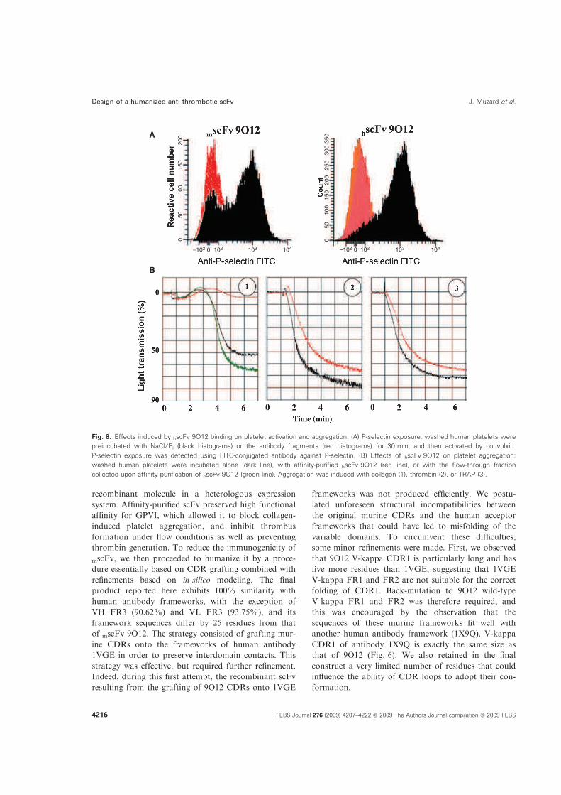
recombinant molecule in a heterologous expression
system. Affinity-purified scFv preserved high functional
affinity for GPVI, which allowed it to block collagen-
induced platelet aggregation, and inhibit thrombus
formation under flow conditions as well as preventing
thrombin generation. To reduce the immunogenicity of
mscFv, we then proceeded to humanize it by a proce-
dure essentially based on CDR grafting combined with
refinements based on in silico modeling. The final
product reported here exhibits 100% similarity with
human antibody frameworks, with the exception of
VH FR3 (90.62%) and VL FR3 (93.75%), and its
framework sequences differ by 25 residues from that
of mscFv 9O12. The strategy consisted of grafting mur-
ine CDRs onto the frameworks of human antibody
1VGE in order to preserve interdomain contacts. This
strategy was effective, but required further refinement.
Indeed, during this first attempt, the recombinant scFv
resulting from the grafting of 9O12 CDRs onto 1VGE
frameworks was not produced efficiently. We postu-
lated unforeseen structural incompatibilities between
the original murine CDRs and the human acceptor
frameworks that could have led to misfolding of the
variable domains. To circumvent these difficulties,
some minor refinements were made. First, we observed
that 9O12 V-kappa CDR1 is particularly long and has
five more residues than 1VGE, suggesting that 1VGE
V-kappa FR1 and FR2 are not suitable for the correct
folding of CDR1. Back-mutation to 9O12 wild-type
V-kappa FR1 and FR2 was therefore required, and
this was encouraged by the observation that the
sequences of these murine frameworks fit well with
another human antibody framework (1X9Q). V-kappa
CDR1 of antibody 1X9Q is exactly the same size as
that of 9O12 (Fig. 6). We also retained in the final
construct a very limited number of residues that could
influence the ability of CDR loops to adopt their con-
formation.
–102 102
A
B
103 104 0
0
0
50
1
00
1
50
2
00
2
50
3
00
35
0
50
1
00
1
50
2
00
–102 102 103 104 0
Fig. 8. Effects induced by hscFv 9O12 binding on platelet activation and aggregation. (A) P-selectin exposure: washed human platelets were
preincubated with NaCl ⁄ Pi (black histograms) or the antibody fragments (red histograms) for 30 min, and then activated by convulxin.
P-selectin exposure was detected using FITC-conjugated antibody against P-selectin. (B) Effects of hscFv 9O12 on platelet aggregation:
washed human platelets were incubated alone (dark line), with affinity-purified hscFv 9O12 (red line), or with the flow-through fraction
collected upon affinity purification of hscFv 9O12 (green line). Aggregation was induced with collagen (1), thrombin (2), or TRAP (3).
Design of a humanized anti-thrombotic scFv J. Muzard et al.
4216 FEBS Journal 276 (2009) 4207–4222 ª 2009 The Authors Journal compilation ª 2009 FEBS

Two critical areas were preserved. The first one
was the dipeptide Leu-Asp at position 59–60 in the
V-kappa domain. Leu59 (Pro in the human template)
was considered to be potentially significant, as it is in
the close vicinity of the residues of V-kappa CDR2.
Although Leu and Pro are both hydrophobic, Pro
has a cyclical side chain and is known to have spe-
cific effects on the protein backbone structure [43]. In
addition, we noticed that Leu occurs much less often
than Pro at this position (2%), and this may be
indicative of a specific role [44]. The other unmutated
murine residues with no similarities to the human
template (1VGE) were located in IGHV FR3 (Ala71,
Lys73, and Arg76; Kabbat numbering). In silico
observations and previous analysis have shown that
Ala71 has an important role in the conformation of
IGHV CDR2 [45]. Finally, only five murine residues
in V-kappa and 10 in IGHV were maintained in the
human frameworks selected for humanization. Other
humanized scFvs reported in the literature have
almost always required the insertion of numerous
murine residues into the reshaped molecule in order
to preserve strong binding activity and specificity [46].
fasta searches of the final humanized IGHV and
V-kappa sequences (with the exclusion of the CDRs)
against the UniProt data bank showed that the five
best-fitting sequences were all derived from human
antibodies, whereas the same search performed for
murine 9O12 variable domains led exclusively to
murine antibody sequences.
The main parameters usually affected by humaniza-
tion were well preserved in our final product. Affinity-
purified hscFv 9O12 was fully functional, with high
affinity for GPVI, a major point for biological applica-
tions. Fluorescence-activated cell sorting analysis also
indicated that hscFv 9O12 recognizes the same epitope
on human platelets as mouse 9O12 Fab, because its
binding was specifically blocked in the presence of a
molar excess of 9O12 Fab. However, despite the use of
codons optimized for prokaryotic expression in the
synthetic cDNA encoding hscFv 9O12, the production
yield remained low, and other expression systems will
have to be tested in the future [47,48]. For this reason,
the ability of hscFv to inhibit aggregation in flow con-
ditions was not investigated here. However, we clearly
demonstrate that binding of hscFv 9O12 inhibits
GPVI-dependent platelet activation and aggregation.
In conclusion, this study provides the first experi-
mental evidence that murine anti-human GPVI plate-
lets can be humanized without deleterious effects,
making hscFv 9O12 a powerful building block for the
generation and evaluation of new compounds with
potentially therapeutic antithrombotic properties.
Experimental procedures
Materials
Hybridoma cell line 9O12 secreting a monoclonal IgG1,k
directed against the GPVI of human platelets, and IgG and
Fab fragment preparation and purification, have been
reported previously [20]. The scFv 9C2 directed against a
scorpion toxin, irrelevant to GPVI, was prepared as in [49].
The antibody against flag (9E10) was used either free or
conjugated to horseradish peroxidase (Sigma-Aldrich, Saint
Quentin Fallavier, France) or to fluorescein isothiocyanate
(FITC) (Invitrogen, Cergy Pontoise, France). FITC-coupled
goat anti-mouse IgGs were from Sigma-Aldrich.
The recombinant soluble human GPVI protein, consisting
of two extracellular domains of the receptor, coupled to the
Fc fragment of human IgG1 was produced and purified as in
[20]. GPVI was coupled to cyanogen bromide-activated
Sepharose according to the manufacturer’s instructions
(Amersham-Pharmacia, Les Ulis, France). The gel was
stored at 4 !C in NaCl ⁄Pi containing sodium azide (1%).
Blood was collected from healthy volunteers. Washed
human platelets were obtained according to a previously
described procedure [24].
All chemicals were of standard grade from Sigma-Aldrich
or equivalent.
Construction of the single chain antibody genes
mRNA was isolated from freshly subcloned hybridoma
9O12. cDNAs encoding the antibody variable domains
(V-kappa and IGHV) were cloned after RT-PCR, essen-
tially as previously reported [21,22]. Murine scFv 9O12 was
created by PCR splicing with overlap extensions using oli-
gonucleotides that encode the (G4S)3 peptide linker between
the C-terminus of IGHV and the N-terminus of V-kappa.
First, IGHV and V-kappa genes were modified by PCR
amplification with primers VHRev and VHlinkFor or
VLlinkRev and VLFor, respectively. VHRev (5¢-CAG GTG
CAG CTG CAG GAG TCA GG-3¢) encoded the N-termi-
nal sequence of IGHV containing a PstI site, and VHlink-
For (5¢-ACC ACC GGA TCC GCC TCC GCC TGAGGA
GAC GGT GAC CGT-3¢) encoded the C-terminus of
IGHV and part of the linker. VLlinkRev (5¢-GGA
GGC GGA TCC GGT GGT GGC GGA TCT GGA GGT
GGC GGA AGC GAT GTT TTG ATG ACC CAA ACT
CCA CT-3¢) and VLFor (5¢-GAC CCT CGA GCG TTT
GAT CTC CAG CTT GGT-3¢), which contains a XhoI
site, were used to amplify and modify the V-kappa
domain. Both genes were assembled by ‘splicing by overlap
extension’ with primers VHRev and VLFor. The gene frag-
ment of the appropriate size was purified, cleaved with PstI
and XhoI, and cloned into the expression vector pSWI
restricted in the same manner before being sequenced. This
made it possible to clone the scFv gene in-frame with the
J. Muzard et al. Design of a humanized anti-thrombotic scFv
FEBS Journal 276 (2009) 4207–4222 ª 2009 The Authors Journal compilation ª 2009 FEBS 4217

pelB leader sequence at its 5¢-end and, downstream, a
sequence encoding the c-Myc tag. The constructed vector
pSW–mscFv9O12 was cloned into the ToppI E. coli strain
(Stratagene, La Jolla, USA).
Computational analysis and synthetic gene
design
Frameworks and the hypervariable loops (CDRs) of anti-
body variable domains were identified using the Kabbat
nomenclature [50]. An in silico 3D model of 9O12 Fv was
constructed by homology modeling based on V-kappa and
IGHV domains with maximum sequence identity and a
known tertiary structure. First, the protein structure
sequence Protein Data Bank was searched for antibody
sequence similarities with each individual 9O12 variable
domain, using the fasta 3 program at the European Bioin-
formatics Institute (EMBL-EBI, Cambridge, UK). Then,
modeler 3.0 software (Accelerys, San Diego, CA, USA)
was used to generate up to 20 models for each variable
domain, the best one being selected on the basis of the
rmsd value.
To design a humanized scFv (hscFv), the 9O12 variable
domain sequences were independently subjected to a fasta
search against the Protein Data Bank, and the closest
human antibody variable domains were selected. The same
search was also performed after excluding the CDR
regions. The scaffolds of variable domains belonging to
the same antibody molecule were chosen in order to mini-
mize the risk of lowering the stability of the interaction
between variable domains in the recombinant scFvs while
attempting to preserve the scaffold required for the integ-
rity of the antigen-binding site. In the first step, the
human antibody 1VGE variable domains were selected as
templates, and the murine 9O12 CDRs were grafted onto
them in silico. The framework sequences were inspected to
see whether any buried residues had been conserved. The
packing of the grafted loops was also evaluated by visual
inspection, and FR refinements were performed by in silico
substitution.
The changes in the amino acids required for the human-
ization were obtained by de novo DNA synthesis. Optimiza-
tion of the DNA design was performed using the codon
usage table for expression in E. coli cells [51]. Restriction
sites were also inserted at the extremities of the CDRs to
make it possible to carry out the cDNA sequence adjust-
ments that would be required to optimize the structural or
functional characteristics of the recombinant protein. The
synthetic gene encoding hscFv 9O12 was cloned into the
expression vector pSW1 restricted with PstI and XhoI, lead-
ing to a vector designated pSW–hscFv 9O12, which allows
periplasmic expression of hscFv 9O12 fused to the c-Myc
tag. The gene encoding hscFv 9O12 fused to the c-Myc tag
was also cloned into the pET-22b(+) vector (Merck
Chemicals Ltd., Nottingham, UK), which carries a T7
promoter. To do this, the sequence encoding hscFv 9O12
fused to the c-Myc tag was first reamplified from
pSW–hscFv 9O12 by PCR using primers VHpEtRev
(5¢-TG GCC ATG GCC CAG GTG CAG CTG CAG G-
3¢) and 9O12pEtFor (5¢-TG GTG CGG CCG CTT AT-
T AAT TCA GAT CCT CTT CTG A-3¢). The amplified
cDNA was then digested with NcoI and NotI, leading to a
820 bp sequence that was cloned in-frame with the pelB
leader sequence into pET-22b(+) restricted in the same
manner. The constructed vector, designated pET-
22–hscFv 9O12, was cloned into the BL21DE3 E. coli
strain (Merck Chemicals Ltd.).
Production and purification of recombinant
antibody fragments
For expression of the recombinant scFvs, bacteria con-
taining the desired plasmids were grown in 500 mL of 2·
TY medium (Difco, Le Pont de Claix, France) containing
ampicillin (100 lgÆmL)1) at 37 !C under rotative agitation
(125 r.p.m.), until A600 nm reached 0.6 (BL21DE3 E. coli)
or 1.5 (ToppI E. coli). Then, 0.8 mm isopropyl-thio-b-d-
galactoside was added to the medium, and incubation
was continued for 16 h at 16 !C under rotative agitation
(75 r.p.m.) to induce scFv production. Bacteria were then
collected by centrifugation (3600 g, 20 min, 4 !C). Peri-
plasmic extracts containing scFv were prepared by resu-
spending the pellet in 10 mL of ice-cold TES buffer
(0.2 m Tris ⁄HCl, pH 8.0, containing 0.5 mm EDTA and
0.5 m sucrose), and incubating for 30 min. Cells were
subjected to osmotic shock by adding TES buffer diluted
1 : 4 for 30 min on ice, and then centrifuging at 15 000 g
for 30 min at 4 !C to remove insoluble material. Deoxyri-
bonuclease A (50 U) and aprotinin (2 lgÆmL)) were
added to the supernatant before extensive dialysis against
NaCl ⁄Pi at 4 !C.
To purify scFv, the periplasmic extract (30 mL) was incu-
bated with 500 lL of GPVI coupled to Sepharose beads for
12 h at 4 !C and for 4 h at room temperature. The mixture
was loaded onto a microcolumn. After washing with
NaCl ⁄Pi at pH 7.4, bound proteins were eluted with 0.1 m
glycine-HCl (pH 3.0) in 0.4 mL fractions, and immediately
neutralized with 5 lL of 3 m Tris on ice. Fractions with
A280 nm higher than 0.2 were pooled and extensively
dialyzed against NaCl ⁄Pi.
The purity was checked by SDS ⁄PAGE using a 15% gel
followed by staining with Coomassie brilliant blue, or by
western blotting and immunostaining with horseradish
peroxidase-coupled antibody against c-Myc, essentially as
in [22]. The integrity of the purified recombinant protein
was also investigated by MALDI-TOF MS on a 4700
Proteomics Analyzer MALDI-time of flight (TOF)/TOF
apparatus (Applied Biosystems, Foster City, CA, USA).
Finally, the affinity-purified scFv (200 ll, 100 lgÆmL))
was analyzed by gel filtration using a Superdex 75 column
Design of a humanized anti-thrombotic scFv J. Muzard et al.
4218 FEBS Journal 276 (2009) 4207–4222 ª 2009 The Authors Journal compilation ª 2009 FEBS

(Amersham Bioscience, Les Ulis, France) calibrated using
standards from Boehringer Mannheim (Meylan, France).
Proteins were eluted with NaCl ⁄Pi at a flow rate of
0.5 mLÆmin)1, and detected with a UV recorder at 280 nm.
The concentration of the purified scFvs was evaluated
after measuring their absorbance at 280 nm, and using the
Swiss Institute of Bioinformatics software (protparam
tool) to determine the theoretical molecular mass of the
recombinant scFvs and their extinction coefficient [52].
Affinity-purified hscFv 9O12 was aliquoted and stored at
)20 !C in NaCl ⁄Pi containing 0.1% BSA until further use.
Immunochemical characterization of the scFv
fragments
ELISA
Microtitration plates were coated with GPVI in NaCl ⁄Pi
(10 lgÆmL)1, 100 lL per well) overnight at 4 !C. Nonspe-
cific binding sites were saturated with 100 lL of 1% BSA
in NaCl ⁄Pi for 90 min. The plates were then incubated
with increasing concentrations of the scFv preparation
(0–20 lgÆmL)1; 100 lL) for 90 min. They were incubated
for an additional 90 min with peroxidase-coupled antibody
against c-Myc (100 lL, 1 : 750 in NaCl ⁄Pi). All incuba-
tions were carried out at room temperature. Finally,
100 lL of the substrate solution (orthophenylene diamine;
Sigma-Aldrich) was added to each well for 5 min, and the
absorbance was read at 492 nm. Two controls were per-
formed: the first one used the irrelevant scFv 9C2 instead
of scFv 9O12, and for the second, coating with GPVI was
omitted. Five washes with NaCl ⁄Pi containing 0.05%
Tween and 0.1 mgÆmL)1 BSA were performed between
each of the intermediate steps. In competitive assays,
scFv 9O12 (100 lgÆmL)1) was mixed with increasing
concentrations of 9O12 (0–100 lgÆmL)1), before being
delivered into GPVI-coated microtitration wells. Bound
scFv was detected as described above.
SPR
The BIAcore 2000 instrument and all the reagents for anal-
ysis were obtained from BIAcore. GPVI was immobilized
(approximatively 600 RU) on a carboxymethyldextran CM5
sensor chip activated with a 1 :1 mix of N-hydroxysuccini-
mide (50 mm) and N-ethyl-N¢-(dimethylaminopropyl)-
carbo-di-imide (200 mm) by a 7 min pulse. Affinity-purified
antibody fragments were then passed over the immobilized
GPVI in HBS-EP buffer [0.01 m Hepes (pH 7.4), 0.15 m
NaCl, 0.005% polysorbate 20 (v ⁄ v)] at a flow rate of
20 lLÆmin)1 at 25 !C. Glycine-HCl (10 mm, pH 2.5) was
injected for 30 s at 20 lLÆmin)1 to regenerate the sensor
chip between successive samples. Kinetic constants (kon,
koff) were deduced from the analysis of association and
dissociation rates at four different antibody fragment
concentrations, ranging from 5 to 40 lgÆmL)1.The dissocia-
tion constant KD was calculated from KD = koff ⁄ kon. Sen-
sorgrams were analyzed using biaevaluation version 3.1
software. All experiments were carried out in quadruplicate
at the Institute Jacques Monod platform (Paris, France).
Flow cytometry
scFv binding
Washed human platelets (2 · 107 mL)1) from several
healthy volunteers were incubated for 30 min at room tem-
perature with 10–40 lgÆmL)1 purified scFv, and then incu-
bated again for 30 min at room temperature with 5 lL of
FITC-coupled anti-c-Myc IgG (dilution 1 : 60). Cell fluo-
rescence was measured using a flow cytometer (Epics XL,
Beckman Coulter, Villepinte, France). Background was
determined by using the irrelevant scFv 9C2 instead of the
scFv 9O12 variants. All incubations were carried out in the
dark. When 9O12 Fab (40 lgÆmL)1) binding to platelets
was investigated, FITC-conjugated goat anti-(mouse IgG)
(Sigma-Aldrich) (1 : 100) was used instead of the FITC-
coupled anti-c-Myc IgG.
scFv binding inhibition
Cells were incubated with blocking Fab 9O12 (10 lgÆmL)1)
for 10 min. They were then mixed with purified hscFv 9O12
(40 lgÆmL)1), and incubated for 30 min. Finally, 5 lL of
FITC-coupled anti-c-Myc IgG (diluted 1 : 60) was added to
the cells for a further 30 min before analysis of the cell
suspension by flow cytometry.
Inhibition of platelet activation
Platelets were incubated for 30 min. with scFv (10–
40 lgÆmL)1) or NaCl ⁄Pi. Then, platelets were activated
with convulxin (0.3 nm) for 15 min at 20 !C. Five micro-
liters of an anti-P-selectin IgG conjugated to FITC (Beck-
man Coulter, Villepinte, France) was added to the cells for
30 min before analysis of the cell suspension by flow cytom-
etry. All experiments were carried out at least in triplicate.
Ability of the antibody fragments to block GPVI
binding to collagen
Wells of a microtitration plate (Immulon 2; Dynex, VWF,
France) were coated overnight with 100 lL of collagen
type I (equine tendon; Horm, Nycomed, Munich)
(20 lgÆmL)1) and blocked with BSA (0.2% in NaCl ⁄Pi) for
2 h. Then, 100 lL of GPVI (10, 20 or 40 lgÆmL)1) that had
been preincubated for 30 min with increasing amounts of
antibody fragments (0–20 lgÆmL)1) was added to each well.
After incubation for 2 h, the bound GPVI was detected
J. Muzard et al. Design of a humanized anti-thrombotic scFv
FEBS Journal 276 (2009) 4207–4222 ª 2009 The Authors Journal compilation ª 2009 FEBS 4219

using a peroxidase-coupled anti-human Fc (Jackson
Immuno-Research Labs Inc., West Grove, PA, USA) and
orthophenylene dimine. All incubations were performed at
room temperature, and five washes with 300 lL of NaCl ⁄Pi
containing 0.1% Tween-20 and 1% BSA were carried out
between each step. All assays were conducted in triplicate.
The percentage of residual GPVI binding to collagen was
determined using mean values.
Platelet aggregation assays
Platelet aggregation
Washed human platelets (3 · 108 mL)1) were preincubated
for 5 min at 37 !C with antibody fragments in NaCl ⁄Pi
(25 lgÆmL)1), without stirring. Platelet aggregation was
then initiated by adding type I collagen to a final concen-
tration of 1 lgÆmL)1, or thrombin (1 nm) or the thrombin
receptor agonist peptide TRAP (10 lm) [20]. Platelet aggre-
gation induced changes in light transmission that were con-
tinuously recorded (Chronolog Aggregometer Chrono Log
Corp., Harveston, PA, USA).
Platelet aggregation under flow conditions
Platelet adhesion to collagen under flow conditions was
measured essentially as described previously [20]. Glass cov-
erslips were coated with fibrillar type I collagen
(50 lgÆmL)1). Blood from healthy volunteers was collected
on 40 lm PPACK, and labeled with DIOC-6 (1 lm). Blood
aliquots were incubated for 15 min at room temperature
with buffer or purified antibody fragment (9O12 Fab,
mscFv 9O12, scFv 9C2) at a final concentration of
50 lgÆmL)1. The mixture was then perfused over the colla-
gen-coated coverslips inserted in a flow chamber at
1500 s)1 for 5 min. Transmission and fluorescent images
were recorded in real time using a fluorescence microscope.
Fluorescent images were obtained from at least 10 different
collagen-containing microscopic fields, which were arbi-
trarily chosen at the end of perfusion. The area coverage of
fluorescent images was analyzed off-line using histolab
software (Microvision, Evry, France). Assays were per-
formed using blood from two healthy volunteers and two
distinct preparations of affinity-purified scFv.
Thrombin generation
Thrombin generation was continuously measured in plate-
let-rich plasma (PRP) using the thrombogram method as
previously described [53]. Briefly, citrated PRP
(1.5 · 108 platelets mL)1) was incubated with the antibody
fragments (50 lgÆmL)1) for 10 min at 37 !C before addition
of the collagen (5 lgÆmL)1). Ten minutes later, thrombin
generation was initiated by transferring the samples into the
wells of a microtitration plate containing tissue factor
(0.5 pm). After 5 min at 37 !C, the reaction was initiated by
addition of buffer containing CaCl2 (16.6 mm) and the fluo-
rescent thrombin substrate Z-GGR-AMC (Stago, Asnieres,
France). Fluorescence accumulation of the cleaved substrate
was continuously measured at excitation and emission wave-
lengths of 390 and 460 nm, respectively. First derivative
curves of fluorescence accumulation were converted into
thrombin concentration curves using a thrombin calibrator
[54]. The peak height is an indicator of the maximum rate of
thrombin formation, and is sensitive to platelet activation.
Acknowledgements
We are indebted to J. M. Camadro for stimulating dis-
cussions. We also acknowledge the invaluable technical
help provided by S. Loyau. J. Muzard was a research
fellow of Stago (Asnieres, France) and the Fondation
pour la Recherche Medicale (FRM). This work was
supported by grant no. 2007001960 from the Fonda-
tion de France and grant no. 07-EMPB-002-01 from
the Agence Nationale de la Recherche (ANR).
References
1 Bhatt DL & Topol EJ (2003) Scientific and therapeutic
advances in antiplatelet therapy. Nat Rev Drug Discov
2, 15–28.
2 Jackson SP & Schoenwaelder SM. (2003) Antiplatelet
therapy: in search of the ‘magic bullet’. Nat Rev Drug
Discov 2, 775–789.
3 Lindemann S, Kramer B, Daub K, Stellos K & Gawaz
M (2007) Molecular pathways used by platelets to initi-
ate and accelerate atherogenesis. Curr Opin Lipidol 18,
566–573.
4 Konishi H, Katoh Y, Takaya N, Kashiwakura Y, Itoh
S, Ra C & Daida H (2002) Platelets activated by colla-
gen through immunoreceptor tyrosine-based activation
motif play pivotal role in initiation and generation of
neointimal hyperplasia after vascular injury. Circulation
105, 912–916.
5 Jandrot-Perrus M, Busfield S, Lagrue AH, Xiong X,
Debili N, Chickering T, Le Couedic JP, Goodearl A,
Dussault B, Fraser C et al. (2000) Cloning, character-
ization, and functional studies of human and mouse gly-
coprotein VI: a platelet-specific collagen receptor from
the immunoglobulin superfamily. Blood 96, 1798–1807.
6 Moroi M, Jung SM, Okuma M & Shinmyozu K. (1989)
A patient with platelets deficient in glycoprotein VI that
lack both collagen-induced aggregation and adhesion.
J Clin Invest 84, 1440–1445.
7 Boylan B, Chen H, Rathore V, Paddock C, Salacz M,
Friedman KD, Curtis BR, Stapleton M, Newman DK,
Kahn ML et al. (2004) Anti-GPVI-associated ITP:
Design of a humanized anti-thrombotic scFv J. Muzard et al.
4220 FEBS Journal 276 (2009) 4207–4222 ª 2009 The Authors Journal compilation ª 2009 FEBS

an acquired platelet disorder caused by autoantibody-
mediated clearance of the GPVI ⁄FcRgamma-chain
complex from the human platelet surface. Blood 104,
1350–1355.
8 Massberg S, Konrad I, Bultmann A, Schulz C, Munch
G, Peluso M, Lorenz M, Schneider S, Besta F, Muller I
et al. (2004) Soluble glycoprotein VI dimer inhibits
platelet adhesion and aggregation to the injured vessel
wall in vivo. FASEB J 18, 397–399.
9 Gruner S, Prostredna M, Koch M, Miura Y, Schulte V,
Jung SM, Moroi M & Nieswandt B (2005) Relative anti-
thrombotic effect of soluble GPVI dimer compared with
anti-GPVI antibodies in mice. Blood 105, 1492–1499.
10 Nieswandt B, Schulte V, Bergmeier W, Mokhtari-Nejad
R, Rackebrandt K, Cazenave JP, Ohlmann P, Gachet
C & Zirngibl H (2001) Long-term antithrombotic pro-
tection by in vivo depletion of platelet glycoprotein VI
in mice. J Exp Med 193, 459–469.
11 Schulte V, Rabie T, Prostredna M, Aktas B, Gruner S
& Nieswandt B (2003) Targeting of the collagen-binding
site on glycoprotein VI is not essential for in vivo deple-
tion of the receptor. Blood 10, 3948–3952.
12 Massberg S, Gawaz M, Gruner S, Schulte V, Konrad I,
Zohlnhofer D, Heinzmann U & Nieswandt B (2003) A
crucial role of glycoprotein VI for platelet recruitment
to the injured arterial wall in vivo. J Exp Med 197,
41–49.
13 Qian MD, Villeval JL, Xiong X, Jandrot-Perrus M,
Nagashima K, Tonra J, McDonald K, Goodearl A &
Gill D (2002) Anti GPVI human antibodies neutralizing
collagen-induced platelet aggregation isolated from a
combinatorial phage display library. Hum Antibodies
11, 97–105.
14 Smethurst PA, Joutsi-Korhonen L, O’Connor MN, Wil-
son E, Jennings NS, Garner SF, Zhang Y, Knight CG,
Dafforn TR, Buckle A et al. (2004) Identification of the
primary collagen-binding surface on human glycopro-
tein VI by site-directed mutagenesis and by a blocking
phage antibody. Blood 103, 903–911.
15 Siljander PR, Munnix IC, Smethurst PA, Deckmyn H,
Lindhout T, Ouwehand WH, Farndale RW & Heems-
kerk JW (2004) Platelet receptor interplay regulates col-
lagen-induced thrombus formation in flowing human
blood. Blood 103, 1333–1334.
16 Matsumoto Y, Takizawa H, Nakama K, Gong X,
Yamada Y, Tandon NN & Kambayashi J (2006) Ex vivo
evaluation of anti-GPVI antibody in cynomolgus
monkeys: dissociation between anti-platelet aggregatory
effect and bleeding time. Thromb Haemost 96, 1671–
1675.
17 Matsumoto Y, Takizawa H, Gong X, Le S, Lockyer S,
Okuyama K, Tanaka M, Yoshitake M, Tandon NN &
Kambayashi J (2007) Highly potent anti-human GPVI
monoclonal antibodies derived from GPVI knockout
mouse immunization. Thromb Res 119, 319–329.
18 Li H, Lockyer S, Concepcion A, Gong X, Takizawa H,
Guertin M, Matsumoto Y, Kambayashi J, Tandon NN
& Liu Y (2007) The Fab fragment of a novel anti-GPVI
monoclonal antibody, OM4, reduces in vivo thrombosis
without bleeding risk in rats. Arterioscler Thromb Vasc
Biol 27, 1199–1205.
19 Takayama H, Hosaka Y, Nakayama K, Shirakawa K,
Naitoh K, Matsusue T, Shinozaki M, Honda M, Yata-
gai Y, Kawahara T et al. (2008) A novel antiplatelet
antibody therapy that induces cAMP-dependent endo-
cytosis of the GPVI ⁄Fc receptor gamma-chain complex.
J Clin Invest 118, 1785–1795.
20 Lecut C, Feeney LA, Kingsbury G, Hopkins J, Lanza
F, Gachet C, Villeval JL & Jandrot-Perrus M (2003)
Human platelet glycoprotein VI function is antagonized
by monoclonal antibody-derived Fab fragments.
J Thromb Haemost. 1, 2653–2662.
21 Juste M, Muzard J & Billiald P (2006) Cloning of the
antibody kappa light chain V-gene from murine hybri-
domas by bypassing the aberrant MOPC21-derived
transcript. Anal Biochem 349, 159–161.
22 Devaux C, Moreau E, Goyffon M, Rochat H & Billiald
P (2001) Construction and functional evaluation of a
single-chain antibody fragment that neutralizes toxin
AahI from the venom of the scorpion Androctonus
australis hector. Eur J Biochem 268, 694–702.
23 Lecut C, Schoolmeester A, Kuijpers MJ, Broers JL,
van Zandvoort MA, Vanhoorelbeke K, Deckmyn H,
Jandrot-Perrus M & Heemskerk JW (2004) Principal
role of glycoprotein I in alpha2beta1 and alphaIIbbe-
ta3 activation during collagen-induced thrombus
formation. Arterioscler Thromb Vasc Biol 24, 1727–
1733.
24 Jandrot-Perrus M, Lagrue AH, Okuma M & Bon C.
(1997) Adhesion and activation of human platelets
induced by convulxin involve glycoprotein VI and
integrin alpha2beta1. J Biol Chem 272, 27035–27041.
25 Kastrati A, Mehilli J, Neumann FJ, Dotzer F, ten Berg
J, Bollwein H, Graf I, Ibrahim M, Pache J, Seyfarth M
et al. (2006) Abciximab in patients with acute coronary
syndromes undergoing percutaneous coronary interven-
tion after clopidogrel pretreatment: the ISAR-REACT
2 randomized trial. JAMA 295, 1531–1538.
26 De Luca G, Suryapranata H, Stone GW, Antoniucci D,
Tcheng JE, Neumann FJ, Van de Werf F, Antman EM
& Topol EJ (2005) Abciximab as adjunctive therapy to
reperfusion in acute ST-segment elevation myocardial
infarction: a meta-analysis of randomized trials. JAMA
293, 1759–1765.
27 Clemetson JM, Polgar J, Magnenat E, Wells TN &
Clemetson KJ (1999) The platelet collagen receptor
glycoprotein VI is a member of the immunoglobulin
superfamily closely related to FcalphaR and the
natural killer receptors. J Biol Chem 274, 29019–
29024.
J. Muzard et al. Design of a humanized anti-thrombotic scFv
FEBS Journal 276 (2009) 4207–4222 ª 2009 The Authors Journal compilation ª 2009 FEBS 4221

28 Laffly E & Sodoyer R (2005) Monoclonal and recombi-
nant antibodies, 30 years after... Hum Antibodies 14,
33–55.
29 Chen H, Locke D, Liu Y, Liu C & Kahn ML (2002)
The platelet receptor GPVI mediates both adhesion
and signaling responses to collagen in a receptor
density-dependent fashion. J Biol Chem 277, 3011–
3019.
30 Gardiner EE, Arthur JF, Kahn ML, Berndt MC &
Andrews RK (2004) Regulation of platelet membrane
levels of glycoprotein VI by a platelet-derived metallo-
proteinase. Blood 104, 3611–3617.
31 Lagrue-Lak-Hal AH, Debili N, Kingbury G, Lecut C,
Le Couedic JP, Villeval JL, Jandrot-Perrus M &
Vainchenker W (2001) Expression and function of the
collagen receptor GPVI during megakaryocyte
maturation. J Biol Chem 276, 15316–15325.
32 Ohlmann P, Hechler B, Ravanat C, Loyau S, Her-
renschmidt N, Wanert F, Jandrot-Perrus M & Gachet
C (2008) Ex vivo inhibition of thrombus formation by
an anti-glycoprotein VI Fab fragment in non-human
primates without modification of glycoprotein VI
expression. J Thromb Haemost. 6, 1003–1011.
33 Hwang WY & Foote J (2005) Immunogenicity of engi-
neered antibodies. Methods 36, 3–10.
34 Curtis BR, Swyers J, Divgi A, McFarland JG & Aster
RH (2002) Thrombocytopenia after second exposure to
abciximab is caused by antibodies that recognize abcix-
imab-coated platelets. Blood 99, 2054–2059.
35 Parker C (2009) Eculizumab for paroxysmal nocturnal
haemoglobinuria. Lancet 373, 759–767.
36 Tsurushita N, Hinton PR & Kumar S (2005) Design of
humanized antibodies: from anti-Tac to Zenapax. Meth-
ods 36, 69–83.
37 Fontayne A, Vanhoorelbeke K, Pareyn I, Van Rom-
paey I, Meiring M, Lamprecht S, Roodt J, Desmet J
& Deckmyn H (2006) Rational humanization of the
powerful antithrombotic anti-GPIbalpha antibody:
6B4. Thromb Haemost 96, 671–684.
38 Lazar GA, Desjarlais JR, Jacinto J, Karki S & Ham-
mond PW (2007) A molecular immunology approach to
antibody humanization and functional optimization.
Mol Immunol 44, 1986–1998.
39 Jones PT, Dear PH, Foote J, Neuberger MS & Winter
G (1986) Replacing the complementarity-determining
regions in a human antibody with those from a mouse.
Nature 321, 522–525.
40 Verhoeyen M, Milstein C & Winter G (1988) Reshaping
human antibodies: grafting an antilysozyme activity.
Science 239, 1534–1536.
41 Caldas C, Coelho V, Kalil J, Moro AM, Maranhao
AQ & Brıgido MM (2003) Humanization of the anti-
CD18 antibody 6.7: an unexpected effect of a frame-
work residue in binding to antigen. Mol Immunol 39,
941–952.
42 Gonzales NR, Padlan E, De Pascalis R, Schuck P, Sch-
lom J & Kashmiri SV (2004) SDR grafting of a murine
antibody using multiple human germline templates to
minimize its immunogenicity. Mol Immunol 41, 863–
872.
43 MacArthur MW & Thornton JM (1999) Protein side-
chain conformation: a systematic variation of chi 1
mean values with resolution – a consequence of multiple
rotameric states? Acta Crystallogr D Biol Crystallogr 55,
994–1004.
44 Honegger A & Pluckthun A (2001) The influence of
the buried glutamine or glutamate residue in position 6
on the structure of immunoglobulin variable domains.
J Mol Biol 309, 687–699.
45 Tramontano A, Chothia C & Lesk AM (1990) Frame-
work residue 71 is a major determinant of the position
and conformation of the second hypervariable region in
the VH domains of immunoglobulins. J Mol Biol 215,
175–182.
46 Woodle ES, Thistlethwaite JR, Jolliffe LK, Zivin RA,
Collins A, Adair JR, Bodmer M, Athwal D, Alegre ML
& Bluestone JA (1992) Humanized OKT3 antibodies:
successful transfer of immune modulating properties
and idiotype expression. J Immunol 148, 2756–2763.
47 Humphreys DP & Glover DJ (2001) Therapeutic anti-
body production technologies: molecules, applications,
expression and purification. Curr Opin Drug Discov
Devel 4, 172–185.
48 Carson KL (2005) Flexibility – the guiding principle for
antibody manufacturing. Nat Biotechnol 23, 1054–1058.
49 Aubrey N, Devaux C, Sizaret PY, Rochat H, Goyffon
M & Billiald P (2003) Design and evaluation of a
diabody to improve protection against a potent scor-
pion neurotoxin. Cell Mol Life Sci 60, 617–628.
50 Wu TT & Kabbat EA (1970) An analysis of the
sequences of the variable regions of Bence Jones proteins
and myeloma light chains and their implications for anti-
body complementarity. J Exp Med 132, 211–250.
51 Sharp PM & Li WH (1986) Codon usage in regulatory
genes in Escherichia coli does not reflect selection for
‘rare’ codons. Nucleic Acids Res 14, 7737–7749.
52 Appel RD, Bairoch A & Hochstrasser DF (1994) A
new generation of information retrieval tools for biolo-
gists: the example of the ExPASy WWW server. Trends
Biochem Sci 19, 258–260.
53 Lecut C, Feijge MA, Cosemans JM, Jandrot-Perrus M
& Heemskerk JW (2005) Fibrillar type I collagens
enhance platelet-dependent thrombin generation via
glycoprotein VI with direct support of alpha2beta1 but
not alphaIIbbeta3 integrin. Thromb Haemost 94, 107–
114.
54 Hemker HC, Giesen PL, Ramjee M, Wagenvoord R &
Beguin S (2000) The thrombogram: monitoring throm-
bin generation in platelet-rich plasma. Thromb Haemost
83, 589–591.
Design of a humanized anti-thrombotic scFv J. Muzard et al.
4222 FEBS Journal 276 (2009) 4207–4222 ª 2009 The Authors Journal compilation ª 2009 FEBS

!

Abstract:
Human platelets glycoprotein VI (GPVI) is evidenced to be a platelet receptor of major
importance in the occurrence of arterial thrombosis. Thus, it can be considered to be of
great interest in diagnosis and therapeutic of atheriosclerotic diseases. Antibodies are
powerful molecules which can be used in both diagnostic as well as for therapeutic
purposes due to their unique characteristics. Monoclonal and recombinant antibodies have
antigen restricted specificity, high affinity and can be used in various assays. Moreover, the
good knowledge of their structure and molecular engineering facilities now allows the
antibody modulation according to desired properties.
Our group has already produced several monoclonal antibodies to human GPVI by gene
gun immunization against the immunoadhesin hGPVI-Fc, which differ in fine epitope
specificity, affinity and other functional properties (Lecut et al. 2003). One, 3J24, with
diagnostic potential while the other, 9O12, has a therapeutic potential because it blocks the
binding of GPVI to collagen. Its Fab fragment has been extensively characterized in vitro,
ex vivo and in vivo for its antithrombotic properties.
Here, we designed and reshaped a single-chain antibody fragment (scFv) based on 3J24
variable domains for the quantification of GPVI with diagnostic potential. We were also
involved in the design, production and functional evaluation of humanized anti-GPVI
recombinant antibody fragments (scFvs and Fabs) with therapeutic properties.
Key words: recombinant antibody fragments, scFv, arterial thrombosis, platelets,
glycoprotein VI
Copyright © 2022 FDOKUMEN