Antinuclear and antiphospholipid antibodies versus disease ...
96
Linköping University Medical Dissertation No. 1731 Antinuclear and antiphospholipid antibodies versus disease manifestations and clinical outcomes in systemic lupus erythematosus Martina Frodlund
-
Upload
khangminh22 -
Category
Documents
-
view
1 -
download
0
Transcript of Antinuclear and antiphospholipid antibodies versus disease ...

Linköping University Medical Dissertation No. 1731
Antinuclear and antiphospholipid antibodies versus disease
manifestations and clinical outcomes in systemic lupus erythematosus
Martina Frodlund

Linköping University Medical Dissertations
No. 1731
Antinuclear and antiphospholipid
antibodies versus disease manifestations
and clinical outcomes in systemic lupus
erythematosus
Martina Frodlund
Department of Biomedical and Clinical Sciences
Faculty of Medicine and Health Sciences, Linköping University
SE-581 85 Linköping, Sweden
Linköping 2020

© Martina Frodlund, 2020
Front cover by Martina Frodlund
Reprints of published articles I-V were made with permission from the copyright holders. Photos of ANA staining pattern were reprinted with permission from Dr Edward Chan and front/back page with permission from senior professor Thomas Skogh.
Printed in Sweden by LiU-Tryck, Linköping, Sweden, 2020.
ISBN: 978-91-7929-895-1
ISSN: 0345-0082

”The more you think, the more you realize
that there is no simple answer”
-Winnie-the-Pooh (A. A. Milne)

Supervisor
Associate Professor Christopher Sjöwall
Department of Biomedical and Clinical Sciences
Linköping University
Co-supervisors
Associate Professor Jonas Wetterö
Department of Biomedical and Clinical Sciences
Linköping University
Senior Professor Thomas Skogh
Department of Biomedical and Clinical Sciences
Linköping University
Faculty Opponent
Professor Lene Dreyer
Department of Clinical Medicine
The Faculty of Medicine
Aalborg University

TABLE OF CONTENTS
INTRODUCTION ................................................................................................................................................ 1
HISTORICAL BACKGROUND ....................................................................................................................... 1
PATHOGENESIS & AETIOLOGY................................................................................................................... 1
CLASSIFICATION ........................................................................................................................................... 6
EPIDEMIOLOGY .............................................................................................................................................. 8
CLINICAL FEATURES & OUTCOME ............................................................................................................ 9
ASSESSMENT OF DISEASE ACTIVITY ...................................................................................................... 11
ASSESSMENT OF ORGAN DAMAGE ......................................................................................................... 14
THERAPY ....................................................................................................................................................... 14
HISTORICAL BACKGROUND OF ANA & FINE SPECIFICITIES ............................................................. 15
ANA PATTERNS ............................................................................................................................................ 16
OSTEOPONTIN & OTHER BIOMARKERS .................................................................................................. 21
AIM ...................................................................................................................................................................... 23
PATIENTS & METHODS................................................................................................................................. 24
KLURING ........................................................................................................................................................ 24
PATIENTS & CONTROLS ............................................................................................................................. 25
METHODS ...................................................................................................................................................... 25
IMMUNOFLUOROSCENSE MICROSCOPY FOR ANA (IF-ANA) & ANTI- DNA................................. 26
IMMUNOASSAYS ......................................................................................................................................... 28
OSTEOPONTIN ASSAY................................................................................................................................. 30
ANA FINE SPECIFICITIES ............................................................................................................................ 31
ANTI-PHOSPHOLIPID SYNDROME & RELATED ANTIBODIES ............................................................ 32
STATISTICS .................................................................................................................................................... 33
RESULTS & DISCUSSION .............................................................................................................................. 35
PAPER I ........................................................................................................................................................... 35
PAPER II .......................................................................................................................................................... 37
PAPER III ......................................................................................................................................................... 42
PAPER IV ........................................................................................................................................................ 46
PAPER V .......................................................................................................................................................... 52
CONCLUSION ................................................................................................................................................... 58
FUTURE PERSPECTIVES ............................................................................................................................... 60
SAMMANFATTNING PÅ SVENSKA ............................................................................................................. 62
TACK .................................................................................................................................................................. 64
REFERENCES ................................................................................................................................................... 67
ds


ABSTRACT
Systemic lupus erythematosus (SLE) has an exceptionally heterogeneous clinical spectrum, ranging from mild disease limited to skin and joints to severe manifestations with renal disorder, central nervous system disease, severe cytopenias and thromboembolic events. Important clinical challenges include the prediction of disease flares and the identification of individuals that are likely to evolve severe disease with accrual of organ damage and worse prognosis. Autoantibodies, i.e. antinuclear antibodies (ANA) and antiphospholipid antibodies (aPL), and interferon alpha (IFN-α) that contribute to formation of immune complexes with nuclear antigens, are hallmarks considered to drive the disease in a vicious circle of antigen exposure, autoantibody production, inflammation and organ damage. There are few good biomarkers to predict severe SLE and organ damage. The aim of this PhD project was thus to increase the knowledge regarding ANA as well as aPL, and other potential biomarkers in relation to clinical features and disease outcomes in SLE.
As expected, we found that the homogeneous ANA staining pattern was most common, and that it was associated with the occurrence of the ‘immunological disorder’ criterion. Speckled ANA was the second most common staining pattern, and it was inversely associated with arthritis, the ‘immunological disorder’ criterion and organ damage (Paper I). We also demonstrated that a considerable proportion of the patients lost ANA-positivity over time, whereas consistent staining patterns were most frequent (Paper V).
Survival of patients with SLE has improved. Yet, in comparison to the general population, irreversible organ damage and increased mortality remains a critical concern. In Paper II, our cross-sectional analysis showed that more than a quarter of the patients had any aPL isotype (IgG, IgM or IgA class), and 14% were classified with antiphospholipid antibody syndrome (APS). A positive lupus anticoagulant (LA) test and/or IgG aPL tests were associated with most APS-related events and organ damage. Lupus nephritis, tobacco smoking, LA-positivity and the use of statins and/or corticosteroids were strongly associated with damage accrual, while hydroxychloroquine seemed to be protective. IgA aPL was not uncommon (16%) in Swedish cases of SLE, and analysis of IgA aPL may add information among clinically suspected APS-patients testing negative for LA and other aPL isotypes.
Despite modern management and tax-funded health care with universal access, almost two-thirds of the patients accrued organ damage over time, and the main causes of death were identified as malignancy, infection, and cardiovascular disease. We could confirm well-established risk factors for organ damage such as APS, hypertension, and/or the use of corticosteroids, but we also observed that other factors such as pericarditis, haemolytic anaemia, lymphopenia and myositis seems to be of importance in this view (Paper IV).
We also demonstrated that levels of the extracellular matrix protein osteopontin (OPN) was correlated with disease activity in patients with recent-onset SLE. In addition, OPN levels reflected global organ damage and were associated with APS and could have potential as a valuable biomarker in SLE (Paper III).
Additional studies are warranted to further establish the clinical and mechanistic relevance of ANA seroconversion, OPN, as well as the importance of IgA aPL. Vigilance for malignancies, a restricted use of corticosteroids and prevention of cardiovascular disease and APS events are among modifiable factors to prevent organ damage and premature mortality.
This thesis emphasizes the importance of autoantibodies in the pathogenesis, and diagnosis, of SLE. The autoantibody profile can be of great importance for tailored therapy in order to minimize the risk of organ damage accrual, morbidity as well as mortality.


LIST OF ORIGINAL PUPLICATIONS
This thesis is based on the following papers, which will be referred to in the text by their Roman
numerals (I-V):
I Frodlund M, Dahlström Ö, Kastbom A, Skogh T, Sjöwall C.
Associations between antinuclear antibody staining patterns and clinical features of
systemic lupus erythematosus: analysis of a regional Swedish register.
British Medical Journal Open 2013;3(10):e003608
II Frodlund M, Vikerfors A, Grosso G, Skogh T, Wetterö J, Elvin K, Gunnarsson I,
Kastbom A, Dahlström Ö, Rönnelid J, Svenungsson E, Sjöwall C.
Immunoglobulin A anti-phospholipid antibodies in Swedish cases of systemic lupus
erythematosus: associations with disease phenotypes, vascular events and damage
accrual.
Clinical & Experimental Immunology 2018;194(1):27–38
III Wirestam L, Frodlund M, Enocsson H, Skogh T, Wetterö J, Sjöwall C.
Osteopontin is associated with disease severity and antiphospholipid syndrome in well
characterised Swedish cases of SLE.
Lupus Science & Medicine 2017;4(1):e000225
IV Frodlund M, Reid S, Wetterö J, Dahlström Ö, Sjöwall C, Leonard D.
The majority of Swedish systemic lupus erythematosus patients are still affected by
irreversible organ impairment: factors related to damage accrual in two regional
cohorts.
Lupus 2019;28(10):1261–1272
V Frodlund M, Wetterö J, Dahle C, Dahlström Ö, Skogh T, Rönnelid J, Sjöwall C.
Longitudinal antinuclear antibody (ANA) seroconversion in systemic lupus
erythematosus: a prospective study of Swedish cases with recent-onset disease.
Clinical & Experimental Immunology 2020;199(3):245–254

ABBREVIATIONS
ACR American College of Rheumatology
ALBIA addressable laser bead immuno assay
ANA antinuclear antibody
APS antiphospholipid antibody syndrome
anti-dsDNA anti-double-stranded DNA antibody
aCL anti-cardiolipin antibodies
anti-β2GPI anti-β2 glycoprotein-I antibodies
aPL anti-phospholipid antibodies
APS anti-phospholipid antibody syndrome
BLyS B-lymphocyte stimulator
C-ANA centromere ANA
CAPS Catastrophic APS
CI confidence interval
CLIFT Crithidia luciliae immunofluorescence test
CNS central nervous system
CRP C-reactive protein
CSA cyclosporine
CYC cyclophosphamide
DHEAS dehydroepiandrosterone
DIL drug-induced lupus
dRVVT dilute Russell´s viper venom time
DMARD disease modifying antirheumatic drug
EBV Epstein-Barr virus
ELISA enzyme-linked immunosorbent assay
ESR erythrocyte sedimentation rate
EULAR European League Against Rheumatism
FEIA fluoroenzyme-immunoassay
FcγRIIa Fcγ-receptor IIa
GAPSS global APS score
GAS gamma-activated sequences
GFR glomerular filtration rate
H-ANA homogeneous ANA
HCQ hydroxychloroquine
HEp-2 human epidermoid carcinoma cells
HMGB1 high mobility group box chromosomal protein 1
HLA human leukocyte antigen

HN-ANA homogeneous and nucleolar ANA
HS-ANA homogeneous and speckled ANA
ICAP International Consensus of ANA Patterns
IFN interferon
IFNAR IFN-α/β receptor
IF-ANA immunofluorescence ANA
IC immune complex
Ig immunoglobulin
ISGF3 IFN-stimulated gene factor 3 complex
ISN/RPS International Society of Nephrology/Renal Pathology society
ISRE IFN-stimulated response elements
KLURING Kliniskt LUpus Register I Nordöstra Götaland (Swedish); Clinical Lupus Register In Northeastern Gothia (English)
LA lupus anticoagulant
LE cell lupus erythematosus cell
MND multiple nuclear dots
MMF mycophenolate mofetil
mSLEDAI-2K modified SLE disease activity index 2000
N-ANA nucleolar ANA
NET neutrophil extracellular traps
OPN osteopontin
OR odds ratio
pDC plasmacytoid dendritic cell
pSS primary Sjögren’s syndrome
RA rheumatoid arthritis
RNP ribonucleoprotein
S-ANA speckled ANA
SD standard deviation
SDI SLICC/ACR damage index
SLE systemic lupus erythematosus
SLEDAI-2K SLE disease activity index 2000
SLICC Systemic Lupus International Collaborating Clinics
Sm Smith antigen
SN-ANA Speckled and nucleolar ANA
snRNP small nuclear ribonucleoprotein
suPAR soluble urokinase plasminogen activator receptor
SS Sjögren’s syndrome
WHO World Health Organization


1
INTRODUCTION
Systemic lupus erythematosus (SLE) is a chronic, autoimmune systemic disease. Clinically SLE is characterized by involvement of multiple organ systems, ranging from mild to life-threatening. Periods of flares are often followed by remission. The female-to-male sex ratio for SLE is approximately 9:1, but can vary in different ages and ethnic groups (1-3). The clinical picture is distinguished by systemic inflammation and tissue damage which contributes to morbidity as well as increased mortality compared to the general population. The breakdown of self-tolerance and production of autoantibodies such as antinuclear antibodies (ANA) and anti-double-stranded DNA (anti-dsDNA) in SLE is thought to be the result of a combination of genetic susceptibility and environmental factors such as smoking, viral infections, hormones and drugs (4).
HISTORICAL BACKGROUND
The disease was first reported in the middle ages describing a severe facial rash resembling a wolf´s bite, Lupus (latin for wolf) erythematosus (latin for red) (5). In 1872, Kaposi described the systemic nature of the disease and Sir William Osler further established this by including cardiac, renal and pulmonary involvement, and he also coined the term systemic lupus erythematosus (6, 7). The implication of the immune system was acknowledged first in 1948 when Hargraves et al. discovered the LE (lupus erythematosus) cell in bone marrow from a patient with SLE (8). The first identification of a nuclear staining on sections of rat tissue by use of immunofluorescence (IF) microscopy was made by Holman and Kunkel in 1957 (9). Friou’s recognition of the “antinuclear factor” hereafter formed the foundation of modern ANA diagnostics by indirect IF microscopy (10). In SLE, loss of immune tolerance leads to production of autoantibodies by hyperreactive B cells, e.g. ANA, formation of immune complexes (IC), production of interferon-α (IFN-α), tissue inflammation and organ dysfunction.
PATHOGENESIS & AETIOLOGY
The pathogenesis of SLE is multifactorial including ethnicity, environmental and hormonal factors, but to a large extent it is unknown. The pathogenesis of SLE comprises dysregulated apoptosis and inefficient removal of apoptotic cellular material (11). Such cellular debris will expose nuclear constituents as well as phospholipids on its surface, which under certain conditions may undergo conformational changes, and become immunogenic. This may result in loss of B cell tolerance and production of autoantibodies against nuclear and phospholipid/phospholipid-related antigens of which some play central roles in autoimmunity (for instance, antibodies against cardiolipin and β2-glycoprotein-I in the antiphospholipid antibody syndrome (APS) and antibodies against dsDNA in SLE); Figure 1 (12, 13).

2
Figure 1: Simplified figure of the pathogenesis in SLE and the so-called vicious circle. Figure by Lina Wirestam.
A prominent production of autoantibodies and immune complexes, and an increased expression of type I interferon regulated genes, recognized as the IFN-signature is often seen (14). According to one study 50-75% of adults and 90% of children with SLE displayed a type I IFN signature, whereas circulating levels of IFN-α in adults with SLE may be considerable lower (15, 16). This observation as well as reports that IFN-α therapy can induce lupus-like disease suggests that IFNs have an important role in the SLE pathogenesis. Both environmental and genetic factors increase the activation of the type I IFN system in patients with SLE (14). UV-light exposure leads to cell death and leakage of nuclear antigens which enhances binding by autoantibodies and formation of ICs that enhance type I IFN production in the skin in SLE patients (14). Genetic associations with SLE have been found for more than 80 genetic loci and more than half of them are connected to the type I IFN signature. This includes TLR7, IRF5, TYK2 and STAT4, all of them being central in type I IFN production and signalling (14). Further on, control and regulation of type I IFN production in SLE are dysregulated with loss of proper negative feedback mechanisms. Consequently, type I IFN has been suggested being one of the driving forces behind the disease and new treatments aiming to inhibit IFN production or diminish their immunomodulatory effects in SLE are under development.
Normally, IFN-α production ceases once the pathogen has been eliminated and the plasmacytoid dendritic cells (pDCs) become temporarily refractory to new stimuli due to inhibition and degradation of transcription factors and signal transducers. In SLE patients a persistent stimulation of pDCs are seen by endogenous nucleic acids keeping IFN-α continuously active. The increased apoptosis and impaired clearance of apoptotic material in SLE patients contribute to formation of interferogenic ICs. Schematically, the Fc-parts of IC-Ig:s binds to FcγRIIa receptors on pDCs, and ICs are endocytosed. The nucleic acid content
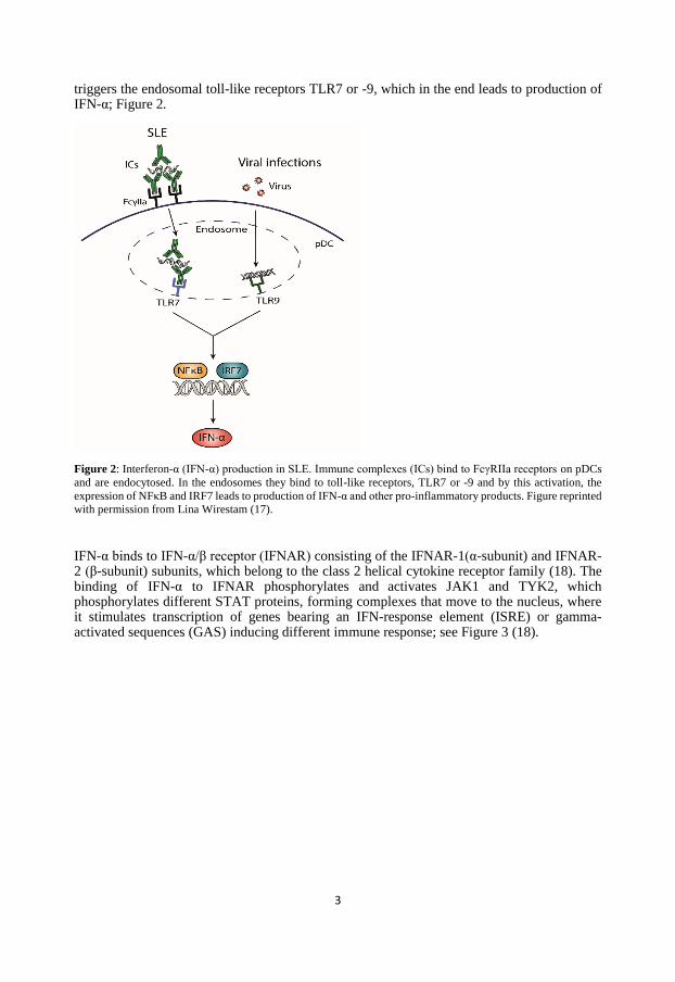
3
triggers the endosomal toll-like receptors TLR7 or -9, which in the end leads to production of IFN-α; Figure 2.
Figure 2: Interferon-α (IFN-α) production in SLE. Immune complexes (ICs) bind to FcγRIIa receptors on pDCs
and are endocytosed. In the endosomes they bind to toll-like receptors, TLR7 or -9 and by this activation, the
expression of NFκB and IRF7 leads to production of IFN-α and other pro-inflammatory products. Figure reprinted
with permission from Lina Wirestam (17).
IFN-α binds to IFN-α/β receptor (IFNAR) consisting of the IFNAR-1(α-subunit) and IFNAR-2 (β-subunit) subunits, which belong to the class 2 helical cytokine receptor family (18). The binding of IFN-α to IFNAR phosphorylates and activates JAK1 and TYK2, which phosphorylates different STAT proteins, forming complexes that move to the nucleus, where it stimulates transcription of genes bearing an IFN-response element (ISRE) or gamma-activated sequences (GAS) inducing different immune response; see Figure 3 (18).

4
Figure 3: IFN-α binds to its receptor IFNAR (containing two subunits, IFNAR-1 and -2), thereby activating
TYK2 and JAK1 which activates different STATS. STAT1, STAT2 and IRF9 constitutes the IFN-stimulated gene
factor 3 complex (ISGF3), which can bind to IFN-stimulated response elements (ISRE), generating an antiviral
response. STAT1 homodimers attaches to gamma-activated sequences (GAS) causing a pro-inflammatory
response. Homodimers of STAT3 conversely, induces an anti-inflammatory response via GAS. Figure reprinted
with permission from Lina Wirestam (17).
All nuclear bearing cells have the ability to produce and respond to IFN-α although pDCs are the main source of IFN-α and can produce up to 1000-fold more than other cells (19). IFN-α (and IFN-β) are crucial for stimulation of pDCs and activation of T cells, B cell development and antibody production (19).
Other endogenous IFN inducers in SLE are peptides from neutrophil extracellular traps (NET) and self-nucleic acids in complex with DNA- or RNA-binding proteins for example high mobility group box chromosomal protein 1 (HMGB1) and heat shock protein 90. In murine models it is shown that depletion of pDCs reduces the type I IFN signature and improves the pathology (20).
The human leukocyte antigen (HLA) region, in particular HLA-DRB1, represent a susceptibility loci associated with SLE, whereas many other non-HLA SLE susceptibility loci are situated within or close to genes with functional relevance in the immune system, e.g. IFN as well as B and T cell signalling, clearance of dead cellular debris and complement deficiencies (21). Studies including monozygotic twins and familial aggregation support the idea of genetic predisposition. The risk of developing SLE among siblings have been shown to be almost 30-fold higher than in the general population and ANA-positivity has been shown to be present in 27% of offspring to mothers with SLE compared to 7% in controls (22, 23). However, in families with several affected members, the genetic component is complex and does not follow a classical Mendelian inheritance pattern, and only a few cases can be attributed to highly penetrating rare mutations (24). Moreover, there is a ten-fold raised risk in monozygotic twins compared to dizygotic twins to develop SLE (21).

5
A strong association between mutations in the classical complement pathway and SLE susceptibility is seen e.g. over 90% of individuals with homozygote C1q-deficiency and more than 75% of individuals with homozygote C4 deficiency develop SLE (25, 26). A possible explanation is the important role of the complement system in clearing ICs and removing apoptotic cell debris (27). A hallmark for SLE is the overexpression of type I interferon and non-HLA gene variants involved in IFN-signalling have recently been proven to associate with the disease (28).
Well-known environmental risk factors in SLE development are cigarette smoking, silica, postmenopausal hormone replacement therapy and oral contraceptives (29, 30). Ultraviolet light, infections like the Epstein-Barr virus (EBV), air pollution, pesticides and heavy metals constitutes possible risk factors, still debated (29, 30). Biological processes associating environmental exposures and SLE risk comprise inflammatory cytokine upregulation, systemic inflammation, hormonal effects and increased oxidative stress (29, 30). Exposure to UV-light can cause photosensitivity and cutaneous lupus as well as induce systemic flares (31, 32). Moreover, SLE exacerbations are more often seen in the spring and summer in Scandinavia and outdoor work is associated with the risk of developing SLE (33, 34). Viral infections including EBV, cytomegalovirus and parvovirus B19 have been proposed to trigger SLE and increased prevalence of EBV infections have been reported before onset of SLE (35-37). Cross-reactivity between EBV and autoantigens, e.g. Ro60/SSA and Sm have been suggested to trigger SLE onset (38).
Cigarette smoking is associated with a modestly elevated risk of SLE, whereas a moderate intake of alcohol may protect against SLE development, but conflicting data have been reported (39-41). Sex hormones may play a role in the pathogenesis of SLE and can be illustrated by observations such as the large predominance of SLE among women in reproductive age, whereas the female-to-male ratio is low before puberty and the fact that the use of high oestrogen-containing contraceptives, postmenopausal hormone replacement therapy, ovarian stimulation as well as pregnancy can give rise to disease exacerbations (2, 42-46). Factors related to the X-chromosome may also be of importance to predispose women to SLE (28).
Pharmacological agents might induce SLE in predisposed individuals or cause a syndrome called drug-induced lupus (DIL) (47, 48). More than 80 different drugs, e.g. procainamide, hydralazine, sulfasalazine and anti-tumor necrosis factor agents, have been associated with DIL and some of them have been demonstrated to enhance autoimmunity (48, 49).

6
CLASSIFICATION
Classification criteria for SLE have been developed for the purpose of research and surveillance. The most commonly used criteria were composed by the American College of Rheumatology (ACR) in 1982 (Table 1) and revised in 1997, adding aPL antibodies and deleting the “positive LE cell preparation” (50).
Table 1. 1982 classification criteria for systemic lupus erythematosus.
Requirements: ≥4 criteria presented serially or simultaneously
Criterion Definition
1. Malar rash Erythema over the malar eminences
2. Discoid rash Erythematous raised patches
3. Photosensitivity Skin rash as a result of unusual reaction to sunlight
4. Oral ulcers Oral or nasopharyngeal ulceration
5. Arthritis Non-erosive arthritis involving 2 or more peripheral joints
6. Serositis Pleuritis or pericarditis
7. Renal disorder Proteinuria or cellular casts
8. Neurologic disorder Seizures or psychosis
9. Hematologic disorder Haemolytic anaemia or leukopenia or lymphopenia or
thrombocytopenia
10. Immunologic disorder Positive LE cell preparation or abnormal serum level of
anti-dsDNA or anti-Sm or Wasserman reaction
11. Antinuclear antibody ANA in abnormal level and absence of drugs associated
with ‘drug-induced lupus’ syndrome
ANA = antinuclear antibodies, dsDNA = double-stranded deoxyribonucleic acid, LE = Lupus erythematosus, Sm
= Smith.
Summarized from Tan EM, Cohen AS, Fries JF, Masi AT, McShane DJ, Rothfield NF, et al. The 1982 revised
criteria for the classification of systemic lupus erythematosus. Arthritis Rheum. 1982;25(11):1271-7 (51).
The Fries’ diagnostic principle is often used in clinical practice and is based on the presence of abnormal ANA titre and at least two typical organ manifestations at the time of diagnosis (52). A new set of classification criteria was introduced by the Systemic Lupus International Collaborating Clinics (SLICC), an international group of rheumatologists and methodologists in 2012, (Table 2), based on the analysis of the limitations of the 1997 criteria (53).

7
Table 2. 2012 SLICC classification criteria for systemic lupus erythematosus.
Requirements: ≥4 criteria, at least 1 clinical and 1 immunologic criteria
OR biopsy-proven nephritis with positive ANA and/or anti-dsDNA antibodies
Clinical criteria Immunological criteria
1. Acute cutaneous lupus 1. ANA #
2. Chronic cutaneous lupus 2. Anti-dsDNA #
3. Oral or nasal ulcers 3. Anti-Sm #
4. Nonscarring alopecia 4. Anti-phospholipid antibodies *
5. Synovitis 5. Low complement
6. Serositis 6. Positive direct Coombs’ test in the
absence of hemolytic anemia
7. Renal
8. Neurologic
9. Hemolytic anemia
10. Leukopenia or lymphopenia
11. Thrombocytopenia
ANA = antinuclear antibodies, dsDNA = double-stranded deoxyribonucleic acid, Sm = Smith, * anti-cardiolipin (aCL)
antibodies and/or anti-beta2-glycoprotein (anti-β2-GPI) of IgA, IgG or IgM isotype at abnormal serum levels and/or a positive
Lupus anticoagulant test. # at abnormal serum levels
Summarized from Petri M, et al. Derivation and validation of the Systemic Lupus International Collaborating
Clinics classification criteria for systemic lupus erythematosus. Arthritis Rheum. 2012;64:2677-86 (53).
In 2019, the European League AgaMMinst Rheumatism (EULAR) and ACR presented new classification criteria with a positive ANA with a titre of ≥1:80 on human epidermoid carcinoma (HEp-2) cells or an equivalent positive test as an entry criterion and with weighted items; Figure 4 (54). The items must be attributed to SLE and 10 points is the cut-off for fulfilling the criteria. These criteria are now awaiting validation in other cohorts and to what extent their usage in clinical practice will be, are still to be shown (55).
Figure 4: Summary of the EULAR/ACR 2019 classification criteria domains and weights.
PLT, platelets; Leuko, leukocytes; ACLE, acute cutaneous lupus erythematosus; DLE, discoid lupus erythematosus; SCLE,
subacute cutaneous lupus erythematosus; GN III/IV International Society of Nephrology/Renal Pathology Society (ISN/RPS)
class III or IV lupus glomerulonephritis; GN II/V ISN/RPS class II or V lupus glomerulonephritis; U-prot/urinary protein
ratio; aPL (anti-cardiolipin or anti-β2-glycoprotein I antibodies or lupus anticoagulant) (54).

8
EPIDEMIOLOGY
The prevalence of SLE seems to be increasing, probably due to identification of milder cases as well as improved survival (1). The prevalence in the USA has been estimated to around 73/100.000, being higher in Afro-American and Afro-Caribbean populations (56). The prevalence thus varies between ethnic groups and the prevalence as well as the burden of the disease is considerable elevated in non-Caucasian populations, even after taking into account socio-economic factors (56-58). In Sweden, the prevalence is estimated to around 60/100.000 (59, 60).
In Sweden, the annual incidence is approximately 3-5/100.000 (60, 61). The incidence is highest among women in childbearing age, with a women to men ratio of about 9:1 commonly reported (1, 56). This female predominance is less pronounced in juvenile and elderly populations. The reason for this is unknown but hormones as well as genetics, including the double X-chromosome, is likely to contribute (58, 62). The aetiology is not fully understood, but genetics of SLE is known to be a strong link, as well as environmental factors, both attributing to the irreversible breakdown in immunologic self-tolerance characterizing the disease.

9
CLINICAL FEATURES & OUTCOME
SLE has a heterogeneous nature with periods of exacerbations (flares) and periods of low activity or remission. The most frequently affected organs are joints, skin and blood although the prevalence of different symptoms varies between different population and ethnicities; Figure 5. Other common manifestations are lupus nephritis, serositis and involvement of the central nervous system (CNS); Figure 5.
Figure 5: Depiction of the most frequently affected organs in SLE. Figure from MedicineNet, Systemic Lupus
Archived at the Wayback Machine Last Editorial Review: 2009-12-20.

10
General symptoms, not unique for SLE, as muscle pain, weight loss, fever, Raynaud’s phenomenon, alopecia, rash, lymphadenopathy and fatigue are common (3). Some patients can have a mild disease with few symptoms and others a life-threatening disease. The diversity of symptoms and the fact that symptoms may develop gradually over many years also constitutes a major challenge for the clinicians. Lupus nephritis and CNS involvement are considered as some of the most serious manifestations (63-65). Depending on the type of renal involvement, different outcomes are observed, and different treatment is needed for different subclasses of nephritis. Hence, renal biopsy is crucial and the World Health Organization (WHO) published morphologic classification data in 1974, which were revised in 1982 and 1995 (Table 3) (66, 67). Revised criteria were presented by the International Society of Nephrology/Renal Pathology society (ISN/RPS) in 2003 in order to accommodate the pathogenic and clinicopathological knowledge acquired the last decades (68).
Table 3. 1995 WHO classification criteria of lupus nephritis
I Normal glomeruli
(A) normal by all techniques
(B) normal on light microscopy but deposits on immunohistology and/or electron microscopy
II Pure mesangial alterations
(A) mesangial widening and/or mild hypercellularity
(B) mesangial cell proliferation
III Focal segmental glomerulonephritis (associated with mild/moderate mesangial alterations, and/or
segmental epimembranous deposits)
(A) active necrotizing lesions
(B) active and sclerosing lesions
(C) sclerosing lesions
IV Diffuse glomerulonephritis (severe mesangial/ mesangiocapillary with extensive subendothelial
deposits. Mesangial deposits always present, and frequently subepithelial deposits)
(A) with segmental lesions
(B) with active necrotizing lesions
(C) with active and sclerosing lesions
(D) with sclerosing lesions
V Diffuse membranous glomerulonephritis
(A) pure membranous glomerulonephritis
(B) associated with lesions of category II (a or b)
VI Advanced sclerosing glomerulonephritis Summarized from Cameron, Lupus Nephritis J Am Soc Nephrol 10: 413–424 (67).
The neuropsychiatric manifestations are diverse including seizures, psychosis, delirium as well as migraine, neuropathy, myelitis and stroke. The pathogenesis is multifactorial and differs in-between the manifestations which are often difficult to diagnose (1, 3). Fatigue is very common and a large proportion of cases with SLE consider it to be one of the most disabling disease symptoms (69).
Some patients have other concomitant autoimmune diseases such as Sjögren’s syndrome (70, 71) or APS. Cases with SLE are also at risk of comorbidities such as depression (72).
Clinical differences are seen between male and female lupus, with men being more likely to accrue organ damage, including more renal, serological and haematological involvement whereas skin and joint manifestations are more common in women (73-75).
Important clinical challenges include the prediction of disease flares and the identification of individuals that are at risk for evolving severe disease. The development of irreversible organ

11
damage (as defined by the SLICC/ACR damage index) is strongly associated with the clinical outcome (i.e. prognosis, renal failure and mortality) (76-79). Long-term inflammation, comorbidities and drug-related side-effects may eventually result in damage accrual. Other factors that associates with organ damage is male gender, disease duration, hypertension, recurrent flares and aPL as well as having APS (80, 81). When it comes to treatment, high accumulated doses of corticosteroids and the usage of cyclophosphamide (CYC) have been shown to be associated with accrual of organ damage whereas the usage of antimalarials seem to be protective (80-86). Thus, it is of outmost importance to as early as possible identify patients at risk of a severe disease course with organ damage.
The 5-year survival rate of SLE has improved from approximately 50% since the 1950s to almost 95% in the 2000s (63, 87). Increased awareness of the importance of an early diagnosis and knowledge of risk factors has contributed to the identification of milder cases and a more efficient clinical care and treatment (88). Although, age-related mortality remain significantly increased in cases with SLE compared to the general population and the survival rates have evened out since the mid-1990s regardless of improved knowledge of SLE pathogenesis and new, more targeted therapies (88-92). The main causes of death are disease activity with organ damage, thromboembolic events, infections and cardio-cerebrovascular disease (64, 90, 93, 94). A recent meta-analysis concluded that malignancies are overrepresented in patients with SLE, for most types including haematological, lung, kidney and cervical cancer (95, 96). Early cardiovascular disease is frequently observed in cases with SLE, particularly in women with high relative risks even before menopause (64, 97). Both traditional risk factors (e.g. hyperlipidaemia, hypertension and smoking) and specific SLE related risk factors such as disease activity and duration contributes to the elevated risk of atherosclerotic cardiovascular disease in SLE (97).
Most studies show a higher burden of the spectrum of autoantibodies, renal disease and worse outcome for SLE cases of non-Caucasian origin, even when taking into account socioeconomic factors (75, 80, 85, 98).
ASSESSMENT OF DISEASE ACTIVITY
As in many autoimmune disorders, SLE patients have periods of exacerbation (flares) followed by periods with no or few symptoms (remission). It is of utmost importance to be able to estimate disease activity in order to give optimal clinical care. Several methods to evaluate disease activity have been developed over the years, e.g. the European Consensus Lupus Activity measurement (ECLAM), the British Isles Lupus Assessment Group (BILAG) index, the SLE disease activity index (SLEDAI) and the Lupus Activity Index (LAI). SLEDAI covers 24 different conditions and gives a weighted score between 0-105 (99). Many clinicians prefer SLEDAI as it is considered easy to use. mSLEDAI is a modified version where some laboratory items (i.e. complement and anti-dsDNA) have been excluded.
SLEDAI 2000 (SLEDAI-2K) or mSLEDAI was used throughout this thesis to evaluate disease activity (Table 4). An evaluation by the physician of both clinical and laboratory parameters as being present or not in the patient for the last 10 days is made. The manifestations are weighted according to their severity and summed to a final score. Zero means “no activity/remission”, 1-5 reflect “mild activity”, 6-10 represents “moderate activity and above 11 means “high activity”. Furthermore, a raise in the SLEDAI-2K of 4 or more is usually regarded as a flare (100). Disease activity refers to the manifestations of the underlying inflammatory process and except SLEDAI, a combination of physical examination, clinical history, laboratory and serologic markers as well as organ-specific tests are used to assess disease activity and severity

12
in clinical practice. The physical examination should be extensive and include examination of the skin, lymph nodes, as well as respiratory, cardiovascular, abdominal, musculoskeletal, and neurologic systems as almost all organs can be affected. Moreover, manifestations attributable to active SLE must be distinguished from chronic damage, drug side-effects, and other conditions such as infection. As an example, albuminuria and a diminished glomerular filtration rate may be a result of either active inflammation or damaged glomeruli, respectively. Differentiating between the two causes has significant therapeutic implications, since immunosuppressive therapy should not be used if the damage is already established. Complete blood count with cytopenia may reflect active disease but can also be due to drug side-effects. An increased erythrocyte sedimentation rate (ESR) or C-reactive protein (CRP) concentration can be associated with disease activity although an elevated CRP in SLE is more often a sign of infection (101-104). Serum creatinine and estimated glomerular filtration rate (eGFR) as well as urinary sediment with proteinuria, haematuria or cellular casts may reflect lupus nephritis and a urine protein-to-creatinine ratio can quantify the proteinuria and help to assess the severity of glomerular disease. Although a renal biopsy is required to further evaluate and treat the lupus nephritis. One of the most useful laboratory tests to predict an SLE flare, especially lupus nephritis, are the onset of an increased serum titre of anti-dsDNA antibodies and a reduction in complement levels e.g. C3 and C4 (105-107). Unfortunately, these serological markers are not applicable for all patients (108).
Myalgia and myositis can occur in SLE and serum levels of creatine kinase (CK) are normally elevated in these cases. Furthermore, myopathy can be a result of the use of corticosteroids or hydroxychloroquine (HCQ), where CK is normal (109).

13
Table 4. SLEDAI-2K descriptors and scores.
SLEDAI-
2K score
Descriptor Definition
8 Seizure Recent onset, exclude metabolic, infectious or drug causes.
8 Psychosis Altered ability to function in normal activity due to severe
disturbance in the perception of reality.
8 Organic brain syndrome Altered mental function with impaired orientation, memory or
other intellectual function.
8 Visual disturbance Retinal changes.
8 Cranial nerve disorder New onset of sensory or motor neuropathy involving cranial
nerves.
8 Lupus headache Severe, persistent headache which may be migrainous, but
must be nonresponsive to narcotic analgesia.
8 Cerebrovascular accident New onset of cerebrovascular accident(s). Exclude
arteriosclerosis.
8 Vasculitis Ulceration, gangrene, tender finger nodules, periungual
infarction, splinter haemorrhages, or biopsy or angiogram
proof of vasculitis.
4 Arthritis ≥2 joints with pain and signs of inflammation (i.e. tenderness,
swelling or effusion).
4 Myositis Proximal muscle aching/weakness, associated with elevated
creatine phosphokinase/aldolase or electromyogram changes
or biopsy showing myositis.
4 Urinary casts Heme granular or red blood cell casts.
4 Haematuria >5 red blood cells/high power field. Exclude stone, infection
or other cause.
4 Proteinuria >0.5 gram/24 hours.
4 Pyuria >5 white blood cells/high power field. Exclude infection.
2 Rash Inflammatory type rash.
2 Alopecia Abnormal, patchy or diffuse loss of hair.
2 Mucosal ulcers Oral or nasal ulcerations.
2 Pleurisy Pleuritic chest pain with pleural rub or effusion, or pleural
thickening.
2 Pericarditis Pericardial pain with at least 1 of the following: rub, effusion,
or electrocardiogram or echocardiogram confirmation.
2 Low complement Decrease in CH50, C3 or C4.
2 Increased DNA binding Increased DNA binding by Farr assay.
1 Fever >38°C. Exclude infectious cause.
1 Thrombocytopenia <100 000 platelets / x109/L, exclude drug causes.
1 Leukopenia <3000 white blood cells / x109/L, exclude drug causes.
C3 = Complement protein 3, C4 = Complement protein 4, CH50 = 50% haemolytic complement activity, DNA =
deoxyribonuclease, SLEDAI-2K = SLE disease activity index 2000
Summarized from Gladman DD, Ibanez D, Urowitz MB. Systemic lupus erythematosus disease activity index
2000. J Rheumatol. 2002;29:288-91 (99).

14
ASSESSMENT OF ORGAN DAMAGE
To measure accumulation of organ damage the SLICC/ACR Damage Index (SDI) is often used (Table 5). Contrary to SLEDAI measuring active inflammation, SDI reflects non-reversible organ damage and to help to differentiate this, the manifestation must have been present at least 6 months. Twelve different organ systems are included, and the score ranges from zero to a maximum score of 45 (76). Some of the items reflect damage attributed to the disease whereas others have emerged due to side-effects of SLE treatment or comorbidity. SDI is a good predictor of further accrual of damage as well as of mortality (78).
Table 5. SLICC/ACR Damage Index (SDI).
Non-reversible change present for at least 6 months Organ system Example of damage Maximum score
Ocular Cataract 2
Neuropsychiatric Cerebrovascular accident 6
Renal Glomerular filtration rate <50% 3
Pulmonary Pulmonary hypertension 5
Cardiovascular Myocardial infarction 6
Peripheral vascular Venous thrombosis 5
Gastrointestinal Infarction or resection of bowel
below duodenum, spleen, liver or
gall bladder
6
Musculoskeletal Osteoporosis with fracture 6
Skin Scarring chronic alopecia 2
Premature gonadal failure 1
Diabetes mellitus 1
Malignancy 2
Summarized from Gladman D, Ginzler E, Goldsmith C, Fortin P, Liang M, Urowitz M, et al. The development
and initial validation of the Systemic Lupus International Collaborating Clinics/American College of
Rheumatology damage index for systemic lupus erythematosus. Arthritis Rheum. 1996;39:363-9 (76).
THERAPY
Due to the variable disease course, patients may present with a wide spectrum of symptoms and laboratory findings, and the prognosis depend on disease severity as well as type of organ involvement. The goals of current guidelines are to suppress the disease activity as much as possible, prevent organ damage, to minimize drug toxicity and to ensure long-term survival and improve quality of life (110). Antimalarial treatment such as HCQ or chloroquine can decrease musculoskeletal and mucocutaneous manifestations and meta-analysis point out its ability to reduce flares, thrombotic events, organ damage accrual as well as mortality (84, 111). Cessation of antimalarials before or during pregnancy has been associated with a raised SLE activity, whereas continuation increases the possibility of a successful pregnancy. Anti-Ro/SSA-positive pregnant SLE patients who receive antimalarials have lower risk of giving birth to a child with neonatal lupus-associated heart block (112, 113). Disease modifying drugs (DMARDS) such as methotrexate, mycophenolate mofetil (MMF) and azathioprine are used as maintenance therapies (110, 114, 115).

15
Glucocorticoids are often used to treat exacerbations and quickly reduce inflammation. However, a significant proportion of organ damage accrual could be attributed to long term use of corticosteroids, and the lowest possible dose for long time use should be aimed for.
Severe flares with e.g. neurologic or renal manifestations demands treatment with CYC, MMF or, more recently discovered bortezomib or rituximab in refractory cases (116-120). Belimumab, a humanized monoclonal antibody against B-lymphocyte stimulator (BAFF/BLyS), is a relatively new option for treatment in therapy resistant cases with musculoskeletal, mucocutaneous or joint involvement (121, 122). Janus kinase-inhibitors, and cytokine-targeted therapies are also promising future therapies where trials are ongoing (120). Recently, anifrolumab, a human monoclonal antibody against type I IFNAR subunit-1 reached its end points in a phase-3 randomized controlled trial, with a higher proportion of response among patients with a high IFN gene signature (123). As the immunopathological mechanisms of different organ manifestations becomes clearer, a more precision medicine approach will be possible aiming to treat as efficient as possible without causing side-effects (124).
Several non-pharmacological measures and other medical interventions are of importance in management of SLE. Patients should be counselled to stop smoking as smoking has been associated with a more active disease, give accelerated atherosclerosis, chronic damage and have been suggested to diminish the efficacy of HCQ (125, 126).
A review concluded that dehydroepiandrosterone (DHEAS) had some impact on health related quality of life in the short term but not on disease activity whereas other studies has not seen any efficacy in treatment of fatigue (127-129). There is some support for exercise as a way to diminish fatigue in patients with SLE and occupational therapy can give relief in joint manifestations (130). Sun protection is of great importance as UV-light may induce or exacerbate manifestations of SLE. When possible, patients should receive appropriate vaccinations e.g. against influenza, pneumococcus and human papilloma virus before initializing immunosuppressive treatment. Many SLE patients have low serum levels of 25-hydroxyvitamin D, because of avoidance of sun exposure, and a deficiency should be supplemented with vitamin D (131).
HISTORICAL BACKGROUND OF ANA & FINE
SPECIFICITIES
Already in 1957 Holman and Kunkel identified autoantibodies directed against nuclear constitutes, previously known as LE cells, by use of immunofluorescence (IF) microscopy (9). Hereafter, Friou’s recognition of the “antinuclear factor” by indirect IF-microscopy formed the base of modern ANA diagnostics (10). Depending on which nuclear antigens that were targeted by the autoantibody, different staining patterns appeared (10). These autoantibodies can facilitate to establish the right diagnosis in several autoimmune diseases and some of the autoantibodies can provide guidance in the follow-up of treatment. Over the past 50 years, other techniques have been introduced, such as double immunodiffusion to detect antibodies to saline-soluble antinuclear antigens, ELISA to detect chromatin and histone antibodies, and immunoprecipitation and immunoblotting for detection of multiple antibodies against naturally occurring proteins (132). Advances in molecular and cellular biology have made these techniques possible, and sera from index patients (the first identified patient with a particular condition) have been of great importance to identify novel intracellular macromolecules (132). Antibodies, which target histone protein subunits or histone complexes in the nucleus, are not

16
only found in SLE, but in other autoimmune diseases as well as in DIL (132, 133). A new autoantibody, anti-Smith (Sm), characterized on the basis of a speckled pattern on IF and a distinct immunoprecipitation reaction in double immunodiffusion was reported in 1966 and was named after the serum used in these studies from Stephanie Smith, an artist who developed SLE (132, 134). This antibody is included in classification criteria, has been proven to be a highly specific marker (99%) in SLE and is associated with lupus nephritis (51, 135). The prevalence of anti-Sm is reported to range between 5-30%, with higher frequencies within Afro-American populations (136-138). The Sm and U1 RNP autoantigens co-localize in distinct cellular structures known as small nuclear ribonucleoproteins (snRNPs) and anti-Sm often coexists with anti-U1RNP (132). Anti-U1RNP is associated with Raynaud’s phenomenon, myositis and is pathognomonic for mixed connective tissue disease (136).
Autoantibodies against DNA were first described in the 1950s and nowadays there are many methods to detect and quantify anti-dsDNA antibodies e.g. the Crithidia luciliae IF test (CLIFT), immunoprecipitation, ELISA, line-blot and bead-based multiplex assay (ALBIA) (10, 137, 139). Anti-dsDNA is strongly associated with lupus nephritis and disease activity (140).
Furthermore, antibodies to ribosomal P proteins were discovered in 1979 and produce a finely speckled cytoplasmic staining pattern on HEp-2 cells (141). Anti-ribosomal P protein has a high specificity for SLE and has been associated with CNS symptoms, lupus nephritis and hepatitis, although controversy exists (142).
Serum containing autoantibodies directed against Ro/SSA and La/SSB antigens were detected in 1975 (143) and are mainly associated with Sjögren’s syndrome and in SLE with sicca symptoms and skin manifestations (144, 145).
ANA PATTERNS
The ANA pattern refers to the distribution of fluorescence staining pattern(s) generated by autoantibodies binding to antigens in the HEp-2 cell nucleus and/or cytoplasm. The major advantages using the HEp-2 cell substrate to detect ANA is the large number of autoantibodies that can be detected as well as their large nucleus and high rate of cell division. The cut-off is strongly dependent on the equipment and antigen source used by each laboratory, including factors specific to HEp-2 slide producers and lot-to-lot variations, microscope settings, fluorochrome conjugated secondary antibody reagents, serum dilutions among other variables. Although there are clear international recommendations for IF-ANA cut-off level, these recommendations are not always complied with (146).
As the assessment of IF-ANA is based on the subjective judgement at ocular inspection under the microscope and as equipment and procedures differ amidst laboratories, titres cannot be compared directly in between laboratories.
In 2014 an international workshop reached a consensus on the nomenclature of ANA staining patterns, AC1-28 (147). HEp-2 cell patterns can be divided into nuclear, cytoplasmic and mitotic pattern. Some of the patterns constitutes a basic level that all laboratories should be able to report while others are not to report as they are considered to be on an expert-level (147). Herein the focus is on the nuclear patterns which are grouped into 7 major pattern groups and 13 minor subgroups. The major staining patterns of clinical relevance in this thesis, according to the International Consensus of ANA Patterns (ICAP) nomenclature are homogeneous (AC-1), dense-fine speckled (AC-2), fine speckled (AC-4) or coarse speckled

17
(AC-5), nucleolar (AC-8), centromere (AC-3) and multiple nuclear dots (AC-6) (Table 6) (147).
Table 6. Nuclear patterns and association with specific antigens and diseases
Staining pattern Antigen
association
Disease association
Homogeneous (AC-1) dsDNA, nucleosomes,
histones
SLE, drug-induced lupus, Juvenile
idiopathic arthritis
Dense fine speckled (AC-2) DFS70/LEDGF None. (Rare in SLE, SjS, SSc)
Fine speckled (AC-4) Ro /SS-A (Ro60),
La/SS-B, Ku
SjS, SLE, DM, SSc/PM overlap
Coarse speckled (AC-5) U1RNP, Sm, RNA
polymerase III
MCTD, SLE, SSc
Centromere (AC-3) CENP-A/B (C) Limited cutaneous SSc, PBC
Multiple nuclear dots (AC-6) Sp100, PML proteins PBC, SARD, PM/DM
Nucleolar homogeneous (AC-8) PM/Scl-75, PM/Scl-
100
SSc, SSc/PM overlap
Summarized from Chan EK, Damoiseaux J, Carballo OG, Conrad K, de Melo Cruvinel W, Francescantonio
PL, et al. Report of the First International Consensus on Standardized Nomenclature of Antinuclear Antibody
HEp-2 Cell Patterns 2014-2015. Front Immunol. 2015;6:412 (147).
SjS, Sjögrens syndrome; SSc, systemic scleroderma; PM, polymyositis; PBC, primary biliary cholangitis;
SARD, systemic autoimmune rheumatic disease; DM, dermatomyositis; MCTD, mixed connective tissue
disease.

18
The homogeneous ANA pattern refers to homogeneous, regular fluorescence across all nucleoplasm in resting cells; Figure 6. The nucleoli may be stained or not depending on the cell substrate. There is also intensely staining of the chromosome region in mitotic cells. The targets of antibodies are e.g. dsDNA, nucleosomes and histones. A homogeneous pattern is often seen in SLE, juvenile idiopathic arthritis, chronic autoimmune hepatitis but can also be found among healthy individuals.
Figure 6: Homogeneous ANA, AC-1 from homepage of International consensus of ANA patterns,
www.ANApatterns.org (147)
Speckled pattern is distributed across the interphase nucleus with typical heterogeneity in the brightness, size and dispersion of the speckles; Figure 7-9. Some denser and looser areas of speckles can be seen throughout the interphase nucleus.
There are three types of nuclear speckled patterns, described as fine dense, fine and coarse pattern; Figure 7-9. In the dense fine speckled pattern, the speckles are distributed throughout the nucleus of interphase cells, excluding nucleoli; Figure 7. This pattern differs from the fine and coarse speckled patterns in that the speckles associate with chromosomes in dividing cells. This pattern can be difficult to distinguish, is not associated with autoimmune disease and classified as only for “competent-level reporting” by ICAP(147).

19
Figure 7: Fine dense speckled ANA, AC-2 from homepage of International consensus of ANA patterns,
www.ANApatterns.org (147)
The fine speckled staining refers to fine tiny speckles throughout the nucleus. The nucleoli may be stained or not; Figure 8. In the dividing cells, the chromatin mass is not stained, e.g. anti-Ro/SSA, anti-La/SSB.
Figure 8: Fine speckled ANA, AC-4 from homepage of International consensus of ANA patterns,
www.ANApatterns.org (147)
20
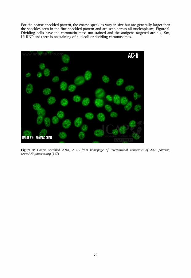
For the coarse speckled pattern, the coarse speckles vary in size but are generally larger than the speckles seen in the fine speckled pattern and are seen across all nucleoplasm; Figure 9. Dividing cells have the chromatin mass not stained and the antigens targeted are e.g. Sm, U1RNP and there is no staining of nucleoli or dividing chromosomes.
Figure 9: Coarse speckled ANA, AC-5 from homepage of International consensus of ANA patterns,
www.ANApatterns.org (147)

21
The centromere staining pattern is characterized by the presence of 40-80 discrete, large speckles present in the nucleus of resting cells; Figure 10. The speckles are larger and fewer in number than those seen in the fine and coarse speckled patterns. The speckles align with the chromosome region in dividing cells. Antigens which are associated are CENP-A/B and the centromere pattern is associated mainly with limited cutaneous systemic scleroderma and primary biliary cholangitis (147).
Figure 10: Centromere ANA, AC-3 from homepage of International consensus of ANA patterns,
www.ANApatterns.org (147)
OSTEOPONTIN & OTHER BIOMARKERS
Osteopontin (OPN) is an extracellular matrix protein as well as a soluble cytokine, also known as e.g. bone sialoprotein I (BSP-1) or early T-lymphocyte activation (ETA-1), and is involved in bone remodelling and has also immune modulating properties (148). OPN is produced by many different cells including macrophages, fibroblasts, neurons, B and T cells, DCs, neutrophils and bone cells. Furthermore OPN is upregulated in response to inflammation and injury (148). OPN induces cell adhesion and migration, regulates the differentiation of proinflammatory lymphocytes, and inhibits the apoptosis of inflammatory cells. In macrophages OPN acts by upregulating interleukin-12 production and mediates T helper 1 development. Moreover, OPN affects T helper cells, increasing the production of IL-17 and obstructing secretion of IL-10 giving raise to Th17 polarization. OPN can play a regulating role in arterial mineral deposition and in atherosclerotic lesions and in pDCs, it increases IFN-α expression (149). OPN is increased in SLE and is likely to play a critical role in SLE probably because of insufficient removal of cellular debris. Overexpression of OPN in lupus susceptible mice activates B cells and give rise to a subsequent production of anti-dsDNA antibodies (150,

22
151) and intracellular production of OPN in pDCs is necessary for TLR9-dependent expression of IFN-α (152). The anti-dsDNA antibodies can produce immune complexes that may deposit in tissue and generate inflammation in situ. Moreover, OPN can induce migration, activation and cytokine production by macrophages (153, 154). In SLE patients, raised levels of OPN have been shown in comparison with healthy controls (155). Moreover, OPN has been found to reflect disease activity (156, 157) and has been proposed to precede organ damage in SLE (158) although a large study with patients from the SLICC inception cohort did not find OPN to be a good predictor of organ damage over time (157) .
Another potential biomarker is soluble urokinase plasminogen activator receptor (suPAR), which is a part of the plasminogen activation system and may be engaged in inflammation, cancer metastasis, infections and tissue remodelling (159, 160). suPAR have been shown to associate and be a predictor of organ damage in newly diagnosed cases with SLE (160, 161).
CRP is normally an established biomarker of systemic inflammation, with exceptions for viral infections and SLE activity, likely due to IFN-α dependent suppression of CRP production by hepatocytes (162, 163).
Apoptosis stimulation fragment (Fas/CD95), mFAS as well as soluble Fas have in small cross-sectional studies shown an association to damage accrual (164, 165).

23
AIM
The general aim of this thesis was to increase the knowledge of immunoglobulin G (IgG) antinuclear antibodies by immunofluorescence microscopy (IF-ANA), as well as of antiphospholipid antibodies of several isotypes and other potential biomarkers, in relation to clinical features and disease outcomes in systemic lupus erythematosus (SLE).
Specific Aims
To address the clinical relevance of IF-ANA staining patterns in relation to disease manifestations in well-characterized SLE patients (Paper I).
To elucidate IgG-/IgA-/IgM-anti-cardiolipin and anti-β2-glycoprotein-I occurrence in relation to disease phenotype, smoking habits, pharmacotherapy, antiphospholipid antibody syndrome and damage accrual in SLE patients (Paper II).
To evaluate OPN as a potential marker of disease activity, disease phenotypes and
acquired organ damage in SLE (Paper III).
To characterize accumulated organ damage and causes of death in two regional
Swedish cohorts and examine associations between damage assessed by the SLICC/ACR damage index and demographic and disease variables, including serologies and medication (Paper IV)
To study seroconversion of ANA over time in Swedish patients with SLE (Paper V).

24
PATIENTS & METHODS
KLURING
KLURING is a regional SLE register and a biobank which was initiated by Christopher Sjöwall in 2008 at the University hospital in Linköping, Sweden. KLURING is a Swedish acronym for ‘Kliniskt LupusRegister I Nordöstra Götaland’. Inclusion criteria was a “clinical” diagnosis of SLE, age ≥18 years and either fulfilment of at least 4 of the ACR-82 classification criteria (Table 1) and/or the Fries’ diagnostic principle, meaning a positive ANA test combined with characteristic symptoms from at least two organ systems. Both incident and prevalent cases were enrolled after informed consent. In the beginning of 2020, the cohort contained data from more than 300 individual patients.
Blood samples were collected and saved in the biobank for future research at inclusion and at each visit at the rheumatology clinic hereafter. Clinical routine analyses (e.g. CRP, ESR, leukocyte variables, creatinine, ALAT, complement and urine) were collected and assessment of disease activity, organ damage accrual and medical therapies were registered in a database at every visit. Furthermore, patient-reported outcome measures comprising longitudinal data on pain, fatigue and quality of life were noted.
Serum was processed and stored at -70°C, thereafter, thawed and divided into aliquots. IF-ANA, ANA fine specificities and anti-phospholipid autoantibodies were analysed from aliquots that had been freeze-thawed 2-3 times. Patients with suspected nephritis have in clinical routine been subject to a renal biopsy conducted by percutaneous ultrasonography-guided puncture in agreement with routine guidelines. The acquired renal material was classified according to the WHO classification criteria for lupus nephritis (Table 3) (66). Figure 11 display the age and sex distribution at disease onset in the KLURING cohort, showing the disease onset to be most frequent in women in fertile age.
Figure 11: Percentages of cases with SLE by decade of age and sex at disease onset. Figure from Frodlund M, et
al. BMJ Open 2013; 3(10):e003608 (166).

25
PATIENTS & CONTROLS
All patients in the five studies (Paper I-V) fulfilled classification criteria for SLE and/or the Fries’ diagnostic principle. In the first study with 222 patients from the KLURING cohort all fulfilled either ACR-82 and/or the Fries’ diagnostic principle. In the second study, 231 cases from Linköping (KLURING) and 295 from Karolinska University hospital, Stockholm, (167) were included, all of them fulfilling the ACR-97 classification criteria. In this study 100 patients with rheumatoid arthritis (RA), 50 patients with primary Sjögren’s syndrome (pSS) and 507 control sera served as controls. Out of the 507 control sera, 212 were healthy blood donors and 295 were controls from the general population. Participants included in Paper III-V all fulfilled ACR-82 and/or the SLICC-12 classification criteria. In Paper III and V, 240 respective 54 patients from Linköping (KLURING) were enrolled. Paper IV consisted of 543 patients, whereof 296 were from Linköping (KLURING) and 247 from Uppsala University hospital (168). In Paper II and IV collaborations with other University hospitals were performed to increase statistical power.
METHODS
In Paper I data from medical records has been revised retrospectively, wherein ANA was detected by indirect IF-microscopy with HEp-2 cell as antigen substrate. ANA fine specificities were explored regarding anti-Ro/SSA, anti-La/SSB, anti-Sm, anti-snRNP, anti-dsDNA, anti-Scl-70 and anti-Jo1 by immunodiffusion and/or line-blot technique and anti-dsDNA sometimes by CLIFT.
In Paper II IgG, IgA and IgM aCL and anti-β2-GPI were analysed in the accredited immunology laboratories at Linköping, Uppsala and Karolinska University hospitals using fluoroenzyme-immunoassays (FEIA). Regarding LA in Linköping it was determined by the dilute Russell’s viper venom time (dRVVT) and at Karolinska by a modified dRVVT using bioclot LA.
An enzyme-linked immunosorbent assay (ELISA) kit was used to investigate OPN levels and for IgG/IgM aCL and anti-β2-GPI a FEIA was used in Paper III.
In Paper IV, data from medical records has been revised retrospectively, in which immunodiffusion, line-blot technique and/or addressable laser bead immuno assay (ALBIA) were used to analyse ANA fine specificities and/or CLIFT for anti-dsDNA.
In Paper V ANA was explored by indirect IF microscopy, with fixed HEp-2 cells as antigen substrate and a fluorescein-isothiocyanate conjugated γ-chain-specific anti-human IgG as detection antibody. The cut-off level was set at a titre of 800, which corresponded to the 95th percentile among 752 healthy blood donors (50% men, 50% women). The positive IF-ANA samples were titred in 2-fold dilution steps up to 1:12800. The staining patterns were categorized according to ICAP (147). A representative proportion of the samples were re-analysed at the Clinical Immunology laboratory in Uppsala with a concordance of >96%. Regarding ANA fine specificities (autoantibodies to dsDNA, Ro52/SSA, Ro60/SSA, La/SSB, Sm, Sm/RNP, U1RNP, ribosomal P protein and histone) an ALBIA at Clinical Immunology laboratory in Linköping was used. To avoid inter-assay variation all IF-ANA and sub specificities were analysed at the same time-point and by the same two experienced individuals.

26
IMMUNOFLUOROSCENSE MICROSCOPY FOR ANA (IF-
ANA) & ANTI-dsDNA
An “abnormal serum titre” of ANA, assessed by indirect immunofluorescence microscopy utilizing fixed HEp-2 cells as source of nuclear antigens and γ-chain specific secondary antibodies to pinpoint IgG-class IF-ANA, is one of the 11 classification criteria for SLE according to the ACR-82 (51), and remains a classification criterion in SLICC-12 although the method is not specified. In the new, 2019 SLE classification criteria from EULAR and ACR, the occurrence of IF-ANA has a capital role and serves as an entry criterion with a specified titre of ≥1:80 (54). In Paper I-V, IF-ANA was used for visualization of ANA as well as for ANA staining pattern. Different IF-ANA cell staining patterns arise depending on which nuclear antigens are being targeted by the autoantibodies; Figure 12.
Figure 12: Principles and staining patterns of indirect immunofluorescence antinuclear antibodies (IF-ANA)
employed in Paper I-V. HEp-2 cells are incubated with serum from the patient. If the serum contains antinuclear
antibodies, they will bind to the nuclear antigens. A second antibody coupled with a fluorescent marker is added
and the different ANA can hereby be visualized regarding immuno-morphological staining patterns (e.g.
homogeneous, centromere, speckled, nucleolar and mixed forms) in the microscope depending on the ANA fine
specificities. The concentration of ANA can be measured by stepwise dilution e.g. titration. Figure reprinted with
permission from Lina Wirestam (17).
The recognition of specific ANA patterns is based on a subjective evaluation. Different serum dilutions can give raise to varying nuclear patterns and one nuclear pattern may conceal and hinder the recognition of another pattern if several antibodies are present at the same time. Autoantibodies against e.g. dsDNA, DNA-histone and histones complexes typically generate a homogeneous nuclear staining pattern in non-mitotic cells and staining of the precipitated chromatin-associated antigens in dividing cells. Contrary, IF-ANA with specificity for extrachromosomal antigens, such as anti-Sm and anti-RNP, can be identified as a speckled nuclear pattern in non-mitotic cells, and scattered extra-chromosomal staining of mitotic cells. Other antigens give rise to other staining patterns (e.g. centromere, nucleolar, nuclear dots and

27
nuclear membrane). In Swedish SLE cases, the “homogeneous/ chromosomal” staining pattern (H-ANA) is most common followed by the “speckled/extrachromosomal” (S-ANA), combined “homogeneous and speckled” (HS-ANA), “nucleolar” (N-ANA), and “centromere” (C-ANA) staining pattern (166, 169). In Paper V, IF-ANA were analysed and categorized with regard to staining patterns in accordance with the ICAP nomenclature (170).
Crithidia luciliae is a flagellate parasite, distinguished by the occurrence of the kinetoplast, a network of interlinked circular DNA in a large mitochondrion; Figure 13. The dsDNA in the kinetoplast is used as a source of antigen by indirect IF microscopy to detect anti-dsDNA antibodies which have a high diagnostic specificity for SLE(171).
Figure 13: Crithidia luciliae immunofluorescence test (CLIFT) is used to detect anti-dsDNA antibodies in Paper
I and IV.

28
IMMUNOASSAYS
In the enzyme-linked immunosorbent assay (ELISA) antigens are bound to a surface. When patient serum is added, the antibodies bind to the antigens, if present. After adding a secondary antibody attached to an enzyme, a substance with the enzyme's substrate is added. The subsequent reaction gives a detectable signal, in most cases a colour change (172); see Figure 14.
Figure 14: An ELISA was used in Paper III to analyse osteopontin.
Fluoroenzyme-immunoassays (FEIA), use the same principle as ELISA, beside the detection- antibody binding to the antigen and antibody complex, being linked to a fluorescent enzyme that can be detected (172). Addressable laser bead immuno assay (ALBIA) is a flow cytometry analysis, in which patient serum is applied to color-coded antigen-marked beads, whereas antibodies, if present, bind to the specific antigen. A secondary fluorescent antibody against the first antibody is added and can then be detected and quantified by a laser detection instrument (172); Figure 15.

29
Figure 15: An ALBIA was used in Paper IV-V to detect ANA fine specificities.
Immunodiffusion (precipitation) is an old technique where the diffusion of antigen or antibody across a semisolid medium, generally agarose gel, with a following precipitin reaction are seen (172); see Figure 16.
Figure 16: Immunodiffusion for detection of ANA fine specificities was used in Paper I and IV.

30
Line blot technique (immunoblot) is a common method to detect and analyse proteins. Firstly, the proteins are separated into bands by gel-electrophoresis. Hereafter the proteins are transferred, also known as blotted, to a membrane, and the protein bands targeted are identified with primary antibodies specific to the determined protein. The primary antibodies are then distinguished with secondary antibodies, which can be either fluorescence or enzymatic labelled (172); Figure 17.
Figure 17. Line blot strip for analysis of ANA fine specificities is used in Paper I and Paper IV. The strip is coated
with parallel lines of 18 antigens.
OSTEOPONTIN ASSAY
The OPN ELISA that is being utilized in Paper III (from R&D Systems) is validated for both serum and plasma. In KLURING, merely serum samples were at hand and thus a correlation study was performed. Plasma and serum samples were simultaneously taken from 8 patients (6 women, 2 men; mean age 34.5 years; range 24-46 years). A correlation between plasma and serum samples was seen (r=0.77, p=0.027), although, the levels of OPN had a tendency to be slightly lower in the serum samples; see Figure 18. This was in agreement with the validated data from the manufacturer.

31
Figure 18: Pearson correlation of serum and plasma osteopontin (OPN) collected at the same time point in 8
individuals and detected by an enzyme-linked immunosorbent assay (ELISA). R= Pearson correlation coefficient.
Figure by Lina Wirestam (17).
ANA FINE SPECIFICITIES
As a complement to IF-ANA, analyses of ANA fine specificities (i.e. characterization of the autoantibody specificity) are performed to receive further clinical information. This can be done by fluorescent-based multiplex analyses such as ALBIA, Western blot, ELISA, immunoprecipitation and other techniques.
Anti-dsDNA antibodies are applied to diagnose and classify SLE, furthermore they indicate renal engagement and enhanced disease activity and thereby serve as a biomarker. However, anti-dsDNA levels are assumed to be lower, regardless of renal disease, in patients with inactive or well treated SLE (171, 173, 174). Crithidia luciliae as a source of antigen by indirect IF microscopy is used to detect ds-DNA with a specificity around 95% for SLE (171). To quantify anti-dsDNA, ALBIA can be used.
In SLE autoantibodies against ribonucleoproteins, e.g. Ro/SSA, La/SSB, Sm and U1RNP can be found. Anti-Ro/SSA associates with skin involvement and sicca symptoms (175). Autoantibodies targeted at the Ro/SSA antigens may recognize two different proteins with molecular weights of 52 and 60 kD, thereby denoted as “Ro52” and ”Ro60”, yet Ro52 belongs to the tripartite motife proteins (TRIMS) and is also referred to as TRIM21 (176). Anti-Ro/SSA and anti-La/SSB in pregnant women are associated with neonatal lupus, where congenital heart block is the most severe manifestation. Anti-Sm is included in classification criteria for SLE and have a high specificity for SLE, but is quite rare, especially in Caucasians (136). Anti-Sm antibodies bind to Sm proteins designated SmB, SmD1, SmD2, SmD3, SmE, SmF, and SmG that constitute a core of U1, U2, U4 and U5 small nuclear ribonucleoproteins (snRNPs) (136). Anti-Sm is often associated with anti-RNP, as they may share antigenic epitopes, which is prevalent in some SLE patients, and frequently seen in patients with Raynaud’s phenomenon (136). Anti-RNP antibodies react with one or more of three proteins (70-kD, A, and C) that are associated with U1 RNA and form U1snRNP (136). Autoantibodies against ribosomal P protein seem to be rather specific for SLE and have been proposed to be associated to hepatitis and nephritis, albeit the association with neuropsychiatric SLE

32
symptoms are more disputed (142). In 2013, Arbuckle et al. showed the presence of autoantibodies preceding clinical symptoms of SLE with an accumulation just before clinical onset (177).
ANTI-PHOSPHOLIPID SYNDROME & RELATED
ANTIBODIES
Anti-phospholipid syndrome antibodies (aPL) form a heterogeneous group of antibodies targeting phospholipid-binding proteins and phospholipids, which have many important roles, for example in the coagulation system (178). The aPL require the presence of serum-derived cofactors for the binding to anionic phospholipids. These cofactors are phospholipid-associated or cell surface receptor-bound molecules such as β2-GPI and prothrombin (179). The aPL included in the APS classification criteria are lupus anticoagulant (LA), anti-cardiolipin (aCL) antibody (IgG or IgM) and anti-beta2-glycoprotein I (anti-β2-GPI) antibody (IgG or IgM) (180). APS is defined by venous or arterial thrombosis and/or pregnancy morbidity and persistence of ≥1 positive aPL test (180). APS is an autoimmune disorder which can occur as a primary condition as well as secondary to SLE or other systemic autoimmune diseases (181). Thrombocytopenia, livedo reticularis, cognitive impairment and valvular heart disease are examples of “extra-criterial symptoms” of APS (180). The presence of isotypes IgM and IgG detected by ‘standard method’ plus the functional LA test were included 23 years ago with the ACR-97 criteria. 15 years after that, in SLICC-12, also IgA antibodies against cardiolipin and β2-GPI were included, despite surprisingly limited scientific evidence (50, 53). IgA aPL is not yet included in classification criteria for primary APS and the clinical relevance seem to be greater for IgA anti-β2-GPI than for IgA aCL both in primary and secondary APS (180, 182-184). Direct comparisons between studies are difficult due to differences in study cohorts e.g. different ethnicities and lack of diagnostic methodology standards including cut-off levels for positive results (185-188). IgA aPL are not used in clinical practice in Scandinavia, but the International Congress on aPL task force recommends testing of the IgA isotype in patients with clinically suspected APS, with negative tests for the IgG and IgM isotypes of aCL and anti-β2-GPI and a negative LA test (189). 30-40% of all SLE patients display increased levels of at least one aPL at some point during the disease course according to recent reviews (190, 191). Regardless of this, only about half of them fulfil APS classification criteria (1, 190, 191). Antibodies directed against other proteins of the coagulation cascade such as prothrombin and phosphatidylserine-prothrombin complexes are examples of other aPL that are not included in APS classification criteria as they are not used routinely because of uncertainty about their clinical significance and lack of standardized testing (192). Some studies have shown that 1 of the five domains of β2-GPI, domain I, is particularly important in the pathogenesis of APS and antibodies against this specific domain, appear to be more strongly associated with thrombosis as well as to obstetric complications than antibodies against the whole β2-GPI molecule (179, 193). Although, assays against β2-GPI domain I seem promising, further harmonization of the method and longitudinal, prospective studies are warranted to clarify the clinical utility of this test (194).
There are at least 3 different scoring systems in APS, aiming to quantify the risk of APS manifestations and help the physicians to stratify patients according to risk. One of them, the global APS score (GAPSS) is not only considering the aPL profile, but also include the autoimmune antibody profile and the conventional cardiovascular risk factors. Yet, its application should be further evaluated in prospective studies with not solely primary APS patients (194).

33
Both APS as well as having antiphospholipid antibodies have been linked to accrual of organ damage in SLE (94, 195, 196).
Presence of triple positivity (meaning at least one isotype of aCL, anti-β2-GPI and a positive LA test) or an isolated positive LA test have been associated with the highest risk of APS manifestations (195, 197). Regarding primary prevention for asymptomatic carriers of aPL there is still controversy whether pharmacological thromboprophylaxis should be given or not. An accurate assessment of risk of thrombosis is recommended, taking into account the aPL profile, co-occurring prothrombotic risk as well as other autoimmune risk factors (178). As aPL positive patients with SLE are at larger risk of thrombosis as well as of organ damage accrual current guidelines strongly recommend low-dose aspirin as well as HCQ (111). Patients with definite APS and a venous or an arterial thrombosis should be given oral anticoagulation therapy (e.g. warfarin, aiming to achieve PK-INR 2.0-3.0) (178). Antimalarials have been proven to give anti-thrombotic effects in APS with concomitant SLE due to platelet inhibition and reduction of anti-β2-GPI P1 complexes binding to the surfaces of phospholipids (178, 198, 199). In women with persistent aPL and a history of miscarriages a combination of low molecular heparin and low-dose aspirin is recommended (178).
A rare form of APS is catastrophic APS (CAPS) giving excessive thrombosis at multiple sites, often involving small vessels resulting in multi-organ failure. A combination of anticoagulation therapy, high doses of glucocorticoids, intravenous immunoglobulin or plasma exchange early during the disease course of CAPS can reduce morbidity and mortality (178).
STATISTICS
Descriptive statistics were used for characterization of study cohorts. Such data is presented by means and standard deviations, medians and ranges or as counts and percentages. Differences in categorical data were analysed using the Chi-square (χ2) test of independence, alternatively Fisher’s exact test in cases with small expected frequencies (when more than 20% of expected frequencies are < 5).
Independent samples t-tests were used when comparing approximately normally distributed continuous variables between groups. For comparisons of continuous variables where normal distributions could not be assumed the non-parametric Mann–Whitney U-test (two groups) or Kruskal–Wallis test (more than two groups) were performed.
Comparisons of means between groups were performed using independent samples t-tests (two groups) or one-way analysis of variance (ANOVA; three or more groups) with Tukey’s as post hoc test, or Mann-Whitney U test (two groups) or Kruskal-Wallis test (three or more groups) when the assumptions of normal distribution were not met.
Associations between variables were examined using Pearson correlation or Spearman’s rank correlation when assumptions of normality were not hold.
Poisson regression was used to estimate odds ratios for organ damage comparing different exposure groups and to control for confounding effects of different variables.
Two tailed P-values < 0.05 were considered significant. The statistical analyses were performed with SPSS Statistics version 23.0 (IBM, Armonk, NY, USA) or GraphPad Prism, version 6.07 (GraphPad Software, La Jolla, CA, USA).
No power calculations were performed since the cohort-sizes were a limiting factor.

34
In Paper I clinical and laboratory parameters as well as autoantibodies for each of the most observed groups of IF-ANA staining pattern were described by their frequencies. Regarding differences in the distribution of staining patterns and features, χ2 test or Fisher’s exact test with Cramer’s V as measure of effect size were used.
In Paper II, χ2 or Fisher’s exact test (when more than 20% of expected frequencies were < 5), were performed analysing associations between aPL antibody positivity and SLE phenotypes, pharmacotherapy, APS-related events and organ damage. The Mann–Whitney U-test was used for comparisons of aPL levels between groups and to establish potential differences in aPL levels within SLE cases and blood donors. Correlation analyses between aPL levels and age in disease controls, healthy blood donors and SLE cases were determined using Spearman’s rho. To examine the empirical relationship between organ damage and each of the aPL isotypes including LA positivity, disease duration, smoking habits, age, hypertension, lupus nephritis, ongoing treatments and a prednisolone dose of ≥ 7.5 mg/day an univariate model of Poisson regression was computed. Furthermore, all significant variables from the univariate model were combined and the non-significant (P ≥ 0.05) variables were stepwise eliminated until a multiple model with solely significant variables remained (a model with the highest pseudo-R2 with only significant predictors).
Independent samples t-test was applied to examine differences in OPN levels between cases with SLE and controls in Paper III. Correlation analyses between OPN and disease activity variables were accomplished using Spearman rank correlation and significant associations were further examined in a univariate general linear model to adjust for disease duration, sex, age and corticosteroid use. Relations between disease activity and damage accrual, respectively, with OPN as the response variable, were calculated using a stepwise linear regression model including disease duration, age, sex, SLEDAI-2K, SDI and corticosteroid medication. To evaluate statistical differences between the groups with nephritis, in patients with extensive, moderate or no organ damage, one-way ANOVA with Tukey’s post hoc test was conducted.
Comparisons of frequency distributions were calculated using χ2 or Fisher’s exact test with the phi coefficient as a measure of effect size (ES) in Paper IV. Comparisons between groups, e.g. patients without damage (SDI=0) versus patients with damage (SDI≥1) or patients with extensive damage (SDI≥3), were examined for frequency distributions and measures on interval-/ratio scales. The comparisons of measures on interval-/ratio scales were executed using students t-test or Mann–Whitney U test (when assumptions of normality were not met) with r as a measure of ES. The Kruskal–Wallis test was carried out for comparisons between groups with varying SDI scores and disease duration.
Poisson regression was used to examine the associations between variables and organ damage (SDI≥1). Univariate associations were assessed in a simple Poisson regression model and thereafter associations were examined, adjusting for age at diagnosis and disease. Finally, all variables showing associations with damage accrual in the univariate model were incorporated in a multiple Poisson regression model ensued by backward elimination of non-significant variables.
In Paper V the Mann–Whitney U test was performed to compare each ANA fine specificity autoantibody level (at all available time-points as well as longitudinally) with gender, smoking habits, presence of low complement levels, lupus nephritis, serositis, haematological disorder and organ damage. To evaluate relations between disease activity and every autoantibody specificity Spearman’s rank-order correlation test were performed and for comparisons between groups, χ2 or Fisher’s exact test (when more than 20% of expected frequencies were < 5) were used.

35
RESULTS & DISCUSSION
PAPER I
Associations between antinuclear antibody staining patterns and clinical features of systemic lupus erythematosus: analysis of a regional Swedish register
As the fluctuation and diversity of symptoms and severity in SLE are exceedingly variable the search for clinical and serological biomarkers is indeed important. Flares, with ongoing inflammation in organs such as the kidney, joints, skin, bone marrow and CNS effect quality of life and can induce irreversible organ damage. Analysis of antinuclear antibodies by immunofluorescence microscopy prevails a diagnostic characteristic of SLE. Diverse IF-ANA staining patterns arise as different nuclear antigens are being targeted.
ANA fine specificities in relation to disease manifestations have been explored repeatedly (140, 144, 175, 200-202), whereas the clinical relevance of IF-ANA staining patterns is scarcely studied.
In paper I, 222 well-defined SLE cases (89% women, 93% Caucasians) included in KLURING were consecutively recruited and followed with clinical evaluation and serum samples at each visit to the rheumatologist. The data from medical records has been revised retrospectively.
99% of the patients displayed a positive ANA and 54% had a homogeneous staining pattern (H-ANA). The second most frequently observed pattern was speckled ANA (S-ANA 22%), followed by homogeneous-speckled ANA (HS-ANA 11%), nucleolar ANA (N-ANA± other patterns 9%) and centromere ANA (C-ANA 1%); Figure 19.

36
Figure 19: Distribution of ANA staining patterns among the 219 ANA-positive cases with SLE.
The staining patterns did not differ between the cases fulfilling only the Fries´ diagnostic principle or ACR-82, and the dominant pattern was H-ANA regardless of how many ACR-82 criteria being fulfilled; see Figure 20.
Figure 20: IF-ANA staining patterns in relation to fulfilled ACR-82 classification criteria.

37
As expected, anti-dsDNA was more often associated with H-ANA, (p<0.001) and this pattern was significantly more frequent in cases with biopsy proven proliferative nephritis (classified as WHO class 3 or 4), (p<0.001) compared to other IF-ANA patterns.
ANA fine specificities were also explored regarding anti-dsDNA by CLIFT and anti-Ro/SSA, anti-La/SSB, anti-Sm, anti-snRNP, anti-Scl-70 and anti-Jo1 by immunodiffusion and/or line-blot technique.
As anticipated, a positive test for anti-dsDNA antibodies was significantly associated with renal disorder (p<0.001), whereas having anti-Sm antibodies were associated with lymphocytopenia (p=0.014). Moreover, photosensitivity was significantly associated with anti-Ro/SSA antibodies (p=0.023), whereas synovitis was less frequent in patients with anti-Ro/SSA-positivity (p=0.016). The association between photosensitivity and other lupus related skin manifestations is well-known while the correlation of anti-Ro/SSA and a lower frequency of joint involvement is more controversial.
Furthermore, S-ANA was less common in patients with organ damage (SDI ≥1). This has not been demonstrated earlier and calls for confirmatory studies. A possible explanation may be the strong association between anti-dsDNA and renal disease which in turn gives an increased likelihood of organ damage accrual, whereas other staining patterns including S-ANA may be of lower risk of developing lupus nephritis rendering them less likely to develop organ damage. The well-known fact that anti-Ro/SSA and anti-La/SSB are associated to milder SLE features, such as photosensitivity and malar rash, might be another explanation.
The predominant IF-ANA staining pattern among Swedish cases with SLE was H-ANA which was associated with the immunological criteria according to ACR-82. S-ANA was found to be the second most frequent pattern and had an inverse association with synovitis and organ damage accrual. The latter findings require confirmation in further studies.
Strengths of this study was the large and well-characterized SLE cohort and well-defined cut-off levels in testing of ANA and ANA fine specificities at one accredited laboratory. Even though this study confirmed several well-known associations and found some new ones, we were unable to perform comparisons of unusual clinical features and serological findings due to low power.
Taken together, IF-ANA staining patterns give some information of potential diagnostic and prognostic relevance, but ANA fine specificities are important complement and give further clinical correlations.
PAPER II
Immunoglobulin A anti-phospholipid antibodies in Swedish cases of systemic lupus erythematosus: associations with disease phenotypes, vascular events and damage accrual
APS is defined by vascular thrombosis and/or pregnancy morbidity and recurrent increased levels of IgG and/or IgM isotype aCL and/or anti-β2-GPI antibodies and/or a positive LA test (180). As the presence of aPL is associated with increased morbidity and mortality in SLE, it is important to screen newly diagnosed cases with SLE. Despite limited scientific evidence, the IgA isotype of aCL and anti-β2-GPI was added as part of the laboratory criteria in the 2012 SLICC criteria for SLE.

38
Hence, we analysed aPL isotypes and related antibody status to disease phenotypes, damage accrual, smoking habits and APS-related events in 526 cases of well-defined SLE.
Totally, 14% of the cases fulfilled the APS classification criteria. As expected, patients with triple-positivity, as well as patients with a positive LA test alone and/or the aPL IgG isotype, were associated with multiple APS manifestations and accrual of damage in several organ domains of the SDI. This result agrees with previous studies, including primary APS (190, 197).
In the control groups, 12% of the pSS patients and 14% of the RA cases tested positive for at least one aPL isotype. One of the pSS cases respectively two RA cases had suffered from cerebrovascular or cardiovascular events, which is in line with the frequency of APS events in these disease groups according to other studies (203, 204). The monitoring of the disease control groups over a long time is an asset of this study, as both pSS and RA can mimic SLE symptoms, particularly, early during the disease course.
In total, 138 (26%) of the SLE patients had elevated levels above the 99th percentile of a healthy population for at least one aPL isotype including IgA. In total, IgA aPL were found in 82 (16%) of the 526 cases with SLE; Figure 21.
Figure 21: Distribution of IgA aCL and anti-β2-GPI- positive patients in the SLE population. 82 (16%) of the
cases with SLE had IgA positivity.
37 37

39
In Figure 22 and 23 the different isotypes of aCL and anti-β2-GPI, respectively are illustrated. Figure 22 demonstrates 45 (9%) IgA aCL positive patients, 20 (4%) being positive in the absence of IgG/IgM isotypes.
Figure 22: Distribution of IgG/M/A isotypes of aCL in the SLE population. 89 (17%) of the patients with SLE
were positive for at least one aCL isotype.

40
Regarding IgA anti-β2-GPI, 74 (14%) of the SLE patients were positive, of which 34 (6%) were positive in the absence of IgG/IgM isotypes; see Figure 23.
Figure 23: Distribution of IgG/M/A isotypes of anti-β2-GPI in the SLE population. 121 (23%) of the patients
with SLE were positive for at least one anti-β2-GPI isotype.
IgG(n=38, 7%)
IgM(n=63, 12%)
IgA(n=74, 14%)

41
In Figure 24, the exclusively IgA positive cases are shown, 8 (2%) of aCL and 16 (3%) of anti-β2-GPI, with 4 of the cases being double positive.
Figure 24: Distribution of exclusively IgA aCL and IgA anti-β2-GPI-positive patients in the SLE population. 20
(4%) of the cases with SLE tested positive for at least one IgA isotype of either aCL and/or anti-β2-GPI. Each
asterisk (*) indicates one case with an APS-related manifestation.
There is substantial inconsistency regarding the reported importance of IgA aPL in previous studies, where some found associations for IgA aCL and/or anti-β2-GPI and thromboembolic events as well as pregnancy morbidity while others have been inconclusive (205-213). In a review from 2013 it was concluded that there was not yet enough proof to recommend analysis of the IgA isotype in clinical routine in order to improve the diagnostic accuracy of APS (214). Nevertheless, to compare studies may be difficult due to the absence of gold standards concerning methodology, including definition of cut-off levels for positive outcomes, and differences in study cohorts.
The conclusion from another review article by Andreoli et al. was that IgA anti-β2-GPI were of clinical relevance while the importance of IgA aCL was less evident (182). In addition, this conclusion was endorsed by other studies demonstrating that exclusive presence of IgA anti-β2-GPI was associated with APS manifestations (215, 216). Likewise, a higher clinical importance of IgA anti-β2-GPI compared to IgA aCL was seen in studies of primary APS (183, 184). In our study, 6 (1%) of the 20 cases with exclusively positive IgA aPL had suffered from an APS manifestation, and thereby 6 additional cases would have been diagnosed with APS if the IgA isotype was included in the APS criteria.
To have a positive LA test and/or IgA anti-β2-GPI antibodies present were significantly associated with tobacco smoking (past or present), which is consistent with a previous study (167). To be of Caucasian ethnicity was more common in cases with IgG anti-β2-GPI, regardless of other aPL isotypes. In contrast, non-Caucasian origin was significantly associated with being exclusively positive for IgA anti-β2-GPI. This was partly in line with a study with
IgA Anti-β2-glycoprotein-I
(n=16, 3%)
IgA Anti-cardiolipin
(n=8, 2%)
* *** **

42
an African-American SLE population, which reported increased rates of IgA aCL and anti-β2-GPI compared to other ethnicities (206). Although, to note is that the non-Caucasians were fewer than 10% of the patients in our cohort.
Disease duration, age, being an ever smoker, lupus nephritis, LA positivity and treatment with statins or prednisolone ≥7.5mg were all associated with organ damage accrual, while ongoing treatment with HCQ seemed to be protective. Similar results were found in several previous studies (80, 85, 94, 195, 196).
Exclusive occurrence of IgA anti-β2GPI ± IgA aCL was associated with pulmonary damage, use of cyclosporine (CSA)/sirolimus, salicylic acid and the occurence of anti-Ro/SSA antibodies.
To conclude, the occurrence of IgA aPL (16%) are not uncommon in Swedish patients with SLE. 4% had IgA aCL and/or anti-β2GPI in the absence of IgG and IgM isotypes. The addition of IgA isotypes, particularly IgA anti-β2GPI, provides some additional clinical information. However, further longitudinal studies are required before introducing IgA aPL in clinical routine. A limitation of the present study was the cross-sectional design, leaving the question considering aPL positivity over time unanswered.
In agreement with recent consensus documents (189), we concluded that analysing IgA aPL can be of additional value among clinically suspected APS-patients with negative tests for other isotypes of aPL and LA.
In addition, a positive LA, IgG aPL tests and triple positivity were frequently associated with APS-related manifestations as well as organ damage accrual.
PAPER III
Osteopontin is associated with disease severity and antiphospholipid syndrome in well characterised Swedish cases of SLE
The spectrum of disease phenotypes in cases with SLE challenges the determination of new biomarkers reflecting disease activity and/or organ damage. OPN is an extracellular matrix protein with many functions including immunomodulating features. Despite that increased levels have been demonstrated, the pathogenic implications and clinical usefulness of OPN as a biomarker in SLE are uncertain. Hence, the aim of this cross-sectional study was to characterize OPN in SLE. OPN in sera from 240 well-characterized cases with SLE fulfilling either the 1982 ACR and/or the 2012 SLICC criteria, and from 240 population-based controls were analysed. The SLEDAI-2K was utilized to assess disease activity and the SDI to register damage accrual.
Correlation analyses between OPN and items from SLEDAI-2K were effectuated. An inverse significant association was found for OPN and haemoglobin (p<0.0001), while positive associations were demonstrated for creatinine (p<0.0001) and ESR (p=0.001).
OPN levels in sera were in average increased fourfold in patients with SLE in comparison with the controls (p<0.0001); see Figure 25.

43
Figure 25: Distribution of serum osteopontin (OPN) levels in cases with SLE and population-based controls. OPN
levels in sera, measured by ELISA, were significantly higher in cases with SLE (mean 40.6 ng/mL) in relation to
controls (mean 10.1 ng/mL).
OPN showed a correlation to SLEDAI-2K, particularly in cases with a disease duration of less than 1 year (r=0.67, p=0.028). Comparable findings have been described in a previous study (155). The cross-sectional data herein implies that OPN serves as a biomarker of disease activity among cases with recent-onset SLE, while in established disease it functions as a marker of organ damage accrual.
Furthermore, cases with current nephritis had increased levels of OPN in relation to cases with nephritis in the past (p=0.008) and cases without nephritis (p<0.0001).
In addition, OPN was highly associated with SDI and cases with extensive damage (SDI≥3) presented with raised levels of OPN in comparison with patients with no or moderate damage (SDI 1-2); Figure 26. In line with this, Rullo et al. observed that enhanced levels of OPN preceded accumulation of disease activity and organ damage, particularly in paediatric SLE (158).

44
Figure 26: Patients with SDI ≥3 presented with raised levels of OPN (mean 68.4 ng/mL) in comparison to cases
with moderate damage (SDI 1-2; mean 36.0 ng/mL) and without damage (SDI 0; mean 35.6 ng/mL).
Moreover, a separation of organ damage into different domains showed significantly positive impact on OPN levels especially in the cardiovascular, renal and malignancy domains (Table 7). Several previous studies have also demonstrated associations between increased levels of OPN and renal failure (156, 157, 217).
This may implicate OPN to play a role in a vicious circle of renal inflammation, causing continuous albuminuria and interstitial fibrosis (218, 219). Moreover, giving anti-OPN treatment in nephritic rodents decreased proteinuria and in OPN knockout mice diminished infiltration of macrophages and less fibrosis was observed (220, 221).
Variable B p-value
SDI/ SDI domain
Global SLICC/ACR DI 6.5 <0.0001 Renal 18.8 <0.0001 Cardiovascular 12.3 <0.0001 Malignancy 18.1 0.012
Table 7: Demonstrate association between OPN and organ damage accrual with SDI including significant
associations in different domains.
To examine a potential predictive value of OPN, the alteration in the SDI score between enrolment and 2-6 years after enrolment was computed. In cases with both a moderately (SDI raise 1-2; p=0.001) or highly (SDI raise 3-8; p=0.029) raised SDI, significantly elevated OPN levels were found compared to in cases without a raise in SDI score; see Figure 27. Furthermore, an increase in death rates was seen among cases in the two groups with an elevated SDI; see Figure 27. These results imply that OPN is a biomarker of future damage accrual. It is an established fact that the SDI value is a good predictor of further damage accrual and mortality (78, 85). The finding of elevated death rates in the two groups with increased SDI is in line with this, although these results can be biased by the restricted follow-up period (2-6years) as well as the fact that irreversible organ damage per se predicts future damage.
SDI 0 SDI 1-2 SDI 30
50
100
150
200
250
OP
N n
g/m
l
p<0.0001
p<0.0001

45
Figure 27: Cases with a highly elevated SDI (SDI increase 3-8; mean 62.9 ng/mL) and moderately raised SDI
(SDI increase 1-2; mean 50.4 ng/mL) displayed significantly increased OPN levels in comparison with patients
without increased SDI (SDI=0; mean 34.8 ng/mL). Crosses indicate deceased cases (in percentage) for each SDI
category.
Moreover, OPN levels in relation to APS showed a significant association (p=0.009). For the different APS manifestations, a positive impact was noted for arterial event (p=0.044), arterial emboli (p=0.031), ischaemic stroke (p=0.026) and valvular heart disease (p<0.0001). Similarly, earlier studies have linked raised OPN levels to arterial events such as increased risk of major cardiac insults, severe coronary and peripheral arterial atherosclerosis (222, 223). Although, in what way OPN play a role in cardiovascular disease is partly unknown. Whereas some studies indicated that the pro-inflammatory properties of OPN enhance atherosclerosis, others have hypothesized OPN to give rise to a protective effect in post-myocardial infarction by recruiting neutrophils and macrophages to clear cellular debris (222, 224).
Looking at the serological elements in the classification criteria for APS, the LA test (p=0.033), and IgM aCL antibodies (p=0.027), had a positive impact on OPN levels. Unexpectedly, no associations were seen for OPN levels and triple positivity (i.e. positive LA test, IgG and/or IgM aCL and IgG and/or IgM anti-β2-glycoprotein I antibodies).
A limitation of this study was the few patients with incipient disease (17%). Future longitudinal studies with newly diagnosed SLE cases are needed, to further explore if increased OPN levels precede organ damage accrual and thereby acts as a predictor.
In murine models of SLE an enhanced expression of OPN stimulate B cell activity, ensuing anti-dsDNA antibody production (150, 151). These antibodies can form immune complexes which cause inflammation when they are deposited in the kidneys. In addition, OPN can induce migration and cytokine production by macrophages (153, 154). A principal feature in the pathogenesis of SLE is an insufficient clearance of apoptotic material and OPN has been found to obstruct apoptosis thereby contributing to reduced clearance of cellular debris, antigen exposure, autoantibody production, persistent inflammation and organ damage in a vicious circle (151, 225).
0 1-2 3-80
50
100
150
200
250
SDI increase
OP
N n
g/m
l
p=0.001
p=0.029
n=153 n=67 n=7
† 3%† 22%
† 71%

46
To conclude, circulating OPN was correlated with disease activity in newly diagnosed cases with SLE, reflected irreversible organ damage and was associated with APS manifestations, primarily on the arterial side. Unfortunately, in a follow up study in the SLICC inception cohort, OPN could not predict development of organ damage, but was still raised 4-fold and was associated to enhanced disease activity at inclusion and over time (157).
PAPER IV
The majority of Swedish systemic lupus erythematosus patients are still affected by irreversible organ impairment: factors related to damage accrual in two regional cohorts
Systemic lupus erythematosus (SLE) is a chronic inflammatory autoimmune disease with an unpredictable disease course and involving many organs such as the kidneys, joints, skin and CNS (226). Prevailing inflammation, side-effects from treatment and comorbidities can cause accrual of irreversible organ damage which is closely related to mortality (79, 85, 227). Although the survival rate in cases with SLE has improved since the 1950s, little further improvement has been seen since the mid-1990s, despite new therapies and a clearer perception of the disease pathogenesis, and irreversible organ damage remains a critical concern (63, 87, 88, 91).
Knowledge of specific disease variables related to poor SLE outcome is indeed important given the great diversity of the disease and severity. To evaluate accumulation of organ damage, clinical associations and causes of death in SLE, 543 consecutively recruited and well-characterized cases from Uppsala and Linköping (1998‒2017) were studied. The SDI was utilized to estimate damage. Increased SDI scores are related to further accrual of organ damage, lower quality of life and an increased risk of mortality whereas absence of SDI points are associated with mild or well-controlled SLE (64, 78, 79, 85).
Furthermore, we examined factors associated with damage accrual including disease features, demographics, autoantibody specificities and medical treatment.
86% of the cases were women and 90% of Caucasian origin. The mean age at diagnosis was 37 years and the mean disease duration at data extraction was 17 years. Most patients had established SLE at data extraction and only 4% had recent-onset disease (meaning having less than one years´ disease duration). 99% of the patients were ANA positive and 49% had detectable aPL at least once. The most frequent ACR-82 criterion was arthritis (75%), haematological disorder (63%), followed by photosensitivity (59%) and malar rash (54%). Lupus nephritis was found in 29% and neurologic disorder in 6% of the patients. The differences in data between the Uppsala and the Linköping cohorts were small and thereby further analyses were performed using fused data. 318 of all patients (59%) presented with “any organ damage” (SDI >0) and 137 (25%) of these had “extensive damage” (SDI ≥3); see Figure 28.

47
Figure 28: Distribution of points corresponding to the Systemic Lupus International Collaborating
Clinics/American College of Rheumatology damage index (SDI). 318 (59%) of the cases with SLE had SDI ≥1
and 137 (25%) had SDI ≥3.
In other studies with similar follow-up time and distribution of ethnicity the prevalence of any organ damage were 36-69% (64, 81, 196). In the SLICC cohort, with approximately half of the cases being of Caucasian ethnicity, 51% of the patients presented with organ damage already after 6 years’ duration of SLE (85). In a more recently published cross-sectional study with 344 patients with recent-onset SLE, 29% had accrued damage after 5 years (157). In Figure 29, the percentage of cases with any damage year by year is shown. After 6 years’ disease duration, 33% of the patients had accrued at least one organ damage, which is in line with other European studies (64, 94) and a previous Swedish study from Lund (78).
Figure 29: Percentage of patients with any organ damage yearly from SLE onset.

48
Engagement of one organ domain was most common, albeit some individuals had damage in several domains. The most frequently affected organ domains were the neuropsychiatric (25%) followed by the ocular (18%), musculoskeletal (16%), cardiovascular (16%) and malignancy (13%) domains; Figure 30. In a comparison with a Portuguese study the findings were similar, with exceptions for higher rates of renal, musculoskeletal and pulmonary damage among the Portuguese cases, whereas cardiovascular damage and malignancies were more frequent in this study. The observed differences can reflect genetic variation or be due to a shorter follow-up time, different coverage of the study population and the cross-sectional design of the Portuguese study (196).
Figure 30: Distribution of the frequencies (%) of each involved organ domain in all 543 cases.
Moreover, we analysed time to first damage in each organ domain; Figure 31. Damage in the skin (median time 9 months), diabetes mellitus (median time 12 months) and peripheral vascular (median time 2 years) domains were the first to appear whereas the longest time to injury were seen in the malignancy (median time 13 years) domain. In line with previous studies, our data demonstrate that male gender (median 2 vs. 6 years, p<0.001) and testing positive for LA (median 3 vs. 6 years, p=0.005) was associated with a shorter time to first damage (64, 73). In cases with malar rash, depression, hypothyroidism and La/SSB antibodies a significantly longer time to first organ damage was observed. These factors may constitute markers of mild SLE and to note, all these four factors were more frequent in females than in male cases, and might thus explain a part of the gender difference of SDI (166).

49
Figure 31: Distribution of median time in years to first organ damage in relation to affected domain.
As previously demonstrated by others, cases with SDI ≥1 had a longer disease duration (mean 20 vs. 12 years), were to a greater extent of Caucasian ethnicity (93% vs. 87%), were older at diagnosis (mean age 39 vs. 33 years) and fulfilled a higher number of SLICC-12 criteria (6.7 vs. 6.2) (81, 86, 94, 196). Furthermore, hypertension, hyperlipidaemia, depression, Sjögrens syndrome, neurologic disorder (SLICC-12 definition), aPL (SLICC-12 definition), positive IgG anti-β2GPI, positive LA test as well as clinical APS were more frequent in cases with any damage. Looking at therapies, the use of CSA, MMF and CYC were more frequently used in patients with accrued organ damage, while ongoing antimalarial therapy was less common. Having positive anti-La/SSB antibodies were associated with the absence of damage. As SDI does not have to be attributed to SLE, factors like increased sensitivity to drug adverse advents in elderly and comorbidities are also important. Certain types of damage, like cataract, cerebrovascular insult and osteoporosis are more common in elderly in general and may not solely be explained by long SLE duration, high disease activity or corticosteroid side effects (85). Others have demonstrated that, patients with non-Caucasian ethnicity are afflicted by damage earlier during the disease course, have an increased risk of nephritis and a worse outcome overall (56, 80, 195). Increased genetic burden, socioeconomics and a more frequent presence of autoantibodies may contribute to more severe SLE phenotypes in non-Caucasians (57, 80, 195). This could not be confirmed in this study, possibly due to the low number of non-Caucasians included.
SS was observed in approximately 20% of the cases, a frequency similar in other European studies (228, 229). In line with a previous study, SS was more common in patients with accrued damage (196). Additional autoimmune disease in SLE such as SS, has been associated with increased damage and mortality (201). Furthermore, SS has been found to be more common among Caucasians than in other ethnic groups (201). In addition, antidepressant medication was more prevalent in cases with damage. If depression is directly associated to SLE, or if it constitutes a result of high disease burden, remains to be elucidated.
Moreover, a comparison was made between patients with extensive damage (SDI ≥3) and those without any damage. All above mentioned significant variables remained and in addition serositis, haematological and renal disorder, as well as interstitial lung disease, secondary Sjögren’s syndrome and IgG aCL were associated with extensive damage. Renal disorder as

50
well as persistent proteinuria have been associated with a more aggressive SLE, which was confirmed in this study as renal disorder was more frequent in cases with extensive damage (63, 77, 80, 94, 196).
Furthermore, we conducted regression analyses with the SDI at data extraction and adjusted the model for age and disease duration. Age at diagnosis, SLE duration, pericarditis, haemolytic anaemia, lymphopenia, myositis, use of antihypertensives, statins, CSA or CYC (ever), the use of corticosteroid doses corresponding to ≥7.5 mg prednisolone daily (at last visit), neurologic disorder and APS remained as independent risk factors for damage accrual. The overall pseudo-R2 was 0.52 which indicates that more than 50% of the total variation of the global SDI score can be explained by the above-mentioned factors in the multiple model. Hypertension and APS are well established risk factors for damage accrual and antihypertensive treatment can be a proxy for renal disorder, as angiotensin converting enzyme inhibitors are used to reduce proteinuria (77, 94, 196). Neurologic disorder has been found to be associated with SDI whereas haemolytic anaemia, lymphopenia, pericarditis and myositis have not previously been identified as risk factors for damage (196). Although, myositis in SLE has been related to a more active disease, which may explain the association with damage accrual. Additionally, SDI may be reflected by long-term use of high doses of corticosteroids in manifestations such as serositis, severe cytopenia and pulmonary disorder, where other immunosuppressive treatment occasionally may be insufficient. With regard to the association with SDI and the use of CSA or CYC, these therapies are more likely to be used in cases with more severe disease (e.g. proliferative nephritis or neurologic disorder) or as a late alternative medication in patients who already have acquired damage. Furthermore, a well-known side effect of CYC is premature gonadal failure, which is an item in the SDI. Contrary, the use of antimalarials was associated with absence of damage, which has also been observed in previous studies (84, 85). In our cohort, 63% of the cases remained on antimalarials at last visit. Antimalarials remain the cornerstone in treating SLE and is efficient for skin and joint manifestations, reduces flares as well as gives an improved lipid profile, glucose levels and contributes to antithrombotic effects (230). On the other hand, mild cases of SLE is more likely to receive antimalarials as monotherapy, whereas more severe or highly active disease are more prone to receive glucocorticoids and other immunosuppressive therapy as a complement to antimalarials. Antimalarials are inhibiting interferon-signalling in SLE, and alternative treatments are under development for patients unable to tolerate HCQ in order to target this pathway and reduce the risk of long-term morbidity (84, 85, 227).
Despite improving survival rates since the 1950’s, the mortality rates have stagnated in the last decades and remain higher compared to the general population (88, 91). At last follow-up, 54 (10%) of the SLE cases were deceased, whereof 7 patients were included as incident cases. The mean age at death among the 54 patients was 70 years (range 27-96) and 10 of them died before the age of 60. In comparison, the average age for mortality in the normal population in Sweden was 2017 82 years (231). The causes of death are presented in Figure 32. The leading cause of death was malignancy, followed by infections and cardiovascular disease. Similar results have been reported by others, but some have also observed higher rates of thrombotic events, cerebrovascular disease and “active disease” as causes of death (64, 89, 167, 232). A possible explanation might be an underestimation of the SLE related causes of death, recently highlighted in a Swedish study (233).

51
Figure 32: Cause of death in accordance with death certificates among the 54 deceased patients at the time point
of data extraction (31 December 2017). CVI: cerebrovascular insult.
Out of the 18 deaths caused by malignancies, 5 were lung cancer and 5 haematological malignancies. This was in line with a recent meta-analysis and a study in a large international SLE cohort in which also hepatobiliary malignancy was found to be overrepresented (89, 95). The most common deadly infections were septicaemia (6 cases) and pneumonia (6 cases). Additionally, infections can be related to high disease activity, immunosuppressive therapy, high doses of corticosteroids, hospitalization and have been observed to be a leading cause of death in early SLE (232, 234). In the cardiovascular group, ischemic heart disease, heart failure, cardiomyopathies and pulmonary arterial hypertension were included. An overrepresentation of early cardiovascular disease, especially among women, and related deaths are previously reported (64, 97). Among the deceased patients a significant higher SDI score was observed in comparison to the cases still alive at data extraction (SDI 5.3 vs. 1.3, p<0.0001).
Strengths of the present study are the large and well-characterized SLE cohort and a high coverage, as our university hospital is a tertiary referral centre and Swedish health care being tax-funded, giving the patients´ universal access. This makes the risk of selection bias low. Unfortunately, data on accumulated doses of corticosteroids are not available and the number of non-Caucasians is low, which may hamper generalization to other parts of the world.
To conclude, after a mean disease duration of 17 years, the main part of Swedish SLE patients have accrued organ damage. Earlier established risk factors of damage accrual were confirmed. Additionally, Sjögren’s syndrome was associated with extensive damage, whereas neurologic disorder, myositis, lymphopenia, pericarditis and haemolytic anaemia were linked to global SDI in a multiple Poisson regression model.
Among modifiable factors, tight surveillance and prevention of cardiovascular disease and infections, watchfulness for malignancies, a judicious use of corticosteroids as well as giving all SLE patients antimalarials may decrease the risk of organ damage and premature mortality.

52
PAPER V
Longitudinal antinuclear antibody (ANA) seroconversion in systemic lupus erythematosus: a prospective study of Swedish cases with recent-onset disease
Regardless of the considerable clinical diversities between cases with SLE, the presence of ANA at the time of diagnosis has been regarded as a finding with very few exceptions (226). Thus, an ‘abnormal titre’ of ANA identified by immunofluorescence microscopy (IF-ANA) is one of the 11 criteria for SLE according to the validated 1982 ACR classification criteria (ACR-82) as well as the 1997 revised criteria (ACR-97) (50, 51). In addition, the Systemic Lupus International Collaborating Clinics criteria (SLICC-12) state that an ANA test ‘above the laboratory reference value’ remains a criterion for SLE, but with no specification of the method for ANA assessment (53) and in the 2019 SLE classification criteria from EULAR and ACR, ANA has a cardinal role and serves as an entry criterion, at the stipulated titre of ≥1:80 (54). The use of IF microscopy to detect ANAs was introduced by Holman, Kunkel and Friou already in the 1950s, and persists as the gold standard of ANA diagnostics even though rat tissue as antigen source has been replaced by a human epithelial cell line in the last decades (9, 10, 235). Different IF-ANA cell staining patterns arise depending on which nuclear antigens are being targeted by the autoantibodies. In Swedish SLE materials, the “homogeneous/chromosomal” staining pattern (H-ANA) is the most common, followed by the “speckled/extrachromosomal” (S-ANA), combined “homogeneous and speckled” (HS-ANA), “nucleolar” (N-ANA), and “centromere” (C-ANA) staining pattern (166). In Linköping we use a cut-off level corresponding to the 95th percentile among healthy blood donors to define an abnormal level of ANA detected by indirect IF microscopy with fixed HEp-2 cells as source of nuclear antigens and γ-chain specific secondary antibodies to determine IgG-class IF-ANA (166). Using this cut-off level, ANA has a very high diagnostic sensitivity for recent-onset SLE, but low diagnostic specificity, with nearly 5% prevalence among healthy blood donors. Whether the diagnostic sensitivity of ANA change over time in patients with established SLE remains uncertain. It is often stated that >95% of SLE patients are ANA positive (226). This is likely to hold true for ‘ever ANA positive’ whereas the frequency of IF-ANA in established SLE cases is probably lower. In two previous retrospective studies, it was demonstrated that abnormal ANA titres are less common than generally assumed in established cases of SLE (169, 236). Additionally, the investigators in the randomized controlled study of belimumab (anti-BLyS therapy), found that a considerable proportion of the previous ANA positive cases were ANA negative at inclusion. This matter was debated being one of the possible reasons for the trial not reaching its primary endpoints (237). In a large study from the SLICC inception cohort with recent onset SLE, 6% of the patients were judged ANA negative (238). Contrary, another study showed ANA negative cases at recruitment to become ANA positive over 5 years (239). Pisetsky et al. have emphasized that divergent ANA results could partly be due to differences in laboratory routines and assays and the cut-off value not being clear-cut (240). Furthermore, ANA fine specificities have not been standardized regarding diagnostic specificity.
As controversy consists, and IF-ANA to our knowledge has never been evaluated in a prospective study, we decided to design such study with longitudinal serological and clinical follow-up for patients with recent-onset SLE.
Accordingly, 54 cases with newly diagnosed SLE (≤ 6months of symptoms) were included and followed for at least five years. At recruitment all cases were judged ANA positive at a titre of ≥800 and the distribution of staining patterns were coded according to the ICAP nomenclature, with homogeneous (H, AC-1, 46%), speckled (S, AC-4 and AC-5, 31%) and

53
homogeneous/speckled (HS, AC-1/4, 11%) patterns being most common followed by homogeneous/nucleolar (HN, AC1/8, 6%), nucleolar (N, AC-8-10, 4%) and multiple nuclear dots (MND, AC-6, 2%). See Figure 33 for IF-ANA staining patterns longitudinally. 93 % of the cases kept their ANA pattern during follow-up and decreasing ANA titre over time was more frequent than the opposite. No significant associations between ANA titre changes and conversion of staining patterns or to disease activity were observed. Yet, most of our patients had low to moderate disease activity and thereby such association cannot be excluded in more active SLE.
Figure 33: IF-ANA staining patterns in all 54 cases longitudinally.
Arbuckle and co-workers showed in 2003 that IF-ANA as well as ANA fine specificities can be detected in an increasing rate, several years before and up to the time of the SLE diagnosis, where after the autoantibody accrual was stopped (177). Swedish biobank material have shown similar results (241). In this study 7 out of the 54 patients (13%) had lost their ANA positivity at last follow-up; see Figure 34.

54
Figure 34: The distribution of IF-ANA titres longitudinally for each patient.
In Figure 35, changes in dilution steps from enrolment to last follow-up for all 54 patients are demonstrated.
Figure 35: Changes in dilution steps for all 54 patients, from recruitment to last data extraction. Red bars indicate
patients seroconverting from IF-ANA positive to negative. (Note that each empty space on the X-axis indicates
that no change in IF-ANA titre was observed from first to last follow-up).

55
This finding of IF-ANA seroconversion from positive to negative is similar to studies showing that prevailing ANA positivity is not as common as previously thought (169, 236-238). However, opposing data has been presented by Ippolito et al (242). In the 7 cases seroconverting, the ANA titres at recruitment were in the range of 800-6400, all were prescribed antimalarials and 3 with a combination of methotrexate and one with mycophenolate mofetil. No significant associations were found between IF-ANA seroconversion and any specific phenotypes, gender, smoking or hypocomplementemia. Solely 2 of the 7 patients who lost ANA positivity were positive for anti-Ro/SSA antibodies, implying that IF-ANA negative cases are not restricted to only anti-Ro/SSA positive patients (169, 243). In 3 of the 7 seroconverting cases, all fine specificities were negative at inclusion and continued to be negative over time whereas 1 case stayed positive for both anti-Ro52/SSA and anti-U1RNP and 1 case for both anti-U1RNP and anti-Sm/RNP. In 2 patients, anti-U1RNP or anti-dsDNA became negative at the seroconversion of ANA, yet these 2 patients remained positive for anti-ribosomal P protein or Ro52/SSA and La/SSB. As IF-ANA rests on a subjective evaluation under the microscope, as well as that the equipment and procedures vary at different laboratories, the titres cannot be directly comparable in between them (244). Despite that international recommendation for IF-ANA cut-off levels is clear, it is not always complied with and even in the 2019 SLE classification criteria from EULAR and ACR a titre of ≥1:80 is stipulated even if this titre do not reflect the 95th percentile at all laboratories (54, 146). If the selected cut-off level for IF-ANA is set too low, a considerable proportion of healthy individuals will present with ANA-positivity, and thus it is very important that every IF-ANA laboratory calibrate their cut-off level based on their microscope and defined reference material with the use of the 95th percentile in a healthy population (51, 169, 244-246).
Associations between ANA fine specificities and clinical manifestations, such as anti-dsDNA and nephritis, anti-U1RNP with Raynaud´s phenomenon and anti-Ro52/Ro60/SSA antibodies with sicca symptoms and skin involvement are well-established (140, 175). For other ANA specificities and clinical features conflicting results are demonstrated in different studies, possible due to differences in patient selection, methodology, cut-off levels as well as ethnicity in the selected populations (140, 144, 166, 175). Herein, we confirmed the established associations between the presence of anti-dsDNA and low complement as well as for renal disorder; see Figure 36 (98, 140, 247).
Figure 36: Anti-dsDNA levels over time in cases with and without lupus nephritis.

56
In line with a European study an association between anti-U1RNP and haematological involvement was found (144). Despite multiple clinical associations ANA reactivities are not routinely followed longitudinally except for anti-dsDNA, which may correlate with renal disorder and flares in general (247). Nevertheless, concentrations of other ANA fine specificities can fluctuate over time, there seem to be limited correlation between concentrations and disease activity as well as for most manifestations (248, 249). Anti-dsDNA; see Figure 37, and anti-Sm fluctuated most over time whereas anti-Ro/SSA and anti-La/SSB were more stable longitudinally. These findings were in line with other studies (249-251).
Figure 37: Illustration of the distribution of anti-dsDNA titres longitudinally for each patient.
Opposing our results with higher Ro52/SSA levels in men was found in two non-European studies (252, 253). This may be explained by low number of male patients in our cohort or due to differences in ethnicity. Furthermore, ethnicity is well-known to influence the prevalence of autoantibodies, being higher in non-Caucasians (254, 255). As 90% of the patients herein were of Caucasian origin, we were not able to evaluate ethnic differences systematically. Further, we could not find any association between previously shown cigarette smoking and anti-dsDNA, nor for any other ANA reactivity (256). Strengths of this study were the well-characterized population with few missing data and the prospective design. Additionally, all IF-ANA samples were analysed at the same occasion and evaluated by the same qualified individuals at the Clinical immunology laboratory in Linköping, to avoid inter-assay variation. Furthermore, several samples were re-evaluated regarding IF-ANA at the Clinical Immunology laboratory in Uppsala, with a concordance rate over 96%. The relatively small number of included cases was a limitation of the study, as correlations between ANA fine specificities and rare manifestations could not be analysed due to statistical power issues.
Nevertheless, ANA fine specificities are associated with certain clinical symptoms and fluctuate in varying degree over time, there seem to be of little clinical relevance to measure the autoantibodies repeatedly, except for anti-dsDNA. A considerable proportion of the SLE

57
cases converted from positive to negative IF-ANA over time. Besides, no obvious associations were observed between clinical manifestations, gender, smoking habits or complement levels in the patients who seroconverted. Neither, was simultaneously seroconversion of ANA fine specificities at the time of IF-ANA seroconversion seen in general, but in individual cases. Further, larger prospective studies are required as it is unclear whether seroconversion from IF-ANA positive to negative mirrors the natural history of disease, variability in test kits or are consequences of treatment.

58
CONCLUSION
IF-ANA remains a hallmark of SLE and the most common pattern in Swedish cases is homogeneous ANA, followed by speckled ANA and combined homogeneous/speckled-ANA. H-ANA was associated with immunologic disorder, whereas S-ANA was inversely associated with arthritis, immunologic disorder as well as with accrual of organ damage.
Over time a considerable proportion of SLE patients seroconvert regarding IF-ANA from positive to negative, whereas the staining patterns largely remain stable. Furthermore, ANA fine specificities fluctuate in varying degree over time. Yet, the only autoantibody which seems to have clinical relevance to follow over time was anti-dsDNA, where the titre was associated with low complement and renal involvement.
The biomarker OPN, was elevated 4-fold in SLE cases in comparison to in healthy controls and was found to be correlated with disease activity (measured by mSLEDAI-2K), especially during the early disease course. OPN levels were also associated with accrual of organ damage and APS.
In addition, we observed that almost 60% of Swedish SLE patients had acquired organ damage, and 25% extensive damage, after a mean disease duration of 17 years. Organ damage was associated with age at disease onset, SLE duration, number of SLICC-12 criteria, APS, neurologic disorder, hypertension, hyperlipidaemia, depression and secondary Sjögren’s syndrome.
IgA aPL was found in 16% of the patients and 1% had IgA aPL and APS manifestations without having other aPL isotypes or a positive LA test. Additionally, nephritis, smoking, positive LA test, the use of statins and corticosteroids were associated with damage accrual whereas antimalarials appeared to be protective. The most common causes of death among the SLE cases were malignancy, infections and cardiovascular disease.
To conclude, further longitudinal studies are warranted to see if OPN can predict organ damage and to explore the clinical relevance of IF-ANA seroconversion. Moreover, the analyses of IgA aPL can be of additional value in a limited number of SLE cases with APS manifestation testing negative for other isotypes and LA. Even though Swedish health care is tax-funded and offers universal access, the majority of SLE patients still suffer from irreversible organ damage over time. Among the factors being possible to modify are a judicious use of corticosteroids, the prevention of infections, cardiovascular disease and APS manifestations as well as the watchfulness for malignancies. There is still a great need for new biomarkers to predict severe disease as well as for new effective treatment to prevent organ damage and premature death in SLE.
A graphical summary of the projects in this thesis is demonstrated in Figure 38.

59
Figure 38: Schematic illustration of the projects in the thesis with potential interactions. Patients with SLE have
an increased apoptosis and necrosis as well as an impaired clearance of cell debris, this contributes to loss of self-
tolerance, production of autoantibodies by B cells, and deposition of immune complexes of antigens and
antibodies in tissue, resulting in organ damage. Homogeneous IF-ANA was most common in Swedish cases with
SLE (Paper I) and 13% of the patients seroconverted from being IF-ANA positive to negative over time (Paper
V). IgA aPL can be of additional value in cases with SLE and suspected APS when testing negative for other aPL
isotypes and LA (Paper II). The levels of OPN were elevated in cases with SLE and were associated with disease
activity, organ damage and APS (Paper III). Almost two-thirds of the cases with SLE accrued organ damage over
time (Paper IV). Figure by Lina Wirestam from original drawing by Martina Frodlund.

60
FUTURE PERSPECTIVES
The results of this thesis demonstrate that IF-ANA staining patterns have certain clinical correlates of potential diagnostic and prognostic importance beside traditional antigen-specific immunoassays. The observation that speckled ANA was less often associated with arthritis and acquired organ damage compared with other staining patterns could be of clinical relevance when trying to tailor treatment, but call for confirmatory studies before regarding SLE in patients with speckled ANA as a milder form of SLE.
It is not clarified whether seroconversion in general (i.e. from IF-ANA positive to IF-ANA negative), which I observed herein, reflects the natural history of disease or consequences of therapy. Therefore, further evaluation of larger longitudinal prospective studies is required. However, it is of utmost importance that the international recommendation is followed, requiring all IF-ANA laboratories to calibrate their cut-off levels based upon defined reference material and use a 95th percentile cut-off level of a healthy population. Moreover, ANA seroconversion has been debated as a possible reason for the failure of the clinical randomised trial of belimumab to meet its primary endpoints (237). Thus, a greater consideration to ANA status will probably be taken in future clinical trials. I would like to perform another follow-up of the 54 patients (in Paper I) after a follow-up time of 10 years or even longer time, to analyse if a larger proportion of the ANA seroconverts and if it possible to predict any specific disease phenotypes including ANA fine specificities, in patients who have undergone seroconversion.
A limitation of Paper II was the cross-sectional design which leaves unanswered the question concerning changes in aPL positivity over time. Further longitudinal studies regarding IgA aPL as well as of other potentially even more promising autoantibodies when it comes to clinical associations with APS (e.g. the anti-β2-GPI domain I) are required before being introduced in general clinical routine. Additionally, harmonization of the many different types of aPL tests is needed and scoring systems can be helpful to support clinical decision-making. A study with a prospective longitudinal analysis of aPL of IgG, IgM and IgA isotypes in patients with recent-onset SLE is in the pipeline. The data is already available, and we will start to analyse the results this fall.
Although circulating OPN in our study was raised 4-fold and was associated with disease activity as well as APS and organ damage, OPN could unfortunately not predict development of organ damage, but was associated with increased disease activity at inclusion and up to five years observational time, in a follow up study in the SLICC inception cohort (157). A longer follow-up time and to analyse OPN not only at inclusion but longitudinally to examine the predictive value of OPN for various outcome measures as SLE flares and damage accrual would be of interest.
The need for better biomarkers is essential in SLE as the disease still constitutes a great challenge for physicians due to its’ large diversity and the difficulty to distinguish disease activity from irreversible damage. A perfect biomarker in SLE, yet unlikely to be found, would be able to discriminate between disease activity, infection and organ damage as well as being easy and cheap to measure in clinical routine practise. Such biomarkers, and possibly also measurement of “the IFN signature”, could potentially be of guidance in the development of new medical therapies. There are no available tests to directly measure type I IFN in medical routine today, but the development of such tests could also be helpful in monitoring disease activity in SLE. The ELISAs and multiplex techniques available today are either insensitive or unreliable, but new techniques are developing and there is still a need for a clear definition of the type I IFN signature. Previous studies have shown that detecting downstream response molecules at transcription level of the IFN signature are more reliable than measuring IFN-α

61
level directly in serum/plasma (14). Further evaluation of already existing biomarkers and the search for new ones will hopefully bring new knowledge into pathogenesis and management of SLE.
As a continuation of my fourth study showing that the main part of patients with SLE still accrue organ damage and cancer being a common cause of death, one of my forthcoming projects include delivering data to a large international inception cohort evaluating cancer risk in patients with SLE.

62
SAMMANFATTNING PÅ SVENSKA
Systemisk lupus erytematosus (SLE) är en autoimmun sjukdom som i stort sett kan drabba kroppens samtliga organ. Vid autoimmun sjukdom är immunsystemet aktivt även när det inte ska försvara oss mot infektioner och bildar antikroppar som bidrar till inflammation och skador på kroppsegen vävnad. Manifestationerna sträcker sig från relativt mild sjukdom med t.ex. hudutslag och svullna leder till att vara av en mer allvarlig karaktär med engagemang av njurar, det centrala nervsystemet och proppbildning i blodkärl. En klinisk utmaning är att identifiera vilka individer som riskerar att utveckla allvarlig sjukdom med ökad risk för organskada och sämre prognos. Antinukleära antikroppar (ANA), antifosfolipid-antikroppar (aPL) och interferon-α är kännetecknande för SLE och anses driva sjukdomen i en ond cirkel med antigen-exponering, antikroppsproduktion, bildning av immunkomplex och inflammation som kan ge organskada. Organskada och allvarlig SLE är starkt kopplat till ökad risk för förtida död och det finns tyvärr ännu inga riktigt bra enskilda biomarkörer som speglar framtida risk.
Syftet med den här avhandlingen var att öka kunskapen om ANA, aPL och andra potentiella biomarkörer i relation till klinisk bild och prognos vid SLE.
Vi fann att homogent ANA-immunfluorescensmönster var det vanligaste bland svenska patienter med SLE och att detta mönster var associerat med klassifikations-kriteriet ”immunologisk avvikelse”. Kornig ANA var det näst vanligaste mönstret, och det var kopplat till lägre risk för ledinflammation (artrit), klassifikations kriteriet ”immunologisk avvikelse” och organskada (Artikel I). Vidare fann vi att en betydande andel av patienterna förlorade sin ANA-positivitet över tid, men hos de patienter som bibehöll ANA-positivitet kvarstod mönstertypen i de flesta fall (Artikel V).
Överlevnaden vid SLE har förbättrats de senaste decennierna, men risken för organskada och ökad dödlighet i jämförelse med normalbefolkningen är fortfarande en realitet. I Artikel II visade vår tvärsnittsstudie att mer än 25% av patienterna uppvisade ≥1 aPL-isotyp (IgG, IgM eller IgA-klass) och 14% klassificerades med antifosfolipidsyndrom (APS). Ett positivt lupus-antikoagulans test (LA) och/eller IgG aPL test kunde associeras till flest APS-manifestationer och organskada. Inflammation i njurarna (nefrit), rökning, positivt LA-test samt användning av blodfettssänkande läkemedel och/eller kortikosteroider var starkt associerat med organskada, medan behandling med hydroxiklorokin tycktes vara skyddande. Förekomst av IgA aPL var relativt vanligt (16%) hos svenska patienter med SLE. Analys av IgA aPL kan vara av kliniskt värde vid misstänkt APS där rutintester för övriga aPL-isotyper och LA utfallit negativt.
I Artikel IV observerade vi att nästan 60% av patienter med SLE i Sverige drabbades av organskada trots dagens moderna behandling och skattefinansierade vård. Vi fann därutöver att de vanligaste dödsorsakerna var cancer, infektioner och hjärt-kärlsjukdom. Vi kunde bekräfta väletablerade riskfaktorer för organskada som APS, hypertoni och användningen av kortikosteroider, men vi observerade även andra faktorer som kan ha betydelse för organskadeutveckling, såsom. perikardit, hemolytisk anemi, lymfopeni och myosit.
Vi visade även att nivån av det extracellulära matrix-proteinet osteopontin (OPN) korrelerade till sjukdomsaktivitet hos nyinsjuknade patienter med SLE. Att OPN-nivåer reflekterar organskada och är associerat med APS indikerar dess potential som biomarkör vid SLE (Artikel III).
Den här avhandlingen betonar betydelsen av autoantikroppar i patogenes och diagnostik vid SLE. Antikroppsprofilen kan ha stor betydelse för att hjälpa till att skräddarsy behandling och minimera risken för organskada och ökad dödlighet vid SLE. Ytterligare studier behövs för att

63
vidare kunna fastställa den kliniska och mekanistiska relevansen av ANA-serokonversion, OPN och IgA aPL.
64
TACK
Jag vill börja med att tacka alla som har hjälpt och stöttat mig under mina år som doktorand!
Stort tack till alla patienter som är med i KLURING och till alla reumatologer som hjälpt till att samla in data och som därmed har gjort dessa studier möjliga!
Allra främst vill jag tacka min huvudhandledare Christopher Sjöwall som trots mitt initiala motstånd till att ta mig an den ”hopplöst svårbegripliga” sjukdomen SLE, fick in mig i forskningsvärlden via mitt ST-projekt om ANA-mönster vid SLE. Efter publikationen av det manuset övertalade Christopher mig att doktorandanmäla mig ”eftersom nu var ju redan ¼ av avhandlingen klar” och bara utförsbacken var kvar.
Ett stort tack Christopher för ditt oerhörda engagemang och stöttning när helst jag har behövt det! Din enorma ambitionsnivå och driv inom forskning liksom inom kliniken är imponerande och inspirerande. Att tid på dygnet eller helgdag inte spelar någon roll när det gäller forskningen har jag också fått erfara . Du har alltid funnits tillgänglig för små och stora frågor och är alltid vänlig oavsett stressnivå.
Ett stort tack till min bihandledare, Jonas Wetterö, som bidragit med manusbearbetning, ”endnote-rescuehjälp” och en humor och entusiasm som lyser upp trista dagar.
Jag har även haft den stora förmånen att ha Thomas Skogh som bihandledare, som inspirerat genom sin enorma kunskap och idérikedom.
Stort tack till Örjan Dahlström, utan vars stora insats gällande statistik, det inte hade blivit någon avhandling. Tack för att du alltid tagit dig tid för att svara på alla mina frågor.
Stort tack till Alf Kastbom, som alltid uppmuntrar, stöttar och bidrar med ditt stora kunnande.
Jag vill också tacka Marianne Petersson, reumatologens forskningssköterska, för allt ditt jobb, för trevligt sällskap när vi stått lutade över frysboxen för att leta prover och att du alltid tar dig tid att lyssna.
Stort tack till Jan Cedergren, min kliniska handledare under reumatologi-ST:n och som fortfarande alltid tar dig tid för att diskutera fall och förenkla klinikvardagen.
Tack till Olle Svernell, som fick mig att börja med reumatologi och som har varit en stor förebild med din enorma kunskap samt ödmjukhet i rollen som läkare. Tack för att du alltid ”sökte mig när du hade intressanta fall” och var intresserad av mina synpunkter från dag 1.

65
Stort tack till Reumatologigruppen på AIR och framför allt till Lina Wirestam, Helena Enocsson, och Rebecca Heijke för givande diskussioner och roliga kongresser tillsammans. Extra stort tack till Lina som bidragit med många fina bilder i avhandlingen.
Ett stort tack till Lotta Dahle, Christina Olsson, Eva Lindbeck och Carina Bergvall på immunologen för all hjälp med analys av prover, att ni lärt mig läsa av ”ANor”, för fina diskussioner och att ni alltid är så positiva.
Tack till Michael Ziegelasch och Anna Nilsson som varit mina doktorandkollegor som jag kunnat bolla idéer och diskuterat forskningsupplägg med, även om vi fortfarande inte lyckats få till någon ”doktorandlunch”.
Tack till alla övriga kollegor på reumatologen och framför allt till Per Eriksson, Claudia Jacobs, Elsa Hermansson och Åsa Reckner för handledning och stöd genom åren.
Stort tack till Karin Sjöstedt, min chef, som haft förtroende för mig, stöttat och gett mig tid för att möjliggöra avhandlingen.
Under min tid som doktorand har vi samarbetat och träffat andra grupper som forskat inom reumatologi. Jag vill tacka Stockholms-gruppen och särskilt Iva Gunnarsson, Elisabeth Svennungsson och Anna Vikerfors, för ett fint samarbete med ”IgA-manuset” samt till Uppsala-gruppen och framför allt Dag Leonard och Sarah Reid för all hjälp med ”Organskademanuset”. Tack till er också för alla trevliga middagar och utflykter på kongresser och möten.
Tack till Sofia Pihl, Gabriel Lindahl och Vendela Scheer och övriga doktorander för alla trevliga middagar och diskussioner på FORSS-internaten.
Tack Helen, för din fantastiska vänskap, där vi följt varandra åt sedan gymnasiet, rest jorden runt tillsammans och kan prata om allt. Tack också för att du alltid ordnar de bästa festerna!
Tack mina fina vänner Karolina, Leila, Lina, Ellen, Lisa, Anna-Kajsa, Christina, Emmeli och Marie-Louise för att ni finns där både i medgång och motgång och gör mitt liv roligare!
Tack till mitt gamla ”korre-gäng” från Umeå; Kerstin, Anton, Marie, Mats och Britta för att ni fortfarande är mina vänner fast det är många år sen vi alla flyttade söderut.
Stort tack till min fantastiska familj som alltid finns där och stöttar!
Mamma, som kommer och hänger med oss och alltid ställer upp och tar hand om killarna när jag behöver hjälp.

66
Anna och Nick för härliga besök och disco-dans i köket. Jag hoppas att ni snart flyttar till Sverige!
Cissi, Johan, Edvard och Melker för mysigt om än högljutt umgänge i ur och skur.
Sist men inte minst, tack till mina fina pojkar, Sixten och Axel, som frågar om ”det fortfarande är de där gröna bollarna som jag forskar om”. Tack för att ni förgyller mitt liv!!!
Finansiell support
Studierna i den här avhandlingen har erhållit finansiellt stöd från Reumatikerförbundet, Region Östergötland (ALF medel), Svenska Läkaresällskapet, Svenska Sällskapet för Medicinsk Forskning, Ingegerd Johanssons donation, Selanders stiftelse, Ingrid Asps stiftelse, Stiftelsen Professor Nanna Svartz fond, Hjärt-Lungfonden, Stiftelsen Konung Gustaf V:s 80-årsfond och Konung Gustaf V:s och Drottning Victorias Frimurarstiftelse.

67
REFERENCES
1. Lisnevskaia L, Murphy G, Isenberg D. Systemic lupus erythematosus. Lancet. 2014;384(9957):1878-88. 2. McMurray RW, May W. Sex hormones and systemic lupus erythematosus: review and meta-analysis. Arthritis and rheumatism. 2003;48(8):2100-10. 3. Oku K, Atsumi T. Systemic lupus erythematosus: nothing stale her infinite variety. Mod Rheumatol. 2018;28(5):758-65. 4. Olsen NJ, Karp DR. Autoantibodies and SLE: the threshold for disease. Nat Rev Rheumatol. 2014;10(3):181-6. 5. Smith CD, Cyr M. The history of lupus erythematosus. From Hippocrates to Osler. Rheum Dis Clin North Am. 1988;14(1):1-14. 6. Osler W. Classics: On the visceral complications of erythema exudativum multiforme. William Osler, M.D., F.R.C.P. Lond. Am J Med Sci. 1976;271(1):106-17. 7. Kaposi M. Neue Beitrage zur Keantiss des lupus erythematosus. Arch Dermatol Syph. 1872;4:36-78. 8. Hargraves MM, Richmond H, Morton R. Presentation of two bone marrow elements; the tart cell and the L.E. cell. Proc Staff Meet Mayo Clin. 1948;23(2):25-8. 9. Holman HR, Kunkel HG. Affinity between the lupus erythematosus serum factor and cell nuclei and nucleoprotein. Science. 1957;126(3265):162-3. 10. Friou GJ. Antinuclear antibodies: diagnostic significance and methods. Arthritis and rheumatism. 1967;10(2):151-9. 11. Munoz LE, Lauber K, Schiller M, Manfredi AA, Herrmann M. The role of defective clearance of apoptotic cells in systemic autoimmunity. Nat Rev Rheumatol. 2010;6(5):280-9. 12. Andreoli L, Fredi M, Nalli C, Franceschini F, Meroni PL, Tincani A. Antiphospholipid antibodies mediate autoimmunity against dying cells. Autoimmunity. 2013;46(5):302-6. 13. Isenberg DA, Manson JJ, Ehrenstein MR, Rahman A. Fifty years of anti-ds DNA antibodies: are we approaching journey's end? Rheumatology (Oxford, England). 2007;46(7):1052-6. 14. Bengtsson AA, Ronnblom L. Role of interferons in SLE. Best Pract Res Clin Rheumatol. 2017;31(3):415-28. 15. Bennett L, Palucka AK, Arce E, Cantrell V, Borvak J, Banchereau J, et al. Interferon and granulopoiesis signatures in systemic lupus erythematosus blood. J Exp Med. 2003;197(6):711-23. 16. Enocsson H, Sjowall C, Kastbom A, Skogh T, Eloranta ML, Ronnblom L, et al. Association of serum C-reactive protein levels with lupus disease activity in the absence of measurable interferon-alpha and a C-reactive protein gene variant. Arthritis & rheumatology (Hoboken, NJ). 2014;66(6):1568-73. 17. Wirestam L. Biomarkers of disease activity and organ damage in systemic lupus erythematosus. Linkoping University Medical Dissertations No 1576. 2017. 18. Chyuan IT, Tzeng HT, Chen JY. Signaling Pathways of Type I and Type III Interferons and Targeted Therapies in Systemic Lupus Erythematosus. Cells. 2019;8(9). 19. Akdis M, Aab A, Altunbulakli C, Azkur K, Costa RA, Crameri R, et al. Interleukins (from IL-1 to IL-38), interferons, transforming growth factor beta, and TNF-alpha: Receptors, functions, and roles in diseases. J Allergy Clin Immunol. 2016;138(4):984-1010. 20. Hagberg N, Ronnblom L. Systemic Lupus Erythematosus--A Disease with A Dysregulated Type I Interferon System. Scand J Immunol. 2015;82(3):199-207. 21. Ghodke-Puranik Y, Niewold TB. Immunogenetics of systemic lupus erythematosus: A comprehensive review. J Autoimmun. 2015;64:125-36. 22. Alarcon-Segovia D, Alarcon-Riquelme ME, Cardiel MH, Caeiro F, Massardo L, Villa AR, et al. Familial aggregation of systemic lupus erythematosus, rheumatoid arthritis, and other autoimmune

68
diseases in 1,177 lupus patients from the GLADEL cohort. Arthritis and rheumatism. 2005;52(4):1138-47. 23. Murashima A, Fukazawa T, Hirashima M, Takasaki Y, Oonishi M, Niijima S, et al. Long term prognosis of children born to lupus patients. Ann Rheum Dis. 2004;63(1):50-3. 24. Lawrence RC, Hochberg MC, Kelsey JL, McDuffie FC, Medsger TA, Jr., Felts WR, et al. Estimates of the prevalence of selected arthritic and musculoskeletal diseases in the United States. The Journal of rheumatology. 1989;16(4):427-41. 25. Tsao BP, Cantor RM, Kalunian KC, Wallace DJ, Hahn BH, Rotter JI. The genetic basis of systemic lupus erythematosus. Proc Assoc Am Physicians. 1998;110(2):113-7. 26. Pickering MC, Botto M, Taylor PR, Lachmann PJ, Walport MJ. Systemic lupus erythematosus, complement deficiency, and apoptosis. Adv Immunol. 2000;76:227-324. 27. Truedsson L, Bengtsson AA, Sturfelt G. Complement deficiencies and systemic lupus erythematosus. Autoimmunity. 2007;40(8):560-6. 28. Rullo OJ, Tsao BP. Recent insights into the genetic basis of systemic lupus erythematosus. Ann Rheum Dis. 2013;72 Suppl 2:ii56-61. 29. Barbhaiya M, Costenbader KH. Environmental exposures and the development of systemic lupus erythematosus. Curr Opin Rheumatol. 2016;28(5):497-505. 30. Parks CG, de Souza Espindola Santos A, Barbhaiya M, Costenbader KH. Understanding the role of environmental factors in the development of systemic lupus erythematosus. Best Pract Res Clin Rheumatol. 2017;31(3):306-20. 31. Kuhn A, Wozniacka A, Szepietowski JC, Glaser R, Lehmann P, Haust M, et al. Photoprovocation in cutaneous lupus erythematosus: a multicenter study evaluating a standardized protocol. J Invest Dermatol. 2011;131(8):1622-30. 32. Nived O, Sturfelt G, Ryberg B. Two UV-induced episodes of myelitis in a patient with systemic lupus erythematosus. J Intern Med. 1992;232(5):461-3. 33. Sturfelt G, Nived O. Clinical inconsistency, benign course and normal employment rates in unselected systemic lupus erythematosus. Clin Exp Rheumatol. 1985;3(4):303-10. 34. Cooper GS, Wither J, Bernatsky S, Claudio JO, Clarke A, Rioux JD, et al. Occupational and environmental exposures and risk of systemic lupus erythematosus: silica, sunlight, solvents. Rheumatology (Oxford, England). 2010;49(11):2172-80. 35. James JA, Neas BR, Moser KL, Hall T, Bruner GR, Sestak AL, et al. Systemic lupus erythematosus in adults is associated with previous Epstein-Barr virus exposure. Arthritis and rheumatism. 2001;44(5):1122-6. 36. James JA, Kaufman KM, Farris AD, Taylor-Albert E, Lehman TJ, Harley JB. An increased prevalence of Epstein-Barr virus infection in young patients suggests a possible etiology for systemic lupus erythematosus. J Clin Invest. 1997;100(12):3019-26. 37. Verdolini R, Bugatti L, Giangiacomi M, Nicolini M, Filosa G, Cerio R. Systemic lupus erythematosus induced by Epstein-Barr virus infection. Br J Dermatol. 2002;146(5):877-81. 38. McClain MT, Heinlen LD, Dennis GJ, Roebuck J, Harley JB, James JA. Early events in lupus humoral autoimmunity suggest initiation through molecular mimicry. Nat Med. 2005;11(1):85-9. 39. Costenbader KH, Kim DJ, Peerzada J, Lockman S, Nobles-Knight D, Petri M, et al. Cigarette smoking and the risk of systemic lupus erythematosus: a meta-analysis. Arthritis and rheumatism. 2004;50(3):849-57. 40. Wang J, Pan HF, Ye DQ, Su H, Li XP. Moderate alcohol drinking might be protective for systemic lupus erythematosus: a systematic review and meta-analysis. Clin Rheumatol. 2008;27(12):1557-63. 41. Wang J, Kay AB, Fletcher J, Formica MK, McAlindon TE. Alcohol consumption is not protective for systemic lupus erythematosus. Ann Rheum Dis. 2009;68(3):345-8. 42. Kaslow RA, Masi AT. Age, Sex, and Race Effects on Mortality from Systemic Lupus-Erythematosus in United-States. Arthritis and rheumatism. 1978;21(4):473-9.

69
43. Cortes-Hernandez J, Ordi-Ros J, Paredes F, Casellas M, Castillo F, Vilardell-Tarres M. Clinical predictors of fetal and maternal outcome in systemic lupus erythematosus: a prospective study of 103 pregnancies. Rheumatology (Oxford, England). 2002;41(6):643-50. 44. Costenbader KH, Feskanich D, Stampfer MJ, Karlson EW. Reproductive and menopausal factors and risk of systemic lupus erythematosus in women. Arthritis and rheumatism. 2007;56(4):1251-62. 45. Buyon JP, Petri MA, Kim MY, Kalunian KC, Grossman J, Hahn BH, et al. The effect of combined estrogen and progesterone hormone replacement therapy on disease activity in systemic lupus erythematosus: a randomized trial. Ann Intern Med. 2005;142(12 Pt 1):953-62. 46. Lahita RG. The role of sex hormones in systemic lupus erythematosus. Curr Opin Rheumatol. 1999;11(5):352-6. 47. Arnaud L, Mertz P, Gavand PE, Martin T, Chasset F, Tebacher-Alt M, et al. Drug-induced systemic lupus: revisiting the ever-changing spectrum of the disease using the WHO pharmacovigilance database. Ann Rheum Dis. 2019;78(4):504-8. 48. He Y, Sawalha AH. Drug-induced lupus erythematosus: an update on drugs and mechanisms. Curr Opin Rheumatol. 2018;30(5):490-7. 49. Zhou Y, Lu Q. DNA methylation in T cells from idiopathic lupus and drug-induced lupus patients. Autoimmun Rev. 2008;7(5):376-83. 50. Hochberg MC. Updating the American College of Rheumatology revised criteria for the classification of systemic lupus erythematosus. Arthritis and rheumatism. 1997;40(9):1725. 51. Tan EM, Cohen AS, Fries JF, Masi AT, McShane DJ, Rothfield NF, et al. The 1982 revised criteria for the classification of systemic lupus erythematosus. Arthritis and rheumatism. 1982;25(11):1271-7. 52. Fries JF, Holman HR. Systemic lupus erythematosus: a clinical analysis. Major Probl Intern Med. 1975;6:v-199. 53. Petri M, Orbai AM, Alarcon GS, Gordon C, Merrill JT, Fortin PR, et al. Derivation and validation of the Systemic Lupus International Collaborating Clinics classification criteria for systemic lupus erythematosus. Arthritis and rheumatism. 2012;64(8):2677-86. 54. Aringer M, Costenbader K, Daikh D, Brinks R, Mosca M, Ramsey-Goldman R, et al. 2019 European League Against Rheumatism/American College of Rheumatology classification criteria for systemic lupus erythematosus. Annals of the Rheumatic Diseases. 2019;78(9):1151-9. 55. Dahlstrom O, Sjowall C. The diagnostic accuracies of the 2012 SLICC criteria and the proposed EULAR/ACR criteria for systemic lupus erythematosus classification are comparable. Lupus. 2019;28(6):778-82. 56. Lewis MJ, Jawad AS. The effect of ethnicity and genetic ancestry on the epidemiology, clinical features and outcome of systemic lupus erythematosus. Rheumatology. 2017;56:67-77. 57. Danchenko N, Satia JA, Anthony MS. Epidemiology of systemic lupus erythematosus: a comparison of worldwide disease burden. Lupus. 2006;15(5):308-18. 58. Rees F, Doherty M, Grainge MJ, Lanyon P, Zhang W. The worldwide incidence and prevalence of systemic lupus erythematosus: a systematic review of epidemiological studies. Rheumatology (Oxford, England). 2017;56(11):1945-61. 59. Simard JF, Sjowall C, Ronnblom L, Jonsen A, Svenungsson E. Systemic lupus erythematosus prevalence in Sweden in 2010: what do national registers say? Arthritis Care Res (Hoboken). 2014;66(11):1710-7. 60. Stahl-Hallengren C, Jonsen A, Nived O, Sturfelt G. Incidence studies of systemic lupus erythematosus in Southern Sweden: increasing age, decreasing frequency of renal manifestations and good prognosis. The Journal of rheumatology. 2000;27(3):685-91. 61. Ingvarsson RF, Bengtsson AA, Jonsen A. Variations in the epidemiology of systemic lupus erythematosus in southern Sweden. Lupus. 2016;25(7):772-80.

70
62. Strickland FM, Hewagama A, Lu Q, Wu A, Hinderer R, Webb R, et al. Environmental exposure, estrogen and two X chromosomes are required for disease development in an epigenetic model of lupus. J Autoimmun. 2012;38(2-3):J135-43. 63. Mak A, Cheung MW, Chiew HJ, Liu Y, Ho RC. Global trend of survival and damage of systemic lupus erythematosus: meta-analysis and meta-regression of observational studies from the 1950s to 2000s. Semin Arthritis Rheum. 2012;41(6):830-9. 64. Chambers SA, Allen E, Rahman A, Isenberg D. Damage and mortality in a group of British patients with systemic lupus erythematosus followed up for over 10 years. Rheumatology (Oxford, England). 2009;48(6):673-5. 65. Yu F, Haas M, Glassock R, Zhao MH. Redefining lupus nephritis: clinical implications of pathophysiologic subtypes. Nat Rev Nephrol. 2017;13(8):483-95. 66. Churg J, Sobin LH. Renal disease : classification and atlas of glomerular diseases. 1st ed. Tokyo ; New York: Igaku-Shoin; 1982. xii, 359 p. p. 67. Cameron JS. Lupus nephritis. J Am Soc Nephrol. 1999;10(2):413-24. 68. Weening JJ, D'Agati VD, Schwartz MM, Seshan SV, Alpers CE, Appel GB, et al. The classification of glomerulonephritis in systemic lupus erythematosus revisited. J Am Soc Nephrol. 2004;15(2):241-50. 69. Kier AO, Midtgaard J, Hougaard KS, Berggreen A, Bukh G, Hansen RB, et al. How do women with lupus manage fatigue? A focus group study. Clinical Rheumatology. 2016;35(8):1957-65. 70. Theander E, Jacobsson LT. Relationship of Sjogren's syndrome to other connective tissue and autoimmune disorders. Rheum Dis Clin North Am. 2008;34(4):935-47, viii-ix. 71. Chambers SA, Charman SC, Rahman A, Isenberg DA. Development of additional autoimmune diseases in a multiethnic cohort of patients with systemic lupus erythematosus with reference to damage and mortality. Ann Rheum Dis. 2007;66(9):1173-7. 72. Hesselvig JH, Egeberg A, Kofoed K, Gislason G, Dreyer L. Increased risk of depression in patients with cutaneous lupus erythematosus and systemic lupus erythematosus: a Danish nationwide cohort study. Br J Dermatol. 2018;179(5):1095-101. 73. Tan TC, Fang H, Magder LS, Petri MA. Differences between male and female systemic lupus erythematosus in a multiethnic population. The Journal of rheumatology. 2012;39(4):759-69. 74. Ramirez Sepulveda JI, Bolin K, Mofors J, Leonard D, Svenungsson E, Jonsen A, et al. Sex differences in clinical presentation of systemic lupus erythematosus. Biol Sex Differ. 2019;10(1):60. 75. Cervera R, Doria A, Amoura Z, Khamashta M, Schneider M, Guillemin F, et al. Patterns of systemic lupus erythematosus expression in Europe. Autoimmun Rev. 2014;13(6):621-9. 76. Gladman D, Ginzler E, Goldsmith C, Fortin P, Liang M, Urowitz M, et al. The development and initial validation of the Systemic Lupus International Collaborating Clinics/American College of Rheumatology damage index for systemic lupus erythematosus. Arthritis and rheumatism. 1996;39(3):363-9. 77. Stoll T, Seifert B, Isenberg DA. SLICC/ACR Damage Index is valid, and renal and pulmonary organ scores are predictors of severe outcome in patients with systemic lupus erythematosus. Br J Rheumatol. 1996;35(3):248-54. 78. Nived O, Jonsen A, Bengtsson AA, Bengtsson C, Sturfelt G. High predictive value of the Systemic Lupus International Collaborating Clinics/American College of Rheumatology damage index for survival in systemic lupus erythematosus. The Journal of rheumatology. 2002;29(7):1398-400. 79. Rahman P, Gladman DD, Urowitz MB, Hallett D, Tam LS. Early damage as measured by the SLICC/ACR damage index is a predictor of mortality in systemic lupus erythematosus. Lupus. 2001;10(2):93-6. 80. Petri M, Purvey S, Fang H, Magder LS. Predictors of organ damage in systemic lupus erythematosus: the Hopkins Lupus Cohort. Arthritis and rheumatism. 2012;64(12):4021-8. 81. Conti F, Ceccarelli F, Perricone C, Leccese I, Massaro L, Pacucci VA, et al. The chronic damage in systemic lupus erythematosus is driven by flares, glucocorticoids and antiphospholipid antibodies: results from a monocentric cohort. Lupus. 2016;25(7):719-26.

71
82. Ruiz-Irastorza G, Danza A, Khamashta M. Glucocorticoid use and abuse in SLE. Rheumatology (Oxford, England). 2012;51(7):1145-53. 83. Gladman DD, Urowitz MB, Rahman P, Ibanez D, Tam LS. Accrual of organ damage over time in patients with systemic lupus erythematosus. The Journal of rheumatology. 2003;30(9):1955-9. 84. Akhavan PS, Su J, Lou W, Gladman DD, Urowitz MB, Fortin PR. The early protective effect of hydroxychloroquine on the risk of cumulative damage in patients with systemic lupus erythematosus. The Journal of rheumatology. 2013;40(6):831-41. 85. Bruce IN, O'Keeffe AG, Farewell V, Hanly JG, Manzi S, Su L, et al. Factors associated with damage accrual in patients with systemic lupus erythematosus: results from the Systemic Lupus International Collaborating Clinics (SLICC) Inception Cohort. Annals of the Rheumatic Diseases. 2015;74(9):1706-13. 86. Sutton EJ, Davidson JE, Bruce IN. The systemic lupus international collaborating clinics (SLICC) damage index: a systematic literature review. Semin Arthritis Rheum. 2013;43(3):352-61. 87. Urowitz MB, Gladman DD, Tom BD, Ibanez D, Farewell VT. Changing patterns in mortality and disease outcomes for patients with systemic lupus erythematosus. The Journal of rheumatology. 2008;35(11):2152-8. 88. Tektonidou MG, Lewandowski LB, Hu J, Dasgupta A, Ward MM. Survival in adults and children with systemic lupus erythematosus: a systematic review and Bayesian meta-analysis of studies from 1950 to 2016. Ann Rheum Dis. 2017;76(12):2009-16. 89. Bernatsky S, Boivin JF, Joseph L, Manzi S, Ginzler E, Gladman DD, et al. Mortality in systemic lupus erythematosus. Arthritis and rheumatism. 2006;54(8):2550-7. 90. Lee YH, Choi SJ, Ji JD, Song GG. Overall and cause-specific mortality in systemic lupus erythematosus: an updated meta-analysis. Lupus. 2016;25(7):727-34. 91. Jorge AM, Lu N, Zhang Y, Rai SK, Choi HK. Unchanging premature mortality trends in systemic lupus erythematosus: a general population-based study (1999-2014). Rheumatology (Oxford, England). 2018;57(2):337-44. 92. Lerang K, Gilboe IM, Steinar Thelle D, Gran JT. Mortality and years of potential life loss in systemic lupus erythematosus: a population-based cohort study. Lupus. 2014;23(14):1546-52. 93. Cervera R, Khamashta MA, Font J, Sebastiani GD, Gil A, Lavilla P, et al. Morbidity and mortality in systemic lupus erythematosus during a 5-year period. A multicenter prospective study of 1,000 patients. European Working Party on Systemic Lupus Erythematosus. Medicine (Baltimore). 1999;78(3):167-75. 94. Ruiz-Irastorza G, Egurbide MV, Ugalde J, Aguirre C. High impact of antiphospholipid syndrome on irreversible organ damage and survival of patients with systemic lupus erythematosus. Arch Intern Med. 2004;164(1):77-82. 95. Song L, Wang Y, Zhang J, Song N, Xu X, Lu Y. The risks of cancer development in systemic lupus erythematosus (SLE) patients: a systematic review and meta-analysis. Arthritis Res Ther. 2018;20(1):270. 96. Tallbacka KR, Pettersson T, Pukkala E. Increased incidence of cancer in systemic lupus erythematosus: a Finnish cohort study with more than 25 years of follow-up. Scand J Rheumatol. 2018;47(6):461-4. 97. Schoenfeld SR, Kasturi S, Costenbader KH. The epidemiology of atherosclerotic cardiovascular disease among patients with SLE: a systematic review. Semin Arthritis Rheum. 2013;43(1):77-95. 98. Isenberg DA, Garton M, Reichlin MW, Reichlin M. Long-term follow-up of autoantibody profiles in black female lupus patients and clinical comparison with Caucasian and Asian patients. Br J Rheumatol. 1997;36(2):229-33. 99. Gladman DD, Ibanez D, Urowitz MB. Systemic lupus erythematosus disease activity index 2000. The Journal of rheumatology. 2002;29(2):288-91.

72
100. Parodis I, Emamikia S, Gomez A, Gunnarsson I, van Vollenhoven RF, Chatzidionysiou K. Clinical SLEDAI-2K zero may be a pragmatic outcome measure in SLE studies. Expert Opin Biol Ther. 2019;19(2):157-68. 101. Vila LM, Alarcon GS, McGwin G, Jr., Bastian HM, Fessler BJ, Reveille JD, et al. Systemic lupus erythematosus in a multiethnic cohort (LUMINA): XXIX. Elevation of erythrocyte sedimentation rate is associated with disease activity and damage accrual. The Journal of rheumatology. 2005;32(11):2150-5. 102. Stojan G, Fang H, Magder L, Petri M. Erythrocyte sedimentation rate is a predictor of renal and overall SLE disease activity. Lupus. 2013;22(8):827-34. 103. Gaitonde S, Samols D, Kushner I. C-reactive protein and systemic lupus erythematosus. Arthritis and rheumatism. 2008;59(12):1814-20. 104. Firooz N, Albert DA, Wallace DJ, Ishimori M, Berel D, Weisman MH. High-sensitivity C-reactive protein and erythrocyte sedimentation rate in systemic lupus erythematosus. Lupus. 2011;20(6):588-97. 105. Ho A, Barr SG, Magder LS, Petri M. A decrease in complement is associated with increased renal and hematologic activity in patients with systemic lupus erythematosus. Arthritis and rheumatism. 2001;44(10):2350-7. 106. Petri MA, van Vollenhoven RF, Buyon J, Levy RA, Navarra SV, Cervera R, et al. Baseline Predictors of Systemic Lupus Erythematosus Flares: Data From the Combined Placebo Groups in the Phase III Belimumab Trials. Arthritis and rheumatism. 2013;65(8):2143-53. 107. Akhter E, Burlingame RW, Seaman AL, Magder L, Petri M. Anti-C1q antibodies have higher correlation with flares of lupus nephritis than other serum markers. Lupus. 2011;20(12):1267-74. 108. Esdaile JM, Abrahamowicz M, Joseph L, MacKenzie T, Li Y, Danoff D. Laboratory tests as predictors of disease exacerbations in systemic lupus erythematosus - Why some tests fail. Arthritis and rheumatism. 1996;39(3):370-8. 109. Isenberg DA, Snaith ML. Muscle Disease in Systemic Lupus-Erythematosus - a Study of Its Nature, Frequency and Cause. Journal of Rheumatology. 1981;8(6):917-24. 110. Fanouriakis A, Kostopoulou M, Alunno A, Aringer M, Bajema I, Boletis JN, et al. 2019 update of the EULAR recommendations for the management of systemic lupus erythematosus. Ann Rheum Dis. 2019;78(6):736-45. 111. Ruiz-Irastorza G, Cuadrado MJ, Ruiz-Arruza I, Brey R, Crowther M, Derksen R, et al. Evidence-based recommendations for the prevention and long-term management of thrombosis in antiphospholipid antibody-positive patients: report of a task force at the 13th International Congress on antiphospholipid antibodies. Lupus. 2011;20(2):206-18. 112. Sciascia S, Amigo MC, Roccatello D, Khamashta M. Diagnosing antiphospholipid syndrome: 'extra-criteria' manifestations and technical advances. Nat Rev Rheumatol. 2017;13(9):548-60. 113. Peart E, Clowse ME. Systemic lupus erythematosus and pregnancy outcomes: an update and review of the literature. Curr Opin Rheumatol. 2014;26(2):118-23. 114. Klippel JH. Indications for, and use of, cytotoxic agents in SLE. Baillieres Clin Rheumatol. 1998;12(3):511-27. 115. Pisoni CN, Karim Y, Cuadrado MJ. Mycophenolate mofetil and systemic lupus erythematosus: an overview. Lupus. 2005;14 Suppl 1:s9-11. 116. Rovin BH, Furie R, Latinis K, Looney RJ, Fervenza FC, Sanchez-Guerrero J, et al. Efficacy and safety of rituximab in patients with active proliferative lupus nephritis: the Lupus Nephritis Assessment with Rituximab study. Arthritis and rheumatism. 2012;64(4):1215-26. 117. Sjowall C, Hjorth M, Eriksson P. Successful treatment of refractory systemic lupus erythematosus using proteasome inhibitor bortezomib followed by belimumab: description of two cases. Lupus. 2017;26(12):1333-8.

73
118. Alexander T, Cheng Q, Klotsche J, Khodadadi L, Waka A, Biesen R, et al. Proteasome inhibition with bortezomib induces a therapeutically relevant depletion of plasma cells in SLE but does not target their precursors. Eur J Immunol. 2018;48(9):1573-9. 119. Ishii T, Tanaka Y, Kawakami A, Saito K, Ichinose K, Fujii H, et al. Multicenter double-blind randomized controlled trial to evaluate the effectiveness and safety of bortezomib as a treatment for refractory systemic lupus erythematosus. Mod Rheumatol. 2018;28(6):986-92. 120. Magro R. Biological therapies and their clinical impact in the treatment of systemic lupus erythematosus. Ther Adv Musculoskelet Dis. 2019;11:1759720X19874309. 121. Manzi S, Sanchez-Guerrero J, Merrill JT, Furie R, Gladman D, Navarra SV, et al. Effects of belimumab, a B lymphocyte stimulator-specific inhibitor, on disease activity across multiple organ domains in patients with systemic lupus erythematosus: combined results from two phase III trials. Ann Rheum Dis. 2012;71(11):1833-8. 122. Furie R, Petri M, Zamani O, Cervera R, Wallace DJ, Tegzova D, et al. A phase III, randomized, placebo-controlled study of belimumab, a monoclonal antibody that inhibits B lymphocyte stimulator, in patients with systemic lupus erythematosus. Arthritis and rheumatism. 2011;63(12):3918-30. 123. Morand EF, Furie R, Tanaka Y, Bruce IN, Askanase AD, Richez C, et al. Trial of Anifrolumab in Active Systemic Lupus Erythematosus. N Engl J Med. 2019. 124. Lever E, Alves MR, Isenberg DA. Towards Precision Medicine in Systemic Lupus Erythematosus. Pharmgenomics Pers Med. 2020;13:39-49. 125. Montes RA, Mocarzel LO, Lanzieri PG, Lopes LM, Carvalho A, Almeida JR. Smoking and its association with morbidity in systemic lupus erythematosus evaluated by the SLICC/ACR-DI: Preliminary data and systematic review. Arthritis & rheumatology (Hoboken, NJ). 2015. 126. Chasset F, Frances C, Barete S, Amoura Z, Arnaud L. Influence of smoking on the efficacy of antimalarials in cutaneous lupus: a meta-analysis of the literature. J Am Acad Dermatol. 2015;72(4):634-9. 127. Crosbie D, Black C, McIntyre L, Royle PL, Thomas S. Dehydroepiandrosterone for systemic lupus erythematosus. Cochrane Db Syst Rev. 2007(4). 128. Overman CL, Hartkamp A, Bossema ER, Bijl M, Godaert GL, Bijlsma JW, et al. Fatigue in patients with systemic lupus erythematosus: the role of dehydroepiandrosterone sulphate. Lupus. 2012;21(14):1515-21. 129. Yuen HK, Cunningham MA. Optimal management of fatigue in patients with systemic lupus erythematosus: a systematic review. Ther Clin Risk Manag. 2014;10:775-86. 130. Tench CM, McCarthy J, McCurdie I, White PD, D'Cruz DP. Fatigue in systemic lupus erythematosus: a randomized controlled trial of exercise. Rheumatology (Oxford, England). 2003;42(9):1050-4. 131. Toloza SM, Cole DE, Gladman DD, Ibanez D, Urowitz MB. Vitamin D insufficiency in a large female SLE cohort. Lupus. 2010;19(1):13-9. 132. Fritzler MJ. Clinical relevance of autoantibodies in systemic rheumatic diseases. Mol Biol Rep. 1996;23(3-4):133-45. 133. Holman HR, Deicher HR, Kunkel HG. The L. E. cell and the L. E. serum factors. Bull N Y Acad Med. 1959;35(7):409-18. 134. Tan EM, Kunkel HG. Characteristics of a soluble nuclear antigen precipitating with sera of patients with systemic lupus erythematosus. J Immunol. 1966;96(3):464-71. 135. Ahn SS, Yoo BW, Song JJ, Park YB, Lee SK, Lee SW. Anti-Sm is associated with the early poor outcome of lupus nephritis. Int J Rheum Dis. 2016;19(9):897-902. 136. Migliorini P, Baldini C, Rocchi V, Bombardieri S. Anti-Sm and anti-RNP antibodies. Autoimmunity. 2005;38(1):47-54. 137. Sawalha AH, Harley JB. Antinuclear autoantibodies in systemic lupus erythematosus. Curr Opin Rheumatol. 2004;16(5):534-40.

74
138. Arroyo-Avila M, Santiago-Casas Y, McGwin G, Jr., Cantor RS, Petri M, Ramsey-Goldman R, et al. Clinical associations of anti-Smith antibodies in PROFILE: a multi-ethnic lupus cohort. Clin Rheumatol. 2015;34(7):1217-23. 139. Enocsson H, Sjowall C, Wirestam L, Dahle C, Kastbom A, Ronnelid J, et al. Four Anti-dsDNA Antibody Assays in Relation to Systemic Lupus Erythematosus Disease Specificity and Activity. The Journal of rheumatology. 2015;42(5):817-25. 140. Alba P, Bento L, Cuadrado MJ, Karim Y, Tungekar MF, Abbs I, et al. Anti-dsDNA, anti-Sm antibodies, and the lupus anticoagulant: significant factors associated with lupus nephritis. Ann Rheum Dis. 2003;62(6):556-60. 141. Koffler D, Miller TE, Lahita RG. Studies on the specificity and clinical correlation of antiribosomal antibodies in systemic lupus erythematosus sera. Arthritis and rheumatism. 1979;22(5):463-70. 142. Mahler M, Kessenbrock K, Szmyrka M, Takasaki Y, Garcia-De La Torre I, Shoenfeld Y, et al. International multicenter evaluation of autoantibodies to ribosomal P proteins. Clin Vaccine Immunol. 2006;13(1):77-83. 143. Alspaugh MA, Talal N, Tan EM. Differentiation and characterization of autoantibodies and their antigens in Sjogren's syndrome. Arthritis and rheumatism. 1976;19(2):216-22. 144. Hoffman IE, Peene I, Meheus L, Huizinga TW, Cebecauer L, Isenberg D, et al. Specific antinuclear antibodies are associated with clinical features in systemic lupus erythematosus. Ann Rheum Dis. 2004;63(9):1155-8. 145. Wasicek CA, Reichlin M. Clinical and serological differences between systemic lupus erythematosus patients with antibodies to Ro versus patients with antibodies to Ro and La. J Clin Invest. 1982;69(4):835-43. 146. Agmon-Levin N, Damoiseaux J, Kallenberg C, Sack U, Witte T, Herold M, et al. International recommendations for the assessment of autoantibodies to cellular antigens referred to as anti-nuclear antibodies. Ann Rheum Dis. 2014;73(1):17-23. 147. Chan EK, Damoiseaux J, Carballo OG, Conrad K, de Melo Cruvinel W, Francescantonio PL, et al. Report of the First International Consensus on Standardized Nomenclature of Antinuclear Antibody HEp-2 Cell Patterns 2014-2015. Front Immunol. 2015;6:412. 148. Kaleta B. Role of osteopontin in systemic lupus erythematosus. Arch Immunol Ther Exp (Warsz). 2014;62(6):475-82. 149. Clemente N, Raineri D, Cappellano G, Boggio E, Favero F, Soluri MF, et al. Osteopontin Bridging Innate and Adaptive Immunity in Autoimmune Diseases. J Immunol Res. 2016;2016:7675437. 150. Iizuka J, Katagiri Y, Tada N, Murakami M, Ikeda T, Sato M, et al. Introduction of an osteopontin gene confers the increase in B1 cell population and the production of anti-DNA autoantibodies. Lab Invest. 1998;78(12):1523-33. 151. Sakamoto K, Fukushima Y, Ito K, Matsuda M, Nagata S, Minato N, et al. Osteopontin in Spontaneous Germinal Centers Inhibits Apoptotic Cell Engulfment and Promotes Anti-Nuclear Antibody Production in Lupus-Prone Mice. J Immunol. 2016;197(6):2177-86. 152. Shinohara ML, Lu L, Bu J, Werneck MB, Kobayashi KS, Glimcher LH, et al. Osteopontin expression is essential for interferon-alpha production by plasmacytoid dendritic cells. Nat Immunol. 2006;7(5):498-506. 153. Weber GF, Zawaideh S, Hikita S, Kumar VA, Cantor H, Ashkar S. Phosphorylation-dependent interaction of osteopontin with its receptors regulates macrophage migration and activation. J Leukoc Biol. 2002;72(4):752-61. 154. Ma R, Jiang W, Li Z, Sun Y, Wei Z. Intrarenal macrophage infiltration induced by T cells is associated with podocyte injury in lupus nephritis patients. Lupus. 2016;25(14):1577-86. 155. Lee YH, Song GG. Correlation between circulating osteopontin level in systemic lupus erythematosus and disease activity and associations between osteopontin polymorphisms and disease susceptibility: A meta-analysis. Lupus. 2016.

75
156. Quaglia M, Chiocchetti A, Cena T, Musetti C, Monti S, Clemente N, et al. Osteopontin circulating levels correlate with renal involvement in systemic lupus erythematosus and are lower in ACE inhibitor-treated patients. Clin Rheumatol. 2014;33(9):1263-71. 157. Wirestam L, Enocsson H, Skogh T, Padyukov L, Jonsen A, Urowitz MB, et al. Osteopontin and Disease Activity in Patients with Recent-onset Systemic Lupus Erythematosus: Results from the SLICC Inception Cohort. The Journal of rheumatology. 2019;46(5):492-500. 158. Rullo OJ, Woo JM, Parsa MF, Hoftman AD, Maranian P, Elashoff DA, et al. Plasma levels of osteopontin identify patients at risk for organ damage in systemic lupus erythematosus. Arthritis Res Ther. 2013;15(1):R18. 159. Thuno M, Macho B, Eugen-Olsen J. suPAR: the molecular crystal ball. Dis Markers. 2009;27(3):157-72. 160. Enocsson H, Wettero J, Skogh T, Sjowall C. Soluble urokinase plasminogen activator receptor levels reflect organ damage in systemic lupus erythematosus. Transl Res. 2013;162(5):287-96. 161. Enocsson H, Wirestam L, Dahle C, Padyukov L, Jonsen A, Urowitz MB, et al. Soluble urokinase plasminogen activator receptor (suPAR) levels predict damage accrual in patients with recent-onset systemic lupus erythematosus. J Autoimmun. 2020;106:102340. 162. Enocsson H, Sjowall C, Skogh T, Eloranta ML, Ronnblom L, Wettero J. Interferon-alpha mediates suppression of C-reactive protein: explanation for muted C-reactive protein response in lupus flares? Arthritis and rheumatism. 2009;60(12):3755-60. 163. Rezaieyazdi Z, Sahebari M, Hatef MR, Abbasi B, Rafatpanah H, Afshari JT, et al. Is there any correlation between high sensitive CRP and disease activity in systemic lupus erythematosus? Lupus. 2011;20(14):1494-500. 164. Al-Maini MH, Mountz JD, Al-Mohri HA, El-Ageb EM, Al-Riyami BM, Svenson KL, et al. Serum levels of soluble Fas correlate with indices of organ damage in systemic lupus erythematosus. Lupus. 2000;9(2):132-9. 165. Li LH, Li WX, Wu O, Zhang GQ, Pan HF, Li XP, et al. Fas expression on peripheral blood lymphocytes in systemic lupus erythematosus: relation to the organ damage and lymphocytes apoptosis. Mol Biol Rep. 2009;36(8):2047-52. 166. Frodlund M, Dahlstrom O, Kastbom A, Skogh T, Sjowall C. Associations between antinuclear antibody staining patterns and clinical features of systemic lupus erythematosus: analysis of a regional Swedish register. BMJ Open. 2013;3(10):e003608. 167. Gustafsson JT, Gunnarsson I, Kallberg H, Pettersson S, Zickert A, Vikerfors A, et al. Cigarette smoking, antiphospholipid antibodies and vascular events in Systemic Lupus Erythematosus. Ann Rheum Dis. 2015;74(8):1537-43. 168. Leonard D, Svenungsson E, Sandling JK, Berggren O, Jonsen A, Bengtsson C, et al. Coronary heart disease in systemic lupus erythematosus is associated with interferon regulatory factor-8 gene variants. Circ Cardiovasc Genet. 2013;6(3):255-63. 169. Sjowall C, Sturm M, Dahle C, Bengtsson AA, Jonsen A, Sturfelt G, et al. Abnormal antinuclear antibody titers are less common than generally assumed in established cases of systemic lupus erythematosus. The Journal of rheumatology. 2008;35(10):1994-2000. 170. Damoiseaux J, Andrade LEC, Carballo OG, Conrad K, Francescantonio PLC, Fritzler MJ, et al. Clinical relevance of HEp-2 indirect immunofluorescent patterns: the International Consensus on ANA patterns (ICAP) perspective. Ann Rheum Dis In press,doi:101136/annrheumdis-2018-214436. 2019. 171. Mummert E, Fritzler MJ, Sjowall C, Bentow C, Mahler M. The clinical utility of anti-double-stranded DNA antibodies and the challenges of their determination. J Immunol Methods. 2018. 172. Truedsson L. Klinisk Immunologi. Lund: Studentlitteratur; 2012. p. 187-237.

76
173. Pisetsky DS, Rovin BH, Lipsky PE. New Perspectives in Rheumatology: Biomarkers as Entry Criteria for Clinical Trials of New Therapies for Systemic Lupus Erythematosus: The Example of Antinuclear Antibodies and Anti-DNA. Arthritis & rheumatology (Hoboken, NJ). 2017;69(3):487-93. 174. Bentow C, Lakos G, Martis P, Wahl E, Garcia M, Vinas O, et al. International multi-center evaluation of a novel chemiluminescence assay for the detection of anti-dsDNA antibodies. Lupus. 2016;25(8):864-72. 175. Fredi M, Cavazzana I, Quinzanini M, Taraborelli M, Cartella S, Tincani A, et al. Rare autoantibodies to cellular antigens in systemic lupus erythematosus. Lupus. 2014;23(7):672-7. 176. Oke V, Wahren-Herlenius M. The immunobiology of Ro52 (TRIM21) in autoimmunity: a critical review. J Autoimmun. 2012;39(1-2):77-82. 177. Arbuckle MR, McClain MT, Rubertone MV, Scofield RH, Dennis GJ, James JA, et al. Development of autoantibodies before the clinical onset of systemic lupus erythematosus. N Engl J Med. 2003;349(16):1526-33. 178. Linnemann B. Antiphospholipid syndrome - an update. Vasa. 2018;47(6):451-64. 179. Ioannou Y, Romay-Penabad Z, Pericleous C, Giles I, Papalardo E, Vargas G, et al. In vivo inhibition of antiphospholipid antibody-induced pathogenicity utilizing the antigenic target peptide domain I of beta(2)-glycoprotein I: proof of concept. Journal of Thrombosis and Haemostasis. 2009;7(5):833-42. 180. Miyakis S, Lockshin MD, Atsumi T, Branch DW, Brey RL, Cervera R, et al. International consensus statement on an update of the classification criteria for definite antiphospholipid syndrome (APS). J Thromb Haemost. 2006;4(2):295-306. 181. Lim W. Antiphospholipid syndrome. Hematology Am Soc Hematol Educ Program. 2013;2013:675-80. 182. Andreoli L, Fredi M, Nalli C, Piantoni S, Reggia R, Dall'Ara F, et al. Clinical significance of IgA anti-cardiolipin and IgA anti-beta2glycoprotein I antibodies. Curr Rheumatol Rep. 2013;15(7):343. 183. Despierres L, Beziane A, Kaplanski G, Granel B, Serratrice J, Cohen W, et al. Contribution of anti-beta2glycoprotein I IgA antibodies to the diagnosis of anti-phospholipid syndrome: potential interest of target domains to discriminate thrombotic and non-thrombotic patients. Rheumatology (Oxford, England). 2014;53(7):1215-8. 184. Mattia E, Ruffatti A, Tonello M, Meneghel L, Robecchi B, Pittoni M, et al. IgA anticardiolipin and IgA anti-beta2 glycoprotein I antibody positivity determined by fluorescence enzyme immunoassay in primary antiphospholipid syndrome. Clin Chem Lab Med. 2014;52(9):1329-33. 185. Bruce IN, Clark-Soloninka CA, Spitzer KA, Gladman DD, Urowitz MB, Laskin CA. Prevalence of antibodies to beta2-glycoprotein I in systemic lupus erythematosus and their association with antiphospholipid antibody syndrome criteria: a single center study and literature review. The Journal of rheumatology. 2000;27(12):2833-7. 186. Devreese KM. Antiphospholipid antibody testing and standardization. Int J Lab Hematol. 2014;36(3):352-63. 187. Tebo AE, Willis R, Jaskowski TD, Guerra M, Pierangeli SS, Salmon J, et al. Clinical significance and correlations between anti-beta2 glycoprotein I IgA assays in antiphospholipid syndrome and/or systemic lupus erythematosus. Clin Chim Acta. 2016;460:107-13. 188. Martinez-Flores JA, Serrano M, Alfaro J, Mora S, Paz-Artal E, Morales JM, et al. Heterogeneity between diagnostic tests for IgA anti-beta2 glycoprotein I: explaining the controversy in studies of association with vascular pathology. Anal Chem. 2013;85(24):12093-8. 189. Erkan D, Aguiar CL, Andrade D, Cohen H, Cuadrado MJ, Danowski A, et al. 14th International Congress on Antiphospholipid Antibodies: task force report on antiphospholipid syndrome treatment trends. Autoimmun Rev. 2014;13(6):685-96. 190. Pons-Estel GJ, Andreoli L, Scanzi F, Cervera R, Tincani A. The antiphospholipid syndrome in patients with systemic lupus erythematosus. J Autoimmun. 2017;76:10-20.

77
191. Garcia D, Erkan D. Diagnosis and Management of the Antiphospholipid Syndrome. N Engl J Med. 2018;378(21):2010-21. 192. Marchetti T, Ribi C, Perneger T, Trendelenburg M, Huynh-Do U, de Moerloose P, et al. Prevalence, persistence and clinical correlations of classic and novel antiphospholipid antibodies in systemic lupus erythematosus. Rheumatology (Oxford, England). 2018. 193. De Laat B, Pengo V, Pabinger I, Musial J, Voskuyl AE, Bultink IEM, et al. The association between circulating antibodies against domain I of beta2-glycoprotein I and thrombosis: an international multicenter study. Journal of Thrombosis and Haemostasis. 2009;7(11):1767-73. 194. Bertolaccini ML, Amengual O, Andreoli L, Atsumi T, Chighizola CB, Forastiero R, et al. 14th International Congress on Antiphospholipid Antibodies Task Force. Report on antiphospholipid syndrome laboratory diagnostics and trends. Autoimmun Rev. 2014;13(9):917-30. 195. Taraborelli M, Leuenberger L, Lazzaroni MG, Martinazzi N, Zhang W, Franceschini F, et al. The contribution of antiphospholipid antibodies to organ damage in systemic lupus erythematosus. Lupus. 2016;25(12):1365-8. 196. Goncalves MJ, Sousa S, Ines LS, Duarte C, Borges J, Silva C, et al. Characterization of damage in Portuguese lupus patients: analysis of a national lupus registry. Lupus. 2015;24(3):256-62. 197. Pengo V, Ruffatti A, Legnani C, Testa S, Fierro T, Marongiu F, et al. Incidence of a first thromboembolic event in asymptomatic carriers of high-risk antiphospholipid antibody profile: a multicenter prospective study. Blood. 2011;118(17):4714-8. 198. Ruiz-Irastorza G, Ramos-Casals M, Brito-Zeron P, Khamashta MA. Clinical efficacy and side effects of antimalarials in systemic lupus erythematosus: a systematic review. Ann Rheum Dis. 2010;69(1):20-8. 199. Espinosa G, Cervera R. Current treatment of antiphospholipid syndrome: lights and shadows. Nat Rev Rheumatol. 2015;11(10):586-96. 200. Mond CB, Peterson MG, Rothfield NF. Correlation of anti-Ro antibody with photosensitivity rash in systemic lupus erythematosus patients. Arthritis and rheumatism. 1989;32(2):202-4. 201. Baer AN, Maynard JW, Shaikh F, Magder LS, Petri M. Secondary Sjogren's syndrome in systemic lupus erythematosus defines a distinct disease subset. The Journal of rheumatology. 2010;37(6):1143-9. 202. Valoes CC, Molinari BC, Pitta AC, Gormezano NW, Farhat SC, Kozu K, et al. Anti-ribosomal P antibody: a multicenter study in childhood-onset systemic lupus erythematosus patients. Lupus. 2017;26(5):484-9. 203. Pasoto SG, Chakkour HP, Natalino RR, Viana VS, Bueno C, Lianza AC, et al. Lupus anticoagulant: a marker for stroke and venous thrombosis in primary Sjogren's syndrome. Clin Rheumatol. 2012;31(9):1331-8. 204. Olech E, Merrill JT. The prevalence and clinical significance of antiphospholipid antibodies in rheumatoid arthritis. Curr Rheumatol Rep. 2006;8(2):100-8. 205. Shen YM, Lee R, Frenkel E, Sarode R. IgA antiphospholipid antibodies are an independent risk factor for thromboses. Lupus. 2008;17(11):996-1003. 206. Cucurull E, Gharavi AE, Diri E, Mendez E, Kapoor D, Espinoza LR. IgA anticardiolipin and anti-beta2-glycoprotein I are the most prevalent isotypes in African American patients with systemic lupus erythematosus. Am J Med Sci. 1999;318(1):55-60. 207. Sweiss NJ, Bo R, Kapadia R, Manst D, Mahmood F, Adhikari T, et al. IgA anti-beta2-glycoprotein I autoantibodies are associated with an increased risk of thromboembolic events in patients with systemic lupus erythematosus. PLoS One. 2010;5(8):e12280. 208. Mehrani T, Petri M. Association of IgA Anti-beta2 glycoprotein I with clinical and laboratory manifestations of systemic lupus erythematosus. The Journal of rheumatology. 2011;38(1):64-8.

78
209. Tortosa C, Cabrera-Marante O, Serrano M, Martinez-Flores JA, Perez D, Lora D, et al. Incidence of thromboembolic events in asymptomatic carriers of IgA anti-B2 glycoprotein-I antibodies. PLoS One. 2017;12(7):e0178889. 210. Bertolaccini ML, Atsumi T, Escudero Contreras A, Khamashta MA, Hughes GR. The value of IgA antiphospholipid testing for diagnosis of antiphospholipid (Hughes) syndrome in systemic lupus erythematosus. The Journal of rheumatology. 2001;28(12):2637-43. 211. Molina JF, Gutierrez-Urena S, Molina J, Uribe O, Richards S, De Ceulaer C, et al. Variability of anticardiolipin antibody isotype distribution in 3 geographic populations of patients with systemic lupus erythematosus. The Journal of rheumatology. 1997;24(2):291-6. 212. Danowski A, Kickler TS, Petri M. Anti-beta2-glycoprotein I: prevalence, clinical correlations, and importance of persistent positivity in patients with antiphospholipid syndrome and systemic lupus erythematosus. The Journal of rheumatology. 2006;33(9):1775-9. 213. Samarkos M, Davies KA, Gordon C, Loizou S. Clinical significance of IgA anticardiolipin and anti-beta2-GP1 antibodies in patients with systemic lupus erythematosus and primary antiphospholipid syndrome. Clin Rheumatol. 2006;25(2):199-204. 214. Meijide H, Sciascia S, Sanna G, Khamashta MA, Bertolaccini ML. The clinical relevance of IgA anticardiolipin and IgA anti-beta2 glycoprotein I antiphospholipid antibodies: a systematic review. Autoimmun Rev. 2013;12(3):421-5. 215. Pericleous C, Ferreira I, Borghi O, Pregnolato F, McDonnell T, Garza-Garcia A, et al. Measuring IgA Anti-beta2-Glycoprotein I and IgG/IgA Anti-Domain I Antibodies Adds Value to Current Serological Assays for the Antiphospholipid Syndrome. PLoS One. 2016;11(6):e0156407. 216. Murthy V, Willis R, Romay-Penabad Z, Ruiz-Limon P, Martinez-Martinez LA, Jatwani S, et al. Value of isolated IgA anti-beta2 -glycoprotein I positivity in the diagnosis of the antiphospholipid syndrome. Arthritis and rheumatism. 2013;65(12):3186-93. 217. Wong CK, Lit LC, Tam LS, Li EK, Lam CW. Elevation of plasma osteopontin concentration is correlated with disease activity in patients with systemic lupus erythematosus. Rheumatology (Oxford, England). 2005;44(5):602-6. 218. Kramer AB, Ricardo SD, Kelly DJ, Waanders F, van Goor H, Navis G. Modulation of osteopontin in proteinuria-induced renal interstitial fibrosis. J Pathol. 2005;207(4):483-92. 219. Hsieh C, Chang A, Brandt D, Guttikonda R, Utset TO, Clark MR. Predicting outcomes of lupus nephritis with tubulointerstitial inflammation and scarring. Arthritis Care Res (Hoboken). 2011;63(6):865-74. 220. Persy VP, Verhulst A, Ysebaert DK, De Greef KE, De Broe ME. Reduced postischemic macrophage infiltration and interstitial fibrosis in osteopontin knockout mice. Kidney Int. 2003;63(2):543-53. 221. Panzer U, Thaiss F, Zahner G, Barth P, Reszka M, Reinking RR, et al. Monocyte chemoattractant protein-1 and osteopontin differentially regulate monocytes recruitment in experimental glomerulonephritis. Kidney Int. 2001;59(5):1762-9. 222. Wolak T. Osteopontin - a multi-modal marker and mediator in atherosclerotic vascular disease. Atherosclerosis. 2014;236(2):327-37. 223. Koshikawa M, Aizawa K, Kasai H, Izawa A, Tomita T, Kumazaki S, et al. Elevated osteopontin levels in patients with peripheral arterial disease. Angiology. 2009;60(1):42-5. 224. Singh M, Foster CR, Dalal S, Singh K. Role of osteopontin in heart failure associated with aging. Heart Fail Rev. 2010;15(5):487-94. 225. Fan K, Dai J, Wang H, Wei H, Cao Z, Hou S, et al. Treatment of collagen-induced arthritis with an anti-osteopontin monoclonal antibody through promotion of apoptosis of both murine and human activated T cells. Arthritis and rheumatism. 2008;58(7):2041-52. 226. Rahman A, Isenberg DA. Systemic lupus erythematosus. N Engl J Med. 2008;358(9):929-39. 227. Bengtsson AA, Ronnblom L. Systemic lupus erythematosus: still a challenge for physicians. J Intern Med. 2017;281(1):52-64.

79
228. Nossent JC, Swaak AJ. Systemic lupus erythematosus VII: frequency and impact of secondary Sjogren's syndrome. Lupus. 1998;7(4):231-4. 229. McDonagh JE, Isenberg DA. Development of additional autoimmune diseases in a population of patients with systemic lupus erythematosus. Ann Rheum Dis. 2000;59(3):230-2. 230. Wallace DJ. Antimalarial agents and lupus. Rheum Dis Clin North Am. 1994;20(1):243-63. 231. www.folkhalsomyndigheten.se/halsa/medellivslangd. Medellivslängd-Folkhälsomyndigheten. 6 mars 2019. 232. Cervera R, Khamashta M, Hughes G. The Euro-lupus project: epidemiology of systemic lupus erythematosus in Europe. Lupus. 2009;18(10):869-74. 233. Falasinnu T, Rossides M, Chaichian Y, Simard JF. Do Death Certificates Underestimate the Burden of Rare Diseases? The Example of Systemic Lupus Erythematosus Mortality, Sweden, 2001-2013. Public Health Rep. 2018;133(4):481-8. 234. Hou C, Jin O, Zhang X. Clinical characteristics and risk factors of infections in patients with systemic lupus erythematosus. Clin Rheumatol. 2018. 235. Solomon DH, Kavanaugh AJ, Schur PH, American College of Rheumatology Ad Hoc Committee on Immunologic Testing G. Evidence-based guidelines for the use of immunologic tests: antinuclear antibody testing. Arthritis and rheumatism. 2002;47(4):434-44. 236. Acosta-Merida A, Isenberg DA. Antinuclear antibodies seroconversion in 100 patients with lupus. Clin Exp Rheumatol. 2013;31(4):656. 237. Wallace DJ, Stohl W, Furie RA, Lisse JR, McKay JD, Merrill JT, et al. A phase II, randomized, double-blind, placebo-controlled, dose-ranging study of belimumab in patients with active systemic lupus erythematosus. Arthritis and rheumatism. 2009;61(9):1168-78. 238. Choi MY, Clarke AE, St Pierre Y, Hanly JG, Urowitz MB, Romero-Diaz J, et al. Antinuclear Antibody-Negative Systemic Lupus Erythematosus in an International Inception Cohort. Arthritis Care Res (Hoboken). 2019;71(7):893-902. 239. Urowitz MB, Gladman DD, Ibanez D, Fortin PR, Bae SC, Gordon C, et al. Evolution of disease burden over five years in a multicenter inception systemic lupus erythematosus cohort. Arthritis Care Res (Hoboken). 2012;64(1):132-7. 240. Pisetsky DS, Spencer DM, Lipsky PE, Rovin BH. Assay variation in the detection of antinuclear antibodies in the sera of patients with established SLE. Ann Rheum Dis. 2018;77(6):911-3. 241. Eriksson C, Kokkonen H, Johansson M, Hallmans G, Wadell G, Rantapaa-Dahlqvist S. Autoantibodies predate the onset of systemic lupus erythematosus in northern Sweden. Arthritis Res Ther. 2011;13(1):R30. 242. Ippolito A, Wallace DJ, Gladman D, Fortin PR, Urowitz M, Werth V, et al. Autoantibodies in systemic lupus erythematosus: comparison of historical and current assessment of seropositivity. Lupus. 2011;20(3):250-5. 243. Blomberg S, Ronnblom L, Wallgren AC, Nilsson B, Karlsson-Parra A. Anti-SSA/Ro antibody determination by enzyme-linked immunosorbent assay as a supplement to standard immunofluorescence in antinuclear antibody screening. Scand J Immunol. 2000;51(6):612-7. 244. Tan EM, Feltkamp TE, Smolen JS, Butcher B, Dawkins R, Fritzler MJ, et al. Range of antinuclear antibodies in "healthy" individuals. Arthritis and rheumatism. 1997;40(9):1601-11. 245. Abeles AM, Abeles M. The clinical utility of a positive antinuclear antibody test result. Am J Med. 2013;126(4):342-8. 246. Dahle C, Skogh T, Aberg AK, Jalal A, Olcen P. Methods of choice for diagnostic antinuclear antibody (ANA) screening: benefit of adding antigen-specific assays to immunofluorescence microscopy. J Autoimmun. 2004;22(3):241-8. 247. Swaak AJ, Groenwold J, Bronsveld W. Predictive value of complement profiles and anti-dsDNA in systemic lupus erythematosus. Ann Rheum Dis. 1986;45(5):359-66. 248. Agarwal S, Harper J, Kiely PD. Concentration of antibodies to extractable nuclear antigens and disease activity in systemic lupus erythematosus. Lupus. 2009;18(5):407-12.

80
249. Faria AC, Barcellos KS, Andrade LE. Longitudinal fluctuation of antibodies to extractable nuclear antigens in systemic lupus erythematosus. The Journal of rheumatology. 2005;32(7):1267-72. 250. Praprotnik S, Bozic B, Kveder T, Rozman B. Fluctuation of anti-Ro/SS-A antibody levels in patients with systemic lupus erythematosus and Sjogren's syndrome: a prospective study. Clin Exp Rheumatol. 1999;17(1):63-8. 251. Hassan AB, Lundberg IE, Isenberg D, Wahren-Herlenius M. Serial analysis of Ro/SSA and La/SSB antibody levels and correlation with clinical disease activity in patients with systemic lupus erythematosus. Scand J Rheumatol. 2002;31(3):133-9. 252. Mok CC, Lau CS, Chan TM, Wong RW. Clinical characteristics and outcome of southern Chinese males with systemic lupus erythematosus. Lupus. 1999;8(3):188-96. 253. Koh WH, Fong KY, Boey ML, Feng PH. Systemic lupus erythematosus in 61 Oriental males. A study of clinical and laboratory manifestations. Br J Rheumatol. 1994;33(4):339-42. 254. Tench CM, Isenberg DA. The variation in anti-ENA characteristics between different ethnic populations with systemic lupus erythematosus over a 10-year period. Lupus. 2000;9(5):374-6. 255. Morais SA, Isenberg DA. A study of the influence of ethnicity on serology and clinical features in lupus. Lupus. 2017;26(1):17-26. 256. Barbhaiya M, Tedeschi SK, Lu B, Malspeis S, Kreps D, Sparks JA, et al. Cigarette smoking and the risk of systemic lupus erythematosus, overall and by anti-double stranded DNA antibody subtype, in the Nurses' Health Study cohorts. Ann Rheum Dis. 2018;77(2):196-202.

Papers
The papers associated with this thesis have been removed for
copyright reasons. For more details about these see:
http://urn.kb.se/resolve?urn=urn:nbn:se:liu:diva-164957


FACULTY OF MEDICINE AND HEALTH SCIENCES
Linköping University Medical Dissertation No. 1731, 2020 Department of Biomedical and Clinical Sciences
Linköping University SE-581 83 Linköping, Sweden
www.liu.se