Antinuclear and antiphospholipid antibodies versus disease ...
Upload
khangminh22Category
view
0download
0
Paraneoplastic AntibodiesMolecular and clinical studies
Peter Maat

Paraneoplastic AntibodiesMolecular and clinical studies
Peter Maat

Copyright: P. Maat, 2016
All rights reserved. No part of this thesis may be reproduced, stored in a retrieval system, or transmitted in any form or by any means, electronic, mechanical, photo- copying, recording or otherwise without the prior written permission of the copyright owner.
ISBN: 978-94-91602-45-0Ontwerp omslag: J. MaatGebruikte uurwerk: skeletklok gemaakt door M. Van Manen, 2001
Vomgeving en druk; Proefschriftenprinten.nl

Paraneoplastic AntibodiesMolecular and clinical studies
Paraneoplastische antistoffenMoleculaire en klinische studies
Proefschrift
ter verkrijging van de graad van doctor aan deErasmus Universiteit Rotterdam
op gezag van derector magnificus
Prof.dr. H.A.P. Pols
en volgens besluit van het College voor Promoties. De openbare verdediging zal plaatsvinden op
donderdag 24 maart 2016 om 11.30 uur
door
Peter Maatgeboren te Rijssen

Promotiecommissie:
Promotor: Prof.dr. P.A.E. Sillevis Smitt
Overige leden: Prof.dr. J.M. Rozemuller Prof.dr. M.J. van den Bent
Prof.dr. J.M. Kros
Copromotor: Dr. T.M. Luider
TAble Of COnTenTs
Chapter 1 Introduction 7
Chapter 2 Paraneoplastic Neurologic Syndromes Based on: Paraneoplastic Neurologic Syndromes. In: Encyclopedia of Life Sciences(ELS) 2009. John Wiley & Sons, Ltd: Chichester.
15
Chapter 3 Mass spectrometric detection of antigen-specific immunoglobulin peptides in paraneoplastic patient sera Journal of Autoimmunity 2012;38(4):354-360.
37
Chapter 4 Multiplex serology of paraneoplastic antineuronal antibodies Journal of Immunological Methods, 2013; 398(1-2):125-132.
59
Chapter 5 Identification of delta/notch-like epidermal growth factor-related receptor as the Tr antigen in paraneoplastic cerebellar degeneration Annals of Neurology, 2012; 71: 815-824
81
Chapter 6 Psychiatric phenomena as initial manifestation of encephalitis by anti-NMDA receptor antibodies Acta Neuropsychiatrica 2013; 25(3): 128-136
105
Chapter 7 Pathologically confirmed autoimmune encephalitis in suspected Creutzfeldt-Jakob disease Neurology Neuroimmunology Neuroinflammation, December 2015; 2(6) no. 6 e178
123
summary and discussion 149
Appendices- Samenvatting- List of abbreviations- Dankwoord- PhD portfolio- Curriculum vitae- List of publications
159160164167170172173


Introduction
1Back to table of contents

8
Paraneoplastic neurological syndromes (PNS) are remote effects of cancer. These are not caused by invasion of the tumor or its metastasis nor by other direct effects of the tumor or its treatment, such as infection, ischemia, metabolic and nutritional deficits, drug neurotoxicity or surgery.1 PNS are rare, affecting less than 0.1% of all cancer patients.2 PNS generally have a subacute course, leaving the patient severely disabled in weeks to months. Often the patient is not yet known to suffer from cancer and the rapid neurological deterioration can present a diagnostic challenge.1 The discovery of paraneoplastic antineuronal antibodies not only facilitated the diagnosis of PNS but also resulted in the general belief that these disorders are triggered by the often aberrant expression of neuronal antigens in the associated tumor.3
Several classification systems for antineuronal antibodies have been deployed. In chapter 2 we classify antibodies by their level of characterization.4 In this classification, ‘well characterized onconeural antibodies’ are defined by (a) recognizable patterns on routine rat brain immunohistochemistry and positive immunoblotting on recombinant antigen proteins; (b) the number of cases antibody-positive reported associated with tumors; (c) the description of well characterized neurological syndromes associated with the antibodies; (d) the unambiguous identification of the Abs among different studies, and (e) the frequency of these Abs in patients without cancer.4 These ‘well-characterized’ onconeural antibodies are by definition almost exclusively found in patients with cancer and include anti-Hu, Yo, CV2, Ri, Ma2 and amphiphysin.4 Most of the ‘well-characterized’ onconeural antibodies are directed against intracellular antigen. Studies of patients’ brain tissues obtained at autopsy, cancer, CSF and blood suggest that these disorders are mediated by a cytotoxic T cell response, explaining the generally poor response to treatment and poor prognosis. The ‘onconeural’ antibodies are probably not pathogenic and markers of neoplasia. The other antibody categories included the ‘partially characterized paraneoplastic antibodies’ and ‘antibodies that occur with and without cancer’.4
More recently a still growing number of autoantibodies directed against synaptic or neuronal cell-surface antigens has been identified, such as the metabotropic glutamate receptors 1 and 5 (mGluR1,5), N-methyl-D-aspartate receptors (NMDAR), alpha-amino-3-hydroxy-5-methyl-4-isoxazolepropionic acid receptors (AMPAR) or gamma-aminobutyric acid receptors (GABAR) (Table 1).5-8 These autoantibodies have direct access to their target antigen and are potentially pathogenic.5,9 The associated clinical syndromes may be paraneoplastic (with underlying tumor) or may represent an autoimmune encephalitis (without underlying tumor). In general, patients harboring autoantibodies directed against synaptic or neuronal cell-surface antigens respond favorably to immunotherapy with a good outcome in up to 80%.7,10

9
intro
du
ction
1Table 1. Autoantibodies associated with paraneoplastic neurological syndromes and autoimmune encephalitis
Intracellular antigens
Antigen Main clinical syndrome Tumor
Hu (ANNA-1) Encephalomyelitis, sensory neuronopathy, limibic
encephalitis, subacute cerebellar degeneration,
autonomic neuropathy
SCLC, neuroblastoma,
prostate carcinoma14-17
Yo (PCA-1) Subacute cerebellar ataxia Ovary, breast, fallopian tube,
endometrium carcinoma16,18,19
CV2 (CRMP-5) Encephalomyelitis, limbic encephalitis, chorea, optic
neuritis, sensory and sensorimotor neuropathy,
subacute cerebellar degeneration
SCLC, thymoma, renal
cel land tyroid gland
carcinoma16,20
Ri (ANNA-2) Opsoclonus-myoclonus, brainstem encephalitis Breast carcinoma, SCLC16,21
Ma2 (Ta) Limbic/diencephalic/brainstem encephalitis, subacute
cerebellar ataxia
Testis (men), ovary, breast,
lung carcinoma16,22-24
Intracellular synaptic antigens
GAD65 Subacute cerebellar ataxia, limbic encephalitis Rarely lung, colon, pancreas,
breast, thyroid, renal cell
carcinoma, thymoma25
Amphiphysin Stiff-person syndrome, encephalomyelitis, subacute
sensory neuronopathy, sensorimotor neuropathy
Breast carcinoma, SCLC,
melanoma16,26
Synaptic or cell-surface antigens
NMDAR Anti-NMDAR encephalitis (complex neuropsychiatric
syndrome)
Ovarian teratoma27,28
(Age related)
AMPAR Limbic encephalitis, psychosis Lung, breast carcinoma,
thymoma29
GABABR Limbic encephalitis with prominent seizures SCLC30
GABAAR Status epilepticus, refractory seizures Rarely thymoma31
LGI1 Limbic encephalitis, faciobrachial dystonic seizures,
hyponatremia
<10% thymoma32,33
Caspr2 Neuromyotonia, Morvan’s syndrome Rarely thymoma34,35
DNER/Tr Subacute cerebellar ataxia Hodgkin lymphoma13,36
VGCC Subacute cerebellar ataxia, Lambert-Eaton myasthenic
syndrome
SCLC37-40
GlyR Stiff-person syndrome, hyperekplexia, PERM Rarely Hodgkin lymphoma,
breast, lung carcinoma41-43
mGluR1 Subacute cerebellar degeneration Hodgkin lymphoma44
mGluR5 Limbic encephalitis (Ophelia syndrome) Hodgkin lymphoma45
Dopamine-2R Basal ganglia encephalitis, Sydenham’s chorea No tumors reported46,47
DPPX Hallucinations, agitation, myoclonus, tremor, seizures,
diarrhea
No tumors reported48
IgLON5 NREM/REM parasomnia, sleep breathing disorder No tumors reported49

10
At present, antibody associated disorders of the central nervous system are divided broadly into three groups based on the location of the target antigens (Table 1).11 The first and second groups of disorders are characterized by autoantibodies against intracellular and synaptic or cell-surface antigens respectively. In the third group of disorders antibodies target intracellular synaptic proteins (e.g. 65kDa glutamic acid decarboxylase [GAD65] and amphiphysin) that might be vulnerable to antibody-mediated disruption during synaptic vesicle fusion and reuptake. However, at present it is still unclear whether antibodies or T cell mechanisms mediate the neuronal damage.12
This thesis focuses on paraneoplastic antineuronal antibodies and includes studies on new methods of autoantibody detection, identification of novel paraneoplastic antigen(s) and the description of clinical syndromes associated with newly detected paraneoplastic antibodies. In chapter 2, the epidemiology, pathogenesis, diagnosis and treatment and prognosis of paraneoplastic neurological syndromes are reviewed. The ‘well characterized’ onconeural antibodies and classical paraneoplastic syndromes are described in detail.
In chapter 3, advanced mass spectrometry is used to detect specific petide sequences in the antigen binding site of IgG that are shared between patients harboring anti-HuD onconeural antibodies. The ultimate aim of this approach is to identify specific IgG molecules by their peptide sequences rather than by the antigen that they bind. Chapter 4 focuses on the development of a ‘multiplex’ assay for the simultaneous and quantitative detection of onconeural antibodies using color-coded polystyrene beads. Subsequently we compare this technology with the ‘gold standard’ (i.e. combined immunohistochemistry and Western blotting). In chapter 5 mass spectrometry is used to identify the long sought target antigen recognized by anti-Tr antibodies, that are marker of paraneoplastic cerebellar degeneration and Hodgkin’s disease.13 Mass spectrometry analysis of immunopurified rat brain extract using 4 different anti-Tr positive sera led to the identification of Delta/notch-like epidermal growth factor-related receptor (DNER) as the Tr antigen. Chapter 6 describes the presenting symptoms in the first 15 Dutch patients with NMDAR encephalitis, with special focus on the psychiatric phenomena. In chapter 7, we describe the clinical features and presence in CSF of antineuronal antibodies in 21 patients with pathologically proven autoimmune encephalitis. The study was performed in collaboration with The Dutch Surveillance Centre for Prion Diseases (DSCPD) and the 21 patients with pathologically proven autoimmune encephalitis were identified from 384 autopsies performed on patients with suspected CJD over a 14-year period (1998 – 2011). The main findings are summarized and discussed in chapter 8.

11
intro
du
ction
1RefeRenCes
1. de Beukelaar JW, Sillevis Smitt PA. Managing
paraneoplastic neurological disorders.
Oncologist 2006;11:292-305.
2. Dalmau J, Rosenfeld MR. Paraneoplastic
syndromes of the CNS. Lancet Neurol
2008;7:327-40.
3. Darnell JC, Posner JB. Paraneoplastic syndromes
involving the nervous system. N Engl J Med
2003;349:1543-54.
4. Graus F, Delattre JY, Antoine JC, et al.
Recommended diagnostic criteria for
paraneoplastic neurological syndromes. J
Neurol Neurosurg Psychiatry 2004;75:1135-40.
5. Sillevis Smitt P, Kinoshita A, DeLeeuw B, et
al. Paraneoplastic cerebellar ataxia due to
autoantibodies against a glutamate receptor.
New Engl J Med 2000;342:21-7.
6. Dalmau J, Tuzun E, Wu HY, et al. Paraneoplastic
anti-N-methyl-D-aspartate receptor encephalitis
associated with ovarian teratoma. Ann Neurol
2007;61:25-36.
7. Titulaer MJ, Kayser MS, Dalmau J. Prevalence and
treatment of anti-NMDA receptor encephalitis -
Authors’ reply. Lancet Neurol 2013;12:425-6.
8. van Coevorden-Hameete MH, de Graaff E,
Titulaer MJ, Hoogenraad CC, Sillevis Smitt PA.
Molecular and cellular mechanisms underlying
anti-neuronal antibody mediated disorders of
the central nervous system. Autoimmun Rev
2014;13:299-312.
9. Planaguma J, Leypoldt F, Mannara F, et al.
Human N-methyl D-aspartate receptor
antibodies alter memory and behaviour in mice.
Brain 2015;138:94-109.
10. Hoftberger R, van Sonderen A, Leypoldt F, et
al. Encephalitis and AMPA receptor antibodies:
Novel findings in a case series of 22 patients.
Neurology 2015;84:2403-12.
11. Lancaster E, Dalmau J. Neuronal autoantigens-
pathogenesis, associated disorders and
antibody testing. Nature reviews Neurology
2012;8:380-90.
12. Rosenfeld MR. Antibody-mediated central
nervous system diseases: disease recognition
and treatment challenges. Clin Exp Immunol
2014;178 Suppl 1:30-2.
13. Bernal F, Shams’ili S, Rojas I, et al. Anti-Tr
antibodies as markers of paraneoplastic
cerebellar degeneration and Hodgkin’s disease.
Neurology 2003;60:230-4.
14. Dalmau J, Graus F, Rosenblum MK, Posner
JB. Anti-Hu--associated paraneoplastic
encephalomyelitis/sensory neuronopathy. A
clinical study of 71 patients. Medicine (Baltimore)
1992;71:59-72.
15. Dalmau J, Graus F, Cheung NK, et al. Major
histocompatibility proteins, anti-Hu antibodies,
and paraneoplastic encephalomyelitis in
neuroblastoma and small cell lung cancer.
Cancer 1995;75:99-109.
16. de Beukelaar J, Sillevis Smitt P. Managing
paraneoplastic neurological disorders.
Oncologist 2006;11:292-305.
17. Graus F, Keime-Guibert F, Rene R, et al. Anti-Hu-
associated paraneoplastic encephalomyelitis:
analysis of 200 patients. Brain 2001;124:1138-
48.
18. Eichler TW, Totland C, Haugen M, et al. CDR2L
Antibodies: A New Player in Paraneoplastic
Cerebellar Degeneration. PLoS ONE
2013;8:e66002.
19. Peterson K, Rosenblum MK, Kotanides H, Posner
JB. Paraneoplastic cerebellar degeneration. I. A
clinical analysis of 55 anti-Yo antibody-positive
patients. Neurology 1992;42:1931-7.
20. Honnorat J, Antoine J, Derrington E, Aguera

12
M, Belin M. Antibodies to a subpopulation of
glial cells and a 66 kDa developmental protein
in patients with paraneoplastic neurological
syndromes. J Neurol Neurosurg Psychiatry
1996;61:270-8.
21. Pittock S, Lucchinetti C, Lennon V. Anti-neuronal
nuclear autoantibody type 2: paraneoplastic
accompaniments. Ann Neurol 2003;53:580-7.
22. Castle J, Sakonju A, Dalmau J, Newman-Toker D.
Anti-Ma2-associated encephalitis with normal
FDG-PET: a case of pseudo-Whipple’s disease.
Nat Clin Pract Neurol 2006;2:566-72; quiz 73.
23. Cui T, Hurtig M, Elgue G, et al. Paraneoplastic
antigen Ma2 autoantibodies as specific blood
biomarkers for detection of early recurrence of
small intestine neuroendocrine tumors. PLoS
ONE 2010;5:e16010.
24. Dalmau J, Graus F, Villarejo A, et al. Clinical
analysis of anti-Ma2-associated encephalitis.
Brain 2004;127:1831-44.
25. Melzer N, Meuth SG, Wiendl H. Paraneoplastic
and non-paraneoplastic autoimmunity to
neurons in the central nervous system. J Neurol
2013;260:1215-33.
26. Pittock S, Lucchinetti C, Parisi J, et al.
Amphiphysin autoimmunity: paraneoplastic
accompaniments. Ann Neurol 2005;58:96-107.
27. Titulaer MJ, McCracken L, Gabilondo I, et al.
Treatment and prognostic factors for long-term
outcome in patients with anti-NMDA receptor
encephalitis: an observational cohort study.
Lancet Neurol 2013;12:157-65.
28. Dalmau J, Gleichman A, Hughes E, et al. Anti-
NMDA-receptor encephalitis: case series and
analysis of the effects of antibodies. Lancet
Neurol 2008;7:1091-8.
29. Lai M, Hughes EG, Peng X, et al. AMPA receptor
antibodies in limbic encephalitis alter synaptic
receptor location. Ann Neurol 2009;65:424-34.
30. Lancaster E, Lai M, Peng X, et al. Antibodies to
the GABA(B) receptor in limbic encephalitis with
seizures: case series and characterisation of the
antigen. Lancet Neurol 2010;9:67-76.
31. Simabukuro MM, Petit-Pedrol M, Castro LH, et al.
GABAA receptor and LGI1 antibody encephalitis
in a patient with thymoma. Neurology(R)
neuroimmunology & neuroinflammation
2015;2:e73.
32. Lai M, Huijbers MG, Lancaster E, et al.
Investigation of LGI1 as the antigen in limbic
encephalitis previously attributed to potassium
channels: a case series. Lancet Neurol
2010;9:776-85.
33. Irani SR, Alexander S, Waters P, et al. Antibodies
to Kv1 potassium channel-complex proteins
leucine-rich, glioma inactivated 1 protein
and contactin-associated protein-2 in limbic
encephalitis, Morvan’s syndrome and acquired
neuromyotonia. Brain : a journal of neurology
2010;133:2734-48.
34. Sunwoo JS, Lee ST, Byun JI, et al. Clinical
manifestations of patients with CASPR2
antibodies. J Neuroimmunol 2015;281:17-22.
35. Lee CH, Lin JJ, Lin KJ, et al. Caspr2 antibody
limbic encephalitis is associated with Hashimoto
thyroiditis and thymoma. J Neurol Sci
2014;341:36-40.
36. de Graaff E, Maat P, Hulsenboom E, et al.
Identification of delta/notch-like epidermal
growth factor-related receptor as the Tr antigen
in paraneoplastic cerebellar degeneration. Ann
Neurol 2012;71:815-24.
37. Sakai W, Nakane S, Matsuo H. [Autoantibody
against the presynaptic P/Q-type voltage-gated
calcium channel in Lambert-Eaton myasthenic
syndrome]. Brain Nerve 2013;65:441-8.
38. Titulaer MJ, Lang B, Verschuuren JJ. Lambert-
Eaton myasthenic syndrome: from clinical
characteristics to therapeutic strategies. Lancet
Neurol 2011;10:1098-107.

13
intro
du
ction
139. Payne M, Bradbury P, Lang B, et al. Prospective
study into the incidence of Lambert Eaton
myasthenic syndrome in small cell lung cancer.
Journal of thoracic oncology : official publication
of the International Association for the Study of
Lung Cancer 2010;5:34-8.
40. Vincent A, Lang B, Newsom DJ. Autoimmunity
to the voltage-gated calcium channel underlies
the Lambert-Eaton myasthenic syndrome,
a paraneoplastic disorder. Trends Neurosci
1989;12:496-502.
41. Kyskan R, Chapman K, Mattman A, Sin
D. Antiglycine receptor antibody and
encephalomyelitis with rigidity and myoclonus
(PERM) related to small cell lung cancer. BMJ
case reports 2013;2013.
42. Derksen A, Stettner M, Stocker W, Seitz RJ.
Antiglycine receptor-related stiff limb syndrome
in a patient with chronic lymphocytic leukaemia.
BMJ case reports 2013;2013.
43. De Blauwe SN, Santens P, Vanopdenbosch
LJ. Anti-glycine receptor antibody mediated
progressive encephalomyelitis with rigidity
and myoclonus associated with breast
cancer. Case reports in neurological medicine
2013;2013:589154.
44. Sillevis Smitt P, Kinoshita A, De Leeuw B, et
al. Paraneoplastic cerebellar ataxia due to
autoantibodies against a glutamate receptor. N
Engl J Med 2000;342:21-7.
45. Lancaster E, Martinez-Hernandez E, Titulaer
MJ, et al. Antibodies to metabotropic glutamate
receptor 5 in the Ophelia syndrome. Neurology
2011;77:1698-701.
46. Dale RC, Merheb V, Pillai S, et al. Antibodies to
surface dopamine-2 receptor in autoimmune
movement and psychiatric disorders. Brain
2012;135:3453-68.
47. Pathmanandavel K, Starling J, Merheb V, et al.
Antibodies to surface dopamine-2 receptor
and N-methyl-D-aspartate receptor in the first
episode of acute psychosis in children. Biol
Psychiatry 2015;77:537-47.
48. Tobin WO, Lennon VA, Komorowski L, et al.
DPPX potassium channel antibody: frequency,
clinical accompaniments, and outcomes in 20
patients. Neurology 2014;83:1797-803.
49. Sabater L, Gaig C, Gelpi E, et al. A novel non-
rapid-eye movement and rapid-eye-movement
parasomnia with sleep breathing disorder
associated with antibodies to IgLON5: a case
series, characterisation of the antigen, and post-
mortem study. Lancet Neurol 2014;13:575-86.


A general introduction on Paraneoplastic neurologic syndromes
Peter Maat, Peter A.E. Sillevis Smitt
Based on:
Maat P, Sillevis Smitt PAE. Paraneoplastic Neurologic Syndromes
In: Encyclopedia of Life Sciences(ELS), 2009. John Wiley & Sons, Ltd: Chichester
2Back to table of contents

16
AbsTRACT Paraneoplastic neurologic syndromes (PNS) are remote effects of cancer. Immunological factors appear important in the pathogenesis of PNS since the detection of antineuronal autoantibodies. Detection of paraneoplastic antibodies is very helpful in diagnosing an unexplained neurological syndrome as paraneoplastic and in directing the search towards the underlying neoplasm. There are seven well-characterized paraneoplastic antibodies reactive with the onconeural Hu, Yo, Ri, Ma2, CV2, amphiphysin and recoverin antigens. Recently, anti-VGKC and anti-NMDA receptor antibodies were identified in patients with limbic encephalitis (LE). 30% of patients with anti-VGKC and LE have an underlying tumor while most patients with anti-NMDAR and LE are young women with an ovarian teratoma. Both VGKC and NMDAR related LE respond well to immunotherapy. In contrast, the effect of immunotherapy in patients with PNS associated with antibodies against the intracellular onconeural antigens is disappointing. These patients benefit most from early detection and treatment of the underlying tumor.

17
A gen
erAl intro
du
ction
on
PArAneo
PlAstic neu
rolo
gic syn
dro
mes
2
InTROduCTIOnParaneoplastic neurologic syndromes (PNS) are syndromes that result from dysfunction of the nervous system, caused by a benign or malignant tumor via mechanisms other than direct tumor cell infiltration, metastasis, coagulopathy, infection or any treatment side effects.1 All parts of the central and peripheral nervous system may be affected by PNS and, as a result, signs and symptoms are diverse. Most PNS of the central nervous system have in common a subacute course leaving the patients severely disabled in weeks to months and inflammatory changes in the CSF, including moderate lymphocytic pleocytosis, increased protein levels and IgG index and presence of CSF-specific oligoclonal bands.2 Early recognition may be difficult because PNS is the first manifestation of the tumor in approximately 70% of patients. In 1919 Brouwer described a first case of paraneoplastic cerebellar degeneration3 and with Biemond he postulated a “toxicosis” generated by the presence of a tumor as the cause in 1938.4 The term ‘paraneoplastic’ was first used by Guichard and Vignon in 1949 in a case of metastatic neuropathies.2 Later on the term was used for a scala of syndromes that were related to the presence of a neoplasm, but of unclear origin. In 1965, Wilkinson and Zeromski first described the presence of antineuronal antibodies in patients with lung cancer and paraneoplastic sensory neuropathy.5 Since then, an ever growing number of well characterized paraneoplastic antibodies has been defined and detection of these highly specific antibodies in about 60% of PNS patients nowadays facilitates early diagnosis and helps direct the search to an underlying tumor.1 Notwithstanding the presumed autoimmune mechanisms of PNS, the results of immunosuppressive and immunomodulating treatments have been disappointing in most cases. Detection and subsequent treatment of the underlying tumor offers best chance of preventing further neurological deterioration.1
ePIdeMIOlOgyParaneoplastic syndromes are rare, affecting less than 0.1% of all patients with cancer.2 However, the incidence of PNS varies with the neurological syndrome and with the tumor. Approximately 10% of patients with tumors derived from cells that produce immunoglobulins (plasma cell disorders, B-cell lymphoma) are affected by a paraneoplastic peripheral neuropathy. Over half of the patients with the rare osteosclerotic form of myeloma develop a severe predominantly motor paraneoplastic peripheral neuropathy. In other hematological malignancies, the incidence of PNS is very low with the exception of Hodgkin’s disease. However, the incidence of PNS is even in Hodgkin’s disease well below 1%. Tumors commonly involved in PNS of the central nervous system express neuroendocrine proteins (small cell lung cancer (SCLC) and neuroblastoma), affect organs with immunoregulatory properties (thymoma) or contain mature or immature neuronal tissue (teratomas). As a result, in SCLC the frequency of Lambert-Eaton myasthenic syndrome (LEMS) is 3%6 and myasthenia gravis (MG) occurs in 15% of patients harboring thymomas.
18
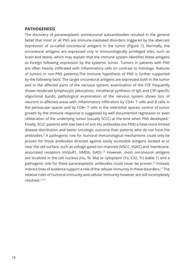
PAThOgenesIsThe discovery of paraneoplastic antineuronal autoantibodies resulted in the general belief that most or all PNS are immune-mediated disorders triggered by the aberrant expression of so-called onconeural antigens in the tumor (Figure 1). Normally, the onconeural antigens are expressed only in immunologically privileged sites, such as brain and testis(, which may explain that the immune system identifies these antigens as foreign following expression by the systemic tumor. Tumors in patients with PNS are often heavily infiltrated with inflammatory cells (in contrast to histologic features of tumors in non-PNS patients).The immune hypothesis of PNS is further supported by the following facts: The target onconeural antigens are expressed both in the tumor and in the affected parts of the nervous system; examination of the CSF frequently shows moderate lymphocytic pleocytosis, intrathecal synthesis of IgG and CSF-specific oligoclonal bands; pathological examination of the nervous system shows loss of neurons in affected areas with inflammatory infiltration by CD4+ T cells and B cells in the perivacular spaces and by CD8+ T cells in the interstitial spaces; control of tumor growth by the immune response is suggested by well documented regression or even obliteration of the underlying tumor (usually SCLC) at the time when PNS developed.7 Finally, SCLC patients with low titers of anti-Hu antibodies (no PNS) a have more limited disease distribution and better oncologic outcome than patients who do not have the antibodies.8 A pathogenic role for humoral immunological mechanisms could only be proven for those antibodies directed against easily accessible antigens located at or near the cell surface, such as voltage-gated ion channels (VGCC, VGKC) and membrane-associated receptors (mGluR1, NMDA, GAD).2,9 However, most onconeural antigens are localized in the cell nucleus (Hu, Ri, Ma) or cytoplasm (Yo, CV2, Tr) (table 1) and a pathogenic role for these paraneoplastic antibodies could never be proven.10 Instead, indirect lines of evidence support a role of the cellular immunity in these disorders.11 The relative roles of humoral immunity and cellular immunity however are still incompletely resolved.12,13

19
A gen
erAl intro
du
ction
on
PArAneo
PlAstic neu
rolo
gic syn
dro
mes
2
figure 1. The hypothesized pathogenesis of antibody-associated PNS. Onconeural antigen expressing tumour cells are phagocytozed by antigen-presenting cells (APC) that migrate to lymph nodes, where they present antigenic peptides to specific CD8+ and CD4+ T cells via HLA-class I and HLA-class II molecules, respectively. Tumour cells themselves present onconeural antigens to CD8+ T cells via HLA-class I molecules. The CD4+ T cells support CD8+ T-cell activation and proliferation by cytokines such as IFN-g and IL-2. CD4+ T cells stimulate B cells through cytokines including interleukin (IL)-4 and IL-5. B cells recognize soluble onconeural antigens throughtheir B-cell receptor. After activation, B cells differentiate into plasma cells,which secrete specific antibodies.Upon engagement of their TCR and accessorymolecules,CD8+ T cells can destroy tumour cells by secreting granzymes, perforins and cytokines such asTNF-a, or by upregulation ofCD95 (Fasligand) on tumour cells. The remnants of destroyed tumour cells can be taken up by APC, processed and presented to T cells. In addition, they can be specifically recognized by antibodies and eliminated via Fc receptor-expressing phagocytes. Cytotoxic specific CD8+ T cells not only slow the tumour growth, but they also cross the blood brain barrier and similarly attack neurons expressing the onconeural antigen, causing severe neurological damage in these patients. Reprinted with permission of J.W.K. de Beukelaar14

20
Table 1. Antibodies, antigen location, neurological syndrome and associated tumors
Antibody Antigenic location neurological syndromes Associated tumors
Well characterized paraneoplastic antibodies
Anti-Hu (ANNA-1) Nucleus more than cytoplasm(all neurons)
Encephalomyelitislimbic encephalitissensory neuronopathysubacute cerebellar degeneration autonomic neuropathy
SCLC, neuroblastoma, prostate
Anti-Yo (PCA-1) Cytoplasm, Purkinje cells Subacute cerebellar degeneration Ovary, breast
Anti-CV2 (CRMP5) Cytoplasm, oligodendrocytes, neurons
Encephalomyelitis limbic encephalitischoreasensory neuronopathysensorimotor neuropathyoptic neuritissubacute cerebellar degenerationautonomic neuropathy
SCLC Thymoma
Anti-Ri (ANNA-2) Nucleus more than cytoplasm(central nervous system neurons)
Opsoclonus-myoclonusbrainstem encephalitis
Breast, SCLC
Anti-Ma2 (Ta)* Neurons (subnucleus) Limbic/diencephalic/brainstem encephalitis,subacute cerebellar degeneration
Testis, lung
Anti-amphiphysin Presynaptic nerveterminals
Stiff-person syndromeEncephalomyelitissubacute sensory neuronopathysensorimotor neuropathy
Breast, SCLC
Anti-recoverin Photoreceptors, ganglion cells Cancer associated retinopathy SCLC
Partially characterized paraneoplastic antibodies
Anti-Tr (PCA-Tr) Cytoplasm, Purkinje cells Subacute cerebellar degeneration Hodgkin’s disease
ANNA-3 Nuclei, Purkinje cells Encephalomyelitissubacute sensory neuronopathy
SCLC
PCA-2 Cytoplasm, Purkinje cells and other neurons
Encephalomyelitissubacute cerebellar degeneration
SCLC
Anti-Zic4 Subacute cerebellar degeneration SCLC
Anti-NMDAR Axonal membrane Limbic encephalitis Ovarian teratoma
Anti-mGluR1 Purkinje cells, olfactory neurons, hippocampus
Subacute cerebellar degeneration Hodgkin’s disease
Antibodies that occur with and without cancer
Anti-VGCC Presynaptic neuromuscularjunction
Lambert-Eaton myasthenic syndromesubacute cerebellar degeneration
SCLC
Anti-AchR Neuromuscular junction Myasthenia gravis Thymoma
Anti-VGKC Peripheral nerve Limbic encephalitisneuromyotonia
Thymoma, SCLC
*Patients with brainstem encephalitis or subacute cerebellar degeneration usually associate with tumors other than testicular cancer and their sera also react with Ma1 protein. ANNA = antineuronal nuclear antibody; SCLC = Small Cell Lung Carcinoma; VGCC = voltage gated calcium channels; PCA = Purkinje cytoplasmic antibody;mGluR1 = metabotropic glutamate receptor type 1; VGKC = voltage gated potassium channel
21
A gen
erAl intro
du
ction
on
PArAneo
PlAstic neu
rolo
gic syn
dro
mes
2
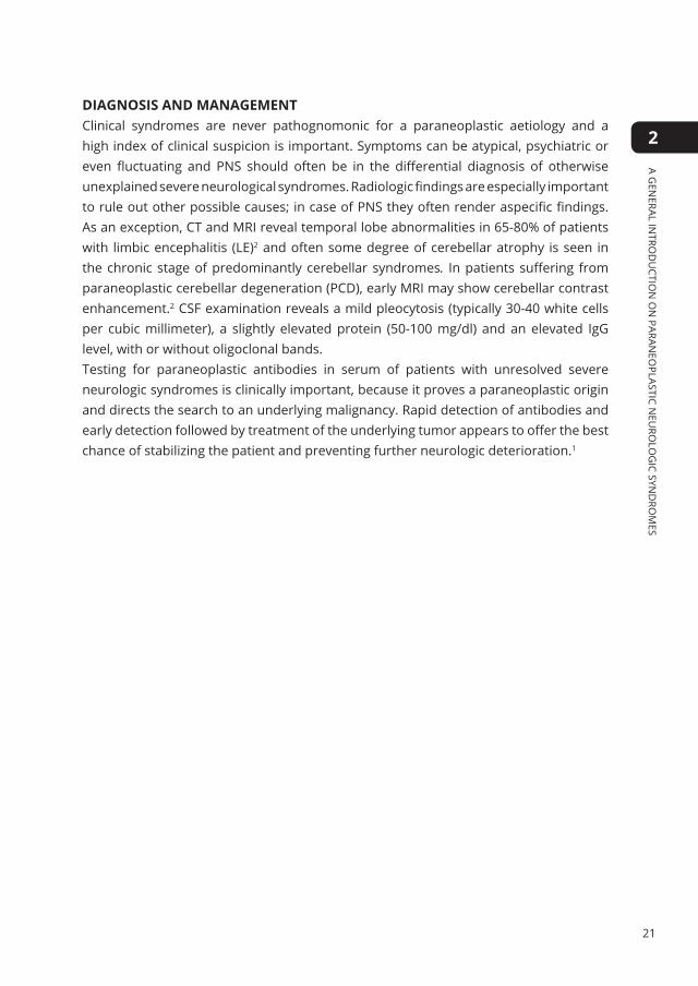
dIAgnOsIs And MAnAgeMenTClinical syndromes are never pathognomonic for a paraneoplastic aetiology and a high index of clinical suspicion is important. Symptoms can be atypical, psychiatric or even fluctuating and PNS should often be in the differential diagnosis of otherwise unexplained severe neurological syndromes. Radiologic findings are especially important to rule out other possible causes; in case of PNS they often render aspecific findings. As an exception, CT and MRI reveal temporal lobe abnormalities in 65-80% of patients with limbic encephalitis (LE)2 and often some degree of cerebellar atrophy is seen in the chronic stage of predominantly cerebellar syndromes. In patients suffering from paraneoplastic cerebellar degeneration (PCD), early MRI may show cerebellar contrast enhancement.2 CSF examination reveals a mild pleocytosis (typically 30-40 white cells per cubic millimeter), a slightly elevated protein (50-100 mg/dl) and an elevated IgG level, with or without oligoclonal bands. Testing for paraneoplastic antibodies in serum of patients with unresolved severe neurologic syndromes is clinically important, because it proves a paraneoplastic origin and directs the search to an underlying malignancy. Rapid detection of antibodies and early detection followed by treatment of the underlying tumor appears to offer the best chance of stabilizing the patient and preventing further neurologic deterioration.1
22
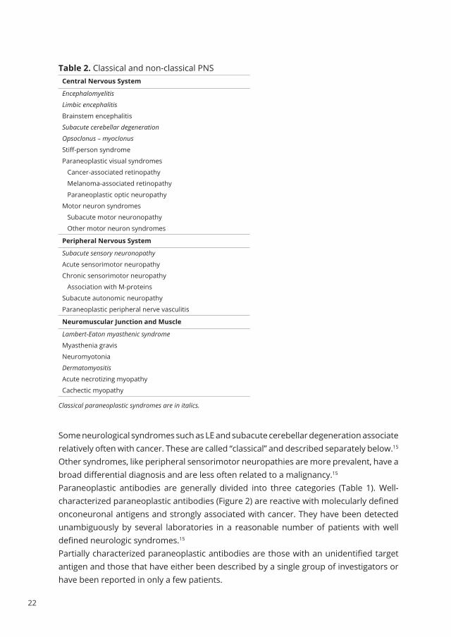
Table 2. Classical and non-classical PNSCentral nervous system
Encephalomyelitis
Limbic encephalitis
Brainstem encephalitis
Subacute cerebellar degeneration
Opsoclonus – myoclonus
Stiff-person syndrome
Paraneoplastic visual syndromes
Cancer-associated retinopathy
Melanoma-associated retinopathy
Paraneoplastic optic neuropathy
Motor neuron syndromes
Subacute motor neuronopathy
Other motor neuron syndromes
Peripheral nervous system
Subacute sensory neuronopathy
Acute sensorimotor neuropathy
Chronic sensorimotor neuropathy
Association with M-proteins
Subacute autonomic neuropathy
Paraneoplastic peripheral nerve vasculitis
neuromuscular Junction and Muscle
Lambert-Eaton myasthenic syndrome
Myasthenia gravis
Neuromyotonia
Dermatomyositis
Acute necrotizing myopathy
Cachectic myopathy
Classical paraneoplastic syndromes are in italics.
Some neurological syndromes such as LE and subacute cerebellar degeneration associate relatively often with cancer. These are called “classical” and described separately below.15 Other syndromes, like peripheral sensorimotor neuropathies are more prevalent, have a broad differential diagnosis and are less often related to a malignancy.15 Paraneoplastic antibodies are generally divided into three categories (Table 1). Well-characterized paraneoplastic antibodies (Figure 2) are reactive with molecularly defined onconeuronal antigens and strongly associated with cancer. They have been detected unambiguously by several laboratories in a reasonable number of patients with well defined neurologic syndromes.15 Partially characterized paraneoplastic antibodies are those with an unidentified target antigen and those that have either been described by a single group of investigators or have been reported in only a few patients.

23
A gen
erAl intro
du
ction
on
PArAneo
PlAstic neu
rolo
gic syn
dro
mes
2
The third group consists of antibodies that are associated with specific disorders but do not differentiate between paraneoplastic and non-paraneoplastic cases. Because of overlapping syndromes in separate antibodies, paraneoplastic antibodies should be searched for by screening rather than by focusing on a specific antibody. Unfortunately, not all patients with paraneoplastic syndromes have identifiable antibodies in their serum.
Once a paraneoplastic etiology is strongly suspected, a careful search for the underlying neoplasm is mandatory. If detailed history taking, thorough physical examination and high resolution computed tomography (CT) of chest, abdomen and pelvis do not show a primary tumor, whole body positron-emission tomography (PET) or PET/CT may be the best screening method for locating the occult cancer.16 Besides, the type of antibody and paraneoplastic syndrome may suggest a specific underlying tumor and indicate more directed diagnostic tests, such as mammography (or MRI) when breast cancer is suspected. With positive antibody screening and strong suspicion of paraneoplastic disease, negative tumor search should be repeated at 3-6 months intervals for 2-3 years.
Table 3. Diagnostic criteria for definite and possible PNSDefinite
1. A classical syndrome (i.e. encephalomyelitis, limbic encephalitis, subacute cerebellar degeneration,
opsoclonus-myoclonus, subacute sensory neuronopathy, chronic gastrointestinal pseudo-obstruction,
LEMS or dermatomyositis) and cancer that develops within 5 years of the diagnosis of the neurological
disorder, regardless of the presence of paraneoplastic antibodies
2. A non-classical syndrome that objectively improves or resolves after cancer treatment, provided that
the syndrome is not susceptible to spontaneous remission.
3. A non-classical syndrome with paraneoplastic antibodies (well characterized or not) and cancer that
develops within 5 years of the diagnosis of the neurological disorder.
4. A neurological syndrome (classical or not) with well characterized paraneoplastic antibodies (i.e. Hu, Yo,
Ri, amphiphysin, CV2 or Ma2).
Possible
1. A classical syndrome without paraneoplastic antibodies and no cancer but at high risk to have an
underlying tumor (e.g. smoking habit).
2. A neurological syndrome (classical or not) without cancer but with partially characterized
paraneoplastic antibodies.
3. A non-classical neurological syndrome, no paraneoplastic antibodies and cancer that presents within
two years of the neurological syndrome.

24
PNS are rare disorders and even neurologists interested in the field personally examine only a few patients per year. Because of the difficulties in diagnosing a neurologic syndrome as paraneoplastic, an international panel of neurologists has established diagnostic criteria that divide patients with a suspected paraneoplastic syndrome into ‘definite’and ‘probable’ categories (Table 3). These criteria are based on the type of clinical syndrome, the presence of ‘well characterized’ antibodies and on the presence or absence of cancer.15 However, the detection of additional antibodies (e.g. anti-NMDAR and anti-VGKC) and their clinical associations suggests that these criteria already need revision.17-19
figure 2. Antineuronal antibodies in paraneoplastic cerebellar degeneration.Staining of rat cerebellum (a–e) or cortex (f) using patients’ sera as primary antibody and peroxidase labelled secondary anti-IgG antibodies show nuclear more than cytoplasmic staining of granular and molecular layer neurons and Purkinje cells using anti-Hu (a) antibody positive serum; characteristic cytoplasmic staining of Purkinje cells by anti-Yo positive serum(b); cytoplasmic staining of Purkinje cells with staining of fine dots in the molecular layer by anti-Tr positive serum (c); strong staining of the molecular layer by anti-mGluR1 positive serum (d); synaptic interstitial staining of the molecular and granular layer by CV2 positive serum (e) and sub-nuclear staining of cortical neurons using anti-Ma2 positive serum (f). ML, molecular layer; PC, Purkinje cell; GL, granular layer; N, (cortical) neuron.
TheRAPy And PROgnOsIsBecause PNS are considered to be immune-mediated, two treatment approaches look obvious: removal of the source of antigen by tumor treatment, and immunosuppressive treatment. For most PNS the effect of immunotherapy is disappointing, underlining the importance of tumor search and tumor treatment.Immunotherapy is most effective in PNS associated with antibodies that are directed
25
A gen
erAl intro
du
ction
on
PArAneo
PlAstic neu
rolo
gic syn
dro
mes
2
against antigens located at the cell-surface and therefore accessible to circulating antibodies. These include syndromes of the peripheral (neuromyotonia, LEMS and MG) and central nervous system (anti-mGluR1 associated PCD9 , anti-amphipysin associated stiff-person syndrome20 and anti-VGKC and anti-NMDAR associated LE.21 ). Recommended modalities include plasma exchange, immunoadsorption (serum IgG extraction over a protein A column), steroids and intravenous immune globulins (IVIG). In PNS where the antigen is either nuclear or cytoplasmic, the dysfunction is probably the result of cellular immune mechanisms. Hence removal of antibodies is not expected to be beneficial. Therapies that modulate the activation and function of effector T cells could be more promising, but until now only limited evidence for the effectiveness of steroids, cyclophosphamide, IVIG or other immunosuppressive therapies exists.22 B cell depletion by monoclonal anti-CD20-antibodies (rituximab) has been encouraging in opsoclonus-myoclonus syndrome (OMS) in children23 , although results in adults were less convincing.24 These effects probably result from influencing the cell signaling between B-cells and T-cells and macrophages.Anti-tumor therapy offers the best chance to stop the paraneoplastic neurological deterioration and leaves the patients, on average, in a better condition.25,26 In severely debilitated patients, e.g. the elderly and bedridden, an underlying tumor is often not treated because chances of clinically relevant improvement are small.
ClAssICAl syndROMesEncephalomyelitis Paraneoplastic encephalomyelitis (PEM) is characterized by involvement of several areas of the nervous system, including temporal lobes and limbic system (LE), brainstem (brainstem encephalitis), cerebellum (PCD), spinal cord (myelitis), dorsal root ganglia (SSN) and autonomous nervous system (autonomic neuropathy).27 Patients with predominant involvement of one area but clinical evidence of only mild involvements of other areas are usually classified according to the predominant syndrome. Symptoms can include diplopia, dysarthria, dysphagia, gaze abnormalities (nuclear, internuclear or supranuclear), facial numbness and subacute hearing loss. More rarely, patients present with hypoventilation28 , epilepsia partialis continua29 or nonconvulsive status epilepticus.30 Most patients have an underlying SCLC and harbor anti-Hu antibodies (ANNA-1) in serum and CSF. Other associated antibodies are anti-CV2(CRMP5)2 , anti-amphihysin31 , anti-Ri (ANNA-2)32 and the less well characterized ANNA-333 and PCA-234 antibodies. Approximately 5% of patients with anti-Hu antibodies develop no cancer despite longterm follow-up.35 The absence of distinguishing clinical features between anti-Hu positive patients without cancer and those who develop cancer supports the possibility that these patients developed an effective Hu—probably a T cell-mediated—immune response that eliminated the tumour at an early stage of its development. Tumor
26
treatment offers the best chance of stabilizing the patient’s neurological condition while immunomodulating therapies do not appear to modify the outcome of PEM.27 In spite of descriptions of spontaneous improvement and incidental good response to immunosuppressive treatment, overall functional outcome is bad and more than 50% of patients are confined to bed or chair in the chronic phase of disease.27 Limbic encephalitis and variantsLE is characterized by a subacute onset (days to a few months) of short memory loss, seizures, confusion and psychiatric symptoms suggesting involvement of the limbic system. In patients primarily presenting with confusion or multiple seizures, the memory loss may not be evident. Approximately 80% of patients have MRI T2 and FLAIR hyperintensities involving one or both medial temporal lobes.36 FDG-PET may show hypermetabolism in one or both temporal lobes, which could (when seizures are excluded) be a hallmark of the inflammatory or immune-mediated process.2 More than half of the patients presenting with limbic encephalitis have an underlying neoplasm.17,37 Associated tumors include SCLC, testicular germ-cell tumors, thymoma, breast cancer, Hodgkin’s disease and teratoma.38 More than 80% of patients with LE harbour antineuronal antibodies and recent studies emphasize the importance of classifying antibody-positive LE according to the location of the target antigens.2,17
LE with antibodies to intraneuronal antigensThe main intracellular antigens related to LE are Hu, Ma1/2, and less frequently CV2(CRMP-5) and amphiphysin. Most patients with LE and anti-hu antibodies have symptoms suggesting dysfunction of areas of the nervous system distant from the limbic system, such as brainstem, cerebellum, dorsal root ganglia, cerebral cortex, spinal cord or autonomic nervous system.38 The median age is around 62 years and in most patients an underlying tumor is identified, usually SCLC followed by prostate cancer.37 The neurological outcome is generally poor although some improvement may occur in up to 38% of the patients.37 Anti-Ma antibodies react with Ma1 and Ma2 onconeural antigens while anti-Ta antibodies only react with Ma2.39 Patients with anti-Ma often present with limbic encephalitis and/or cerebellar symptoms. They are more often female with a median age of around 61 years. A wide range of tumors is associated, including lung and breast cancer, lymphoma and germ cell tumors. In most patients the neurological disease is progressive.39 Patients with anti-Ta present with limbic symptoms often in combination with hypothalamic deficits and brainstem symptoms. The patients are predominantly male with a median age of 34-44 years.39 Anti-Ta is associated with testicular germ-cell tumors that are often nearly undetectable at presentation.39 Most of these patients benefit from orchidectomy and they may also respond to immunotherapy.39 A recent review of the literature extended the age range (21-81 years) and sex (8/36 female) of anti-Ta

27
A gen
erAl intro
du
ction
on
PArAneo
PlAstic neu
rolo
gic syn
dro
mes
2
positive patients.39 In patients with anti-CV2 antibodies, the symptoms are rarely confined to the limbic system as they generally present with encephalomyelitis, sensory neuronopathy (SN), ataxia, chorea and optic neuritis. Development of myelitis and optical neuritis may initially resemble Devic’s syndrome. The anti-CV2 antibodies are mostly associated with SCLC, neuroendodrine tumors and thymomas.2 In PLE associated with intraneuronal antigens, spontaneous complete recovery has been described, although very rarely.1 Immunotherapy is largely ineffective39 , but multiple cases benefiting from anti-tumor treatment have been reported.1 Therefore, all efforts should be directed at identifying and treating the underlying tumor.
LE with antibodies to neuronal cell-surface antigensUp to 40% of patients with antibody associated LE harbour antibodies reactive with voltage-gated potassium channels (anti-VgKC).17,37 These patients either present with a typical limbic encephalitis or with a less focal encephalitis that is associated with psychiatric symptoms, peripheral nerve hyperexcitability (myokymia, neuromyotonia), hyperhydrosis and other symptoms of autonomic dysfunction occur (Morvan’s syndrome). REM sleep disturbances and hyponatremia are common in both groups About 30% of patients have tumors, most often SCLC or thymomas. Corticosteroid treatment, plasma-exchange and IVIG are beneficial in about 80% of patients.2 Antibodies to the NR1/NR2 heteromer of the N-methyl-D-aspartate receptors (anti-nMdAR) were first described in young women harbouring an ovarian teratoma.40 The patients presented with prodromal symptoms including headache, fever or a viral-like illness followed by severe psychiatric symptoms or memory loss, seizures, a decreased consciousness accompanied by dyskinesias, hypoventilation or autonomic instability.40 When untreated, many patients require long term mechanical ventilation. Treatment of the tumor combined with immunosuppressive or immunomodulating therapy often results in complete recovery.2 More recently, anti-NMDAR antibodies have also been described in male patients with LE41 and in LE patients without a tumor or with an underlying SCLC.17 Approximately 25% of patients presenting with LE harbour antibodies reactive with unidentified neuronal surface antigen (i.e. not anti-NMDAR or anti-VGKC).17 These patients usually have additional onconeural antibodies including antibodies against amphiphysin, Hu, CV2, SOX1 and GAD.17 These PLE patients generally have no clinical response to treatment.
Paraneoplastic cerebellar degeneration (PCD) PCD is one of the most common and characteristic paraneoplastic syndromes. In series of patients with antibody-associated PNS, presentation with cerebellar signs occurred in 37%. Usually the syndrome starts acutely with nausea, vomiting, dizziness
28
and slight incoordination of walking, evolving rapidly over weeks to a few months with progressive ataxia of gait, limbs and trunk, dysarthria and often nystagmus associated with oscillopsia. The disease is progressive in months and then stabilizes. By that time most patients are severely disabled: walking without support, sitting unsupported and self-feeding becomes difficult while handwriting is often impossible. Signs are always bilateral but may be asymmetrical. Diplopia is common at presentation although ocular movement may seem normal at investigation. Symptoms and signs are mostly limited to the cerebellum and cerebellar pathways, but on careful examination other mild neurological abnormalities e.g. hearing loss, dysphagia, pyramidal and extrapyramidal tract involvement, mental status change and peripheral neuropathy may be found.1 Approximately 50% of patients presenting with acute or subacute, non-familial ataxia are estimated to have an underlying malignancy. Associated malignancies include ovarian, breast and lung cancer (SCLC) and lymphomas (Hodgkin’s disease) Symptoms precede cancer diagnosis in 60-70% of patients. In the initial stages MRI of the brain is normal but may exceptionally show diffuse enlargement of the cerebellar hemispheres or contrast enhancement of the sulci.2 Brain FDG-PET scan and SPECT may show cerebellar hypermetabolism and increased perfusion during the acute stages of PCD.42 In the chronic phase, CT and MRI often reveal cerebellar atrophy. In the search for antibodies and associated malignancy anti-Yo (PCA-1), anti-Tr (PCA-Tr) and anti-mGluR1 are associated with relatively ‘pure’ cerebellar syndromes. Anti-Yo antibodies point at breast, ovarian, endometrium and fallopian tube cancers. Rarely, anti-Yo associated PCD occurs in male patients, usually associated with a gastrointestinal adenocarcinoma, expressing the cdr2 antigen.43 Anti-Tr antibodies appear specific for Hodgkin’s disease44 and anti-mGluR1 antibodies have been found in two patients with PCD and Hodgkin’s disease.9 About 50% of PCD patients with an underlying SCLC have high titers of anti-Hu antibodies while the remaining patients are likely to have anti-P/Q-type VGCC antibodies. These antibodies were present in all patients who also had Lambert-Eaton myasthenic syndrome (LEMS). In patients with anti-amphihysin or anti-CV2 antibodies, the cerebellar degeneration is often part of the PEM syndrome and more widespread neurological symptoms and signs are usually found. The less well characterized PCA-2, ANNA-3 and Zic4 antibodies are associated with lung cancer and various neurological syndromes including PCD. The outcome of PCD is usually poor and the best chance to at least stabilize the syndrome is to treat the underlying tumor. In patients with anti-Yo-associated PCD, the prognosis is better for patients with breast cancer than for those with gynecologic cancer. An also better prognosis is found in patients with Hodgkin’s disease and anti-Tr or anti-mGluR1 antibodies. With successful treatment of the tumor and/or immunotherapy, symptoms may disappear and the antibodies vanish.9,44

29
A gen
erAl intro
du
ction
on
PArAneo
PlAstic neu
rolo
gic syn
dro
mes
2
Opsoclonus-Myoclonus syndrome (OMS) OMS – the ‘dancing eye and dancing feet syndrome’ – is a disorder of ocular motility that consists of involuntary, arrhythmic, high amplitude conjugate saccades in all directions. Opsoclonus may occur intermittently or constantly in more severe cases and it does not remit when the eyes are closed or in the darkness. There is often a diffuse or focal myoclonus and other cerebellar and brainstem signs. An excessive startle response reminiscent of hyperekplexia may also occur.45 MRI is usually normal but may show hyperintensities in the brainstem on T2 weighted images. Recent pathological and fMRI studies suggest that disinhibition of the fastigial nuclei of the cerebellum is involved, probably resulting from dysfuntion of Purkinje cells in the cerebellar vermis.46 Approximately 20% of adult patients with OMS have a previously undiscovered malignancy, most commonly SCLC and breast and gynecologic cancers. In some patients, paraneoplastic OMS resembles PCD. The prominent opsoclonus and truncal rather than appendicular ataxia distinguish this syndrome from anti-Yo and anti-Hu associated PCD Adult patients with paraneoplastic OMS are older (median age 66 years) than patients with the idiopathic syndrome (median age 40 years). Non-malignant causes for opsoclonus include infections (West Nile virus, Lyme disease), celiac disease, metabolic or toxic influences and allogeneic hematopoietic stem cell transplantation.46 In women, anti-Ri antibodies (ANNA-2) are frequently associated with breast and gynecologic tumors. In male patients anti-Ri has occasionally been found in bladder cancer and SCLC. Paraneoplastic OMS can also be associated with anti-Hu antibodies, usually as part of a more widespread PEM. In children with OMS, almost 50% have an underlying neuroblastoma. Conversely, approximately 2-3% of children with neuroblastoma have paraneoplastic OMS. Non-paraneoplastic pediatric OMS occurs as a self-limiting illness and is probably the result of a viral infection of the brainstem. The search for an occult neuroblastoma should include imaging of chest and abdomen (CT scan or MRI), urine catecholamine measurements and metaiodobenzylguanidine scan.1 When negative, the evaluation should be repeated after several months. Sera of children (not of adults) with OMS have antibodies that react with the cell surfaces of cerebellar granule neurons and neuroblastoma cells.47 Incubation of neuroblastoma cell lines with these antibodies inhibits cell proliferation and induces apoptosis, indicating that humoral immune mechanisms have an important pathogenic role in pediatric OMS. In contrast to most of the other PNS, paraneoplastic OMS may remit either spontaneously, following treatment of the tumor, or in association with clonazepam or thiamine treatment. Most patients with idiopathic OMS make a good recovery that seems to be accelerated by immunosuppressive therapy. Paraneoplastic OMS has usually a more severe clinical course and treatment with steroids or IVIG appears ineffective. However, improvement following immunosuppressive or immunomodulating therapy. In
30
children, paraneoplastic OMS may improve following treatment with ACTH, prednisone, azathioprine or IVIG, but residual neurologic signs are frequent.
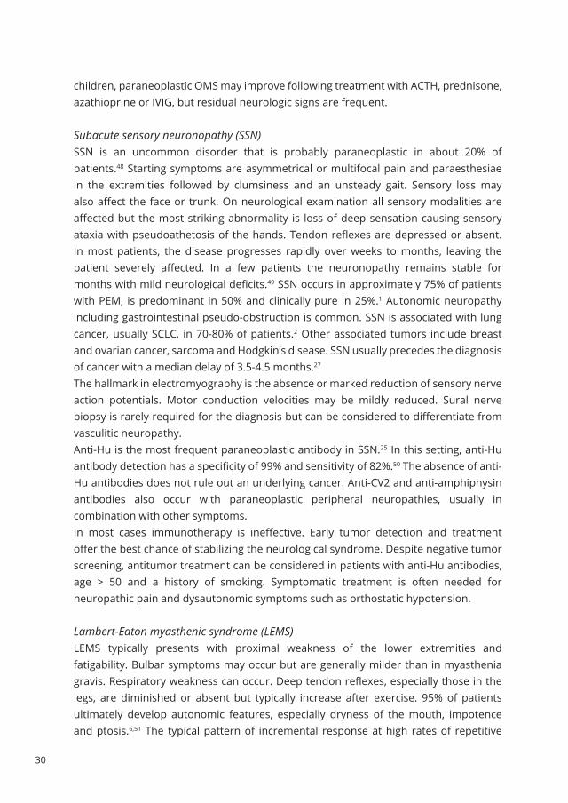
Subacute sensory neuronopathy (SSN)SSN is an uncommon disorder that is probably paraneoplastic in about 20% of patients.48 Starting symptoms are asymmetrical or multifocal pain and paraesthesiae in the extremities followed by clumsiness and an unsteady gait. Sensory loss may also affect the face or trunk. On neurological examination all sensory modalities are affected but the most striking abnormality is loss of deep sensation causing sensory ataxia with pseudoathetosis of the hands. Tendon reflexes are depressed or absent. In most patients, the disease progresses rapidly over weeks to months, leaving the patient severely affected. In a few patients the neuronopathy remains stable for months with mild neurological deficits.49 SSN occurs in approximately 75% of patients with PEM, is predominant in 50% and clinically pure in 25%.1 Autonomic neuropathy including gastrointestinal pseudo-obstruction is common. SSN is associated with lung cancer, usually SCLC, in 70-80% of patients.2 Other associated tumors include breast and ovarian cancer, sarcoma and Hodgkin’s disease. SSN usually precedes the diagnosis of cancer with a median delay of 3.5-4.5 months.27 The hallmark in electromyography is the absence or marked reduction of sensory nerve action potentials. Motor conduction velocities may be mildly reduced. Sural nerve biopsy is rarely required for the diagnosis but can be considered to differentiate from vasculitic neuropathy. Anti-Hu is the most frequent paraneoplastic antibody in SSN.25 In this setting, anti-Hu antibody detection has a specificity of 99% and sensitivity of 82%.50 The absence of anti-Hu antibodies does not rule out an underlying cancer. Anti-CV2 and anti-amphiphysin antibodies also occur with paraneoplastic peripheral neuropathies, usually in combination with other symptoms.In most cases immunotherapy is ineffective. Early tumor detection and treatment offer the best chance of stabilizing the neurological syndrome. Despite negative tumor screening, antitumor treatment can be considered in patients with anti-Hu antibodies, age > 50 and a history of smoking. Symptomatic treatment is often needed for neuropathic pain and dysautonomic symptoms such as orthostatic hypotension.
Lambert-Eaton myasthenic syndrome (LEMS)LEMS typically presents with proximal weakness of the lower extremities and fatigability. Bulbar symptoms may occur but are generally milder than in myasthenia gravis. Respiratory weakness can occur. Deep tendon reflexes, especially those in the legs, are diminished or absent but typically increase after exercise. 95% of patients ultimately develop autonomic features, especially dryness of the mouth, impotence and ptosis.6,51 The typical pattern of incremental response at high rates of repetitive

31
A gen
erAl intro
du
ction
on
PArAneo
PlAstic neu
rolo
gic syn
dro
mes
2
stimulation (50 Hz) or following 15-30 seconds of maximal voluntary contraction forms the electromyographic hallmark of LEMS. Most patients with LEMS have antibodies against P/Q type voltage-gated calcium channels (VGCC).Approximately 50-60% of patients with LEMS have cancer, mostly SCLC.6 In some patients, LEMS may develop in association with other paraneoplastic syndromes, including paraneoplastic cerebellar degeneration and encephalomyelitis. Other tumors include small cell carcinomas of the prostate and cervix, lymphomas and adenocarcinomas. The prevalence of LEMS in SCLC is approximately 3%.6 History of smoking, absence of the HLA-B8 genotype and presence of SOX1 antibodies strongly predict an underlying SCLC.18 Patients with SCLC and LEMS survive significantly longer than SCLC patients who do not have the paraneoplastic syndrome.6 Treatment of LEMS must be tailored to the individual, based on severity of the symptoms, underlying disease, life expectancy and previous treatment response. Treatment of the tumor frequently leads to neurological improvement. Symptoms are treated with drugs that facilitate the release of acetylcholine from motor nerve terminals such as 3,4-diaminopyridine (3,4-DAP). Cholinesterase inhibitors (e.g. pyridostigmine) may improve dryness of mouth but rarely relieve weakness. If these treatments are not effective enough, immunosuppressive therapy must be considered. Removal of the pathogenic anti-VGCC antibodies by plasma exchange and IVIG can give quick but transient relief.
Dermatomyositis In dermatomyositis the characteristic heliotrope rash (purplish discoloration of the eyelids) often precedes the appearance of proximal muscle weakness. Other manifestations include arthralgia, myocarditis and congestive heart failure and interstitial lung disease. The standardized incidence rate for a malignant disease in dermatomyositis is 6.2 (95% confidence interval 3.9 – 10.0).52 Similar clinical, electromyographical, and pathological results are found compared to dermatomyositis without presence of malignancy.52 Dermatomyositis is associated with cancer of the ovary, lung, pancreas, stomach, colorectal and breast and with non-Hodgkin lymphoma. Most patients have elevated serum creatine kinase levels and electromyographic evidence of myopathy. Muscle or skin biopsy is the definitive diagnostic procedure and shows inflammatory infiltrates. Muscle imaging (CT or MRI) may help in confirming the diagnosis and determining the type of an inflammatory myopathy and in selecting an appropriate biopsy site. Antibodies to the Mi-2 protein complex are specific for dermatomyositis and are present in high titers in about 35% of cases.53 Treatment of patients with paraneoplastic dermatomyositis does not differ from those without a tumor. Nearly all patients respond to corticosteroids. Refractory patients and patients requiring a lower dose of steroids can be treated with azathioprine. Methotrexate and cyclophosphamide may also be considered.

32
RefeRenCes
1. de Beukelaar J, Sillevis Smitt P. Managing
paraneoplastic neurological disorders.
Oncologist 2006;11:292-305.
2. Dalmau J, Rosenfeld M. Paraneoplastic
syndromes of the CNS. Lancet Neurol
2008;7:327-40.
3. Brouwer B. Beitrag zur Kenntniss der
chronischen diffusen Kleinhirnerkrankungen.
Neurol Centralblatt1919:674-82.
4. Brouwer B. Les affections parenchymateuse du
cervelet et leur signification du point de vue de
l’anatomie et de la physiologie de cet organe.
In: Biemond A, ed.: J Belge Neurol Psychiatry;
1938:691–757.
5. Wilkinson PC, Zeromski J. Immunofluorescent
detection of antibodies against neurones in
sensory carcinomatous neuropathy. Brain
1965;88:529-83.
6. Titulaer M, Verschuuren J. Lambert-Eaton
myasthenic syndrome: tumor versus nontumor
forms. Ann N Y Acad Sci 2008;1132:129-34.
7. Horino T, Takao T, Yamamoto M, Geshi T,
Hashimoto K. Spontaneous remission of small
cell lung cancer: a case report and review in the
literature. Lung Cancer 2006;53:249-52.
8. Dalmau J, Furneaux H, Gralla R, Kris M, Posner
J. Detection of the anti-Hu antibody in the
serum of patients with small cell lung cancer--a
quantitative western blot analysis. Ann Neurol
1990;27:544-52.
9. Sillevis Smitt P, Kinoshita A, De Leeuw B, et
al. Paraneoplastic cerebellar ataxia due to
autoantibodies against a glutamate receptor. N
Engl J Med 2000;342:21-7.
10. Sillevis Smitt P, Manley G, Posner J. Immunization
with the paraneoplastic encephalomyelitis
antigen HuD does not cause neurologic disease
in mice. Neurology 1995;45:1873-8.
11. de Beukelaar J, Verjans G, van Norden Y, et al.
No evidence for circulating HuD-specific CD8+ T
cells in patients with paraneoplastic neurological
syndromes and Hu antibodies. Cancer Immunol
Immunother 2007;56:1501-6.
12. de Beukelaar J, Sillevis Smitt P, Hop W, et al.
Imbalances in circulating lymphocyte subsets
in Hu antibody associated paraneoplastic
neurological syndromes. Eur J Neurol
2007;14:1383-91.
13. de Graaf M, de Beukelaar J, Bergsma J, et al. B
and T cell imbalances in CSF of patients with
Hu-antibody associated PNS. J Neuroimmunol
2008;195:164-70.
14. De Beukelaar JWK. The role of T Lymphocytes
in the pathogenesis of Hu antibody associated
paraenoplastic neurological syndromes (Thesis).
Rotterdam: ErasmusMC; 2007.
15. Graus F, Delattre J, Antoine J, et al. Recommended
diagnostic criteria for paraneoplastic
neurological syndromes. J Neurol Neurosurg
Psychiatry 2004;75:1135-40.
16. Patel RR, Subramaniam RM, Mandrekar JN,
Hammack JE, Lowe VJ, Jett JR. Occult malignancy
in patients with suspected paraneoplastic
neurologic syndromes: value of positron
emission tomography in diagnosis. Mayo Clinic
proceedings Mayo Clinic 2008;83:917-22.
17. Graus F, Saiz A. Limbic encephalitis: an
expanding concept. Neurology 2008;70:500-1.
18. Sabater L, Titulaer M, Saiz A, Verschuuren J,
Güre A, Graus F. SOX1 antibodies are markers
of paraneoplastic Lambert-Eaton myasthenic
syndrome. Neurology 2008;70:924-8.
19. Sabater L, Bataller L, Suárez-Calvet M, Saiz
A, Dalmau J, Graus F. ZIC antibodies in
paraneoplastic cerebellar degeneration and
small cell lung cancer. J Neuroimmunol 2008.

33
A gen
erAl intro
du
ction
on
PArAneo
PlAstic neu
rolo
gic syn
dro
mes
2
20. Sommer C, Weishaupt A, Brinkhoff J, et al.
Paraneoplastic stiff-person syndrome: passive
transfer to rats by means of IgG antibodies to
amphiphysin. Lancet 2005;365:1406-11.
21. Anderson N, Barber P. Limbic encephalitis - a
review. J Clin Neurosci 2008;15:961-71.
22. Keime-Guibert F, Graus F, Fleury A, et al.
Treatment of paraneoplastic neurological
syndromes with antineuronal antibodies
(Anti-Hu, anti-Yo) with a combination of
immunoglobulins, cyclophosphamide, and
methylprednisolone. J Neurol Neurosurg
Psychiatry 2000;68:479-82.
23. Pranzatelli M, Tate E, Travelstead A, et al.
Rituximab (anti-CD20) adjunctive therapy for
opsoclonus-myoclonus syndrome. J Pediatr
Hematol Oncol 2006;28:585-93.
24. Shams’ili S, de Beukelaar J, Gratama J, et al. An
uncontrolled trial of rituximab for antibody
associated paraneoplastic neurological
syndromes. J Neurol 2006;253:16-20.
25. Sillevis Smitt P, Grefkens J, de Leeuw B, et al.
Survival and outcome in 73 anti-Hu positive
patients with paraneoplastic encephalomyelitis/
sensory neuronopathy. J Neurol 2002;249:745-
53.
26. Graus F, Keime-Guibert F, Rene R, et al. Anti-Hu-
associated paraneoplastic encephalomyelitis:
analysis of 200 patients. Brain 2001;124:1138-
48.
27. Sillevis Smitt P. Paraneoplastic neurological
syndromes. Lancet Neurol 2002;1:408;
discussion
28. Gomez-Choco MJ, Zarranz JJ, Saiz A, Forcadas
MI, Graus F. Central hypoventilation as
the presenting symptom in Hu associated
paraneoplastic encephalomyelitis. J Neurol
Neurosurg Psychiatry 2007;78:1143-5.
29. Kinirons P, O’Dwyer JP, Connolly S, Hutchinson
M. Paraneoplastic limbic encephalitis presenting
as lingual epilepsia partialis continua. J Neurol
2006;253:256-7.
30. Espay AJ, Kumar V, Sarpel G. Anti-Hu-associated
paraneoplastic limbic encephalitis presenting
as rapidly progressive non-convulsive status
epilepticus. J Neurol Sci 2006;246:149-52.
31. Dropcho E. Antiamphiphysin antibodies with
small-cell lung carcinoma and paraneoplastic
encephalomyelitis. Ann Neurol 1996;39:659-67.
32. Pittock S, Lucchinetti C, Lennon V. Anti-neuronal
nuclear autoantibody type 2: paraneoplastic
accompaniments. Ann Neurol 2003;53:580-7.
33. Chan K, Vernino S, Lennon V. ANNA-3 anti-
neuronal nuclear antibody: marker of lung
cancer-related autoimmunity. Ann Neurol
2001;50:301-11.
34. Vernino S, Lennon V. New Purkinje cell
antibody (PCA-2): marker of lung cancer-
related neurological autoimmunity. Ann Neurol
2000;47:297-305.
35. Lladó A, Carpentier A, Honnorat J, et al. Hu-
antibody-positive patients with or without
cancer have similar clinical profiles. J Neurol
Neurosurg Psychiatry 2006;77:996-7.
36. Graus F, Dalmau J. Paraneoplastic neurological
syndromes: diagnosis and treatment. Curr Opin
Neurol 2007;20:732-7.
37. Jarius S, Hoffmann L, Stich O, et al. Relative
frequency of VGKC and ‘classical’ paraneoplastic
antibodies in patients with limbic encephalitis. J
Neurol 2008;255:1100-1.
38. Gultekin S, Rosenfeld M, Voltz R, Eichen J, Posner
J, Dalmau J. Paraneoplastic limbic encephalitis:
neurological symptoms, immunological findings
and tumour association in 50 patients. Brain
2000;123 ( Pt 7):1481-94.
39. Hoffmann LA, Jarius S, Pellkofer HL, et al. Anti-
Ma and anti-Ta associated paraneoplastic
neurological syndromes: 22 newly diagnosed
patients and review of previous cases. J Neurol

34
Neurosurg Psychiatry 2008;79:767-73.
40. Dalmau J, Tüzün E, Wu H, et al. Paraneoplastic
anti-N-methyl-D-aspartate receptor encephalitis
associated with ovarian teratoma. Ann Neurol
2007;61:25-36.
41. Novillo-López M, Rossi J, Dalmau J, Masjuan
J. Treatment-responsive subacute limbic
encephalitis and NMDA receptor antibodies in a
man. Neurology 2008;70:728-9.
42. Choi K, Kim J, Park S, Kim Y, Kim S, Smitt P.
Cerebellar hypermetabolism in paraneoplastic
cerebellar degeneration. J Neurol Neurosurg
Psychiatry 2006;77:525-8.
43. Debes J, Lagarde S, Hulsenboom E, et al.
Anti-Yo-associated paraneoplastic cerebellar
degeneration in a man with adenocarcinoma
of the gastroesophageal junction. Dig Surg
2007;24:395-7.
44. Bernal F, Shams’ili S, Rojas I, et al. Anti-Tr
antibodies as markers of paraneoplastic
cerebellar degeneration and Hodgkin’s disease.
Neurology 2003;60:230-4.
45. Wirtz P, Sillevis Smitt P, Hoff J, et al. Anti-Ri
antibody positive opsoclonus-myoclonus in a
male patient with breast carcinoma. J Neurol
2002;249:1710-2.
46. Wong A. An update on opsoclonus. Curr Opin
Neurol 2007;20:25-31.
47. Blaes F, Fuhlhuber V, Korfei M, et al. Surface-
binding autoantibodies to cerebellar neurons in
opsoclonus syndrome. Ann Neurol 2005;58:313-
7.
48. Chalk C, Windebank A, Kimmel D, McManis P.
The distinctive clinical features of paraneoplastic
sensory neuronopathy. Can J Neurol Sci
1992;19:346-51.
49. Graus F, Bonaventura I, Uchuya M, et al. Indolent
anti-Hu-associated paraneoplastic sensory
neuropathy. Neurology 1994;44:2258-61.
50. Molinuevo JL, Graus F, Serrano C, Rene R,
Guerrero A, Illa I. Utility of anti-Hu antibodies
in the diagnosis of paraneoplastic sensory
neuropathy. Ann Neurol 1998;44:976-80.
51. O’Neill J, Murray N, Newsom-Davis J. The
Lambert-Eaton myasthenic syndrome. A review
of 50 cases. Brain 1988;111 ( Pt 3):577-96.
52. Buchbinder R, Hill C. Malignancy in patients with
inflammatory myopathy. Curr Rheumatol Rep
2002;4:415-26.
53. Targoff IN. Update on myositis-specific and
myositis-associated autoantibodies. Curr Opin
Rheumatol 2000;12:475-81.

35
A gen
erAl intro
du
ction
on
PArAneo
PlAstic neu
rolo
gic syn
dro
mes
2


Mass spectrometric detection of antigen-specific immunoglobulin peptides in
paraneoplastic patient sera
Peter Maat, Martijn M. VanDuijn, Eric Brouwer, Lennard H. Dekker, Lona Zeneyedpour, Theo Luider, Peter A.E. Sillevis Smitt
Journal of Autoimmunity June 2012;38(4):354-360
3Back to table of contents

38
AbsTRACTParaneoplastic neurological syndromes (PNS) are severe immune mediated effects of cancer. The presence of IgG autoantibodies against onconeural antigens in serum is a hallmark of the disease. Multiple paraneoplastic antibodies have been described, including antibodies against HuD, Yo, amphiphysin and CV2. In this study, we test the hypothesis that primary amino-acid structures of the antigen binding part of antibodies from various individuals share common sequences that are specific for each auto-antigen.We selected 60 patients with PNS, associated with antibodies against HuD, Yo, Amp or CV2.Affinity purified IgG was separated using SDS-PAGE and IgG heavy chains were excised,trypsinized and subjected to tandem mass spectrometry. We selected masses that uniquely identified a PNS autoantibody group, and used MS/MS fragmentation spectra to obtain information on peptide sequences. Out of 19,173 unique masses, 28 immunoglobulin derived peptides were found exclusively in samples from a single autoantibody defined PNS group.Our results confirm that specific peptide structures exist in the antigen binding site of IgG that are shared between individuals harboring autoantibodies against the same onconeural antigen. Thus, the immune response in these patients followed converging paths during the rearrangement, selection and maturation of immunoglobulin sequences. The identified peptides can be applied in the diagnosis of PNS, but these data also indicate that a similar approach in a variety of other diseases involving an immune response would have an appealing outlook.
KeywORdsParaneoplastic neurological syndrome; antibody; immunoglobulin; repertoire; proteomics
ACKnOwledgeMenTsThis study is supported by Horizon valorisation grant 93515502 and Zenith grant 93511034 from the The Netherlands Organisation for Scientific Research (NWO). PM was supported by a research grant of Masspac, Eurostars.
39
Mass spectro
Metric d
etection
of an
tigen
-specific iMM
un
og
lobu
lin peptid
es in pn
s
3
InTROduCTIOnParaneoplastic neurological syndromes (PNS) are devastating, remote effects of cancer that are not caused by tumor growth, infiltration, metastasis or tumor treatment.1 In about 70% of cases, the neurological symptoms precede the cancer diagnosis. Clinical outcome depends mostly on an early diagnosis of PNS combined with rapid detection and treatment of the underlying tumor.1 Early diagnosis is aided by the detection of anti-neuronal antibodies that are present in the serum of approximately 50-70% of patients with PNS. These autoantibodies are directed against neuronal antigens that are ectopically expressed by the tumor (i.e. onconeural antigens). Autoantibodies against several onconeural antigens have been described including anti-HuD (HuD-Ab), anti-Yo (Yo-Ab), anti-amphiphysin (Amp-Ab), and anti-CV2 (CV2-Ab) antibodies.1,2 Antibodies against onconeural antigens are now routinely tested in diagnostic assays that employ proteins produced by recombinant technology.Immunoglobulin G (IgG) consists of 4 chains: two identical heterodimers of a heavy and a light chain held together covalently by disulfide bonds. Both heavy and light chains have a constant part and a variable antigen binding part. Within the antigen binding part, a set of 3 complementary determining regions (CDRs) embedded in framework regions form a groove that fits the epitope of an antigen. The CDRs and framework regions are selected from rearrangements of V-, D- and J-genes, and subsequent somatic hypermutation during affinity maturation.3 The CDR3 region of the heavy chain contributes the most to both the variability in immunoglobulin sequences, and also to their specificity to an antigen.3,4
Although rearrangement and junctional diversity in the heavy chain can yield more than 107 different variable regions 3, many reports show an unexpected sequence overlap in different species.5-8 Based on the fact that a few major epitopes are present within an antigen molecule, including HuD and that selection pressures exist for the best binding variants during antibody development, we hypothesize that the diversity in the variable epitope binding regions of immunoglobulins will be significantly less than the total potential diversity.7-9 As a result, we expect that individuals harboring HuD-Ab in their serum may share some of the amino-acid sequences of the antigen binding regions of HuD-Ab with those of other individuals with HuD-Ab.Previous work of our group showed that, after immunization of outbred rats with recombinant HuD protein, shared identical CDR-derived IgG peptides specific for HuD-Ab could indeed be detected, and that sera could be distinguished in this manner from that of animals immunized with another antigen10, Therefore, it seems plausible that also in humans, antigen specific IgG derived peptides might be shared between individuals who have in their serum autoantibodies reactive with the same antigen.Mass spectrometry (MS) is a reliable technique to quantify peptides, even in low nanomolar amounts.11 The instrument also provides peptide fragmentation spectra that can be used to obtain sequence information on the peptide of interest. Commonly,

40
a search algorithm screens a protein database in combination with the spectra to arrive at identification. This approach is not available for work on antigen binding regions, as these variable parts are hardly covered by protein databases. Recent advances in de novo sequencing of unidentified MS/MS spectra offer opportunities to construct an optimal sequence hypothesis for a peptide without the use of a database. First reports of IgG amino acid sequencing show that a proteomic approach is feasible.10,12 In the current work, the goal is to establish that the proteomic analysis of immunoglobulins can differentiate between immunoglobulin samples from human patients with a specific immune response. The clinical parameters of paraneoplastic disease are well-defined and the recombinant onconeural target antigens and sera specific for these antigens are well validated and available. Therefore, PNS serves as a suitable model to test the feasibility of detecting antigen-specific immunoglobulin peptides in affinity purified patient sera. While conventional diagnostic tests for the paraneoplastic antigens described here are currently adequate, this work sets the stage for diseases where diagnostics remain more challenging, such as paraneoplastic diseases that require cumbersome cell-based assays, radioactive assays, or conditions where the antigen remains unidentified.
MATeRIAls And MeThOdsSamplesWe selected 20 HuD-Ab positive sera that were negative for antibodies against the onconeural antigens NOVA (Ri), CDR62 (Yo), amphiphysin, CV2 (CRMP-5), Ma2, recoverin and Tr.1 In addition, we selected 20 Yo-Ab positive sera, 10 Amp-Ab positive and 10 CV2-Ab positive sera. All sera reacted with only one of the onconeural antigens mentioned above, and thus could also be used as controls for other antigen groups. IgG titers of the paraneoplastic antibodies were determined by endpoint titration on rat cerebellar sections using peroxidase immunohistochemistry as described before.13 From all patients clinical information was retrieved from the treating neurologist. Characteristics of patients and sera are depicted in Table 1. All patients had a ‘definite PNS’ according to published criteria and all sera had been stored at -800 C until use.14 The study was approved by the Erasmus MC institutional review board, and patients consented to the use of their samples for research.
Materials and ReagentsAll chemicals were, unless otherwise noted, purchased from Sigma-Aldrich, Saint Louis, MO.
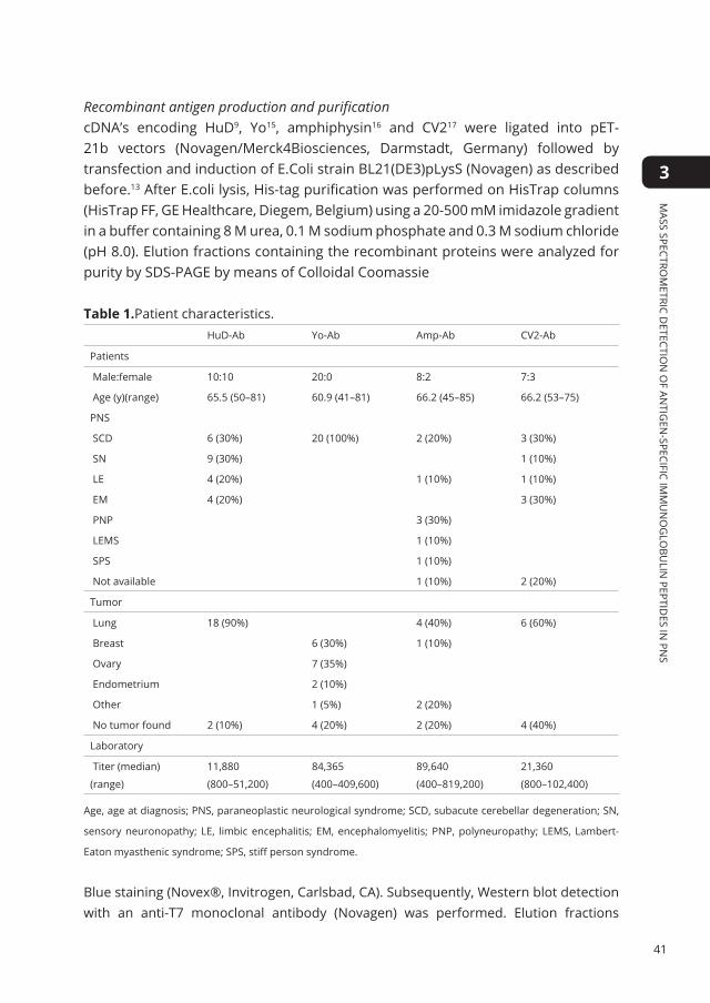
41
Mass spectro
Metric d
etection
of an
tigen
-specific iMM
un
og
lobu
lin peptid
es in pn
s
3
Recombinant antigen production and purification cDNA’s encoding HuD9, Yo15, amphiphysin16 and CV217 were ligated into pET-21b vectors (Novagen/Merck4Biosciences, Darmstadt, Germany) followed by transfection and induction of E.Coli strain BL21(DE3)pLysS (Novagen) as described before.13 After E.coli lysis, His-tag purification was performed on HisTrap columns (HisTrap FF, GE Healthcare, Diegem, Belgium) using a 20-500 mM imidazole gradient in a buffer containing 8 M urea, 0.1 M sodium phosphate and 0.3 M sodium chloride (pH 8.0). Elution fractions containing the recombinant proteins were analyzed for purity by SDS-PAGE by means of Colloidal Coomassie
Table 1.Patient characteristics.HuD-Ab Yo-Ab Amp-Ab CV2-Ab
Patients
Male:female 10:10 20:0 8:2 7:3
Age (y)(range) 65.5 (50–81) 60.9 (41–81) 66.2 (45–85) 66.2 (53–75)
PNS
SCD 6 (30%) 20 (100%) 2 (20%) 3 (30%)
SN 9 (30%) 1 (10%)
LE 4 (20%) 1 (10%) 1 (10%)
EM 4 (20%) 3 (30%)
PNP 3 (30%)
LEMS 1 (10%)
SPS 1 (10%)
Not available 1 (10%) 2 (20%)
Tumor
Lung 18 (90%) 4 (40%) 6 (60%)
Breast 6 (30%) 1 (10%)
Ovary 7 (35%)
Endometrium 2 (10%)
Other 1 (5%) 2 (20%)
No tumor found 2 (10%) 4 (20%) 2 (20%) 4 (40%)
Laboratory
Titer (median)
(range)
11,880
(800–51,200)
84,365
(400–409,600)
89,640
(400–819,200)
21,360
(800–102,400)
Age, age at diagnosis; PNS, paraneoplastic neurological syndrome; SCD, subacute cerebellar degeneration; SN,
sensory neuronopathy; LE, limbic encephalitis; EM, encephalomyelitis; PNP, polyneuropathy; LEMS, Lambert-
Eaton myasthenic syndrome; SPS, stiff person syndrome.
Blue staining (Novex®, Invitrogen, Carlsbad, CA). Subsequently, Western blot detection with an anti-T7 monoclonal antibody (Novagen) was performed. Elution fractions

42
with the highest concentration of recombinant protein were subjected to further purification by ion exchange chromatography on HiTrapQ HP columns (GE Healthcare) using a buffered pH gradient of 12 to 2.5 in a buffer containing 8M urea, 0.01M sodium phosphate, 0.01M citric acid, 0.01M glycine, 0.01M tricine and 0.01M aminocaproic acid.18 Fractions containing the highest purity of recombinant proteins were pooled, the buffer was exchanged to 8M urea and 0.1M sodium phosphate (pH 8.0) using a desalting column (Hitrap Desalting, GE Healthcare), and the sample was concentrated to 1 mg onconeural antigen / ml by ultrafiltration (Ultra-4 30kDa NWCO, Millipore, Amsterdam, The Netherlands), analysed by SDSPAGE as mentioned above and aliquots were stored at -800C.
Affinity purificationIgG was purified from serum samples by Melon Gel purification (Melon Gel IgG Purification Kit, Pierce Biotechnology, Rockford, IL) according to the manufacturer’s protocol. Recombinant purified HuD, Yo, amphiphysin and CV2 proteins were each coupled to carboxylated magnetic beads (CMS-30-10, Spherotech, Lake Forest, Illinois, US). For each serum sample, 107 beads were used. After washing the beads with water, 20 μl 50 mg/ml N-hydroxysulfosuccinimide (Pierce Biotechnology), 20 μl 50 mg/ml carbodiimide EDC (Pierce Biotechnology) and 160 μl 100 mM sodium phosphate buffer, pH 6.2 were added to the beads. After incubation for 20 minutes, the activated beads were washed with 2-(N-morpholino) ethanesulfonic acid, pH 5.0. Subsequently, 200 pmoles of recombinant protein per 107 beads were added. After incubation, the coupled beads were blocked with PBS, 0.1% BSA, 0.02% tween-20, 0.05% NaN3 pH 7.4 and stored at 400C until use.10 For use, the beads were washed twice using PBS-0.02% Tween-20 (PBS-T) and incubated for 1 hour at room temperature with 2.5 ml of purified IgG, 1:10 diluted in Melon purification buffer, followed by centrifugation (1000g) for 30 seconds. Supernatant was removed while the tube was placed on a magnet to prevent loss of beads. For washing, we added 2.5 ml PBS-T followed by vigorous mixing for 10 seconds, washing on roller bank for 5 minutes, followed again by mixing. This procedure was repeated twice. For elution of the bound antibodies, 225 μl 100 mM glycine-HCl pH 2.6 was added followed by mixing. After 5 minutes of incubation, the samples were centrifuged and the purified antibody-containing supernatant was collected on a magnetic sample holder. The pH was reconstituted to pH 8.0 using 1M Tris-HCl pH 9. As a control, a Yo-Ab positive serum was subjected to affinity purification on HuD coated beads (Supplementary Figure 4).
Electrophoresis and digestion of proteinsSDS-PAGE was performed as previously described.9 Polyacrylamide gels (10%) of 0.75 mm thickness were used. To 15 μL sample 5 μL sample buffer was added and samples were heated for 10 minutes at 90⁰ C. Proteins in the gel were stained with Colloidal

43
Mass spectro
Metric d
etection
of an
tigen
-specific iMM
un
og
lobu
lin peptid
es in pn
s
3
Coomassie Blue staining (Invitrogen) following the manufacturer�s instructions. The concentrations of the affinity purified IgG fractions were then calculated with serial dilutions of commercial human IgG (Nanogam®, Sanquin, Amsterdam, The Netherlands) as a reference. From the Colloidal Coomassie Blue stained gels, IgG heavy chain bands were excised. The gel plugs were reduced and alkylated with iodoacetamide and digested with trypsin in the presence of RapiGestSF (Waters Corporation, Milford, MA) as described previously.10
LTQ-OrbitrapBased on the UV absorption of the peptides in a test chromatography run, an optimal injection volume of 15 μL was selected for nanoLC (nanoLC Ultimate 3000; Dionex, Sunnyvale, CA) separation, followed by Orbitrap MS (LTQ Orbitrap XL ETD, Thermo Scientific, Bremen, Germany), as described before.19 All samples were measured in a data dependent acquisition mode. MS spectra from the Orbitrap were acquired with a resolution of 30,000. Under dynamic exclusion rules, the five most intense masses in the mass spectrum were selected for collision-induced dissociation (CID) in the linear ion trap of the instrument. Before each run, a blank LC-MS run was performed to monitor system background and sample carry-over.
Data analysesRaw mass spectrometry data files were loaded into Progenesis LCMS labelfree quantitation software (version 2.6; Nonlinear Dynamics Ltd, New Castle, UK). This software aligns the retention times of all samples, detects mass features in the data, defines a region of interest around these and integrates signal intensities in such regions for all samples. Also, available MS/MS spectra and peptide identifications are associated with the features. As our analysis has focus on the presence of peptide markers that are absent in control groups, the software output is limited by the fact that the reported abundances simply reflect the amount of signal within the region of interest, without verification of the presence of a proper isotopic pattern for a peptide of interest in each sample. To address this, we only consider feature abundance as valid for a given sample when we found a corresponding feature after analysis of just this sample in isolation. If that was not the case, an abundance of ‘0’ was assigned with a script written in R (R 2.12.2, www.r-project.org). Where possible, features were annotated in Progenesis with sequence information. First, Mascot (version 2.2.06; Matrix Science Inc., London, UK) was used to perform database searches against the human subset of the NCBI nonredundant database (version 20090311; Homo sapiens species restriction; 222,066 sequences). For the database search the following settings were used: a maximum of two miss cleavages, oxidation as a variable modification of methionine, deamidation as a variable modification of asparagine or glutamine, carbamidomethylation as a fixed modification of cysteine, and trypsin as protease. A peptide mass tolerance of

44
10 ppm and a fragment mass tolerance of 0.5 Da were allowed. An ion score of 25 was used as a cut-off value. In addition, the MS/MS data were loaded into Peaks Studio 5.2 (Bioinformatics Solutions Inc., Waterloo, Canada) and de novo sequencing was performed for all specific features. The parent peptide mass accuracy was set at 6 ppm and for the MS/MS fragments 0.5 Da tolerance was allowed. Oxidation was allowed as a variable modification of methionine, carbamidomethylation as a fixed modification of cysteine, and trypsin as protease.From the feature abundance data, we selected as potential markers those that were present in many patients for an antigen while absent in all other antigens. Some of the features emerging from that step were identified as antigen-derived, and were excluded from the marker set at that point. We also excluded features that were associated with common environmental or serum contaminants (e.g. keratins, complement factors, plasminogen, transferrin). A permutation analysis was performed to determine the false discovery rate. We randomized the group assignments 30 times and determined the number of background features matching our selection criteria.Sequence information of features that were identified as potential markers was manually curated after assessing homology of the sequence to known germline sequences (Spider search in Peaks), re-analysis with summed MS/MS spectra, and rejection of outlier MS/MS spectra after pairwise comparison of their similarity with R. All final amino-acid sequences of antibody group specific features identified by Mascot database search or proposed by de novo sequencing were aligned to human immunoglobulin germline sequences using the IMGT (ImMunoGeneTics information system® http://www.imgt.org; Montpellier, France) database as described before.19,20 The alignment with the most homologous germ allele was registered as provided by the IMGT tool where Smith-Waterman homology scores exceeded 35. An additional homology search with the BLAST algorithm against the NCBI nonredundant database was used to exclude the presence of variants of serum contaminants in the dataset.21
ResulTsAffinity purificationSera were collected from a total of 60 PNS patients (Table 1). Antigen-specific immunoglobulins were affinity purified from the sera with beads covalently coupled to recombinant antigen. The concentration of affinity purified IgG, expressed as serum concentration, was for HuD-Ab 0.017 ± 0.013 mg/mL (mean ± SD), Yo-Ab 0.036 ± 0.027 mg/mL, Amp-Ab 0.007 ± 0.010 mg/mL and CV2-Ab 0.020 ± 0.013 mg/mL as determined by densitometry of Coomassie stained SDS-PAGE gel bands. Two of the 60 affinity purified IgG bands (one HuD-Ab and one Yo-Ab) were absent on visual inspection of the Coomassie stained gels. These two samples were excluded from further analysis.

45
Mass spectro
Metric d
etection
of an
tigen
-specific iMM
un
og
lobu
lin peptid
es in pn
s
3
Feature detection (Progenesis) and identification (Mascot)The antigen-specific IgG heavy chain bands were excised from the gels, digested with trypsin, and analyzed by nano-LC and mass spectrometry (LC-MS). The Progenesis label-free analysis software identified 19,173 peptide features in the MS dataset. A Mascot search of the data against the NCBI non-redundant protein database yielded amino acid sequences for 2904 peptides, of which 2,765 were unique. In addition, 10,705 peptides were associated with a de novo sequence hypothesis, and 5,564 peptides remained without sequence identification.As we seek peptides that uniquely identify a patient group, it is important to give an accurate assessment on the presence or absence of a particular peptide in a sample. The common workflow for label-free analysis in proteomics enumerates the total signal in a region of interest (a ‘feature’) in all samples. This approach works relatively well when looking at concentration differences in abundant peptides, but is prone to report an erroneous abundance in absence of a peptide due to signals from noise or partial signals from other peptides present near in the region of interest (Supplementary Figure 1). As explained in the Materials and Methods section (2.7), we constructed a refinement of the workflow that is much more effective at judging the presence or absence of peptides in each individual sample. The inadvertent release of antigen from the affinity resin provides us with nice markers in the samples to test our analysis, as e.g. HuD-derived peptides cannot be present in the Yo samples that were enriched on a Yo affinity resin. This is indeed the outcome of our analysis, but not that of the initial data produced by the label-free Progenesis analysis software (Figure 1). This shows that our additional data-analysis was both necessary and sufficient to produce reliable data for marker discovery.
Figure 1. Result of the additional processing of abundance data from label-free analysis. A) Shows the initial abundance data reported by the software for the peptide AINTLNGLR in all samples. The peptide comes from the HuD antigen which was released during the affinity purification, and cannot be present in samples from other groups. B) After processing (right panels), abundances are only accepted when a peptide feature could also be detected based on data of the just the single patient in isolation. Indeed, the peptide is now only detected in samples where its presence is plausible. A value of 1 was added to all data points to also allow the plotting of an abundance of 0 on the logarithmic axis.

46
Selection of group-specific featuresFrom the dataset we selected discriminating features that were present in more than 4 samples from Amp and CV2, or more than 5 samples for HuD and Yo, while being absent in the other antigen groups. By these abundance criteria, 67 features were included. To get an impression of the false discovery rate, we performed a repeated (n=30) random permutation of the group assignments. In this way, it was found that our criteria yielded 0.9±1.1 false positive markers from the dataset.Some of the 67 features were rejected because of the sequence information associated with them. Among them, we found some peptides from the onconeural antigens that apparently dissociated from the affinity resin during sample preparations. We found 1 peptide from amphiphysin, 14 from CV2 and 4 from HuD in the marker set. For unknown reasons, several human Plasminogen peptides were found in the Yo patient samples. Although the latter behavior was selective, we rejected these and also other serum components other than immunoglobulins. After these rejections, a set of 30 potential markers remained. Two pairs of features that were found represented the 2+ and 3+ charge states of the same peptides, which leaves 28 unique peptides: 2 specific for CV2, 11 for HuD and 15 for Yo (Figure 2, Table 2). P-values were determined for each feature with a Kruskall-Wallis test (non-parametric ANOVA, corrected for ties) and found to be 0.005 or lower. Not all peptides were present in all individuals, suggesting that a panel of peptides will be required to develop a sensitive bio-assay. Based on the current samples and all markers, we would detect 0/10 Amp cases, 7/10 CV2 cases, 17/19 HuD cases, and 19/19 Yo cases. However, further independent validation isrequired to properly assess the sensitivity that can be obtained with these markers.
Figure 2. Abundance of marker candidates. Two examples are shown of markers that were selected from the data. Abundances are plotted for all samples in the dataset, and the most probable sequence of the peptide is shown in the plot. TPENMRDK was present in 16/19 Yo samples, ITYSPNTSK was present in 11/19 HuD samples. A value of 1 was added to all data points to also allow the plotting of an abundance of 0 on the logarithmic axis.

47
Mass spectro
Metric d
etection
of an
tigen
-specific iMM
un
og
lobu
lin peptid
es in pn
s
3
Table 2. Group-specific features identified from the dataset.
m/z z Sequence Confi-dence
Ig homologya
Amp n = 10
CV2 n = 10
HuD n = 19
Yo n = 19
P value
475.2434 3 WWHHLSTLTSR 34 0 7 0 0 5.5E-08
586.6333 3 YYWWQRLPTTSTTR IGLV1 0 5 0 0 1.4E-05
505.7594 2 ITYSPNTSK 33 IGHV6 0 0 11 0 7.9E-06
414.8735 3 – 0 0 10 0 3.1E-05
521.7283 2 QHMMSQHK 9 0 0 8 0 4.1E-04
670.7657 2 CGGMYYDYAPK 23 IGHV3 0 0 7 0 1.4E-03
482.2487 2 LEMTVQAR 42 0 0 7 0 1.4E-03
419.5449 3 RENMSVHAGVR 14 0 0 7 0 1.4E-03
530.7611 2 YQAWTTYK 23 0 0 7 0 1.4E-03
404.7195 2 ARLSMSK 11 0 0 7 0 1.4E-03
630.2904 2 NSYNDYAVSAR 63 IGHV6 0 0 6 0 4.4E-03
732.3613 2 YKNYNDYAVSVK 43 IGHV6 0 0 6 0 4.4E-03
488.5770 3 43 IGHV6 0 0 6 0
816.3728 2 SVTPEDTAVYYCARb 95 IGHV6 0 0 6 0 4.4E-03
503.7358 2 TPENMRDK 32 0 0 0 16 3.7E-09
620.9439 3 CFWNMELGVVEMSSR 25 0 0 0 16 3.7E-09
930.9108 2
410.5117 3 WMYSNHGYR 0 0 0 11 7.9E-06
410.5117 3 YMPPWFGCR 0 0 0 11 7.9E-06
511.7344 2 MYFSPSYK 0 0 0 9 1.2E-04
543.5950 3 REYNFNTTASQAAR 27 0 0 0 9 1.2E-04
476.8943 3 NKTNFSEFLDGR 38 0 0 0 8 4.1E-04
852.9618 2 ACPKAWRKANALYR 35 0 0 0 7 1.4E-03
973.9224 2 RQMNSPNGMHQLGCWK 25 0 0 0 7 1.4E-03
424.2188 2 HYVDSVKb 40 IGHV3 0 0 0 6 4.4E-03
763.3966 2 WGIGTMVTVSSASTKb 27 IGHJ3 0 0 0 6 4.4E-03
413.8777 3 WWWLANHAR 0 0 0 6 4.4E-03
520.7771 2 NGVAVPSPSGR 28 0 0 0 6 4.4E-03
1128.1498 3 WRWWRCWMWCYVQTFPTCEWWR 6 0 0 0 6 4.4E-03
887.0502 3 HMWWRWYMHYTSHDDVHQK 7 IGHV4 0 0 0 6 4.4E-03
Shown are the mass and charge of the feature that was found, the sequence that was obtained based on
the MS/MS spectra, a confidence score for the sequence based on the MS/MS spectra, the most homologous
IgG germline allele, and the number of individuals in which these markers were found. a. Smith–Waterman
homology score > 35. b. Mascot database identification.

48
MS/MS spectraTo further characterize the PNS antigen specific MS features, we compared the MS/MS spectra, which were available for 27 of the 28 potential markers. For each feature, we performed a similarity analysis on the MS/MS spectra recorded from all samples using unsupervised clustering based on pairwise dot products. As illustrated in Figure 3, the majority of MS/MS spectra recorded for this feature specific to the Yo-Ab patient group are similar, showing that they indeed relate to the same peptide. All of these spectra originated from samples of patients with Yo-Ab related PNS. In addition, some distinct spectra were included, even in absence of the Yo-specific peptide of interest in the samples from other patient groups. Such signals were dissimilar, as shown in cluster analysis for HuD and CV2 samples (Figure 3A) as well as in the spectra (Figure 3B). Such signals may originate from co-eluting material or from adjacent peptide features (Supplemental Figure 2).Similar MS/MS results were obtained for the other antigen-specific features in our dataset. While the quality of these deviating spectra usually does not result in high confidence sequence information, it is prudent to remove them from the analysis in order to prevent improper sequence information for a feature.
Figure 3. MS/MS fragmentation spectra associated with the marker peptides as found by the Progenesis software. Shown is data for peptide CFWNMELGVVEMSSR, 620.94393+, which was only found in Yo-Ab PNS samples. (A) Hierarchical clustering of spectra based on their similarity. The majority of spectra is similar and originates from Yo patients. MS/MS spectra originating from other patient groups (HuD, CV2) cluster separately, showing they probably relate to other co-eluting material. Thus, peptide CFWNMELGVVEMSSR is indeed unique to Yo associated PNS. (B) Waterfall plot of all spectra in this set. The bottom cluster of panel A is shown in the foreground of the waterfall plot. It can be appreciated that the Yo-related spectra in the background are all similar.
49
Mass spectro
Metric d
etection
of an
tigen
-specific iMM
un
og
lobu
lin peptid
es in pn
s
3
Peptide sequencesWithin the 28 potential marker peptides, 2 were identified in the Mascot database search as immunoglobulin peptides. For all but one of the remaining features de novo sequences could be constructed, although some were of limited quality. Some sequences were modified to a more plausible one after assessing homology to immunoglobulin germ line alleles using the Peaks Spider tool, or after manual inspection. As we used several methods to obtain peptide sequences, (Mascot search, de novo sequencing, adjusted de novo sequences), we constructed a general confidence parameter to describe the match to MS/MS spectra and sequence confidence. A pseudo-database was generated in Mascot containing the proposed peptide sequences. Then, a search was performed with the experimental MS/MS spectra, and we included the best score obtained for each feature in Table 2. The two features with mass 410.5117 are very similar, but do represent distinct peptides that could be found eluting sequentially in a sample. The de novo sequence information obtained for these features was of insufficient quality for comparison, but the MS/MS spectra were highly similar. Identical peptides except for the order of two adjacent amino acids could be a plausible explanation for these similar features.
Immunoglobulin germ line alignment (IMGT)With the putative sequences of the features of interest, a comparison was made with immunoglobulin germline sequences in order to identify those with a homology. When the Smith-Waterman homology score exceeded 35, the allele was reported in Table 2. Features with such homology to IgG germline sequences generally also had a higher sequence confidence (p<0.05). With an additional homology search, it was verified that the peptides were not related to the antigens used in the affinity purification or to other serum contaminants.
dIsCussIOnIn the work described in this paper, we compared human immunoglobulin samples with a well-described affinity against onconeural antigens. Because of the huge potential diversity ofimmunoglobulins, one might predict that the recombination and affinity maturation of immunoglobulins during the immune response should result in sequences of the antigen binding regions that are essentially random. In that case, our proteomic analysis of these samples would have revealed a collection of peptides from the constant regions that is shared among all patients, and a collection of peptides from the variable regions that are unique to each patient. Rather, we could identify 30 peptides that were shared between up to 16 individuals from our cohort, but that were also specific to only one of the four onconeural antigens that we investigated. This finding corroborates our earlier findings on our hypothesis that selection pressures in immunoglobulin development
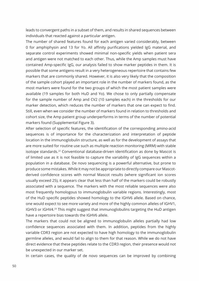
50
leads to convergent paths in a subset of them, and results in shared sequences between individuals that reacted against a particular antigen.The number of shared features found for each antigen varied considerably, between 0 for amphiphysin and 13 for Yo. All affinity purifications yielded IgG material, and separate control experiments showed minimal non-specific yields when patient sera and antigen were not matched to each other. Thus, while the Amp samples must have contained Amp-specific IgG, our analysis failed to show marker peptides in them. It is possible that some antigens result in a very heterogeneous repertoire that contains few markers that are commonly shared. However, it is also very likely that the composition of the sample cohort played an important role in the number of markers found, as the most markers were found for the two groups of which the most patient samples were available (19 samples for both HuD and Yo). We chose to only partially compensate for the sample number of Amp and CV2 (10 samples each) in the thresholds for our marker detection, which reduces the number of markers that one can expect to find. Still, even when we consider the number of markers found in relation to thresholds and cohort size, the Amp patient group underperforms in terms of the number of potential markers found (Supplemental Figure 3).After selection of specific features, the identification of the corresponding amino-acid sequences is of importance for the characterization and interpretation of peptide location in the immunoglobulin structure, as well as for the development of assays that are more suited for routine use such as multiple reaction monitoring (MRM) with stable isotope standards.22 Conventional database-driven identification as done by Mascot is of limited use as it is not feasible to capture the variability of IgG sequences within a population in a database. De novo sequencing is a powerful alternative, but prone to produce some mistakes. While it may not be appropriate to directly compare our Mascot-derived confidence scores with normal Mascot results (where significant ion scores usually exceed 25), it appears clear that less than half of the markers could be robustly associated with a sequence. The markers with the most reliable sequences were also most frequently homologous to immunoglobulin variable regions. Interestingly, most of the HuD specific peptides showed homology to the IGHV6 allele. Based on chance, one would expect to see more variety and more of the highly common alleles of IGHV1, IGHV3 or IGHV4.23 This might suggest that immunoglobulins targeting the HuD antigen have a repertoire bias towards the IGHV6 allele. The markers that could not be aligned to immunoglobulin alleles partially had low confidence sequences associated with them. In addition, peptides from the highly variable CDR3 region are not expected to have high homology to the immunoglobulin germline alleles, and would fail to align to them for that reason. While we do not have direct evidence that these peptides relate to the CDR3 region, their presence would not be unexpected in our marker set.In certain cases, the quality of de novo sequences can be improved by combining
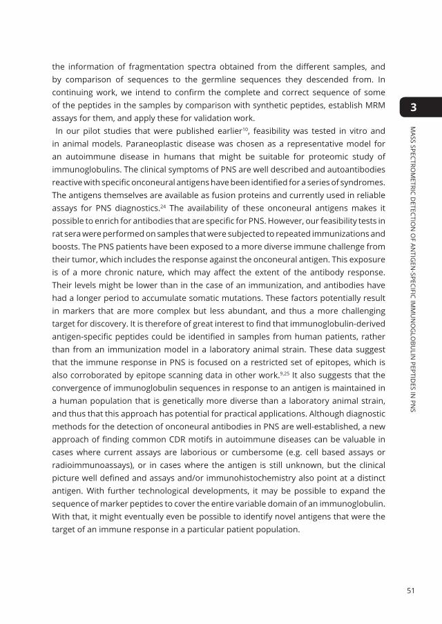
51
Mass spectro
Metric d
etection
of an
tigen
-specific iMM
un
og
lobu
lin peptid
es in pn
s
3
the information of fragmentation spectra obtained from the different samples, and by comparison of sequences to the germline sequences they descended from. In continuing work, we intend to confirm the complete and correct sequence of some of the peptides in the samples by comparison with synthetic peptides, establish MRM assays for them, and apply these for validation work. In our pilot studies that were published earlier10, feasibility was tested in vitro and in animal models. Paraneoplastic disease was chosen as a representative model for an autoimmune disease in humans that might be suitable for proteomic study of immunoglobulins. The clinical symptoms of PNS are well described and autoantibodies reactive with specific onconeural antigens have been identified for a series of syndromes. The antigens themselves are available as fusion proteins and currently used in reliable assays for PNS diagnostics.24 The availability of these onconeural antigens makes it possible to enrich for antibodies that are specific for PNS. However, our feasibility tests in rat sera were performed on samples that were subjected to repeated immunizations and boosts. The PNS patients have been exposed to a more diverse immune challenge from their tumor, which includes the response against the onconeural antigen. This exposure is of a more chronic nature, which may affect the extent of the antibody response. Their levels might be lower than in the case of an immunization, and antibodies have had a longer period to accumulate somatic mutations. These factors potentially result in markers that are more complex but less abundant, and thus a more challenging target for discovery. It is therefore of great interest to find that immunoglobulin-derived antigen-specific peptides could be identified in samples from human patients, rather than from an immunization model in a laboratory animal strain. These data suggest that the immune response in PNS is focused on a restricted set of epitopes, which is also corroborated by epitope scanning data in other work.9,25 It also suggests that the convergence of immunoglobulin sequences in response to an antigen is maintained in a human population that is genetically more diverse than a laboratory animal strain, and thus that this approach has potential for practical applications. Although diagnostic methods for the detection of onconeural antibodies in PNS are well-established, a new approach of finding common CDR motifs in autoimmune diseases can be valuable in cases where current assays are laborious or cumbersome (e.g. cell based assays or radioimmunoassays), or in cases where the antigen is still unknown, but the clinical picture well defined and assays and/or immunohistochemistry also point at a distinct antigen. With further technological developments, it may be possible to expand the sequence of marker peptides to cover the entire variable domain of an immunoglobulin. With that, it might eventually even be possible to identify novel antigens that were the target of an immune response in a particular patient population.

52
suPPleMenTARy dATA
Supplementary Figure S1. Errors in abundance quantification for label-free analyses. A. MS signal for a sample from a HuD-Ab PNS patient, containing a peptide from the HuD antigen AINTLNGLR. The software integrates signals in the green region of interest for abundance metrics. B. The same region of interest as in panel A is also used for this sample from a Yo-Ab PNS patient. The sample does not contain the peptide AINTNGLR, but signals in the region of interest still result in a reported abundance for the peptide.

53
Mass spectro
Metric d
etection
of an
tigen
-specific iMM
un
og
lobu
lin peptid
es in pn
s
3
Supplementary Figure S2. Sources of error in assigning MS2 spectra to a feature. All panels show MS data for the Yo-Ab specific feature identified as CFWNMELGVVEMSSR (Green region of interest). Red circles indicate the time and mass where an MS2 spectrum was triggered, and subsequently assigned to this feature. A. Yo-Ab PNS patient sample. Peptide CFWNMELGVVEMSSR is present, and MS2 spectrum conformed to the majority shown in Figure 2. B. Yo-Ab PNS patient sample. Peptide CFWNMELGVVEMSSR is present, but MS2 spectrum did not conform to the majority shown in Figure 2, and was triggered by a distinct peptide. C. HuD-Ab PNS patient sample. Peptide CFWNMELGVVEMSSR is not present, and MS2 spectrum did not conform to the majority shown in Figure 2, and was triggered by a distinct peptide.
54
Supplementary Figure S3. Number of group-specific features detected as function of the selection parameter. The number of features found was divided by the group size to correct for differences in sample numbers.
Supplementary Figure S4. HuD-Ab affinity purification using HuD coated beads. The figure shows Western Blot detection on nitrocellulose using AP-labeled anti-human IgG. The upper panel shows the affinity purification of an HuD-Ab serum over HuD antigen coated beads. The first lane shows the total amount of IgG loaded on the beads. Most IgG does not bind to the beads and is present in the pass through (lane u). After washing, the HuD specific IgG is eluted (eluate) and clearly represents a small fraction of the total IgG. The lower panel shows the same procedure performed with a Yo-Ab serum over HuD antigen coated beads. In the eluate no IgG is detectable, reflecting absence of HuD specific IgG in Yo-Ab serum.

55
Mass spectro
Metric d
etection
of an
tigen
-specific iMM
un
og
lobu
lin peptid
es in pn
s
3
RefeRenCes
1. de Beukelaar J, Sillevis Smitt P. Managing
paraneoplastic neurological disorders.
Oncologist 2006;11:292-305.
2. Dalmau J, Rosenfeld M. Paraneoplastic
syndromes of the CNS. Lancet Neurol
2008;7:327-40.
3. Schroeder HW, Jr., Cavacini L. Structure and
function of immunoglobulins. The Journal of
allergy and clinical immunology 2010;125:S41-
52.
4. Xu JL, Davis MM. Diversity in the CDR3 region of
V(H) is sufficient for most antibody specificities.
Immunity 2000;13:37-45.
5. Weinstein JA, Jiang N, White RA, 3rd, Fisher
DS, Quake SR. High-throughput sequencing
of the zebrafish antibody repertoire. Science
2009;324:807-10.
6. Foreman AL, Lemercier B, Lim A, et al. VH gene
usage and CDR3 analysis of B cell receptor
in the peripheral blood of patients with PBC.
Autoimmunity 2008;41:80-6.
7. Foreman AL, Van de Water J, Gougeon ML,
Gershwin ME. B cells in autoimmune diseases:
insights from analyses of immunoglobulin
variable (Ig V) gene usage. Autoimmun Rev
2007;6:387-401.
8. Scheid JF, Mouquet H, Ueberheide B, et al.
Sequence and structural convergence of broad
and potent HIV antibodies that mimic CD4
binding. Science 2011;333:1633-7.
9. Manley GT, Smitt PS, Dalmau J, Posner JB. Hu
antigens: reactivity with Hu antibodies, tumor
expression, and major immunogenic sites. Ann
Neurol 1995;38:102-10.
10. VanDuijn MM, Dekker LJ, Zeneyedpour L, Smitt PA,
Luider TM. Immune responses are characterized
by specific shared immunoglobulin peptides
that can be detected by proteomic techniques.
J Biol Chem 2010;285:29247-53.
11. Dekker LJ, Zeneyedpour L, Brouwer E, van Duijn
MM, Sillevis Smitt PA, Luider TM. An antibody-
based biomarker discovery method by mass
spectrometry sequencing of complementarity
determining regions. Anal Bioanal Chem 2010.
12. Obermeier B, Mentele R, Malotka J, et al.
Matching of oligoclonal immunoglobulin
transcriptomes and proteomes of cerebrospinal
fluid in multiple sclerosis. Nat Med 2008;14:688-
93.
13. Moll JW, Antoine JC, Brashear HR, et al.
Guidelines on the detection of paraneoplastic
anti-neuronal-specific antibodies: report from
the Workshop to the Fourth Meeting of the
International Society of Neuro-Immunology
on paraneoplastic neurological disease,
held October 22-23, 1994, in Rotterdam, The
Netherlands. Neurology 1995;45:1937-41.
14. Graus F, Delattre JY, Antoine JC, et al.
Recommended diagnostic criteria for
paraneoplastic neurological syndromes. J
Neurol Neurosurg Psychiatry 2004;75:1135-40.
15. Fathallah-Shaykh H, Wolf S, Wong E, Posner
JB, Furneaux HM. Cloning of a leucine-zipper
protein recognized by the sera of patients
with antibody-associated paraneoplastic
cerebellar degeneration. Proc Natl Acad Sci U S
A 1991;88:3451-4.
16. DeCamilli P, Thomas A, Cofiell R, et al. The
synaptic vesicle-associated protein amphiphysin
is the 128-kD autoantigen of stiff-man syndrome
with breast cancer. J Exp Med 1993;178:2219-23.
17. Yu Z, Kryzer T, Griesmann G, Kim K, Benarroch
E, Lennon V. CRMP-5 neuronal autoantibody:
marker of lung cancer and thymoma-related
autoimmunity. Ann Neurol 2001;49:146-54.
18. Ahamed T, Chilamkurthi S, Nfor BK, et al.

56
Selection of pH-related parameters in ion-
exchange chromatography using pH-gradient
operations. J Chromatogr A 2008;1194:22-9.
19. de Costa D, Broodman I, Vanduijn MM, et al.
Sequencing and quantifying IgG fragments and
antigen-binding regions by mass spectrometry. J
Proteome Res 2010;9:2937-45.
20. Ehrenmann F, Kaas Q, Lefranc MP.
IMGT/3Dstructure-DB and IMGT/
DomainGapAlign: a database and a tool for
immunoglobulins or antibodies, T cell receptors,
MHC, IgSF and MhcSF. Nucleic Acids Res
2010;38:D301-7.
21. Camacho C, Coulouris G, Avagyan V, et al.
BLAST+: architecture and applications. BMC
Bioinformatics 2009;10:421.
22. Gallien S, Duriez E, Domon B. Selected reaction
monitoring applied to proteomics. J Mass
Spectrom 2011;46:298-312.
23. IMGT®; the international ImMunoGeneTics
information system ®. 2011.
24. Monstad SE, Knudsen A, Salvesen HB, Aarseth
JH, Vedeler CA. Onconeural antibodies in sera
from patients with various types of tumours.
Cancer immunology, immunotherapy : CII
2009;58:1795-800.
25. Graus YF, Verschuuren JJ, Degenhardt A, et
al. Selection of recombinant anti-HuD Fab
fragments from a phage display antibody library
of a lung cancer patient with paraneoplastic
encephalomyelitis. J Neuroimmunol
1998;82:200-9.

57
Mass spectro
Metric d
etection
of an
tigen
-specific iMM
un
og
lobu
lin peptid
es in pn
s
3


Multiplex serology of paraneoplastic antineuronal antibodies
Peter Maat, Eric Brouwer, Esther Hulsenboom, Martijn VanDuijn, Marco W.J. Schreurs,
Herbert Hooijkaas, Peter A.E. Sillevis Smitt
Journal of Immunological Methods, May 2013; 398(1-2):125-132
4Back to table of contents
60
AbsTRACTParaneoplastic neurological syndromes (PNS) are devastating neurological disorders secondary to cancer, associated with onconeural autoantibodies. Such antibodies are directed against neuronal antigens aberrantly expressed by the tumor. The detection of onconeural antibodies in a patient is extremely important in diagnosing a neurological syndrome as paraneoplastic (70% is not yet known to have cancer) and in directing the search for the underlying neoplasm. At present six onconeural antibodies are considered ‘well characterized’ and recognize the antigens HuD, CDR62 (Yo), amphiphysin, CRMP-5 (CV2), NOVA-1 (Ri), and Ma2. The gold standard of detection is the characteristic immunohistochemical staining pattern on brain tissue sections combined with confirmation by immunoblotting using recombinant purified proteins. Since all six onconeural antibodies are usually analyzed simultaneously and objective cut-off values for these analyses are warranted, we developed a multiplex assay based on Luminex technology. Reaction of serial dilutions of six onconeural standard sera with microsphere-bound antigens showed lower limits of detection than with Western blotting. Using the six standard sera at a dilution of 1:200, the average within-run coefficient of variation (CV) was 4% (range 1.9–7.3%). The average between-run within-day CV was 5.1% (range 2.9–6.7%) while the average between-day CV was 8.1% (range 2.8–11.6%). The shelf-life of the antigen coupled microspheres was at least two months. The sensitivity of the multiplex assay ranged from 83% (Ri) to 100% (Yo, amphiphysin, CV2) and the specificity from 96% (CV2) to 100% (Ri). In conclusion, Luminex-based multiplex serology is highly reproducible with high sensitivity and specificity for the detection of onconeural antibodies. Conventional immunoblotting for diagnosis of onconeural antibodies in the setting of a routine laboratory may be replaced by this novel, robust technology.
KeywORdsParaneoplastic neurological syndromes; Onconeural autoantibodies; Multiplex serology; Cancer

61
Mu
ltiplex serolo
gy o
f paraneo
plastic antin
euro
nal an
tibod
ies
4
InTROduCTIOnParaneoplastic neurological syndromes (PNS) are defined as neurological syndromes that often associate with cancer.1 The discovery that many PNS are associated with antibodies that are directed against neural antigens expressed by the tumor (onconeural antibodies) has suggested that most PNS are immune-mediated.2-4 The detection of onconeural antibodies is extremely useful in indicating the presence and type of an underlying tumor and in diagnosing a neurological syndrome as paraneoplastic.5 In 2004, an international panel of neurologists suggested two levels of evidence (‘definite’ and ‘possible’) to define a neurological syndrome as paraneoplastic, based on the presence or absence of cancer, and the definitions of ‘well characterized’ onconeural antibodies and ‘classical’ clinical syndrome.6 According to this set of criteria, in the absence of a detected cancer, only ‘well characterized’ onconeural antibodies can be used to classify the associated disorder as ‘definite’ PNS.5 The ‘well characterized’ onconeural antibodies are those for which there exist recognizable patterns on routine immunohistochemistry and for which immunoblotting on recombinant proteins must be used to confirm their specificity .5 Other criteria are (2) the number of cases associated with tumors; (3) the description of well characterized neurological syndromes associated with them; (4) the unambiguous identification of the antibodies among different studies; and (5) the frequency in patients without cancer. Based on these five criteria there are at present six ‘well characterized’ onconeural antibodies, directed against the antigens HuD, CDR62 (Yo), amphiphysin, CRMP-5 (CV2), NOVA-1 (Ri), and Ma2 .5
Immunoblotting, required for confirmation of the specificity of the onconeural antibodies, was originally performed by Western blotting with the purified recombinant proteins as substrate.7 This technology has been more and more replaced by commercially available dot blots that combine the purified recombinant onconeural antigens produced either in Escherichia Coli (EUROIMMUN AG, Lübeck, Germany) or insect cells (Ravo Diagnostika, Freiburg, Germany) on a single strip, allowing the detection of all six different autoantibodies in a single assay.8-10 More recently, an in vitro transcription–translation and immunoprecipitation technique has been described that is potentially more sensitive but also more laborious.11,12 Since different onconeural antibodies can be associated with the same clinical findings13 and the same antibody can occur with different clinical syndromes 14,15, all six well characterized onconeural antibodies are generally tested simultaneously.16 A relatively new ‘multiplexing’ technology uses color-coded polystyrene beads (xMAP, Luminex, Austin, TX) as a solid support for the target protein. The beads are filled with 2 fluorescent dyes in various ratios to produce potentially 100 different bead sets that can be distinguished by their internal color. The Luminex analyzer resembles a flow cytometer with two lasers. Excitation with the red laser allows the identification of the bead set while the green laser is used to excite the reporter fluorescent label bound to each bead.17 This technology provides quantitative
62
results with predefined objective cut-off scores while immunohistochemistry, Western blotting and dot blots require visual analysis by an experienced observer. In this paper we develop a multiplex assay based on Luminex xMAP technology to provide more objective criteria to separate background from low signal and compare results with Western blotting for the confirmation of six onconeural antibodies using sera from 119 patients with ‘definite’ PNS, 40 autoimmune control samples and 100 blood bank controls. We found that multiplex serology is highly reproducible with a high sensitivity and specificity for the detection of onconeural antibodies. In addition, the shelf life of beads with covalently linked onconeural antigens was at least 2 months.
MeThOdsHuman seraFrom our database we selected 119 patients with a diagnosis of ‘definite’ paraneoplastic neurological syndrome .5 All patients had onconeural antibodies directed against HuD (54), Yo (23), amphiphysin (25), CV2 (16), Ri (12) or Ma2 (11). 19 patients had multiple onconeural antibodies: anti-Hu and anti-CV2 in 9; anti-Hu and anti-amphiphysin in 5; anti-Hu and anti-Ri in 2; anti-Hu, anti-amphiphysin and anti-CV2 in 1; anti-Hu, anti-Yo and anti-amphiphysin in 1; and anti-amphiphysin and anti-CV2 in 1. IgG titers of the paraneoplastic antibodies were determined by endpoint titration on rat cerebellar sections using peroxidase immunohistochemistry as described before.18 From all patients clinical information was retrieved from the treating neurologist.A syndrome was classified as encephalomyelitis (EM) when 2 or more of the following structures were affected: the limbic system, basal ganglia, brainstem, medulla, basal ganglia, cerebellum, optic nerves and peripheral nerves.5 Syndromes with unifocal affection were classified according to the affected area, including limbic encephalitis (LE), brainstem encephalitis (BE), cerebellar ataxia (CA), opsoclonus-myoclonus (OM), sensory neuronopathy (SN), peripheral neuropathy (PNP), stiff-person syndrome (SPS) and Lambert–Eaton myasthenic syndrome (LEMS). Characteristics of patients and sera are depicted in Table 1. All sera were aliquoted and stored at − 80 °C until use. The study was approved by the Erasmus MC institutional review board. Negative control samples included 100 blood bank controls while autoimmune control samples were collected from 20 patients with rheumatoid arthritis, 10 patients with SLE and 10 patients with Sjögren’s syndrome.
Recombinant antigen production and purification cDNAs encoding the antigens HuD 7, CDR62 (Yo) 19, CRMP-5 (CV2) 20, NOVA-1 (Ri) 21 and Ma2 22 were ligated into pET-21b vector (Novagen/ Merck4Biosciences, Darmstadt, Germany). This vector encodes a N-terminal T7 tag while a polyhistidine (6xHis) region is added to the C-terminus. Amphiphysin 23 was ligated into the pTrcHisC vector (Novagen/ Merck4Biosciences) that has 6xHis at the N-terminus. All vectors were transfected and

63
Mu
ltiplex serolo
gy o
f paraneo
plastic antin
euro
nal an
tibod
ies
4
Table 1. Characteristics of patients and sera.Hu-Ab Yo-Ab Amphiphysin-Ab CV2-Ab Ri-Ab Ma2-Ab
GeneralN (M/F) 54 (27/27) 23 (1/22) 25 (16/9) 15 (9/6) 12 (3/9) 11 (7/4)Age (mean) 66 63 66 68 62 59Range 32–86 43–83 55–87 55–83 53–77 23–88AntibodyTiter (median) 4800 12,800 3200 9600 3200 38,400
Range 100–204,800 400–409,600 400–819,2001600–1,000,000
400–25,600
1600–1,000,000
Neurologya
EM 12 2 3 4 2LE 2 4 2 1BE (+ LE) 1 1 1 5CA 8 22 3 4 1 2OM 5SN 17 2 4 1PNP 10 10 1 1SPS 1 1LEMS 1Other 1No neuro S&S 2 2No information 1TumorAny tumor 47 21 19 13 10 8Lung 42 10 9 4 2 SCLC 33 6 6 3 NSCLC 4 1 1 2 Radiological 5 3 3Small cell cancer 2 4Neuro-endocrine cancer
2 1
Renal cell cancer 1 1Gastro-intestinal 1 1Gynecological 14 1 Ovarian 11 Endometrium 2 Cervix 1 Tuba 1 Breast 1 7 1 1 5Lymphoma NHL 1Thymoma 1 1Thymic cancer 1Testis 3Bladder 1No tumor 6 2 6 1 2 3No information 1 1
EM, encephalomyelitis; LE, limbic encephalitis; CA, cerebellar ataxia; OM, opsoclonus-myoclonus; SN, sensory neuronopathy; SPS, stiff person syndrome; LEMS, Lambert–Eaton myasthenic syndrome; S&S, signs and symptoms; (N)SCLC, (non)small cell lung cancer; NHL, Non-Hodgkin Lymphoma. a Most prominent clinical syndrome (one syndrome per patient).
64
induced in E. coli strain BL21(DE3)pLysS (Novagen) as described before.7 After E. coli lysis, His-tag purification was performed on HisTrap columns (HisTrap FF, GE Healthcare, Diegem, Belgium) using a 20�500 mM imidazole gradient in a buffer containing 8 M urea, 0.1 M sodium phosphate and 0.3 M sodium chloride (pH 8.0). Elution fractions containing the recombinant proteins were analyzed for purity by SDS-PAGE and Colloidal Coomassie Blue staining (Novex®, Invitrogen, Carlsbad, CA). Subsequently, Western blot detection with an anti-T7 monoclonal antibody (Merck Millipore, Darmstadt, Germany) was performed. Elution fractions with the highest concentration of recombinant protein were subjected to further purification by ion exchange chromatography on HiTrapQ HP columns (GE Healthcare) using a buffered pH gradient of 12 to 2.5 in a buffer containing 8 M urea, 0.01 M sodium phosphate, 0.01 M citric acid, 0.01 M glycine, 0.01 M tricine and 0.01 M aminocaproic acid. Fractions containing the highest purity of recombinant proteins were pooled, the buffer was exchanged to 8 M urea and 0.1 M sodium phosphate (pH 8.0) using a desalting column (Hitrap Desalting, GE Healthcare), and the sample was concentrated to 1 mg onconeural antigen/ml by ultrafiltration (Ultra-4 30 kDa NWCO, Millipore, Amsterdam, The Netherlands), analyzed by SDS-PAGE as mentioned above. Aliquots were stored at −80 °C.
Western blotting of recombinant onconeural antigensRecombinant purified onconeural antigens (0.2–1.66 μg) were loaded on separate 12% SDS-PAGE curtain gels and subsequently blotted to nitrocellulose membranes (GE Healthcare), as previously described.7 Pre-stained SDS-PAGE Broad Range Standards (Bio-Rad, Hercules, CA) were added on both sides of the gels. Blots were cut into 4 mm strips and kept at −20 °C until use. Strips were incubated in serum diluted in TBS 1:200. Between steps strips were washed with TBS-T (0.05% Tween-20, Sigma Aldrich, Zwijndrecht, The Netherlands) and TBS. Strips were then incubated with alkaline phosphatase labeled secondary antibodies (Vector AP-3000, Burlingame, CA) and stained with AP Color Kit (Bio-Rad).
Antigen couplingFor all assays, xMap microspheres (Luminex, Austin, TX) were used. Covalent coupling of purified recombinant onconeural proteins to the microspheres was performed according to the manufacturer’s instructions. All centrifugation steps were performed for 2 min at 12.000 x g and the microspheres were resuspended by vortexing and sonification for 20– 30 s. To create binding curves, 12.5 x 10⁶ microspheres were pelleted and the supernatant was removed. The microspheres were washed once with 250 μl of water and resuspended in 160 μl of 100 mM monobasic sodium phosphate pH 6.2. 20 μl of 50 mg/ml Sulfo-NHS (N-hydroxysulfosuccinimide) (Thermo Fisher Scientific, Rockford, IL). Immediately, 20 μl of 50 mg/ml EDC (1-ethyl-3-(3-dimethylaminopropyl)carbodiimide HCl) (Thermo Fisher Scientific) was added. The microspheres were then incubated for 20 min at room temperature with gentle mixing. After pelleting, the microspheres were

65
Mu
ltiplex serolo
gy o
f paraneo
plastic antin
euro
nal an
tibod
ies
4
washed twice with 500 μl of 50 mM MES (2-(N-morpholino)ethanesulfonic acid sodium salt) (Sigma Aldrich) pH 5.0 and resuspended in 500 μl of MES. Initially, a serial dilution of the antigens was made and antigens were added from 0.7 to 333 pmol per 1 x 10⁶ microspheres and the total volume was set to 1000 μl with MES. After vortexing, the suspension was incubated for 2 h with mixing at room temperature. The microspheres were pelleted and 1000 μl of PBS/TBN (PBS (Lonza, Basel, Switzerland) pH 7.4 with 0.02% Tween-20 (Sigma Aldrich), 0.1% BSA (Life Technologies, Paisley, UK) and 0.05% sodium azide (Sigma Aldrich)) was added. The microspheres were vortexed and incubated for 30 min with mixing at room temperature. Finally, the coupled microspheres were washed twice with PBS/BN (PBS pH 7.4 with 1% BSA and 0.05% sodium azide) and taken up in 500 μl of PBS/BN. Microsphere suspensions were counted using a hemacytometer, set to a concentration of 2 x 10⁶ microspheres/ml and stored in the dark at 4 °C. For the remainder of the experiments, 25 x 106 microspheres were prepared as described above and resuspended in 500 μl of MES. Antigens were added in an amount of 100 pmol per 1 x 10⁶ microspheres, the total volume was set to 1500 μl with MES and coupling, counting and storage were performed as described above. In addition to these covalently coupled microspheres, also one set of microspheres was prepared as negative control by leaving out the capture antigen.
Multiplex assaySera were diluted in PBS/BN. Bead sets carrying different antigens were mixed, and 50 μl of mixed beads (5000 per set) were combined with 50 μl of diluted serum in 96-well plates with filter bottoms (MABVN1250 plates, Merck Millipore) prewetted with PBS/BN. Alternatively, instead of diluted serum, anti-His was added in 50 μl of PBS/BN (final dilution 1:500). The mixture was incubated for 30 min at room temperature in the dark on a plate shaker at 800 rpm, then washed twice with 200 μl of PBS/BN followed by resuspension of the microspheres in 50 μl of PBS/BN. The plate was then incubated with 50 μl PE-conjugated anti-human IgG (Jackson Immunoresearch, Suffolk, UK) or 50 μl PE-conjugated anti-mouse IgG (Caltag Medsystems, Buckingham, UK) for 30 min with mixing at room temperature in the dark. After one wash with 200 μl of PBS/BN the microspheres were resuspended in 100 μl of PBS/BN. Reporter fluorescence of the beads was determined with the Luminex 100 (Luminex) analyzer and expressed as median fluorescent intensity (MFI) of at least 100 microspheres per set per well, with a maximum analysis time of 1 min. To calculate the antigen-specific MFI, the MFI of the control bead set was subtracted from the antigen MFI. A negative result was set at zero. For each onconeural antibody, one positive serum was selected as a standard that was added to each 96 well plate to determine assay reproducibility and precision. These standard sera were aliquoted and stored at −80 °C.

66
Monoplex assay5000 microspheres in 50 μl of PBS/BN were combined with 50 μl of diluted serum in 96-well plates with filter-bottom prewetted with PBS/BN. Alternatively, instead of diluted serum, anti-His was added in 50 μl of PBS/BN (final dilution 1:500). The mixture was incubated, washed and resuspended as described above. The plate was then incubated with either 50 μl PE-conjugated anti-human IgG or 50 μl PE-conjugated anti-mouse IgG (Caltag Medsystems, Buckingham, UK) for 30 min with mixing at room temperature in the dark. Subsequent washes and fluorescence analysis was performed as described above.
Statistical analysisThe program Graphpad Prism version 5.01 (2007, Graphpad software Inc. La Jolla, CA) was used for statistical analysis. The average antigen-specific MFI and standard deviation of all 140 negative control samples were calculated for all bead sets. A sample was regarded positive when its antigen-specific MFI was higher than the average antigen specific MFI plus 3 times the standard deviation of the control samples. Differences in antigen-specific MFI in carry over experiments were analyzed using ANOVA. Stability of the antigen-coupled microspheres was assessed by linear regression through MFI data obtained at 7 different time points. When the 95% confidence interval of the slope of the fitted line included 0, we concluded that no significant decay of MFI signal over time had occurred.
ResulTsAntigen binding curvesMicrospheres were covalently coupled in triplicate with serial dilutions of the 6 onconeural antigens. Bound antigen was quantified by the monoclonal anti-His (1:500) antibody (Figure S1) directed against the 6xHis region present in either the C-terminus (Hu, Yo, Ri, Ma2, CV2) or N-terminus (amphiphysin) of the onconeural proteins. At a concentration of 67 pmol of protein per 106 microspheres, all proteins had (almost) reached saturation and a concentration of 100 pmol per 106 microspheres was used for all further experiments. Anti-His detected no amphiphysin-specific signal indicating either no binding of ampiphysin to the beads or masking of the N-terminal 6xHis region (as opposed to the C-terminal 6xHis region in the other 5 antigens). Polyclonal human autoantibodies recognize larger regions and naturally folded structures of proteins and we therefore also performed binding studies followed by quantitation using human sera (1:200 dilution) reactive with the corresponding antigens. In contrast to the results obtained with anti-His, amphiphysin-specific MFI is detected by human anti-amphiphysin antibody (Amp-Ab) in a concentration dependent manner indicating efficient binding of amphiphysin antigen to the microspheres (Figure 1).

67
Mu
ltiplex serolo
gy o
f paraneo
plastic antin
euro
nal an
tibod
ies
4
figure 1. Binding curves of 6 recombinant purified onconeural proteins on Luminex xMap microspheres. Luminex xMap microspheres were coupled with serial dilutions (range 0.7–333 pmol/106 microspheres) of the 6 recombinant purified onconeural antigens. Bound antigen was quantified using human sera containing the corresponding antibodies. All human sera were used at a dilution of 1:200. Data points represent mean values of 2 or 3 independent experiments; error bars represent SD.
Antigen carry overTo determine the carry over of proteins from one microsphere set to another, we coupled 25 x 106 microspheres to HuD and prepared 25 x 106 mock coupled microspheres by omitting the capture antigen. Then 1 x 10⁴ HuD-microspheres were mixed with 1 x 10⁴ mock-coupled microspheres. After one week, we performed a multiplex assay on the earlier mixed microspheres and on 1 x 10⁴ freshly mixed HuD and mock coupled microspheres. In addition a monoplex assay was performed with 5 x 10³ HuD and mock coupled microspheres. No significant change in HuD-specific MFI was observed between the microsphere sets that had been stored mixed for one week, were freshly mixed or analyzed in a monoplex assay. These results indicate that no significant carry over of HuD protein to the mock-coupled microspheres had occurred (Figure S2).
Dynamic range and detection limit of multiplex serologyTo determine the dynamic range and detection limit of the multiplex assay, we incubated the antigen loaded beads with serial dilutions (1:10 to 1:106) of the 6 standard sera (Figure 2). Differentiation of negative and positive sera was allowed over a wide range. The signal of a positive serum was higher than the mean plus 3 SD of the 5 negative sera, at ultimate dilutions ranging from 1 x 10⁵ (CV2) to >106 (5 other antigens; Table 2). We then compared the detection limit of the multiplex serology with Western blotting, using the same serial dilutions (Figure S3). For all 6 standard sera the lower limits of detection were reached at higher dilutions with multiplex serology than with Western blotting (Table 2). At a serum dilution of 1:200 all assays were in the dynamic range with
68
good signal to noise ratios varying from 37 (CV2) to 191 (Yo). Therefore we decided to use a standard 1:200 dilution of patient sera for further assessment of the sensitivity and specificity of multiplex serology (Figure 2).
Table 2. Signal to noise ratio and detection limit of multiplex serology compared to Western blotting.
Standard serum Signal to noise ratioa Endpoint titerb Endpoint titerc
Serum dilution 1:200 Multiplex assay Western blot
Anti-HuD 75 > 106 5 × 10³
Anti-Yo 191 > 106 2 × 10⁴
Anti-Ri 68 > 106 2 × 10⁴
Anti-amphiphysin 38 > 106 2 × 10³
Anti-Ma2 47 > 106 104
Anti-CV2 37 105 2 × 10³
a Signal to noise ratio is determined at a serum dilution of 1:200 as the ratio between the antigen MFI and the MFI
on non-coupled microspheres. b Endpoint titer is defined as the highest titer with an antigen specific MFI of the
positive standard serum that is higher than the mean MFI plus 3SD of the 5 negative sera at the same dilution.
c Endpoint titer is defined as a clearly visible band at the appropriate molecular weight.
Reproducibility of multiplex serology To examine the reproducibility of the multiplex assay, we determined the within-run, between-run within-day and between-day coefficients of variation (CVs) (Table 3). To determine the within-run variation, the six standard sera were analyzed three times as technical replicates in one run. The average within-run CV was 4.0% (range 1.9–7.3%). The between-run within-day variation was analyzed by performing three separate runs on one day resulting in an average CV of 5.1% (range 2.9–6.7%). The six human serum samples were finally analyzed on five different days resulting in an average between-day CV of 8.1% (range 2.8–11.6%).
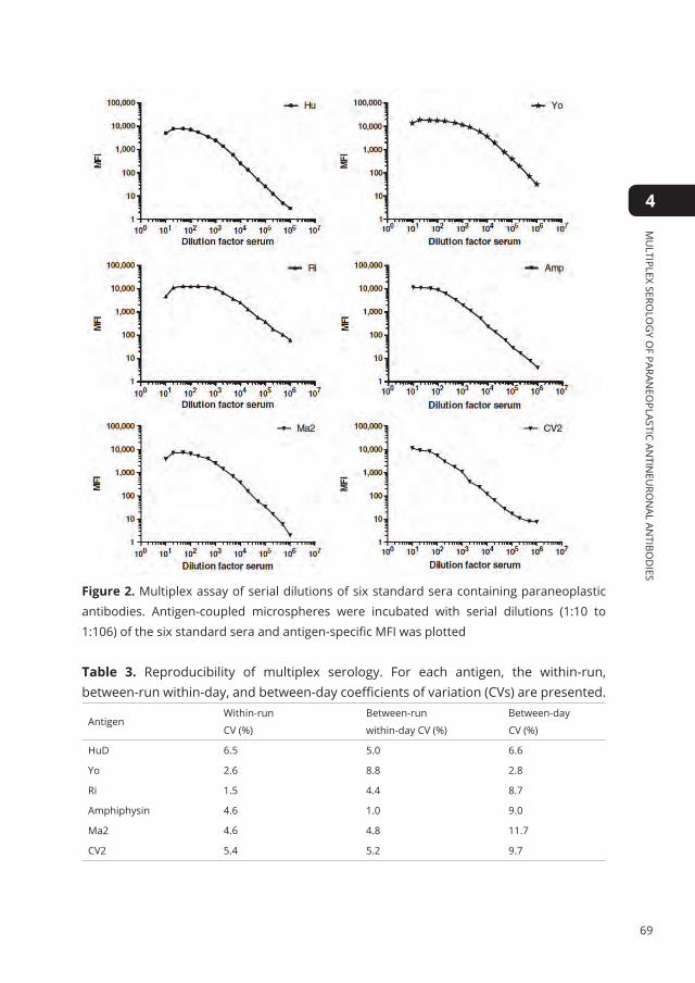
69
Mu
ltiplex serolo
gy o
f paraneo
plastic antin
euro
nal an
tibod
ies
4
figure 2. Multiplex assay of serial dilutions of six standard sera containing paraneoplastic antibodies. Antigen-coupled microspheres were incubated with serial dilutions (1:10 to 1:106) of the six standard sera and antigen-specific MFI was plotted
Table 3. Reproducibility of multiplex serology. For each antigen, the within-run, between-run within-day, and between-day coefficients of variation (CVs) are presented.
AntigenWithin-run
CV (%)
Between-run
within-day CV (%)
Between-day
CV (%)
HuD 6.5 5.0 6.6
Yo 2.6 8.8 2.8
Ri 1.5 4.4 8.7
Amphiphysin 4.6 1.0 9.0
Ma2 4.6 4.8 11.7
CV2 5.4 5.2 9.7

70
Table 4. Sensitivity and specificity of Luminex multiplex serology for onconeural antibodies.
Antibody Sensitivity Specificity
HuD-Ab 93% 97%
Yo-Ab 100% 97%
Amphiphysin-Ab 100% 97%
CV2-Ab 100% 96%
Ri-Ab 83% 100%
Ma2-Ab 91% 99%
Stability of the coupled microspheres To test the stability of antigen coupled microspheres over time, we coupled all six antigens to 12.5 x 10⁶ microspheres each. The antigen-coupled microspheres were stored separately at 4 °C. Multiplex serology was performed on days 1, 21, 24, 25, 28, 63 and 255 after coupling (Figure S4). Although a decreasing trend was observed, the antigen-specific MFI did not change significantly over time for any of the six standard sera. Figure S4 shows that the antigen-coupled microspheres continued to perform adequately after storage in excess of two months.
Thirty-two false positive onconeural antibody results were recorded in 29 samples in the multiplex assay (Table S1). Of these 32 false positive results, 16 were also positive on Western blot but did not show the characteristic staining pattern on immunohistochemistry. Twenty false positives were detected in 19 samples from PNS patients with well-characterized onconeural antibodies (Table S3). In one sample, from a patient with anti-HuD associated PNS, additional antibodies against Yo and CV2 were falsely detected. False positive reactions against the onconeural antigens occurred at different frequencies: anti-HuD in 2; anti-Yo in 3, anti-amphiphysin in 4, anti-Ma2 in 2; and anti-CV2 in 9. Interestingly, of the 9 PNS patients with falsely positive anti-CV2 results, 6 had small cell tumors known to be associated with anti-CV2. The other 3 had no identified tumor but harbored anti-HuD (2x) or anti-HuD and anti-amphiphysin (1x) antibodies, strongly suggesting an underlying malignancy.Two of 40 autoimmune control and 8 of 100 blood bank control samples tested falsely positive for an onconeural antibody in the multiplex assay (Table S3). While CV2 gave 9/20 false positive results in the 119 PNS sera, only 1/12 of the false positive results in the 140 blood bank and autoimmune controls were anti-CV2.
dIsCussIOnThe serologic method presented here combines a well-validated expression and purification system for recombinant proteins with a high-throughput multiplex suspension array. All six purified onconeural antigens were efficiently bound to Luminex
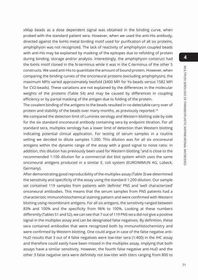
71
Mu
ltiplex serolo
gy o
f paraneo
plastic antin
euro
nal an
tibod
ies
4
xMap beads as a dose dependent signal was obtained in the binding curve, when probed with the standard patient sera. However, when we used the anti-His antibody, directed against the 6xHis metal binding motif used for purification of all six proteins, amphiphysin was not recognized. The lack of reactivity of amphiphysin coupled beads with anti-His may be explained by masking of the epitopes due to refolding of protein during binding, storage and/or analysis. Interestingly, the amphiphysin construct had the 6xHis motif cloned in the N-terminus while it was in the C-terminus of the other 5 constructs. We used anti-His to quantitate the amount of bound protein. However, when comparing the binding curves of the onconeural proteins (excluding amphiphysin), the maximum MFIs varied approximately twofold (3400 MFI for Yo-beads versus 1582 MFI for CV2-beads). These variations are not explained by the differences in the molecular weights of the proteins (Table S4) and may be caused by differences in coupling efficiency or by partial masking of the antigen due to folding of the protein.The covalent binding of the antigens to the beads resulted in no detectable carry over of protein and stability of the beads over many months, as previously reported.24 We compared the detection limit of Luminex serology and Western blotting side by side for the six standard onconeural antibody containing sera by endpoint titration. For all standard sera, multiplex serology has a lower limit of detection than Western blotting indicating potential clinical application. For testing of serum samples in a routine setting we decided to dilute samples 1:200. This dilution was for all six onconeural antigens within the dynamic range of the assay with a good signal to noise ratio. In addition, this dilution has previously been used for Western blotting 7and is close to the recommended 1:100 dilution for a commercial dot blot system which uses the same onconeural antigens produced in a similar E. coli system (EUROIMMUN AG, Lübeck, Germany).After demonstrating good reproducibility of the multiplex assay (Table 3) we determined the sensitivity and specificity of the assay using the standard 1:200 dilution. Our sample set contained 119 samples from patients with ‘definite’ PNS and ‘well characterized’ onconeural antibodies. This means that the serum samples from PNS patients had a characteristic immunohistochemical staining pattern and were confirmed with Western blotting using recombinant antigens. For all six antigens, the sensitivity ranged between 83% and 100% and the specificity from 96% to 100%. Looking at these numbers differently (Tables S1 and S2), we can see that 7 out of 119 PNS sera did not give a positive signal in the multiplex assay and can be designated false negatives. By definition, these sera contained antibodies that were recognized both by immunohistochemistry and were confirmed by Western blotting. One could argue in case of the false negative anti-HuD results that 3 out of 4 false negatives were low-titer sera (1:400) in the IHC assay and therefore could easily have been missed in the multiplex assay, implying that both assays have a similar sensitivity. However, the fourth false negative anti-HuD and the other 3 false negative sera were definitely not low-titer with titers ranging from 800 to
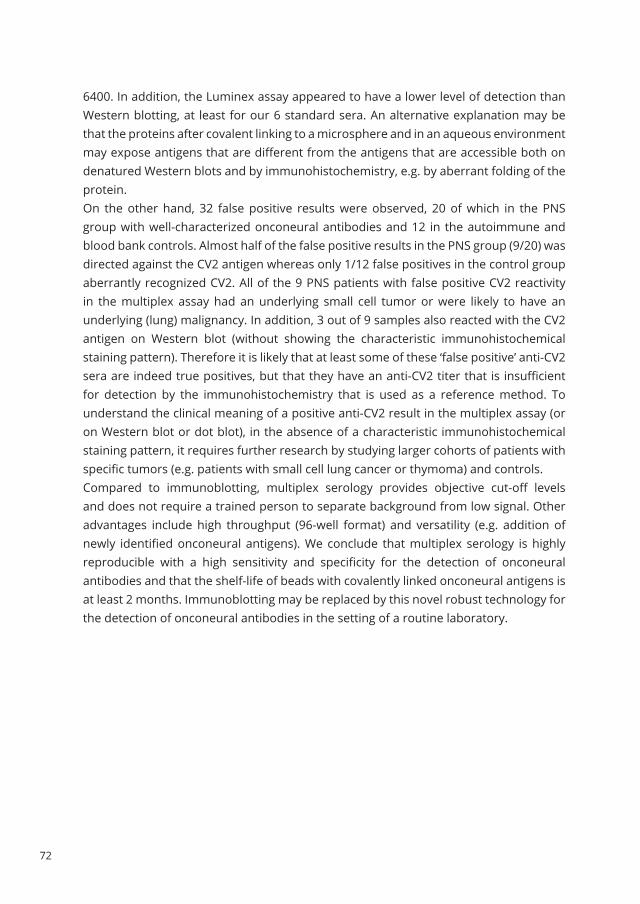
72
6400. In addition, the Luminex assay appeared to have a lower level of detection than Western blotting, at least for our 6 standard sera. An alternative explanation may be that the proteins after covalent linking to a microsphere and in an aqueous environment may expose antigens that are different from the antigens that are accessible both on denatured Western blots and by immunohistochemistry, e.g. by aberrant folding of the protein.On the other hand, 32 false positive results were observed, 20 of which in the PNS group with well-characterized onconeural antibodies and 12 in the autoimmune and blood bank controls. Almost half of the false positive results in the PNS group (9/20) was directed against the CV2 antigen whereas only 1/12 false positives in the control group aberrantly recognized CV2. All of the 9 PNS patients with false positive CV2 reactivity in the multiplex assay had an underlying small cell tumor or were likely to have an underlying (lung) malignancy. In addition, 3 out of 9 samples also reacted with the CV2 antigen on Western blot (without showing the characteristic immunohistochemical staining pattern). Therefore it is likely that at least some of these ‘false positive’ anti-CV2 sera are indeed true positives, but that they have an anti-CV2 titer that is insufficient for detection by the immunohistochemistry that is used as a reference method. To understand the clinical meaning of a positive anti-CV2 result in the multiplex assay (or on Western blot or dot blot), in the absence of a characteristic immunohistochemical staining pattern, it requires further research by studying larger cohorts of patients with specific tumors (e.g. patients with small cell lung cancer or thymoma) and controls.Compared to immunoblotting, multiplex serology provides objective cut-off levels and does not require a trained person to separate background from low signal. Other advantages include high throughput (96-well format) and versatility (e.g. addition of newly identified onconeural antigens). We conclude that multiplex serology is highly reproducible with a high sensitivity and specificity for the detection of onconeural antibodies and that the shelf-life of beads with covalently linked onconeural antigens is at least 2 months. Immunoblotting may be replaced by this novel robust technology for the detection of onconeural antibodies in the setting of a routine laboratory.

73
Mu
ltiplex serolo
gy o
f paraneo
plastic antin
euro
nal an
tibod
ies
4
suPPleMenTARy dATA
figure s1. Binding curves of 6 recombinant purified onconeural proteins on Luminex xMap microspheres. Luminex xMap microspheres were coupled with serial dilutions (range 0.7�333 pmol/106 microspheres) of the 6 recombinant purified onconeural antigens. Bound antigen was quantified using anti-His (1:500) monoclonal antibody. In the amphiphysin construct, the 6 × His region is encoded in the N-terminus while it is encoded in the C-terminus of the other five constructs. This N-terminal position of 6 × His in amphiphysin may explain the lack of amphiphysin-specific signal with anti-His.
figure s2. Antigen carry over. HuD and mock coupled microspheres were prepared and stored mixed and separately. After one week multiplex serology was performed on microspheres that had been stored mixed for one week or were freshly mixed just prior to multiplex serology. In addition, a monoplex assay was performed on non-mixed HuD coupled microsphere. Serology was performed using an HuD-Ab containing standard serum. One week of mixed storage of HuD-coupled and mock-coupled microspheres did not result in significant decrease of HuD-specific MFI of the HuD-coupled microspheres (ANOVA, p = 0.94) nor was an increase in HuD-specific MFI noted in the mock-coupled microspheres (ANOVA, p = 0.28)
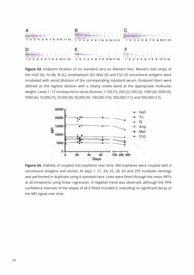
74
figure s3. Endpoint titration of six standard sera on Western blot. Western blot strips of the HuD (A), Yo (B), Ri (C), amphiphysin (D), Ma2 (E) and CV2 (F) onconeural antigens were incubated with serial dilutions of the corresponding standard serum. Endpoint titers were defined as the highest dilution with a clearly visible band at the appropriate molecular weight. Lanes 1–12 correspond to serial dilutions 1:100 (1); 200 (2); 500 (3); 1000 (4); 2000 (5); 5000 (6); 10,000 (7); 20,000 (8); 50,000 (9); 100,000 (10); 200,000 (11); and 500,000 (12).
Figure S4. Stability of coupled microspheres over time. Microspheres were coupled with 6 onconeural antigens and stored. At days 1, 21, 24, 25, 28, 63 and 255 multiplex serology was performed in duplicate using 6 standard sera. Lines were fitted through the mean MFI’s at all timepoints using linear regression. A negative trend was observed, although the 95% confidence intervals of the slopes of all 6 fitted included 0, indicating no significant decay of the MFI signal over time.

75
Mu
ltiplex serolo
gy o
f paraneo
plastic antin
euro
nal an
tibod
ies
4
Table s1. Sensitivity and specificity of Luminex-based multiplex serology for onconeural Ab determination, compared to gold standard. False negative multiplex results are colored yellow and false positive results orange.
HU PNS+ PNS- SENS SPEC
LMNX+ 50 6 93% 97%
LMNX- 4 199
YO PNS+ PNS-
LMNX+ 23 6 100% 97%
LMNX- 0 230
RI PNS+ PNS-
LMNX+ 10 0 83% 100%
LMNX- 2 247
AMP PNS+ PNS-
LMNX+ 25 7 100% 97%
LMNX- 0 227
MA2 PNS+ PNS-
LMNX+ 10 3 91% 99%
LMNX- 1 245
CV2 PNS+ PNS-
LMNX+ 15 10 100% 96%
LMNX- 0 234
Table s2. False negative Luminex-based serology resultsPNS
Total number of samples 119
HuD 4
Yo -
Amphiphysin -
CV2 -
Ri 2
Ma2 1
Number of false negative samples (%) 7 (6%)

76
Table s3. False positive Luminex-based serology resultsPNS Autoimmune Bloodbank controls
Total number of samples 119 40 100
HuD 2 1 3
Yo 3 1 2
Amphiphysin 4 3
CV2 9 1
Ri
Ma2 2 1
Number of false positive samples (%) 19 (16%)* 2 (5%) 8 (8%)**
* One anti-HuD sample contained two false positive reactivities (against Yo and CV2)** Two bloodbank control samples contained two false positive reactivies (one sample CV2and HuD; the other Ma2 and Yo)
Table s4. Calculated molecular weight of the six onconeural recombinant proteinsOnconeural antigen Molecular weight (kD)
HuD 43
Yo 50
Ri 56
Amp 75
CV2 62
Ma2 40

77
Mu
ltiplex serolo
gy o
f paraneo
plastic antin
euro
nal an
tibod
ies
4
RefeRenCes
1. Posner JB. Paraneoplastic syndromes.
Neurologic complications of cancer.
Philadelphia: FA Davis; 1995:353-85.
2. Darnell R, Posner J. Paraneoplastic syndromes
involving the nervous system. N Engl J Med
2003;349:1543-54.
3. de Beukelaar J, Sillevis Smitt P. Managing
paraneoplastic neurological disorders.
Oncologist 2006;11:292-305.
4. Iorio R, Lennon VA. Neural antigen-specific
autoimmune disorders. Immunol Rev
2012;248:104-21.
5. Graus F, Delattre JY, Antoine JC, et al.
Recommended diagnostic criteria for
paraneoplastic neurological syndromes. J
Neurol Neurosurg Psychiatry 2004;75:1135-40.
6. Graus F, Dalmou J, Rene R, et al. Anti-Hu
antibodies in patients with small-cell lung
cancer: association with complete response
to therapy and improved survival. J Clin Oncol
1997;15:2866-72.
7. Manley GT, Smitt PS, Dalmau J, Posner JB. Hu
antigens: reactivity with Hu antibodies, tumor
expression, and major immunogenic sites. Ann
Neurol 1995;38:102-10.
8. Willison HJ, Ang W, Gilhus NE, et al. EFNS task
force report: a questionnaire-based survey on
the service provision and quality assurance
for determination of diagnostic autoantibody
tests in European neuroimmunology centres.
European Federation of Neurological Societies.
European journal of neurology : the official
journal of the European Federation of
Neurological Societies 2000;7:625-8.
9. Jarius S, Stich O, Rasiah C, Voltz R, Rauer S.
Qualitative evidence of Ri specific IgG-synthesis
in the cerebrospinal fluid from patients with
paraneoplastic neurological syndromes. J
Neurol Sci 2008;268:65-8.
10. Vigliani MC, Honnorat J, Antoine JC, et al.
Chorea and related movement disorders of
paraneoplastic origin: the PNS EuroNetwork
experience. J Neurol 2011;258:2058-68.
11. Monstad SE, Knudsen A, Salvesen HB, Aarseth
JH, Vedeler CA. Onconeural antibodies in sera
from patients with various types of tumours.
Cancer immunology, immunotherapy : CII
2009;58:1795-800.
12. Storstein A, Monstad SE, Haugen M, et al.
Onconeural antibodies: improved detection
and clinical correlations. J Neuroimmunol
2011;232:166-70.
13. Shams’ili S, Grefkens J, de Leeuw B, et al.
Paraneoplastic cerebellar degeneration
associated with antineuronal antibodies:
analysis of 50 patients. Brain 2003;126:1409-18.
14. Graus F, Keime-Guibert F, Rene R, et al. Anti-Hu-
associated paraneoplastic encephalomyelitis:
analysis of 200 patients. Brain 2001;124:1138-
48.
15. Sillevis Smitt P, Grefkens J, de Leeuw B, et al.
Survival and outcome in 73 anti-Hu positive
patients with paraneoplastic encephalomyelitis/
sensory neuronopathy. J Neurol 2002;249:745-
53.
16. Pittock S, Kryzer T, Lennon V. Paraneoplastic
antibodies coexist and predict cancer,
not neurological syndrome. Ann Neurol
2004;56:715-9.
17. Waterboer T, Sehr P, Michael KM, et al. Multiplex
human papillomavirus serology based on in
situ-purified glutathione s-transferase fusion
proteins. Clin Chem 2005;51:1845-53.
18. Moll JW, Vecht CJ. Immune diagnosis of
paraneoplastic neurological disease. Clin Neurol
Neurosurg 1995;97:71-81.

78
19. Fathallah-Shaykh H, Wolf S, Wong E, Posner
JB, Furneaux HM. Cloning of a leucine-zipper
protein recognized by the sera of patients
with antibody-associated paraneoplastic
cerebellar degeneration. Proc Natl Acad Sci U S
A 1991;88:3451-4.
20. Yu Z, Kryzer T, Griesmann G, Kim K, Benarroch
E, Lennon V. CRMP-5 neuronal autoantibody:
marker of lung cancer and thymoma-related
autoimmunity. Ann Neurol 2001;49:146-54.
21. Buckanovich R, Posner J, Darnell R. Nova, the
paraneoplastic Ri antigen, is homologous to
an RNA-binding protein and is specifically
expressed in the developing motor system.
Neuron 1993;11:657-72.
22. Voltz R, Gultekin SH, Rosenfeld MR, et al. A
serologic marker of paraneoplastic limbic
and brain-stem encephalitis in patients with
testicular cancer. N Engl J Med 1999;340:1788-
95.
23. DeCamilli P, Thomas A, Cofiell R, et al. The
synaptic vesicle-associated protein amphiphysin
is the 128-kD autoantigen of stiff-man syndrome
with breast cancer. J Exp Med 1993;178:2219-23.
24. Verkaik N, Brouwer E, Hooijkaas H, van
Belkum A, van Wamel W. Comparison of
carboxylated and Penta-His microspheres for
semi-quantitative measurement of antibody
responses to His-tagged proteins. J Immunol
Methods 2008;335:121-5.

79
Mu
ltiplex serolo
gy o
f paraneo
plastic antin
euro
nal an
tibod
ies
4


Identification of delta/notch-like epidermal growth factor-related receptor as the Tr antigen in
paraneoplastic cerebellar degeneration
Esther de Graaff, Peter Maat, Esther Hulsenboom, Robert van den Berg, Martin J. van den Bent,
Jeroen Demmers, Pieternella J. Lugtenburg, Casper C. Hoogenraad, Peter A.E. Sillevis Smitt
Annals of Neurology, March 2012; 71: 815-824
5Back to table of contents

82
AbsTRACTObjectiveAnti-Tr is among the better described autoantibodies in paraneoplastic cerebellar degeneration (PCD) combined with Hodgkin lymphoma (HL); however, the Tr antigen remains unidentified.
MethodsWe used immunoprecipitation of total rat brain extract followed by mass spectrometry to identify the antigen recognized by anti-Tr–positive sera. By Western blotting and cell-based assays, we tested a total of 12 anti-Tr–positive and 246 control sera and determined the region of the epitope recognized by the anti-Tr antibodies. Deletion and mutant constructs were generated to further map the antigenic region.
ResultsMass spectrometry analysis of immunopurified rat brain extract using 4 different anti-Tr–positive sera led to the identification of Delta/Notch-like epidermal growth factor-related receptor (DNER) as the Tr antigen. All but 1 of 246 control samples were negative in the HeLa cell-based screening assay, whereas 12 of the 12 anti-Tr–positive sera stained hemagglutinin-tagged DNER-expressing cells. Only 1 control subject with HL but no ataxia was found to be both DNER and Tr positive. Using deletion constructs, we pinpointed the main epitope to the extracellular domain. Knockdown of endogenous DNER in hippocampal and N-glycosylation mutations abolished the anti-Tr staining, indicating that glycosylation of DNER is required for it to be recognized by anti-Tr antibodies.
InterpretationDNER is the antigen detected by anti-Tr–positive sera. Presence of anti-Tr antibodies in patients with PCD and HL or HL only can now be screened quickly and reliably by using a cell-based screening assay.

83
Iden
tIfIcatIon
of d
neR as th
e tR antIg
en
5
InTROduCTIOnParaneoplastic cerebellar degeneration (PCD) is a heterogeneous group of disorders that are characterized by subacute cerebellar ataxia coinciding with the presence of specific tumor types and antineuronal antibodies.1-3 The association between 1 specific tumor type, Hodgkin lymphoma (HL), with PCD has long been known4 and was followed by the identification of antibodies against cerebellar Purkinje cells in several of these patients’ sera.5 Later called anti-Tr antibodies, these recognize a specific staining in the cerebellum, with punctuate immunoreactivity detected in the large dendritic tree as well as the soma of the Purkinje cells, but not in the axons.5-7 This characteristic immunoreactive pattern is considered to be a good hallmark for the presence of anti-Tr antibodies, but requires further confirmation by epitope blocking. For this, rat cerebellar slices show absent immunoreactivity with the biotinylated immunoglobulin G (IgG) from a positive anti-Tr serum when preincubated with undiluted serum that required the testing.8 To aid this diagnosis, and to understand the pathogenic nature of PCD, we aimed to identify the antigen recognized by anti-Tr antibodies.
subJeCTs And MeThOdsPatientsThe most relevant clinical features of 11 of the 12 anti-Tr–positive patients are given in Table 1. Clinical information was lacking from Tr-positive Patient 5. All patients suffered subacute and severe truncal and limb ataxia with nystagmus and/or cerebellar dysarthria. Ten patients were diagnosed with HL, and in all of them the cerebellar symptoms preceded the detection of HL. The patients’ disability was assessed using a modified Rankin scale9 (Supplementary Materials and Methods).
Antibodies and Expression ConstructsFor immunocytochemistry, antibodies were used in the following dilutions: mouse anti–β-galactosidase (Promega, Leiden, the Netherlands) 1:2,000, goat anti–Delta/Notch-like epidermal growth factor (EGF)-related receptor (DNER; R&D Systems, Abingdon, UK) 1:400 to 1:2,000, rabbit anti-hemagglutinin (HA; Santa Cruz Biotechnology, Heidelberg, Germany) 1:400, patients’ sera 1:50 to 1:20,000. Cy3-conjugated donkey antihuman antibody (Jackson ImmunoResearch, Newmarket, UK), Alexa 488- and Alexa 568-conjugated secondary antibodies (Molecular Probes, Breda, the Netherlands), andfluorescein isothiocyanate-conjugated antigoat antibody (Jackson ImmunoResearch)were used in a 1:400 dilution. For Western blotting, horseradish peroxidase (HRP)-conjugated donkey antihuman (Calbiochem, Darmstadt, Germany) 1:10,000 and HRP-conjugated swine antirabbit, antimouse, or antigoat (Dako, Heverlee, Belgium) 1:2,500antibodies were used.
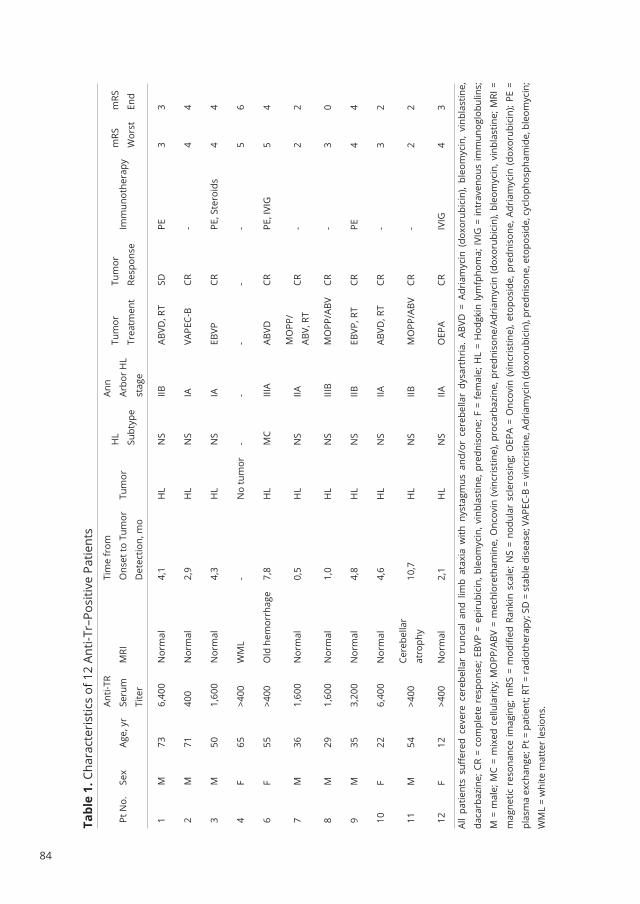
84
Tabl
e 1.
Cha
ract
eris
tics
of 1
2 An
ti-Tr
–Pos
itive
Pat
ient
s
Pt N
o.Se
xAg
e, y
r
Anti-
TR
Seru
m
Tite
r
MRI
Tim
e fr
om
Ons
et to
Tum
or
Det
ectio
n, m
o
Tum
orH
L
Subt
ype
Ann
Arbo
r H
L
stag
e
Tum
or
Trea
tmen
t
Tum
or
Resp
onse
Imm
unot
hera
pym
RS
Wor
st
mRS
End
1M
736,
400
Nor
mal
4,1
HL
NS
IIBAB
VD, R
TSD
PE3
3
2M
7140
0N
orm
al2,
9H
LN
SIA
VAPE
C-B
CR-
44
3M
501,
600
Nor
mal
4,3
HL
NS
IAEB
VPCR
PE, S
tero
ids
44
4F
65>4
00W
ML
-N
o tu
mor
--
--
-5
6
6F
55>4
00O
ld h
emor
rhag
e7,
8H
LM
CIII
AAB
VDCR
PE, I
VIG
54
7M
361,
600
Nor
mal
0,5
HL
NS
IIAM
OPP
/
ABV,
RT
CR-
22
8M
291,
600
Nor
mal
1,0
HL
NS
IIIB
MO
PP/A
BVCR
-3
0
9M
353,
200
Nor
mal
4,8
HL
NS
IIBEB
VP, R
TCR
PE4
4
10F
226,
400
Nor
mal
4,6
HL
NS
IIAAB
VD, R
TCR
-3
2
11M
54>4
00Ce
rebe
llar
atro
phy
10,7
HL
NS
IIBM
OPP
/ABV
CR-
22
12F
12>4
00N
orm
al2,
1H
LN
SIIA
OEP
ACR
IVIG
43
All
patie
nts
suff
ered
cev
ere
cere
bella
r tr
unca
l an
d lim
b at
axia
with
nys
tagm
us a
nd/o
r ce
rebe
llar
dysa
rthr
ia.
ABVD
= A
dria
myc
in (
doxo
rubi
cin)
, bl
eom
ycin
, vi
nbla
stin
e,
daca
rbaz
ine;
CR
= co
mpl
ete
resp
onse
; EBV
P =
epir
ubic
in, b
leom
ycin
, vin
blas
tine,
pre
dnis
one;
F =
fem
ale;
HL
= H
odgk
in ly
mfp
hom
a; IV
IG =
intr
aven
ous
imm
unog
lobu
lins;
M =
mal
e; M
C =
mix
ed c
ellu
lari
ty; M
OPP
/ABV
= m
echl
oret
ham
ine,
Onc
ovin
(vin
cris
tine)
, pro
carb
azin
e, p
redn
ison
e/Ad
riam
ycin
(dox
orub
icin
), bl
eom
ycin
, vin
blas
tine;
MRI
=
mag
netic
res
onan
ce im
agin
g; m
RS =
mod
ified
Ran
kin
scal
e; N
S =
nodu
lar
scle
rosi
ng; O
EPA
= O
ncov
in (v
incr
istin
e), e
topo
side
, pre
dnis
one,
Adr
iam
ycin
(dox
orub
icin
); PE
=
plas
ma
exch
ange
; Pt =
pat
ient
; RT
= ra
diot
hera
py; S
D =
sta
ble
dise
ase;
VAP
EC-B
= v
incr
istin
e, A
dria
myc
in (d
oxor
ubic
in),
pred
niso
ne, e
topo
side
, cyc
loph
osph
amid
e, b
leom
ycin
;
WM
L =
whi
te m
atte
r le
sion
s.

85
Iden
tIfIcatIon
of d
neR as th
e tR antIg
en
5
Full-length wild-type HA-tagged mouse DNER cDNA in pDisplay was kindly provided by M. Kengaku (Kyoto, Japan) and was previously described.10 A DNER construct allowing biotinylation (DNER-GFP-bio), and the deletion and glycosylation mutants were generated by polymerase chain reaction and cloning in pGW1.11 Full details of these constructs and all other materials and methods are given in the Supplementary Materials and Methods.
ResulTsIdentification of DNER as Tr Antigen by Mass SpectrometryTo identify the antigen recognized by Tr antibodies, we performed immunoprecipitation experiments followed by mass spectrometry on crude rat brain extracts using 4 different anti-Tr–positive sera and 3 control sera containing antibodies against the Hu antigen, metabotropic glutamate receptor 1 (mGluR1), or glutamate receptor 1 (GluR1). Size fractionation of the purified complexes on sodium dodecyl sulfate–polyacrylamide gel electrophoresis and subsequent staining with Coomassie blue showed no apparent protein band shared by all 4 anti-Tr–positive sera (Figure1A). We nevertheless cut each lane into 15 slices, digested them with trypsin, and performed mass spectrometry. The mass spectrometry data of each anti-Tr serum was first analyzed and compared to the results obtained with the Hu, mGluR1, and GluR1 control sera, leaving DNER as the unique protein identified by the anti-Tr sera only, making DNER a good candidate as Tr antigen.
DNER Is the Tr Antigen in Cell-Based AssayWe next performed a cell-based assay to find further evidence for DNER as the Tr antigen. HeLa cells expressing full-length HA-tagged DNER were incubated with anti-HA antibodies and either of 2 anti-Tr sera, an anti-Tr cerebrospinal fluid (CSF), and a control serum. The 2 anti-Tr sera and CSF strongly labeled cells expressing HA-DNER, but not untransfected cells (stained with DAPI only; see Figure1B, top 3 panels). In contrast, the staining was absent in control sera known to detect Hu, mGLUR1, or GluR1 (see Figure1B, lower panel and data not shown). We next tested all 12 anti-Tr sera as well as 246 control samples (Table 2), using the same cell-based screening assay. All anti-Tr sera positively stained HeLa cells expressing HA-DNER, whereas all but 1 control samples were negative (see Figure1B and data not shown). Interestingly, the 1 control sample that contained anti-DNER antibodies was from a patient suffering from HL without any signs of ataxia. Testing this serum on rat brain sections resulted in the characteristic punctate staining typical for Tr (data not shown). As DNER is normally expressed in pyramidal hippocampal neurons, we performed costaining in primary hippocampal neuron cultures. Under permeabilizing conditions, we detected punctuate somatodendritic anti-DNER staining of endogenous DNER, which fully colocalizes with

86
figure 1. Identification of Delta/Notch-like epidermal growth factor-related receptor (DNER) as the Tr antigen. (A) Coomassie-stained gel of total rat brain extracts immunoprecipitated with anti-Tr–positive sera and used for mass spectrometry analysis. (B) HeLa cells expressing hemagglutinin (HA)-DNER (green) are positively stained by 2 anti-Tr sera (Tr9 and Tr6, 1:200, red) and cerebrospinal fluid (csf) of the same patient (Tr6, 1:100, red) but not by the control sera (ctrl, 1:200, red). The merged images reveal nontransfected cells by DAPI staining. Scale bar = 10μm. (C) Western blot analysis of total HEK293T cell extracts expressing HA-DNER (input) and purified HA-DNER. Untransfected (untr) HEK293T cells (mock) are used as control. HA-DNER was purified by immunoprecipitation using anti-HA antibodies. Asterisks denote background bands also present in untransfected HEK293T cells. (D) Representative images of hippocampal neurons (days in vitro, 11) stained with anti-DNER (green) and anti-Tr serum Tr9 (red). Zoom indicates punctuate DNER staining in neurites that strongly colocalize with anti-Tr serum. Scale bar = 20μm. (E) Representative image of hippocampal neuron (days in vitro, 7 + 1) transfected with HA-DNER and surface stained with both rabbit anti-HA (green) and anti-Tr serum Tr9 (red). Zoom indicates strong, but not perfect surface colocalization, due to the presence of endogenous DNER at the surface (arrowhead). Scale bar = 20μm.Tr-positive sera. These results further confirm that DNER is the Tr antigen.
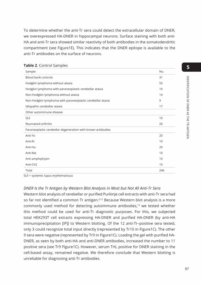
87
Iden
tIfIcatIon
of d
neR as th
e tR antIg
en
5
To determine whether the anti-Tr sera could detect the extracellular domain of DNER, we overexpressed HA-DNER in hippocampal neurons. Surface staining with both anti-HA and anti-Tr sera showed similar reactivity of both antibodies in the somatodendritic compartment (see Figure1E). This indicates that the DNER epitope is available to the anti-Tr antibodies on the surface of neurons.
Table 2. Control SamplesSample No.
Blood bank controls 31
Hodgkin lymphoma without ataxia 55
Hodgkin lymphoma with paraneoplastic cerebellar ataxia 10
Non-Hodgkin lymphoma without ataxia 14
Non-Hodgkin lymphoma with paraneoplastic cerebellar ataxia 9
Idiopathic cerebellar ataxia 17
Other autoimmune disease
SLE 10
Reumatoid arthritis 20
Paraneoplastic cerebellar degeneration with known antibodies
Anti-Yo 20
Anti-Ri 10
Anti-Hu 20
Anti-Ma 10
Anti-amphiphysin 10
Anti-CV2 10
Total 246
SLE = systemic lupus erythematosus
DNER Is the Tr Antigen by Western Blot Analysis in Most but Not All Anti-Tr SeraWestern blot analysis of cerebellar or purified Purkinje cell extracts with anti-Tr sera had so far not identified a common Tr antigen.6,12 Because Western blot analysis is a more commonly used method for detecting autoimmune antibodies,13 we tested whether this method could be used for anti-Tr diagnostic purposes. For this, we subjected total HEK293T cell extracts expressing HA-DNER and purified HA-DNER (by anti-HA immunoprecipitation [IP]) to Western blotting. Of the 12 anti-Tr–positive sera tested, only 3 could recognize total input directly (represented by Tr10 in Figure1C). The other 9 sera were negative (represented by Tr9 in Figure1C). Loading the gel with purified HA-DNER, as seen by both anti-HA and anti-DNER antibodies, increased the number to 11 positive sera (see Tr9 Figure1C). However, serum Tr6, positive for DNER staining in the cell-based assay, remained negative. We therefore conclude that Western blotting is unreliable for diagnosing anti-Tr antibodies.

88
DNER Blocks Anti-Tr Antibody Reactivity on Brain SectionsPresence of anti-Tr antibodies is usually diagnosed by strict immunohistochemical criteria of rat frozen cerebellar sections: cytoplasmic labeling of Purkinje cells, combined with a characteristic punctuated somatodendritic staining in the molecular layer (Figure2A and 2A′).7 Staining rat brain slices with the commercial antibody against DNER resulted in the identical somatodendritic, punctuate staining of Purkinje cells (Figure2B and 2B′). Both anti-Tr sera and anti-DNER antibodies stained isolated pyramidal neurons in the hippocampus (data not shown), as described before.6,10,13 The characteristic punctuate staining of anti-Tr sera is only 1 prerequisite of the diagnosis; the diagnosis is usually confirmed by performing antigen-blocking experiments.2,8 For this, cerebellar sections are first preincubated with undiluted anti-Tr sera that require testing and subsequently incubated with diluted biotinylated standard anti-Tr serum. In all 7 cases analyzed this way the tested anti-Tr sera abolished the characteristic staining pattern of the standard anti-Tr serum (Figure2C–E).In addition, we performed cerebellar immunohistochemistry with anti-Tr–positive sera from which the anti-DNER antibodies were depleted. Sera incubated with DNER-GFP-bio coupled to streptavidin beads prior to the immunohistochemistry lost their Tr-characteristic staining of Purkinje cells (compare Figure2G to F). In contrast, eluted anti-DNER antibodies showed staining of the Purkinje cells similar to the full serum (compare Figure2H to F).
Knockdown of DNER Removes Tr Staining in Hippocampal NeuronsTo further demonstrate that DNER is the only protein detected by anti-Tr–positive sera, we performed DNER knockdown experiments. Two independent short hairpin RNA (shRNA)-producing constructs directed against DNER were generated and transfected into hippocampal neurons. As endogenous DNER is expressed in pyramidal hippocampal neurons, we counted all transfected neurons as judged by the presence of β-galactosidase and checked for reactivity with anti-DNER and anti-Tr. In control transfections, 73% of the β-galactosidase–expressing neurons (118 of 162) were positive for both DNER and Tr (Figure3A, top panel). In contrast, transfection with 2 independent shRNA constructs targeting DNER removed both DNER staining, as observed by the commercial anti-DNER antibodies, and Tr staining (Tr9; see Figure3A, lower panel) in all transfected neurons (133 and 134 neurons, respectively, for construct 1 and 2; see Figure3B), further confirming the anti-Tr specificity of DNER.

89
Iden
tIfIcatIon
of d
neR as th
e tR antIg
en
5
figure 2. Immunohistochemistry of anti–Delta/Notch-like epidermal growth factor-related receptor (DNER) and anti-Tr sera on rat cerebella. (A, B) Sagittal section of rat brain stained with serum of a patient known to contain (A) the anti-Tr antibodies (B) or commercial anti-DNER. (A′, B′) Higher magnification shows that anti-DNER antibodies lead to the identical dotlike somatodendritic staining as described for anti-Tr sera. (C–E) Standard blocking procedure currently used to test for presence of anti-Tr antibodies in sera. (C) Punctate staining after incubation with standard biotinylated serum. (D, E) Staining disappears when previously incubated with (D) unbiotinylated serum from the same patient or (E) serum from another patient. (F–H) Depletion of anti-DNER antibodies from anti-Tr–positive sera (F) obliterates the anti-Tr staining pattern (G). The eluted fraction, containing purified anti-DNER antibodies, retains the normal punctuate staining (H). Note that a lower dilution was used in F–H (1/20,000) than in A,C–E (1/400). Scale bar = 200μm in A, B and 25μm in A′, B′, C–H.

90
figure 3. Knockdown of Delta/Notch-like epidermal growth factor-related receptor (DNER) abolishes Tr reactivity in hippocampal neurons. (A) In neurons transfected with a control (CTRL) construct, normal DNER (green) and Tr (red) reactivity is observed (upper panel). In all neurons transfected with constructs expressing short hairpin RNA (shRNA) against DNER and β-galactosidase (blue), both DNER expression and Tr reactivity are lacking (lower panel). Scale bar = 20μm. (B) Summary of DNER knockdown experiments. Quantification of the number of Tr- and DNER-positive neurons expressing control shRNA (n = 162), DNER-shRNA#1 (n = 133), or DNER-shRNA#2 (n = 134).
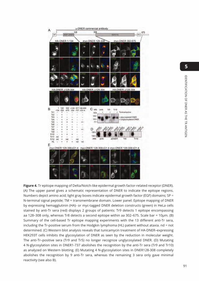
91
Iden
tIfIcatIon
of d
neR as th
e tR antIg
en
5
Figure 4. Tr epitope mapping of Delta/Notch-like epidermal growth factor-related receptor (DNER). (A) The upper panel gives a schematic representation of DNER to indicate the epitope regions.Numbers depict amino acid; light gray boxes indicate epidermal growth factor (EGF) domains; SP = N-terminal signal peptide; TM = transmembrane domain. Lower panel: Epitope mapping of DNERby expressing hemagglutinin (HA)- or myc-tagged DNER deletion constructs (green) in HeLa cellsstained by anti-Tr sera (red) displays 2 groups of patients: Tr9 detects 1 epitope encompassingaa 128–308 only, whereas Tr8 detects a second epitope within aa 302–675. Scale bar = 10μm. (B)Summary of the cell-based Tr epitope mapping experiments with the 13 different anti-Tr sera,including the Tr-positive serum from the Hodgkin lymphoma (HL) patient without ataxia. nd = notdetermined. (C) Western blot analysis reveals that tunicamycin treatment of HA-DNER–expressingHEK293T cells inhibits the glycosylation of DNER as seen by the reduction in molecular weight.The anti-Tr–positive sera (Tr9 and Tr5) no longer recognize unglycosylated DNER. (D) Mutating4 N-glycosylation sites in DNER1-737 abolishes the recognition by the anti-Tr sera (Tr9 and Tr10)as analyzed on Western blotting. (E) Mutating 4 N-glycosylation sites in DNER128-308 completelyabolishes the recognition by 9 anti-Tr sera, whereas the remaining 3 sera only gave minimalreactivity (see also B).
92
Mapping of the Tr Epitope in DNERTo identify the epitope recognized by Tr-positive sera (including the anti-Tr serum from the HL patient without ataxia), we generated different deletion mutants of DNER. We expressed all mutants in HeLa cells and scored whether the different anti-Tr sera reacted with DNER. All anti-Tr sera were found to recognize the extracellular domain of DNER, specifically the region between the second and third EGF-like domain (myc-DNER128-308). Interestingly, 2 different Tr epitopes are identified within the 13 anti-Tr sera. Seven of 13 sera recognized only the DNER region between amino acids 128 and 308, and were negative for the DNER fusion protein lacking this part (represented by Tr9 in Figure4A, HA-DNERΔ128-304). In addition, 6 sera recognized a second epitope within the DNER region of amino acid 302-675 and were positive for the DNER fusion protein lacking DNER128-308 (represented by Tr8 in Figure4A, myc-DNER302-675 and HA-DNERΔ128-304, respectively). An overview of the epitopes recognized by all 13 different anti-Tr sera using the cell-based assays is given in Figure 4B. Recognition of the second epitope did not correlate with age, neurological symptoms, underlying type or stage of HL, or functional status of the patients (see Table 1).
DNER Glycosylation Is Required for Tr AntigenicityDNER is highly glycosylated.13 To understand the role of glycosylation in the antigenicity of DNER, we incubated HEK293T cells, expressing HA-DNER without or with glycosylation inhibitor tunicamycin (20μg/ml, for 24 hours), and analyzed anti-HA immunoprecipitated extracts using Western blot analysis. The removal of glycosylated chains in the majority of DNER protein was confirmed by the reduction in molecular mass of DNER to 90kDa, close to the expected molecular mass (see Figure4C). Although nonglycosylated DNER could still be recognized by the commercial anti-DNER antibody (data not shown), both anti-Tr–positive sera tested no longer recognized the unglycosylated protein. This was confirmed by the lack of staining by anti-Tr sera of Hela cells expressing DNER constructs lacking the N-terminal signal peptide, which block DNER targeting into the endoplasmic reticulum and prevent glycosylation (data not shown). Glycosylation prediction software NetNGlyc (www.cbs.dtu.dk/services/NetNGlyc/) reveals the presence of 4 N-glycosylation sites in the common DNER epitope (amino acids 128–304). We mutated all 4 N-glycosylation sites into alanines (HA-DNERΔG1-4) and analyzed anti-Tr reactivity by immunoprecipitation followed by Western blotting. The commercial anti-DNER polyclonal antibodies recognized mutated DNERΔG1-4, albeit with lower affinity. However, the 2 anti-Tr–positive sera tested no longer recognized the mutated DNER (see Figure4D, Tr9 and Tr10). The role of N-glycosylation for Tr antigenicity was further studied using the cell-based assay in which we expressed myc-tagged DNER128-308ΔG1-4. Two of the anti-Tr sera still recognized mutated DNER, but only very weakly (see Figure4E, Tr5 and Tr8), whereas all other sera were negative. This strongly indicates the importance of N-glycosylation in recognition of the main DNER epitope by anti-Tr sera.

93
Iden
tIfIcatIon
of d
neR as th
e tR antIg
en
5
dIsCussIOnIn this study, we report the presence of anti-DNER antibodies in serum of anti-Tr–positive PCD patients and provide evidence that DNER is the long sought Tr antigen. All 12 anti-Tr–positive sera tested detected DNER in a cell-based assay, whereas 245 of 246 control sera did not (see Table 2). The only DNER-positive control serum was from a patient with HL without ataxia and demonstrating the characteristic Tr immunoreactivity on rat brain sections. We mapped the main epitope for all 13 anti-Tr sera (including the serum from the HL patient without ataxia) to an extracellular region of 176 amino acids between amino acids 128 and 304, and determined that Tr antigenicity is dependent on N-glycosylation of this region. Whether this is caused by recognition of the glycogen chains or due to the folding of the protein remains to be determined. Consistently, anti-Tr sera depleted of DNER-antibodies no longer displayed the typical somatodendritic punctuate staining. Because tissue of DNER-null mice was not available, we used shRNA constructs to knock down DNER in hippocampal neurons and demonstrated absence of both DNER and Tr staining in transfected pyramidal neurons. These data strongly indicate that the EGF-related receptor DNER is the Tr antigen. Finally, we show that Western blotting is unfit for diagnosing anti-Tr antibodies.
In recent years, progress has been made in the identification of various antineuronal antibodies in different ways. Overlapping expression patterns led to the identification of antibodies against mGluR1 in PCD associated with HL14 and anti–N-methyl-D-aspartate receptor (NMDAR) antibodies in limbic encephalitis.15 Recently, IP followed by mass spectrometry (MS) analysis directed the identification of antibodies against the glutamate receptor subunits GluR1 and GluR216, the gamma amino butyric acid B receptor 1 and 2 subunits17, mGluR518, and leucine-rich, glioma inactivated 1 protein19, subsequently confirmed by a candidate protein approach20, in limbic encephalitis. In these cases, clear bands were already visible on Coomassie-stained gels after immunoprecipitation. As this was not the case here we took advantage of the fact that all anti-Tr sera recognize the same epitope (see Figure2C–E and Bernal et al8). This enabled us to analyze in detail the entire peptide data set obtained by IP-MS to identify proteins that were precipitated by all 4 anti-Tr sera. In the future, additional neuronal antigens associated with other neuronal autoimmune disorders could be identified in this way. The success rate will largely depend on the excellent clinical and serological characterization of the patients combined with sensitive MS analyses.
Although the main epitope recognized by all 12 anti-Tr–positive sera from PCD patients was mapped to an extracellular 176 aa region between EGF domains 2 and 3, 5 of these sera could recognize a second epitope, also in the extracellular region of DNER. The clinical phenotype, known in 4 of these 5 patients, was not different from the 7 other patients (see Table 1). The 13th anti-Tr–positive serum from the nonataxic HL

94
patient also detected both epitopes, identically to the 5 PCD patients. Therefore, the lack of neurological symptoms in this patient is not explained by an alternative epitope recognition pattern and remains unexplained. However, the presence of paraneoplastic antibodies in patients with cancer is well known and, for example, anti-Hu antibodies occur in up to 16% of small cell lung cancer patients without neurological symptoms.21
It is unclear what caused the presence of anti-DNER antibodies in our patients. Previous studies using anti-Tr IgG from 2 patients could demonstrate expression of Tr antigen in only 1 of 15 HL samples tested.8 In preliminary studies (not shown) we could not detect DNER protein in HL biopsies either. On the other hand, the association between HL and anti-Tr is very strong although not absolute. In the largest Tr-series described8, 25 of 28 anti-Tr patients harbored an underlying HL, whereas in the other 3 patients no tumor was detected. Also in our series, 12 of 13 anti-Tr–positive patients had HL, whereas in Patient 4 no tumor was detected (see Table 1). More recently, Briani et al3 reported 18 anti-Tr–positive patients, 11 of whom had HL. Of the other 7 patients, 3 had no tumor, 2 had an underlying non-Hodgkin lymphoma, and 1 each suffered from tongue or lung cancer. The more pleiotropic picture in the latter study may have partially resulted from lack of central immunohistochemistry review and/or blocking studies. The cell-based diagnostic DNER assay that becomes available with this study will facilitate demonstration of reactivity with the same antigen by all putative anti-Tr sera. The lack of DNER and mGluR115 expression by HL in PCD patients with anti-Tr or anti-mGluR1 antibodies suggests that the immune response is not triggered directly by expression of the autoantigen by the tumor. Other mechanisms such as dysregulation of the immune system in HL and an etiologic role for viral infections have been postulated previously.22
The Tr antigen DNER is expressed throughout the brain during development and adulthood, but is highly expressed in the Purkinje cells in a punctuate somatodendritic manner.10,13 Similar to the subacute cerebellar ataxia observed in anti-Tr–positive patients, mice lacking DNER exhibited motor incoordination in the fixed bar and rotarod tests.23 Moreover, the cerebellum from knockout mice showed significant retardation in morphogenesis and persistent abnormality in fissure organization.23 Based on the extracellular location of the DNER epitopes, accessible to antibodies even in the brain, as previously shown for mGluR1 autoantibodies and the ataxia in DNER knockout mice, we hypothesized that anti-Tr may cause ataxia by interfering with DNER function. We therefore tested the effect of anti-Tr antibodies on neuronal morphology in both cerebellar organotypic and primary hippocampal neuron cultures. However, although treating slices or dissociated neurons for 2 weeks with anti-Tr–positive sera led to marked endogenous staining of Purkinje cells and hippocampal neurons, no obvious abnormalities in morphology were observed in these in vitro assays (data not shown). Because DNER has been suggested to mediate neuron–glia interactions through Notch signaling24, future experiments should focus on the effect of

95
Iden
tIfIcatIon
of d
neR as th
e tR antIg
en
5
Tr antibodies on the communication between Purkinje cells and Bergmann glia in the cerebellum. Alternatively, the anti-Tr antibodies may not be pathogenic but biomarkers of a concomitant cytotoxic T-cell immune response, as proposed in other forms of PCD.22,25 Recently, several new forms of autoimmune encephalitis caused by antibodies that interfere with the function of neuronal cell surface proteins such as NMDAR27 or AMPA17 have been described. These disorders were often found to be reversible, with fairly good prognosis after thorough immunotherapy. In contrast, our patients showed either none or little improvement on the modified Rankin scale (see Table 1), strongly suggesting a nonreversible phenotype. This finding is consistent with previous observations in anti-Tr–associated PCD.8 and with the total loss of cerebellar Purkinje cells observed at autopsy.8 Despite the reactivity with an extracellular epitope, the lack of effect of anti-DNER antibodies on neuronal morphology, the poor prognosis, and the loss of Purkinje cells at autopsy all suggest a concomitant cytotoxic T-cell response.22,25
The identification of DNER as Tr antigen in this study simplifies the method for determining the presence of anti-Tr antibodies in patients with subacute cerebellar symptoms. Instead of using the strict immunohistochemical criteria6 followed by a blocking experiment8, a simple cellular assay based on the expression of exogenous DNER can now be used. Importantly, the detection of anti-Tr antibodies in patients with subacute cerebellar ataxia warrants a thorough search for an underlying HL.
ACKnOwledgeMenTsWe thank the patients and physicians who supplied us with clinical information on the patients, Dr M. Kengaku for kindly providing us with DNER constructs, and N. Keijzer and M. Kuijpers for technical assistance with neuronal cultures
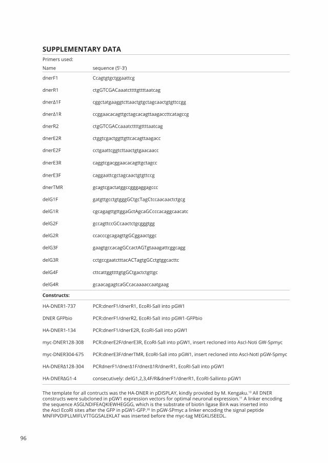
96
suPPleMenTARy dATAPrimers used:
Name sequence (5’-3’)
dnerF1 Ccagtgtgctggaattcg
dnerR1 ctgGTCGACaaatcttttgttttaatcag
dnerΔ1F cggctatgaaggtcttaactgtgctagcaactgtgttccgg
dnerΔ1R ccggaacacagttgctagcacagttaagaccttcatagccg
dnerR2 ctgGTCGACcaaatcttttgttttaatcag
dnerE2R ctggtcgactggttgttcacagttaagacc
dnerE2F cctgaattcggtcttaactgtgaacaacc
dnerE3R caggtcgacggaacacagttgctagcc
dnerE3F caggaattcgctagcaactgtgttccg
dnerTMR gcagtcgactatggccgggaggagccc
delG1F gatgttgcctgtgggGCtgcTagCtccaacaactctgcg
delG1R cgcagagttgttggaGctAgcaGCcccacaggcaacatc
delG2F gccagttccGCcaactctgcgggtgg
delG2R ccacccgcagagttgGCggaactggc
delG3F gaagtgccacagGCcactAGTgtaaagattcggcagg
delG3R cctgccgaatctttacACTagtgGCctgtggcacttc
delG4F cttcattggttttgtgGCtgactctgttgc
delG4R gcaacagagtcaGCcacaaaaccaatgaag
Constructs:
HA-DNER1-737 PCR:dnerF1/dnerR1, EcoRI-SalI into pGW1
DNER GFPbio PCR:dnerF1/dnerR2, EcoRI-SalI into pGW1-GFPbio
HA-DNER1-134 PCR:dnerF1/dnerE2R, EcoRI-SalI into pGW1
myc-DNER128-308 PCR:dnerE2F/dnerE3R, EcoRI-SalI into pGW1, insert recloned into AscI-Noti GW-Spmyc
myc-DNER304-675 PCR:dnerE3F/dnerTMR, EcoRI-SalI into pGW1, insert recloned into AscI-NotI pGW-Spmyc
HA-DNERΔ128-304 PCRdnerF1/dnerΔ1F/dnerΔ1R/dnerR1, EcoRI-SalI into pGW1
HA-DNERΔG1-4 consecutively: delG1,2,3,4F/R&dnerF1/dnerR1, EcoRI-SalIinto pGW1
The template for all contructs was the HA-DNER in pDISPLAY, kindly provided by M. Kengaku.10 All DNER constructs were subcloned in pGW1 expression vectors for optimal neuronal expression.11 A linker encoding the sequence ASGLNDIFEAQKIEWHEGGG, which is the substrate of biotin ligase BirA was inserted into the AscI EcoRI sites after the GFP in pGW1-GFP.26 In pGW-SPmyc a linker encoding the signal peptide MNFIPVDIPLLMIFLVTTGGSALEKLAT was inserted before the myc-tag MEGKLISEEDL.
97
Iden
tIfIcatIon
of d
neR as th
e tR antIg
en
5
AssessMenT Of PATIenT’s dIsAbIlITyThe patients’ disability was assessed using a modified Rankin scale (mRS). On the mRS a score of 0 represents an asymptomatic patient; 1, symptoms that do not interfere with lifestyle; 2, symptoms that lead to some restriction of lifestyle but do not prevent totally independent existence; 3, symptoms significantly interfere with lifestyle or prevent totally independent existence; 4, symptoms clearly prevent independent existence, although the patient does not need constant attention; 5, severe disability with total dependence requiring constant attention; 6, death from neurological cause.9
IMMunOPReCIPITATIOn And MAss sPeCTROMeTRyExtracts were made from adult total rat brain using 1% NP40 lysis buffer (10mM Hepes, pH 7.5, 150 mM NaCl, 1% NP40 and complete protease inhibitor (Roche). 2.5 mg protein was precleared with 50 ul protA / protG sepharose, mixed 1:1, for 1 hour at 4oC and the supernatant was subsequently incubated overnight at 4oC with 10 ul sera. The following morning 50 ul protA / protG (1:1) sepharose was added and left to incubate for an additional 4 hours at 4oC. The sepharose beads were washed 5 times with lysis buffer and loaded on a 4-12% Bis-Tris gel (Invitrogen, Breda, Netherlands). Mass spectrometry was mainly performed as described before.26,27 For mass spectrometry analysis, 1D SDS-PAGE gel lanes were cut into 2-mm slices using an automatic gel slicer and subjected to in gel reduction with dithiothreitol, alkylation with iodoacetamide and digestion with trypsin (sequencing grade, Promega, Leiden, Netherlands), essentially as described previously.28 Nanoflow LCMS/MS was performed on an 1100 series capillary LC system (Agilent Technologies, Amstelveen, Netherlands) coupled to an LTQ linear ion trap mass spectrometer (Thermo, Breda, Netherlands) operating in positive mode and equipped with a nanospray source. Peptide mixtures were trapped on a ReproSil C18 reversed phase column (column dimensions 1.5 cm × 100 μm, packed in-house; Dr Maisch GmbH, Ammerbuch-Entringen, Germany) at a flow rate of 8 μl/min. Peptide separation was performed on ReproSil C18 reversed phase column (column dimensions 15 cm × 50 μm, packed inhouse; Dr Maisch GmbH, Ammerbuch-Entringen, Germany) using a linear gradient from 0 to 80% B (A = 0.1 M acetic acid; B = 80% (v/v) acetonitrile, 0.1 M acetic acid) in 70 min and at a constant flow rate of 200 nl/min using a splitter. The column eluent was directly sprayed into the ESI source of the mass spectrometer. Mass spectra were acquired in continuum mode; fragmentation of the 4 peptides was performed in data-dependent mode. Peak lists were automatically created from raw data files using the Mascot Distiller software (version 2.1; MatrixScience, London, UK). The Mascot search algorithm (version 2.2) was used for searching against the International Protein Index database (release number IPI_rat_20091106.fasta). The peptide tolerance was typically set to 2 Da and the fragment ion tolerance to 0.8 Da. A maximum number of 2 missed cleavages by trypsin were allowed and carbamidomethylated cysteine and oxidized methionine were

98
set as fixed and variable modifications, respectively. The Mascot score cut-off value for a positive protein hit was set to 31. Proteins present in the anti-Hu serum were omitted from the individual lists obtained by immunoprecipitation with the 4 anti-Tr positive sera. The remaining proteins of the 4 lists were compared by hand, leading to 12 proteins identified in all 4 anti-Tr immunoprecipitations. These were all further checked for absence in the list obtained by the 2 remaining negative controls (mGluR1, GluR1).
CulTuRIng And IMMunOblOTTIngHEK293T en HeLa cells were grown in DMEM:HAMF10 (1:1) medium containing 10%FBS and 1% penicillin/streptomycin at 37oC and 5%CO2. Cells were transiently transfected with various DNA constructs using Polyethylenimin (Polyscience, Eppelheim, Germany) and grown for 24 hours after transfection. For glycosylation experiments, cells were treated with Tunicamycin (20 ug/ml, Merck Chemicals) for 24 hours prior to immunoblotting. For immunoblotting cells were harvested in phosphate buffered saline (PBS) and lysed for 15 minutes at 4oC in 1% Triton, 20mM Tris pH7.5, 150mM NaCl supplemented with complete protease inhibitors (Roche, Woerden, Netherlands). After a 15 seconds sonification step, samples were spun for 5 minutes at 13.000rpm at 4oC. The supernatant was diluted in 2x Sample buffer (8% SDS/25% Glycerol/0.05M Tris pH 6.8/200mM DTT/Bromophenol Blue/H2O) and separated on 8 or 10% SDS-PAGE gels, followed by blotting onto PVDF membranes (Biorad, Veenendaal, Netherlands). Blots were blocked with either 2% BSA/0.05% Tween/PBS (commercial antibodies) or with 0.05% Tween/ skimmed milk (patient sera) and incubated overnight at 4oC with the appropriate antibody. Blots were washed with 0.05%Tween/PBS or 0.05%Tween/skimmed milk for 3 times 5 minutes each at room temperature and incubated with the appropriate secondary antibody. After washing 3 times in 0.05% Tween/ PBS and once in PBS, blots were developed with Enhanced Chemiluminescent Western Blotting Substrate (Pierce, Etten-Leur, Netherlands).
IMMunOCyTOCheMIsTRyFor immunocytochemistry, HeLa cells were grown on Lab-tek chamber slides (Nunc, Uden, Netherlands) and fixed with 4% PFA for 10 minutes, followed by 3 washes of 5 minutes each with PBS and 1 wash of 5 minutes with PBS+ (0.5% Protifar (Nutricia, Zoetermeer, Netherlands), 0.15% glycin, 0.05%Triton in PBS). Cells were stained with the primairy antibody (2 hours at room temperature) in PBS+, washed 3 times in PBS and subsequently incubated with secondary antibodies in PBS+ for 1 hour at room temperature. After 3 subsequent washes in PBS slides were mounted in Vectashield (Vector Laboratories, Peterborough, UK) and studied on a DMRBE microscope (Leica, Rijswijk, Netherlands). For surface staining of DNER, hippocampal neurons were incubated with serum of Tr9 diluted 1:50 and rabbit anti-HA diluted 1:100, in Neurobasal medium (NB) for 15
99
Iden
tIfIcatIon
of d
neR as th
e tR antIg
en
5
minutes at 37oC, followed by rinsing in NB at 37oC. After fixing the cells for 10 minutes in 4%PFA / 4%sucrose cells were washed three times with PBS, followed by incubation with secondary antibodies in GDB (0.1% gelatin, 16.7 mM Phosphate buffer and 450 mM NaCl) for 1 hour at room temperature. After three subsequent washes in PBS the coverslips were mounted and studied as described above. Permeabilised staining of hippocampal neurons was performed by fixing the neurons in 4% PFA / 4% sucrose, 3 washes with PBS and incubation of the primary antibodies in GDB+0.6% Triton, overnight at 4oC. After three washes with PBS, neurons were incubated with secondary antibodies for 1 hour at room temperature in GDB+ 0.6%Triton. Subsequent washing and mounting were as described above.
IMMunOhIsTOCheMIsTRySnap frozen rat brain and cerebellar cryosections were stained as described before.6 Cryosectons of 6-8 µm were air-dried and fixed in acetone (4oC) for 10 minutes. Endogenous peroxidase was blocked with 0.3% H2O2 in PBS. Slides were treated with 10% goat serum for 30 minutes at room temperature (RT), incubated either 2 hours at RT or overnight at 4oC with patient serum diluted in blocking serum (1:100 or 1:400), with PBS washes between the different steps. Slides were incubated with biotin conjugated goat anti-human IgG antibodies (1: 2000, Vector, Peterborough, UK) in blocking serum for 45 minutes. After incubation with Vectastain Elite ABC complex (Vector Labs, Burlingame, CA) for 30 minutes, the reaction was developed with 0.05% diaminobenzidine and 0.01% H2O2 diluted in PBS with 0.5% Triton X-100 followed by staining with Diaminobenzidine tetrahydrochlorid dihydrate (DAB, Fluka, Zwijndrecht, Netherlands) slides were dehydrated with ethanol series (50-100%) and mounted with Pertex (Klinipath, Duiven, Netherlands). To test whether the different Tr-identified sera recognize a similar epitope, rat cerebellar sections were incubated with undiluted anti-Tr-positive serum for 3 hours at RT, after the initial blocking in goat serum. This was followed by overnight incubation at 4°C with biotinylated purified IgG (biotinylated using EZ-Link® Sulfo-NHS-LC-Biotinylation Kit (Pierce Biotechnology, Rockford Il, US), diluted 1:100 in PBS 1% BSA, from a serum which had been used in Tr-blocking experiments described before.8 Staining was developed as described above.
PuRIfICATIOn Of dneR ReACTIng AnTIbOdIes fROM TR seRuMBioGFP-DNER was expressed in HEK293T cells for 24 hours, washed with cold PBS and lysed for 15 min on ice in 20mM HEPES pH7.5, 150 mM KCl, 1% Triton X-100, complete protease inhibitors (Roche, Woerden, Netherlands), followed by 2 times 10 seconds sonification steps. Extracts were spun at 13.000 rpm for 10 minutes after which the supernatant was incubated with preblocked (1% BSA, 0.05% Tween in PBS for 30 minutes) Dynal M280 beads (Invitrogen, Breda, Netherlands) for 1 hour. Beads were
100
washed 5 times with HEPES pH7.5, 600mM KCl, 0.1% Triton X100 and subsequently incubated for 3 hours with patients sera diluted 1:15 in PBS containing 1% BSA and 0.05% Tween at 4°C. The beads were washed 4 times with 20mM HEPES pH7.5, 600mM KCl, 0.1% Triton X100 and antibodies were eluted with 200mM glycine pH2.8, after which the pH was neutralized using 1/10th volume of 1M Tris, pH8.0. Both depleted serum and the purified antibodies were washed with PBS using Vivaspin sample concentrators (GE Healthcare, Hoevelaken, Netherlands) and stored in PBS containing 1%BSA.
dneR KnOCK dOwn In hIPPOCAMPAl neuROnsPrimary hippocampal cultures were prepared from embryonic day 18 (E18) rat brains.29 Cells were plated on coverslips coated with poly-L- lysine (30 μg/ml) and laminin (2 μg/ml) at a density of 75,000/well. Hippocampal cultures were grown in Neurobasal medium (NB) supplemented with B27, 0.5 mM glutamine, 12.5 μM glutamate and penicillin/streptomycin as described in Jaworski et al.26 The following shRNA sequences targeting rat DNER were used in this study: shRNA#1 (AAACCCTTGGTCACACTGA) and shRNA#2 (GATTCTGTTGCTAAGTCCA). The complementary oligo’s were annealed and inserted into pSUPER. Hippocampal neurons were transfected at 7 days in vitro (DIV) using Lipofectamine 2000 (Invitrogen). DNA (3.6 μg /well) was mixed with 3.3 μl Lipofectamine 2000 in 200 μl NB, incubated for 30 minutes and then added to the neurons in NB at 37°C in 5% CO2 for 45 min. Next, neurons were washed with NB and transferred in the original medium at 37°C in 5% CO2 for 4 days. Cells were co-transfected with β-galactosidase to identify the transfected cells.
101
Iden
tIfIcatIon
of d
neR as th
e tR antIg
en
5
RefeRenCes
1. Posner J. Paraneoplastic syndromes.
Philadelphia: FA Davis: Contemporary
Neurological series; 1995:353-85.
2. Shams’ili S, Grefkens J, de Leeuw B, et al.
Paraneoplastic cerebellar degeneration
associated with antineuronal antibodies:
analysis of 50 patients. Brain 2003;126:1409-18.
3. Briani C, Vitaliani R, Grisold W, et al. Spectrum
of paraneoplastic disease associated with
lymphoma. Neurology 2011;76:705-10.
4. Horwich L, Buxton PH, Ryan GM. Cerebellar
degeneration with Hodgkin’s disease. J Neurol
Neurosurg Psychiatry 1966;29:45-51.
5. Trotter JL, Hendin BA, Osterland CK. Cerebellar
degeneration with Hodgkin disease. An
immunological study. Arch Neurol 1976;33:660-
1.
6. Graus F, Dalmau J, Valldeoriola F, et al.
Immunological characterization of a neuronal
antibody (anti-Tr) associated with paraneoplastic
cerebellar degeneration and Hodgkin’s disease.
J Neuroimmunol 1997;74:55-61.
7. Graus F, Gultekin S, Ferrer I, Reiriz J, Alberch J,
Dalmau J. Localization of the neuronal antigen
recognized by anti-Tr antibodies from patients
with paraneoplastic cerebellar degeneration
and Hodgkin’s disease in the rat nervous system.
Acta Neuropathol 1998;96:1-7.
8. Bernal F, Shams’ili S, Rojas I, et al. Anti-Tr
antibodies as markers of paraneoplastic
cerebellar degeneration and Hodgkin’s disease.
Neurology 2003;60:230-4.
9. Keime-Guibert F, Graus F, Broet P, et al. Clinical
outcome of patients with anti-Hu-associated
encephalomyelitis after treatment of the tumor.
Neurology 1999;53:1719-23.
10. Eiraku M, Hirata Y, Takeshima H, Hirano T,
Kengaku M. Delta/notch-like epidermal growth
factor (EGF)-related receptor, a novel EGF-like
repeat-containing protein targeted to dendrites
of developing and adult central nervous system
neurons. J Biol Chem 2002;277:25400-7.
11. Hoogenraad CC, Feliu-Mojer MI, Spangler SA,
et al. Liprinalpha1 degradation by calcium/
calmodulin-dependent protein kinase II
regulates LAR receptor tyrosine phosphatase
distribution and dendrite development. Dev Cell
2007;12:587-602.
12. Hammack J, Kotanides H, Rosenblum MK, Posner
JB. Paraneoplastic cerebellar degeneration.
II. Clinical and immunologic findings in 21
patients with Hodgkin’s disease. Neurology
1992;42:1938-43.
13. Nishizumi H, Komiyama T, Miyabayashi T,
Sakano S, Sakano H. BET, a novel neuronal
transmembrane protein with multiple EGF-like
motifs. Neuroreport 2002;13:909-15.
14. Sillevis Smitt P, Kinoshita A, De Leeuw B, et
al. Paraneoplastic cerebellar ataxia due to
autoantibodies against a glutamate receptor. N
Engl J Med 2000;342:21-7.
15. Dalmau J, Tüzün E, Wu H, et al. Paraneoplastic
anti-N-methyl-D-aspartate receptor encephalitis
associated with ovarian teratoma. Ann Neurol
2007;61:25-36.
16. Lai M, Hughes EG, Peng X, et al. AMPA receptor
antibodies in limbic encephalitis alter synaptic
receptor location. Ann Neurol 2009;65:424-34.
17. Lancaster E, Lai M, Peng X, et al. Antibodies to
the GABA(B) receptor in limbic encephalitis with
seizures: case series and characterisation of the
antigen. Lancet Neurol 2010;9:67-76.
18. Lancaster E, Martinez-Hernandez E, Titulaer
MJ, et al. Antibodies to metabotropic glutamate
receptor 5 in the Ophelia syndrome. Neurology
2011;77:1698-701.

102
19. Lai M, Huijbers MG, Lancaster E, et al.
Investigation of LGI1 as the antigen in limbic
encephalitis previously attributed to potassium
channels: a case series. Lancet Neurol
2010;9:776-85.
20. Irani SR, Bera K, Waters P, et al. N-methyl-D-
aspartate antibody encephalitis: temporal
progression of clinical and paraclinical
observations in a predominantly non-
paraneoplastic disorder of both sexes. Brain : a
journal of neurology 2010;133:1655-67.
21. Graus F, Dalmou J, Rene R, et al. Anti-Hu
antibodies in patients with small-cell lung
cancer: association with complete response
to therapy and improved survival. J Clin Oncol
1997;15:2866-72.
22. de Beukelaar J, Sillevis Smitt P. Managing
paraneoplastic neurological disorders.
Oncologist 2006;11:292-305.
23. Tohgo A, Eiraku M, Miyazaki T, et al. Impaired
cerebellar functions in mutant mice lacking
DNER. Mol Cell Neurosci 2006;31:326-33.
24. Eiraku M, Tohgo A, Ono K, et al. DNER acts as a
neuron-specific Notch ligand during Bergmann
glial development. Nat Neurosci 2005;8:873-80.
25. Dalmau J, Rosenfeld M. Paraneoplastic
syndromes of the CNS. Lancet Neurol
2008;7:327-40.
26. Jaworski J, Kapitein LC, Gouveia SM, et al.
Dynamic microtubules regulate dendritic spine
morphology and synaptic plasticity. Neuron
2009;61:85-100.
27. Lansbergen G, Grigoriev I, Mimori-Kiyosue Y, et
al. CLASPs attach microtubule plus ends to the
cell cortex through a complex with LL5beta. Dev
Cell 2006;11:21-32.
28. Wilm M, Shevchenko A, Houthaeve T, et al.
Femtomole sequencing of proteins from
polyacrylamide gels by nano-electrospray mass
spectrometry. Nature 1996;379:466-9.
29. Banker G GK. Culturing Nerve Cells. Cambridge
MA1998.

103
Iden
tIfIcatIon
of d
neR as th
e tR antIg
en
5


Psychiatric phenomena as initial manifestation of encephalitis by anti-nMdA receptor antibodies
Peter Maat, Esther de Graaff, Nico M. van Beveren, Esther Hulsenboom, Robert M. Verdijk, Kathelijne Koorengevel,
Martijn van Duijn, Herbert Hooijkaas, Casper Hoogenraad, Peter A.E. Sillevis Smitt
Acta Neuropsychiatrica June 2013; 25(3): 128-136
6Back to table of contents

106
AbsTRACT
ObJeCTIVeAutoimmune encephalitis associated with autoantibodies against the N-methyl-D-aspartate receptor (NMDAR) often presents with behavioral change. Our objective was to describe in detail the psychiatric presentation and pathways to care in order to aid the early diagnosis of NMDAR encephalitis.
MeThOdsSera and CSF from patients with suspected NMDAR encephalitis were tested on HEK 293 cells transfected with the NR1 subunit of the NMDAR. Clinical information was obtained from the referring psychiatrists and neurologists and by review of the clinical records.
ResulTsSamples from 15 patients (13 female, 2 male, mean age 24 years, range 5-56 years) tested anti-NMDAR positive. Twelve of the 15 patients (80%) presented with prominent psychiatric symptoms and 8 were initially referred to a psychiatric service. The most prominent initial psychiatric symptoms were anxiety in 7 (47%), behavioural change (often bizarre) in 6 (40%) and agitation in 5 (33%). All patients developed psychiatric symptoms in the first 6 weeks of illness. Thirteen patients received psychotropic medications: antipsychotics in 12 and benzodiazepines in 11. Treating physicians considered the psychotropic medication not effective in 11 patients resulting in many drug switches. At nadir, all patients were in a very poor condition. However, 8 patients (53%) recovered (almost) completely. Outcome tended to be better in patients who had received early immunotherapy or tumor removal.
COnClusIOns:Autoimmune encephalitis and anti-NMDAR testing in serum and CSF should be considered in patients, especially young females, presenting with atypical psychiatric phenomena. Early diagnosis and treatment will likely improve the prognosis of NMDAR encephalitis.
Keywords: Anti-NMDAR Encephalitis, Anti-NMDAR antibodies, Psychotic disorders
ACKnOwledgeMenTsThe authors gratefully acknowledge the physicians who referred patients. The authors wish to thank Marcel van der Weiden for performing NMDAR immunostaining of tissue slides.

107
Psychiatric Ph
eno
men
a in an
ti-nm
Da recePto
r encePh
alitis
6
InTROduCTIOnN-methyl-D-aspartate receptor (NMDAR) encephalitis is a severe form of encephalitisassociated with antibodies against the NR1 and NR2 subunits of the NMDAR and oftenpresents with severe psychiatric symptoms mostly in young women. In approximately70% of patients, the encephalitis is preceded by a prodromal phase of one to two weeksconsisting of fever and malaise resembling a viral illness.1,2 The presentation of NMDARencephalitis itself is characterized by prominent emotional and behavioral changefollowed by amnesia, seizures and in some cases a choreiform movement disorder,evolving to a catatonic state. Autonomic instability and central hypoventilation oftenlead to ICU admission, intubation and mechanical ventilation1-5 NMDAR encephalitis isassociated with ovarian teratomas in approximately 60% of patients, especially youngwomen.2-5 The disorder also occurs in children who often present with behavioral andpersonality changes, later complemented by abnormal movements, epilepsy, sleepdysfunction and speech problems.6-8 In children and adolescents, the frequency of non-paraneoplastic cases is apparently much higher than in adults 6,9; only 9% of girls < 9years of age had an underlying tumor.9 Recovery with no or only mild residual deficitsoccurs in approximately three quarters of the patients.2,4,7,8 However, outcome dependsgreatly on early diagnosis followed by immediate removal of the underlying tumor (ifpresent) and/or immunomodulatory therapy.2,4,5,7,8
Several lines of evidence strongly suggest that anti-NMDAR antibodies and otherantibodies against neuronal surface antigens are pathogenic by functional interferencewith the receptor.10,11 This functional interference is reversible, at least initially,resulting in a good outcome in most patients when treated early. In contrast, patientswith paraneoplastic limbic encephalitis associated with antibodies directed againstintracellular antigens have a poor prognosis that is probably related to irreversibleT-cell mediated damage.12,13
The differential diagnosis of NMDAR encephalitis is vast and includes psychoticdisorders, mood dysregulation disorders, sleep disorders and disorders of impulsecontrol.14 Also patients with a first episode of schizophrenia according to DSM IV criteriacan have an underlying NMDAR encephalitis.15 As a result patients are often initiallyadmitted for psychiatric evaluation and psychiatrists may encounter patients withNMDAR encephalitis in all kinds of settings including emergency departments, inpatientunits, consultation services and outpatient offices.To facilitate early recognition of this potentially lethal but highly treatable disorder, wereport the clinical findings in 15 Dutch patients with NMDAR encephalitis with specialfocus on psychiatric symptoms and pathways to care in the early stages of the disease.

108
PATIenTs And MeThOdsPatientsAll available clinical records were retrospectively examined in detail for psychiatric and neurological signs and symptoms at presentation and during subsequent evolution of the syndrome. We defined symptoms as either neurological or psychiatric based on the interpretation by the attending physicians.The results of brain MRI, EEG and CSF examinations were collected as were the results of tumor screening. We were able to review the MRI’s of 8/15 patients. In the other 7/15 patients we could only read the results. In addition, the timing and type of treatment (symptomatic treatment, immunotherapy and/or tumor treatment) were scored. One of the patients presented here has been reported before.16 The study was approved by the Erasmus MC institutional review board.
OutcomeThe patients’ disability was assessed using a modified Rankin scale (mRS).17 On the mRS a score of 0 represents an asymptomatic patient; 1, symptoms that do not interfere with lifestyle; 2, symptoms that lead to some restriction of lifestyle but do not prevent totally independent existence; 3, symptoms significantly interfere with lifestyle or prevent totally independent existence; 4, symptoms clearly prevent independent existence, although the patient does not need constant attention; 5, severe disability with total dependence requiring constant attention; 6, death from neurological cause. The outcome was assessed at least six months after the onset of symptoms. A patient was considered to have a good outcome when the mRS was 0-1. In all other patients the outcome was considered poor (mRS ≥ 2). Early treatment was defined as any tumor or immunomodulatory treatment started within two months from symptom onset.
Statistical analysisDue to the retrospective nature and objective of this study the symptoms and referrals were qualitatively assessed. The proportions of patients with a good outcome in the group receiving early treatment versus late or no treatment were compared using the Chi-square test (GraphPad Prism version 5, GraphPad Software Inc., La Jolla, CA).
Cell based assay for anti-NMDAR antibodiesThe NMDAR1 subunit (NR1) complementary (c)DNA fused in frame to YFP was a kind gift from B. Laube.18 HEK293 cells (ATCC #CRL1537) were grown on glass coverslips in DMEM and Ham’s F10 (Gibco, Breda, Netherlands) supplemented with 10% FBS (Gibco) and 1% penicillin and streptomycin (Gibco). After 24h, cells were transfected with the YFP-tagged NR1 contruct using Fugene-6 (Roche, Woerden, Netherlands) at a ratio of 3:1. After 24h, live cells were fixed with 4% paraformaldehyde (Sigma, MO) for 15 min, washed 5 min PBS with 0.2 % Triton X-100 (Merck, Darmstadt, Germany) followed by
109
Psychiatric Ph
eno
men
a in an
ti-nm
Da recePto
r encePh
alitis
6
two times 15 min in PBS with 0.5 % BSA (Sigma) and 7.0 mg glycine (Sigma) (PBS+) prior to incubation with patient sera (1:50 and 1:200) or CSF (undiluted and 1:5) for 1 h. After washing, cells were incubated for 1 h with anti-human CY3 1:200 made in donkey (Jackson ImmunoResearch, Westgrove, PA). The samples were visualized and scored by two independent observers using a Leica DM RXA microscope (Leica, Wetzlar, Germany).
HistologyDeparaffinized 4 micron tissue sections from the patient’s teratoma were pretreated in citric acid (pH 6.0) at 100°C for 30 min for antigen unmasking. Slides were washed and incubated with mouse-anti NMADR1 antibody (BD Pharmingen, Franklin Lakes, NJ) at a 1:250 dilution. The PowerVision poly AP anti-mouse IgG multilink system (Leica Novocastra, Newcastle upon Tyne, UK) was used according to the manufacturer’s specifications for visualization. Characterization of inflammatory infiltrates in brain tissue obtained at autopsy was performed on non-pretreated deparaffinized 4 micron tissue sections using antibodies specific for CD3, CD5, CD4, CD8, CD56, CD20, CD79a, CD138, IgG, IgM, IgA, IgD, C4d and CD68 (BD Pharmingen). The PowerVision DAP substrate (Leica Novocastra) was used for visualization as described above.
ResulTsSerum and/or CSF samples from 15 patients tested anti-NMDAR positive (Figure 1D-F). In three patients, the anti-NMDAR antibodies were only detectable in CSF and absent in the paired serum sample. Thirteen patients were adults (≥ 18 years). Clinical characteristics are summarized in Table 1.
Initial presentation and referralSupplementary Table S1 shows a detailed overview of the patients. At presentation, 12 (80%) of the 15 patients had prominent psychiatric symptoms. Eight of the 13 (62%) adult patients were initially referred to a psychiatric service. Two children (5 and 8 years of age) were first seen by a pediatric neurologist. The most prominent psychiatric symptoms at initial presentation of the patients were anxiety in 7 (47%), behaviorial change (often bizarre) in 6 (40%), agitation in 5 (33%), delusions in 4 (27%) and altered mood in 3 (20%) (supplementary Table S1).

110
Table 1. Clinical characteristics in 15 patients with NMDAR encephalitisN=15
Age (years) 19 (5-56) a
Sex (F/M) 13 / 2
Predominant initial symptoms
Psychiatric
Neurological
12 (80%)
3 (20%)
Diagnostic delay (months)b 1.5 (0.4-99.6) a
Ancillary investigations
CSF abnormal
EEG abnormal
MRI abnormal
13/14 (93%)
13/13 (100%)
6/15 (40%)
NMDAR antibody testing
CSF positive
Serum positive
13/13 (100%)
11/14 (79%)
Teratoma present 5 (33%)
Treatment
Antitumor treatment
Steroids
IVIG
Plasma Exchange
Otherc
5/5 (100%)
12/15 (80%)
5/15 (33%)
2/15 (13%)
2/15 (13%)
Outcome
mRS≤2
mRS>2
10 (67%)
5 (33%)
aMedian (range); bDelay between onset of symptoms and final diagnosis (positive antibody testing); cOther therapies include electroconvulsive therapy (ECT) and rituximab in one and mitoxantrone in another patient ; IVIG, intravenous immunoglobulins; mRS, modified Rankin scale
Early symptomsIn the first 6 weeks after the onset of the syndrome, psychiatric symptoms were present in all fifteen patients (100%). The most frequent psychiatric symptoms in the first 6 weeks (Table 2) were behavioral change in 14 (93%), including bizarre or childish behavior in 9 (60%) and catatonia in 6 (40%). Perceptual disturbances (hallucinations) were present in 9 patients (60%), impaired consciousness in 9 (60%), anxiety or depression in 7 (47%) and disorientation in 6 (40%). Neurological symptoms during the first 6 weeks (Table 3) included epileptic seizures in 12 (80%), choreoathetoid and complex movementsin 7 (47%), sleep disturbances in eight (53%), autonomic instability in seven (47%) andcentral hypoventilation requiring admission to an intensive care unit for mechanicalventilation in 6 (40%). The time interval between onset of symptoms and diagnosis ofNMDAR-encephalitis ranged between 2 weeks and 8.3 years (median 1.5 months).Two patients were pregnant at presentation and both lost their pregnancies in the acute phase of their illness at 13 and 24 weeks of gestation.
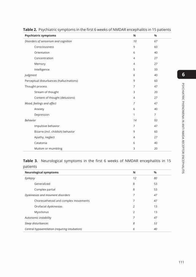
111
Psychiatric Ph
eno
men
a in an
ti-nm
Da recePto
r encePh
alitis
6
Table 2. Psychiatric symptoms in the first 6 weeks of NMDAR encephalitis in 15 patientsPsychiatric symptoms n %
Disorders of sensorium and cognition 10 67
Consciousness 9 60
Orientation 6 40
Concentration 4 27
Memory 4 27
Intelligence 5 33
Judgment 6 40
Perceptual disturbances (hallucinations) 9 60
Thought process 7 47
Stream of thought 3 20
Content of thought (delusions) 4 27
Mood, feelings and affect 7 47
Anxiety 6 40
Depression 1 7
Behavior 14 93
Impulsive behavior 7 47
Bizarre (incl. childish) behavior 9 60
Apathy, neglect 4 27
Catatonia 6 40
Mutism or mumbling 3 20
Table 3. Neurological symptoms in the first 6 weeks of NMDAR encephalitis in 15 patients
neurological symptoms n %
Epilepsy 12 80
Generalized 8 53
Complex partial 8 53
Dyskinesias and movment disorders 7 47
Choreoathetoid and complex movements 7 47
Orofacial dyskinesias 2 13
Myoclonus 2 13
Autonomic instability 7 47
Sleep disturbances 8 53
Central hypoventilation (requiring intubation) 6 40
112
Ancillary investigationsEEG was performed in 13 patients and showed abnormal slowing of the background pattern in all patients with additional epileptiform features in 9 of them.MRI was abnormal in 6/15 patients (40%). MRI changes consisted of increased signal intensities in T2 and fluid-attenuated inversion recovery (FLAIR) images. The changes were typically localized in the medial temporal lobes in four patients (Figure 1A, arrows), with additional cortical localizations in one and post-gadolinium enhancement in another. One patient showed subcortical changes (thalamus and putamen) and another showed changes in the periventricular white matter.CSF was abnormal in 13/14 patients. Abnormalities included WBC pleocytosis (8), oligoclonal banding (7) and an increased IgG index (7), indicating intrathecal antibody synthesis. Total protein concentration was normal in all CSF samples.
Underlying tumorIn 5 of the 15 patients an ovarian teratoma was detected 1-22 months after onset of the neuropsychiatric symptoms. In two patients the ovarian teratoma was detected and removed within a month after onset of symptoms (Figure 1B). Immunohistochemistry revealed expression of the NR1 subunit of the NMDA receptor in the ovarian teratoma (Figure 1C). In one 5 year-old patient multiple ovarian cysts were detected by MRI (no further follow-up data yet available). In two patients, the teratoma was detected almost two years after onset of symptoms and removed to prevent relapse of the NMDAR encephalitis. In the remaining 10 patients no teratoma or other tumours were detected.
Symptomatic treatmentTargeted management of psychiatric symptoms was mainly directed at psychotic symptoms, anxiety, agitation and mood and sleep disorders. Thirteen patients were treated with psychotropic medication including an antipsychotic drug combined with a benzodiazepine in 10 patients, antipsychotic medication only in 2 and benzodiazepine only in 1. Two patients received additional melatonin for insomnia. In only 2 of the 13 patients, the treating physicians felt that the medication was effective in controlling the psychiatric symptoms, resulting in many medication switches. The mean number of prescribed antipsychotic drugs was 1.8 (range 1-4) and the mean number of administered benzodiazepines was 1.7 (range 1-4).Prescribed antipsychotics included the classical antipsychotics haloperidol (in 7 patients), zuclopentixol (2), levopromazine (1) and pipamperon (1) and the atypical antipsychotics risperidon (6) and olanzapine (5). The following benzodiazepines were prescribed: lorazepam (7), diazepam (3), oxazepam (2) and to one patient each midazolam, clonazepam, alprazolam, nitrazepam, temazepam and clorazepate.

113
Psychiatric Ph
eno
men
a in an
ti-nm
Da recePto
r encePh
alitis
6
Treatment and prognosisTwo patients died 2 weeks and 5 months after onset of symptoms (mRS 6). Follow-up in the other patients ranged from 3 months to 9 years (median 14 months). At the nadir of the disease, all patients were severely disabled and completely dependent (mRS 5). The outcome was favourable (mRS <2) in 8 patients while 5 patients fared less well (mRS 2-4). Twelve patients received treatment with steroids combined with intravenous immunoglobulins (IVIg) in 5, plasma exchange in 2 and rituximab in one. Treatment was initiated 0.1 – 10 months after onset of symptoms (median 1.1 month). Three patients did not receive any form of immunotherapy, one died while the other two recovered with mild (mRS 1) to moderate (mRS 2) symptoms. All 5 patients with a pathologically proven and removed ovarian teratoma had an excellent prognosis (mRS 0). However, in two of these patients, the teratoma was found and removed long after recovery from the NMDAR encephalitis (22 months after onset of symptoms). In 2 patients the follow-up was shorter than 6 months and outcome was not yet evaluable. Of the evaluable patients, 7 had received early treatment, within 2 months from onset. In 6 of these early treatment patients, the outcome was good (mRS 0-1) while it was poor (mRS ≥ 2) in one. Of 6 evaluable patients with no or late treatment, the outcome was poor in 4 and favourable in 2 (P=0.053).Autopsy in both deceased patients showed inflammatory changes in the brain, mainly in the medial temporal lobe and hippocampus (Figure S1). Inflammatory infiltrates consisted of CD8 positive T lymphocytes and CD20 positive B cells in perivascular cuffs and in the neuropil. Also CD68 positive macrophages and activated microglia were observed.
dIsCussIOnTwelve of 15 patients (80%) with anti-NMDAR encephalitis had prominent psychiatric symptoms at first presentation. Eight adult patients were initially admitted to a psychiatric unit. The most common diagnosis was ‘atypical psychosis’, followed by schizophrenia, depression and panic attacks. Subsequently, patients developed seizures, decline of consciousness, catatonia, abnormal movements, autonomic instability and central hypoventilation. Thirteen patients received multiple psychotropic drugs with very limited symptomatic control. Most patients were young women and in 5 of them pathological or radiological evidence of an underlying teratoma was found. Early aggressive treatment consisting of tumor treatment (if present) and immunotherapy, correlated with a good outcome in 8 of 13 evaluable patients.Evaluation of the initial stages of diagnostics and patterns of referral showed that a substantial proportion of the patients initially presented with symptoms suggestive of a psychiatric disorder. At the same time, clinical records indicated that most psychiatrists were aware of an atypical presentation of the symptoms early on, leading to the

114
suspicion of an ‘organic syndrome’, and subsequent consultation of a neurologist. Conversely, when patients were initially seen by a neurologist, an atypical presentation was also recognized, leading to psychiatric consultation. Generally, patients initially presented with little obvious biological alterations (focal neurological deficits, blood chemistry, MRI), which in many cases prompted further interpretation of symptoms as caused by a psychiatric syndrome. Recognition of the ‘neurological background’ of the syndrome usually came when either prominent EEG alterations were found, or when obvious neurological symptoms began to dominate the clinical picture (mostly seizures, and decline of consciousness). Our study and previous studies show that most of the patients with NMDAR encephalitis are initially evaluated by a psychiatrist. In the series of Dalmau et al.4 80% of the patients were initially evaluated by a psychiatrist while Irani et al.5 report early psychiatric symptoms in 77% of patients. In patients of 40 years or older, with no previous history of psychiatric illness, presentation with psychiatric symptoms should always raise suspicion of an underlying neurological or other disorder. In young female patients, de novo presentation with ‘atypical’ psychosis, in particular when combined with unusual and complex behavioural changes, a diagnosis of NMDAR encephalitis should be considered.19
It is well known that expert clinical decision making greatly depends on clinical pattern recognition. It seems that the initial clinical presentation of patients with NMDAR encephalitis has elements of two apparently conflicting clinical patterns, contributing to diagnostic uncertainty. The dominant presentation of young, previously healthy women showing psychotic phenomena obviously invokes the clinical pattern of ‘psychotic disorder/schizophrenia’. However, the alternative clinical pattern (seizures, movement alterations and speech impediments in patients with good premorbid functioning without psychiatric problems) invokes in psychiatrists the clinical pattern ‘organic syndrome’. Diagnostic delay was caused by the relative absence of objective biological alteration in the early stages, in combination with unfamiliarity with this recently identified syndrome. In our series 4 out of 15 patients had been misdiagnosed as atypical psychosis or schizophrenia for months to years (range 4.2 to 99.6 months). It is well possible that more cases of NMDAR encephalitis with predominantly psychiatric symptoms are still misdiagnosed as primary psychiatric syndromes.15,20 On the other hand, a recent cross sectional study in 50 patients diagnosed with a psychotic disorder according to DSM IV criteria (including both first-episode as well as chronic schizophrenic inpatients) did not detect any anti-NMDAR antibodies.21
NMDAR-encephalitis is diagnosed by detection of anti-NMDAR antibodies in serum and/or CSF. Importantly, in three of our patients (20%) the anti-NMDAR antibodies were exclusively detectable in CSF. Dalmau et al. 4 described intrathecal synthesis of anti-NMDAR antibodies in 53 paired serum – CSF samples resulting in a higher normalized antibody concentration in CSF, using an ELISA specific for the NR1 subunit of NMDAR. Irani et al. 5 found higher absolute anti-NMDAR titers in serum than in CSF examining
115
Psychiatric Ph
eno
men
a in an
ti-nm
Da recePto
r encePh
alitis
6
14 matched serum – CSF sample pairs, using a cell-based assay. One CSF sample was negative while all other samples clearly showed intrathecal synthesis of anti-NMDAR. In our cell-based assay, a negative serum test does not rule out NMDAR encephalitis and when suspected, CSF should be examined as well. Negative serum titers in the presence of persistent CSF titers apparently occur in the setting of prolonged duration of the illness and after plasma exchange or other forms of immunotherapy.13 In addition, CSF will almost always be abnormal and may show lymphocytic pleocytosis, oligoclonal banding and intrathecal IgG synthesis. EEG was abnormal in 100% of our patients and in 80-92% of patients in other series.4,5 In contrast, MRI was normal in our series in 60% similar do the series of Dalmau et al.4 (45%) and Irani et al.5 (77%). Antibodies from patients with NMDAR encephalitis cause a selective and reversible decrease in NDMAR surface density and synaptic localization in hippocampal slices and in Lewis rats 22 and suppress induction of long-term potentiation in cultured hippocampal neurons.23 In addition, the intensity of NR1 staining is strongly reduced in the hippocampus from patients with NMDAR-encephalitis as compared to control brain.22 These findings indicate that the anti-NMDAR antibodies may be pathogenic by disturbing glutamatergic neurotransmission. Indeed, the common psychotic symptoms in NMDAR-encephalitis followed by amnesia 24 and seizures resemble the memory impairment and psychosis caused by NMDA receptor antagonists such as ketamine25 or the recreational drug phencyclidine, also known as “angel dust”.26,27 Moreover, there is evidence that in the schizophrenia syndrome altered activity of the NMDA receptor on GABA interneurons in the prefrontal cortex plays a role.28
The efficacy of both classical and atypical antipsychotics and benzodiazepines alone or in combination was very limited in this patient population, as previously described.14,29 As a result, the 13 patients treated with psychotropic medication received 1-4 different antipsychotics and 1-4 different benzodiazepines. The limited efficacy of psychotropic medication may be related to profound NMDA receptor downregulation by the antibodies. The most effective treatment of the psychiatric symptoms consisted of tumor removal (if present) and immunotherapy. Similarly, encephalitis related seizures are notoriously difficult to treat with antiepileptic drugs and respond much better to immunotherapy.30,31 In the literature, no guidelines exist on the psychiatric treatment of autoimmune encephalitis and conclusions are difficult to draw because of the concurrent use of other treatment modalities.14 Generally, psychiatric and behavioral symptoms respond best when the immune response is suppressed or reversed. Amelioration of problematic symptoms depended on early aggressive immunotherapy, removal of the underlying tumor and intensive care.8,14
In a third of our patients, the encephalitis was triggered by an underlying teratoma. In young women, ultrasound or MRI are generally preferred over CT for teratoma screening.2 The cause of the anti-NMDAR immune response in the patients who do

116
not harbor an underlying tumor is not clear. Prodromal symptoms, which may indicate a triggering viral infection, were reported by 27% of our patients. In other series, prodromal symptoms were found in 27% – 86%.1,4,5,8
In conclusion, autoimmune encephalitis should be considered in patients, especially young females, presenting with atypical psychiatric phenomena. The diagnosis is made by anti-NMDAR testing in blood and/or CSF and if blood testing is negative and the condition is strongly suspected, testing for antibodies in CSF should be done. With subsequent early treatment, the prognosis of NMDAR encephalitis is relatively favorable.
figure 1. Case vignette of a patient with NMDAR encephalitis. A 21 year old female college student (Table 1; 4th row) training to become a primary school teacher was presented to the emergency room (ER) of a general hospital because of an acute confusional state. She had always been healthy and had no psychiatric or medical history. That morning while teaching, she had become agitated and confused after a request for a vacation was denied. At the ER, her behaviour apparently normalized and she was sent home with her mother after she was seen by an internist, a neurologist, as well as a psychiatrist. No medication had been given. The next day, she made statements like ‘I have to speak more softly, then the children will listen better’ and ‘I have to jump off the balcony’ and was acutely admitted to the psychiatry department. We saw an agitated patient in a catatonic state who followed commands but did not answer questions. The patient’s mother in retrospect recalled that her daughter had a depressed mood for a period of about two weeks, prior to the admission. Furthermore, after the admission the mother visited her daughter’s house and found it covered with yellow post-it papers with unintelligible ‘to-do reminders’. The clinical state at admission was described as ‘atypical psychosis’, and the possible underlying syndromes listed in the patient’s file were: stress induced psychotic disorder, drug-induced psychotic disorder, schizophrenia, mood disorder/mania, ‘decompensation as part of developing personality disorder’ or ‘psycho-organic’ (encephalitis or non-convulsive epilepsy). Interestingly, these two latter options were explicitly written down at admission. Diagnoses were made by a senior resident after telephone consultation of the supervising psychiatrist.

117
Psychiatric Ph
eno
men
a in an
ti-nm
Da recePto
r encePh
alitis
6
Initial treatment with benzodiazepines and haloperidol was started. Because the patient was unresponsive, entered other patients’ rooms and showed generally disturbed behaviour, seclusion was needed from the first day on. Routine neurological examination showed no abnormalities. Routine laboratory measurements showed slightly elevated C-reactive protein (12 mg/l, normal range 0-9 mg/l) and leucocytes (10.9 x 10⁹ cells/l, normal range 3-10 x 109 cells/l), but no other abnormalities. Anti-NMDA receptor antibodies were not measured at the time. Over the next few days a clinical state emerged in which the patient was at times responsive (although with some slurred speech), but at other times nonresponsive, with inappropriate laughing or crying and unusual movements. Diagnosis was now: ‘catatonic psychosis’, possibly mania, and haloperidol treatment was changed to olanzapine. A neurologist was consulted who found no focal neurological deficits and no abnormalities on a CT-scan of the brain. Encephalitis or non-convulsive epilepsy was possible, but deemed very unlikely. Psychiatric examination after 6 days of admission read: in seclusion chamber; limitedly adequate and generally unresponsive; doesn’t know where she is; somnolent; slightly slurred speech; disorientated in all modalities; disorders of formal thought: loose words and remarks, thought content: fragmented ideas; mood: slightly anxious.As the patient did not react to the medication regime, and psychoactive medication might cloud the clinical picture, olanzapine and benzodiazepine were stopped 6 days after admission. The patient continued to clinically deteriorate. Consciousness became increasingly often clouded. Agitated behaviour and responsiveness became absent. After 12 days on the psychiatry department there was no reaction to painful stimuli but she actively resisted attempts at opening her eyes. Because of decreased consciousness and general nonresponsiveness, she was transferred to the neurology ward and shortly after to the intensive care unit (ICU) where she was intubated because of central hypoventilation. Differential diagnosis now included (paraneoplastic) limbic and viral encephalitis. MRI of the brain showed medial temporal lobe (A, arrows) and insula (A, arrowheads) hyperintensities in fluid-attenuated inversion recovery (FLAIR) images. A CT-scan of the abdomen showed a 2.5 by 2.3 cm left ovarian lesion (B, arrow). EEG showed a slow background pattern without epileptic phenomena. In the CSF there was a pleocytosis with 33 WBC/μl (normal <5 WBC/μl), mainly mononuclear cells. The main differential diagnosis of the pleocytosis was viral meningo-encephalitis or an immune-mediated disorder. PCR for HSV and other neurotropic viruses was negative. The left ovary was subsequently excised and pathology showed a mature teratoma with neuronal tissue expressing the NR1 subunit of the NMDA receptor (C, red signal: AP-labeled anti-NR1 antibody; 400x). The patient’s serum and CSF were tested for autoantibodies against the NR1 subunit of NMDAR. HEK 293 cells were transfected with an YFP tagged construct containing the NR1 subunit; NR1 expressing cells become green fluorescent (D). Incubation of these transfected cells with the patient’s CSF followed by incubation with Cy3 labelled anti-human IgG secondary antibody showed red labelling of transfected cells only (E). Colocalization of the green and red signals to yellow was shown more clearly in F. During her stay at the ICU, she developed mild orofacial dyskinesias and athetoid movements in the left arm. She was subsequently treated with plasma exchanges and steroids. Her condition slowly improved and she was discharged from the ICU after four weeks. She fully recovered over the next 6 months and started working as a primary school teacher one year after onset of symptoms.

118
suPPleMenTARy dATATable s1. Initial referral and predominant initial symptoms in 15 patients with NMDAR encephalitisSex Age Delay (mo)a Initial referral to: Psychiatric symptomsb neurological symptomsb
M 18 4.2 Psychiatryc Paranoid delusions Central hypoventilation (ICU admission)
F 20 10.2 Psychiatry Auditory and visual hallucinations and delusions (initial diagnosis of pyschosis/schizophrenia was made)
Epileptic seizures
F 17 2.5 Psychiatry Anxiety, ‘psychosis’, behavioral changes and catatonia
Epileptic seizures (after approx. 3 weeks)
F 21 0.8 Psychiatry (transfer to neurology after 10 days)
Bizarre behavioral changes and delusions(‘delirium-like organic syndrome’)
Impaired consciousness
F 56 1.7 Psychiatryd Auditory hallucinations, depressed mood, episodes of anxiety and severe agitation combined with bizarre behavior
Short-term memory impedimentsAutonomic instability (hypothermia, hypotension, bradycardia) developed after 4 weeks, requiring ICU admission
F 18 1.3 Psychiatrye Acute panic attacks, strange movements of limbs and body and bizarre behavior
Headache and nausea
F 50 1.1 Psychiatry (transfer to neurology after 3 days)
Depressed mood, agitation and catatonia
Impaired consciousness
F 32 0.8 Psychiatry Panic attacks, severe agitation and derealization
Epileptic seizures
F 18 99.6 Neurology and psychiatryf
Catatonia, severe agitation and aggression, speech impediments (‘disorganized speech’)
Epileptic seizures
F 22 48.7 Neurologyg Anxiety, delusions, severe agitation and change in personality
Headache Impaired consciousness
F 19 2.9 Neurology Hallucinations and bizarre behavior developed over two weeks time
Epileptic seizures
F 5 1.5 Pediatric neurology Altered mood and agitation Epileptic seizuresM 8 0.8 Pediatric neurology Anxiety and behavioral
changesSpeech disturbances
F 19 0.6 Neurology None mentioned Nausea, collapse, fever and dyskinesias
F 34 0.4 Neurologyh Anxiety and altered and disorganized behavior
Speech disturbances
aDelay between onset of symptoms and final diagnosisbSymptoms in normal fond emerged first; symptoms in italics developed later. cThis patient was diagnosed with epilepsy five years earlier. Initially the paranoid delusions were interpreted as arising from a separate (psychiatric) disorder.dGeneral practioner referred the patient to the emergency department. The consulting neurologist and psychiatrist made a diagnosis of ‘mood disorder’, and the patient was subsequently admitted to a psychiatry department. eHeadache and nausea were present from the start of the episode. Neurological origin was however only considered as a likely cause after 4 weeks, based on EEG alterations.fThis patient was initially referred to neurology. Psychiatric consultation initially focused on the management of agitation. Later, due to the absence of prominent biological alterations, the patient was transferred to a psychiatry department, and primarily psychiatric diagnoses and causes were considered. Finally a diagnosis of both epilepsy and personality disorder was made. A final diagnosis of NMDAR encephalitis was made 8 years later, after a psychiatric readmission for ‘post-epileptic psychosis’.gPatient was first referred to neurology and viral meningitis was diagnosed based on symptoms and mild lymphocytic pleiocytosis in CSF. After 8 days transfer followed to psychiatry because of ‘psychotic decompensation’. Three days later she became increasingly unresponsive and was transferred to a neuro-ICU.hNeurologists consulted psychiatrist because no obvious biological alterations (lab, MRI, EEG) could be found; psychiatrist diagnosed ‘organic syndrome’.
119
Psychiatric Ph
eno
men
a in an
ti-nm
Da recePto
r encePh
alitis
6
RefeRenCes
1. Peery HE, Day GS, Dunn S, et al. Anti-NMDA
receptor encephalitis. The disorder, the
diagnosis and the immunobiology. Autoimmun
Rev 2012.
2. Dalmau J, Lancaster E, Martinez-Hernandez
E, Rosenfeld MR, Balice-Gordon R. Clinical
experience and laboratory investigations in
patients with anti-NMDAR encephalitis. Lancet
Neurol 2011;10:63-74.
3. Dalmau J, Tüzün E, Wu H, et al. Paraneoplastic
anti-N-methyl-D-aspartate receptor encephalitis
associated with ovarian teratoma. Ann Neurol
2007;61:25-36.
4. Dalmau J, Gleichman A, Hughes E, et al. Anti-
NMDA-receptor encephalitis: case series and
analysis of the effects of antibodies. Lancet
Neurol 2008;7:1091-8.
5. Irani SR, Bera K, Waters P, et al. N-methyl-D-
aspartate antibody encephalitis: temporal
progression of clinical and paraclinical
observations in a predominantly non-
paraneoplastic disorder of both sexes. Brain : a
journal of neurology 2010;133:1655-67.
6. Dale RC, Irani SR, Brilot F, et al. N-methyl-D-
aspartate receptor antibodies in pediatric
dyskinetic encephalitis lethargica. Ann Neurol
2009;66:704-9.
7. Florance N, Davis R, Lam C, et al. Anti-N-methyl-
D-aspartate receptor (NMDAR) encephalitis
in children and adolescents. Ann Neurol
2009;66:11-8.
8. Titulaer M, McCracken L, Gabilondo Cuellar
I, et al. Clinical Features, Treatment, and
Outcome of 500 Patients with Anti-NMDA
Receptor Encephalitis (PL01.001). Neurology
2012;78:PL01.001.
9. Florance NR, Davis RL, Lam C, et al. Anti-
N-methyl-D-aspartate receptor (NMDAR)
encephalitis in children and adolescents. Ann
Neurol 2009;66:11-8.
10. Dalmau J, Gleichman AJ, Hughes EG, et al. Anti-
NMDA-receptor encephalitis: case series and
analysis of the effects of antibodies. Lancet
Neurol 2008;7:1091-8.
11. Hughes EG, Peng X, Gleichman AJ, et al.
Cellular and synaptic mechanisms of anti-
NMDA receptor encephalitis. The Journal of
neuroscience : the official journal of the Society
for Neuroscience 2010;30:5866-75.
12. Zuliani L, Graus F, Giometto B, Bien C, Vincent
A. Central nervous system neuronal surface
antibody associated syndromes: review and
guidelines for recognition. Journal of neurology,
neurosurgery, and psychiatry 2012;83:638-45.
13. Lancaster E, Dalmau J. Neuronal autoantigens-
pathogenesis, associated disorders and
antibody testing. Nature reviews Neurology
2012;8:380-90.
14. Chapman MR, Vause HE. Anti-NMDA receptor
encephalitis: diagnosis, psychiatric presentation,
and treatment. The American journal of
psychiatry 2011;168:245-51.
15. Zandi MS, Irani SR, Lang B, et al. Disease-relevant
autoantibodies in first episode schizophrenia. J
Neurol 2011;258:686-8.
16. de Bot ST, Dorresteijn LD, Haaxma CA, Kappelle
AC, van de Warrenburg BP. [From psychiatric
symptoms to paraneoplastic syndrome].
Tijdschrift voor psychiatrie 2008;50:603-9.
17. Keime-Guibert F, Graus F, Broet P, et al. Clinical
outcome of patients with anti-Hu-associated
encephalomyelitis after treatment of the tumor.
Neurology 1999;53:1719-23.
18. Schuler T, Mesic I, Madry C, Bartholomaus I,
Laube B. Formation of NR1/NR2 and NR1/NR3
heterodimers constitutes the initial step in

120
N-methyl-D-aspartate receptor assembly. The
Journal of biological chemistry 2008;283:37-46.
19. Kayser MS, Kohler CG, Dalmau J. Psychiatric
manifestations of paraneoplastic disorders. The
American journal of psychiatry 2010;167:1039-
50.
20. Barry H, Hardiman O, Healy DG, et al. Anti-
NMDA receptor encephalitis: an important
differential diagnosis in psychosis. The British
journal of psychiatry : the journal of mental
science 2011;199:508-9.
21. Haussleiter IS, Emons B, Schaub M, et al.
Investigation of antibodies against synaptic
proteins in a cross-sectional cohort of psychotic
patients. Schizophr Res 2012.
22. Hughes E, Peng X, Gleichman A, et al. Cellular and
synaptic mechanisms of anti-NMDA receptor
encephalitis. J Neurosci 2010;30:5866-75.
23. Zhuo M, Hawkins RD. Long-term depression:
a learning-related type of synaptic plasticity in
the mammalian central nervous system. Rev
Neursci 1995;6:259-77.
24. Finke C, Kopp UA, Pruss H, Dalmau J, Wandinger
KP, Ploner CJ. Cognitive deficits following
anti-NMDA receptor encephalitis. Journal
of neurology, neurosurgery, and psychiatry
2012;83:195-8.
25. Newcomer JW, Farber NB, Jevtovic-Todorovic
V, et al. Ketamine-induced NMDA receptor
hypofunction as a model of memory impairment
and psychosis. Neuropsychopharmacology :
official publication of the American College of
Neuropsychopharmacology 1999;20:106-18.
26. Paparelli A, Di Forti M, Morrison PD, Murray
RM. Drug-induced psychosis: how to avoid star
gazing in schizophrenia research by looking
at more obvious sources of light. Front Behav
Neurosci 2011;5:1.
27. Rainey JM, Jr., Crowder MK. Prolonged psychosis
attributed to phencyclidine: report of three
cases. The American journal of psychiatry
1975;132:1076-8.
28. Coyle JT. Glutamate and schizophrenia: beyond
the dopamine hypothesis. Cell Mol Neurobiol
2006;26:365-84.
29. Sansing L, Tüzün E, Ko M, Baccon J, Lynch D,
Dalmau J. A patient with encephalitis associated
with NMDA receptor antibodies. Nat Clin Pract
Neurol 2007;3:291-6.
30. Irani S, Lang B. Autoantibody-mediated
disorders of the central nervous system.
Autoimmunity 2008;41:55-65.
31. Irani SR, Bien CG, Lang B. Autoimmune
epilepsies. Curr Opin Neurol 2011;24:146-53.

121
Psychiatric Ph
eno
men
a in an
ti-nm
Da recePto
r encePh
alitis
6

Pathologically confirmed autoimmune encephalitis in suspected Creutzfeldt-Jakob disease
Peter Maat, Janet W.K. de Beukelaar, Casper Jansen, Maaike Schuur, Cornelia M. van Duijn, Marleen H. van Coevorden, Esther
de Graaff, Maarten Titulaer, Annemieke J. Rozemuller, Peter A.E. Sillevis Smitt
Neurology Neuroimmunology Neuroinflammation, December 2015; 2(6) no. 6 e178
7Back to table of contents
124
AbsTRACT
ObJeCTIVeTo determine the clinical features and presence in CSF of antineuronal antibodies in patients with pathologically proven autoimmune encephalitis derived from a cohort of patients with suspected CJD.
MeThOds The Dutch Surveillance Centre for Prion Diseases (DSCPD) performed 384 autopsies on patients with suspected CJD over a 14-year period (1998 – 2011). Clinical information was collected from treating physicians. Antineuronal antibodies were tested in CSF obtained postmortem by immunohistochemistry on fresh frozen rat brain sections; by Luminex assay for the presence of well characterized onconeural antibodies; and by cell based assays for antibodies against NMDAR, GABABR1/2,GABAAR GLUR1/2, LGI1, Caspr2 and DPPX.
ResulTsIn 203 patients a diagnosis of definite CJD was made while in 181 a variety of other conditions was diagnosed, mainly neurodegenerative. In 22 of these 181 the neuropathologist diagnosed autoimmune encephalitis. One patient was excluded because of lack of clinical information. Inflammatory infiltrates were predominantly perivascular and consisted mainly of T cells. The predominant locations were basal ganglia and thalamus (90%) and temporal lobes and hippocampus (81%). In 6 patients (29%) antineuronal antibodies were detected in postmortem CSF, directed against Hu, NMDAR, GABABR1/2, Caspr2 and an unidentified synaptic antigen in two. The most frequent symptoms were dementia (90%), gait disturbance (86%), cerebellar signs (67%) and neuropsychiatric symptoms (67%). Immunopathological and clinical findings did not differ between autoantibody negative patients and the patients with antineuronal antibodies.
COnClusIOnsIt is important to consider immune-mediated disorders in the differential diagnosis of rapidly progressive neurological deficits.
Key words: autoimmune encephalitis, Creutzfeldt-Jakob disease, paraneoplastic neurological syndrome, NMDAR, Caspr2, GABABR

125
Patho
log
ically con
firmed
auto
imm
un
e encePh
alitis in su
sPected creu
tzfeldt-Jako
b disease
7
InTROduCTIOnAutoimmune encephalitis and sporadic Creutzfeldt-Jakob disease (sCJD) may present with similar clinical, radiological, electrophysiological and laboratory findings.1-4 In sCJD, the course of disease is fatal while autoimmune encephalitis associated with antibodies directed against neuronal surface antigens (NSA-Abs) usually responds well to treatment.5,6 We encountered several patients with rapidly progressive dementia diagnosed as possible or probable sCJD, who at autopsy at the Dutch Surveillance Centre for Prion Diseases (DSCPD) turned out to have suffered from potentially treatable autoimmune encephalitis. We retrospectively reviewed all prion-negative autopsy brain reports from the DSCPD to determine the number of pathologically confirmed cases of autoimmune encephalitis in a cohort of patients with suspected sCJD. In addition, we determined the clinical, radiological and neurophysiological characteristics of patients with pathologically confirmed autoimmune encephalitis and examined the CSF for the presence of antineuronal autoantibodies.
MeThOdsPatientsThe Dutch Surveillance Centre for Prion Diseases (DSCPD) performed 384 autopsies on patients with suspected CJD over a 14-year period (1998 – 2011).7 At brain autopsy, definite CJD was diagnosed in 203 patients based on the presence of spongiosis and positive prion staining. In 181 patients, neuropathology showed a variety of other conditions, mainly neurodegenerative disorders. In 22 patients the pathologist identified inflammatory infiltrates consistent with a diagnosis of autoimmune encephalitis. Of these 22 patients, we collected clinical information from treating physicians, including discharge notes, laboratory, neurophysiology and radiology results. One patient lacked sufficient clinical information and was excluded from the study. Based on the clinical information, the 21 patients with pathologically proven autoimmune encephalitis were retrospectively classified as probable, possible or no CJD according to CDC’s Diagnostic Criteria for Creutzfeldt-Jakob Disease, 2010(http://www.cdc.gov/ncidod/dvrd/cjd/resources/CDCs_Diagnostic_Criteria_for_CJD-2010.pdf).Local ethical committee approval was obtained for research on retained tissues after written informed consent was given by the patients during life or their next of kin after death (Medical Ethics Committee of the University Medical Centre Utrecht 11-531/C). All information was analyzed anonymously.
Antineuronal antibody testingPost-mortem derived CSF samples from all cases were stored at -800C until testing. As sampling was done post-mortem, no serum samples were available at the DSCPD.
126
All CSF samples were tested for autoimmune encephalitis associated antineuronal antibodies in several assays at a dilution of 1:2. CSF samples were tested on fresh frozen rat brain sections for auto-antibodies against neuronal cell surface antigens, as previously described.8 Antibodies against the onconeural antigens HuD, CDR62 (Yo), Tr (DNER), amphiphysin, CRMP-5 (CV2), NOVA-1 (Ri), Ma1/2 and Zic4 were assayed, using another immunohistochemistry protocol9 combined with a recently described multiplex bead-based assay.10 In addition, CSF samples were tested on cultured hippocampal neurons and on HeLa cells overexpressing NMDAR1, GABABR1/2, GABAAR, GLUR1/2 and DPPX as previously described.11,12 Finally, samples were tested in a commercial assay (EUROIMMUN, Lübeck, Germany) for anti-LGI1 and anti-Caspr2 antibodies (previously tested as VGKC-complex antibodies). The sensitive assays on frozen rat brain sections and cultured hippocampal neurons detect clinically relevant (high titer) GAD65 and LGI1 antibodies in CSF, precluding under reporting of antibodies by the lack of serum availability.
NeuropathologyBrains were removed at autopsy and selected tissue samples were immediately frozen and stored at -800C while the rest of the brain was fixed in formalin and used for histological and immunological purposes. Histopathological examination was performed as described before.7,13 In short, relevant sections were stained for alpha-synuclein, ubiquitin, amyloid beta and tau in all patients. In a subset of patients additional immune stainings were carried out for B cells (CD20), T cells (CD3, CD4 and CD8) and macrophages (CD68).
ResulTsPathology consistent with autoimmune encephalitis was found at autopsy in 22 out of 384 (6%) patients with clinically suspected CJD, constituting 12% (22/181) of the patients who turned out at autopsy not to have suffered CJD. Because one patient lacked sufficient clinical information, we present here the clinical, pathological and serological findings in 21 patients with pathologically confirmed autoimmune encephalitis. Detailed information on the patients is summarized in Tables S1 and S2.
NeuropathologyIn all 21 patients inflammatory infiltrates were present (Fig. S1). Gliosis was observed in 10 (48%) patients while vasculitis was seen in none. The infiltrates were predominantly perivascular in 15 (71%), predominantly parenchymal in 5 (24%) and both perivascular and parenchymal in one (5%) patient. The infiltrates consisted of CD3 positive T cells in 14/15 (93%) of patients tested. CD8 positive cells were observed in 7/8 (88%) and CD4 positive cells in 4/5 (80%) of patients examined. In 8/12 (67%) of patients CD20 positive B cells were present, mainly in the perivascular infiltrates. CD68 positive cells were seen

127
Patho
log
ically con
firmed
auto
imm
un
e encePh
alitis in su
sPected creu
tzfeldt-Jako
b disease
7
in 11/13 (85%) of patients.Infiltrates were mainly located in the basal ganglia and thalamus 19 (90%), temporal lobes and hippocampus 17 (81%), cerebellum 12 (57%), frontal lobes 11 (52%), other cortical areas 12 (57%), brainstem 7 (33%) and periventricular white matter 4 (19%). Neuropathological changes were far more extensive than MRI changes.
Postmortem CSF testing for antineuronal antibodiesAntineuronal antibodies were detected in 6/21 (29%) of the postmortem collected CSF samples. Five of these antibodies were directed against NSA. One each was directed against GABAB1/B2, NMDAR and Caspr2 whilst two CSF samples harbored antibodies against an unidentified synaptic antigen (Figure 1). One additional sample contained antibodies against the intracellular onconeural Hu antigen. Of the four known antibodies detected in CSF, the Hu antibody had been described long before the time of the patient’s illness while GABAB1/B2, NMDAR and Caspr2 antibodies were first reported after the death of the respective patients. Three of the six patients with antineuronal antibodies presented with limbic encephalitis, two with encephalomyelitis and one with Morvan syndrome (associated with anti-Caspr2 antibodies). Further clinical findings in the six patients with pathologically confirmed autoimmune encephalitis and antineuronal antibodies are summarized in Table 1 and supplementary information (1). The phenotypes associated with the known antibodies are classical for these disorders. The localization and cellular composition of the inflammatory infiltrates found at autopsy did not differ between the antibody positive and negative patients (Table S3). Summarizing, 5/21 (24%) patients with pathologically confirmed autoimmune encephalitis had CSF autoantibodies directed against NSA (three against well-defined and two against unknown antigens).

128
figure 1. Unidentified synaptic antigen in CSF of two patients with autoimmune encephalitis.In two patients a synaptic staining pattern in the hippocampus was observed not corresponding to any of the known antigens tested. One patient was a 73 year old male with limbic encephalitis (A,B; patient #11) and the other a 77 year old male with encephalomyelitis (C, D; patient #18). Immunohistochemistry was performed as described in the method section (A, C). To confirm the synaptic staining pattern in these two patients, CSF samples were incubated (1:2 dilution) live with primary hippocampal cultures (B, D) as previously described. 12

129
Patho
log
ically con
firmed
auto
imm
un
e encePh
alitis in su
sPected creu
tzfeldt-Jako
b disease
7
Tabl
e 1.
Clin
ical
sum
mar
y of
six
pat
ient
s w
ith p
atho
logi
cally
con
firm
ed a
utoi
mm
une
ence
phal
itis
and
asso
ciat
ed a
ntin
euro
nal
auto
antib
odie
sAn
tigen
spe
cific
ity
Char
acte
rist
ic a
ntib
ody
Hu
GAB
A B1/2
NM
DAR
Casp
r2Sy
napt
icSy
napt
icSe
x/ag
e at
ons
et (y
)M
/74
F/70
F/49
M/5
5M
/77
M/7
3CJ
D1
Poss
ible
No
Poss
ible
Poss
ible
Prob
able
No
Ons
etSu
bacu
teSu
bacu
teSu
bacu
teSu
bacu
teSu
bacu
teSu
bacu
teFi
rst s
ympt
oms
Falls
rel
ated
to o
rtho
stat
ic
hypo
tens
ion
Irri
tabi
lity
and
seiz
ure
Beha
vior
al d
istu
rban
ceBe
havi
oral
dis
turb
ance
Hal
luci
natio
ns a
nd
delu
sion
sCo
nfus
ion
and
hallu
cina
tions
Clas
sica
l syn
drom
eEM
LELE
Mor
van
synd
rom
eEM
LECo
gniti
ve d
eclin
eYe
sYe
sYe
sYe
sYe
sYe
sM
yocl
onus
Yes
No
Yes
No
Yes
Yes
Cere
bella
r si
gns
Yes
No
No
Yes
Yes
No
Pyra
mid
al s
igns
Yes
No
No
No
Yes
No
Akin
etic
mut
ism
No
Yes
No
No
No
No
Neu
rops
ychi
atri
cN
oYe
sYe
sYe
sYe
sYe
sD
isor
der
of fr
onta
l bra
inN
oYe
sYe
sYe
sYe
sYe
sG
ait d
istu
rban
ceYe
sYe
sYe
sYe
sYe
sN
oEp
ileps
yYe
sYe
sYe
sYe
sN
oYe
sAp
hasi
aN
oYe
sN
oN
oYe
sN
oAu
tono
mic
dys
func
tion
Yes
No
No
No
No
No
Cent
ral h
ypov
entil
atio
nN
oN
oN
oN
oN
oN
oCS
F ce
ll co
unt
Unk
now
n14
WBC
47 W
BCU
nkno
wn
18 W
BC8
WBC
CSF
14-3
-3Po
sitiv
eN
ot te
sted
Neg
ativ
eN
egat
ive
Neg
ativ
ePo
sitiv
eEE
GEp
ileps
y, s
low
ing
and
trip
hasi
c co
mpl
exes
Epile
psy
Slow
bac
kgro
und
and
epile
psy
Slow
bac
kgro
und,
ep
ileps
y an
d tr
ipha
sic
com
plex
es
Trip
hasi
c pe
riod
ic
com
plex
es a
nd
slow
ing
Nor
mal
MRI
Unr
elat
ed2
Nor
mal
Nor
mal
FLAI
R in
crea
sed
sign
al
med
ial t
empo
ral l
obes
an
d hi
ppoc
ampu
s
Unr
elat
ed3
Incr
ease
d T2
si
gnal
in le
ft
hipp
ocam
pus
Canc
erSC
LCN
oN
oN
oN
oN
oTr
eatm
ent
noN
oM
P, P
, IVI
G, o
vari
ecto
my
No
No
No
Impr
ovem
ent (
mRS
)N
oN
oN
oN
oN
oN
oD
isea
se d
urat
ion
(mo)
2.6
1.5
12.3
3.3
1.3
4.7
1 re
tros
pect
ive
clas
sific
atio
n ba
sed
on c
linic
al in
form
atio
n (b
efor
e au
tops
y), a
ccor
ding
to C
DC’
s D
iagn
ostic
Cri
teri
a fo
r Cr
eutz
feld
t-Ja
kob
Dis
ease
, 201
0 (s
ee m
etho
ds)
2 gen
eral
ized
atr
ophy
, per
iven
tric
ular
leuk
o-ar
aios
is a
nd m
ultip
le la
cuna
r le
sion
s 3 as
peci
fic p
roba
bly
vasc
ular
whi
te m
atte
r le
sion
s an
d m
ild h
ippo
cam
pus
atro
phy
(Sch
elte
ns g
rade
II)
EM, e
ncep
halo
mye
litis
; LE,
lim
bic
ence
phal
itis;
WBC
, whi
te b
lood
cel
ls; S
CLC,
sm
all c
ell l
ung
canc
er; M
P, m
ethy
lpre
dnis
olon
e; P
, pre
dnis
one;
IVIG
, int
rave
nous
imm
unog
lobu
lins
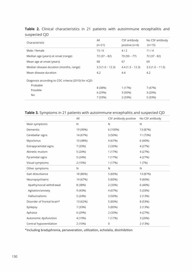
130
Table 2. Clinical characteristics in 21 patients with autoimmune encephalitis and suspected CJD
CharacteristicAll
(n=21)
CSF antibody
positive (n=6)
No CSF antibody
(n=15)
Male / female 15 / 6 4 / 2 11 / 4
Median age (years) at onset (range) 72 (37 – 82) 70 (50 – 77) 72 (37 - 82)
Mean age at onset (years) 68 67 69
Median disease duration (months, range) 3.3 (1.0 – 12.3) 4.4 (1.3 – 12.3) 3.3 (1.0 – 11.5)
Mean disease duration 4.2 4.4 4.2
Diagnosis according to CDC criteria (2010) for sCJD:
Probable
Possible
No
8 (38%)
6 (29%)
7 (33%)
1 (17%)
3 (50%)
2 (33%)
7 (47%)
3 (20%)
5 (33%)
Table 3. Symptoms in 21 patients with autoimmune encephalitis and suspected CJD All CSF antibody positive No CSF antibody
Main symptoms N N N
Dementia 19 (90%) 6 (100%) 13 (87%)
Cerebellar signs 14 (67%) 3 (50%) 11 (73%)
Myoclonus 10 (48%) 4 (67%) 6 (40%)
Extrapyramidal signs 7 (33%) 2 (33%) 4 (27%)
Akinetic mutism 5 (24%) 1 (17%) 4 (27%)
Pyramidal signs 5 (24%) 1 (17%) 4 (27%)
Visual symptoms 2 (10%) 1 (17%) 1 (7%)
Other symptoms N N N
Gait disturbance 18 (86%) 5 (83%) 13 (87%)
Neuropsychiatric 14 (67%) 5 (83%) 9 (60%)
Apathy/social withdrawal 8 (38%) 2 (33%) 6 (40%)
Agitation/anxiety 9 (43%) 4 (67%) 5 (33%)
Hallucinations 5 (24%) 3 (50%) 2 (13%)
Disorder of frontal brain* 13 (62%) 5 (83%) 8 (53%)
Epilepsy 7 (33%) 5 (83%) 2 (13%)
Aphasia 6 (29%) 2 (33%) 4 (27%)
Autonomic dysfunction 4 (19%) 1 (17%) 3 (20%)
Central hypoventilation 2 (10%) 0 2 (13%)
*Including bradyphrenia, perseveration, utilization, echolalia, disinhibition

131
Patho
log
ically con
firmed
auto
imm
un
e encePh
alitis in su
sPected creu
tzfeldt-Jako
b disease
7
Clinical characteristicsAll 21 patients with autoimmune encephalitis suffered from a rapidly progressive illness with a median duration of 3.3 months and died within 13 months of first symptoms (Table 2 and Tables S1-2). In retrospect, 14 of the 21 (67%) autoimmune encephalitis patients fulfilled the clinical 2010 CDC criteria for possible or probable sCJD while seven (33%) did not. In the subgroup of six patients with antineuronal antibody associated autoimmune encephalitis, also 67% (four patients) fulfilled CDC criteria for possible or probable sCJD (Table 2). The most frequent main symptoms included dementia (90%), cerebellar signs (67%), myoclonus (48%) and extrapyramidal signs (33%) (Table 3). Other symptoms that were frequently present included gait disturbance (86%), neuropsychiatric (67%) and frontal lobe symptoms (62%). There were no clear differences in clinical presentation of autoimmune encephalitis with or without detectable antineuronal antibodies in CSF (Table S5). Only four patients received immunotherapy consisting of steroids in two, intravenous immunoglobulins (ivIg) in one and both ivIg and steroids in one. None of the patients showed substantial improvement and immunotherapy was discontinued in all four.
Additional investigationsThe CSF had been examined in 18 of the 21 patients and was abnormal in 14/18 (78%) showing increased protein in 14 (78%) pleiocytosis in 10 (56%) and positive 14-3-3 protein in 9 (50%) (Table 4A).EEG was performed at least once in 19 of the patients and was abnormal in 18/19 (95%) with an increase of slow wave activities in 15/19 (79%). Seven of the patients showed triphasic periodic complexes that were considered compatible with CJD in 6 (32%). Epileptiform abnormalities were registered in 7 (37%) of the patients (Table 4B). MRI was abnormal in 10/17 (59%) of patients, showing increased signal on T2 weighted or FLAIR images (Table 4C). Imaging abnormalities were located in the medial temporal lobes in 3, periventricular in 5 (most likely vascular lesions in 3), basal ganglia and thalamus in 2 and brainstem in 1. None of the patients had MRI changes typical of sCJD, according to 2010 CDC criteria, although most investigations lacked DWI sequences.

132
Table 4A. CSF findings in 18/21 patients with autoimmune encephalitisAll Csf antibody positive no Csf antibody
CSF sampled (N/N) 18 / 21 6 / 6 12 / 15
Protein or cell count: N N N
Abnormal 14 (78%) 4 (67%) 10 (83%)
Normal 2 (11%) 0 2 (17%)
Not known 2 (11%) 2 (33%) 0
White cell count:
Median (x10E9/L; range) 12 (0-47) 16 (8-47) 11 (0-33)
Increased (>5x10E9/L) 10 (56%) 4 (67%) 6 (50%)
Normal 4 (22%) 0 4 (33%)
Not known 4 (22%) 2 (33%) 2 (17%)
14-3-3 protein
Positive 9 (50%) 2 (33%) 7 (58%)
Negative 5 (28%) 3 (50%) 2 (17%)
Not tested 4 (22%) 1 (17%) 3 (25%)
Table 4b. EEG in 19/21 patients with autoimmune encephalitisAll Csf antibody positive no Csf antibody
EEG performed (N/N) 19 / 21 6 / 6 13 / 15
EEG overall abnormal 18 (95%) 5 (83%) 13 (100%)
Increase in slow wave activities 15 (79%) 4 (67%) 11 (85%)
Triphasic periodic complexes 7 (37%) 3 (50%) 4 (31%)
Complexes compatible with CJD 6 (32%) 2 (33%) 4 (31%)
Table 4C. MRI findings in 17/21 patients with autoimmune encephalitisAll Csf antibody positive no Csf antibody
MRI performed (N/N) 17 / 21 6 / 6 11 / 15
MRI overall
Abnormal 10 (59%) 4 (67%) 6 (55%)
Normal 7 (41%) 2 (33%) 5 (45%)
T2/FLAIR increased signal
Yes 10 (59%) 4 (67%) 6 (55%)
No 7 (41%) 2 (33%) 5 (45%)
Gadolineum enhancement
Yes 0
No 17 (100%)
133
Patho
log
ically con
firmed
auto
imm
un
e encePh
alitis in su
sPected creu
tzfeldt-Jako
b disease
7
The autoimmune encephalitis patients with antineuronal autoantibodies in their CSF did not clearly differ from the antibody negative patients in CSF, EEG and MRI findings (Table 4A-C).Areas with increased T2/FLAIR signal included periventricular white matter (5), mesiotemporal lobe and hippocampus (3), brainstem (3), basal ganglia and thalamus (2), cortex (2) and internal capsule (1). Generalized atrophy was present in five patients, bifrontal atrophy in two, and hippocampal atrophy in one patient.
Associated tumorsIn nine patients (43%) a tumor was present. Based on criteria published by an EU consortium, the etiology of the neurological syndrome could be classified as definitely paraneoplastic in five and possibly paraneoplastic in two.14 The tumor was probably unrelated in two of the patients precluding a paraneoplastic etiology. Detailed clinical and oncological findings are presented in supplementary Table S2 and supplementary information (2).
dIsCussIOnThe first main finding of this study is that approximately 7% of patients with suspected CJD have autoimmune encephalitis at autopsy. Secondly, antineuronal autoantibodies could be demonstrated in the CSF from six of 21 (29%) patients with pathologically confirmed autoimmune encephalitis. One case each of anti-Hu, GABAB1/B2, NMDAR, Caspr2 was detected, while two CSF samples had antibodies against an unidentified NSA. Thirdly, in retrospect 67% of the patients fulfilled the 2010 CDC criteria for probable or possible CJD, both in the antibody positive and negative group. Several other studies have addressed the clinical overlap between prion disorders and autoimmune encephalitis. Chitravas et al.3 performed a retrospective clinical and neuropathological review of prion-negative brain autopsy cases referred to the US National Prion Disease Pathology Surveillance Center. Neuropathology diagnosed an immune-mediated disorder in 26/304 (8.5%) of the prion-negative cases with histology, including primary angiitis of the CNS (7), ADEM (6), limbic encephalitis (6), neurosarcoidosis (4), paraneoplastic cerebellar degeneration (2) and granulomatosis with polyangiitis (Wegener’s) (1). This study did not report the antineuronal antibody status of the patients. In contrast to our study, most of the misdiagnosed patients did not fulfill WHO criteria for CJD. Grau-Rivera et al.1 examined the frequency of NSA-Abs in the CSF of patients with suspected or pathologically confirmed CJD. NSA-abs were detected in 6 of 346 patients (1.7%) with rapid neurologic deterioration suggestive of CJD during life. In contrast to our study, none of these 6 patients fulfilled the diagnostic criteria for probable or possible
134
CJD. The target antigens included Caspr2, LGI1, NMDAR, aquaporin 4, Tr (DNER), and an unknown protein. All patients improved or stabilized after appropriate treatment. None of the 49 patients with definite CJD had NSA-abs. Another study reported serum NSA-Abs in <5% of patients with sCJD, all at very low titers.2 Our study is the first to test CSF of all patients identified with autoimmune encephalitis at autopsy. CSF is proven more sensitive and specific to detect NSA-Abs as shown for NMDR,15 AMPAR,16 and GABABR.17 In 50% of our patients with autoimmune encephalitis tested, the CSF 14-3-3 protein was elevated (part of the CDC’s 2010 diagnostic criteria) casting further doubt on the utility of this test in the diagnosis of CJD.18 EEG was also not very useful as triphasic waves were present in 32% of our autoimmune patients. None of our autoimmune encephalitis patients showed sCJD specific MRI abnormalities, supporting the higher specificity of sCJD MRI criteria over 14-3-3.18,19 However, DWI was not performed in most of our patients and we do not know whether DWI would have retained its specificity in this series. On the other hand, 56% of patients had an increased WBC count (>5x10E9/L) in their CSF. Although the pleiocytosis was usually mild it does provide a clue to an extensive search into an inflammatory etiology.Other studies describing clinical overlap between prion and autoimmune disorders focused primarily on rapidly progressive dementia, detecting a 2% to 9% prevalence of immune-mediated CNS disorders.20-22 In a series of patients with suspected autoimmune dementia and good response to immunotherapy, almost 9% of the disorders had initially been diagnosed as sCJD.23 In particular, patients with encephalitis associated with LGI1-Abs may present with myoclonus-like movements and other symptoms that can be mistaken for CJD.4 In general, the prognosis of autoimmune encephalitis appears to be determined by the involved immunopathogenic mechanisms.24 Disorders associated with antibodies directed against intracellular antigens are probably T cell mediated and respond poorly to immunotherapy. Most paraneoplastic neurological disorders with onconeural antibodies belong to this category. On the other hand, in disorders associated with NSA-Abs, the antibodies are probably pathogenic and these disorders respond favorably to first and second line immunotherapy. Examples include NMDAR,5 LGI125,26 and Caspr226,27 encephalitis. The immunopathology of NSA-Abs associated encephalitis shows mild to moderate, mainly perivascular, lymphocytic infiltrations containing both B and T cells, antibody producing cells and IgG deposits.28-30 It is possible that we missed some cases with ‘burned out’ or with very bland neuropathology and the 22 cases that we identified represent an underestimation. The encephalitis associated with anti-Hu or Ma2 antibodies is characterized by extensive B and T cell lymphocytic infiltrations, containing T cells expressing markers of cytotoxicity such as TIA-1, granzyme B, perforin and Fas/Fas-ligand.30-32 In our series, immunopathological findings did not differ between autoantibody negative patients and the patients with NSA-Ab (Table S3), nor did clinical, CSF, radiological or electrophysiological characteristics (Tables S4-S8). The

135
Patho
log
ically con
firmed
auto
imm
un
e encePh
alitis in su
sPected creu
tzfeldt-Jako
b disease
7
immunopathological mechanism in the 15 autoimmune encephalitis patients without detectable autoantibodies remains unclear. It cannot be excluded that some of these patients harbored NSA-Abs that were degraded by postmortem interval or during storage at -800C in low protein CSF.Obviously all limitations based on the retrospective nature of the study are applicable. In addition, bias was introduced by the fact that all patients had ‘suspected CJD’ and had come to autopsy. In particular the poor outcome of the patients with a median survival of 3.3 months (range 1.0 – 12.3 months) in this study of autoimmune encephalitis was largely determined by this inclusion bias. In conclusion, it is important to consider both prion and immune-mediated disorders in the differential diagnosis of rapidly progressive neurological deficits, knowing that EEG and 14-3-3 in CSF do not discriminate sufficiently to distinguish the two. Detection of specific antineuronal antibodies in serum and/or CSF can establish a diagnosis of autoimmune encephalitis and guide appropriate treatment.

136
suPPleMenTARy dATA
SuPPlemeNTary INFormaTIoN (1)PATIenTs wITh AnTIneuROnAl AuTOAnTIbOdIes In Csf OR seRuM
PATIenT 6, AnTI-huA 74-year old man was admitted to the hospital because of a progressive gait disturbance with orthostatic hypotension followed by rapidly progressive dementia, pyramidal and cerebellar signs, myoclonus and focal epileptic seizures. Brain MRI showed generalized atrophy, periventricular leuko-araiosis and multiple lacunar lesions. EEG showed epileptiform activity, slowing and triphasic periodic complexes. CSF showed moderately elevated 14-3-3 protein. Paraneoplastic antibody testing showed anti-Hu and anti-Zic4 antibodies in serum (test result came in after the patient had died). Despite an extensive search, no tumor was detected. The patient developed pneumonia and died 2.6 months after presentation. At autopsy a 3 cm tumor was found in the apex of the lower lobe of the right lung. Microscopy showed limbic encephalitis and ganglionitis of the gut. The tumor was SCLC.CommentThe patient presented with rapidly progressive symptoms indicating involvement of many different areas of the nervous system, consistent with paraneoplastic encephalomyelitis. A paraneoplastic etiology was confirmed by detection of a lung tumor at autopsy and the presence of anti-Hu antibodies. Moderately increased 14-3-3 protein has been reported in many rapidly progressive disorders with neuronal loss, including anti-Hu associated limbic encephalitis. 33
PATIenT 15, AnTI-gAbAB1/2
A 70-year old woman was admitted to the hospital after a single vehicle accident, probably caused by an epileptic seizure. On neurological examination she was disoriented without focal signs. During admission, her behavior became increasingly childish and frontal (e.g. disinhibition while eating). She developed hallucinations and delusions followed by episodes of obtundation. The family suggested that in the 1-2 months before admission she was more irritable, aggressive and sometimes disinhibited.Brain MRI was normal while EEG showed left temporal epileptiform activity. CSF analysis showed normal protein, 14 WBC, IgG-G index 1.12. PCR for neurotropic viruses and Borrelia serology were negative. She died one week after admission. Autopsy (brain only) showed lymphocytic perivascular infiltrates and microgliosis in the frontal and temporal lobes and hippocampus (Fig. S1).CommentA subacute onset with seizures, confusion and behavioral problems is typical of autoimmune encephalitis associated with antibodies against the GABAB receptor.34 In
137
Patho
log
ically con
firmed
auto
imm
un
e encePh
alitis in su
sPected creu
tzfeldt-Jako
b disease
7
approximately 50% of the patients an underlying tumor, usually SCLC, can be detected. The majority of patients have an abnormal MRI and improve after immunotherapy and tumor treatment (if a tumor is found). In this patient the MRI was normal, although the diagnostic value was limited by movement artefacts. No tumor search had been performed and no treatment had been started, mainly due to lack of diagnosis.
PATIenT 17, AnTI-nMdARA 49-year old woman was admitted to a psychiatric hospital because of apathy and catatonia. Six months earlier, her husband had first noted that she was more irritable. A month before admission she became more emotional, complained of headache and photophobia and working on a computer became difficult to impossible. A few days before admission her behavior became chaotic. Hyperactivity (e.g. washing) and moments of complete apathy alternated. She could hardly stand or walk. At nights there was dystonic positioning and twisting movements of her hands. On examination she lay in bed with her eyes opened, did not follow commands and did not speak (E4M4V1). There were no focal signs. Myoclonic jerks were observed in her face, around her mouth and left hand. After 3 days she was transferred to a general hospital because of decreased consciousness, admitted to the ICU and intubated. Brain MRI was normal and EEG showed multifocal status epilepticus. CSF analysis showed 47 WBC with normal protein and glucose. She developed treatment resistant multifocal status epilepticus requiring multiple inductions of phenobarbital coma. On suspicion of limbic encephalitis, she was treated with methylprednisolone (1000 mg/d, during 3 days), prednisone (60 mg/d oral) and IVIG without improvement. Just before the patient’s admission, a series of young women presenting with prominent psychiatric symptoms, seizures, memory deficits, and decreased level of consciousness associated with ovarian teratomas had been published. 35 Therefore, both ovaries were removed, but no teratoma was found at pathological examination. She never regained consciousness and died 5 months after ICU admission. Autopsy showed perivascular lymphocytic infiltrations in centrum semiovale, basal ganglia and hippocampus.CommentThe clinical picture is typical of anti-NMDAR encephalitis and with current knowledge the patient would have been treated with second line immunotherapy (e.g. rituximab and/or cyclophosphamide).5
PATIenT 21, AnTI-CAsPR2A 55-year old man developed over three months progressive compulsive behavior, ataxia, dysarthria, insomnia and extrapyramidal signs. Brain MRI showed increased signal on T2 weighted images in both medial temporal lobes. CSF showed normal concentration of 14-3-3 protein.

138
After choking on a piece of bread he was found without pulse and was resuscitated. In the ICU he developed multiple organ failure, persistent status epilepticus and ultimately became asystolic and died 3.3 months after onset. Autopsy demonstrated extensive hypoxic changes in the cerebral cortex and loss of cerebellar Purkinje cells. Additional staining showed extensive lymphocytic infiltrates. CommentThe clinical presentation with neuropsychiatric symptoms, ataxia and insomnia is consistent with anti-Caspr2 associated Morvan syndrome.30,36
PATIenT 18, synAPTIC A 77-year old man was admitted with rapidly progressive cognitive deterioriation. Symptoms started with hallucinations and delusions followed by unsteady gait, trouble finding and understanding words, urinary incontinence and fluctuating levels of consciousness. On examination he was anxious and agitated while showing disinhibited behaviour. He showed dressing apraxia, perseveration, utilisation behaviour and echolalia. He had second degree nystagmus, gait ataxia, sporadic myoclonus and positive frontal release signs. MRI showed aspecific probably vascular white matter lesions and mild hippocampus atrophy (Scheltens grade II). The CSF showed 18 WBC/ml (95% lymphocytes), total protein 0.7 g/l (mildly elevated), normal tau and negative 14-3-3 protein, positive oligoclonal bands (more than in serum). Negative cytology.EEG showed increase of slow wave activities and appearance of triphasic periodic complexes.During admission his condition rapidly worsened with increasing anxiety, agitation and myoclonus. He died approximately 5 weeks after onset of symptoms. At autopsy perivascular inflammatory infiltrates consisting of B and T cells were noted in hippocampus and basal ganglia with local infiltration of the parenchyma (also consisting of B and T cells) (Fig. S1).CommentThe patient fulfilled the criteria of probable CJD (see Methods) and did not receive immunotherapy. Autopsy showed typical autoimmune or paraneoplastic encephalitis. The unspecified synaptic antibody detected in CSF collected postmortem (Fig. S2) may have contributed to the pathogenesis.
PATIenT 11, synAPTICA 73-year old male patient presented with confusion and auditory hallucinations. Later he developed seizures. On examination he was disinhibited, the MMSE was 22 out of 30 and myoclonus was observed. MRI showed increased T2 signal in the left hippocampus. CSF showed a mononuclear pleiocytosis of 8 cell/�l, elevated tau and positive 14-3-3 protein. PCR for neurotropic
139
Patho
log
ically con
firmed
auto
imm
un
e encePh
alitis in su
sPected creu
tzfeldt-Jako
b disease
7
viruses was negative as was anti-viral serology. EEG was normal.The patient died of aspiration pneumonia, 4.7 months after onset of symptoms. Autopsy showed perivascular B and T cells mainly in the basal ganglia and hippocampus. In addition, in the basal ganglia intraparenchymal T cells were observed. CD68 positive macrophages were present in affected areas with perivascular and intraparenchymal distribution.CommentThe patient did not receive immunotherapy; the unspecified synaptic antibody detected in CSF collected postmortem (Fig. S2) may have contributed to the pathogenesis.
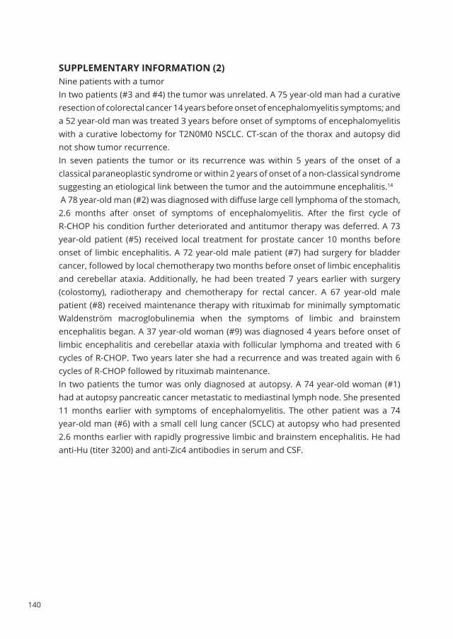
140
SuPPlemeNTary INFormaTIoN (2)Nine patients with a tumorIn two patients (#3 and #4) the tumor was unrelated. A 75 year-old man had a curative resection of colorectal cancer 14 years before onset of encephalomyelitis symptoms; and a 52 year-old man was treated 3 years before onset of symptoms of encephalomyelitis with a curative lobectomy for T2N0M0 NSCLC. CT-scan of the thorax and autopsy did not show tumor recurrence.In seven patients the tumor or its recurrence was within 5 years of the onset of a classical paraneoplastic syndrome or within 2 years of onset of a non-classical syndrome suggesting an etiological link between the tumor and the autoimmune encephalitis.14
A 78 year-old man (#2) was diagnosed with diffuse large cell lymphoma of the stomach, 2.6 months after onset of symptoms of encephalomyelitis. After the first cycle of R-CHOP his condition further deteriorated and antitumor therapy was deferred. A 73year-old patient (#5) received local treatment for prostate cancer 10 months beforeonset of limbic encephalitis. A 72 year-old male patient (#7) had surgery for bladdercancer, followed by local chemotherapy two months before onset of limbic encephalitisand cerebellar ataxia. Additionally, he had been treated 7 years earlier with surgery(colostomy), radiotherapy and chemotherapy for rectal cancer. A 67 year-old malepatient (#8) received maintenance therapy with rituximab for minimally symptomaticWaldenström macroglobulinemia when the symptoms of limbic and brainstemencephalitis began. A 37 year-old woman (#9) was diagnosed 4 years before onset oflimbic encephalitis and cerebellar ataxia with follicular lymphoma and treated with 6cycles of R-CHOP. Two years later she had a recurrence and was treated again with 6cycles of R-CHOP followed by rituximab maintenance.In two patients the tumor was only diagnosed at autopsy. A 74 year-old woman (#1)had at autopsy pancreatic cancer metastatic to mediastinal lymph node. She presented11 months earlier with symptoms of encephalomyelitis. The other patient was a 74year-old man (#6) with a small cell lung cancer (SCLC) at autopsy who had presented2.6 months earlier with rapidly progressive limbic and brainstem encephalitis. He hadanti-Hu (titer 3200) and anti-Zic4 antibodies in serum and CSF.
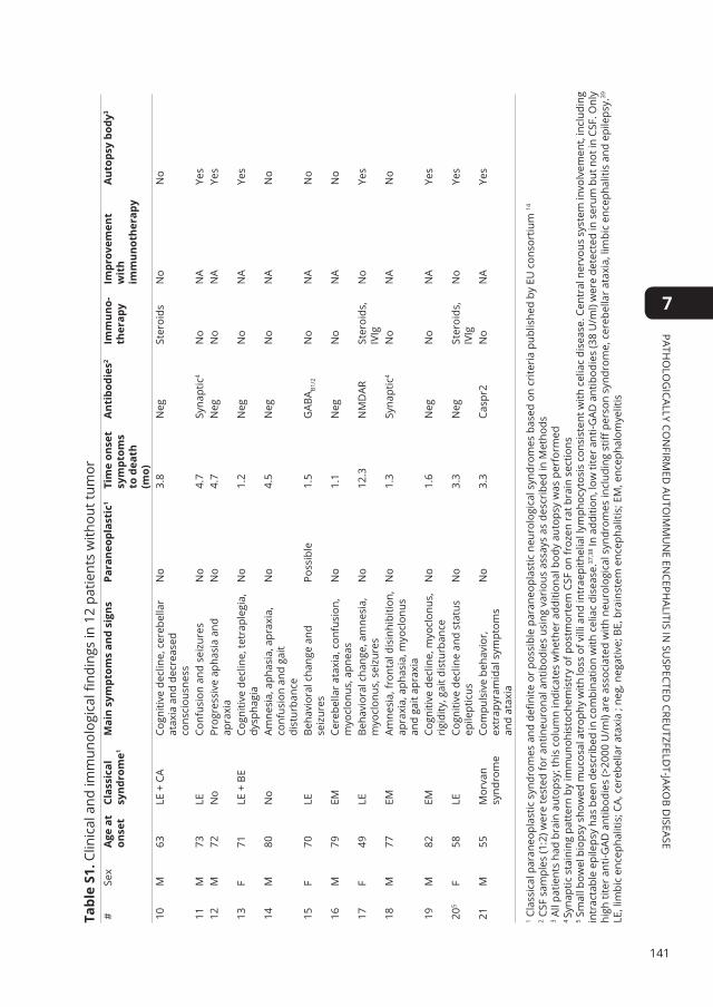
141
Patho
log
ically con
firmed
auto
imm
un
e encePh
alitis in su
sPected creu
tzfeldt-Jako
b disease
7
Tabl
e s1
. Clin
ical
and
imm
unol
ogic
al fi
ndin
gs in
12
patie
nts
with
out t
umor
#Se
xA
ge a
t on
set
Clas
sica
l sy
ndro
me1
Mai
n sy
mpt
oms
and
sign
sPa
rane
opla
stic
1Ti
me
onse
t sy
mpt
oms
to d
eath
(m
o)
Ant
ibod
ies2
Imm
uno-
ther
apy
Impr
ovem
ent
wit
h im
mun
othe
rapy
Aut
opsy
bod
y3
10M
63LE
+ C
ACo
gniti
ve d
eclin
e, c
ereb
ella
r at
axia
and
dec
reas
ed
cons
ciou
snes
s
No
3.8
Neg
Ster
oids
No
No
11M
73LE
Conf
usio
n an
d se
izur
esN
o4.
7Sy
napt
ic4
No
NA
Yes
12M
72N
oPr
ogre
ssiv
e ap
hasi
a an
d ap
raxi
aN
o4.
7N
egN
oN
AYe
s
13F
71LE
+ B
ECo
gniti
ve d
eclin
e, te
trap
legi
a,
dysp
hagi
aN
o1.
2N
egN
oN
AYe
s
14M
80N
oAm
nesi
a, a
phas
ia, a
prax
ia,
conf
usio
n an
d ga
it di
stur
banc
e
No
4.5
Neg
No
NA
No
15F
70LE
Beha
vior
al c
hang
e an
d se
izur
esPo
ssib
le1.
5G
ABA B1
/2N
oN
AN
o
16M
79EM
Cere
bella
r at
axia
, con
fusi
on,
myo
clon
us, a
pnea
sN
o1.
1N
egN
oN
AN
o
17F
49LE
Beha
vior
al c
hang
e, a
mne
sia,
m
yocl
onus
, sei
zure
sN
o12
.3N
MD
AR
Ster
oids
, IV
IgN
oYe
s
18M
77EM
Amne
sia,
fron
tal d
isin
hibi
tion,
ap
raxi
a, a
phas
ia, m
yocl
onus
an
d ga
it ap
raxi
a
No
1.3
Syna
ptic
4N
oN
AN
o
19M
82EM
Cogn
itive
dec
line,
myo
clon
us,
rigi
dity
, gai
t dis
turb
ance
No
1.6
Neg
No
NA
Yes
205
F58
LECo
gniti
ve d
eclin
e an
d st
atus
ep
ilept
icus
No
3.3
Neg
Ster
oids
, IV
IgN
oYe
s
21M
55M
orva
n sy
ndro
me
Com
puls
ive
beha
vior
, ex
trap
yram
idal
sym
ptom
s an
d at
axia
No
3.3
Casp
r2N
oN
AYe
s
1 Cla
ssic
al p
aran
eopl
astic
syn
drom
es a
nd d
efini
te o
r po
ssib
le p
aran
eopl
astic
neu
rolo
gica
l syn
drom
es b
ased
on
crite
ria
publ
ishe
d by
EU
con
sort
ium
14
2 CSF
sam
ples
(1:2
) wer
e te
sted
for
antin
euro
nal a
ntib
odie
s us
ing
vari
ous
assa
ys a
s de
scri
bed
in M
etho
ds3 A
ll pa
tient
s ha
d br
ain
auto
psy;
this
col
umn
indi
cate
s w
heth
er a
dditi
onal
bod
y au
tops
y w
as p
erfo
rmed
4 Sy
napt
ic s
tain
ing
patt
ern
by im
mun
ohis
toch
emis
try
of p
ostm
orte
m C
SF o
n fr
ozen
rat
bra
in s
ectio
ns5 S
mal
l bow
el b
iops
y sh
owed
muc
osal
atr
ophy
with
loss
of v
illi a
nd in
trae
pith
elia
l lym
phoc
ytos
is c
onsi
sten
t with
cel
iac
dise
ase.
Cen
tral
ner
vous
sys
tem
invo
lvem
ent,
incl
udin
g in
trac
tabl
e ep
ileps
y ha
s be
en d
escr
ibed
in c
ombi
natio
n w
ith c
elia
c di
seas
e.37
,38 I
n ad
ditio
n, lo
w ti
ter a
nti-G
AD a
ntib
odie
s (3
8 U
/ml)
wer
e de
tect
ed in
ser
um b
ut n
ot in
CSF
. Onl
y hi
gh ti
ter
anti-
GAD
ant
ibod
ies
(>20
00 U
/ml)
are
asso
ciat
ed w
ith n
euro
logi
cal s
yndr
omes
incl
udin
g st
iff p
erso
n sy
ndro
me,
cer
ebel
lar
atax
ia, l
imbi
c en
ceph
aliti
s an
d ep
ileps
y.39
LE, l
imbi
c en
ceph
aliti
s; C
A, c
ereb
ella
r at
axia
; ne
g, n
egat
ive;
BE,
bra
inst
em e
ncep
halit
is; E
M, e
ncep
halo
mye
litis
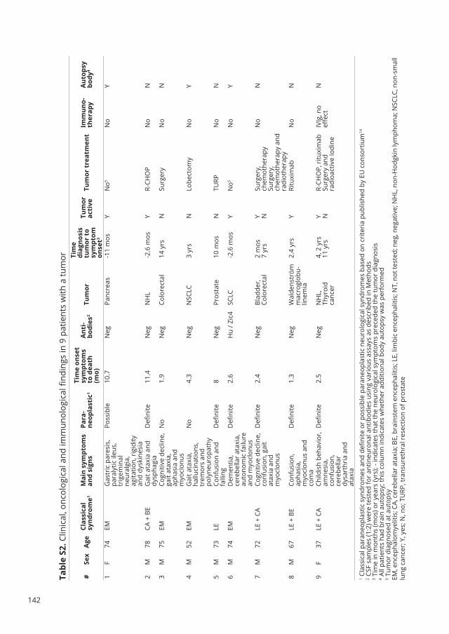
142
Tabl
e s2
. Clin
ical
, onc
olog
ical
and
imm
unol
ogic
al fi
ndin
gs in
9 p
atie
nts
with
a tu
mor
#se
xA
geCl
assi
cal
synd
rom
e1M
ain
sym
ptom
s an
d si
gns
Para
-ne
opla
stic
1
Tim
e on
set
sym
ptom
s to
dea
th
(mo)
Ant
i-bo
dies
2Tu
mor
Tim
e di
agno
sis
tum
or t
o sy
mpt
om
onse
t3
Tum
or
acti
veTu
mor
tre
atm
ent
Imm
uno-
ther
apy
Aut
opsy
bo
dy4
1F
74EM
Gas
tric
par
esis
, pa
raly
tic il
eus,
tr
igem
inal
ne
ural
gia,
ag
itatio
n, r
igid
ity
and
dysk
ines
ia
Poss
ible
10.7
Neg
Panc
reas
-11
mos
YN
o5N
oY
2M
78CA
+ B
EG
ait a
taxi
a an
d dy
spha
gia
Defi
nite
11.4
Neg
NH
L-2
.6 m
osY
R-CH
OP
No
N
3M
75EM
Cogn
itive
dec
line,
ga
it at
axia
, ap
hasi
a an
d m
yocl
onus
No
1.9
Neg
Colo
rect
al14
yrs
NSu
rger
yN
oN
4M
52EM
Gai
t ata
xia,
ha
lluci
natio
ns,
trem
ors
and
poly
neur
opat
hy
No
4.3
Neg
NSC
LC3
yrs
NLo
bect
omy
No
Y
5M
73LE
Conf
usio
n an
d fa
lling
Defi
nite
8N
egPr
osta
te10
mos
NTU
RPN
oN
6M
74EM
Dem
entia
, ce
rebe
llar
atax
ia,
auto
nom
ic fa
ilure
an
d m
yocl
onus
Defi
nite
2.6
Hu
/ Zic
4SC
LC-2
.6 m
osY
No5
No
Y
7M
72LE
+ C
ACo
gniti
ve d
eclin
e,
conf
usio
n, g
ait
atax
ia a
nd
myo
clon
us
Defi
nite
2.4
Neg
Blad
der,
Colo
rect
al2
mos
7 yr
sY N
Surg
ery,
ch
emot
hera
pySu
rger
y,
chem
othe
rapy
and
ra
diot
hera
py
No
N
8M
67LE
+ B
ECo
nfus
ion,
ap
hasi
a,
myo
clon
us a
nd
com
a
Defi
nite
1.3
Neg
Wal
dens
tröm
m
acro
glob
u-lin
emia
2.4
yrs
YRi
tuxi
mab
No
N
9F
37LE
+ C
ACh
ildis
h be
havi
or,
amne
sia,
co
nfus
ion,
ce
rebe
llar
dysa
rthr
ia a
nd
atax
ia
Defi
nite
2.5
Neg
NH
L,Th
yroi
d ca
ncer
4, 2
yrs
11 y
rsY N
R-CH
OP,
ritu
xim
abSu
rger
y an
d ra
dioa
ctiv
e Io
dine
IVIg
, no
effec
tN
1 Cla
ssic
al p
aran
eopl
astic
syn
drom
es a
nd d
efini
te o
r po
ssib
le p
aran
eopl
astic
neu
rolo
gica
l syn
drom
es b
ased
on
crite
ria
publ
ishe
d by
EU
con
sort
ium
14
2 CSF
sam
ples
(1:2
) wer
e te
sted
for
antin
euro
nal a
ntib
odie
s us
ing
vari
ous
assa
ys a
s de
scri
bed
in M
etho
ds3 T
ime
in m
onth
s (m
os) o
r ye
ars
(yrs
); - i
ndic
ates
that
the
neur
olog
ical
sym
ptom
s pr
eced
ed th
e tu
mor
dia
gnos
is4 A
ll pa
tient
s ha
d br
ain
auto
psy;
this
col
umn
indi
cate
s w
heth
er a
dditi
onal
bod
y au
tops
y w
as p
erfo
rmed
5 Tum
or d
iagn
osed
at a
utop
syEM
, enc
epha
lom
yelit
is; C
A, c
ereb
ella
r ata
xia;
BE,
bra
inst
em e
ncep
halit
is; L
E, li
mbi
c en
ceph
aliti
s; N
T, n
ot te
sted
; neg
, neg
ativ
e; N
HL,
non
-Hod
gkin
lym
phom
a; N
SCLC
, non
-sm
all
lung
can
cer;
Y, y
es; N
, no;
TU
RP, t
rans
uret
hral
res
ectio
n of
pro
stat
e

143
Patho
log
ically con
firmed
auto
imm
un
e encePh
alitis in su
sPected creu
tzfeldt-Jako
b disease
7
Table s3. Neuropathological findings in 21 patients with autoimmune encephalitis and suspected CJD
Predominant localization of
infiltrates:
NSA-Abs positive
(n=5)
Anti-Hu positive
(n=1)
Antibody negative
(n=15)
All patients
(n=21)
Perivascular 4 1 10 15
Parenchymal 1 4 5
Perivascular and parenchymal 1 1
Composition of infiltrates:
CD3 positive 5/5 Nt 9/10 14/15
CD8 positive 1/1 Nt 6/7 7/8
CD4 positive 1/1 Nt 3/4 4/5
CD20 positive 3/4 Nt 5/8 8/12
CD68 positive 2/2 1/1 8/10 11/13
NSA-Abs, antibodies directed against neuronal surface antigens; Nt, not tested

144
suPPleMenTARy fIguRes
figure s1 Neuropathology in autoimmune encephalitis associated with antibodies directed against GABAB1/2 and an unidentified synaptic antigen(A,B) CD3 positive T cells in the hippocampus of a 70 year old woman with limbic encephalitis and GABAB1/2 antibodies in the CSF (patient #15). Note T cells both in the parenchyma and perivascularly; magnification 5x (A) and 20x (B) objective respectively. (C) CD3 positive T cells in the temporal cortex of a 77 y old man with rapidly progressive encephalomyelitis with antibodies in the CSF against unknown epitope (patient #18); magnification 10x objective. (D) CD3 positive T cells in the globus pallidus of a 73 y old psychotic and epileptic patient with antibodies in the CSF against unknown epitope (patient # 11); magnification 20x objective.

145
Patho
log
ically con
firmed
auto
imm
un
e encePh
alitis in su
sPected creu
tzfeldt-Jako
b disease
7
RefeRenCes
1. Grau-Rivera O, Sanchez-Valle R, Saiz A, et
al. Determination of neuronal antibodies
in suspected and definite Creutzfeldt-Jakob
disease. JAMA neurology 2014;71:74-8.
2. Rossi M, Mead S, Collinge J, Rudge P, Vincent A.
Neuronal antibodies in patients with suspected
or confirmed sporadic Creutzfeldt-Jakob disease.
J Neurol Neurosurg Psychiatry 2015;86:692-4.
3. Chitravas N, Jung RS, Kofskey DM, et al.
Treatable neurological disorders misdiagnosed
as Creutzfeldt-Jakob disease. Ann Neurol
2011;70:437-44.
4. Geschwind MD, Tan KM, Lennon VA, et al.
Voltage-gated potassium channel autoimmunity
mimicking creutzfeldt-jakob disease. Arch
Neurol 2008;65:1341-6.
5. Titulaer MJ, McCracken L, Gabilondo I, et al.
Treatment and prognostic factors for long-term
outcome in patients with anti-NMDA receptor
encephalitis: an observational cohort study.
Lancet Neurol 2013;12:157-65.
6. Vincent A, Buckley C, Schott JM, et al. Potassium
channel antibody-associated encephalopathy: a
potentially immunotherapy-responsive form of
limbic encephalitis. Brain 2004;127:701-12.
7. Jansen C, Parchi P, Capellari S, et al. Human
prion diseases in the Netherlands (1998-2009):
clinical, genetic and molecular aspects. PloS one
2012;7:e36333.
8. Petit-Pedrol M, Armangue T, Peng X, et al.
Encephalitis with refractory seizures, status
epilepticus, and antibodies to the GABAA
receptor: a case series, characterisation of the
antigen, and analysis of the effects of antibodies.
Lancet Neurol 2014;13:276-86.
9. Moll JWB, Vecht CJ. Immune diagnosis of
paraneoplastic neurological disease. Clin Neurol
Neurosurg 1995;97:71-81.
10. Maat P, Brouwer E, Hulsenboom E, et
al. Multiplex serology of paraneoplastic
antineuronal antibodies. J Immunol Methods
2013;391:125-32.
11. Maat P, de Graaff E, van Beveren NM, et al.
Psychiatric phenomena as initial manifestation
of encephalitis by anti-NMDA receptor
antibodies. Submitted 2011.
12. de Graaff E, Maat P, Hulsenboom E, et al.
Identification of delta/notch-like epidermal
growth factor-related receptor as the Tr antigen
in paraneoplastic cerebellar degeneration. Ann
Neurol 2012;71:815-24.
13. Jansen C, Parchi P, Capellari S, et al. Prion
protein amyloidosis with divergent phenotype
associated with two novel nonsense mutations
in PRNP. Acta Neuropathol 2010;119:189-97.
14. Graus F, Delattre JY, Antoine JC, et al.
Recommended diagnostic criteria for
paraneoplastic neurological syndromes. J
Neurol Neurosurg Psychiatry 2004;75:1135-40.
15. Gresa-Arribas N, Titulaer MJ, Torrents A, et al.
Antibody titres at diagnosis and during follow-
up of anti-NMDA receptor encephalitis: a
retrospective study. Lancet Neurol 2014;13:167-
77.
16. Hoftberger R, van Sonderen A, Leypoldt F, et
al. Encephalitis and AMPA receptor antibodies:
Novel findings in a case series of 22 patients.
Neurology 2015.
17. Hoftberger R, Titulaer MJ, Sabater L, et al.
Encephalitis and GABAB receptor antibodies:
novel findings in a new case series of 20 patients.
Neurology 2013;81:1500-6.
18. Paterson RW, Torres-Chae CC, Kuo AL, et al.
Differential diagnosis of Jakob-Creutzfeldt
disease. Arch Neurol 2012;69:1578-82.
19. Vitali P, Maccagnano E, Caverzasi E, et al.

146
Diffusion-weighted MRI hyperintensity patterns
differentiate CJD from other rapid dementias.
Neurology 2011;76:1711-9.
20. Geschwind MD, Shu H, Haman A, Sejvar JJ, Miller
BL. Rapidly progressive dementia. Ann Neurol
2008;64:97-108.
21. Papageorgiou SG, Kontaxis T, Bonakis A,
Karahalios G, Kalfakis N, Vassilopoulos D.
Rapidly progressive dementia: causes found
in a Greek tertiary referral center in Athens.
Alzheimer Dis Assoc Disord 2009;23:337-46.
22. Sala I, Marquie M, Sanchez-Saudinos MB, et al.
Rapidly progressive dementia: experience in
a tertiary care medical center. Alzheimer Dis
Assoc Disord 2012;26:267-71.
23. Flanagan EP, McKeon A, Lennon VA, et al.
Autoimmune dementia: clinical course and
predictors of immunotherapy response. Mayo
Clin Proc 2010;85:881-97.
24. Lancaster E, Dalmau J. Neuronal autoantigens-
-pathogenesis, associated disorders and
antibody testing. Nat Rev Neurol 2012;8:380-90.
25. Lai M, Huijbers MG, Lancaster E, et al.
Investigation of LGI1 as the antigen in limbic
encephalitis previously attributed to potassium
channels: a case series. Lancet Neurol
2010;9:776-85.
26. Irani SR, Alexander S, Waters P, et al. Antibodies
to Kv1 potassium channel-complex proteins
leucine-rich, glioma inactivated 1 protein
and contactin-associated protein-2 in limbic
encephalitis, Morvan’s syndrome and acquired
neuromyotonia. Brain 2010;133:2734-48.
27. Lancaster E, Huijbers MG, Bar V, et al.
Investigations of caspr2, an autoantigen of
encephalitis and neuromyotonia. Ann Neurol
2011;69:303-11.
28. Tuzun E, Zhou L, Baehring JM, Bannykh S,
Rosenfeld MR, Dalmau J. Evidence for antibody-
mediated pathogenesis in anti-NMDAR
encephalitis associated with ovarian teratoma.
Acta Neuropathol 2009.
29. Martinez-Hernandez E, Horvath J, Shiloh-
Malawsky Y, Sangha N, Martinez-Lage M,
Dalmau J. Analysis of complement and plasma
cells in the brain of patients with anti-NMDAR
encephalitis. Neurology 2011;77:589-93.
30. Bien CG, Vincent A, Barnett MH, et al.
Immunopathology of autoantibody-associated
encephalitides: clues for pathogenesis. Brain
2012;135:1622-38.
31. Bernal F, Graus F, Pifarre A, Saiz A, Benyahia
B, Ribalta T. Immunohistochemical analysis
of anti-Hu-associated paraneoplastic
encephalomyelitis. Acta Neuropathol (Berl)
2002;103:509-15.
32. Blumenthal DT, Salzman KL, Digre KB, Jensen RL,
Dunson WA, Dalmau J. Early pathologic findings
and long-term improvement in anti-Ma2-
associated encephalitis. Neurology 2006;67:146-
9.
33. Saiz A, Graus F, Dalmau J, Pifarre A, Marin C,
Tolosa E. Detection of 14-3-3 brain protein
in the cerebrospinal fluid of patients with
paraneoplastic neurological disorders. Ann
Neurol 1999;46:774-7.
34. Lancaster E, Lai M, Peng X, et al. Antibodies to
the GABA(B) receptor in limbic encephalitis with
seizures: case series and characterisation of the
antigen. Lancet Neurol 2010;9:67-76.
35. Vitaliani R, Mason W, Ances B, Zwerdling T,
Jiang Z, Dalmau J. Paraneoplastic encephalitis,
psychiatric symptoms, and hypoventilation in
ovarian teratoma. Ann Neurol 2005;58:594-604.
36. Irani S, Pettingill P, Kleopa KA, et al. Morvan
syndrome: Clinical and serological observations
in 29 cases. Ann Neurol 2012;72:241-55.
37. Ribaldone DG, Astegiano M, Fagoonee S,
Rizzetto M, Pellicano R. Epilepsy and celiac
disease: review of literature. Panminerva Med

147
Patho
log
ically con
firmed
auto
imm
un
e encePh
alitis in su
sPected creu
tzfeldt-Jako
b disease
7
2011;53:213-6.
38. Green PH, Cellier C. Celiac disease. N Engl J Med
2007;357:1731-43.
39. Saiz A, Blanco Y, Sabater L, et al. Spectrum
of neurological syndromes associated with
glutamic acid decarboxylase antibodies:
diagnostic clues for this association. Brain
2008;131:2553-63.


summary and discussion
SBack to table of contents

150
The aim of this thesis was to study new methods of autoantibody detection, to identify novel paraneoplastic antigen(s) and to describe clinical syndromes associated with newly detected antineuronal antibodies. In this chapter the main findings of the studies will be summarized and discussed.
Paraneoplastic neurological syndromes (PNS) are devastating neurological disorders secondary to cancer, that are associated with onconeural autoantibodies. Such antibodies are directed against neuronal antigens aberrantly expressed by the tumor.1 The detection of onconeural antibodies in a patient is extremely important in diagnosing a clinical neurological syndrome as paraneoplastic, as 70% of patients is not yet known to have cancer. If tested positive, it warrants and directs the search for the underlying neoplasm. In chapter 2, at the onset of the studies described in this thesis, we review the literature on paraneoplastic neurological syndromes and associated antibodies and cancers.
The presence of IgG autoantibodies against onconeural antigens in serum is a hallmark of PNS. The ‘well characterized’ onconeural antibodies include anti-HuD, Yo (CDR62), CV2 (CRMP5), amphiphysin (Amp), Ri (NOVA) and Ma2. In chapter 3, we tested the hypothesis that primary amino-acid structures of the antigen binding part of antibodies from various individuals share common sequences that are specific for each auto-antigen. The hypervariable complementarity determining regions (CDRs) of immunoglobulins form the binding surface for the antigen which evoked the immune response. Estimates on the total possible variation in immunoglobulins vary depending on the assumptions being made from 1013 to more than 1050.2,3 Based on these numbers, one could statistically assume that antibody sequences found in a person must be unique, and that such antibodies will not be found in other subjects. However, evidence is emerging that this may not be correct and that antibodies are subjected to selection pressures after rearrangement and affinity maturation, driving developing B cells and antibodies in convergent directions. Indeed, similar CDR3 sequences were found in different human subjects following tetanus toxin immunization and a sequencing project revealed identical CDR3 sequencing shared among several zebra fish in an untreated population.4,5 In a previous study, we tested the feasibility of antibody sequences as biomarkers by immunizing rats with either dinitrophenol or HuD.6 Proteomics analysis of affinity purified IgG detected peptides that were differentially present in one of the two treatment groups. Based on these experiments, we selected 60 patients with PNS, associated with antibodies against well-described antigens HuD, Yo, Amp or CV2. We used recombinant antigen coated magnetic beads to immunoprecipitate antigen-specific antibodies. The affinity purified IgG samples were separated using SDS-PAGE and IgG heavy chains were excised, trypsinized and subjected to tandem mass spectrometry. We selected masses that uniquely identified a PNS autoantibody group,

151
Sum
mary an
d d
iScuSSio
n
S
and used MS/MS fragmentation spectra to obtain information on peptide sequences. Out of 19,173 unique masses, 28 immunoglobulin-derived peptides were found exclusively in samples from a single autoantibody defined PNS group. These results confirm that specific peptide structures exist in the antigen binding site of IgG that are shared between individuals harboring autoantibodies against the same onconeural antigen. Thus, the immune response in these patients followed converging paths during the rearrangement, selection and maturation of immunoglobulin sequences. The identified peptides can be applied in the diagnosis of PNS, but these data also indicate that a similar approach in a variety of other diseases involving an immune response would have an appealing outlook.
Chapter 4 is focused on the six ‘well characterized’ onconeural antibodies that recognize the antigens HuD, CDR62 (Yo), amphiphysin (Amp), CRMP-5 (CV2), NOVA-1 (Ri), and Ma2.7 The ‘gold standard’ for detection of these antibodies is immunohistochemistry on frozen rodent brain sections followed by Western blotting with the recombinant onconeural antigen as substrate. All six onconeural antibodies are usually analyzed simultaneously because clinical syndromes associated with these antibodies often overlap and more than one onconeural antibody can be present in a single patient.8 In most laboratories multiple dot-blots are used that lack objective cut-off values. Hence, we set out to develop a quantitative multiplex assay based on Luminex technology.9 Reaction of serial dilutions of six onconeural standard sera with microsphere-bound antigens showed lower limits of detection than with Western blotting. Using the six standard sera at a dilution of 1:200, we reached low average within-run coefficients of variation (CV) of 4% (range 1.9–7.3%). The average between-run within-day CV was 5.1% (range 2.9–6.7%) while the average between-day CV was 8.1% (range 2.8–11.6%). The shelf-life of the antigen coupled microspheres was at least two months. The sensitivity of the multiplex assay ranged from 83% (Ri) to 100% (Yo, amphiphysin, CV2) and the specificity from 96% (CV2) to 100% (Ri). The lower specificity of anti-CV2 antibody detection may relate to the difficult recognition of anti-CV2 staining pattern in rat brain immunohistochemistry. We conclude that Luminex-based multiplex serology is highly reproducible with high sensitivity and specificity for the detection of onconeural antibodies. Conventional immunoblotting for diagnosis of onconeural antibodies in the setting of a routine laboratory may be replaced by this novel, robust technology.
In chapter 5 we focus on the until then only well described, but still not characterized anti-TR antibody. The association between Hodgkin lymphoma and paraneoplastic cerebellar degeneration (PCD) has long been known and was followed by the identification of antibodies against cerebellar Purkinje cells in several of these patients’ sera.10 These antibodies were later called anti-Tr and recognize a specific staining in the cerebellum with punctate immunoreactivity in the large dendritic tree as well as the
152
soma of the Purkinje cells but not in the axons.11-13 This characteristic staining pattern can diagnose anti-Tr but requires cumbersome confirmation by epitope blocking. Despite many attempts at identification, the anti-Tr target antigen remained elusive. We used four different anti-Tr positive sera to immunoprecipitate the target antigen from total rat brain extract. Mass spectrometric analysis of the rat brain extract immunoprecipitates led to the identification of Delta/Notch-like epidermal growth factor-related receptor (DNER) as the Tr antigen. We then tested 12 anti-Tr positive sera and 246 negative controls in a DNER transfected HeLa cell based assay. All 12 anti-Tr sera tested positive in the cell-based assay, as did one of 246 control samples. The latter sample was derived from a patient with Hodgkin lymphoma but no ataxia. On immunohistochemistry, this patient’s sample also showed the characteristic anti-Tr staining pattern. Western blotting using purified DNER as substrate could detect most but not all anti-Tr sera (11 out of 12). Using deletion constructs, we pinpointed the main epitope to the extracellular domain. Both knockdown of endogenous DNER in hippocampal and N-glycosylation mutations abolished the anti-Tr staining, indicating that glycosylation of DNER is required for it to be recognized by anti-Tr antibodies. Thus we identified DNER as the antigen recognized by anti-Tr sera. Anti-Tr antibodies can now be screened for quickly and reliably by using a cell-based assay. Meanwhile, two anti-DNER assays have become commercially available.
In 2007 a novel antineuronal autoantibody was described, reactive with of the N-methyl-D-aspartate receptor (NMDAR).14 The clinical syndrome was highly characteristic with prominent psychiatric symptoms and amnesia followed by seizures, frequent dyskinesias, autonomic dysfunction, and decreased level of consciousness often requiring ventilatory support. Many of the patients were young females with an underlying teratoma. In Chapter 6 we describe in detail the psychiatric presentation and pathways to care in the first Dutch patients with NMDAR encephalitis, in order to aid the early diagnosis of this treatable disorder. Sera and cerebrospinal fluid (CSF) from patients with suspected NMDAR encephalitis were tested in ELISA on HEK293 cells transfected with the NR1 subunit of the NMDAR. Samples from 15 patients (13 female, 2 male, mean age 24 years, range 5–56 years) tested anti-NMDAR positive. Importantly, in 3 of our patients (20%) the anti-NMDAR antibodies were exclusively detectable in CSF. Clinical information was obtained from the referring psychiatrists and neurologists and by review of the clinical records. Twelve of the 15 patients (80%) presented with prominent psychiatric symptoms and 8 were initially referred to a psychiatric service. The most prominent initial psychiatric symptoms were anxiety in seven (47%), behavioral change (often bizarre) in six (40%) and agitation in five (33%). All patients developed psychiatric symptoms in the first 6 weeks of illness. Thirteen patients received psychotropic medications: antipsychotics in 12 and benzodiazepines in 11. Treating physicians considered the psychotropic medication not effective in 11
153
Sum
mary an
d d
iScuSSio
n
S
patients resulting in many drug switches. At nadir, all patients were in a very poor condition. However, eight patients (53%) recovered (almost) completely. Outcome tended to be better in patients who had received early immunotherapy or tumor removal. So, autoimmune encephalitis and anti-NMDAR testing in serum and CSF should be considered in patients, especially young females, presenting with atypical psychiatric phenomena. Anti NR1 NMDAR antibodies cannot be detected using ELISA or immunoblots with membrane proteins indicating that the recognized epitope has a specific extracellular conformation.18 Analysis of antibody profiles in patients with psychiatric sequelae of other autoimmune disease, such as mixed connected tissue diseases showed a cross-reactivity of anti-double stranded DNA antibodies with the NR2 subunit of the NMDAR, detected in ELISA assays.19 However, the relationship between comorbid autoimmune disease and primary psychiatric disorders is still unclear and the full clinical picture is distinct compared to specific NMDAR NR1 antibody mediated encephalitis.20,21 More recently the clinical spectrum of glutamate receptor antibody mediated encephalitis has been more unraveled, showing a “kaleidoscope” of different antibody mediated encephalitis syndromes.21 Pathogenic mechanisms seem to differ between Abs. In NMDAR encephalitis antibody binding and crosslinking lead to receptor internalization. Blocking of the GABA(B) receptor leads to altered receptor function. GABA(A) antibodies cause translocation of the receptor to extrasynaptic sites. Binding of antibodies against the secreted synaptic peptide LGI1 inhibits its binding to postsynaptic membrane protein ADAM22 and subsequently decrease of AMPAR function.21-23 Moreover the role of distinct antibodies in other autoimmune disease, such as mixed connected tissue disease (e.g. systemic lupus erythematosus), which have other antigen recognition profiles, remains controversial with respect to their role in causing neuropsychiatric symptoms.24
Paraneoplastic and autoimmune encephalitis are often subacute presenting with rapidly progressive neurological deficits. The rapidly progressive cognitive defects have a challenging differential diagnosis that also contains sporadic Creutzfeldt-Jakob disease (sCJD). 25-30 We encountered several patients with rapidly progressive dementia diagnosed as possible or probable sCJD, who at autopsy at the Dutch Surveillance Centre for Prion Diseases (DSCPD) turned out to have suffered from potentially treatable autoimmune encephalitis. Chapter 7 aims to determine the clinical features and presence in CSF of antineuronal antibodies in patients with pathologically proven autoimmune encephalitis derived from a cohort of patients with suspected CJD. Within a cohort of 384 autopsies of suspected CJD performed over a 14-year period (1998-2011) the neuropathologist diagnosed 22 cases of autoimmune encephalitis based on pathological and immunohistochemical findings. Immunohistochemistry identified antineuronal antibodies in postmortem CSF from 6 patients (29%), directed against HuD, NMDAR, GABABR1/2, Caspr2 and an unidentified synaptic antigen in two.
154
Neuropathology showed inflammatory infiltrates that were predominantly perivascular and consisted mainly of T cells. The predominant locations were basal ganglia and thalamus (90%) and temporal lobes and hippocampus (81%).The most frequent symptoms were dementia (90%), gait disturbance (86%), cerebellar signs (67%) and neuropsychiatric symptoms (67%). Immunopathological and clinical findings did not differ between autoantibody negative patients and the patients with antineuronal antibodies. This study confirms that it is important to consider both prion and immune-mediated disorders in the differential diagnosis of rapidly progressive neurological deficits. Detection of specific antineuronal antibodies in serum and/or CSF can establish a diagnosis of autoimmune encephalitis and guide appropriate treatment.
This thesis describes various aspects of antibody mediated neurological disease, combining clinical and molecular aspects. The list of candidate antibodies is growing and one goal of future research could be the detection of novel antibodies.31 One approach could be to take the clinical syndrome as the entry point, focusing e.g. on subacute cerebellar ataxia, atypical first psychosis or epilepsy syndromes. Using a prospective approach allows better collection of detailed clinical information and estimation of the prevalence.Although immunoassays such as Western blotting, ELISA and immunohistochemistry are extremely sensitive, aspecific background binding can be problematic, depending on the antigen and type of assay. In chapter 3, we investigated the feasibility of characterizing IgG antibodies by the aminoacid sequence of their variable region rather than by their antigen binding characteristics. Combining top-down proteomic mass spectrometric approaches (larger peptides such as complete Fab fragments) could narrow down the large number of nonspecific peptides. Future studies could confirm the specificity of detected antibody peptide sequences by comparison with the results of RNA sequencing of isolated antigen specific B-cells or plasma-cells isolated from serum or CSF. These developments are facilitated by the still ongoing improvents of the mass spectrometry instruments.
Finally, detailed analysis of the inflammatory infiltrates in autopsied cases could shed more light on the immunopathogenic mechanisms involved in paraneoplastic and autoimmune encephalitis. The Dutch Surveillance Centre for Prion Diseases is valuable source of prospectively collected brain tissues including many cases with pathologically proven autoimmune encephalitis (chapter 7). Detailed immunopathological analysis, including markers of B and T cell activation, can relatively easily be carried out.
155
Sum
mary an
d d
iScuSSio
n
S
RefeRenCes
1. de Beukelaar JW, Sillevis Smitt PA. Managing
paraneoplastic neurological disorders.
Oncologist 2006;11:292-305.
2. Saada R, Weinberger M, Shahaf G, Mehr R. Models
for antigen receptor gene rearrangement: CDR3
length. Immunol Cell Biol 2007;85:323-32.
3. Janeway C. Immunobiology: The Immune
System in Health and Disease, 6th Ed. New York:
Garland Science; 2005.
4. Poulsen TR, Meijer PJ, Jensen A, Nielsen LS,
Andersen PS. Kinetic, affinity, and diversity limits
of human polyclonal antibody responses against
tetanus toxoid. J Immunol 2007;179:3841-50.
5. Weinstein JA, Jiang N, White RA, 3rd, Fisher
DS, Quake SR. High-throughput sequencing
of the zebrafish antibody repertoire. Science
2009;324:807-10.
6. VanDuijn MM, Dekker LJ, Zeneyedpour L, Smitt PA,
Luider TM. Immune responses are characterized
by specific shared immunoglobulin peptides
that can be detected by proteomic techniques.
J Biol Chem 2010;285:29247-53.
7. Graus F, Delattre JY, Antoine JC, et al.
Recommended diagnostic criteria for
paraneoplastic neurological syndromes. J
Neurol Neurosurg Psychiatry 2004;75:1135-40.
8. Pittock SJ, Kryzer TJ, Lennon VA. Paraneoplastic
antibodies coexist and predict cancer,
not neurological syndrome. Ann Neurol
2004;56:715-9.
9. Waterboer T, Sehr P, Michael KM, et al. Multiplex
human papillomavirus serology based on in
situ-purified glutathione s-transferase fusion
proteins. Clin Chem 2005;51:1845-53.
10. Trotter JL, Hendin BA, Osterland CK. Cerebellar
degeneration with Hodgkin disease. An
immunological study. Arch Neurol 1976;33:660-
1.
11. Bernal F, Shams’ili S, Rojas I, et al. Anti-Tr
antibodies as markers of paraneoplastic
cerebellar degeneration and Hodgkin’s disease.
Neurology 2003;60:230-4.
12. Graus F, Gultekin SH, Ferrer I, Reiriz J, Alberch J,
Dalmau J. Localization of the neuronal antigen
recognized by anti-Tr antibodies from patients
with paraneoplastic cerebellar degeneration
and Hodgkin’s disease in the rat nervous system.
Acta Neuropathol (Berl) 1998;96:1-7.
13. Graus F, Dalmau J, Valldeoriola F, et al.
Immunological characterization of a neuronal
antibody (anti-Tr) associated with paraneoplastic
cerebellar degeneration and Hodgkin’s disease.
J Neuroimmunol 1997;74:55-61.
14. Dalmau J, Tuzun E, Wu HY, et al. Paraneoplastic
anti-N-methyl-D-aspartate receptor encephalitis
associated with ovarian teratoma. Ann Neurol
2007;61:25-36.
15. Gleichman AJ, Spruce LA, Dalmau J, Seeholzer
SH, Lynch DR. Anti-NMDA receptor encephalitis
antibody binding is dependent on amino acid
identity of a small region within the GluN1 amino
terminal domain. J Neurosci 2012;32:11082-94.
16. Dalmau J, Gleichman A, Hughes E, et al. Anti-
NMDA-receptor encephalitis: case series and
analysis of the effects of antibodies. Lancet
Neurol 2008;7:1091-8.
17. Titulaer MJ, McCracken L, Gabilondo I, et al.
Treatment and prognostic factors for long-term
outcome in patients with anti-NMDA receptor
encephalitis: an observational cohort study.
Lancet Neurol 2013;12:157-65.
18. Dalmau J, Tüzün E, Wu H, et al. Paraneoplastic
anti-N-methyl-D-aspartate receptor encephalitis
associated with ovarian teratoma. Ann Neurol
2007;61:25-36.
19. Tay SH, Mak A. Anti-NR2A/B Antibodies and
156
Other Major Molecular Mechanisms in the
Pathogenesis of Cognitive Dysfunction in
Systemic Lupus Erythematosus. Int J Mol Sci
2015;16:10281-300.
20. Pearlman DM, Najjar S. Meta-analysis of the
association between N-methyl-d-aspartate
receptor antibodies and schizophrenia,
schizoaffective disorder, bipolar disorder,
and major depressive disorder. Schizophr Res
2014;157:249-58.
21. Levite M. Glutamate receptor antibodies
in neurological diseases: anti-AMPA-GluR3
antibodies, anti-NMDA-NR1 antibodies, anti-
NMDA-NR2A/B antibodies, anti-mGluR1
antibodies or anti-mGluR5 antibodies are
present in subpopulations of patients with
either: epilepsy, encephalitis, cerebellar
ataxia, systemic lupus erythematosus
(SLE) and neuropsychiatric SLE, Sjogren’s
syndrome, schizophrenia, mania or stroke.
These autoimmune anti-glutamate receptor
antibodies can bind neurons in few brain
regions, activate glutamate receptors, decrease
glutamate receptor’s expression, impair
glutamate-induced signaling and function,
activate blood brain barrier endothelial cells, kill
neurons, damage the brain, induce behavioral/
psychiatric/cognitive abnormalities and ataxia in
animal models, and can be removed or silenced
in some patients by immunotherapy. J Neural
Transm 2014;121:1029-75.
22. Leypoldt F, Armangue T, Dalmau J. Autoimmune
encephalopathies. Ann N Y Acad Sci
2015;1338:94-114.
23. Ohkawa T, Fukata Y, Yamasaki M, et al.
Autoantibodies to epilepsy-related LGI1 in limbic
encephalitis neutralize LGI1-ADAM22 interaction
and reduce synaptic AMPA receptors. J Neurosci
2013;33:18161-74.
24. Jeltsch-David H, Muller S. Neuropsychiatric
systemic lupus erythematosus: pathogenesis
and biomarkers. Nature reviews Neurology
2014;10:579-96.
25. Geschwind M, Shu H, Haman A, Sejvar J, Miller
B. Rapidly progressive dementia. Ann Neurol
2008;64:97-108.
26. Rosenbloom MH, Atri A. The evaluation of
rapidly progressive dementia. The neurologist
2011;17:67-74.
27. Grau-Rivera O, Sanchez-Valle R, Saiz A, et
al. Determination of neuronal antibodies
in suspected and definite Creutzfeldt-Jakob
disease. JAMA neurology 2014;71:74-8.
28. Rossi M, Mead S, Collinge J, Rudge P, Vincent A.
Neuronal antibodies in patients with suspected
or confirmed sporadic Creutzfeldt-Jakob
disease. J Neurol Neurosurg Psychiatry 2014;In
Press.
29. Chitravas N, Jung RS, Kofskey DM, et al.
Treatable neurological disorders misdiagnosed
as Creutzfeldt-Jakob disease. Ann Neurol
2011;70:437-44.
30. Geschwind MD, Tan KM, Lennon VA, et al.
Voltage-gated potassium channel autoimmunity
mimicking creutzfeldt-jakob disease. Arch
Neurol 2008;65:1341-6.
31. Hoftberger R. Neuroimmunology: an expanding
frontier in autoimmunity. Front Immunol
2015;6:206.
32. Pruss H, Leubner J, Wenke NK, Czirjak GA,
Szentiks CA, Greenwood AD. Anti-NMDA
Receptor Encephalitis in the Polar Bear (Ursus
maritimus) Knut. Sci Rep 2015;5:12805.
33. Bien CG, Vincent A, Barnett MH, et al.
Immunopathology of autoantibody-associated
encephalitides: clues for pathogenesis. Brain : a
journal of neurology 2012;135:1622-38.
34. Melzer N, Meuth SG, Wiendl H. Paraneoplastic
and non-paraneoplastic autoimmunity to
neurons in the central nervous system. J Neurol

157
Sum
mary an
d d
iScuSSio
n
S
2013;260:1215-33.
35. de Jongste AH, de Graaf MT, Martinuzzi E, et al.
Three sensitive assays do not provide evidence
for circulating HuD-specific T cells in the blood
of patients with paraneoplastic neurological
syndromes with anti-Hu antibodies. Neuro
Oncol 2012;14:841-8.


A
Appendices
Samenvatting -List of abbreviations -
Dankwoord -PhD portfolio -
Curriculum vitea -List of publications -
Back to table of contents
160
sAMenVATTIngHet doel van dit proefschrift was om nieuwe methoden van autoantistof bepaling te bestuderen, om nieuwe paraneoplastische antigenen te identificeren en klinische syndromen geassocieerd met nieuw ontdekte anti-neuronale antistoffen te beschrijven. In dit hoofdstuk worden de belangrijkste bevindingen van het onderzoek samengevat en besproken.
Paraneoplastische neurologische syndromen (PNS) zijn ernstige neurologische aandoeningen die indirect het gevolg zijn van kanker. Bij patiënten die lijden aan PNS kunnen in het bloed onconeuronale antistoffen gevonden worden. Dit zijn antistoffen gericht tegen antigenen die normaliter alleen voorkomen in zenuwweefsel. In PNS komen deze antigenen ook tot expressie in de onderliggende tumor. De detectie van deze antistoffen bij een patiënt is van groot belang omdat bij 70% van de patiënten die zich met een een klinisch paraneoplastisch neurologisch syndroom presenteren, de tumor zelf nog niet bekend is. Als de test positief is, moet men dus op zoek naar de onderliggende tumor. In hoofdstuk 2 bespreken we de huidige kennis over paraneoplastische neurologische syndromen en bijbehorende antistoffen en tumoren.
De aanwezigheid van IgG autoantistoffen tegen onconeurale antigenen in serum is een kenmerk van PNS. De ‘goed gekarakteriseerde’ onconeurale antistoffen omvatten anti-HuD, anti-Yo (CDR62), anti-CV2 (CRMP5), anti-amphiphysine (Amp), anti-Ri (NOVA) en Ma2. In hoofdstuk 3 testen we de hypothese dat tussen individuen met eenzelfde onconeurale antistof gemeenschappelijke aminozuur volgordes bestaan als we specifiek kijken naar het antigeen-bindende deel van het antistof molecuul. Als deze bestaan zijn ze mogelijk uniek voor deze antistof. Het antigeen bindende deel van een antistof bevat de ‘complementary determining regions’ (CDR’s), deze zijn hypervariabel. De CDR3 is het belangrijkste in de antistofbinding. Schattingen van de totale mogelijke variatie in deze CDR’s leiden tot een variatie in IgG’s van 1013 tot meer dan 1050! Op basis van deze schattingen zou men kunnen aannemen dat statistisch gezien antistofsequenties in een persoon uniek zijn en niet in andere individuen zullen worden aangetroffen. Er zijn echter aanwijzingen dat antistoffen een soort selectie ondergaan en dat in B-cellen een mechanisme optreedt van antistofselectie en bindingsrijping die leidt tot beperking in die diversiteit. Zo werden inderdaad na inenting met tetanus toxine soortgelijke CDR3 aminozuur volgorden aangetroffen bij verschillende proefpersonen. Genetisch onderzoek onthulde ook gemeenschappelijke CDR3 coderende DNA sequenties in één populatie. In een eerdere studie hebben we de haalbaarheid van antistofsequenties als biomarkers getest door het immuniseren van ratten met hetzij dinitrofenol of HuD. Proteomics analyse van affiniteit gezuiverde IgG’s toonde gemeenschappelijke peptiden specifiek voor een van de behandelingsgroepen. Op basis van deze experimenten, hebben we 60 patiënten met PNS, geassocieerd met antistoffen tegen HuD, Yo, Amp of CV2 geselecteerd.

161
APPEND
ICES
A
We gebruikten magnetisch geladen micro-bolletjes gecoat met recombinant antigeen om antigeen specifieke antistoffen te isoleren. De affiniteit gezuiverde IgG-monsters werden gescheiden door SDS-PAGE en IgG zware ketens werden uitgesneden, getrypsiniseerd en onderworpen aan tandem massaspectrometrie. We selecteerden massa’s die uniek geïdentificeerd werden binnen een PNS groep en bekeken de MS / MS fragmentatie spectra om informatie over peptide sequenties te verkrijgen. Binnen 19.173 unieke gevonden massa’s werden 28-immunoglobuline afgeleide peptiden geïdentificeerd die uitsluitend gevonden werden in monsters van een enkele autoantistof gedefinieerde PNS groep. Deze resultaten bevestigen dat specifiek peptide structuren bestaan in de antigeenbindingsplaats van IgG die worden gedeeld tussen individuen die autoantistoffen tegen het zelfde onconeurale antigeen herbergen. Dit bevestigt ook dat bij een rijpende immuun respons er selectie bestaat van aminozuur sequenties binnen immunoglobulines. De geïdentificeerde peptiden kunnen worden toegepast bij de diagnose van PNS. Deze benadering is mogelijk ook toepasbaar bij een verscheidenheid van andere antistof gerelateerde ziektebeelden.
Hoofdstuk 4 richt zich op de zes ‘goed gekarakteriseerde’ onconeurale antistoffen gericht tegen de antigenen HuD, Yo(CDR62), amphiphysine (Amp), CV2(CRMP-5), Ri(NOVA-1), en Ma2. De ‘gouden standaard’ voor detectie van deze antistoffen is immunohistochemie op coupes van rattenbrein gevolgd door Western blotting met het recombinante onconeurale antigeen als substraat. Alle zes onconeurale antistoffen worden gewoonlijk gelijktijdig geanalyseerd omdat klinische syndromen geassocieerd met deze antistoffen vaak overlappen en meerdere onconeurale antistof kunnen voorkomen in één patient. In de meeste laboratoria worden meervoudige ‘dot-blots’ gebruikt, die echter geen duidelijke grenswaarden hebben. Daarom ontwikkelden we een kwantitatieve multiplex assay gebaseerd op de Luminex® technologie. Testen van seriële verdunningen van zes standaard PNS sera met microbolletjes gecoat met antigenen toonden lagere detectielimieten dan met Western blotting. Bij de standaard sera vonden we in een verdunning van 1:200 een lage gemiddelde ‘within-run’ variatiecoëfficiënt (CV) van 4% (range 1,9-7,3%). Het gemiddelde ‘within- day’ CV was 5,1% (range 2,9-6,7%), terwijl het gemiddelde ‘between-day’ CV 8,1% (range 2,8-11,6%) was. De houdbaarheid van het antigeen gekoppelde microbolletjes was ten minste twee maanden. De gevoeligheid van de multiplex assay varieerde van 83% (Ri) tot 100% (Yo, amphiphysin, CV2) en de specificiteit van 96% (CV2) tot 100% (Ri). De lagere specificiteit van anti-CV2 antistof detectie kan komen door de moeilijke herkenning van het anti-CV2 kleuringspatroon in de immunohistochemie. We concluderen dat Luminex® gebaseerde multiplex serologie zeer reproduceerbaar is met een hoge gevoeligheid en specificiteit voor de detectie van onconeurale antistoffen. Conventionele immunoblotting voor de diagnose van onconeurale antistoffen in de setting van een routine laboratorium kan worden vervangen door deze nieuwe, robuuste technologie.
162
In hoofdstuk 5 richten we ons op de tot dan toe alleen goed beschreven, maar nog steeds niet ‘gekarakteriseerde’ anti-Tr antistof. De associatie tussen Hodgkin lymfoom en paraneoplastische cerebellaire degeneratie (PCD) is al lang bekend en werd gevolgd door de identificatie van antistoffen tegen cerebellaire Purkinje cellen in verschillende sera. Naar de eerste patiënt werden deze antistoffen later anti-Tr genoemd. De antistoffen geven bij immuunhistochemie een specifieke kleuring in het cerebellum met gestippelde immunoreactiviteit in de dendritische boom en soma van de Purkinje-cellen, maar niet in de axonen. Dit karakteristieke patroon wordt gebruikt om anti-Tr te diagnosticeren, maar vereist omslachtige bevestiging door blokkeren van het epitoop met een controle serum. Ondanks vele pogingen tot identificatie, was het anti-Tr doelwit antigeen nog onbekend. Wij gebruikten vier verschillende anti-Tr positieve sera voor immunopreciptiatie met extract van rattenhersenen als substraat. Massaspectrometrische analyse van de immunoprecipitaten leidde tot de identificatie van ‘Delta/Notch-like epidermal growth factor-related Receptor’ (DNER) als Tr antigeen. Vervolgens testten we 12 anti-Tr positieve sera en 246 negatieve controles op DNER getransfecteerde HeLa-cellen. Alle 12 anti-Tr sera testten positief, evenals een van 246 controle sera. Dat laatste serum bleek afkomstig van een patiënt mét Hodgkin lymfoom, maar zonder ataxie. Dit sample toonde op immunohistochemie ook het kenmerkende anti-Tr kleuringspatroon. Western blotting met gebruikmaking van gezuiverd DNER als substraat kon de meeste positieve samples opsporen, maar niet alle anti-Tr sera (11 van 12). Met behulp van deletie constructen konden we vaststellen dat het belangrijkste epitoop zich in het extracellulaire domein bevond. Zowel knockdown van endogene DNER in hippocampale neuronen en N-glycosylatie mutaties leidde tot verdwijnen van de anti-Tr kleuring, wat aangeeft dat glycosylering van DNER vereist is om te kunnen worden herkend door anti-Tr-antistoffen. Zo identificeerden we DNER als het antigeen dat herkend wordt door anti-Tr sera. Anti-Tr antistoffen kunnen nu snel en betrouwbaar gedetecteerd worden in een cell-based assay.
In 2007 werd een nieuwe anti-neuronale autoantistof beschreven, reagerend met de N-methyl-d-aspartaat receptor (NMDAR). Het bijbehorende klinische syndroom was zeer karakteristiek met prominente psychiatrische symptomen en amnesie gevolgd door epileptische aanvallen, dyskinesieën, autonome dysfunctie en verminderd bewustzijn waarvoor vaak beademing nodig was. Veel van de patiënten waren jonge vrouwen met een onderliggend teratoma. In hoofdstuk 6 beschrijven we in detail de psychiatrische presentatie bij de eerste Nederlandse patiënten met NMDAR encefalitis, om vroege diagnose en behandeling van deze behandelbare aandoening te bevorderen. Serum en liquor (CSF) van patiënten met verdenking anti-NMDAR encefalitis werden getest op HEK293 cellen die met de NR1 subunit van de NMDAR zijn getransfecteerd. Monsters van 15 patiënten (13 vrouwen, 2 mannen, gemiddelde leeftijd 24 jaar, bereik 5-56 jaar) testten positief op anti-NMDAR antistoffen. In 3 van onze patiënten (20%) waren de anti-NMDAR antistoffen alleen detecteerbaar in CSF. Klinische informatie werd opgevraagd bij

163
APPEND
ICES
A
de verwijzende psychiaters en neurologen en geïnventariseerd. Twaalf van de 15 patiënten (80%) presenteerden met prominente psychiatrische symptomen en 8 werden aanvankelijk primair psychiatrisch geduid. De belangrijkste initiële psychiatrische symptomen waren angst bij zeven (47%), gedragsverandering (vaak bizarre) bij zes (40%) en agitatie bij vijf (33%) patiënten. Alle patiënten ontwikkelden psychiatrische symptomen in de eerste 6 weken van de ziekte. Dertien patiënten ontvingen psychofarmaca: antipsychotica in 12 en benzodiazepines in 11. Behandelend artsen beschouwden de psychotrope medicatie niet effectief bij 11 patiënten, wat leidde tot veel medicatie veranderingen. Alle patiënten waren op het dieptepunt in een zeer slechte conditie. Echter, acht patiënten (53%) herstelden (nagenoeg) volledig. Het herstel lijkt beter bij patiënten die vroeg immunotherapie ontvingen of bij wie in een vroeg stadium een onderliggende tumor verwijderd werd. Daarom moet autoimmuun-encefalitis en het testen op anti-NMDAR testen in serum en CSF worden overwogen bij patiënten, die zich presenteren met atypische psychiatrische verschijnselen.
Paraneoplastische en autoimmuun encefalitis presenteren vaak met subacute en snel progressieve neurologische symptomen. Vooral de snel progressieve cognitieve stoornissen hebben een uitgebreide differentiaal diagnose die ook de sporadische ziekte van Creutzfeldt-Jakob (sCJD) bevat. We ontdekten 22 patiënten die vanwege snel progressieve dementie, gediagnosticeerd waren als mogelijk of waarschijnlijk sCJD, maar die bij obductie in het Nederlandse Surveillance Centrum voor Prionziekten (DSCPD) retrospectief bleken te hebben geleden aan mogelijk behandelbare autoimmuun encefalitis. Hoofdstuk 7 heeft tot doel de klinische kenmerken en de aanwezigheid in CSF van antineuronale antistoffen bij deze patiënten te beschrijven. Binnen een cohort van 384 obducties van vermoedelijke CJD uitgevoerd over een periode van 14 jaar (1998-2011) diagnosticeerde de neuropatholoog bij 22 gevallen autoimmuun-encefalitis op basis van pathologische en immunohistochemische bevindingen. Wij vonden antineuronale antistoffen in post-mortem afgenomen CSF van 6 patiënten (29%), gericht tegen HuD, NMDAR, GABABR1/2, Caspr2 en een onbekend synaptisch antigeen in twee casus. Neuropathologisch onderzoek toonde inflammatoire infiltraten die voornamelijk perivasculair voorkwamen, hoofdzakelijk bestaand uit T-cellen. De meest voorkomende locaties waren basale ganglia en thalamus (90%), temporale kwabben en de hippocampus (81%).Uit opgevraagde klinische gegevens bleken de meest voorkomende symptomen dementie (90%), gestoord looppatroon (86%), cerebellaire stoornissen (67%) en neuropsychiatrische symptomen (67%). Immunopathologische en klinische bevindingen verschilden niet tussen de auto-antistof negatieve patiënten en patiënten met anti-neuronale antistoffen. Deze studie bevestigt dat het belangrijk is om immuun-gemedieerde aandoeningen te overwegen in de differentiële diagnose van snel progressieve neurologische gebreken. Detectie van specifieke antineuronal antistoffen in serum en/of CSF kan de diagnose van auto-immune encefalitis stellen en leiden tot gepaste behandeling.

164
lIsT Of AbbReVIATIOns
(g)AchR acetylcholine receptor; g, ganglionic;AMPAR alpha-amino-3-hydroxy-5-methyl-4-osixazolepropionic acid receptorAN autonomic neuropathyANNA anti-neuronal nuclear antibodyAPC antigen presenting cellsBE brainstem encephalitisBSA bovine serum albuminCaspr2 contactin-associated protein-like 2CDR complementarity determining regionsCDR2 cerebellar degeneration-Related Protein 2, 62kD (Yo-antigen)CJD Creutzfeldt-Jakob diseaseCRMP-5 collapsin response mediator protein 5 (CV2 antigen)CSF cerebrospinal fluidCT computed tomographyCTX cyclophosphamideCV coefficient of variationDAPI 4’,6-diamidino-2-fenylindoleDNER delta/notch-like epidermal growth factor-related receptorDPPX dipeptidyl-peptidase-like protein-6DSCPD Dutch surveillance centre for prion diseasesEDC 1-ethyl-3-(3-dimethylaminopropyl)carbodiimide HCLELISA enzyme-linked immuno sorbent assayEM encephalomyelitisFab fragment antigen bindingFc fragment crystallisableGABAAR gamma-aminobutyric acid A receptorGABABR gamma-aminobutyric acid B receptorGAD glutamic acid decarboxylasehCG human chorionic gonadotropinHu antigen called after the first patient’s initialsHu-Ab Hu-antibodyHuC neuronspecific RNA binding proteinHuD neuronspecific RNA binding proteinICU intensive care unitIEF isoelectric focusingIg immunoglobulinIgG immunoglobulin GIIF indirect immunofluorescence

165
APPEND
ICES
A
ivIg intravenous immunoglobulinsLC liquid chromatographyLC /ESI-MS liquid chromatography coupled to electrospray ionization mass
spectrometryLC-MS liquid chromatography-mass SpectrometryLGI1 leucine-rich glioma inactivated 1LE limbic encephalitisLEMS Lambert-Eaton myasthenic syndromeLP lumbar puncturem/z Ratio of mass to number of charges (regardless of sign).Ma antigen called after the first patient’s initialsMaldi-FT MS Matrix-assisted Laser Desorption/Ionization-Fourier Transform MassMaldi-TOF MS Matrix-assisted Laser Desorption/Ionization-Time-of-Flight Mass
SpectrometryMES 2-(N-morpholino)ethanesulfonic acid sodium saltMFI median fluorescent intensitymGluR metabotropic glutamate receptor.MP methylprednisoloneMRI magnetic resonance imagingMRM multiple reaction monitoringmRS modified Rankin scaleMS mass spectrometryMS/MS tandem mass spectrometryNMDAR N-methyl-D-aspartate receptorNOVA1 Neuro-oncological ventral antigen 1 (Ri antigen)NSCLC non-small cell lung cancerPBS phosphate buffered salinePCA Purkinje cell cytoplasmatic autoantibodiesPCD paraneoplastic cerebellar degenerationPCR polymerase chain reactionPE plasma exchangePEM paraneoplastic encephalomyelitisPET positron emission tomographyPFA paraformaldehydePLE paraneoplastic limbic encephalitisPMN polymorphonuclear cellsPNS paraneoplastic neurological syndromesRi antigen called after the first patient’s initialsRT radiotherapySCLC small cell lung cancer
166
SDS-PAGE sodium dodecyl sulfate polyacrylamide gel electrophoresisSMN sensory motor neuropathySOX1 Sex determining region Y-box 1SPS stiff-person syndromeSPECT single-photon emission computed tomographySRM selective reaction monitoringSSN subacute sensory neuronopathyTa antigen called after the first patient’s initialsTBS Tris (tris(hydroxymethyl)aminomethaan) buffered salineTr antigen called after the first patient’s initialsVGCC voltage-gated calcium channelVGKC voltage gated potassium channelYo antigen called after the first patient’s initialsZIC4 zinc-finger protein of the cerebellum

167
APPEND
ICES
A
dAnKwOORd
Eindelijk is het dan zover! Na lange tijd zwoegen ligt het boekje voor u, waar al vaak naar geïnformeerd, op aan gedrongen of verwachtingsvol over gezwegen werd.Ik ben blij dat al deze verwachtingen nu waargemaakt worden.
Een aantal mensen wil ik bedanken voor hun aandeel in het resultaat:
Bij een onderzoek naar zeldzame neurologische ziekten zijn veel patiënten of hun ziektegeschiedenissen betrokken. Vaak ongemerkt leveren zij of hun families een belangrijke bijdrage aan verdere ontwikkeling van kennis in de geneeskunde. Belangrijke link is daarbij ook hun behandelend huisarts, medisch specialist/neuroloog of zelfs oplettende verpleegkundige. Een dankwoord kan niet passeren zonder hen daarin te noemen en te erkennen.
Daarnaast gaat mijn dank naar mijn promotor, Prof.dr. P.A.E. Sillevis Smitt. Beste Peter, ik weet nog goed hoe je mij op woensdag 24 oktober 2007 tijdens mijn oncologie stage die ik vanuit de opleiding tot neuroloog in Tilburg liep, in de Daniel Den Hoed aansprak met de vraag of ik geïnteresseerd was in het doen van translationeel onderzoek. Een dag later had ik een projectbeschrijving per mail, waarin een nieuwe wereld van massaspectrometrie voor me open ging. Tijdens mijn stage, de periode op “je” PNS-lab in het Josephine Nefkens Instituut, maar ook tijdens mijn afrondende AIOS-jaren en zelfs tijdens mijn werk als neuroloog in Ede hadden we intensief contact over dat translationele gedeelte, maar ook over de vele klinische zijsporen die we zijn ingeslagen. Enkele daarvan hebben tot relevante resultaten geleid, zoals in dit proefschrift beschreven. Ik waardeer je wetenschappelijke maar ook persoonlijke betrokkenheid en je bereidwilligheid om telkens in je overvolle agenda een plek te vinden voor overleg.In dat verband wil ik niet zeker voorbij gaan aan de hulp van je secretaresses Arjenne Romeijn (jammer dat je uiteindelijk moest stoppen met je werk) en Marja Klüver!
Mijn copromotor Dr. T. Luider, beste Theo. Uiteindelijk was jij een van de stuwers achter het oorspronkelijke MassPac project en de eigenaar van het lab, de “technische baas” over alles wat met massaspectrometrie te maken had. Dank voor je steunende rol in deze.
Esther Hulsenboom en Eric Brouwer: Jullie waren toch wel de constante factor op de werkvloer; dank voor jullie hulp bij het aanleren van alle technische vaardigheden. We hebben een leerzame maar ook gezellige periode gehad. Ik kan me voorstellen dat het soms wel vreemd was om zo’n clinicus in hart en nieren als ik was, alle technische foefjes te leren. Ik heb er veel aan gehad; het was een ervaring om nooit te vergeten!
168
Ook de voorgangers: Janet de Beukelaar, Marieke de Graaf en Arjen de Jongste: fijn om een clubje van “gelijkgerichten” te hebben. In het belang van het onderwerp fijn om te zien dat dit gegroeid is met Maarten Titulaer en de opvolger uit Tilburg: Marienke de Bruijn. De koppeling tussen kliniek en laboratorium blijft essentieel.
De sfeer op het lab was warm. Met de hele club waaronder Pim French, Linda Bralten, Mathilde Kouwenhoven, Lonneke Gravendeel, Nanne Kloosterhof, Jenneke Kloezeman en vele anderen hadden we een warme wetenschappelijk stimulerende omgeving. Geweldig was het dat er onder de lunch of tijdens een “ DE-momentje” ook over veel maatschappelijke issues werd gediscussieerd!
Wellicht iets minder intensief, maar niet minder inspirerend waren de contacten met de proteomics groep. Met Ingrid Broodman en Dominique optimaliseerden we veel technieken om de IgG’s te zuiveren en tot nog specifiekere resultaten te komen, met de steun van Lennard Dekker en Lona Zeneyedpour als het op de echte massaspectrometrie samples aankwam.
Erg veel steun op het hybride vlak van antistof-werk en chemie heb ik gehad van Martijn Van Duijn. Beste Martijn, dank voor je heerlijk analytische blik, je scherpe vindingrijkheid in veel zaken; dat deed me goed!Esther de Graaff, wat geweldig dat ik de kans had om met je samen te werken! De overleg momenten bij Casper Hoogenraad waren erg enthousiasmerend.
En dan was er natuurlijk het achterland: de opleidingskliniek in Tilburg. Na het eerder genoemde mailtje over het doen van translationeel onderzoek volgde snel overleg met dr. C. Tijssen, op dat moment opleider in Tilburg. Beste Cees, wat ik zo aan je waardeer, is je niet aflatende steunende en bemoedigende interesse in het beloop. Dank voor het feit dat je me gevormd hebt tot de neuroloog die ik nu ben. Ik ben ook blij dat je bij de ultieme ceremoniele afsluiting van het traject een rol speelt! Natuurlijk mogen de andere supervisoren niet vergeten worden. Paul de Kort, als latere opleider, Edo Arnoldus, Geert Brekelmans, Leo Visser, Gerwin Roks, Peter Nijssen, Ben Jansen, Hub van der Leeuw, Thies van Asseldonk, Jordie van Tuijl, Mariël Wohlgemut… Dank voor jullie steun!Ook de AIOS groep. Ik maakte een gekke move door een periode “uit de opleiding te stappen” door de lab-tijd in Rotterdam en dat leverde wellicht een vreemde plek in de latere groep op. We hielden en houden contact! Jörgen Rovers en Martijn Smidt, ik ben blij dat we samen een constructief “Tilburgs trio” vormen in Ede.
Inmiddels hebben we Tilburg verruild voor de mooie Gelderse Vallei. Aan alle vakgroepsleden neurologie: Cees en Paul Jansen, Marcel Smits, Sven Matthijs, naast

169
APPEND
ICES
A
natuurlijk de eerder genoemde Jörgen en Martijn, dank voor jullie steun in het drukke laatste traject. We hopen op een patiëntgerichte en voorspoedige toekomst samen!
Mark Snitselaar, wat is het leuk om oude Utrechtse studiegenoten weer binnen de eigen vakgroep terug te zien. Zo zie je nog maar eens dat “hardware” (het brein) en “software” (psyche/gedrag) toch meer verweven zijn dan de historisch gegroeide dichotomie doet vermoeden. Fijn dat je ook mijn paranimf wilt zijn.
Arian Paul, ik herinner me nog goed ons eerste treffen in Gent, Belgie. Wat hebben we daar een leuke tijd gehad! De gezamenlijke maaltijden met Pieter van Roekel en Michiel van Schaik en de gezellige avonden bij familie Bor zijn om nooit te vergeten. Fijn dat we – ondanks dat we vanuit Gent gescheiden internationale wegen gingen – toch altijd contact hebben gehouden en dat je me vandaag aan de andere zijde als paranimf bijstaat. Die studie geneeskunde in Gent is een goede basis geweest!
Geen mens kan zonder familie. Lieve pa en ma! Dank voor jullie steun en interesse. Ik hoop dat het met dit boekje duidelijker is geworden waar ik me al die tijd mee bezig hield. Ook alle broers en zussen: dank dat jullie meeleefden. Zus Jacoline: dank voor de hulp in het vormgeven van dit boekje.Ook uit de schoonfamilie ontving ik veel steun. Janine: je kent het wereldje van binnenuit en begrijpt als geen ander hoeveel zweet en zwoegen er met een promotietraject gemoeid is. Zwager Marinus, ik ben trots dat één van de door jouw gemaakte ‘skeletklokken’ het verbindende thema is in de vormgeving van mijn proefschrift.Erg bijzonder is de band met Rijk, Jeannette, Labrinus en Anne-Marie. De start van dit onderzoek viel samen met het vaststellen van een ernstige ziekte bij Jan-Willem. Helaas werd hij niet meer beter. Maar de gedachte aan Jan-Willem blijft levend! Het intensieve contact uit die tijd is gebleven. Het onderzoek en Jan-Willems ziekte zijn daarom ook gevoelsmatig aan elkaar verbonden.
Jos Hodde, de zwemcoach. Het is waarschijnlijk een grote verrassing voor je dat ik je noem. Het wekelijkse zwemmen met de zwagers Martin en Rijk werd een belangrijk mentaal rustpunt temidden van vaak drukke dagen. Ik heb geleerd dat promoveren wel iets weg heeft van snorkelen: je moet diep gaan, maar zorgen dat je altijd voldoende adem overhoudt om je mondstuk leeg te blazen en weer te gaan voor die volgende duik!
Als laatste maar allerbelangrijkste mijn enorme dank aan jou, Janneke. Je bent een lieve en fantastische vrouw, en je was een enorme steun voor me als het soms wat moeizaam ging. Lieve Hugo en Anne-Ruth! Zelfs tijdens de vele vroege ochtenduren kwamen jullie papa regelmatig stilletjes gezelschap houden. De dagen dat papa zich opsluit in de studeerkamer worden minder; we gaan veel nieuwe avonturen beleven!

170
Phd PORTfOlIOName Peter Maat Erasmus MC, department NeurologyPhD period 2008 – 2012Research school Molecular MedicinePromotor Prof.dr. P.A.E Sillevis Smitt
Year Workload (ECTS)
General academic skills
Biomedical English Writing and Communication
Research Integrity
PhD day 2010
Molecular Medicine (MolMed) Course Adobe Photoshop/Indesign
Course “Implementatie opleidingscurriculum/feedback”
2010
2010
2010
2011
2011
4
2
1
0.5
0.5
research skills
Statistics: Nihes Classical methods for data-analysis (CC02)
Methodologie van patiënt gebonden onderzoek en voorbereiding van
subsidieaanvragen
Writing DEC Protocol for acquisition of surplus-rat brains
Writing METC approval “clinical information gathering in PNS”
2008
2009
2010
2010
10
0.5
0.5
0.5
In-depth courses
Molecular immunology 2009 4
Oral presentations
MolMed Day 2010 (Poster)
Landelijke werkgroep neuro-oncologie (LWNO) annual meeting (PPT)
5 x IgG/proteomics workgroup meetings(PPT)
4x Neuro-oncology/proteomics meeting (PPT)
2x JNI scientific lab meeting (PPT)
4x Neuro-oncology journal club (PPT)
Molmed Day 2011 (Poster)
Meeting Nederlandse Vereniging Neuropathologie (PPT)
Refereeravond Neurologie ErasmusMC/St.Elisabeth Ziekenhuis (PPT)
2010
2010
2009-2011
2009-2011
2008-2011
2008-2011
2011
2011
2011
1
1
2.5
2
2
1
1
1
1
national conferences
MolMed day 2009
Neuro-Oncology course II Den Haag(Boerhaave Nascholing)
Landelijke werkgroep neuro-oncologie (LWNO) annual meeting
Landelijke werkgroep neuro-oncologie Investigators (LWNOi) Meeting
2009
2008
2009+201
2009-2011
0.5
0.2
0.5
0.5
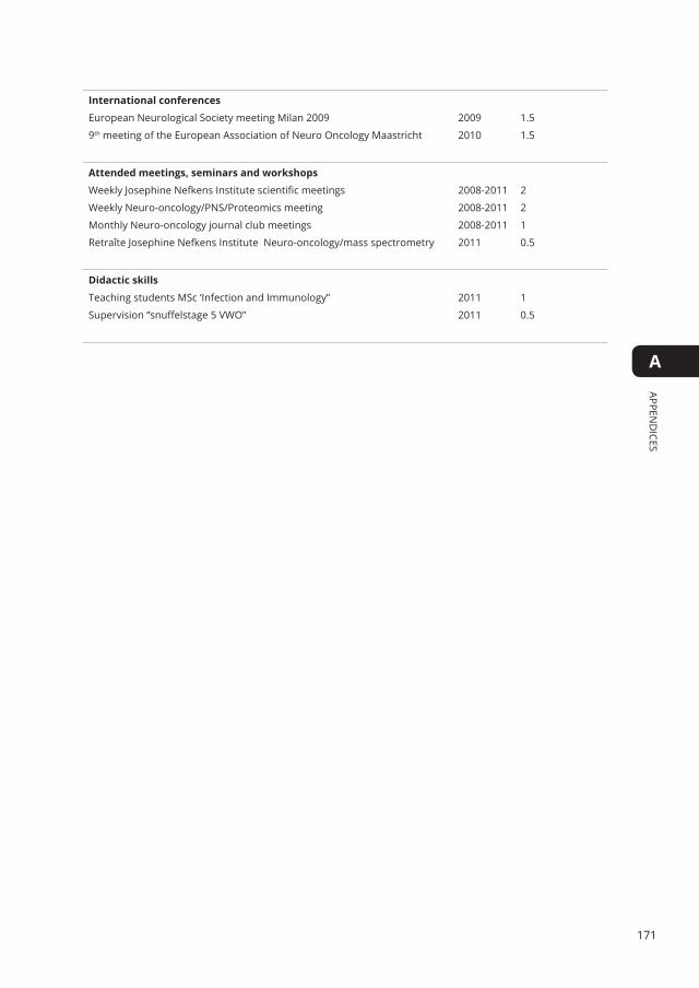
171
APPEND
ICES
A
International conferences
European Neurological Society meeting Milan 2009
9th meeting of the European Association of Neuro Oncology Maastricht
2009
2010
1.5
1.5
attended meetings, seminars and workshops
Weekly Josephine Nefkens Institute scientific meetings
Weekly Neuro-oncology/PNS/Proteomics meeting
Monthly Neuro-oncology journal club meetings
Retraîte Josephine Nefkens Institute Neuro-oncology/mass spectrometry
2008-2011
2008-2011
2008-2011
2011
2
2
1
0.5
Didactic skills
Teaching students MSc ‘Infection and Immunology”
Supervision “snuffelstage 5 VWO”
2011
2011
1
0.5

172
CuRRICuluM VITAe PeTeR MAAT
Peter Maat was born on September 12th , 1976 in RijssenHe attended secondary school at Van Lodenstein College in Amersfoort and graduated in 1994. Because of numerus fixus and not being admitted in dutch medical studies, he started at the medical faculty of the University of Ghent, Belgium in the same year, where he obtained the “Kandidaat Arts” degree (cum Laude). After 4 years he was able to continue medical studies at the Utrecht University. Having finished medical internships he obtained his medical degree in 2003. He worked as resident (“ANIOS”) in the St.-Antonius Ziekenhuis in Nieuwegein and subsequently in the St.-Elisabeth Ziekenhuis and TweeSteden (2004) Ziekenhuis in Tilburg. In 2004 he enrolled the neurology traineeship at the department of Neurology at the St.-Elisabeth Ziekenhuis (“AIOS”). For neuro-oncology he attended a 6 months period of traineeship in the “Daniel den Hoed Kliniek”, part of ErasmusMC Rotterdam (2007). There first contacts were made leading to the work described in this thesis. Neurology internship was changed for 3 years of laboratory work at the Josephine Nefkens Institute, laboratory for Neurology and cancer proteomics (head: Dr. T.M. Luider) of the Erasmus MC. Focus was paraneoplastic neurological syndromes. By april 2011 he returned to the Elisabeth Ziekenshuis Tilburg and finished the neurology training and became Neurologist by December 2012. At present Peter is working as a neurologist at the Ziekenhuis Gelderse Vallei, Ede.He married Janneke in 2003, they have two children: Hugo (2007) and Anne-Ruth (2011).

173
APPEND
ICES
A
lIsT Of PublICATIOns
1. Fonville S, van der Worp HB, Maat P, Aldenhoven M, Algra A, van Gijn J. Accuracy and inter-observer variation in the classification of dysarthria from speech recordings. J Neurol Oct 2008;255:1545-8.
2. Maat P, Sillevis Smitt PAE. Paraneoplastic Neurologic Syndromes. In: Encyclopedia of Life Sciences(ELS), Sept 2009. John Wiley & Sons, Ltd: Chichester.
3. De Graaff E, Maat P, Hulsenboom E, et al. Identification of delta/notch-like epidermal growth factor-related receptor as the Tr antigen in paraneoplastic cerebellar degeneration. Ann Neurol Jun 2012;71:815-24.
4. Maat P, VanDuijn M, Brouwer E, Dekker LH, Zeneyedpour L, Luider TM, Sillevis Smitt PAE. Mass spectrometric detection of antigen-specific immunoglobulin peptides in paraneoplastic patient sera. J Autoimmun Jun 2012;38:354-60.
5. Maat P, Brouwer E, Hulsenboom E, van Duijn MM, Schreurs MW, Hooijkaas H, Sillevis Smitt PAE. Multiplex serology of paraneoplastic antineuronal antibodies. J Immunol Methods May 2013;391:125-32.
6. Maat P, de Graaff E, van Beveren NM, Hulsenboom E, Verdijk RM, Koorengevel K, van Duijn MM, Hooijkaas H, Hoogenraad C, Sillevis Smitt PAE. Psychiatric phenomena as initial manifestation of encephalitis by anti-NMDAR antibodies. Acta Neuropsychiatr Jun 2013;25:128-36.
7. Maat P, de Beukelaar JWK, Jansen C, Schuur M, van Duijn CM, van Coevorden MH, de Graaff E, Titulaer M, Rozemuller JM, Sillevis Smitt PAE. Pathologically confirmed autoimmune encephalitis in suspected Creutzfeldt-Jakob disease. Neurol Neuroimmunol Neuroinflamm 2015

Copyright © 2022 FDOKUMEN