Engineering of stable bispecific antibodies targeting IL17A and IL23
13
Engineering of stable bispecific antibodies targeting IL-17A and IL-23 Robert Mabry 1,6 , Katherine E. Lewis 2 , Margaret Moore 3 , Patricia A. McKernan 1 , Thomas R. Bukowski 3 , Kristen Bontadelli 2 , Ty Brender 1 , Shannon Okada 2 , Karen Lum 3 , James West 3 , Joseph L. Kuijper 1 , Dan Ardourel 1 , Secil Franke 1 , Luann Lockwood 1 , Tuyen Vu 1 , Amanda Frank 1 , Mark W. Appleby 1 , Anitra Wolf 3 , Brian Reardon 3 , Nels B. Hamacher 3 , Brenda Stevens 3 , Patsy Lewis 3 , Kenneth B. Lewis 3 , Debra G. Gilbertson 1 , Megan Lantry 3 , Susan H. Julien 3 , Craig Ostrander 3 , Chung Chan 3 , Kelly Byrnes-Blake 4 , Jennifer Brody 5 , Scott Presnell 5 , Brent Meengs 3 , Steven D. Levin 2 and Mark Snavely 1 1 Molecular Biology and Protein Engineering, 2 Immunology, 3 Protein Science, 4 Pre-clinical Development and 5 Scientific Computing, ZymoGenetics, Inc., 1201 Eastlake Ave. E, Seattle, WA 98102, USA 6 To whom correspondence should be addressed. E-mail: [email protected] Received July 7, 2009; revised October 1, 2009; accepted October 20, 2009 Edited by Philipp Holliger Bispecific antibodies (bsAbs) present an attractive oppor- tunity to combine the additive and potentially synergistic effects exhibited by combinations of monoclonal anti- bodies (mAbs). Current challenges for engineering bsAbs include retention of the binding affinity of the parent mAb or antibody fragment, the ability to bind both targets simultaneously, and matching valency with biology. Other factors to consider include structural stab- ility and expression of the recombinant molecule, both of which may have significant impact on its development as a therapeutic. Here, we incorporate selection of stable, potent single-chain variable fragments (scFvs) early in the engineering process to assemble bsAbs for therapeutic applications targeting the cytokines IL-17A/A and IL-23. Stable scFvs directed against human cytokines IL-23p19 and IL-17A/A were isolated from a human Fab phage display library via batch conversion of panning output from Fabs to scFvs. This strategy integrated a step for shuffling V regions during the conversion and permitted the rescue of scFv molecules in both the V H V L and the V L V H orientations. Stable scFvs were identified and assembled into several bispecific formats as fusions to the Fc domain of human IgG1. The engineered bsAbs are potent neutralizers of the biological activity of both cyto- kines (IC 50 < 1 nM), demonstrate the ability to bind both target ligands simultaneously and display stability and productivity advantageous for successful manufacture of a therapeutic molecule. Pharmacokinetic analysis of the bsAbs in mice revealed serum half-lives similar to human mAbs. Assembly of bispecific molecules using stable anti- body fragments offers an alternative to reformatting mAbs and minimizes subsequent structure-related and manufacturing concerns. Keywords: antibody/bispecific/IL-17A/IL-23/scFv Introduction The value of combining treatments to impact multiple targets in a disease pathway has been the focus of much recent inter- est. Bispecific antibodies (bsAbs) are dual-targeting anti- bodies that bind two distinct proteins and can minimize the regulatory and commercial issues that stem from adminis- tration of multiple therapeutic molecules (Hoggenboom, 1997; Fischer and Le ´ger, 2007; Lum and Al-Kadhimi, 2008; Chames and Baty, 2009; Dimassi et al., 2009; Parren and Burton, 2009). bsAbs are commonly based on the structure of immunoglobulins (Ig). Incorporation of an Fc domain from IgG improves the half-life in serum and can provide antibody-like effector function, if desired. An extensive array of formats have been developed and tailored for affinity, valency, expression, stability and pharmacokinetics (Plu ¨ckthun and Pack, 1997; Todorovska et al., 2001; Marvin and Zhu, 2005; Beckman et al., 2007; Demarest and Glaser, 2008). Although engineered bsAbs have been reported for over two decades, difficulties with manufacturing related pri- marily to stability remain a significant obstacle to their development as therapeutics (Kontermann, 2005; Fischer and Le ´ger, 2007; Demarest and Glaser, 2008). Typically, the con- struction of bsAbs has been approached through reconfigura- tion of monoclonal antibodies (mAbs) and antibody fragments, generating molecules which may not possess the same physical properties as the parental antibodies. This reformatting frequently creates issues related to affinity, stab- ility and production (Holliger et al., 1993; Coloma and Morrison, 1997; Lu et al., 2003b) that may be resolved by additional engineering of the antibody moieties (Wo ¨rn and Plu ¨ckthun, 2001; Demarest et al., 2006), a process that can prove labor-intensive and costly. Thus, it has been proposed that the construction of bsAbs requires the use of stable monomeric units as building blocks (Holliger and Winter, 1993; Demarest and Glaser, 2008; Saerens et al., 2008). Because of their modular properties, antibody fragments based on variable light and heavy chains tethered by an amino acid linker [single-chain variable fragments (scFvs)] are attractive building blocks for the construction of bsAbs. The primary advantage of scFvs is the production of a single polypeptide chain, which obviates the difficulties that arise in pairing appropriately the separate chains of a heteromulti- meric protein (Coloma and Morrison, 1997). Although thera- peutic applications of scFvs have been widely anticipated, their development has been severely limited due to issues associated primarily with aggregation and serum stability (Jung et al., 1999; Lu et al., 2003a). In recent reports, these problems have been addressed by applying techniques designed to enhance stability through additional engineering steps that have successfully yielded clones with optimized # The Author 2009. Published by Oxford University Press. All rights reserved. For Permissions, please e-mail: [email protected] Page 1 of 13 Protein Engineering, Design & Selection pp. 1–13, 2009 doi:10.1093/protein/gzp073 PEDS Advance Access published December 18, 2009 by guest on September 2, 2016 http://peds.oxfordjournals.org/ Downloaded from
-
Upload
independent -
Category
Documents
-
view
4 -
download
0
Transcript of Engineering of stable bispecific antibodies targeting IL17A and IL23
Engineering of stable bispecific antibodies targetingIL-17A and IL-23
Robert Mabry1,6, Katherine E. Lewis2, Margaret Moore3,Patricia A. McKernan1, Thomas R. Bukowski3,Kristen Bontadelli2, Ty Brender1, Shannon Okada2,Karen Lum3, James West3, Joseph L. Kuijper1,Dan Ardourel1, Secil Franke1, Luann Lockwood1,Tuyen Vu1, Amanda Frank1, Mark W. Appleby1,Anitra Wolf3, Brian Reardon3, Nels B. Hamacher3,Brenda Stevens3, Patsy Lewis3, Kenneth B. Lewis3,Debra G. Gilbertson1, Megan Lantry3, Susan H. Julien3,Craig Ostrander3, Chung Chan3, Kelly Byrnes-Blake4,Jennifer Brody5, Scott Presnell5, Brent Meengs3,Steven D. Levin2 and Mark Snavely1
1Molecular Biology and Protein Engineering, 2Immunology, 3ProteinScience, 4Pre-clinical Development and 5Scientific Computing,ZymoGenetics, Inc., 1201 Eastlake Ave. E, Seattle, WA 98102, USA
6To whom correspondence should be addressed.E-mail: [email protected]
Received July 7, 2009; revised October 1, 2009;accepted October 20, 2009
Edited by Philipp Holliger
Bispecific antibodies (bsAbs) present an attractive oppor-tunity to combine the additive and potentially synergisticeffects exhibited by combinations of monoclonal anti-bodies (mAbs). Current challenges for engineering bsAbsinclude retention of the binding affinity of the parentmAb or antibody fragment, the ability to bind bothtargets simultaneously, and matching valency withbiology. Other factors to consider include structural stab-ility and expression of the recombinant molecule, both ofwhich may have significant impact on its development asa therapeutic. Here, we incorporate selection of stable,potent single-chain variable fragments (scFvs) early inthe engineering process to assemble bsAbs for therapeuticapplications targeting the cytokines IL-17A/A and IL-23.Stable scFvs directed against human cytokines IL-23p19and IL-17A/A were isolated from a human Fab phagedisplay library via batch conversion of panning outputfrom Fabs to scFvs. This strategy integrated a step forshuffling V regions during the conversion and permittedthe rescue of scFv molecules in both the VHVL and theVLVH orientations. Stable scFvs were identified andassembled into several bispecific formats as fusions to theFc domain of human IgG1. The engineered bsAbs arepotent neutralizers of the biological activity of both cyto-kines (IC50 < 1 nM), demonstrate the ability to bind bothtarget ligands simultaneously and display stability andproductivity advantageous for successful manufacture ofa therapeutic molecule. Pharmacokinetic analysis of thebsAbs in mice revealed serum half-lives similar to humanmAbs. Assembly of bispecific molecules using stable anti-body fragments offers an alternative to reformattingmAbs and minimizes subsequent structure-related andmanufacturing concerns.
Keywords: antibody/bispecific/IL-17A/IL-23/scFv
Introduction
The value of combining treatments to impact multiple targetsin a disease pathway has been the focus of much recent inter-est. Bispecific antibodies (bsAbs) are dual-targeting anti-bodies that bind two distinct proteins and can minimize theregulatory and commercial issues that stem from adminis-tration of multiple therapeutic molecules (Hoggenboom,1997; Fischer and Leger, 2007; Lum and Al-Kadhimi, 2008;Chames and Baty, 2009; Dimassi et al., 2009; Parren andBurton, 2009). bsAbs are commonly based on the structureof immunoglobulins (Ig). Incorporation of an Fc domainfrom IgG improves the half-life in serum and can provideantibody-like effector function, if desired. An extensivearray of formats have been developed and tailored foraffinity, valency, expression, stability and pharmacokinetics(Pluckthun and Pack, 1997; Todorovska et al., 2001; Marvinand Zhu, 2005; Beckman et al., 2007; Demarest and Glaser,2008). Although engineered bsAbs have been reported forover two decades, difficulties with manufacturing related pri-marily to stability remain a significant obstacle to theirdevelopment as therapeutics (Kontermann, 2005; Fischer andLeger, 2007; Demarest and Glaser, 2008). Typically, the con-struction of bsAbs has been approached through reconfigura-tion of monoclonal antibodies (mAbs) and antibodyfragments, generating molecules which may not possess thesame physical properties as the parental antibodies. Thisreformatting frequently creates issues related to affinity, stab-ility and production (Holliger et al., 1993; Coloma andMorrison, 1997; Lu et al., 2003b) that may be resolved byadditional engineering of the antibody moieties (Worn andPluckthun, 2001; Demarest et al., 2006), a process that canprove labor-intensive and costly. Thus, it has been proposedthat the construction of bsAbs requires the use of stablemonomeric units as building blocks (Holliger and Winter,1993; Demarest and Glaser, 2008; Saerens et al., 2008).
Because of their modular properties, antibody fragmentsbased on variable light and heavy chains tethered by anamino acid linker [single-chain variable fragments (scFvs)]are attractive building blocks for the construction of bsAbs.The primary advantage of scFvs is the production of a singlepolypeptide chain, which obviates the difficulties that arisein pairing appropriately the separate chains of a heteromulti-meric protein (Coloma and Morrison, 1997). Although thera-peutic applications of scFvs have been widely anticipated,their development has been severely limited due to issuesassociated primarily with aggregation and serum stability(Jung et al., 1999; Lu et al., 2003a). In recent reports, theseproblems have been addressed by applying techniquesdesigned to enhance stability through additional engineeringsteps that have successfully yielded clones with optimized
# The Author 2009. Published by Oxford University Press. All rights reserved.
For Permissions, please e-mail: [email protected]
Page 1 of 13
Protein Engineering, Design & Selection pp. 1–13, 2009doi:10.1093/protein/gzp073
PEDS Advance Access published December 18, 2009 by guest on Septem
ber 2, 2016http://peds.oxfordjournals.org/
Dow
nloaded from
physical properties (Jung et al., 1999). However, attempts toidentify stable, fully human scFvs in the absence ofadditional grafting or maturation techniques have resulted infew therapeutic candidates. A method for the early selectionof stable, potent scFvs from large human libraries may serveas an alternative to rigorous engineering of the therapeuticcandidate.
Phage display is the most widely used display platform forengineering therapeutic antibodies. Libraries have been con-structed in several formats using variable regions isolatedfrom a human repertoire (Marks et al., 1991) and/or syntheticcomplementary determining regions (CDRs) grafted ontohuman framework regions (Soderlind et al., 1995; Kobayashiet al., 1997; Knappik et al., 2000; Sidhu et al., 2004; Volkelet al., 2004; Hoet et al., 2005). These phage libraries facili-tate the isolation of human antibodies, reducing the risk ofimmunogenicity associated with chimeric mAbs and avoid-ing the rigors of humanizing murine antibodies. Advances inlibrary construction have resulted in theoretical diversitiessurpassing 1 � 1010 unique sequences (Hoet et al., 2005;Rothe et al., 2007). Even so, the use of display technologiesfrequently fails to produce antibody fragments with thedesired affinity, potency and/or stability. As a result, selec-tion from phage libraries is often complemented with muta-genesis, shuffling, additional display technologies and othercombinatorial procedures between iterative rounds of selec-tion to improve affinity (Kang et al., 1991b; Jirholt et al.,1998; Maynard et al., 2002; Harvey et al., 2004; Groves andOsbourn, 2005). These techniques for affinity maturationtypically focus on optimization of a single antibody fragmentthrough the generation of new libraries for additional selec-tions against target molecules. Although this process fre-quently yields binding entities with improved properties, theprocess is lengthy and labor-intensive. Selection of thedesired properties in batch mode may expedite the engineer-ing of bsAbs, bypassing affinity maturation of single clones.The early identification of stable scFvs that bind with highaffinity to the target would result in more efficient generationof stable bsAbs for development of human therapeutics.
Stable bispecific molecules targeting two soluble cytokinesoffer a novel approach to enhance the therapeutic efficacy forinflammatory diseases. TH17 cells are a recently identifiedsubset of T helper cells associated with several inflammatorydiseases including multiple sclerosis, psoriasis, rheumatoidarthritis (RA) and inflammatory bowel disease (Dong, 2008;McGeachy and Cua, 2008). These cells produce a number ofinflammatory mediators, including two homologous cyto-kines, IL-17A and IL-17F. Both molecules are found ashomodimers (IL-17A/A and IL-17F/F) and as a heterodimer(IL-17A/F), all of which appear to mediate inflammatoryresponses associated with disease pathology (Fossiez et al.,1998; Wright et al., 2007). The cytokine IL-23 plays animportant role in the differentiation and regulation of TH17cells, resulting in production of a number of cytokinesincluding IL-17A/A and IL-17A/F (McGeachy and Cua,2007). IL-23 is a heterodimer consisting of a unique p19subunit and a p40 subunit which is shared with IL-12.Inhibition of the activity of IL-23 on the TH17 pathway,while avoiding interruption of pathways mediated by IL-12,requires a reagent that interacts specifically with the p19subunit (also referred to as IL-23A). A therapeutic moleculetargeting both an expansion and a survival factor for TH17
cells, such as IL-23 and TH17 effector cytokines (IL-17A/Aand IL-17A/F), could be more efficacious in treating anumber of inflammatory diseases than molecules that inhibitactivity mediated by a single cytokine.
In the construction of molecules targeting human IL-17A/A and the p19 subunit of IL-23, we addressed the challengesof bsAb generation by focusing on the identification of stablescFvs and improving the affinity of antibody fragments inbatch mode. The resulting bispecific molecules exhibit theability to bind and neutralize both cytokines, possess stabilitytypical of mAbs and display serum half-lives similar tohuman IgGs. Assembly of bsAbs with stable antibody frag-ments as building blocks proved to be an effective methodfor the development of stable, dual-targeting therapeutic mol-ecules directed against these two attractive targets.
Materials and methods
Protein reagentsIL-17A assay reagents, anti-IL-17A/A mAb (#MAB317) andIL-17A/A pAb (#AF-317 NA), were purchased from R&DSystems (Minneapolis, MN, USA). IL-23 assay reagents,IL-23 (#1290-IL/CF), IL-12 (#219-IL), IL-23R-Fc(#1400-IR-050), IL-23p19 mAb (#MAB1290), IL-23p19pAb (#AF1716) and anti-IL23p40 mAb (#MAB1510), werealso purchased from R&D Systems. The IL-23p19 specificantibody was purchased from eBiosciences (#34-823985, SanDiego, CA, USA). Recombinant human (h) proteinsIL-17RA-Fc, IL-17A/A, IL-17A/F, IL-17F/F, IL-23R-Fc,IL-12 and IL-23 were produced at ZymoGenetics Inc.(Seattle, WA, USA).
Phage panning and conversion of Fabs to scFvsGeneral methods of panning against target antigen using theDyax Fab libraries have been described previously (de Haardet al., 1999). Human IL-23 and IL-17A/A cytokines wereused as single targets for screening the Dyax 310 human Fablibrary (Hoet et al., 2005). For immunotube panning, humanIL-17A/A or IL-23 at a concentration between 10 and100 mg/ml in PBS was coated in a polystyrene immunotube(#444474, Nalge Nunc International, Rochester, NY, USA).After rotating overnight at 48C, immunotubes were blockedwith the addition of 2 ml of blocking buffer (2% milk, PBS,0.1% Tween 20) for 1 h at room temperature (RT) withrotation. Uncoated immunotubes were also blocked fordepletion of phage binding non-specifically. For the firstround of selections, aliquots of the Dyax library were addedto blocking buffer to a volume of 1 ml and incubated at RTfor 1 h with rotation. Phage were then added to one uncoatedimmunotube and incubated for 1 h at RT with rotation.Phage were transferred to the immunotube coated with targetand incubated for 10 min to 1 h before washing. Phage wereeluted from the immunotubes by the addition of 100 mMTriethyleneamine, followed by neutralization with 1 M Tris–HCl, pH 7.4. Iterative rounds of panning (2–4 rounds) wereperformed for enrichment against each target with anincrease in the number of wash steps (between 8 and 10washes with PBS, 0.1% Tween 20) and variations in incu-bation times (5 min–1 h).
Phage output from each round was used to infect TG1cells (TG1) (#200123, Stratagene, La Jolla, CA, USA) in
R.Mabry et al.
Page 2 of 13
by guest on September 2, 2016
http://peds.oxfordjournals.org/D
ownloaded from
a 4 ml culture containing 2� YT supplemented with ampi-cillin (100 mg/ml). Cultures were shaken at 378C to a finalOD600nm of 0.5. For phage production, 0.3 ml of helperphage (#18 311-019, Invitrogen, Carlsbad, CA, USA) wasadded and the mixture was incubated at 308C for 30 minwithout shaking. The mixture was then added to 44 ml ofculture media (2� YT supplemented with ampicillin at100 mg/ml) and incubated at 378C for 6 h with shaking. Thetemperature was adjusted to 308C and the cultures weregrown overnight with shaking. The following day, a 40 mlaliquot of the culture was removed and precipitated withPEG for phage purification. Phage pellets were resuspendedin 1 ml of PBS, quantified and used as an input for iterativepanning rounds or frozen away in 15% glycerol for later use.
For scFv panning, the selections were performed asdescribed above with the integration of thermal treatment.Prior to each selection, the phage input (in PBS) was sub-jected to 1 h incubation at 608C. Phage samples were thespun down at 16K rpm for 10 min and supernatants trans-ferred to fresh 1.5 ml tubes. Phage were then blocked asdescribed above prior to each panning round. One to twoadditional rounds of panning were performed after batch con-version of Fab clones (see Fab to scFv conversion).
Preparation of bacterial supernatants for screeningFor soluble expression of Fabs in scFvs, DNA representingthe phage output against each target was digested to removethe gIII fusion partner and expressed as a soluble Fab proteinin the pMID21 vector (GenBank AY75424). Re-ligatedvectors were transformed into TG1 cells (TG1) (#200123)and plated at several dilutions. Single colonies were picked(QP Display, Genetix, Boston, MA, USA) into a 96-wellplate (Corning, #3359) containing 100 ml of media [2� YTmedia, 2% glucose, ampicillin (100 mg/ml)]. Plates weregrown overnight at 308C with shaking. The next day, eachplate was subcultured into a new plate (Corning, #3956) con-taining 800 ml of fresh media [2� YT media, 0.1% Triton X100, ampicillin (100 mg/ml)] per well and grown for 2.5 h at308C with shaking. Cultures were induced with 1 mM IPTGfor expression and cultured for 15 h at 308C with shaking.Plates were then centrifuged at 6000 rpm for 10 min, andsupernatants containing secreted scFv or Fab were transferredto new 96-well plates for analysis.
ELISAs for primary screening of bacterial supernatantsFor binding ELISAs, Costar (#9018) 96-well plates werecoated with 50 ml IL-17A/A IL-17F/F, IL-23 or IL-12(between 2 and 4 mg/ml) in 0.1 M NaHCO3, pH 9.6 over-night at 48C. The next day, plates were washed three timeswith 0.1% Tween 20/PBS (PBST). Each well was filled with350 ml of 2% milk (#170-6404, Bio-Rad Laboratories,Hercules, CA, USA)/PBST and incubated for 1 h at RT forblocking. Assay plates were then washed three times withPBST. Each well was filled with 50 ml of 2% milk/PBST,followed by the addition of 25 ml of Fab or scFv supernatant.Wells were mixed and incubated for 1 h at RT. ELISA plateswere washed three times with PBST. For Fab detection,50 ml of anti-Human Fab-specific pAb-HRP (#31482, Pierce)diluted 1:4000 in 2% milk/PBST was added to each well for1 h at RT. For scFv detection, 50 ml of anti-His tagmAb-HRP (Sigma, #A7058) diluted 1:4000 in 2% milk/PBST was added to each well for 1 h at RT. Plates were
washed three times with PBST. Fifty microliters of TMB(TMBW-1000-01, BioFX Laboratories) were added to eachwell and the reaction was allowed to develop for 20–30 min,followed by the addition of 50 ml of stop buffer(STPR-1000-01, BioFX Laboratories) to quench the reaction.Plates were read at 450 nm on a plate reader.
For blocking ELISAs, Costar (#9018) 96-well plates werecoated with 50 ml of anti-human IgG Fcg-specific antibody(#109-005-098, Jackson Immunology) at 1 mg/ml in 0.1 MNaHCO3, pH 9.6 overnight at 48C. The next day, plates werewashed three times with 0.1% PBST. Fifty microliters ofIL-17RA at 0.25 mg/ml in PBS were added to each well, fol-lowed by incubation for 1 h at RT. Plates were then washedthree times with PBST. Each well was filled with 350 ml of2% milk (#170-6404, Bio-Rad)/PBST and incubated for 1 hat RT for blocking. During plate blocking, adjacent plateswere set up for pre-incubation of IL-17A or IL-23 with Fabor scFv supernatant. Pre-incubation plates were filled with75 ml of Fab supernatant, followed by the addition of 25 mlof biotinylated IL-17A/A or biotinylated IL-23 at 0.10 mg/mlin 4% milk/PBST. Each well was mixed and then incubatedfor 30 min at RT. Assay plates were washed three times withPBST, and the volume of the supernatant/target complextransferred from the pre-incubation plate to the assay plate,followed by incubation at RT for 1 h. Plates were thenwashed three times with PBST. Fifty microliters of (1:3000)Streptavidin-HRP (#21124, Pierce) in PBST were added toeach well followed by an additional 1 h incubation. Afterthree washes with PBST, 50 ml of TMB (TMBW-1000-01,BioFX Laboratories) was added to each well. The reactionswere allowed to develop for 20–30 min and quenched by theaddition of 50 ml of stop buffer (STPR-1000-01, BioFXLaboratories). Plates were read at 450 nm on a plate reader.
Fab to scFv batch conversionOnce specific binding was verified for each round, FabcDNA served as a template for batch amplification of thelight and heavy chain variable regions. Lambda, kappa andheavy chain variable regions were amplified from pooledDNA taken from rounds 2 and 3 of phage panning. TheDNA served as a template in a three-step process usingprimers directed against framework sequences for eachsubtype. The primers were designed to complement the term-inal sequences of all identified human framework regions 1and 4 (www.kabatdatabase.com). The first PCR step ampli-fied each of the variable frame work regions and addedappropriate overhangs to facilitate round 2 PCR reactions.The second PCR step added appropriate gly/ser linkersequences to the ends of the appropriate products from thefirst and step 3 PCR reactions overlapped the variable lightchain lambda, variable light chain kappa and variable heavychain products to create scFv products in both LH and HLorientations with a 25 mer G4S linker between the V regions.Assembled scFvs were cloned into a phagemid vector as agIIIp fusion. DNA ligations were transformed into TG1 cells(TG1) (#200123) and added to a 200 ml culture of 2� YTsupplemented with ampicillin (100 mg/ml) to a finalOD600nm of 0.2. For phage production, 2 ml (.2 � 1011 pfu)of helper phage (#18311-019, Invitrogen) was added and theculture incubated at 378C for 6 h with shaking. The tempera-ture was then adjusted to 308C and the cultures were grownovernight with shaking. The following day, 2 � 50 ml
Engineering of stable bsAbs targeting IL-17A and IL-23
Page 3 of 13
by guest on September 2, 2016
http://peds.oxfordjournals.org/D
ownloaded from
aliquots of culture were taken from phage culture and preci-pitated with PEG for phage purification. Phage pellets wereresuspended in 1 ml of PBS, quantified and used as phageinput for iterative panning rounds or frozen away in 15% gly-cerol for later use.
Protein expression and purificationSelected Fab or scFv clones were expressed from a proprie-tary vector utilizing the phoA promoter in either BL21(#69449-4, Novagen, Madison, WI, USA) or TG1 cells(#200123). After transformation, colonies were selected forinoculation into 2 ml Luria–Bertani medium supplementedwith antifoam at 100 mg/l (#A8311, Sigma-Aldrich, StLouis, MO, USA) and kanamycin (#60615, Sigma-Aldrich)at 25 mg/ml. Cultures were grown overnight at 378C, shakingat 250 rpm. The overnight cultures were diluted to 0.2% forinoculation into 0.5 l of phosphate-limiting media (Simmonset al., 2002) supplemented with antifoam and antibiotic asabove. Cultures were grown in 2 l side-baffled flasks for 24 hat 308C. Soluble scFv and Fab samples were recoveredfrom wet cell pellets by treatment with periplasting buffercontaining Ready-Lyse lysozyme (#R1802, EpicentreBiotechnologies, Madison, WI, USA) as per manufacturer’sinstructions in the ‘Protocol for the Preparation ofPeriplasmic and Spheroplastic Proteins from .1 ml BacterialCulture’ (Epicentre Biotechnologies).
For the immobilized metal affinity chromatography enrich-ment of scFvs, the periplasmic fraction was passed through a0.22 mm filter and purified by affinity capture with a HisTrapHP column (GE Healthcare, Piscataway, NJ, USA) on aliquid chromatography instrument (Akta Explorer System,GE Healthcare). The bound protein was eluted using400 mM imidazole in 50 mM NaPO4 and 500 mM NaCl atpH 7.4. Protein content was by absorbance at 280 nm andquality was assayed by analytical size exclusion chromato-graphy (SEC) and SDS–PAGE. For Fab enrichment, affinitypurification was performed using Protein A (#17-1279-04,GE Healthcare) in batch mode. The Protein A resin wasadded to the periplasmic extract for overnight incubation at48C with mixing. The following day, the protein was recov-ered using a low pH step elution with 50 mM NaH2PO4 pH2.5, 150 mM NaCl, immediately neutralized with 1 MHEPES pH 7.2, and evaluated as described above.
The three forms of bsAbs were assembled by a combi-nation of PCR and homologous recombination in yeast usinga proprietary mammalian expression vector with DHFR asthe selectable marker. The expression constructs were elec-troporated into CHO DXB11 cells, adapted to grow in sus-pension in a protein-free medium, and subjected to nutrientselection in HT medium followed by selection in methotrex-ate. Pools of selected cells were scaled up in spinner flaskculture for purification and analysis of bispecific molecules.Conditioned media were harvested, passed through a 0.2 mmfilter and adjusted to pH 7.4. The protein was purified fromthe filtered media using a combination of POROSw A50Protein A Affinity Chromatography (Applied Biosciences,Foster City, CA, USA) and Superdex 200 SEC (GEHealthcare). Column fractions were analyzed by SDS–PAGE to determine the appropriate pools. Enriched proteinwas quantified by UV at A280 nm and an analytical SECmethod was used to characterize the purified protein.
Kinetic analysisThe binding affinities of Fabs and scFvs to IL-17A/A,IL-17F/F, IL-23 and IL-12 were evaluated using surfaceplasmon resonance (SPR) on a Biacore T-100 instrument(GE Healthcare). For affinity analysis, recombinant humantarget proteins were diluted to 10 mg/ml in 10 mM acetate(pH 4.0 for IL-17A/A and IL-17F/F, pH 4.5 for IL23 andIL12) and then immobilized onto CM5 chip (GE Healthcare/Biacore#BR-1006-68) using amine coupling. Briefly, thecarboxymethyl dextran surface was activated with a 7 mininjection of a 1:1 ratio of 0.4 M EDC [N-ethyl-N’-(3-diethylamino-propyl) carbodiimide] and 0.1 M NHS(N-hydroxysuccinimide). The final immobilization levelswere between 300 and 480 resonance units (RUs). After theimmobilization procedure, active sites on the flow cell wereblocked with 1 M ethanolamine. The reference cell was acti-vated and then blocked with ethanolamine prior to sampleanalysis. For measuring association and dissociation kinetics,serial dilutions of the anti-IL-17A/A or IL-23 antibodieswere prepared in 1� HBS-EPþ buffer (GE Healthcare/Biacore, #BR-1006-69). Duplicate injections of each concen-tration were performed at 30 ml/min with an association timeof 300 s and a dissociation time of 600 s. Between exper-iments, the immobilized IL-17A/A surface was regeneratedwith a 60 s injection of 10 mM H3PO4 followed by a 60 sinjection of buffer at 50 ml/min. The optimal regenerationcondition for IL-23 analysis was a 60 s injection of 2 MMgCl2 followed by 60 s of buffer injection at a flow rate of50 ml/min.
For analysis of crude supernatants, the chip surfaces wereprepared as described above. Unless otherwise indicated, allbinding experiments were performed at 258C in 1�HBS-EPþ buffer supplemented with, 0.1% BSA. IL-23 orIL-17A/A supernatants were diluted 1:3, and 100 nM ofIL-23R and IL-17RA was prepared. Analyte was injected at30 ml/min with an association time of 300 s and a dis-sociation time of 300 s. Regeneration conditions wereadapted from previous kinetic analysis of IL-23 and IL-17A/A as described above. Crude samples were ranked firstaccording to their off-rates (slowest to fastest) and thenaccording to their binding levels (highest to lowest bindingRUs) during the association phase. These values were com-pared with positive controls using IL-23R or IL-17RA.
Analysis of tetravalent bsAbs was performed using an Fccapture to obtain binding affinities for the ligands. Goat anti-human IgG Fc-gamma (Jackson ImmunoResearch, WestGrove, PA, USA) was covalently immobilized on a CM4chip (GE Healthcare/Biacore, #BR-1005-30) to a density of�4000 RU using amine coupling as described above. EachIL-17/IL-23p19 antagonist was captured onto a separate flowcell of the CM4 chip at an approximate density of 80–100 RU. Capture of the antagonist to the immobilizedsurface was performed at a flow rate of 10 ml/min. Forbinding studies with IL-17A/A, serial 1:3 dilutions of theIL-17A antigen from 20 to 0.03 nM were injected over thesurface. For binding studies with IL-23, serial 1:3 dilutionsof the IL-23 antigen from 50 to 0.07 nM were injected overthe surface. Duplicate injections of each antigen concen-tration were performed with an association time of 7 min anda dissociation time of either 10 or 60 min. Kinetic bindingstudies were performed with a flow rate of 30 ml/min. The
R.Mabry et al.
Page 4 of 13
by guest on September 2, 2016
http://peds.oxfordjournals.org/D
ownloaded from
flow cell was washed between cycles with 20 mM hydro-chloric acid to regenerate the surface.
For co-binding analysis of bispecific constructs, recombi-nant human IL-17A/A was immobilized onto a CM5 sensorchip using amine coupling as described above. Each IL-17/IL-23p19 antagonist was diluted to 100 nM and injected overthe immobilized IL-17A/A surface at a flow rate of 10 ml/minfor 5 min. The capture level of the IL-17A/IL-23 antagonistsranged from 330 to 910 RU. A saturating concentration of500 nM IL-23 was then flowed over the surface. Binding ofIL-23 was performed for 10 min with a flow rate of 10 ml/min.Regeneration conditions were adapted from previous kineticanalysis of IL-17A/A as explained above.
All data were evaluated using Biacore Evaluation softwareto define either the affinity or the off-rate of the interactions.Baseline stability was assessed to ensure that the regenerationstep provided a consistent binding surface throughout thesequence of injections. Binding curves were normalized bydouble-referencing and the resulting binding curves wereglobally fit using the Biacore Evaluation Software v1.1.1.
Cell-based assay data analysisAll cell-based assay data were analyzed using GraphPadPrism version 4 software (San Diego, CA, USA) with a sig-moidal (four-parameter logistics) non-linear regression curvefit, and the amount of antagonist needed to neutralize 50% ofthe activity of each ligand was calculated (IC50).
IL-17A cell-based assaysFor short-term analysis of the antibody fragments, NIH 3T3mouse fibroblast cells stably expressing an NFkB Luciferasereporter were used. Cells were seeded at 10 000 cells/well in96-well tissue culture plates, in DMEM, 3% fetal bovineserum (FBS), 1 mM sodium pyruvate, 2 mM L-glutamine.After overnight incubation at 378C, 5% CO2, plating mediumwas removed and replaced with DMEM, 0.1% BSA, 1 mMsodium pyruvate, 2 mM L-glutamine, 25 mM HEPES buffer.In a separate plate, hIL-17A/A at a concentration of 1.5 nMwas pre-incubated for 30 min at 378C with 3-fold serialdilutions of antagonists. The IL-17A/A ligand (0.75 nM finalassay concentration) plus antagonists were then added to thecells and incubated at 378C, 5% CO2 for 4 h. After incu-bation, cells were lysed in 25 ml/well of Flash Luciferaselysis buffer (E-1501, Promega Corp, Madison, WI, USA)and read on a Berthold Centro XS3 luminometer (BertholdTechnologies, Wildbad, Germany) with addition of 40 mL/well Flash Luciferase substrate (E-1501) and 5 s integrationof signal.
For long-term analysis in a primary cell assay, humansmall airway epithelial cells (SAEC) [cells, SAEC BasalMedium, and SAEC Singlequot kits (Lonza, Walkersville,MD, USA)] were plated at 8000 cells/well in 96-well flatbottom tissue culture plates and incubated overnight at 378C,5% CO2. The following day, cells were treated with a doserange of the antagonist in combination with 0.25 nMhIL-17A/A. The dose ranges of the antagonists were 0.001–300 nM for Fabs, 0.002–200 nM for scFvs (except forAP1733 scFv which was tested at 0.004–60 nM) and0.0018–30 nM for the bispecific molecules. Ligand andantagonist were incubated together for 30 min at 378C beforeadding to the cells. Triplicate wells were set up for eachdose. After 24 h, supernatants were collected, and stored at
2808C, if not used immediately. Owing to the sensitivity ofprimary SAEC to growth conditions and manipulation, priorto collection of supernatants, the health of the SAEC mono-layer was assessed by visual evaluation on an inverted micro-scope. Any wells exhibiting significant cell death wereexcluded from analysis. Supernatants were assayed using aBio-Plex singleplex bead-based assay for cytokine huG-CSF(Bio-Rad Laboratories), and read on a Bio-Plex array systemutilizing Bio-Plex Manager 3.0 (Bio-Rad Laboratories).
IL-23 cell-based assaysFor short-term analysis of the antibody fragments, BaF3mouse B-cells stably expressing the human IL-23RA andIL-12RB1 receptors were seeded at 50 000 cells/well in96-well tissue culture plates in RPMI 1640, 10% FBS, 1 mMsodium pyruvate, 2 mM L-glutamine, 50 mM BME. In a sep-arate plate, hIL-23 at a concentration of 0.1 nM was pre-incubated for 30 min at 378C with 3-fold serial dilutions ofantagonists. The IL-23 ligand (0.05 nM final assay concen-tration) plus antagonists were then added to the cells andincubated at 378C, 5% CO2 for 15 min. After incubation, thereaction was stopped and cell lysates were created using aBio-Plex Cell Lysis Kit (Bio-Rad Laboratories) according tothe manufacturer’s instructions. Cell lysates were analyzedfor the presence of phosphorylated STAT3 on a Bio-PlexArray Reader (Bio-Rad Laboratories) according to the manu-facturer’s instructions for the Bio-Plex Phospho-STAT3Assay.
For long-term analysis in a primary cell assay, a singlecell suspension of murine splenocytes was prepared fromwhole spleens harvested from C57BL/6 or BALB/c mice.After red blood cell lysis with ACK buffer (0.010 MKHCO3, 0.0001 M EDTA, 0.150 M NH4Cl, pH 7.2), spleno-cytes were washed and resuspended in RPMI buffer (contain-ing 10% FBS, 1 mM sodium pyruvate, 2 mM L-glutamine,0.1 mM non-essential amino acids, 2.5 mM HEPES,0.00035% 2-mercaptoethanol Pen-Strep and 50 ng/ml humanIL-2 (R&D Systems). Cells were seeded at 500 000 cells perwell in a 96-well round-bottom plate. In a separate plate,hIL-23 at a concentration of 10 pM was pre-incubated for30 min at 378C with 3-fold serial dilutions of antagonists atconcentrations ranging from 0 to 343 nM. The IL-23 ligandplus antagonists were then added to the splenocytes andincubated at 378C, 5% CO2 for 24–72 h. The supernatantswere collected and frozen at 2808C until ready to process.The levels of murine IL-17A/A, IL-17F/F, IL17A/F andGM-CSF protein in the supernatants were measured usingbead-based sandwich ELISAs. A commercial kit (BeadlyteCytokine Detection System, Millipore Scientific, Bellerica,MA, USA) was used to measure concentrations of IL-17A/Aand GM-CSF produced. A bead-based ELISA using an anti-body to mouse IL-17F/A (R&D Systems) conjugated to abead was used to measure murine IL-17F/F. A combinationof the commercial beads used to measure IL-17A/A with asecondary antibody to IL-17F/F (R&D Systems) was used tomeasure murine IL-17A/F heterodimer production.
Protein characterizationSEC multi-angle light scattering (SEC-MALS) analysis wasincorporated into the characterization of protein to providean absolute measurement of molecular weight from staticlight scattering in-line with SEC. Samples were analyzed
Engineering of stable bsAbs targeting IL-17A and IL-23
Page 5 of 13
by guest on September 2, 2016
http://peds.oxfordjournals.org/D
ownloaded from
with the three detectors connected in series with UV first(Agilent 1100 HPLC with diode array detector), followedby the LS (Wyatt Technology DAWN EOS Multi-anglelaser light scattering detector) and RI (Wyatt TechnologyOptilab REX Differential Refractometer) detectors. Forsample analysis, 150 mg of each sample was injected to theSEC-MALS system (Amersham Pharmacia Superdex-200SEC column, 10 � 300 mm) in a mobile phase consistingof 25 mM histidine, 150 mM NaCl at pH 6.8 at a flow rateof 0.5 ml/min run at ambient temperature. Data were ana-lyzed with Wyatt Technology ASTRA software, version5.3, according to the Three-detector Method (Wen et al.,1996).
Differential scanning calorimetry (DSC) measurementswere performed using a single-cell VP-DSC instrument(MicroCal, Northampton, MA, USA) at a heating rate of18C/min for bispecific molecules and 1.58C/min for singlechain Fv. Working concentrations of these molecules were0.35 mg/ml. Samples were buffered exchanged over aSuperdex 200 column and a single peak of the targetprotein was collected, concentrated and filtered. The diluentwas collected prior to injection of a bispecific molecule,and was also used as a reference blank for DSC analysis.All samples were stored frozen at 2808C until time ofassay. Samples were thawed at RT, diluted to 0.35 mg/mlaccording to calculated extinction coefficient, and degassedprior to analysis.
The propensity of the antibody fragments and bsAbs toaggregate was evaluated using dynamic light scattering(DLS), a technique that measures the size of molecules insolution as a function of diffusion. Owing to the extremesensitivity to very large species, DLS can be used toreliably quantify large aggregate down to at least 0.01% byweight in a sample containing a range of masses. Each ofthe bsAbs was analyzed in a generic buffer solution(25 mM histidine, 125 mM NaCl, pH 6.8). No attempt wasmade to improve stability through formulation. Sampleswere stored at 2808C prior to thawing and underwent onefreeze–thaw cycle. Each sample was thawed at RT andscanned after two different incubation conditions in con-secutive order: overnight incubation at 58C and incubationfor 1 h at 378C. Sample concentration was normalized to1.3–1.7 mg/ml for low concentration analysis by dilution inthe standard buffer. All samples were centrifuged at14K rpm prior to DLS analysis (DynaPro Plus Platereader,Wyatt Technology).
PharmacokineticsUnanaesthetized female CD-1 mice were administered100 mg of bsAb via intravenous tail injection. Whole bloodwas collected via cardiac puncture under isoflurane anesthe-sia at pre-designated time points out to 504 h (3 weeks) post-dose (n ¼ 3 mice/time point). Serum was generated fromeach sample and stored at 2808C until analyzed. The serumconcentration versus time profiles as determined by bindingELISAs (mentioned above) were subjected to non-compartmental pharmacokinetic analysis using WinNonlin5.0.1 (Pharsight, Inc., Mountain View, CA, USA). The term-inal half-life (t1/2,lz) was calculated from the slope of theregression line that best fit the terminal portion of the log-linear concentration versus time curve.
Results
Characterization of antibody fragmentsIdentification and kinetic screening Dyax libraries (Hoetet al., 2005) were employed for isolation of Fabs thatantagonize the binding of human IL-17A and humanIL-23p19 to their respective receptors. Supernatants frombacterial clones expressing soluble Fab antibody fragmentsenriched through the panning process were screened byELISA for competition with the binding of IL-23p19 orIL-17A/A to the appropriate receptor. DNA sequence analy-sis of antibody fragments that blocked the binding ofIL-17A/A to the soluble receptor IL-17RA-Fc and did notcross-react with human IL-17F/F revealed 104 unique Fabsout of 631 clones examined. A similar analysis of antibodyfragments that inhibited the binding of IL-23(p40p19) to thesoluble receptor IL-23RA-Fc without cross-reactivity tohuman IL-12(p40p35) yielded 144 unique Fabs from 876clones. Antibody fragments were considered unique based ondifferences in protein sequence of at least one amino acid ineither a framework region or a CDR. Variability in proteinsequence of the antibody fragments identified as inhibitorsranged from single amino acid differences to extreme varia-bility within CDRs and/or framework regions. In addition,both light chain isotypes (k and l) were represented withinthe identified set of antibody variable regions. The combinedoutput of panning rounds 2 and 3 for each target represented�105 phage molecules as determined by phage titer. DNAsequence analysis of Fab antagonists demonstrated favorablediversity and validated use of the Fab output as template forthe conversion process.
Fabs were converted to scFvs, as described in Materialsand methods, to provide the preferred modular units forassembly of bsAbs. scFvs were expressed in bacteria, andsupernatants were screened for antagonistic activity. DNAsequence analysis of 355 colonies expressing anti-IL-17AscFvs identified 129 unique antibody fragments, and sequen-cing of 431 colonies expressing anti-IL-23 scFvs identified97 unique antibody fragments.
Clonal supernatants containing scFvs or Fabs with uniqueamino acid sequences were transferred to 96-well plates forevaluation by SPR. The unique fragments were ranked byanalysis of off-rates (Supplementary Fig. 1 available atPEDS online). Antibody fragments with the slowest off-rateswere selected for expression and purification for analysis ofbiological activity in cell-based assays.
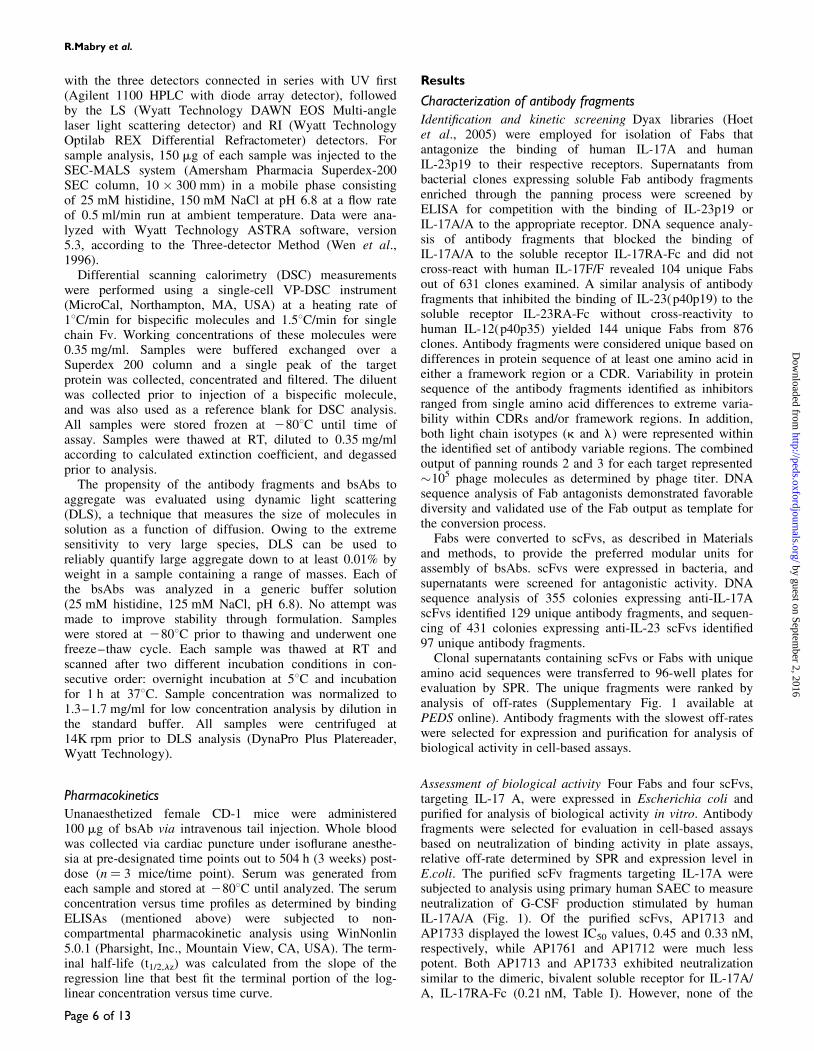
Assessment of biological activity Four Fabs and four scFvs,targeting IL-17 A, were expressed in Escherichia coli andpurified for analysis of biological activity in vitro. Antibodyfragments were selected for evaluation in cell-based assaysbased on neutralization of binding activity in plate assays,relative off-rate determined by SPR and expression level inE.coli. The purified scFv fragments targeting IL-17A weresubjected to analysis using primary human SAEC to measureneutralization of G-CSF production stimulated by humanIL-17A/A (Fig. 1). Of the purified scFvs, AP1713 andAP1733 displayed the lowest IC50 values, 0.45 and 0.33 nM,respectively, while AP1761 and AP1712 were much lesspotent. Both AP1713 and AP1733 exhibited neutralizationsimilar to the dimeric, bivalent soluble receptor for IL-17A/A, IL-17RA-Fc (0.21 nM, Table I). However, none of the
R.Mabry et al.
Page 6 of 13
by guest on September 2, 2016
http://peds.oxfordjournals.org/D
ownloaded from
purified Fabs displayed even moderate neutralization ofG-CSF production in the assay within the concentrationrange tested (0.1–100 nM) (Supplementary Fig. 2A availableat PEDS online). Neutralization of the activity of the humanIL-17A/F heterodimer was also measured in this assay. TheIC50 values obtained for AP1733 were similar to thoseobtained for neutralization of the activity of human IL-17A/A. However, AP1713 was much less potent against thehuman IL-17A/F heterodimer, as evidenced by IC50 values10- to 20-fold higher than those observed with AP1733(Supplementary Fig. 3 available at PEDS online).
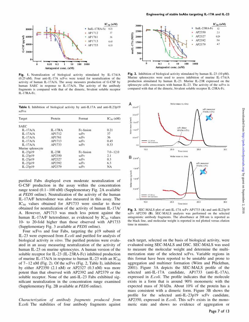
Four scFvs and four Fabs, targeting the p19 subunit ofIL-23 were expressed from E.coli and purified for analysis ofbiological activity in vitro. The purified proteins were evalu-ated in an assay measuring neutralization of the activity ofhuman IL-23 on murine splenocytes. A human version of thesoluble receptor for IL-23 (IL-23RA-Fc) inhibited productionof murine IL-17A/A in response to human IL-23 with an IC50
of 7–12 nM (Fig. 2). Of the scFvs (Fig. 2, Table I), inhibitionby either AP2350 (2.1 nM) or AP2327 (0.3 nM) was morepotent than that observed with AP2392 and AP2379 or thesoluble receptor. None of the anti-IL-23 Fabs exhibited sig-nificant neutralization in the concentration range examined(Supplementary Fig. 2B available at PEDS online).
Characterization of antibody fragments produced fromE.coli The stabilities of four antibody fragments against
each target, selected on the basis of biological activity, wereevaluated using SEC-MALS and DSC. SEC-MALS was usedto measure the molecular weight and determine the multi-merization state of the selected scFvs. Variable regions inthis format have been reported to be unstable and prone toaggregation and multimer formation (Worn and Pluckthun,2001). Figure 3A depicts the SEC-MALS profile of theselected anti-IL-17A candidate, AP1733 (anti-IL-17A),expressed in E.coli. The profile indicates that the moleculeexists in a form that is around 90% monomeric with theexpected mass of 30 kDa. About 10% of the protein has amass consistent with a dimeric form. Figure 3B shows theprofile for the selected anti-IL-23p19 scFv candidate,AP2350, expressed in E.coli. This scFv exists in the mono-meric state and shows no evidence of aggregation or
Fig. 2. Inhibition of biological activity stimulated by human IL-23 (10 pM).Murine splenocytes were used to assess inhibition of murine IL-17A/Aproduction stimulated by human IL-23. Murine IL-23R expressed on thesplenocyte cells cross-reacts with human IL-23. The activity of the scFvs iscompared with that of the dimeric, bivalent soluble receptor IL-23RA-Fc.
Fig. 3. SEC-MALS plot of anti-IL-17A scFv AP1733 (A) and anti-IL23p19scFv AP2350 (B). SEC-MALS analysis was performed on the selectedantagonistic antibody fragments. The absorbance at 208 nm is reported asthe black line, and molecular weight is reported in red plotted versus elutiontime in minutes.
Fig. 1. Neutralization of biological activity stimulated by IL-17A/A(0.25 nM). Four anti-IL-17A scFvs were tested for neutralization of theactivity of human IL-17A/A. The assay measures production of G-CSF byhuman SAEC in response to IL-17A/A. The activity of the antibodyfragments is compared with that of the dimeric, bivalent soluble receptorIL-17RA-Fc.
Table I. Inhibition of biological activity by anti-IL17A and anti-IL23p19
scFvs
Target Protein Format IC50 (nM)
SAECIL-17A/A IL-17RA Fc-fusion 0.21IL-17A/A AP1712 scFv 37IL-17A/A AP1761 scFv 36IL-17A/A AP1713 scFv 0.45IL-17A/A AP1733 scFv 0.33
Murine splenocyteIL-23p19 IL-23R Fc-fusion 7.0–12.0IL-23p19 AP2350 scFv 2.1IL-23p19 AP2327 scFv 0.3IL-23p19 AP2392 scFv 6.3IL-23p19 AP2379 scFv NA
Engineering of stable bsAbs targeting IL-17A and IL-23
Page 7 of 13
by guest on September 2, 2016
http://peds.oxfordjournals.org/D
ownloaded from
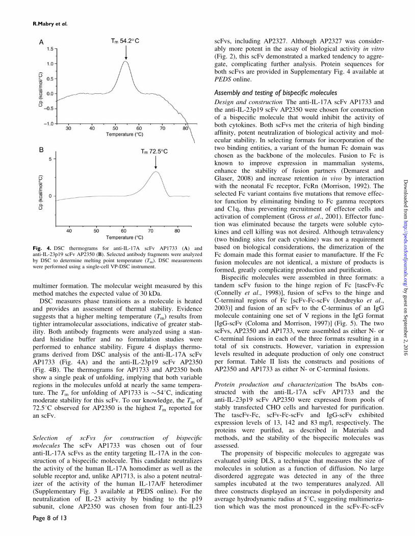
multimer formation. The molecular weight measured by thismethod matches the expected value of 30 kDa.
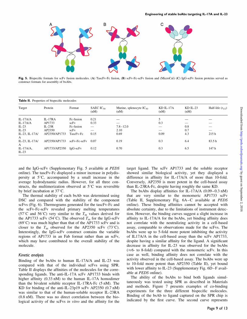
DSC measures phase transitions as a molecule is heatedand provides an assessment of thermal stability. Evidencesuggests that a higher melting temperature (Tm) results fromtighter intramolecular associations, indicative of greater stab-ility. Both antibody fragments were analyzed using a stan-dard histidine buffer and no formulation studies wereperformed to enhance stability. Figure 4 displays thermo-grams derived from DSC analysis of the anti-IL-17A scFvAP1733 (Fig. 4A) and the anti-IL-23p19 scFv AP2350(Fig. 4B). The thermograms for AP1733 and AP2350 bothshow a single peak of unfolding, implying that both variableregions in the molecules unfold at nearly the same tempera-ture. The Tm for unfolding of AP1733 is �548C, indicatingmoderate stability for this scFv. To our knowledge, the Tm of72.58C observed for AP2350 is the highest Tm reported foran scFv.
Selection of scFvs for construction of bispecificmolecules The scFv AP1733 was chosen out of fouranti-IL-17A scFvs as the entity targeting IL-17A in the con-struction of a bispecific molecule. This candidate neutralizesthe activity of the human IL-17A homodimer as well as thesoluble receptor and, unlike AP1713, is also a potent neutral-izer of the activity of the human IL-17A/F heterodimer(Supplementary Fig. 3 available at PEDS online). For theneutralization of IL-23 activity by binding to the p19subunit, clone AP2350 was chosen from four anti-IL23
scFvs, including AP2327. Although AP2327 was consider-ably more potent in the assay of biological activity in vitro(Fig. 2), this scFv demonstrated a marked tendency to aggre-gate, complicating further analysis. Protein sequences forboth scFvs are provided in Supplementary Fig. 4 available atPEDS online.
Assembly and testing of bispecific moleculesDesign and construction The anti-IL-17A scFv AP1733 andthe anti-IL-23p19 scFv AP2350 were chosen for constructionof a bispecific molecule that would inhibit the activity ofboth cytokines. Both scFvs met the criteria of high bindingaffinity, potent neutralization of biological activity and mol-ecular stability. In selecting formats for incorporation of thetwo binding entities, a variant of the human Fc domain waschosen as the backbone of the molecules. Fusion to Fc isknown to improve expression in mammalian systems,enhance the stability of fusion partners (Demarest andGlaser, 2008) and increase retention in vivo by interactionwith the neonatal Fc receptor, FcRn (Morrison, 1992). Theselected Fc variant contains five mutations that remove effec-tor function by eliminating binding to Fc gamma receptorsand C1q, thus preventing recruitment of effector cells andactivation of complement (Gross et al., 2001). Effector func-tion was eliminated because the targets were soluble cyto-kines and cell killing was not desired. Although tetravalency(two binding sites for each cytokine) was not a requirementbased on biological considerations, the dimerization of theFc domain made this format easier to manufacture. If the Fcfusion molecules are not identical, a mixture of products isformed, greatly complicating production and purification.
Bispecific molecules were assembled in three formats: atandem scFv fusion to the hinge region of Fc [tascFv-Fc(Connelly et al., 1998)], fusion of scFvs to the hinge andC-terminal regions of Fc [scFv-Fc-scFv (Jendreyko et al.,2003)] and fusion of an scFv to the C-terminus of an IgGmolecule containing one set of V regions in the IgG format[IgG-scFv (Coloma and Morrison, 1997)] (Fig. 5). The twoscFvs, AP2350 and AP1733, were assembled as either N- orC-terminal fusions in each of the three formats resulting in atotal of six constructs. However, variation in expressionlevels resulted in adequate production of only one constructper format. Table II lists the constructs and positions ofAP2350 and AP1733 as either N- or C-terminal fusions.
Protein production and characterization The bsAbs con-structed with the anti-IL-17A scFv AP1733 and theanti-IL-23p19 scFv AP2350 were expressed from pools ofstably transfected CHO cells and harvested for purification.The tascFv-Fc, scFv-Fc-scFv and IgG-scFv exhibitedexpression levels of 13, 142 and 83 mg/l, respectively. Theproteins were purified, as described in Materials andmethods, and the stability of the bispecific molecules wasassessed.
The propensity of bispecific molecules to aggregate wasevaluated using DLS, a technique that measures the size ofmolecules in solution as a function of diffusion. No largedisordered aggregate was detected in any of the threesamples incubated at the two temperatures analyzed. Allthree constructs displayed an increase in polydispersity andaverage hydrodynamic radius at 58C, suggesting multimeriza-tion which was the most pronounced in the scFv-Fc-scFv
Fig. 4. DSC thermograms for anti-IL-17A scFv AP1733 (A) andanti-IL-23p19 scFv AP2350 (B). Selected antibody fragments were analyzedby DSC to determine melting point temperature (Tm). DSC measurementswere performed using a single-cell VP-DSC instrument.
R.Mabry et al.
Page 8 of 13
by guest on September 2, 2016
http://peds.oxfordjournals.org/D
ownloaded from
and the IgG-scFv (Supplementary Fig. 5 available at PEDSonline). The tascFv-Fc displayed a minor increase in polydis-persity at 58C, accompanied by a small increase in theaverage hydrodynamic radius. However, for all three con-structs, the multimerization observed at 58C was reversibleby brief incubation at 378C.
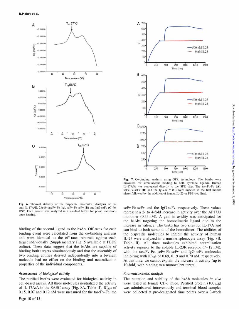
The thermal stability of each bsAb was determined usingDSC and compared with the stability of the componentscFvs (Fig. 6). Thermograms generated for the tascFv-Fc andthe scFv-Fc-scFv revealed primary melting temperatures(578C and 568C) very similar to the Tm values derived forthe AP1733 scFv (548C). The observed Tm for the IgG-scFv(698C) was much higher than that of the AP1733 scFv and iscloser to the Tm observed for the AP2350 scFv (738C).Interestingly, the IgG-scFv construct contains the variableregions of AP1733 in an Fab format rather than an scFv,which may have contributed to the overall stability of themolecule.
Kinetic analysisBinding of the bsAbs to human IL-17A/A and IL-23 wascompared with that of the individual scFvs using SPR.Table II displays the affinities of the molecules for the corre-sponding ligands. The anti-IL-17A scFv AP1733 binds withhigher affinity (0.33 nM) to the human IL-17A homodimerthan the bivalent soluble receptor IL-17RA-Fc (5 nM). TheKD for binding of the anti-IL-23p19 scFv AP2350 (0.7 nM)was similar to that of the human-soluble receptor IL-23RA(0.8 nM). There was no direct correlation between the bio-logical activity of the scFvs in vitro and the affinity for the
target ligand. The scFv AP1733 and the soluble receptorshowed similar biological activity, yet they displayed adifference in affinity for IL-17A/A of more than 10-fold.Conversely, AP2350 is more potent in the cell-based assaythan IL-23RA-Fc, despite having roughly the same KD.
The bsAbs display affinities for IL-17A/A (0.09–0.3 nM)that are very similar to the monomeric AP1733 scFv(Table II, Supplementary Fig. 6A–C available at PEDSonline). These binding affinities cannot be accepted withabsolute certainty, due to the limitations of instrument detec-tion. However, the binding curves suggest a slight increase inaffinity to IL-17A/A for the bsAbs, yet binding affinity doesnot correlate with the neutralizing activity in a cell-basedassay, comparable to observations made for the scFvs. ThebsAbs were up to 5-fold more potent inhibiting the activityof IL17A/A in the cell-based assay than the scFv AP1733,despite having a similar affinity for the ligand. A significantdecrease in affinity for IL-23 was observed for the bsAbs(�6- to 8-fold) compared with the monomeric scFv. In thiscase as well, binding affinity does not correlate with theactivity observed in the cell-based assay. The bsAbs were upto 10-fold more potent than AP2350 (Table II), yet boundwith lower affinity to IL-23 (Supplementary Fig. 6D–F avail-able at PEDS online).
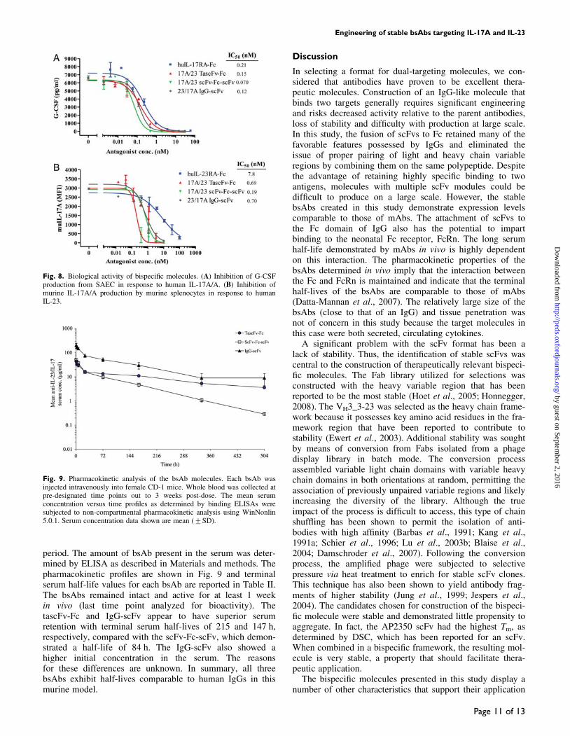
The ability of the bsAbs to bind both ligands simul-taneously was tested using SPR as described in Materialsand methods. Figure 7 presents examples of co-bindingexperiments for the three different bispecific molecules.Binding of the bsAb to ligand captured on the SPR chip isindicated by the first curve. The second curve represents
Fig. 5. Bispecific formats for scFv fusion molecules. (A) TascFv-Fc fusion, (B) scFv-Fc-scFv fusion and (MicroCal) (C) IgG-scFv fusion proteins served asconstruct formats for assembly of bsAbs.
Table II. Properties of bispecific molecules
Target Protein Format SAEC IC50
(nM)Murine, splenocyte IC50
(nM)KD IL-17A(nM)
KD IL-23(nM)
Half-life (t1/2)
IL-17A/A IL-17RA Fc-fusion 0.21 — 5 — —IL-17A/A AP1733 scFv 0.33 — 0.3 — —IL-23 IL-23R Fc-fusion — 7.8–12.0 — 0.8 —IL-23 AP2350 scFv — 2.10 — 0.7 —IL-23, IL-17A/A
AP2350/AP1733 TascFv-Fc 0.15 0.69 0.09 4.3 215 h
IL-23, IL-17A/A
AP2350/AP1733 scFv-Fc-scFv 0.07 0.19 0.3 6.4 83.5 h
IL-17A/A,IL-23
AP1733/AP2350 IgG-scFv 0.12 0.70 0.3 6.5 147 h
Engineering of stable bsAbs targeting IL-17A and IL-23
Page 9 of 13
by guest on September 2, 2016
http://peds.oxfordjournals.org/D
ownloaded from
binding of the second ligand to the bsAb. Off-rates for eachbinding event were calculated from the co-binding analysisand were identical to the off-rates reported against eachtarget individually (Supplementary Fig. 5 available at PEDSonline). These data suggest that the bsAbs are capable ofbinding both targets simultaneously and that the assembly oftwo binding entities derived independently into a bivalentmolecule had no effect on the binding and neutralizationproperties of the individual components.
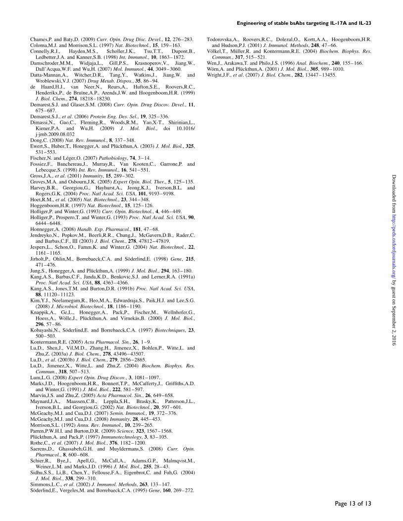
Assessment of biological activityThe purified bsAbs were evaluated for biological activity incell-based assays. All three molecules neutralized the activityof IL-17A/A in the SAEC assay (Fig. 8A, Table II). IC50s of0.15, 0.07 and 0.12 nM were measured for the tascFv-Fc, the
scFv-Fc-scFv and the IgG-scFv, respectively. These valuesrepresent a 2- to 4-fold increase in activity over the AP1733monomer (0.33 nM). A gain in avidity was anticipated forthe bsAbs targeting the homodimeric ligand due to theincrease in valency. The bsAb has two sites for IL-17A andcan bind to both subunits of the homodimer. The abilities ofthe bispecific molecules to inhibit the activity of humanIL-23 were analyzed in a murine splenocyte assay (Fig. 8B,Table II). All three molecules exhibited neutralizationactivity superior to the soluble IL-23R receptor (7–12 nM),with the tascFv-Fc, scFv-Fc-scFv and IgG-scFv moleculesinhibiting with IC50s of 0.69, 0.19 and 0.70 nM, respectively.At this time, we cannot explain the increase in activity (up to10-fold) with binding to a monovalent target.
Pharmacokinetic analysisThe retention and stability of the bsAb molecules in vivowere tested in female CD-1 mice. Purified protein (100 mg)was administered intravenously and terminal blood sampleswere collected at pre-designated time points over a 3-week
Fig. 6. Thermal stability of the bispecific molecules. Analysis of theanti-IL-17A/IL-23p19 tascFv-Fc (A), scFv-Fc-scFv (B) and IgG-scFv (C) byDSC. Each protein was analyzed in a standard buffer for phase transitionsupon heating.
Fig. 7. Co-binding analysis using SPR technology. The bsAbs weremeasured for simultaneous binding to both cytokine ligands. HumanIL-17A/A was conjugated directly to the SPR chip. The tascFv-Fc (A),scFv-Fc-scFv (B) and the IgG-scFv (C) were injected in the first mobilephase followed by the addition of human IL-23 or PBS (red line).
R.Mabry et al.
Page 10 of 13
by guest on September 2, 2016
http://peds.oxfordjournals.org/D
ownloaded from
period. The amount of bsAb present in the serum was deter-mined by ELISA as described in Materials and methods. Thepharmacokinetic profiles are shown in Fig. 9 and terminalserum half-life values for each bsAb are reported in Table II.The bsAbs remained intact and active for at least 1 weekin vivo (last time point analyzed for bioactivity). ThetascFv-Fc and IgG-scFv appear to have superior serumretention with terminal serum half-lives of 215 and 147 h,respectively, compared with the scFv-Fc-scFv, which demon-strated a half-life of 84 h. The IgG-scFv also showed ahigher initial concentration in the serum. The reasonsfor these differences are unknown. In summary, all threebsAbs exhibit half-lives comparable to human IgGs in thismurine model.
Discussion
In selecting a format for dual-targeting molecules, we con-sidered that antibodies have proven to be excellent thera-peutic molecules. Construction of an IgG-like molecule thatbinds two targets generally requires significant engineeringand risks decreased activity relative to the parent antibodies,loss of stability and difficulty with production at large scale.In this study, the fusion of scFvs to Fc retained many of thefavorable features possessed by IgGs and eliminated theissue of proper pairing of light and heavy chain variableregions by combining them on the same polypeptide. Despitethe advantage of retaining highly specific binding to twoantigens, molecules with multiple scFv modules could bedifficult to produce on a large scale. However, the stablebsAbs created in this study demonstrate expression levelscomparable to those of mAbs. The attachment of scFvs tothe Fc domain of IgG also has the potential to impartbinding to the neonatal Fc receptor, FcRn. The long serumhalf-life demonstrated by mAbs in vivo is highly dependenton this interaction. The pharmacokinetic properties of thebsAbs determined in vivo imply that the interaction betweenthe Fc and FcRn is maintained and indicate that the terminalhalf-lives of the bsAbs are comparable to those of mAbs(Datta-Mannan et al., 2007). The relatively large size of thebsAbs (close to that of an IgG) and tissue penetration wasnot of concern in this study because the target molecules inthis case were both secreted, circulating cytokines.
A significant problem with the scFv format has been alack of stability. Thus, the identification of stable scFvs wascentral to the construction of therapeutically relevant bispeci-fic molecules. The Fab library utilized for selections wasconstructed with the heavy variable region that has beenreported to be the most stable (Hoet et al., 2005; Honnegger,2008). The VH3_3-23 was selected as the heavy chain frame-work because it possesses key amino acid residues in the fra-mework region that have been reported to contribute tostability (Ewert et al., 2003). Additional stability was soughtby means of conversion from Fabs isolated from a phagedisplay library in batch mode. The conversion processassembled variable light chain domains with variable heavychain domains in both orientations at random, permitting theassociation of previously unpaired variable regions and likelyincreasing the diversity of the library. Although the trueimpact of the process is difficult to access, this type of chainshuffling has been shown to permit the isolation of anti-bodies with high affinity (Barbas et al., 1991; Kang et al.,1991a; Schier et al., 1996; Lu et al., 2003b; Blaise et al.,2004; Damschroder et al., 2007). Following the conversionprocess, the amplified phage were subjected to selectivepressure via heat treatment to enrich for stable scFv clones.This technique has also been shown to yield antibody frag-ments of higher stability (Jung et al., 1999; Jespers et al.,2004). The candidates chosen for construction of the bispeci-fic molecule were stable and demonstrated little propensity toaggregate. In fact, the AP2350 scFv had the highest Tm, asdetermined by DSC, which has been reported for an scFv.When combined in a bispecific framework, the resulting mol-ecule is very stable, a property that should facilitate thera-peutic application.
The bispecific molecules presented in this study display anumber of other characteristics that support their application
Fig. 8. Biological activity of bispecific molecules. (A) Inhibition of G-CSFproduction from SAEC in response to human IL-17A/A. (B) Inhibition ofmurine IL-17A/A production by murine splenocytes in response to humanIL-23.
Fig. 9. Pharmacokinetic analysis of the bsAb molecules. Each bsAb wasinjected intravenously into female CD-1 mice. Whole blood was collected atpre-designated time points out to 3 weeks post-dose. The mean serumconcentration versus time profiles as determined by binding ELISAs weresubjected to non-compartmental pharmacokinetic analysis using WinNonlin5.0.1. Serum concentration data shown are mean (+SD).
Engineering of stable bsAbs targeting IL-17A and IL-23
Page 11 of 13
by guest on September 2, 2016
http://peds.oxfordjournals.org/D
ownloaded from

as therapeutic agents. Analysis of the final candidates byDLS and DSC indicate that the variable region pairs ident-ified by the process associate tightly, reducing the tendencyof scFv molecules to aggregate, which has been a concernfor the scFv format (Worn and Pluckthun, 2001). Theselected scFvs maintained stability when fused to the Fcdomain of IgG to create bsAbs. Studies using DLS revealedthat the bsAbs also have little tendency to aggregate.Analysis of thermal stability by DSC indicated that thebsAbs possess stability profiles very similar to those of themonomeric units from which they were constructed. Thisobservation substantiates previous reports that suggest simplyfusing an antibody fragment to Fc may not be sufficient toovercome the instability in certain scFvs (Demarest andGlaser, 2008). The results in this study indicate that the useof stable antibody fragments as modular building blocks maysignificantly improve the assembly of bispecific moleculesfor application as therapeutic entities.
In addition to being stable, the scFvs derived from the con-version process bound their targets with high affinity.Following variable region shuffling, additional panning wasperformed on the scFv pool, and stable scFvs with high affi-nity for the target ligands were identified after just one round.The success of the process is indicated by the fact that no affi-nity maturation was required. The absence of neutralizationdemonstrated by the Fabs targeting both cytokines may be aresult of the higher off-rates observed by SPR analysis ofcrude supernatants. Faster off-rates have been reported toinfluence antagonistic activity of antibodies against othersoluble targets (Maynard et al., 2002). However, the purifiedFabs did exhibit potent neutralization of IL-23-mediatedSTAT3 phosphorylation in a short-term assay (15 min) per-formed in human IL-23R-transfected cells (SupplementaryFig. 7 available at PEDS online). Although the data are sug-gestive, it is not possible to conclude from this study that con-version of Fabs to scFvs with variable region shuffling willnecessarily result in antibody fragments with higher affinity.Additional shuffling and panning steps might have producedmore potent Fabs from the original library.
The conversion of Fab to scFv yielded molecules in bothVHVL and VLVH configurations. Previous reports haverevealed the importance of variable region orientation in theidentification of antibody fragments with high activity (Luet al., 2004; Kim et al., 2008). Incorporation of both orien-tations followed by additional rounds of panning and screen-ing allowed selection of scFvs with optimal properties.ScFvs with antagonistic activity were found in either orien-tation, but only a select few V region combinations werefound in both orientations. The combinations observedrevealed a bias for the VHVL domain order in neutralizingscFvs directed against both targets (Supplementary Fig. 8available at PEDS online). For both IL-17A/A and IL-23p19,there were few VHVL pairs recovered both as scFvs and asFabs against their respective targets. It is believed that thenumber of direct Fab to scFv conversions was altered duringthe conversion process by non-complementary primersequences that may have induced changes in terminal frame-work regions and, thus, created unique sequences. While anoverall bias for the VHVL orientation was observed, the twotop candidates targeting IL-17A and IL-23p19 representedboth orientations (AP1733 VLVH, AP2350 VHVL), validatingthe utility of the shuffling process.
The conversion from variable regions of Fabs to thesingle-chain format used a 25-amino acid gly-ser linker tojoin the light and heavy chain variable domains. The linkerwas designed to limit the number of primers needed in thePCR steps. Although previous reports have demonstrated thatdifferent linker lengths can provide increased activity and/orstability to certain variable region pairs (Arndt et al., 1998),the additional stringent panning step selects for candidatescFvs that perform well with this linker length and eliminatesany non-functional pairs among the newly paired scFvs bycompetitive selection against the target protein. Three to fourrounds of phage panning were performed to isolate scFvsthat met pre-selected target criteria.
The application of bsAbs as therapeutic molecules pro-vides the opportunity to increase the efficacy of mAbs bycombining two therapeutic activities in a single entity, eitherthrough interaction with two targets in the same pathway orthrough interaction with targets in different pathways. Thisapplication of bsAbs targets the neutralization of both IL-23and IL-17A in combination to provide a more effectiveblockade of the pro-inflammatory activity of TH17 cells.Coordinated neutralization of IL-23, the growth and survivalfactor that maintains TH17 cells, and IL-17A, a keypro-inflammatory cytokine that they produce, interrupts theinflammatory pathway at two points and should offer anadvantage over targeting either cytokine alone. The highlystable bsAbs assembled in this study exhibit the ability tobind both targets simultaneously. The potential for tetrava-lent binding, while not a requirement for biological activity,provides the opportunity to increase activity throughenhanced avidity. In fact, the bsAbs exhibit activity morethan 2-fold greater than that of the monovalent scFv counter-parts. The increase in activity due to tetravalency may resultin greater efficacy and potentially lower doses in therapeuticsettings.
The addition of dual-targeting biologics to the arsenal ofprotein therapeutics presents significant engineering chal-lenges. Maintaining affinity, potency, stability and pro-ductivity in the construction of bsAbs is a difficult task.However, the tremendous potential of this class of moleculesto advance the efficacy of mAb therapy justifies the effortspent on development. The approach described offers aneffective means to develop stable, potent bsAbs that havepotential as therapeutic molecules.
Acknowledgements
We thank Frank Grant, Monica Tackett, Benjamin Pickett, and ShellieMatthews for voluminous DNA sequence analysis and Kristine Swiderekand Jeff Ellsworth for critical reading of the manuscript.
Funding
All work was funded by ZymoGenetics.
ReferencesArndt,K.M., Muller,K.M. and Pluckthun,A. (1998) Biochemistry, 37,
12918–12926.Barbas,C.F., III, Kang,A.S., Lerner,R.A. and Benkovic,S.J. (1991) Proc. Natl
Acad. Sci. USA, 88, 7978–7982.Beckman,R.A., Weiner,L.M. and Davi,H.M. (2007) Cancer, 109, 170–179.Blaise,L., Wehnert,A., Steukers,M.P., van den Beucken,T.,
Hoogenboom,H.R. and Hufton,S.E. (2004) Gene, 342, 211–218.
R.Mabry et al.
Page 12 of 13
by guest on September 2, 2016
http://peds.oxfordjournals.org/D
ownloaded from
Chames,P. and Baty,D. (2009) Curr. Opin. Drug Disc. Devel., 12, 276–283.Coloma,M.J. and Morrison,S.L. (1997) Nat. Biotechnol., 15, 159–163.Connelly,R.J., Hayden,M.S., Scholler,J.K., Tsu,T.T., Dupont,B.,
Ledbetter,J.A. and Kanner,S.B. (1998) Int. Immunol., 10, 1863–1872.Damschroder,M.M., Widjaja,L., Gill,P.S., Krasnoperov,V., Jiang,W.,
Dall’Acqua,W.F. and Wu,H. (2007) Mol. Immunol., 44, 3049–3060.Datta-Mannan,A., Witcher,D.R., Tang,Y., Watkins,J., Jiang,W. and
Wroblewski,V.J. (2007) Drug Metab. Dispos., 35, 86–94.de Haard,H.J., van Neer,N., Reurs,A., Hufton,S.E., Roovers,R.C.,
Henderikx,P., de Bruıne,A.P., Arends,J.W. and Hoogenboom,H.R. (1999)J. Biol. Chem., 274, 18218–18230.
Demarest,S.J. and Glaser,S.M. (2008) Curr. Opin. Drug Discov. Devel., 11,675–687.
Demarest,S.J., et al. (2006) Protein Eng. Des. Sel., 19, 325–336.Dimassi,N., Gao,C., Fleming,R., Woods,R.M., Yao,X-T., Shirinian,L.,
Kiener,P.A. and Wu,H. (2009) J. Mol. Biol., doi 10.1016/j-jmb.2009.08.032
Dong,C. (2008) Nat. Rev. Immunol., 8, 337–348.Ewert,S., Huber,T., Honegger,A. and Pluckthun,A. (2003) J. Mol. Biol., 325,
531–553.Fischer,N. and Leger,O. (2007) Pathobiology, 74, 3–14.Fossiez,F., Banchereau,J., Murray,R., Van Kooten,C., Garrone,P. and
Lebecque,S. (1998) Int. Rev. Immunol., 16, 541–551.Gross,J.A., et al. (2001) Immunity, 15, 289–302.Groves,M.A. and Osbourn,J.K. (2005) Expert Opin. Biol. Ther., 5, 125–135.Harvey,B.R., Georgiou,G., Hayhurst,A., Jeong,K.J., Iverson,B.L. and
Rogers,G.K. (2004) Proc. Natl Acad. Sci. USA, 101, 9193–9198.Hoet,R.M., et al. (2005) Nat. Biotechnol., 23, 344–348.Hoggenboom,H.R. (1997) Nat. Biotechnol., 15, 125–126.Holliger,P. and Winter,G. (1993) Curr. Opin. Biotechnol., 4, 446–449.Holliger,P., Prospero,T. and Winter,G. (1993) Proc. Natl Acad. Sci. USA, 90,
6444–6448.Honnegger,A. (2008) Handb. Exp. Pharmacol., 181, 47–68.Jendreyko,N., Popkov,M., Beerli,R.R., Chung,J., McGavern,D.B., Rader,C.
and Barbas,C.F., III (2003) J. Biol. Chem., 278, 47812–47819.Jespers,L., Schon,O., Famm,K. and Winter,G. (2004) Nat. Biotechnol., 22,
1161–1165.Jirholt,P., Ohlin,M., Borrebaeck,C.A. and Soderlind,E. (1998) Gene, 215,
471–476.Jung,S., Honegger,A. and Pluckthun,A. (1999) J. Mol. Biol., 294, 163–180.Kang,A.S., Barbas,C.F., Janda,K.D., Benkovic,S.J. and Lerner,R.A. (1991a)
Proc. Natl Acad. Sci. USA, 88, 4363–4366.Kang,A.S., Jones,T.M. and Burton,D.R. (1991b) Proc. Natl Acad. Sci. USA,
88, 11120–11123.Kim,Y.J., Neelamegam,R., Heo,M.A., Edwardraja,S., Paik,H.J. and Lee,S.G.
(2008) J. Microbiol. Biotechnol., 18, 1186–1190.Knappik,A., Ge,L., Honegger,A., Pack,P., Fischer,M., Wellnhofer,G.,
Hoess,A., Wolle,J., Pluckthun,A. and Virnekas,B. (2000) J. Mol. Biol.,296, 57–86.
Kobayashi,N., Soderlind,E. and Borrebaeck,C.A. (1997) Biotechniques, 23,500–503.
Kontermann,R.E. (2005) Acta Pharmacol. Sin., 26, 1–9.Lu,D., Shen,J., Vil,M.D., Zhang,H., Jimenez,X., Bohlen,P., Witte,L. and
Zhu,Z. (2003a) J. Biol. Chem., 278, 43496–43507.Lu,D., et al. (2003b) J. Biol. Chem., 279, 2856–2865.Lu,D., Jimenez,X., Witte,L. and Zhu,Z. (2004) Biochem. Biophys. Res.
Commun., 318, 507–513.Lum,L.G. (2008) Expert Opin. Drug Discov., 3, 1081–1097.Marks,J.D., Hoogenboom,H.R., Bonnert,T.P., McCafferty,J., Griffiths,A.D.
and Winter,G. (1991) J. Mol. Biol., 222, 581–597.Marvin,J.S. and Zhu,Z. (2005) Acta Pharmacol. Sin., 26, 649–658.Maynard,J.A., Maassen,C.B., Leppla,S.H., Brasky,K., Patterson,J.L.,
Iverson,B.L. and Georgiou,G. (2002) Nat. Biotechnol., 20, 597–601.McGeachy,M.J. and Cua,D.J. (2007) Semin. Immunol., 19, 372–376.McGeachy,M.J. and Cua,D.J. (2008) Immunity, 28, 445–453.Morrison,S.L. (1992) Annu. Rev. Immunol., 10, 239–265.Parren,P.W.H.I. and Burton,D.R. (2009) Science, 323, 1567–1568.Pluckthun,A. and Pack,P. (1997) Immunotechnology, 3, 83–105.Rothe,C., et al. (2007) J. Mol. Biol., 376, 1182–1200.Saerens,D., Ghassabeh,G.H. and Muyldermans,S. (2008) Curr. Opin.
Pharmacol., 8, 600–608.Schier,R., Bye,J., Apell,G., McCall,A., Adams,G.P., Malmqvist,M.,
Weiner,L.M. and Marks,J.D. (1996) J. Mol. Biol., 255, 28–43.Sidhu,S.S., Li,B., Chen,Y., Fellouse,F.A., Eigenbrot,C. and Fuh,G. (2004)
J. Mol. Biol., 338, 299–310.Simmons,L.C., et al. (2002) J. Immunol. Methods, 263, 133–147.Soderlind,E., Vergeles,M. and Borrebaeck,C.A. (1995) Gene, 160, 269–272.
Todorovska,A., Roovers,R.C., Dolezal,O., Kortt,A.A., Hoogenboom,H.R.and Hudson,P.J. (2001) J. Immunol. Methods, 248, 47–66.
Volkel,T., Muller,R. and Kontermann,R.E. (2004) Biochem. Biophys. Res.Commun., 317, 515–521.
Wen,J., Arakawa,T. and Philo,J.S. (1996) Anal. Biochem., 240, 155–166.Worn,A. and Pluckthun,A. (2001) J. Mol. Biol., 305, 989–1010.Wright,J.F., et al. (2007) J. Biol. Chem., 282, 13447–13455.
Engineering of stable bsAbs targeting IL-17A and IL-23
Page 13 of 13
by guest on September 2, 2016
http://peds.oxfordjournals.org/D
ownloaded from