Bcl-B Expression in Human Epithelial and Nonepithelial Malignancies
Upload
independentCategory
view
1download
0
Experimental Hematology 2008;36:755–768
Newer monoclonal antibodies for hematological malignancies
Jorge Castillo, Eric Winer, and Peter Quesenberry
Division of Hematology and Oncology, Rhode Island Hospital, Brown University Warren Alpert Medical School, Providence, RI, USA
(Received 28 March 2008; revised 28 April 2008; accepted 28 April 2008)
Since the approval of rituximab in 1997, monoclonal antibodies have come to play an impor-tant role in the therapy of hematological malignancies. Rituximab, gemtuzumab ozogamicin,and alemtuzumab are US Food and Drug Administration–approved for treatment of B-celllymphomas, acute myeloid leukemia, and chronic lymphocytic leukemia, respectively. Multi-ple monoclonal antibodies directed against new and not-so-new cellular antigens are undergo-ing development and investigation all over the world. Most of these new compounds haveundergone primatization or humanization, improving their specificity and decreasing theirantigenicity when compared to earlier murine or chimeric products. This review will focuson three major aspects of monoclonal antibody therapy: 1) new therapeutic approacheswith currently approved agents; 2) preclinical and clinical experience accumulated on newagents in the last few years; discussion will include available phase I, II, and III data on ofa-tumumab, epratuzumab, CMC-544, HeFi-1, SGN-30, MDX-060, HuM195 (lintuzumab), galix-imab, lumiliximab, zanolimumab, and apolizumab; and 3) the role of naked and radiolabeledmonoclonal antibodies in the hematopoietic stem cell transplantation setting. � 2008 ISEH -Society for Hematology and Stem Cells. Published by Elsevier Inc.
Since the discovery of hybridoma technology in 1975 [1],the production and variety of monoclonal antibodies havebeen exponentially increasing. Multiple agents have beendeveloped; initially from murine origin, later chimeric be-tween murine and humanized, and now fully human anti-bodies. This evolution has improved both antigenicity andspecificity for the antigens they target. Initially developedfor detection of cellular antigens for immunohistochemicaldiagnosis, the application of monoclonal antibodies hasrecently broadened to include therapy of multiple diseases.
Development of monoclonal antibodies against cell clus-ters of differentiation (CD) allows targeted therapy of knownpresent antigens in malignant lymphoid or myeloid cells.Many of these antigens play important roles in signal trans-duction and ion transportation inside the cell, and after anti-body modulation, vital survival pathways for malignant andnormal cells are blocked or dysregulated. This effect, alongwith an immunologic boost, namely antibody-dependentcellular cytotoxicity (ADCC) and complement-dependentcytotoxicity (CDC), promotes apoptosis in the targetedmalignant cells [2,3].
Monoclonal antibodies have been used as single-agenttherapies (Table 1) or in combination with other antibodies
Offprint requests to: Jorge Castillo, M.D., Division of Hematology/
Oncology, Rhode Island Hospital, 593 Eddy Street, Providence, RI 02906;
E-mail: [email protected]
0301-472X/08 $–see front matter. Copyright � 2008 ISEH - Society for Hem
doi: 10.1016/j.exphem.2008.04.018
or chemotherapy (Table 2). Different strategies of actionhave been developed using monoclonal antibodies; theseagents have been used naked (without any drug or radioac-tive material attached to them) or as part of radioimmuno-therapy (RIT) or immunotoxins. The purpose of thisreview is to summarize the initial experience with novelmonoclonal antibodies, new therapeutic approaches with al-ready approved agents, and the role of monoclonal anti-bodies in the transplantation setting.
Monoclonal antibodies directedagainst lymphoid antigens
Anti-CD20 antibodiesThe CD20 antigen is a transmembrane protein of 35-kDmolecular weight, which is located mainly on pre-B andmature B lymphocytes [4]. Its function is still unclear, butthere is evidence that it may play a role in regulating cellcycle and differentiation processes and act as a calciumion channel as well [5]. Most B-cell non-Hodgkin’s lym-phomas (NHL) express CD20, but stem cells, pro-B cells,or plasma cells do not. CD20 is not secreted from the cellsurface, and it is not found free in plasma [6,7].
Rituximab (Rituxan). The most commonly used and mostvastly studied anti-CD20 antibody is rituximab, which
atology and Stem Cells. Published by Elsevier Inc.

756 J. Castillo et al./ Experimental Hematology 2008;36:755–768
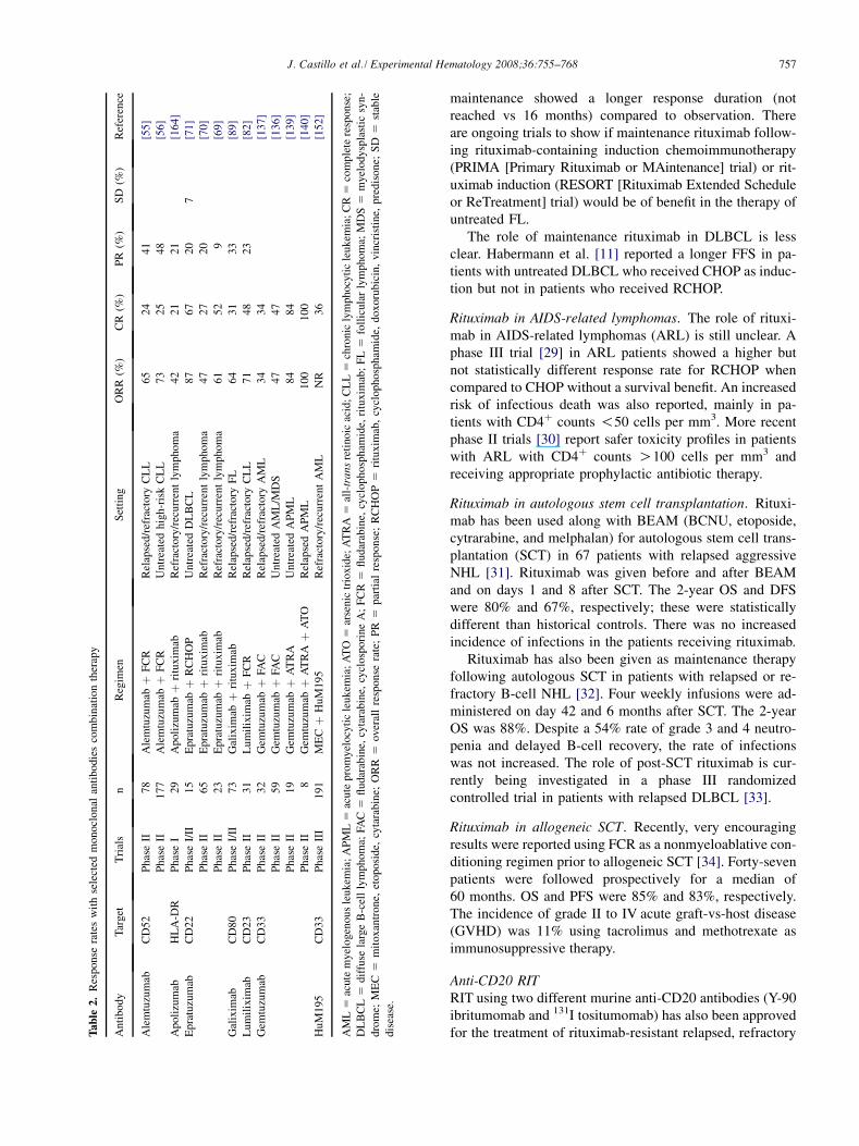
Table 1. Response rates with selected monoclonal antibody single-agent therapy
Antibody Target Trials n Setting ORR (%) CR (%) PR (%) SD (%) Reference
Alemtuzumab (subcutaneous) CD52 Phase II 41 Untreated CLL 87 19 68 [53]
Phase III 297 Untreated CLL (vs chlorambucil) 83 24 59 [54]
Epratuzumab CD22 Phase I/II 56 Refractory/recurrent aggressive lymphoma 10 5 [68]
Phase I/II 55 Refractory/recurrent indolent lymphoma 18 6 [67]
CMC-544 CD22 Phase I 34 Relapsed/refractory lymphoma 35 [79]
Galiximab CD80 Phase II 37 Relapsed/refractory FL 11 11 34 [88]
Lumiliximab CD23 Phase I 46 Refractory/recurrent CLL 0 [81]
Ofatumumab CD20 Phase I/II 33 Relapsed/refractory CLL 46 46 [51]
Phase I/II 40 Relapsed/refractory FL 38 18 20 [50]
Zanolimumab CD4 Phase II 47 Refractory CD4þ CTCL 56 [118]
Phase II 15 Refractory CD4þ PTCL 27 13.5 13.5 [119]
SGN-30 CD30 Phase II 20 Refractory/recurrent systemic ALCL 20 5 15 [102]
Phase II 17 Refractory/recurrent cutaneous ALCL 35 6 29 [104]
Phase II 15 Refractory/recurrent HD 0 50 [103]
MDX-060 CD30 Phase I/II 48 Refractory/recurrent CD30þ lymphoma 11 4 7 35 [106]
HuM195 CD33 Phase II 50 Refractory/recurrent AML 6 4 2 [151]
ALCL 5 anaplastic large T-cell lymphoma; AML 5 acute myelogenous leukemia; CLL 5 chronic lymphocytic leukemia; CR 5 complete response; CTCL 5
cutaneous T-cell lymphoma; FL 5 follicular lymphoma; HD 5 Hodgkin disease; ORR 5 overall response rate; PR 5 partial response; PTCL 5 peripheral
T-cell lymphoma; SD 5 stable disease.
was the first monoclonal antibody to be approved by the USFood and Drug Administration (FDA) for treatment of a hu-man disease in 1997, and is now considered part of the stan-dard therapies of choice for diffuse large B-cell lymphoma(DLBCL) and follicular lymphoma (FL).
Rituximab combined with CHOP (RCHOP) has im-proved the overall survival (OS) and event-free survivalin newly diagnosed patients with DLBCL in multiple phaseIII randomized trials [8–11]; the potential advantage of giv-ing RCHOP every 14 days instead of every 21 days is cur-rently being investigated [12]. Rituximab and CVP(cyclophosphamide, vincristine, and prednisone) haveproven to improve overall response rate (ORR) and pro-gression-free survival (PFS) when compared to CVP alonein patients with FL, but an improvement on OS was not ap-parent [13]; but, recently, a meta-analysis showed that thecombination of rituximab and any type of chemotherapyimproved OS when compared to chemotherapy alone witha hazard ratio (HR) of 0.65 [14,15]. The most commonside effects of rituximab include fever, chills, and rigorsand are mainly infusion-related. The incidence of these re-actions tends to diminish with subsequent infusions. Fatalreactivation of hepatitis B virus has been described in pa-tients undergoing therapy with rituximab [16]; routine hep-atitis B virus serologies should be taken prior to, during,and after rituximab therapy to detect some possible late re-activations [17].
Rituximab in chronic lymphocytic leukemia. Rituximab hasnot been FDA-approved for treatment of chronic lympho-cytic leukemia (CLL), but it is the most commonly usedmonoclonal antibody in this disease. The apoptotic mecha-nisms of rituximab seem to play a more important role inCLL than in NHL. Single-agent rituximab has limited
activity in previously treated [18,19] or untreated [20]CLL. A potential explanation is the weak expression ofCD20 by CLL cells. Dose-escalation [21] and more dose-intensive regimens [22] have been tried and have showedonly limited activity. The combination of fludarabine, cy-clophosphamide, and rituximab (FCR) is considered bymany the standard of care in previously treated or untreatedCLL. In relapsed CLL, FCR achieved an ORR of 37% withcomplete response (CR) rate of 25% and a nodular PR of12% [23]. Furthermore, FCR reached an ORR of 94%with a 72% CR rate and a 4-year OS of 83% in patientswith untreated CLL [24]. Other promising combination isfludarabine and rituximab (FR); in untreated CLL patients,FR has shown an ORR of 90% with a 47% CR rate in theCALGB 9712 study [25], while the CALGB 9011 showedan ORR of 84% with a 38% CR rate [26]. The combinationof FCR and alemtuzumab will be discussed later in thisreview.
Maintenance regimens with rituximab. Two big random-ized trials have shown the clinical benefit of adding rituxi-mab maintenance following induction therapy for advancedrelapsed or refractory FL. Van Oers et al. [27] showed thatmaintenance rituximab improved PFS in patients whoreceived induction CHOP with and without rituximab(HR 5 0.65). There was also an improvement in OS forpatients who responded to induction chemoimmunotherapyand were randomized to maintenance rituximab (HR 5
0.52). Forstpointner et al. [28] achieved similar results byrandomizing patients with relapsed or refractory FL andmantle cell lymphoma to rituximab and fludarabine, cyclo-phosphamide, and mitoxantrone (FCM) or FCM alone.Patients treated with RFCM had a better response ratethan patients who received FCM alone. Rituximab
757J. Castillo et al./ Experimental Hematology 2008;36:755–768
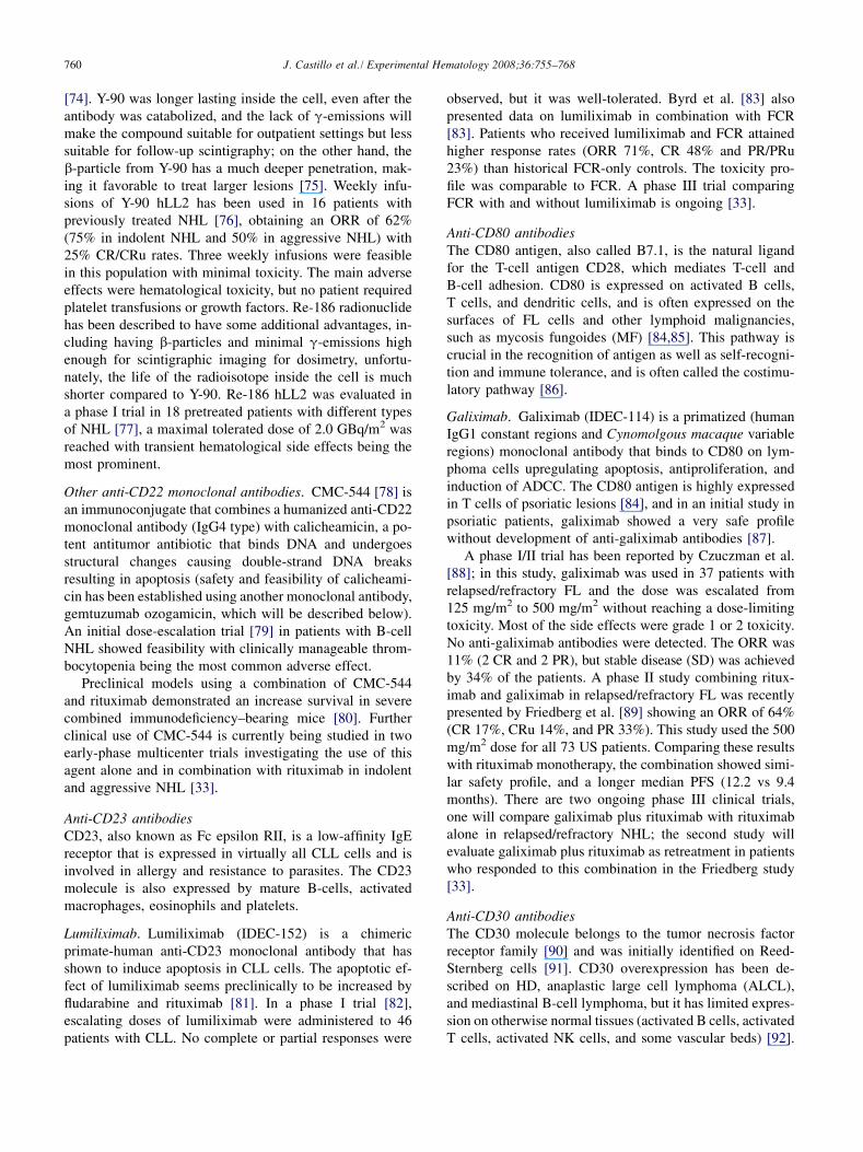
Ta
ble
2.
Res
pon
sera
tes
wit
hse
lect
edm
on
ocl
on
alan
tib
od
ies
com
bin
atio
nth
erap
y
An
tib
od
yT
arge
tT
rial
sn
Reg
imen
Set
tin
gO
RR
(%)
CR
(%)
PR
(%)
SD
(%)
Ref
eren
ce
Ale
mtu
zum
abC
D5
2P
has
eII
78
Ale
mtu
zum
abþ
FC
RR
elap
sed
/ref
ract
ory
CL
L6
52
44
1[5
5]
Ph
ase
II1
77
Ale
mtu
zum
abþ
FC
RU
ntr
eate
dhig
h-r
isk
CL
L73
25
48
[56
]
Ap
oli
zum
abH
LA
-DR
Ph
ase
I2
9A
po
lizu
mabþ
ritu
xim
abR
efra
cto
ry/r
ecu
rren
tly
mp
hom
a4
22
12
1[1
64
]
Ep
ratu
zum
abC
D2
2P
has
eI/
II1
5E
pra
tuzu
mabþ
RC
HO
PU
ntr
eate
dD
LB
CL
87
67
20
7[7
1]
Ph
ase
II6
5E
pra
tuzu
mabþ
ritu
xim
abR
efra
cto
ry/r
ecu
rren
tly
mp
hom
a4
72
72
0[7
0]
Ph
ase
II2
3E
pra
tuzu
mabþ
ritu
xim
abR
efra
cto
ry/r
ecu
rren
tly
mp
hom
a6
15
29
[69
]
Gal
ixim
abC
D8
0P
has
eI/
II7
3G
alix
imabþ
ritu
xim
abR
elap
sed
/ref
ract
ory
FL
64
31
33
[89
]
Lu
mil
ixim
abC
D2
3P
has
eII
31
Lu
mil
ixim
abþ
FC
RR
elap
sed
/ref
ract
ory
CL
L7
14
82
3[8
2]
Gem
tuzu
mab
CD
33
Ph
ase
II3
2G
emtu
zum
abþ
FAC
Rel
apse
d/r
efra
cto
ryA
ML
34
34
[13
7]
Ph
ase
II5
9G
emtu
zum
abþ
FAC
Untr
eate
dA
ML
/MD
S47
47
[13
6]
Ph
ase
II1
9G
emtu
zum
abþ
AT
RA
Untr
eate
dA
PM
L84
84
[13
9]
Ph
ase
II8
Gem
tuzu
mabþ
AT
RAþ
AT
OR
elap
sed
AP
ML
10
01
00
[14
0]
Hu
M1
95
CD
33
Ph
ase
III
19
1M
ECþ
Hu
M19
5R
efra
cto
ry/r
ecu
rren
tA
ML
NR
36
[15
2]
AM
L5
acut
em
yel
oge
no
us
leu
kem
ia;
AP
ML
5ac
ute
pro
my
elo
cyti
cle
uk
emia
;A
TO
5ar
sen
ictr
iox
ide;
AT
RA
5al
l-tr
ans
reti
noic
acid
;C
LL
5ch
ron
icly
mp
hocy
tic
leu
kem
ia;
CR
5co
mp
lete
resp
on
se;
DL
BC
L5
dif
fuse
larg
eB
-cel
lly
mpho
ma;
FAC
5fl
uda
rab
ine,
cyta
rab
ine,
cycl
osp
ori
ne
A;
FC
R5
flu
dar
abin
e,cy
clo
ph
osp
ham
ide,
ritu
xim
ab;
FL
5fo
llic
ula
rly
mp
hom
a;M
DS
5m
yelo
dy
spla
stic
syn
-
dro
me;
ME
C5
mit
oxan
tro
ne,
eto
po
sid
e,cy
tara
bin
e;O
RR
5ov
eral
lre
spo
nse
rate
;P
R5
par
tial
resp
on
se;
RC
HO
P5
ritu
xim
ab,
cycl
oph
osp
ham
ide,
do
xor
ub
icin
,v
incr
isti
ne,
pre
dis
on
e;S
D5
stab
le
dis
ease
.
maintenance showed a longer response duration (notreached vs 16 months) compared to observation. Thereare ongoing trials to show if maintenance rituximab follow-ing rituximab-containing induction chemoimmunotherapy(PRIMA [Primary Rituximab or MAintenance] trial) or rit-uximab induction (RESORT [Rituximab Extended Scheduleor ReTreatment] trial) would be of benefit in the therapy ofuntreated FL.
The role of maintenance rituximab in DLBCL is lessclear. Habermann et al. [11] reported a longer FFS in pa-tients with untreated DLBCL who received CHOP as induc-tion but not in patients who received RCHOP.
Rituximab in AIDS-related lymphomas. The role of rituxi-mab in AIDS-related lymphomas (ARL) is still unclear. Aphase III trial [29] in ARL patients showed a higher butnot statistically different response rate for RCHOP whencompared to CHOP without a survival benefit. An increasedrisk of infectious death was also reported, mainly in pa-tients with CD4þ counts !50 cells per mm3. More recentphase II trials [30] report safer toxicity profiles in patientswith ARL with CD4þ counts O100 cells per mm3 andreceiving appropriate prophylactic antibiotic therapy.
Rituximab in autologous stem cell transplantation. Rituxi-mab has been used along with BEAM (BCNU, etoposide,cytrarabine, and melphalan) for autologous stem cell trans-plantation (SCT) in 67 patients with relapsed aggressiveNHL [31]. Rituximab was given before and after BEAMand on days 1 and 8 after SCT. The 2-year OS and DFSwere 80% and 67%, respectively; these were statisticallydifferent than historical controls. There was no increasedincidence of infections in the patients receiving rituximab.
Rituximab has also been given as maintenance therapyfollowing autologous SCT in patients with relapsed or re-fractory B-cell NHL [32]. Four weekly infusions were ad-ministered on day 42 and 6 months after SCT. The 2-yearOS was 88%. Despite a 54% rate of grade 3 and 4 neutro-penia and delayed B-cell recovery, the rate of infectionswas not increased. The role of post-SCT rituximab is cur-rently being investigated in a phase III randomizedcontrolled trial in patients with relapsed DLBCL [33].
Rituximab in allogeneic SCT. Recently, very encouragingresults were reported using FCR as a nonmyeloablative con-ditioning regimen prior to allogeneic SCT [34]. Forty-sevenpatients were followed prospectively for a median of60 months. OS and PFS were 85% and 83%, respectively.The incidence of grade II to IV acute graft-vs-host disease(GVHD) was 11% using tacrolimus and methotrexate asimmunosuppressive therapy.
Anti-CD20 RITRIT using two different murine anti-CD20 antibodies (Y-90ibritumomab and 131I tositumomab) has also been approvedfor the treatment of rituximab-resistant relapsed, refractory

758 J. Castillo et al./ Experimental Hematology 2008;36:755–768
indolent lymphomas. Y-90 ibritumomab (Zevalin, BiogenInc., Cambridge, MA, USA) achieved higher ORR andCR rates than rituximab in relapsed/refractory FL, buttime to progression (TTP) and duration of remission(DOR) were not statistically significant [35,36]. 131I tositu-momab (Bexxar, GlaxoSmithKline, Research TrianglePark, NC, USA) achieved ORR of 60% to 70% in heavilypretreated patients (some were resistant to rituximab);many patients achieved a durable response rate similar orbetter than rituximab with the obvious convenience of sin-gle-dose therapy [37].
Anti-CD20 RIT in autologous SCT. Use of SCT has im-proved survival in patients with relapsed or refractory lym-phomas, but the majority of patients will invariably relapseand a proportion will not be considered suitable candidatesfor high-dose therapies because of their age, performancestatus, or comorbidities. Radiation therapy (RT) is one ofthe most effective therapies for hematological malignancies.Higher doses of RT have been associated with less relapserates but also with increased toxicities. Using RT directedto tumor cells (i.e., RIT) as conditioning regimen prior toSCT would theoretically allow safer and better responses.
Two major strategies have been developed using RIT aspart of conditioning regimens for autologous SCT:
1. High-dose RIT with or without chemotherapy. TheSeattle Consortium pioneered the high-dose RIT ap-proach using 131I tositumomab [38]. An encouragingCR rate of 84% was achieved with minimal nonhema-tological toxicity. In a cohort analysis, high-dose RITshowed longer 5-year PFS and OS when compared tohigh-dose chemotherapy (48% vs 29% and 67% vs53%, respectively); RIT was also associated with de-creased treatment-related mortality (4% vs 11%)[39]. The same group reported a 3-year PFS of 51%and low incidence of nonhematological toxicity in24 patients older than 60 years [40]. High-dose RIThas been used in combination with high-dose chemo-therapy [41]; this study shows that 131I tositumomabcan be safely given in combination with high-dose eto-poside/cyclophosphamide. Patients who received RITand high-dose chemotherapy seemed to have longerPFS and OS than controls treated with total body irra-diation and high-dose chemotherapy. High-dose RITusing Y-90 ibritumomab in combination with carmus-tine, etoposide, cytarabine, and melphalan (BEAM)showed a 3-year OS of 54% in 44 heavily pretreatedpatients with NHL [42]. In another report [43], 31 pa-tients with relapsed NHL received Y-90 ibritumomaband high-dose etoposide/cyclophosphamide. The 2-year OS was 92% with similar toxicity rates than totalbody irradiation.
2. Standard-dose RIT plus high-dose chemotherapy. Voseet al. [44] showed that 131I tositumomab could be safelygiven prior to BEAM and autologous SCT. This
regimen achieved a CR rate of 57%. A subsequentphase II trial [45] showed an impressive 3-year OS of81% in 40 patients with relapsed but chemosensitiveDLBCL. There is an ongoing phase III trial comparing131I tositumomab-BEAM vs rituximab-BEAM in re-lapsed DLBCL [33]. In a similar fashion, Y-90 ibritu-momab prior to BEAM and autologous SCT hasshown to be safe and effective [46]. This regimenachieved a longer PFS and OS than BEAM alone(88% vs 65% and 72% vs 67%, respectively).
Ofatumumab. Ofatumumab is a fully humanized anti-CD20 monoclonal antibody that has been shown, preclini-cally, to be exceptionally active and produce a strongerCDC by more efficiently binding C1q to the surface ofthe CD20-positive cell [47]. This newer antibody interactswith a different epitope than rituximab, which is locatedin the smaller extracellular loop of CD20 [48], giving ita higher binding affinity.
Initial phase I/II clinical data in relapsed/refractory FLwas presented by Hagenbeek et al. [49,50]. Forty patientswere given escalated doses of ofatumumab from 300 mgto 1000 mg intravenously weekly for 4 weeks, obtainingan ORR of 63% with 57% response in patients previouslytreated with rituximab without reported dose-limiting toxic-ity. Coiffier et al. [51] presented the early results of a phaseI/II trial of ofatumumab in 33 patients with relapsed/refrac-tory CLL reporting significant depletion of CD19þCD5þ
cells by all patients and 67% response at the highest dose(2000 mg) by the 4th week. There are ongoing phase IIItrials of ofatumumab in CLL and FL [33].
Anti CD-52 antibodiesCD52 is a surface peptide expressed in lymphocytes, mono-cytes, macrophages, and some granulocytes. CD52 isstrongly expressed in CLL and some indolent NHL.
Alemtuzumab (Camptah-1H, Genzyme Corporation, Cam-bridge, MA, USA). Alemtuzumab is a humanized anti-CD52monoclonal antibody that has shown to induce apoptosis ofmalignant lymphocytes, CDC, and ADCC in vitro. Alemtu-zumab has been approved by the FDA for treatment of flu-darabine-refractory CLL. Alemtuzumab is able to eradicateminimal residual disease (MRD) from the bone marrow,even in high-risk CLL patients with del(17p13). Patientswho achieved MRD-negative marrow had longer survivalsthan patients who achieved either MRD-positive CR orPR [52]. On the other hand, patients who had bulky lymph-adenopathy had a lower CR rate. The major observed sideeffects are prolonged myelosuppression and lymphopenia.Prophylaxis against Pneumocystis carinii pneumonia andVaricella zoster virus is mandatory in patients receivingalemtuzumab.
To minimize its toxicity, subcutaneous alemtuzumab hasbeen administered three times a week in 41 untreated CLL

759J. Castillo et al./ Experimental Hematology 2008;36:755–768
patients [53]. The ORR was 87%, but it was less effectiveagainst nodal disease. Some patients needed more than 18weeks of therapy to achieve the best response. Subcutaneousalemtuzumab has been compared to chlorambucil in the up-front setting [54], achieving higher ORR (83% vs 55%) andCR rate (22% vs 2%). Again, CLL patients with del(17p13)had superior response to alemtuzumab. Cytomegalovirus(CMV) reactivation was seen in 52% of patients receivingalemtuzumab, making the monitoring of CMV mandatorywith this therapy.
FCR and alemtuzumab (CFAR) has been studies in 78relapsed CLL patients [55]. CFAR obtained an ORR of65% and a CR rate of 24%. Patients received granulocytecolony-stimulating factor (G-CSF) support as well as Pneu-mocystis carinii pneumonia and Varicella zoster virus pro-phylaxis. Valganciclovir showed to be more effective thanvalacyclovir in preventing CMV reactivation. CFAR is be-ing investigated as front-line therapy in 177 patients withhigh-risk CLL defined by high levels of b2-microglobulin[56]. In this population, CFAR showed an ORR of 71%.All patients in CR and near PR were MRD-negative atthe end of therapy. This trial continues accruing.
Alemtuzumab has been used as consolidation therapy af-ter chemotherapy for CLL. The intravenous regimens haveshown to be effective at achieving MRD-negative responsesfollowing fludarabine [57] or fludarabine/cyclophosphamide[58]; the latter study had to be stopped given the elevated tox-icity in the alemtuzumab arm. Subcutaneous alemtuzumab asconsolidation therapy has also proven to be effective in un-treated CLL, but with a safer toxicity profile [59]. A study us-ing fludarabine and rituximab followed by consolidationalemtuzumab in untreated CLL patients is ongoing [33].
Alemtuzumab in SCT. Its T-cell depletive effects makealemtuzumab a potentially useful agent in conditioning reg-imens prior to SCT for multiple hematological malignan-cies. Alemtuzumab could decrease the incidence of acuteand/or chronic GVHD following allogeneic SCT, whileallowing more effective reduced-intensity regimens by en-hancing graft-versus-tumor effect. Combinations of alemtu-zumab and fludarabine/busulfan [60] and fludarabine/melphalan [61] have been reported safe and effective withlower rates of GVHD in acute myeloid leukemia (AML)/MDS. The latter combination has also been used prior toSCT in the treatment of CLL [62], but rates of GVHD ap-peared higher than in the experience reported here. Patientswith Hodgkin’s disease (HD) who received alemtuzumab-containing regimens seem to experience less acute andchronic GVHD without increasing relapse rates [63].
Anti-CD22 antibodies. The CD22 molecule is a transmem-brane glycoprotein that plays a role in cellular adhesion,regulation of B-cell homing and modulation of B-cell acti-vation [64]. B-cell malignancies express CD22 up to 60%to 80% of cases. CD22 is unique in that it is internalized
into the cell when bound by antibody. This property makesit an interesting target for RIT or immunoconjugates [65].
Epratuzumab. Epratuzumab (hLL2) is an IgG1 humanizedmonoclonal antibody that in vitro has shown to bind toCD22 and internalize into the target cell [66]. An initialphase I/II trial was published by Leonard et al. [67]. Thepopulation investigated had pretreated indolent NHL witha median of 3.5 prior therapies, including 44% of patientswho received prior rituximab. Initially this was a dose-escalation trial; with doses ranging from 120 mg/m2 to1000 mg/m2 over 30 to 60 minutes weekly for 4 weeks,a dose-limiting toxicity was not reached. Epratuzumabhad an ORR of 18% with a CR rate of 6% in this heavilypretreated population. In FL, a 43% ORR was achievedwith a dose of 360 mg/m2 weekly. The use of naked epra-tuzumab was then evaluated in aggressive NHL, mostlyDLBCL [68]. This population was heavily pretreated,25% had previous SCT and O80% had bulky disease.The ORR was 10%, for patients with DLBCL the ORRwas 15%. Leonard et al. [69] presented data on 23 patientswith previously treated FL and DLBCL who received epra-tuzumab 360 mg/m2 along with rituximab 375 mg/m2
weekly for 4 weeks. All patients were rituximab naıve. Inthe FL group, an ORR of 67% and a CR rate of 60%were achieved. In the DLBCL group an ORR of 67% anda CR rate of 50% were reported; the incidence of humanantihuman antibodies was null. A similar European studyby Strauss et al. was published recently [70]. Sixty-fivepreviously treated patients were enrolled, 46 had a diagnosisof indolent NHL (34 had FL) and 19 had aggressive NHL(15 were DLBCL). An ORR of 47% was achieved (64%in FL and 47% in DLBCL patients), with a CR/CRu rateof 22% (24% in FL and 33% in DLBCL patients). Finally,the combination of epratuzumab (E) and RCHOP in newlydiagnosed DLBCL has been studied; in a pilot trial,E-RCHOP every 21 days showed feasibility with an ORRof 86% and 87% incidence of grade 4 neutropenia [71].Epratuzumab is being studied for relapsed/refractory Wal-denstrom’s macroglobulinemia [71].
Anti-CD22 RIT. Murine and humanized forms of anti-CD22 antibodies have been used in combination withdifferent radionuclides. 131I-radiolabeled murine LL2, anIgG2a monoclonal antibody, was developed to take advan-tage of the internalizing properties of the CD22 antigen,but it was found to have a very short intracellular life.The compound was cleaved intracellularly and the 131Iwas exocytosed, not allowing a full destructive effect ofthe radioisotope [72]. Despite this molecular finding, thisstudy reported an ORR 55% with 27% CR rate and hema-tological toxicity more prominent in patients with de-creased bone marrow reserve prior to begin the study[73]. When used with humanized LL2 monoclonal anti-body, Y-90 showed different advantageous characteristicsand a more favorable tumor dosimetry compared to 131I
760 J. Castillo et al./ Experimental Hematology 2008;36:755–768
[74]. Y-90 was longer lasting inside the cell, even after theantibody was catabolized, and the lack of g-emissions willmake the compound suitable for outpatient settings but lesssuitable for follow-up scintigraphy; on the other hand, theb-particle from Y-90 has a much deeper penetration, mak-ing it favorable to treat larger lesions [75]. Weekly infu-sions of Y-90 hLL2 has been used in 16 patients withpreviously treated NHL [76], obtaining an ORR of 62%(75% in indolent NHL and 50% in aggressive NHL) with25% CR/CRu rates. Three weekly infusions were feasiblein this population with minimal toxicity. The main adverseeffects were hematological toxicity, but no patient requiredplatelet transfusions or growth factors. Re-186 radionuclidehas been described to have some additional advantages, in-cluding having b-particles and minimal g-emissions highenough for scintigraphic imaging for dosimetry, unfortu-nately, the life of the radioisotope inside the cell is muchshorter compared to Y-90. Re-186 hLL2 was evaluated ina phase I trial in 18 pretreated patients with different typesof NHL [77], a maximal tolerated dose of 2.0 GBq/m2 wasreached with transient hematological side effects being themost prominent.
Other anti-CD22 monoclonal antibodies. CMC-544 [78] isan immunoconjugate that combines a humanized anti-CD22monoclonal antibody (IgG4 type) with calicheamicin, a po-tent antitumor antibiotic that binds DNA and undergoesstructural changes causing double-strand DNA breaksresulting in apoptosis (safety and feasibility of calicheami-cin has been established using another monoclonal antibody,gemtuzumab ozogamicin, which will be described below).An initial dose-escalation trial [79] in patients with B-cellNHL showed feasibility with clinically manageable throm-bocytopenia being the most common adverse effect.
Preclinical models using a combination of CMC-544and rituximab demonstrated an increase survival in severecombined immunodeficiency–bearing mice [80]. Furtherclinical use of CMC-544 is currently being studied in twoearly-phase multicenter trials investigating the use of thisagent alone and in combination with rituximab in indolentand aggressive NHL [33].
Anti-CD23 antibodiesCD23, also known as Fc epsilon RII, is a low-affinity IgEreceptor that is expressed in virtually all CLL cells and isinvolved in allergy and resistance to parasites. The CD23molecule is also expressed by mature B-cells, activatedmacrophages, eosinophils and platelets.
Lumiliximab. Lumiliximab (IDEC-152) is a chimericprimate-human anti-CD23 monoclonal antibody that hasshown to induce apoptosis in CLL cells. The apoptotic ef-fect of lumiliximab seems preclinically to be increased byfludarabine and rituximab [81]. In a phase I trial [82],escalating doses of lumiliximab were administered to 46patients with CLL. No complete or partial responses were
observed, but it was well-tolerated. Byrd et al. [83] alsopresented data on lumiliximab in combination with FCR[83]. Patients who received lumiliximab and FCR attainedhigher response rates (ORR 71%, CR 48% and PR/PRu23%) than historical FCR-only controls. The toxicity pro-file was comparable to FCR. A phase III trial comparingFCR with and without lumiliximab is ongoing [33].
Anti-CD80 antibodiesThe CD80 antigen, also called B7.1, is the natural ligandfor the T-cell antigen CD28, which mediates T-cell andB-cell adhesion. CD80 is expressed on activated B cells,T cells, and dendritic cells, and is often expressed on thesurfaces of FL cells and other lymphoid malignancies,such as mycosis fungoides (MF) [84,85]. This pathway iscrucial in the recognition of antigen as well as self-recogni-tion and immune tolerance, and is often called the costimu-latory pathway [86].
Galiximab. Galiximab (IDEC-114) is a primatized (humanIgG1 constant regions and Cynomolgous macaque variableregions) monoclonal antibody that binds to CD80 on lym-phoma cells upregulating apoptosis, antiproliferation, andinduction of ADCC. The CD80 antigen is highly expressedin T cells of psoriatic lesions [84], and in an initial study inpsoriatic patients, galiximab showed a very safe profilewithout development of anti-galiximab antibodies [87].
A phase I/II trial has been reported by Czuczman et al.[88]; in this study, galiximab was used in 37 patients withrelapsed/refractory FL and the dose was escalated from125 mg/m2 to 500 mg/m2 without reaching a dose-limitingtoxicity. Most of the side effects were grade 1 or 2 toxicity.No anti-galiximab antibodies were detected. The ORR was11% (2 CR and 2 PR), but stable disease (SD) was achievedby 34% of the patients. A phase II study combining ritux-imab and galiximab in relapsed/refractory FL was recentlypresented by Friedberg et al. [89] showing an ORR of 64%(CR 17%, CRu 14%, and PR 33%). This study used the 500mg/m2 dose for all 73 US patients. Comparing these resultswith rituximab monotherapy, the combination showed simi-lar safety profile, and a longer median PFS (12.2 vs 9.4months). There are two ongoing phase III clinical trials,one will compare galiximab plus rituximab with rituximabalone in relapsed/refractory NHL; the second study willevaluate galiximab plus rituximab as retreatment in patientswho responded to this combination in the Friedberg study[33].
Anti-CD30 antibodiesThe CD30 molecule belongs to the tumor necrosis factorreceptor family [90] and was initially identified on Reed-Sternberg cells [91]. CD30 overexpression has been de-scribed on HD, anaplastic large cell lymphoma (ALCL),and mediastinal B-cell lymphoma, but it has limited expres-sion on otherwise normal tissues (activated B cells, activatedT cells, activated NK cells, and some vascular beds) [92].
761J. Castillo et al./ Experimental Hematology 2008;36:755–768
This characteristic makes CD30 a potentially relevant targetfor monoclonal antibody therapy. There is an extensive listof anti-CD30 monoclonal antibodies developed throughoutthe years [93–97]. A murine anti-CD30 was used as part ofRIT and showed to be effective (ORR 5 27%) but wasassociated with severe hematotoxicity [98].
HeFi-1. HeFi-1 is a murine monoclonal antibody that hasproven to be preclinically effective in ALCL, but with a var-iable response in HD [95], and is currently being investi-gated in a phase I trial sponsored by the U.S. NationalCancer Institute in patients with cancer showing at least30% expression of CD30 by immunohistochemistry [33].
SGN-30. SGN-30 is a chimeric monoclonal antibody andhas proven to be effective to arrest Reed-Sternberg cellsof HD growth in vitro and in preclinical xenograft models[99] and to be synergistic to chemotherapy in CD30þ ma-lignancies [100]. Forero-Torres et al. [101,102] presentedpreliminary data from a phase I/II trial using SGN-30 in20 patients with recurrent/relapsed systemic ALCL show-ing an ORR of 20% (one CR and three PR), the study is stillongoing. Ongoing trials are investigating SNG-30 as a sin-gle agent for relapsed/refractory HD [103] and cutaneousALCL [104], and in combination with gemcitabine,vinorelbine and pegylated liposomal doxorubicin (GVD)for patients with relapsed/refractory HD [33].
MDX-060. MDX-060 [105] is a fully human anti-CD30monoclonal antibody that induces killing of CD30þ celllines and has shown to stop growth of HD preclinicalmodels. It has been studied in a dose-escalation phase I/IItrial [106] in refractory/relapsed HD and has shown to beclinically active; in 40 patients with HD achieved one CRand two PR, in six patients with ALCL achieved one CRand one PR with a very acceptable toxicity profile. Two clin-ical trials are currently recruiting patients; one is investigat-ing the effect of MDX-060 in systemic or cutaneous ALCLas single agent, the second trial is a phase II randomized trialand will compare single-agent gemcitabine vs gemcitabineplus MDX-060 vs dexamethasone plus MDX-060 [33].
Anti-CD4 antibodiesThe CD4 antigen belongs to the immunoglobulin superfam-ily, is expressed in helper T cells, regulatory T cells,macrophages, monocytes, and dendritic cells, and acts asthe coreceptor of the T-cell receptor [107]. CD4 has fourdomains (D1–D4); D2 to D4 subdomains share structuralsimilarities with immunoglobulin-constant regions whileD1 subdomain, which interacts with major histocompatibil-ity complex class II [108], is similar to immunoglobulinvariable regions. CD4 is highly expressed by malignantT cells as well, mainly in cutaneous T-cell lymphomas(CTCL) and noncutaneous peripheral T-cell lymphomas(PTCL).
Different monoclonal antibodies against T-cell antigenshave been developed and tried preclinically and clinicallyin T-cell lymphomas without major success. Initially a mu-rine anti-CD5 monoclonal antibody, T101, was developedand used in CLL and CTCL either as a single agent or aspart of immunoconjugates or RIT [109–114]. The majorlimitation of this agent was the development of humananti-mouse antibodies. Recently, the experience withchimeric and development of fully human anti-CD4 mono-clonal antibodies may overcome this initial obstacle. Knoxet al. reported results with SK3, a chimeric anti-CD4 anti-body, which was tried in seven pretreated patients with MFwith some effect, a 28.5% incidence of development of hu-man antichimera antibodies, and a good safety profile, butno significant CD4þ cell depletion could be achieved,even though most of CD4þ cells were coated with antibody[115]. Later, the same group reported clinical experience ineight patients with persistent or progressive MF usingM-T412, an anti-CD4 chimeric antibody directed againsta different epitope of the CD4 molecule, which demon-strated a higher affinity and was able to induce CD4þ
lymphocyte depletion through an Fc-mediated mechanism;the incidence of human antichimera antibodies was lower(12.5%) and showed an ORR of 88% with an averageFFP of 25 weeks [116].
Zanolimumab. In 2004, a fully human anti-CD4 monoclo-nal antibody, zanolimumab (HuMax-CD4), was designateda Fast Track Product by the FDA; initially showed a verysafe profile in a psoriasis vulgaris trial [117]. Currentlyits use is under active investigation for the treatment ofCD4þ malignancies, mainly CTCL in early and advancedstages and other noncutaneous PTCL. Obitz et al. reportedearly results of a phase II trial of zanolimumab in refrac-tory CTCL [118]; a safe profile and a favorable responseof 40% were observed. D’Amore et al. presented datafrom a phase II trial of HuMax-CD4 in non-cutaneousPTCL [119] demonstrating an ORR of 62.5% in the firsteight patients enrolled in the trial and only one relatedcase of febrile neutropenia. Two phase II clinical trialsare evaluating the efficacy of zanolimumab in earlyand late-stage CTCL [33]. A blinded, randomized phaseIII trial comparing two different dosing of zanolimumab(8 mg/m2 vs 14 mg/m2) in previously treated MF isongoing [33].
Monoclonal antibodiesdirected against myeloid antigens
Anti-CD33 antibodiesThe CD33 antigen is a 67-kD protein, which belongs to thesialic acid-binding immunoglobulin-like lectin family[120], and it was cloned and localized in chromosome 19[121]. Andrews et al. [122] later demonstrated that

762 J. Castillo et al./ Experimental Hematology 2008;36:755–768
precursors of human hematopoietic colony-forming cellswere largely CD34þ but CD33–. It was hypothesized thatantibodies targeted against CD33 would likely ablateCD33þ leukemic blast cells without affecting CD33– nor-mal precursor cells, allowing for a full bone marrow recov-ery after treatment [123]. Initial ex vivo studies reportedhigh efficacy of cytosine arabinoside/etoposide in combina-tion with MY9 (a murine anti-CD33 monoclonal antibody)to achieve bone marrow purification in acute nonlympho-cytic leukemia [124]. CD33 is expressed by cells of themyelomonocytic lineage and liver sinusoidal cells, but notby lymphoid cells [125]. Recent reports of CD33 expres-sion in drug-resistant multiple myeloma patients mayhave interesting clinical implications [126].
Gemtuzumab ozogamicin (Mylotarg, Wyeth Pharmaceuti-cals Inc., Philadelphia, PA, USA). Gemtuzumab ozogami-cin (GO) is a humanized anti-CD33 monoclonal antibodylinked to a derivative of calicheamicin, a potent cytotoxicantibiotic that gets released inside the myeloblast by hydro-lysis causing DNA breaks and subsequent cell death [127].Myelosuppression and thrombocytopenia are the most com-mon hematological toxicities seen with regular doses of GO(9 mg/m2), while hepatic veno-occlusive disease has beenreported in the allogeneic transplant setting.
Single-agent GO. As front-line therapy for AML, single-agent GO has shown moderate activity with ORR rangingbetween 17% and 27% [128,129] and a therapy-relatedmortality of up to 17%.
In the relapsed setting, the Mylotarg Study Group pre-sented data on 142 AML patients in first relapse treatedwith single-agent GO in three different phase II trials[130,131] and reported an ORR of 30%, including patientswho achieved CR and CR with incomplete recovery ofplatelets (CRp); an ORR of 26% was reported in patientsolder than 60 years old, with similar survival rates betweenthe CR and CRp groups and a good tolerability but with23% grade 3/4 hyperbilirubinemia and few but life-threat-ening bleeding events likely secondary to thrombocytope-nia. GO got approval by the FDA in 2001 based on thesestudies for patients older than 60 years old with AML infirst relapse who will not be suitable for intensive chemo-therapy; the ORR was used as a surrogate likely to predictclinical benefit [132]. The role of GO in the pretransplantsetting and as maintenance therapy in elderly populationis under current investigation [33].
GO used in combination with chemotherapy. In patientswith untreated AML, GO has been used followed by mitox-antrone, etoposide, and cytarabine [133]. This combinationshowed a response of 54% and is currently being investi-gated in a phase III trial [33]. Lower doses of GO (3 mg/m2) in combination with daunorubicin and cytarabine orfludarabine, cytarabine, G-CSF, and idarubicin showedORR up to 91% [134]. The thioguanine-containing regi-
mens showed an increased rate of venooclusive disorder(VOD) of the liver. This study set the background for therandomized MRC AML 15 trial; a total of 1115 patients(92% of patients had untreated AML) were randomizedto three different GO-containing chemotherapy regimensand two consolidation regimens. A preliminary report[135] showed an ORR of 85%; GO-containing regimenswere associated with reduced relapse rates and longerDFS, but no difference in OS has been observed. GO hasalso been used in combination with fludarabine, cytarabine,and cyclosporine in the front-line [136] and relapsed settings[137]; the combination showed an ORR of 48% and 34%,a median survival of 8 and 5.3 months, and an incidence ofVOD of 7% and 9%, respectively.
Trials evaluating GO in combination with standardinduction chemotherapy for patients with AML are underinvestigation [33].
GO in acute promyelocytic leukemia. Acute promyelocyticleukemia (APML) is a distinct subtype of AML character-ized cytogenetically by t(15;17), which transcribes PML/RAR-a, molecule involved in myeloid differentiation andapoptosis. The APML blasts express high levels of CD33.
Single-agent GO showed to be very active in APML. Inpatients with molecularly relapsed APML, GO achievedmolecular remission in 91% of patients after two dosesand in 100% of patients after three doses [138]. Esteyet al. [139] evaluated the combination of GO and all-trans-retinoic acid in 19 untreated APML patients andreported a CR rate of 84% without increased hepatotoxicity.Twelve patients achieved a PCR-negative response. Thecombination of GO, all-trans-retinoic acid and arsenictrioxide was tried in APML patients in first relapse [140];most of the patients achieved molecular remission and re-mained in a second response that was longer than their firstresponse.
Interestingly, no VOD or significant hepatotoxicity hasbeen reported with GO in APML patients. A potential ex-planation is the binding of GO to circulating APML cells,decreasing binding to hepatic cells.
Other anti-CD33 monoclonal antibodiesM195 is a murine IgG2a anti-CD33 monoclonal antibodyand was initially used as a diagnostic marker [141]. Later,the same group reported a phase I trial using 131I -M195demonstrating rapid and specific uptake by the bone mar-row and subsequent internalization into the target cells[142]. A dose-escalation trial followed in 1993 [143].Twenty-four patients were treated, 96% had decreasedblood cell counts, and 83% had decreased blast cells inthe bone marrow.
In order to counteract the immunogenicity of M195,a humanized compound was developed. HuM195 is anIgG1 computer-modeled version of M195 showing higherbinding avidity compared to the parent compound but

763J. Castillo et al./ Experimental Hematology 2008;36:755–768
with similar immunologic effects and increased ADCC me-diated by mononuclear cells [144]. A phase IB trial [145]using I-131-HuM195 showed a safe side effect profile. Tofurther decrease unwanted cytotoxic effects M195 hasbeen linked to Bi-213, a-particle radioisotope, which mightbe helpful in eradicating minimal residual disease. Bi-213-HuM195 was used in 18 patients with relapsed AML [146],93% of the patients had circulating blast response and 78%had reduction in the number of bone marrow leukemic cellswith pancytopenia being the most common adverse effect.Mulford et al. [147] presented data on cytarabine followedby Bi-213-HuM195 in 25 patients with newly diagnosed orrelapsed/refractory AML, CML in accelerated phase orRAEB/RAEB-T achieving an ORR of 28% with two CR,three CRp, and two PR, as expected all patients developedfebrile neutropenia.
The combination of interleukin (IL)-2 and HuM195 wasstudied based on the finding that IL-2 may potentiateHuM195 effect by increasing the NK-cell dependentADCC [148]. In a phase I trial, this combination showedmodest antileukemic activity, but also significant toxicitywas reported, mostly IL-2–related [149].
Unlabeled antibody has been used as consolidation ther-apy for APML to induce molecular remissions with moder-ate success [150]. A randomized phase II trial comparingtwo different doses of HuM195 in 50 patients (25 patientsin each arm, 12 mg/m2 vs 36 mg/m2) with AML [151] re-ported minimal activity represented by two CR and oneCRp of short duration. The same group reported a phaseIII trial [152] of HuM195 in combination with chemother-apy vs chemotherapy alone in refractory or first-relapsedAML showing a safe administration of the regimen butno real advantage of the addition of the antibody (CR þCRp 36 vs 28%; p 5 0.28). A dose-escalation trial usingHuM195 alone in AML/MDS patients at higher dosesthan previously used is ongoing [33].
Immunotoxins using HuM195 have been developed,conjugating the antibody with gelonin, a plant extractknown to block ribosomal production [153] and later witha recombinant form of gelonin (rGel) [154]; both com-pounds have shown antileukemic efficacy in preclinicalmodels. A phase I trial of HuM195-rGel in advancedAML is undergoing accrual [33].
Monoclonal antibodies directedagainst immune-related antigens
Anti-HLA-DR antibodiesHuman leukocyte antigen (HLA)-DR is a HLA class II mol-ecule and it is involved in presentation of exogenous anti-gens. It is expressed in monocytes, macrophages, and Bcells, so-called professional antigen-presenting cells. Theypresent these antigens to CD4þ helper T cells to initiatehumoral and facilitate cellular immune responses [155].
Apolizumab (Hu1D10; Apo). Apo is a humanized IgG1 anti-HLA class II monoclonal antibody, which binds to 1D10,a variant of the HLA-DRb chain. HLA-DR is expressed innormal and malignant cells, mainly in the B-cell population[156]. Apo has been shown to induce ADCC, CDC, andapoptosis of B cells in preclinical studies [157,158].
Apo was used in a dose-escalation phase I trial in pa-tients with relapsed, 1D10þ NHL [159,160] and in patientswith previously treated CLL/small lymphocytic leukemia[161] and showed minimal toxicity, feasibility, and someresponse of delayed onset (median time to response 106days) in this population. However, results from the phaseII trial were disappointing [162,163]. When the combina-tion of Apo and rituximab was investigated in previouslyuntreated patients with B-cell NHL and CLL, surprisinglyan increased incidence of hemolytic-uremic syndrome wasobserved, likely secondary to endothelial damage inducedby the immunotherapy. The regimen was switched to Apoon day 1 followed by rituximab on day 2 and no hemolytic-uremic syndrome was observed [164]. G-CSF has beenreported to increase cell killing by Apo in preclinical studies,Rech et al. [165] presented data in a small group of patientswith relapsed/refractory 1D10þ NHL using Apo on a threetimes weekly schedule showing feasibility. Recently, multi-ple phase I (ALL, AML, ARL, and Waldenstrom’s macro-globulinemia) and phase II trials (relapsed CLL, relapsedHD, and relapsed NHL) using Apo with and without rituxi-mab have been completed with results not yet available [33].
ConclusionThe future in management of hematological malignancieslikely will imply the ability of institute personalized thera-peutic approaches. In this regard, monoclonal antibody ther-apy may allow us to put in practice, what we call the artof medicine. Nonetheless, there is an imperative need ofmore effective therapeutic strategies using combinations ofchemotherapeutic agents, naked and radiolabeled monoclo-nal antibodies, anti-idiotype vaccines, and the new-comingantiangiogenic (i.e., bevacizumab) and immunomodulatoryagents (i.e., thalidomide). Ideally, our goal would be toimprove the cure rate with these regimens but the achieve-ment of long periods of nonprogression should be consid-ered as secondary objectives of the treatment of incurableconditions such as CLL, FL, or mantle-cell lymphoma.Radioimmunotherapy approaches and vaccines will helperadicate MRD and consolidate the benefits obtained byour induction regimens while maintenance regimens willkeep malignant progression at bay, allowing, hopefully, forprolonged drug-free and relapse-free periods of time.
Everyday we accumulate clinical experience with theuse of these monoclonal antibodies, and they are an excel-lent resource in our armamentarium against hematologicmalignancies. Although binding to target antigens appearsimportant in most instances where monoclonal antibodies

764 J. Castillo et al./ Experimental Hematology 2008;36:755–768
are therapeutically effective, the actual mechanisms oftumor response remain poorly understood. Whether theseresponses are due to direct effects on lymphoma cells orwhether they facilitate preexisting cytotoxic mechanismsis still an open question. Further progress in understandingthese responses should enhance the therapeutic usefulnessof monoclonal antibody therapy.
AcknowledgmentsThe authors indicated no potential conflict of interests.
References1. Kohler G, Milstein C. Continuous cultures of fused cells secreting
antibody of predefined specificity. Nature. 1975;256(5517):495–497.
2. Iannello A, Ahmad A. Role of antibody-dependent cell-mediated
cytotoxicity in the efficacy of therapeutic anti-cancer monoclonal
antibodies. Cancer Metastasis Rev. 2005;24:487–499.
3. Gelderman KA, Tomlinson S, Ross GD, Gorter A. Complement func-
tion in mAb-mediated cancer immunotherapy. Trends Immunol.
2004;25:158–164.
4. Stashenko P, Nadler LM, Hardy R, Schlossman SF. Characterization
of a human B lymphocyte-specific antigen. J Immunol. 1980;125:
1678–1685.
5. Tedder TF, Engel P. CD20: a regulator of cell-cycle progression of B
lymphocytes. Immunol Today. 1994;15:450–454.
6. Press OW, Farr AG, Borroz KI, et al. Endocytosis and degradation of
monoclonal antibodies targeting human B-cell malignancies. Cancer
Res. 1989;49:4906–4912.
7. Press OW, Howell-Clark J, Anderson S, Bernstein I. Retention of B-
cell-specific monoclonal antibodies by human lymphoma cells.
Blood. 1994;83:1390–1397.
8. Coiffier B, Lepage E, Briere J, et al. CHOP chemotherapy plus ritux-
imab compared with CHOP alone in elderly patients with diffuse
large-B-cell lymphoma. N Engl J Med. 2002;346:235–242.
9. Feugier P, Van Hoof A, Sebban C, et al. Long-term results of the
R-CHOP study in the treatment of elderly patients with diffuse large
B-cell lymphoma: a study by the Groupe d’Etude des Lymphomes de
l’Adulte. J Clin Oncol. 2005;23:4117–4126.
10. Pfreundschuh M, Trumper L, Osterborg A, et al. CHOP-like chemo-
therapy plus rituximab versus CHOP-like chemotherapy alone in
young patients with good-prognosis diffuse large-B-cell lymphoma:
a randomised controlled trial by the MabThera International Trial
(MInT) Group. Lancet Oncol. 2006;7:379–391.
11. Habermann TM, Weller EA, Morrison VA, et al. Rituximab-CHOP
versus CHOP alone or with maintenance rituximab in older patients
with diffuse large B-cell lymphoma. J Clin Oncol. 2006;24:3121–
3127.
12. Pfreundschuh M, Kloess M, Zeynalova S, et al. Six vs. eight cycles of
bi-weekly CHOP-14 with or without rituximab for elderly patients
with diffuse large b-cell lymphoma (DLBCL): results of the com-
pleted RICOVER-60 trial of the German High-Grade Non-Hodgkin
Lymphoma Study Group (DSHNHL). Blood (ASH Annual Meeting
Abstracts). 2006;108:Abstract 205.
13. Marcus R, Imrie K, Belch A, et al. CVP chemotherapy plus rituxi-
mab compared with CVP as first-line treatment for advanced follic-
ular lymphoma. Blood. 2005;105:1417–1423.
14. Schulz H, Skoetz N, Bohlius J, et al. Does combined immunochemo-
therapy with the monoclonal antibody rituximab improve overall
survival in the treatment of patients with indolent non-Hodgkin lym-
phoma? Preliminary results of a comprehensive meta-analysis. Blood
(ASH Annual Meeting Abstracts). 2005;106:Abstract 351.
15. Schulz H, Bohlius J, Skoetz N, et al. Combined immunochemother-
apy with rituximab improves overall survival in patients with follic-
ular and mantle cell lymphoma: updated meta-analysis results. Blood
(ASH Annual Meeting Abstracts). 2006;108:Abstract 2760.
16. Westhoff TH, Jochimsen F, Schmittel A, et al. Fatal hepatitis B virus
reactivation by an escape mutant following rituximab therapy. Blood.
2003;102:1930.
17. Perceau G, Diris N, Estines O, Derancourt C, Levy S, Bernard P. Late
lethal hepatitis B virus reactivation after rituximab treatment of low-
grade cutaneous B-cell lymphoma. Br J Dermatol. 2006;155:1053–
1056.
18. McLaughlin P, Grill-Lopez AJ, Link BK, et al. Rituximab chimeric
anti-CD20 monoclonal antibody therapy for relapsed indolent lym-
phoma: half of patients respond to a four-dose treatment program.
J Clin Oncol. 1998;16:2825–2833.
19. Nguyen DT, Amess JA, Doughty H, Hendry L, Diamond LW. IDEC-
C2B8 anti-CD20 (rituximab) immunotherapy in patients with low-
grade non-Hodgkin’s lymphoma and lymphoproliferative disorders:
evaluation of response on 48 patients. Eur J Haematol. 1999;62:
76–82.
20. Hainsworth JD, Litchy S, Barton JH, et al. Single-agent rituximab as
first-line and maintenance treatment for patients with chronic lym-
phocytic leukemia or small lymphocytic lymphoma: a phase II trial
of the Minnie Pearl Cancer Research Network. J Clin Oncol. 2003;
21:1746–1751.
21. O’Brien SM, Kantarjian H, Thomas DA, et al. Rituximab dose-
escalation trial in chronic lymphocytic leukemia. J Clin Oncol.
2001;19:2165–2170.
22. Byrd JC, Murphy T, Howard RS, et al. Rituximab using a thrice
weekly dosing schedule in B-cell chronic lymphocytic leukemia
and small lymphocytic lymphoma demonstrates clinical activity
and acceptable toxicity. J Clin Oncol. 2001;19:2153–2164.
23. Wierda W, O’Brien S, Wen S, et al. Chemoimmunotherapy with
fludarabine, cyclophosphamide, and rituximab for relapsed and re-
fractory chronic lymphocytic leukemia. J Clin Oncol. 2005;23:
4070–4078.
24. Keating MJ, O’Brien S, Albitar M, et al. Early results of a chemoim-
munotherapy regimen of fludarabine, cyclophosphamide, and rituxi-
mab as initial therapy for chronic lymphocytic leukemia. J Clin
Oncol. 2005;23:4079–4088.
25. Byrd JC, Peterson BL, Morrison VA, et al. Randomized phase 2
study of fludarabine with concurrent versus sequential treatment
with rituximab in symptomatic, untreated patients with B-cell
chronic lymphocytic leukemia: results from Cancer and Leukemia
Group B 9712 (CALGB 9712). Blood. 2003;101:6–14.
26. Byrd JC, Rai K, Peterson BL, et al. Addition of rituximab to fludar-
abine may prolong progression-free survival and overall survival in
patients with previously untreated chronic lymphocytic leukemia:
an updated retrospective comparative analysis of CALGB 9712 and
CALGB 9011. Blood. 2005;105:49–53.
27. van Oers MH, Klasa R, Marcus RE, et al. Rituximab maintenance
improves clinical outcome of relapsed/resistant follicular non-Hodg-
kin lymphoma in patients both with and without rituximab during in-
duction: results of a prospective randomized phase 3 intergroup trial.
Blood. 2006;108:3295–3301.
28. Forstpointner R, Unterhalt M, Dreyling M, et al. Maintenance ther-
apy with rituximab leads to a significant prolongation of response
duration after salvage therapy with a combination of rituximab, flu-
darabine, cyclophosphamide, and mitoxantrone (R-FCM) in patients
with recurring and refractory follicular and mantle cell lymphomas:
Results of a prospective randomized study of the German Low Grade
Lymphoma Study Group (GLSG). Blood. 2006;108:4003–4008.
29. Kaplan LD, Lee JY, Ambinder RF, et al. Rituximab does not improve
clinical outcome in a randomized phase 3 trial of CHOP with or with-
out rituximab in patients with HIV-associated non-Hodgkin

765J. Castillo et al./ Experimental Hematology 2008;36:755–768
lymphoma: AIDS-Malignancies Consortium Trial 010. Blood. 2005;
106:1538–1543.
30. Boue F, Gabarre J, Gisselbrecht C, et al. Phase II trial of CHOP plus
rituximab in patients with HIV-associated non-Hodgkin’s lymphoma.
J Clin Oncol. 2006;24:4123–4128.
31. Khouri IF, Saliba RM, Hosing C, et al. Concurrent administration of
high-dose rituximab before and after autologous stem-cell transplan-
tation for relapsed aggressive B-cell non-Hodgkin’s lymphomas.
J Clin Oncol. 2005;23:2240–2247.
32. Horwitz SM, Negrin RS, Blume KG, et al. Rituximab as adjuvant to
high-dose therapy and autologous hematopoietic cell transplantation
for aggressive non-Hodgkin lymphoma. Blood. 2004;103:777–783.
33. Clinical trials.gov: A service of the US National Institutes of Health.
www.clinicaltrials.gov. Accessed 31 December, 2007.
34. Khouri IF, McLaughlin P, Saliba RM, et al. 8-year experience with
allogeneic stem cell transplantation for relapsed follicular lymphoma
after nonmyeloablative conditioning with fludarabine, cyclophospha-
mide and rituximab. Blood; 2008 Apr 14; [Epub ahead of print].
35. Witzig TE, Gordon LI, Cabanillas F, et al. Randomized controlled
trial of yttrium-90-labeled ibritumomab tiuxetan radioimmunother-
apy versus rituximab immunotherapy for patients with relapsed or
refractory low-grade, follicular, or transformed B-cell non-Hodgkin’s
lymphoma. J Clin Oncol. 2002;20:2453–2463.
36. Gordon LI, Witzig T, Molina A, et al. Yttrium 90-labeled ibritumo-
mab tiuxetan radioimmunotherapy produces high response rates and
durable remissions in patients with previously treated B-cell lym-
phoma. Clin Lymphoma. 2004;5:98–101.
37. Fisher RI, Kaminski MS, Wahl RL, et al. Tositumomab and iodine-
131 tositumomab produces durable complete remissions in a subset
of heavily pretreated patients with low-grade and transformed non-
Hodgkin’s lymphomas. J Clin Oncol. 2005;23:7565–7573.
38. Press OW, Eary JF, Appelbaum FR, et al. Radiolabeled-antibody
therapy of B-cell lymphoma with autologous bone marrow support.
N Engl J Med. 1993;329:1219–1224.
39. Gopal AK, Gooley TA, Maloney DG, et al. High-dose radioimmuno-
therapy versus conventional high-dose therapy and autologous
hematopoietic stem cell transplantation for relapsed follicular non-
Hodgkin lymphoma: a multivariable cohort analysis. Blood. 2003;
102:2351–2357.
40. Gopal AK, Rajendran JG, Gooley TA, et al. High-dose [131I]tositu-
momab (anti-CD20) radioimmunotherapy and autologous hemato-
poietic stem-cell transplantation for adults O or 5 60 years old
with relapsed or refractory B-cell lymphoma. J Clin Oncol. 2007;
25:1396–1402.
41. Press OW, Eary JF, Gooley T, et al. A phase I/II trial of iodine-131-
tositumomab (anti-CD20), etoposide, cyclophosphamide, and autolo-
gous stem cell transplantation for relapsed B-cell lymphomas. Blood.
2000;96:2934–2942.
42. Winter JN, Inwards D, Spies S, et al. 90Y Ibritumomab tiuxetan
(zevalin(r); 90yz) doses calculated to deliver up to 1500 cGy to
critical organs may be safely combined with high-dose BEAM and
autotransplant in NHL. ASH Annual Meeting Abstracts. 2006;108:
330.
43. Nademanee A, Forman S, Molina A, et al. A phase 1/2 trial of high-
dose yttrium-90-ibritumomab tiuxetan in combination with high-dose
etoposide and cyclophosphamide followed by autologous stem cell
transplantation in patients with poor-risk or relapsed non-Hodgkin
lymphoma. Blood. 2005;106:2896–2902.
44. Vose JM, Bierman PJ, Enke C, et al. Phase I trial of iodine-131 tosi-
tumomab with high-dose chemotherapy and autologous stem-cell
transplantation for relapsed non-Hodgkin’s lymphoma. J Clin Oncol.
2005;23:461–467.
45. Vose J, Bierman P, Bociek G, et al. Radioimmunotherapy with 131-I
tositumomab enhanced survival in good prognosis relapsed and high-
risk diffuse large B-cell lymphoma (DLBCL) patients receiving high-
dose chemotherapy and autologous stem cell transplantation. J Clin
Oncol (Meeting Abstracts). 2007;25(suppl):8013.
46. Krishnan AY, Nademanee A, Raubitschek A, et al. A comparison of
beam and yttrium 90 ibritumomab tiuxetan (Zevalin(R)) in addition
to beam (Z-BEAM) in older patients undergoing autologous stem
cell transplant (ASCT) for B-cell lymphomas: impact of radioimmu-
notherapy on transplant outcomes. ASH Annual Meeting Abstracts.
2006;108:3043.
47. Teeling JL, French RR, Cragg MS, et al. Characterization of new
human CD20 monoclonal antibodies with potent cytolytic
activity against non-Hodgkin lymphomas. Blood. 2004;104:1793–
1800.
48. Teeling JL, Mackus WJ, Wiegman LJ, et al. The biological activity of
human CD20 monoclonal antibodies is linked to unique epitopes on
CD20. J Immunol. 2006;177:362–371.
49. Hagenbeek A, Plesner T, Walewski J, Hellman A, Link BK, Dalby LW.
HuMax-CD20 fully human monoclonal antibody in follicular lym-
phoma. first human exposure: early results of an ongoing phase I/II
trial. Blood (ASH Annual Meeting Abstracts). 2004;104:1400.
50. Hagenbeek A, Plesner T, Johnson P, et al. HuMax-CD20, a novel
fully human anti-cd20 monoclonal antibody: results of a Phase I/Ii
trial in relapsed or refractory follicular non-Hodgkins’s lymphoma.
Blood (ASH Annual Meeting Abstracts). 2005;106:4760.
51. Coiffier B, Tilly H, Pedersen LM, et al. HuMax CD20 Fully human
monoclonal antibody in chronic lymphocytic leuchemia. early results
from an ongoing phase I/ii clinical trial. Blood (ASH Annual Meeting
Abstracts). 2005;106:448.
52. Moreton P, Kennedy B, Lucas G, et al. Eradication of minimal resid-
ual disease in B-cell chronic lymphocytic leukemia after alemtuzu-
mab therapy is associated with prolonged survival. J Clin Oncol.
2005;23:2971–2979.
53. Lundin J, Kimby E, Bjorkholm M, et al. Phase II trial of subcutane-
ous anti-CD52 monoclonal antibody alemtuzumab (Campath-1H) as
first-line treatment for patients with B-cell chronic lymphocytic
leukemia (B-CLL). Blood. 2002;100:768–773.
54. Hillmen P, Skotnicki AB, Robak T, et al. Alemtuzumab compared
with chlorambucil as first-line therapy for chronic lymphocytic
leukemia. J Clin Oncol. 2007;25:5616–5623.
55. Wierda WG, O’Brien S, Faderl S, et al. Combined cyclophosphamide,
fludarabine, alemtuzumab, and rituximab (CFAR), an active regimen
for heavily treated patients with CLL. ASH Annual Meeting Abstracts.
2006;108:31.
56. Wierda WG, O’Brien S, Ferrajoli A, et al. Combined cyclophospha-
mide, fludarabine, alemtuzumab, and rituximab (CFAR), an active front-
line regimen for high-risk patients with CLL. ASH Annual Meeting
Abstracts. 2007;110:628.
57. O’Brien SM, Kantarjian HM, Thomas DA, et al. Alemtuzumab as
treatment for residual disease after chemotherapy in patients with
chronic lymphocytic leukemia. Cancer. 2003;98:2657–2663.
58. Wendtner CM, Ritgen M, Schweighofer CD, et al. Consolidation
with alemtuzumab in patients with chronic lymphocytic leukemia
(CLL) in first remissiondexperience on safety and efficacy within
a randomized multicenter phase III trial of the German CLL Study
Group (GCLLSG). Leukemia. 2004;18:1093–1101.
59. Montillo M, Tedeschi A, Miqueleiz S, et al. Alemtuzumab as consol-
idation after a response to fludarabine is effective in purging residual
disease in patients with chronic lymphocytic leukemia. J Clin Oncol.
2006;24:2337–2342.
60. Ho AY, Pagliuca A, Kenyon M, et al. Reduced-intensity allogeneic
hematopoietic stem cell transplantation for myelodysplastic syn-
drome and acute myeloid leukemia with multilineage dysplasia using
fludarabine, busulphan, and alemtuzumab (FBC) conditioning.
Blood. 2004;104:1616–1623.
61. van Besien K, Artz A, Smith S, et al. Fludarabine, melphalan, and
alemtuzumab conditioning in adults with standard-risk advanced
766 J. Castillo et al./ Experimental Hematology 2008;36:755–768
acute myeloid leukemia and myelodysplastic syndrome. J Clin On-
col. 2005;23:5728–5738.
62. Delgado J, Thomson K, Russell N, et al. Results of alemtuzumab-
based reduced-intensity allogeneic transplantation for chronic
lymphocytic leukemia: a British Society of Blood and Marrow Trans-
plantation Study. Blood. 2006;107:1724–1730.
63. Peggs KS, Sureda A, Qian W, et al. Reduced-intensity conditioning
for allogeneic haematopoietic stem cell transplantation in relapsed
and refractory Hodgkin lymphoma: impact of alemtuzumab and do-
nor lymphocyte infusions on long-term outcomes. Br J Haematol.
2007;139:70–80.
64. Carnahan J, Wang P, Kendall R, et al. Epratuzumab, a humanized
monoclonal antibody targeting CD22: characterization of in vitro
properties. Clin Cancer Res. 2003;9(10 Pt 2):3982S–3990S.
65. Coleman M, Goldenberg DM, Siegel AB, et al. Epratuzumab: target-
ing B-cell malignancies through CD22. Clin Cancer Res. 2003;9(10
Pt 2):3991S–3994S.
66. Leonard JP, Link BK. Immunotherapy of non-Hodgkin’s lymphoma
with hLL2 (epratuzumab, an anti-CD22 monoclonal antibody) and
Hu1D10 (apolizumab). Semin Oncol. 2002;29(suppl 2):81–86.
67. Leonard JP, Coleman M, Ketas JC, et al. Phase I/II trial of epratuzu-
mab (humanized anti-CD22 antibody) in indolent non-Hodgkin’s
lymphoma. J Clin Oncol. 2003;21:3051–3059.
68. Leonard JP, Coleman M, Ketas JC, et al. Epratuzumab, a humanized
anti-CD22 antibody, in aggressive non-Hodgkin’s lymphoma: phase
I/II clinical trial results. Clin Cancer Res. 2004;10:5327–5334.
69. Leonard JP, Coleman M, Ketas J, et al. Combination antibody ther-
apy with epratuzumab and rituximab in relapsed or refractory non-
Hodgkin’s lymphoma. J Clin Oncol. 2005;23:5044–5051.
70. Strauss SJ, Morschhauser F, Rech J, et al. Multicenter phase II trial of
immunotherapy with the humanized anti-cd22 antibody, epratuzu-
mab, in combination with rituximab, in refractory or recurrent non-
Hodgkin’s lymphoma. J Clin Oncol. 2006;24:3880–3886.
71. Micallef IN, Kahl BS, Gayko U, et al. Initial results of a pilot study
of epratuzumab and rituximab in combination with CHOP chemo-
therapy (ER-CHOP) in previously untreated patients with diffuse
large B-cell lymphoma (DLBCL). ASCO Meeting Abstracts. 2004;
22(suppl):6580.
72. Linden O, Tenvall J, Cavallin-Stahl E, et al. Radioimmunotherapy us-
ing 131I-labeled anti-CD22 monoclonal antibody (LL2) in patients
with previously treated B-cell lymphomas. Clin Cancer Res. 1999;
5(Suppl):3287s–3291s.
73. Linden O, Tennvall J, Hindorf C, et al. 131I-labelled anti-CD22 MAb
(LL2) in patients with B-cell lymphomas failing chemotherapy. Treat-
ment outcome, haematological toxicity and bone marrow absorbed
dose estimates. Acta Oncol. 2002;41:297–303.
74. Juweid ME, Stadtmauer E, Haijar G, et al. Pharmacokinetics, dosim-
etry, and initial therapeutic results with 131I- and (111)In-/90Y-
labeled humanized LL2 anti-CD22 monoclonal antibody in patients
with relapsed, refractory non-Hodgkin’s lymphoma. Clin Cancer
Res. 1999;5(suppl):3292s–3303s.
75. Stein R, Govindan SV, Chen S, et al. Radioimmunotherapy of a hu-
man lung cancer xenograft with monoclonal antibody RS7: evalua-
tion of (177)Lu and comparison of its efficacy with that of (90)Y
and residualizing (131)I. J Nucl Med. 2001;42:967–974.
76. Linden O, Hindorf C, Cavallin-Stahl E, et al. Dose-fractionated
radioimmunotherapy in non-Hodgkin’s lymphoma using DOTA-
conjugated, 90Y-radiolabeled, humanized anti-CD22 monoclonal
antibody, epratuzumab. Clin Cancer Res. 2005;11:5215–5222.
77. Postema EJ, Raemaekers JM, Oyen WJ, et al. Final results of a phase
I radioimmunotherapy trial using (186)Re-epratuzumab for the treat-
ment of patients with non-Hodgkin’s lymphoma. Clin Cancer Res.
2003;9(10 Pt 2):3995S–4002S.
78. DiJoseph JF, Armellino DC, Boghaert ER, et al. Antibody-targeted
chemotherapy with CMC-544: a CD22-targeted immunoconjugate
of calicheamicin for the treatment of B-lymphoid malignancies.
Blood. 2004;103:1807–1814.
79. Advani A, Gine E, Gisselbrecht C, et al. Preliminary report of a phase
1 study of CMC-544, an antibody-targeted chemotherapy agent, in
patients with B-Cell non-Hodgkin’s lymphoma (NHL). Blood
(ASH Annual Meeting Abstracts). 2005;106:230.
80. Hernandez-Ilizaliturri FJ, Devineni S, Arora S, Knight J, Czuczman
MS, et al. Targeting CD20 and CD22 with rituximab in combination
with CMC-544 results in improved anti-tumor activity against non-
Hodgkin’s lymphoma (NHL) pre-clinical models. Blood (ASH An-
nual Meeting Abstracts). 2005;106:1473.
81. Pathan NI, Chu P, Hariharan K, Cheney C, Molina A, Byrd J. Medi-
ation of apoptosis by and antitumor activity of lumiliximab in
chronic lymphocytic leukemia cells and CD23þ lymphoma cell
lines. Blood. 2008;111:1594–1602.
82. Byrd JC, O’Brien S, Flinn IW, et al. Phase 1 study of lumiliximab
with detailed pharmacokinetic and pharmacodynamic measurements
in patients with relapsed or refractory chronic lymphocytic leukemia.
Clin Cancer Res. 2007;13(15 Pt 1):4448–4455.
83. Byrd JC, Castro J, O’Brien S, et al. Comparison of results from
a phase 1/2 study of lumiliximab (anti-CD23) in combination with
FCR for patients with relapsed CLL with published FCR results.
ASH Annual Meeting Abstracts. 2006;108:32.
84. Nickoloff BJ, Nestle FO, Zheng XG, Turka LA. T lymphocytes in
skin lesions of psoriasis and mycosis fungoides express B7-1: a ligand
for CD28. Blood. 1994;83:2580–2586.
85. Younes A, Hariharan K, Allen RS, Leigh BR, et al. Initial trials of
anti-CD80 monoclonal antibody (Galiximab) therapy for patients
with relapsed or refractory follicular lymphoma. Clin Lymphoma.
2003;3:257–259.
86. Lu P, Wang YL, Linsley PS. Regulation of self-tolerance by
CD80/CD86 interactions. Curr Opin Immunol. 1997;9:858–862.
87. Gottlieb AB, Kang S, Linden KG, et al. Evaluation of safety and clin-
ical activity of multiple doses of the anti-CD80 monoclonal antibody,
galiximab, in patients with moderate to severe plaque psoriasis. Clin
Immunol. 2004;111:28–37.
88. Czuczman MS, Thall A, Witzig TE, et al. Phase I/II study of galix-
imab, an anti-CD80 antibody, for relapsed or refractory follicular
lymphoma. J Clin Oncol. 2005;23:4390–4398.
89. Friedberg JW, Leonard JP, Younes, et al. Updated results from a phase
II study of galiximab (anti-CD80) in combination with rituximab for
relapsed or refractory, follicular NHL. ASH Annual Meeting Ab-
stracts. 2005;106:2435.
90. Koon HB, Junghans RP. Anti-CD30 antibody-based therapy. Curr
Opin Oncol. 2000;12:588–593.
91. Stein H, Mason DY, Gerdes J, et al. The expression of the Hodgkin’s
disease associated antigen Ki-1 in reactive and neoplastic lymphoid
tissue: evidence that Reed-Sternberg cells and histiocytic malignan-
cies are derived from activated lymphoid cells. Blood. 1985;66:
848–858.
92. de Bruin PC, Gruss HJ, van der Valk P, Willemze R, Meijer CJ.
CD30 expression in normal and neoplastic lymphoid tissue: bio-
logical aspects and clinical implications. Leukemia. 1995;9:
1620–1627.
93. Zhang M, Yao Z, Zhang Z, et al. Effective therapy for a murine
model of human anaplastic large-cell lymphoma with the anti-
CD30 monoclonal antibody, HeFi-1, does not require activating Fc
receptors. Blood. 2006;108:705–710.
94. Borchmann P, Treml JF, Hansen H, et al. The human anti-CD30 an-
tibody 5F11 shows in vitro and in vivo activity against malignant
lymphoma. Blood. 2003;102:3737–3742.
95. Pfeifer W, Levi E, Petrogiannis-Haliotis T, Lehmann L, Wang Z,
Kadin ME. A murine xenograft model for human CD30þ anaplastic
large cell lymphoma. Successful growth inhibition with an anti-CD30
antibody (HeFi-1). Am J Pathol. 1999;155:1353–1359.
767J. Castillo et al./ Experimental Hematology 2008;36:755–768
96. Hombach A, Pohl C, Heuser C, et al. Generation of the single chain
antibody fragment conserves the idiotypic profile of the anti-CD30
monoclonal antibody HRS3. Scand J Immunol. 1998;48:497–501.
97. Tazzari PL, Bolognesi A, de Totero D, et al. Ber-H2 (anti-CD30)-saporin
immunotoxin: a new tool for the treatment of Hodgkin’s disease and
CD30þ lymphoma: invitro evaluation. Br J Haematol. 1992;81:203–211.
98. Schnell R, Dietlin M, Staak JO, et al. Treatment of refractory Hodg-
kin’s lymphoma patients with an iodine-131-labeled murine anti-
CD30 monoclonal antibody. J Clin Oncol. 2005;23:4669–4678.
99. Wahl AF, Klussman K, Thompson JD, et al. The anti-CD30 mono-
clonal antibody SGN-30 promotes growth arrest and DNA fragmen-
tation in vitro and affects antitumor activity in models of Hodgkin’s
disease. Cancer Res. 2002;62:3736–3742.
100. Cerveny CG, Law CL, McCormick RS, et al. Signaling via the anti-
CD30 mAb SGN-30 sensitizes Hodgkin’s disease cells to conven-
tional chemotherapeutics. Leukemia. 2005;19:1648–1655.
101. Forero-Torres A, Bernstein S, Gopal A, et al. SGN-30 (anti-CD30
monoclonal antibody) is active and well tolerated in patients with re-
fractory or recurrent systemic anaplastic large cell lymphoma. Blood
(ASH Annual Meeting Abstracts). 2004;104:2637.
102. Forero-Torres A, Bernstein SH, Gopal AK, et al. SGN-30 (anti-
CD30 Monoclonal antibody) is active and well tolerated in patients
with refractory or recurrent systemic anaplastic large cell lym-
phoma (ALCL). Blood (ASH Annual Meeting Abstracts). 2005;
106:3356.
103. Leonard JP, Rosenblatt JD, Bartlett NL, et al. Phase II study of SGN-
30 (anti-CD30 monoclonal antibody) in patients with refractory or
recurrent Hodgkin’s disease. Blood (ASH Annual Meeting Ab-
stracts). 2004;104:2635.
104. Duvic M, Kunishige J, Kim YH, et al. Preliminary phase II results of
SGN-30 (anti-CD30 monoclonal antibody) in patients with cutaneous
anaplastic large cell lymphoma (ALCL). Blood (ASH Annual Meet-
ing Abstracts). 2005;106:4802.
105. Hu XF, Xing PX. MDX-060. Medarex. Curr Opin Investig Drugs.
2005;6:1266–1271.
106. Ansell SM, Byrd JC, Horwitz SM, et al. Phase I/II, open-label, dose-
escalating study of MDX-060 administered weekly for 4 weeks in
subjects with refractory/relapsed CD30 positive lymphoma. Blood
(ASH Annual Meeting Abstracts). 2004;104:2636.
107. Springer TA. Adhesion receptors of the immune system. Nature.
1990;346(6283):425–434.
108. Chapman HA. Endosomal proteolysis and MHC class II function.
Curr Opin Immunol. 1998;10:93–102.
109. Bertram JH, Gill PS, Levine AM, et al. Monoclonal antibody T101 in
T cell malignancies: a clinical, pharmacokinetic, and immunologic
correlation. Blood. 1986;68:752–761.
110. Dillman RO, Shawler DL, Dillman JB, Royston I, et al. Therapy of
chronic lymphocytic leukemia and cutaneous T-cell lymphoma
with T101 monoclonal antibody. J Clin Oncol. 1984;2:881–891.
111. Carrasquillo JA, Mulshine JL, Bunn PA Jr, et al. Indium-111 T101
monoclonal antibody is superior to iodine-131 T101 in imaging of
cutaneous T-cell lymphoma. J Nucl Med. 1987;28:281–287.
112. Foss FM, Raubitscheck A, Mulshine JL, et al. Phase I study of the
pharmacokinetics of a radioimmunoconjugate, 90Y-T101, in patients
with CD5-expressing leukemia and lymphoma. Clin Cancer Res.
1998;4:2691–2700.
113. Mulshine JL, Carrasquillo JA, Weinstein JN, et al. Direct intralym-
phatic injection of radiolabeled 111In-T101 in patients with cutane-
ous T-cell lymphoma. Cancer Res. 1991;51:688–695.
114. Hertler AA, Schlossman DM, Borowitz MJ, et al. A phase I study of
T101-ricin A chain immunotoxin in refractory chronic lymphocytic
leukemia. J Biol Response Mod. 1988;7:97–113.
115. Knox SJ, Levy R, Hodgkinson S, et al. Observations on the effect of
chimeric anti-CD4 monoclonal antibody in patients with mycosis
fungoides. Blood. 1991;77:20–30.
116. Knox S, Hoppe RT, Maloney D, et al. Treatment of cutaneous T-cell
lymphoma with chimeric anti-CD4 monoclonal antibody. Blood.
1996;87:893–899.
117. Skov L, Kragballe K, Zachariae C, et al. HuMax-CD4: a fully human
monoclonal anti-CD4 antibody for the treatment of psoriasis vulga-
ris. Arch Dermatol. 2003;139:1433–1439.
118. Obitz E, et al. HuMax-CD4, a fully human monoclonal antibody:
early results of an ongoing phase II trial in cutaneous T cell lym-
phoma (CTCL). Blood (ASH Annual Meeting Abstracts). Blood
(ASH Annual Meeting Abstracts). 2003;102(11).
119. d’Amore F, Relander T, Hagberg H, et al. HuMax-CD4 (Zanolimu-
mab), a fully human monoclonal antibody: early results of an ongoing
clinical trial in CD4þ peripheral T-cell lymphoma of non-cutaneous
type. Blood (ASH Annual Meeting Abstracts). 2005;106:3354.
120. Freeman SD, Kelm S, Barber EK, Crocker PR, et al. Characterization
of CD33 as a new member of the sialoadhesin family of cellular
interaction molecules. Blood. 1995;85:2005–2012.
121. Peiper SC, Ashmun RA, Look AT. Molecular cloning, expression,
and chromosomal localization of a human gene encoding the CD33
myeloid differentiation antigen. Blood. 1988;72:314–321.
122. Andrews RG, Singer JW, Bernstein ID. Precursors of colony-forming
cells in humans can be distinguished from colony-forming cells by
expression of the CD33 and CD34 antigens and light scatter proper-
ties. J Exp Med. 1989;169:1721–1731.
123. Bernstein ID. CD33 as a target for selective ablation of acute myeloid
leukemia. Clin Lymphoma 2002;(suppl 1):S9–S11.
124. Stiff PJ, Schulz WC, Bishop M, Marks L. Anti-CD33 monoclonal an-
tibody and etoposide/cytosine arabinoside combinations for the ex
vivo purification of bone marrow in acute nonlymphocytic leukemia.
Blood. 1991;77:355–362.
125. Sperr WR, Florian S, Hauswirth AW, Valent P. CD 33 as a target of
therapy in acute myeloid leukemia: current status and future perspec-
tives. Leuk Lymphoma. 2005;46:1115–1120.
126. Sahara N, Ohnishi K, Ono T, et al. Clinicopathological and prog-
nostic characteristics of CD33-positive multiple myeloma. Eur J
Haematol. 2006;77:14–18.
127. Ikemoto N, Kumar RA, Ling TT, Ellestad GA, Danishefsky SJ, Patel
DJ. Calicheamicin-DNA complexes: warhead alignment and saccha-
ride recognition of the minor groove. Proc Natl Acad Sci U S A.
1995;92:10506–10510.
128. Nabhan C, Rundhaugen LM, Riley MB, et al. Phase II pilot trial of
gemtuzumab ozogamicin (GO) as first line therapy in acute myeloid
leukemia patients age 65 or older. Leuk Res. 2005;29:53–57.
129. Amadori S, Suciu S, Stasi R, et al. Gemtuzumab ozogamicin (Mylo-
targ) as single-agent treatment for frail patients 61 years of age and
older with acute myeloid leukemia: final results of AML-15B, a phase
2 study of the European Organisation for Research and Treatment of
Cancer and Gruppo Italiano Malattie Ematologiche dell’Adulto Leu-
kemia Groups. Leukemia. 2005;19:1768–1773.
130. Sievers EL, Larson RA, Stadtmauer EA, et al. Efficacy and safety
of gemtuzumab ozogamicin in patients with CD33-positive acute
myeloid leukemia in first relapse. J Clin Oncol. 2001;19:3244–
3254.
131. Larson RA, Sievers EL, Stadtmauer EA, et al. Final report of the ef-
ficacy and safety of gemtuzumab ozogamicin (Mylotarg) in patients
with CD33-positive acute myeloid leukemia in first recurrence. Can-
cer. 2005;104:1442–1452.
132. Bross PF, Beitz J, Chen G, et al. Approval summary: gemtuzumab
ozogamicin in relapsed acute myeloid leukemia. Clin Cancer Res.
2001;7:1490–1496.
133. Amadori S, Suciu S, Willemze R, et al. Sequential administration of
gemtuzumab ozogamicin and conventional chemotherapy as first line
therapy in elderly patients with acute myeloid leukemia: a phase II
study (AML-15) of the EORTC and GIMEMA leukemia groups.
Haematologica. 2004;89:950–956.
768 J. Castillo et al./ Experimental Hematology 2008;36:755–768
134. Kell WJ, Burnett AK, Chopra R, et al. A feasibility study of simul-
taneous administration of gemtuzumab ozogamicin with intensive
chemotherapy in induction and consolidation in younger patients
with acute myeloid leukemia. Blood. 2003;102:4277–4283.
135. Burnett AK, Kell WJ, Goldstone AH, et al. The addition of gemtuzu-
mab ozogamicin to induction chemotherapy for AML improves dis-
ease free survival without extra toxicity: preliminary analysis of 1115
patients in the MRC AML15 Trial. ASH Annual Meeting Abstracts.
2006;108:13.
136. Tsimberidou A, Estey E, Cortes J, et al. Gemtuzumab, fludarabine,
cytarabine, and cyclosporine in patients with newly diagnosed acute
myelogenous leukemia or high-risk myelodysplastic syndromes.
Cancer. 2003;97:1481–1487.
137. Tsimberidou A, Cortes J, Thomas D, et al. Gemtuzumab ozogamicin,
fludarabine, cytarabine and cyclosporine combination regimen in pa-
tients with CD33þ primary resistant or relapsed acute myeloid leu-
kemia. Leuk Res. 2003;27:893–897.
138. Lo-Coco F, Cimino G, Breccia M, et al. Gemtuzumab ozogamicin
(Mylotarg) as a single agent for molecularly relapsed acute promye-
locytic leukemia. Blood. 2004;104:1995–1999.
139. Estey EH, Giles FJ, Beran M, et al. Experience with gemtuzumab
ozogamycin (‘‘mylotarg’’) and all-trans retinoic acid in untreated
acute promyelocytic leukemia. Blood. 2002;99:4222–4224.
140. Aribi A, Kantarjian HM, Estey EH, et al. Combination therapy with
arsenic trioxide, all-trans retinoic acid, and gemtuzumab ozogamicin
in recurrent acute promyelocytic leukemia. Cancer. 2007;109:1355–
1359.
141. Scheinberg DA, Tanimoto M, McKenzie S, Strife A, Old LJ, Clark-
son BD. Monoclonal antibody M195: a diagnostic marker for acute
myelogenous leukemia. Leukemia. 1989;3:440–445.
142. Scheinberg DA, Lovett D, Divgi CR, et al. A phase I trial of mono-
clonal antibody M195 in acute myelogenous leukemia: specific bone
marrow targeting and internalization of radionuclide. J Clin Oncol.
1991;9:478–490.
143. Schwartz MA, Lovett DR, Redner A, et al. Dose-escalation trial of
M195 labeled with iodine 131 for cytoreduction and marrow ablation
in relapsed or refractory myeloid leukemias. J Clin Oncol. 1993;11:
294–303.
144. Caron PC, Co MS, Bull MK, Avdalovic NM, Queen C, Scheinberg
DA. Biological and immunological features of humanized M195
(anti-CD33) monoclonal antibodies. Cancer Res. 1992;52:6761–
6767.
145. Caron PC, Jurcic JG, Scott AM, et al. A phase 1B trial of human-
ized monoclonal antibody M195 (anti-CD33) in myeloid leukemia:
specific targeting without immunogenicity. Blood. 1994;83:1760–
1768.
146. Jurcic JG, Larson SM, Sgouros G, et al. Targeted alpha particle im-
munotherapy for myeloid leukemia. Blood. 2002;100:1233–1239.
147. Mulford DA, Pandit-Taskar N, McDeritt MR, et al. Sequential ther-
apy with cytarabine and bismuth-213 (213Bi)-labeled-HuM195
(anti-CD33) for acute myeloid leukemia (AML). Blood (ASH An-
nual Meeting Abstracts). 2004;104:1790.
148. Caron PC, Lai LT, Scheinberg DA. Interleukin-2 enhancement of
cytotoxicity by humanized monoclonal antibody M195 (anti-CD33)
in myelogenous leukemia. Clin Cancer Res. 1995;1:63–70.
149. Kossman SE, Scheinberg DA, Jurcic JG, Jimenez J, Caron PC. A
phase I trial of humanized monoclonal antibody HuM195 (anti-
CD33) with low-dose interleukin 2 in acute myelogenous leukemia.
Clin Cancer Res. 1999;5:2748–2755.
150. Jurcic JG, DeBlasio T, Dumont L, Yao TJ, Scheinberg DA. Molecu-
lar remission induction with retinoic acid and anti-CD33 monoclonal
antibody HuM195 in acute promyelocytic leukemia. Clin Cancer
Res. 2000;6:372–380.
151. Feldman E, Kalaycio M, Weiner G, et al. Treatment of relapsed or
refractory acute myeloid leukemia with humanized anti-CD33 mono-
clonal antibody HuM195. Leukemia. 2003;17:314–318.
152. Feldman EJ, Brandwein J, Stone R, et al. Phase III randomized mul-
ticenter study of a humanized anti-CD33 monoclonal antibody, lintu-
zumab, in combination with chemotherapy, versus chemotherapy
alone in patients with refractory or first-relapsed acute myeloid
leukemia. J Clin Oncol. 2005;23:4110–4116.
153. McGraw KJ, Rosenblum MG, Cheung L, Scheinberg DA. Character-
ization of murine and humanized anti-CD33, gelonin immunotoxins
reactive against myeloid leukemias. Cancer Immunol Immunother.
1994;39:367–374.
154. Pagliaro LC, Liu B, Munker R. Humanized M195 monoclonal anti-
body conjugated to recombinant gelonin: an anti-CD33 immunotoxin
with antileukemic activity. Clin Cancer Res. 1998;4:1971–1976.
155. Wang E, Marincola FM, Stoncek D. Human leukocyte antigen and
human neutrophil antigen systems. In: Livingstone C, ed. Hematology:
Basic Principles and Practice. New York: Elsevier; 2005. p. 2401–
2413.
156. Gingrich RD, Dahle CE, Hoskins KF, Senneff MJ. Identification and
characterization of a new surface membrane antigen found predom-
inantly on malignant B lymphocytes. Blood. 1990;75:2375–2387.
157. Kostelny SA, Link BK, Tso JY, et al. Humanization and characterization
of the anti-HLA-DR antibody 1D10. Int J Cancer. 2001;93:556–565.
158. Mone AP, Huang P, Pelicano H, et al. Hu1D10 induces apoptosis
concurrent with activation of the AKT survival pathway in human
chronic lymphocytic leukemia cells. Blood. 2004;103:1846–1854.
159. Link B, Wang H, Byrd J, et al. Phase I trial of humanized 1D10 (Hu1D10)
monoclonal antibody targeting class II molecules in patients with re-
lapsed lymphoma. Proc Am Soc Clin Oncol. 2000;19:abstract 86.
160. Link B, Wang H, Byrd JC, et al. Phase I study of Hu1D10 monoclo-
nal antibody in patients with B-cell lymphoma. Proc Am Soc Clin
Oncol. 2001;20. abstract 1135.
161. Abhyankar V, Lucas MS, Stock W, et al. Phase I study of escalated
thrice weekly dosing of Hu1D10 in chronic lymphocytic leukemia/
small lymphocytic leukemia (CLL/SLL): minimal toxicity and early
observation of in vivo tumor cell apoptosis. Proc Am Soc Clin Oncol.
2002;21. abstract 1069.
162. Link B, Kahl B, Czuczman M, et al. A phase II study of Remitogen
(Hu1D10), a humanized monoclonal antibody, in patients with re-
lapsed or refractory follicular, small lymphocytic, or marginal zone/-
MALT B-cell lymphoma. Blood. 2001;98:606a.
163. Mavromatis B, Cheson BD. Monoclonal antibody therapy of chronic
lymphocytic leukemia. J Clin Oncol. 2003;21:1874–1881.
164. Dunleavy K, et al. Phase 1 study of combination rituximab with apoli-
zumab in relapsed/refractory B-cell lymphoma and chronic lympho-
cytic leukemia. JCO ASCO Annu Meet Proceed. 2005;23(16S):6607.
165. Rech J, Repp R, Rech D, et al. A humanized HLA-DR antibody
(hu1D10, apolizumab) in combination with granulocyte colony-
stimulating factor (filgrastim) for the treatment of non-Hodgkin’s
lymphoma: a pilot study. Leuk Lymphoma. 2006;47:2147–2154.
Copyright © 2022 FDOKUMEN