PET in GIT Malignancies - The Egyptian Society of Nuclear ...
Upload
independentCategory
view
0download
0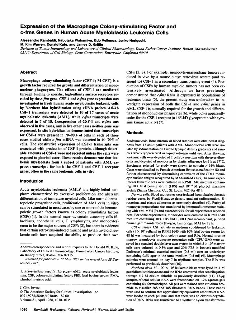
Expression of the Macrophage Colony-stimulating Factor andc-fms Genes in Human Acute Myeloblastic Leukemia CellsAlessandro Rambaldi, Nabutaka Wakamiya, Edo Vellenga, Junko Horiguchi,M. Kim Warren, Donald Kufe, and James D. GriffinDivisions of Tumor Immunology and Laboratory ofClinical Pharmacology, Dana-Farber Cancer Institute, Boston, Massachusetts02115; Department ofCell Biology, Cetus Corporation, Emeryville, California 94608
Abstract
Macrophage colony-stimulating factor (CSF-1; M-CSF) is a
growth factor required for growth and differentiation of mono-nuclear phagocytes. The effects of CSF-1 are mediatedthrough binding to specific, high-affinity surface receptors en-
coded by the c-fms gene. CSF-1 and c-fms gene expression wasinvestigated in fresh human acute myeloblastic leukemic cellsby Northern blot hybridization using cDNA probes. 4.0-kbCSF-1 transcripts were detected in 10 of 17 cases of acutemyeloblastic leukemia (AML), while c-fms transcripts were
detected in 7 of 15. Coexpression of CSF-1 and c-fms was
observed in five cases, and in five other cases neither gene wasexpressed. In situ hybridization demonstrated that transcriptsfor CSF-1 were present in 70-90% of cells in each of threecases studied while c-fms mRNA was detected in 40-70% ofcells. The constitutive expression of CSF-1 transcripts wasassociated with production of CSF-1 protein, although detect-able amounts of CSF-1 were not secreted unless the cells wereexposed to phorbol ester. These results demonstrate that leu-kemic myeloblasts from a subset of patients with AML ex-
press transcripts for both the CSF-1 and CSF-1 receptorgenes, often in the same leukemic cells in vitro.
Introduction
Acute myeloblastic leukemia (AML)' is a highly lethal neo-
plasm characterized by excessive proliferation and aberrantdifferentiation of immature myeloid cells. Like normal hema-topoietic progenitor cells, proliferation of AML cells in vitrocan be stimulated in most cases by one or more ofthe hemato-poietic growth factors known as colony stimulating factors(CSFs) (1). In the normal marrow, certain accessory cells (fi-broblasts, endothelial cells, T lymphocytes, and monocytes)seem to be the major sources ofCSFs (2), but there is evidencethat certain retrovirus-induced murine and avian myeloid leu-kemic cells have acquired the ability to produce their own
Address correspondence and reprint requests to Dr. Donald W. Kufe,Laboratory of Clinical Pharmacology, Dana-Farber Cancer Institute,44 Binney Street, Boston, MA 02115.
Receivedfor publication 27 May 1987 and in revisedform 28 Sep-tember 1987.
1. Abbreviations used in this paper: AML, acute myeloblastic leuke-mia; CSF, colony-stimulating factor; FBS, fetal bovine serum; PMA,phorbol myristic acid.
J. Clin. Invest.© The American Society for Clinical Investigation, Inc.0021-9738/88/04/1030/06 $2.00Volume 81, April 1988, 1030-1035
CSFs (2, 3). For example, monocyte-macrophage tumors in-duced in vivo by a mouse c-myc retrovirus secrete (and re-spond to) CSF-1 as a secondary transforming event (4). Pro-duction of CSFs by human myeloid tumors has not been ex-tensively investigated. Although we have previouslydemonstrated that c-fms RNA is expressed in populations ofleukemic blasts (5), the present study was undertaken to in-vestigate expression of both the CSF-1 and c-fms genes inAML. CSF- 1 is normally required for the growth and differen-tiation of mononuclear phagocytes (6), while c-fms apparentlycodes for the CSF- 1 receptor (a 1 65-kD glycoprotein with tyro-sine kinase activity) (7).
Methods
Leukemic cells. Bone marrow or blood samples were obtained at diag-nosis from 17 adult patients with AML. Mononuclear cells were iso-lated by sedimentation on Ficoll-Hypaque density gradients and sam-ples were cryopreserved in liquid nitrogen until use. After thawing,leukemic cells were depleted ofT cells by rosetting with sheep erythro-cytes and depleted of monocytes by plastic adherence for 1 h at 37°C.Specimens selected for study were shown to contain > 95% blasts.Cases were classified by French-American-British classification (8) andfurther characterized by determining expression of the CD14 mono-cyte surface antigen recognized by MAb anti-MY4 (9). In some exper-iments leukemic cells were cultured in RPMI 1640 medium contain-ing 10% fetal bovine serum (FBS) and 10-10 M phorbol myristateacetate (Sigma Chemical Co., St. Louis, MO) for 48 h.
Normal cells. Blood monocytes were isolated from platelet pheresisresidue packs by Ficoll-Hypaque density gradient sedimentation, E-rosetting, and plastic adherence as previously described (9). Purity ofmonocyte preparations was monitored by immunofluorescence stain-ing with anti-MY4 (9) and exceeded 95% for all experiments reportedhere. For some experiments, monocytes were cultured in RPMI 1640medium containing 10% FBS and 1,000 U/ml recombinant, purifiedhuman gamma-interferon (Biogen, Cambridge, MA) for 18 h.
CSF-J assays. CSF activity in medium conditioned by leukemiccells (1 X 10⁶ cells/ml in RPMI 1640 with 10% fetal bovine serum for48 h) was measured by both colony assay and RIA. Normal murinemarrow granulocyte monocyte progenitor cells (CFU-GM) were as-sayed in a standard double layer agar system in which 5 X 10⁴ marrowcells were cultured in 0.3% agar and 20% FBS in Iscove's modifiedDulbecco's minimal essential medium (0.5 ml) over an underlayercontaining 0.5% agar in the same medium (0.5 ml) (9). Macrophagecolonies were counted on day 7 in triplicate samples. The RIA wasperformed as previously described (10).
Northern blots. 50-100 x 106 leukemic blasts were lysed in 4 Mguanidium isothiocyanate and the RNA recovered after centrifugationthrough 5.7 M cesium chloride as previously described (11). 15-ggsamples of total cellular RNA were fractionated on 1.2% agarose gelscontaining 6% formaldehyde. All gels were stained with ethidium bro-mide to visualize 28S and 18S ribosomal RNA bands. These bandswere used to confirm that approximately equivalent amounts ofRNAwere loaded in each gel lane, and that there was no obvious degrada-tion ofRNA. RNA was transferred to a synthetic nylon transfer mem-
1030 Rambaldi, Wakamiya, Vellenga, Horiguchi, Warren, Kufe, and Griffin

C 8 9 1011 12 13 14 15 16 17 C
28S- _ Os
18S-
Figure 1. Expression of the CSF- I gene by AML cells. Total cellular RNA (15 gg) was fractionated on 1.2% agarose gels. CSF- I mRNA was de-tected using a 3.5-kb segment of the human CSF- I cDNA inserted in the Xho site of the pXMT 2 vector (13). The control lanes (C) showRNA from gamma-interferon-stimulated normal blood monocytes. Unstimulated normal monocytes lack detectable CSF-l RNA (21-23).
brane (GeneScreen Plus, E. I. Dupont de Nemours & Co. Inc., Wil-mington, DE), and prehybridized as previously described (1 1). CSF- 1mRNA was detected using a 3.5-kb segment of the human CSF-lcDNA inserted in the Xho site ofthe pXMT 2 vector, provided by Drs.Gordon Wong and Steven Clark, Genetics Institute, Cambridge, MA.c-fms mRNA was detected with the pSM3 plasmid containing the1.0-kb PstI fragment of v-fms. Probes were labeled using hexanucleo-tide primers and 32P-dCTP to specific activities of IO9 cpm/Ag. Hybrid-ization was performed at 60°C as previously described (1 1). In situhybridization was performed by nick-translating the same cDNA
Table I. Expression ofCSF-I and c-fms Genes by AML Cells
mRNAI
Case No. FAB* %MY4+t CSF-1 c-fms
I Ml 5 ++ +2 Ml 1 ++ -3 M5b NTI" ++ NT4 M5 5 - +
5 M4 30 ++ ++6 M4 NT - ++7 M4 NT + NT8 Ml 0 - -9 M2 2 + +10 Ml NT + +11 M2 26 - +12 M5b 49 + +13 M5a NT ++14 UNC. NT - -15 Ml 1 ++ -16 M2 3 - -17 M4 33 - -
* French-American-British classification (7). UNC, unclassified.* Surface binding of the CDl4 anti-monocyte monoclonal antibodyMY4 was detected by indirect immunofluorescence and flow cytom-etry.0 Data from Figs. 1 and 2. -, no detectable signal; +, detectable butless intense signal for 16 h exposure; ++, intense signal for 16 h ex-posure.11 NT, not tested.
probes using biotinylated dUTP (Bethesda Research Laboratories,Gaithersburg, MD). The hybridization protocol was as previously de-scribed (12).
Results
Light-density leukemic cells were prepared from 17 newlydiagnosed adult patients with AML (Table I). Samples con-tained > 90% blasts and were depleted of adherent cells and Tlymphocytes. A Northern blot analysis of total cellular RNAwas performed with a CSF- 1 cDNA probe previously shown toencode the 6 1-kD form of human urinary CSF- I (13). A 4.0-kb transcript was detected in 10 cases (strong hybridizing sig-nal in 6 cases, less intense signal in 4 cases, 48 h exposure) (Fig.1). The smaller CSF- 1 transcripts previously detected in phor-bol ester-stimulated pancreatic carcinoma cells were not ob-served (10). RNA from 15 of these 17 cases was separatelyanalyzed for the presence of c-fms transcripts (Fig. 2). Sevencases had detectable hybridization with short (16 h) film expo-sures, but there was considerable heterogeneity in the c-fmssignal. Overall, two cases had strongly hybridizing bands ofc-fms, and five cases had readily detectable, but less intense,
C 1 2 4 5 6 8 12 9 11 10C
.~A~~~~~..
I:
Figure 2. Expression of c-fms transcripts by AML cells. c-fms RNAwas detected with the pSM3 plasmid containing the 1.0-kb PstI frag-ment of v-fms.
Colony-stimulating Factor and c-fms Genes in Acute Myeloblastic Leukemia 1031
1 2 3 4 5 6 7
28S-
.. :z!"I 1.
:f:. -,

hybridizing signals. One additional case (case 4) had a veryweak signal of uncertain significance which is called negativein the analysis below (Fig. 2 shows results from 10 cases). Ofthe seven cases expressing c-fms, five cases also expressedCSF- 1 transcripts. Two cases were c-fms positive, CSF- 1 nega-tive, and three cases were CSF-1 positive, c-fms negative. Thecontrol lanes in Fig. 2 show RNA from gamma-interferon-stimulated normal blood monocytes (1,000 U/ml, 18 h). Aspreviously shown, unstimulated normal monocytes do not ex-press detectable CSF- 1 mRNA (12).
To determine the fraction of cells in individual cases ofAML that express these genes, in situ hybridization was per-
Figure 3. In situ mRNA hybridization of leukemic blasts from case5. The pSM3 plasmid containing the 1.0-kb PstI fragment of thev-fms gene, the pc CSF- 12 plasmid containing the 1.6-kb fragment ofa human CSF- I cDNA and pBR322 without insert were nick-trans-lated using biotinylated dUTP. Smears of leukemic myeloblasts were
formed in six cases. Smears were made from leukemic blastsdepleted of T cells and monocytes. Biotinylated probes werehybridized to the cells and detected with streptavidin conju-gated to alkaline phosphatase using nitroblue tetrazolium as asubstrate. In the case shown in Fig. 3 (case 5), 43.9±3.8% oftotal cells had detectable staining for c-fms mRNA, while81.5±5.5% of total cells had detectable CSF-1 transcripts. Incontrast, no hybridization was observed in RNase-treated cellsor in cells hybridized to vector (pBR322) only (Fig. 3). Thus, itis likely in this case that a substantial proportion of leukemiccells coexpressed CSF- 1 and c-fms transcripts in the same cell.
Similar in situ hybridization studies were performed on
......]g....
SI ~~~~~~~~~~~~~~~~~~~~~~~~~~~~~~~~~~~..
|l l Ss'">3}2:ks'~~~~~~~~~~~~~~~~~~~~~~~~~~~~~~~~~.. ... .......
l|c-fm s
..... ..
,,~ ~ ~ ~ ~ C F1).,8r:..
hybridized according to a previously described technique ( 12). (A)pBR322 hybridization. (B and C) c-fms hybridizations. (D and E)CSF- I hybridizations. The cells in C and E were treated with RNAsebefore performing the annealing reaction.
1032 Rambaldi, Wakamiya, Vellenga, Horiguchi, Warren, Kufe, and Griffin

other preparations of leukemic blasts. For example, clearlydetectable staining of blasts from case 9 was obtained with thebiotinylated CSF-1 probe, while c-fis transcripts were barelydetectable in these cells (Fig. 4). A low level of c-fms RNA wassimilarly detected by Northern analysis of blasts from case 9(Fig. 2). The findings by in situ hybridization also corre-sponded with those obtained by Northern blotting for cases 6and 8 (Fig. 4). Thus, c-fms, but not CSF- 1, RNA was detect-able in blasts from case 6. Moreover, neither c-fms nor CSF- 1transcripts were detectable in blasts from case 8. These find-ings further supported the specificity of the in situ hybridiza-tions and demonstrated that, when detectable, c-fms andCSF-1 expression occurs in the majority of blasts from anindividual patient.
CSF- 1
Two assays were used to detect secreted CSF- 1 in mediumconditioned by leukemic blasts for 48 h (Table II). HumanCSF- 1 has been shown to stimulate growth of murine macro-phage colonies in agar (10). AML-conditioned media stimu-lated growth of murine macrophage colonies in none of 14cases tested. Using an RIA (10), CSF- 1 levels were < 20 U/mlin each of 11 cases tested. This finding is similar to previousobservations made on the MIA-PaCa cell line, a pancreaticcarcinoma cell line that constitutively expresses the CSF-1gene, contains intracellular CSF-1 when grown in serum-freemedium, but secretes CSF- 1 only after treatment with phorbolmyristic acid (PMA) (10). We therefore investigated the effectsof PMA on CSF-1 release. Of six cases tested, four expressedCSF- 1 transcripts before PMA treatment (cases 2, 10, 12, 13)
c-fms
Case 9p.*:.
*: t..=,...], , .
i
-bX
Case 6
4~~~~~~~~~~~~~~~~~~~~~,
Case 8
Figure 4. In situ mRNA hybridization of leukemic blasts from cases 9, 6, and 8. Smears of leukemic blasts were hybridized to the biotinylatedc-fms and CSF- 1 probes as described in the legend to Fig. 3.
Colony-stimulating Factor and c-fms Genes in Acute Myeloblastic Leukemia 1033
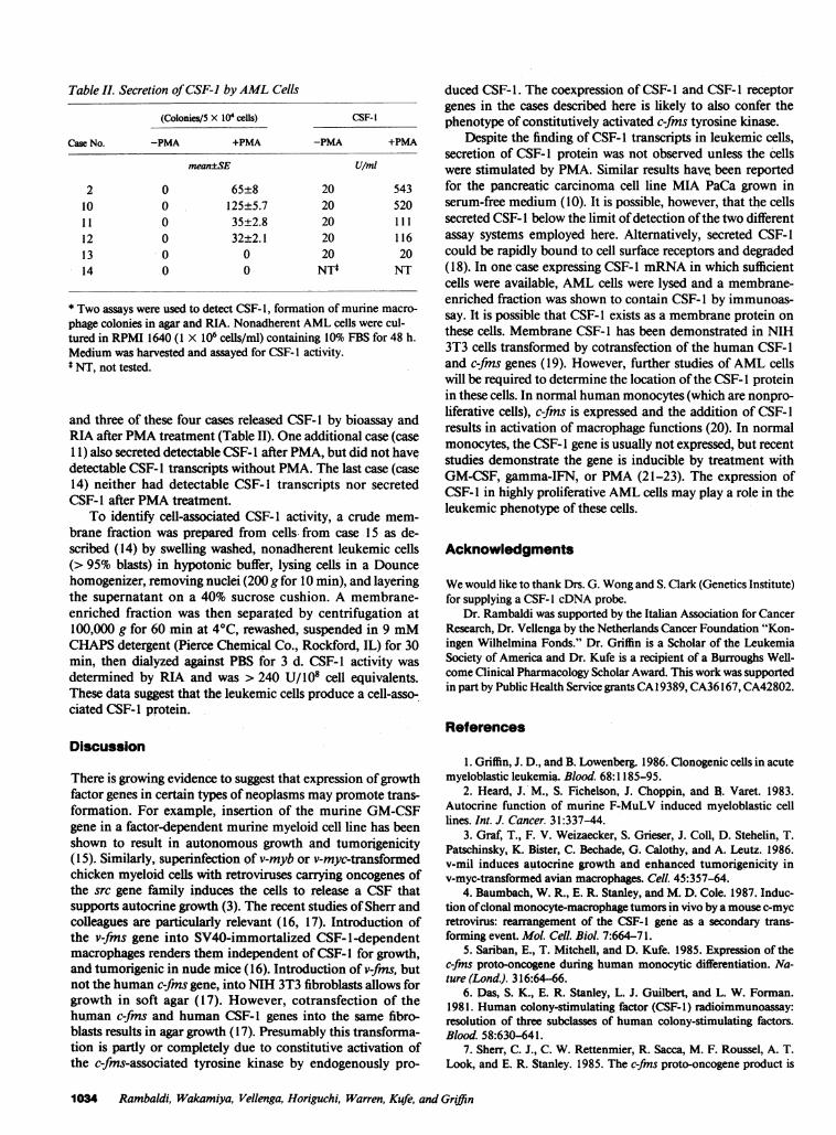
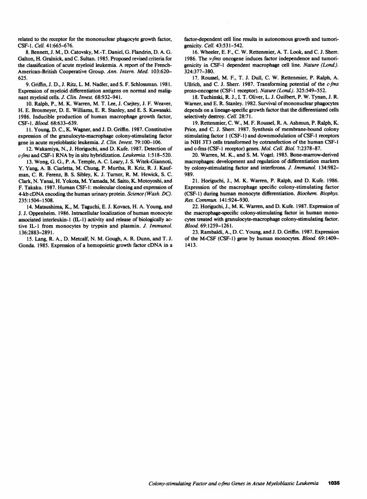
Table I. Secretion ofCSF-1 byAML Cells
(Colonies/5 X 104 cells) CSF-1
Case No. -PMA +PMA -PMA +PMA
mean±SE U/mi
2 0 65±8 20 54310 0 125±5.7 20 52011 0 35±2.8 20 11112 0 32±2.1 20 11613 0 0 20 2014 0 0 NTt NT
* Two assays were used to detect CSF- 1, formation of murine macro-phage colonies in agar and RIA. Nonadherent AML cells were cul-tured in RPMI 1640 (1 X 10⁶ cells/ml) containing 10% FBS for 48 h.Medium was harvested and assayed for CSF- I activity.t NT, not tested.
and three of these four cases released CSF-1 by bioassay andRIA after PMA treatment (Table II). One additional case (caseI 1) also secreted detectable CSF- 1 after PMA, but did not havedetectable CSF-l transcripts without PMA. The last case (case14) neither had detectable CSF- 1 transcripts nor secretedCSF-l after PMA treatment.
To identify cell-associated CSF-1 activity, a crude mem-brane fraction was prepared from cells from case 15 as de-scribed (14) by swelling washed, nonadherent leukemic cells(> 95% blasts) in hypotonic buffer, lysing cells in a Douncehomogenizer, removing nuclei (200 g for 10 min), and layeringthe supernatant on a 40% sucrose cushion. A membrane-enriched fraction was then separated by centrifugation at100,000 g for 60 min at 4°C, rewashed, suspended in 9 mMCHAPS detergent (Pierce Chemical Co., Rockford, IL) for 30min, then dialyzed against PBS for 3 d. CSF-1 activity wasdetermined by RIA and was > 240 U/108 cell equivalents.These data suggest that the leukemic cells produce a cell-asso-ciated CSF- I protein.
Discussion
There is growing evidence to suggest that expression ofgrowthfactor genes in certain types of neoplasms may promote trans-formation. For example, insertion of the murine GM-CSFgene in a factor-dependent murine myeloid cell line has beenshown to result in autonomous growth and tumorigenicity(15). Similarly, superinfection of v-myb or v-myc-transformedchicken myeloid cells with retroviruses carrying oncogenes ofthe src gene family induces the cells to release a CSF thatsupports autocrine growth (3). The recent studies of Sherr andcolleagues are particularly relevant (16, 17). Introduction ofthe v-fms gene into SV40-immortalized CSF-1-dependentmacrophages renders them independent of CSF- 1 for growth,and tumorigenic in nude mice (16). Introduction of v-fms, butnot the human c-fms gene, into NIH 3T3 fibroblasts allows forgrowth in soft agar (17). However, cotransfection of thehuman c-fms and human CSF-1 genes into the same fibro-blasts results in agar growth (17). Presumably this transforma-tion is partly or completely due to constitutive activation ofthe c-fms-associated tyrosine kinase by endogenously pro-
duced CSF- 1. The coexpression of CSF- I and CSF- 1 receptorgenes in the cases described here is likely to also confer thephenotype of constitutively activated c-fms tyrosine kinase.
Despite the finding of CSF-l transcripts in leukemic cells,secretion of CSF-1 protein was not observed unless the cellswere stimulated by PMA. Similar results have, been reportedfor the pancreatic carcinoma cell line MIA PaCa grown inserum-free medium (10). It is possible, however, that the cellssecreted CSF- 1 below the limit ofdetection ofthe two differentassay systems employed here. Alternatively, secreted CSF- 1could be rapidly bound to cell surface receptors and degraded(18). In one case expressing CSF-l mRNA in which sufficientcells were available, AML cells were lysed and a membrane-enriched fraction was shown to contain CSF- 1 by immunoas-say. It is possible that CSF- 1 exists as a membrane protein onthese cells. Membrane CSF-1 has been demonstrated in NIH3T3 cells transformed by cotransfection of the human CSF-land c-fms genes (19). However, further studies of AML cellswill be required to determine the location ofthe CSF- 1 proteinin these cells. In normal human monocytes (which are nonpro-liferative cells), c-fms is expressed and the addition of CSF-lresults in activation of macrophage functions (20). In normalmonocytes, the CSF- 1 gene is usually not expressed, but recentstudies demonstrate the gene is inducible by treatment withGM-CSF, gamma-IFN, or PMA (21-23). The expression ofCSF- I in highly proliferative AML cells may play a role in theleukemic phenotype of these cells.
Acknowledaments
We would like to thank Drs. G. Wong and S. Clark (Genetics Institute)for supplying a CSF- I cDNA probe.
Dr. Rambaldi was supported by the Italian Association for CancerResearch, Dr. Vellenga by the Netherlands Cancer Foundation "Kon-ingen Wilhelmina Fonds." Dr. Griffin is a Scholar of the LeukemiaSociety of America and Dr. Kufe is a recipient of a Burroughs Well-come Clinical Pharmacology Scholar Award. This work was supportedin part by Public Health Service grantsCA 19389, CA36167, CA42802.
References
1. Griffin, J. D., and B. Lowenberg. 1986. Clonogenic cells in acutemyeloblastic leukemia. Blood. 68:1185-95.
2. Heard, J. M., S. Fichelson, J. Choppin, and B. Varet. 1983.Autocrine function of murine F-MuLV induced myeloblastic celllines. Int. J. Cancer. 31:337-44.
3. Graf, T., F. V. Weizaecker, S. Grieser, J. Coll, D. Stehelin, T.Patschinsky, K. Bister, C. Bechade, G. Calothy, and A. Leutz. 1986.v-mil induces autocrine growth and enhanced tumorigenicity inv-myc-transformed avian macrophages. Cell. 45:357-64.
4. Baumbach, W. R., E. R. Stanley, and M. D. Cole. 1987. Induc-tion ofclonal monocyte-macrophage tumors in vivo by a mouse c-mycretrovirus: rearrangement of the CSF-1 gene as a secondary trans-forming event. Mol. Cell. Biol. 7:664-71.
5. Sariban, E., T. Mitchell, and D. Kufe. 1985. Expression of thec-fms proto-oncogene during human monocytic differentiation. Na-ture (Lond.). 316:64-66.
6. Das, S. K., E. R. Stanley, L. J. Guilbert, and L. W. Forman.1981. Human colony-stimulating factor (CSF-1) radioimmunoassay:resolution of three subclasses of human colony-stimulating factors.Blood. 58:630-641.
7. Sherr, C. J., C. W. Rettenmier, R. Sacca, M. F. Roussel, A. T.Look, and E. R. Stanley. 1985. The c-fms proto-oncogene product is
1034 Rambaldi, Wakamiya, Vellenga, Horiguchi, Warren, Kufe, and Griffin
related to the receptor for the mononuclear phagocyte growth factor,CSF-1. Cell. 41:665-676.
8. Bennett, J. M., D. Catovsky, M.-T. Daniel, G. Flandrin, D. A. G.Galton, H. Gralnick, and C. Sultan. 1985. Proposed revised criteria forthe classification of acute myeloid leukemia. A report of the French-American-British Cooperative Group. Ann. Intern. Med. 103:620-625.
9. Griffin, J. D., J. Ritz, L. M. Nadler, and S. F. Schlossman. 1981.Expression of myeloid differentiation antigens on normal and malig-nant myeloid cells. J. Clin. Invest. 68:932-941.
10. Ralph, P., M. K. Warren, M. T. Lee, J. Csejtey, J. F. Weaver,H. E. Broxmeyer, D. E. Williams, E. R. Stanley, and E. S. Kawasaki.1986. Inducible production of human macrophage growth factor,CSF-1. Blood. 68:633-639.
11. Young, D. C., K. Wagner, and J. D. Griffin. 1987. Constitutiveexpression of the granulocyte-macrophage colony-stimulating factorgene in acute myeloblastic leukemia. J. Clin. Invest. 79:100-106.
12. Wakamiya, N., J. Horiguchi, and D. Kufe. 1987. Detection ofc-fms and CSF-I RNA by in situ hybridization. Leukemia. 1:518-520.
13. Wong, G. G., P. A. Temple, A. C. Leary, J. S. Witek-Giannoti,Y. Yang, A. B. Ciarletta, M. Chung, P. Murtha, R. Kriz, R. J. Kauf-man, C. R. Ferenz, B. S. Sibley, K. J. Turner, R. M. Hewick, S. C.Clark, N. Yanai, H. Yokota, M. Yamada, M. Saito, K. Motoyoshi, andF. Takaku. 1987. Human CSF-1: molecular cloning and expression of4-kb cDNA encoding the human urinary protein. Science (Wash. DC).235:1504-1508.
14. Matsushima, K., M. Taguchi, E. J. Kovacs, H. A. Young, andJ. J. Oppenheim. 1986. Intracellular localization of human monocyteassociated interleukin-I (IL-1) activity and release of biologically ac-tive IL- 1 from monocytes by trypsin and plasmin. J. Immunol.136:2883-2891.
15. Lang, R. A., D. Metcalf, N. M. Gough, A. R. Dunn, and T. J.Gonda. 1985. Expression of a hemopoietic growth factor cDNA in a
factor-dependent cell line results in autonomous growth and tumori-genicity. Cell. 43:531-542.
16. Wheeler, E. F., C. W. Rettenmier, A. T. Look, and C. J. Sherr.1986. The v-fms oncogene induces factor independence and tumori-genicity in CSF-1 dependent macrophage cell line. Nature (Lond.).324:377-380.
17. Roussel, M. F., T. J. Dull, C. W. Rettenmier, P. Ralph, A.Ullrich, and C. J. Sherr. 1987. Transforming potential of the c-fmsproto-oncogene (CSF- I receptor). Nature (Lond.). 325:549-552.
18. Tuchinski, R. J., I. T. Oliver, L. J. Guilbert, P. W. Tynan, J. R.Warner, and E. R. Stanley. 1982. Survival ofmononuclear phagocytesdepends on a lineage-specific growth factor that the differentiated cellsselectively destroy. Cell. 28:71.
19. Rettenmier, C. W., M. F. Roussel, R. A. Ashmun, P. Ralph, K.Price, and C. J. Sherr. 1987. Synthesis of membrane-bound colonystimulating factor 1 (CSF- 1) and downmodulation ofCSF- I receptorsin NIH 3T3 cells transformed by cotransfection of the human CSF-land c-fms (CSF-l receptor) genes. Mol. Cell. BioL 7:2378-87.
20. Warren, M. K., and S. M. Vogel. 1985. Bone-marrow-derivedmacrophages: development and regulation of differentiation markersby colony-stimulating factor and interferons. J. Immunol. 134:982-989.
21. Horiguchi, J., M. K. Warren, P. Ralph, and D. Kufe. 1986.Expression of the macrophage specific colony-stimulating factor(CSF-1) during human monocyte differentiation. Biochem. Biophys.Res. Commun. 141:924-930.
22. Horiguchi, J., M. K. Warren, and D. Kufe. 1987. Expression ofthe macrophage-specific colony-stimulating factor in human mono-cytes treated with granulocyte-macrophage colony-stimulating factor.Blood. 69:1259-1261.
23. Rambaldi, A., D. C. Young, and J. D. Griffin. 1987. Expressionof the M-CSF (CSF-1) gene by human monocytes. Blood. 69:1409-1413.
Colony-stimulating Factor and c-fns Genes in Acute Myeloblastic Leukemia 1035
Copyright © 2022 FDOKUMEN