The inhibitory effect of celecoxib and rosiglitazone on experimental endometriosis
6
The inhibitory effect of celecoxib and rosiglitazone on experimental endometriosis Carla Olivares, M.Sc., Anal ıa Ricci, M.Sc., Mariela Bilotas, Ph.D., Rosa In es Bara~ nao, Ph.D., and Gabriela Meresman, Ph.D. Instituto de Biolog ıa y Medicina Experimental (IBYME), CONICET, Ciudad Aut onoma de Buenos Aires, Buenos Aires, Argentina Objective: To evaluate the effects of celecoxib and rosiglitazone on the implantation and growth of endometriotic- like lesions in a murine model of endometriosis. Design: Prospective experimental study. Setting: Animal research and laboratory facility. Animal(s): Two-month-old female BALB/c mice. Intervention(s): Surgically induced endometriosis in female BALB/C mice; 28 days of treatment with celecoxib, rosiglitazone, or their combination; counting, measuring, excising, and fixing lesions. Main Outcome Measure(s): Immunohistochemical examination for proliferating cell nuclear antigen (PCNA), CD31, and CD34 to assess cell proliferation and vascularization, with the terminal deoxynucleotidyl transferase dUTP nick end labeling (TUNEL) technique for apoptosis evaluation. Result(s): Celecoxib and the combined treatment (celecoxib and rosiglitazone) statistically significantly reduced the mean number of lesions established per mouse, and all treatments diminished the implant volume. In addition, cell proliferation within the implants was statistically significantly reduced, and apoptosis was statistically signif- icantly enhanced by all treatments. Also, we found that all treatments diminished the vascularized area in the lesion. Conclusion(s): These results are promising and reveal that celecoxib and rosiglitazone, combined or separately, have a beneficial effect on overall endometriotic growth. (Fertil Steril Ò 2011;96:428–33. Ó2011 by American Society for Reproductive Medicine.) Key Words: Apoptosis, cell proliferation, COX-2 inhibitor, endometriosis, PPARg agonist Endometriosis, a common benign gynecologic disease characterized by the presence and proliferation of endometrial tissue outside the uterine cavity, affects women of reproductive age (1). Because endo- metriosis is an estrogen-dependent disease, the treatment options aim at maintaining a hypoestrogenic environment, which removes the opportunity for conception from women who are under treatment (2). Furthermore, the therapy’s side effects limit its long-term use, and endometriosis recurrence after cessation of therapy is not uncommon (3). Therefore, developing better treatment options for patients with endometriosis is necessary. In the past few years, COX-2, the inducible form of cyclooxyge- nase, has gained special attention. This enzyme, which is involved in inflammatory processes, has been demonstrated to be overexpressed in several pathologic conditions, including endometriosis (4–6). Selective COX-2 inhibitors are a special class of non-steroidal anti-in- flammatory drugs (NSAIDs) that were developed to treat pain and in- flammation without inhibiting COX-1 while sparing the gastrointestinal system. In previous studies, we added a selective COX-2 inhibitor to cultured epithelial endometrial human cells and found that it inhibited cell proliferation, enhanced cell apoptosis, and diminished vascular endothelial growth factor (VEGF) and pros- taglandin (PG) E 2 secretion (7). These drugs have also been used in endometriosis animal models and have been demonstrated to prevent the implantation of endometrium to ectopic sites (8) and to induce the regression of endometrial explants in mice (9) and rats (10). Peroxisome proliferator-activated receptors (PPARs) are a family of nuclear receptors that respond to endogenous ligands and chem- ical compounds modulating the transcription of a large number of genes (11). Three classes of PPARs have been described so far: PPARa, PPARb/d, and PPARg (12). It has been previously described that 15d-PGJ 2 is the natural li- gand of PPARg (13); the thiazolidinediones (TZDs), currently used as antidiabetic drugs, are its synthetic ligands (14). It has been reported that TZDs inhibit tumor growth both in vitro and in vivo (15–17). Panigrahy et al. (17) demonstrated that the inhibi- tion by PPARg ligands of tumor growth was mediated by inhibiting endothelial cell proliferation, thus diminishing angiogenesis within the tumor; some TZDs have been found to be anti-inflammatory agents (18). It has also been shown that at high concentrations COX-2 inhibitors can also act as PPARg agonists (19). Combining the activation of PPARg with the inhibition of COX-2 appears to be a promising strategy in cancer. It was demonstrated that combined treatment with celecoxib and F-L-Leu, a PPARg ago- nist, retards the appearance of tumors in a model of spontaneous breast cancer (20). More recently, similar results were obtained in a human pancreatic cell carcinoma model (21). In endometriosis, one study has targeted PPARg simultaneously with another molecule, retinoid X receptor (RXR), in immortalized endometrial stromal cells, achieving a synergistic inhibition of cell proliferation (22). However, Received April 11, 2011; revised May 18, 2011; accepted May 19, 2011; published online June 20, 2011. C.O. has nothing to disclose. A.R. has nothing to disclose. M.B. has noth- ing to disclose. R.I.B. has nothing to disclose. G.M. has nothing to disclose. Supported by ANPCYT (PICT 6384 BID 1201 OC-AR), CONICET (PIP 5471) and Fundaci on Roemmers, Buenos Aires, Argentina. Reprint requests: Carla Olivares, M.Sc., Instituto de Biolog ıa y Medicina Experimental (IBYME), CONICET, Vuelta de Obligado 2490, (C1428ADN) Ciudad Aut onoma de Buenos Aires, Argentina (E-mail: [email protected]). Fertility and Sterility â Vol. 96, No. 2, August 2011 0015-0282/$36.00 Copyright ª2011 American Society for Reproductive Medicine, Published by Elsevier Inc. doi:10.1016/j.fertnstert.2011.05.063 428
Transcript of The inhibitory effect of celecoxib and rosiglitazone on experimental endometriosis
Received
publishe
C.O. has n
ing to d
disclose
Supported
5471) a
Reprint re
Experim
(C1428A
carla.oli
FC
428
The inhibitory effect of celecoxib and rosiglitazone onexperimental endometriosis
Carla Olivares, M.Sc., Anal�ıa Ricci, M.Sc., Mariela Bilotas, Ph.D., Rosa In�es Bara~nao, Ph.D.,and Gabriela Meresman, Ph.D.
Instituto de Biolog�ıa y Medicina Experimental (IBYME), CONICET, Ciudad Aut�onoma de Buenos Aires, Buenos Aires,
Argentina
Objective: To evaluate the effects of celecoxib and rosiglitazone on the implantation and growth of endometriotic-like lesions in a murine model of endometriosis.Design: Prospective experimental study.Setting: Animal research and laboratory facility.Animal(s): Two-month-old female BALB/c mice.Intervention(s): Surgically induced endometriosis in female BALB/C mice; 28 days of treatment with celecoxib,rosiglitazone, or their combination; counting, measuring, excising, and fixing lesions.Main Outcome Measure(s): Immunohistochemical examination for proliferating cell nuclear antigen (PCNA),CD31, and CD34 to assess cell proliferation and vascularization, with the terminal deoxynucleotidyl transferasedUTP nick end labeling (TUNEL) technique for apoptosis evaluation.Result(s): Celecoxib and the combined treatment (celecoxib and rosiglitazone) statistically significantly reducedthe mean number of lesions established per mouse, and all treatments diminished the implant volume. In addition,cell proliferation within the implants was statistically significantly reduced, and apoptosis was statistically signif-icantly enhanced by all treatments. Also, we found that all treatments diminished the vascularized area in the lesion.Conclusion(s): These results are promising and reveal that celecoxib and rosiglitazone, combined or separately,have a beneficial effect on overall endometriotic growth. (Fertil Steril� 2011;96:428–33. �2011 by AmericanSociety for Reproductive Medicine.)
Key Words: Apoptosis, cell proliferation, COX-2 inhibitor, endometriosis, PPARg agonist
Endometriosis, a common benign gynecologic disease characterizedby the presence and proliferation of endometrial tissue outside theuterine cavity, affects women of reproductive age (1). Because endo-metriosis is an estrogen-dependent disease, the treatment optionsaim at maintaining a hypoestrogenic environment, which removesthe opportunity for conception from women who are undertreatment (2). Furthermore, the therapy’s side effects limit itslong-term use, and endometriosis recurrence after cessation oftherapy is not uncommon (3). Therefore, developing bettertreatment options for patients with endometriosis is necessary.
In the past few years, COX-2, the inducible form of cyclooxyge-nase, has gained special attention. This enzyme, which is involved ininflammatory processes, has been demonstrated to be overexpressedin several pathologic conditions, including endometriosis (4–6).Selective COX-2 inhibitors are a special class of non-steroidal anti-in-flammatory drugs (NSAIDs) that were developed to treat pain and in-flammation without inhibiting COX-1 while sparing thegastrointestinal system. In previous studies, we added a selectiveCOX-2 inhibitor to cultured epithelial endometrial human cells and
April 11, 2011; revised May 18, 2011; accepted May 19, 2011;
d online June 20, 2011.
othing to disclose. A.R. has nothing to disclose. M.B. has noth-
isclose. R.I.B. has nothing to disclose. G.M. has nothing to
.
by ANPCYT (PICT 6384 BID 1201 OC-AR), CONICET (PIP
nd Fundaci�on Roemmers, Buenos Aires, Argentina.
quests: Carla Olivares, M.Sc., Instituto de Biolog�ıa y Medicina
ental (IBYME), CONICET, Vuelta de Obligado 2490,
DN) Ciudad Aut�onoma de Buenos Aires, Argentina (E-mail:
ertility and Sterility� Vol. 96, No. 2, August 2011opyright ª2011 American Society for Reproductive Medicine, P
found that it inhibited cell proliferation, enhanced cell apoptosis,and diminished vascular endothelial growth factor (VEGF) and pros-taglandin (PG) E2 secretion (7). These drugs have also been used inendometriosis animal models and have been demonstrated to preventthe implantation of endometrium to ectopic sites (8) and to induce theregression of endometrial explants in mice (9) and rats (10).
Peroxisome proliferator-activated receptors (PPARs) are a familyof nuclear receptors that respond to endogenous ligands and chem-ical compounds modulating the transcription of a large number ofgenes (11). Three classes of PPARs have been described so far:PPARa, PPARb/d, and PPARg (12).
It has been previously described that 15d-PGJ2 is the natural li-gand of PPARg (13); the thiazolidinediones (TZDs), currentlyused as antidiabetic drugs, are its synthetic ligands (14). It hasbeen reported that TZDs inhibit tumor growth both in vitro andin vivo (15–17). Panigrahy et al. (17) demonstrated that the inhibi-tion by PPARg ligands of tumor growth was mediated by inhibitingendothelial cell proliferation, thus diminishing angiogenesis withinthe tumor; some TZDs have been found to be anti-inflammatoryagents (18). It has also been shown that at high concentrationsCOX-2 inhibitors can also act as PPARg agonists (19).
Combining the activation of PPARgwith the inhibition of COX-2appears to be a promising strategy in cancer. It was demonstratedthat combined treatment with celecoxib and F-L-Leu, a PPARg ago-nist, retards the appearance of tumors in a model of spontaneousbreast cancer (20). More recently, similar results were obtained ina human pancreatic cell carcinoma model (21). In endometriosis,one study has targeted PPARg simultaneously with another molecule,retinoidX receptor (RXR), in immortalized endometrial stromal cells,achieving a synergistic inhibition of cell proliferation (22). However,
0015-0282/$36.00ublished by Elsevier Inc. doi:10.1016/j.fertnstert.2011.05.063
to our knowledge, the effects of a COX-2 inhibitor in combinationwith a PPARg ligand in an endometriosis model have not yet beenstudied.
We examined the efficacy of celecoxib, a potent COX-2 inhibitor,and rosiglitazone, a PPARg ligand, separately and in combination,on the establishment and growth of endometriotic-like lesions ina murine model of endometriosis. In addition, we evaluated cell pro-liferation and apoptosis in the epithelial cells of the endometrioticimplants as well as their vascularization.
MATERIALS AND METHODSAnimalsIn this study, 48 2-month-old female BALB/cmicewere used. All procedures
were performed according to U.S. National Institutes of Health Guidelines
for the Care and Use of Laboratory Animals and were approved by the Ethics
and Research Committee from IBYME, Buenos Aires, Argentina. A total of
seven animals died or had to be sacrificed between 2 to 3 days after surgery
because they did not fully recover from the intervention.
Surgical Induction of Endometriosis and TreatmentEndometriosis-like lesions were induced through transplantation of one of the
uterine horns to the bowel mesentery, as previously described elsewhere
(23, 24). Briefly, the animals were deeply anesthetized with an intraperitoneal
injection of ketamine (100 mg/kg) and xylazine (10 mg/kg). The mice
underwent laparotomy by midventral incision to expose the uterus and
intestine. The right uterine horn was removed, opened longitudinally, and cut
into square pieces that measured approximately 4 mm2. Three equal pieces of
tissue were then sutured onto the serosal layer with a single 6-0 nylon suture
(Supralon; Ethicon) with endometrial tissue facing the serosa. The abdomen
was then closed with a 5-0 nylon suture.
The animals were assigned to four different treatment groups: control (100
mL distilled water with 0.5% carboxymethylcellulose [CMC]); celecoxib
(200 mg/kg in water with 0.5% CMC) (Pfizer); rosiglitazone (0.16 mg/kg)
(Montpellier); or celecoxib þ rosiglitazone with the drugs combined. The
dosages in this study were chosen based on other in vivo reports (25, 26).
The rosiglitazone dosage used is similar to that used by type 2 diabetes
patients (4 or 8 mg daily), whereas the dosage of celecoxib is higher than
that used in cancer patients (800 mg daily). All treatments were
FIGURE 1
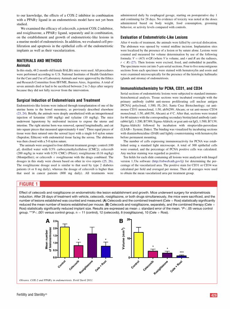
Effect of celecoxib and rosiglitazone on endometriotic-like lesion estab
induction. After 28 days of treatment with vehicle, celecoxib, rosiglitazo
number of lesions established was counted and measured. (A) Celecoxireduced the mean number of lesions established per mouse. (B) CelecoRosi) statistically significantly reduced implant size. Results are express
group. ***P< .001 versus control group. n ¼ 11 (control), 12 (celecoxib),
Olivares. COX-2 and PPARg in endometriosis. Fertil Steril 2011.
Fertility and Sterility�
administered daily by esophageal gavage, starting on postoperative day 1
and continuing for 28 days. No evidence of toxicity was noted at the doses
administered based on body weight, food consumption, grooming
behavior, or activity levels compared with controls.
Evaluation of Endometriotic-Like LesionsAfter 4 weeks of treatment, the animals were killed by cervical dislocation.
The abdomen was opened by ventral midline incision. Implantation sites
were localized by the presence of a lesion or by suture alone. Lesions were
counted and measured for volume determination by use of the following
formula: V ¼ (4/3) pr2R (where V is volume, and r and R are the radiuses,
r < R) (27). Then lesions were excised, fixed, and embedded in paraffin.
The specimens were cut into 5-mm serial sections. Four to five noncontiguous
sections from each specimen were stained with hematoxylin and eosin and
were examined microscopically for the presence of the histologic hallmarks
(glands and stroma) of endometriosis.
Immunohistochemistry for PCNA, CD31, and CD34Serial sections of endometriotic lesions were subjected to standard immuno-
histochemical analysis. Tissue sections were incubated overnight with the
primary antibody (rabbit anti-mouse proliferating cell nuclear antigen
[PCNA] polyclonal, 1:300, FL-261; Santa Cruz Biotechnology; rat anti-
mouse CD31 monoclonal, 1:50, ab56299; Abcam; or rat anti-mouse CD34
monoclonal, 1:50, ab8158; Abcam) at 4�C. After that, sections were treatedfor 60 minutes with the corresponding secondary biotinylated antibody (anti-
rabbit IgG, 1:200, B7389; Sigma-Aldrich; or goat anti-rat IgG, 1:500, B7139;
Sigma-Aldrich) followed by incubation with streptavidin-peroxidase
(LSABþ System; Dako). The binding was visualized by incubating sections
with diaminobenzidine (DAB) and lightly counterstaining with hematoxylin
before permanent mounting.
The number of cells expressing immunoreactivity for PCNA was estab-
lished using a standard light microscope. A total of 300 epithelial cells
were counted, and the percentage of PCNA positive cells was calculated.
Any nuclear staining was regarded as positive.
Ten fields for each slide containing all lesions were analyzed with ImageJ
version 1.33u software (http://rsbweb.nih.gov/ij) for determining the per-
centage of the vascularized area. The positive stain for CD31 or CD34 was
calculated per field and averaged per mouse. Then all averages were used
to obtain the mean vascularized area per treatment group.
lishment and growth. Mice underwent surgery for endometriosis
ne, or both drugs simultaneously, the mice were sacrificed, and the
b and the combined treatment (Cele þ Rosi) statistically significantly
xib and rosiglitazone, separately, and the combined therapy (Cele þed as mean � standard error of the mean. *P< .05 versus control
8 (rosiglitazone), 10 (Cele þ Rosi).
429

FIGURE 2
Effect of celecoxib and rosiglitazone on endometriotic-like lesion
epithelial cell proliferation. Mice underwent surgery forendometriosis induction. After 28 days of treatment with vehicle,
celecoxib, rosiglitazone, or both drugs simultaneously, the mice
were sacrificed, and the implants were removed and fixed. Cell
proliferation within the implants was evaluated byimmunohistochemistry of proliferating cell nuclear antigen
(PCNA). (A) After treatment with celecoxib, rosiglitazone, or both
drugs simultaneously (CeleþRosi), epithelial cell proliferation was
statistically significantly diminished compared with control mice.Results are expressed as mean � standard error of the mean.
**P< .01 versus control group; ***P< .001 versus control group. (B)Representative photographs of PCNA staining. Control group (i),celecoxib group (ii), rosiglitazone group (iii), celecoxib þrosiglitazone group (iv). Inset: negative control, an immunoglobulin
of the same immunoglobulin class and concentration as the
primary antibody was used. Magnification �400. n ¼ 8 (control), 7(celecoxib), 5 (rosiglitazone), 5 (Cele þ Rosi).
Olivares. COX-2 and PPARg in endometriosis. Fertil Steril 2011.
TUNEL AssayFor apoptosis quantification, sections were processed for in situ immunohis-
tochemical localization of nuclei exhibiting DNA fragmentation by use of the
apoptosis detection kit Apoptag Plus (Chemicon International). Sections
were treated according to the manufacturer’s instructions as previously
described elsewhere (28). The number of cells positive for terminal deoxynu-
cleotidyl transferase dUTP nick end labeling (TUNEL) stain was established
by use of a standard light microscope at �400 magnification. A total of 300
epithelial cells were counted, and the percentage of TUNEL positive cells
was calculated.
Statistical AnalysisStatistical analyses were performed using GraphPad Instat version 4.0
software (Windows version; GraphPad Software). Statistical comparisons
between groups were performed by use of parametric analysis of variance
(ANOVA) with Tukey’s multiple comparison post test or nonparametric
ANOVAwith Dunn’s multiple comparison post test. Results were expressed
as mean � standard error of the mean (SEM). P<.05 was considered
statistically significant.
RESULTSEffect of Celecoxib and Rosiglitazone on Endometriotic-Like Lesion Establishment and SizeThe group treated only with celecoxib and the group receiving thecombined therapy had a statistically significant reduction in thenumber of established lesions per animal (P<.05 vs. control group).The results are displayed in Figure 1A.
Regarding the volume of developed lesions, all treatment groupshad a statistically significant reduction in lesion size. As seen inFigure 1B, although there is not a statistically significant differencebetween the treated groups, celecoxib and rosiglitazone combinedtended to be more efficient in reducing the lesion volume (P<.001vs. control group) than either of the treatments alone.
Celecoxib and Rosiglitazone Treatments Affect CellProliferation and Apoptosis in Endometriotic-Like LesionsAll treatment groups had diminished cell proliferation comparedwith the control group (rosiglitazone-only and celecoxib þ rosigli-tazone groups, P<.001 vs. control; celecoxib-only group, P<.01 vs.control). The results are displayed in Figure 2. The combinedtreatment appeared to be slightly more effective in decreasing cellproliferation than either of the drugs separately, but the effect wasnot synergistic nor additive.
All treatments were effective enhancing apoptosis compared withthe control group (P<.05), as seen in Figure 3. Not only did theTUNEL assay demonstrate that apoptosis was augmented in thetreatment groups, but also the regression of the lesions was clearlyvisible in the histologic analysis of the sections (data not shown).
Inhibiting COX-2 and Activating PPARg Decrease VascularDensity in Endometriotic-Like LesionsFigure 4 shows the effect of the treatments on vascular density as as-sessed byCD31 andCD34 immunohistochemistry. As observedwithCD31 staining, rosiglitazone-only and the combined treatment re-sulted in a statistically significant reduction of vascular density(P<.05 vs. control group). When the same parameter wasevaluated via immunostaining with CD34, the celecoxib-only androsiglitazone-only groups showed a statistically significant decreasein vascular density when compared with the control group (P<.05);even though there was no statistically significant difference betweenthe treated groups, when the treatments were administered in combi-nation the reduction tended to be superior (P<.01 vs. control group).
430 Olivares et al. COX-2 and PPARg in endometriosis
DISCUSSIONThe current medical treatment of endometriosis is still associatedwith a high recurrence rate. The therapeutic options include surgeryand hormone therapy; which are often temporarily effective but pro-duce unwanted side effects (2, 29, 30). Given this scenario, it is ofgreat importance to study new strategies to treat endometriosisthat minimize the adverse effects and reduce the rates of recurrence.
Vol. 96, No. 2, August 2011
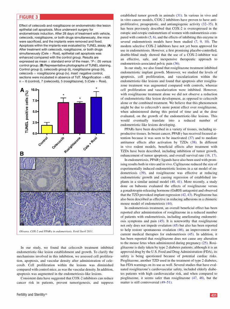
FIGURE 3
Effect of celecoxib and rosiglitazone on endometriotic-like lesion
epithelial cell apoptosis. Mice underwent surgery forendometriosis induction. After 28 days of treatment with vehicle,
celecoxib, rosiglitazone, or both drugs simultaneously, the mice
were sacrificed, and the implants were removed and fixed.
Apoptosis within the implants was evaluated by TUNEL assay. (A)After treatment with celecoxib, rosiglitazone, or both drugs
simultaneously (Cele þ Rosi), epithelial cell apoptosis was
enhanced compared with the control group. Results are
expressed as mean � standard error of the mean. *P< .05 versuscontrol group. (B) Representative photographs of TUNEL staining.
Control group (i), celecoxib group (ii), rosiglitazone group (iii),
celecoxib þ rosiglitazone group (iv). Inset: negative control,sections were incubated in absence of TdT. Magnification �400.
n ¼ 6 (control), 7 (celecoxib), 5 (rosiglitazone), 5 (Cele þ Rosi).
Olivares. COX-2 and PPARg in endometriosis. Fertil Steril 2011.
In our study, we found that celecoxib treatment inhibitedendometriotic-like lesion establishment and growth. To clarify themechanisms involved in this inhibition, we assessed cell prolifera-tion, apoptosis, and vascular density after administration of cele-coxib. Cell proliferation within the lesions was diminishedcompared with control mice, as was the vascular density. In addition,apoptosis was augmented in the endometriosis-like lesions.
Consistent data have suggested that COX-2 inhibitors can reducecancer risk in patients, prevent tumorigenesis, and suppress
Fertility and Sterility�
established tumor growth in animals (31). In various in vivo andin vitro cancer models, COX-2 inhibitors have proven to have anti-proliferative, proapoptotic, and antiangiogenic activity (32–35). Ithas been previously described that COX-2 is overexpressed in theeutopic and ectopic endometrium of women with endometriosis com-pared with controls (5, 6), and the effects of inhibiting this enzyme inseveral endometriosis models have been studied (7, 9, 10). Themodern selective COX-2 inhibitors have not yet been approved foruse in endometriosis. However, a first promising placebo-controlled,double-blind study showed that the use of a COX-2-inhibitor wasan effective, safe, and inexpensive therapeutic approach toendometriosis-associated pelvic pain (36).
In our study, we also found that rosiglitazone treatment inhibitedendometriotic implant growth. Moreover, we studied the levels ofapoptosis, cell proliferation, and vascularization within theendometriotic-like lesions and found that apoptosis after rosiglita-zone treatment was augmented compared with controls, whereascell proliferation and vascularization were inhibited. However,with rosiglitazone treatment alone we did not observe a reductionof endometriotic-like lesion development, as opposed to celecoxibalone or the combined treatment. We believe that this phenomenonmight be due to celecoxib’s more potent effect over rosiglitazone,when administered during this period of time and at the doseevaluated, on the growth of the endometriotic-like lesions. Thiswould eventually translate into a reduced number ofendometriotic-like lesions developing.
PPARs have been described in a variety of tissues, including re-productive tissues. In breast cancer, PPARg has received focused at-tention because it was seen to be inactivated (37) and to mediateantitumor effects after activation by TZDs (38). In differentin vivo rodent models, beneficial effects after treatment withTZDs have been described, including inhibition of tumor growth,enhancement of tumor apoptosis, and overall survival rate (16, 17).
In endometriosis, PPARg ligands have also been used with prom-ising results both in vitro and in vivo. Ciglitazone reduced the size ofexperimentally induced endometriotic lesions in a rat model of en-dometriosis (39), and rosiglitazone was effective at reducingendometriotic growth and causing regression of established im-plants in a similar animal model (40, 41). More recently, a studydone on baboons evaluated the effects of rosiglitazone versusa gonadotropin-releasing hormone (GnRH) antagonist and observedthat the TZD provoked implant regression (42, 43). Pioglitazone hasalso been described as effective in reducing adhesions in a chimericmouse model of endometriosis (44).
In endometriosis treatment, an overall beneficial effect has beenreported after administration of rosiglitazone in a reduced numberof patients with endometriosis, including ameliorating endometri-osis symptoms and pain (45). It is noteworthy that rosiglitazonenot only does not impede ovulation (45) but also has been reportedto help restore spontaneous ovulation (46), an improvement overcurrent medical therapies for endometriosis (45). In addition, ithas been reported that rosiglitazone does not cause any alterationto the mouse fetus when administered during pregnancy (25). Rosi-glitazone is daily taken by type 2 diabetes patients; although it is anapproved drug by the U.S. Food and Drug Administration (FDA), itssafety is being questioned because of potential cardiac risks.Pioglitazone, another TZD used in the treatment of type 2 diabetes,has FDAwarnings on its use as well. Several studies that have eval-uated rosiglitazone’s cardiovascular safety, included elderly diabe-tes patients with high cardiovascular risk, and when compared topioglitazone, it seems safer than rosiglitazone (47, 48), but thematter is still controversial (49–51).
431
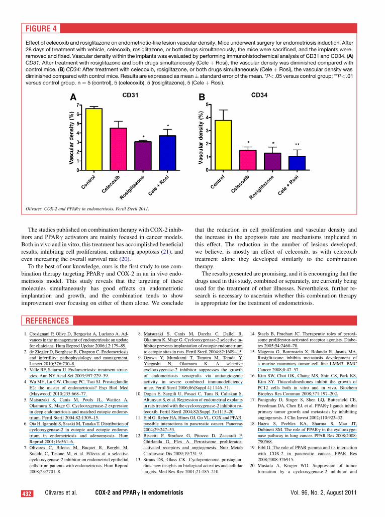
FIGURE 4
Effect of celecoxib and rosiglitazone on endometriotic-like lesion vascular density. Mice underwent surgery for endometriosis induction. After
28 days of treatment with vehicle, celecoxib, rosiglitazone, or both drugs simultaneously, the mice were sacrificed, and the implants wereremoved and fixed. Vascular density within the implants was evaluated by performing immunohistochemical analysis of CD31 and CD34. (A)CD31: After treatment with rosiglitazone and both drugs simultaneously (Cele þ Rosi), the vascular density was diminished compared with
control mice. (B) CD34: After treatment with celecoxib, rosiglitazone, or both drugs simultaneously (Cele þ Rosi), the vascular density was
diminished compared with control mice. Results are expressed as mean� standard error of themean. *P< .05 versus control group; **P< .01versus control group. n ¼ 5 (control), 5 (celecoxib), 5 (rosiglitazone), 5 (Cele þ Rosi).
Olivares. COX-2 and PPARg in endometriosis. Fertil Steril 2011.
The studies published on combination therapy with COX-2 inhib-itors and PPARg activators are mainly focused in cancer models.Both in vivo and in vitro, this treatment has accomplished beneficialresults, inhibiting cell proliferation, enhancing apoptosis (21), andeven increasing the overall survival rate (20).
To the best of our knowledge, ours is the first study to use com-bination therapy targeting PPARg and COX-2 in an in vivo endo-metriosis model. This study reveals that the targeting of thesemolecules simultaneously has good effects on endometrioticimplantation and growth, and the combination tends to showimprovement over focusing on either of them alone. We conclude
432 Olivares et al. COX-2 and PPARg in endometriosis
that the reduction in cell proliferation and vascular density andthe increase in the apoptosis rate are mechanisms implicated inthis effect. The reduction in the number of lesions developed,we believe, is mostly an effect of celecoxib, as with celecoxibtreatment alone they developed similarly to the combinationtherapy.
The results presented are promising, and it is encouraging that thedrugs used in this study, combined or separately, are currently beingused for the treatment of other illnesses. Nevertheless, further re-search is necessary to ascertain whether this combination therapyis appropriate for the treatment of endometriosis.
REFERENCES
1. Crosignani P, Olive D, Bergqvist A, Luciano A. Ad-
vances in the management of endometriosis: an update
for clinicians. Hum Reprod Update 2006;12:179–89.
2. de Ziegler D, Borghese B, Chapron C. Endometriosis
and infertility: pathophysiology and management.
Lancet 2010;376:730–8.
3. Valle RF, Sciarra JJ. Endometriosis: treatment strate-
gies. Ann NYAcad Sci 2003;997:229–39.
4. Wu MH, Lu CW, Chuang PC, Tsai SJ. Prostaglandin
E2: the master of endometriosis? Exp Biol Med
(Maywood) 2010;235:668–77.
5. Matsuzaki S, Canis M, Pouly JL, Wattiez A,
Okamura K, Mage G. Cyclooxygenase-2 expression
in deep endometriosis and matched eutopic endome-
trium. Fertil Steril 2004;82:1309–15.
6. Ota H, Igarashi S, Sasaki M, Tanaka T. Distribution of
cyclooxygenase-2 in eutopic and ectopic endome-
trium in endometriosis and adenomyosis. Hum
Reprod 2001;16:561–6.
7. Olivares C, Bilotas M, Buquet R, Borghi M,
Sueldo C, Tesone M, et al. Effects of a selective
cyclooxygenase-2 inhibitor on endometrial epithelial
cells from patients with endometriosis. Hum Reprod
2008;23:2701–8.
8. Matsuzaki S, Canis M, Darcha C, Dallel R,
Okamura K,Mage G. Cyclooxygenase-2 selective in-
hibitor prevents implantation of eutopic endometrium
to ectopic sites in rats. Fertil Steril 2004;82:1609–15.
9. Ozawa Y, Murakami T, Tamura M, Terada Y,
Yaegashi N, Okamura K. A selective
cyclooxygenase-2 inhibitor suppresses the growth
of endometriosis xenografts via antiangiogenic
activity in severe combined immunodeficiency
mice. Fertil Steril 2006;86(Suppl 4):1146–51.
10. Dogan E, Saygili U, Posaci C, Tuna B, Caliskan S,
Altunyurt S, et al. Regression of endometrial explants
in rats treated with the cyclooxygenase-2 inhibitor ro-
fecoxib. Fertil Steril 2004;82(Suppl 3):1115–20.
11. Eibl G, Reber HA, Hines OJ, Go VL. COX and PPAR:
possible interactions in pancreatic cancer. Pancreas
2004;29:247–53.
12. Biscetti F, Straface G, Pitocco D, Zaccardi F,
Ghirlanda G, Flex A. Peroxisome proliferator-
activated receptors and angiogenesis. Nutr Metab
Cardiovasc Dis 2009;19:751–9.
13. Straus DS, Glass CK. Cyclopentenone prostaglan-
dins: new insights on biological activities and cellular
targets. Med Res Rev 2001;21:185–210.
14. Staels B, Fruchart JC. Therapeutic roles of peroxi-
some proliferator-activated receptor agonists. Diabe-
tes 2005;54:2460–70.
15. Magenta G, Borenstein X, Rolando R, Jasnis MA.
Rosiglitazone inhibits metastasis development of
a murine mammary tumor cell line LMM3. BMC
Cancer 2008;8:47–57.
16. Kim SW, Choi OK, Chang MS, Shin CS, Park KS,
Kim SY. Thiazolidinediones inhibit the growth of
PC12 cells both in vitro and in vivo. Biochem
Biophys Res Commun 2008;371:197–202.
17. Panigrahy D, Singer S, Shen LQ, Butterfield CE,
Freedman DA, Chen EJ, et al. PPARg ligands inhibit
primary tumor growth and metastasis by inhibiting
angiogenesis. J Clin Invest 2002;110:923–32.
18. Hazra S, Peebles KA, Sharma S, Mao JT,
Dubinett SM. The role of PPARg in the cyclooxyge-
nase pathway in lung cancer. PPAR Res 2008;2008:
790568.
19. Eibl G. The role of PPAR-gamma and its interaction
with COX-2 in pancreatic cancer. PPAR Res
2008;2008:326915.
20. Mustafa A, Kruger WD. Suppression of tumor
formation by a cyclooxygenase-2 inhibitor and
Vol. 96, No. 2, August 2011

a peroxisome proliferator-activated receptor gamma
agonist in an in vivo mouse model of spontaneous
breast cancer. Clin Cancer Res 2008;14:4935–42.
21. Sun WH, Chen GS, Ou XL, Yang Y, Luo C, Zhang Y,
et al. Inhibition of COX-2 and activation of peroxi-
some proliferator-activated receptor gamma synergis-
tically inhibits proliferation and induces apoptosis of
human pancreatic carcinoma cells. Cancer Lett
2009;275:247–55.
22. Wu Y, Guo SW. Peroxisome proliferator-activated re-
ceptor-gamma and retinoid X receptor agonists syner-
gistically suppress proliferation of immortalized
endometrial stromal cells. Fertil Steril 2009;91:2142–7.
23. Ricci AG, Olivares CN, Bilotas MA, Meresman GF,
Baranao RI. Effect of vascular endothelial growth
factor inhibition on endometrial implant development
in a murine model of endometriosis. Reprod Sci. Pub-
lished online January 25, 2011.
24. Bilotas M, Meresman G, Stella I, Sueldo C,
Baranao RI. Effect of aromatase inhibitors on ectopic
endometrial growth and peritoneal environment in
a mouse model of endometriosis. Fertil Steril
2010;93:2513–8.
25. Klinkner DB, Lim HJ, Strawn EY Jr, Oldham KT,
Sander TL. An in vivo murine model of rosiglitazone
use in pregnancy. Fertil Steril 2006;86:1074–9.
26. Peluffo GD, Stillitani I, Rodriguez VA, Diament MJ,
Klein SM. Reduction of tumor progression and para-
neoplastic syndrome development in murine lung
adenocarcinoma by nonsteroidal antiinflammatory
drugs. Int J Cancer 2004;110:825–30.
27. Brodie A, Jelovac D, Long BJ. Predictions from a pre-
clinical model: studies of aromatase inhibitors and
antiestrogens. Clin Cancer Res 2003;9:S455–9.
28. MeresmanGF,Vighi S,BuquetRA,Contreras-OrtizO,
TesoneM,RumiLS.Apoptosis and expressionofBcl-2
and Bax in eutopic endometrium fromwomenwith en-
dometriosis. Fertil Steril 2000;74:760–6.
29. Mihalyi A, Simsa P, Mutinda KC, Meuleman C,
Mwenda JM, D’Hooghe TM. Emerging drugs in endo-
metriosis. Expert Opin Emerg Drugs 2006;11:503–24.
30. Rice VM. Conventional medical therapies for endo-
metriosis. Ann NYAcad Sci 2002;955:343–52.
31. Bundred NJ, Barnes NL. Potential use of COX-
2-aromatase inhibitor combinations in breast cancer.
Br J Cancer 2005;93(Suppl 1):S10–5.
Fertility and Sterility�
32. Grosch S, Maier TJ, Schiffmann S, Geisslinger G.
Cyclooxygenase-2 (COX-2)-independent anticarci-
nogenic effects of selective COX-2 inhibitors. J Natl
Cancer Inst 2006;98:736–47.
33. Basu GD, Pathangey LB, Tinder TL, Gendler SJ,
Mukherjee P. Mechanisms underlying the growth in-
hibitory effects of the cyclo-oxygenase-2 inhibitor
celecoxib in human breast cancer cells. Breast Cancer
Res 2005;7:R422–35.
34. Leahy KM, Ornberg RL, Wang Y, Zweifel BS,
Koki AT, Masferrer JL. Cyclooxygenase-2 inhibition
by celecoxib reduces proliferation and induces apo-
ptosis in angiogenic endothelial cells in vivo. Cancer
Res 2002;62:625–31.
35. ElderDJ,HaltonDE,CrewTE,ParaskevaC.Apoptosis
induction and cyclooxygenase-2 regulation in human
colorectal adenoma and carcinoma cell lines by the
cyclooxygenase-2-selective non-steroidal anti-inflam-
matory drug NS-398. Int J Cancer 2000;86:553–60.
36. Cobellis L, Razzi S, De Simone S, Sartini A, Fava A,
Danero S, et al. The treatment with a COX-2 specific
inhibitor is effective in the management of pain re-
lated to endometriosis. Eur J Obstet Gynecol Reprod
Biol 2004;116:100–2.
37. Badawi AF, Badr MZ. Expression of
cyclooxygenase-2 and peroxisome proliferator-
activated receptor-gamma and levels of prostaglandin
E2 and 15-deoxy-d12,14-prostaglandin J2 in human
breast cancer and metastasis. Int J Cancer
2003;103:84–90.
38. Blanquicett C, Roman J, Hart CM. Thiazolidinediones
as anti-cancer agents. Cancer Ther 2008;6:25–34.
39. Lebovic DI, Kir M, Casey CL. Peroxisome
proliferator-activated receptor-gamma induces re-
gression of endometrial explants in a rat model of en-
dometriosis. Fertil Steril 2004;82(Suppl 3):1008–13.
40. Aytan H, Caliskan AC, Demirturk F, Aytan P,
Koseoglu DR. Peroxisome proliferator-activated re-
ceptor-gamma agonist rosiglitazone reduces the size
of experimental endometriosis in the rat model.
Aust NZ J Obstet Gynaecol 2007;47:321–5.
41. Demirturk F, Aytan H, Caliskan AC, Aytan P,
Koseoglu DR. Effect of peroxisome proliferator-
activated receptor-gamma agonist rosiglitazone on
the induction of endometriosis in an experimental
rat model. J Soc Gynecol Investig 2006;13:58–62.
42. Lebovic DI, Mwenda JM, Chai DC, Santi A, Xu X,
D’Hooghe T. Peroxisome proliferator-activated re-
ceptor-g receptor ligand partially prevents the devel-
opment of endometrial explants in baboons:
a prospective, randomized, placebo-controlled study.
Endocrinology 2010;151:1846–52.
43. Lebovic DI, Mwenda JM, Chai DC, Mueller MD,
Santi A, Fisseha S, et al. PPAR-gamma receptor li-
gand induces regression of endometrial explants in
baboons: a prospective, randomized, placebo- and
drug-controlled study. Fertil Steril 2007;88:1108–19.
44. Herington JL, Crispens MA, Carvalho-Macedo AC,
Camargos AF, Lebovic DI, Bruner-Tran KL, et al.
Development and prevention of postsurgical adhe-
sions in a chimeric mouse model of experimental
endometriosis. Fertil Steril 2011;95:1295–301.
45. Moravek MB, Ward EA, Lebovic DI. Thiazolidine-
diones as therapy for endometriosis: a case series.
Gynecol Obstet Invest 2009;68:167–70.
46. Sepilian V, Nagamani M. Effects of rosiglitazone in
obese women with polycystic ovary syndrome and
severe insulin resistance. J Clin Endocrinol Metab
2005;90:60–5.
47. Tolman KG. The safety of thiazolidinediones. Expert
Opin Drug Saf 2011;10:419–28.
48. Schernthaner G, Chilton RJ. Cardiovascular risk and
thiazolidinediones—what do meta-analyses really
tell us? Diabetes Obes Metab 2010;12:1023–35.
49. Saryusz-Wolska M, Szymanska-Garbacz E,
Jablkowski M, Bialkowska J, Pawlowski M,
Kwiecinska E, et al. Rosiglitazone treatment in
nondiabetic subjects with nonalcoholic fatty liver
disease. Pol Arch Med Wewn 2011;121:61–6.
50. Yee MS, Pavitt DV, Dhanjil S, Godsland IF,
Richmond W, Johnston DG. The effects of rosiglita-
zone on atherosclerotic progression in patients with
type 2 diabetes at high cardiovascular risk. Diabet
Med 2010;27:1392–400.
51. Gerstein HC, Ratner RE, Cannon CP, Serruys PW,
Garcia-Garcia HM, van Es GA, et al. Effect of rosigli-
tazone on progression of coronary atherosclerosis in
patients with type 2 diabetes mellitus and coronary
artery disease: the assessment on the prevention of
progression by rosiglitazone on atherosclerosis in
diabetes patients with cardiovascular history trial.
Circulation 2010;121:1176–87.
433