Regression of prostate tumors upon combination of hormone ablation therapy and celecoxib in vivo
Upload
uhh-hawaiiCategory
view
7download
0
Journal of Controlled Release 144 (2010) 233–241
Contents lists available at ScienceDirect
Journal of Controlled Release
j ourna l homepage: www.e lsev ie r.com/ locate / jconre l
NANOMEDICIN
E
Formulation, characterization and pulmonary deposition of nebulized celecoxibencapsulated nanostructured lipid carriers
Ram R. Patlolla a, Mahavir Chougule a, Apurva R. Patel a, Tanise Jackson a, Prasad N.V. Tata b, Mandip Singh a,⁎a College of Pharmacy and Pharmaceutical Sciences, Florida A&M University, Tallahassee, FL 32307, USAb Division of Clinical Pharmacology, Covidien/Mallinckrodt, Inc., St. Louis, MO 63042, USA
⁎ Corresponding author. Tel.: +1 850 561 2790; fax:E-mail address: [email protected] (M. Si
0168-3659/$ – see front matter. Published by Elsevierdoi:10.1016/j.jconrel.2010.02.006
a b s t r a c t
a r t i c l e i n f oArticle history:Received 24 December 2009Accepted 3 February 2010Available online 11 February 2010
Keywords:InhalationNanostructured lipid carrierCelecoxibLung cancerLung disposition
The aim of the current study was to encapsulate celecoxib (Cxb) in the nanostructured lipid carrier (Cxb-NLC) nanoparticles and evaluate the lung disposition of nanoparticles following nebulization in Balb/c mice.Cxb-NLC nanoparticles were prepared with Cxb, Compritol, Miglyol and sodium taurocholate using high-pressure homogenization. Cxb-NLC nanoparticles were characterized for physical and aerosol properties. In-vitro cytotoxicity studies were performed with A549 cells. The lung deposition and pharmacokineticparameters of Cxb-NLC and Cxb solution (Cxb-Soln) formulations were determined using the Inexpose™system and Pari LC star jet nebulizer. The particle size and entrapment efficiency of the Cxb-NLC formulationwere 217±20 nm and N90%, respectively. The Cxb-NLC released the drug in controlled fashion, and in-vitroaerosolization of Cxb-NLC formulation showed an FPF of 75.6±4.6%, MMAD of 1.6±0.13 µm and a GSD of1.2±0.21. Cxb-NLC showed dose and time dependent cytotoxicity against A549 cells. Nebulization of Cxb-NLC demonstrated 4 fold higher AUCt/D in lung tissues compared to the Cxb-Soln. The systemic clearance ofCxb-NLC was slower (0.93 l/h) compared to the Cxb-Soln (20.03 l/h). Cxb encapsulated NLC were found to bestable and aerodynamic properties were within the respirable limits. Aerosolization of Cxb-NLC improvedthe Cxb pulmonary bioavailability compared to solution formulation which will potentially lead to betterpatient compliance with minimal dosing intervals.
+1 850 599 3813.ngh).
B.V.
Published by Elsevier B.V.
1. Introduction
Inhalation drug delivery represents a potential delivery route for thetreatment of several pulmonary disorders. Inhalation drug delivery hasseveral advantages over conventional (parenteral and oral) dosageforms such as a) non-invasive b) circumventing first pass metabolismand systemic toxicity c) reduced frequent dosing and d) the inhaleddrug reaches directly to the lung epithelium thereby enhancing localdrug concentrations. In pulmonary drug delivery systems, surfactantsand co-solvents are often used to prepare stable formulations of highlylipophilic active ingredients and inhalation of formulations utilizingexcipients cause lung inflammation [1]. It is expected that encapsulationof lipophilic compounds in nanoparticles will improve the stability byprotecting the active ingredient from degradation and release theencapsulateddrug in a controlledmanner for a prolongedperiodof time[2]. Among several inhalation drug delivery systems, biodegradablenanoparticles have demonstrated several advantages in terms ofprotecting the active ingredient from degradation and releasing thedrug in a controlledmanner for prolongedperiods of time. Although fewattempts have been made to deliver anticancer agents using nanopar-
ticles and liposomes via an inhalation route, the major limitations ofthese systems are instability during nebulization, biodegradability, drugleakage and associated drug adverse side effects [3,4]. Solid lipidnanoparticles (SLN) have several advantages such as a) good tolerabil-ity, b) biodegradability and c) greater stability against the shear forcesgenerated during nebulization [3,5] compared to polymeric nanopar-ticles [3], liposomes [4] and emulsions [6]. SLN are produced byreplacing the oil lipid of an o/w emulsion with a solid lipid or a blend ofsolid lipids, where the lipid particle matrix being solid at both room andbody temperature. Despite greater stability, SLN have some limitationssuch as low drug loading, risk of gelation and drug leakage duringstorage caused by lipid polymorphism [7]. Therefore, in order todecrease the degree of organization of the lipid matrix in SLN andincrease the drug loading capacity, the nanostructured lipid carriers(NLC) have been developed and reported as the second generation oflipid nanoparticles. NLC nanoparticles are comprised of an inner oil coresurrounded by anouter solid shell and hence allow the highpayloadof alipophilic drug [8]. NLC nanoparticles have been investigated for topicaldelivery of lipophilic anti-inflammatory molecules and also in cosmeticproducts [9,10].
Cyclooxygenase-2 (COX-2) enzyme is over-expressed amongvarious human malignancies and is thought to have a potential rolein the pathogenesis of non-small cell lung cancer (NSCLC) [11].Celecoxib (Cxb), a lipophilic COX-2 inhibitor has shown a synergistic

234 R.R. Patlolla et al. / Journal of Controlled Release 144 (2010) 233–241
NANOMEDICIN
E
anticancer activity in combination with other anticancer agents suchas docetaxel [11]. The preclinical data suggest that the COX-2/prostaglandin E2 signaling pathway plays an essential role inconferring the malignant phenotype in non-small cell lung cancerby stimulating angiogenesis, inhibiting apoptosis and suppressing theimmune response. It has been also shown that Cxb inhibits NF-kBactivation through inhibition of IKK and Akt activation which leads todown-regulation of COX-2 synthesis and other genes needed forinflammation, proliferation, and carcinogenesis [12]. In lung cancerpatients, Cxb is also known to inhibit the overproduction ofprostaglandin E2, as well as modulate the IL-10 production in thelungmicroenvironment [13]. Cxb is a poorly water soluble drug (7 µg/ml) with a partition coefficient of 3.68 [14] and necessitates use ofsurfactants and a co-solvent such as ethanol to formulate an aerosolformulation. Our earlier studies showed that Cxb formulationsprepared using tocopheryl polyethylene glycol succinate and ethanolresulted in improved solubility and enhanced the anticancer activityof nebulized Cxb [15,16]. However, these formulations were un-stableand precipitation was observed on long term storage. Therefore, it isexpected that encapsulation of Cxb in NLC nanoparticles will improvethe stability and alter its pharmacokinetics by enhanced retention[17] and releasing the Cxb in a controlled fashion.
In the present investigation, we explored the feasibility of NLCnanoparticles as a novel carrier system for inhalation drug delivery ofCxb. Therefore in this study, we examined the effect of Cxb-NLC on therelease of Cxb, aerodynamic properties and in-vitro cytotoxicityagainst A549 NSCLC cells. The present investigation was also aimed toevaluate the in-vivo pulmonary deposition and systemic availabilityof aerosolized Cxb loaded NLC (Cxb-NLC) nanoparticles in Balb/c miceutilizing an Inexpose® exposure chamber.
2. Material and methods
2.1. Materials
The Cxbwas a generous gift from Pfizer (Skokie, IL). The triglycerideMiglyol 812 was obtained from Sasol Germany GmbH (Witten,Germany) and Compritol® 888 ATO was a kind gift sample fromGattefosse (Saint Priest, France). The taurocholic acid sodium salt wasprocured from Sigma-Aldrich Chemicals (St. Louis, MO). Dialysis tubing(molecularweight cut-off: 6000–8000 Da andflatwidth of 23 mm)wasobtained from Fisher Scientific (Pittsburg, PA). The polyoxyethylene-20oleyl ether or Volpo-20 (Oleth-20) was a kind gift from Croda Inc (NewJersey, USA). Vivaspin centrifuge filters (molecular weight cut-off: 10,000 Da) were procured from Sartorius Ltd, (Stonehouse, UK). Fetalbovine serum (FBS), antibiotics and DID-oil (a lipophilic fluorescent dyewith excitation 644 nm, emission 665 nm) were procured fromInvitrogen Corp (Eugene, OR). The A549 human NSCLC cell line wasobtained from American Type Culture Collection (Rockville, MD, USA).A549 cells were grown in F12K medium (Sigma, St. Louis, MO, USA)supplemented with 10% FBS. All tissue culture media contained anantibiotic antimycotic solution of penicillin (5000 U/ml), streptomycin(0.1 mg/ml), and neomycin (0.2 mg/ml). The cells were maintained at37 °C in the presence of 5% CO2 in air. All other chemicals used in thisresearch were of analytical grade.
2.2. Animals
Male Balb/c mice (20–25 g; Charles River Laboratories) wereutilized for the studies. The protocol for in-vivo experiments wasapproved by the Animal Care and Use Committee, Florida A&MUniversity. The animals were acclimated to laboratory conditions forone week prior to experiments and were on standard animal chowand water ad libitum. The temperature of the roomwas maintained at22±1 °C and the relative humidity of the experimentation room wasfound in the range of 35–50%. For nebulization studies, 4–5 days prior
to the start of the experiment animals were trained by nebulizingwater for 30 min. This is to acclimate the nebulization environmentand prevent any discomfort during the formulation nebulization.
2.3. Nanoparticle preparation
A Cxb-NLC formulation was prepared by the hot melt homogeniza-tion technique [9]. In brief, 0.02% w/w of Cxb was dissolved indichloromethane and mixed with lipid phase comprised of Compritol(7.0% w/w) and Miglyol (3.0% w/w). Later, organic phase was removedon a rota evaporator for 2–3 h at 80 °C and to the heated lipid phase theaqueous solution (40 ml) containing sodium taurocholate (1.5% w/w)surfactantwas added at the same temperature under high speedmixingusing a Cyclone IQ2 with a Sentry™ Microprocessor (USA) at20,000 rpm for 1 min. The resultant oil-in-water dispersion was passedthrough a Emulsiflex-C5 (Avestin, Ottawa, Canada) high-pressurehomogenizer at 5000 psi for 5 cycles. Throughout the process temper-ature was maintained at 80 °C. The Cxb-NLC and placebo NLC (withoutCxb) formulations were prepared for comparison.
2.4. Characterization of nanoparticles
The particle size of the NLC nanoparticle formulation wasmeasured using a BI-90 particle sizer (Brookhaven Instruments,Boston, USA), which is based on the principle of Dynamic LightScattering and Zeta potential measurement was carried with Zeta plus(Brookhaven Instruments). In order to verify the total amount of drugpresent in the system, 0.1 ml of the Cxb-NLC formulation wasdissolved in 0.9 ml of tetrahydrofuran and subsequent dilutionswere made with acetonitrile. Prior to HPLC analysis for Cxb content,samples were centrifuged at 13,000 rpm for 15 min and 100 µl ofsupernatant was injected into HPLC. Entrapment efficiency wasdetermined using vivaspin centrifuge filters as per reported method[18]. In brief, the Cxb-NLC (0.5 ml) formulation was placed on top ofthe vivaspin centrifuge filter membrane (molecular weight cut-off10,000 Da) and centrifuged at 3500 rpm for 15 min. The aqueousphase collected at the bottom of vivaspin filter membrane wassubjected to high-performance liquid chromatography (HPLC) anal-ysis to determine the Cxb content. To determine the drug loading,1.0 ml of the Cxb-NLC formulation was centrifuged at 16,000 g for1.5 h and the pellet was dissolved in tetrahydrofuran. The amount ofCxb present in the pellet was estimated by HPLC. Drug loading wascalculated based on the amount of Cxb identified in a measuredamount of NLC using the following equation [19]
Drug Content % w=wð Þ = mass of Cxb in NLC × 100ð Þ� mass of NLC recoveredð Þ
ð1Þ
2.5. Differential Scanning Calorimetry
The interaction of Cxb with lipids and the association of Cxb in theNLC nanoparticle formulation were determined using a DSCQ100 (TAinstrument, DE). Approximately 10 mg of the formulation wasweighed into an aluminum pan and sealed hermetically, and thethermal behavior was determined in the range of 10 to 225 °C at aheating rate of 5 °C min−1. Baselines were determined using anempty pan, and all the thermograms were baseline-corrected.Transition temperatures were determined from the endothermicpeakminimawhile transition enthalpies were obtained by integrationof the endothermic transitions using linear baselines.
2.6. In-vitro drug release studies
In-vitro drug release studies were conducted with a cellulosemembrane (6000–8000 molecular weight cut-off) using a USP 1(basket) dissolution apparatus (Vankel, NC) for 72 h with the help of

235R.R. Patlolla et al. / Journal of Controlled Release 144 (2010) 233–241
NANOMEDICIN
E
900 ml of phosphate buffer saline (PBS) pH 7.4 containing 0.1% w/vVolpo-20 as the dissolution medium. Cellulose membranes weresoaked overnight in the dissolution medium. To the pre-swollencellulose membrane bags 2 ml of the Cxb-NLC formulation was placedand both the ends of bags were tied to prevent any leakage. Later,dialysis bags were carefully placed in the baskets and the basketswererotated at 50 rpm for 72 h at 37.0±0.1 °C. As a control, 5 ml of thesaturated solution of Cxb prepared in 0.2%w/v Volpo-20was placed inthe dialysis bag and dialysis was carried in 75 ml of the dissolutionmedium. At regular time intervals 1 ml of the sample was collectedand replaced with an equal volume of the dissolution medium. Theamount of Cxb released in to the medium was determined with thehelp of HPLC analysis. The experiments were carried out in triplicate.
2.7. In-vitro aerosol characterization
Aerodynamic particle size distribution was measured using an8-stage non viable, Anderson cascade impactor (Graseby Andersen,Atlanta, GA, USA) equipped with filters on each stage and a backupfilter. To prevent particle bounce during nebulization, each plate onthe impactor was coated with 10% w/v pluronic L10 in ethanolsolution. The Cxb-NLC formulation was nebulized for 5 min into thecascade impactor operated at a flow rate of 28.3 l/min. Afternebulization with a Pari LC Star jet nebulizer, the amount offormulation deposited on the throat, impactor stages (0–7) andfilter was collected by washing with 5 ml of tetrahydrofuran andcentrifuged at 13,000 rpm for 15 min. The amount of Cxb in thesupernatant was analyzed using HPLC.
The mass median aerodynamic diameter (MMAD) and geometricstandard deviation (GSD)were obtained from impactor data using theBattelle software. The aerodynamic particle size distribution mea-surement is based on the amount of Cxb deposited on each stage ofthe cascade impactor and represents a relative particle distribution.Percent throat deposition, respirable mass, and respirable fractionswere calculated from the known amount of drug deposited on thevarious components. Only particles measuring less than 5 µm indiameter were included in assessing the respirable mass and fraction.Impactor experiments were repeated at least three times.
The Droplet Size Distribution (DSD) measurement of the Cxb-NLCformulation was also conducted by laser diffraction using a MalvernSpraytec® with RT Sizer Software. The Pari LC Star jet nebulizer filledwith 3 ml of NLC formulation was nebulized at a flow rate of 28.3 l/min and aerosol was aspirated with the help of a suction pump on theopposite side of the laser beam. Themeasurements were conducted at3 cm from the laser beam and the focal length of the lens used was100 mm,which has a droplet-size range of 0.5–200 μm. The FPF valuesdetermined using a Malvern Spraytec® equipped with RT SizerSoftware will represent the actual respirable fraction of a formulation.
2.8. In-vitro cytotoxicity studies
The in-vitro cytotoxicity of Cxb-NLCwas evaluated in the A549 cellline as reported earlier [20,21]. For comparison the Cxb-Soln for cellcytotoxicity studies was prepared by dissolving Cxb in dimethylsulfoxide and subsequently diluted with cell culture medium. Thefinal dimethyl sulfoxide concentration was less than 1% v/v in controland treatment groups [21]. The A549 cells were plated in 96-wellmicro titer plates, at a density of 1×10⁶ cells/well and after overnightincubation the cells were treated with various concentrations of Cxb-Soln and Cxb-NLC formulations diluted in F12K medium (20 to320 µg/ml). The cells were incubated for 24, 48 and 72 h at 37±0.2 °Cin a 5% CO2-jacketed incubator. The cell viability in each treatment atregular time intervals was determined using crystal violet assay. Thepercentage of cell survival as a function of drug concentration wasthen plotted to determine the IC50 value (the drug concentrationneeded to prevent cell proliferation by 50%).
2.9. Stability of Cxb-NLC during aerosolization
The in-vitro efficacy of the aerosolized Cxb-NLC formulation wasevaluated using a six-stage viable impactor using A549 cells [22,23]. Todetermine the amount of Cxb deposited on 4, 5 and 6 stages of the viableimpactor, petri plates filled with 15 ml of collection medium (PBS pH7.4) were placed on each stage and the Cxb-NLC formulation wasnebulized for 2 min at a flow rate of 28.3 l/min. The Cxb-NLC depositedon each stage was extracted with 15 ml of tetrahydrofuran and the Cxbconcentration in the solution was estimated using the HPLCmethod. Toascertain the efficacy of aerosolized Cxb-NLC, a similar type ofexperiment was carried out using viable impactor by placing a petriplate on stage5and theamount of freedrug in themediumwas collectedusing Vivaspin centrifuge filters and subjected to HPLC analysis.
2.10. In-vitro cytotoxicity of aerosolized Cxb-NLC against A549 cells
To determine the in-vitro cytotoxicity of aerosolized Cxb-NLCformulation, the viable impactor was connected to the Pari LC Star jetnebulizer through a USP throat and operated at a flow rate of 28.3 l/min. A549 cells (one million in 15 ml of medium per petri plate) thatwere plated in petri plates were placed on stage 5 of the viableimpactor and exposed to the Cxb-NLC formulation for 2 min. After theexposure, the petri plate was taken out from the impactor, coveredwith sterile aluminum lids provided with the petri plate (GrasebyAndersen, Smyrna, GA) and incubated at 37 °C for 72 h. Since all theoperations were performed under biological safety cabinet (Class II,Type A/B3, NuAire, Inc., Plymouth, MN) using established tissueculture precautions, sterility can be maintained throughout theincubation period. At the end of the incubation, the medium in thepetri plate was discarded and the cells were rinsed three times withsterile PBS, and detached by adding trypsin. The cells were spun downwith a centrifuge and resuspended in an appropriate amount ofmedium. The viable cells were then counted with a hemocytometerusing trypan blue solution (0.4%). Untreated cells were used as controland for comparison cytotoxicity of Cxb was carried out by adding thesame amount of the Cxb-Soln in to the petri plates.
2.11. In-vivo lung deposition and systemic pharmacokinetic studies
An Inexpose™ (SCIREQ Scientific Respiratory Equipment Inc,Montreal, QC) system consisting of 12 ports located peripherallyaround a central delivery plenum was utilized for Cxb-NLC andethanolic Cxb-Soln formulations aerosol exposure. For comparison,the Cxb-Soln was prepared by dissolving Cxb in 100% ethanol to get afinal concentration of 2 mg/ml. Aerosols were generated by nebuliza-tion with a Pari LC Star jet nebulizer using dry compressed air at a flowrate of 4.5 l/min. Male Balb/c mice (20±2 g) were restrained in animalholders and placed in an inhalation chamber (SCIREQ, Montreal,Canada) such that only the nose of each mouse was exposed to theaerosol cloud. The nebulizer was connected to the top part of theinhalation chamber fromwhich the generated aerosol flowed down thecentral tower to the peripherally arranged 12mice. The total amount of23±3 mg (Cxb-Soln) and 9±2 mg (Cxb-NLC) Cxb was nebulized toanimals during 30 min of exposure time. The total amount of Cxbdeposited into eachmousewas calculated based on the earlier reportedmethod [16]. The estimated total deposited amount of inhaled Cxb (D)for the ambient air was calculated by the following formula
D = C × V × DI × T ð2Þ
Where; C = concentration of Cxb in aerosol volume (163.60 µg/ml), V = volume of air inspired by the animal during 1 min [for mice,V=1.0l min/kg], DI = estimated deposition index [fraction of inhaleddose deposited throughout the respiratory tract (for mice DI=0.3)],and T = duration of treatment in min (T=30 min).
236 R.R. Patlolla et al. / Journal of Controlled Release 144 (2010) 233–241
NANOMEDICIN
E
After completion of the exposure, at predetermined time points (0,0.5, 1, 2, 4, 6, 12 and 24 h) animals were sacrificed with an overdose ofhalothane anesthesia. At each time interval six mice were sacrificed,blood and lungs were collected and samples were stored at −80 °Cuntil analyzed. Further to understand the lung deposition of NLCnanoparticles a fluorescent dye encapsulated NLC (Fl-NLC) nanopar-ticle was prepared by replacing the Cxb with DID-oil in the oil phase.The Balb/c mice were exposed to the Fl-NLC nanoparticles for 30 minsimilar to the Cxb-NLC formulation. The lungs were collected at 0.5and 4 h of nebulization and processed as per reported method [21].The tissue associated fluorescence was observed in the lung sectionsusing Leica DM6000 inverted Laser Scan Confocal Microscope.
2.12. Plasma and tissue sample analysis
100 µl of plasma or lung tissue samples were spiked with 50 µl ofinternal standard solution (100 µg/ml nimesulide in acetonitrile) andvortexed well. To the resultant samples of 1.5 ml of dichloromethanewas added, vortexed and then subjected to centrifugation (15 min at3500 rpm) to separate aqueous and organic phases. The organic layerwas separated, evaporated to dryness and the residue was thenreconstituted with 200 µl of mobile phase and 100 µl was injectedontoHPLC for quantification. In the caseof lungdeposition studies, lungswere weighed and homogenized with 500 μl of phosphate bufferedsaline (pH 7.4) and processed similarly as that of plasma samples.
2.13. Celecoxib HPLC bioanalytical method
The HPLC analysis of Cxb was performed as per reported method,with minor modifications [24]. Briefly, the method involves injectionof plasma and tissue samples prepared as described above andresolved on the HPLC system comprised of an auto sampler (model717 plus), a binary pump (model 1525), and a Waters UV photodiodearray detector (model 996). The mobile phase consisting ofacetonitrile, water, and acetic acid (54:45:1% v/v) was pumpedthrough the Symmetry C18 column (5 µm, 4.6×250 mm) at a flowrate of 1.0 ml/min and the eluent was monitored at 254 nm. The Cxbstock solution was prepared with acetonitrile and the serial workingstandard solutions were prepared in the mobile phase. Quantificationof Cxb was accomplished by a calibration standard curve between0.1 and 8 µg/ml using nimesulide as the internal standard andthe calibration curve was found to be linear with an r2 value of 0.996.Cxb extraction efficiencies from plasma and lung samples were 86.8±9.3% and 79.74±9.9% respectively.
2.14. Pharmacokinetic and statistical analysis
Routine pharmacokinetic parameters (Cmax, Tmax, AUC and CLt)of Cxb in plasma and lung tissues were computed using a non-compartmental approach (WinNonLin Version 5.2.1, PharsightCorporation, Cary, NC). The plasma pharmacokinetics and lung de-position results were analyzed statistically using Student's indepen-dent samples t test and expressed as a one-way p value. Whencomparisons between groups yielded a value for pb0.05, the dif-ference between these groups was considered statistically significant.All statistical analyses were performed using the prism software.
3. Results and discussion
3.1. Nanoparticle characterization
This is the first study which investigates the delivery of the Cxb-NLC formulation for lung delivery. In the present study, we have usedtriglycerides such as Compritol and Miglyol to prepare the NLCnanoparticle formulation, where Compritol is a mixture of mono-, di-and triglycerides of behenic acid (C22) and form the solid outer shell
of the nanoparticles. Miglyol (caprylic/capric triglycerides) is a liquidlipid, known to enhance the encapsulation of lipophilic drugs in thenanoparticles. Furthermore, the addition of Miglyol to the Compritoltends to promote the formation of a small particle [25]. Developmentof inhalable nanoparticle formulations is quite challenging and in thepresent study we have prepared the Cxb-NLC formulation usingtriglycerides with a particle size of 217±20 nm and a polydispersityof 0.20. Particle size is one critical factor in the aerosolization process,the smaller the particle size the more number of nanoparticles willaccommodate into the micron size aerosol droplets and therebyenhance the delivery of drug to deep lung because of the increaseddiffusional mobility. Another advantage of the smaller particle size isthat the rate of drug absorption increases by promoting a moreuniform drug distribution [26]. The Zeta potential of placebo NLC andCxb-NLC formulations in double distilled water (pH 6.4) was −27.38and −25.30 mV, respectively. The total Cxb content assay resultsindicate that approximately 1.8 mg/ml of Cxb was present in theformulation. The entrapment efficiency (EE) and drug loading of theCxb-NLC formulation were 95.6% and 4% w/w respectively. The liquidstate of Miglyol oil helps to encapsulate the higher amount of drugsand reduces the particle crystallinity which imparts better stabilityand higher suitability for the controlled release [7].
3.2. Differential Scanning Calorimetry
The DSC thermograms of Cxb, Compritol and the physicalmixture ofCxb with Compritol and Miglyol were represented in Fig. 1A. Cxbshowed a characteristic crystalline formmelting peak at 162 °C and thephysical mixture of Cxb and triglycerides show the disappearance of aCxb endothermic peak indicating that there is an interaction betweenthe Cxb and lipid excipients (Fig. 1A). Furthermore, the thermal analysisof the Cxb-NLC formulation also showed the disappearance of the sharpCxb endothermic peak (Fig. 1B). The main advantage of the NLCnanoparticle is the presence of oil which helps to hold a higher amountof the drug and stabilize the nanoparticles.
The DSC thermogram of Compritol alone showed a sharpendothermic peak at 69 °C, following the addition of Miglyol andCxb there was a depression in the endothermic peak mainly becausethese entities behave as impurities. Puglia et al. [25] observed that theaddition of ketoprofen or naproxen to the Compritol formulationsresulted in the broadening of the Compritol endothermic peak.Similarly, DSC thermograms of Cxb-NLC showed broadening ofCompritol peak and the reasons for this observation may be theexcipients which undergo several heating and cooling cycles, thesmaller size of the particles contributes larger surface area and alsoMiglyol, Cxb and surfactant may behave as impurities.
3.3. In-vitro release studies
In-vitro release of Cxb from the NLC nanoparticle formulation wasevaluated using a USP dissolution apparatus, where the sinkconditions were maintained using 900 ml of 0.1% w/v Volpo-20solution in PBS (pH 7.4) and the results are graphically represented inFig. 2. In-vitro release studies are often carried out in a beaker ordissolution apparatus by directly placing nanoparticle formulations inthe dissolution medium [24]. The limitations of these methods areimproper stirring (beaker) and loss of formulation from the mediumat each sampling time point (dissolution). To overcome theseproblems in the present study, nanoparticle formulation was securelyplaced in dialysis bags and in-vitro release studies were carried outwith the USP I dissolution apparatus at a rotation speed of 50 rpm. Cxbis a lipophilic molecule and hence we have used 0.1% w/v Volpo-20 inPBS as a dissolution medium. The saturation solubility of Cxb in 0.1,0.2, 0.5 and 1% w/v Volpo-20 was 30, 60, 158 and 320 µg/ml,respectively. The solubility of Cxb in Volpo-20 is several folds higherthan with sodium lauryl sulfate (30 µg/ml in 1% w/v), which is the
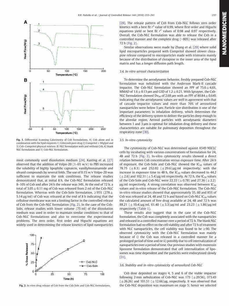
Fig. 1. Differential Scanning Calorimetry of Cxb formulations. A) Cxb alone and incombination with the lipid exipients 1) Celecoxib pure drug 2) Compritol+Miglyol and3) Cxb–Compritol physical mixture. B) NLC formulation with and without Cxb, 4) blankNLC formulation and 5) Cxb-NLC formulation.
237R.R. Patlolla et al. / Journal of Controlled Release 144 (2010) 233–241
NANOMEDICIN
E
most commonly used dissolution medium [24]. Kasting et al. [27]observed that the addition of Volpo-20 (1–6% w/v) to PBS increasedthe solubility of highly lipophilic capsaicin, vanillylnonanamide andolvanil compounds by several folds. The use of 0.1% w/v Volpo-20 wassufficient to maintain the sink conditions. The release studiesdemonstrated that, at initial 8 h, the Cxb-NLC formulation released8–10% of Cxb and after 24 h the release was 34%. At the end of 72 h, atotal of 3.05±0.11 mg of Cxb was released from 2 ml of the Cxb-NLCformulation. Whereas with the Cxb-Soln formulation, ≥95% (278±5.14 µg/ml) of Cxb was released at the end of 8 h indicating that thecellulose membrane was not a limiting factor in the controlled releaseof Cxb from the Cxb-NLC formulation (Fig. 2). In the case of the Cxb-Soln, release studies with lower volume (75 ml) of the dissolutionmedium was used in order to maintain similar conditions to that ofCxb-NLC formulations and also to overcome the experimentalproblems. The zero order, first order and Higuchi equations arewidely used in determining the release kinetics of lipid nanoparticles
Fig. 2. In-vitro drug release of Cxb from the Cxb-Soln and Cxb-NLC formulations.
[28]. The release pattern of Cxb from Cxb-NLC follows zero orderkinetics with a best fit r2 value of 0.99, where first order and Higuchiequations yield or best fit r2 values of 0.98 and 0.97 respectively.Overall, the Cxb-NLC formulation was able to release the Cxb in acontrolled manner and the complete drug (N80%) was released after72 h (Fig. 2).
Similar observations were made by Zhang et al. [29] where solidlipid microparticles prepared with Compritol showed slower cloza-pine release compared to microparticles made with tristearin mainlybecause of the distribution of clozapine in the inner area of the lipidmatrix and has a longer diffusion path length.
3.4. In-vitro aerosol characterization
To determine the aerodynamic behavior, freshly prepared Cxb-NLCformulation was nebulized with the Anderson Mark-II cascadeimpactor. The Cxb-NLC formulation showed an FPF of 75.6±4.6%,MMAD of 1.6±0.13 μm and GSD of 1.2±0.21. With Spraytec, the Cxb-NLC formulation showed Dv50 of 2.68 μm with an FPF of 80.84±0.64%indicating that the aerodynamic values are well in agreement with thatof cascade impactor values and more than 70% of aerosolizednanoparticles were below 5 μm. Particle size distribution is one of theimportant parameters in inhalation delivery, which determines theefficiency of the delivery system to deliver the particles deep enough tothe alveolar region. Aerosol particles with aerodynamic diametersbetween 1 and 3 µm is optimal for inhalation drug delivery and thesecharacteristics are suitable for pulmonary deposition throughout therespiratory zone [30].
3.5. In-vitro cytotoxicity
The cytotoxicity of Cxb-NLC was determined against A549 NSCLCcells by incubating with various concentrations of formulation for 24,48 and 72 h (Fig. 3). In-vitro cytotoxicity results showed a directrelation between Cxb concentration versus exposure time. After 24 hof exposure, the Cxb-Soln and Cxb-NLC showed the IC50 values of78.75 (±8.3) and 252.02 (±29.6)µg/ml respectively; with theincrease in exposure time to 48 h, the IC50 values decreased to 44.2(±2.6) and 102.31 (±5.4)µg/ml respectively. At 72 h, the IC50 valuesof the Cxb-Soln and Cxb-NLC were 22.33 (±0.78) and 27.36 (±2.2)µg/ml respectively. A strong correlation was observed between IC50values and in-vitro release of the Cxb-NLC formulation. The Cxb-NLCin-vitro release studies showed that approximately 35, 60 and 85% ofCxb was released at 24, 48 and 72 h and based on Cxb-NLC IC50 valuesthe calculated amount of free drug available at 24, 48 and 72 h was88.21 (±10.4)µg/ml, 61.40 (±3.3)µg/ml and 23.23 (±1.86)µg/mlrespectively (Table 1).
These results also suggest that in the case of the Cxb-NLCformulation, the Cxb was completely associated with the nanoparticlesand released in a controlledmanner over a period of 72 h. The blankNLCformulation hadno effect on the cell viability and after 72 h of treatmentwith NLC nanoparticles, the cell viability was found to be ≥90. Theobserved cytotoxicity with the Cxb-NLC formulation was mainlybecause of i) the Cxb was released in a controlled manner for aprolongedperiod of time and/or ii) possibly due to cell internalization ofnanoparticles over a period of time. Our previous studieswithmonensinliposome formulation demonstrated that cell internalization of lipo-somes was time dependent and the particles were endocytosed slowly[31].
3.6. Stability and in-vitro cytotoxicity of aerosolized Cxb-NLC
Cxb dose deposited on stages 4, 5 and 6 of the viable impactorfollowing 2 min nebulization of Cxb-NLC was 173 (±29.56), 373.43(±26.26) and 101.51 (±12.66)μg, respectively. It was observed thatthe Cxb-NLC deposition was maximum on stage 5; hence we selected

Fig. 3. In-vitro cytotoxicity profile of the Cxb-NLC formulation in A549 cells after A) 24 hB) 48 h C) 72 h of treatment. Cells were treated with various concentrations of Cxb-NLCand Cxb-Soln formulations and cytotoxicity was determined using a crystal violet dyeassay as described in Materials and methods. A plot of the percentage of cell survivalversus Cxb concentrations used for the determination of IC50 values. Values representthe mean±SD of at least three separate determinations for each treatment group.
Fig. 4. Effect of time on the lung deposition of Cxb-Soln and Cxb-NLC formulations.Following nebulization for 30 min, at regular time intervals 5, 10, 20 and 30 min themice lungs were collected and the amount of Cxb deposited was determined usingthe method as specified in the Materials and methods section. The data represent themean±SD (n=6). • Cxb-Soln and □ Cxb-NLC.
238 R.R. Patlolla et al. / Journal of Controlled Release 144 (2010) 233–241
NANOMEDICIN
E
stage 5 for in-vitro cytotoxicity studies against A549 cells. Furthermore,the aerosolization of the Cxb-NLC formulation was found to be stableagainst shear stresses generated during nebulization with non-
Table 1In-vitro cytotoxicity of Cxb-Soln and Cxb-NLC formulations.
Timepoint(h)
IC50 values (µg/ml) Cxb-NLCin-vitrorelease (%)
Cxb-NLC amountof free Cxbavailable (µg/ml)
Cxb-Soln Cxb-NLC
24 78.75 (±8.3) 252.02 (±29.6) 35 88.21 (±10.4)48 44.2 (±2.6) 102.31 (±5.4) 60 61.40 (±3.3)72 22.33 (±0.78) 27.36 (±2.2) 85 23.23 (±1.86)
significant (pN0.05) change in particle size and loss of originallyentrapped Cxb from theNLC formulation. The aerosolization of Cxb-NLCfor 2 min against A549 cells kept on stage 5 of the viable impactorinhibited the cell viability by 50.04% (±1.49). Incubation of A549 cellswith 373.43 µg of the Cxb-Soln for 72 h showed 58.45% (±1.69) of cellviability inhibition and which was comparable with that of theaerosolized Cxb-NLC.
3.7. Lung deposition of Cxb encapsulated NLC
To understand the lung deposition of nanoparticles, we collectedthe lungs at 5, 10, 20 and 30 min post nebulization and observed thatfollowing 20 min of exposure Cxb lung concentrations were found tobe constant. However, after 30 min of exposure, a two fold increase inlung concentrations was observed (Fig. 4). According to our previousstudies 30 min of nebulization is adequate to deposit a sufficientamount of Cxb in the lungs to elicit anticancer activity [16]; thereforein the present study, the same protocol was adopted.
From Eq. (2), the calculated amount of Cxb deposited following30min of nebulization of Cxb-NLC and the Cxb-Soln was 1.47 and4.82 mg/kg, respectively. The dose variation between the Cxb-Soln andCxb-NLC might be due to the differences in the density of solid contentin respective formulations. Because of these differences the aerosoliza-tion of the Cxb-Soln produce a droplet (finemist) whichwill holdmoreamount of Cxb content compared to aerosolized Cxb-NLC droplets,where later case the formulation contains theuniformly suspended lipidparticles which will have a higher density than the ethanolic solution.Fig. 5 represents the percent of Cxb dose deposited per lung tissue
Fig. 5. Lung deposition of Cxb-Soln and Cxb-NLC formulations after nebulization for30 min using a nose only Inexpose™ system and Pari LC Star jet nebulizer in Balb/c mice.The data represent the percent dose of Cxb deposited per lung tissue was plotted againsttime (mean±SD), n=6. • Cxb-Soln and□ Cxb-NLC, *pb0.05; **pb0.01; ***pb0.001.
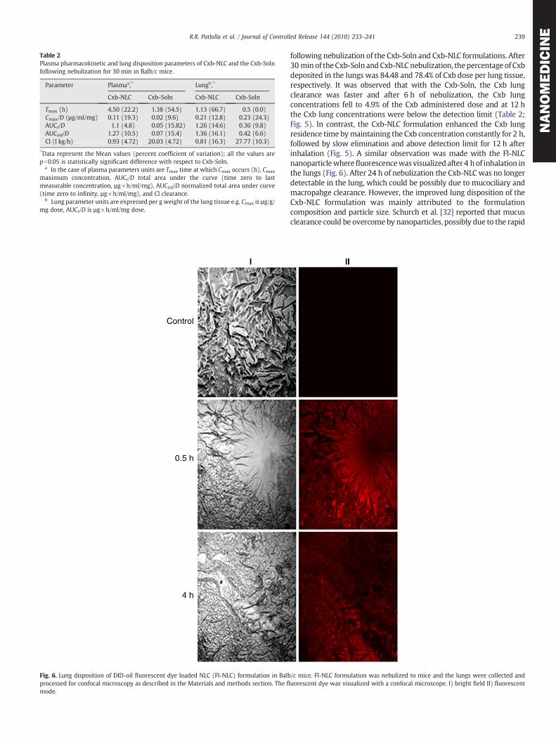
Table 2Plasma pharmacokinetic and lung disposition parameters of Cxb-NLC and the Cxb-Solnfollowing nebulization for 30 min in Balb/c mice.
Parameter Plasmaa,^ Lungb,^
Cxb-NLC Cxb-Soln Cxb-NLC Cxb-Soln
Tmax (h) 4.50 (22.2) 1.38 (54.5) 1.13 (66.7) 0.5 (0.0)Cmax/D (µg/ml/mg) 0.11 (19.3) 0.02 (9.6) 0.21 (12.8) 0.23 (24.3)AUCt/D 1.1 (4.8) 0.05 (15.82) 1.26 (14.6) 0.36 (9.8)AUCinf/D 1.27 (10.5) 0.07 (15.4) 1.36 (16.1) 0.42 (6.6)Cl (l kg/h) 0.93 (4.72) 20.03 (4.72) 0.81 (16.3) 27.77 (10.3)
^Data represent the Mean values (percent coefficient of variation); all the values arepb0.05 is statistically significant difference with respect to Cxb-Soln.
a In the case of plasma parameters units are Tmax time at which Cmax occurs (h), Cmax
maximum concentration, AUCt/D total area under the curve (time zero to lastmeasurable concentration, µg×h/ml/mg), AUCinf/D normalized total area under curve(time zero to infinity, µg×h/ml/mg), and Cl clearance.
b Lung parameter units are expressed per g weight of the lung tissue e.g. Cmax is µg/g/mg dose, AUCt/D is µg×h/ml/mg dose.
Fig. 6. Lung disposition of DID-oil fluorescent dye loaded NLC (Fl-NLC) formulation in Balbprocessed for confocal microscopy as described in the Materials and methods section. The fl
mode.
239R.R. Patlolla et al. / Journal of Controlled Release 144 (2010) 233–241
NANOMEDICIN
E
following nebulization of the Cxb-Soln and Cxb-NLC formulations. After30 min of the Cxb-Soln andCxb-NLCnebulization, thepercentage of Cxbdeposited in the lungs was 84.48 and 78.4% of Cxb dose per lung tissue,respectively. It was observed that with the Cxb-Soln, the Cxb lungclearance was faster and after 6 h of nebulization, the Cxb lungconcentrations fell to 4.9% of the Cxb administered dose and at 12 hthe Cxb lung concentrations were below the detection limit (Table 2;Fig. 5). In contrast, the Cxb-NLC formulation enhanced the Cxb lungresidence time bymaintaining the Cxb concentration constantly for 2 h,followed by slow elimination and above detection limit for 12 h afterinhalation (Fig. 5). A similar observation was made with the Fl-NLCnanoparticlewherefluorescencewasvisualized after 4 hof inhalation inthe lungs (Fig. 6). After 24 h of nebulization the Cxb-NLC was no longerdetectable in the lung, which could be possibly due to mucociliary andmacropahge clearance. However, the improved lung disposition of theCxb-NLC formulation was mainly attributed to the formulationcomposition and particle size. Schurch et al. [32] reported that mucusclearance could be overcome by nanoparticles, possibly due to the rapid
/c mice. Fl-NLC formulation was nebulized to mice and the lungs were collected anduorescent dye was visualized with a confocal microscope. I) bright field II) fluorescent
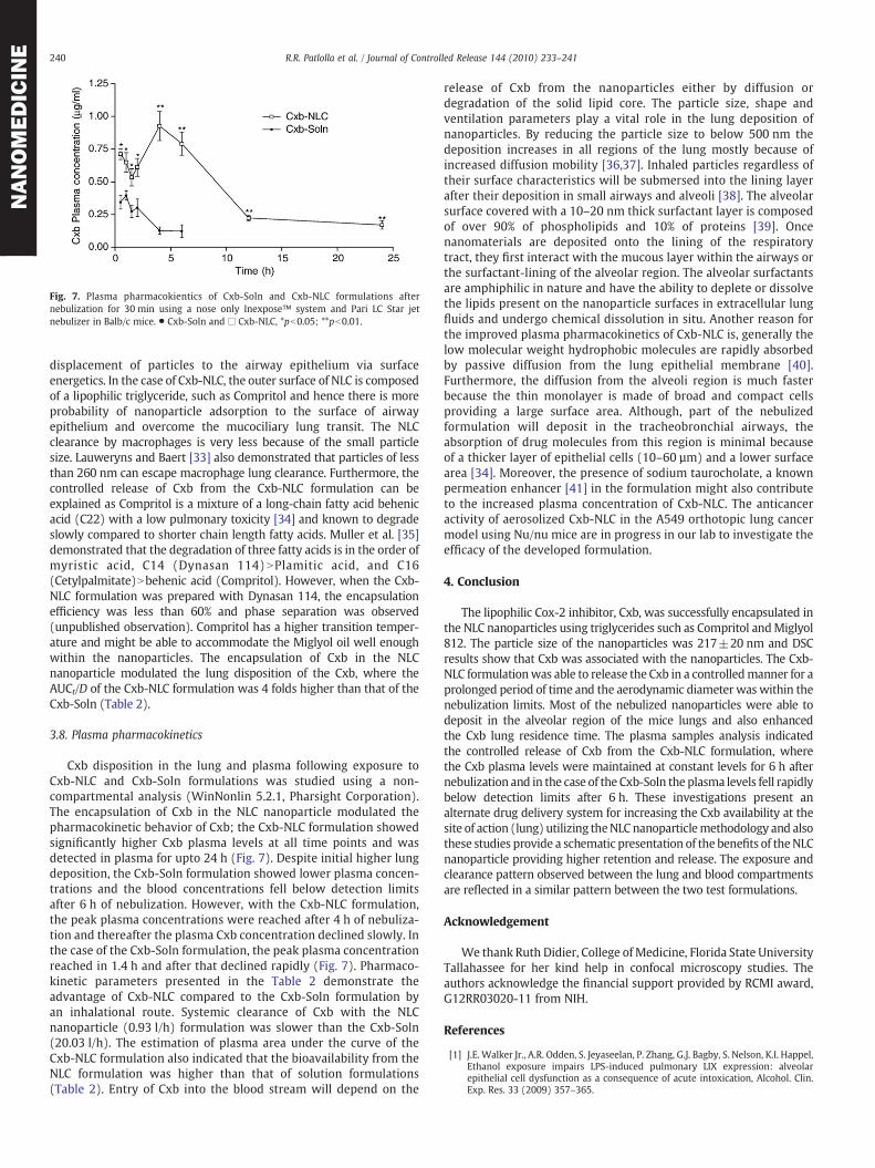
Fig. 7. Plasma pharmacokientics of Cxb-Soln and Cxb-NLC formulations afternebulization for 30 min using a nose only Inexpose™ system and Pari LC Star jetnebulizer in Balb/c mice. • Cxb-Soln and □ Cxb-NLC, *pb0.05; **pb0.01.
240 R.R. Patlolla et al. / Journal of Controlled Release 144 (2010) 233–241
NANOMEDICIN
E
displacement of particles to the airway epithelium via surfaceenergetics. In the case of Cxb-NLC, the outer surface of NLC is composedof a lipophilic triglyceride, such as Compritol and hence there is moreprobability of nanoparticle adsorption to the surface of airwayepithelium and overcome the mucociliary lung transit. The NLCclearance by macrophages is very less because of the small particlesize. Lauweryns and Baert [33] also demonstrated that particles of lessthan 260 nm can escape macrophage lung clearance. Furthermore, thecontrolled release of Cxb from the Cxb-NLC formulation can beexplained as Compritol is a mixture of a long-chain fatty acid behenicacid (C22) with a low pulmonary toxicity [34] and known to degradeslowly compared to shorter chain length fatty acids. Muller et al. [35]demonstrated that the degradation of three fatty acids is in the order ofmyristic acid, C14 (Dynasan 114) NPlamitic acid, and C16(Cetylpalmitate)Nbehenic acid (Compritol). However, when the Cxb-NLC formulation was prepared with Dynasan 114, the encapsulationefficiency was less than 60% and phase separation was observed(unpublished observation). Compritol has a higher transition temper-ature and might be able to accommodate the Miglyol oil well enoughwithin the nanoparticles. The encapsulation of Cxb in the NLCnanoparticle modulated the lung disposition of the Cxb, where theAUCt/D of the Cxb-NLC formulation was 4 folds higher than that of theCxb-Soln (Table 2).
3.8. Plasma pharmacokinetics
Cxb disposition in the lung and plasma following exposure toCxb-NLC and Cxb-Soln formulations was studied using a non-compartmental analysis (WinNonlin 5.2.1, Pharsight Corporation).The encapsulation of Cxb in the NLC nanoparticle modulated thepharmacokinetic behavior of Cxb; the Cxb-NLC formulation showedsignificantly higher Cxb plasma levels at all time points and wasdetected in plasma for upto 24 h (Fig. 7). Despite initial higher lungdeposition, the Cxb-Soln formulation showed lower plasma concen-trations and the blood concentrations fell below detection limitsafter 6 h of nebulization. However, with the Cxb-NLC formulation,the peak plasma concentrations were reached after 4 h of nebuliza-tion and thereafter the plasma Cxb concentration declined slowly. Inthe case of the Cxb-Soln formulation, the peak plasma concentrationreached in 1.4 h and after that declined rapidly (Fig. 7). Pharmaco-kinetic parameters presented in the Table 2 demonstrate theadvantage of Cxb-NLC compared to the Cxb-Soln formulation byan inhalational route. Systemic clearance of Cxb with the NLCnanoparticle (0.93 l/h) formulation was slower than the Cxb-Soln(20.03 l/h). The estimation of plasma area under the curve of theCxb-NLC formulation also indicated that the bioavailability from theNLC formulation was higher than that of solution formulations(Table 2). Entry of Cxb into the blood stream will depend on the
release of Cxb from the nanoparticles either by diffusion ordegradation of the solid lipid core. The particle size, shape andventilation parameters play a vital role in the lung deposition ofnanoparticles. By reducing the particle size to below 500 nm thedeposition increases in all regions of the lung mostly because ofincreased diffusion mobility [36,37]. Inhaled particles regardless oftheir surface characteristics will be submersed into the lining layerafter their deposition in small airways and alveoli [38]. The alveolarsurface covered with a 10–20 nm thick surfactant layer is composedof over 90% of phospholipids and 10% of proteins [39]. Oncenanomaterials are deposited onto the lining of the respiratorytract, they first interact with the mucous layer within the airways orthe surfactant-lining of the alveolar region. The alveolar surfactantsare amphiphilic in nature and have the ability to deplete or dissolvethe lipids present on the nanoparticle surfaces in extracellular lungfluids and undergo chemical dissolution in situ. Another reason forthe improved plasma pharmacokinetics of Cxb-NLC is, generally thelow molecular weight hydrophobic molecules are rapidly absorbedby passive diffusion from the lung epithelial membrane [40].Furthermore, the diffusion from the alveoli region is much fasterbecause the thin monolayer is made of broad and compact cellsproviding a large surface area. Although, part of the nebulizedformulation will deposit in the tracheobronchial airways, theabsorption of drug molecules from this region is minimal becauseof a thicker layer of epithelial cells (10–60 µm) and a lower surfacearea [34]. Moreover, the presence of sodium taurocholate, a knownpermeation enhancer [41] in the formulation might also contributeto the increased plasma concentration of Cxb-NLC. The anticanceractivity of aerosolized Cxb-NLC in the A549 orthotopic lung cancermodel using Nu/nu mice are in progress in our lab to investigate theefficacy of the developed formulation.
4. Conclusion
The lipophilic Cox-2 inhibitor, Cxb, was successfully encapsulated inthe NLC nanoparticles using triglycerides such as Compritol andMiglyol812. The particle size of the nanoparticles was 217±20 nm and DSCresults show that Cxb was associated with the nanoparticles. The Cxb-NLC formulationwas able to release the Cxb in a controlledmanner for aprolonged period of time and the aerodynamic diameter was within thenebulization limits. Most of the nebulized nanoparticles were able todeposit in the alveolar region of the mice lungs and also enhancedthe Cxb lung residence time. The plasma samples analysis indicatedthe controlled release of Cxb from the Cxb-NLC formulation, wherethe Cxb plasma levels were maintained at constant levels for 6 h afternebulization and in the case of the Cxb-Soln the plasma levels fell rapidlybelow detection limits after 6 h. These investigations present analternate drug delivery system for increasing the Cxb availability at thesite of action (lung) utilizing theNLC nanoparticlemethodology and alsothese studies provide a schematic presentation of the benefits of theNLCnanoparticle providing higher retention and release. The exposure andclearance pattern observed between the lung and blood compartmentsare reflected in a similar pattern between the two test formulations.
Acknowledgement
We thank Ruth Didier, College of Medicine, Florida State UniversityTallahassee for her kind help in confocal microscopy studies. Theauthors acknowledge the financial support provided by RCMI award,G12RR03020-11 from NIH.
References
[1] J.E. Walker Jr., A.R. Odden, S. Jeyaseelan, P. Zhang, G.J. Bagby, S. Nelson, K.I. Happel,Ethanol exposure impairs LPS-induced pulmonary LIX expression: alveolarepithelial cell dysfunction as a consequence of acute intoxication, Alcohol. Clin.Exp. Res. 33 (2009) 357–365.

241R.R. Patlolla et al. / Journal of Controlled Release 144 (2010) 233–241
NANOMEDICIN
E
[2] W. Mehnert, K. Mader, Solid lipid nanoparticles: production, characterization andapplications, Adv. Drug Deliv. Rev. 47 (2001) 165–196.
[3] C.J. Hitzman, W.F. Elmquist, L.W. Wattenberg, T.S. Wiedmann, Development of arespirable, sustained release microcarrier for 5-fluorouracil I: in vitro assessment ofliposomes, microspheres, and lipid coated nanoparticles, J. Pharm. Sci. 95 (2006)1114–1126.
[4] M. Chougule, B. Padhi, A. Misra, Nano-liposomal dry powder inhaler of amiloridehydrochloride, J. Nanosci. Nanotechnol. 6 (2006) 3001–3009.
[5] M.A. Videira, M.F. Botelho, A.C. Santos, L.F. Gouveia, J.J. DeLima, A.J. Almeida,Lymphatic uptake of pulmonary delivered radiolabelled solid lipid nanoparticles,J. Drug Target. 10 (2002) 607–613.
[6] P.R. Reddy, V. Venkateswarlu, Pharmacokinetics and tissue distribution of etoposidedelivered in long circulating parenteral emulsion, J. Drug Target. 13 (2005) 543–553.
[7] R.H. Müller, M. Radtke, S.A. Wissing, Solid lipid nanoparticles (SLN) andnanostructured lipid carriers (NLC) in cosmetic and dermatological preparations,Adv. Drug Deliv. Rev. 54 (2002) S131–S155.
[8] R.H. Muller, M. Radtke, S.A. Wissing, Nanostructured lipid matrices for improvedmicroencapsulation of drugs, Int. J. Pharm. 242 (2002) 121–128.
[9] A. Pardeike, A. Hommoss, R.H. Müller, Lipid nanoparticles (SLN, NLC) in cosmeticand pharmaceutical dermal products, Int. J. Pharm. 366 (2009) 170–184.
[10] M. Joshi, V. Patravale, Nanostructured lipid carrier (NLC) based gel of celecoxib,Int. J. Pharm. 346 (1–2) (2008) 124–132.
[11] T. Hida, K.I. Kozaki, H. Muramatsu, A. Masuda, S. Shimizu, T. Mitsudomi, T. Sugiura,M. Ogawa, T. Takahasi, Cyclooxygenase-2 inhibitor induces apoptosis andenhances cytotoxicity of various anticancer agents in non-small cell lung cancercell lines, Clin. Cancer Res. 6 (2000) 2006–2011.
[12] S. Shishodia, D. Koul, B.B. Aggarwal, Cyclooxygenase (COX)-2 inhibitor celecoxibabrogates TNF-induced NF-κB activation through inhibition of activation of IκBαkinase and Akt in human non-small cell lung carcinoma: correlation withsuppression of COX-2 synthesis, J. Immunol. 173 (2004) 2011–2022.
[13] J.T. Mao, M.D. Roth, K.J. Serio, F. Baratelli, L. Zhu, E.C. Holmes, R.M. Strieter, S.M.Dubinett, Celecoxib modulates the capacity for prostaglandin E2 and interleukin-10production in alveolar macrophages from active smokers, Clin. Cancer Res. 9 (2003)5835–5841.
[14] N. Seedher, S. Bhatia, Solubility enhancement of cox-2 inhibitors using varioussolvent systems, AAPS PharmSciTech 4 (2003) E33.
[15] A. Haynes, M.S. Shaik, A. Chatterjee, M. Singh, Formulation and evaluation ofaerosolized celecoxib for the treatment of lung cancer, Pharm. Res. 22 (2005)427–439.
[16] S.V. Fulzele, A. Chatterjee, M.S. Shaik, T. Jackson, M. Singh, Inhalation delivery andanti-tumor activity of celecoxib in human orthotopic non-small cell lung cancerxenograft model, Pharm. Res. 23 (2006) 2094–2106.
[17] H. Thakkar, R.K. Sharma, R.S.R. Murthy, Enhanced retention of celecoxib-loadedsolid lipid nanoparticles after intra-articular administration, Drugs R D 8 (5)(2007) 275–285.
[18] R.R. Patlolla, V. Vobalaboina, Pharmacokinetics and tissue distribution of etoposidedelivered in parenteral emulsion, J. Pharm. Sci. 94 (2005) 437–445.
[19] Q.Y. Xiang,M.T.Wang, F. Chen, T. Gong, Y.L. Jian, Z.R. Zhang, Y.Huang, Lung-targetingdeliveryofdexamethasoneacetate loadedsolid lipidnanoparticles, Arch. Pharm. Res.30 (4) (2007) 519–525.
[20] T. Jackson, M.B. Chougule, N. Ichite, R.R. Patlolla, M. Singh, Antitumor activity ofnoscapine in humannon-small cell lung cancer xenograftmodel, Cancer Chemother.Pharmacol. 63 (2008) 117–126.
[21] N. Ichite, M.B. Chougule, T. Jackson, S.V. Fulzele, S. Safe, M. Singh, Enhancement ofdocetaxel anticancer activity by a novel diindolylmethane compound in humannon-small cell lung cancer, Clin. Cancer Res. 15 (2009) 543–552.
[22] D. Cooney, M. Kazantseva, A.J. Hickey, Development of a size-dependant aerosoldeposition model utilizing airway epithelial cells for evaluating aerosol drugdelivery, Altern to Labor Animals, 32, 2004, pp. 581–590.
[23] M. Kazantseva, D. Cooney, A.J. Hickey, Development of a lung model utilizinghuman alveolar epithelial cells for evaluating aerosol drug delivery, in: P.R. Byron,R.N. Dalby, J. Peart, S.J. Farr (Eds.), Respiratory Drug Delivery VIII, Davis HorwoodInternational Publishing, Ltd., Raleigh, NC, USA, 2002, pp. 707–710.
[24] A. Tan, S. Simovic, K.A. Davey, T. Rades, A.C. Prestidge, Silica-lipid hybrid (SLH)microcapsules: a novel oral delivery system for poorly soluble drugs, J. Control.Release 134 (2009) 62–70.
[25] C. Puglia, P. Blasi, L. Rizza, A. Schoubben, F. Bonina, C. Rossi, M. Ricci, Lipidnanoparticles for prolonged topical delivery: an in vitro and in vivo investigation,Int. J. Pharm. 357 (2008) 295–304.
[26] W. Yang, J.I. Peters, R.O. Williams III, Inhaled nanoparticles— a current review, Int.J. Pharm. 356 (2008) 239–247.
[27] G.B. Kasting, W.R. Francis, L.A. Bowman, G.O. Kinnett, Percutaneous absorption ofvanilloids: in vivo and in vitro studies, J. Pharm. Sci. 86 (1997) 142–146.
[28] V. Venkateswarlu, K. Manjunath, Preparation, characterization and in vitro releasekinetics of clozapine solid lipid nanoparticles, J. Control. Release 24 (2004)627–638.
[29] L. Zhang, Y. Qian, C. Long, Y. Chen, Systematic procedures for formulation design ofdrug-loaded solid lipid microparticles: selection of carrier material and stabilizer,Ind. Eng. Chem. Res. 47 (2004) 6091–6100.
[30] P.R. Byron, Prediction of drug residence times in regions of the human respiratorytract following aerosol inhalation, J. Pharm. Sci. 75 (1986) 433–438.
[31] M. Singh, A.J. Ferdous, T.L. Jackson, Stealth monensin liposomes as a potentiator ofadriamycin in cancer treatment, J. Control. Release 59 (1999) 43–53.
[32] S. Schurch, P. Gehr, V. Im Hof, M. Geiser, F. Green, Surfactant displaces particlestoward the epithelium in airways and alveoli, Respir. Physiol. 80 (1990) 17–32.
[33] J.M. Lauweryns, J.H. Baert, Alveolar clearance and the role of the pulmonarylymphatics, Am. Rev. Respir. Dis. 115 (1977) 625–683.
[34] V. Sanna, N. Kirschvink, P. Gustin, E. Gavini, I. Roland, L. Delattre, B. Evrard,Preparation and in vivo toxicity study of solid lipid microparticles as carrier forpulmonary administration, AAPS PharmSciTech 5 (2004) e27.
[35] R.H. Muller, D. Ruhl, A.S. Runge, Biodegradation of solid lipid nanoparticles as afunction of lipase incubation time, Int. J. Pharm. 144 (1996) 115–121.
[36] P.R. Byron, J.S. Patton, Drugdelivery via the respiratory tract, J. AerosolMed. 7 (1994)49–75.
[37] P.A. Jaques, C.S. Kim, Measurement of total lung deposition of inhaled ultrafineparticles in healthy men and women, Inhal. Toxicol. 12 (2000) 715–731.
[38] M. Geiser, S. Schurch, P. Gehr, Influence of surface chemistry and topography ofparticles on their immersion into the lung's surface-lining layer, J. Appl. Physiol.94 (2003) 1793–1801.
[39] J. Goerke, Pulmonary surfactant: functions and molecular composition, Biochim.Biophys. Acta 1408 (1998) 79–89.
[40] J.S. Patton, P.R. Byron, Inhaling medicines: delivering drugs to the body throughthe lungs, Nat. Rev. Drug Discov. 6 (2007) 67–74.
[41] S. Kobayashi, S. Kondo, K. Juni, Pulmonary delivery of salmon calcitonin drypowders containing absorption enhancers in rats, Pharm. Res. 13 (1996) 80–83.
Copyright © 2022 FDOKUMEN