Regression of prostate tumors upon combination of hormone ablation therapy and celecoxib in vivo
23
Regression of Prostate Tumors upon Combination of Hormone Ablation Therapy and Celecoxib in vivo Parisa Abedinpour, Véronique T. Baron, John Welsh, and Per Borgström § Vaccine Research Institute of San Diego, San Diego, CA 92121 Abstract Background—Hormonal ablation is the standard of treatment for advanced androgen-dependent prostate cancer. Although tumor regression is usually achieved at first, the cancer inevitably evolves toward androgen-independence, in part because of the development of mechanisms of resistance and in part because at the tissue level androgen withdrawal is not fully attained. Current research efforts are focused on new therapeutic strategies that will increase the effectiveness of androgen withdrawal and delay recurrence. We used a syngeneic pseudo-orthotropic mouse model of prostate cancer to test the efficacy of combining androgen withdrawal with FDA-approved COX-2 inhibitor celecoxib. Methods—GFP-tagged TRAMP-C2 cells were co-implanted with prostate tissue in the dorsal chamber model and tumors were allowed to establish and vascularize. Tumor growth and angiogenesis were monitored in real-time using fluorescent intravital microscopy (IVM). Androgen withdrawal in mice was achieved using surgical castration or chemical hormonal ablation, alone or in combination with celecoxib (15 mg/kg, twice daily). Results—Celecoxib alone decreased the growth of prostate tumors mostly by inducing mitotic failure, which resulted in increased apoptosis. Surprisingly, celecoxib did not possess significant angiostatic activity. Surgical or chemical castration prevented the growth of prostate tumors and this, on the other hand, was associated with disruption of the tumor vasculature. Finally, androgen withdrawal combined with celecoxib caused tumor regression through decreased angiogenesis and increased mitosis arrest and apoptosis. Conclusion—Celecoxib, a relatively safe COX-2-selective anti-inflammatory drug, significantly increases the efficacy of androgen withdrawal in vivo and warrants further investigation as a complement therapy for advanced prostate cancer. Keywords Prostate cancer; androgen therapy; COX-2; intravital microscopy; Celecoxib; Celebrex INTRODUCTION Current therapeutic interventions for advanced prostate cancer are not curative. Although androgen ablation does initially deliver a response, the return of hormone-refractory tumors invariably prevents long-term patient survival. More effective strategies are needed to extend life expectancy and improve the quality of life for patients with advanced prostate cancer. New strategies may involve the combination of known effective treatments such as androgen withdrawal with drugs that have relatively minor side effects. § Corresponding author: Vaccine Research Institute of San Diego (VRISD), 10835 Road to the Cure, Suite 150, San Diego, CA 92121, USA. Phone: (858) 775-1736; [email protected] . NIH Public Access Author Manuscript Prostate. Author manuscript; available in PMC 2012 June 1. Published in final edited form as: Prostate. 2011 June 1; 71(8): 813–823. doi:10.1002/pros.21297. NIH-PA Author Manuscript NIH-PA Author Manuscript NIH-PA Author Manuscript
-
Upload
independent -
Category
Documents
-
view
3 -
download
0
Transcript of Regression of prostate tumors upon combination of hormone ablation therapy and celecoxib in vivo
Regression of Prostate Tumors upon Combination of HormoneAblation Therapy and Celecoxib in vivo
Parisa Abedinpour, Véronique T. Baron, John Welsh, and Per Borgström§
Vaccine Research Institute of San Diego, San Diego, CA 92121
AbstractBackground—Hormonal ablation is the standard of treatment for advanced androgen-dependentprostate cancer. Although tumor regression is usually achieved at first, the cancer inevitablyevolves toward androgen-independence, in part because of the development of mechanisms ofresistance and in part because at the tissue level androgen withdrawal is not fully attained. Currentresearch efforts are focused on new therapeutic strategies that will increase the effectiveness ofandrogen withdrawal and delay recurrence. We used a syngeneic pseudo-orthotropic mouse modelof prostate cancer to test the efficacy of combining androgen withdrawal with FDA-approvedCOX-2 inhibitor celecoxib.
Methods—GFP-tagged TRAMP-C2 cells were co-implanted with prostate tissue in the dorsalchamber model and tumors were allowed to establish and vascularize. Tumor growth andangiogenesis were monitored in real-time using fluorescent intravital microscopy (IVM).Androgen withdrawal in mice was achieved using surgical castration or chemical hormonalablation, alone or in combination with celecoxib (15 mg/kg, twice daily).
Results—Celecoxib alone decreased the growth of prostate tumors mostly by inducing mitoticfailure, which resulted in increased apoptosis. Surprisingly, celecoxib did not possess significantangiostatic activity. Surgical or chemical castration prevented the growth of prostate tumors andthis, on the other hand, was associated with disruption of the tumor vasculature. Finally, androgenwithdrawal combined with celecoxib caused tumor regression through decreased angiogenesis andincreased mitosis arrest and apoptosis.
Conclusion—Celecoxib, a relatively safe COX-2-selective anti-inflammatory drug, significantlyincreases the efficacy of androgen withdrawal in vivo and warrants further investigation as acomplement therapy for advanced prostate cancer.
KeywordsProstate cancer; androgen therapy; COX-2; intravital microscopy; Celecoxib; Celebrex
INTRODUCTIONCurrent therapeutic interventions for advanced prostate cancer are not curative. Althoughandrogen ablation does initially deliver a response, the return of hormone-refractory tumorsinvariably prevents long-term patient survival. More effective strategies are needed toextend life expectancy and improve the quality of life for patients with advanced prostatecancer. New strategies may involve the combination of known effective treatments such asandrogen withdrawal with drugs that have relatively minor side effects.
§Corresponding author: Vaccine Research Institute of San Diego (VRISD), 10835 Road to the Cure, Suite 150, San Diego, CA 92121,USA. Phone: (858) 775-1736; [email protected] .
NIH Public AccessAuthor ManuscriptProstate. Author manuscript; available in PMC 2012 June 1.
Published in final edited form as:Prostate. 2011 June 1; 71(8): 813–823. doi:10.1002/pros.21297.
NIH
-PA Author Manuscript
NIH
-PA Author Manuscript
NIH
-PA Author Manuscript
Cyclooxygenase (COX), the key regulatory enzyme for prostaglandin synthesis, istranscribed from two distinct genes. COX-1 is expressed constitutively in most tissues,while COX-2 expression is normally low and induced by a wide variety of stimuli (it wasinitially identified as an immediate-early growth response gene). Cyclooxygenases catalyzethe formation of prostaglandins (PGs), which are involved in tumor initiation and/orprogression. For example, COX-1 and COX-2 promote inflammation, which may directlycontribute to the development of prostate cancer (1). In addition, COX-2-induced PGE2activates cell signaling involved in proliferation and thereby directly promotes tumor cellgrowth. COX-2 is overexpressed in prostate cancer and its level of expression correlateswith Gleason score and cancer progression (2).
Nonsteroidal anti-inflammatory drugs (NSAIDs) that inhibit both COX-1 and COX-2, aswell as COX-2 selective inhibitors, are currently being evaluated clinically for theprevention of major types of cancer because of their positive effects in epidemiological andanimal studies. Indeed, a recent meta-analysis of epidemiological studies concluded thatNSAIDs – whether or not they are selective for COX-2 – have a chemopreventive effectagainst cancer of the colon, breast, lung and prostate (3). In addition, COX-2 promotesangiogenesis and therefore COX-2 inhibitors may impair tumor growth by blockingangiogenesis (4). Whereas genetic ablation of COX-2 decreases tumor formation in mousemodels, its overexpression favors transformation and cancer progression (reviewed in (2)).COX-2 is overexpressed in a number of malignancies and is associated with increasedproduction of PGE2, which plays a role in the initiation and progression of tumors (2). It isnow well accepted that COX-2 contributes to prostate cancer and the evidence that COX-2inhibitor celecoxib may be beneficial to prostate cancer patients is mounting (5,6).
In animal models, celecoxib (alone or in combination with another drug) decreased thegrowth of androgen-independent PC3 xenograft (7) and suppressed the re-growth of LNCaPxenografts following androgen withdrawal (8). In addition, several clinical studies havestarted to evaluate the effect of celecoxib in various therapeutic settings. These trials showthat celecoxib is safe, with a low cytotoxicity profile. Two studies have describedneoadjuvant celecoxib prior to prostatectomy in men with clinically localized prostatecancer, showing measurable amounts of celecoxib in tumors (9) and measurable biologicaleffects in prostate cancer tissue (10) but lacking clear clinical benefit. Other studies haveexamined the effect of celecoxib in combination with chemotherapy for the treatment ofadvanced, hormone-independent prostate cancer, without much success (11,12). A largertrial is underway (13), which will provide further information regarding the potentialefficacy of celecoxib for advanced prostate cancer. Finally, the efficiency of celecoxib wasassessed in patients with recurrent prostate cancer following radiation therapy or radicalprostatectomy. A decline or stabilization of PSA levels was observed in both trials,indicative of biological activity and suggesting that the drug may delay the growth ofrecurrent tumors and extend time before hormone-deprivation therapy (14,15). Of note,there has been no clinical trial so far to assess whether celecoxib may delay the progressionof prostate cancer toward androgen-independence in patients undergoing hormone-deprivation therapy. Thus, more studies are warranted to discover the best use for COX-2inhibitors and examine the efficacy of various strategies.
We have previously described a syngeneic pseudo-orthotropic model to study prostatecancer progression in vivo (16). This model is based on the dorsal skinfold chambertechnique, in which a transparent chamber for microscopy is positioned in the dorsalskinfold of a mouse. Mouse cells derived from the prostate tumor of a TRAMP mouse,known as TRAMP-C2 cells (17), were implanted into mouse dorsal chambers. A H2B-GFPfusion protein was stably introduced into the TRAMP-C2 cells by retroviral transduction. Asshown by Kanda et al., the H2B-GFP fusion protein is incorporated into chromatin without
Abedinpour et al. Page 2
Prostate. Author manuscript; available in PMC 2012 June 1.
NIH
-PA Author Manuscript
NIH
-PA Author Manuscript
NIH
-PA Author Manuscript

affecting cell cycle progression (18). Because the cells are stably transfected withfluorescent H2B-GFP, tumors can be visualized and imaged in real time using intravitalfluorescence video-microscopy (IVM). IVM allows measuring tumor growth, vascularparameters and intratumoral mitotic and apoptotic indices. To create a pseudo-orthotropicmicroenvironment, prostate tissue from a donor mouse was co-implanted with TRAMP-C2-GFP in the chambers. We have shown previously that after 1-2 weeks post-implantation theprostate tissue grafted into the chamber was able to connect its vasculature to the skinvasculature of the recipient mouse, which in turn supported angiogenesis within the growingtumor (16).
The present study investigated the effect of celecoxib alone or in combination with surgicaland chemical castration on the growth of TRAMP-C2-GFP tumors.
METHODSAntibodies and reagents
Cell culture media, culture-grade PBS, L-Glutamine, Trypsin-EDTA, penicillin/streptomycin, and FBS (Fetal bovine Serum) were from Mediatech (Herndon, VA). G418and Insulin-Selenium-Transferrin supplement (#41400-Gibco) was from Invitrogen(Carlsbad, CA). Celecoxib was from Toronto Research Chemicals (Ontario, Canada).
Antibodies against phospho-ERK1/2, PARP, and phospho-histone H3 (Ser10; Ab 6G3) werefrom Cell Signaling Technology (Danvers, MA). Antibodies to β-actin (AC-15) were fromSigma-Aldrich (St Louis, MO). Monoclonal anti-p27Kip1 antibodies were from BDPharmingen (San Jose, CA). Alexa Fluor-488 goat anti-mouse antibodies were fromInvitrogen (Carlsbad, CA).
Cell culture—H2B-GFP/TRAMP-C2 (TRAMP-C2-GFP) cells that stably express histoneH2B-GFP fusion protein (18) were generated as described (19). TRAMP-C2-GFP cells weregrown in RPMI containing 10% FBS, 2mM L-glutamine, 100 U/ml penicillin/100 μg/mlstreptomycin, insulin-selenium-transferrin (10 μg/ml insulin), and DHT 10−8M final. G418(100 μg/ml) was added to maintain stable expression of H2B-GFP. Androgen withdrawalwas achieved by keeping the cells in phenol red-free RPMI medium containing 10%charcoal-treated FBS and the same supplements as in the normal medium except for DHT.
Human prostate cancer cells DU145 and PC3 were grown in RPMI containing 10% FBS,2mM L-glutamine, 100 U/ml penicillin/100 μg/ml streptomycin. Cells were maintained in ahumidified incubator at 37°C and 5% CO2.
Measurement of cell growth in vitro—Cell growth was monitored by direct counting.Cells in 12-well plates were washed once with PBS, detached using Trypsin, and transferredto a suspension vial in a final volume of 10ml PBS. Cells were counted using aCOULTER™ Multisizer II instrument (Beckman Coulter Inc., Hialeah, FL) gated for theappropriate cell size and corrected for particulate debris. Each experiment was performed inbiological replicates and each vial was counted twice.
Flow cytometry quantification of mitosis—Cells were treated with celecoxib for 24hrs, suspended using trypsin, fixed and permeabilized using BD cytofix/cytoperm solution(BD Pharmingen, San Jose, CA) according to instructions. Cells were incubated withantibodies to phospho-histone H3 for 30 min, washed three times in BD perm/wash buffer,and stained with Alexa Fluor-488 anti-mouse antibodies for 20 min followed by three morewashes. The cells were resuspended at a density of approximately 106 cells/0.5ml in BDperm/wash buffer containing 50 μg/ml DNase-free RNase A, and 50 μg/ml propidium
Abedinpour et al. Page 3
Prostate. Author manuscript; available in PMC 2012 June 1.
NIH
-PA Author Manuscript
NIH
-PA Author Manuscript
NIH
-PA Author Manuscript

iodine. Fluorescence of single cells was recorded using a Facscan flow cytometer (BDPharmingen, San Jose, CA). FlowJo™ Software was used for data analysis.
Western-blot analysis of protein expression—Cells treated with 40 μM celecoxibfor the indicated times were lysed on ice in the presence of phosphatase and proteaseinhibitors. Lysates were clarified by centrifugation and the protein concentration in eachsample was measured using a BCA assay (Pierce, Rockford, IL). Lysates were submitted toSDS-PAGE electrophoresis. Proteins were transferred to Immobilon-P® membranes(Millipore, Billerica, MA), which were incubated with a blocking buffer for 20 min (Pierce,Rockford, IL). The first antibody was incubated overnight. Peroxidase-conjugatedantibodies (Amersham Biosciences, Piscataway, NJ) were added for 45min. Proteins wererevealed using a chromogenic stabilized substrate from Promega (Madison, WI). Whenappropriate, membranes were stripped using Restore™ Stripping Buffer (Pierce) for 15minand reprobed.
Animal model and surgical techniques—Animal experiments have been approved byour Institutional IACUC and were conducted in accordance to NIH guidelines. The dorsalskinfold chambers were prepared as described previously (16,19). Briefly, male C57/bl6mice (25-30 g body weight) were anesthetized and placed on a heating pad. Twosymmetrical titanium frames were implanted into the dorsal skinfold. A circular layer wasexcised from one of the skin layers. The underlying muscle and subcutaneous tissues werecovered with a glass coverslip incorporated in one of the frames. After a recovery period of2-3 days, prostate tissue and tumor cells were carefully placed in the chamber.
Preparation of tumor spheroids—TRAMP-C2-GFP cells were trypsinized andadjusted to a concentration of 250,000 cells/ml. Cell suspensions were then overlaid into 96-well round bottom plates coated with 1% agarose (100ul cell suspension/well). Cellspheroids were allowed to compact for 48 hours and were washed in serum-free mediumbefore implantation into the mouse chambers.
Implantation of prostate tissue and cancer cells—Anterior prostate tissue wasexcised from a normal C57/BL6 mouse, minced into small pieces (< 1 mm2), and implantedinto a chamber. A small indentation was made in the center of the prostate tissue, in which apre-formed tumor spheroid was placed. The prostate tissue and tumor spheroid were allowedto re-vascularize prior to experimentation.
Surgical Castration—Mice were anesthetized with 7.3 mg ketamine hydrochloride and2.3 mg xylazine /100 g body weight, i.p. A lateral incision across the scrotum was made andthe testes were individually ligated and excised. The wound was cauterized. The incisionwas then sutured and sealed with Nexaband® acrylic.
Chemical Castration—The mice were chemically castrated through oral administrationof Cyproterone acetate twice daily (0.5mg/kg) and injection of Leuprolide acetate daily(0.07mg/kg).
Celecoxib Treatment—Celecoxib was administered orally twice daily (15mg/kg/administration).
Intravital microscopy—Fluorescence microscopy, image analysis, measurement oftumor growth and vascular parameters, calculation of mitotic and apoptotic indices havebeen carefully detailed in our previous study (16).
Abedinpour et al. Page 4
Prostate. Author manuscript; available in PMC 2012 June 1.
NIH
-PA Author Manuscript
NIH
-PA Author Manuscript
NIH
-PA Author Manuscript
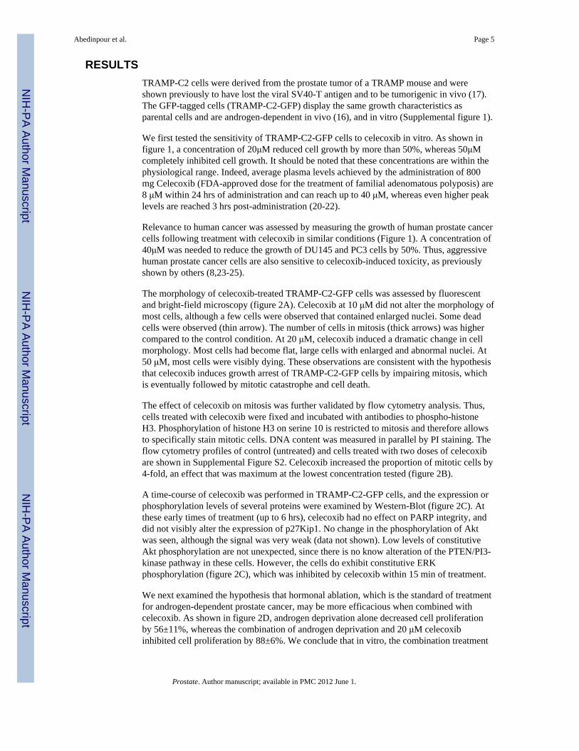
RESULTSTRAMP-C2 cells were derived from the prostate tumor of a TRAMP mouse and wereshown previously to have lost the viral SV40-T antigen and to be tumorigenic in vivo (17).The GFP-tagged cells (TRAMP-C2-GFP) display the same growth characteristics asparental cells and are androgen-dependent in vivo (16), and in vitro (Supplemental figure 1).
We first tested the sensitivity of TRAMP-C2-GFP cells to celecoxib in vitro. As shown infigure 1, a concentration of 20μM reduced cell growth by more than 50%, whereas 50μMcompletely inhibited cell growth. It should be noted that these concentrations are within thephysiological range. Indeed, average plasma levels achieved by the administration of 800mg Celecoxib (FDA-approved dose for the treatment of familial adenomatous polyposis) are8 μM within 24 hrs of administration and can reach up to 40 μM, whereas even higher peaklevels are reached 3 hrs post-administration (20-22).
Relevance to human cancer was assessed by measuring the growth of human prostate cancercells following treatment with celecoxib in similar conditions (Figure 1). A concentration of40μM was needed to reduce the growth of DU145 and PC3 cells by 50%. Thus, aggressivehuman prostate cancer cells are also sensitive to celecoxib-induced toxicity, as previouslyshown by others (8,23-25).
The morphology of celecoxib-treated TRAMP-C2-GFP cells was assessed by fluorescentand bright-field microscopy (figure 2A). Celecoxib at 10 μM did not alter the morphology ofmost cells, although a few cells were observed that contained enlarged nuclei. Some deadcells were observed (thin arrow). The number of cells in mitosis (thick arrows) was highercompared to the control condition. At 20 μM, celecoxib induced a dramatic change in cellmorphology. Most cells had become flat, large cells with enlarged and abnormal nuclei. At50 μM, most cells were visibly dying. These observations are consistent with the hypothesisthat celecoxib induces growth arrest of TRAMP-C2-GFP cells by impairing mitosis, whichis eventually followed by mitotic catastrophe and cell death.
The effect of celecoxib on mitosis was further validated by flow cytometry analysis. Thus,cells treated with celecoxib were fixed and incubated with antibodies to phospho-histoneH3. Phosphorylation of histone H3 on serine 10 is restricted to mitosis and therefore allowsto specifically stain mitotic cells. DNA content was measured in parallel by PI staining. Theflow cytometry profiles of control (untreated) and cells treated with two doses of celecoxibare shown in Supplemental Figure S2. Celecoxib increased the proportion of mitotic cells by4-fold, an effect that was maximum at the lowest concentration tested (figure 2B).
A time-course of celecoxib was performed in TRAMP-C2-GFP cells, and the expression orphosphorylation levels of several proteins were examined by Western-Blot (figure 2C). Atthese early times of treatment (up to 6 hrs), celecoxib had no effect on PARP integrity, anddid not visibly alter the expression of p27Kip1. No change in the phosphorylation of Aktwas seen, although the signal was very weak (data not shown). Low levels of constitutiveAkt phosphorylation are not unexpected, since there is no know alteration of the PTEN/PI3-kinase pathway in these cells. However, the cells do exhibit constitutive ERKphosphorylation (figure 2C), which was inhibited by celecoxib within 15 min of treatment.
We next examined the hypothesis that hormonal ablation, which is the standard of treatmentfor androgen-dependent prostate cancer, may be more efficacious when combined withcelecoxib. As shown in figure 2D, androgen deprivation alone decreased cell proliferationby 56±11%, whereas the combination of androgen deprivation and 20 μM celecoxibinhibited cell proliferation by 88±6%. We conclude that in vitro, the combination treatment
Abedinpour et al. Page 5
Prostate. Author manuscript; available in PMC 2012 June 1.
NIH
-PA Author Manuscript
NIH
-PA Author Manuscript
NIH
-PA Author Manuscript
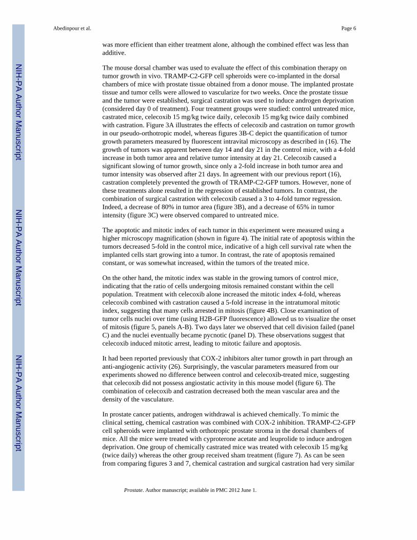
was more efficient than either treatment alone, although the combined effect was less thanadditive.
The mouse dorsal chamber was used to evaluate the effect of this combination therapy ontumor growth in vivo. TRAMP-C2-GFP cell spheroids were co-implanted in the dorsalchambers of mice with prostate tissue obtained from a donor mouse. The implanted prostatetissue and tumor cells were allowed to vascularize for two weeks. Once the prostate tissueand the tumor were established, surgical castration was used to induce androgen deprivation(considered day 0 of treatment). Four treatment groups were studied: control untreated mice,castrated mice, celecoxib 15 mg/kg twice daily, celecoxib 15 mg/kg twice daily combinedwith castration. Figure 3A illustrates the effects of celecoxib and castration on tumor growthin our pseudo-orthotropic model, whereas figures 3B-C depict the quantification of tumorgrowth parameters measured by fluorescent intravital microscopy as described in (16). Thegrowth of tumors was apparent between day 14 and day 21 in the control mice, with a 4-foldincrease in both tumor area and relative tumor intensity at day 21. Celecoxib caused asignificant slowing of tumor growth, since only a 2-fold increase in both tumor area andtumor intensity was observed after 21 days. In agreement with our previous report (16),castration completely prevented the growth of TRAMP-C2-GFP tumors. However, none ofthese treatments alone resulted in the regression of established tumors. In contrast, thecombination of surgical castration with celecoxib caused a 3 to 4-fold tumor regression.Indeed, a decrease of 80% in tumor area (figure 3B), and a decrease of 65% in tumorintensity (figure 3C) were observed compared to untreated mice.
The apoptotic and mitotic index of each tumor in this experiment were measured using ahigher microscopy magnification (shown in figure 4). The initial rate of apoptosis within thetumors decreased 5-fold in the control mice, indicative of a high cell survival rate when theimplanted cells start growing into a tumor. In contrast, the rate of apoptosis remainedconstant, or was somewhat increased, within the tumors of the treated mice.
On the other hand, the mitotic index was stable in the growing tumors of control mice,indicating that the ratio of cells undergoing mitosis remained constant within the cellpopulation. Treatment with celecoxib alone increased the mitotic index 4-fold, whereascelecoxib combined with castration caused a 5-fold increase in the intratumoral mitoticindex, suggesting that many cells arrested in mitosis (figure 4B). Close examination oftumor cells nuclei over time (using H2B-GFP fluorescence) allowed us to visualize the onsetof mitosis (figure 5, panels A-B). Two days later we observed that cell division failed (panelC) and the nuclei eventually became pycnotic (panel D). These observations suggest thatcelecoxib induced mitotic arrest, leading to mitotic failure and apoptosis.
It had been reported previously that COX-2 inhibitors alter tumor growth in part through ananti-angiogenic activity (26). Surprisingly, the vascular parameters measured from ourexperiments showed no difference between control and celecoxib-treated mice, suggestingthat celecoxib did not possess angiostatic activity in this mouse model (figure 6). Thecombination of celecoxib and castration decreased both the mean vascular area and thedensity of the vasculature.
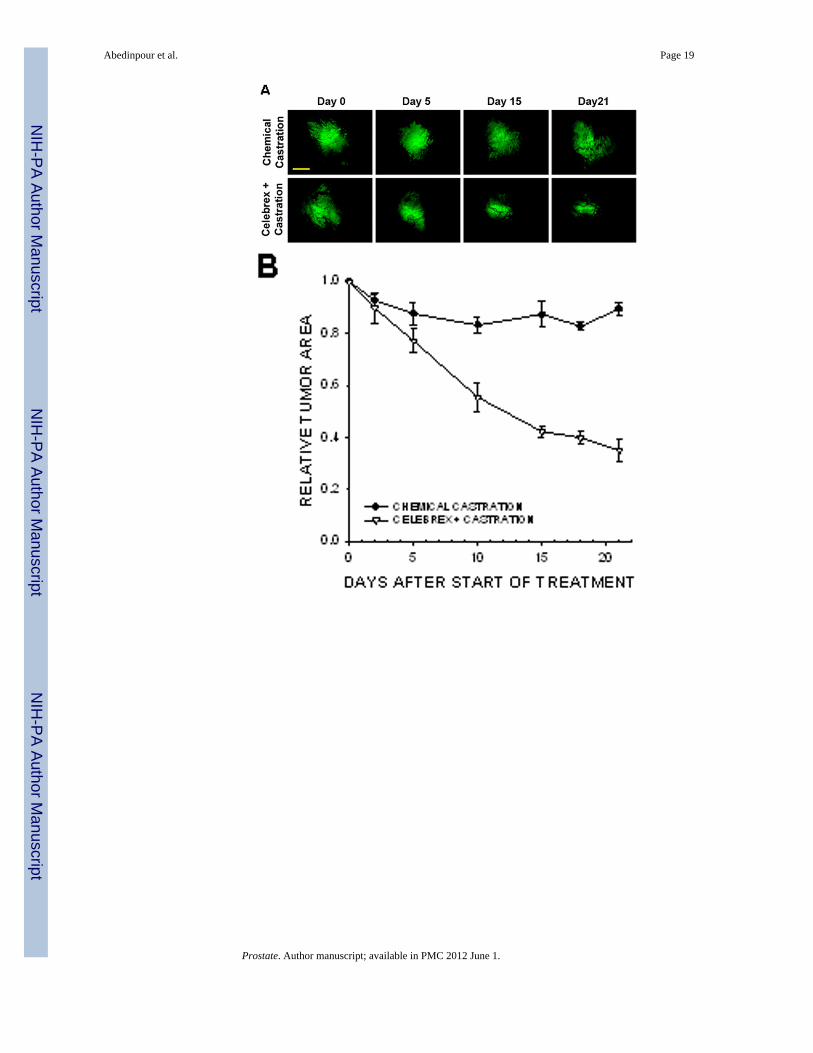
In prostate cancer patients, androgen withdrawal is achieved chemically. To mimic theclinical setting, chemical castration was combined with COX-2 inhibition. TRAMP-C2-GFPcell spheroids were implanted with orthotropic prostate stroma in the dorsal chambers ofmice. All the mice were treated with cyproterone acetate and leuprolide to induce androgendeprivation. One group of chemically castrated mice was treated with celecoxib 15 mg/kg(twice daily) whereas the other group received sham treatment (figure 7). As can be seenfrom comparing figures 3 and 7, chemical castration and surgical castration had very similar
Abedinpour et al. Page 6
Prostate. Author manuscript; available in PMC 2012 June 1.
NIH
-PA Author Manuscript
NIH
-PA Author Manuscript
NIH
-PA Author Manuscript
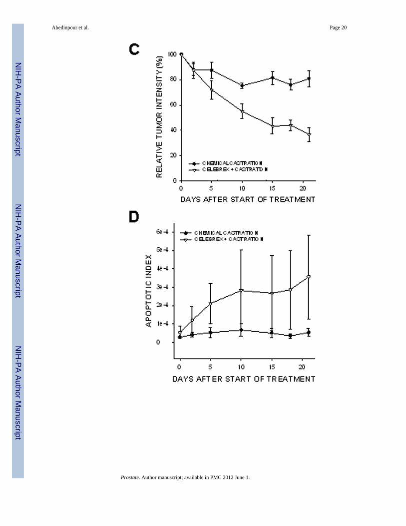
effects on tumor growth. Combining celecoxib treatment with chemical castration causedtumor regression, similarly to the combination of celecoxib and surgical castration. The rateof mitosis increased significantly in tumors of mice treated with combination therapycompared to androgen deprivation alone (figure 7E). Our experiments demonstrate thatsurgical and chemical castration have similar effect on tumor regression when combinedwith celecoxib treatment.
In this model of the clinical condition, we observed a deep regression of angiogenesis withinonly 2 days, as shown in figure 8. Androgen deprivation combined with celecoxib causedthe vasculature to shrink, as measured by the vascular area and the vascular diameter (panelB).
In conclusion, celecoxib alone decreased tumor growth by causing cell cycle arrest andmitotic failure. It had no measurable effect on vascular parameters in our model. Castration,which directly inhibited the proliferation of prostate cancer cells in vitro (Suppl figure 1),blocked tumor growth in vivo but did not result in regression. The combination of celecoxiband androgen withdrawal, however, resulted in tumor regression and was associated withrapid shrinkage of the vasculature.
DISCUSSIONAnimal models are crucial to our understanding of the mechanisms underlying tumorprogression and growth. Current rodent models such as xenograft human tumors inimmunodeficient mice do not sufficiently represent relevant clinical cancer models,especially with regard to angiogenesis and drug sensitivity. Transgenic animals, on the otherhand, do not permit the direct measurement of tumor growth, and time-dependentobservations can be made only by inference after killing mice at various time points. Thedorsal skinfold chamber allows repeated observations in the same animal over extended timeperiods. Thus, evaluation of vascular responses to treatment can be done in real-time. Theuse of TRAMP-C2 cells transfected with H2B-GFP also allows us to measure increases ordecreases in tumor growth and to assess other underlying mechanisms (mitosis or apoptosis)that influence tumor progression.
We have used this model to examine the effect of COX-2 inhibitor celecoxib in combinationwith androgen withdrawal for the treatment of prostate cancer. Surgical castration combinedwith celecoxib caused tumor regression, which was not observed with castration orcelecoxib alone.
These results are in line with the recent finding that a combination of celecoxib andandrogen withdrawal delayed the acquisition of androgen-independence in a xenograftmodel using human LNCaP cells (8), which suggests that these observations may berelevant to human disease.
Observation of various tumor parameters indicated that regression was caused by acombination of decreased vascularization due to androgen withdrawal, together with tumorcells growth arrest due mostly to celecoxib treatment. Thus, the efficacy of the combinationwas much better in vivo than in vitro, because of the separate effects of each treatment ondistinct biological compartments (vasculature, stroma, possibly inflammatory cells) that arenot represented in the culture of cell lines.
A critical aspect in understanding and treating cancer progression is the relationship betweenthe tumor and the “soil” that supports its growth and progression. It is well known thatstromal-epithelial interactions are very important for androgen dependent prostate cancer(27). Thus, co-implanting TRAMP-C2 cells with prostate stroma obtained from a donor
Abedinpour et al. Page 7
Prostate. Author manuscript; available in PMC 2012 June 1.
NIH
-PA Author Manuscript
NIH
-PA Author Manuscript
NIH
-PA Author Manuscript
mouse provides the tumor cells with an environment which closely resembles orthotropicimplantation. When cancer cells were implanted in the chambers in the absence of prostatetissue, they did not grow into tumors, and treatment with celecoxib did not alter tumorparameters (supplemental figure 3), exemplifying the importance of the orthotropic milieu inassessing treatment parameters.
The in vivo mechanism by which COX-2 affects tumor growth is still not completelyunderstood. It has been suggested that COX-2 inhibitors induce cell cycle arrest andapoptosis in cancer cells through a mechanism that is fundamentally different from theapoptosis caused by cancer chemotherapeutic agents. Our results showing that celecoxibcaused growth arrest and mitotic failure, characterized by deep alterations of nucleimorphology, and followed by mitotic catastrophe and cell death, are in agreement with thishypothesis.
Celecoxib did not alter protein levels of p53 or PARP, which are involved at early stages ofapoptosis. Although in some cell lines celecoxib inhibits the PI3-kinase pathway anddecreases the phosphorylation of Akt (28), we did not detect changes in phospho-Akt.However, the PI3-kinase pathway is not constitutively active in the TRAMP-C2 cells,therefore levels of phospho-Akt are barely detectable and probably not amenable tocelecoxib alteration. In contrast, ERK phosphorylation was constitutively high and wasinhibited by celecoxib, a result consistent with previous observations (reviewed in (29)).
We conclude that COX-2 selective inhibitor celecoxib, through COX-2 dependent andindependent mechanisms, significantly increases the efficacy of androgen withdrawal in amouse model of prostate cancer. This combination warrants further investigation as acomplement therapy for aggressive prostate cancer.
Supplementary MaterialRefer to Web version on PubMed Central for supplementary material.
AcknowledgmentsWe thank Dale Winger for his excellent assistance in the preparation of the dorsal skinfold chambers and ReetiBandyopadhyay for technical assistance. We also thank Dr. David Chambers (Salk Insitute, San Diego) for his helpwith Flow Cytometry.
This work was supported by funding from Pfizer, contract COXAV-V0190-010 (Dr. Per Borgström); NIH/NCIgrant RO1-CA102688 (Dr. Véronique Baron); NIH/NCI grant R21CA133638 (Dr. John Welsh); DOD-CDMRPgrant W81XWH-09-1-0280 and UC/CBCRP 151B-0133 (Dr. Per Borgström and Dr. John Welsh); FellowshipCTS07:1 from Carl Tryggers Stiftelse for Scientific Research, Stockholm (Dr. Parisa Abedinpour).
REFERENCES1. Stock D, Groome PA, Siemens DR. Inflammation and prostate cancer: a future target for prevention
and therapy? The Urologic clinics of North America. 2008; 35(1):117–130. vii. [PubMed:18061030]
2. Furstenberger G, Krieg P, Muller-Decker K, Habenicht AJ. What are cyclooxygenases andlipoxygenases doing in the driver’s seat of carcinogenesis? International journal of cancer. 2006;119(10):2247–2254.
3. Harris RE. Cyclooxygenase-2 (cox-2) blockade in the chemoprevention of cancers of the colon,breast, prostate, and lung. Inflammopharmacology. 2009; 17(2):55–67. [PubMed: 19340409]
4. Sahin M, Sahin E, Gumuslu S. Cyclooxygenase-2 in cancer and angiogenesis. Angiology. 2009;60(2):242–253. [PubMed: 18505747]
Abedinpour et al. Page 8
Prostate. Author manuscript; available in PMC 2012 June 1.
NIH
-PA Author Manuscript
NIH
-PA Author Manuscript
NIH
-PA Author Manuscript
5. Pruthi RS, Wallen EM. Cyclooxygenase-2: a therapeutic target for prostate cancer. Clinicalgenitourinary cancer. 2005; 4(3):203–211. [PubMed: 16425990]
6. Sooriakumaran P, Langley SE, Laing RW, Coley HM. COX-2 inhibition: a possible role in themanagement of prostate cancer? Journal of chemotherapy (Florence, Italy). 2007; 19(1):21–32.
7. Zheng X, Cui XX, Avila GE, Huang MT, Liu Y, Patel J, Kong AN, Paulino R, Shih WJ, Lin Y,Rabson AB, Reddy BS, Conney AH. Atorvastatin and celecoxib inhibit prostate PC-3 tumors inimmunodeficient mice. Clin Cancer Res. 2007; 13(18 Pt 1):5480–5487. [PubMed: 17875778]
8. Zheng X, Cui XX, Gao Z, Zhao Y, Lin Y, Shih WJ, Huang MT, Liu Y, Rabson A, Reddy B, YangCS, Conney AH. Atorvastatin and celecoxib in combination inhibits the progression of androgen-dependent LNCaP xenograft prostate tumors to androgen independence. Cancer prevention research(Philadelphia, Pa. 2010; 3(1):114–124.
9. Antonarakis ES, Heath EI, Walczak JR, Nelson WG, Fedor H, De Marzo AM, Zahurak ML,Piantadosi S, Dannenberg AJ, Gurganus RT, Baker SD, Parnes HL, DeWeese TL, Partin AW,Carducci MA. Phase II, randomized, placebo-controlled trial of neoadjuvant celecoxib in men withclinically localized prostate cancer: evaluation of drug-specific biomarkers. J Clin Oncol. 2009;27(30):4986–4993. [PubMed: 19720908]
10. Sooriakumaran P, Coley HM, Fox SB, Macanas-Pirard P, Lovell DP, Henderson A, Eden CG,Miller PD, Langley SE, Laing RW. A randomized controlled trial investigating the effects ofcelecoxib in patients with localized prostate cancer. Anticancer Res. 2009; 29(5):1483–1488.[PubMed: 19443354]
11. Carles J, Font A, Mellado B, Domenech M, Gallardo E, Gonzalez-Larriba JL, Catalan G, Alfaro J,Del Alba A Gonzalez, Nogue M, Lianes P, Tello JM. Weekly administration of docetaxel incombination with estramustine and celecoxib in patients with advanced hormone-refractoryprostate cancer: final results from a phase II study. Br J Cancer. 2007; 97(9):1206–1210.[PubMed: 17955053]
12. Fontana A, Galli L, Fioravanti A, Orlandi P, Galli C, Landi L, Bursi S, Allegrini G, Fontana E, DiMarsico R, Antonuzzo A, D’Arcangelo M, Danesi R, Del Tacca M, Falcone A, Bocci G. Clinicaland pharmacodynamic evaluation of metronomic cyclophosphamide, celecoxib, anddexamethasone in advanced hormone-refractory prostate cancer. Clin Cancer Res. 2009; 15(15):4954–4962. [PubMed: 19622584]
13. James ND, Sydes MR, Clarke NW, Mason MD, Dearnaley DP, Anderson J, Popert RJ, Sanders K,Morgan RC, Stansfeld J, Dwyer J, Masters J, Parmar MK. Systemic therapy for advancing ormetastatic prostate cancer (STAMPEDE): a multi-arm, multistage randomized controlled trial.BJU international. 2009; 103(4):464–469. [PubMed: 18990168]
14. Pruthi RS, Derksen JE, Moore D, Carson CC, Grigson G, Watkins C, Wallen E. Phase II trial ofcelecoxib in prostate-specific antigen recurrent prostate cancer after definitive radiation therapy orradical prostatectomy. Clin Cancer Res. 2006; 12(7 Pt 1):2172–2177. [PubMed: 16609031]
15. Smith MR, Manola J, Kaufman DS, Oh WK, Bubley GJ, Kantoff PW. Celecoxib versus placebofor men with prostate cancer and a rising serum prostate-specific antigen after radicalprostatectomy and/or radiation therapy. J Clin Oncol. 2006; 24(18):2723–2728. [PubMed:16782912]
16. Frost GI, Lustgarten J, Dudouet B, Nyberg L, Hartley-Asp B, Borgstrom P. Novel syngeneicpseudo-orthotopic prostate cancer model: vascular, mitotic and apoptotic responses to castration.Microvascular research. 2005; 69(1-2):1–9. [PubMed: 15797254]
17. Foster BA, Gingrich JR, Kwon ED, Madias C, Greenberg NM. Characterization of prostaticepithelial cell lines derived from transgenic adenocarcinoma of the mouse prostate (TRAMP)model. Cancer research. 1997; 57(16):3325–3330. [PubMed: 9269988]
18. Kanda T, Sullivan KF, Wahl GM. Histone-GFP fusion protein enables sensitive analysis ofchromosome dynamics in living mammalian cells. Curr Biol. 1998; 8(7):377–385. [PubMed:9545195]
19. Frost GI, Borgstrom P. Real time in vivo quantitation of tumor angiogenesis. Methods in molecularmedicine. 2003; 85:65–78. [PubMed: 12710198]
20. Brautigam L, Vetter G, Tegeder I, Heinkele G, Geisslinger G. Determination of celecoxib inhuman plasma and rat microdialysis samples by liquid chromatography tandem massspectrometry. Journal of chromatography. 2001; 761(2):203–212. [PubMed: 11587350]
Abedinpour et al. Page 9
Prostate. Author manuscript; available in PMC 2012 June 1.
NIH
-PA Author Manuscript
NIH
-PA Author Manuscript
NIH
-PA Author Manuscript
21. Chow HH, Anavy N, Salazar D, Frank DH, Alberts DS. Determination of celecoxib in humanplasma using solid-phase extraction and high-performance liquid chromatography. Journal ofpharmaceutical and biomedical analysis. 2004; 34(1):167–174. [PubMed: 14738931]
22. McAdam BF, Catella-Lawson F, Mardini IA, Kapoor S, Lawson JA, FitzGerald GA. Systemicbiosynthesis of prostacyclin by cyclooxygenase (COX)-2: the human pharmacology of a selectiveinhibitor of COX-2. Proc Natl Acad Sci U S A. 1999; 96(1):272–277. [PubMed: 9874808]
23. Lu W, Tinsley HN, Keeton A, Qu Z, Piazza GA, Li Y. Suppression of Wnt/beta-catenin signalinginhibits prostate cancer cell proliferation. European journal of pharmacology. 2009; 602(1):8–14.[PubMed: 19026633]
24. Narayanan NK, Narayanan BA, Reddy BS. A combination of docosahexaenoic acid and celecoxibprevents prostate cancer cell growth in vitro and is associated with modulation of nuclear factor-kappaB, and steroid hormone receptors. Int J Oncol. 2005; 26(3):785–792. [PubMed: 15703837]
25. Patel MI, Subbaramaiah K, Du B, Chang M, Yang P, Newman RA, Cordon-Cardo C, Thaler HT,Dannenberg AJ. Celecoxib inhibits prostate cancer growth: evidence of a cyclooxygenase-2-independent mechanism. Clin Cancer Res. 2005; 11(5):1999–2007. [PubMed: 15756026]
26. Masferrer JL, Leahy KM, Koki AT, Zweifel BS, Settle SL, Woerner BM, Edwards DA, FlickingerAG, Moore RJ, Seibert K. Antiangiogenic and antitumor activities of cyclooxygenase-2 inhibitors.Cancer research. 2000; 60(5):1306–1311. [PubMed: 10728691]
27. Cunha GR, Hayward SW, Wang YZ, Ricke WA. Role of the stromal microenvironment incarcinogenesis of the prostate. International journal of cancer. 2003; 107(1):1–10.
28. Kulp SK, Yang YT, Hung CC, Chen KF, Lai JP, Tseng PH, Fowble JW, Ward PJ, Chen CS. 3-phosphoinositide-dependent protein kinase-1/Akt signaling represents a major cyclooxygenase-2-independent target for celecoxib in prostate cancer cells. Cancer research. 2004; 64(4):1444–1451.[PubMed: 14973075]
29. Grosch S, Maier TJ, Schiffmann S, Geisslinger G. Cyclooxygenase-2 (COX-2)-independentanticarcinogenic effects of selective COX-2 inhibitors. Journal of the National Cancer Institute.2006; 98(11):736–747. [PubMed: 16757698]
Abedinpour et al. Page 10
Prostate. Author manuscript; available in PMC 2012 June 1.
NIH
-PA Author Manuscript
NIH
-PA Author Manuscript
NIH
-PA Author Manuscript
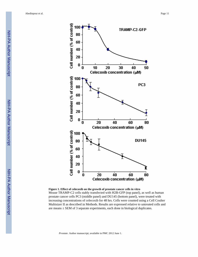
Figure 1. Effect of celecoxib on the growth of prostate cancer cells in vitroMouse TRAMP-C2 cells stably transfected with H2B-GFP (top panel), as well as humanprostate cancer cells PC3 (middle panel) and DU145 (bottom panel), were treated withincreasing concentrations of celecoxib for 48 hrs. Cells were counted using a Cell CoulterMultisizer II as described in Methods. Results are expressed relative to untreated cells andare means ± SEM of 3 separate experiments, each done in biological duplicates.
Abedinpour et al. Page 11
Prostate. Author manuscript; available in PMC 2012 June 1.
NIH
-PA Author Manuscript
NIH
-PA Author Manuscript
NIH
-PA Author Manuscript

Abedinpour et al. Page 12
Prostate. Author manuscript; available in PMC 2012 June 1.
NIH
-PA Author Manuscript
NIH
-PA Author Manuscript
NIH
-PA Author Manuscript

Figure 2. Effect of celecoxib in TRAMP-C2-GFP prostate cancer cellsPanel A: Bright field microscopy (right) and fluorescence microscopy (left) of TRAMP-C2-GFP cells treated with increasing doses of Celecoxib for 48 hrs. Thick arrows point tomitotic cells; thin arrows point to dead cells. Panel B: Cells were treated with the indicatedconcentrations of celecoxib for 24 hrs. Cells were detached using trypsin, fixed, and co-stained for phospho-H3 (alexa-fluor 488) and DNA content (propidium iodide). The graphshows the proportion of cells in mitosis as compared to control, determined by flowcytometry on Facscan (BD Biosciences). Panel C: TRAMP-C2-GFP cells were treated with40 μM celecoxib for the indicated times. Cells were lysed and protein expression wasanalyzed by western blot. Blot membranes were stripped and reprobed using the indicatedantibodies. P-ERK: phosphorylation-specific antibodies to ERK. Panel D: TRAMP-C2-GFPcells were incubated in medium with or without androgen and treated with increasingconcentrations of celecoxib for 48 hrs before cell counting. Results are expressed relative tountreated cells grown in medium containing androgen, and are means ± SEM of 3 separateexperiments, each done in biological triplicate.
Abedinpour et al. Page 13
Prostate. Author manuscript; available in PMC 2012 June 1.
NIH
-PA Author Manuscript
NIH
-PA Author Manuscript
NIH
-PA Author Manuscript

Abedinpour et al. Page 14
Prostate. Author manuscript; available in PMC 2012 June 1.
NIH
-PA Author Manuscript
NIH
-PA Author Manuscript
NIH
-PA Author Manuscript

Figure 3. Effect of celecoxib and/or surgical castration on prostate tumor growth in vivoTRAMP-C2-GFP cell spheroids were co-implanted with prostate tissue and allowed tovascularize. When there was proper blood flow within the growing tumors, the mice weresurgically castrated (Day 0) and Celecoxib treatment (15 mg/kg/administration) was startedby oral administration twice daily. Tumors were imaged by intravital microscopy once aweek. Panel A: a representative collage of tumor growth in the four treatment groups. Bar ~500μm. Panel B-C: graphic representation of relative tumor areas (B) and relative tumorintensities (C) calculated from intravital microscopy data (log scale).
Abedinpour et al. Page 15
Prostate. Author manuscript; available in PMC 2012 June 1.
NIH
-PA Author Manuscript
NIH
-PA Author Manuscript
NIH
-PA Author Manuscript
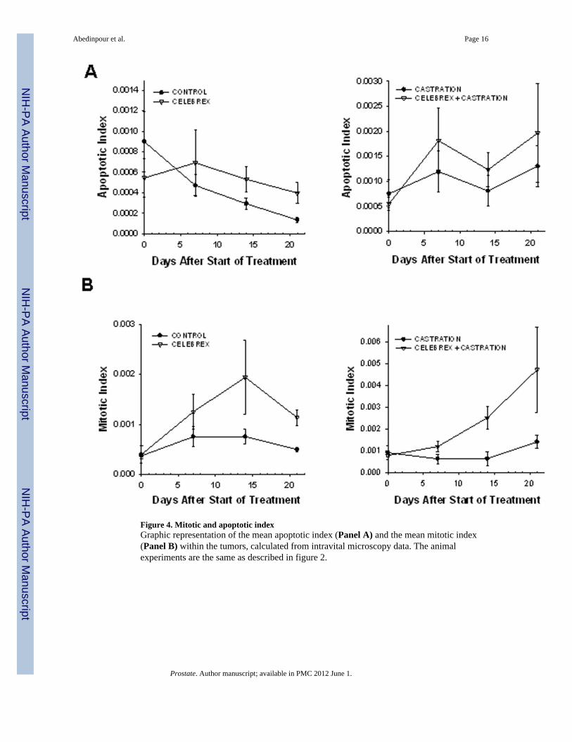
Figure 4. Mitotic and apoptotic indexGraphic representation of the mean apoptotic index (Panel A) and the mean mitotic index(Panel B) within the tumors, calculated from intravital microscopy data. The animalexperiments are the same as described in figure 2.
Abedinpour et al. Page 16
Prostate. Author manuscript; available in PMC 2012 June 1.
NIH
-PA Author Manuscript
NIH
-PA Author Manuscript
NIH
-PA Author Manuscript

Figure 5. Intravital microscopy at high magnification of celecoxib-treated TRAMP-C2-GFPtumorsTumors from celecoxib-treated mice (shown in figure 2) were imaged by intravitalmicroscopy at high magnification. Panels A and B: H2B-GFP fluorescence of TRAMP-C2-GFP tumors showing the onset of mitosis at day 3. Bar ~25μ (A); ~10μ (B). Panels C andD: failed mitosis (C) with the nuclei becoming pycnotic at day 5 (D). Bar ~25μ (C, D).
Abedinpour et al. Page 17
Prostate. Author manuscript; available in PMC 2012 June 1.
NIH
-PA Author Manuscript
NIH
-PA Author Manuscript
NIH
-PA Author Manuscript

Figure 6. Effect of celecoxib treatment on intra-tumoral angiogenesisTumors were imaged by intravital microscopy and vascular parameters were calculated.Panel A: Graphic representation of vascular parameters (area, diameter and density) forcontrol, celecoxib-treated and celecoxib + castrated animals.
Abedinpour et al. Page 18
Prostate. Author manuscript; available in PMC 2012 June 1.
NIH
-PA Author Manuscript
NIH
-PA Author Manuscript
NIH
-PA Author Manuscript
Abedinpour et al. Page 19
Prostate. Author manuscript; available in PMC 2012 June 1.
NIH
-PA Author Manuscript
NIH
-PA Author Manuscript
NIH
-PA Author Manuscript
Abedinpour et al. Page 20
Prostate. Author manuscript; available in PMC 2012 June 1.
NIH
-PA Author Manuscript
NIH
-PA Author Manuscript
NIH
-PA Author Manuscript
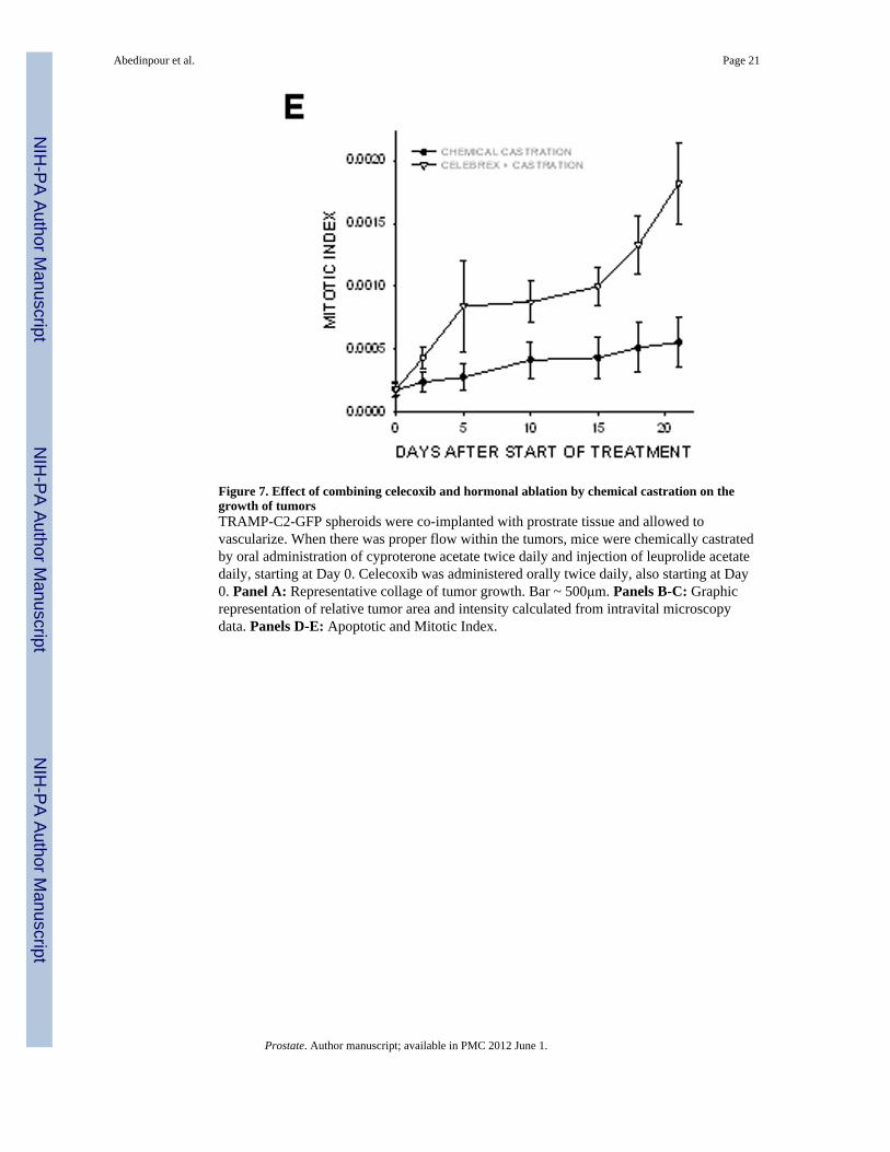
Figure 7. Effect of combining celecoxib and hormonal ablation by chemical castration on thegrowth of tumorsTRAMP-C2-GFP spheroids were co-implanted with prostrate tissue and allowed tovascularize. When there was proper flow within the tumors, mice were chemically castratedby oral administration of cyproterone acetate twice daily and injection of leuprolide acetatedaily, starting at Day 0. Celecoxib was administered orally twice daily, also starting at Day0. Panel A: Representative collage of tumor growth. Bar ~ 500μm. Panels B-C: Graphicrepresentation of relative tumor area and intensity calculated from intravital microscopydata. Panels D-E: Apoptotic and Mitotic Index.
Abedinpour et al. Page 21
Prostate. Author manuscript; available in PMC 2012 June 1.
NIH
-PA Author Manuscript
NIH
-PA Author Manuscript
NIH
-PA Author Manuscript
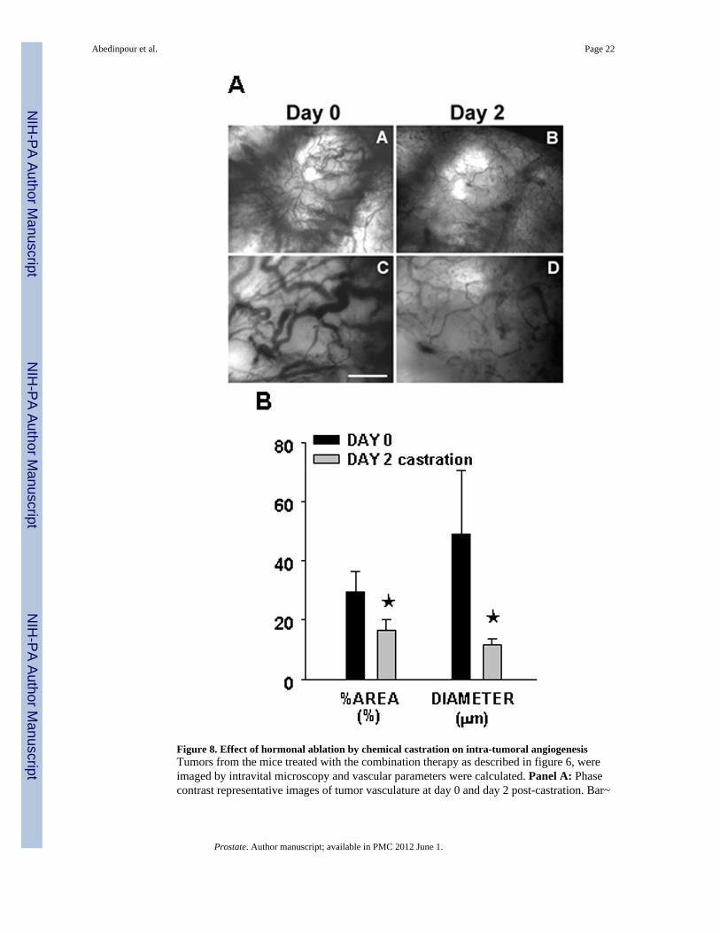
Figure 8. Effect of hormonal ablation by chemical castration on intra-tumoral angiogenesisTumors from the mice treated with the combination therapy as described in figure 6, wereimaged by intravital microscopy and vascular parameters were calculated. Panel A: Phasecontrast representative images of tumor vasculature at day 0 and day 2 post-castration. Bar~
Abedinpour et al. Page 22
Prostate. Author manuscript; available in PMC 2012 June 1.
NIH
-PA Author Manuscript
NIH
-PA Author Manuscript
NIH
-PA Author Manuscript
A,B 500μm, C,D 50μm. Panel B: Graphic representation of the vascular area and the meandiameter of tumor vasculature calculated from intravital microscopy data.
Abedinpour et al. Page 23
Prostate. Author manuscript; available in PMC 2012 June 1.
NIH
-PA Author Manuscript
NIH
-PA Author Manuscript
NIH
-PA Author Manuscript