Regression of prostate tumors upon combination of hormone ablation therapy and celecoxib in vivo
Upload
independentCategory
view
1download
0
RADIO FREQUENCY ABLATION OF SMALL RENAL TUMORS:INTERMEDIATE RESULTS
J. J. HWANG, M. M. WALTHER*, S. E. PAUTLER†, J. A. COLEMAN, J. HVIZDA, JAMESPETERSON, W. M. LINEHAN, and B. J. WOODFrom the Urologic Oncology Branch, National Cancer Institute (JJH, JAC, SEP, JP, WML, MMW)and the Department of Radiology, Clinical Center (JH, BJW), National Institutes of Health, Bethesda,Maryland
AbstractPurpose—With evolving radio frequency technology, the clinical application of radio frequencyablation (RFA) has been actively investigated in the treatment for small renal tumors. We presentour intermediate patient outcomes after RFA.
Materials and Methods—Since January 2001, 17 patients with a total of 24 hereditary renaltumors ranging from 1.2 to 2.85 cm were treated with RFA using the 200 W Cool-tip RF System(Radionics, Burlington, Massachusetts) under laparoscopic (9) or percutaneous (8) guidance and hada minimum 1-year followup. A percutaneous approach was considered unsuitable if kidney tumorswere contiguous to bowel, ureter or large vessels. Treatment eligibility criteria included an averagetumor diameter of less than 3.0 cm, tumor growth during 1 year and solid appearance with contrastenhancement (HU change greater than 20) on computerized tomography (CT). Postoperativefollowup consisted of CT with and without intravenous contrast, and renal function assessment atregular intervals.
Results—Median patient age was 38 years (range 20 to 51). At a median followup of 385 days(range 342 to 691), median tumor or thermal lesion diameter decreased from 2.26 to 1.62 cm (p =0.0013), and only 1 lesion (4%), which was located centrally near the hilum, exhibited contrastenhancement (HU change greater than 10) on CT at 12 months. Of the 15 renal tumors ablatedlaparoscopically, 13 were in direct contact with the bowel and 2 were abutting the ureter, necessitatingmobilization before RFA. Laparoscopic ultrasound was used to guide radio frequency electrodeplacement and monitor the ablation process in these cases. Operative time and intraoperative bloodloss (mean ± standard mean of error) were 243 ± 29 minutes and 67 ± 9 cc, respectively. In 1 patientwhose ureter was adherent to the tumor a ureteropelvic junction obstruction developed afterlaparoscopic RFA, requiring open repair.
Conclusions—At the minimum 1-year followup 23 of 24 ablated tumors lacked contrast uptakeon CT, meeting our radiographic criteria of successful RFA treatment. RFA treatment of small renaltumors using the Radionics system appears to result in superior treatment outcomes compared tothose of earlier series with lower radio frequency power generators. A high wattage generator mightattain more consistent energy deposition with subsequent cell death in the targeted tissue due to lessconvective heat loss.
* Correspondence: UOB/DCT/NCI, Building 10, Room 2B47, 10 Center Drive, MSC 1501, Bethesda, Maryland 20892-1501 (telephone:301-402-2251; FAX: 301-402-0922; e-mail: [email protected]).†Current address: Division of Urology, University of Western Ontario, London, Ontario, Canada.Editor’s Note: This article is the fifth of 5 published in this issue for which category 1 CME credits can be earned. Instructions forobtaining credits are given with the questions on pages 1916 and 1917.Nothing to disclose.
NIH Public AccessAuthor ManuscriptJ Urol. Author manuscript; available in PMC 2008 June 3.
Published in final edited form as:J Urol. 2004 May ; 171(5): 1814–1818.
NIH
-PA Author Manuscript
NIH
-PA Author Manuscript
NIH
-PA Author Manuscript

Keywordslaparoscopy; kidney neoplasms
Recent advances in ablative and imaging technology have led to the application of variousminimally invasive modalities in the treatment of small renal tumors. Although still consideredexperimental in cancer treatment, minimally invasive therapy potentially offers severaladvantages compared to conventional open renal surgery including shorter convalescence,decreased cost, improved cosmesis and decreased postoperative pain.
Radio frequency interstitial tissue ablation (RFA) is a Food and Drug Administration approveddevice for treating soft tissue. Energy is delivered to tissues via specially designed needles,resulting in heating the tissues up to 105C, leading to cell death and coagulation necrosis. Inrecent years an increasing number of tertiary centers have used the RFA based strategy forsmall renal tumors and the early results have been controversial with several centers reportingincomplete tumor ablation in many treated patients.1–7 High failure rates in these studies canbe attributed to several factors such as a learning curve, variable or suboptimal technique, orfailure to achieve adequate “kill” temperature within the targeted tumor. It is now recognizedthat a 50 W radio frequency generator, the system commonly used in early studies, isunderpowered and susceptible to energy loss in highly perfused tissues such as the kidney dueto a heat sink phenomenon from nearby blood vessels and adjacent normal tissue parenchyma.
Previously we published our initial experience with percutaneous RFA of small, hereditarybased renal tumors using a 50 W RFA system and reported modest treatment success at 2months.2 Since January 2001 we have used a 200 W radio frequency (RF) generator for RFAof small renal tumors and we now report our intermediate results (minimum 1-year followup)with this device.
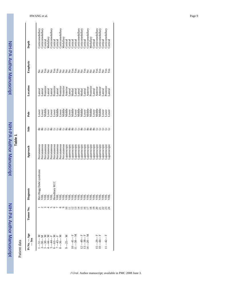
METHODSSince January 2001, 17 patients with a total of 24 hereditary based renal tumors underwentRFA treatment and had a minimum 1-year followup. Patients consisted of 9 men and 8 women,and all but 2 (1 each with the Birt-Hogg-Dubé syndrome and hereditary renal cell carcinoma[RCC]) had von Hippel-Lindau (VHL) disease. Treatment eligibility criteria included anaverage tumor diameter less than 3.0 cm, tumor growth for 1 year, solid appearance withcontrast enhancement (HU change greater than 20) on computerized tomography (CT), and24-hour creatinine clearance greater than 60 ml per minute. There was no selection process inthis patient population, with all participants who met the aforementioned inclusion criteriareceiving treatment. Medullary and central tumors were not excluded from analysis (seefigure). This study was undertaken as part of an Investigational Review Board approved phaseII clinical trial to evaluate radio frequency ablation of renal cancer.
Depending on tumor accessibility as determined by an interventional radiologist (BJW), RFAwas performed under percutaneous (8 patients) or laparoscopic (9 patients) guidance. RFA ofrenal tumor was performed as previously described2 except that the Radionics 200 W systemwas used instead of the RITA 50 W first generation system. The change was made because theformer was the first system available with increased wattage. An additional concern was thepossibility of lacerating blood vessels adjacent to a central tumor using a deployable array.With this system the single needle minimizes the risk of this potential problem.
All percutaneous treatment was performed and monitored with CT (with and without contrastas indicated) and real-time ultrasound in the CT scanning suite with patients under conscioussedation (7) or general anesthesia (1). With a laparoscopic approach, upon isolating the targeted
HWANG et al. Page 2
J Urol. Author manuscript; available in PMC 2008 June 3.
NIH
-PA Author Manuscript
NIH
-PA Author Manuscript
NIH
-PA Author Manuscript

tumor(s) away from adjacent normal structures, laparoscopic ultrasound was used to guide theRF electrode placement and monitor the ablation process in all cases. Renal tumors wereablated using the Cool-tip RF System, a 200 W, 480 kHz RFA generator with a 17.5 gaugestraight electrode probe that has a 3 cm exposed tip and thermocouple at the electrode tip.Continuous internal cycling of chilled saline through the electrode was used to minimize tissuecharring and increased tissue impedance. A single electrode probe powered up to 200 W wasused for each RFA procedure. The probe was repositioned for repeat or overlapping ablationif indicated, and up to 4 ablation cycles of 12 minutes each were applied as necessary. Tissuetemperatures were recorded at 1 and 3 minutes after RFA as a measure of the cooling curveafter treatment, which corresponds to the completeness of ablation (unpublished data). Uponcompletion of tumor RFA, the probe track was cauterized to 70C without saline perfusion sincethe probe was being removed to decrease the potential risk of track seeding.
Postoperative followup consisted of CT with and without intravenous contrast, and renalfunction assessment at 2, 6 and 12 months, semiannually thereafter. A total of 24 urinecreatinine clearance collections were performed before RFA and at 6 and 12 months after RFA.RFA treatment was deemed unsuccessful if followup CT demonstrated contrast enhancement(HU change greater than 10). Intraoperative and postoperative complications were notedaccording to the National Cancer Institute Common Toxicity Criteria.2 The Mann-Whitney UTest was applied to determine the significance between any 2 groups in comparison. All datalisted represent mean plus or minus standard mean of error.
RESULTSMedian patient age was 38 years (range 20 to 51) at RFA treatment. Patient and tumorcharacteristics are listed in table 1. At a median followup of 385 days (range 342 to 691) meantumor or thermal lesion diameter decreased from 2.26 ± 0.10 to 1.62 ± 0.11 cm (p = 0.0013).Of the 24 tumors treated only 1 (4%) exhibited contrast enhancement (HU change greater than10) and met radiographic criteria for tumor recurrence. This case was notable because the tumorwas located centrally adjacent to the renal hilum, and required laparoscopic ultrasonographyfor tumor visualization and ablation. The recurrence occurred as a rim of brightly enhancingtissue (HU change of 211) along the tumor/hilar interface. In the remaining 23 tumors meanHU change changed from 69 ± 36 to 1 ± 6 after RFA treatment (p < 0.0001). Treatmentparameters and results are listed in table 2.
Of the 15 renal tumors ablated laparoscopically 13 were in direct contact with bowel and 2were abutting the ureter, necessitating mobilization before RFA. Laparoscopic ultrasound wasused to assess RF electrode placement and monitor the ablation process in all cases. Meanoperative time and intraoperative blood loss were 243 ± 29 minutes and 67 ± 9 cc, respectively.Average postoperative stay was 2.9 days (range 2 to 5).
There were no immediate postoperative complications excluding 2 cases of transient grosshematuria after laparoscopic guided RFA of centrally located lesions. One patient who wisheda noninvasive treatment had a ureter densely adherent to the adjacent tumor which preventedlaparoscopic mobilization. An asymptomatic ureteropelvic junction (UPJ) obstructiondeveloped in this patient, who underwent an open surgical repair after 9 months of conservativemanagement. The previously ablated tumor in this patient was excised at UPJ repair and therewas no evidence of viable tumor tissue on pathological examination. There was no statisticallysignificant change in renal function after RFA treatment in this patient cohort. Mean 24-hourcreatinine clearance before and after RFA treatment was 115 ± 9 and 102 ± 7 ml per minuterespectively.
HWANG et al. Page 3
J Urol. Author manuscript; available in PMC 2008 June 3.
NIH
-PA Author Manuscript
NIH
-PA Author Manuscript
NIH
-PA Author Manuscript
DISCUSSIONRenal tumors are being detected at increasing rates due to the widespread use of modernimaging techniques. Typically these tumors are found incidentally and tend to be small withlower stage, yielding better survival outcomes than tumors diagnosed in symptomatic patients.8 Recent advances in ablative technology have led to the application of radio frequencyinterstitial tissue ablation as a minimally invasive strategy for select renal tumors. There are 4systems with United States Food and Drug Administration clearance for soft tissue ablationwith radio frequency energy. The cytotoxic mechanism involves desiccation due to highintracellular temperatures. The ablation process is continuously monitored using temperatureand/or impedance feedback aided by various imaging modalities. Imaging and immediatetemperature, and impedance monitoring provide predictable real-time control of tissueablation.
The majority of human experience with RFA has been in the management of liver neoplasms.9, 10 Since the introduction of renal RFA by Zlotta et al in 199711 early results of RFA forrenal tumors have been controversial. Rendon et al from Canada reported their experience withpercutaneous RFA of small renal tumors (mean 2.4 cm) using the Model 2000 System (RadioTherapeutics Corporation, Sunnyvale, California) followed by immediate or delayednephrectomy in 10 patients.3 The treatment protocol developed in the porcine model12 allowedfor the maximum power input of 75 W for the 3 cm electrode. In the immediate group 5 tumorsin 4 patients were treated with open RFA followed immediately by partial or radicalnephrectomy. In the delayed group 6 tumors were initially treated with percutaneous RFA 7days before open resection. On pathological examination 4 of the 5 tumors in the immediategroup and 3 of the 6 tumors in the delayed group demonstrated persistent viable cancer cells.It is not clear whether the treatment resulted in satisfactory intratumoral temperatures sincetissue temperature during RFA was not reported.
Early pathological and histological examination may be misleading, and tissue may look viablewhen cells are not actually alive. For example, hematoxylin and eosin stains may give the falseimpression of viability in nonsurvival studies or when excision occurs immediately after RFA.Vital stains like nicotinamide adenine dinucleotide diaphorase or tetrazolium may provide amore accurate assessment of tissue viability immediately after thermal insult.5, 13 The delayedgroup treatment failures may represent under treated tumors due to a combination of factors.The kidney is a highly perfused organ, and the convection of heat due to overall perfusion andinto blood vessels may result in the under treatment of tumors in the kidney. This finding isespecially true when ablation algorithms are applied that were developed specifically for livertissue or for less perfused tissue. Kidney perfusion is approximately 4 times that of the liver.14 This difference drastically alters the bioheat equation (which estimates the amount of energywhich can be deposited in tissue) by increasing the energy lost term.14 In addition, the firstgeneration generator (less than 200 W) may not provide enough power to overcome the heatsink effect from the high tissue perfusion.
Similar unfavorable RFA treatment findings were also noted by the Lahey Clinic group.5Michaels et al performed RFA in 15 patients with a total of 20 tumors (mean 2.4 cm) followedby partial nephrectomy.5 Ablated with a RITA Model 500 (the first 15 cases) or Model 1500(the last 5 cases) system, tumors demonstrated viable tumor cells on pathological inspection(hematoxylin and eosin and/or nicotinamide adenine dinucleotide diaphorase staining). Thegenerator delivered up to 110 W of power and tumors were heated to 90 to 110C. It remainsto be elucidated whether incomplete tumor destruction was a result of lack of time betweenRFA and histological analysis. In addition, if the generator only delivered 110 W of power,the technique or algorithm may need to be further optimized for the kidney to allow deliveryof more of the available power.
HWANG et al. Page 4
J Urol. Author manuscript; available in PMC 2008 June 3.
NIH
-PA Author Manuscript
NIH
-PA Author Manuscript
NIH
-PA Author Manuscript

Differences in kidney perfusion or tissue dielectrics (thermal and electrical conductivities) mayaccount for treatment effect shortcomings as well. We have tried to compensate for this effectby treating tumors more than once in similar locations. Normally in the liver 1, 12-minutetreatment might suffice for a 3 cm thermal lesion. In the kidney we have found that it may take2 or 3, 12-minute overlapping treatment sessions to treat the same volume of tissue. Thesemodifications are somewhat subjective and difficult to translate to general practice. Thus thereis a need for development of more standardized tissue or organ specific algorithms to facilitatetranslation of success with this technology in the kidney to the community.
In contrast to the previously mentioned findings 2 recent publications report excellent resultsfrom RFA treatment of renal tumors using high wattage generators. Jacomides et al used aRITA Model 1500X system to treat 17 renal tumors (mean 1.96 cm) laparoscopically.7 Fiveablated lesions were immediately excised for pathological analysis. At a mean followup of 9.8months (range 1.5 to 22) all treated tumors left in situ remained recurrence-free on followupimaging. Of the resected tumors only 1 lesion had a focally positive margin. Matlaga et al alsoreported similar results from an open RFA treatment program (using the Radionics Cool-tipSystem) of 10 tumors followed by immediate partial or radical nephrectomy.6 Mean tumorsize was 3.2 cm (range 1.4 to 8.0). Of the 10 tumors treated 8 were completely ablated onpathological review. Two incompletely treated tumors (a 1 cm and an 8 cm tumor) were notablefor insufficient intratumoral temperature achievement after RFA.
Previously we reported our initial results on RFA of small hereditary based renal tumors usingthe RITA Model 500 electrosurgical generator.2 We reported on 24 ablations performed in 21patients with renal tumors and at 2-month followup a majority of tumors (19 of 24, 79%) ceasedto enhance on contrast CT.2 Further followup is now available on these patients treated withthe old generation RITA device (unpublished data). At a median followup of 24 months 10 ofthe ablated tumors (40%) demonstrated contrast enhancement (HU change greater than 10) onfollowup CT (unpublished data), 9 of the tumors have been surgically removed andpathologically confirmed as viable clear cell carcinoma and the remaining case has been re-treated with RFA using the higher wattage Radionics system with success based on imagingcriteria.
With the 50 W RITA RFA system we have noted that tumor recurrence or persistence wasoften observed along the medullary portion of the ablated tumor, usually at the interfacebetween the tumor and renal hilum (unpublished data). RFA may be less effective in medullarylesions than in cortical, peripheral or exophytic lesions that have less convective heat loss andmore insulating effect from perirenal fat. Low wattage electrosurgical generators are moresusceptible to rapid energy loss since heat propagates toward the periphery of the thermallesion. Energy distribution is further impeded by the development of tissue charring aroundthe probe tip with a subsequent increase in tissue impedance. Potential dielectric disparities inelectrical and thermal conductivity between cortex and medulla may also affect the ablationresponse of central tumors. Compared to centrally located tumors, exophytic, cortical tumorsappear to respond well to RFA, likely due to the heat insulating effect by surrounding fat.Therefore, tumor location may have an important role when selecting a suitable candidate, andthis factor may be especially important with low wattage electrosurgical generators. Generallyspeaking, in our experience low watt generators should be avoided in the treatment of RCC.
There were 5 tumors which did not achieve a temperature greater than 70C after treatment(table 2). Tumor location was in the cortex and the medulla. It is assumed that blood vesselsadjacent to the tumors acted as a heat sink and resulted in an altered temperature profile. Tumorsnot heated to more than 70C were re-treated. The needle was sometimes repositioned if a vesselwas next to the temperature sensor on its tip. Patients with larger tumors or with contiguous
HWANG et al. Page 5
J Urol. Author manuscript; available in PMC 2008 June 3.
NIH
-PA Author Manuscript
NIH
-PA Author Manuscript
NIH
-PA Author Manuscript
tumors were treated with overlapping RFA fields. All 5 tumors appeared well treated withoutrecurrence on followup.
Our experience with this 200 W system which includes treating 13 centrally located tumorswith a deep medullary component has been excellent, with only 1 of 24 ablated tumors showingradiographic evidence of recurrence at 12 months of followup. Differences in patient selection,RFA system used and procedure technique preclude any direct comparison of treatmentoutcomes from various RFA series reported in the literature, but our data suggest that highwattage RF generators may be less susceptible to the heat sink phenomenon and better suitedto overcome anatomical factors such as tumor location and characteristics. With furtherimprovements in RF technology and equipment, more consistent, favorable outcomes may bethe result. The exact role of RFA will soon become more clearly defined as part of the minimallyinvasive therapy arsenal for renal tumors.
CONCLUSIONSWith evolving technology of radio frequency generators and improved probe design, RFA isa promising treatment alternative for small renal tumors. The 200 W power results in superiortreatment outcomes compared to low wattage systems, likely due to less convective heat lossand more consistent energy distribution throughout the tumor. Although promising in thetreatment of cancer, RFA remains somewhat experimental in the kidney until long-termefficacy is validated. Further development of kidney specific techniques and algorithms shouldimprove outcomes. At this time this technique should be reserved for experienced centers inspecific clinical scenarios.
References1. Gervais DA, McGovern FJ, Wood BJ, Goldberg SN, McDougal WS, Mueller PR. Radio-frequency
ablation of renal cell carcinoma: early clinical experience. Radiology 2000;217:665. [PubMed:11110926]
2. Pavlovich CP, Walther MM, Choyke PL, Pautler SE, Chang R, Linehan WM, et al. Percutaneous radiofrequency ablation of small renal tumors: initial results. J Urol 2002;167:10. [PubMed: 11743264]
3. Rendon RA, Kachura JR, Sweet JM, Gertner MR, Sherar MD, Robinette M, et al. The uncertainty ofradio frequency treatment of renal cell carcinoma: findings at immediate and delayed nephrectomy. JUrol 2002;167:1587. [PubMed: 11912369]
4. de Baere T, Kuoch V, Smayra T, Dromain C, Cabrera T, Court B, et al. Radio frequency ablation ofrenal cell carcinoma: preliminary clinical experience. J Urol 2002;167:1961. [PubMed: 11956417]
5. Michaels MJ, Rhee HK, Mourtzinos AP, Summerhayes IC, Silverman ML, Libertino JA. Incompleterenal tumor destruction using radio frequency interstitial ablation. J Urol 2002;168:2406. [PubMed:12441927]
6. Matlaga BR, Zagoria RJ, Woodruff RD, Torti FM, Hall MC. Phase II trial of radio frequency ablationof renal cancer: evaluation of the kill zone. J Urol 2002;168:2401. [PubMed: 12441926]
7. Jacomides L, Ogan K, Watumull L, Cadeddu JA. Laparoscopic application of radio frequency energyenables in situ renal tumor ablation and partial nephrectomy. J Urol 2003;169:49. [PubMed: 12478100]
8. Luciani LG, Cestari R, Tallarigo C. Incidental renal cell carcinoma-age and stage characterization andclinical implications: study of 1092 patients (1982–1997). Urology 2000;56:58. [PubMed: 10869624]
9. McGahan JP, Brock JM, Tesluk H, Gu WZ, Schneider P, Browning PD. Hepatic ablation with use ofradiofrequency electrocautery in the animal model. J Vasc Interv Radiol 1992;3:291. [PubMed:1627876]
10. Rossi S, Di Stasi M, Buscarini E, Quaretti P, Garbagnati F, Squassante L, et al. Percutaneous RFinterstitial thermal ablation in the treatment of hepatic cancer. AJR Am J Roentgenol 1996;167:759.[PubMed: 8751696]
HWANG et al. Page 6
J Urol. Author manuscript; available in PMC 2008 June 3.
NIH
-PA Author Manuscript
NIH
-PA Author Manuscript
NIH
-PA Author Manuscript
11. Zlotta AR, Wildschutz T, Raviv G, Peny MO, van Gansbeke D, Noel JC, et al. Radiofrequencyinterstitial tumor ablation (RITA) is a possible new modality for treatment of renal cancer: ex vivoand in vivo experience. J Endourol 1997;11:251. [PubMed: 9376843]
12. Rendon RA, Gertner MR, Sherar MD, Asch MR, Kachura JR, Sweet J, et al. Development of aradiofrequency based thermal therapy technique in an in vivo porcine model for the treatment ofsmall renal masses. J Urol 2001;166:292. [PubMed: 11435889]
13. Crowley JD, Shelton J, Iverson AJ, Burton MP, Dalrymple NC, Bishoff JT. Laparoscopic andcomputed tomography-guided percutaneous radiofrequency ablation of renal tissue: acute andchronic effects in an animal model. Urology 2001;57:976. [PubMed: 11337311]
14. Duck, FA. Physical Properties of Tissue. London: Academic Press Limited; 1990. Appendix A: Tissueperfusion rates.
HWANG et al. Page 7
J Urol. Author manuscript; available in PMC 2008 June 3.
NIH
-PA Author Manuscript
NIH
-PA Author Manuscript
NIH
-PA Author Manuscript

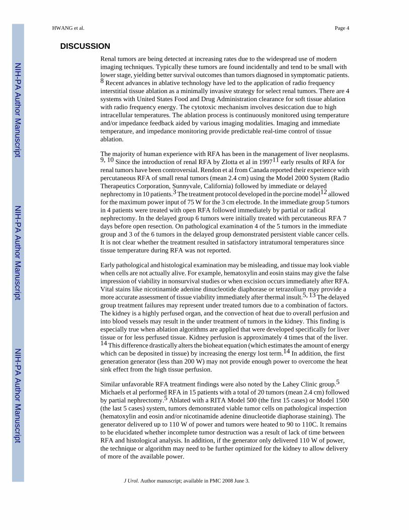
Figure.A, medullary tumor (arrow) is shown in lower pole of left kidney surrounded by abundantperinephric adipose tissue. Tumor was ablated percutaneously. B, year after RFA CT revealsnonenhancing lesion that has decreased in size.
HWANG et al. Page 8
J Urol. Author manuscript; available in PMC 2008 June 3.
NIH
-PA Author Manuscript
NIH
-PA Author Manuscript
NIH
-PA Author Manuscript
NIH
-PA Author Manuscript
NIH
-PA Author Manuscript
NIH
-PA Author Manuscript
HWANG et al. Page 9Ta
ble
1Pa
tient
dat
a
Pt N
o. —
Age
— S
exT
umor
No.
Dia
gnos
isA
ppro
ach
Side
Pole
Loc
atio
nE
xoph
ytic
Dep
th
1 —
51—
M1
Birt
-Hog
g D
ubé
synd
rom
ePe
rcut
aneo
usR
tLo
wer
Late
ral
No
Cor
ticom
edul
lary
2—30
— M
2V
HL
Perc
utan
eous
Rt
Low
erLa
tera
lN
oC
ortic
omed
ulla
ry3
—30
— M
3V
HL
Perc
utan
eous
LtM
iddl
ePo
ster
ior
Yes
Cor
tical
4 —
44—
M4
VH
LPe
rcut
aneo
usLt
Low
erLa
tera
lN
oM
edul
lary
5 —
44—
F5
VH
LPe
rcut
aneo
usR
tLo
wer
Late
ral
No
Cor
ticom
edul
lary
6 —
63—
M6
Her
edita
ry R
CC
Perc
utan
eous
LtLo
wer
Post
erio
rY
esC
ortic
al7
—43
— F
7V
HL
Perc
utan
eous
LtM
iddl
eLa
tera
lY
esC
ortic
al8
—36
— M
8V
HL
Perc
utan
eous
Rt
Low
erPo
ster
ior
No
Cor
ticom
edul
lary
9V
HL
Perc
utan
eous
LtM
iddl
ePo
ster
ior
No
Med
ulla
ry9
—25
— M
10V
HL
Lapa
rosc
opic
Rt
Mid
dle
Ant
erio
rN
oC
ortic
al11
VH
LLa
paro
scop
icR
tLo
wer
Late
ral
No
Cor
tical
10 —
41—
F12
VH
LLa
paro
scop
icR
tM
iddl
eM
edia
lY
esC
ortic
al11
—38
— M
13V
HL
Lapa
rosc
opic
LtLo
wer
Late
ral
Yes
Cor
tical
14V
HL
Lapa
rosc
opic
LtM
iddl
eLa
tera
lN
oC
ortic
omed
ulla
ry12
—49
— F
15V
HL
Lapa
rosc
opic
Rt
Mid
dle
Med
ial
No
Cor
ticom
edul
lary
13 —
38—
F16
VH
LLa
paro
scop
icLt
Low
erLa
tera
lY
esC
ortic
al14
—20
— M
17V
HL
Lapa
rosc
opic
Rt
Mid
dle
Ant
erio
rN
oC
ortic
omed
ulla
ry18
VH
LLa
paro
scop
icR
tM
iddl
ePo
ster
ior
No
Med
ulla
ry19
VH
LLa
paro
scop
icR
tLo
wer
Late
ral
No
Cor
tical
15 —
29—
F20
VH
LLa
paro
scop
icLt
Mid
dle
Ant
erio
rY
esC
ortic
omed
ulla
ry10
—44
— F
21V
HL
Lapa
rosc
opic
LtM
iddl
eA
nter
ior
Yes
Cor
ticom
edul
lary
22V
HL
Lapa
rosc
opic
LtLo
wer
Late
ral
No
Cor
tical
11 —
42—
F23
VH
LLa
paro
scop
icLt
Low
erPo
ster
ior
Yes
Cor
ticom
edul
lary
24V
HL
Lapa
rosc
opic
LtLo
wer
Late
ral
Yes
Cor
tical
J Urol. Author manuscript; available in PMC 2008 June 3.

NIH
-PA Author Manuscript
NIH
-PA Author Manuscript
NIH
-PA Author Manuscript
HWANG et al. Page 10Ta
ble
2Tr
eatm
ent p
aram
eter
s
Tum
or N
o.R
FA T
empe
ratu
reG
reat
er T
han
70C
*
No.
Cyc
les
Day
s Fol
low
upPr
e-R
FA S
ize
(cm
)Po
st-R
FA S
ize(
cm)
HU
Cha
nge
Bef
ore
RFA
HU
Cha
nge
Afte
rR
FA
1Y
es2
404
1.75
1.65
109
82
Yes
434
22.
682.
2578
13
Yes
242
71.
431.
8466
24
Yes
437
22.
392.
2642
−75
Yes
238
52.
051.
4557
16
Yes
257
22.
041.
6378
47
Yes
137
02.
292.
3986
68
Yes
257
82.
652.
3048
−19
Yes
345
42.
512.
3540
−710
No
136
22.
641.
9076
011
No
236
22.
241.
6017
20
12*
Yes
229
4†2.
85N
ot a
pplic
able
Not
app
licab
leN
ot a
pplic
able
13Y
esN
ot a
pplic
able
355
1.80
1.39
363
14Y
esN
ot a
pplic
able
355
2.59
1.39
533
15Y
es4
363
2.79
0.99
156
211
16Y
es3
574
2.71
2.41
782
17N
o2
356
2.09
1.41
539
18N
o1
356
2.29
1.40
64−2
19Y
es3
356
1.45
0.75
40−1
520
No
453
72.
241.
1547
521
Yes
253
71.
691.
5076
522
Yes
253
71.
90N
o lo
nger
vis
ible
7223
Yes
269
12.
551.
8827
−624
Yes
269
11.
20N
o lo
nger
vis
ible
27
Size
exp
ress
ed in
geo
met
ric m
ean.
* RFA
tem
pera
ture
mea
sure
d tu
mor
tem
pera
ture
1 m
inut
e af
ter R
FA tr
eatm
ent.
† Stat
us a
fter U
PJ re
pair.
J Urol. Author manuscript; available in PMC 2008 June 3.
Copyright © 2022 FDOKUMEN


![[INTERMEDIATE 3D MODELING IN TINKERCAD]](https://static.fdokumen.com/doc/165x107/63336b2d4cd921f2410cdab7/intermediate-3d-modeling-in-tinkercad.jpg)