DNA methylation and its role in the trophoblast cell lineage
9
DNA methylation and its role in the trophoblast cell lineage SATOSHI TANAKA*, MOMO O. NAKANISHI 1 and KUNIO SHIOTA Laboratory of Cellular Biochemistry, Department of Animal Resource Sciences/Veterinary Medical Sciences, The University of Tokyo, Tokyo, Japan ABSTRACT DNA methylation functions as cellular memory beyond generations of cells and is involved in many biological processes. Because of its relatively stable nature compared with the transcriptome, the DNA methylation profile of cells can also be used to evaluate developmental similarity and cellular phenotypes. Recent insights into 5-hydroxymethylcytosine have started to reshape our view of the epigenetic regulation of mammalian development. Both global DNA meth- ylation and hydroxymethylation levels change dynamically during preimplantation embryogenesis. It is known that DNA methylation plays an essential role in embryonic cell fate restriction, whereas its role in trophoblast development requires further research. Two distinct blastocyst-derived stem cell lines, embryonic stem (ES) cells and trophoblast stem (TS) cells, are used to study the epigenetic mechanisms underlying cell lineage maintenance and the regulation of cell differentiation. Such studies will allow us to understand the details of the epigenetic landscape of trophoblast develop- ment, which should offer valuable information for managing pregnancy-related diseases in humans. KEY WORDS: DNA methylation, blastocyst, ICM, TE, TS cell Introduction Starting from a single totipotent cell (i.e., a fertilized egg), the process of mammalian development generates more than 200 different types of cells (Alberts et al., 2007), almost all of which possess an identical genomic DNA sequence. In addition to the networks of transcription factors, by changing the chromatin structure, epigenetic systems play a fundamental role in the cell type-specific use of genetic information stored in genomic DNA. DNA methylation is one of the best-studied epigenetic modifications and is involved in many biological processes such as repression of transcription, genome imprinting, suppression of retrotransposons, and X chromosome inactivation (Bird, 2002; Jaenisch and Bird, 2003; Smith and Meissner, 2013). DNA methylation at cytosine bases has long been regarded as the only covalent modification of mammalian DNA. However, recent discoveries of enzymes that catalyze the oxidation of methylated cytosine to produce 5-hydroxymethylcytosine (5hmC) have started to reshape our view of the epigenetic landscape of mammalian development. The first cell differentiation in mammalian develop- ment segregates the trophoblast cell lineage from embryonic cell lineage, resulting in the formation of the trophectoderm (TE) and inner cell mass (ICM) at the early blastocyst stage. After implanta- tion of the blastocyst to the uterus, the TE produces trophoblast cells that constitute most of the placenta on the fetal side and an outermost membrane that surrounds the developing fetus. The Int. J. Dev. Biol. 58: 231-238 (2014) doi: 10.1387/ijdb.140053st www.intjdevbiol.com *Address correspondence to: Satoshi Tanaka. Laboratory of Cellular Biochemistry, Animal Resource Sciences/Veterinary Medical Sciences,The University ofTokyo, 1-1-1 Yayoi, Bunkyo-ku, Tokyo 113-8657, Japan. Tel: 81-3-5841-5372. Fax: 81-3-5841-8014 E-mail: [email protected] Final, author-corrected PDF published online: 8 July 2014 ISSN: Online 1696-3547, Print 0214-6282 © 2014 UBC Press Printed in Spain Abbreviations used in this paper: 5hmC, 5-hydroxymethylcytosine; 5mC, 5-methyl- cytosine; Dnmt, DNA methyltransferase; Dnmt TKO, triple knockout of Dnnt1, Dnmt3a and Dnmt3b; Dnmt TKO-NT, nuclear transfer from Dnmt TKO cell; ES cell, embryonic stem cell; ICM, inner cell mass; NT, nuclear transfer; TE, trophectoderm; TS cell, trophoblast stem cell; T-DMR, tissue-dependent and differentially methylated region; Tet, ten-eleven translocation. ICM gives rise to three germ layers and germ cells. In mice, stem cell lines have been derived successfully from these two tissues, thereby recapitulating their developmental potency: trophoblast stem (TS) cells from TE and the embryonic stem (ES) cells from the ICM (Evans and Kaufman, 1981; Martin, 1981; Tanaka et al., 1998). Much of the knowledge about the epigenetic status of the trophoblast and embryonic cell lineages has been obtained from research comparing these two distinct stem cells. In this review, we first summarize basic information about DNA methylation and hydroxymethylation. We then focus on recent insights into these epigenetic modifications obtained from ES and TS cells and early embryos, and we discuss the possible involvement of these epigenetic modifications in the development and function of the trophoblast cell lineage. DNA methylation and further oxidation In mammals, DNA methylation occurs predominantly on cyto- sine bases in 5’–CG–3’ dinucleotide (CpG) sequences to produce
Transcript of DNA methylation and its role in the trophoblast cell lineage
DNA methylation and its role in the trophoblast cell lineageSATOSHI TANAKA*, MOMO O. NAKANISHI1 and KUNIO SHIOTA
Laboratory of Cellular Biochemistry, Department of Animal Resource Sciences/Veterinary Medical Sciences, The University of Tokyo, Tokyo, Japan
ABSTRACT DNA methylation functions as cellular memory beyond generations of cells and is involved in many biological processes. Because of its relatively stable nature compared with the transcriptome, the DNA methylation profile of cells can also be used to evaluate developmental similarity and cellular phenotypes. Recent insights into 5-hydroxymethylcytosine have started to reshape our view of the epigenetic regulation of mammalian development. Both global DNA meth-ylation and hydroxymethylation levels change dynamically during preimplantation embryogenesis. It is known that DNA methylation plays an essential role in embryonic cell fate restriction, whereas its role in trophoblast development requires further research. Two distinct blastocyst-derived stem cell lines, embryonic stem (ES) cells and trophoblast stem (TS) cells, are used to study the epigenetic mechanisms underlying cell lineage maintenance and the regulation of cell differentiation. Such studies will allow us to understand the details of the epigenetic landscape of trophoblast develop-ment, which should offer valuable information for managing pregnancy-related diseases in humans.
KEY WORDS: DNA methylation, blastocyst, ICM, TE, TS cell
Introduction
Starting from a single totipotent cell (i.e., a fertilized egg), the process of mammalian development generates more than 200 different types of cells (Alberts et al., 2007), almost all of which possess an identical genomic DNA sequence. In addition to the networks of transcription factors, by changing the chromatin structure, epigenetic systems play a fundamental role in the cell type-specific use of genetic information stored in genomic DNA. DNA methylation is one of the best-studied epigenetic modifications and is involved in many biological processes such as repression of transcription, genome imprinting, suppression of retrotransposons, and X chromosome inactivation (Bird, 2002; Jaenisch and Bird, 2003; Smith and Meissner, 2013).
DNA methylation at cytosine bases has long been regarded as the only covalent modification of mammalian DNA. However, recent discoveries of enzymes that catalyze the oxidation of methylated cytosine to produce 5-hydroxymethylcytosine (5hmC) have started to reshape our view of the epigenetic landscape of mammalian development. The first cell differentiation in mammalian develop-ment segregates the trophoblast cell lineage from embryonic cell lineage, resulting in the formation of the trophectoderm (TE) and inner cell mass (ICM) at the early blastocyst stage. After implanta-tion of the blastocyst to the uterus, the TE produces trophoblast cells that constitute most of the placenta on the fetal side and an outermost membrane that surrounds the developing fetus. The
Int. J. Dev. Biol. 58: 231-238 (2014)doi: 10.1387/ijdb.140053st
www.intjdevbiol.com
*Address correspondence to: Satoshi Tanaka. Laboratory of Cellular Biochemistry, Animal Resource Sciences/Veterinary Medical Sciences, The University of Tokyo, 1-1-1 Yayoi, Bunkyo-ku, Tokyo 113-8657, Japan. Tel: 81-3-5841-5372. Fax: 81-3-5841-8014 E-mail: [email protected]
Final, author-corrected PDF published online: 8 July 2014
ISSN: Online 1696-3547, Print 0214-6282© 2014 UBC PressPrinted in Spain
Abbreviations used in this paper: 5hmC, 5-hydroxymethylcytosine; 5mC, 5-methyl-cytosine; Dnmt, DNA methyltransferase; Dnmt TKO, triple knockout of Dnnt1, Dnmt3a and Dnmt3b; Dnmt TKO-NT, nuclear transfer from Dnmt TKO cell; ES cell, embryonic stem cell; ICM, inner cell mass; NT, nuclear transfer; TE, trophectoderm; TS cell, trophoblast stem cell; T-DMR, tissue-dependent and differentially methylated region; Tet, ten-eleven translocation.
ICM gives rise to three germ layers and germ cells. In mice, stem cell lines have been derived successfully from these two tissues, thereby recapitulating their developmental potency: trophoblast stem (TS) cells from TE and the embryonic stem (ES) cells from the ICM (Evans and Kaufman, 1981; Martin, 1981; Tanaka et al., 1998). Much of the knowledge about the epigenetic status of the trophoblast and embryonic cell lineages has been obtained from research comparing these two distinct stem cells. In this review, we first summarize basic information about DNA methylation and hydroxymethylation. We then focus on recent insights into these epigenetic modifications obtained from ES and TS cells and early embryos, and we discuss the possible involvement of these epigenetic modifications in the development and function of the trophoblast cell lineage.
DNA methylation and further oxidation
In mammals, DNA methylation occurs predominantly on cyto-sine bases in 5’–CG–3’ dinucleotide (CpG) sequences to produce
232 S. Tanaka et al.
5-methylcytosine (5mC). Non-CpG methylation has also been detected at some specific loci (Ichiyanagi et al., 2013; Imamura et al., 2005; Nishino et al., 2011), but its biological significance in mammals is not known. Non-CpG methylation appears to be more enriched in the genome of germ cells and ES cells (Ramsahoye et al., 2000; Shirane et al., 2013). Although CpG methylation is associated mostly with repression of gene expression (Weber et al., 2007; Yagi et al., 2008, 2012), the presence of methylated promoters with low CpG content at transcriptionally active genes has also been noted by genome-wide DNA methylation and gene expression analyses (Weber et al., 2007). This suggests that sparse CpG methylation does not interfere with the transcription machinery and/or that CpG methylation in a particular sequence context activates transcription. The latter is not totally improbable because it has been shown that the CpG methylation within CRE sequences (TGACGTCA) creates a binding site for the transcription factor C/EBPa and results in activation of CRE sequence-associated tissue-specific genes (Rishi et al., 2010).
Cytosine to 5mC conversion is catalyzed by three members of the DNA methyltransferase (Dnmt) family. Dnmt1, referred to as “maintenance methyltransferase,” prefers hemimethylated DNA to nonmethylated DNA as its substrate in vitro (Gruenbaum et al., 1982) and localizes at replication fork through interaction with its chaperone protein Np95/Uhrf1 (Sharif et al., 2007). This ensures immediate and accurate copying of the CpG methylation pattern from a parent strand to the newly synthesized daughter strand during replication. Disappearance of Dnmt1 (or inactivation of its enzymatic activity) therefore leads to a gradual dilution of DNA methylation in dividing cells (theoretically, 1/2 per cell cycle) in a replication-dependent manner, which is called “passive demethylation.”
By contrast, two other members of the Dnmt family, Dnmt3a and Dnmt3b, are essential for methylation of nonmethylated DNA (Okano et al., 1999) and are thus called “de novo methyltransfer-ases.” There are at least two isoforms of Dnmt3a in the human and mouse: a long isoform DNMT3A1/Dnmt3a1 and a short isoform DNMT3A2/Dnmt3a2. Both isoforms have enzymatic activity, but show different localization patterns in the nucleus. Dnmt3a1 is more concentrated at the densely DAPI-stained heterochromatic region of the nucleus, whereas Dnmt3a2 appears to be excluded from the heterochromatic region, which suggests that these Dnmt3a isoforms have distinct genomic targets (Chen et al., 2002). Dnmt3L, another member of the Dnmt family, does not contain a catalytic domain conserved among other members and shows no Dnmt activity on its own, but it is also essential for de novo DNA methylation in germ cells (Hata et al., 2002, 2006). Dnmt3L has been shown to interact with both Dnmt3a and Dnmt3b, and to stimulate their activity in vitro (Gowher et al., 2005, Suetake et al., 2004). It has been reported that Dnmt3L physically interacts with Dnmt3a2, but not with Dnmt3a1 or Dnmt3b, in ES cells (Nimura et al., 2006).
Conversion of 5mC back to unmodified cytosine independent of DNA replication, so-called “active demethylation,” takes place on the genome-wide scale during mammalian development. Very recently, this was proven experimentally to occur in primordial germ cells (Kawasaki et al., 2014). Another massive active demethylation had been thought to occur soon after the fertilization on the paternal genome, although this is now somewhat controversial (see below). Identification of genuine DNA demethylase in mammals has been a long-standing issue in the field of epigenetic research. It was found recently that 5mC can be successively oxidized to 5hmC, 5-form-
ylcytosine (5fC), and 5-carboxylcytosine (5caC) by the ten-eleven translocation (Tet) family of Fe(II) and 2-oxoglutarate-dependent DNA dioxygenases (He et al., 2011; Ito et al., 2010; Ito et al., 2011; Tahiliani et al., 2009). Three members of the Tet family, Tet1–3, have been identified and shown to play essential roles in diverse biological processes. Because 5fC and 5caC can be excised and repaired to regenerate unmodified cytosine by the thymine DNA glycosidase and base excision repair pathways, 5hmC is now regarded as an essential intermediate of active demethylation (Kohli and Zhang, 2013; Wu and Zhang, 2014).
DNA methylation profile of cells as an identifier of cell type
Although the DNA methylation pattern of cells is stably transmit-ted from parent cell to daughter cells, a certain portion of CpGs change their methylation status as the cells differentiate, resulting in a renovation of the DNA methylation pattern of the genome, or DNA methylation profile, which is unique to each cell type. Initial attempts to identify such CpGs were pursued by restriction land-mark genomic scanning (RLGS) using a methylation-sensitive restriction enzyme NotI as a methylation sensor (Ohgane et al., 1998, 2002). By comparing ES cells, embryonic germ (EG) cells, TS cells, germ cells, and several somatic tissues, RLGS identified >200 tissue-dependent and differentially methylated regions (T-DMRs) out of ~1,500 analyzable NotI sites (Shiota et al., 2002). Although NotI sites tend to locate within CpG islands or CpG-rich regions, T-DMRs were distributed disproportionately in the non-CpG island loci (Sakamoto et al., 2007). Interestingly, even with fewer NotI sites compared with recent deep sequencing-based methylome analyses, hierarchical clustering of the methylation profiles could be used to define developmental similarity and cel-lular phenotypes (Sakamoto et al., 2007), demonstrating that the DNA methylation profile of cells is a powerful index for evaluating the relatedness of different cell types.
This concept was expanded further and confirmed by show-ing that EG cells and iPS cells show a DNA methylation profile very similar to but still distinctive from that of ES cells (Sato et al., 2010). A series of studies has identified T-DMRs that are differen-tially methylated between TS cells and ES cells (TS–ES T-DMRs) (Nakanishi et al., 2012; Shiota et al., 2002). One example is the T-DMRs at the pluripotency-related Pou5f1 (Oct4) gene locus that are heavily methylated in TS cells when the gene is silent but are hypomethylated in ES cells (Hattori et al., 2004). Treatment with the Dnmt inhibitor 5-aza-2’-deoxycytidine (5-aza-dC) and Dnmt1 deficiency caused the ectopic expression of Oct4 in TS cells and in placental tissue, respectively, showing that DNA methylation plays an essential role in the suppression of Oct4 expression (Hattori et al., 2004). Another pluripotency-related gene, Nanog, has also been reported to have T-DMRs and to be regulated by DNA methylation (Hattori et al., 2007). Thus, DNA methylation of T-DMRs around some specific genes should play important roles in restricting cell potency.
DNA methylation dynamics during embryogenesis
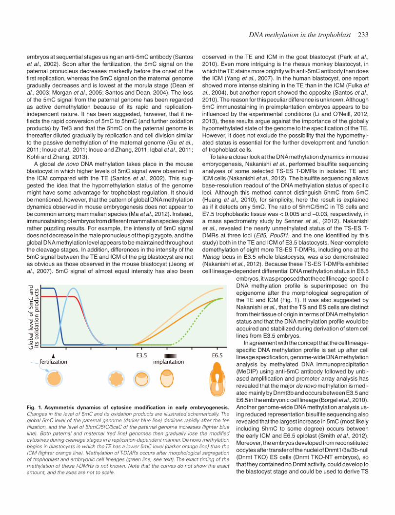
The mammalian genome undergoes dynamic changes in the global DNA methylation level during preimplantation embryogen-esis (Fig. 1), which can be visualized by immunostaining of mouse
DNA methylation in the trophoblast 233
embryos at sequential stages using an anti-5mC antibody (Santos et al., 2002). Soon after the fertilization, the 5mC signal on the paternal pronucleus decreases markedly before the onset of the first replication, whereas the 5mC signal on the maternal genome gradually decreases and is lowest at the morula stage (Dean et al., 2003; Morgan et al., 2005; Santos and Dean, 2004). The loss of the 5mC signal from the paternal genome has been regarded as active demethylation because of its rapid and replication-independent nature. It has been suggested, however, that it re-flects the rapid conversion of 5mC to 5hmC (and further oxidation products) by Tet3 and that the 5hmC on the paternal genome is thereafter diluted gradually by replication and cell division similar to the passive demethylation of the maternal genome (Gu et al., 2011; Inoue et al., 2011; Inoue and Zhang, 2011; Iqbal et al., 2011; Kohli and Zhang, 2013).
A global de novo DNA methylation takes place in the mouse blastocyst in which higher levels of 5mC signal were observed in the ICM compared with the TE (Santos et al., 2002). This sug-gested the idea that the hypomethylation status of the genome might have some advantage for trophoblast regulation. It should be mentioned, however, that the pattern of global DNA methylation dynamics observed in mouse embryogenesis does not appear to be common among mammalian species (Ma et al., 2012). Instead, immunostaining of embryos from different mammalian species gives rather puzzling results. For example, the intensity of 5mC signal does not decrease in the male pronucleus of the pig zygote, and the global DNA methylation level appears to be maintained throughout the cleavage stages. In addition, differences in the intensity of the 5mC signal between the TE and ICM of the pig blastocyst are not as obvious as those observed in the mouse blastocyst (Jeong et al., 2007). 5mC signal of almost equal intensity has also been
Glo
bal
lev
el o
f 5
mC
and
its
oxid
atio
n p
roduct
s
E3.5fertilization
E6.5implantation
Fig. 1. Asymmetric dynamics of cytosine modification in early embryogenesis. Changes in the level of 5mC and its oxidation products are illustrated schematically. The global 5mC level of the paternal genome (darker blue line) declines rapidly after the fer-tilization, and the level of 5hmC/5fC/5caC of the paternal genome increases (lighter blue line). Both paternal and maternal (red line) genomes then gradually lose the modified cytosines during cleavage stages in a replication-dependent manner. De novo methylation begins in blastocysts in which the TE has a lower 5mC level (darker orange line) than the ICM (lighter orange line). Methylation of T-DMRs occurs after morphological segregation of trophoblast and embryonic cell lineages (green line, see text). The exact timing of the methylation of these T-DMRs is not known. Note that the curves do not show the exact amount, and the axes are not to scale.
embryos, it was proposed that the cell lineage-specific DNA methylation profile is superimposed on the epigenome after the morphological segregation of the TE and ICM (Fig. 1). It was also suggested by Nakanishi et al., that the TS and ES cells are distinct from their tissue of origin in terms of DNA methylation status and that the DNA methylation profile would be acquired and stabilized during derivation of stem cell lines from E3.5 embryos.
In agreement with the concept that the cell lineage-specific DNA methylation profile is set up after cell lineage specification, genome-wide DNA methylation analysis by methylated DNA immunoprecipitation (MeDIP) using anti-5mC antibody followed by unbi-ased amplification and promoter array analysis has revealed that the major de novo methylation is medi-ated mainly by Dnmt3b and occurs between E3.5 and E6.5 in the embryonic cell lineage (Borgel et al., 2010). Another genome-wide DNA methylation analysis us-ing reduced representation bisulfite sequencing also revealed that the largest increase in 5mC (most likely including 5hmC to some degree) occurs between the early ICM and E6.5 epiblast (Smith et al., 2012). Moreover, the embryos developed from reconstituted oocytes after transfer of the nuclei of Dnmt1/3a/3b-null (Dnmt TKO) ES cells (Dnmt TKO-NT embryos), so that they contained no Dnmt activity, could develop to the blastocyst stage and could be used to derive TS
observed in the TE and ICM in the goat blastocyst (Park et al., 2010). Even more intriguing is the rhesus monkey blastocyst, in which the TE stains more brightly with anti-5mC antibody than does the ICM (Yang et al., 2007). In the human blastocyst, one report showed more intense staining in the TE than in the ICM (Fulka et al., 2004), but another report showed the opposite (Santos et al., 2010). The reason for this peculiar difference is unknown. Although 5mC immunostaining in preimplantation embryos appears to be influenced by the experimental conditions (Li and O’Neill, 2012, 2013), these results argue against the importance of the globally hypomethylated state of the genome to the specification of the TE. However, it does not exclude the possibility that the hypomethyl-ated status is essential for the further development and function of trophoblast cells.
To take a closer look at the DNA methylation dynamics in mouse embryogenesis, Nakanishi et al., performed bisulfite sequencing analyses of some selected TS-ES T-DMRs in isolated TE and ICM cells (Nakanishi et al., 2012). The bisulfite sequencing allows base-resolution readout of the DNA methylation status of specific loci. Although this method cannot distinguish 5hmC from 5mC (Huang et al., 2010), for simplicity, here the result is explained as if it detects only 5mC. The ratio of 5hmC/5mC in TS cells and E7.5 trophoblastic tissue was < 0.005 and ~0.03, respectively, in a mass spectrometry study by Senner et al., (2012). Nakanishi et al., revealed the nearly unmethylated status of the TS-ES T-DMRs at three loci (Elf5, Pou5f1, and the one identified by this study) both in the TE and ICM of E3.5 blastocysts. Near-complete demethylation of eight more TS-ES T-DMRs, including one at the Nanog locus in E3.5 whole blastocysts, was also demonstrated (Nakanishi et al., 2012). Because these TS-ES T-DMRs exhibited cell lineage-dependent differential DNA methylation status in E6.5

234 S. Tanaka et al.
cells (TKO ntTS), indicating that DNA methylation is dispensable for specification of the TE and ICM (Sakaue et al., 2010). These reports imply that DNA methylation at specific genomic regions is likely to be important for the maintenance of cell-lineage identity but not for the specification of extraembryonic and embryonic cell lineages, at least in the mouse.
At exactly what stage is the cell lineage-specific DNA methylation profile established? By what kind of cue is this de novo methylation ignited? And what kind of mechanisms underlie establishment of cell lineage- and region-specific DNA methylation? These ques-tions should be addressed in future to understand the regulation of the trophoblast lineage by epigenetic systems including DNA methylation.
Mouse ES cells can be converted to TS-like cells by manipu-lating the expression of a single transcription factor, such as the induced depletion of Oct4 or the induced activation of Cdx2 or Eomes (Niwa et al., 2000, 2005). These systems offer the oppor-tunity to analyze the mechanisms underlying the establishment of the trophoblast cell lineage-specific epigenome. For example, the inducible Oct4-depletion system was used to understand why the mouse trophoblast genome shows global hypomethylation com-pared with ES and somatic cells (Oda et al., 2013). This analysis found reduced expression of Np95 and failure of Dnmt1 to localize at replication foci in induced trophoblast cells. Although the local-ization pattern of Dnmt1 was restored by overexpression of Np95, DNA hypomethylation was maintained. From these results, it was concluded that the trophoblast cells (at least the induced cells) might have a mechanism to resist a genome-wide increase in DNA methylation. This resistance should be region-specific because, as mentioned above, some TS-ES T-DMRs show the hyper- and hypomethylated state in TS and ES cells, respectively (Hattori et al., 2004, 2007, Nakanishi et al., 2012). Transcription factors confer such region specificity at least in part. Carey et al., used Cdx2-inducible ES cells and showed that de novo DNA methylation on Oct4 T-DMRs follows transcriptional repression by direct binding of Cdx2 and changes in histone acetylation around the promoter region. Although Oct4 and Nanog were silenced 48 hours after the induction of Cdx2, only a slight increase in DNA methylation was observed at 72 hours. This increase reached a similar level to that in TS cells at 120 hours after induction (Carey et al., 2014). This result suggests that the de novo methylation of TS-ES T-DMRs occurs autonomously as a consequence of transcription repression. It should be noted, however, that T-E T-DMRs, including Nanog T-DMR, were barely methylated in diapause blastocysts (4 days in the diapause state after E3.5 when Nanog is already shut off in the TE) and that some of the T-E T-DMRs are not located near any known promoters (Nakanishi et al., 2012). Thus, the de novo methylation in trophoblast cells cannot be explained simply by transcription factor-directed stepwise mechanisms.
Cell fate restriction in embryonic cell lineage by DNA methylation
The restriction of embryonic cell lineage fate by DNA methyla-tion has been elucidated. Insufficient DNA methylation in ES cells caused by Dnmt1 deficiency and/or Dnmt3a/3b deficiency causes the ectopic expression of trophoblast-specific genes such as Pl1 (Prl3d1) and Tpbpa when the cells are differentiated in vitro, whereas expression of these genes was very low in the wild-type
control (Jackson et al., 2004; Ng et al., 2008). Ectopic expres-sion of trophoblast marker genes was also detected in embryonic tissue of E9.5 Dnmt1–/– embryos (Ng et al., 2008). Moreover, derivatives of Dnmt TKO-NT embryos contributed predominantly to the placenta in the context of the chimeric conceptus between wild-type embryos (Sakaue et al., 2010). A genome-wide screen for promoter methylation by MeDIP array hybridization identified a promoter region of the Elf5 locus encoding a transcription factor of the Ets family that is essential for trophoblast lineage develop-ment (Donnison et al., 2005), as a target of DNA methylation in ES cells (Ng et al., 2008). Elf5 is not expressed and its promoter is heavily methylated (90%) in wild-type ES cells, whereas the gene is expressed and is hypomethylated (9.3%) in TS cells. The DNA methylation level of the Elf5 promoter region was decreased to 41.5% in Dnmt1–/– ES cells, which resulted in ectopic activa-tion of Elf5 gene in differentiating Dnmt1–/–- ES cells. The forced expression of Elf5 in wild-type ES cells induced expression of other transcription factors Cdx2 and Eomes (Ng et al., 2008) both of which can provoke wild-type ES cells to adopt the trophoblast cell fate (Niwa et al., 2005). Thus, DNA methylation works to fix cell lineage restriction through the regulation of the Elf5 locus.
Besides Elf5, another gene might also be involved in DNA methylation-mediated cell lineage restriction. It was recently reported that the forced expression of the noncoding RNA, H19, induces expression of trophoblast lineage markers in ES cells under differentiation conditions (Fujimori et al., 2013). H19 is a well-known imprinted gene, and the DNA methylation of the Igf2–H19 imprinting control region (ICR) on paternally derived chromosomes suppresses transcription of H19, allowing the maternal allele-specific expression of this gene (Kurukuti et al., 2006). Hypomethylation of the ICR and induction of H19 is evident in Dnmt1–/– ES cells (Biniszkiewicz et al., 2002). Therefore, it is possible that upregulation of H19 also increases the transdif-ferentiation of Dnmt1–/– ES cells toward the trophoblast cell fate. It would be interesting to determine whether Dnmt1–/– ES cells efficiently differentiate into trophoblast cells even when Elf5 and/or H19 is depleted.
A causal role of H19 in ES-to-trophoblast transdifferentiation is also speculated in poly(ADP-ribose) polymerase-1 (Parp1)-deficient ES cells (Fujimori et al., 2013). Parp1–/– ES cells differ-entiate into trophoblast derivatives in vitro and in ES cell-derived tumors (Hemberger et al., 2003; Nozaki et al., 2013; Nozaki et al., 1999; Ogino et al., 2007). Significant upregulation of H19 in Parp1–/– ES was detected by a microarray analysis and was then validated by RT-PCR (Ogino et al., 2007). Given that H19 can unleash the trophoblast cell fate in ES cells (Fujimori et al., 2013), it is also possible that increased expression of H19 leads to differentiation of Parp1–/– ES cells toward the trophoblast cell fate. However, the involvement of DNA methylation remains equivocal. Parp1 is thought to inhibit Dnmt1 activity (Caiafa et al., 2009). Parp activity is also suggested to be involved in the active demethylation process in primordial germ cells (Ciccarone et al., 2012; Kawasaki et al., 2014). Based on these reports, one may expect hypermethylation of the genome in Parp1-deficient ES cells in contrast to Dnmt1-deficient ES cells, although this has not been reported yet. Thus, it is unclear why H19 is upregulated in Parp1–/– ES cells. Interestingly, transient treatment of preimplanta-tion mouse embryos with the Parp inhibitor 3-aminobenzamide for 24 hours around the eight-cell to morula stages completely
DNA methylation in the trophoblast 235
blocked blastocyst formation (Imamura et al., 2004). This sug-gests that Parp activity plays pivotal role in the specification or maintenance of the TE.
Gene regulation by DNA methylation in the trophoblast cell lineage
Compared with the role of DNA methylation in the embryonic cell lineage, the role in the trophoblast lineage has not been explored deeply. Dnmt TKO-NT embryos show normal development until the blastocyst stage (Sakaue et al., 2010). Cells derived from ei-ther Dnmt TKO-NT embryos or TKO ntTS cells contributed to and survived in the placenta in the context of the chimeric conceptus between wild-type embryos. This indicates that DNA methylation is dispensable for specification of the TE, but it does not show conclusively whether the Dnmt-TKO trophoblast cells are function-ally normal. As mentioned above, ectopic expression of Oct4 has been detected by RT-PCR in the Dnmt1-deficient E10.5 placenta (Hattori et al., 2004). Oct4 alone has been shown to reprogram TS cells into ES-like cells, although this occurs at a low efficiency (Wu et al., 2011b). It is therefore possible that the ectopic expres-sion of Oct4 in trophoblast cells compromises their identity and causes yet-to-be unrevealed abnormalities. Loss of DNA methyla-tion should also affect the regulation of imprinted genes. It has been shown that the loss of maternal imprint because of a lack of Dnmt3L during oogenesis results in placental defects even in heterozygous conceptuses (Arima et al., 2006), which suggests that Dnmt-deficient trophoblast cells also show abnormalities because of the deregulated expression of imprinted genes. In addition, insufficient DNA methylation has been implicated in pregnancy-associated diseases in humans (Novakovic and Saffery, 2012). Thus, the evidence points to an essential role of DNA methylation in the normal development and function of trophoblast cells. Fur-ther examination of mouse models such as conditional knockout of Dnmt genes in trophoblast cells should unveil the roles of DNA methylation in trophoblast cell lineage.
An irregular increase in DNA methylation may also be harmful for trophoblast development and placental function. The genes of two human endogenous retroviruses, Syncytin-1 (ERVWE1) and Syncytin-2 (ERVFRDE1), contribute to the formation of the multinucleated syncytiotrophoblast, which forms a physical bar-rier to maternal blood in the chorionic villi of the human placenta (Mi et al., 2000). Expression of these fusogenic proteins is tightly restricted to placental trophoblast cells. It has been shown that these genes are silenced in somatic cells by DNA methylation of CpGs within 5’ long terminal repeats (LTRs) (Matousková et al., 2006; Trejbalová et al., 2011). Aberrant methylation of these 5’ LTRs in trophoblast cells might lead to the inadequate formation of the syncytiotrophoblast and a malfunctioning placenta. DNA methylation is also implicated in the differentiation state-dependent control of gene expression in mouse trophoblast cells. For example, the dimethylarginine dimethylaminohydrolase 2 (Ddah2) gene is suppressed in TS cells with a hypermethylated enhancer both in vivo and in vitro, and the enhancer is demethylated in differentiated trophoblast cells in which the gene is expressed. Treatment with 5-aza-dC induces ectopic expression of Ddah2 in undifferentiated TS cells. Reporter assay analyses showed that the methylation of the Ddah2 enhancer diminished activity of the Ddah2 promoter, which suggests that DNA methylation of the enhancer suppresses
Ddah2 expression in vivo (Tomikawa et al., 2006). Again, aberrant methylation of the Ddah2 enhancer in differentiated trophoblast cells might cause reduced expression of this gene. In a classical RLGS analysis, 30 T-DMRs were detected through a comparison between TS cells and differentiated TS cells. One half of the T-DMRs showed greater methylation in undifferentiated cells, and the other half showed greater methylation in differentiated cells, which suggests that the fine control of DNA methylation accompanies even trophoblast differentiation (Shiota et al., 2002). Overall, DNA methylation should play a pivotal role in the regulation of trophoblast differentiation and function.
Possible role of ten-eleven translocation (Tet) in tro-phoblast regulation
A growing body of evidence now suggests that 5hmC is not just a transient intermediate of active demethylation but that it plays a unique role as an epigenetic mark (Inoue and Zhang, 2011; Iqbal et al., 2011; Ruzov et al., 2011; Salvaing et al., 2012). Possible contribution of Tet genes in the regulation of trophoblast develop-ment has been suggested by loss-of-function analyses of Tet genes. In mouse blastocysts, immunostaining using anti-5hmC antibody revealed slightly higher content of 5hmC in the ICM than in the TE (Ruzov et al., 2011). Tet1 protein also appears to be enriched in the ICM compared with the TE (Ito et al., 2010). Similar asymmetry was also reported between ES and TS cells. The expression levels of Tet1 and Tet2 mRNAs as well as the global 5hmC level analyzed by mass spectrometry are significantly lower in TS cells than in ES cells (Ito et al., 2010; Senner et al., 2012). Reduction of Tet1 expression in one blastomere of mouse two-cell stage embryos biased the blastomere’s cell fate toward the TE (Ito et al., 2010). These findings imply that Tet1 plays an essential role in the first cell-fate decision during mouse embryogenesis. However, it has been reported that Tet1 and Tet2 are dispensable for blastocyst formation and for postnatal development (Dawlaty et al., 2013; Dawlaty et al., 2011), which refutes the idea of an essential role of Tet1 and Tet2 in the specification of the TE and ICM. It is possible that the asymmetry in the global level of 5hmC between two blas-tomeres of two-cell stage embryos caused by Tet1 knockdown in one blastomere somehow skewed the equivalence of blastomeres, whereas other mechanisms governed the cell-fate decision without such asymmetry in Tet1/Tet2-deficient embryos. It is also possible that the loss of Tet1 and Tet2 was compensated by induction of Tet3 in early embryos, as seems to be the case at least in part in tissues of Tet1/Tet2-deficient adult mice (Dawlaty et al., 2013).
Tet1-deficient and Tet1/Tet2-deficient ES cells show induction of TS cell marker genes such as Cdx2, Eomes, and Elf5 in vitro and form hemorrhagic teratomas with trophoblast-like cells, which suggests that 5hmC also plays a pivotal role in cell-fate restriction in the embryonic cell lineage (Dawlaty et al., 2013, Dawlaty et al., 2011, Koh et al., 2011). However, it should be kept in mind that Tet1 protein is implicated in the repression of Polycomb-targeted developmental regulators in ES cells independent of its enzymatic activity (Wu et al., 2011a), which makes it less likely that 5hmC plays a major role in cell-fate restriction. Nevertheless, the importance of Tet1 in the trophoblast lineage has been suggested. Some of the Tet1/Tet2-deficient embryos show midgestation lethality with a wide variety of abnormalities including a smaller fetus compared with normal littermates at E10.5 (Dawlaty et al., 2013). Tet1-deficient

236 S. Tanaka et al.
embryos also show a mild developmental delay at E12.5, and the mutant pups are smaller in size and weight. A placental defect was suggested for Tet1–/– mice because the tetraploid complementation rescued these phenotypes (Dawlaty et al., 2011). Taken together, these data suggest that Tet proteins and 5hmC are likely to be involved in the development and function of the trophoblast cell lineage.
Perspective
Loss-of-function analyses in mice have revealed insights into the genetic regulation of the development, differentiation, and func-tion of trophoblast cells (Cross, 2005, Watson and Cross, 2005), but our knowledge about epigenetic regulation remains limited. Genetic ablation of epigenetic factors including members of the Dnmt and Tet families in a TE-specific or trophoblast subtype-specific manner will be required for further understanding of the epigenetic regulation of trophoblast cells. Genome-wide analyses of DNA methylation and hydroxymethylation in the context of gene expression and histone modification patterns should be also performed in trophoblast cells of different subtypes and at vari-ous differentiation stages. Even with recent technical advances, it is still difficult to perform such epigenome analyses with in vivo materials because of the small amount in early pregnancy and the complexity at mid to late pregnancy. Mouse TS cells will provide a useful tool for circumventing such difficulties. Development of culture conditions that direct the differentiation of TS cells toward specific subtypes will be the next challenge for this purpose. This type of research will help us elucidate the details of the epigenetic landscape of trophoblast development, which should supply valu-able information for understanding the epigenetic mechanisms underlying pregnancy-related diseases in humans.
References
ALBERTS, B., JOHNSON, A., LEWIS, J., RAFF, M., ROBERTS, K. and WALTER, P. (2007). Molecular Biology of The Cell. Garland Science, New York.
ARIMA, T., HATA, K., TANAKA, S., KUSUMI, M., LI, E., KATO, K., SHIOTA, K., SASAKI, H. and WAKE, N. (2006). Loss of the maternal imprint in Dnmt3Lmat-/- mice leads to a differentiation defect in the extraembryonic tissue. Dev. Biol. 297: 361-373.
BINISZKIEWICZ, D., GRIBNAU, J., RAMSAHOYE, B., GAUDET, F., EGGAN, K., HUMPHERYS, D., MASTRANGELO, M.-A., JUN, Z., WALTER, J. and JAENISCH, R. (2002). Dnmt1 overexpression causes genomic hypermethylation, loss of imprinting, and embryonic lethality. Mol. Cell. Biol. 22: 2124-2135.
BIRD, A. (2002). DNA methylation patterns and epigenetic memory. Genes Dev. 16: 6-21.
BORGEL, J., GUIBERT, S., LI, Y., CHIBA, H., SCHÜBELER, D., SASAKI, H., FORNÉ, T. and WEBER, M. (2010). Targets and dynamics of promoter DNA methylation during early mouse development. Nature Genet. 42: 1093-1100.
CAIAFA, P., GUASTAFIERRO, T. and ZAMPIERI, M. (2009). Epigenetics: poly(ADP-ribosyl)ation of PARP-1 regulates genomic methylation patterns. FASEB J. 23: 672-678.
CAREY, T.S., CHOI, I., WILSON, C.A., FLOER, M. and KNOTT, J.G. (2014). Tran-scriptional reprogramming and chromatin remodeling accompanies oct4 and nanog silencing in mouse trophoblast lineage. Stem Cells Dev. 23: 219-229.
CHEN, T., UEDA, Y., XIE, S. and LI, E. (2002). A novel Dnmt3a isoform produced from an alternative promoter localizes to euchromatin and its expression correlates with active de novo methylation. J. Biol. Chem. 277: 38746-38754.
CICCARONE, F., KLINGER, F.G., CATIZONE, A., CALABRESE, R., ZAMPIERI, M., BACALINI, M.G., DE FELICI, M. and CAIAFA, P. (2012). Poly(ADP-ribosyl)ation Acts in the DNA Demethylation of Mouse Primordial Germ Cells Also with DNA Damage-Independent Roles. PLOS ONE 7: e46927.
CROSS, J.C. (2005). How to make a placenta: mechanisms of trophoblast cell dif-ferentiation in mice--a review. Placenta 26 Suppl A: S3-9.
DAWLATY, M.M., BREILING, A., LE, T., RADDATZ, G., BARRASA, M.I., CHENG, A.W., GAO, Q., POWELL, B.E., LI, Z., XU, M. et al., (2013). Combined deficiency of Tet1 and Tet2 causes epigenetic abnormalities but is compatible with postnatal development. Dev. Cell 24: 310-323.
DAWLATY, M.M., GANZ, K., POWELL, B.E., HU, Y.-C., MARKOULAKI, S., CHENG, A.W., GAO, Q., KIM, J., CHOI, S.-W., PAGE, D.C. et al., (2011). Tet1 is dispens-able for maintaining pluripotency and its loss is compatible with embryonic and postnatal development. Cell Stem Cell 9: 166-175.
DEAN, W., SANTOS, F. and REIK, W. (2003). Epigenetic reprogramming in early mammalian development and following somatic nuclear transfer. Sem. Cell Dev. Biol. 14: 93-100.
DONNISON, M., BEATON, A., DAVEY, H.W., BROADHURST, R., L’HUILLIER, P. and PFEFFER, P.L. (2005). Loss of the extraembryonic ectoderm in Elf5 mutants leads to defects in embryonic patterning. Development 132: 2299-2308.
EVANS, M.J. and KAUFMAN, M.H. (1981). Establishment in culture of pluripotential cells from mouse embryos. Nature 292: 154-156.
FUJIMORI, H., MUKAI, H., MURAKAMI, Y., HEMBERGER, M., HIPPO, Y. and MA-SUTANI, M. (2013). The H19 induction triggers trophoblast lineage commitment in mouse ES cells. Biochem. Biophys. Res. Commun. 436: 313-318.
FULKA, H., MRAZEK, M., TEPLA, O. and FULKA, J. (2004). DNA methylation pat-tern in human zygotes and developing embryos. Reproduction 128: 703-708.
GOWHER, H., LIEBERT, K., HERMANN, A., XU, G. and JELTSCH, A. (2005). Mechanism of stimulation of catalytic activity of Dnmt3A and Dnmt3B DNA-(cytosine-C5)-methyltransferases by Dnmt3L. J. Biol. Chem. 280: 13341-13348.
GRUENBAUM, Y., CEDAR, H. and RAZIN, A. (1982). Substrate and sequence specificity of a eukaryotic DNA methylase. Nature 295: 620-622.
GU, T.-P., GUO, F., YANG, H., WU, H.-P., XU, G.-F., LIU, W., XIE, Z.-G., SHI, L., HE, X., JIN, S.-G. et al., (2011). The role of Tet3 DNA dioxygenase in epigenetic reprogramming by oocytes. Nature 477: 606-610.
HATA, K., KUSUMI, M., YOKOMINE, T., LI, E. and SASAKI, H. (2006). Meiotic and epigenetic aberrations in Dnmt3L-deficient male germ cells. Mol. Reprod. Dev. 73: 116-122.
HATA, K., OKANO, M., LEI, H. and LI, E. (2002). Dnmt3L cooperates with the Dnmt3 family of de novo DNA methyltransferases to establish maternal imprints in mice. Development 129: 1983-1993.
HATTORI, N., IMAO, Y., NISHINO, K., HATTORI, N., OHGANE, J., YAGI, S., TANAKA, S. and SHIOTA, K. (2007). Epigenetic regulation of Nanog gene in embryonic stem and trophoblast stem cells. Genes Cells 12: 387-396.
HATTORI, N., NISHINO, K., KO, Y.-G., HATTORI, N., OHGANE, J., TANAKA, S. and SHIOTA, K. (2004). Epigenetic control of mouse Oct-4 gene expression in embryonic stem cells and trophoblast stem cells. J. Biol. Chem. 279: 17063-17069.
HE, Y.-F., LI, B.-Z., LI, Z., LIU, P., WANG, Y., TANG, Q., DING, J., JIA, Y., CHEN, Z., LI, L. et al., (2011). Tet-mediated formation of 5-carboxylcytosine and its excision by TDG in mammalian DNA. Science 333: 1303-1307.
HEMBERGER, M., NOZAKI, T., WINTERHAGER, E., YAMAMOTO, H., NAKAGAMA, H., KAMADA, N., SUZUKI, H., OHTA, T., OHKI, M., MASUTANI, M. et al., (2003). Parp1-deficiency induces differentiation of ES cells into trophoblast derivatives. Dev. Biol. 257: 371-381.
HUANG, Y., PASTOR, W.A., SHEN, Y., TAHILIANI, M., LIU, D.R. and RAO, A. (2010). The behaviour of 5-hydroxymethylcytosine in bisulfite sequencing. PLOS ONE 5: e8888.
ICHIYANAGI, T., ICHIYANAGI, K., MIYAKE, M. and SASAKI, H. (2013). Accumulation and loss of asymmetric non-CpG methylation during male germ-cell development. Nucleic Acids Res. 41: 738-745.
IMAMURA, T., KERJEAN, A., HEAMS, T., KUPIEC, J.-J., THENEVIN, C. and PÀLDI, A. (2005). Dynamic CpG and non-CpG methylation of the Peg1/Mest gene in the mouse oocyte and preimplantation embryo. J. Biol. Chem. 280: 20171-20175.
IMAMURA, T., NEILDEZ, T.M.A., THENEVIN, C. and PALDI, A. (2004). Essential role for poly (ADP-ribosyl)ation in mouse preimplantation development. BMC Mol. Biol. 5: 4.
INOUE, A., SHEN, L., DAI, Q., HE, C. and ZHANG, Y. (2011). Generation and replication-dependent dilution of 5fC and 5caC during mouse preimplantation development. Cell Res. 21: 1670-1676.

DNA methylation in the trophoblast 237
INOUE, A. and ZHANG, Y. (2011). Replication-dependent loss of 5-hydroxymethyl-cytosine in mouse preimplantation embryos. Science 334: 194.
IQBAL, K., JIN, S.-G., PFEIFER, G.P. and SZABÓ, P.E. (2011). Reprogramming of the paternal genome upon fertilization involves genome-wide oxidation of 5-methylcytosine. Proc. Natl. Acad. Sci. USA 108: 3642-3647.
ITO, S., D’ALESSIO, A.C., TARANOVA, O.V., HONG, K., SOWERS, L.C. and ZHANG, Y. (2010). Role of Tet proteins in 5mC to 5hmC conversion, ES-cell self-renewal and inner cell mass specification. Nature 466: 1129-1133.
ITO, S., SHEN, L., DAI, Q., WU, S.C., COLLINS, L.B., SWENBERG, J.A., HE, C. and ZHANG, Y. (2011). Tet proteins can convert 5-methylcytosine to 5-formylcytosine and 5-carboxylcytosine. Science 333: 1300-1303.
JACKSON, M., KRASSOWSKA, A., GILBERT, N., CHEVASSUT, T., FORRESTER, L., ANSELL, J. and RAMSAHOYE, B. (2004). Severe global DNA hypomethylation blocks differentiation and induces histone hyperacetylation in embryonic stem cells. Mol. Cell. Biol. 24: 8862-8871.
JAENISCH, R. and BIRD, A. (2003). Epigenetic regulation of gene expression: how the genome integrates intrinsic and environmental signals. Nature Genet. 33 Suppl: 245-254.
JEONG, Y.-S., YEO, S., PARK, J.S., KOO, D.-B., CHANG, W.-K., LEE, K.-K. and KANG, Y.-K. (2007). DNA methylation state is preserved in the sperm-derived pronucleus of the pig zygote. Int. J. Dev. Biol. 51: 707-714.
KAWASAKI, Y., LEE, J., MATSUZAWA, A., KOHDA, T., KANEKO-ISHINO, T. and ISHINO, F. (2014). Active DNA demethylation is required for complete imprint erasure in primordial germ cells. Scientific reports 4: 3658.
KOH, K.P., YABUUCHI, A., RAO, S., HUANG, Y., CUNNIFF, K., NARDONE, J., LAIHO, A., TAHILIANI, M., SOMMER, C.A., MOSTOSLAVSKY, G. et al., (2011). Tet1 and Tet2 regulate 5-hydroxymethylcytosine production and cell lineage specification in mouse embryonic stem cells. Cell Stem Cell 8: 200-213.
KOHLI, R.M. and ZHANG, Y. (2013). TET enzymes, TDG and the dynamics of DNA demethylation. Nature 502: 472-479.
KURUKUTI, S., TIWARI, V.K., TAVOOSIDANA, G., PUGACHEVA, E., MURRELL, A., ZHAO, Z., LOBANENKOV, V., REIK, W. and OHLSSON, R. (2006). CTCF binding at the H19 imprinting control region mediates maternally inherited higher-order chromatin conformation to restrict enhancer access to Igf2. Proc. Natl. Acad. Sci. USA 103: 10684-10689.
LI, Y. and O’NEILL, C. (2012). Persistence of cytosine methylation of DNA following fertilisation in the mouse. PLOS ONE 7: e30687.
LI, Y. and O’NEILL, C. (2013). 5’-Methylcytosine and 5’-hydroxymethylcytosine each provide epigenetic information to the mouse zygote. PLOS ONE 8: e63689.
MA, J.-Y., LIANG, X.-W., SCHATTEN, H. and SUN, Q.-Y. (2012). Active DNA demethyl-ation in mammalian preimplantation embryos: new insights and new perspectives. Mol. Hum. Reprod. 18: 333-340.
MARTIN, G.R. (1981). Isolation of a pluripotent cell line from early mouse embryos cultured in medium conditioned by teratocarcinoma stem cells. Proc. Natl. Acad. Sci. USA 78: 7634-7638.
MATOUSKOVÁ, M., BLAZKOVÁ, J., PAJER, P., PAVLÍCEK, A. and HEJNAR, J. (2006). CpG methylation suppresses transcriptional activity of human syncytin-1 in non-placental tissues. Exp. Cell. Res. 312: 1011-1020.
MI, S., LEE, X., LI, X., VELDMAN, G.M., FINNERTY, H., RACIE, L., LAVALLIE, E., TANG, X.Y., EDOUARD, P., HOWES, S. et al., (2000). Syncytin is a captive retroviral envelope protein involved in human placental morphogenesis. Nature 403: 785-789.
MORGAN, H.D., SANTOS, F., GREEN, K., DEAN, W. and REIK, W. (2005). Epigenetic reprogramming in mammals. Hum. Mol. Genet. 14 Spec No 1: R47-58.
NAKANISHI, M.O., HAYAKAWA, K., NAKABAYASHI, K., HATA, K., SHIOTA, K. and TANAKA, S. (2012). Trophoblast-specific DNA methylation occurs after the seg-regation of the trophectoderm and inner cell mass in the mouse periimplantation embryo. Epigenetics 7: 173-182.
NG, R.K., DEAN, W., DAWSON, C., LUCIFERO, D., MADEJA, Z., REIK, W. and HEMBERGER, M. (2008). Epigenetic restriction of embryonic cell lineage fate by methylation of Elf5. Nature Cell Biol. 10: 1280-1290.
NIMURA, K., ISHIDA, C., KORIYAMA, H., HATA, K., YAMANAKA, S., LI, E., URA, K. and KANEDA, Y. (2006). Dnmt3a2 targets endogenous Dnmt3L to ES cell chromatin and induces regional DNA methylation. Genes Cells 11: 1225-1237.
NISHINO, K., HATTORI, N., SATO, S., ARAI, Y., TANAKA, S., NAGY, A. and SHIOTA, K. (2011). Non-CpG methylation occurs in the regulatory region of the Sry gene.
J. Reprod. Dev. 57: 586-593.NIWA, H., MIYAZAKI, J. and SMITH, A.G. (2000). Quantitative expression of Oct-
3/4 defines differentiation, dedifferentiation or self-renewal of ES cells. Nature Genet. 24: 372-376.
NIWA, H., TOYOOKA, Y., SHIMOSATO, D., STRUMPF, D., TAKAHASHI, K., YAGI, R. and ROSSANT, J. (2005). Interaction between Oct3/4 and Cdx2 determines trophectoderm differentiation. Cell 123: 917-929.
NOVAKOVIC, B. and SAFFERY, R. (2012). The ever growing complexity of placen-tal epigenetics - role in adverse pregnancy outcomes and fetal programming. Placenta 33: 959-970.
NOZAKI, T., FUJIMORI, H., WANG, J., SUZUKI, H., IMAI, H., WATANABE, M., OHURA, K. and MASUTANI, M. (2013). Parp-1 deficiency in ES cells promotes invasive and metastatic lesions accompanying induction of trophoblast giant cells during tumorigenesis in uterine environment. Pathology Int. 63: 408-414.
NOZAKI, T., MASUTANI, M., WATANABE, M., OCHIYA, T., HASEGAWA, F., NAK-AGAMA, H., SUZUKI, H. and SUGIMURA, T. (1999). Syncytiotrophoblastic giant cells in teratocarcinoma-like tumors derived from Parp-disrupted mouse embryonic stem cells. Proc. Natl. Acad. Sci. USA 96: 13345-13350.
ODA, M., OXLEY, D., DEAN, W. and REIK, W. (2013). Regulation of lineage specific DNA hypomethylation in mouse trophectoderm. PLOS ONE 8: e68846.
OGINO, H., NOZAKI, T., GUNJI, A., MAEDA, M., SUZUKI, H., OHTA, T., MURAKAMI, Y., NAKAGAMA, H., SUGIMURA, T. and MASUTANI, M. (2007). Loss of Parp-1 affects gene expression profile in a genome-wide manner in ES cells and liver cells. BMC genomics 8: 41.
OHGANE, J., AIKAWA, J., OGURA, A., HATTORI, N., OGAWA, T. and SHIOTA, K. (1998). Analysis of CpG islands of trophoblast giant cells by restriction landmark genomic scanning. Dev. Genet. 22: 132-140.
OHGANE, J., HATTORI, N., ODA, M., TANAKA, S. and SHIOTA, K. (2002). Differentia-tion of trophoblast lineage is associated with DNA methylation and demethylation. Biochem. Biophys. Res. Commun. 290: 701-706.
OKANO, M., BELL, D.W., HABER, D.A. and LI, E. (1999). DNA methyltransferases Dnmt3a and Dnmt3b are essential for de novo methylation and mammalian development. Cell 99: 247-257.
PARK, J.S., LEE, D., CHO, S., SHIN, S.-T. and KANG, Y.-K. (2010). Active loss of DNA methylation in two-cell stage goat embryos. Int. J. Dev. Biol. 54: 1323-1328.
RAMSAHOYE, B.H., BINISZKIEWICZ, D., LYKO, F., CLARK, V., BIRD, A.P. and JAENISCH, R. (2000). Non-CpG methylation is prevalent in embryonic stem cells and may be mediated by DNA methyltransferase 3a. Proc. Natl. Acad. Sci. USA 97: 5237-5242.
RISHI, V., BHATTACHARYA, P., CHATTERJEE, R., ROZENBERG, J., ZHAO, J., GLASS, K., FITZGERALD, P. and VINSON, C. (2010). CpG methylation of half-CRE sequences creates C/EBPalpha binding sites that activate some tissue-specific genes. Proc. Natl. Acad. Sci. USA 107: 20311-20316.
RUZOV, A., TSENKINA, Y., SERIO, A., DUDNAKOVA, T., FLETCHER, J., BAI, Y., CHEBOTAREVA, T., PELLS, S., HANNOUN, Z., SULLIVAN, G. et al., (2011). Lineage-specific distribution of high levels of genomic 5-hydroxymethylcytosine in mammalian development. Cell Res. 21: 1332-1342.
SAKAMOTO, H., SUZUKI, M., ABE, T., HOSOYAMA, T., HIMENO, E., TANAKA, S., GREALLY, J.M., HATTORI, N., YAGI, S. and SHIOTA, K. (2007). Cell type-specific methylation profiles occurring disproportionately in CpG-less regions that delineate developmental similarity. Genes Cells 12: 1123-1132.
SAKAUE, M., OHTA, H., KUMAKI, Y., ODA, M., SAKAIDE, Y., MATSUOKA, C., YA-MAGIWA, A., NIWA, H., WAKAYAMA, T. and OKANO, M. (2010). DNA methylation is dispensable for the growth and survival of the extraembryonic lineages. Curr. Biol. 20: 1452-1457.
SALVAING, J., AGUIRRE-LAVIN, T., BOULESTEIX, C., LEHMANN, G., DEBEY, P. and BEAUJEAN, N. (2012). 5-Methylcytosine and 5-hydroxymethylcytosine spatiotemporal profiles in the mouse zygote. PLOS ONE 7: e38156.
SANTOS, F. and DEAN, W. (2004). Epigenetic reprogramming during early develop-ment in mammals. Reproduction 127: 643-651.
SANTOS, F., HENDRICH, B., REIK, W. and DEAN, W. (2002). Dynamic reprogram-ming of DNA methylation in the early mouse embryo. Dev. Biol. 241: 172-182.
SANTOS, F., HYSLOP, L., STOJKOVIC, P., LEARY, C., MURDOCH, A., REIK, W., STOJKOVIĆ, M., HERBERT, M. and DEAN, W. (2010). Evaluation of epigenetic marks in human embryos derived from IVF and ICSI. Hum. Reprod. 25: 2387-2395.
SATO, S., YAGI, S., ARAI, Y., HIRABAYASHI, K., HATTORI, N., IWATANI, M., OKITA,

238 S. Tanaka et al.
K., OHGANE, J., TANAKA, S., WAKAYAMA, T. et al., (2010). Genome-wide DNA methylation profile of tissue-dependent and differentially methylated regions (T-DMRs) residing in mouse pluripotent stem cells. Genes Cells 15: 607-618.
SENNER, C.E., KRUEGER, F., OXLEY, D., ANDREWS, S. and HEMBERGER, M. (2012). DNA methylation profiles define stem cell identity and reveal a tight embryonic-extraembryonic lineage boundary. Stem Cells 30: 2732-2745.
SHARIF, J., MUTO, M., TAKEBAYASHI, S.-I., SUETAKE, I., IWAMATSU, A., ENDO, T.A., SHINGA, J., MIZUTANI-KOSEKI, Y., TOYODA, T., OKAMURA, K. et al., (2007). The SRA protein Np95 mediates epigenetic inheritance by recruiting Dnmt1 to methylated DNA. Nature 450: 908-912.
SHIOTA, K., KOGO, Y., OHGANE, J., IMAMURA, T., URANO, A., NISHINO, K., TANAKA, S. and HATTORI, N. (2002). Epigenetic marks by DNA methylation specific to stem, germ and somatic cells in mice. Genes Cells 7: 961-969.
SHIRANE, K., TOH, H., KOBAYASHI, H., MIURA, F., CHIBA, H., ITO, T., KONO, T. and SASAKI, H. (2013). Mouse oocyte methylomes at base resolution reveal genome-wide accumulation of non-CpG methylation and role of DNA methyl-transferases. PLoS Genet. 9: e1003439.
SMITH, Z.D., CHAN, M.M., MIKKELSEN, T.S., GU, H., GNIRKE, A., REGEV, A. and MEISSNER, A. (2012). A unique regulatory phase of DNA methylation in the early mammalian embryo. Nature 484: 339-344.
SMITH, Z.D. and MEISSNER, A. (2013). DNA methylation: roles in mammalian development. Nature Rev. Genet. 14: 204-220.
SUETAKE, I., SHINOZAKI, F., MIYAGAWA, J., TAKESHIMA, H. and TAJIMA, S. (2004). DNMT3L stimulates the DNA methylation activity of Dnmt3a and Dnmt3b through a direct interaction. J. Biol. Chem. 279: 27816-27823.
TAHILIANI, M., KOH, K.P., SHEN, Y., PASTOR, W.A., BANDUKWALA, H., BRUDNO, Y., AGARWAL, S., IYER, L.M., LIU, D.R., ARAVIND, L. et al., (2009). Conversion of 5-methylcytosine to 5-hydroxymethylcytosine in mammalian DNA by MLL partner TET1. Science 324: 930-935.
TANAKA, S., KUNATH, T., HADJANTONAKIS, A.-K., NAGY, A. and ROSSANT, J. (1998). Promotion of trophoblast stem cell proliferation by FGF4. Science 282: 2072-2075.
TOMIKAWA, J., FUKATSU, K., TANAKA, S. and SHIOTA, K. (2006). DNA methylation-dependent epigenetic regulation of dimethylarginine dimethylaminohydrolase 2 gene in trophoblast cell lineage. J. Biol. Chem. 281: 12163-12169.
TREJBALOVÁ, K., BLAZKOVÁ, J., MATOUSKOVÁ, M., KUCEROVÁ, D., PECNOVÁ, L., VERNEROVÁ, Z., HERÁCEK, J., HIRSCH, I. and HEJNAR, J. (2011). Epigen-etic regulation of transcription and splicing of syncytins, fusogenic glycoproteins of retroviral origin. Nucleic Acids Res. 39: 8728-8739.
WATSON, E.D. and CROSS, J.C. (2005). Development of structures and transport functions in the mouse placenta. Physiology 20: 180-193.
WEBER, M., HELLMANN, I., STADLER, M.B., RAMOS, L., PÄÄBO, S., REBHAN, M. and SCHÜBELER, D. (2007). Distribution, silencing potential and evolution-ary impact of promoter DNA methylation in the human genome. Nature Genet. 39: 457-466.
WU, H., D’ALESSIO, A.C., ITO, S., XIA, K., WANG, Z., CUI, K., ZHAO, K., SUN, Y.E. and ZHANG, Y. (2011a). Dual functions of Tet1 in transcriptional regulation in mouse embryonic stem cells. Nature 473: 389-393.
WU, H. and ZHANG, Y. (2014). Reversing DNA methylation: mechanisms, genomics, and biological functions. Cell 156: 45-68.
WU, T., WANG, H., HE, J., KANG, L., JIANG, Y., LIU, J., ZHANG, Y., KOU, Z., LIU, L., ZHANG, X. et al., (2011b). Reprogramming of trophoblast stem cells into pluripotent stem cells by Oct4. Stem Cells 29: 755-763.
YAGI, S., HIRABAYASHI, K., SATO, S., LI, W., TAKAHASHI, Y., HIRAKAWA, T., WU, G., HATTORI, N., HATTORI, N., OHGANE, J. et al., (2008). DNA methylation profile of tissue-dependent and differentially methylated regions (T-DMRs) in mouse promoter regions demonstrating tissue-specific gene expression. Genome Res. 18: 1969-1978.
YAGI, S., HIROSAWA, M. and SHIOTA, K. (2012). DNA methylation profile: a com-poser-, conductor-, and player-orchestrated Mammalian genome consisting of genes and transposable genetic elements. J. Reprod. Dev. 58: 265-273.
YANG, J., YANG, S., BEAUJEAN, N., NIU, Y., HE, X., XIE, Y., TANG, X., WANG, L., ZHOU, Q. and JI, W. (2007). Epigenetic marks in cloned rhesus monkey embryos: comparison with counterparts produced in vitro. Biol. Reprod. 76: 36-42.

5 yr ISI Impact Factor (2011) = 2.959
Further Related Reading, published previously in the Int. J. Dev. Biol.
Gene expression in the placenta: maternal stress and epigenetic responses J Ciprian P. Gheorghe, Ravi Goyal, Ashwani Mittal and Lawrence D. Longo Int. J. Dev. Biol. (2010) 54: 507 - 523 http://www.ijdb.ehu.es/web/paper/082770cg
Active loss of DNA methylation in two-cell stage goat embryos Jung S. Park, Doosoo Lee, Sunwha Cho, Sang-Tae Shin and Yong-Kook Kang Int. J. Dev. Biol. (2010) 54: 1323-1328 http://www.intjdevbiol.com/web/paper/092973jp Development and function of trophoblast giant cells in the rodent placenta Dong Hu and James C. Cross Int. J. Dev. Biol. (2010) 54: 341-354 http://www.intjdevbiol.com/web/paper/082768dh Interplay between DNA methylation, histone modification and chromatin remodeling in stem cells and during development Kohta Ikegami, Jun Ohgane, Satoshi Tanaka, Shintaro Yagi, and Kunio Shiota Int. J. Dev. Biol. (2009) 53: 203-214 http://www.intjdevbiol.com/web/paper/082741ki Hypomethylation of paternal DNA in the late mouse zygote is not essential for development Zbigniew Polanski, Nami Motosugi, Chizuko Tsurumi, Takashi Hiiragi and Steffen Hoffmann Int. J. Dev. Biol. (2008) 52: 295 - 298 http://www.ijdb.ehu.es/web/paper/072347zp