Electronic and Optical Properties of Surface-Anchored Metal ...
The Rockefeller University Press, 0021-9525/2000/03/899/15 $5.00The Journal of Cell Biology, Volume 148, Number 5, March 6, 2000 899–913http://www.jcb.org 899
Mechanism of Residence of Cytochrome b(5), a Tail-anchored Protein, in the Endoplasmic Reticulum
Emanuela Pedrazzini,* Antonello Villa,*
‡
Renato Longhi,
§
Alessandra Bulbarelli,* and Nica Borgese*
i
*Consiglio Nazionale Ricerche Cellular and Molecular Pharmacology Center, Department of Pharmacology, University of Milan, Italy 20129;
‡
Biological and Technological Research Department, Scientific Institute San Raffaele, Milan, Italy 20132;
§
Consiglio Nazionale Ricerche Institute of Biocatalysis and Molecular Recognition, Milan, Italy 20133; and
i
Faculty of Pharmacy, University of Catanzaro “Magna Graecia”, Roccelletta di Borgia (Catanzaro), Italy 88021
Abstract.
Endoplasmic reticulum (ER) proteins main-tain their residency by static retention, dynamic re-trieval, or a combination of the two. Tail-anchored pro-teins that contain a cytosolic domain associated with the lipid bilayer via a hydrophobic stretch close to the COOH terminus are sorted within the secretory path-way by largely unknown mechanisms. Here, we have in-vestigated the mode of insertion in the bilayer and the intracellular trafficking of cytochrome b(5) (b[5]),
taken as a model for ER-resident tail-anchored proteins. We first demonstrated that b(5) can acquire a trans-membrane topology posttranslationally, and then used two tagged versions of b(5), N-glyc and O-glyc b(5), containing potential N- and O-glycosylation sites, re-spectively, at the COOH-terminal lumenal extremity, to
discriminate between retention and retrieval mecha-nisms. Whereas the N-linked oligosaccharide provided no evidence for retrieval from a downstream compart-ment, a more stringent assay based on carbohydrate ac-quisition by O-glyc b(5) showed that b(5) gains access to enzymes catalyzing the first steps of O-glycosylation. These results suggest that b(5) slowly recycles between the ER and the
cis
-Golgi complex and that dynamic re-
trieval as well as retention are involved in sorting of tail-anchored proteins.
Key words: cell compartmentation • endoplasmic reticulum • glycosylation • Golgi apparatus • mem-brane proteins
Introduction
Notwithstanding the intense membrane traffic taking placewithin eukaryotic cells, subcellular compartments main-tain their molecular identity with remarkable accuracy.Within the secretory pathway, proteins that reside in agiven compartment must avoid being relocated to neigh-boring compartments in the face of ongoing transport.Two different mechanisms, static retention and dynamicretrieval, can, in principle, ensure that each compartmentkeeps its cohort of resident proteins. Retained proteins arethought to be simply excluded from transport out of theircompartment of residence, whereas retrieved proteins doescape to a downstream organelle from which they are re-cycled by retrograde transport (Pelham and Munro, 1993).
Consistent with its diverse functions, the membrane ofthe ER has a complex molecular composition, and manystudies have been devoted to the mechanisms through
which it keeps its numerous resident polypeptides fromfollowing transported proteins down the secretory path-way. These studies have shown that both retrieval via coatprotein I–coated vesicles and retention are operative indetermining residence of ER membrane proteins (for re-view see Teasdale and Jackson, 1996). Of the two mecha-nisms, the former is better understood, and retrieval sig-nals have been identified in the cytosolic tails of type I andtype II ER resident membrane proteins (Cosson and Le-tourneur, 1994; Teasdale and Jackson, 1996; Cosson et al.,1998). In addition, a number of membrane proteins areretrieved by interaction of their transmembrane domains(TMDs)
1
with a recently identified receptor, rer1p (Sato etal., 1996, 1997). Less information is available on static re-tention. Although it could be argued that retention is ef-
Address correspondence to Nica Borgese, Consiglio Nazionale RicercheCellular and Molecular Pharmacology Center, via Vanvitelli 32, 20129 Mi-lan, Italy. Tel.: 39-02-7014-6250. Fax: 39-02-7490-574. E-mail: [email protected]
1
Abbreviations used in this paper:
Ab, antibody; b(5), cytochrome b(5);BFA, brefeldin A; CHX, cycloheximide; Endo, endoglycosidase; GalNac,N-acetylgalactosamine; GlcNac, N-acetylglucosamine; GlcNH
2
, glucos-amine; IC, intermediate compartment; OKA, okadaic acid; PDI, proteindisulfide isomerase; PNS, postnuclear supernatant(s); SLO, streptolysinO; TA, tail-anchored; TMD, transmembrane domain; wt, wild-type.
on June 11, 2015jcb.rupress.org
Dow
nloaded from
Published March 6, 2000

The Journal of Cell Biology, Volume 148, 2000 900
fected simply by the absence of positive signals requiredfor efficient recruitment of cargo protein into transportvesicles (Springer et al., 1999), it is likely that specific re-tention signals or features are also necessary to prevent ac-cess of retained proteins into budding vesicles (Sonnichsenet al., 1994).
Although there is evidence that both retrieval and re-tention operate at the ER–Golgi complex boundary, animportant question that is still largely unresolved are therelative roles played by these two mechanisms in deter-mining the residency of ER membrane proteins. Post-translational modifications are generally used to follow in-tracellular trafficking between the ER and the Golgi.However, many ER membrane proteins do not haveconsensus sites for such modifications and are thus diffi-cult to study. For instance, membrane proteins with theso-called tail-anchored (TA) topology, consisting of anNH
2
-terminal active cytosolic domain and a hydrophobicmembrane-anchoring region close to the COOH termi-nus, essentially lack a lumenal domain (for reviews seeBorgese et al., 1993; Kutay et al., 1993). Many ER en-zymes whose catalytic activity is at the cytosolic side of themembrane have a TA topology, e.g., cytochrome b(5) (b[5]),heme oxygenase, aldehyde dehydrogenase, and ubiquitinconjugating enzyme 6, and unraveling their sorting mecha-nisms would make an important contribution to our un-derstanding of the genesis and maintenance of the molecu-lar identity of the ER. In our laboratory we have beenusing b(5) as a model to study the targeting and sorting ofER-resident TA proteins. b(5) is an
z
15,000-
M
r
proteinwith an NH
2
-terminal, globular cytosolic heme-bindingdomain, a short connecting region, and a hydrophobic an-choring domain followed by seven polar residues at the ex-treme COOH terminus (for review see Borgese et al.,1993). Since ER-resident TA enzymes generally haveshort hydrophobic anchoring domains (14–18 residues),we investigated a possible role of the length of the hydro-phobic domain in sorting and demonstrated that a shortanchoring domain is required to keep b(5) from escapingfrom the ER and from travelling down the secretory path-way to the cell surface (Pedrazzini et al., 1996). A similarobservation was subsequently made on other ER-residentTA proteins (Rayner and Pelham, 1997; Yang et al., 1997),establishing the generality of tail length–dependent sort-ing. However, an open question remained as to whetherthe short hydrophobic domain effects ER residence of TAproteins by a static retention or by a dynamic retrievalmechanism. In this study, we have investigated this ques-tion. Using tagged versions of b(5) with N- or O-glycosyla-tion sites attached at the COOH terminus, we show thatb(5) gains access to enzymes catalyzing the first steps ofO-glycosylation, suggesting that it slowly recycles betweenthe ER and the
cis
-Golgi.
Materials and Methods
Plasmid Constructions
A cDNA coding for the rabbit ER form of b(5) (Giordano and Steggles,1992) and subcloned into pGEM 4 and into the mammalian expressionvector pCB6 (Brewer and Roth, 1991), has been described in previouswork (Pedrazzini et al., 1996). We refer to the protein expressed from thisplasmid as wild-type (wt) b(5).
To facilitate addition of sequence tags at the COOH terminus of b(5),we introduced a unique Age1 site at the border between the hydrophobicdomain and the extreme COOH-terminal polar region, which did not alterthe encoded amino acid sequence, and Xba1 and Hind3 sites immediatelyafter the stop codon. The three restriction sites were introduced by substi-tuting a 3
9
terminal Aat2-Hind3 620-bp fragment of wt b(5) in pGEM4(the Aat2 site at position 335 and the Hind3 site in the polylinker) with a214-bp, PCR-generated fragment. The resulting plasmid (pGb[5]AX) wasused to construct the N-glyc b(5) and O-glyc b(5) mutants, which con-tained, close to their COOH termini, potential N- and O-glycosylationsites, respectively. The 25-bp Age1-Xba1 fragment of pGb(5)AX was re-placed with an 89-bp duplex or 86-bp duplex obtained by annealing twocomplementary synthetic oligonucleotides coding for the sequences illus-trated in Fig. 2 A. The constructs were checked by sequencing and thensubcloned into the Kpn1-Hind3 sites of pCB6 with a modified polylinker(De Silvestris et al., 1995).
Cell Culture and Transfection
Most of the experiments in this study were conducted on transiently trans-fected CV1 cells that were cultured and transfected with the Ca
2
1
PO
4
method as described previously (De Silvestris et al., 1995). The efficiencyof transfection was usually monitored by cotransfecting with green fluo-rescent protein (Clontech). For the fractionation experiment (see Fig. 10),transfection was carried out with lipofectamine (GIBCO BRL) with 0.03
m
gof b(5) cDNA/cm
2
of culture dish surface. Cells were used for immuno-fluorescence or biochemical experiments within 24 h after transfection.In one experiment (see Fig. 1), an MDCK line stably expressing wt b(5)was used. MDCK cells were cultured and transfected as described previ-ously (Borgese et al., 1996). Several b(5)-expressing clones were obtained;however, the expression was completely lost after only a few passagesin culture, so that further experiments with these clones could not be car-ried out.
For the experiment shown in Fig. 6 B we used CHO-15B cells grown ina 5% CO
2
incubator in MEM with Earle’s salts
a
-modification, supple-mented with 7.5% FCS, 2 mM glutamine, and antibiotics. These cells weretransfected with the lipofectin (GIBCO BRL), using 0.2
m
g of plasmid/cm
2
of culture dish surface.
Antibodies
The following antibodies (Abs) were produced in our laboratory: (a) af-finity-purified polyclonal Abs against b(5) raised in rabbits in our labora-tory (Borgese and Meldolesi, 1976; De Silvestris et al., 1995); (b) an af-finity-purified polyclonal Ab directed against the 10 COOH-terminalresidues of b(5). 2.4 mg of the peptide (LMYRLYMADD) was conju-gated to 3 mg of chicken ovalbumin with succinimidyl 4-[
N
-maleimido-methyl]-cyclohexane-1-carboxylate (Pierce) and used to immunize a rab-bit. In addition, the following Abs were generously donated by theindicated investigators: a monoclonal anti-b(5) mAb from Dr. S. Park (In-dustrial Health Research Institute, Inchon, Korea) (Park et al., 1992); anantipeptide polyclonal antiserum against the 10 COOH-terminal aminoacids of ERGIC 53 (QQEAAAKKFF) from Dr. Stefano Bonatti (Univer-sity of Naples, Naples, Italy); polyclonal Abs against ribophorin I from Dr.Giovanni Migliaccio (Istituto di Ricerche di Biologia Molecolare, Pome-zia, Italy) (Nicchitta et al., 1991) and Dr. Gert Kreibich (New York Uni-versity School of Medicine, New York, NY) (Yu et al., 1990); an anti–
b
tubulin mAb (DM1
a
) from Dr. Mark Kirschner (Harvard MedicalSchool, Boston, MA); a mAb against the NH
2
-terminal peptide of bovineopsin (R2-15) from Dr. Paul Hargrave (University of Florida, Gainesville,FL) (Adamus et al., 1991); an antigiantin polyclonal serum from Dr. M.Renz (Institute of Immunology and Molecular Genetics, Karlsruhe, Ger-many) (Seelig et al., 1994). Anti–protein disulfide isomerase (PDI) andanti-GS28 mAbs were from StressGen Biotechnologies.
Immunofluorescence
Paraformaldehyde-fixed cells were permeabilized with Triton X-100 andprocessed for immunofluorescence as described previously (De Silvestriset al., 1995). In some experiments, permeabilization of the plasma mem-brane was carried out with streptolysin O (SLO), obtained from Dr. S.Bhakdi (Johannes-Gutenberg Universität, Mainz, Germany) (Bhakdi etal., 1993). Cells grown on coverslips were incubated with a buffer contain-ing 115 mM K
1
acetate, 25 mM Hepes-KOH, pH 7.0, 2.5 mM MgCl
2
, 0.2mM CaCl
2
, 1 mM EGTA, 12 mM glucose, 5–30 U/ml SLO for 10 min at
on June 11, 2015jcb.rupress.org
Dow
nloaded from
Published March 6, 2000

Pedrazzini et al.
Intracellular Sorting of Cytochrome b(5)
901
4
8
C. After removal of excess SLO, incubation was continued in the samebuffer lacking SLO at 37
8
C for 30 min. Cells were observed under a ZeissAxioplan microscope equipped for epifluorescence or with a Bio-RadMRC 1024 ES laser confocal microscope.
Velocity Sucrose Gradient Centrifugation
CV1 cells transfected with wt b(5) in a 10-cm petri dish were washed freeof medium with PBS, detached with a rubber policeman, sedimented, andresuspended in 200
m
l of PBS and lysed for 10 min at 0
8
C with an equalvolume of 2% Triton X-100, 40 mM NaCl, 50 mM Tris-Cl, pH 7.4, andprotease inhibitors (2 mM phenylmethylsulfonyl fluoride, 1
m
g/ml leupep-tin, 1.4
m
g/ml pepstatin, 2
m
g/ml chymostatin) for 10 min on ice. Acleared lysate was obtained after centrifugation at 700
g
for 10 min. Thecleared lysate or a fraction of rat liver microsomes (1.3 mg of protein) sol-ubilized under the same conditions as the cultured cells was loaded on topof a 12-ml 5–20% linear sucrose gradient containing 20 mM NaCl, 25 mMTris-Cl, pH 7.4, 0.2% Triton X-100, in tubes of the SW40 rotor (BeckmanInstruments, Inc.), and centrifuged at 39,000 rpm at 4
8
C for 16 h. Coloredmarkers for sedimentation rates (cytochrome c and catalase) were run ona separate gradient centrifuged in parallel. 15 fractions were collectedfrom the top with an Auto Densiflow probe (Buchler Instruments) andsubjected to precipitation with TCA in the presence of 80
m
g of cytochromec as carrier. The precipitated proteins were analyzed by SDS-PAGE fol-lowed by Western blotting.
In Vitro Transcription and Translation
N-glyc and O-glyc b(5) in pGEM4 and a cDNA coding for the plant pro-tein phaseolin cloned in pSP64T were transcribed from the SP6 promoter,and the resulting synthetic mRNA was translated for 1 h at 32
8
C in 10 or20
m
l of rabbit reticulocyte lysate (Promega Corp.) as described previ-ously (Ceriotti et al., 1991), in the presence or absence of 1
m
l of dog pan-creas microsomes (Promega Corp.). In some samples, microsomes wereadded posttranslationally. In this case, the translation, carried out in theabsence of microsomes, was stopped by addition of cycloheximide (CHX)(30
m
g/ml), and elimination of ribosomes by centrifugation at 55,000 rpmfor 1 h at 4
8
C in the Beckman TLA 100.3 rotor. The ribosome-free super-natants were then incubated for a further hour at 32
8
C in the presence ofmicrosomes.
Metabolic Labeling Experiments
Metabolic labeling was carried out on CV1 or CHO15B cells, plated on10-cm petri dishes, and transfected with b(5) or tagged versions thereofthe day before exposure to the radioactive precursor. Labeling with 0.1–0.2 mCi/ml [
35
S]Met/Cys (Promix; Amersham Pharmacia Biotech) wascarried out as described previously (Borgese et al., 1996). For labelingwith high specific activity [
3
H]glucosamine (GlcNH
2
) or galactose (NENLife Science Products or American Radiolabeled Chemicals, Inc.), cellswere incubated for 1.5 h in MEM with Earle’s salts containing glucoseat reduced concentration (0.1 g/l) and 3% dialyzed FCS, before additionof the concentrated radioactive sugar to a final concentration of 0.3–0.6mCi/ml. The length of the incubations, and the concentrations of addeddrugs (brefeldin A [BFA], okadaic acid [OKA], CHX; Sigma ChemicalCo.) are specified in the figure legends.
Immunoprecipitation
Metabolically labeled cells were collected in PBS, lysed for 10 min at 0
8
Cwith an equal volume of 200 mM NaCl, 50 mM Tris-Cl, pH 7.4, 20 mMEDTA, 4% Triton X-100, and protease inhibitors. After clearing by cen-trifugation (1,000
g
for 10 min), the lysates were analyzed for incorpora-tion of the radioactive precursor and protein content. Aliquots of the ly-sates, containing equal amounts of incorporated radioactivity or equalamounts of protein, were precleared by incubation with protein A– orprotein G–Sephrose beads in the presence of 0.2% gelatin, then incubatedwith anti-b(5) polyclonal Abs or antiopsin mAbs. The immune complexeswere harvested with protein A– or protein G–Sepharose beads, in somecases treated with endoglycosidases, and finally analyzed by SDS-PAGEfluorography.
Cell Fractionation
Cell fractionation was carried out on cells plated on eight 10-cm petridishes transfected with O-glyc b(5) and metabolically labeled with
[
3
H]GlcNH
2
. All operations
were carried out at 4
8
C. Cells were washedfree of medium and detached with a rubber policeman. After collection bycentrifugation, they were washed with homogenization solution (0.25 Msucrose, 0.5 mM EDTA, 0.5 mM EGTA, 20 mM Tris-Cl, pH 7.5, and pro-tease inhibitors), resuspended in 1 ml of the same solution, and rupturedby eight passages through a cell cracker with a 0.0009-inch clearance.After elimination of nuclei by centrifugation (700
g
for
10 min), the post-nuclear supernatant (PNS) was brought to 27% wt/vol Optiprep (Ny-comed), 20 mM tricine/NaOH, pH 7.8, 1.3% sucrose, and 1 mM EDTA,and layered under a linear 9.5-ml 8–22% isosmotic Optiprep gradient withthe same ions as above. The gradients were topped with 0.3 ml of 8% su-crose plus
ions, and centrifuged for 2 h at 28,000 rpm at 4
8
C in a BeckmanSW41 rotor. Fractions were collected and equal aliquots were subjected toimmunoprecipitation followed by SDS-PAGE fluorography or to West-ern blot analysis.
Biochemical Assays
Most of the techniques used in this study (SDS-PAGE, Western blotting,NADPH–cytochrome c enzyme assay) are described in previous publica-tions (Borgese and Pietrini, 1986; Borgese et al., 1996). Autoradiogramsand fluorograms were scanned and band intensities determined using theNIH Image software.
For endoglycosidase (Endo) H treatment, immune complexes were de-tached from protein G–Sepharose beads by incubation for 3 min at 100
8
Cin an equal volume of 40 mM Tris-Cl, pH 6.8, 4% SDS, 2%
b
-mercapto-ethanol, and 17% glycerol. After sedimentation of the beads, the superna-tant was supplemented with five volumes of 0.1 M Na
1
citrate buffer, pH5.5, 0.6
m
l of a 100-mg/ml solution of BSA and protease inhibitors, andthen incubated overnight at 37
8
C with or without 10 mU of Endo H (Boeh-ringer Mannheim). 5–10
m
l of in vitro translation mix was similarly dena-tured and digested. For Endo D treatment, samples were denatured as forEndo H digestions, but the SDS-containing supernatants were dilutedwith five volumes of 0.25 M Na
1
phosphate, pH 7.2, 10 mM EDTA plusBSA, and protease inhibitors. Digestion was carried out overnight with 3mU of Endo D (Boehringer Mannheim).
Results
Transmembrane Topology of b(5)
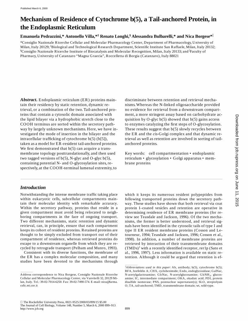
To plan a strategy for the investigation of the mechanismof residence of b(5) in the ER, it was first necessary to de-fine the topology of the hydrophobic membrane-anchor-ing domain. Although b(5) is perhaps the first TA proteinto have been characterized, there has been controversy asto whether it adopts a transmembrane disposition orwhether it loops back into the cytosol. We investigated thisproblem with two different approaches as described below.
In the first approach, we used an Ab raised against theCOOH terminus of b(5) (see Materials and Methods) toprobe the accessibility of the COOH-terminal polar pep-tide by immunofluorescence in an MDCK cell line stablyexpressing b(5). Although this cell line originated from asingle clone, only
z
30% of the cells expressed detectablelevels of b(5) at any one time. Cells were treated eitherwith SLO to selectively permeabilize the plasma mem-brane (Bhakdi et al., 1993), or with Triton X-100 after fixa-tion to permeabilize intracellular membranes. As shown inFig. 1, when the cells were permeabilized only with SLO,the cytosol became accessible to IgG, as demonstrated bythe staining with antitubulin mAbs, which were equally ef-fective with or without Triton X-100 permeabilization(compare Fig. 1, B and D). In contrast, the ER lumen ofSLO-permeabilized cells was poorly accessible, as demon-strated by the low (probably nonspecific) staining ob-tained with mAbs against the lumenal protein PDI (com-pare Fig. 1, F and H). Under this same condition, the
on June 11, 2015jcb.rupress.org
Dow
nloaded from
Published March 6, 2000
The Journal of Cell Biology, Volume 148, 2000 902
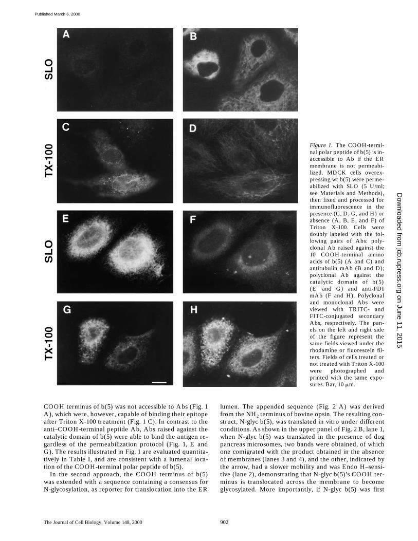
COOH terminus of b(5) was not accessible to Abs (Fig. 1A), which were, however, capable of binding their epitopeafter Triton X-100 treatment (Fig. 1 C). In contrast to theanti–COOH-terminal peptide Ab, Abs raised against thecatalytic domain of b(5) were able to bind the antigen re-gardless of the permeabilization protocol (Fig. 1, E andG). The results illustrated in Fig. 1 are evaluated quantita-tively in Table I, and are consistent with a lumenal loca-tion of the COOH-terminal polar peptide of b(5).
In the second approach, the COOH terminus of b(5)was extended with a sequence containing a consensus forN-glycosylation, as reporter for translocation into the ER
lumen. The appended sequence (Fig. 2 A) was derivedfrom the NH
2
terminus of bovine opsin. The resulting con-struct, N-glyc b(5), was translated in vitro under differentconditions. As shown in the upper panel of Fig. 2 B, lane 1,when N-glyc b(5) was translated in the presence of dogpancreas microsomes, two bands were obtained, of whichone comigrated with the product obtained in the absenceof membranes (lanes 3 and 4), and the other, indicated bythe arrow, had a slower mobility and was Endo H–sensi-tive (lane 2), demonstrating that N-glyc b(5)’s COOH ter-minus is translocated across the membrane to becomeglycosylated. More importantly, if N-glyc b(5) was first
Figure 1. The COOH-termi-nal polar peptide of b(5) is in-accessible to Ab if the ERmembrane is not permeabi-lized. MDCK cells overex-pressing wt b(5) were perme-abilized with SLO (5 U/ml;see Materials and Methods),then fixed and processed forimmunofluorescence in thepresence (C, D, G, and H) orabsence (A, B, E, and F) ofTriton X-100. Cells weredoubly labeled with the fol-lowing pairs of Abs: poly-clonal Ab raised against the10 COOH-terminal aminoacids of b(5) (A and C) andantitubulin mAb (B and D);polyclonal Ab against thecatalytic domain of b(5)(E and G) and anti-PDImAb (F and H). Polyclonaland monoclonal Abs wereviewed with TRITC- andFITC-conjugated secondaryAbs, respectively. The pan-els on the left and right sideof the figure represent thesame fields viewed under therhodamine or fluorescein fil-ters. Fields of cells treated ornot treated with Triton X-100were photographed andprinted with the same expo-sures. Bar, 10 mm.
on June 11, 2015jcb.rupress.org
Dow
nloaded from
Published March 6, 2000
Pedrazzini et al.
Intracellular Sorting of Cytochrome b(5)
903
translated, and microsomes that were added after transla-tion were blocked by CHX, and ribosomes were removedby sedimentation, N-glyc b(5) was still glycosylated withhigh efficiency (lanes 5 and 6), demonstrating that the hy-drophobic domain is capable of translocating downstreamresidues posttranslationally. As a control, we analyzed thebehavior of phaseolin, a plant vacuolar glycoprotein that istranslocated across the ER cotranslationally (Ceriotti etal., 1991). As shown in the lower panel of Fig. 2 B, lane 1,phaseolin was modified to the mono- and diglycosylatedforms (marked by one and two asterisks, respectively)when microsomes were present during translation, but re-mained unglycosylated when membranes were added aftertranslation (lanes 5 and 6).
b(5) Overexpressed in CV1 Cells Is in the ER in a Nonaggregated State
In mammalian cells, the first station after exit from the ERis a tubular vesicular compartment, also known as an inter-mediate compartment (IC) (Bannykh and Balch, 1997).Some proteins that are kept in the ER by a retrieval mech-anism can be detected by immunofluorescence in the IC(Jackson et al., 1993; Martire et al., 1996; Lotti et al., 1999),and this IC localization can be increased by incubation at15
8
C, a temperature that blocks exit from the IC (Jacksonet al., 1993). We investigated whether this was also thecase for b(5) by comparing its intracellular distributionwith that of ERGIC 53, a marker for the IC (Schweizer et al.,1988), after incubation of transfected CV1 cells at 37 or15
8
C. As can be seen in Fig. 3, b(5) could not be detectedin structures enriched in ERGIC 53 either under normalincubation conditions (Fig. 3, A–C), or after prolonged in-cubation at 15
8
C (Fig. 3, D–F). Under the latter condition,ERGIC 53–containing elements clustered in the Golgiarea (compare Fig. 3, E and B) in agreement with the re-cent observations of Klumperman et al. (1998). It shouldbe mentioned that at longer times after transfection (
.
24 h),b(5) was often seen to concentrate in large structures thatwere positive for ER markers (see Fig. 10). These struc-tures may be related to the karmellae, which are inducedin yeast cells by overexpression of b(5) (Vergères et al.,1993). To avoid excessive formation of these structures, allthe experiments described in this paper were initiatedwithin 24 h after transfection.
Table I. Quantitative Analysis of b(5) Immunofluorescence in Transfected MDCK Cells Permeabilized with SLO or withSLO
1
Triton X-100
Triton X-100 Antibodies againstTotal cells
counted Positive cells
Number %
1
Catalytic domain 303 102 33.7
2
Catalytic domain 218 77 35.3
1
COOH-terminal polar peptide 282 71 25.2
2
COOH-terminal polar peptide 168 0 0
MDCK cells expressing b(5) were permeabilized with 5 U/ml SLO and then fixed with4% paraformaldehyde (see Materials and Methods). Coverslips were processed forimmunofluorescence using primary Abs with the indicated specificities in the absenceor presence of 0.6% Triton X-100. 10 photomicrographs of random fields, each inphase-contrast and in epifluorescence, were taken with a 40
3
magnification lens withthe same exposure for each sample, and total cells as well as positive immunofluorescentcells were counted.
Figure 2. In vitro translated N-glyc b(5) becomes glycosylated inthe presence of dog pancreas microsomes added either during orafter translation. (A) COOH-terminal sequences of wt b(5) andof the N-glyc and O-glyc constructs. In all constructs, the hatchedrectangle represents the COOH-terminal part of the membrane-anchoring domain. The underlined sequence in N-glyc b(5) cor-responds to the first 19 amino acids of bovine opsin, and is con-nected to the COOH terminus of b(5) through a Ser and an Argresidue (italics). An N-glycosylation consensus sequence isboxed. The underlined sequence of O-glyc b(5), connected to theCOOH terminus of b(5) via the SR sequence (italics), corre-sponds to residues 36–48 and residue 50 of mature human gly-cophorin A. The residues in boldface (Y between V48 and T50and the three COOH-terminal prolines) were included to create anoptimal consensus for O-glycosylation (Yoshida et al., 1997). Theboxed residues are those that are glycosylated in native glyco-phorin A (Tomita and Marchesi, 1975). (B) N-glyc b(5) (upperpanel) or bean phaseolin (lower panel) synthetic RNAs weretranslated for 1 h in the rabbit reticulocyte system in the presence(lanes 1 and 2; Mbs co) or absence (lanes 3–6) of dog pancreasmicrosomes. One sample, which was first incubated in the ab-sence of microsomes, was then treated with CHX, subjected toultracentrifugation to remove ribosomes (see Materials andMethods), and further incubated for 1 h with microsomes (lanes 5and 6; Mbs post). At the end of the incubations, samples were di-vided into two, denatured with SDS, and then incubated over-night in the presence (lanes 2, 4, and 6) or absence (lanes 1, 3, and5) of Endo H (see Materials and Methods). The arrow in the up-per panel indicates the Endo H–sensitive polypeptide (glyco-sylated N-glyc b[5]) generated when microsomes were presentduring or after translation. The single and double asterisks in thelower panel indicate the mono- and diglycosylated forms ofphaseolin, respectively (Ceriotti et al., 1991), which were gener-ated only when membranes were present during the translation.Numbers on the left indicate the position and size (in kD) of Mrmarkers.
on June 11, 2015jcb.rupress.org
Dow
nloaded from
Published March 6, 2000

The Journal of Cell Biology, Volume 148, 2000 904
Because a number of ER membrane proteins, includingthe ribophorins, are found in rapidly sedimenting aggre-gates after solubilization of microsomes with nonionic de-tergents, it is thought that in situ they form an intramem-
branous proteinaceous network, which could also be thebasis for their retention in the ER (Yu et al., 1989). To in-vestigate whether overexpressed b(5) might belong tosuch an aggregate, we analyzed its sedimentation on su-crose velocity gradients after detergent extraction fromtransfected CV1 cells and compared it with that of ribo-phorin I and of b(5) endogenous to rat liver microsomes.As can be seen from Fig. 4, under the conditions used, ri-bophorin I extracted both from rat liver microsomes (Fig.4 A) and from transfected CV1 cells (Fig. 4 B) formed rap-idly sedimenting aggregates, in agreement with publishedwork (Hortsch et al., 1986; Yu et al., 1989). In contrast,b(5) from rat liver microsomes or transfected CV1 cellswas recovered in the upper fractions of the gradient,cosedimenting with cytochrome c as expected for themonomeric form of the protein (
M
r
z
15,000), suggestingthat it exists in a freely diffusible form within the ER ofthe transfected cells.
Use of Tagged Versions of b(5) to Follow Its Intracellular Trafficking
To investigate with biochemical methodology whether
Figure 3. Comparison of thedistribution of overexpressedwt b(5) with that of endoge-nous ERGIC 53 in trans-fected CV1 cells incubated at37 or 158C. Transfected cellswere incubated in DME,Hepes modification (SigmaChemical Co.) in thermo-stated water baths for 5 h at378C (A–C) or 158C (D–F),fixed and permeabilized, anddoubly stained with anti-b(5)mAbs followed by TRITC-conjugated anti–mouse IgG(A and D), and polyclonalanti–ERGIC 53 followed byFITC-conjugated anti–rab-bit IgG (B and E). A singleconfocal section is shown foreach incubation condition.Images of the same fieldwere acquired separatelywith Texas red (A and D)and fluorescein (B and E) fil-ters. C and F show themerged images of the twoacquisitions. Bar, 10 mm.
Figure 4.
Velocity gradient analysis of b(5) solubilized from ratliver microsomes or transfected CV1 cells. Rat liver microsomes
(A) or CV1 cells transfected with wt b(5) cDNA were lysed andloaded onto a 5–20% sucrose gradient containing 0.2% TritonX-100 and low salt, as described in Materials and Methods. Frac-tions were analyzed by Western blotting. Ribophorin (R) formsaggregates which distribute throughout the gradient and sedi-ment into the pellet, whereas b(5) is found only in the top frac-tions, cosedimenting with cytochrome c. The vertical large andsmall arrowheads in both panels indicate the positions reachedby cytochrome c (
M
r
z
12,300) and catalase (
M
r
z
232,000), re-spectively. Numbers on the left of the panels indicate the positionand size (in kD) of
M
r
markers used in the Western blot analysis.
on June 11, 2015jcb.rupress.org
Dow
nloaded from
Published March 6, 2000
Pedrazzini et al.
Intracellular Sorting of Cytochrome b(5)
905
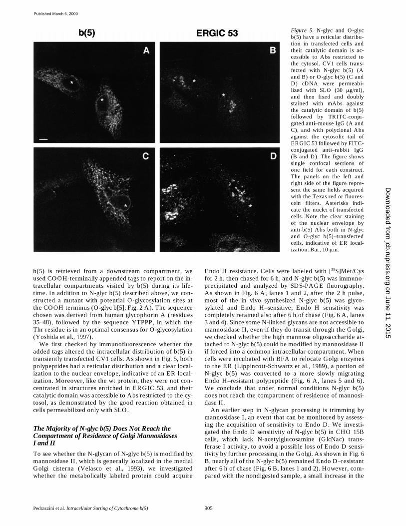
b(5) is retrieved from a downstream compartment, weused COOH-terminally appended tags to report on the in-tracellular compartments visited by b(5) during its life-time. In addition to N-glyc b(5) described above, we con-structed a mutant with potential O-glycosylation sites atthe COOH terminus (O-glyc b[5]; Fig. 2 A). The sequencechosen was derived from human glycophorin A (residues35–48), followed by the sequence YTPPP, in which theThr residue is in an optimal consensus for O-glycosylation(Yoshida et al., 1997).
We first checked by immunofluorescence whether theadded tags altered the intracellular distribution of b(5) intransiently transfected CV1 cells. As shown in Fig. 5, bothpolypeptides had a reticular distribution and a clear local-ization to the nuclear envelope, indicative of an ER local-ization. Moreover, like the wt protein, they were not con-centrated in structures enriched in ERGIC 53, and theircatalytic domain was accessible to Abs restricted to the cy-tosol, as demonstrated by the good reaction obtained incells permeabilized only with SLO.
The Majority of N-glyc b(5) Does Not Reach the Compartment of Residence of Golgi MannosidasesI and II
To see whether the N-glycan of N-glyc b(5) is modified bymannosidase II, which is generally localized in the medialGolgi cisterna (Velasco et al., 1993), we investigatedwhether the metabolically labeled protein could acquire
Endo H resistance. Cells were labeled with [
35
S]Met/Cysfor 2 h, then chased for 6 h, and N-glyc b(5) was immuno-precipitated and analyzed by SDS-PAGE fluorography.As shown in Fig. 6 A, lanes 1 and 2, after the 2 h pulse,most of the in vivo synthesized N-glyc b(5) was glyco-sylated and Endo H–sensitive; Endo H sensitivity wascompletely retained also after 6 h of chase (Fig. 6 A, lanes3 and 4). Since some N-linked glycans are not accessible tomannosidase II, even if they do transit through the Golgi,we checked whether the high mannose oligosaccharide at-tached to N-glyc b(5) could be modified by mannosidase IIif forced into a common intracellular compartment. Whencells were incubated with BFA to relocate Golgi enzymesto the ER (Lippincott-Schwartz et al., 1989), a portion ofN-glyc b(5) was converted to a more slowly migratingEndo H–resistant polypeptide (Fig. 6 A, lanes 5 and 6).We conclude that under normal conditions N-glyc b(5)does not reach the compartment of residence of mannosi-dase II.
An earlier step in N-glycan processing is trimming bymannosidase I, an event that can be monitored by assess-ing the acquisition of sensitivity to Endo D. We investi-gated the Endo D sensitivity of N-glyc b(5) in CHO 15Bcells, which lack N-acetylglucosamine (GlcNac) trans-ferase I activity, to avoid a possible loss of Endo D sensi-tivity by further processing in the Golgi. As shown in Fig. 6B, nearly all of the N-glyc b(5) remained Endo D–resistantafter 6 h of chase (Fig. 6 B, lanes 1 and 2). However, com-pared with the nondigested sample, a small increase in the
Figure 5. N-glyc and O-glycb(5) have a reticular distribu-tion in transfected cells andtheir catalytic domain is ac-cessible to Abs restricted tothe cytosol. CV1 cells trans-fected with N-glyc b(5) (Aand B) or O-glyc b(5) (C andD) cDNA were permeabi-lized with SLO (30 mg/ml),and then fixed and doublystained with mAbs againstthe catalytic domain of b(5)followed by TRITC-conju-gated anti–mouse IgG (A andC), and with polyclonal Absagainst the cytosolic tail ofERGIC 53 followed by FITC-conjugated anti–rabbit IgG(B and D). The figure showssingle confocal sections ofone field for each construct.The panels on the left andright side of the figure repre-sent the same fields acquiredwith the Texas red or fluores-cein filters. Asterisks indi-cate the nuclei of transfectedcells. Note the clear stainingof the nuclear envelope byanti-b(5) Abs both in N-glycand O-glyc b(5)–transfectedcells, indicative of ER local-ization. Bar, 10 mm.
on June 11, 2015jcb.rupress.org
Dow
nloaded from
Published March 6, 2000

The Journal of Cell Biology, Volume 148, 2000 906
amount of deglycoyslated polypeptide was visible afterEndo D treatment (Fig. 6 B, arrow), suggesting that asmall proportion of the tagged b(5) may have had accessto mannosidase I. After incubation with BFA, all of theN-glyc b(5) became Endo D–sensitive (Fig. 6 B, lanes 3and 4). These results indicate that the vast majority ofN-glyc b(5) molecules do not reach the compartment ofresidence of mannosidase I during a 6-h chase.
Glycosylation of O-glyc b(5)
The results presented in Fig. 6 indicate that N-glyc b(5)does not recycle through the medial Golgi; however, it waspossible that it reaches an earlier Golgi subcompartment.To investigate this possibility, we turned to the O-glyc b(5)construct. The first step in O-glycosylation is attachmentof an N-acetylgalactosamine (GalNac) residue to Thr orSer residues, and the enzymes responsible for this reac-tion, the GalNac transferases, have been localized to the
cis
-Golgi as well as to more distal cisternae (Röttger et al.,1998).
We first compared the electrophoretic mobility of invivo synthesized O-glyc b(5) with that of the in vitro gen-erated primary translation product. As shown in Fig. 7 A,the product expressed in transfected cells (Fig. 7 A, lane 1)
comigrated with the product synthesized in vitro in the ab-sence (Fig. 7 A, lane 2) or presence (Fig. 7 A, lane 3) ofmembranes. However, if cells were incubated with BFA,O-glyc b(5) was shifted to a higher apparent
M
r
(compareFig. 7 B, lanes 3 and 4, or Fig. 7 B, lanes 5 and 6), indicat-ing that at least one of the potential O-glycosylation sitescould be used if the protein came into contact with the rel-evant enzymes. To see whether in the absence of BFA theinitial steps of O-glycosylation were also taking place butwere not resulting in reduced electrophoretic mobility ofO-glyc b(5), we carried out metabolic labeling experi-ments with
3
H-labeled sugars. As shown in Fig. 7 B, incu-bation with [
3
H]GlcNH
2
, which serves as precursor both toUDP-GlcNac and to UDP-GalNac, resulted in labeling ofO-glyc b(5) both in the absence (Fig. 7 B, lane 3) and thepresence (Fig. 7 B, lane 4) of BFA. The labeling was spe-cific, because under the same conditions, wt b(5) was notlabeled (Fig. 7 B, lanes 1 and 2), although it was efficientlyexpressed, as shown by the Western blot in the lower partof Fig. 7 B.
The second sugar to be added on O-linked oligosaccha-rides is galactose. With [
3
H]galactose as radioactive pre-cursor, O-glyc b(5) became labeled in the presence ofBFA (Fig. 7 B, lane 6), and a weak signal was obtainedalso in the absence of the drug (Fig. 7 B, lane 5). Thus, atsteady-state, a small portion of the O-glyc mutant has alsoundergone the second step of O-glycosylation.
To investigate how rapidly glycosylation of O-glyc b(5)occurs after its synthesis, we followed the time course ofincorporation of
3
H-labeled sugar after blocking proteinsynthesis with CHX (Fig. 8). Cells were first incubated for3 h with [
3H]GlcNH2 to allow for equilibration betweenUDP-[3H]GlcNac and UDP-[3H]GalNac. Incubation wasthen continued in the presence or absence of CHX. Tocontrol for the efficacy of the block in protein synthesis, aseparate set of dishes of transfected cells was incubated inparallel with [35S]Met/Cys in the presence or absence ofCHX, and O-glyc b(5) immunoprecipitates were then ana-lyzed (Fig. 8 B). CHX efficiently blocked the synthesis ofO-glyc b(5) (Fig. 8 B; compare lanes 2 and 4 with lanes 1and 3). However, as shown in Fig. 8 A and on the graph onthe right side of Fig. 8 C (obtained by normalizing theamount of immunoprecipitated 3H-labeled O-glyc b[5] tothe amount of O-glyc b[5] expressed, as determined byWestern blotting), the block in protein synthesis had no ef-fect at all on the rate of sugar incorporation, which contin-ued at a linear rate for 4 h after addition of the drug. Thisresult stands in sharp contrast to the strong inhibitory ef-fect of CHX on the incorporation of labeled sugar into to-tal cellular proteins (graph on the left side of Fig. 8 C).Thus, there is a large pool of unglycosylated O-glyc b(5)molecules that acquire O-linked sugar at a low rate. Itshould be mentioned that incubation with CHX for 4 h didnot cause an appreciable decrease in the total amount ofO-glyc b(5) or in the accumulation of the labeled species(compare Fig. 8 A, lanes 4 and 5) in this and other experi-ments, indicating that under these conditions O-glyc b(5)is stable. The variations in the amounts sometimes de-tected in Western blots (e.g., Fig. 8 A, lanes 3–5 vs. Fig. 8A, lanes 1 and 2) are presumably due to differences intransfection efficiencies in the different dishes.
The bulk of the available evidence indicates that the
Figure 6. The N-linked oligosaccharide of N-glyc b(5) retainsEndo H sensitivity and Endo D resistance. (A) CV1 cells trans-fected with N-glyc b(5) cDNA were labeled for 2 h with [35S]Met/Cys, then chased for a further 6 h. One set of cells was exposed toBFA (10 mg/ml) 30 min before starting the labeling, and was keptwith the drug for the entire pulse and chase period (lanes 5 and6). At the end of the pulse (lanes 1 and 2) or after the chase(lanes 2–6), cells were harvested, lysed, and subjected to immu-noprecipitation with antiopsin mAbs. Aliquots containing thesame amounts of incorporated radioactive amino acid were usedfor each immunoprecipitation. One half of each immunoprecipi-tate was treated with Endo H (lanes 2, 4, and 6), whereas theother half was left untreated (lanes 1, 3, and 5). (B) CHO 15Bcells, transfected with N-glyc b(5) cDNA, were labeled for 2 hand chased for 6 h with [35S]Met/Cys. One set of cells (lanes 3 and4) was exposed to BFA as in A. At the end of the chase period,cells were harvested, lysed, and subjected to immunoprecipita-tion as in A. One half of the immunoprecipitates was treated withEndo D (lanes 2 and 4), whereas the other half was left untreated(lanes 1 and 3). The figure shows the result of the SDS-PAGEfluorography analysis of the Endo H– or Endo D–treated and un-treated immunoprecipitates. The position of the 21-kD molecularmass marker is shown at the left of each panel. The vertical arrowin lane 2 of B indicates the position of the unglycosylated N-glycb(5).
on June 11, 2015jcb.rupress.org
Dow
nloaded from
Published March 6, 2000

Pedrazzini et al. Intracellular Sorting of Cytochrome b(5) 907
process of O-glycosylation occurs in the Golgi complex(Roth et al., 1994; Schweizer et al., 1994; Röttger et al.,1998). However, under some conditions, especially in in-fected cells, the initial steps may take place in a compart-ment upstream of the Golgi (discussed in Röttger et al.,1998). Moreover, recent evidence indicates that Golgi-res-ident enzymes slowly recycle through the ER (Cole et al.,1998; Storrie et al., 1998; Girod et al., 1999). To investigatewhether O-glyc b(5) must leave the ER to become glyco-sylated, we treated cells with the phosphatase inhibitorOKA, a drug which blocks exit from the ER (Davidson et al.,1992; Pryde et al., 1998). Transfected cells were exposed to[3H]GlcNH2 for 3 h and then incubated further for anotherhour in the presence or absence of OKA. At this time cellswere collected for biochemical analysis or analyzed by im-munofluorescence. As shown in Fig. 9 C, the intracellularlocalization of O-glyc b(5) (Fig. 9 C, panels a and d) and ofa Golgi marker (giantin; Fig. 9 C, panels c and f) were un-affected by OKA. In contrast, ERGIC 53 was dispersedfrom the Golgi area to more peripherally located struc-tures (compare Fig. 9 C, panels e and b). These results areconsistent with OKA’s inhibition of ER-to-Golgi trans-port. At slightly later times of incubation, ERGIC 53 ap-peared uniformly distributed within the ER, but the cellsbegan to suffer and to retract from the substrate (data notshown). Analysis by immunoprecipitation and Westernblotting (Fig. 9, A and B) showed a 3.2-fold increase in la-beling of O-glyc b(5) between 3 and 4 h in the absence ofOKA, and an z50% inhibition of this increase by OKA.This is a fairly strong effect if one considers that OKAdoes not exert its full inhibitory activity immediately afteraddition (Davidson et al., 1992).
To exclude the possibility that OKA was decreasingO-glyc b(5) glycosylation through a direct or indirect inhibi-
tion of glycosyl transferases and not by a transport block,we analyzed its effect in the presence of BFA. As can beseen in Fig. 9 A, the effect of OKA was reversed in thepresence of BFA (Western blot; Fig. 9 A, lanes 3 and 5).During a 1-h period of incubation with BFA, 41.7 6 1.7%and 37.6 6 1.6% (average 6 half-range of two experi-ments) of O-glyc b(5) was converted to the slower migrat-ing, fully processed form in the absence and presence ofOKA, respectively.
Another important piece of information in Fig. 9 can beobtained by examining the ratio of specific radioactivity(immunoprecipitate/Western) of O-glyc b(5) attained inthe absence and presence of BFA (Fig. 9 A, lanes 2 and 3).By scanning a lower exposure of fluorographs in which thebands labeled in the presence of BFA had not saturatedthe film (data not shown), we found that the specific radio-activity attained in 1 h by O-glyc b(5) in the absence ofdrugs was z7% that attained by the lower Mr polypeptideof the doublet generated in the presence of BFA. Since thehigher Mr band presumably contains additional labeledsugar, it could not be compared with the more rapidly mi-grating polypeptide. If one assumes that the lower Mrband of Fig. 9 A, lane 3 is 100% glycosylated, this wouldindicate a half-time for glycosylation of O-glyc b(5) (in theabsence of BFA) of z10 h, in agreement with the slow ac-quisition of sugar after the blockade of protein synthesisdemonstrated in Fig. 8. This half-time may be an underes-timate, since a fraction of the low Mr polypeptide mole-cules of Fig. 9 A, lane 3 may be unglycosylated.
The results of Fig. 9 suggest that O-glyc b(5) must leavethe ER to become glycosylated. But does O-glyc b(5) re-turn to its compartment of origin once it has exited? Thisquestion was investigated both by immunofluorescenceand by cell fractionation (Fig. 10). We carried out immuno-
Figure 7. O-glyc b(5) becomes glycosylated invivo. (A) Comparison of SDS-PAGE mobility ofin vivo and in vitro synthesized O-glyc b(5). CV1cells transfected with O-glyc b(5) cDNA were la-beled for 2 h with [35S]Met/Cys, harvested, andsubjected to immunoprecipitation with anti-b(5)Abs (lane 1). Alternatively, synthetic RNA cod-ing for O-glyc b(5) was translated in vitro eitherin the absence (lane 2) or in the presence (lane 3)of dog pancreas microsomes. SDS-PAGE fol-lowed by fluorography fails to reveal a differencebetween the apparent Mr of the protein synthe-sized in vivo and that of the primary translationproduct produced in vitro. (B) Metabolic label-ing of O-glyc b(5) with 3H-labeled sugars. CV1cells, transfected with wt b(5) (lanes 1 and 2) orwith O-glyc b(5) (lanes 3–6) cDNA, were incu-bated for 7 h with [3H]GlcNH2 (lanes 1–4) or[3H]Gal (lanes 5 and 6). For each labeling condi-tion, one set of cells (lanes 2, 4, and 6) received10 mg/ml of BFA 30 min before exposure to thelabeled sugar, and the drug was maintained inthe medium during the entire subsequent incuba-tion. At the end of the incubation, cells were har-vested, lysed, and aliquots containing the sameamounts of incorporated radioactive sugar were
subjected to immunoprecipitation with anti-b(5) Abs. Immunoprecipitates were analyzed by SDS-PAGE fluorography (upper panel).One fourth of the amounts used for immunoprecipitation were analyzed by Western blotting with anti-b(5) Abs (lower panel). The lineon the left side of all panels indicates the position of the 21-kD molecular mass marker.
on June 11, 2015jcb.rupress.org
Dow
nloaded from
Published March 6, 2000

The Journal of Cell Biology, Volume 148, 2000 908
fluorescence analysis of transfected cells after long expo-sures (4 and 8 h) to CHX. From the data of Fig. 8, O-glycb(5) acquires sugar linearly for up to 4 h in the presence ofCHX, and on the basis of the estimate of 10 h for the half-
time of O-glycosylation (Fig. 9), z25% of O-glyc b(5)molecules should have acquired sugar at this time. A con-centration of these glycosylated molecules in the glycosy-lation compartment (cis-Golgi) should have been detect-able by immunofluorescence. However, as shown in Fig.10 A, the tagged protein retained a similar distribution tothat of the ER protein ribophorin I at 4 h of incubation(Fig. 10 A, panels a–c), and did not colocalize with theGolgi marker giantin even after 8 h of incubation withCHX (Fig. 10 A, panels d–f). Fig. 10 A, panels a–c alsoshow an example of the b(5)-rich structures that began toappear in some of the cells in this experiment (the arrow-heads indicate two such structures). As shown in Fig. 10 A,panels b and c, ribophorin I also localized to these struc-tures, suggesting that they represent proliferations of theER. The frequency of these structures was, in any case, thesame in cells incubated with or without CHX (data notshown).
The subcellular localization of glycosylated O-glyc b(5)was also analyzed by Optiprep density gradient centrifu-gation of a PNS from cells metabolically labeled with[3H]GlcNH2 (Fig. 10 B). Under the conditions used, theER marker NADPH-cyt P450 reductase showed a sharppeak of activity centered at fraction 6, corresponding to adensity of 1.1 (Fig. 10 B, panel b), whereas GS28, a markerfor the cis-Golgi (Subramaniam et al., 1995), was broadlydistributed throughout the gradient and present also in thebottom fractions immediately adjacent to the load zone(Fig. 10 B, panel d). Total O-glyc b(5), detected by West-ern blot (Fig. 10 B, panel a), peaked at a slightly lowerdensity (fraction 5) than the reductase. A portion ofO-glyc b(5) floated to the top of the gradient. This mate-rial probably corresponds to membrane fragments of lowdensity, which we have not characterized further. Finally,3H-labeled O-glyc b(5), detected by immunoprecipitation(Fig. 10 B, panel c), had a distribution similar to that of to-tal O-glyc b(5), and was depleted from the region of thegradient more enriched in GS28 (fractions 7–9). These re-sults suggest that unglycosylated and glycosylated O-glycb(5) molecules coexist within the same compartment, andalso that both may be enriched in smooth subdomains ofthe ER.
DiscussionBoth retention and retrieval mechanisms are thought to beinvolved in keeping ER resident proteins from escapingdown the secretory pathway. The identification of specificretrieval sequences has spurred a large amount of workthat has resulted in a significant increase in our under-standing of the retrograde pathway (Pelham and Munro,1993; Cosson and Letourneur, 1994; Teasdale and Jack-son, 1996; Cosson et al., 1998). However, most of this workhas been carried out on chimeras, consisting of a retrievalsignal grafted onto a protein normally destined for exportfrom the ER, and there is still not much information onthe relative roles of retention and retrieval in determiningthe residence of bona fide ER proteins. A few mammalianER membrane proteins have been studied by analyzingthe processing of N-linked glycans attached to endogenousor engineered consensus sites. Due the failure of theseproteins to acquire Endo H resistance, it has been con-
Figure 8. Time course of posttranslational addition of 3H-labeledsugar to O-glyc b(5). (A) Metabolic labeling with [3H]GlcNH2.Cells transfected with O-glyc b(5) were incubated for 3 h with[3H]GlcNH2 to allow for equilibration between UDP-[3H]GlcNAc and UDP-[3H]GalNAc. Cells from one dish wereharvested at this point (lane 1), whereas the others were incu-bated for a further 1 (lanes 2 and 3) or 4 (lanes 4 and 5) h in thepresence or absence of 15 mg/ml CHX as indicated. For each timepoint, cell lysates were prepared, and equal amounts of proteinwere used for immunoprecipitation with anti-b(5) Abs (upperpanel). A proportional amount of each lysate (one fifth of theamount used for immunoprecipitation) was analyzed by Westernblotting (lower panel). (B) Block of O-glyc b(5) synthesis byCHX. Cells transfected with O-glyc b(5) were incubated in paral-lel with those in A, but without added [3H]GlcNH2. After 3 h,[35S]Met/Cys was added and incubation was continued in thepresence or absence of 15 mg/ml CHX, as indicated. At the indi-cated times, cell lysates were prepared and equal amounts of pro-tein were used for immunoprecipitation, as in A. (C) Graphicalrepresentation of the results of A compared with the incorpora-tion of 3H-labeled sugar into total protein. The graph on the rightshows the ratios of band intensities of fluorographs (metaboli-cally labeled O-glyc b[5]) to those of the Western blots (totalO-glyc b[5]). Shown are the means from three sets of dishes 6SEM. The value for the ratio at 3 h (time of addition of CHX) hasbeen normalized to 1. The graph on the left represents totalTCA-precipitable 3H-labeled sugar normalized to total proteincontent. Squares, no CHX; triangles, CHX.
on June 11, 2015jcb.rupress.org
Dow
nloaded from
Published March 6, 2000

Pedrazzini et al. Intracellular Sorting of Cytochrome b(5) 909
cluded that they reside in the ER by a true retentionmechanism (Ahn et al., 1993; Masaki et al., 1996; Duvetet al., 1998; Honsho et al., 1998). However, since Endo Hresistance is acquired in the medial Golgi, these studiescould not exclude that retrieval from an earlier Golgi com-partment was occurring. In this study, we have addressedthis problem by attaching short sequences, containing ei-ther N- or O-glycosylation sites, to the COOH terminus ofb(5), a TA protein of the ER. We show that in the O-glycmutant at least one glycosylation site is used, indicatingthat the tagged protein comes in contact with cis-Golgi en-
zymes, an event which could not be detected by analysis ofthe oligosaccharide of the N-glyc–tagged version. Themethodology used here should also be useful for the studyof the trafficking of other ER membrane proteins, particu-larly of those lacking lumenal domains with endogenousglycosylation sites.
Posttranslational Translocation of the COOH Terminus of b(5) across the ER Membrane
Before undertaking an investigation of the intracellular
Figure 9. OKA inhibits sugaracquisition by O-glyc b(5).(A) CV1 cells transfectedwith O-glyc b(5) were incu-bated with [3H]GlcNH2 for3 h to allow for equilibrationbetween UDP-[3H]GlcNAcand UDP-[3H]GalNAc. Atthis time, one set of cells washarvested (lane 1), whereasthe others were incubated fora further hour in the presence(lanes 4 and 5) or absence(lanes 2 and 3) of 1 mM OKAwith or without 10 mg/mlBFA, as indicated. Afterclearing of lysates, equalamounts of protein were sub-jected to immunoprecipita-tion with anti-b(5) Abs (up-per panel). A proportionalamount of each lysate (onefifth of the amount used forimmunoprecipitation) wasanalyzed by Western blotting(lower panel). (B) Graphicalrepresentation of the resultsof two experiments like theone in A. Two separate setsof dishes were labeled as de-scribed for A. The ratio of theintensities of the O-glyc b(5)bands detected by fluorogra-phy to that of the bands incorresponding samples de-tected by Western blotting isplotted. Shown are the meansvalues 6 half-range. (C) Im-munofluorescence analysis ofcells treated or not treatedwith 1 mM OKA. After 1 hwith (C, panels d–f) or with-out (C, panels a–c) OKA,cells were fixed. Separatecoverslips were analyzed forb(5) (C, panels a and d),ERGIC 53 (C, panels b ande), and giantin (C, panels cand f). Note the redistribu-tion of ERGIC 53 induced byOKA, whereas O-glyc b(5)and giantin appear unaffectedby the drug. Bar, 20 mm.
on June 11, 2015jcb.rupress.org
Dow
nloaded from
Published March 6, 2000

The Journal of Cell Biology, Volume 148, 2000 910
sorting of b(5), we judged it necessary to define the topol-ogy of its membrane-anchoring domain. Although thisproblem has been the object of numerous investigations,there were still conflicting views as to whether b(5)’s hy-
drophobic domain adopts a transmembrane dispositionwith the extreme COOH-terminal residues in the lumen ofthe ER, or whether it has a hairpin topology with theCOOH terminus looping back into the cytosol (Dailey and
Figure 10. Intracellular dis-tribution of O-glyc b(5). (A)O-glyc b(5) remains in theER after prolonged incuba-tion with CHX. CV1 cellstransfected with O-glyc b(5)were incubated under thesame conditions as in the ex-periments of Figs. 8 and 9,but without the labeled pre-cursor and in the presence of20 mg/ml of CHX for 4 (A,panels a–c) or 8 (A, panelsd–f) h. Fixed cells were dou-bly labeled with anti-b(5)and anti–ribophorin I Abs(A, panels a–c), or with anti-b(5) and antigiantin Abs (A,panels d–f). A single confo-cal section is shown for the 4-and 8-h incubation. Imagesof the same field were ac-quired separately with theTexas red (A, panels a and d)and the fluorescein (A, pan-els b and e) filters. The corre-sponding merged images areshown in A, panels c and f(O-glyc b[5] in red, markerprotein in green). Note theabsence of colocalization ofO-glyc b(5) with giantin andits similar distribution to ri-bophorin I. The arrowheadsin A, panels a–c indicatestructures in which O-glycb(5) accumulates at longtimes after transfection, andwhich are also positive for ri-bophorin I. Bar: (panels a–c)10 mm; (panels d–f) 20 mm.(B) Optiprep density gradi-ent analysis of CV1 cellstransfected with O-glyc b(5)and metabolically labeledwith [3H]GlcNH2. Cells weremetabolically labeled with[3H]GlcNH2 for 6 h, then re-turned to DME for 4 h, be-fore collection, homogeniza-tion, and preparation of aPNS, which was loaded un-der an 8–22% Optiprep gra-dient (see Materials andMethods). After centrifuga-tion, fractions were collectedand analyzed for O-glyc b(5)by Western blotting (B,panel a), NADPH-cyt c re-ductase enzyme activity (B,panel b), 3H-labeled O-glyc
b(5) by immunoprecipitation followed by SDS-PAGE fluorography (B, panel c), and GS28 by Western blotting (B, panel d). The thickline positioned at the right of each graph covers the fractions containing the load zone of the gradient.
on June 11, 2015jcb.rupress.org
Dow
nloaded from
Published March 6, 2000
Pedrazzini et al. Intracellular Sorting of Cytochrome b(5) 911
Strittmatter, 1981; Arinc et al., 1987; Ozols, 1989; Chesteret al., 1992; Borgese et al., 1993; Vergères et al., 1995;Kuroda et al., 1996). However, strong evidence for a trans-membrane topology for b(5) was provided recently by thefinding that a tagged version bearing an N-glycosylationconsensus sequence at the COOH terminus acquiredN-linked carbohydrate (Honsho et al., 1998). Althoughthis study proved that the COOH terminus of this con-struct is translocated into the ER lumen in vivo, it did notinvestigate the mechanism of this translocation, leavingopen the possibility that the addition of polar residues atthe COOH terminus could cause a translocation that doesnot occur with the wt, untagged polypeptide. For instance,the additional 15–20 COOH-terminal amino acids couldlead to a partial exposure of residues of the hydrophobicdomain before termination of translation, resulting in thebinding of the signal recognition particle and in a translo-con-dependent translocation. For this reason, and sincethe wt protein is synthesized on free polysomes and insertsinto membranes after release from the ribosomes, it wasimportant to establish that the translocation of the COOHterminus of COOH-terminally tagged b(5) could occurposttranslationally in the absence of ribosomes. Our re-sults with the N-glyc b(5) mutant show that this is indeedthe case, a strong indication that in the wt protein theseven polar COOH-terminal residues are also in the lu-men of the ER. In agreement with this conclusion, in im-munofluorescence experiments we found that the COOH-terminal polar peptide is not accessible to Abs unless theER membrane is permeabilized with Triton X-100. Takentogether these results justify the use of COOH-terminallytagged versions of b(5) to study its intracellular trafficking.
Although the hydrophobic domain of b(5), in its abilityto translocate downstream residues across the ER mem-brane, superficially resembles the signal anchor sequencesof classical type II proteins, the mechanism by which itfunctions is quite different, since it operates on the re-leased polypeptide in the absence of ribosomes. An invitro posttranslational translocation similar to that of theb(5) COOH terminus has been shown also for synaptobre-vin I (Kutay et al., 1995), a synaptic vesicle soluble N-eth-ylmaleimide–sensitive factor attachment protein receptorwith a considerably longer hydrophobic domain than b(5).The insertion of synaptobrevin appeared to require pro-teins different from those of the classical Sec61-basedtranslocation apparatus. It is likely that b(5) and synapto-brevin, as well as all other TA proteins, translocate theirCOOH terminus with the same mechanism, which cer-tainly deserves further investigation.
Retention or Retrieval?
To investigate whether b(5) is retrieved from the IC orfrom the cis-Golgi, we first compared its distribution byimmunofluorescence with that of the IC marker, ERGIC53, after incubation either under normal conditions or at158C, a temperature which interferes with trafficking be-tween the ER and the Golgi, and which has been shown insome cases to cause accumulation of recycling ER proteinsin the IC (Jackson et al., 1993). However, for b(5), this ap-proach did not yield any evidence in favor of its movementout of the ER. Proteins that recycle slowly may not be cap-tured in the IC by incubation at low temperature (Jackson
et al., 1993), presumably because entry as well as exit fromthe IC may be slowed. Therefore, we turned to the use oflumenally tagged versions of b(5) (N-glyc and O-glycb[5]), with the aim of using the attached oligosaccharidechains as reporters for the intracellular compartments vis-ited by b(5). These tagged constructs were inserted intomembranes with the correct topology, and had an intracel-lular distribution indistinguishable from that of transientlyexpressed wt b(5). During a 6-h chase, N-glyc b(5) did notacquire Endo H resistance, and also remained nearly com-pletely Endo D–resistant, suggesting that it was not reach-ing the medial Golgi. However, when we turned to a morestringent assay based on the incorporation of sugar intothe O-glyc mutant, we found that at least one O-glycosyla-tion site was used, indicating that the protein was cominginto contact with a GalNac transferase, enzymes which arelocalized to the Golgi complex (Röttger et al., 1998). Webelieve that the acquisition of carbohydrate by O-glyc b(5)is reporting on a recycling event through the cis-Golgi, forthe reasons detailed below.
Processing of a glycoprotein by Golgi enzymes has nor-mally been taken as evidence for its passage though theGolgi complex. However, recent evidence showing thatGolgi enzymes recycle slowly through the ER (Cole et al.,1998; Storrie et al., 1998; Girod et al., 1999) makes it moredifficult to interpret the significance of Golgi modifica-tions on ER proteins. Nonetheless, since ER proteins gen-erally do not carry Golgi modifications, it seems that theserecycling Golgi enzymes are not active within the ER.Therefore, it is probable that O-glyc b(5) acquired itssugar after leaving the ER, and this idea is supported bythe finding that glycosylation of O-glyc b(5) could be in-hibited by the phosphatase inhibitor OKA, which blockstransport out of the ER (Davidson et al., 1992; Pryde et al.,1998). If O-glyc b(5) had been modified within the ER byrecycling Golgi enzymes, OKA might actually have in-creased the processing of O-glyc b(5) by the trapped gly-cosyl transferases. Another explanation for our data is thatthe tagged protein, after exiting the ER and becoming gly-cosylated, did not return to its compartment of origin. Thisinterpretation is unlikely, since no redistribution of O-glycb(5) from the ER to another compartment could be ob-served after a chase with CHX under conditions in whichthe proportion of glycosylated O-glyc b(5) moleculeswas increasing at a linear rate. Moreover, [3H]GlcNH2-labeled and total O-glyc b(5) had similar distributions onan Optiprep density gradient, suggesting that they coexistin the same intracellular compartment.
Does the behavior of overexpressed O-glyc b(5) reflectthat of endogenous b(5) in mammalian cells? It is possiblethat overexpression causes leakage from the ER of thetagged b(5) or that the tag itself alters the behavior of b(5).Although we cannot exclude either of these possibilities,the important finding here is that b(5) appears to have thecapacity to be captured and returned to the ER from adownstream compartment, even though the extent of exitfrom the ER for the endogenous molecule and the overex-pressed tagged mutant may be different. The rate of acqui-sition of carbohydrate by O-glyc b(5) was in any case quiteslow: by comparing the extent of labeling of the taggedprotein in the presence or absence of BFA, we estimated ahalf-time for acquisition of O-linked GalNac of z10 h.This slow rate of glycosylation, as well as the lack of colo-
on June 11, 2015jcb.rupress.org
Dow
nloaded from
Published March 6, 2000
The Journal of Cell Biology, Volume 148, 2000 912
calization of wt and tagged versions of b(5) with markersfor the IC, suggest that b(5) tends to be excluded frombudding transport vesicles, and that retrieval from a down-stream compartment represents a salvage mechanism forthose molecules that occasionally leak out of the ER. Inother words, it would appear that both retention and re-trieval are operating together to keep b(5) in the ER. Wedo not know at present whether retention is based simplyon the lack of a positive export signal and/or the presenceof a positive retention signal or feature in b(5). Formationof oligomeric complexes has been suggested to result in re-tention of some ER proteins (Yu et al., 1989). However,this mechanism would not seem to be responsible for b(5)retention. Indeed, our sucrose gradient analysis did notreveal the presence of b(5) oligomers under conditionsin which high-Mr complexes containing ribophorin Iwere preserved. Consistent with this result, preliminary ex-periments carried out with the fluorescence recovery af-ter photobleaching technique suggest that green fluores-cent protein–b(5) chimeras diffuse rapidly across the ER(Snapp, E., and J. Lippincott-Schwartz, personal commu-nication).
Our previous work demonstrated that a short hydropho-bic domain is required to keep b(5) in the ER; lengtheningof the TMD results in escape of b(5) from the ER and itsarrival at the plasma membrane (Pedrazzini et al., 1996).The extension of this finding by other laboratories to otherTA proteins and reporter constructs (Rayner and Pelham,1997; Yang et al., 1997; Honsho et al., 1998) suggests thatTMD-dependent sorting may be a general phenomenonfor TA proteins and may occur by a common mechanism.The results of the present study, which suggest that bothretention and retrieval mechanisms are operating to keepb(5) in the ER, raise the question as to whether the shortTMD is required for retention, retrieval, or for both thesephenomena. If the TMD is involved in retrieval, a candi-date recycling receptor involved in this process is rer1p, aprotein demonstrated to be required for ER residence of anumber of membrane polypeptides in yeast via recogni-tion of their TMDs (Sato et al., 1996, 1997). This receptor,which also has been cloned from mammalian cells (Fulle-krug et al., 1997), appears to prefer short and somewhathydrophilic TMDs (Letourneur and Cosson, 1998). On theother hand, retrieval of TA proteins may occur by a com-pletely different mechanism, since Ufe1p, a TA target sol-uble N-ethylmaleimide–sensitive factor attachment pro-tein receptor of the ER, does not change its localization inrer1p-deficient yeast cells (Rayner and Pelham, 1997).Further work with tagged versions of TA proteins like theones described in this study should help to define at themolecular level the sorting mechanisms of this class of pro-teins.
In addition to all the people who kindly donated the antibodies listed inthe Materials and Methods section, we would like to thank StefanoBonatti, Alessandro Vitale, and Pietro De Camilli for helpful discussionand for critically reading the manuscript; Franca Serafini-Cessi for helpfuladvice on glycoprotein biochemistry; and Francesco Clementi for contin-ued support. In addition, we are grateful to Sara Colombo and MassimoBarberi for their help in the laboratory and in some of the experiments.
This work was supported by grants from Associazione Italiana per laRicerca sul Cancro (AIRC) and from the Italian Ministry of Research(COFIN97). E. Pedrazzini was supported by an AIRC fellowship.
Submitted: 8 July 1999Revised: 10 December 1999Accepted: 18 January 2000
References
Adamus, G., A. Arendt, and P.A. Hargrave. 1991. Genetic control of antibodyresponse to bovine rhodopsin in mice: epitope mapping of rhodopsin struc-ture. J. Neuroimmunol. 34:89–97.
Ahn, K., E. Szczesna-Skorupa, and B. Kemper. 1993. The amino-terminal 29amino acids of cytochrome P450 2C1 are sufficient for retention in the endo-plasmic reticulum. J. Biol. Chem. 268:18726–18733.
Arinc, E., L.M. Rzepecki, and P. Strittmatter. 1987. Topography of the C-termi-nus of cytochrome b5 tightly bound to dimyristoylphosphatidylcholine vesi-cles. J. Biol. Chem. 262:15563–15567.
Bannykh, S.I., and W.E. Balch. 1997. Membrane dynamics at the endoplasmicreticulum-Golgi interface. J. Cell Biol. 138:1–4.
Bhakdi, S., U. Weller, I. Walev, E. Martin, D. Jonas, and M. Palmer. 1993. Aguide to the use of pore-forming toxins for controlled permeabilization ofcell membranes. Med. Microbiol. Immunol. 182:167–175.
Borgese, N., and J. Meldolesi. 1976. Immunological similarity of the NADH-cytochrome c electron transport system in microsomes, Golgi complex andmitochondrial outer membrane of rat liver cells. FEBS Lett. 63:231–234.
Borgese, N., and G. Pietrini. 1986. Distribution of the integral membrane pro-tein NADH-cytochrome b5 reductase in rat liver cells, studied with a quanti-tative radioimmunoblotting assay. Biochem. J. 239:393–403.
Borgese, N., A. D’Arrigo, M. De Silvestris, and G. Pietrini. 1993. NADH-cyto-chrome b5 reductase and cytochrome b5—the problem of posttranslationaltargeting to the endoplasmic reticulum. In Subcellular Biochemistry. Vol.21. N. Borgese and J.R. Harris, editors. Plenum Press, New York. 313–341.
Borgese, N., D. Aggujaro, P. Carrera, G. Pietrini, and M. Bassetti. 1996. A rolefor N-myristoylation in protein targeting: NADH-cytochrome b5 reductaserequires myristic acid for association with outer mitochondrial but not endo-plasmic reticulum membranes. J. Cell Biol. 135:1501–1513.
Brewer, C.B., and M.G. Roth. 1991. A single amino acid change in the cytoplas-mic domain alters the polarized delivery of influenza virus hemagglutinin. J.Cell Biol. 114:413–421.
Ceriotti, A., E. Pedrazzini, M.S. Fabbrini, M. Zoppè, R. Bollini, and A. Vitale.1991. Expression of the wild-type and mutated vacuolar storage proteinphaseolin in Xenopus oocytes reveals relationships between assembly andintracellular transport. Eur. J. Biochem. 202:959–968.
Chester, D.W., V. Skita, H.S. Young, T. Mavromoustakos, and P. Strittmatter.1992. Bilayer structure and physical dynamics of the cytochrome b(5)dimyristoylphosphatidylcholine interaction. Biophys. J. 61:1224–1243.
Cole, N.B., J. Ellenberg, J. Song, D. DiEuliis, and J. Lippincott-Schwartz. 1998.Retrograde transport of Golgi-localized proteins to the ER. J. Cell Biol. 140:1–15.
Cosson, P., and F. Letourneur. 1994. Coatomer interaction with di-lysine endo-plasmic reticulum retention motifs. Science. 263:1629–1631.
Cosson, P., Y. Lefkir, C. Démollière, and F. Letourneur. 1998. New COP1-binding motifs involved in ER retrieval. EMBO (Eur. Mol. Biol. Organ.) J.17:6863–6870.
Dailey, H.A., and P. Strittmatter. 1981. Orientation of the carboxyl and NH2termini of the membrane-binding segment of cytochrome b5 on the sameside of phospholipid bilayers. J. Biol. Chem. 256:3951–3955.
Davidson, H.W., C.H. McGowan, and W.E. Balch. 1992. Evidence for the regu-lation of exocytic transport by protein phosphorylation. J. Cell Biol. 116:1343–1355.
De Silvestris, M., A. D’Arrigo, and N. Borgese. 1995. The targeting informationof the mitochondrial outer membrane isoform of cytochrome b5 is containedwithin the carboxyl-terminal region. FEBS Lett. 370:69–74.
Duvet, S., L. Cocquerel, A. Pillez, R. Cacan, A. Verbert, D. Moradpour, C. Wy-chowski, and J. Dubuisson. 1998. Hepatitis C virus glycoprotein complex lo-calization in the endoplasmic reticulum involves a determinant for retentionand not retrieval. J. Biol. Chem. 273:32088–32095.
Fullekrug, J., J. Boehm, S. Rottger, T. Nilsson, G. Mieskes, and H.D. Schmitt.1997. Human Rer1 is localized to the Golgi apparatus and complements thedeletion of the homologous Rer1 protein of Saccharomyces cerevisiae. Eur.J. Cell Biol. 74:31–40.
Giordano, S.J., and A.W. Steggles. 1992. Identification and nucleotide se-quence of the leukocyte and reticulocyte forms of rabbit cytochrome b5mRNA. SAAS Bull. Biochem. Biotechnol. 5:13–17.
Girod, A., B. Storrie, J.C. Simpson, L. Johannes, B. Goud, L.M. Roberts, J.M.Lord, T. Nilsson, and R. Pepperkok. 1999. Evidence for a COP-I-indepen-dent transport route from the Golgi complex to the endoplasmic reticulum.Nat. Cell Biol. 1:423–430.
Honsho, M., J. Mitoma, and A. Ito. 1998. Retention of cytochrome b(5) in theendoplasmic reticulum is transmembrane and luminal domain-dependent. J.Biol. Chem. 273:20860–20866.
Hortsch, M., D. Avossa, and D.I. Meyer. 1986. Characterization of secretoryprotein translocation: ribosome-membrane interaction in endoplasmic retic-ulum. J. Cell Biol. 103:241–253.
Jackson, M.R., T. Nilsson, and P.A. Peterson. 1993. Retrieval of transmem-
on June 11, 2015jcb.rupress.org
Dow
nloaded from
Published March 6, 2000

Pedrazzini et al. Intracellular Sorting of Cytochrome b(5) 913
brane proteins to the endoplasmic reticulum. J. Cell Biol. 121:317–333.Klumperman, J., A. Schweizer, H. Clausen, B.L. Tang, W. Hong, V. Oorschot,
and H.-P. Hauri. 1998. The recycling pathway of protein ERGIC-53 and dy-namics of the ER-Golgi intermediate compartment. J. Cell Sci. 111:3411–3425.
Kuroda, R., J. Kinoshita, M. Honsho, J. Mitoma, and A. Ito. 1996. In situ topol-ogy of cytochrome b(5) in the endoplasmic reticulum membrane. J. Bio-chem. (Tokyo). 120:828–833.
Kutay, U., E. Hartmann, and T.A. Rapoport. 1993. A class of membrane pro-teins with C-terminal anchor. Trends Cell Biol. 3:72–75.
Kutay, U., G. Ahnert-Hilgen, E. Hartmann, B. Wiedenmann, and T.A. Rapo-port. 1995. Transport route for synaptobrevin via a novel pathway of inser-tion into the endoplasmic reticulum membrane. EMBO (Eur. Mol. Biol. Or-gan.) J. 14:224–231.
Letourneur, F., and P. Cosson. 1998. Targeting to the endoplasmic reticulum inyeast cells by determinants present in transmembrane domains. J. Biol.Chem. 273:33273–33278.
Lippincott-Schwartz, J., L.C. Yuan, J.S. Bonifacino, and R.D. Klausner. 1989.Rapid redistribution of Golgi proteins into the ER in cells treated withbrefeldin A: evidence for membrane cycling from Golgi to ER. Cell. 56:801–813.
Lotti, L.V., G. Mottola, M.R. Torrisi, and S. Bonatti. 1999. A different intracel-lular distribution of a single reporter protein is determined at steady state byKKXX or KDEL retrieval signals. J. Biol. Chem. 274:10413–10420.
Martire, G., G. Mottola, M.C. Pascale, N. Malagolini, I. Turrini, F. Serafini-Cessi, M.R. Jackson, and S. Bonatti. 1996. Different fate of a single reporterprotein containing KDEL or KKXX targeting signals stably expressed inmammalian cells. J. Biol. Chem. 271:3541–3547.
Masaki, R., A. Yamamoto, and Y. Tashiro. 1996. Membrane topology and re-tention of microsomal aldehyde dehydrogenase in the endoplasmic reticu-lum. J. Biol. Chem. 271:16939–16944.
Nicchitta, C.V., G. Migliaccio, and G. Blobel. 1991. Biochemical fractionationand assembly of the membrane components that mediate nascent chain tar-geting and translocation. Cell. 65:587–598.
Ozols, J. 1989. Structure of cytochrome b(5) and its topology in the microsomalmembrane. Biochim. Biophys. Acta. 997:121–130.
Park, S.S., W. Walker, T. Aoyama, D.P. Lapenson, D.J. Waxman, F.J. Gonza-les, and H.V. Gelboin. 1992. Monoclonal antibodies to rat liver microsomalcytochrome b5. Biochem. Pharmacol. 43:2201–2208.
Pedrazzini, E., A. Villa, and N. Borgese. 1996. A mutant cytochrome b5 with alengthened membrane anchor escapes from the endoplasmic reticulum andreaches the plasma membrane. Proc. Natl. Acad. Sci. USA. 93:4207–4212.
Pelham, H.R.B., and S. Munro. 1993. Sorting of membrane proteins in thesecretory pathway. Cell. 75:603–605.
Pryde, J.G., T. Farmaki, and J.M. Lucocq. 1998. Okadaic acid induces selectivearrest of protein transport in the rough endoplasmic reticulum and preventsexport into COPII-coated structures. Mol. Cell. Biol. 18:1125–1135.
Rayner, J.C., and H.R.B. Pelham. 1997. Transmembrane domain-dependentsorting of proteins to the ER and plasma membrane in yeast. EMBO (Eur.Mol. Biol. Organ.) J. 16:1832–1841.
Roth, J., Y. Wang, A.E. Eckhardt, and R.L. Hill. 1994. Subcellular localizationof the UDP-N-acetyl-D-galactosamine: polypeptide N-acetylgalactosaminyl-transferase-mediated O-glycosylation reaction in the submaxillary gland.Proc. Natl. Acad. Sci. USA. 91:8935–8939.
Röttger, S., J. White, H.H. Wandall, J.-C. Olivo, A. Stark, E.P. Bennett, C.Whitehouse, E.G. Berger, H. Clausen, and T. Nilsson. 1998. Localization ofthree human polypeptide GalNAc-transferases in HeLa cells suggests initia-tion of O-linked glycosylation throughout the Golgi apparatus. J. Cell Sci.111:45–60.
Sato, K., M. Sato, and A. Nakano. 1997. Rer1p as common machinery for theendoplasmic reticulum localization of membrane proteins. Proc. Natl. Acad.
Sci. USA. 94:9693–9698.Sato, M., K. Sato, and A. Nakano. 1996. Endoplasmic reticulum localization of
Sec12p is achieved by two mechanisms: Rer1p-dependent retrieval that re-quires the transmembrane domain and Rer1p-independent retention that in-volves the cytoplasmic domain. J. Cell Biol. 134:279–293.
Schweizer, A., J.A.M. Fransen, T. Bachi, L. Ginsel, and H.-P. Hauri. 1988.Identification, by a monoclonal antibody, of a 53-kD protein associated witha tubulo-vesicular compartment at the cis-side of the Golgi apparatus. J. CellBiol. 107:1643–1653.
Schweizer, A., H. Clausen, G. van Meer, and H.-P. Hauri. 1994. Localization ofO-glycan initiation, sphingomyelin synthesis, and glycosylceramide synthesisin Vero cells with respect to the endoplasmic reticulum-Golgi intermediatecompartment. J. Biol. Chem. 269:4035–4041.
Seelig, H.P., P. Schranz, H. Schröter, C. Wiemann, and M. Renz. 1994. Mac-rogolgin—a new 376 kD Golgi complex outer membrane protein as target ofantibodies in patients with rheumatic disease and HIV infections. J. Autoim-mun. 7:67–91.
Sonnichsen, B., J. Fullekrug, P. Nguyen Van, W. Diekmann, D.G. Robinson,and G. Mieskes. 1994. Retention and retrieval: both mechanisms cooperateto maintain calreticulin in the endoplasmic reticulum. J. Cell Sci. 107:2705–2717.
Springer, S., A. Spang, and R. Schekman. 1999. A primer on vesicle budding.Cell. 97:145–148.
Storrie, B., J. White, S. Rottger, E.H.K. Stelzer, T. Sugunuma, and T. Nilsson.1998. Recycling of Golgi-resident glycosyltransferases through the ER re-veals a novel pathway and provides an explanation for nocodazole-inducedGolgi scattering. J. Cell Biol. 143:1505–1521.
Subramaniam, V.N., J. Krijnse-Locker, B.L. Tang, M. Ericsson, A.R. Yusoff,G. Griffiths, and W. Hong. 1995. Monoclonal antibody HFD9 identifies anovel 28 kD integral membrane protein on the cis-Golgi. J. Cell Sci. 108:2405–2414.
Teasdale, R.D., and M.R. Jackson. 1996. Signal-mediated sorting of membraneproteins between the endoplasmic reticulum and the Golgi apparatus. Annu.Rev. Cell Dev. Biol. 12:27–54.
Tomita, M., and V.T. Marchesi. 1975. Amino-acid sequence and oligosaccha-ride attachment sites of human erythrocyte glycophorin. Proc. Natl. Acad.Sci. USA. 72:2964–2968.
Velasco, A., L. Hendricks, K.W. Moremen, D.R. Tulsiani, O. Touster, andM.G. Farquhar. 1993. Cell type-dependent variations in the subcellular dis-tribution of alpha-mannosidase I and II. J. Cell Biol. 122:39–51.
Vergères, G., T.S. Yen, J. Aggeler, J. Lausier, and L. Waskell. 1993. A modelsystem for studying membrane biogenesis. Overexpression of cytochrome b5in yeast results in marked proliferation of the intracellular membrane. J. CellSci. 106:249–259.
Vergères, G., J. Ramsden, and L. Waskell. 1995. The carboxyl terminus of themembrane-binding domain of cytochrome b5 spans the bilayer of the endo-plasmic reticulum. J. Biol. Chem. 270:3414–3422.
Yang, M., J. Ellenberg, J.S. Bonifacino, and A.M. Weissman. 1997. The trans-membrane domain of a carboxyl-terminal anchored protein determines lo-calization to the endoplasmic reticulum. J. Biol. Chem. 272:1970–1975.
Yoshida, A., M. Suzuki, H. Ikenaga, and M. Takeuchi. 1997. Discovery of theshortest sequence motif for high level mucin-type O-glycosylation. J. Biol.Chem. 272:16884–16888.
Yu, Y., Y. Zhang, D.D. Sabatini, and G. Kreibich. 1989. Reconstitution oftranslocation-competent membrane vesicles from detergent-solubilized dogpancreas rough microsomes. Proc. Natl. Acad. Sci. USA. 86:9931–9935.
Yu, Y., D.D. Sabatini, and G. Kreibich. 1990. Antiribophorin antibodies inhibitthe targeting to the ER membrane of ribosomes containing nascent secre-tory polypeptides. J. Cell Biol. 111:1335–1342.
on June 11, 2015jcb.rupress.org
Dow
nloaded from
Published March 6, 2000
Copyright © 2022 FDOKUMEN