Physicochemical and clinico-immunologic studies on the allergenic significance of Aspergillus...
9
Please cite this article in press as: Vermani, M., et al., Physico-chemical and clinico-immunologic studies on the allergenic significance of Aspergillus tamarii, a common airborne fungus. Immunobiology (2010), doi:10.1016/j.imbio.2010.06.011 ARTICLE IN PRESS G Model IMBIO-50612; No. of Pages 9 Immunobiology xxx (2010) xxx–xxx Contents lists available at ScienceDirect Immunobiology journal homepage: www.elsevier.de/imbio Physico-chemical and clinico-immunologic studies on the allergenic significance of Aspergillus tamarii, a common airborne fungus Maansi Vermani a , Vannan K. Vijayan b , Balakrishnan Menon a , Mohd A. Kausar a , Mahendra K. Agarwal a,∗,1 a Department of Respiratory Allergy & Applied Immunology, Vallabhbhai Patel Chest Institute, University of Delhi, Delhi 110007, India b Departments of Respiratory Medicine, Vallabhbhai Patel Chest Institute, University of Delhi, Delhi, India article info Article history: Received 30 January 2010 Received in revised form 14 June 2010 Accepted 28 June 2010 Keywords: Inhalant allergens Aspergillus tamarii Airborne content Major allergens Heterogeneity Cross-reactivity abstract Aspergillus-derived inhalant allergens play an important role in the etiology of allergic respiratory dis- eases. In the present study, we investigated the allergenic potential of Aspergillus tamarii, quantified its airborne content, identified its major/minor allergens, evaluated heterogeneity of patients’ IgE response to its allergens and cross-reactivity of its allergens with other Aspergillus allergens. Skin prick tests with A tamarii extract were performed on 300 patients of bronchial asthma/allergic rhinitis and 20 healthy volunteers. Sixty-six patients (22%) elicited positive cutaneous reactions to A tamarii extract. Only one of the 20 non-allergic healthy volunteer showed a mild positive cutaneous reaction. Allergen-specific IgE levels increased with increase in patients’ cutaneous response (0% in negative to 100% in 3+/4+). The skin positivity and allergen-specific IgE levels were significantly higher in patients compared to healthy volunteers (P > 0.05). However, no differences were found for these two parameters among patients of bronchial asthma, allergic rhinitis and bronchial asthma with allergic rhinitis. The airborne A tamarii allergen content was highest in February and October. A tamarii extract revealed at least 22 proteins (13.3–120 kDa). Seventeen of these proteins bound patients’ IgE with six being major allergens (13.3, 23, 25, 34, 39.5, 43 kDa). Three major allergens (13.3, 34, 43 kDa) were found to cross-react with A flavus and one (34 kDa) with A niger. Our results revealed that A tamarii allergen(s) are present in the air, which might serve as important inhalant allergens in IgE-mediated allergic respiratory diseases. © 2010 Elsevier GmbH. All rights reserved. Introduction Allergic diseases of respiratory system, such as bronchial asthma and allergic rhinitis, are increasingly becoming global health prob- lems (Masoli et al. 2004; Bousquet et al. 2008). The phenomenal increase in prevalence of allergies in the past decades is a result of changes in environment (Koppelman 2007). Fungi, ubiquitous in occurrence, have a great capacity to colonize many kinds of sub- strata and may develop in extreme environmental conditions, from where they become airborne. The most important fungi causing Abbreviations: A tamarii, Aspergillus tamarii; BSA, bovine serum albumin; GPBS, 50% glycerinated phosphate buffered saline; BCIP/NBT, 5-bromo-4-chloro-3-indolyl phosphate/Nitro blue tetrazolium chloride; NHV, non-allergic healthy volunteers; NHS, normal human serum; OD, optical density; PPS (Ata), pool of sera from A tamarii hypersensitive patients; EAST, enzyme allergosorbent test; SDS-PAGE, sodium dodecyl sulphate-polyacrylamide gel electrophoresis; SPT, skin prick test; TBS, Tris buffered saline. ∗ Corresponding author. Tel.: +91 11 27667667x128; fax: +91 11 27667420. E-mail address: [email protected] (M.K. Agarwal). 1 Present address: 90, Anupam Apartments, Vasundhra Enclave, Delhi- 110096, India. Tel. +91 9810655102. respiratory allergies include Aspergillus, Cladosporium, Penicillium, Alternaria, Epicoccum, Curvularia, Bipolaris, Candida, etc. (Agarwal et al., 1969; Aggarwal et al., 2000; Agarwal and Shivpuri 1974; Crameri et al. 2008; Horner et al. 1995; Kurup and Banerjee 2000; Shivpuri and Agarwal 1969; Simon-Nobbe et al. 2008). Crude allergen extracts are used for the diagnosis and immunotherapy of patients suffering with respiratory allergies. These extracts are usually complex mixtures of allergenic (major as well as minor allergens) and non-allergenic molecules (King et al. 1994; Larsen and Dreborg 2008). The precision of diagnosis and efficacy of immunotherapy depends on the quality of extracts used. Hence, it is absolutely necessary to standardize crude fungal extracts with respect to their allergenic proteins as well as their total allergenic potency. For quality control purposes, profile of allergenic proteins in crude extracts (especially major allergens) should be studied (Lockey et al. 2003). Besides, identification of cross-reactive proteins may reduce the number of allergens and thus overcome problems associated with standardizing crude fun- gal extracts (Lockey et al. 2003; Crameri et al. 2008). Among the fungi, airborne Aspergillus species are sizable environmental components involved in the pathogenesis and per- sistence of allergic respiratory diseases (Aggarwal et al. 2000; 0171-2985/$ – see front matter © 2010 Elsevier GmbH. All rights reserved. doi:10.1016/j.imbio.2010.06.011
-
Upload
independent -
Category
Documents
-
view
0 -
download
0
Transcript of Physicochemical and clinico-immunologic studies on the allergenic significance of Aspergillus...

G
I
Po
M
a
b
a
ARRA
KIAAMHC
I
alioisw
5pNAsT
I
0d
ARTICLE IN PRESSModel
MBIO-50612; No. of Pages 9
Immunobiology xxx (2010) xxx–xxx
Contents lists available at ScienceDirect
Immunobiology
journa l homepage: www.e lsev ier .de / imbio
hysico-chemical and clinico-immunologic studies on the allergenic significancef Aspergillus tamarii, a common airborne fungus
aansi Vermania, Vannan K. Vijayanb, Balakrishnan Menona, Mohd A. Kausara, Mahendra K. Agarwala,∗,1
Department of Respiratory Allergy & Applied Immunology, Vallabhbhai Patel Chest Institute, University of Delhi, Delhi 110007, IndiaDepartments of Respiratory Medicine, Vallabhbhai Patel Chest Institute, University of Delhi, Delhi, India
r t i c l e i n f o
rticle history:eceived 30 January 2010eceived in revised form 14 June 2010ccepted 28 June 2010
eywords:nhalant allergensspergillus tamariiirborne contentajor allergens
a b s t r a c t
Aspergillus-derived inhalant allergens play an important role in the etiology of allergic respiratory dis-eases. In the present study, we investigated the allergenic potential of Aspergillus tamarii, quantified itsairborne content, identified its major/minor allergens, evaluated heterogeneity of patients’ IgE responseto its allergens and cross-reactivity of its allergens with other Aspergillus allergens. Skin prick tests withA tamarii extract were performed on 300 patients of bronchial asthma/allergic rhinitis and 20 healthyvolunteers. Sixty-six patients (22%) elicited positive cutaneous reactions to A tamarii extract. Only oneof the 20 non-allergic healthy volunteer showed a mild positive cutaneous reaction. Allergen-specificIgE levels increased with increase in patients’ cutaneous response (0% in negative to 100% in 3+/4+). Theskin positivity and allergen-specific IgE levels were significantly higher in patients compared to healthy
eterogeneityross-reactivity
volunteers (P > 0.05). However, no differences were found for these two parameters among patients ofbronchial asthma, allergic rhinitis and bronchial asthma with allergic rhinitis. The airborne A tamariiallergen content was highest in February and October. A tamarii extract revealed at least 22 proteins(13.3–120 kDa). Seventeen of these proteins bound patients’ IgE with six being major allergens (13.3, 23,25, 34, 39.5, 43 kDa). Three major allergens (13.3, 34, 43 kDa) were found to cross-react with A flavus andone (34 kDa) with A niger. Our results revealed that A tamarii allergen(s) are present in the air, which
inha
might serve as importantntroduction
Allergic diseases of respiratory system, such as bronchial asthmand allergic rhinitis, are increasingly becoming global health prob-ems (Masoli et al. 2004; Bousquet et al. 2008). The phenomenalncrease in prevalence of allergies in the past decades is a result
Please cite this article in press as: Vermani, M., et al., Physico-chemical and cltamarii, a common airborne fungus. Immunobiology (2010), doi:10.1016/j
f changes in environment (Koppelman 2007). Fungi, ubiquitousn occurrence, have a great capacity to colonize many kinds of sub-trata and may develop in extreme environmental conditions, fromhere they become airborne. The most important fungi causing
Abbreviations: A tamarii, Aspergillus tamarii; BSA, bovine serum albumin; GPBS,0% glycerinated phosphate buffered saline; BCIP/NBT, 5-bromo-4-chloro-3-indolylhosphate/Nitro blue tetrazolium chloride; NHV, non-allergic healthy volunteers;HS, normal human serum; OD, optical density; PPS (Ata), pool of sera fromtamarii hypersensitive patients; EAST, enzyme allergosorbent test; SDS-PAGE,
odium dodecyl sulphate-polyacrylamide gel electrophoresis; SPT, skin prick test;BS, Tris buffered saline.∗ Corresponding author. Tel.: +91 11 27667667x128; fax: +91 11 27667420.
E-mail address: [email protected] (M.K. Agarwal).1 Present address: 90, Anupam Apartments, Vasundhra Enclave, Delhi- 110096,
ndia. Tel. +91 9810655102.
171-2985/$ – see front matter © 2010 Elsevier GmbH. All rights reserved.oi:10.1016/j.imbio.2010.06.011
lant allergens in IgE-mediated allergic respiratory diseases.© 2010 Elsevier GmbH. All rights reserved.
respiratory allergies include Aspergillus, Cladosporium, Penicillium,Alternaria, Epicoccum, Curvularia, Bipolaris, Candida, etc. (Agarwalet al., 1969; Aggarwal et al., 2000; Agarwal and Shivpuri 1974;Crameri et al. 2008; Horner et al. 1995; Kurup and Banerjee 2000;Shivpuri and Agarwal 1969; Simon-Nobbe et al. 2008).
Crude allergen extracts are used for the diagnosis andimmunotherapy of patients suffering with respiratory allergies.These extracts are usually complex mixtures of allergenic (majoras well as minor allergens) and non-allergenic molecules (Kinget al. 1994; Larsen and Dreborg 2008). The precision of diagnosisand efficacy of immunotherapy depends on the quality of extractsused. Hence, it is absolutely necessary to standardize crude fungalextracts with respect to their allergenic proteins as well as theirtotal allergenic potency. For quality control purposes, profile ofallergenic proteins in crude extracts (especially major allergens)should be studied (Lockey et al. 2003). Besides, identification ofcross-reactive proteins may reduce the number of allergens and
inico-immunologic studies on the allergenic significance of Aspergillus.imbio.2010.06.011
thus overcome problems associated with standardizing crude fun-gal extracts (Lockey et al. 2003; Crameri et al. 2008).
Among the fungi, airborne Aspergillus species are sizableenvironmental components involved in the pathogenesis and per-sistence of allergic respiratory diseases (Aggarwal et al. 2000;

ING
I
2 nobio
CalaBiflsStcBvoa(
sSorgiptn
M
A
mAewmwwbtmpe1
P
seb(1sbtadPd
H
a
ARTICLEModel
MBIO-50612; No. of Pages 9
M. Vermani et al. / Immu
rameri et al. 2008; Kurup and Banerjee 2000; Simon-Nobbe etl. 2008). The fungi grow outdoors as well as indoors, releasearge quantities of conidiospores and hyphal fragments, which acts sources of inhalant allergens (Crameri et al. 2008; Kurup andanerjee 2000; Simon-Nobbe et al. 2008). The allergenic signif-
cance of various airborne Aspergillus species like A fumigatus, Aavus, A niger, A nidulans, A versicolor and A oryzae has been exten-ively studied (Aggarwal et al. 2000; Kurup and Banerjee 2000;imon-Nobbe et al. 2008; Shen et al. 1999). A number of A fumiga-us allergens have been cloned from cDNA/phage display libraries,haracterized and purified as recombinant proteins (Crameri andlaser 1996; Crameri et al. 2006; Vailes et al. 2001). Alkaline- andacuolar-serine proteases have been identified as major allergensf A flavus (Shen et al. 1999, 2007; Kurup and Banerjee 2000). Sharednd specific allergens among these species have also been reportedAggarwal et al. 2000; Shen et al. 1999, 2007; Aalberse 2007).
Aspergillus tamarii has also been identified as an importantource of allergens (Agarwal et al., 1969; Aggarwal et al., 2000;hivpuri and Agarwal 1969; Khan et al. 1999). However, to the bestf our knowledge, a detailed analysis of its allergens has not beeneported. The aim of the present study was to investigate the aller-enic potential of A tamarii, quantify its airborne allergen content,dentify its major and minor allergens, evaluate heterogeneity ofatients’ IgE response to its allergens and its cross-reactivity withhree common Aspergillus species viz. A flavus, A fumigatus and Aiger.
aterials and methods
spergillus extracts
Lyophilized A tamarii extract was procured from a commercialanufacturer (All Cure Pharma Pvt. Ltd., Haryana, India). Besides,flavus, A fumigatus and A niger extracts were also procured. These
xtracts were prepared from the strains isolated from Delhi. Fungiere cultured in a synthetic medium for 3 weeks at 28 ± 2 ◦C. Fungalats were separated from culture medium by filtration, washedith distilled water, lyophilized and finely powdered. The powdersere defatted with diethyl ether, extracted in 0.1 M ammonium
icarbonate (pH 7.8), dialyzed and lyophilized. Protein content ofhe four Aspergillus extracts was quantified by modified Lowry’s
ethod using phosphotungstic acid reagent (15% in 10% HCl) forrotein precipitation (Lowry et al. 1951). Carbohydrate content inach extract was quantified by anthrone method (Scott and Melvin953).
rotein profiling of Aspergillus extracts
Protein profiles of the four Aspergillus extracts were studied byubjecting them to sodium dodecyl sulphate-polyacrylamide gellectrophoresis (SDS-PAGE) using vertical slab gel in discontinuousuffer system containing 0.025 M Tris, 0.2 M glycine and 0.1% SDSLaemmli 1970). The stacking and resolving gels contained 5% and2% polyacrylamide, respectively. Lyophilized extract was recon-tituted in sample buffer containing �-mercaptoethanol followedy boiling at 100 ◦C for 5 min. The sample containing 100 �g of pro-ein was applied in each well and electrophoresis was carried outt 80 volts for 30 min followed by 120 V for 2 h. The bands wereetected by staining with 0.1% Coomassie brilliant blue R-250 stain.rotein standards of 19.8–103 kDa (Bio-Rad, CA, USA) were used toetermine the molecular weights of allergenic proteins.
Please cite this article in press as: Vermani, M., et al., Physico-chemical and cltamarii, a common airborne fungus. Immunobiology (2010), doi:10.1016/j
uman subjects
A total number of 300 patients suffering with bronchial asthmand/or allergic rhinitis were included in the present study. These
PRESSlogy xxx (2010) xxx–xxx
patients were recruited at outpatients department in ViswanathanChest Hospital, VP Chest Institute. The diagnosis of bronchialasthma and allergic rhinitis was ascertained as per GINA andARIA guidelines, respectively (Masoli et al. 2004; Bousquet et al.2008). The detailed history including age, sex, duration of illness,family history of illness, onset of symptoms etc. of each patientwas recorded. Besides, 20 non-allergic, healthy volunteers (NHV)with no personal or family history of any allergic disorders werealso studied. Phenothiazines and tricyclic antidepressants werediscontinued 1–2 weeks before performing diagnostic skin tests.Hydroxyzine, cetrizine and loratadine were discontinued for 3–10days (Kilinc and Alper 2005). Experimental protocol was approvedby Institutional Ethics Committee. Informed, written consent wasobtained from each subject.
Skin prick tests (SPTs)
SPT were performed on patients and NHV with Aspergillusextracts reconstituted in 50% glycerinated phosphate bufferedsaline (GPBS; 1:20, w/v) (Agarwal et al. 2003). Histamine (5 mg/mlin GPBS) and 50% GPBS were also tested as positive and negativecontrols, respectively. Skin wheal diameters were measured after15 min in two axes (one longest and the other perpendicular to it),excluding any pseudopodia. The two diameters of each test wererecorded and average was taken as wheal diameter for grading thecutaneous response. Allergen induced skin wheal response, equiv-alent to histamine response, was graded as 2+. Skin responses of2 mm less or 2 mm more than histamine reaction were graded as1+ and 3+, respectively. Wheal response greater than 3+ was gradedas 4+ (Agarwal et al. 2003; Kausar et al., 2007). The positive reac-tions (1+ to 4+) were confirmed by repeating the tests on the sameday. Blood samples (15 ml) were collected from each patient andNHV, serum separated and stored at −20 ◦C for performing variousclinico-immunologic studies.
Specific IgE estimation
Specific IgE levels against A tamarii were estimated by enzymelinked allergosorbent test (EAST) in the serum of individual patients(Johnstone and Thorpe 1987). Briefly, 1 �g A tamarii protein in100 �l of carbonate buffer (pH 9.5) was coated in the wells of a 96-well microtitre plate for 16 h at 4 ◦C. After washing the unboundantigen with PBS containing 1% BSA and 0.5% Tween-20 (PBS-BSA-T), unoccupied binding sites were blocked with PBS-BSA (1%) for3 h at room temperature. This was followed by three washes withPBS-BSA-T and overnight incubation with 100 �l of patient’s serumdiluted 1:5 (v/v) in PBS-BSA-T at 4 ◦C. The plate was washed threetimes with PBS-BSA-T. The wells were incubated with 100 �l ofdiluted alkaline phosphatase-conjugated monoclonal antihumanIgE (Sigma, A3076; 1:1000, v/v in Tris buffered saline; TBS) for 3 hat room temperature. Thereafter, the wells were washed four timeswith PBS containing 0.05% Tween-20 and the color was developedwith 100 �l of substrate solution (1 mg/ml p-nitophenyl phosphatein 0.1 M diethanolamine; pH 10.3). The reaction was stopped after30 min with addition of 100 �l of 0.75 N sodium hydroxide andabsorbance read at 405 nm using an automated microplate reader(Bio-Rad, CA, USA).
EAST was also performed with the sera of 20 healthy con-trols. Optical density (OD) 405 values greater than mean OD valueobtained with healthy controls + 3 standard deviations were con-sidered positive (Bernstein et al. 2008).
inico-immunologic studies on the allergenic significance of Aspergillus.imbio.2010.06.011
EAST binding assays
For various immunobiochemical studies, pooled patients’ sera,PPS (Ata) was prepared by pooling equal volumes of hypersensitive

IN PRESSG
I
nobiology xxx (2010) xxx–xxx 3
pEpbtoaws
S
i1mmfpi
i
Avwjcstaoi
Q
mfihtolfswaaslrOeew
v
i
a
Et
Fig. 1. SDS-PAGE profile of the four Aspergillus extracts. Approximately 100 �g pro-
ARTICLEModel
MBIO-50612; No. of Pages 9
M. Vermani et al. / Immu
atients’ sera (n = 26), who showed highly positive skin test andAST response. Sera from 11 non-allergic healthy subjects wereooled to be used as a negative control (NHS). Volume of PPS toe used for performing A tamarii IgE EAST inhibition was ascer-ained by performing EAST binding assays using increasing amountf PPS (2, 5, 10, 20 and 50 �l). The binding curve was plotted and themount of PPS (Ata) giving an optimum binding (in the linear range)as determined. Besides, binding curves were generated with the
era of ten individual A tamarii hypersensitive patients.
pecific IgE EAST inhibition
To evaluate the specificity of A tamarii EAST, IgE binding wasnhibited by pre-incubating PPS (Ata) (1:10, v/v in PBS-BSA-T) with–1000 ng of A tamarii extract for 16 h at 4 ◦C. The pre-incubatedixture (100 �l) was then added to different wells of a pre-coatedicrotitre plate coated with A tamarii extract and EAST was per-
ormed. PPS (Ata) without any inhibitor (100 �l) was used as aositive control and NHS as a negative control. EAST Percentage
nhibition was calculated as under:
nhibition (%) =[
1 − OD of PPS with inhibitorOD of PPS without inhibitor
]× 100
dose-dependent curve was plotted (amount of allergen extracts. % inhibition). Inhibition of A tamarii EAST was also attemptedith five unrelated heterologous allergen extracts, pollen – Prosopis
uliflora, Ricinus communis, insect – mosquito (Culex quinquefas-iatus), moth (Spodoptera litura) and horse dander to check thepecificity of inhibition assays. For cross-reactivity studies, inhibi-ion of A tamarii EAST was also attempted with A niger, A fumigatusnd A flavus extracts as liquid phase inhibitors. Besides, inhibitionf A tamarii EAST was conducted using sera from A tamarii sensitivendividuals (n = 4).
uantification of airborne A tamarii allergens
The airborne A tamarii allergens in Delhi metropolitan area wereeasured as described earlier (Agarwal et al. 1981). A volumetric
ltration sampler was installed on the open terrace of Vallabhb-ai Patel Chest Institute, Delhi, India at a height of 20 m abovehe ground unobstructed by buildings or trees. The sampler wasperated at a flow rate of 40 liter/min. Airborne particles were col-ected on a poly-tetra-fluoro-ethylene membrane for 24 h, weeklyor 1 year (May 2007–April 2008). The exposed membranes wereoaked in sterile 0.1 mol/l ammonium bicarbonate (10 ml) for 72 hith intermittent stirring. The eluates were dialyzed, lyophilized
nd reconstituted in 1 ml of PBS. The allergen content in these elu-tes was measured by EAST inhibition assays using A tamarii asolid phase, PPS (Ata) as source of IgE antibodies and eluates asiquid phase inhibitors (50 �l aliquots). Besides, a dose-dependenteference inhibition curve was generated with A tamarii extract.n interpolating the values of percent inhibition induced by anluate in reference curve, amount of allergen extract in 50 �l ofluate was calculated. The amount of A tamarii allergen/m3 of airas calculated as
= total air sampled in 24 h(in m3)
The eluate of 24 h air samples was lyophilized and reconstitutedn 1000 �l:
mount of allergen/m3of air =(
allergen in 50 �l of air sample)
Please cite this article in press as: Vermani, M., et al., Physico-chemical and cltamarii, a common airborne fungus. Immunobiology (2010), doi:10.1016/j
v m3
× 20
qual volumes of 11 eluates showing high A tamarii allergen con-ent were pooled. Inhibition of A tamarii EAST was conducted with
tein was loaded in each well. The proteins were electrophoresed at 80 V for 30 minfollowed by 120 V for 2 h and bands were visualized with Coomassie stain. M, molec-ular weight markers (in kilo Daltons); 1, A flavus; 2, A fumigatus; 3, A niger; 4, Atamarii.
increasing volume (1.56–100 �l) of this pool. Besides, homologousextract and eluate of a non-exposed filter membrane were used aspositive and negative control inhibitors.
IgE immunoblots
Following SDS-PAGE, A tamarii proteins were electrophoreti-cally transferred to nitrocellulose membrane at 30 V in transferbuffer containing 25 mM Tris, 192 mM glycine and 20% methanol(pH 8.3) (Towbin et al. 1979). Protein markers (19.5–103 kDa;Bio-Rad, CA) were also transferred along with sample proteins.Non-specific sites were blocked with 3% BSA in TBS for 1 h at 37 ◦C.The membrane was washed with TBS and incubated overnight withPPS (Ata) or individual A tamarii sensitive patients’ sera (1:5, v/vin TBS) at 4 ◦C. As a negative control, a strip of A tamarii pro-teins was incubated with NHS (1:5, v/v in TBS). After washingfour times with TBS containing 0.05% Tween-20 (TBST), mon-oclonal anti-human IgE-alkaline phosphatase antibody (Sigma;1:1000, v/v in TBS) was added to the membrane for 3 h at37 ◦C. The unbound antibodies were removed by washing withTBST and blots were developed using 5-bromo-4-chloro-3-indolylphosphate/Nitro blue tetrazolium chloride substrate (BCIP/NBT)solution. The blots were washed after 15 min with stop solution(0.5 M ethylenediaminetetraacetic acid in 50.0 ml TBS) to stop thecolor reaction. As a control, immunoblot experiments with PPS(Ata) were performed by substituting A tamarii extract with anon-specific mosquito extract. Besides, immunoblots were alsoperformed with three heterologous Aspergillus extracts namely Aniger, A fumigatus and A flavus using PPS (Ata) to detect any pro-tein(s) in these extracts which bound IgE from sera pool of A tamariihypersensitive patients.
PPS (Ata) incubated with nitrocellulose membrane strip blottedwith A tamarii proteins (400 �g) was recovered and used in repeat Atamarii immunoblots to detect inhibition of IgE binding to homolo-gous proteins. Besides, absorbed PPS (Ata) samples recovered afterincubation with A niger, A fumigatus and A flavus protein strips werealso used in repeat A tamarii immunoblots to detect inhibition ofIgE binding to heterologous proteins (if any).
inico-immunologic studies on the allergenic significance of Aspergillus.imbio.2010.06.011
Results
The protein contents in the four Aspergillus extractswere: A tamarii = 127 �g/mg, A flavus = 140 �g/mg, A fumi-gatus = 164 �g/mg, A niger = 184 �g/mg. The corresponding

ARTICLE IN PRESSG Model
IMBIO-50612; No. of Pages 9
4 M. Vermani et al. / Immunobiology xxx (2010) xxx–xxx
Table 1Clinical characteristics of 300 patients included in the present study.
Patient groups Number of patients
GenderMale 161Female 139
DiagnosisBronchial asthma 144Allergic rhinitis 58Bronchial asthma with allergic rhinitis 98
Age of onset<20 years 104≥20 years 196
Duration of disease<5 years 137≥5 years 163
ctdd(
aihTP(pSh
Table 3A tamarii EAST positivity in the sera of patients showing different grades of skinprick test response.
Skin prick test response Total tests done Positive EASTresponse
No. %
−ve 25 – –1+ 23 16 69.62+ 23 18 78.3
of A tamarii EAST, giving evidence for its specificity (Fig. 3). For 50%
TS
Fdr
Family history of allergic diseasesPositive 128Negative 172
arbohydrate contents were 290, 188, 622, 285 �g/mg, respec-ively. At least 22 distinct proteins (13.3–120 kDa) could beetected in A tamarii extract (Fig. 1). The protein profile wasifferent from that of A flavus, A fumigatus and A niger extractsFig. 1).
SPT were performed on 300 patients of bronchial asthma and/orllergic rhinitis with A tamarii extract. The age of patients var-ed from 14 to 57 years (mean = 33.9 ± 13.3 years) and of 20ealthy volunteers from 20 to 38 years (mean = 29 ± 5.9 years).he detailed characteristics of patients are presented in Table 1.ositive cutaneous reactivity was observed in 22% (66) patients
Please cite this article in press as: Vermani, M., et al., Physico-chemical and cltamarii, a common airborne fungus. Immunobiology (2010), doi:10.1016/j
Table 2). Only one of 20 NHV showed a 1+ reaction. Markedlyositive skin reactions were not observed in healthy volunteers.ignificant differences for SPT were observed between patients andealthy volunteers (P < 0.05). However, no significant difference
able 2kin test reactivity of 300 hypersensitive patients and 20 non-allergic healthy controls to
Allergen extract Skin prick test response
Patients
TTD 1+ 2+ 3+
A tamarii 300 30 30 5
ig. 2. (A) Comparison of positive SPT results of patients and controls (NHV). (B) OD (mifferences were observed between SPT and EAST results of patients and NHV (P < 0.05).hinitis.
3+/4+ 5 5 100.0
Total positive (1+ to 4+) 51 39 76.5
Sera from 20 normal healthy volunteers were uniformly negative in A tamarii EAST.
was observed in cutaneous responses among patients of bronchialasthma, allergic rhinitis and bronchial asthma with allergic rhini-tis (P > 0.05; Fig. 2A). A tamarii-specific IgE values were determinedby EAST in the sera of 51 patients that were positive for SPT (1+to 4+). Significantly elevated IgE levels were observed in all groupsof patients as compared to normal controls (Fig. 2B; P < 0.05). Thecutoff value of normal human sera OD for considering an EAST tobe positive was 0.078. Specific IgE values in patients representedby OD ranged from 0.2325 to 0.705. A tamarii EAST was positivein 76.5% of SPT positive cases (Table 3). None of the 25 patientsshowing a negative SPT response to A tamarii showed a positiveEAST.
To establish the specificity of EAST, binding assays wereperformed. PPS (Ata) produced dose-dependent binding in homolo-gous EAST. Based on the curve, 5 �l volume of PPS (giving optimumbinding in linear range) was used for subsequent EAST inhibitionassays. Homologous extract produced dose-dependent inhibition
inico-immunologic studies on the allergenic significance of Aspergillus.imbio.2010.06.011
inhibition of IgE binding to A tamarii extract bound to solid phase,50 ng of self-extract was required. All the five non-related allergenextracts failed to produce significant inhibition even with 10 �g(Fig. 3).
A tamarii.
Healthy controls
4+ 1+ to 4+ No. (%) TTD 1+ No. (%)
1 66 (22.0) 20 1 (5%)
ean ± SD) values of sera from patients and controls in A tamarii EAST. SignificantBA, bronchial asthma; AR, allergic rhinitis; BA + AR, bronchial asthma with allergic

ARTICLE ING Model
IMBIO-50612; No. of Pages 9
M. Vermani et al. / Immunobio
Fig. 3. IgE EAST inhibition of A tamarii extract with homologous, three heterolo-gous Aspergillus extracts and five unrelated allergen extracts. Microtitre plates werectwr
basdb(a
I1oA
aac
Firw
oated with A tamarii extract (1 �g/well), blocked and incubated with a mix con-aining 50 �l pooled sera (1:10, v/v) and 50 �l of extract (1–1000 ng). The bound IgEas determined using antihuman IgE-alkaline phosphatase conjugate. The values
epresent mean ± SD of three independent experiments.
The airborne A tamarii allergen content varied on a dailyasis (0.7–1398 ng/m3 of air; Fig. 4) and was highest in Febru-ry (1398.6 ng/m3) and October (384.0 ng/m3). A pool of 11 airamples showing high A tamarii allergen concentration producedose-dependent inhibition of A tamarii EAST. Slope of this inhi-ition line was parallel to that produced by homologous extractP > 0.05). Extract of a non-exposed filter membrane did not showny inhibition in A tamarii EAST.
Immunoblotting of A tamarii extract with PPS (Ata) revealed 17gE-binding proteins of 13.3–81.5 kDa (Fig. 5, Table 4). Proteins of3.3, 23, 25, 34, 39.5 and 43 kDa were recognized by more than 50%f individual patients’ sera, hence designated as major allergens of
Please cite this article in press as: Vermani, M., et al., Physico-chemical and cltamarii, a common airborne fungus. Immunobiology (2010), doi:10.1016/j
tamarii extract (Larsen and Lowenstein 1996; Fig. 6, Table 4).To evaluate the heterogeneity of patients’ IgE response to major
nd minor allergens of A tamarii extract, EAST binding, inhibitionnd immunoblot experiments using multiple patients’ sera wereonducted. Position and slopes of EAST binding curves obtained
ig. 4. Airborne A tamarii allergen content in Delhi from May 2007 to April 2008. A volumen a week) were collected on a PTFE membrane (flow rate = 40 l/min). The exposed membreconstituted in PBS. The processed eluates were used as liquid phase inhibitors in A tamaith homologous extract.
PRESSlogy xxx (2010) xxx–xxx 5
with 10 individual sera varied from patient to patient (Fig. 7A).Similarly, position and slopes of EAST inhibition curves (withhomologous extract as inhibitor) using sera from 4 patients alsovaried, suggesting qualitative as well as quantitative heterogene-ity of specific IgE profile against A tamarii proteins (Fig. 7B). Inimmunoblots performed with 26 sera, the number and molecularweight of IgE-binding proteins detected in A tamarii extract variedamong patients, giving conclusive evidence for heterogeneity of IgEresponse to A tamarii allergens (Fig. 5, Table 4).
To identify the cross-reactivity of A tamarii extract, SPT resultswere analyzed and EAST inhibition was carried out with PPS (Ata)using heterologous Aspergillus extracts i.e. A flavus, A fumigatus andA niger extract as inhibitors. A preliminary analysis of SPT resultsrevealed that of the 66 patients who showed positive cutaneousresponse to A tamarii extract, 66.7%, 63.6% and 48.4% were also SPTpositive to A flavus, A fumigatus and A niger extract. In A tamarii EAST,homologous as well as A niger and A flavus extracts induced dose-dependent inhibition, giving evidence for the presence of someA tamarii allergens in these two species (Fig. 3). The amount ofA tamarii (homologous) extract required for 50% inhibition of Atamarii EAST was 50 ng as against 152 ng and 4.2 �g of A flavus and Aniger extracts, respectively. A fumigatus extract failed to induce anyinhibition even with an amount of 10 �g used as inhibitor (Fig. 3).
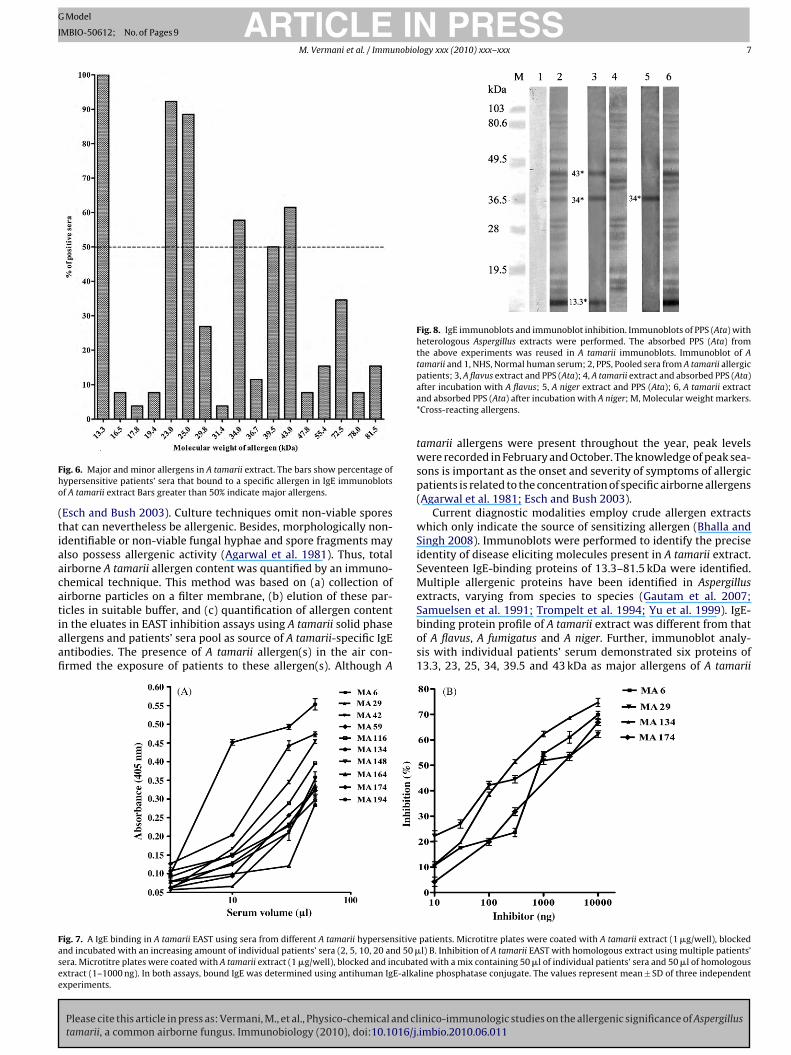
IgE-binding proteins common in A tamarii and the aboveAspergillus species were identified in immunoblot inhibition exper-iments. Immunoblot of PPS (Ata) with heterologous Aspergillusextracts revealed three IgE-binding proteins of 43, 34 and 13.3 kDain A flavus, one (34 kDa) in A niger and none in A fumigatus extract(Fig. 8). When the absorbed PPS (Ata) from the above experimentswas reused in A tamarii immunoblots (IgE immunoblot inhibition):(a) A tamarii absorbed PPS (Ata) didn’t show any allergenic proteins,(b) A flavus absorbed PPS (Ata) detected all the allergenic proteinsexcept 43, 34 and 13.3 kDa proteins, and (c) A niger absorbed PPS(Ata) detected all the allergenic proteins except 34 kDa. However,IgE binding to A tamarii proteins was not inhibited by A fumigatusextract. Thus, of the six major allergens of A tamarii extract, threewere shared with A flavus and only one with A niger.
Discussion
A few preliminary studies are available on the allergenic sig-
inico-immunologic studies on the allergenic significance of Aspergillus.imbio.2010.06.011
nificance of A tamarii, a common airborne Aspergillus species tilldate (Aggarwal et al. 2000; Shivpuri and Agarwal 1969). We reporta detailed analysis of allergenicity of A tamarii, its major andminor allergens and cross-reactivity with other Aspergillus species.The allergenic potential of A tamarii was evaluated by SPT and
tric filtration sampler was installed outdoors at 20 m height and 24-h samples (onceanes were extracted in 0.1 mol/l ammonium bicarbonate, dialyzed, lyophilized andrii EAST. Allergen content was calculated from reference inhibition curve generated

ARTICLE IN PRESSG Model
IMBIO-50612; No. of Pages 9
6 M. Vermani et al. / Immunobiology xxx (2010) xxx–xxx
F arii pl especta strates .
EgtoEtoatt
ig. 5. IgE immunoblot of A tamarii extract using hypersensitive patients’ sera. A tamulose membrane (0.45 �m). The protein strips were blocked and incubation with rntibody. The bound anti-human IgE antibodies were detected with BCIP/NBT subera; PPS, probed with pooled sera; M, Molecular weight markers. *Major allergens
AST. Positive cutaneous responses were elicited in 22% of aller-ic patients. However, in some earlier studies, positive intradermalest responses with A tamarii extract were observed in 16–17%f patients (Aggarwal et al. 2000; Shivpuri and Agarwal 1969).ven though, skin prick tests are less sensitive than intradermal
Please cite this article in press as: Vermani, M., et al., Physico-chemical and cltamarii, a common airborne fungus. Immunobiology (2010), doi:10.1016/j
ests, higher incidence of skin prick test reactivity was observed inur study. This variability may occur due to various factors suchs differences in patients’ population, extent of exposure, charac-eristics of diagnostic extracts (potency and protein profile), skinesting methods (SPT/intradermal), the site of performance and
Table 4The frequency of specific IgE-binding proteins in 26 patients sensitiz
MW, molecular weight of allergenic protein.aMajor allergens.
roteins (separated by SDS-PAGE) were electrophoretically transferred to a nitrocel-ive serum (1:5, v/v) followed by monoclonal anti-human IgE-alkaline phosphatase. NHS, probed with normal human serum; 1–26, probed with individual patients’
criterion used for grading. The allergenic potency of A tamariiextract was evaluated by EAST inhibition. Allergen-specific IgElevels were significantly higher in patients, compared to normalcontrols. Homologous extract induced a dose-dependent responsein A tamarii EAST. Besides, non-related heterologous extracts failed
inico-immunologic studies on the allergenic significance of Aspergillus.imbio.2010.06.011
to induce any inhibition in A tamarii EAST, further demonstratingthe specificity of EAST inhibition.
Direct measurement of fungal allergen concentration in the airhas been considered to be a more relevant parameter, as com-pared to colony counts, to assess their role as inhalant allergens
ed to A tamarii.
ARTICLE IN PRESSG Model
IMBIO-50612; No. of Pages 9
M. Vermani et al. / Immunobiology xxx (2010) xxx–xxx 7
Fho
(tiaacatiaafi
Fig. 8. IgE immunoblots and immunoblot inhibition. Immunoblots of PPS (Ata) withheterologous Aspergillus extracts were performed. The absorbed PPS (Ata) fromthe above experiments was reused in A tamarii immunoblots. Immunoblot of Atamarii and 1, NHS, Normal human serum; 2, PPS, Pooled sera from A tamarii allergic
Fasee
ig. 6. Major and minor allergens in A tamarii extract. The bars show percentage ofypersensitive patients’ sera that bound to a specific allergen in IgE immunoblotsf A tamarii extract Bars greater than 50% indicate major allergens.
Esch and Bush 2003). Culture techniques omit non-viable sporeshat can nevertheless be allergenic. Besides, morphologically non-dentifiable or non-viable fungal hyphae and spore fragments maylso possess allergenic activity (Agarwal et al. 1981). Thus, totalirborne A tamarii allergen content was quantified by an immuno-hemical technique. This method was based on (a) collection ofirborne particles on a filter membrane, (b) elution of these par-
Please cite this article in press as: Vermani, M., et al., Physico-chemical and cltamarii, a common airborne fungus. Immunobiology (2010), doi:10.1016/j
icles in suitable buffer, and (c) quantification of allergen contentn the eluates in EAST inhibition assays using A tamarii solid phasellergens and patients’ sera pool as source of A tamarii-specific IgEntibodies. The presence of A tamarii allergen(s) in the air con-rmed the exposure of patients to these allergen(s). Although A
ig. 7. A IgE binding in A tamarii EAST using sera from different A tamarii hypersensitivend incubated with an increasing amount of individual patients’ sera (2, 5, 10, 20 and 50 �era. Microtitre plates were coated with A tamarii extract (1 �g/well), blocked and incubatxtract (1–1000 ng). In both assays, bound IgE was determined using antihuman IgE-alkaxperiments.
patients; 3, A flavus extract and PPS (Ata); 4, A tamarii extract and absorbed PPS (Ata)after incubation with A flavus; 5, A niger extract and PPS (Ata); 6, A tamarii extractand absorbed PPS (Ata) after incubation with A niger; M, Molecular weight markers.*Cross-reacting allergens.
tamarii allergens were present throughout the year, peak levelswere recorded in February and October. The knowledge of peak sea-sons is important as the onset and severity of symptoms of allergicpatients is related to the concentration of specific airborne allergens(Agarwal et al. 1981; Esch and Bush 2003).
Current diagnostic modalities employ crude allergen extractswhich only indicate the source of sensitizing allergen (Bhalla andSingh 2008). Immunoblots were performed to identify the preciseidentity of disease eliciting molecules present in A tamarii extract.Seventeen IgE-binding proteins of 13.3–81.5 kDa were identified.Multiple allergenic proteins have been identified in Aspergillusextracts, varying from species to species (Gautam et al. 2007;
inico-immunologic studies on the allergenic significance of Aspergillus.imbio.2010.06.011
Samuelsen et al. 1991; Trompelt et al. 1994; Yu et al. 1999). IgE-binding protein profile of A tamarii extract was different from thatof A flavus, A fumigatus and A niger. Further, immunoblot analy-sis with individual patients’ serum demonstrated six proteins of13.3, 23, 25, 34, 39.5 and 43 kDa as major allergens of A tamarii
patients. Microtitre plates were coated with A tamarii extract (1 �g/well), blockedl) B. Inhibition of A tamarii EAST with homologous extract using multiple patients’
ed with a mix containing 50 �l of individual patients’ sera and 50 �l of homologousline phosphatase conjugate. The values represent mean ± SD of three independent

ING
I
8 nobio
eampo
octdthtwaSwi2pGo
d(2tibgAC2tgmflINwai
AsgiapboIAttcflfltt1ohtIa
ARTICLEModel
MBIO-50612; No. of Pages 9
M. Vermani et al. / Immu
xtract. The 13.3 and 23 kDa allergenic proteins may be considereds most important major allergens as they showed IgE-binding withore than 90% of the patients’ sera tested. These major allergens are
romising candidate molecules for component resolved diagnosisf hypersensitive patients.
IgE response against various major/minor allergenic proteinsf an extract varies from patient to patient. The modern con-ept of component resolved diagnosis is based on the fact thathe actual clinical sensitivity of a patient to an allergen extractepends on the number and type of its constituent allergenic pro-eins (Bhalla and Singh 2008). Thus, sera from multiple A tamariiypersensitive patients were used in EAST binding, EAST inhibi-ion and immunoblot experiments. While dose-dependent bindingas obtained in A tamarii EAST with multiple sera (n = 10), slopes
nd position of the binding curves varied from patient to patient.imilarly, slopes and position of A tamarii EAST inhibition curvesith six different patients’ sera were also different. Besides, in
mmunoblots, variable IgE-binding profiles were observed with6 individual sera, giving definitive evidence for heterogeneity ofatients’ IgE response to different A tamarii allergenic proteins.autam and associates also reported varying sensitization profilesf 8 allergic patients to A fumigatus proteins (Gautam et al. 2007).
Cross-reactivity among allergens has been observed amongifferent species/genera of fungi including Aspergillus speciesCrameri et al. 2008; Shen et al. 1999, 2007; Simon-Nobbe et al.008). The knowledge of shared allergens is helpful in the diagnos-ics because it reduces the number of allergens for testing. Besides,t is also beneficial in the formulation of immunotherapy vaccines asroad-spectrum effect may be obtained with lesser number of aller-ens (Lockey et al. 2003). Cross-reactivity among various species ofspergillus has been demonstrated earlier (Aggarwal et al. 2000;rameri et al. 2008; Kurup and Banerjee 2000; Shen et al. 1999,007; Simon-Nobbe et al. 2008). Alkaline and vacuolar-serine pro-eases (32–34 kDa) from Aspergillus species termed as group 13 androup 18 allergens, respectively, have been studied in detail at theolecular level. A fumigatus (Asp f 13 and Asp f 18), A flavus (Asp13 and Asp fl 18) and A oryzae (Asp o 13) serine proteases share
gE and IgG epitopes with each other (Shen et al. 1999; Simon-obbe et al. 2008). In the present study, cross-reactivity of A tamariiith three Aspergillus species that are well established sources of
irborne allergens, namely A flavus, A fumigatus and A niger wasnvestigated.
Dose-dependent inhibition of A tamarii EAST with A flavus andniger extracts indicated that some IgE molecules present in the
era pool of A tamarii sensitive patients also reacted with the aller-ens present in these extracts. There was no significant differencen slopes and position of inhibition curves induced by A tamariind A flavus extracts, suggesting close identity in the allergenicroteins of the two extracts. In a previous study, cross-reactivityetween A tamarii and A flavus has been reported on the basisf IgE radioallergosorbent test inhibition (Aggarwal et al. 2000).n contrast, slope and position of inhibition curve induced with
niger extract was significantly different from the curve with Aamarii extract, indicating a lower degree of cross-reactivity inhese extracts. IgE immunoblot inhibition assays demonstratedross-reactive proteins of 13.3, 34 and 43 kDa in A tamarii and Aavus extract. Presence of three shared allergenic proteins in Aavus and A tamarii may be attributed to a close phylogenetic rela-ionship between these two species i.e. they belong to the sameaxonomic group – the A flavus-oryzae group (Raper and Fennel965). Phylogenetically related species show high degree of homol-
Please cite this article in press as: Vermani, M., et al., Physico-chemical and cltamarii, a common airborne fungus. Immunobiology (2010), doi:10.1016/j
gy in the primary structure of their proteins which results inomologous three-dimensional structures (epitopes) and, poten-ially, shared allergenic proteins in them (Aalberse 2007). Besides,gE immunoblot inhibition revealed that only one major A tamariillergen (34 kDa) was present in A niger extract. The other three
PRESSlogy xxx (2010) xxx–xxx
major allergens of 23, 25 and 39.5 kDa were specific to A tamariiextract.
In conclusion, our results revealed that airborne A tamarii-derived allergens are present in the air, which might serve asimportant inhalant allergens in IgE-mediated allergic respira-tory diseases. Seventeen different IgE-binding proteins of crudeA tamarii extract have been identified with six of them beingmajor allergens. At least 3 A tamarii allergenic proteins (43, 34and 13.3 kDa) were present in A flavus and one of 34 kDa in Aniger extract (cross-reactive allergenic proteins). In future, purifi-cation and characterization of clinically important cross-reactiveallergens is important for producing better diagnostic as well aseffective therapeutic reagents for patients allergic to A tamarii andallergenically related Aspergillus species.
Acknowledgements
The authors thankfully acknowledge the financial support(Senior Research Fellowship) from Council of Scientific and Indus-trial Research, India to one of the co-authors (MV).
References
Aalberse, R.C., 2007. Assessment of allergen cross-reactivity. Clin. Mol. Allergy 5,2–7.
Agarwal, M.K., Shivpuri, D.N., 1974. Fungal spores – their role in respiratory allergy.Adv. Pollen-Spore Res. 1, 78–128.
Agarwal, M.K., Gupta, S., Chawla, S., Bansal, S.K., Vijayan, V.K., 2003. Cross-reactingand unique allergenic and antigenic components in insect extracts used for thediagnosis and immunotherapy of patients suffering with respiratory allergy. In:Sarma, P.U., Singh, B.P., Rao, D.N., Sharma, P., Murthy, P.S., Shetty, T., et al. (Eds.),Trends in Clinical Biochemistry and Laboratory Medicine. Association of ClinicalBiochemists India, Delhi, India, pp. 314–324.
Agarwal, M.K., Mukerji, K.G., Shivpuri, D.N., 1969. Studies on the allergenic fun-gal spores of Delhi, India, metropolitan area. Botanical aspects. J. Allergy 44,193–203.
Agarwal, M.K., Yunginger, J.W., Swanson, M.C., Reed, C.E., 1981. An immunochemicalmethod to measure atmospheric allergens. J. Allergy Clin. Immunol. 68, 194–200.
Aggarwal, S., Chhabra, S.K., Saxena, R.K., Agarwal, M.K., 2000. Heterogeneity ofimmune responses to various Aspergillus species in patients with allergic res-piratory diseases. Indian J. Chest Dis. Allied Sci. 42, 249–258.
Bernstein, I.L., Li, J.T., Bernstein, D.I., Hamilton, R., Spector, S.L., Tan, R., et al., 2008.Allergy diagnostic testing: an updated practice parameter. Ann. Allergy AsthmaImmunol. 100, S1–S147.
Bhalla, P.L., Singh, M.B., 2008. Biotechnology based allergy diagnosis and vaccination.Trends Biotech. 26, 153–161.
Bousquet, J., Khaltaev, N., Cruz, A.A., Denburg, J., Fokkens, W.J., Togias, A., et al., 2008.Allergic Rhinitis and its Impact on Asthma (ARIA) 2008 update (in collaborationwith the World Health Organization, GA(2)LEN and AllerGen). Allergy 63, 8–160.
Crameri, R., Blaser, K., 1996. Cloning Aspergillus fumigatus allergens by the pJuFofilamentous phage display system. Int. Arch. Allergy Immunol. 110, 41–45.
Crameri, R., Weichel, M., Fluckiger, S., Glaser, A.G., Rhyner, C., 2006. Fungal allergies:a yet unsolved problem. Chem. Immunol. Allergy 91, 121–133.
Crameri, R., Zeller, S., Glaser, A.G., Vihelmsson, M., Rhyner, C., 2008. Cross-reactivityamong fungal allergens: a clinically relevant phenomenon? Mycoses 52, 99–106.
Esch, R.E., Bush, R.K., 2003. Aerobiology of outdoor allergens. In: Adkinson Jr., N.F.,Yunginger, J.W., Busse, W.W., Bochner, B.S., Holgate, S.T., Simons, F.E.R. (Eds.),Middleton’s Allergy: Principles and Practice, 6th ed. Mosby, Philadelphia, pp.529–554.
Gautam, P., Sundaram, C.S., Madan, T., Gade, W.N., Shahz, A., Sirdeshmukh, R.,Sarma, P.U., 2007. Identification of novel allergens of Aspergillus fumigatus usingimmunoproteomics approach. Clin. Exp. Allergy 37, 1239–1249.
Horner, W.E., Helbling, A., Salvaggio, J.E., Lehrer, S.B., 1995. Fungal allergens. Clin.Microbiol. Rev. 8, 161–179.
Johnstone, A., Thorpe, R. (Eds.), 1987. Immunochemistry in Practice, 2nd ed. Black-well Scientific Publication, Oxford, pp. 257–259.
Kausar, M.A., Vijayan, V.K., Bansal, S.K., Menon, B.K., Vermani, M., Agarwal, M.K.,2007. Mosquitoes as sources of inhalant allergens: clinico-immunologic andbiochemical studies. J. Allergy Clin. Immunol. 120, 1219–1221.
Khan, Z.U., Khan, M.A., Chandy, R., Sharma, P.N., 1999. Aspergillus and other mouldsin the air of Kuwait. Mycopathologia 146, 25–32.
Kilinc, I., Alper, S., 2005. Adverse effects of antihistamines on skin tests and antihis-tamines in pregnancy. Curr. Med. Chem. Anti-Inflamm. Anti-Allergy Agents 4,
inico-immunologic studies on the allergenic significance of Aspergillus.imbio.2010.06.011
517–519.King, T.P., Hoffman, D., Lowenstein, H., Marsh, D.G., Platts-Mills, T.A., Thomas, W.,
1994. Allergen nomenclature. WHO/IUIS Allergen nomenclature subcommittee.Int. Arch. Allergy Immunol. 105, 224–233.
Koppelman, G.H., 2007. Gene–environment interaction in allergic disease: morequestions, more answers? J. Allergy Clin. Immunol. 120, 1266–1268.

ING
I
nobio
K
L
L
L
L
L
M
R
S
S
Vailes, L., Sridhara, S., Cromwell, O., Weber, B., Breitenbach, M., Chapman, M., 2001.Quantitation of the major fungal allergens, Alt a 1 and Asp f 1, in commercial
ARTICLEModel
MBIO-50612; No. of Pages 9
M. Vermani et al. / Immu
urup, V.P., Banerjee, B., 2000. Fungal allergens and peptide epitopes. Peptides 21,589–599.
aemmli, U.K., 1970. Cleavage of structural proteins using the assembly of the headof bacteriophage T4. Nature (Lond.) 227, 680–685.
arsen, J.N., Dreborg, S., 2008. Standardization of allergen extracts. Methods Mol.Med. 138, 133–145.
arsen, J.N., Lowenstein, H., 1996. Allergen nomenclature (editorial). J. Allergy Clin.Immunol. 97, 577–578.
ockey, R.F., Slater, J.E., Esch, R.E., 2003. Preparation and standardization of allergenextracts. In: Adkinson Jr., N.F., Yunginger, J.W., Busse, W.W., Bochner, B.S., Hol-gate, S.T., Simons, F.E.R. (Eds.), Middleton’s Allergy: Principles and Practice, 6thed. Mosby, Philadelphia, pp. 273–283.
owry, O.H., Rosebrough, N.J., Farr, A.L., Randall, R.J., 1951. Protein measurementwith the Folin phenol reagent. J. Biol. Chem. 193, 265–275.
asoli, M., Fabian, D., Holt, S., Beasley, R., 2004. The global burden of asthma:executive summary of the GINA Dissemination Committee report. Allergy 59,469–478.
Please cite this article in press as: Vermani, M., et al., Physico-chemical and cltamarii, a common airborne fungus. Immunobiology (2010), doi:10.1016/j
aper, K.B., Fennel, D.I., 1965. The Genus Aspergillus. The Williams and Wikins Co.,Baltimore.
amuelsen, H., Karlsson-Borga, A., Paulsen, B.S., Wold, J.K., Rolfsen, W., 1991. Purifi-cation of a 20 kD allergen from Aspergillus fumigatus. Allergy 46, 115–124.
cott, T.A., Melvin, E.H., 1953. Determination of dextran with anthrone. Anal. Chem.25, 1656–1661.
PRESSlogy xxx (2010) xxx–xxx 9
Shen, H.D., Tam, M.F., Chou, H., Han, S.H., 1999. The importance of serine pro-teinases as aeroallergens associated with asthma. Int. Arch. Allergy Immunol.119, 259–264.
Shen, H.D., Tam, M.F., Tang, R.B., Chou, H., 2007. Aspergillus and Penicillium allergens:focus on proteases. Curr. Allergy Asthma Rep. 7, 351–356.
Shivpuri, D.N., Agarwal, M.K., 1969. Studies on the allergenic fungal spores of Delhi,India, metropolitan area. J. Allergy 44, 204–213.
Simon-Nobbe, B., Denk, U., Pöll, V., Rid, R., Breitenbach, M., 2008. The spectrum offungal allergy. Int. Arch. Allergy Immunol. 145, 58–86.
Towbin, H., Staehelin, T., Gorden, J., 1979. Electrophoresis transfer of proteins frompolyacrylamide gel to nitrocellulose sheet: procedures and some applications.Proc. Natl. Acad. Sci. 76, 4350–4354.
Trompelt, J., Becker, W.M., Schlaak, M., 1994. Analysis of IgG subclass and IgEresponse in allergic disease caused by Aspergillus fumigatus by immunoblottingtechniques. Int. Arch. Allergy Immunol. 104, 390–398.
inico-immunologic studies on the allergenic significance of Aspergillus.imbio.2010.06.011
allergenic products. J. Allergy Clin. Immunol. 107, 641–646.Yu, C.J., Chiou, S.H., Lai, W.Y., Chiang, B.L., Chow, L.P., 1999. Characterization of a novel
allergen, a major IgE-binding protein from Aspergillus flavus, as an alkaline serineprotease. Biochem. Biophys. Res. Comm. 261, 669–675.