Effects of cypermethrin on some clinico-hemato-biochemical and pathological parameters in male dwarf...
10
Experimental and Toxicologic Pathology 61 (2009) 151–160 Effects of cypermethrin on some clinico-hemato-biochemical and pathological parameters in male dwarf goats (Capra hircus) $ Ahrar Khan a, , Hafiz A.M. Faridi a , Muhammad Ali a , Muhammad Zargham Khan a , Muhammad Siddique b , Iftikhar Hussain b , Maqbool Ahmad c a Department of Veterinary Pathology, University of Agriculture, Faisalabad 38040, Pakistan b Department of Microbiology, , University of Agriculture, Faisalabad 38040, Pakistan c Department of Animal Reproduction, University of Agriculture, Faisalabad 38040, Pakistan Received 23 April 2008; accepted 7 July 2008 Abstract This study was carried out on 30 dwarf bucks to determine the effects of cypermethrin (CY) on clinical, hemato- biochemical and histopathological parameters. Animals were divided randomly into five equal groups, and each group was dipped in 0%, 0.1%, 0.4%, 0.8% or 1.6% CY, on days 0 and 15. Animals were monitored for clinical signs. Blood and serum samples were collected on day 0 and then fortnightly till day 75. Severe clinical signs comprising itching, restlessness, salivation, skin scratching and head shaking appeared at high doses (0.8% and 1.6% CY). Erythrocyte counts, hemoglobin, hematocrit, total protein, globulin and fibrinogen decreased significantly while total leukocyte counts, alanine aminotransferase and aspartate aminotransferase concentration increased significantly in all the treated groups. In the liver, necrosis of hepatocytes along with cytoplasmic vacuolation and fibroblasts proliferation were observed at a high dose of CY (1.6%). Microscopically kidneys showed congestion of parenchyma and condensation of epithelial cells of tubules along with deposition of casts in tubules. Shrinkage of glomerular capillaries and increased urinary spaces were pronounced in the high-dose group. Lungs exhibited accumulation of fibrinous exudation, thickening of alveolar walls, collapse and broken alveoli in animals treated with a high dose of CY. It was concluded that CY caused dose-dependent effects on all parameters studied. High doses of CY (0.8% and 1.6% solution) affected the parameters on erythrocytes and leukocytes for whole evaluation period, while effects on plasma proteins were transient and on ALT, AST and fibrinogen were transient but lasted a few weeks longer. r 2008 Elsevier GmbH. All rights reserved. Keywords: Cypermethrin; Goats; Clinical signs; Hematology; Biochemical parameters; Histopathological lesions Introduction Pyrethroid insecticides have been used for more than 40 years and account for 25% of the worldwide insecticide market (Shafer et al., 2005). Cypermethrin (CY), a synthetic pyrethroid, belongs to a group of potent insecticides that are environmentally compatible by virtue of their moderate persistence, low volatility ARTICLE IN PRESS www.elsevier.de/etp 0940-2993/$ - see front matter r 2008 Elsevier GmbH. All rights reserved. doi:10.1016/j.etp.2008.07.001 $ The experiment was designed and conducted considering all the national legislation regarding protection of animal welfare and following the guidelines of the Advanced Studies and Research Board of the University. Corresponding author. Tel.: +92 41 9200161 170x3119, +92 3336517844; fax: +92 41 9200764. E-mail address: [email protected] (A. Khan).
-
Upload
independent -
Category
Documents
-
view
1 -
download
0
Transcript of Effects of cypermethrin on some clinico-hemato-biochemical and pathological parameters in male dwarf...

ARTICLE IN PRESS
0940-2993/$ - se
doi:10.1016/j.et
$The experi
national legisla
following the gu
of the Universi�Correspond
+92 333651784
E-mail addr
Experimental and Toxicologic Pathology 61 (2009) 151–160
www.elsevier.de/etp
Effects of cypermethrin on some clinico-hemato-biochemical and
pathological parameters in male dwarf goats (Capra hircus)$
Ahrar Khana,�, Hafiz A.M. Faridia, Muhammad Alia, Muhammad Zargham Khana,Muhammad Siddiqueb, Iftikhar Hussainb, Maqbool Ahmadc
aDepartment of Veterinary Pathology, University of Agriculture, Faisalabad 38040, PakistanbDepartment of Microbiology, , University of Agriculture, Faisalabad 38040, PakistancDepartment of Animal Reproduction, University of Agriculture, Faisalabad 38040, Pakistan
Received 23 April 2008; accepted 7 July 2008
Abstract
This study was carried out on 30 dwarf bucks to determine the effects of cypermethrin (CY) on clinical, hemato-biochemical and histopathological parameters. Animals were divided randomly into five equal groups, and each groupwas dipped in 0%, 0.1%, 0.4%, 0.8% or 1.6% CY, on days 0 and 15. Animals were monitored for clinical signs. Bloodand serum samples were collected on day 0 and then fortnightly till day 75. Severe clinical signs comprising itching,restlessness, salivation, skin scratching and head shaking appeared at high doses (0.8% and 1.6% CY). Erythrocytecounts, hemoglobin, hematocrit, total protein, globulin and fibrinogen decreased significantly while total leukocytecounts, alanine aminotransferase and aspartate aminotransferase concentration increased significantly in all thetreated groups. In the liver, necrosis of hepatocytes along with cytoplasmic vacuolation and fibroblasts proliferationwere observed at a high dose of CY (1.6%). Microscopically kidneys showed congestion of parenchyma andcondensation of epithelial cells of tubules along with deposition of casts in tubules. Shrinkage of glomerular capillariesand increased urinary spaces were pronounced in the high-dose group. Lungs exhibited accumulation of fibrinousexudation, thickening of alveolar walls, collapse and broken alveoli in animals treated with a high dose of CY. It wasconcluded that CY caused dose-dependent effects on all parameters studied. High doses of CY (0.8% and 1.6%solution) affected the parameters on erythrocytes and leukocytes for whole evaluation period, while effects on plasmaproteins were transient and on ALT, AST and fibrinogen were transient but lasted a few weeks longer.r 2008 Elsevier GmbH. All rights reserved.
Keywords: Cypermethrin; Goats; Clinical signs; Hematology; Biochemical parameters; Histopathological lesions
e front matter r 2008 Elsevier GmbH. All rights reserved.
p.2008.07.001
ment was designed and conducted considering all the
tion regarding protection of animal welfare and
idelines of the Advanced Studies and Research Board
ty.
ing author. Tel.: +9241 9200161 170x3119,
4; fax: +92 41 9200764.
ess: [email protected] (A. Khan).
Introduction
Pyrethroid insecticides have been used for more than40 years and account for 25% of the worldwideinsecticide market (Shafer et al., 2005). Cypermethrin(CY), a synthetic pyrethroid, belongs to a group ofpotent insecticides that are environmentally compatibleby virtue of their moderate persistence, low volatility

ARTICLE IN PRESSA. Khan et al. / Experimental and Toxicologic Pathology 61 (2009) 151–160152
and poor aqueous mobility in soil. Though CY has awide margin of safety for farm animals, side effects alsooccur. High doses of CY may result in teeth grinding,hyperesthesia, excessive salivation, muscular tremors,in-coordination, dyspnea, opisthotonos and death(Tamang et al., 1991).
Hematological values are widely used to determinesystemic relationship and physiological adaptationsincluding the assessment of the general health conditionof animals. Most studies on the effects of pyrethroidsare confined to reporting biochemical and physiologicalchanges, and little attention has been paid to thehematological modulations induced by these pesticides(Atamanalp et al., 2002). CY significantly reduced redblood cell and hematocrit in sheep (Yousef et al., 1998)and rats (Mansee, 1998). There are controversial reportsabout the effects of CY on hemoglobin (Hb), totalleukocyte counts (TLC), alanine aminotransferase(ALT), aspartate aminotransferase (AST) and alkalinephosphatase (ALP). According to Mansee (1998), CYhas no effect on the Hb concentration in rats, whereasdecreased Hb has been reported in sheep (Yousef et al.,1998) and rats (Matsushima et al., 2003). Matsushimaet al. (2003) reported significantly reduced TLC in rats,whereas Yousef et al. (1998) reported increased TLC insheep with the treatment of CY. CY treatment at highdoses significantly (Po0.05) decreased total protein (TP)and albumin in sheep (Yousef et al., 2003). Significantlyincreased ALT and AST in sheep (Abdulaziz and Hristev,1996) and rams (Yousef et al., 1999) while decreased ALTand AST in sheep (Yousef et al., 1998) and buffalo calves(Jagvinder et al., 2001) have been reported. DecreasedALP in sheep (Yousef et al., 1998) and albino rats(Samita et al., 1999) and increased ALP in buffalo calves(Jagvinder et al., 2001) have also been documented. Thevariability described above may result from differentdoses, species, times of sampling, environmental tem-perature, etc.
Numerous changes of CY toxicity at the cellular levelhave been documented. With the treatment of CY,morphological and ultra-structural changes in kidneys,heart, lungs and liver have been reported (Latuszynskaet al., 1999). Contrary to these results no histologicalchanges were found in kidneys and heart (Samita et al.,1999). Kidneys of the rats showed chronic nephrosisthat was histologically characterized by tubular dilata-tions, interstitial chronic inflammatory infiltration andglomerular scarring. In the liver, CY treatment inducedhepatocyte vacuolation and fibrosis in hepatic cords(Latuszynska et al., 1999).
From the above facts, it is clear that CY has manyside effects, but such information in dwarf goats issparse. Moreover, the long follow-up after dermaldosing/exposure by CY is missing. Fibrinogen concen-tration in CY-treated animals has also not yet beenreported in the published literature. Therefore, this
study had been executed with the objective to knowchanges in clinical, hematological, biochemical andpathological parameters in male dwarf goats dipped atlow to high doses of CY.
Materials and methods
This experiment was designed and conducted, con-sidering all the national legislation regarding protectionof animal welfare and following the guidelines of theAdvanced Studies and Research Board of the Univer-sity.
Experimental animals and management
This experiment was conducted from February toApril 2005. Ambient temperature during this periodranged from 14.9 to 26.6 1C, with a relative humidityfrom 67.7% (February) to 35% (April). Thirty healthydwarf bucks at 11
222 years of age having similar weight
(27–30 kg) were procured from government farm. Allanimals were kept under similar management andfeeding conditions throughout the experiment. Briefly,goats of each group were housed together in a shed.Sheds were covered from three sides and one side wasopen; when necessary, the open side was covered withcurtains to protect the animals from cold. Animals wereput on pasture (green fodder Trifolium alexandrinum;Barseem) early in the morning and returned in theevening. No concentrate or fodder was provided in theshed at night. Water was available ad libitum in the shed.The animals were vaccinated against pleuropneumoniaand enterotoxaemia and dewormed twice yearly.
Experimental design
This experiment was conducted under a completelyrandomized block design. Animals were randomlydivided into five equal groups (six animals each) afterfive days of acclimatization. CY (Ecofleeces, Bimeda,Ireland) was procured from the market. Its 0.1%solution is recommended for dipping. Therefore, therecommended level and above were selected for thetreatment trials to assess CY side effects in male goats.All the animals in five groups were dipped on days 0 and15 early in the morning with 0%, 0.1%, 0.4%, 0.8% or1.6% solution of CY. The group with 0% CY solutionserved as control. A fresh aqueous solution of CY wasprepared in a tub for each animal in each group, andkeeping its mouth out, the animal was dipped in theprepared concentration until its body was wet (1–2min).After each dipping, clinical signs were observed twicedaily (morning and evening). The duration of this studywas 75 days.

ARTICLE IN PRESSA. Khan et al. / Experimental and Toxicologic Pathology 61 (2009) 151–160 153
Hematological and biochemical studies
Blood samples with and without anticoagulant(Na2EDTA; 1mg/mL) from all animals were collectedprior to treatment (one day before the start oftreatment) and then fortnightly until the end ofexperiment, i.e. 75 days after the first treatment. Bloodsamples collected with anticoagulant were used for thedetermination of total erythrocyte counts (TEC) andTLC (Benjamin, 1978). Hematocrit and hemoglobin(Hb) were determined by the microhematocrit andcyanmethemoglobin method (Benjamin, 1978), respec-tively.
Serum was extracted from blood samples collectedwithout anticoagulant and stored at �20 1C till analysis.ALT, AST and ALP were measured using commerciallyavailable colorimetric kits (Randox Laboratories Ltd.,Crumlin, UK, Cat # AS 147 and ALP 146, respectively,and Diasys Diagnostic systems, Germany, Cat # 10041021). The bases for AST, ALT and ALP measurementswere to monitor the concentration of oxaloacetatehydrazone formed with 2,4-dinitrophenylhydrazine,pyruvate hydrazone formed with 2,4-dinitrophenylhy-drazine and of p-nitrophenol formed with water atwavelengths of 546, 645 and 405 nm with a spectro-photometer, respectively. Serum TPs were estimatedby the Biuret method (Oser, 1976). Albumin wasdetermined by the bromocresol green dye-bindingtechnique, whereas globulin concentration wasobtained by subtracting albumin from total serumproteins.
Table 1. Intensity of clinical signs observed in various groups
of bucks treated with different concentration of cypermethrin
Clinical signs Dose (% solution) of cypermethrin
0 0.1 0.4 0.8 1.6
Itching � � + ++ +++
Restlessness � � ++ ++ +++
Skin scratching � � ++ ++ +++
Salivation � � ++ ++ ++
Head shaking � � + ++ ++
Each group consisted of six animals and was dipped on days 0 and 15
with various concentrations of cypermethrin. Signs were graded on the
basis of numerics allotted and then reported as no (�), mild (+),
moderate (++) and severe (+++) lesions.
Gross and histopathological studies
At the end of the experiment, each animal waseuthanised by injecting IV overdose of sodium pento-barbital (90mg/kg b. wt.). Each carcass was examinedthoroughly for gross lesions within 1
2h after euthanasia.
Gross lesions present in the liver, lungs, kidneys, heartand intestines were recorded and tissues showing lesionswere fixed in 10% buffered formalin immediately. Thesame process was repeated for each animal. Fixed tissueswere processed by the routine method of dehydrationand paraffin embedding. Sections of 4–5-mm thicknesswere cut and stained with hematoxylin and eosin (Lilleand Fullmer, 1976). Slides were analyzed for histo-pathological lesions by two persons, and if differencewas found, a third person was consulted for opinion.Histopathological lesions were graded by allottingnumerics to the lesions. All the slides in each groupwere thrashed; average of the numerics was obtainedand an accumulative picture of each lesion in each groupwas drawn. Frequency of lesions in each group wasrecorded and the incidence (%) of lesions was calcu-lated.
Statistical analysis
Data collected were subjected to the Chi-square testor to the analysis of variance (ANOVA) and means werecompared by Duncan’s multiple range (DMR) test on apersonal computer using the Minitab statistical softwarepackage. The significance level was Po0.05.
Results
Clinical signs
No pronounced clinical signs were observed inanimals treated with 0.1% and 0.4% CY solution, whileitching, restlessness, salivation, skin scratching and headshaking were observed in animals treated with 0.8% and1.6% CY solution for two days post dipping (Table 1).
Hemato-biochemical alterations
The data indicated that CY affected erythrocyte andleukocyte counts for the whole evaluation period, whileeffects on plasma proteins were transient. Similarlyeffects on ALT, AST and fibrinogen were also transientbut lasted a few weeks longer.
A dose-dependent decrease (Po0.05) in TEC andhematocrit was observed from day 15 to 75 of theexperiment, while Hb concentration showed a signifi-cant decrease from day 45 to 75 of the experimentcompared with the control group (Table 2). TLCincreased significantly in all the treatment groups ascompared to the control group (Fig. 1).
TP including albumin and globulin did not showdifference between the treatment and control groups ondays 0 and 45–75 of experiment. A dose-dependentdecrease (Po0.05) in TP was observed on days 15 and30 compared with the control group. This decrease wasalso evident in the concentration of albumin andglobulin on day 30 (Table 3). Similarly, fibrinogen

ARTICLE IN PRESS
Table 2. Hemogram of male dwarf goats dipped with
different concentrations of cypermethrin
Days Dose (% solution) of cypermethrin
0 0.1 0.4 0.8 1.6
Total erythrocyte counts (� 1012/L)
0 13.070.9 13.270.7 14.670.8 14.371.2 14.570.7
15 13.970.8 12.470.8 13.870.7 13.071.3 11.470.8*
30 13.371.0 10.370.9* 12.170.9 10.770.7* 8.570.9*
45 13.670.8 10.970.4* 11.570.7* 10.270.6* 10.470.6*
60 13.970.6 11.170.5* 11.270.5* 10.170.7* 10.470.5*
75 14.370.4 11.870.6* 11.370.3* 10.270.7* 10.970.3*
Hemoglobin concentration (g/dL)
0 9.670.4 8.970.3 9.370.4 9.270.5 9.670.4
15 8.970.6 8.570.5 8.870.4 7.770.4 9.270.5
30 8.670.3 8.570.2 8.570.3 8.670.4 9.370.5
45 9.070.4 7.970.3* 8.370.2* 8.070.2* 7.970.3*
60 9.070.4 8.270.3* 8.670.4 8.270.3* 8.570.3
75 9.070.3 8.170.2 8.470.5 8.070.2* 8.670.2
Hematocrit (%)
0 28.271.9 24.771.7 27.270.9 25.371.5 27.871.6
15 27.071.0 23.070.7* 24.170.8* 21.871.4* 24.271.7*
30 26.871.1 20.870.9* 23.271.4* 20.371.2* 20.071.9*
45 28.771.4 22.070.9* 23.070.8* 20.270.8* 22.271.0*
60 29.371.4 23.870.8* 23.871.3* 23.271.0* 24.370.8*
75 30.370.9 23.770.8* 24.871.2* 24.071.1* 24.270.9*
Each group consisted of six animals and was dipped on days 0 and 15
with various concentrations of cypermethrin. Asterisk indicates
significant (Po0.05) difference compared to the non-CY-treated
control (0% CY) at a sampling day.
10
9
8
7
6
5
4
3
2
1
00 15 30 45 60 75
EXPERIMENTAL DAYS
109 /L
0% 0.10% 0.40% 1.60%0.80%
** * *
*
* * * * * * * *
Fig. 1. Leukocyte counts of bucks on various days of the experiment. Each group consisted of six animals and was dipped on days 0
and 15 with various concentrations of cypermethrin. Asterisk indicates significant (Po0.05) difference compared to the non-CY-
treated control (0% CY) at a sampling day.
Table 3. Protein and its fractions concentration in male
dwarf goats dipped with different concentrations of cyperme-
thrin on days 0 and 15
Days Dose (% solution) of cypermethrin
0 0.1 0.4 0.8 1.6
Total protein (g/dL)
0 12.671.9 12.171.7 12.871.2 13.071.4 11.771.4
15 14.070.8 12.871.4 12.371.5 11.370.7* 11.870.6*
30 13.271.4 10.271.1* 10.470.8* 10.171.1* 9.471.6*
45 12.770.9 12.470.7 12.671.3 12.671.1 12.872.1
60 13.171.9 12.570.8 13.072.4 12.170.6 11.873.2
75 13.771.6 12.471.7 12.671.2 12.070.7 12.071.2
Albumin (g/dL)
0 5.770.6 5.470.3 5.870.3 5.970.5 5.370.5
15 6.370.5 5.170.4 5.370.5 5.870.4 5.670.6
30 5.970.6 4.670.6* 4.770.4* 4.670.6* 4.670.6*
45 5.770.9 5.670.5 5.770.2 5.770.4 5.870.7
60 5.970.5 5.570.4 5.970.8 5.470.5 5.370.6
75 6.170.6 5.071.1 5.070.6 5.770.8 5.470.7
Globulin (g/dL)
0 6.972.1 6.771.1 7.071.1 7.171.7 6.471.5
15 7.771.8 7.471.0 7.370.8 7.071.2 6.771.4
30 7.370.9 5.770.8* 5.771.0* 5.671.1* 4.971.2*
45 7.071.5 6.871.4 6.971.7 6.970.9 7.271.3
60 7.471.7 7.370.9 7.171.2 6.770.9 6.570.8
75 7.570.9 6.270.9 6.470.8 7.270.8 6.371.2
Each group consisted of six animals and was dipped on days 0 and 15
with various concentrations of cypermethrin. Asterisk indicates
significant (Po0.05) difference compared to the non-CY-treated
control (0% CY) at a sampling day.
A. Khan et al. / Experimental and Toxicologic Pathology 61 (2009) 151–160154

ARTICLE IN PRESS
0100200300400500600700800
0EXPERIMENTAL DAYS
mg/
dL
0% 0.10% 0.40% 0.80% 1.60%
**** * * * * * * *
15 30 45 60 75
Fig. 2. Fibrinogen concentration in bucks on various days of experiment. Each group consisted of six animals and was dipped on
days 0 and 15 with various concentrations of cypermethrin. Asterisk indicates significant (Po0.05) difference compared to the non-
CY-treated control (0% CY) at a sampling day.
Table 4. ALT and AST concentrations in male dwarf goats dipped with different concentrations of cypermethrin on days 0 and 15
Days Dose (% solution) of cypermethrin
0 0.1 0.4 0.8 1.6
ALT (IU)
0 15.272.0 15.873.4 15.472.2 15.873.1 16.573.9
15 15.772.8 16.975.2 28.771.3* 31.671.5* 34.173.6*
30 20.972.2 21.471.5 30.672.6* 39.973.4* 40.772.3*
45 21.973.9 17.071.9 22.273.7 28.272.7 29.971.2*
60 20.672.9 19.073.1 21.373.9 22.872.1 21.672.2
75 22.972.8 23.671.9 21.372.1 22.972.2 22.772.5
AST (IU)
0 61.772.2 65.972.1 60.372.5 63.071.7 61.871.9
15 58.073.4 53.771.9 57.171.1 63.173.5 68.772.5*
30 60.772.2 65.273.8 59.675.3 67.572.5* 65.672.3*
45 62.173.4 62.973.1 65.971.5 61.372.2 64.073.1
60 60.372.0 65.775.7 60.274.2 65.672.1 63.073.5
75 61.072.8 56.772.7 57.673.0 64.575.6 66.572.4
ALP (IU)
0 88.2714.6 99.5714.0 88.8720.4 88.7712.3 97.4716.7
15 91.2712.4 94.0713.9 99.3714.9 88.7719.3 86.1711.5
30 98.0723.0 94.0715.2 89.2713.2 101.8721.8 101.0716.2
45 97.8717.8 100.0720.4 98.0723.7 92.6715.7 91.8714.9
60 98.2717.1 95.0721.3 86.5716.6 95.0722.3 98.3717.7
75 83.4723.0 107.3727.4 84.8715.7 95.7723.4 91.5715.2
Each group consisted of six animals and was dipped on days 0 and 15 with various concentrations of cypermethrin. Asterisk indicates significant
(Po0.05) difference compared to the non-CY-treated control (0% CY) at a sampling day.
A. Khan et al. / Experimental and Toxicologic Pathology 61 (2009) 151–160 155
concentration was decreased (Po0.05) in treatmentgroups as compared to the control group on days 15–45of the experiment (Fig. 2). Significantly increasedconcentrations of ALT and AST were observed on days15–30 and 15–45, respectively, compared with thecontrol group. Concentration of ALP did not varybetween the control and treatment groups on variousexperimental days (Table 4).
Gross and histopathological lesions
Grossly diffused hemorrhages were present in kid-neys, parietal surface of liver, lung’s parenchyma,ventricles and endocardium in groups treated with ahigh dose of CY (0.8% and 1.6% CY solution).
Grading and incidence of histopathological lesionsrevealed dose-dependent effects in liver, kidneys, lungs

ARTICLE IN PRESS
Table 5. Frequency and incidence (%) of histopathological lesions observed in goats treated with different doses of cypermethrin
Organ/lesions Dose (% solution) of cypermethrin
0.1 0.4 0.8 1.6
*F **I F I (%) F I (%) F I (%)
Liver
Condensed/pyknotic nuclei � 0 + 33.3 + 50.0 ++ 66.6
Cytoplasmic vacuolation + 33.3 ++ 50.0 +++ 66.6 +++ 100
Fibrous tissue proliferation � 0 � 0 + 50.0 + 50.0
Necrosis + 33.3 + 33.3 ++ 66.6 ++ 83.3
Regeneration + 33.3 + 50.0 ++ 50.0 ++ 66.6
Kidneys
Fibrous tissue proliferation � 0 � 0 + 33.3 + 33.3
Glomerulus shrinkage + 16.6 + 16.6 ++ 50.0 +++ 100
Tubules
Congestion � 0 + 33.3 + 50.0 ++ 66.6
Deposition of casts � 0 + 16.6 ++ 66.6 +++ 100
Epithelium desquamation � 0 + 33.3 + 50.0 ++ 83.3
Necrosis + 33.3 ++ 50.0 ++ 66.6 +++ 83.3
Pyknotic nuclei, + 16.6 ++ 50.0 ++ 66.6 +++ 83.3
Lungs
Alveoli
Collapsed + 16.6 ++ 50.0 +++ 66.6 +++ 66.6
Oedema � 0 + 33.3 + 33.3 + 33.3
Walls, thickened � 0 + 33.3 ++ 50.0 ++ 66.6
Atelectasis � 0 ++ 50.0 ++ 50.0 +++ 66.6
Cellular infiltration � 0 + 16.6 + 16.6 + 16.6
Fibrous tissue proliferation � 0 + 16.6 ++ 50.0 ++ 50.0
Intestines
Cellular Infiltration � 0 + 16.6 + 16.6 + 16.6
Desquamation, epithelium + 33.3 + 33.3 ++ 50.0 ++ 66.6
Fibrous tissue proliferation � 0 � 0 + 33.3 + 33.3
Heart
Condensed/pyknotic nuclei � 0 + 16.6 ++ 50.0 ++ 50.0
Hemorrhages
Endocardium � 0 + 16.6 + 33.3 + 33.3
Pericardium � 0 + 16.6 + 33.3 + 33.3
Each group consisted of six animals and was dipped on days 0 and 15 with various concentrations of cypermethrin. Lesions were graded on the basis
of numerics allotted and then reported as no (�), mild (+), moderate (++) and severe (+++) lesions. No lesions were recorded in the control
group, therefore, not included in the table. *F ¼ frequency; **I ¼ incidence (%).
A. Khan et al. / Experimental and Toxicologic Pathology 61 (2009) 151–160156
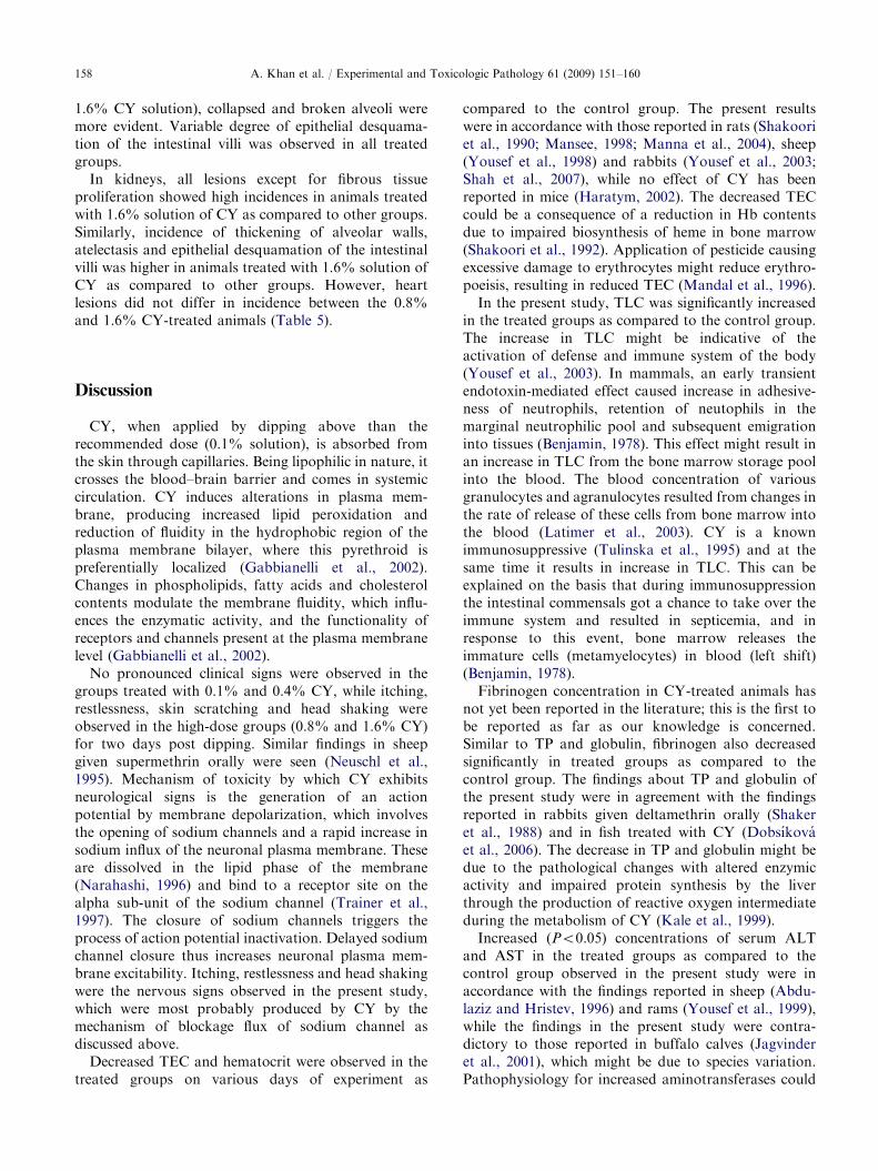
and intestines, after dermal exposure (Table 5). Severeforms of lesions were observed in animals treated with1.6% CY, followed by groups treated with 0.8%, 0.4%and 0.1% solution of CY. Various stages of degenera-tion including condensed/pyknotic nuclei and necrosisof hepatocytes along with cytoplasmic vacuolation andfibroblasts proliferation were observed in the grouptreated with 1.6% solution of CY (Table 5). Dividinghepatocytes were observed in the same group, indicatingregeneration potential of the liver (Fig. 3).
High incidence of cytoplasmic vacuolation in hepa-tocytes (100%) was recorded in animals treated with
1.6% solution of CY followed by other groups.Necrosis, condensed/pyknotic nuclei and regenerationof hepatocytes also showed higher incidence in 1.6%solution of CY; however, fibrous tissue proliferation hadequal incidence in 0.8% and 1.6% CY-treated animals(Table 5).
In the high-CY group (1.6% CY), parenchyma wascongested. Epithelium cells of both proximal and distalconvoluted tubules in kidneys had pyknotic nuclei andwere necrosed and desquamated. In the same group,shrinkage of tuft of capillaries in the glomerulus, leadingto increase in the urinary spaces, was noted. Nearly

ARTICLE IN PRESS
Fig. 3. Photomicrograph of liver showing various stages of degeneration in a buck treated with 1.6% solution of cypermethrin.
Condensation of nuclei, necrosis (arrows), cytoplasmic vacuolation and regenerating hepatocytes (arrowheads).H&E: 400� .
Fig. 4. Photomicrograph of kidneys of a buck treated with 1.6% solution of cypermethrin. Condensed nuclei and necrosis of the
epithelial lining of tubules (arrows) and deposition of epithelial casts in tubules (D). H&E: 200� .
A. Khan et al. / Experimental and Toxicologic Pathology 61 (2009) 151–160 157
25%, 50%, 75% and 100% tubules and glomerularspaces in animals treated with 0.1%, 0.4%, 0.8% and1.6% CY solution, respectively, were filled with
epithelial casts (Fig. 4). In lungs, there was accumulationof fibrinous exudates in some alveoli and alveolar wallswere thickened. In the high-dose groups (0.8% and
ARTICLE IN PRESSA. Khan et al. / Experimental and Toxicologic Pathology 61 (2009) 151–160158
1.6% CY solution), collapsed and broken alveoli weremore evident. Variable degree of epithelial desquama-tion of the intestinal villi was observed in all treatedgroups.
In kidneys, all lesions except for fibrous tissueproliferation showed high incidences in animals treatedwith 1.6% solution of CY as compared to other groups.Similarly, incidence of thickening of alveolar walls,atelectasis and epithelial desquamation of the intestinalvilli was higher in animals treated with 1.6% solution ofCY as compared to other groups. However, heartlesions did not differ in incidence between the 0.8%and 1.6% CY-treated animals (Table 5).
Discussion
CY, when applied by dipping above than therecommended dose (0.1% solution), is absorbed fromthe skin through capillaries. Being lipophilic in nature, itcrosses the blood–brain barrier and comes in systemiccirculation. CY induces alterations in plasma mem-brane, producing increased lipid peroxidation andreduction of fluidity in the hydrophobic region of theplasma membrane bilayer, where this pyrethroid ispreferentially localized (Gabbianelli et al., 2002).Changes in phospholipids, fatty acids and cholesterolcontents modulate the membrane fluidity, which influ-ences the enzymatic activity, and the functionality ofreceptors and channels present at the plasma membranelevel (Gabbianelli et al., 2002).
No pronounced clinical signs were observed in thegroups treated with 0.1% and 0.4% CY, while itching,restlessness, skin scratching and head shaking wereobserved in the high-dose groups (0.8% and 1.6% CY)for two days post dipping. Similar findings in sheepgiven supermethrin orally were seen (Neuschl et al.,1995). Mechanism of toxicity by which CY exhibitsneurological signs is the generation of an actionpotential by membrane depolarization, which involvesthe opening of sodium channels and a rapid increase insodium influx of the neuronal plasma membrane. Theseare dissolved in the lipid phase of the membrane(Narahashi, 1996) and bind to a receptor site on thealpha sub-unit of the sodium channel (Trainer et al.,1997). The closure of sodium channels triggers theprocess of action potential inactivation. Delayed sodiumchannel closure thus increases neuronal plasma mem-brane excitability. Itching, restlessness and head shakingwere the nervous signs observed in the present study,which were most probably produced by CY by themechanism of blockage flux of sodium channel asdiscussed above.
Decreased TEC and hematocrit were observed in thetreated groups on various days of experiment as
compared to the control group. The present resultswere in accordance with those reported in rats (Shakooriet al., 1990; Mansee, 1998; Manna et al., 2004), sheep(Yousef et al., 1998) and rabbits (Yousef et al., 2003;Shah et al., 2007), while no effect of CY has beenreported in mice (Haratym, 2002). The decreased TECcould be a consequence of a reduction in Hb contentsdue to impaired biosynthesis of heme in bone marrow(Shakoori et al., 1992). Application of pesticide causingexcessive damage to erythrocytes might reduce erythro-poeisis, resulting in reduced TEC (Mandal et al., 1996).
In the present study, TLC was significantly increasedin the treated groups as compared to the control group.The increase in TLC might be indicative of theactivation of defense and immune system of the body(Yousef et al., 2003). In mammals, an early transientendotoxin-mediated effect caused increase in adhesive-ness of neutrophils, retention of neutophils in themarginal neutrophilic pool and subsequent emigrationinto tissues (Benjamin, 1978). This effect might result inan increase in TLC from the bone marrow storage poolinto the blood. The blood concentration of variousgranulocytes and agranulocytes resulted from changes inthe rate of release of these cells from bone marrow intothe blood (Latimer et al., 2003). CY is a knownimmunosuppressive (Tulinska et al., 1995) and at thesame time it results in increase in TLC. This can beexplained on the basis that during immunosuppressionthe intestinal commensals got a chance to take over theimmune system and resulted in septicemia, and inresponse to this event, bone marrow releases theimmature cells (metamyelocytes) in blood (left shift)(Benjamin, 1978).
Fibrinogen concentration in CY-treated animals hasnot yet been reported in the literature; this is the first tobe reported as far as our knowledge is concerned.Similar to TP and globulin, fibrinogen also decreasedsignificantly in treated groups as compared to thecontrol group. The findings about TP and globulin ofthe present study were in agreement with the findingsreported in rabbits given deltamethrin orally (Shakeret al., 1988) and in fish treated with CY (Dobsıkovaet al., 2006). The decrease in TP and globulin might bedue to the pathological changes with altered enzymicactivity and impaired protein synthesis by the liverthrough the production of reactive oxygen intermediateduring the metabolism of CY (Kale et al., 1999).
Increased (Po0.05) concentrations of serum ALTand AST in the treated groups as compared to thecontrol group observed in the present study were inaccordance with the findings reported in sheep (Abdu-laziz and Hristev, 1996) and rams (Yousef et al., 1999),while the findings in the present study were contra-dictory to those reported in buffalo calves (Jagvinderet al., 2001), which might be due to species variation.Pathophysiology for increased aminotransferases could

ARTICLE IN PRESSA. Khan et al. / Experimental and Toxicologic Pathology 61 (2009) 151–160 159
be due to the injury to hepatocytes, which could haveresulted from free radicals generated from the biode-gradation of CY. Moreover, the activities of enzymes inblood plasma can also be used as a relevant stressindicator (Velisek et al., 2006). Increased activities ofboth transaminases in the present study indicatedamplified transamination processes. An increase intransamination occurs due to amino acid input intothe TCA cycle in order to cope with the energy crisisduring pyrethroid-based stress (Philip et al., 1995).
In the present study various stages of degenerationwere observed in hepatocytes including pyknosis,necrosis and vacuolation. CY metabolizes in the livervia hydrolytic ester cleavage and oxidative pathway bythe cytochrome P-450 microsomal enzyme system,which caused oxidative stress by reducing the activityof superoxide dismutase and glycogen level, leading tohepatic degeneration and necrosis (Manna et al., 2004).During degradation of pyrethroid by the liver, theyinterfere with intercellular communication throughinhibition of mitochondrial respiration. Moreover,degeneration of hepatocytes in periportal zones canimply the influence of toxic compounds in the digestivetract (Velisek et al., 2006). Also the biochemical changesin the liver profile could be related to hepatocytedamage. Degenerative changes in the liver with thedermal application of chlorpyrifos and CY in Wisterrats have also been reported (Latuszynska et al., 1999).Sarkar et al. (2005) found significant changes such ashyperplasia, disintegration of hepatic mass, and focalcoagulative necrosis in Labeo rohita exposed to CY.Another view that has been expressed is that aninhibitory effect of CY on the activity of total ATP inthe liver of rats may cause disorders in the activetransport of Na+, K+ and Ca2+ ions, and lead topathologic changes in the liver cells (Velisek et al., 2006).
In the present study, high CY doses (1.6% CY)caused various degenerative changes in kidneys andaccumulation of epithelial casts in the lumen of renaltubules. Epithelial casts are coagulated proteins intubules to which desquamated epithelial cells areadhered (Benjamin, 1978). These casts are produced asa result of protein leakage accumulates in the lumen.The possible phenomenon of cast development could bethat disruption in the filtration and selective barrier (thebasement membrane) and pores between podocyte footprocesses lead to leakage of proteins and accumulationin the lumen of tubules (Haratym, 2002). Histopatho-logical lesions of kidneys in the present study indicatedthat CY doses resulted in disruption in the above-mentioned filtration barriers, and desquamated epithe-lial cells from tubules might have mixed together withthe leaked proteins, resulting in the formation ofepithelial casts in the tubular lumen.
Mild-to-moderate histological alterations in lungs,liver and kidneys with the treatment of alpha-CY in rats
have already been documented (Manna et al., 2004).Moreover, in other subacute and acute studies on thetoxicity of pyrethroids on laboratory animals, adminis-tration of high doses of CY caused a decrease in thebody weight and hypertrophy in the liver and kidneys(Perger and Szadkowski, 1994).
The data indicated that CY caused dose-dependenteffects on all parameters studied. The changes inhematological parameters at the lowest dose of CY(0.1% solution) suggested a mild toxic reaction inanimals after two dippings 15 days apart. Moreover,effects of CY on plasma proteins, ALT and AST weretransient. However, the CY effect on fibrinogen wastransient but lasted a few weeks longer. Dose-dependenteffects on histopathological lesions in liver, kidneys, lungsand intestines after CY dermal exposure were recorded.
References
Abdulaziz M, Hristev H. Serum aminotransferase, alkaline
transferase and lactate dehydrogenase responses to oral
consecutive doses of cyano-3 alpha-phenoxybenzyl pyre-
throids on sheep. Bulg J Agri Sci 1996;2:661–6.
Atamanalp M, Keles MS, Haliloglu HI, Aras MS. The effects
of cypermethrin (a synthetic pyrethroid) on some biochem-
ical parameters (Ca, P, Na and TP) of rainbow trout
(Oncorhynchus mykiss). Turk J Vet Anim Sci 2002;26:
1157–60.
Benjamin MM. Outline of veterinary clinical pathology, 3rd
ed. Ames, IA, USA: Iowa State University Press; 1978.
Dobsıkova R, Velısek J, Wlasow T, et al. Effects of
cypermethrin on some haematological, biochemical and
histopathological parameters of common carp (Cyprinus
carpio L.). Neuroendocrinol Lett 2006;2:91–5.
Gabbianelli R, Falcioni G, Nasuti C, Cantalamessa F.
Cypermethrin-induced plasma membrane perturbation on
erythrocytes from rats: reduction of fluidity in the hydro-
phobic core and in glutathione peroxidase activity. Tox-
icology 2002;175:91–101.
Haratym MA. Hematological alternations after pyrethroids
poisoning in mice. Ann Agri Environ Med 2002;9:199–206.
Jagvinder K, Sandhu HS, Kaur J. Subacute oral toxicity of
cypermethrin and deltamethrin in buffalo calves. Indian
J Anim Sci 2001;71:1150–2.
Kale M, Rathore N, John S, Bhatnagar D. Lipid peroxidative
damage on pyrethroid exposure and alternations in
antioxidant status in rat erythrocytes: a possible involve-
ment of reactive oxygen species. Toxicol Lett 1999;105:
197–205.
Latimer KS, Mahaffey EA, Prasse KW. Clinical pathology
veterinary laboratory medicine, 4th ed. Iowa, USA: Iowa
State Press; 2003.
Latuszynska J, Luty S, Halliop J, et al. Studies of toxicity of
dermally absorbed Nurelle D 550 EC preparations. Ann
Agri Environ Med 1999;6:151–9.
Lille RD, Fullmer HM. Histopathlogic technique and practical
histochemistry, 4th ed. New York, USA: MacGroaw-Hill
Book Co.; 1976.

ARTICLE IN PRESSA. Khan et al. / Experimental and Toxicologic Pathology 61 (2009) 151–160160
Mandal A, Chakraborty S, Lahiri P. Hematological changes
produced by lindane (–HCH) in six species of birds.
Toxicology 1996;40:103–11.
Manna S, Bhattacharyya D, Mandal TK, Das S. Repeated
dose toxicity of alfa-cypermethrin in rats. J Vet Sci 2004;
5:241–5.
Mansee AHM. Persistence of cypermethrin and permethrin
and their effects on rat blood haematological character-
istics. Sultan Qaboos Univ J Sci Res Agri Mar Sci 1998;3:
35–9.
Matsushima Y, Uchida O, Saitoh M, et al. 28 day repeated
dose oral toxicity test of synergist of a pyrethroid
insecticide, 2, 3, 3, 3, 3, 20, 30, 30, 30-octachlorodipropyl
ether (S-421) in rats. Koturisty Lyakuhin Shokuhin Eisei
Kenkyusho Hokoku 2003;121:40–7.
Narahashi T. Neuronal ion channels as the target sites of
insecticides. Pharmacol Toxicol 1996;78:1–14.
Neuschl J, Legath J, Kacmar B, et al. The effect of
supermethrin an insecticide on health status indicator in
sheep during subchronic poisoning. Vet Med 1995;40:
377–82.
Oser BL. Hawk’s physiological chemistry. New Delhi, India:
Tata McGraw-Hill Publ. Co.; 1976.
Perger G, Szadkowski D. Toxicology of pyrethroids and their
relevance to human health. Ann Agri Environ Med 1994;1:
11–7.
Philip GH, Reddy PM, Sridevi G. Cypermethrin-induced in vivo
alterations in the carbohydrate metabolism of freshwater fish,
Labeo rohita. Ecotoxicol Environ Saf 1995;31:173–8.
Samita R, Dua KK, Rani S. Cypermethrin toxicity induced
histological and biochemical changes in the liver of albino
rats (Rattus norvegicus). J Ecotoxicol Environ Monitor
1999;9:41–6.
Sarkar B, Chatterjee A, Adhikari S, Ayyappan S. Carbofuran-
and cypermethrin-induced histopathological alterations in
the liver of Labeo rohita (Hamilton) and its recovery.
J Appl Ichthyol 2005;21:131–5.
Shafer TJ, Meyer DA, Crofton KM. Developmental neuro-
toxicity of pyrethroid insecticides: critical review and
future research needs. Environ Health Perspect 2005;113:
123–36.
Shah MK, Khan A, Rizvi F, Siddique M, Sadeeq-ur-Rehman.
Effect of cypermethrin on clinico-haematological para-
meters in rabbits. Pakistan Vet J 2007;27:171–5.
Shaker N, Hassan GA, El-Nouty FD, Abo-Elezz Z, Abd-
Allah GA. In vivo chronic effect of dimethoate and
deltamethrin on rabbits. J Environ Sci Health B 1988;
23:387–99.
Shakoori AR, Aziz F, Alam J, Ali SS. Toxic effects of talastar,
a new synthetic pyrethroid, on blood and liver of rabbit.
Pak J Zool 1990;23:289–300.
Shakoori AR, Aslam M, Sabir M, Ali SS. Effect of prolonged
administration of insecticide (cyhalothrin/karate) on the
blood and liver of rabbits. Folia Biol 1992;40:91–9.
Tamang RK, Jha GJ, Singh KK. Clinico pathology of acute
cypermethrin toxicity in goats. Indian J Anim Sci 1991;61:
493–4.
Trainer VL, McPhee JC, Boutelet BH, et al. High affinity
binding of pyrethroids to the alpha subunit of brain sodium
channels. Mol Pharmacol 1997;51:651–7.
Tulinska J, Kubova J, Janota S, Nyulassy S. Investigation of
immunotoxicity of supercypermethrin forte in the Wistar
rat. Hum Exp Toxicol 1995;14:399–403.
Velisek J, Wlasow T, Gomulka P, et al. Effects of cyperme-
thrin on rainbow trout (Oncorhynchus mykiss). Vet Med
2006;51:469–76.
Yousef MI, Ibrahim HZ, Yacout MHM, Hassan A. Effect of
cypermethrin and dimethoate, on some physiological and
biochemical parameters in Bakri sheep. Egypt J Nutr Feeds
1998;1:41–52.
Yousef MI, Abbassi MS, Yacout MHM. Assessment of
cypermethrin and dimethoate toxicity in Barky sheep,
Biochemical and histological changes and tissue residues.
Egypt J Anim Prod 1999;36:25–41.
Yousef MI, El-Demerdash FM, Kamil KI, Al-Salhen KS.
Changes in some hematological and biochemical indices of
rabbits induced by isoflavonen and cypermethrin. Toxicol-
ogy 2003;189:223–4.