Apoptotic cell death in the central nervous system of Bufo arenarum tadpoles induced by cypermethrin
13
Cell Biol Toxicol 2006; 22: 199–211. DOI: 10.1007/s10565-006-0174-1 C Springer 2006 Apoptotic cell death in the central nervous system of Bufo arenarum tadpoles induced by cypermethrin V.H. Casco 1 , M.F. Izaguirre 1 , L. Mar´ ın 1 , M.N. Vergara 1 , R.C. Lajmanovich 2 , P. Peltzer 2 and A. Peralta Soler 3 1 Microscopy Laboratory, School of Engineering-Bioengineering-UNER, Paran´ a, Entre R´ ıos; 2 National Institute of Limnology (INALI-CONICET), Santo Tom´ e (Sta. Fe) Argentina; 3 The Lankenau Institute for Medical Research, Wynnewood, Pennsylvania, USA Received 17 January 2005; accepted 19 January 2006 Keywords: apoptosis, Bufo arenarum, cypermethrin, neuronal Abstract Tadpoles of the toad Bufo arenarum treated with cypermethrin (CY) at concentrations above 39 μg CY/L showed dose-dependent apoptotic cell death in immature cells of the central nervous system as demonstrated by morphometric analysis, the TUNEL method, and DNA fragmentation assay. Light-and electron-microscopic studies showed structural alterations in the intermediate and marginal layers of the brain. Immature cerebral tissue showed cellular shrinkage, nuclear fragmentation and increase of intercellular spaces. In this study we demonstrated high toxicity of CY to larval stages of Bufo arenarum. Our results show that doses lower than those used in routine insecticide applications can cause massive apoptosis in the immature cells of the central nervous system. These results coincide with our previous studies in Physalaemus biligonigerus, confirming the severe toxic effects of CY to the central nervous system of anuran species from Argentina. This may increase the mortality index in wild animals and contribute to the loss of biodiversity in our agroecosystems. We postulate that CY induces apoptosis in central nervous system cells of Bufo arenarum tadpoles by specific neurotoxic mechanisms. Abbreviations: APW, artificial pond water; CY, cypermethrin; DAPI, 4 ,6-diamidino-2-phenylindole; EDTA, ethylenediaminetetraacetic acid; GABA, γ-aminobutiric acid; hCG, human chorionic go- nadotropin; TUNEL, terminal deoxynucleotidyl transferase-mediated dUTP-fluorescein nick-end labeling Introduction Cypermethrin (CY; (R,S)-α-cyano-3-phenoxyb- enzyl-(1R,S)-cis,trans-3-(2,2-dichlorovinyl)-2,2- dimethylcyclopropane carboxylate) is a racemic mixture of eight isomers, a highly active synthetic pyrethroid type II. These are among the most active insecticides and at concentrations as low as 0.02–0.05% (5–200 g/L per ha) can be as efficient as organophosphorus compounds (Kaloyanova et al., 1991). The alpha-CN derivatives (type II) include deltamethrin, cypermethrin, cyhalothrin, fenvalerate, fenpropathrin. These contain the cyano group in the S-configuration of the 3- phenoxybenzyl alcohol, showing 3-fold to 6-fold higher potency against insects than the non-CN
-
Upload
independent -
Category
Documents
-
view
2 -
download
0
Transcript of Apoptotic cell death in the central nervous system of Bufo arenarum tadpoles induced by cypermethrin

Cell Biol Toxicol 2006; 22: 199–211.
DOI: 10.1007/s10565-006-0174-1 C© Springer 2006
Apoptotic cell death in the central nervous system of Bufo arenarum tadpolesinduced by cypermethrin
V.H. Casco1, M.F. Izaguirre 1, L. Marın 1, M.N. Vergara1, R.C. Lajmanovich2, P. Peltzer2
and A. Peralta Soler3
1Microscopy Laboratory, School of Engineering-Bioengineering-UNER, Parana, Entre Rıos; 2NationalInstitute of Limnology (INALI-CONICET), Santo Tome (Sta. Fe) Argentina; 3The Lankenau Institute forMedical Research, Wynnewood, Pennsylvania, USA
Received 17 January 2005; accepted 19 January 2006
Keywords: apoptosis, Bufo arenarum, cypermethrin, neuronal
Abstract
Tadpoles of the toad Bufo arenarum treated with cypermethrin (CY) at concentrations above 39 μgCY/L showed dose-dependent apoptotic cell death in immature cells of the central nervous system asdemonstrated by morphometric analysis, the TUNEL method, and DNA fragmentation assay. Light-andelectron-microscopic studies showed structural alterations in the intermediate and marginal layers ofthe brain. Immature cerebral tissue showed cellular shrinkage, nuclear fragmentation and increase ofintercellular spaces. In this study we demonstrated high toxicity of CY to larval stages of Bufo arenarum.Our results show that doses lower than those used in routine insecticide applications can cause massiveapoptosis in the immature cells of the central nervous system. These results coincide with our previousstudies in Physalaemus biligonigerus, confirming the severe toxic effects of CY to the central nervoussystem of anuran species from Argentina. This may increase the mortality index in wild animals andcontribute to the loss of biodiversity in our agroecosystems. We postulate that CY induces apoptosis incentral nervous system cells of Bufo arenarum tadpoles by specific neurotoxic mechanisms.
Abbreviations: APW, artificial pond water; CY, cypermethrin; DAPI, 4′,6-diamidino-2-phenylindole;EDTA, ethylenediaminetetraacetic acid; GABA, γ-aminobutiric acid; hCG, human chorionic go-nadotropin; TUNEL, terminal deoxynucleotidyl transferase-mediated dUTP-fluorescein nick-endlabeling
Introduction
Cypermethrin (CY; (R,S)-α-cyano-3-phenoxyb-enzyl-(1R,S)-cis,trans-3-(2,2-dichlorovinyl)-2,2-dimethylcyclopropane carboxylate) is a racemicmixture of eight isomers, a highly active syntheticpyrethroid type II. These are among the mostactive insecticides and at concentrations as low as
0.02–0.05% (5–200 g/L per ha) can be as efficientas organophosphorus compounds (Kaloyanovaet al., 1991). The alpha-CN derivatives (type II)include deltamethrin, cypermethrin, cyhalothrin,fenvalerate, fenpropathrin. These contain thecyano group in the S-configuration of the 3-phenoxybenzyl alcohol, showing 3-fold to 6-foldhigher potency against insects than the non-CN

200
derivatives (type I) permethrin, alphamethrinand bioallethrin (Kaloyanova et al., 1991).Pyrethroids are used as insecticides in agricul-tural fields, orchards, and greenhouses, and instored farm products. They are effective againstpests resistant to other insecticides (Kaloyanovaet al., 1991). Synthetic pyrethroids have gainedpopularity for controlling insect pests in agricul-tural and aquatic systems (Smith and Stratton,1986) because they combine high insecticidalactivity with a moderate to low oral mammalianand avian toxicity and good physicochemicalpersistence (Elliott, 1976; Miyamoto, 1976).However, arthropods, fish, and frogs are verysensitive to the toxic effects of pyrethroids (Jollyet al., 1978; Holcombe et al., 1982; Cole andCasida, 1983; Salibian, 1992; Little et al., 1993;Izaguirre et al., 2000, 2001). Species specificityis conferred by differences in both detoxifica-tion and target site sensitivity (Casida et al.,1983).
Pyrethroids are characterized by strong, broad-spectrum insecticidal activity, based on theirneurotoxicity (Cole and Casida, 1983; Kaloy-anova et al., 1991; Salibian, 1992; Izaguirre et al.,2000; 2001). They act on the axons of the pe-ripheral and central nervous system and displacethe specific binding of kainic acid by interact-ing with sodium channel gating mechanisms.Synthetic pyrethroids induce delay in closingof sodium channels (Kaloyanova et al., 1991),producing alterations in the ion conductance ofnerve cell membranes, which may be one of themechanisms of pyrethroid toxicity (Salibian andMarazzo, 1995). This in turn results in increasedtransmembrane sodium influx and inhibition ofion-dependent ATPases in the nervous systemsof insects, squids, and toads (Berlin et al., 1984).Changes in the flow of K+ and Na+ are knownto play a critical role in the activation of neuronalapoptosis. Our previous studies demonstratedthat CY caused apoptotic cell death in thetelencephalon of anuran larvae (Izaguirre et al.,2000, 2001). These results provide evidence ofion-mediated neurotoxicity mechanisms for CY.
Furthermore, calcium-sensitive proteins involvedin synaptic transmission appear to be the majoractive targets of type II pyrethroids (Enan andMatsumara, 1993).
In fish brains, pyrethroids interfere with thefunction of GABA receptors indirectly throughinteraction with the voltage-dependent sodiumchannel and consequently perturbing chloride in-flux, possibly through a voltage-dependent chlo-ride channel (Eshleman and Murray, 1991). Inmammals, pyrethroids cause muscle contractionand positive inotropic effects by increasing trans-membrane sodium influx and catecholamine re-lease from sympathetic nerve terminals (Berlinet al., 1984). Moreover, pyrethroids have recentlybeen shown to induce apoptosis in the testicu-lar tissues of rats, by mechanisms mediated vianitric oxide and other reactive oxygen species(El-Gohary et al., 1999).
The purpose of this work was to study the apop-totic effects of CY in Bufo arenarum tadpoles, atoad species that is one of the most important rep-resentatives of anurans in our region. It is a modelsystem that can easily be fertilized and is easilymanipulated in the laboratory, and it is sensitiveto CY toxic effects similar to those previouslyreported in a more vulnerable species (Izaguirreet al., 2000, 2001).
Materials and methods
Animals
The production of oocytes in Bufo arenarumadult females was induced using intraperi-toneal injection of 2500 IU human chorionicgonadotropin (hCG, Endocorion, Elea, BuenosAires, Argentina). Larvae were obtained by arti-ficial fertilization, mixing oocytes with testis ho-mogenates, and cultured in Holtfreter’s solution(Holtfreter, 1931).
Embryos were maintained in Holtfreter’s solu-tion until hatching. The tadpoles were acclima-tized in glass tanks (35 cm diameter and 60 cmhigh) containing artificial pond water (APW, pH

201
8.1, conductivity 410 μS cm−1, hardness 83 mg/Las CaCO3 at 22 ± 2◦C, dissolved oxygen concen-tration 5.5 mg/L) and exposed to 12 h–12 h light–darkness cycles.
Bioassay
The 96-hour toxicity test was conducted ac-cording to USEPA (1975, 1989) standard meth-ods, with prometamorphic tadpoles of stage 28(Gosner, 1960). The assayed product was a com-mercial formula containing 25% CY in xylene(Sherpa, Rhone Poulenc Argentina Fomental,Buenos Aires, Argentina). Bioassays were doneby triplicate, at population density in the con-tainers (35 cm diameter, 60 cm high) of 10 lar-vae/L (n = 3), at 20 ± 2◦C and in 12 h–12 h light–darkness conditions.
Solutions were renewed daily. In the final tox-icity survival test, the concentrations used were39, 156, 625, 2500, and 10, 000 μg CY/L and themortality was registered at 24, 48, 72, and 96 h.Tadpoles were considered dead when they did notexhibit heartbeat or did not respond to gentle prod-ding, and dead tadpoles were removed from assaycontainers. Results were expressed as cumulativemortality (Ferrari et al., 1997).
Controls were conducted in triplicate in APWwith or without vehicle (xylene) during the sameperiods. The 50% lethal concentration (LC50) withconfidence limits (p < 0.05) was estimated byusing a Probit analysis program (Finney, 1971).Data from control and experimental groups wereanalyzed by one-way analysis of variance in con-junction with LSD test (Sokal and Rohlf, 1979).Neurotoxicity was evaluated as described previ-ously Salibian (1992).
Histological and ultrastructural studies
Control and treated surviving tadpoles from testsat 24, 48, 72, and 96 h were fixed in a solution of3% glutaraldehyde, 3% formaldehyde, saturatedsolution of 1% picric acid in 0.1 mol/L, phosphate-buffered saline (PBS), pH 7.2 for 4 h at room tem-
perature. Animals treated with 10 000 μg CY/Lwere not analyzed because of the high mortalityexhibited as early as 24 h of treatment.
Brains of fixed animals were dissected, washedin PBS, post-fixed in 1% OsO4 for 2 h at roomtemperature, dehydrated in increasing concentra-tions of acetone, and embedded in araldite resin(Araldite 6005 Resin, Ladd Research Industries,Inc. Burlington, VT, USA). After polymerization,the larval brains were cross-sectioned at 0.5 μmevery 50 μm using a Reichert Ultracut–S ultra-microtome. Sections were stained with toluidineblue and photographed using a PM20 camera in anOlympus BX50 microscope. Electron microscopywas done in 70 nm thick sections from preselectedareas of the brain in a 201 EM Philips electron mi-croscope.
Tissue processing for morphometry and TUNELstudies
For morphometric studies, groups of control andtreated surviving tadpoles were fixed in 4%formaldehyde in 0.1 mol/L PBS at pH 7.2; forTUNEL studies, tadpoles were fixed in Carnoy’ssolution. The tissues were washed in PBS and de-hydrated in increasing-concentration ethanol se-ries, cleared with xylene, and embedded in paraf-fin (Cicarelli, San Lorenzo, Santa Fe, Argentina).Sagital 5 μm thick sections were obtained witha Reichert Hn 40 microtome and placed on 1%gelatin-coated glass slides.
Morphometry
Brain serial sections of the five surviving tadpolesfrom the control and 96 h treatments, for each CYconcentration, were stained with hematoxylin andeosin. The apoptotic cell counting of the brain wasdone manually by two different operators using aLeitz compound microscope with a ×400 mag-nification. For the morphological identification ofapoptotic cells versus normal cells, a previouslydefined criterion (Izaguirre et al., 2000) was fol-lowed. Cells were considered apoptotic when they

202
appeared surrounded by wider intercellular spacesand exhibited pyknotic nuclei and cellular shrink-age.
For each brain, an apoptotic index was calcu-lated as follows:
Ai = An/Bd
where Ai = apoptotic index, and An/Bd = apop-totic cell number/brain diameter. When the apop-totic index exceeded 0.60, cerebral apoptosis wasconsidered significant.
Each experimental point was obtained fromgroups of 5 animals for each CY concentrationand the data were expressed as mean ± standarddeviation. Variance analysis of the results was per-formed using the Kruskal–Wallis test (nonpara-metric ANOVA) using the multiple-comparisonDunn post test. Differences were considered sig-nificant at p < 0.05.
In addition, morphometric data were trans-formed (1/x) to calculate the 50% effective con-centration (EC50) at 96 h treatment. These val-ues were estimated within confidence limits (p <
0.05) using a Probit analysis program (Finney,1971). All the analyses were processed with a sta-tistical computer program (GraphPad Instat Soft-ware 3.1 1994, serial number 50533-353).
TUNEL assay
To confirm the presence of apoptotic cells inbrain of CY-treated tadpoles, a fluorescein-basedTUNEL assay was used (Cell Death Detection,Boehringer Mannheim, Germany). Sagital 0.5 μmthick sections of control Bufo arenarum tadpolesand tadpoles exposed to cypermethrin for 96 hwere deparaffinized in xylene and rehydrated inan ethanol series and distilled water. Sectionswere incubated with K proteinase (20 μg/ml in10 mmol/L Tris-HCl, pH 7.4–8) for 15–30 min at37 ◦C, and washed twice in PBS.
Oligonucleosomal DNA fragmentation char-acteristic of apoptotic cells were visualized bylabeling the 3′-OH ends of DNA strands withfluorescein-labeled nucleotides through an enzy-
matic reaction catalyzed by the terminal deoxynu-cleotidyl transferase (TdT) enzyme for 60 min at37 ◦C in a moisturized, dark chamber. The reactionwas carried out following the manufacturer’s in-structions, using 50 μl volumes per section. Neg-ative controls were done using enzyme-free solu-tion.
Sections treated with DNase I (100 μg/ml inTris-HCl 50 mmol/L, pH 7.5, MgCl2 1 mmol/L,1 mg/ml BSA) for 10 min at room temperature toinduce cleavage of DNA strands were used as pos-itive controls. Slides were incubated in a moist,dark chamber for 60 min at 37 ◦C, washed, andmounted with Vectashield mounting medium con-taining DAPI (Vector Laboratories, Burlingame,CA, USA). The sections were analyzed and pho-tographed using a fluorescence BX50 Olympusmicroscope.
DNA fragmentation assay
For analysis of DNA fragmentation by agarosegel electrophoresis, total DNA was isolated frombrain tissues of three surviving tadpoles from thecontrol and 96 h treatments, for each CY concen-tration, using a DNA extraction kit (ApopLad-der EXTM, TaKaRa, Shiga, Japan). The isolatedDNA was suspended in TE buffer (10 mmol/LTris-HCl, 1 mmol/L EDTA, pH 7.5) and quan-tified by absorbance at 260 nm. The DNA sam-ples, 200 μg/ml per lane, were loaded onto 2.0%agarose gel (NuSieveR 3:1 Agarose, FMC Bio-Products, Rockland, ME, USA) containing TBEbuffer (89 mmol/L Tris-borate, 2 mmol/L EDTA,pH 8.0) and ethidium bromide and were sep-arated by electrophoresis for 30 min at 100 V.The gels were dried for 3 h at room temperature.DNA bands were visualized with a UPV Tran-silluminator TFM-20 (Kodak Co.) before beingphotographed with a Polaroid (Cambridge, MA,USA) camera.
In each agarose gel, images were measured us-ing software developed in Matlab 6.5. The datawere plotted as integrated intensity, taking 30 ran-dom lines of each band fragment.

203
Data were expressed as mean ± standard de-viation and analyzed with GraphPad Instat 3.06(GraphPad Software, San Diego, CA, USA). Vari-ance analysis of the results was performed usingthe Kruskal–Wallis test (nonparametric ANOVA)using the multiple-comparison Dunn post test.Differences were considered significant at p <
0.05.
Results
Bioassay
High death rates (∼65–70%) were observed onexposing tadpoles at CY concentrations of 39 and156 μg CY/L for 96 h (Figure 1). There were nodeaths among tadpoles treated with vehicle solu-tion. At 625 and 2500 μg CY/L, the survival ratewas approximately 15% at 96 h. At 24 h, practi-cally all the tadpoles treated with 10, 000 μg CY/Ldied (Figure 1).
LC50 values found at different times of exposureto CY are shown in Table 1. The LC50 valueswere 6, 13, and 30 times lower at 48, 72, and96 h respectively, than at 24 h, indicating a linearincrease in toxicity with increased exposure time.
Figure 1. Survival curves for Bufo arenarum tadpoles exposed to
cypermethrin; n = 3 independent experiments, 10 larvae each.
Table 1. Acute toxicity response (LC50) of Bufo arenarumtadpoles exposed to cypermethrin, n = 3
95% Confidence limitsTime LC50
(h) (μg CY/L) Lower Upper
24 3274 428 9550
48 488 47 2350
72 249 12 1104
96 110 3 476
Moreover, a dose-dependent hyperactivity syn-drome and abnormal behavior of the tadpoles wereobserved, including lateral curving of the tails,arching of the cephalic–caudal axis, twisting andsinuous writhing of the body, and circular move-ment activity.
Histological and ultrastructural studies
The marginal and intermediate cell layers sur-rounding the brain ventricles of Bufo arenarumtadpoles treated for 96 h with increasing doses ofCY (39, 156, 625, and 2500 μg CY/L) were themost affected areas. The tissues showed a decreasein cell volume and number, and loss of cell adhe-sion (Figure 2a–d). The number of neurons alsodecreased beneath the intermediate cell layer. In-creased intercellular spaces and shrunken, apop-totic cells containing pyknotic nuclei and con-densed cytoplasm were observed (Figure 2b–d).Although this effect was more evident at doseshigher than 156 μg CY/L, apoptotic cells were ob-served at all doses and times analyzed, showinga dose-dependent increase (Figure 2a–d). No sig-nificant changes were observed in other tissues,even with the highest doses used (not shown).
At the electron-microscopic level, immaturebrain cells from animals treated with vehi-cle control showed no structural abnormalities(Figure 3a) and only rare apoptotic cells in un-treated normal tissue (not shown). A dose- andtime-dependent increase in apoptotic cells wasobserved in CY-treated tadpoles. Immature cellsshowed nuclei with the chromatin condensedinto multiple clumps, dense aggregates lining
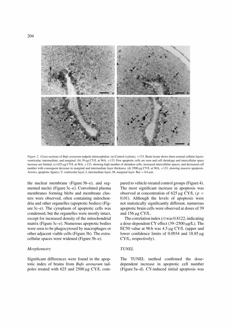
204
Figure 2. Cross-sections of Bufo arenarum tadpole telencephalon. (a) Control (xylene), ×133. Brain tissue shows three normal cellular layers:
ventricular, intermediate, and marginal. (b) 39 μg CY/L at 96 h, ×133. Few apoptotic cells are seen and cell shrinkage and intercellular space
increase are limited. (c) 625 μg CY/L at 96 h, ×133, showing high number of shrunken cells, increased intercellular spaces, and decreased cell
number with consequent decrease in marginal and intermediate layer thickness. (d) 2500 μg CY/L at 96 h, ×133, showing massive apoptosis.
Arrows, apoptotic figures; V, ventricular layer, I, intermediate layer; M, marginal layer. Bar = 8.6 μm.
the nuclear membrane (Figure 3b–e), and seg-mented nuclei (Figure 3c–e). Convoluted plasmamembranes forming blebs and membrane clus-ters were observed, often containing mitochon-dria and other organelles (apoptotic bodies) (Fig-ure 3c–e). The cytoplasm of apoptotic cells wascondensed, but the organelles were mostly intact,except for increased density of the mitochondrialmatrix (Figure 3c–e). Numerous apoptotic bodieswere seen to be phagocytosed by macrophages orother adjacent viable cells (Figure 3b). The extra-cellular spaces were widened (Figure 3b–e).
Morphometry
Significant differences were found in the apop-totic index of brains from Bufo arenarum tad-poles treated with 625 and 2500 μg CY/L com-
pared to vehicle-treated control groups (Figure 4).The most significant increase in apoptosis wasobserved at concentration of 625 μg CY/L (p <
0.01). Although the levels of apoptosis werenot statistically significantly different, numerousapoptotic brain cells were observed at doses of 39and 156 μg CY/L.
The correlation index (r) was 0.8122, indicatinga dose-dependent CY effect (39–2500 μg/L). TheEC50 value at 96 h was 4.5 μg CY/L (upper andlower confidence limits of 0.0934 and 18.85 μgCY/L, respectively).
TUNEL
The TUNEL method confirmed the dose-dependent increase in apoptotic cell number(Figure 5a–d). CY-induced initial apoptosis was

205
Figure 3. Electron microscopy of Bufo arenarum tadpole telencephalon. (a) Control (xylene), ×3200. (b) 39 μg CY/L at 96 h, ×3200. Note
the presence of apoptotic cell with highly pyknotic nucleus. (c) 2500 μg CY/L at 96 h, ×3200. The increase in apoptotic bodies and apoptotic
cells with segmented or condensed nuclear chromatin and dense cytoplasm is evident. (d) 2500 μg CY/L at 96 h, ×7000. Detail of two apoptotic
cells at different stages of cell death. Note that the cytoplasmic organelles remain relatively well preserved. (e) 2500 μg CY/L at 96 h, ×7000.
Detail of an apoptotic cell showing chromatin segmentation. Ap, apoptotic cell; N, viable neural cell; Apb, apoptotic bodies; M, mitochondria.
Bar = 1 μm.

206
Control and CY treated animals
Ap
op
totic
ind
ex
0.0
0.2
0.4
0.6
0.8
1.0
1.2
1.4
1.6
Xilene
39 μg
156 μg
625 μg
2,500 μg
*
**
Figure 4. Apoptotic index of Bufo arenarum tadpole brain (mean ± SE). Xylene represents control. ∗ p < 0.05, ∗∗ p < 0.01.
observed in brain cell nuclei from 39 μg CY/L(Figure 5a). The highest number of apoptotic cellswas detected at a dose of 2500 μg CY/L, identi-fied by numerous fluorescent nuclei (Figure 5c).No selectivity was observed in the different cere-bral regions studied in relation to the number ofapoptotic cells.
DNA fragmentation assay
For the different treatments, lines of DNAfragmentation were evaluated as the integratedintensity of the electrophoretic bands usingMatlab-based software designed for that purpose.
The DNA fragmentation assay showed that theDNA breakup pattern increased parallel to the CYconcentration. Compared to the control line, sta-tistically significant differences were found with156, 625 and 2500 μg CY/L (Figures 6 and 7).
Analysis of DNA fragmentation shows no sig-nificant differences between 39 μg CY/L and con-trol.
Discussion
Amphibians are particularly vulnerable to pesti-cides because their life-cycle stages evolve aroundponds and streams, and most species remainnear these sites even in adulthood. Fresh wa-ter can accumulate pesticides and toxic productsfrom cultivated land where pesticides are applied.Moreover, amphibian larval development occursin spring–summer contemporaneously with in-crease in pesticide applications (Materna et al.,1995). In Argentina, the application rates of in-secticides in cultivated lands are between 1.5 and120 g CY active ingredient per hectare (CASAFE,1995).
Variations in the acute toxicity effects dependon the chemical structure and isomerism of theindividual compounds and on the vehicle used,as well on as the species, sex, age, and diet ofthe animal. In such studies, the values reporteddiffered markedly depending on the model used(Kaloyanova et al., 1991). Cypermethrin is toxicto fish (in laboratory tests 96 h LC50 values wereobtained at doses of 0.4–2.8 μg/L) and aquatic

207
Figure 5. Detection (by TUNEL or DAPI) of fragmented DNA in Bufo arenarum tadpole brain. (a) 39 μg CY/L at 96 h (TUNEL). (b) 39 μg
CY/L at 96 h (DAPI.) (c) 2500 μg CY/L at 96 h (TUNEL). (d) 2500 μg CY/L at 96 h (DAPI). Bar = 10 μm.

208
Figure 6. DNA fragmentation pattern in Bufo arenarum cerebral
cells by electrophoresis. MW, molecular weight ladder; lane 1, con-
trol; lane 2, 39 μg CY/L; lane 3, 156 μg CY/L; lane 4, 625 μg CY/L;
lane 5, 2500 μg CY/L.
invertebrates (at doses of 0.01 to >5 μg/L) (WHO,1989) but is not very toxic for birds, and earth-worms are not sensitive (WHO, 1989). In stud-
Figure 7. Quantification of DNA fragments, based on integrated intensity of electrophoresis bands. ∗∗ p < 0.01, ∗∗∗ p < 0.001.
ies involving deliberate spraying of experimentalponds under field conditions, concentrations of2.6 μg CY/L were measured in the water and, al-though fish were not severely affected at thoseconcentrations, populations of crustaceae, mites,and surface-breathing insects were drastically re-duced (WHO, 1989).
Frogs are highly sensitive to most pyrethroids,including CY in subcutaneous and intraperitonealapplications (Cole and Casida, 1983). LC50 valuesfor frogs treated subcutaneously are more similarto those for insects treated topically than those forrats or mice receiving oral, intraperitoneal, or in-travenous treatments (Casida et al., 1983). Acutetoxicity tests showed high sensitivity to CY treat-ment in Physalaemus biligonigerus frog tadpoles(Izaguirre et al., 2000, 2001).
In this work, CY treatments of Bufo arenarumtadpoles with doses starting at 39 μg CY/L in-duced all the neurotoxic phases I to IV describedby Salibian (1992), similarly to the effects found inadult frogs (Cole and Casida, 1983). The toxic ef-fects were characterized by clustering, swimming

209
at full speed, twisting, lateral curving of thetail, and arching and sinuous writhing along thecephalic–caudal axis. After 96 h of treatment, the110 μg CY/L LC50 values were higher than thosefound in fish (96 h LC50 0.4–2.8 μg/L) (WHO,1989). Morphological and morphometric studies,TUNEL analysis, and DNA fragment quantifica-tion of treated tadpoles showed pathological alter-ations in the central nervous system, even at CYconcentrations as low as 39 μg CY/L.
Dose-dependent apoptosis was observed in thecentral nervous system cells starting at 39 μgCY/L with a dramatic increase in apoptosis at adose of 156 μg CY/L. Most significantly, mor-phometric analysis showed that CY concentra-tion as low as 4.5 μg CY/L can produce toxiceffects, approaching fish toxicity values. Thedose-dependent apoptosis produced by CY in thebrain of Bufo arenarum tadpoles was similar tothe effect found in Physalaemus biligonigerus tad-poles (Izaguirre et al., 2000, 2001). The 96 h LC50
(129 μg CY/L) values for the above-mentionedspecies were also similar to Bufo arenarum 96 hLC50 values (110 μg CY/L).
Morphologically, we observed brain alterationsin the intermediate and marginal layers. Cerebralimmature cells showed cellular shrinkage, nu-clear fragmentation, and increase of intercellularspaces, typical of apoptotic cell death (Kerret al., 1972; Wyllie, 1980; Izaguirre et al., 2000,2001). The TUNEL method confirmed DNAfragmentation, a typical feature of apoptosisinduced by both physiological and pathologicalmechanisms (Bortner and Cidlowski, 1996). Thequantitative method applied to determine theDNA fragment concentration demonstrated thatthe highest increase in apoptosis is producedfrom 156 μg CY/L. As was expected, the highestconcentration of small DNA fragments (172 bp)occurred at the highest dose of 2500 μg CY/L at96 h.
A major morphological change during apop-tosis is the shrinkage of cells (Kerr et al., 1972;Wyllie, 1980), suggesting a fundamental role ofcell volume loss in this form of cell death. How-
ever, the mechanism of cell volume loss and itsprecise role in the activation of apoptosis has notyet been clarified.
Modulation of cell volume appears to regu-late cellular metabolism and gene expression,and changes in cell volume can act in signaltransducer systems during multiple cellular pro-cesses (Haussinger et al., 1994). Several studieshave implicated a role for potassium in apopto-sis (Barbiero et al., 1995; Beauvais et al., 1995;Benson et al., 1996; Bortner et al., 1997) and haveshown that the shrunken apoptotic cells have re-duced intracellular levels of K+ and Na+ (Bortneret al., 1997). The loss of K+ and Na+ duringapoptosis appears to occur in most animal speciesindependently of the mode of activation of apop-tosis. Although K+ depletion alone in cells ex-posed to hypotonic conditions in the absence ofan apoptotic stimulus does not trigger apoptosis oralter long-term viability (Bortner and Cidlowski,1996), a reduced intracellular K+ concentrationenhances the activation of cell death programs(Bortner et al., 1997). In vitro studies showed thatnucleases that degrade DNA during apoptosis, aswell as the activity of caspase-1 (ICE), are ef-fectively inhibited at physiological K+ concen-trations (Walev et al., 1995; Hughes et al., 1997;Montague et al., 1997). Activation of procaspaseby dATP and cytochrome c is also inhibited byphysiological K+ concentrations (Hughes et al.,1997). These results suggest that the transition ofan apoptotic cell from a state of high ionic strengthfacilitates both the cell volume loss and the acti-vation of enzymes that mediate apoptosis.
In this study we have demonstrated the hightoxicity of cypermethrin to Bufo arenarum larvae.The present results and previous results (Izaguirreel al., 2000, 2001) show that lower doses thanthose used in routine applications can cause mas-sive apoptosis in the central nervous system. Wepostulate that CY-induced apoptotic cell death ofcells of the central nervous system, through spe-cific neurotoxic mechanisms, is the major factorfor the high mortality of Bufo arenarum causedby CY.
210
Acknowledgements
This work was supported by a grant fromSCYTFRH-UNER, PID 6053-1 (to V.H.C.) andin part by The Lankenau Foundation.
References
Barbiero G, Duranti F, Bonelli G, Amenta JS, Baccino FM. Intra-
cellular ionic variations in the apoptotic death of L cells by in-
hibitors of cell cycle progression. Exp Cell Res. 1995;217:410–
8.
Beauvais F, Michel L, Dubertret L. Human eosinophils in culture
undergo a striking and rapid shrinkage during apoptosis. Role of
K+ channels. J Leukoc Biol. 1995;57:851–5.
Benson RSP, Heer S, Dive C, Watson AJM. Characterization of cell
volume loss in CEM-C7A cells during dexamethasone-induced
apoptosis. Am J Physiol. 1996;270:1190–203.
Berlin JR, Akera T, Brody TM, Matsumura F. The ionotropic effects
of a synthetic pyrethroid decamethrin on isolated guinea pig atrial
muscle. Eur J Pharmacol. 1984;98:313–22.
Bortner CD, Cidlowski JA. Absence of volume regulatory mecha-
nisms contributes to the rapid activation of apoptosis in thymo-
cytes. Am J Physiol. 1996;271:950–61.
Bortner CD, Hughes FM Jr, Cidlowski JA. A primary role for K+and Na+ efflux in the activation of apoptosis. J Biol Chem.
1997;272:32436–42.
CASAFE: Camara de Sanidad Agropecuaria y Fertilizantes de la
Republica Argentina.
CASAFE: Guıa de Productos Fitosanitarios para la Republica
Argentina. Buenos Aires CASAFE, 1995.
Casida JE, Gammon DW, Glickman AH, Lawrence LJ. Mechanisms
of selective action of pyrethroid insectices. Annu Rev Pharmacol
Toxicol. 1983;23:413.
Cole LM, Casida JE. Pyrethroid toxicology in the frog. Pesticide
Biochem Physiol. 1983;20:217–24.
El-Gohary M, Awara WM, Nassar S, Hawas S. Deltamethrin-induced
testicular apoptosis in rats: the protective effect of nitric oxide
synthase inhibitor. Toxicology. 1999;132:1–8.
Elliott M. Properties and applications of pyrethroids. Environ Health
Perspect. 1976;14:3–13.
Enan E, Matsumara F. Activation of phosphoinositide/protein kinase
C pathway in rat brain tissue by pyrethroids. Biochem Pharmacol.
1993;45:703–10.
Eshleman AJ, Murray TF. Pyrethroid insecticides indirectly inhibit
GABA-dependent 36Cl-influx in synaptoneurosomes from the
trout brain. Neuropharmacology. 1991;30:1333–41.
Ferrari L, Demichelis SO, Garcıa ME, De La Torre FR, Salibian
A. Premetamorphic anuran tadpoles as test organism for an
acute aquatic toxicity assay. Environ Toxicol Water Qual.
1997;12:118–21.
Finney DJ. Probit analysis. New York: Cambridge University Press;
1971.
Gosner KL. A simplified table for staging anuran embryos and lar-
vae, with notes on identification. Herpetology. 1960;16:183–
90.
Haussinger D, Lang F, Gerok W. Regulation of cell function
by the cellular hydration state. Am J Physiol. 1994;267:343–
55.
Holcombe GW, Phipps GL, Tanner DK. The acute toxicity of
kelthane, dursban, disulfoton, pydrin, and permethrin to fathead
minnows Pimephales promelas and rainbow trout Salmo gaird-neri. Environ Pollut. 1982;29:167–78.
Holtfreter J. Uber die Aufzucht isolierter Teile des Amphi-
bienkeimes. II Zuchtung von Keimen und Keimteilen in
Salzlosung. Roux Arch. 1931;124:405–64.
Hughes FM, Bortner CD, Purdy G, Cidlowski JA. Intracellular K+suppresses the activation of apoptosis in lymphocytes. J Biol
Chem. 1997;272:30567–76.
Izaguirre MF, Lajmanovich RC, Peltzer PM, Peralta Soler A, Casco
VH. Cypermethrin-induced apoptosis in the telencephalon of
Physalaemus biligonigerus tadpoles (Anura: Leptodactylidae).
Bull Environ Contam Toxicol. 2000;65:501–7.
Izaguirre MF, Lajmanovich RC, Peltzer PM, Peralta Soler A, Casco
VH. Induction of cell death by the synthetic pyrethroid insecti-
cide cypermethrin in the developing brain of physalaemus biligo-nigerus tadpoles from Argentina. Froglog. 2001;43:3–4.
Jolly AL Jr, Avault JW, Koonee KL, Graves JB. Acute toxicity
of permethrin to several aquatic animals. Trans Am Fish Soc.
1978;107:825–7.
Kaloyanova F, Batawi E, Mostafa A. Human toxicology of pesticides.
Boca Raton: CRC Press; 1991.
Kerr JFR, Wyllie AH, Currie AR. Apoptosis: a basic biological phe-
nomenon with wide-ranging implications in tissue kinetics. Br J
Cancer. 1972;26:239–57.
Little EE, Dwyer FJ, Fairchild JF, DeLonay AJ, Zajicek JL. Sur-
vival of bluegill and their behavioral responses during continuous
and pulsed exposures to esfenvalerate, a pyrethroid insecticide.
Environ Toxicol Chem. 1993;12:871–8.
Materna EJ, Rabeni CF, LaPoint TW. Effects of the synthetic
pyrethroid insecticide, esfenvalerate, on larval leopard frogs
(Rana spp.). Environ Toxicol Chem. 1995;14:613–22.
Miyamoto J. Degradation, metabolism and toxicity of synthetic
pyrethroids. Environ Health Perspect. 1976;14:15–28.
Montague JW, Hughes FMJr, Cidlowski JA. Native recombinant cy-
clophilins A, B, and C degrade DNA independently of peptidyl-
prolyl cis-trans-isomerase activity. Potential roles of cyclophilins
in apoptosis. J Biol Chem. 1997;272:6677–84.
Salibian A. Effects of deltamethrin on the South American toad, Bufoarenarum, tadpoles. Bull Eviron Contam Toxicol. 1992;48:616–
21.
Salibian A, Marazzo L. Studies on the effects of deltamethrin on
sodium net transport through the in vivo amphibian skin. Biomed
Environ Sci. 1995;8:165–8.
Smith TM, Stratton GW. Effects of synthetic pyrethroid insecticides
on non-target organisms. Residue Rev. 1986;97:93–120.
Sokal RR, Rohlf FJ. Biometrıa. Revised edn. Madrid: Ediciones
Blume; 1979.
USEPA: United States Environmental Protection Agency. Methods
for acute toxicity test with fish, macroinvertebrate, and amphib-
ians. Ecol Res Ser. 1975; EPA-660/3-75-009.

211
USEPA: United States Environmental Protection Agency. Short-
term methods for estimating the chronic toxicity of effluents
and receiving waters to freshwater organisms. Revised edn.
1989.
Walev I, Reske K, Palmer M, Valeva A, Bhakdi S. Potassium-
inhibited processing of IL-1 beta in human monocytes. EMBO
J. 1995;14: 1607–14.
WHO: World Health Organization. Environmental Health Criteria
82: Cypermethrin. Revised edn. Geneva: WHO; 1989.
Wyllie AH. Glucocorticoid-induced thymocyte apoptosis is asso-
ciated with endogenous endonuclease activation. Nature. 1980;
284: 555–6.
Address for correspondence: V.H. Casco, Microscopy Lab-
oratory, School of Engineering – Bioengineering – UNER,
CC 47, Suc. 3, 3100 Parana, Entre Rıos, Argentina.
E-mail: [email protected]