Fetal-Monitoring-FHR-Presentation.pdf - Huntington Hospital
69
1 INTRAPARTUM FETAL HEART RATE MONITORING Definitions, Interpretation and Management Applying Principles of Patient Safety David A. Miller, M.D. Professor of Obstetrics, Gynecology and Pediatrics Division of Maternal Fetal Medicine University of Southern California Keck School of Medicine Childrens Hospital Los Angeles Financial Relationships Disclosures for Presenters at Educational Programs I have the following relevant financial relationship with a commercial interest: Co‐author: “Fetal Monitoring – A Multidisciplinary Approach” Mosby’s Pocket Guide Series Distributed by Mosby‐Elsevier Co‐author: “Electronic Fetal Heart Rate Monitoring Interpretation and Management” On‐line, Interactive Educational Program: Distributed by GE Healthcare Consulting agreement Clinical Computer Systems, Inc Makers of OBIX As a medical professional, there are many things on your plate, and fetal monitoring is only one of them It might seem that a disproportionate amount of time and energy is dedicated to this one area of medicine But that is because fetal monitoring… 1. Is the most common procedure you will perform in obstetrics 2. Involves the potential for preventable lifelong brain damage 3. Represents an overwhelmingly disproportionate share of the medicolegal risk you will face throughout your career 4. Our primary goal is to optimize outcomes…a secondary goal is to minimize risks
-
Upload
khangminh22 -
Category
Documents
-
view
1 -
download
0
Transcript of Fetal-Monitoring-FHR-Presentation.pdf - Huntington Hospital

1
INTRAPARTUM FETAL HEART RATE MONITORING
Definitions, Interpretation and Management
Applying Principles of Patient Safety
David A. Miller, M.D.Professor of Obstetrics, Gynecology and Pediatrics
Division of Maternal Fetal MedicineUniversity of Southern California Keck School of Medicine
Childrens Hospital Los Angeles
Financial Relationships Disclosures
for Presenters at Educational Programs
I have the following relevant financial relationship with a commercial interest:
Co‐author:“Fetal Monitoring – A Multidisciplinary Approach”
Mosby’s Pocket Guide SeriesDistributed by Mosby‐Elsevier
Co‐author:“Electronic Fetal Heart Rate Monitoring
Interpretation and Management”On‐line, Interactive Educational Program:
Distributed by GE Healthcare
Consulting agreementClinical Computer Systems, Inc
Makers of OBIX
As a medical professional, there are many things on your plate, and fetal monitoring is only one of them
It might seem that a disproportionate amount of time and energy is dedicated to this one area of medicine
But that is because fetal monitoring…
1. Is the most common procedure you will perform in obstetrics2. Involves the potential for preventable lifelong brain damage3. Represents an overwhelmingly disproportionate share of the
medicolegal risk you will face throughout your career4. Our primary goal is to optimize outcomes…a secondary goal
is to minimize risks

2
The most effective way to optimize outcomes AND minimize medical‐legal
risk is to practice according to…
“Standard of Care”
Define “Standard of Care”
• Level of care provided by best practitioners in the community?
• Level of care provided by average practitioners in the community?
• Level of care provided by most practitioners in the community?
• Minimally acceptable level of care?• Level of care dictated by AWHONN and ACOG?• Level of care dictated by standard textbooks?
“Standard of Care”
Level of care expected of a reasonable practitioner
Who makes that determination?

3
How do they decide?
Optimize
outcomes
Standard of care
Reasonable
Credible
Factually accurate and articulate
“I don’t know the specific definition, but I know it when I see it.”
Factual accuracy and ability to articulate are NOT optional
Even if you never encounter a legal challenge in your career, if you cannot communicate adequately to obtain appropriate informed consent, you have not met the standard of care

4
Intrapartum FHR monitoring is one of the most common obstetric procedures in the US, impacting the lives of almost 8 million mothers and babies every year
However, for 4 decades, a lack of standardized training and competency testing in intrapartum FHR monitoring has led to:
Ill‐defined, confusing, controversial terms (“perinatal asphyxia”, “fetal distress”)
Unsubstantiated theories, hypotheses…unscientific dogma
Myths, urban legends and folklore passed down from resident to resident, nurse to nurse and generation to generation
A breakdown in communication that jeopardizes patient safety challenges the credibility of our profession
Theories and Hypotheses
New Technology
Pioneering phase Mature technology
ScientificProcess
True
False

5
LEVELS OF SCIENTIFIC EVIDENCE
Since 1997 there have been several important consensus publications that have reshaped the fetal monitoring landscape:
• 1997 – First NICHD Consensus Statement
• 1999 – International Cerebral Palsy Task Force Consensus Statement
• 2003 – ACOG‐AAP Cerebral Palsy Task Force Consensus Statement
• 2005 – ACOG/AWHONN endorsement
• 2006 – ACNM endorsement
• 2008 – Second NICHD consensus report
• 2009 – ACOG Practice Bulletin 106
• 2010 – ACOG Practice Bulletin 116
Why the need to standardize?

6
Pettker Am J Obstet Gynecol. 2009;200:492.e1‐8
Standardization can reduce adverse outcomes and professional liability claims
Impact of a comprehensive patient safety strategy on obstetric adverse events
Clark SL Obstet Gynecol. 2008 Dec;112(6):1279‐83.
Reducing obstetric litigation through alterations in practice patterns
In‐house obstetric coverageMedication protocolsVBAC protocolsShoulder dystocia protocols
Call revision
Obstetric drills
EFM
Course
Team
training
QI
What can the technology really do?

7
A FHR tracing with minimal‐absent variability and late decelerations accurately predicts cerebral palsy 1 time out of 500 (99.8% false positive rate)
The population incidence of cerebral palsy is ~ 1 per 500
Nelson KB, Dambrosia JM, Ting TY, Grether JK. Uncertain value of electronic fetal monitoring in predicting cerebral palsy N Eng J Med 1996;334:613‐8
The fact is…most “non‐reassuring” FHR tracings predict neurologic injury no better than randomly selecting a name from the telephone book
The fact is…most “non‐reassuring” FHR tracings predict neurologic injury no better than randomly selecting a name from the telephone book
Electronic FHR monitoring is NOT a diagnostic test
It is a screening testExcept in the most extreme cases, it has never been capable of reliably diagnosing
fetal injury or “impending injury”

8
Start with the basics
Undertake the simple exercise of deconstructing fetal heart
rate monitoring into its essential components
FHR monitoring consists of three components:
Intrapartum FHR Monitoring
Definition Interpretation Management
210
180
150
120
90
60
Normal baseline rate 110‐160 bpm
Mean FHR rounded to increments of 5 bpm in a 10‐minute window

9
210
180
150
120
90
60
Variability is defined as fluctuations in the baseline that are irregular in amplitude and frequency…
No distinction is made between short‐term (beat‐to‐beat) variability and long term variability because in actual practice
they are visually determined as a unit
The fluctuations are measured from peak‐to‐trough in bpm
AccelerationAbrupt increase (onset to peak < 30 sec) from baseline
32 weeks and beyond – 15 x 15
Before 32 weeks – 10 x 10
Decelerations
EarlyLateVariableProlonged

10
Late versus variable
Late Deceleration
Gradual decrease in FHR associated with a contraction
Onset, nadir, and recovery occur after the beginning, peak, and ending of the contraction
Variable Deceleration
Abrupt decrease in FHR at least 15 bpm for at least 15 seconds
11
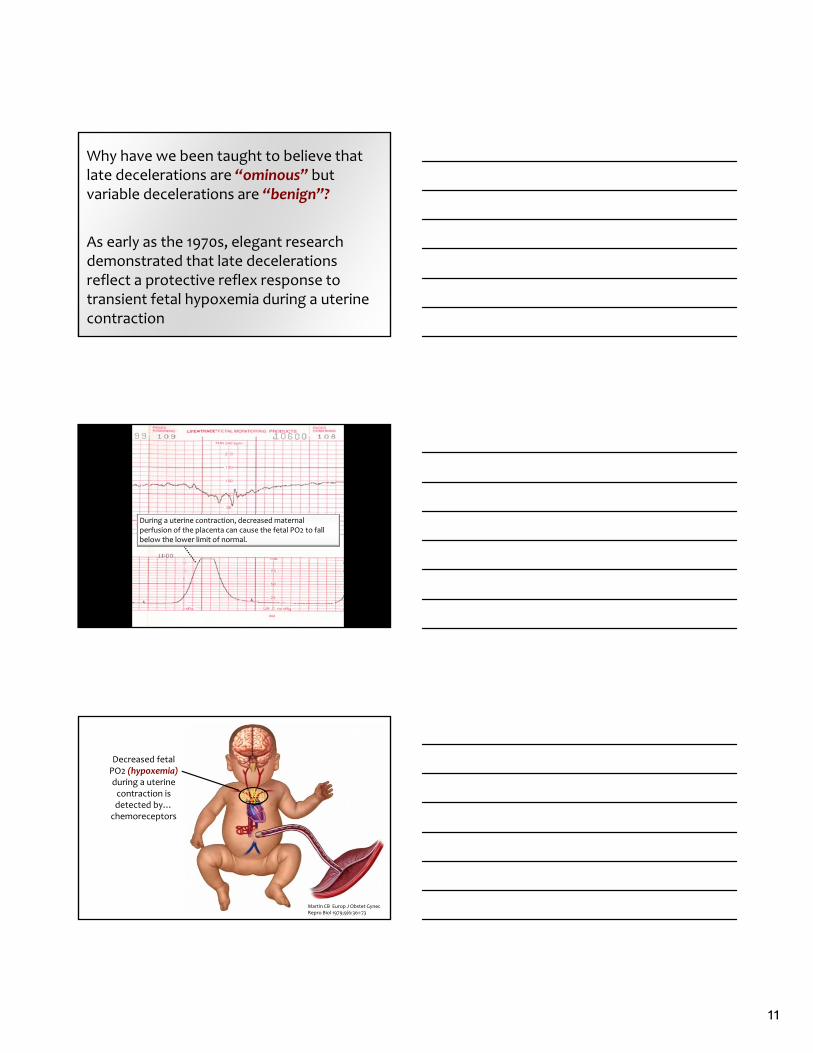
Why have we been taught to believe that late decelerations are “ominous” but variable decelerations are “benign”?
As early as the 1970s, elegant research demonstrated that late decelerations reflect a protective reflex response to transient fetal hypoxemia during a uterine contraction
During a uterine contraction, decreased maternal perfusion of the placenta can cause the fetal PO2 to fall below the lower limit of normal.
Decreased fetal PO2 (hypoxemia)during a uterine contraction is detected by…chemoreceptors
Martin CB Europ J Obstet Gynec Repro Biol 1979;9/6:361‐73

12
Chemoreceptors signal thebrain stem
Martin CB Europ J Obstet Gynec Repro Biol 1979;9/6:361‐73
Martin CB Europ J Obstet Gynec Repro Biol 1979;9/6:361‐73
In order to shunt oxygenated blood to the vital organs of the brain, heart, adrenal glands and
placenta…
Sympathetic outflow causes peripheral vasoconstriction to redistribute oxygenated blood away from the extremities, gut
and kidneys
Peripheral vasoconstriction causes the blood pressure to rise
Martin CB Europ J Obstet Gynec Repro Biol 1979;9/6:361‐73

13
Martin CB Europ J Obstet Gynec Repro Biol 1979;9/6:361‐73
Rising blood pressure is
detected by… baroreceptors
Baroreceptors signal the brain stem
Martin CB Europ J Obstet Gynec Repro Biol 1979;9/6:361‐73
Martin CB Europ J Obstet Gynec Repro Biol 1979;9/6:361‐73
Parasympathetic (vagal) outflow slows the FHR to reduce cardiac
output and lower blood pressure

14
This reflex can be seen in the fetal heart rate tracing as a late deceleration
As the uterine contraction subsides, oxygenated maternal blood enters the
intervillous space
Fetal PO2 rises. The autonomic reflex subsides and the FHR returns to baseline.

15
If this description is accurate, what would you expect to see?
Ball (9)
Itskovitz (8)
Ball (7)
Jensen (6)
Reid (5)
Field (4)
Richardson (3)
Peeters (2)
Cohn (1)
BrainReference
Heart
Adrenal
1. AJOG 1974;120:817‐242. AJOG 1979;135:637‐463. J Dev Physiol 1989;11:37‐434. J Dev Physiol 1990;14:131‐7
5. J Dev Physiol 1991;15:183‐86. J Dev Physiol 1991;15:309‐237. AJOG 1994;170:156‐618. Am J Physiol 1987;252:H100‐99. AJOG 1994;171:1549‐55
Blood Flow in Fetal Lamb in response to hypoxemia
BP
Kidney
Body
Variable Deceleration
Occlusion of the umbilical cord
causes the blood pressure to…
RISE

16
Rising blood pressure is
detected by… baroreceptors
Baroreceptors signal the brain stem
Parasympathetic (vagal) outflow slows the FHR to reduce cardiac
output and lower blood pressure

17
Late decelerations and variable decelerations are protective autonomic reflex responses
Neither is inherently “ominous”
Neither is inherently “benign”
The 2008 NICHD Workshop Report on Electronic Fetal Monitoring
A very brief update
Obstet Gynecol 2008;112:661‐6
Previous classification system
“Reassuring”
“Non‐reassuring”
Reassuring: (adj)
“Restoring confidence and relieving anxiety”

18
New “Three‐Tier” Fetal Heart Rate Classification System
Category I – “Normal”
Category II – “Indeterminate”
Category III – “Abnormal”
Obstet Gynecol 2008;112:661‐6
Category I – “Normal”
Baseline rate: 110‐160 bpmVariability: ModerateDecelerations: No late, variable or prolonged
Obstet Gynecol 2008;112:661‐6
New “Three‐Tier” Fetal Heart Rate Classification System
Category III – “Abnormal”
1. Absent variability with recurrent late decelerations
2. Absent variability with recurrent variable decelerations
3. Absent variability with bradycardia for at least 10 min
4. Sinusoidal pattern for at least 20 min
Obstet Gynecol 2008;112:661‐6
New “Three‐Tier” Fetal Heart Rate Classification System

19
Category II?
EverythingElse
Definitions:•Baseline•Variability•Accelerations•Decelerations•Changes or trends over time•“CATEGORY”
Factual Accuracy

20
Interpretation
Patient Safety+
Standard of Care
• Standard• Simple• Factually Accurate• Articulate
“Ominous overshoot pattern”
“Variable with a late component”
“Checkmark pattern”
“V‐volume‐variable = oligohydramnios”
“W variable = nuchal cord”
“Icicle deceleration”
“Carrot‐stick deceleration”
“Uniform accelerations = umbilical vein compression”
“Atypical variables”
“Ominous Conversion Factor”
“Wandering baseline”
“Variability at the base of a late decel is reassuring”
“Classifying decelerations as mild‐moderate‐severe”

21
LEVELS OF SCIENTIFIC EVIDENCE
Level II evidence REQUIRES “appropriate control of confounding factors”, including baseline rate, variability, accelerations and frequency of decelerations
In the next few minutes, 40 years of research in intrapartum FHR interpretation will be distilled into
3central principles that are evidence based, reflect consensus in the literature and most importantly
are simple, practical and teachable

22
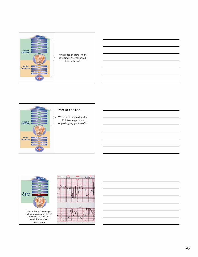
Intrapartum FHR monitoring is intended to assess
fetal oxygenation
Fetal oxygenation involves the transfer of oxygen from the environment to the fetus…
And the subsequent fetal physiologic response if oxygen
transfer is interrupted…
23
What does the fetal heart rate tracing reveal about
this pathway?
What information does the FHR tracing provide
regarding oxygen transfer?
Start at the top
Interruption of the oxygen pathway by compression of
the umbilical cord can result in a variable
deceleration
Cord

24
Interruption of the oxygen pathway at the level of the uterus or placenta can
result in a late deceleration
Placenta
Uterus
Interruption of the oxygen pathway at any point can result in a prolonged
deceleration
Placenta
Uterus
Heart
Lungs
Cord
Vasculature
ALL clinically significant FHR decelerations (late, variable, prolonged)
HAVE EXACTLY THE SAME TRIGGER…
Interruption of the oxygen pathway at one or more points

25
It’s a variable!!!
It’s a variable!
It’s a VARIABLE!!!It’s a LATE!!!
Labor and Delivery: 3:00 amIt’s a late!
It’s a late!!!
VARIABLE!!!
LATE!!!
This is unnecessary
It’s a VARIABLE!!!
It’s a LATE!!!
Step away from the edge…

26
Make it easy for yourself and your team…
All FHR decelerations that have any potential clinical significance have the same common trigger…
Interruption of oxygen transfer from the environment to the fetus at one or more points along the oxygen pathway
Principle #1Variable, late or prolonged
decelerations signal interruption of the oxygen pathway at one or
more points

27
What information can the FHR tracing provide
regarding the fetal response to interruption of the oxygen pathway?
The second half of the pathway
The 2008 NICHD consensus statement identified two fetal heart rate characteristics that reliably predict the absence of fetal metabolic acidemia at the
time they are observed
Obstet Gynecol 2008;112:661-7
Principle # 2Moderate variability or accelerations reliably predict the absence of fetal metabolic acidemia at the time they
are observedMetabolic acidemia

28
What is the physiologic significance of excluding
metabolic acidemia?
Supporters included:
1. American College of Obstetricians and Gynecologists2. American Gynecological and Obstetrical Society3. Australian College of Midwives4. Hong Kong Society of Neonatal Medicine5. Institute of Obstetrics and Gynaecology of the Royal College of Physicians of Ireland6. International Society of Perinatal Obstetricians7. New Zealand College of Midwives8. Paediatric Society of New Zealand9. Perinatal Society of Australia and New Zealand10. Royal Australasian College of Physicians, Paediatric Division11. Royal Australian College of General Practitioners12. Royal Australian College of Obstetricians and Gynaecologists13. Royal College of Obstetricians and Gynaecologists14. Royal College of Pathologists of Australasia15. Royal New Zealand College of Obstetricians and Gynaecologists16. Society of Obstetricians and Gynaecologists of Canada

29
The publication was endorsed by:
1. American College of Obstetricians and Gynecologists2. American Academy of Pediatrics3. Centers for Disease Control and Prevention4. Child Neurology Society5. March of Dimes Birth Defects Foundation6. National Institute of Child Health and Human Development7. Royal Australian and New Zealand College of Obstetricians and
Gynecologists8. Society for Maternal‐Fetal Medicine9. Society of Obstetricians and Gynaecologists of Canada
Metabolic acidemia
Metabolic acidemiais an essential prerequisite to
intrapartum hypoxic neurologic injury
(pH < 7, BD ≥ 12 mmol/L)

30
Is this simple enough to be taught and retained?
In 2009, the Los Angeles County Department of Health mandated FHR competency testing (OVMC, HUCLA, LAC+USC)
After a series of training sessions on standard NICHD FHR definitions, NICHD categories and 3 simplified principles of interpretation, a formal written test was administered to all care providers at all levels
A two‐year quality improvement initiative to standardize the methods by which obstetric team
members interpret, communicate, document and manage fetal heart
rate tracings
400 representatives from 90 of New York’s 140
hospitals
49%
85% 80% 84%
Pre‐test 6/7‐09
Pre and post‐test mean percent correct responses
Post‐test 6/7‐09 Post‐test 12‐09 Post‐test 12‐10

31
Reviewers demonstrated agreement on:
Baseline rate 0.97Moderate variability 0.80Accelerations 0.62Decelerations 0.63Category 0.68Exclude fetal metabolic acidemia 0.82
Kappa value Agreement< 0.40 Poor
.41 – .60 Moderate
.61 – .80 Substantial
.81 – 1.0 Excellent
Interobserver Reliability of Fetal Heart Rate Pattern Interpretation Using NICHD Definitions
Epstein A, et al. Am J Perinatol 2012 – in press
Substantial to Excellentagreement on all components
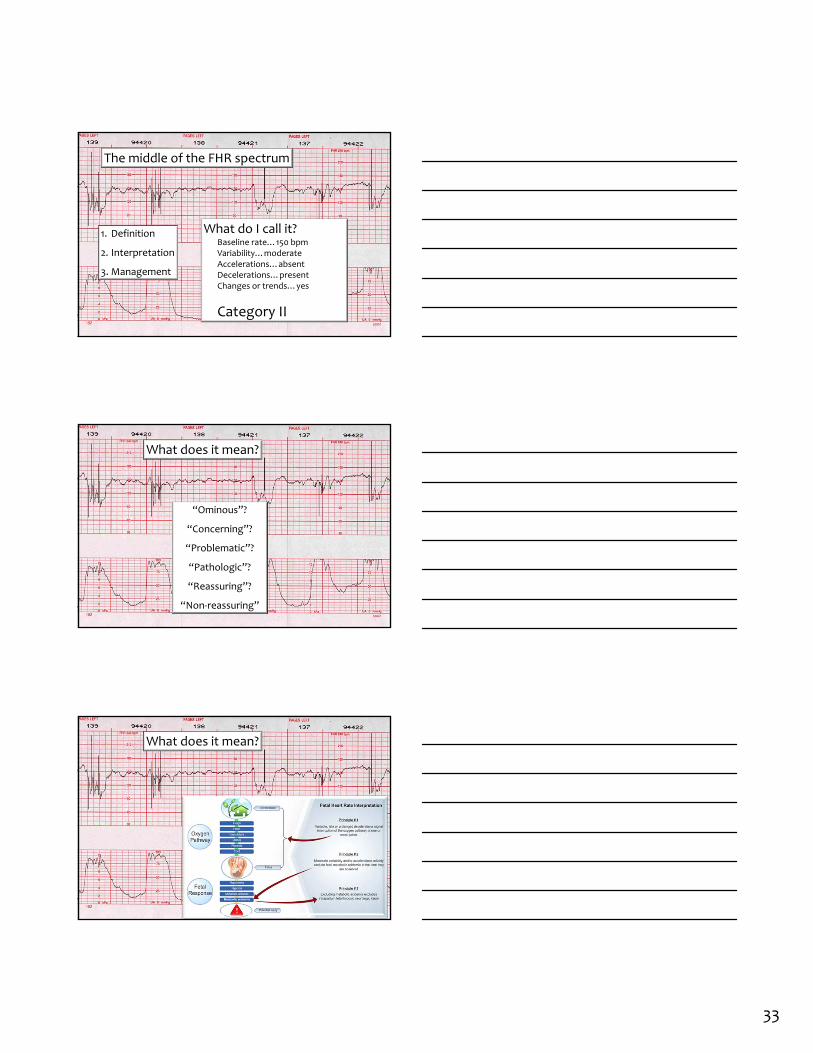
Does it have a practical application?
What do I call it?Baseline rate…130 bpmVariability…moderateAccelerations…presentDecelerations…absentChanges or trends over time…none
Category I
1. Definition
2. Interpretation
3. Management
One end of the FHR spectrum

32
What does it mean?
“Happy baby”?
“Baby’s fine”?
“Nothing to worry about”?
“Reassuring”?
What does it mean?
What do I do about it?
Standardized management coming up next
33
1. Definition
2. Interpretation
3. Management
What do I call it?Baseline rate…150 bpmVariability…moderateAccelerations…absentDecelerations…presentChanges or trends…yes
Category II
The middle of the FHR spectrum
What does it mean?
“Ominous”?
“Concerning”?
“Problematic”?
“Pathologic”?
“Reassuring”?
“Non‐reassuring”
What does it mean?

34
What do I do about it?
Management coming up next
1. Definition
2. Interpretation
3. Management
What do I call it?Baseline rate…165Variability…absentAccelerations…absentDeceleration…present, recurrentChanges or trends…yes
Category III
The far end of the FHR spectrum
What does it mean?

35
What does it mean?
What do I do about it?
Management coming up
Factual Accuracy
Standard DefinitionsWe have achieved consensus in theUnited States on the terminology used to describe the five components of a FHR tracing
Standard interpretationThree central concepts of FHR interpretation are evidence‐based and reflect consensus in the literature

36
Ability to Articulate
Standardized management is the next challenge
This will be the topic of the breakout sessions
Standardized management is NOT intended to replace individual clinical judgment
On the contrary…
Standardized management is intended to encourage the systematic application of
individual clinical judgment
Risk factors for error
Random recall
Lack of a checklist
Unnecessary complexity

37
Random recall
What do I call it?Baseline rate…130 bpmVariability…moderateAccelerations…presentDecelerations…absentChanges or trends over time…none
One end of the FHR spectrum – Category I
What does it mean?

38
Standardized Management
Is the patient low‐risk?
Routine Surveillance
No
Yes
Intrapartum Fetal Heart Rate Management Decision Model
•Every 30 min in the 1st stage of labor•Every 15 min in the 2nd stage of labor
Confirm FHR and uterine activity
I
•Every 15 min in the 1st stage of labor•Every 5 min in the 2nd stage of labor
FHR Category?
Heightened Surveillance
Is the patient low‐risk?
“ABCD”
Routine Surveillance
No
Yes
Intrapartum Fetal Heart Rate Management Decision Model
II or III
•Every 30 min in the 1st stage of labor•Every 15 min in the 2nd stage of labor
Confirm FHR and uterine activity
I
•Every 15 min in the 1st stage of labor•Every 5 min in the 2nd stage of labor
FHR Category?
Heightened Surveillance

39
Standardized Intrapartum FHR Management
Four Elements
“ABCD”
A – Assess the oxygen pathwayB – Begin corrective measures
Is the patient low‐risk?
“A” – Assess oxygen pathway“B” – Begin corrective measures
“ABCD”
Routine Surveillance
No
Yes
Intrapartum Fetal Heart Rate Management Decision Model
II or III
•Every 30 min in the 1st stage of labor•Every 15 min in the 2nd stage of labor
Confirm FHR and uterine activity
I
•Every 15 min in the 1st stage of labor•Every 5 min in the 2nd stage of labor
FHR Category?
Heightened Surveillance
“A”
Assess Oxygen
Pathway
“B”
Begin Corrective
Measures if Indicated
Lungs Airway and breathing Supplemental oxygen
Heart Heart rate and rhythm
Position changeFluid bolusCorrect hypotension
VasculatureBlood pressureVolume status
Uterus
Contraction strengthContraction frequencyBaseline uterine toneExclude uterine rupture Stop or reduce stimulant
Consider uterine relaxant
PlacentaPlacental separationBleeding vasa previa
CordVaginal examExclude cord prolapse
Consider amnioinfusion

40
“A”
Assess Oxygen
Pathway
“B”
Begin Corrective
Measures if Indicated
Lungs Airway and breathing Supplemental oxygen
Heart Heart rate and rhythm
Position changeFluid bolusCorrect hypotension
VasculatureBlood pressureVolume status
Uterus
Contraction strengthContraction frequencyBaseline uterine toneExclude uterine rupture Stop or reduce stimulant
Consider uterine relaxant
PlacentaPlacental separationBleeding vasa previa
CordVaginal examExclude cord prolapse
Consider amnioinfusion
Is the patient low‐risk?
“A” – Assess oxygen pathway“B” – Begin corrective measures
“ABCD”
Routine Surveillance
No
Yes
Intrapartum Fetal Heart Rate Management Decision Model
II or III
FHR Category?
IIIIII
•Every 30 min in the 1st stage of labor•Every 15 min in the 2nd stage of labor
Confirm FHR and uterine activity
I
•Every 15 min in the 1st stage of labor•Every 5 min in the 2nd stage of labor
FHR Category?
Heightened Surveillance Expedite Delivery
??

41
What fetal heart rate characteristics tell you it is
safe to continue surveillance?
Step away from the edge…
Make it easy for yourself and your team…

42
If you have any question…
…the safest approach is to proceed to the next step
“C”
Cesarean Section

43
NO
“C”Cesarean?
Call for Cesarean?
Crash Cesarean?
Call for the vacuum?
Commit to cesarean?
Commit to delivery?
Cancel clinic?
Standardized Intrapartum Management
“ABCD”
A – Assess the oxygen pathwayB – Begin corrective measuresC – Clear obstacles to rapid delivery

44
Clear obstacles to rapid deliveryIf conservative measures do not correct the FHR tracing, it is prudent to plan ahead for the possible need for rapid delivery
This does NOT commit the patient to delivery
It simply identifies common sources of unnecessary delay in a systematic way so they can be addressed in timely fashion
By doing this, it demonstrates reasonableness and prudence…two elements that define the standard of care

45

46
FacilityStaffMotherFetusLabor
Consider individual characteristics of
“A”
Assess Oxygen
Pathway
“B”
Begin Corrective
Measures if Indicated
“C”
Clear Obstacles to
Rapid Delivery
“D”
Determine Decision
to Delivery Time
Lungs Airway and breathing Supplemental oxygen FacilityOR availabilityEquipment
Facility response time
Heart Heart rate and rhythm
Position changesFluid bolusCorrect hypotension
Staff
NotifyObstetricianSurgical assistantAnesthesiologistNeonatologistPediatricianNursing staff
Consider staff:AvailabilityTrainingExperience
VasculatureBlood pressureVolume status
Mother
Informed consentAnesthesia optionsLaboratory testsBlood productsIntravenous accessUrinary catheterAbdominal prepTransfer to OR
Surgical considerations(prior abdominal or uterine surgery )Medical considerations(obesity, hypertension, diabetes, SLE)Obstetric considerations(parity, pelvimetry, placental location)
Uterus
Contraction strengthContraction frequencyBaseline uterine toneExclude uterine rupture Stop or reduce stimulant
Consider uterine relaxantFetus
ConfirmEstimated fetal weightGestational agePresentationPosition
Consider factors such as:Estimated fetal weightGestational agePresentationPositionInfection MeconiumPlacenta
Placental separationBleeding vasa previa
CordVaginal examExclude cord prolapse
Consider amnioinfusion Labor Consider IUPC
Consider factors such as:Arrest disorderProtracted laborRemote from deliveryPoor expulsive efforts

47
“A”
Assess Oxygen
Pathway
“B”
Begin Corrective
Measures if Indicated
“C”
Clear Obstacles to
Rapid Delivery
“D”
Determine Decision
to Delivery Time
Lungs Airway and breathing Supplemental oxygen FacilityOR availabilityEquipment
Facility response time
Heart Heart rate and rhythm
Position changesFluid bolusCorrect hypotension
Staff
NotifyObstetricianSurgical assistantAnesthesiologistNeonatologistPediatricianNursing staff
Consider staff:AvailabilityTrainingExperience
VasculatureBlood pressureVolume status
Mother
Informed consentAnesthesia optionsLaboratory testsBlood productsIntravenous accessUrinary catheterAbdominal prepTransfer to OR
Surgical considerations(prior abdominal or uterine surgery )Medical considerations(obesity, hypertension, diabetes, SLE)Obstetric considerations(parity, pelvimetry, placental location)
Uterus
Contraction strengthContraction frequencyBaseline uterine toneExclude uterine rupture Stop or reduce stimulant
Consider uterine relaxantFetus
ConfirmEstimated fetal weightGestational agePresentationPosition
Consider factors such as:Estimated fetal weightGestational agePresentationPosition
PlacentaPlacental separationBleeding vasa previa
CordVaginal examExclude cord prolapse
Consider amnioinfusion Labor Consider IUPC
Consider factors such as:Arrest disorderProtracted laborRemote from deliveryPoor expulsive efforts
Is the patient low‐risk?
“A” – Assess oxygen pathway“B” – Begin corrective measures
“C” – Clear obstacles to rapid delivery“D” – Determine decision to delivery time
“ABCD”
Routine Surveillance
No
Yes
Intrapartum Fetal Heart Rate Management Decision Model
II or III
FHR Category?
IIIIII
Is vaginal delivery likely before the onset of metabolic acidemia and potential injury?
•Every 30 min in the 1st stage of labor•Every 15 min in the 2nd stage of labor
Confirm FHR and uterine activity
I
•Every 15 min in the 1st stage of labor•Every 5 min in the 2nd stage of labor
No
Yes
FHR Category?
Heightened Surveillance Expedite Delivery
Presence of moderate variability or accelerationsand
Absence of clinically significant decelerations
This is sometimes a very tough decision to make
No matter what our decision is, we’ll never be able to guarantee a good outcome
Having a bad outcome despite a well‐thought out plan is not necessarily unreasonable
It is much more difficult to convince someone that our actions were reasonable if we neglect to make a plan…fail to make a decision at a critical point

48
Deciding to wait
is distinctly different from
Waiting to decide
Kickin’ the can down the road

49
Is the patient low‐risk?
“A” – Assess oxygen pathway“B” – Begin corrective measures
“C” – Clear obstacles to rapid delivery“D” – Determine decision to delivery time
“ABCD”
Routine Surveillance
No
No
Yes
Yes
Intrapartum Fetal Heart Rate Management Decision Model
II or III
FHR Category?
IIIIII
Is vaginal delivery likely before the onset of metabolic acidemia and potential injury?
•Every 30 min in the 1st stage of labor•Every 15 min in the 2nd stage of labor
Confirm FHR and uterine activity
I
•Every 15 min in the 1st stage of labor•Every 5 min in the 2nd stage of labor
No
FHR Category?
Heightened Surveillance Expedite Delivery
Presence of moderate variability or accelerationsand
Absence of clinically significant decelerationsYes
“A”
Assess Oxygen
Pathway
“B”
Begin Corrective
Measures if Indicated
“C”
Clear Obstacles to
Rapid Delivery
“D”
Determine Decision
to Delivery Time
Lungs Airway and breathing Supplemental oxygen FacilityOR availabilityEquipment
Facility response time
Heart Heart rate and rhythm
Position changesFluid bolusCorrect hypotension
Staff
NotifyObstetricianSurgical assistantAnesthesiologistNeonatologistPediatricianNursing staff
Consider staff:AvailabilityTrainingExperience
VasculatureBlood pressureVolume status
Mother
Informed consentAnesthesia optionsLaboratory testsBlood productsIntravenous accessUrinary catheterAbdominal prepTransfer to OR
Surgical considerations(prior abdominal or uterine surgery )Medical considerations(obesity, hypertension, diabetes, SLE)Obstetric considerations(parity, pelvimetry, placental location)
Uterus
Contraction strengthContraction frequencyBaseline uterine toneExclude uterine rupture Stop or reduce stimulant
Consider uterine relaxantFetus
ConfirmEstimated fetal weightGestational agePresentationPosition
Consider factors such as:Estimated fetal weightGestational agePresentationPosition
PlacentaPlacental separationBleeding vasa previa
CordVaginal examExclude cord prolapse
Consider amnioinfusion Labor Consider IUPC
Consider factors such as:Arrest disorderProtracted laborRemote from deliveryPoor expulsive efforts
How do you document this?
If the “Objective” section of your SOAP note indicates:
“145 bpm, mod var, occ accels, occ late decels”
Does the “Assessment” section need to read:
“Late decelerations indicate interruption of the oxygen pathway, however moderate variability and accelerations reliably exclude metabolic acidemia, therefore reliably exclude hypoxic neurologic injury at this time”

50
Of course not
How about this?
If the “Objective” section of your SOAP note indicates:
“145 bpm, mod var, occ accels, occ late decels”
Why not write:
“Occasional late decelerations. Moderate variability and accelerations confirm adequate oxygenation”
Or even simpler
“Adequate oxygenation”
What would be your indication for cesarean?
“Fetal distress”?
“Fetal intolerance to labor”?
“Non‐reassuring fetal status”?
Why not…
“Recurrent decelerations, minimal variability, remote from delivery”?

51
Myth Busting 101Separating Fact from Fiction
David A. Miller, M.D.Professor of Obstetrics, Gynecology and Pediatrics
University of Southern CaliforniaChildrens Hospital Los Angeles
“It ain’t so much the things we don’t know that get us into trouble.
It’s the things we know that just ain’t so,”
LEVELS OF SCIENTIFIC EVIDENCE

52
“Atypical variable decelerations”
“Overshoot”
?“Overshoot”No consensus regarding:• Definition• Clinical significance• Management
“Overshoot”
The term “overshoot” has been used to describe a FHR pattern characterized by persistently absent variability, absent accelerations and a variable deceleration followed by a smooth, prolonged rise in the FHR above the previous baseline with gradual return.
As with the “wandering baseline”, essential elements of this uncommon pattern include the persistent absence of variability and the absence of accelerations.

53
“Overshoot”
The “overshoot” pattern has been attributed to a range of conditions, including
“mild fetal hypoxia above the deceleration threshold”
“chronic fetal distress”
“repetitive transient central nervous system ischemia”
“Overshoot”
However, all of these associations are speculative and none has been substantiated by available scientific evidence
“Overshoot”
The physiologic mechanisms responsible for the “overshoot” pattern are not known
However, the pattern has been described in association with abnormal neurologic outcome with or without metabolic acidemia, suggesting that it might indicate preexisting neurologic injury

54
“Overshoot”
Because of the wide variation in reported associations and the total lack of agreement regarding the definition and clinical significance of “overshoot”, it is best to avoid the use of this term in favor of specific terminology
All evidence regarding the “overshoot” pattern in humans is Level III.
“Atypical variable decelerations”
Krebs HB, Petres RE, Dunn LJ. Intrapartum fetal heart rate monitoring VIII. Atypical variable decelerations. Am J Obstet Obset Gynecol 1983;145:297‐305.
LEVELS OF SCIENTIFIC EVIDENCE
Level II evidence requires “appropriate control of confounding factors”. Evidence that does not rise to this level does not satisfy criteria for Level II

55
Known confounding factors in fetal monitoring
• Presence or absence of normal baseline rate
• Presence or absence of moderate variability
• Presence or absence of accelerations
• Presence or absence of antecedent decelerations
Studies that do not control for these known sources of confounding bias do not meet criteria for inclusion in Level II. ‐US Preventive Services Taskforce
U.S. Preventive Services Task Force Procedure Manual. AHRQ Publication No. 08‐05118‐EF, July 2008. http://www.uspreventiveservicestaskforce.org/uspstf08/methods/procmanual.htm
“Atypical variable decelerations”
“Variable deceleration with a late component”
“Atypical variable decelerations”
“Variable deceleration with a late component”
The specific physiologic mechanism has not been studied systematically.
There is no Level I supporting evidence
There is no Level II evidence with appropriate control of confounding factors
In the absence of a standard definition of this pattern, its use is best avoided in favor of standard terminology. For example: “variable deceleration with gradual return to baseline”

56
2008 NICHD
“Variable decelerations may be accompanied by other characteristics, the clinical significance of which requires further research investigation.”
“Some examples include a slow return of the FHR after the end of the contraction (variable with a late component)….
“Atypical variable decelerations”
“Mild”, “Moderate” and “Severe” variable decelerations
The depth and duration of variable decelerations have been suggested as predictors of newborn outcome
Kubli and colleagues proposed three categories of variable decelerations based upon these characteristics
“Atypical variable decelerations”
“Mild”, “Moderate” and “Severe” variable decelerations
According to this classification system, a mild variable deceleration was defined by a duration < 30 seconds regardless of depth, a depth no lower than 80 bpm or a depth of 70‐80 bpm lasting < 60 seconds
A moderate variable deceleration was defined by a depth < 70 bpm lasting 30‐60 seconds or a depth of 70‐80 bpm lasting more than 60 seconds.
A severe deceleration was defined as a deceleration below 70 bpm lasting more than 60 seconds

57
“Atypical variable decelerations”
“Mild”, “Moderate” and “Severe” variable decelerations
There is no level I or level II evidence in the literature that the depth of any type of deceleration (early, variable, late or prolonged) is predictive of fetal metabolic acidemia or newborn outcome independent of other important FHR characteristics such as baseline rate, variability, accelerations and frequency of decelerations
“Atypical variable decelerations”
“Mild”, “Moderate” and “Severe” variable decelerations
Therefore, “mild”, “moderate” and “severe” categories are not included in standard NICHD definitions of FHR decelerations
Consistent with NICHD terminology, all decelerations are quantitated by depth in beats per minute and duration in minutes and seconds
2008 NICHD
“Some authors have suggested grading of decelerations (mild, moderate, severe) based on the depth of the deceleration or absolute nadir in beats per minute and duration.”
“These grading systems require further investigation as to their predictive value.”
This categorization system was specifically addressed by the 2008 NICHD consensus panel and specifically rejected for lack of evidence
58
“Atypical variable decelerations”
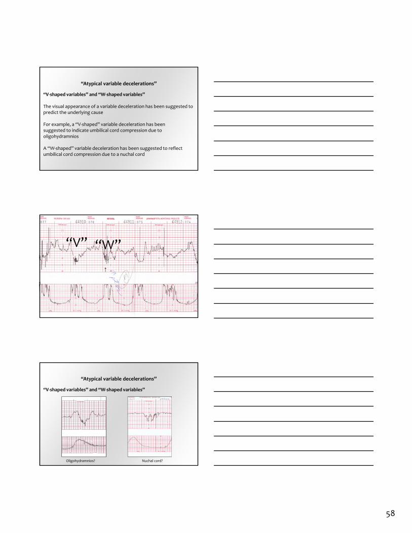
“V‐shaped variables” and “W‐shaped variables”
The visual appearance of a variable deceleration has been suggested to predict the underlying cause
For example, a “V‐shaped” variable deceleration has beensuggested to indicate umbilical cord compression due to oligohydramnios
A “W‐shaped” variable deceleration has been suggested to reflect umbilical cord compression due to a nuchal cord
“V” “W”
“Atypical variable decelerations”
“V‐shaped variables” and “W‐shaped variables”
Oligohydramnios? Nuchal cord?

59
“Atypical variable decelerations”
“V‐shaped variables” and “W‐shaped variables”
Although such claims likely have little impact on patient care, there is no supporting evidence in the literature
These terms are not included in standardized NICHD terminology
2008 NICHD
“Variable decelerations may be accompanied by other characteristics, the clinical significance of which requires further research investigation.”
“Some examplesinclude biphasicdecelerations”
(W‐shaped)
Other myths…
“Good variability within the deceleration”
At the nadir of a variable or late deceleration, the FHR frequently appears irregular, similar to the appearance of moderate variability
The visual similarity has led some to suggest that “variability” during a deceleration has the same clinical significance as baseline variability

60
While the concept is not physiologically implausible, there is no supporting Level I or Level II evidence – the only levels of evidence that are capable of establishing such a relationship
Other myths…
“Good variability within the deceleration”
In addition, it is inconsistent with standard terminology.
Variability is a characteristic of the FHR baseline
The term “variability” is not used to qualify periodic or episodic decelerations that interrupt the baseline
In the absence of evidence, the safest approach is to avoid assigning undue significance to this observation
Other myths…
“The constant pounding of the fetal head on the maternal pelvis causes local cerebral ischemia and brain damage WITHOUT systemic metabolic acidemia and WITHOUT the necessity of neonatal encephalopathy”

61
Scenario
• Term labor• Uncomplicated vaginal delivery• Normal Apgar scores• Normal umbilical artery blood gas results• Normal newborn course• Home with mother on PPD 2• Neurologic symptoms noted at 18 months
Claim
“Silent” cerebral ischemia…Not global hypoxia
Mechanical head compression
There are no analytic studies in the literature to support this hypothetical mechanism of injury
Analytic (case‐control) studies evaluating risk factors for cerebral palsy have never identified any degree of uterine activity as a risk factor

62
Case‐control Studies Failing to Identify Uterine Activity as a Risk Factor
Kułak W, Okurowska‐Zawada B, Sienkiewicz D, Paszko‐Patej G, Krajewska‐Kułak E Risk factors for cerebral palsy in term birth infants. Adv Med Sci. 2010;55(2):216‐21.
Walstab J, Bell R, Reddihough D, et al. Antenatal and intrapartum antecedents of cerebral palsy: a case‐control study. Aust N Z J Obstet Gynaecol. 2002 May;42(2):138‐46.
Nelson KB, Ellenberg JH. Antecedents of cerebral palsy. Univariate analysis of risks. Am J Dis Child. 1985;139(10):1031‐1038.
Nelson KB, Ellenberg JH Antecedents of cerebral palsy. Multivariate analysis of risk. N Engl J Med. 1986 Jul 10;315(2):81‐6.
Badawi N, Kurinczuk JJ, Keogh JM, et al. Intrapartum risk factors for newborn encephalopathy: the Western Australian case‐control study. BMJ 1998; 317:1554–1558.
Suvanand S, Kapoor SK, Reddaiah VP, Singh U, Sundaram KR. Risk Factors for Cerebral Palsy. Indian J Pediatr 1997; 64:677‐685.
Towner D, NEJM 1999;341:1709‐14
Spontaneous vaginal deliveries = 387,799

63
One of the latest myths…
MORE CATEGORIES ARE BETTER

64
“5‐tier system”
Most studies have ignored the difference between respiratory and metabolic acidemia
Respiratory Acidemia
• Common• Low pH• PCO2 > 50 mmHg• Base deficit < 12• Clinically benign
Metabolic/mixed Acidemia
• Uncommon (< 2 %)• Low pH• Normal or high PCO2• Base deficit ≥ 12• Prerequisite to injury
If a study does not differentiate between benign respiratory acidemia and potentially‐pathologic metabolic acidemia, no meaningful conclusions can be made regarding a relationship between “5‐tiers” and adverse outcome
“5‐tier system”
The studies that have assessed metabolic acidemia have never demonstrated more than 2‐3 separate categories of risk
Statistically identicalrates of metabolic acidemia
“5 tiers” = Only 2 distinct categories of risk for metabolic acidemia
Statistically identicalrates of metabolic acidemia
Coletta, et al. Am J Obstet Gynecol 2012;206:226.e1‐5.
“5‐tier system”
The studies that have assessed metabolic acidemia have never demonstrated more than 2‐3 separate categories of risk
“5 tiers” = Only 3 distinct categories of risk for metabolic acidemia
1 2 3
Elliott C, Warrick PA, Graham E, Hamilton EF. Am J Obstet Gynecol 2010;202:258.e1‐8.

65
“5‐tier system”
Elliott C, Warrick PA, Graham E, Hamilton EF. Graded classification of fetal heart rate tracings: association with neonatal metabolic acidosis and neurologic morbidity. Am J Obstet Gynecol 2010;202:258.e1‐8.
Sadaka A, Furuhashi M, Minami H, Miyazaki K, Yoshida K, Ishikawa K. Observation on validity of the five‐tier system for fetal heart rate pattern interpretation proposed by Japan Society of Obstetricians and Gynecologists. J Maternal Fetal Neonatal Med. 2011;24(12):1465‐9.
Coletta J, Murphy E, Rubeo Z, et al. The 5‐tier system of assessing fetal heart rate tracings is superior to the 3‐tier system in identifying fetal acidemia. Am J Obstet Gynecol 2012;206:226.e1‐5.
IDENTIFIED ONLY 2‐3 DISTINCT CATEGORIES OF
RISK
“5‐tier system”
The system does not include management recommendations not already published in the model presented here, by AWHONN and in ACOG Practice Bulletin 116 using the much simpler 3‐tier system
“3‐tier” versus “5‐tier system”
The current 3‐tier system is not perfect, but it is simple and practical. Minor refinements are certainly worth considering
However, the solution to the imperfections of a simple, standard 3‐tier system is NOT to replace it with a cumbersome, highly complex 5‐tier system that does not identify 5 tiers of risk and offers no new recommendations for management

66
SIX FATAL FLAWS OF A “5‐TIER SYSTEM”
Patient SafetyNot standard (rejected by 2008 NICHD)Not simple (134 combinations?)
Standard of CareFactually inaccurate (“mild, moderate, severe”)
Cannot be articulatedCommon Sense
Does not identify 5 risk tiers
Offers no new management recs
WHY IS THIS SO IMPORTANT?
After multiple highly‐publicized broad‐based consensus reports, we are finally making meaningful headway in fetal monitoring standardization and simplification, factual accuracy and ability to articulate a rational plan
WHY IS THIS SO IMPORTANT?
Continued refusal to accept and adopt standard fetal monitoring principles endorsed by our professional societies not only arrests this forward progress…
…it sends us back to the past when EFM was dominated by unproven myths, lacked standardization and consensus, was unnecessarily complex and inconsistent to the point of threatening patient safety

67
Fetal Heart Rate Monitoring
The days are over when individual practitioners, individual hospitals or individual hospital systems can make up their own approaches to fetal monitoring that directly contradict the standard, evidence‐based consensus of all major organizations representing providers of obstetric care in the United States
These days are overCowboy Maverick
Lone Wolf Hot Dog

68
Summary
Fetal monitoring is a SCREENING TEST
Fetal monitoring CANNOT diagnose cerebral palsy
Use standard definitions and interpretation
Summary
Develop and maintain a “shared mental model”
KEEP IT SIMPLE
Unnecessary complexity predisposes to error
Don’t hesitate to use flow charts and checklists
Summary
A standardized approach to intrapartum FHR definition, interpretation and management demonstrates reasonableness
The essential element that defines the standard of care

69
Cord gas?
“Physicians should attempt to obtain venous and arterial cord blood samples in the following situations:
• Cesarean delivery for fetal compromise• Low 5‐minute Apgar score• Severe growth restriction• Abnormal fetal heart rate tracing• Maternal thyroid disease• Intrapartum fever• Multifetal gestations”
Umbilical Cord Blood Gas Acid‐Base AnalysisCommittee Opinion Number 348Reaffirmed 2010
Other myths…
Intrapartum asphyxia is a leading cause of cerebral palsy
Intrapartum hypoxia is a leading cause of mental retardation
Intrapartum events are responsible for autism and ADD
The FHR tracing can define the timing of fetal stroke
The “30‐minute rule” defines the standard of care
Other myths…
Minimal‐absent variability diagnoses fetal metabolic acidemia and asphyxia
Late decelerations are caused by fetal asphyxia
Late decelerations are always “ominous”
Meconium is a sign of asphyxia
Checklists on L&D just get you into trouble
Standardized training in intrapartum FHR monitoring is part of residency training
Amnioinfusion causes amniotic fluid embolism