Persistent Inflammation Alters the Function of the Endogenous ...
16
Persistent Inflammation Alters the Function of the Endogenous Brain Stem Cell Compartment Citation Pluchino, Stefano, Luca Muzio, Jaime Imitola, Michela Deleidi, Clara Alfaro-Cervello, Giuliana Salani, Cristina Porcheri, et al. 2008. Persistent inflammation alters the function of the endogenous brain stem cell compartment. Brain 131, no. 10: 2564-2578. Published Version doi://10.1093/brain/awn198 Permanent link http://nrs.harvard.edu/urn-3:HUL.InstRepos:5978706 Terms of Use This article was downloaded from Harvard University’s DASH repository, and is made available under the terms and conditions applicable to Other Posted Material, as set forth at http:// nrs.harvard.edu/urn-3:HUL.InstRepos:dash.current.terms-of-use#LAA Share Your Story The Harvard community has made this article openly available. Please share how this access benefits you. Submit a story . Accessibility
-
Upload
khangminh22 -
Category
Documents
-
view
5 -
download
0
Transcript of Persistent Inflammation Alters the Function of the Endogenous ...

Persistent Inflammation Alters the Function of the Endogenous Brain Stem Cell Compartment
CitationPluchino, Stefano, Luca Muzio, Jaime Imitola, Michela Deleidi, Clara Alfaro-Cervello, Giuliana Salani, Cristina Porcheri, et al. 2008. Persistent inflammation alters the function of the endogenous brain stem cell compartment. Brain 131, no. 10: 2564-2578.
Published Versiondoi://10.1093/brain/awn198
Permanent linkhttp://nrs.harvard.edu/urn-3:HUL.InstRepos:5978706
Terms of UseThis article was downloaded from Harvard University’s DASH repository, and is made available under the terms and conditions applicable to Other Posted Material, as set forth at http://nrs.harvard.edu/urn-3:HUL.InstRepos:dash.current.terms-of-use#LAA
Share Your StoryThe Harvard community has made this article openly available.Please share how this access benefits you. Submit a story .
Accessibility

Persistent inflammation alters the function of theendogenous brain stem cell compartmentStefano Pluchino,1,2 Luca Muzio,1,2 Jaime Imitola,3 Michela Deleidi,1,2 Clara Alfaro-Cervello,4
Giuliana Salani,1,2 Cristina Porcheri,1,2 Elena Brambilla,1,2 Francesca Cavasinni,1,2 Andrea Bergamaschi,1,2
Jose Manuel Garcia-Verdugo,4,5 Giancarlo Comi,2 Samia J. Khoury3,* and Gianvito Martino1,2,*
1Neuroimmunology Unit, DIBIT, 2Institute of Experimental Neurology (INSPE), San Raffaele Scientific Institute, 20132 Milan,Italy, 3Center for Neurologic Diseases and Partners Multiple Sclerosis Center, Department of Neurology, Brigham andWomen’s Hospital, Harvard Medical School, Boston, MA 02115, USA, 4Department Comparative Neurobiology, InstitutoCavanilles, University of Valencia, 46980 Valencia and 5Department of CellularTherapy, Centro de Investigacio¤ n Prı́ncipeFelipe, 46013 Valencia, Spain
*These authors contributed equally to this work.
Correspondence to: Gianvito Martino, MD, Neuroimmunology Unit ^ DIBITand INSPE, San Raffaele Scientific Institute,Via Olgettina 58, 20132 Milano, ItalyE-mail: [email protected]
Endogenous neural stem/precursor cells (NPCs) are considered a functional reservoir for promoting tissuehomeostasis and repair after injury, therefore regenerative strategies that mobilize these cells have recentlybeen proposed. Despite evidence of increased neurogenesis upon acute inflammatory insults (e.g. ischaemicstroke), the plasticity of the endogenous brain stem cell compartment in chronic CNS inflammatory disordersremains poorly characterized.Here we show that persistent brain inflammation, induced by immune cells tar-geting myelin, extensively alters the proliferative and migratory properties of subventricular zone (SVZ)-resi-dent NPCs in vivo leading to significant accumulation of non-migratory neuroblasts within the SVZ germinalniche. In parallel, we demonstrate a quantitative reduction of the putative brain stem cells proliferation in theSVZ during persistent brain inflammation, which is completely reversed after in vitro culture of the isolatedNPCs.Together, these data indicate that the inflamed brain microenvironment sustains a non cell-autonomousdysfunction of the endogenous CNS stem cell compartment and challenge the potential efficacy of proposedtherapies aimed at mobilizing endogenous precursors in chronic inflammatory brain disorders.
Keywords: neurogenesis; neural stem cells; inflammation; experimental autoimmune encephalomyelitis; multiple sclerosis
Abbreviations: BrdU=50 -bromo-20 -deoxyuridine; CFA=complete Freund’s adjuvant; dpi=days post-immunization;EAE=experimental autoimmune encephalomyelitis; EGF=epidermal growth factor; EM=electron microscopy;FGF-II= fibroblast growth factor; GFAP=glial-fibrillary acidic protein; HC=healthy control; IddU=50 -iodo-20 -deoxyuridine;IFN-g= interferon-g; IL-1b= interleukin-1b; L.I. = labelling index; MOG=myelin oligodendrocyte glycoprotein;NCFCA=Neural Colony Forming Cell Assay; NS-A=Neurosphere Assay; NPC=neural stem/precursor cells;OB=olfactory bulb; PDGF=platelet-derived growth factor; PSA-NCAM=polysialylated form of neural cell adhesionmolecule; RMS=rostral migratory stream; SVZ=subventricular zone; TLDA=TaqMan� Low-Density Array;TNF-a=tumour necrosis factor-a
Received March 10, 2008. Revised July 27, 2008. Accepted July 31, 2008. Advance Access publication August 30, 2008
IntroductionMultipotent neural stem/precursor cells (NPCs), supportingself-renewal and differentiation, reside within specializedcompartments or niches in the adult mammalian CNS(Doetsch, 2003). One of the best characterized niches is thesubventricular zone (SVZ), a layer of cells lying immediately
under the ependymal lining of the lateral ventricles (Martinoand Pluchino, 2006). The SVZ has great potential forneurogenesis, both in rodents and humans, and containsthree major cell types. The CNS stem cells (type B cells) thatdisplay an astrocyte-like phenotype express the glial-fibrillaryacidic protein (GFAP) and give rise to intermediate transit
doi:10.1093/brain/awn198 Brain (2008), 131, 2564^2578
� 2008 The Author(s)This is an Open Access article distributed under the terms of the Creative Commons Attribution Non-Commercial License (http://creativecommons.org/licenses/by-nc/2.0/uk/) whichpermits unrestricted non-commercial use, distribution, and reproduction in any medium, provided the original work is properly cited.

amplifying progenitors (type C cells), which lose theGFAP immunoreactivity and acquire the expression of thedistal-less homeobox (Dlx)-2. These type C cells can, in turn,differentiate into neuroblasts (type A cells) that express thepolysialylated form of neural cell adhesion molecule (PSA-NCAM) in addition to doublecortin, and migrate to theolfactory bulb (OB). The cell lineage differentiation pathwayproceeds from type B, through type C, to type A cells, withthe type B cell believed to be the self-renewing CNS stem cell(Alvarez-Buylla and Garcia-Verdugo, 2002).
The relative contribution of the different cell types of theSVZ germinal niche to tissue repair upon CNS injury is stillmatter of debate. While it is clear that there is a boost ofneurogenesis following acute inflammatory insults, such asischaemic stroke (Zhang et al., 2004; Jin et al., 2006), there iscontradictory evidence on the response of the endogenousbrain stem cell compartment to chronic CNS inflammationleading to neurodegeneration. CNS germinal niches maybe impaired following either cranial irradiation or lipo-polysaccharide-mediated acute inflammation (Martino andPluchino, 2006). Previous studies in mice with experimentalautoimmune encephalomyelitis (EAE) have reported tran-sient increase in proliferation of PSA-NCAM+ and NG2+
proteoglycan cells in the corpus callosum, inferred asevidence of enhanced proliferation of stem and precursorcells in the SVZ (Calza et al., 1998; Picard-Riera et al., 2002;Nait-Oumesmar et al., 2007). However, none of these studieshas directly addressed the changes in the specific cell types inthe SVZ during EAE.
Here we have studied the effects chronic inflammatorydemyelination on the SVZ germinal niche using EAE, amouse model of multiple sclerosis (Pluchino et al., 2003).This model is very useful for studying the impact of chronicinflammation on the SVZ because in the C57BL/6 mousestrain the autoimmune reaction in the brain occurs asa consequence of the subcutaneous immunization withthe myelin oligodendrocyte glycoprotein (MOG) (peptide35–55) that targets the rostral and periventricular forebrainregions. (Brown and Sawchenko, 2007; Politi et al., 2007).
Materials and MethodsEAE inductionChronic-progressive EAE was induced in 6- to 8-week-old C57Bl/6female mice, as described (Pluchino et al., 2003; Amadio et al.,2006). For targeted EAE, MOG 35-55-immunized mice [20 dayspost immunization (dpi)] and healthy controls (HC) were stereo-taxically injected in the brain with 230 ng TNF-a and 250 ng IFN-g(BD Biosciences, Franklin Lakes, NJ, USA) dissolved in a totalvolume of 2 ml of sterile saline. Further information is provided inthe Supplementary data.
In vivo analysis of cell cycle length and growthfractionIn order to label the entire population of fast proliferating SVZ cells,mice were intraperitoneally injected with 50-iodo-20-deoxyuridine
(IddU, the first injection was done at the concentration of 100 mg/kgin 0.007 N NaOH in 0.9% saline, while the following injections weredone 70 mg/kg in 0.007 N NaOH in 0.9% saline) every 2 h for a totalof 14 h, as described (Morshead and van der Kooy, 1992). Furtherinformation is provided in the Supplementary data.
In vivo analysis of slowly-dividing neural stem cellsIn order to label the slowly dividing relatively quiescent SVZputative neural stem cells, EAE mice received intraperitoneal 50-bromo-20-deoxyuridine (BrdU, 70 mg/kg in 0.007 N NaOH in 0.9%saline) every 8 h for a total of 6 consecutive days starting at either 13or 30 dpi, as described (Morshead et al., 1998). A washout period of40 days was then chased before sacrifice. At sacrifice, brains wereremoved and fixed as described and serial 10 mm coronal cryo-sections were generated and stained with rat anti-BrdU (cloneICR1; Abcam, Cambridge, UK) and rabbit anti-GFAP (DAKO)antibodies. BrdU+/GFAP+ cells were scored in a region rangingfrom bregma +1.2 to bregma +0 throughout the entire SVZ. Onesection every 70 mm was analysed with 40� Leica Confocal (SP2)objectives. Double-labelled cells were confirmed by computer-aidedthree-dimensional reconstruction.
Retroviral injections for in vivo studiesMice (n = 3 per group) were anesthetized with 2,2,2-tribromoetha-nol (10 mg/ml; 1/27 of body weight) and the head was placed in astereotactic injection apparatus (David Kopf Instruments, Tujunga,CA, USA). As previously described (Carleton et al., 2003; Mennet al., 2006), a replication-defective retrovirus expressing the GFPunder the CMV promoter (rkat 43.2) (Montini et al., 2006) wasinjected within the right lateral ventricle at the followingcoordinates: A. +0; L. + 0.8 and D. �2.4. Further information isprovided in the Supplementary data.
In situ hybridizationIn situ hybridization was performed according to standard methods,as described (Mallamaci et al., 2000). Further information isprovided in the Supplementary data.
Immunofluorescence and immunohistochemistryAt the time of sacrifice, mice were anesthetized and transcardiallyperfused with PBS followed by 4% paraformaldehyde. The brainswere removed and processed for light and electron microscopy(EM), as described (Pluchino et al., 2003). Antigen retrieval, whenappropriate, was performed as previously indicated (Muzio et al.,2005), while endogenous peroxidase blocking reaction was obtainedby incubating sections 20 min in methanol/H2O2 3% solution.Further information is provided in the Supplementary data.
EMTransmitted EM was performed as described previously(Doetsch et al., 1997). Further information is provided in theSupplementary data.
Primary neural stem/precursor cell culturesNeural stem cell cultures were established from the lateralventricular walls of C57BL/6 6- to 8-week-old female mice, asdescribed (Reynolds and Weiss, 1992). To analyse cell proliferation,primary cells isolated as above were plated at 8000 cells/cm2 in
Inflammation impairs brain stem cells Brain (2008), 131, 2564^2578 2565

neurosphere growth medium (DMEM/F12 containing 2 mML-glutamine, 0.6% glucose, 0.1 mg/ml apo-transferrin, 0.025 mg/mlinsulin, 9.6mg/ml putrescin, 6.3 ng/ml progesterone, 5.2 ng/ml Naselenite, 2mg/ml heparin) supplemented with epidermal growth factor(EGF) (20 ng/ml) and fibroblast growth factor (FGF-II) (10 ng/ml),and spheres were collected after 3–5 days, as described (Politi et al.,2007). Further information is provided in the Supplementary data.
In vitro Neurosphere Formation Assay (NS-A)Primary cells isolated as above from HC, complete Freund’s adjuvant(CFA) and EAE immunized mice (n = 3 each) and plated in 24-well(0.5 ml/well) uncoated plates (Corning, Corning, NY, USA) at a den-sity of 8000 cells/cm2 in growth medium, as described (Politi et al.,2007). Further information is provided in the Supplementary data.
Neural Colony Forming Cell Assay (NCFCA)Primary cells isolated as above from HC, CFA and EAE immunizedmice (n = 6 each) were plated using the mouse NeuroCult neuralcolony-forming cell assay kit (StemCell Technologies, Vancouver,BC, Canada) as per the manufacturer’s instructions, at a density of6.5� 105 cells per 35 mm cell culture dish with 2 mm grid (Nunc,Rochester, NY, USA), as described. Further information is providedin the Supplementary data.
ImmunocytochemistryFixed cells were rinsed with PBS and incubated for 90 min at 37�Cin PBS containing 10% normal goat serum (NGS), 0.3% Triton X-100, and appropriate primary antibodies or antisera, as described.Further information is provided in the Supplementary data.
Gene expression analysisTwo 96b Format TaqMan� Low-Density-based Array (TLDA) (167different genes and 3 house keeping), for gene expression, wereperformed onto the SVZ tissue freshly derived from both EAE miceand HC. At due time points, SVZ tissue samples (n = 6 mice pergroup) were homogenized in 1 ml of QIAzol Lysis Reagent (Qiagen,Valencia, CA, USA, #79306) using a rotor-stator homogenizer. TotalRNA was isolated from homogenized tissue samples using RNeasyLipid Tissue Kit (Qiagen, #74804) including Dnase digestion. At theend, RNA samples were redissolved in 30ml of RNase-free waterand their concentrations were determinated spectrophotometricallyby A260 (Nanodrop-ND 1000, Nanodrop, Wilmington, DE, USA).Further information is provided in the Supplementary data.
Sample preparation for laser-capturemicrodissectionAt sacrifice, mice (n5 4 per group) were deeply anesthetized via anintraperitoneal injection of chloralium hydrate 8% and then wererapidly perfused transcardially with RNase-free 0.9% salinecontaining 10 U/ml of heparin. Brains were removed from theskulls and then OCT embedded and rapidly frozen by immersion inliquid nitrogen. Brains were stored at �80�C. Laser microdissec-tion was performed using a Leica AS LMD instrument (LeicaMicrosystems, Milan, Italy). Further information is provided in theSupplementary data.
Flow cytometric and nuclear DNA analysisCells were plated at 8000 cells/cm2 in growth medium withor without either Th1 [500 IU/ml recombinant mouse IFN-g
(BD Biosciences), 200 UI/ml recombinant mouse TNF-a (Pepro TechInc., Rocky Hill, NJ, USA), 100 UI/ml recombinant mouse IL-1b(Euroclone, Siziano, Italy)] or Th2 [10 ng/ml recombinant murineIL-4 (R&D, Minneapolis, MN, USA), 10 ng/ml recombinant mouseIL-5 (R&D), 10 ng/ml recombinant mouse IL-13 (R&D)] cytokinemixes. After 48 h, cells were collected, mechanically dissociated andwashed in PBS three times. For nuclear DNA content analysis, cellswere then re-suspended in 81% PBS 1�, 1% ND P-40, 10%Propidium Iodide (PI), 8% RNAse (Ribonuclease A 300 Kuntz Units)solution. Nuclear DNA content was analysed, as described (Chewet al., 2005), and data collection was gated utilizing forward lightscatter and side light scatter to exclude cell debris and aggregates.Growing fractions of both HC and cytokine-conditioned NPCs werealso determined by the expression of the Ki67 antigen, which isexpressed exclusively in proliferating cells during the late G1, S, G2 orM phase of the cell cycle. Following permeabilization with ice-cold70% ethanol, cells were incubated with fluorescein isothiocyanate(FITC)-conjugated anti-Ki67 antibody (BD PharMingen, FranklinLakes, NJ, USA). The PI fluorescence was measured on a linear scaleusing a FACS Cyan flow cytometer (Dako, Glostrup, Denmark),while the Ki67 staining was analysed using a BD FACSCanto II flowcytometer. All data were acquired and analysed using FCS 3 software(Becton Dickinson, Franklin Lakes, NJ, USA). At least 2� 104 events/sample were analysed.
Statistical analysisData were compared using the Student’s t-test for paired or unpaireddata or the Mann–Whitney U-test for non-parametric data.
ResultsImpaired proliferation of adultSVZ-resident NPCsWe first studied the influence of chronic CNS inflammationon the proliferation kinetics and fate of mitotically activeprogenitor cells (type C, B and A cells) in the SVZ of EAEand HC mice. The mouse model we adopted is highlysuitable to study the interactions between inflammation andSVZ-resident NPCs as one third of the inflammatoryinfiltrates in the anterior forebrain at the peak of inflamma-tion are specifically found within the ventral and dorsal SVZ.Six EAE mice were sacrificed at 30 dpi and forebraininflammatory lesions were quantified. A total of 89 lesions(14, eight per mouse) were enumerated. Total 30/89 (33.7%)were classified as intraparenchymal lesions, 26/89 (29.2%) asSVZ lesions and 33/89 (37%) as leptomeningeal lesions(mean number per area: intraparenchymal = 5.0� 1.85;SVZ = 4.3� 0.85; leptomeningeal = 5.5� 1.54) (Supplemen-tary Fig. 1). To study the cell cycle length and the growthfraction with the S-phase cumulative labelling technique,mice were intraperitoneally injected with saturating doses ofIddU (Morshead and van der Kooy, 1992; Takahashi et al.,1992). IddU+ cells were counted within the dorso- andventro-lateral SVZ in a telencephalic region ranging frombregma +1.2 to bregma +0.
To study the rapidly dividing transit-amplifying progeni-tor cells (type C cells) we used short-term IddU labelling
2566 Brain (2008), 131, 2564^2578 S. Pluchino et al.

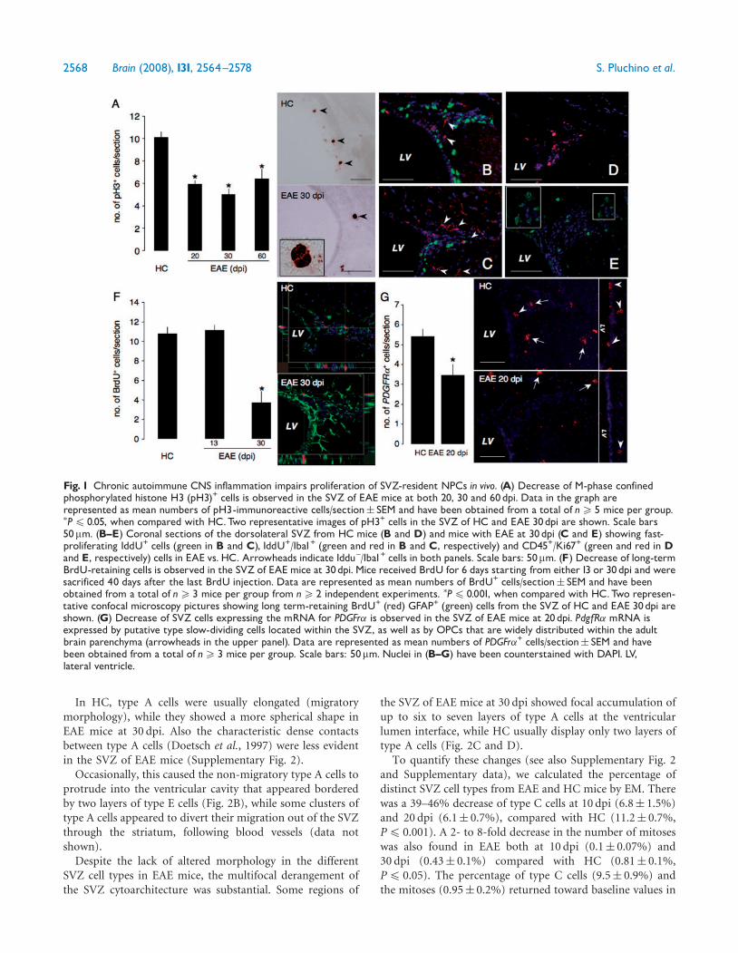
protocols (see Supplementary data). We did not find anysignificant difference in the cell cycle length (Tc) in the SVZof EAE versus HC mice (Table 1). On the other hand, the GFof EAE mice at 20 and 30 dpi was significantly lowercompared with HC (both P4 0.05), though a return towardbaseline values was observed between 60 and 90 dpi(Table 1). Accordingly, a significant reduction in the absolutenumbers of M-phase-confined phosphorylated histone H3(pH3)+ SVZ cells was observed in EAE mice at 20, 30 and60 dpi (Figure 1A) when compared with HC (all P4 0.05).Again, a return toward baseline values in the numbers ofpH3+ cells was observed in EAE at 90 dpi (data not shown).Finally, few—if any—IddU+/Iba1+ and/or CD45+/Ki67+ cellswere found within the SVZ of EAE mice (Figure 1B–E), thussuggesting that the proliferation of either local microglialcells or CNS-infiltrating blood-derived leucocytes did notcontribute significantly to the total GF quantified within thedorso-lateral SVZ. Apoptosis of neural cells could notaccount for the observed decreased GF, as no difference inthe number of caspase-3+ SVZ cells was found between EAEmice and HC (data not shown).
These data suggest that chronic CNS inflammation leadsto a significant reduction of the GF—but not of the Tc—ofSVZ-resident transit amplifying neural progenitors (type Ccells), whose proliferation defect tends to recover as early asinflammation fades out.
To study the slowly dividing relatively quiescent putativeCNS stem cells (type B cells), a long-term retention analysis(upon BrdU labelling) was performed (Morshead et al.,1998). Label-retaining putative CNS stem cells of the adultSVZ (type B cells) share some key features with both radialglia as well as parenchymal astrocytes—including immuno-reactivity for GFAP (Doetsch et al., 1999). As unambiguousidentification of these cells in vivo is difficult with conven-tional immuno detection techniques—due to the unavail-ability of exclusive molecular markers—CNS stem cells aredefined by a combination of morphological, biological andmolecular criteria as true glial cells, akin to astroglia (Pintoand Gotz, 2007). While no difference of label-retaining
SVZ cell was found at 13 dpi, a significant decrease in BrdU-retaining GFAP+ SVZ cells was observed in EAE mice at30 dpi compared with HC (P4 0.001) (Fig. 1F). We also,performed in situ hybridization for the platelet-derivedgrowth factor (PDGF) receptor a, a molecular marker whichhas been shown to be expressed by both relatively quiescentoligodendrocytes precursor cells (OPCs) of the rodent opticnerve and spinal cord (Raff et al., 1983a, b) as well as by up to80% of type B cells of the adult SVZ (Jackson et al., 2006;Menn et al., 2006). Consistent with the long-term retentionanalysis, substantial reduction of the SVZ cells expressing themRNA for PDGFr� was observed in EAE at 20 dpi, comparedwith HC (P4 0.05) (Fig. 1G).
Therefore, also a decrease of slowly dividing SVZ putativestem cells (type B cells) is observed at the peak of theinflammatory phase of EAE.
Altered temporal and spatial relationshipsbetween cells in the SVZ nicheTo better characterize the morphological features of thedifferent SVZ cell types suffering functional derangement inEAE, we performed light and EM analyses on transversesections of the ventricular lateral wall of the anterior horn ofthe SVZ. Sections were obtained from both HC as well asEAE mice at 10, 20, 30 and 60 dpi and distinct SVZ cell typesidentified and scored according to established ultrastructuralcriteria (Doetsch et al., 1997).
We did not observe any alteration of the morphologicalcharacteristics of each of the different cell types—such asincreased smooth or rough endoplasmic reticula, oraccumulation of mitochondria or lisosomes—in EAE micecompared with HC at each of the time points analysed.However, significant differences in cell distribution andorganization were observed. Both semithin section analysis(Fig. 2A) as well as EM showed accumulation of type Aprogenitor cells and decrease of both type C and type B cellsabutting the ventricle—probably activated type B cells—below the ependymal ribbon in EAE mice at 10–30 dpi(Fig. 2A, B and E).
Table 1 Cell cycle analysis of rapidly-dividing (type C) progenitor cells in the SVZ of EAE and HC mice
Micea No. Tcb GFc r2d P-value
HC (6 weeks old) 27 12.2 27.2� 0.004 0.94 NAHC (18 weeks old) 6 13.0 24.0� 0.4 0.89 NS��
CFA (6 weeks old) 8 12.2 26.9� 0.7 0.93 NS��
EAE 20dpi 15 12.1 20.0� 2.1 0.89 0.002y
EAE 30dpi 15 12.9 19.1�1.2 0.86 0.001y
EAE 60dpi 12 11.9 27.0�1 0.86 NSy
EAE 90dpi 8 12.3 24.7�0.79 0.89 NS�
NA=not applicable; NS=not significant.aSix-weeks-old-mice have been immunized for EAE.bData are expressed as hours. Tc is the length of the entire cell cycle.cData are expressed as mean percentage of IddU+ cells (�SD) over total cell counts.GF represents the labelling time sufficient to label100% of proliferating cells.dRegression analysis between the labelling index and the hours elapsed from the first IddU injection.�� versus HC 6 weeks old; y versus CFA; � versus HC18 weeks old (all calculated using Student ‘t-test).
Inflammation impairs brain stem cells Brain (2008), 131, 2564^2578 2567
In HC, type A cells were usually elongated (migratorymorphology), while they showed a more spherical shape inEAE mice at 30 dpi. Also the characteristic dense contactsbetween type A cells (Doetsch et al., 1997) were less evidentin the SVZ of EAE mice (Supplementary Fig. 2).
Occasionally, this caused the non-migratory type A cells toprotrude into the ventricular cavity that appeared borderedby two layers of type E cells (Fig. 2B), while some clusters oftype A cells appeared to divert their migration out of the SVZthrough the striatum, following blood vessels (data notshown).
Despite the lack of altered morphology in the differentSVZ cell types in EAE mice, the multifocal derangement ofthe SVZ cytoarchitecture was substantial. Some regions of
the SVZ of EAE mice at 30 dpi showed focal accumulation ofup to six to seven layers of type A cells at the ventricularlumen interface, while HC usually display only two layers oftype A cells (Fig. 2C and D).
To quantify these changes (see also Supplementary Fig. 2and Supplementary data), we calculated the percentage ofdistinct SVZ cell types from EAE and HC mice by EM. Therewas a 39–46% decrease of type C cells at 10 dpi (6.8� 1.5%)and 20 dpi (6.1� 0.7%), compared with HC (11.2� 0.7%,P4 0.001). A 2- to 8-fold decrease in the number of mitoseswas also found in EAE both at 10 dpi (0.1� 0.07%) and30 dpi (0.43� 0.1%) compared with HC (0.81� 0.1%,P4 0.05). The percentage of type C cells (9.5� 0.9%) andthe mitoses (0.95� 0.2%) returned toward baseline values in
Fig. 1 Chronic autoimmune CNS inflammation impairs proliferation of SVZ-resident NPCs in vivo. (A) Decrease of M-phase confinedphosphorylated histone H3 (pH3)+ cells is observed in the SVZ of EAE mice at both 20, 30 and 60dpi. Data in the graph arerepresented as mean numbers of pH3-immunoreactive cells/section� SEM and have been obtained from a total of n5 5 mice per group.�P4 0.05, when compared with HC.Two representative images of pH3+ cells in the SVZ of HC and EAE 30dpi are shown. Scale bars50mm. (B^E) Coronal sections of the dorsolateral SVZ from HC mice (B and D) and mice with EAE at 30dpi (C and E) showing fast-proliferating IddU+ cells (green in B and C), IddU+/Iba1+ (green and red in B and C, respectively) and CD45+/Ki67+ (green and red in Dand E, respectively) cells in EAE vs. HC. Arrowheads indicate Iddu^/Iba1+ cells in both panels. Scale bars: 50mm. (F) Decrease of long-termBrdU-retaining cells is observed in the SVZ of EAE mice at 30dpi. Mice received BrdU for 6 days starting from either13 or 30dpi and weresacrificed 40 days after the last BrdU injection. Data are represented as mean numbers of BrdU+ cells/section� SEM and have beenobtained from a total of n5 3 mice per group from n5 2 independent experiments. �P4 0.001, when compared with HC.Two represen-tative confocal microscopy pictures showing long term-retaining BrdU+ (red) GFAP+ (green) cells from the SVZ of HC and EAE 30dpi areshown. (G) Decrease of SVZ cells expressing the mRNA for PDGFr� is observed in the SVZ of EAE mice at 20dpi. PdgfR� mRNA isexpressed by putative type slow-dividing cells located within the SVZ, as well as by OPCs that are widely distributed within the adultbrain parenchyma (arrowheads in the upper panel). Data are represented as mean numbers of PDGFr�+ cells/section� SEM and havebeen obtained from a total of n5 3 mice per group. Scale bars: 50mm. Nuclei in (B^G) have been counterstained with DAPI. LV,lateral ventricle.
2568 Brain (2008), 131, 2564^2578 S. Pluchino et al.

EAE mice at 60 dpi. Furthermore, we found almost a 2-foldincrease in perivascular (activated) microglial cells in EAEmice at 20 dpi (0.56� 0.3%) compared with HC(0.35� 0.1%) (Fig. 2F). Finally, picnotic cells in EAE mice
were significantly increased only at 20 dpi (1.12� 0.5%),compared with HC (0.3� 0.06%, P4 0.05) (Fig. 2G). Therelative percentages of ependymal cells, astrocytes andneurons did not vary among groups.
Fig. 2 Chronic autoimmune CNS inflammation impairs cell organization in the SVZ germinal niche. (A) Diagrams of cell organization alongthe SVZ of HC (a) and EAE mice at 30 (b) and 60 dpi (c). Representative areas are shown at higher magnification. Note the homogeneousorganization in HC, while in EAE mice at 30dpi, cells accumulate and form large cell clusters. These clusters tend to recover at 60dpi.Ependymal cells are depicted in black, while SVZ cells in grey; (B) Transmision EM of the EAE SVZ region at 30dpi. A chain of migratingneuroblasts (A) appears in the lateral ventricle (LV), delimited with a discontinuous line.Ventricle walls are covered by ependymal cells (E);(C) EAE mice at 30dpi show large cell clusters, composed of several layers of accumulated migrating cells (A); (D) The HC SVZ shows ahomogeneous group of migrating neuroblasts (A). Arrows indicate the typical free spaces between type A cells, which become lessnoticeable in EAE mice; (E) Representative images of a single type B cell or astrocyte (B) touching the ventricular cavity (LV) through anexpansion (arrows). These astrocytes might be activated in response to proliferation signals and tend to disappear within the SVZ of EAEmice; (F) Representative image of a single microglial cell (Mi). Some of these cells are filled with dense bodies, are frequently located inclose proximity to blood vessels (BLV) and tend to increase in the SVZ of EAE mice at 10^30dpi; (G) Picnotic cells (Pic) are transientlyincreased in the SVZ of EAE mice. Scale bars in B,C and D: 5mm, while in E and F: 2mm.
Inflammation impairs brain stem cells Brain (2008), 131, 2564^2578 2569

Migratory defect of neuroblasts in therostral migratory stream of EAEmiceWe then hypothesized that an alteration of the migratorycapacity of type A cells—with non-migratory neuroblastsaccumulating in the SVZ—was occurring in EAE mice as aconsequence of persistent CNS inflammation. To addressthis issue we performed semithin section analysis of therostral migratory stream (RMS) in EAE versus HC mice.
We found that the RMS of EAE mice at 30 dpi wasprofoundly disorganized (Fig. 3A), with significantly lessmigrating neuroblasts (Fig. 3A, red cells in lower panel) andmore numerous astrocytes (Fig. 3A, green cells in lowerpanel). The RMS of EAE mice at 60 dpi (Fig. 3B) showed atrend to recovery to normal morphology (Fig. 3C). EMconfirmed cell identities in the RMS of both groups ofmice (data not shown). Accordingly, in EAE mice at 20 and30 dpi, immunofluorescence analysis of sagittal brain sec-tions revealed increased accumulation of PSA-NCAM+
neuroblasts within the more rostral SVZ, which were not
clustered in tight rows as detected in HC but appearedscattered within the RMS, possibly indicating migratorydefects (Fig. 3D, magnified insets). Indeed, the astrocyticglial tubes surrounding the RMS in EAE mice appeareddisrupted with accumulation of GFAP+ cells either inside theRMS or in the lateral parenchyma.
We also investigated whether the observed accumulationof non-migratory neural progenitors might mask a switchtowards oligodendro- versus neuro-genesis. We did not findevidence of PSA-NCAM+ cells co-expressing any of thetwo putative markers of OPCs, NG2 (Fig. 3E and F) and Olig2 (Fig. 3G and H). Furthermore, laser-capture microdissec-tion (LCM)-based gene expression analysis of major(oligodendro)glial cell fate determinants (e.g. Olig1, Olig2,Nkx2.1, Nkx2.2) in the SVZ, showed no differences betweenEAE 20 dpi and HC (Supplementary Fig. 3). Similarly,we did not find at 30 dpi any increase of mature NeuN+
neurons within the SVZ (Fig. 3I and J). This latter find-ing was also confirmed by EM analysis showing anon-significant difference between the number of SVZ
Fig. 3 Chronic autoimmune CNS inflammation impairs migration of SVZ neuroblasts. (A^C) Representative toluidine blue-stained semi-thin sections of the RMS of EAE mice at 30 (A) and 60 dpi (B) and HC (C). The same sections were taken-off, and ultrathin sections werere-cut. Cells were studied at the EM and identified as neuroblasts or astrocytes (red and green in lower panels). Note the disorganizationof the RMS of EAE mice at 30dpi (A), with significantly low numbers of migrating neuroblasts and numerous astrocytes. The RMS of EAEmice at 60dpi showed a trend to recovery normal morphology (B), as compared with HC (C). The arrow at 30dpi shows a microglial cell(purple). Scale bar: 20mm. (D) Sagittal reconstructions of the whole RMS from a representative HC (left panel) and a mouse with EAE at30dpi (right panel). Migratory neuroblasts are detected by PSA-NCAM (red), while astrocytes are detected by GFAP (green).Note normalPSA-NCAM+ chains of migratory cells in the HC mouse (left panel). In contrast, the PSA-NCAM+ cells in the RMS of EAE mice at 30dpiappear deranged in their migratory path toward olfactory bulbs (right panel). EAE mice also display increased numbers of GFAP+ astro-cytes within the parenchyma surrounding the RMS.Coronal sections show clear disorganization of migratory chains with less PSA-NCAM+
neuroblasts (purple cells in the magnified inset) at the dorsal SVZ level in EAE mice. Nuclei in magnified insets have been counterstainedwith DAPI. Scale bars: 50mm. LV= lateral ventricle. (E^J) Coronal brain sections showing double immunofluorescence for PSA-NCAM(red) and NG2 (green in E and F) or Olig2 (green in G and H). Note the absence of co-staining for both NG2 and Olig2 in EAE mice at30dpi (F and H) and HC (E and G). In I and J, NeuN staining (green) is shown in a HC mouse (I) and in a EAE mouse (J) at 30dpi,respectively. Note that very few cells are stained in the SVZ (surrounded by a continuous line) of both mice while the majority of striatalneurons are stained (arrowheads). In all panels, nuclei have been counterstained with DAPI and the scale bar is 100mm.
2570 Brain (2008), 131, 2564^2578 S. Pluchino et al.

neurons in HC (0.34� 0.1) versus EAE at 30 dpi (0.57� 0.2;P = NS).
Finally, we directly analysed how the newly generated SVZcells migrate towards physiological (e.g. the OB) routes ofmigration. To do this we used a replication-defective retro-virus expressing the green fluorescent protein (GFP). Thevector was injected into the right ventricle of both HC andEAE mice at 20 dpi and the number of GFP+ cells wasquantified 19 days after the injection in a region spanningfrom the anterior SVZ to the OB (Fig. 4A). When comparedwith HC, EAE mice showed a significant (P4 0.05)reduction of GFP+ cells migrating tangentially along theRMS up to the OB and differentiating into granule andperiglomerular neurons (Fig. 4B). In contrast, we did notobserve any quantitative difference—between EAE 20 dpiand HC—in the subset of non-migratory GFP+ cells beingdetected at the level of the SVZ (Fig. 4B–F).
These results provide evidence of a prominent migratoryimpairment and exclude the occurrence of a switch towards(oligodendro)gliogenesis of the newly generated SVZ pro-geny in EAE mice.
Impaired clonal efficiency of NPCs fromEAEmice in vitroWe next studied the self-renewal of CNS stem/progenitorcells by using an ex vivo assay. It has been shown thatneurosphere-forming cells consist mostly of type C (andtype B) cells in vitro (Morshead et al., 1994). Therefore,
we used the classical in vitro NS-A to further characterize theSVZ stem cell population of EAE mice, with the numbers ofclonal neurospheres in vitro as a measure of the absolutenumber(s) of putative stem cells in vivo (Reynolds andWeiss, 1992; Morshead et al., 1994).
Neurospheres (both primary and secondary) from EAEmice did not show differences in size at any time point,compared with neurospheres from both HC and CFA-immunized mice (Supplementary Fig. 4), thus suggestingthat SVZ stem cells from symptomatic EAE mice do notdiffer in their mitogenic capacity from cells derived fromcontrols. However, the clonal efficiency of SVZ primaryneurospheres—while comparable between asymptomatic(e.g. 10 dpi) EAE mice and HC—became different onceclinical EAE started. In fact, significantly less primaryneurospheres were derived from EAE mice at 20, 30, 60,120 and 240 dpi (all P4 0.0001), when compared withcontrols (Fig. 5A).
This decline in clonal efficiency was confirmed insecondary neurospheres at the same time points (Supple-mentary Fig. 4), further corroborating the previous in vivoobservation that significant impairment of the SVZ stem/precursor cell compartment occurs at certain time points ofchronic EAE in mice. Interestingly, in vitro extended cul-turing of neurospheres from EAE mice with FGF-II and EGFfor a total of 10 passages of amplification completely reversedthe impairment of clonal efficiency observed in primary andsecondary neurospheres (Fig. 5B). In order to solve some ofthe limitations of the classical NS-A (Bull and Bartlett, 2005;
Fig. 4 Retroviral cell tracing shows impaired migratory behaviour of SVZ progeny. (A) A replication-defective retrovirus expressing thegreen fluorescent protein (GFP) was injected stereotaxically in the lateral ventricle so to infect S-phase confined cells.The green line showsthe needle track, while the red line indicates the RMS. Nineteen days after the injection,GFP+ cells migrating along the RMS were num-bered in a region spanning from the anterior SVZ to the OB.OB, CTX=cortex; LV= lateral ventricle. (B) EAE mice display a significantreduction of the subset of GFP+ cells migrating to the OB and an increase in non-migrating GFP+ cells detected very close to the SVZ,when compared with HC.Data are expressed as mean percentage of detected cells (over total labelled)� SEM from n=3 mice per group.�P4 0.05. (C^F) GFP+ cells (green) are detected both in the SVZ lining and in the OB from HC (C and D) and EAE at 20dpi (E and F).The arrows in C and E indicate SVZGFP+ cells. The arrowheads in D and F show OB GFP+ cells. Nuclei are counterstained with DAPI.Scale bar: 150mm. LV, lateral ventricle.
Inflammation impairs brain stem cells Brain (2008), 131, 2564^2578 2571

Singec et al., 2006), we repeated the in vitro quantification ofstem-like cells by using a size-based NCFCA, which has beenrecently developed to differentiate between colonies formedby bona fide neural stem cells from those formed by pro-genitor cells, based on their different proliferative potentialin vitro (Louis et al., 2008). A significant decrease in large-size neural colony-forming cells (with putative large self-renewal capacity) (Fig. 5C, P4 0.05 at both 20, 30 and60 dpi) as well as medium-sized colonies (with putativelimited self-renewal capacity) (Fig. 5D, P4 0.05 at both 20and 60 dpi) was observed in cells from the SVZ of EAE mice,when compared with HC. In parallel, a significant increase ofsmall-size colonies (with low, if any, self-renewal capacity)(Fig. 5E, P4 0.05 at both 20 and 60 dpi) was also found inthe cells from the SVZ of EAE mice, when compared withHC. Interestingly, the major in vitro biological propertiesof SVZ stem cells from the SVZ of EAE mice—namelyproliferation with mitogens, self-renewal and multipotencyupon mitogen removal—were not significantly altered,
when compared with both HC and CFA-immunized controls(Supplementary Fig. 5).
While further confirming that inflammation occurringduring EAE significantly impairs the kinetics of both slowand fast proliferating SVZ cells, the in vitro data alsosuggested that the chemically defined growth factor-enriched(FGF-II plus EGF) microenvironment into which neuro-spheres are grown might be per se sufficient to re-establishthe observed cell non-autonomous dysfunction.
Inflammation-dependent deregulation ofcell cycle progressionIn order to elucidate some of the molecular mechanism(s)underlying the inflammation-induced failure of SVZ cells, weperformed a wide TaqMan� Low-Density Array (TLDA)-based gene expression profiling in the acutely dissociatedSVZ of EAE mice at different time points (e.g. 10, 20, 30 and120 dpi). One hundred and sixty-seven different mRNA
Fig. 5 Neurospheres from mice with chronic autoimmune CNS inflammation display reduced self-renewal. (A) Quantitative analysis ofclonal efficiency of HC (black circles) and CFA-immunized controls (white circles) as well as EAE mice (grey circles). Note the significantand persistent impairment of clonal efficiency of primary neurospheres from EAE mice at 20, 30, 60, 120 and 240dpi, when compared withcontrols. (B) The clonal efficiency of primary neurospheres from EAE mice is completely reverted after n=10 passages of amplificationin vitro and it is comparable HC and CFA-immunized controls. A total of three to six mice per group per experiments were analysed pertime point. Data are represented as mean clonal efficiency� SEM from a total of n4 3 independent experiments. ��P4 0.0001. (C^E)Neural Colony Forming Cell (NCFC) assay-based quantification of large- (C), medium- (D) and small-sized (E) colonies. Note the signifi-cant and persistent reduction of large- and medium-sized colonies, while increase in small-sized colonies in EAE mice (grey bars), whencompared with HC (black bars). Data are represented as mean percentage of colonies� SEM over the total number of plated cells from atotal of n5 3 independent experiments. A total of six mice per group per experiments were analysed per time point. �P4 0.05;�P4 0.005. �, colony diameter.
2572 Brain (2008), 131, 2564^2578 S. Pluchino et al.

species involved in stemness, inflammation, immunity, self-renewal, differentiation and migration were measured (seeSupplementary information). Differences in gene expressionwere mostly seen in EAE at 20 and 30 dpi (r2 = 0.60 andr2 = 0.59, respectively), when compared with 10 and 120 dpi(both r2 = 0.92) (Supplementary Fig. 6).
Interestingly, 60.5% (101/167) and 27.5% (46/167) ofscreened genes either did not change at any time point orwere not expressed at more than one time point, respectively.Only 9% (15/167) of screened genes were up regulated in theSVZ of EAE mice at 20 and/or 30 dpi, when compared withHC. On the other hand, 3% (5/167) of the genes were down-regulated in the EAE mice among which we identified at leasttwo major markers for stem/precursor cells of the SVZgerminal niche, such as Prominin-1 and Tenascin-C (Martinoand Pluchino, 2006) (Fig. 6A). According to LCM analysis ofthe SVZ (Supplementary Fig. 3), we did not find any upregulation of (oligodendro)glial cell fate gene determinants,such as Nkx2.2 (Fig. 6A and B).
Among up-regulated genes, the most prominent wereinflammatory regulators (e.g. CCL5/Rantes, CXCL10/IP-10and Heme-oxigenase1), a transcription factor downstreamthe IFN-g signalling (e.g. STAT-1), a number of cell cycleregulators (e.g. Cdc2a/p34, Cdkn1a/p21 and Cdkn1c/p57),and two neuronal-lineage specific transcription factors (e.g.Dlx1 and Dlx2) (Fig. 6B). As an in vivo confirmation of theselatter data, an increased number of cells expressing Cdkn1a/p21+ and Dlx2+ mRNA cells was observed in the SVZ of EAEmice at 30 dpi, compared with HC (Fig. 6C–F).
Induction of quiescence of SVZNPCsin vitro by inflammatory cytokinesThe gene expression pattern in the SVZ of EAE micesupports the idea that the impairment of proliferation andmigration, we observed in vivo, might have occurred as aconsequence of a CNS-confined inflammatory processleading to cell cycle deregulation. We found a significantup-regulation of mRNA levels for pro-inflammatory (Th1)cytokines, such as interferon (IFN)-g and tumour necrosisfactor (TNF)-a (Fig. 6G and H, respectively), but not ofinterleukin (IL)-1b (Fig. 6I), at 20 and 30 dpi in the SVZfrom EAE mice (P4 0.005, when compared with CFA-immunized mice). Given the central role played by Th1cytokines in triggering and perpetuating chronic inflamma-tion in EAE, we thus hypothesized that Th1 cytokines mighthave contributed to the impaired proliferation of stem/precursor cells observed in the SVZ of EAE mice. PrimarySVZ cells from HC were grown in the continuous presence ofTh1 (e.g. IFN-g, TNF-a and IL-1b) or Th2 (e.g. IL-4, IL-5and IL-13) cytokine mixes. When plated in presence ofTh1, but not Th2, cytokines SVZ cells showed dramaticimpairment in the formation of large-size neural colony-forming cells (P4 0.0001, when compared with controlSVZ cells). This was paralleled by a significant increaseof small-sized colonies with low self-renewal capacity
(P4 0.05, when compared with control SVZ cells). Theobserved results were not obtained when cytokines of eithertype were added to the SVZ cell assay only for 48 h (Fig. 7A).To confirm whether NPC self-renewal could be affected byinflammatory Th1 cytokines, we generated continuousgrowth curves of neurospheres cultured in completegrowth medium (CGM), or in CGM enriched with eitherTh1 or Th2 cytokines. Only neurospheres cultured in Th1-enriched CGM showed progressive and significant (from twoto six passages of amplification) decrease of growth efficiency(Fig. 7B, all P4 0.05, when compared with control neuro-spheres), while no difference was observed in neurospherescultured in Th2-enriched CGM. Interestingly, as early Th1cytokines were removed from the CGM and cell amplifica-tion was carried on for n = 6 further passages of amplifica-tion, the growth efficiency of neurospheres previouslyexposed to cytokines returned to control values (Fig. 7C).Among Th1 cytokines, IFN-g—but not TNF-a and IL-1b—seemed to play a crucial role in the impairment of long-termproliferating capacity of neurospheres (P4 0.05, whencompared with control neurospheres) (Fig. 7D). As a furtherconfirmation, we found that exposure of SVZ NPCs to Th1but not Th2 cytokines lead to significant increase (33–46%)of cells in the G0/G1 phase (P4 0.005) and to a paralleldecrease of cells in the S phase (P4 0.005), when comparedwith controls. No difference was observed in G2/M phase-restricted cells between groups (Fig. 7E).
We then sought to analyse in more details the growingfractions of cytokine-conditioned NPCs as determined by theexpression of the Ki67 antigen, a molecular marker that isexpressed exclusively in proliferating cells during the lateG1, S, G2 or M phase of the cell cycle. Accordingly, asignificant increase in the percentage of Ki67– G0-phaseconfined quiescent NPCs—representing the 70.6� 1.9% ofthe cultured cell population—was observed upon exposureof NPCs to Th1 but not Th2 cytokines (Figure 7F,P4 0.0001, when compared with both control and Th2-conditioned neurospheres). Cell apoptosis assessed byAnnexin/PI staining did not account for any of the observedphenomena (data not shown).
Alteration of SVZ cell cycle byinflammatory cytokines in vivoTo further investigate the role played by pro-inflammatorycytokines in deregulating the cell cycle of NPCs in vivo,we analysed the SVZ of HC and EAE mice uponstereotactical injection of a Th1 cytokine mix (e.g. TNF-aand IFN-g) in a brain area close to the SVZ, as previouslydescribed (Merkler et al., 2006). Mice were sacrificed 3 and10 days after cytokine injection and in vivo cell cycle analysiswas performed using systemic IddU to label proliferatingcells. Severe impairment of SVZ fast-proliferating transitamplifying precursors (type C cells) was found in TNF-a/IFN-g-injected HC (P4 0.005, when compared withuntreated HC mice) (Fig. 7G). Similarly, IddU+ precursors
Inflammation impairs brain stem cells Brain (2008), 131, 2564^2578 2573

Fig. 6 CNS inflammation induces expression of cell cycle regulators and neurogenic transcription factors at mRNA level. (A,B)Semi-quantitative analysis of selected mRNAs.The graph in A includes a list of n=5 selected genes showing significant down-regulation(fold reduction51) at any time point in the SVZ of EAE mice, when compared with CFA-immunized controls.The graph in B includes a listof n=15 selected genes showing significant up-regulation (fold induction 51) at any time point in the SVZ of EAE mice, when comparedwith CFA-immunized controls. (C^F) In situ hybridization for Dlx2 (C and D) and Cdkn1a/p21 (E and F) on coronal brain sections fromboth HC (C and E) and EAE 30dpi (D and F). EAE mice show increased numbers of Dlx2+ and Cdkn1a/p21+ cells within the SVZ, whencompared with HC. Autoradiographic grains (red) were captured with 20� objective in a dark field light microscopy and tissue was visua-lized by DNA staining (DAPI, blue). LV= lateral ventricle. Scale bars: 50mm. (G^I) Significant increase of IFN-g and TNF-a mRNA levels(G and H, respectively) in the SVZ of EAE mice at 20 and 30dpi compared with CFA-immunized controls. The graph in F shows thesignificant decrease of IL-1�mRNA levels in the SVZ of EAE mice at 20 and 30dpi. Data are represented as mean arbitrary units� SEMand have been obtained in single from the SVZs of n5 6 per mice per group per time point. �P4 0.05; ��P4 0.0001.
2574 Brain (2008), 131, 2564^2578 S. Pluchino et al.

Fig. 7 Chronic CNS inflammation induces quiescence of neural stem cells in vitro and in vivo. (A) NCFC assay performed on primary SVZNPCs in the presence of eitherTh1 cytokines (IFN-g, 500 U/ml; TNF-a, 200 U/ml; IL-1b100 U/ml) orTh2 cytokines (IL-4, IL-5 and IL-13, all10ng/ml). SVZ cells were plated at clonal density in cytokine-enriched NeuroCult for either the last 48h (white bars) or for the wholelength of the assay (black bars). No difference in the percentage(s) of generated colonies were found upon 48h of conditioning either withTh1 orTh2 cytokines, while longer (3 weeks) Th1 cytokine conditioning induced a complete failure of the generation of large-size colonies.Data are represented as mean percentage of colonies/size over total plated cells� SEM and have been obtained from a total of n5 3independent experiments. �P4 0.0001; �P4 0.05. (B^D) Proliferation analysis of NPCs grown in vitro with CGM enriched with cytokines.(B) Th1 (white circles) orTh2 cytokines (grey circles) are compared with CGM alone (black circles). Note the significant decrease of pro-liferation rate of Th1 conditioned cells appearing as early as after n=2 passages of amplification, while no difference was observed inneurospheres cultured inTh2-enriched CGM. (C) AfterTh1 cytokines withdrawal, NPCs are kept growing in CGM alone for further n=6passage of amplification and no differences in growth efficiency are observed when previously exposed NPCs toTh1 cytokines (whitecircles) are compared with control NPCs (black circles). (D) Proliferation analysis of NPCs grown in vitro in CGM enriched with singleTh1cytokines. IFN-g alone (white diamonds) induces a significant reduction of growth rate (from four to six passages of amplification), whilecells conditioned with eitherTNF-a or IL1-b do not show difference in growing efficiency at any time point. Data are represented asabsolute numbers of cells� SEM from a total of n5 3 independent experiments. �P4 0.05, when compared with controls. (E) NPCs werecultured for 48h in CGM enriched with eitherTh1 (white bars) orTh2 cytokines (grey bars). NPCs cultured in CGM alone were used ascontrols (black bars). Note the significant shift (33^46% of cells) in the cells toward G0/G1, and a decline in cells in S phase upon exposuretoTh1but not Th2 cytokines. Data are represented as mean percentage of gated cells� SEM for a total of n5 5 independent experiments.��P4 0.05, when compared with controls. (F) FACS analysis for Ki67.Note the significant increase of G0-confined Ki67^ cells (grey bars) inTh1 cytokine-conditioned but not in unconditioned (Ctrl), orTh2-cytokine conditioned NPCs. Black bars indicate Ki67+ cell. Data arerepresented as mean percentage of gated cells� SEM for a total of n510 independent experiments. P4 0.0001, when compared withcontrols. (G) Significant reduction of the GF upon focal injection of IFN-g/TNF-a into the dorsolateral SVZwas observed in both HC orEAE 20dpi mice 3 days (grey bars) after cytokine injection, compared with baseline values. Ten days after cytokine injection (white bars),the GF still remained significantly lower than normal values in HC only.NPCs proliferation was assayed by continuous exposure to systemicIddU for a total of 10h before the sacrifice and evaluated by counting only IddU+/Iba1^ (green and red cells in the panels, respectively) cellsin the SVZ. Black bars represent baseline time points. Data are represented as mean labelling index (L.I.)� SEM from a total of n5 3 mice.In the panels, images of the SVZs of two representative HC and EAE mice injected with IFN-g/TNF-a are shown. Nuclei are counter-stained with DAPI. Scale bars: 50mm. �P4 0.05; ��P4 0.005. LV= lateral ventricle.
Inflammation impairs brain stem cells Brain (2008), 131, 2564^2578 2575

were further reduced in TNF-a/IFN-g-injected EAE mice ascompared with untreated EAE mice (P4 0.05). Thisphenotype was partially reversed as early as 10 days afterthe initial cytokine injection (Fig. 7G), the expected half-lifeof molecules such as cytokines in vivo.
DiscussionHere we show a significant decrease of proliferation of bothfast cycling (type C cells) and slowly dividing (type B cells)cells accompanied by an increase of newly generated non-migratory neuroblasts (type A cells) in the SVZ of EAE miceduring the peak of CNS-confined chronic inflammation.This alteration is associated with in vitro and in vivo changes:the expression of the cyclin-dependent kinase inhibitorCdkn1a/p21 (Gartel et al., 1996) is increased in the SVZ fromEAE mice; SVZ-derived neurospheres from EAE mice displaylow clonal efficiency; and the percentage of Ki67– G0 phase-confined quiescent NPCs increases up to 70% upon exposureto pro- (Th1) but not anti- (Th2) inflammatory cytokines.These results suggest that persistent CNS inflammationsignificantly alters the cell kinetics of precursors and stemcells residing within the SVZ germinal niche by inhibitingtheir entry into the cell cycle by up-regulation of cell cycleinhibitors.
While the central role played by chronic inflammation indecreasing the GF of SVZ cells is confirmed by the fact thatthe proliferation capacity of fast cycling cells tends to returnto normal values in EAE mice as early as inflammation fadesout (from 60 dpi on), it is less clear why there is a parallelaccumulation of neuroblasts within the SVZ. Here weprovide direct evidence for the inability of SVZ cells tomigrate tangentially along the RMS up to the OB within achronically inflamed microenvironment. This latter finding,along with the EM evidence of a preserved morphology butaltered location of SVZ cells in EAE, prompts us to hypo-thesize that persistent CNS-confined inflammation mightalso affect the parenchymal astrocytes forming the glial tubeswithin the RMS and, as a consequence, alter the migra-tory capacity of the SVZ progeny (the newly generatedPSA-NCAM+ cells). Likewise, it would be also very likely that
some neuroblasts divert their physiological rostral migra-tion towards more caudal inflammatory lesions in thecorpus callosum, as previously suggested (Picard-Rieraet al., 2002).
Interestingly, clonal efficiency of SVZ-derived neuro-spheres is significantly lower at the peak of inflammationin EAE mice but returns to normal values upon extensivein vitro culturing with growth factors. This result—whichagrees with the in vivo evidence showing that SVZ residentcells tend to behave normally as soon as CNS inflammationabates—suggests that certain cell non-autonomous factorsmay transiently alter NPC self-renewal both in vivo andin vitro. Among such factors, pro-inflammatory cytokinesseem to play a major role. We found that IFN-g inducesalteration of NPC kinetics in vitro and Stat-1—which belongsto IFN-g intracellular signalling pathway (Robinson andO’Garra, 2002)—is up-regulated in SVZ from EAE miceduring the peak of inflammation. Furthermore, IFN-g is alsoable to restrict NPC cell cycle progression to the G0 phasein vitro and to impair proliferation of SVZ cells in vivo. TheseIFN-g-mediated effects are transient, reversible, not asso-ciated with induction of apoptosis, and supported by pre-vious evidence. It has been previously shown that types I andII IFNs can affect the growth of different cell types throughthe Janus kinase-signal transducers and activators oftranscription pathway and in particular trough the activationof the transcription factor Stat-1 (Bromberg et al., 1996). Thecentral role played by inflammatory molecules in alteringNPC self-renewal is also supported by the impairment ofhyppocampal neurogenesis induced by the pro-inflammatorycytokine IL-6 (Monje et al., 2003).
The effects described here are relevant to the specific braindisease model we have examined, and it is likely that dif-ferent types of brain damage (e.g. ischaemic stroke, neuro-degenerative diseases, etc.) may induce opposite findings(Calza et al., 1998; Picard-Riera et al., 2002; Zhang et al.,2004; Jin et al., 2006; Nait-Oumesmar et al., 2007), such asthe recent demonstration that different inflammatory players(e.g. IL-4 and IFN-g) or immune cells themselves mayvariably support either glio- or neuro-genesis (Butovskyet al., 2006; Ziv et al., 2006).
Fig. 7 Continued.
2576 Brain (2008), 131, 2564^2578 S. Pluchino et al.

Thus, in this model, persistent CNS-compartmentalizedinflammation mediates changes in the SVZ cells that recoverwhen inflammation abates. What would be the outcome ofrecurrent bouts of inflammation as might be seen inrelapsing-remitting multiple sclerosis? Other models ofEAE (such as the SJL/J model) will need to be evaluated toanswer this question. It would also be interesting todetermine whether the defect in the SVZ cell proliferationwill continue to be reversible after multiple inflammatoryattacks. Further studies will be needed in order to clarifywhether recurrent hits of inflammation induce permanentchanges in the SVZ that would then lead to irreversiblefailure of the homeostatic and repair capabilities of theendogenous neural stem/precursor cell compartment. Ourfindings have implications for therapeutic strategies targetingNPC mobilization from germinal niches, whose effectivenesswill depend on elimination of inflammation as well as on thelong-term plasticity of the SVZ cells.
Supplementary materialSupplementary material is available at Brain online.
AcknowledgementsWe thank John A. Alberta and Charles D. Stiles forproviding the polyclonal antibodies specific for Olig1(DF389) and Olig2 (DF308), Tatsunori Seki for providingthe monoclonal antibody specific for PSA-NCAM(mAB12E3), Eugenio Montini for providing the CMV-GFP retrovirus (rkat 43.2), Giacomo Consalez for providingthe PDGFr� riboprobe, Vania Broccoli for providing theDlx2 riboprobe, and Angelo Corti for providing mouseTNF-a.
FundingItalian Multiple Sclerosis Foundation (partial, 2004/R/15 toS.P. and 2005/R/15 to G.M.); the National Multiple SclerosisSociety (RG 3591-A-1 to G.M., partial RG-4000-A-1 to S.P.and RG 3945-A-10 to S.J.K.); the National Institutes of Health(AI 043496 and AI071448 to S.J.K.); the Italian Ministry ofResearch and University; Italian Ministry of Health, BancaAgricola Popolare di Ragusa and BMW Italy Group.
ReferencesAlvarez-Buylla A, Garcia-Verdugo JM. Neurogenesis in adult subventri-
cular zone. J Neurosci 2002; 22: 629–34.
Amadio S, Pluchino S, Brini E, Morana P, Guerriero R, Boneschi FM,
et al. Motor evoked potentials in a mouse model of chronic multiple
sclerosis. Muscle Nerve 2006; 33: 265–73.
Bromberg JF, Horvath CM, Wen Z, Schreiber RD, Darnell JE Jr.
Transcriptionally active Stat1 is required for the antiproliferative effects
of both interferon alpha and interferon gamma. Proc Natl Acad Sci USA
1996; 93: 7673–8.
Brown DA, Sawchenko PE. Time course and distribution of inflammatory
and neurodegenerative events suggest structural bases for the pathogen-
esis of experimental autoimmune encephalomyelitis. J Comp Neurol
2007; 502: 236–60.
Bull ND, Bartlett PF. The adult mouse hippocampal progenitor is
neurogenic but not a stem cell. J Neurosci 2005; 25: 10815–21.
Butovsky O, Ziv Y, Schwartz A, Landa G, Talpalar AE, Pluchino S, et al.
Microglia activated by IL-4 or IFN-gamma differentially induce
neurogenesis and oligodendrogenesis from adult stem/progenitor cells.
Mol Cell Neurosci 2006; 31: 149–60.
Calza L, Giardino L, Pozza M, Bettelli C, Micera A, Aloe L. Proliferation
and phenotype regulation in the subventricular zone during experi-
mental allergic encephalomyelitis: in vivo evidence of a role for nerve
growth factor. Proc Natl Acad Sci USA 1998; 95: 3209–14.
Carleton A, Petreanu LT, Lansford R, Alvarez-Buylla A, Lledo PM. Becoming
a new neuron in the adult olfactory bulb. Nat Neurosci 2003; 6: 507–18.
Chew LJ, King WC, Kennedy A, Gallo V. Interferon-gamma inhibits cell
cycle exit in differentiating oligodendrocyte progenitor cells. Glia 2005;
52: 127–43.
Doetsch F. The glial identity of neural stem cells. Nat Neurosci 2003; 6:
1127–34.
Doetsch F, Caille I, Lim DA, Garcia-Verdugo JM, Alvarez-Buylla A.
Subventricular zone astrocytes are neural stem cells in the adult
mammalian brain. Cell 1999; 97: 703–16.
Doetsch F, Garcia-Verdugo JM, Alvarez-Buylla A. Cellular composition and
three-dimensional organization of the subventricular germinal zone in
the adult mammalian brain. J Neurosci 1997; 17: 5046–61.
Gartel AL, Serfas MS, Tyner AL. p21 – negative regulator of the cell cycle.
Proc Soc Exp Biol Med 1996; 213: 138–49.
Jackson EL, Garcia-Verdugo JM, Gil-Perotin S, Roy M,
Quinones-Hinojosa A, VandenBerg S, et al. PDGFR alpha-positive B
cells are neural stem cells in the adult SVZ that form glioma-like growths
in response to increased PDGF signaling. Neuron 2006; 51: 187–99.
Jin K, Wang X, Xie L, Mao XO, Zhu W, Wang Y, et al. Evidence for
stroke-induced neurogenesis in the human brain. Proc Natl Acad Sci
USA 2006; 103: 13198–202.
Louis SA, Rietze RL, Deleyrolle L, Wagey RE, Thomas TE, Eaves AC, et al.
Enumeration of neural stem and progenitor cells in the Neural Colony
Forming Cell Assay. Stem Cells 2008; 26: 988–96.
Mallamaci A, Muzio L, Chan CH, Parnavelas J, Boncinelli E. Area identity
shifts in the early cerebral cortex of Emx2-/- mutant mice. Nat Neurosci
2000; 3: 679–86.
Martino G, Pluchino S. The therapeutic potential of neural stem cells.
Nat Rev Neurosci 2006; 7: 395–406.
Menn B, Garcia-Verdugo JM, Yaschine C, Gonzalez-Perez O, Rowitch D,
Alvarez-Buylla A. Origin of oligodendrocytes in the subventricular zone
of the adult brain. J Neurosci 2006; 26: 7907–18.
Merkler D, Ernsting T, Kerschensteiner M, Bruck W, Stadelmann C. A new
focal EAE model of cortical demyelination: multiple sclerosis-like lesions
with rapid resolution of inflammation and extensive remyelination.
Brain 2006; 129: 1972–83.
Monje ML, Toda H, Palmer TD. Inflammatory blockade restores adult
hippocampal neurogenesis. Science 2003; 302: 1760–5.
Montini E, Cesana D, Schmidt M, Sanvito F, Ponzoni M, Bartholomae C,
et al. Hematopoietic stem cell gene transfer in a tumor-prone mouse
model uncovers low genotoxicity of lentiviral vector integration. Nat
Biotechnol 2006; 24: 687–96.
Morshead CM, Craig CG, van der Kooy D. In vivo clonal analyses reveal
the properties of endogenous neural stem cell proliferation in the adult
mammalian forebrain. Development 1998; 125: 2251–61.
Morshead CM, Reynolds BA, Craig CG, McBurney MW, Staines WA,
Morassutti D, et al. Neural stem cells in the adult mammalian forebrain:
a relatively quiescent subpopulation of subependymal cells. Neuron
1994; 13: 1071–82.
Morshead CM, van der Kooy D. Postmitotic death is the fate of
constitutively proliferating cells in the subependymal layer of the adult
mouse brain. J Neurosci 1992; 12: 249–56.
Muzio L, Soria JM, Pannese M, Piccolo S, Mallamaci A. A mutually
stimulating loop involving emx2 and canonical wnt signalling specifically
promotes expansion of occipital cortex and hippocampus. Cereb Cortex
2005; 15: 2021–8.
Inflammation impairs brain stem cells Brain (2008), 131, 2564^2578 2577

Nait-Oumesmar B, Picard-Riera N, Kerninon C, Decker L, Seilhean D,
Hoglinger GU, et al. Activation of the subventricular zone in multiple
sclerosis: Evidence for early glial progenitors. Proc Natl Acad Sci USA
2007; 104: 4694–9.
Picard-Riera N, Decker L, Delarasse C, Goude K, Nait-Oumesmar B,
Liblau R, et al. Experimental autoimmune encephalomyelitis mobilizes
neural progenitors from the subventricular zone to undergo oligoden-
drogenesis in adult mice. Proc Natl Acad Sci USA 2002; 99: 13211–6.
Pinto L, Gotz M. Radial glial cell heterogeneity–the source of diverse
progeny in the CNS. Prog Neurobiol 2007; 83: 2–23.
Pluchino S, Quattrini A, Brambilla E, Gritti A, Salani G, Dina G, et al.
Injection of adult neurospheres induces recovery in a chronic model of
multiple sclerosis. Nature 2003; 422: 688–94.
Politi LS, Bacigaluppi M, Brambilla E, Cadioli M, Falini A, Comi G, et al.
Magnetic resonance-based tracking and quantification of intravenously
injected neural stem cell accumulation in the brains of mice with
experimental multiple sclerosis. Stem Cells 2007; 25: 2583–92.
Raff MC, Abney ER, Cohen J, Lindsay R, Noble M. Two types of astrocytes in
cultures of developing rat white matter: differences in morphology, surface
gangliosides, and growth characteristics. J Neurosci 1983a; 3: 1289–1300.
Raff MC, Miller RH, Noble M. Glial cell lineages in the rat optic nerve.
Cold Spring Harb Symp Quant Biol 1983b; 48 (Pt 2): 569–72.
Reynolds BA, Weiss S. Generation of neurons and astrocytes from isolated
cells of the adult mammalian central nervous system. Science 1992; 255:
1707–10.
Robinson DS, O’Garra A. Further checkpoints in Th1 development.
Immunity 2002; 16: 755–8.
Singec I, Knoth R, Meyer RP, Maciaczyk J, Volk B, Nikkhah G, et al.
Defining the actual sensitivity and specificity of the neurosphere assay in
stem cell biology. Nat Methods 2006; 3: 801–6.
Takahashi T, Nowakowski RS, Caviness VS Jr. BUdR as an S-phase marker
for quantitative studies of cytokinetic behaviour in the murine cerebral
ventricular zone. J Neurocytol 1992; 21: 185–97.
Zhang R, Zhang Z, Zhang C, Zhang L, Robin A, Wang Y, et al. Stroke
transiently increases subventricular zone cell division from asymmetric
to symmetric and increases neuronal differentiation in the adult rat.
J Neurosci 2004; 24: 5810–5.
Ziv Y, Ron N, Butovsky O, Landa G, Sudai E, Greenberg N, et al. Immune
cells contribute to the maintenance of neurogenesis and spatial learning
abilities in adulthood. Nat Neurosci 2006; 9: 268–75.
2578 Brain (2008), 131, 2564^2578 S. Pluchino et al.