Determination of Major Endogenous FAHFAs in Healthy ...
17
biomolecules Article Determination of Major Endogenous FAHFAs in Healthy Human Circulation: The Correlations with Several Circulating Cardiovascular-Related Biomarkers and Anti-Inflammatory Effects on RAW 264.7 Cells Rachmad Anres Dongoran 1,2, † , Tsung-Jen Lin 1, † , Akhsholphan Byekyet 1 , Sheau-Chung Tang 3 , Jen-Hung Yang 4 and Chin-Hung Liu 1,5, * 1 Program in Pharmacology and Toxicology, School of Medicine, Tzu Chi University, Hualien 970004, Taiwan; [email protected] (R.A.D.); [email protected] (T.-J.L.); [email protected] (A.B.) 2 Indonesian Food and Drug Authority (Indonesian FDA), Jakarta 10560, Indonesia 3 Department ofNursing, National Taichung University of Scienceand Technology, Taichung 40640, Taiwan; [email protected] 4 Department of Dermatology, Changhua Christian Hospital, Changhua 500, Taiwan; [email protected] 5 Department of Pharmacology, School of Medicine, Tzu Chi University, Hualien 970004, Taiwan * Correspondence: [email protected] † These authors contributed equally to this work. Received: 12 November 2020; Accepted: 16 December 2020; Published: 17 December 2020 Abstract: Fatty acid esters of hydroxy fatty acids (FAHFAs) are newly discovered long-chain fatty acids. However, the major endogenous FAHFAs in healthy human circulation, their correlation with cardiovascular (CV) biomarkers, and their anti-inflammatory effects have not been investigated and remain unclear. In the present study, a total of 57 healthy subjects were recruited. Liquid chromatography–mass spectrometry (LC-MS) was developed for the simultaneous determination of seven FAHFAs, four long-chain fatty acids, and four non-traditional circulating CV-related biomarkers. We found two major types of FAHFAs in healthy human circulation, palmitoleic acid ester of 9-hydroxystearic acid (9-POHSA), and oleic acid ester of 9-hydroxystearic acid (9-OAHSA). Both 9-POHSA and 9-OAHSA had a strong positive correlation with each other and were negatively correlated with fasting blood glucose, S-adenosyl-l-homocysteine (SAH), and trimethylamine N-oxide (TMAO), but not with l-homocysteine. 9-POHSA was also positively correlated with l-carnitine. Moreover, we confirmed that both 9-POHSA and 9-OAHSA exhibited an anti-inflammatory effect by suppressing LPS stimulated cytokines, including IL-1β and IL-6 in RAW 264.7 cells. In addition, palmitoleic acid also had a positive correlation with 9-POHSA and 9-OAHSA. As far as we know, this is the first report showing the major endogenous FAHFAs in healthy subjects and their CV protection potential which might be correlated with SAH and TMAO reduction, l-Carnitine elevation, and their anti-inflammatory effects. Keywords: FAHFAs; 9-POHSA; 9-OAHSA; cardiovascular disease; anti-inflammatory; SAH; TMAO; l-carnitine 1. Introduction Branched fatty acid esters of hydroxy fatty acids (FAHFAs) are a new class of endogenous bioactive lipids that have been found to be enriched in adipose tissue and circulation [1]. In chemical synthesis, the esterification of FAHFAs could be derived from several fatty acids (FA), including Biomolecules 2020, 10, 1689; doi:10.3390/biom10121689 www.mdpi.com/journal/biomolecules
-
Upload
khangminh22 -
Category
Documents
-
view
1 -
download
0
Transcript of Determination of Major Endogenous FAHFAs in Healthy ...

biomolecules
Article
Determination of Major Endogenous FAHFAs inHealthy Human Circulation: The Correlations withSeveral Circulating Cardiovascular-RelatedBiomarkers and Anti-Inflammatory Effectson RAW 264.7 Cells
Rachmad Anres Dongoran 1,2,† , Tsung-Jen Lin 1,† , Akhsholphan Byekyet 1,Sheau-Chung Tang 3 , Jen-Hung Yang 4 and Chin-Hung Liu 1,5,*
1 Program in Pharmacology and Toxicology, School of Medicine, Tzu Chi University, Hualien 970004, Taiwan;[email protected] (R.A.D.); [email protected] (T.-J.L.); [email protected] (A.B.)
2 Indonesian Food and Drug Authority (Indonesian FDA), Jakarta 10560, Indonesia3 Department of Nursing, National Taichung University of Science and Technology, Taichung 40640, Taiwan;
[email protected] Department of Dermatology, Changhua Christian Hospital, Changhua 500, Taiwan; [email protected] Department of Pharmacology, School of Medicine, Tzu Chi University, Hualien 970004, Taiwan* Correspondence: [email protected]† These authors contributed equally to this work.
Received: 12 November 2020; Accepted: 16 December 2020; Published: 17 December 2020 �����������������
Abstract: Fatty acid esters of hydroxy fatty acids (FAHFAs) are newly discovered long-chain fattyacids. However, the major endogenous FAHFAs in healthy human circulation, their correlationwith cardiovascular (CV) biomarkers, and their anti-inflammatory effects have not been investigatedand remain unclear. In the present study, a total of 57 healthy subjects were recruited. Liquidchromatography–mass spectrometry (LC-MS) was developed for the simultaneous determinationof seven FAHFAs, four long-chain fatty acids, and four non-traditional circulating CV-relatedbiomarkers. We found two major types of FAHFAs in healthy human circulation, palmitoleic acidester of 9-hydroxystearic acid (9-POHSA), and oleic acid ester of 9-hydroxystearic acid (9-OAHSA).Both 9-POHSA and 9-OAHSA had a strong positive correlation with each other and were negativelycorrelated with fasting blood glucose, S-adenosyl-l-homocysteine (SAH), and trimethylamine N-oxide(TMAO), but not with l-homocysteine. 9-POHSA was also positively correlated with l-carnitine.Moreover, we confirmed that both 9-POHSA and 9-OAHSA exhibited an anti-inflammatory effectby suppressing LPS stimulated cytokines, including IL-1β and IL-6 in RAW 264.7 cells. In addition,palmitoleic acid also had a positive correlation with 9-POHSA and 9-OAHSA. As far as we know,this is the first report showing the major endogenous FAHFAs in healthy subjects and their CVprotection potential which might be correlated with SAH and TMAO reduction, l-Carnitine elevation,and their anti-inflammatory effects.
Keywords: FAHFAs; 9-POHSA; 9-OAHSA; cardiovascular disease; anti-inflammatory; SAH; TMAO;l-carnitine
1. Introduction
Branched fatty acid esters of hydroxy fatty acids (FAHFAs) are a new class of endogenousbioactive lipids that have been found to be enriched in adipose tissue and circulation [1]. In chemicalsynthesis, the esterification of FAHFAs could be derived from several fatty acids (FA), including
Biomolecules 2020, 10, 1689; doi:10.3390/biom10121689 www.mdpi.com/journal/biomolecules

Biomolecules 2020, 10, 1689 2 of 17
palmitic acid (C16:0, PA), palmitoleic acid (C16:1n-7, POA), stearic acid (C18:0, SA), or oleic acid(C18:1n-9, OA) with their corresponding hydroxylated fatty acid (HFA) to form a palmitic acid ester of5-hydroxystearic acid (5-PAHSA), palmitic acid ester of 9-hydroxystearic acid (9-PAHSA), palmitic acidester of 12-hydroxystearic acid (12-PAHSA), palmitic acid ester of 9-hydroxypalmitic acid (9-PAHPA),stearic acid ester of 9-hydroxystearic acid (9-SAHSA), palmitoleic acid ester of 9-hydroxystearic acid(9-POHSA), and oleic acid ester of 9-hydroxystearicacid (9-OAHSA), etc. [2], and the different bondingposition of single carbon branch defines different structural regioisomers (e.g., 5-PAHSA, 9-PAHSA or12-PAHSA). FAHFA-specific hydrolases, androgen-induced gene 1 protein (AIG1), androgen-dependentTFPI-regulating protein (ADTRP), and carboxyl ester lipase (CEL) have been identified in vivo [3,4];however, the endogenous biosynthesis pathway of FAHFAs has not been well investigated [1,2].The synthesis occurs de novo in tissues catalyzed by a fatty acyltransferase to transfer a fatty acid to anHFA [1]. Kuda et al. [5] reported that the levels of DHAHLA, LAHDHA, DHAHDHA, and some otherFAHFAs derived from omega-3 fatty acids supplementation, i.e., linoleic acid (LA) and docosahexaenoicacid (DHA) increased after omega-3 fatty acids administration in diabetic patients and obese mice.To date, including all the regioisomers, hundreds of known FAHFAs have been identified [6], but onlya few of these have been investigated for their biological activity [1,5,7–10]. Therefore, there is a needto explore the importance of the biological activities of various FAHFAs in the human body.
Endogenous FAHFAs have also exhibited type 2 diabetes treatment potential and anti-inflammatoryeffects in several previous studies [1,5,8]. PAHSAs, which are among the most studied, have beenreported to reduce the blood glucose levels and improve insulin sensitivity in an animal model [1].In addition, 9-PAHSA also inhibited LPS-induced pro-inflammatory cytokine production in amacrophage-like cell line (RAW 264.7 cells) [5]. Moreover, administration of 9-PAHSA loweredthe levels of inflammatory macrophages in the adipose tissue of mice on a high-fat diet [1], and reducedclinical and pathological disease severity in a mouse model of colitis [7].
FAHFAs are newly discovered branched fatty acids; however, the major endogenous FAHFAsin healthy human circulation and their correlation with cardiovascular (CV) biomarkers have notbeen investigated. CVDs present the highest risk of mortality globally [11,12]. Traditional risk factors,including diabetes mellitus (DM), obesity, hypertension, arteriosclerosis, higher triglyceride (TG),total cholesterol (TCH), low-density lipoprotein cholesterol (LDL-C), and reduction in high-densitylipoprotein cholesterol (HDL-C), may lead to the development of CVD [13–15]. The fundamental roleof inflammation has been proved in basic and clinical investigations in CVD [16,17]. Inflammatorycytokines and vascular imaging support the systemic and diffuse nature of inflammation associatedwith CVD [18,19]. Several studies have also shown that low-grade systemic inflammation leads to anincreased risk of CVD [19–21]. The common inflammatory cytokines include interleukin-1β (IL-1β)and interleukin-6 (IL-6) which indicate greater susceptibility to the development of CVD [22–24].Therefore, to investigate the CVDs protection effect of FAHFAs, we explored the correlation of majorendogenous FAHFAS with several cardiovascular biomarkers and their anti-inflammatory effects onRAW 264.7 macrophage cells.
In the present study, liquid chromatography–mass spectrometry (LC-MS) was developed for thedetermination of seven representative types of FAHFAs (9-POHSA, 9-OAHSA, 5-PAHSA, 9-PAHSA,12-PAHSA, 9-PAHPA, and 9-SAHSA), four long-chain fatty acids (PA, SA, POA, and OA), whichare endogenous precursors of FAHFAs, and four non-traditional circulating CV-related biomarkers[l-homocysteine (l-Hcy), S-adenosyl-l-homocysteine (SAH), l-carnitine (l-Car), and trimethylamineN-oxide (TMAO)] in plasma of a total 57 healthy subjects. The main purpose of this study wasto investigate the major FAHFA in healthy subjects, their association with CV-related biomarkers,and further explore their anti-inflammatory effects in RAW 264.7 murine macrophage cell line.Our results showed that the major types of FAHFAs in healthy human circulation were 9-POHSAand 9-OAHSA. Both 9-POHSA and 9-OAHSA had a significant correlation with CV protection andpossessed anti-inflammatory effects. In addition, increasing the level of endogenous POA might becorrelated with increase the levels of both FAHFAs.

Biomolecules 2020, 10, 1689 3 of 17
2. Materials and Methods
2.1. Participants
In the beginning, the prospective enrollment number was 80 healthy participants. Afterbaseline characteristics and health criteria selection, a total of 57 healthy subjects were enrolledfrom the Department of Dermatology, Buddhist Tzu-Chi General Hospital from May 2016 to April2018. The baseline characteristics, including age, height, body weight, and body mass index (BMI),were measured or calculated. To ensure a healthy status, the subjects were interviewed to know theirmedical history. We excluded subjects who were smokers, alcoholics, pregnant, or drug-addicted.Additionally, the selection criteria were added including participants without chronic kidney or hepaticdiseases, hypertension, autoimmune diseases, metabolic diseases, and cancers. The age of subjects hasbeen restricted from 30 to 70 years old (Figure 1). This study was approved by the ethics committee ofBuddhist Tzu-Chi General Hospital Taiwan (IRB105-137-B) and carried out in accordance with theDeclaration of Helsinki. All of the subjects have signed the consent forms and agreed to this study andfasting whole blood was then collected from each subject.
Biomolecules 2020, 10, x 3 of 17
OAHSA. Both 9-POHSA and 9-OAHSA had a significant correlation with CV protection and
possessed anti-inflammatory effects. In addition, increasing the level of endogenous POA might be
correlated with increase the levels of both FAHFAs.
2. Materials and Methods
2.1. Participants
In the beginning, the prospective enrollment number was 80 healthy participants. After baseline
characteristics and health criteria selection, a total of 57 healthy subjects were enrolled from the
Department of Dermatology, Buddhist Tzu-Chi General Hospital from May 2016 to April 2018. The
baseline characteristics, including age, height, body weight, and body mass index (BMI), were
measured or calculated. To ensure a healthy status, the subjects were interviewed to know their
medical history. We excluded subjects who were smokers, alcoholics, pregnant, or drug-addicted.
Additionally, the selection criteria were added including participants without chronic kidney or
hepatic diseases, hypertension, autoimmune diseases, metabolic diseases, and cancers. The age of
subjects has been restricted from 30 to 70 years old (Figure 1). This study was approved by the ethics
committee of Buddhist Tzu-Chi General Hospital Taiwan (IRB105-137-B) and carried out in
accordance with the Declaration of Helsinki. All of the subjects have signed the consent forms and
agreed to this study and fasting whole blood was then collected from each subject.
Figure 1. Flow diagram of healthy subject enrollment.
2.2. Biochemical Analysis
The blood samples (5 mL) were collected by using Vacutainer K2E-EDTA tubes and transferred
into 15 mL centrifuge tube contained Ficoll-paque PLUS medium [hall blood/Ficoll-paque PLUS
medium = 4/3 (v/v)] (GE Healthcare Life Sciences, Pittsburgh, PA, USA), then centrifuged (3000 rpm,
30 min). After centrifugation, the supernatant was collected in sterile tubes and 1 N of acetic acid was
added to prevent degradation. All the samples were stored at −80 °C and the storage time was less
than one year. The plasma levels of fasting blood glucose (Glu-AC) and lipid profiles (TG, TCH, HDL-
Figure 1. Flow diagram of healthy subject enrollment.
2.2. Biochemical Analysis
The blood samples (5 mL) were collected by using Vacutainer K2E-EDTA tubes and transferredinto 15 mL centrifuge tube contained Ficoll-paque PLUS medium [hall blood/Ficoll-paque PLUSmedium = 4/3 (v/v)] (GE Healthcare Life Sciences, Pittsburgh, PA, USA), then centrifuged (3000 rpm,30 min). After centrifugation, the supernatant was collected in sterile tubes and 1 N of acetic acidwas added to prevent degradation. All the samples were stored at −80 ◦C and the storage time wasless than one year. The plasma levels of fasting blood glucose (Glu-AC) and lipid profiles (TG, TCH,HDL-C, and LDL-C) were determined using the COBAS Integra 800 autoanalyzer (Roche Diagnostics,Basel, Switzerland) [25,26].

Biomolecules 2020, 10, 1689 4 of 17
2.3. Standards Preparation and Calibration Curves
The standard stock solutions of 5-PAHSA, 9-PAHSA, 12-PAHSA, 9-PAHPA, 9-SAHSA, 9-POHSA,9-OAHSA, PA, SA, POA, and OA were purchased from Cayman Chemical (Ann Arbor, MI, USA).The standards of l-Hcy, SAH, l-Car, and TMAO (Sigma Aldrich, St. Louis, MO, USA) were dissolved indouble-distilled water (ddH2O) or HPLC grade methanol (MeOH) with final concentration 0.1 mg/mLfor stock solution. The stable isotopes 13C16-palmitic acid (13C16-PA) and d3, l-methionine (d3-l-Met)were obtained from Sigma Aldrich (St. Louis, MO, USA), and the d9-TMAO was purchased fromCambridge Isotope Laboratories (Andover, MA, USA). All of the stock solutions and stable isotopessolution were stored at −20 ◦C and the storage time was less than one year. The calibration curveswere prepared by using stock dilution and formulated by using the peak area ratio of the analyticalstandards and the internal standards.
2.4. Extraction and Determination of the FAHFAs and Fatty Acids
After thawing at room temperature (RT) for 10–20 min, 100 µL plasma was transferred into thenew 1.5 mL centrifuge tube. Deproteinization was done by the addition of 3 parts of methanol andincubated in RT for 20 min. The supernatant was collected in a glass vial after being centrifuged at3000× g for 10 min at 4 ◦C. The supernatant was dried by using the nitrogen gas and the sample wasfurther recombined in 100 µL chloroform. The solid-phase extraction (SPE) columns (Strata-X-C 33 µmpolymeric strong cation 60 mg/3 mL, Phenomenex, Torrance, CA, USA) were installed and followed byadjunction of 1.0–1.5 mL hexane to the columns for activation (vacuum for 20–30 s). The recombinantsamples were loaded into each column and 1.0–1.5 mL ethyl acetate was added for extraction into thenew sample vials (vacuum for 30–45 s). Nitrogen gas was used to dry down all of the samples againand reconstituted in 100 µL methanol. The internal standards were added before deproteinization.Finally, the sample was transferred into an LC-MS sample vial for 30 µL injection.
2.5. Extraction and Determination of the l-Hcy, SAH, l-Car, and TMAO
The traditional deproteinized extraction method and the Novum® simplified liquid extraction(SLE) column (Phenomenex, Torrance, CA, USA) extraction method were used for l-Hcy, SAH, l-Car,and TMAO extraction. Briefly, the plasma sample was thawed at room temperature for 10–20 min and100 µL plasma were transferred into another new 1.5 mL centrifuge tube. The plasma samples weredeproteinized by the addition of 3 parts of methanol and incubated at room temperature for 20 min oradjunction of 100 µL of 50 mM Sodium phosphate dibasic heptahydrate (Sigma Aldrich, MO, USA)solution for half dilution and mixed briefly (approximately 5 s). After 20 min, the deproteinizationsample was centrifuged at 3000× g for 10 min at 4 ◦C, and the half dilution sample was loaded into theNovum® SLE column with a gentle pulse of vacuum (15–20 s) until the sample moved into the filtervalve. After 5 min, 1.2–1.5 mL ethyl acetate was added for extraction (vacuum for 30–45 s), and thesupernatant of centrifugation was collected, and the nitrogen gas was used to dry down in parallel.The samples were reconstituted in 100–300 µL methanol and filtered. After being mixed thoroughly(5–10 s) the samples were transferred into LC-MS sample vials for 30 µL injection.
2.6. LC-MS Conditions
The Waters e2695 high-performance liquid chromatography (HPLC) system connects with a singlequadrupole mass spectrometer (ACQUITY QDa®, Waters Corp., Milford, MA, USA) was used foranalysis. The Phenomenex Luna® C18(2) column (5 µ, 250 × 4.60 mm, 100 Å) and combination with aguard cartridge system (KJ0-4282, Phenomenex) was used for the separation of analytes. For FAHFAsand fatty acids analysis, the temperature of the column was set at 35 ◦C and the flow rate of the mobilephase was set at 0.6 mL/min. Mobile phase A was composed of (60: 40 acetonitrile: ddH2O) and mobilephase B was composed of (90: 10 isopropanol: acetonitrile). Both mobile phases A and B contained9.2 mM ammonium acetate (Sigma Aldrich, St. Louis, MO, USA). The gradient of the mobile phase
Biomolecules 2020, 10, 1689 5 of 17
was started at 15% B and changed into 30% B in 8 min, then up to 48% B in another 2 min and to 82%B until 44 min. The 99% B was achieved in 2 min and hold for another 2 min. Subsequently, 15% Bstarted at 48.4 min and was held until 60 min. After analysis, the column was washed by isopropanoland stored to avoid drying. The MS-QDa detector settings were as follows, vaporization temperature400 ◦C, capillary voltage 0.8 kV, and sample cone 20.0 V. The LC-MS condition for l-Hcy, SAH, l-Car,and TMAO followed the analysis condition according to our previous study [25]. The single ionrecording (SIR) mode was used for FAHFAs, fatty acids, l-Hcy, SAH, l-Car, and TMAO. The FAHFAsand fatty acids, including 5-PAHSA 537.5 m/z, 9-PAHSA 537.5 m/z, 12-PAHSA 537.5 m/z, 9-PAHPA509.4 m/z, 9-SAHSA 565.5 m/z, 9-POHSA 535.4 m/z, 9-OAHSA 563.5 m/z, PA 255.1 m/z, POA 253.2 m/z,OA 281.2 m/z, SA 283.2 m/z, and 13C16-PA 271.4 m/z were detected in negative ion mode detection.The l-Hcy 136.1 m/z, SAH 385.0 m/z, l-Car 162.1 m/z, TMAO 76.0 m/z, d9-TMAO 85.1 m/z, and d3- l-Met154.1 m/z were detected in positive ion mode detection. The LC-MS data were analyzed using LC-MSEmpower 3 software (Waters Corp., Milford, MA, USA). The detection results were quantified by peakareas and compared with calibration curves obtained from the standards solution.
2.7. Cell Culture
RAW 264.7, mouse macrophage cells, were originally obtained from the American Type CultureCollection (ATTC, Manassas, VA, USA). Cells were cultured in Dulbecco’s modified Eagle medium(DMEM, Gibco, New York, NY, USA) supplemented with 10% fetal bovine serum (FBS, Gibco,USA) and 1% penicillin-streptomycin (Mediatech, Manassas, VA, USA) at 37 ◦C in a humidified5% CO2 atmosphere.
2.8. RNA Isolation and Quantitative Reverse Transcription–Polymerase Chain Reaction (RT-qPCR) Analysis
The mRNA expression was determined by quantitative reverse transcription–polymerase chainreaction (RT-qPCR). RAW 264.7 cells were seeded at a density of 3 × 10⁶ cells into a 10 cm petri dish.The cells were then treated with the indicated concentration of lipopolysaccharide (LPS, 100 ng/mL),9-PAHSA, 9-POHSA, and 9-OAHSA (2 and 10 µM) or other treatments, where 0.2% dimethyl sulfoxide(DMSO, Sigma Aldrich, St. Louis, MO, USA) was used as a vehicle control group. After 24 h oftreatment, total RNA was extracted using the Total RNA Isolation Kit (GeneDirex, Taoyuan, Taiwan)according to the manufacturer’s protocol. The purity and quantity of RNA in each sample wasdetermined using a NanoDrop 2000C Spectrophotometer (Thermo Fisher, Waltham, MA, USA).The GScript First-Strand Synthesis Kit (GeneDirex, Taoyuan, Taiwan) was used to perform reversetranscription from RNA to cDNA according to the manufacturer’s protocol. The primers andannealing temperature of genes analyzed by RT-qPCR are glyceraldehyde 3-phosphate dehydrogenase(GAPDH) (F: AGGTCGGTGTGAACGGATTTG, R: TGTAGACCATGTAGTTGAGGTCA, 60 ◦C), IL-1β(F: ACCTGGGCTGTCCTGATGAGAG, R: CCACGGGAAAGACACAGGTAGC, 60 ◦C), and IL-6(F: AACCACCGCCTTCCCTACTT, R: GCCATTGCACAACTCTTTTCTC, 60 ◦C). GAPDH was used asan internal control. The final concentration of cDNA and primers was 100 ng and 900 nM, respectively.Power SYBR Green PCR Master Mix (Thermo Fisher, USA) was used for performing real-time PCRassay. The PCR reaction with 40 cycles (10 min hold at 95 ◦C, 15 s denature at 95 ◦C, annealing,and extension 1 m at 60 ◦C) of amplification was performed using instrument QuantStudio® 5 System(Thermo Fisher, USA) and analyzed using comparative CT (44CT). Each RT-qPCR was repeated withat least four to five different RNA and cDNA preparations for both control and treated cells [27].
2.9. Statistical Analysis
GraphPad Prism 6.0 (GraphPad Software, San Diego, CA, USA) was used to present the data,perform statistical methods of correlation analysis, and two-tailed Student’s independent t-test,as shown in Figures 2–6. SPSS for Windows (version 20.0; SPSS Inc., Chicago, IL, USA) was used tocheck data distribution by using the Shapiro-Wilk normality test. The Pearson correlation coefficient

Biomolecules 2020, 10, 1689 6 of 17
(rp) and Spearman correlation coefficient (rs) were used for the normal distribution and non-normaldistribution data, respectively. The statistical significance was set at * p < 0.05.Biomolecules 2020, 10, x 7 of 17
Figure 2. Determination of fatty acid esters of hydroxy fatty acids (FAHFAs), long-chain fatty acids, and cardiovascular (CV)-related biomarkers by using liquid chromatography-mass spectrometry (LC-MS). Seven types of circulating FAHFAs (5-PAHSA, 9-PAHSA, 12-PAHSA, 9-PAHPA, 9-SAHSA, 9-POHSA, and 9-OAHSA) (A), four long-chain fatty acids (PA, SA, POA, and OA) (B), and four non-traditional circulating CV-related biomarkers (L-Hcy, SAH, L-Car, and TMAO) (C) were analyzed by LC-MS.
Figure 2. Determination of fatty acid esters of hydroxy fatty acids (FAHFAs), long-chain fatty acids,and cardiovascular (CV)-related biomarkers by using liquid chromatography-mass spectrometry(LC-MS). Seven types of circulating FAHFAs (5-PAHSA, 9-PAHSA, 12-PAHSA, 9-PAHPA, 9-SAHSA,9-POHSA, and 9-OAHSA) (A), four long-chain fatty acids (PA, SA, POA, and OA) (B), and fournon-traditional circulating CV-related biomarkers (l-Hcy, SAH, l-Car, and TMAO) (C) were analyzedby LC-MS.

Biomolecules 2020, 10, 1689 7 of 17
Biomolecules 2020, 10, x 9 of 17
Figure 3. 9-POHSA and 9-OAHSA were major endogenous FAHFAs in healthy subjects. Both 9-POHSA and 9-OAHSA were clearly measured in healthy subjects, where 10 µM 13C-palmitic acid (PA) was used as an internal standard (A). 9-POHSA and 9-OAHSA were major endogenous FAHFAs and had a strong positive correlation with each other (r = 0.9254, p < 0.001) while the other five types of FAHFAs (5-PAHSA, 9-PAHSA, 12-PAHSA, 9-PAHPA, and 9-SAHSA) were below the limit of detection or undetected (B). rp: Pearson correlation coefficient. Data are presented as mean ± SD, U.D. = undetected (*** p < 0.001).
3.4. The Correlation of FAHFAs with Fasting Blood Glucose and Lipid Profiles
As shown in Figure 4A, both 9-POHSA and 9-OAHSA had a significant negative correlation with Glu-AC (r = −0.300, p = 0.027 and r = −0.308, p = 0.024, respectively), but did not have any significant correlation with lipid profiles (TG, TCH, HDL-C, or LDL-C) (data not shown).
Figure 3. 9-POHSA and 9-OAHSA were major endogenous FAHFAs in healthy subjects. Both 9-POHSAand 9-OAHSA were clearly measured in healthy subjects, where 10 µM 13C-palmitic acid (PA) wasused as an internal standard (A). 9-POHSA and 9-OAHSA were major endogenous FAHFAs and had astrong positive correlation with each other (r = 0.9254, p < 0.001) while the other five types of FAHFAs(5-PAHSA, 9-PAHSA, 12-PAHSA, 9-PAHPA, and 9-SAHSA) were below the limit of detection orundetected (B). rp: Pearson correlation coefficient. Data are presented as mean ± SD, U.D. = undetected(*** p < 0.001).

Biomolecules 2020, 10, 1689 8 of 17
Biomolecules 2020, 10, x 10 of 17
Figure 4. The correlation of FAHFAs with non-traditional CV-related biomarkers. 9-POHSA and 9-OAHSA had a significant negative correlation with Glu-AC (A) and SAH (B), but not with L-Hcy (C). L-Car was also positively correlated with 9-POHSA (D). TMAO had a significant negative correlation with both 9-POHSA and 9-OAHSA (E). rs: Spearman correlation coefficient, rp: Pearson correlation coefficient. Data are presented as mean ± SD (* p < 0.05, ** p < 0.01).
3.5. The Correlation of FAHFAs with Non-Traditional CV-Related Biomarkers
To investigate the CVD protection of FAHFAs in healthy subjects, we analyzed the correlation of circulating 9-POHSA and 9-OAHSA with four non-traditional circulating CV-related biomarkers.
Figure 4. The correlation of FAHFAs with non-traditional CV-related biomarkers. 9-POHSA and9-OAHSA had a significant negative correlation with Glu-AC (A) and SAH (B), but not with l-Hcy (C).l-Car was also positively correlated with 9-POHSA (D). TMAO had a significant negative correlationwith both 9-POHSA and 9-OAHSA (E). rs: Spearman correlation coefficient, rp: Pearson correlationcoefficient. Data are presented as mean ± SD (* p < 0.05, ** p < 0.01).

Biomolecules 2020, 10, 1689 9 of 17
Biomolecules 2020, 10, x 11 of 17
As shown in Figure 4B,C, 9-POHSA (r = −0.303, p = 0.026) and 9-OAHSA (r = −0.281, p = 0.040) had a significant negative correlation with SAH, but not with L-Hcy (r = −0.089, p = 0.508 and r = −0.112, p = 0.409, respectively). L-Car was also positively correlated with 9-POHSA (r = 0.265, p = 0.046), but not with 9-OAHSA (r = 0.164, p = 0.223) (Figure 4D). In addition, TMAO had a significant negative correlation with both 9-POHSA (r = −0.274, p = 0.041) and 9-OAHSA (r = −0.346, p = 0.009) (Figure 4E). Together, 9-POHSA and 9-OAHSA had CVD protection in healthy subjects.
3.6. 9-POHSA and 9-OAHSA Possessed Anti-Inflammatory Effects on RAW 264.7 Cells
We investigated the anti-inflammatory effects of 9-POHSA and 9-OAHSA on suppression of LPS stimulated cytokines, including IL-1β and IL-6 gene expression in RAW 264.7 cells. To evaluate the anti-inflammatory effects of FAHFAs, we used dexamethasone as a positive control for cytokines inhibition. We also treated the cells with 9-PAHSA since its anti-inflammatory activity has been reported in vitro and in vivo [1,5,7]. As shown in Figure 5, LPS stimulation increased the gene expression of cytokines IL-1β and IL-6, and our data further clearly showed that 2 to 10 µM of 9-PAHSA, 9-POHSA, and 9-OAHSA functioned as 10 µM dexamethasone to suppress LPS stimulated IL-1β and IL-6 gene expression. Compared to dexamethasone, both 9-PAHSA and 9-OAHSA had the same anti-inflammatory potency on IL-1β and IL-6 inhibition.
Figure 5. 9-POHSA and 9-OAHSA possessed anti-inflammatory effects. RAW 264.7 cells were treated with the indicated concentration of lipopolysaccharide (LPS, 100 ng/mL), 9-PAHSA, 9-POHSA, 9-OAHSA (2 and 10 µM), or co-treatments, where dimethyl sulfoxide (DMSO, Sigma, USA) was used as a vehicle control group. After 24 h, the cells were harvested and further processed for RT-qPCR. 9-POHSA and 9-OAHSA suppressed LPS stimulated cytokines IL-1β (A) and IL-6 (B) gene expression.
Figure 5. 9-POHSA and 9-OAHSA possessed anti-inflammatory effects. RAW 264.7 cells weretreated with the indicated concentration of lipopolysaccharide (LPS, 100 ng/mL), 9-PAHSA, 9-POHSA,9-OAHSA (2 and 10 µM), or co-treatments, where dimethyl sulfoxide (DMSO, Sigma, USA) was usedas a vehicle control group. After 24 h, the cells were harvested and further processed for RT-qPCR.9-POHSA and 9-OAHSA suppressed LPS stimulated cytokines IL-1β (A) and IL-6 (B) gene expression.The dashed line across all bars to axis X indicated control(s) which assigned as one. Data are presentedas mean ± SD, n = 4~5, (*** p < 0.001, n.s. = not significant).

Biomolecules 2020, 10, 1689 10 of 17
Biomolecules 2020, 10, x 12 of 17
The dashed line across all bars to axis X indicated control(s) which assigned as one. Data are presented as mean ± SD, n = 4~5, (*** p < 0.001, n.s. = not significant).
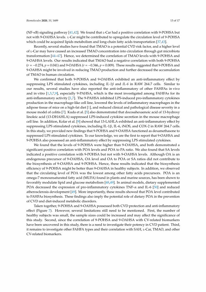
3.7. The Correlation of FAHFAs with Their Fatty Acid Precursors
The correlation between PA, SA, POA, and OA with 9-POHSA and 9-OAHSA were shown in Figure S1 and Figure 6. Higher POA level (Figure S1C), or when more POA was formed (represented from POA to PA ratio, Figure 6A), had a positive correlation with 9-POHSA (r = 0.3511, p = 0.0086) and 9-OAHSA (r = 0.4034, p = 0.0023). However, higher-level OA (Figure S1D), or when more OA was formed (represented from OA to SA and OA to POA ratio, Figure 6B,C), did not contribute to the formation of 9-POHSA (r = −0.3071, p = 0.0213) and 9-OAHSA (r = −0.3285, p = 0.0134).
Figure 6. The correlation of FAHFAs with their fatty acid precursor ratio. The correlations between 9-POHSA and 9-OAHSA with POA/PA ratio, OA/SA ratio, and OA/POA ratio are shown in (A–C), respectively. rp: Pearson correlation coefficient. Data are presented as mean ± SD (* p < 0.05, ** p < 0.01).
4. Discussion
In this study, we found that 9-POHSA and 9-OAHSA were two major types of FAHFAs in healthy human circulation. To investigate the CVD protection of FAHFAs, we analyzed their correlations with CV-related biomarkers. We found that both 9-POHSA and 9-OAHSA had a negative correlation with Glu-AC, SAH, and TMAO, but had no correlation with L-Hcy and lipid profiles (TG, TCH, HDL-C, and LDL-C). 9-POHSA had a positive correlation with L-Car, but 9-OAHSA did not. Increasing the level of endogenous POA in the body might be related to the increases of both FAHFAs. Moreover, 9-POHSA and 9-OAHSA possessed an anti-inflammatory effect on
Figure 6. The correlation of FAHFAs with their fatty acid precursor ratio. The correlations between9-POHSA and 9-OAHSA with POA/PA ratio, OA/SA ratio, and OA/POA ratio are shown in (A–C),respectively. rp: Pearson correlation coefficient. Data are presented as mean ± SD (* p < 0.05, ** p < 0.01).
3. Results
3.1. Enrollment and Characteristics of Healthy Subjects
Subjects whose health status had been ensured were considered to be eligible for the study.The baseline characteristics were measured or calculated to confirm the health status of the subjects,as shown in Table 1. A total of 57 subjects were healthy.
Table 1. Baseline characteristics of 57 healthy subjects.
Variable Subjects (Male n = 24, Female n = 33)Mean ± SD Normal Range
Age (years old) 49.9 ± 12.4 n/aHeight (cm) 162.2 ± 8.5 n/a
Body weight (kg) 63.2 ± 13.2 n/aBody mass index (BMI, kg/m2) 23.6 ± 3.3 n/aFasting blood glucose (mg/dL) 95.0 ± 25.7 <125
Biomolecules 2020, 10, 1689 11 of 17
Table 1. Cont.
Variable Subjects (Male n = 24, Female n = 33)Mean ± SD Normal Range
Triglyceride (TG, mg/dL) 122.4 ± 66.4 <200Total cholesterol (TCH, mg/dL) 174.2 ± 31.9 <200
High-density lipoprotein cholesterol (HDL-C, mg/dL) 54.5 ± 13.7 >40Low-density lipoprotein cholesterol (LDL-C, mg/dL) 101.0 ± 25.7 <130
n/a—not applicable.
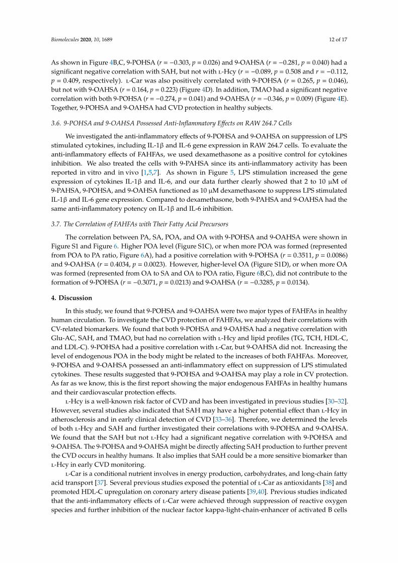
3.2. Linearity and Plasma Analysis
The gradient system of LC-MS methods was modified from the previous studies [28,29] andrenewed according to our ideas. The calibration curves were calculated by using the peak arearatio of the analytical standards and the internal standards (13C16-PA, d3- l-Met, or d9-TMAO).All of the coefficients of determinations (R2) of linearity were greater than 0.995. Seven types ofcirculating FAHFAs (5-PAHSA, 9-PAHSA, 12-PAHSA, 9-PAHPA, 9-SAHSA, 9-POHSA, and 9-OAHSA),four long-chain fatty acids (PA, SA, POA, and OA) that are endogenous precursors of FAHFAs, and fournon-traditional circulating CV-related biomarkers (l-Hcy, SAH, l-Car, and TMAO) were analyzed byLC-MS. The chromatograms are shown in Figure 2, and their levels in healthy subjects are shownin Table 2.
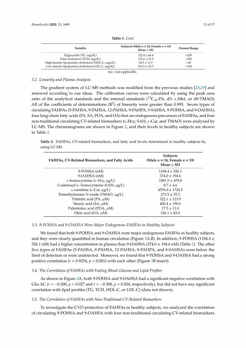
Table 2. FAHFAs, CV-related biomarkers, and fatty acid levels determined in healthy subjects byusing LC-MS.
FAHFAs, CV-Related Biomarkers, and Fatty AcidsSubjects
(Male n = 24, Female n = 33)Mean ± SD
9-POHSA (nM) 1184.4 ± 526.19-OAHSA (nM) 374.0 ± 194.6
l-homocysteine (l-Hcy, µg/L) 1081.5 ± 479.8S-adenosyl-L- homocysteine (SAH, µg/L) 8.7 ± 4.6
l-carnitine (l-Car, µg/L) 4576.0 ± 1724.5Trimethylamine N-oxide (TMAO, µg/L) 273.5 ± 55.2
Palmitic acid (PA, µM) 322.1 ± 123.9Stearic acid (SA, µM) 400.4 ± 190.6
Palmitoleic acid (POA, µM) 17.5 ± 11.0Oleic acid (OA, µM) 226.1 ± 83.0
3.3. 9-POHSA and 9-OAHSA Were Major Endogenous FAHFAs in Healthy Subjects
We found that both 9-POHSA and 9-OAHSA were major endogenous FAHFAs in healthy subjects,and they were clearly quantified in human circulation (Figure 3A,B). In addition, 9-POHSA (1184.4 ±526.1 nM) had a higher concentration in plasma than 9-OAHSA (374.0 ± 194.6 nM) (Table 2). The otherfive types of FAHFAs (5-PAHSA, 9-PAHSA, 12-PAHSA, 9-PAHPA, and 9-SAHSA) were below thelimit of detection or were undetected. Moreover, we found that 9-POHSA and 9-OAHSA had a strongpositive correlation (r = 0.9254, p < 0.001) with each other (Figure 3B insert).
3.4. The Correlation of FAHFAs with Fasting Blood Glucose and Lipid Profiles
As shown in Figure 4A, both 9-POHSA and 9-OAHSA had a significant negative correlation withGlu-AC (r = −0.300, p = 0.027 and r = −0.308, p = 0.024, respectively), but did not have any significantcorrelation with lipid profiles (TG, TCH, HDL-C, or LDL-C) (data not shown).
3.5. The Correlation of FAHFAs with Non-Traditional CV-Related Biomarkers
To investigate the CVD protection of FAHFAs in healthy subjects, we analyzed the correlationof circulating 9-POHSA and 9-OAHSA with four non-traditional circulating CV-related biomarkers.
Biomolecules 2020, 10, 1689 12 of 17
As shown in Figure 4B,C, 9-POHSA (r = −0.303, p = 0.026) and 9-OAHSA (r = −0.281, p = 0.040) had asignificant negative correlation with SAH, but not with l-Hcy (r = −0.089, p = 0.508 and r = −0.112,p = 0.409, respectively). l-Car was also positively correlated with 9-POHSA (r = 0.265, p = 0.046),but not with 9-OAHSA (r = 0.164, p = 0.223) (Figure 4D). In addition, TMAO had a significant negativecorrelation with both 9-POHSA (r = −0.274, p = 0.041) and 9-OAHSA (r = −0.346, p = 0.009) (Figure 4E).Together, 9-POHSA and 9-OAHSA had CVD protection in healthy subjects.
3.6. 9-POHSA and 9-OAHSA Possessed Anti-Inflammatory Effects on RAW 264.7 Cells
We investigated the anti-inflammatory effects of 9-POHSA and 9-OAHSA on suppression of LPSstimulated cytokines, including IL-1β and IL-6 gene expression in RAW 264.7 cells. To evaluate theanti-inflammatory effects of FAHFAs, we used dexamethasone as a positive control for cytokinesinhibition. We also treated the cells with 9-PAHSA since its anti-inflammatory activity has beenreported in vitro and in vivo [1,5,7]. As shown in Figure 5, LPS stimulation increased the geneexpression of cytokines IL-1β and IL-6, and our data further clearly showed that 2 to 10 µM of9-PAHSA, 9-POHSA, and 9-OAHSA functioned as 10 µM dexamethasone to suppress LPS stimulatedIL-1β and IL-6 gene expression. Compared to dexamethasone, both 9-PAHSA and 9-OAHSA had thesame anti-inflammatory potency on IL-1β and IL-6 inhibition.
3.7. The Correlation of FAHFAs with Their Fatty Acid Precursors
The correlation between PA, SA, POA, and OA with 9-POHSA and 9-OAHSA were shown inFigure S1 and Figure 6. Higher POA level (Figure S1C), or when more POA was formed (representedfrom POA to PA ratio, Figure 6A), had a positive correlation with 9-POHSA (r = 0.3511, p = 0.0086)and 9-OAHSA (r = 0.4034, p = 0.0023). However, higher-level OA (Figure S1D), or when more OAwas formed (represented from OA to SA and OA to POA ratio, Figure 6B,C), did not contribute to theformation of 9-POHSA (r = −0.3071, p = 0.0213) and 9-OAHSA (r = −0.3285, p = 0.0134).
4. Discussion
In this study, we found that 9-POHSA and 9-OAHSA were two major types of FAHFAs in healthyhuman circulation. To investigate the CVD protection of FAHFAs, we analyzed their correlations withCV-related biomarkers. We found that both 9-POHSA and 9-OAHSA had a negative correlation withGlu-AC, SAH, and TMAO, but had no correlation with l-Hcy and lipid profiles (TG, TCH, HDL-C,and LDL-C). 9-POHSA had a positive correlation with l-Car, but 9-OAHSA did not. Increasing thelevel of endogenous POA in the body might be related to the increases of both FAHFAs. Moreover,9-POHSA and 9-OAHSA possessed an anti-inflammatory effect on suppression of LPS stimulatedcytokines. These results suggested that 9-POHSA and 9-OAHSA may play a role in CV protection.As far as we know, this is the first report showing the major endogenous FAHFAs in healthy humansand their cardiovascular protection effects.
l-Hcy is a well-known risk factor of CVD and has been investigated in previous studies [30–32].However, several studies also indicated that SAH may have a higher potential effect than l-Hcy inatherosclerosis and in early clinical detection of CVD [33–36]. Therefore, we determined the levelsof both l-Hcy and SAH and further investigated their correlations with 9-POHSA and 9-OAHSA.We found that the SAH but not l-Hcy had a significant negative correlation with 9-POHSA and9-OAHSA. The 9-POHSA and 9-OAHSA might be directly affecting SAH production to further preventthe CVD occurs in healthy humans. It also implies that SAH could be a more sensitive biomarker thanl-Hcy in early CVD monitoring.
l-Car is a conditional nutrient involves in energy production, carbohydrates, and long-chain fattyacid transport [37]. Several previous studies exposed the potential of l-Car as antioxidants [38] andpromoted HDL-C upregulation on coronary artery disease patients [39,40]. Previous studies indicatedthat the anti-inflammatory effects of l-Car were achieved through suppression of reactive oxygenspecies and further inhibition of the nuclear factor kappa-light-chain-enhancer of activated B cells
Biomolecules 2020, 10, 1689 13 of 17
(NF-κB) signaling pathway [41,42]. We found that l-Car had a positive correlation with 9-POHSA butnot with 9-OAHSA levels. l-Car might be contributed to upregulate the circulation level of 9-POHSAwhich could be acquired lipids metabolism and long-chain fatty acids transportation [37,43].
Recently, several studies have found that TMAO is a potential CVD risk factor, and a higher levelof l-Car may have caused an increased TMAO concentration into circulation through gut microbiotatransformation [44–47]. Hence, we determined the correlation of TMAO levels with 9-POHSA and9-OAHSA levels. Our results indicated that TMAO had a negative correlation with both 9-POHSA(r = −0.274, p = 0.041) and 9-OAHSA (r = −0.346, p = 0.009). These results suggested that 9-POHSA and9-OAHSA might be involved in reducing TMAO production and further decreased the accumulationof TMAO in human circulation.
We confirmed that both 9-POHSA and 9-OAHSA exhibited an anti-inflammatory effect bysuppressing LPS stimulated cytokines, including IL-1β and IL-6 in RAW 264.7 cells. Similar toour results, several studies have also reported the anti-inflammatory of other FAHFAs in vivoand in vitro [1,5,7,9], especially 9-PAHSA, which is the most investigated among FAHFAs for itsanti-inflammatory activity [1,7]. The 9-PAHSA inhibited LPS-induced pro-inflammatory cytokinesproduction in the macrophage-like cell line, lowered the levels of inflammatory macrophages in theadipose tissue of mice on a high-fat diet [1], and reduced clinical and pathological disease severity in amouse model of colitis [7]. Kuda et al. [5] also demonstrated that docosahexaenoic acid of 13-hydroxylinoleic acid (13-DHAHLA) suppressed LPS-induced cytokine secretion in the mouse macrophagecell line. In addition, Kolar et al. [9] showed that 13-LAHLA exhibited an anti-inflammatory effect bysuppressing LPS stimulated cytokines, including IL-1β, IL-6, iNOS, and COX-2 in RAW 264.7 cells.In this study, we provided new findings that 9-POHSA and 9-OAHSA functioned as dexamethasone tosuppressed LPS stimulated cytokines. To our knowledge, we are the first to report that 9-OAHSA and9-POHSA also possessed an anti-inflammatory effect by suppressing LPS stimulated cytokines.
We found that the levels of 9-POHSA were higher than 9-OAHSA, and both demonstrated asignificant positive correlation with POA levels and POA to PA ratio. We also found that SA levelsindicated a positive correlation with 9-POHSA but not with 9-OAHSA levels. Although OA is anendogenous precursor of 9-OAHSA, OA level and OA to POA or SA ratios did not contribute tothe biosynthesis of 9-OAHSA and 9-POHSA. Hence, these results indicated that the biosynthesisefficiency of 9-POHSA might be better than 9-OAHSA in healthy subjects. In addition, we observedthat the circulating level of POA was the lowest among other fatty acids precursors. POA is anomega-7 monounsaturated fatty acid (MUFA) found in plants and marine sources, has been shown tofavorably modulate lipid and glucose metabolism [48,49]. In animal models, dietary supplementedPOA decreased the expression of pro-inflammatory cytokines TNF-α and IL-6 [50] and reducedatherosclerosis development [49]. More importantly, these results showed that POA level contributedto FAHFAs biosynthesis. These findings also imply the potential role of dietary POA in the preventionof CVD and diet-induced metabolic disorders.
Taken together, 9-POHSA and 9-OAHSA possessed both CVD protection and anti-inflammatoryeffect (Figure 7). However, several limitations still need to be mentioned. First, the number ofhealthy subjects was small, the sample sizes could be increased and may affect the significance ofthis study. Second, since the correlation of 9-POHSA and 9-OAHSA with CV-related biomarkershave been uncovered in this study, there is a need to investigate their potency in CVD patient. Third,it remains to investigate other FAHFA types and their correlation with SAH, l-Car, TMAO, and otherCV-related biomarkers.
Biomolecules 2020, 10, 1689 14 of 17
Biomolecules 2020, 10, x 14 of 17
atherosclerosis development [49]. More importantly, these results showed that POA level contributed to FAHFAs biosynthesis. These findings also imply the potential role of dietary POA in the prevention of CVD and diet-induced metabolic disorders.
Taken together, 9-POHSA and 9-OAHSA possessed both CVD protection and anti-inflammatory effect (Figure 7). However, several limitations still need to be mentioned. First, the number of healthy subjects was small, the sample sizes could be increased and may affect the significance of this study. Second, since the correlation of 9-POHSA and 9-OAHSA with CV-related biomarkers have been uncovered in this study, there is a need to investigate their potency in CVD patient. Third, it remains to investigate other FAHFA types and their correlation with SAH, L-Car, TMAO, and other CV-related biomarkers.
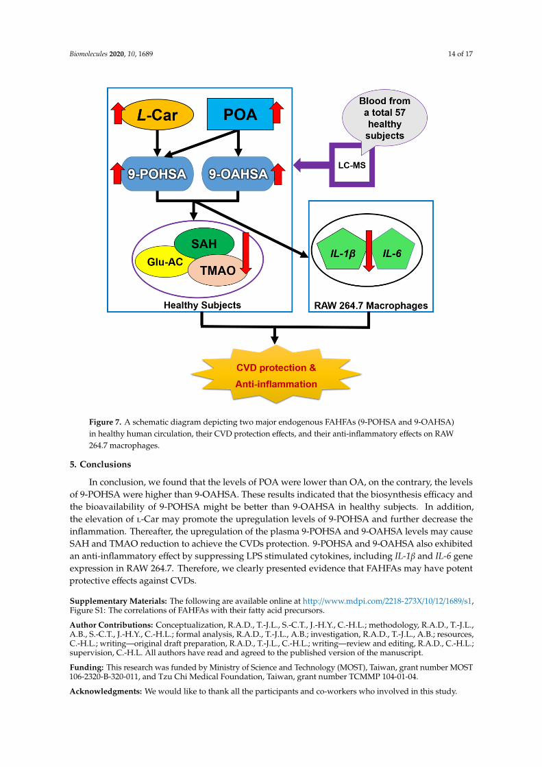
Figure 7. A schematic diagram depicting two major endogenous FAHFAs (9-POHSA and 9-OAHSA) in healthy human circulation, their CVD protection effects, and their anti-inflammatory effects on RAW 264.7 macrophages.
5. Conclusions
In conclusion, we found that the levels of POA were lower than OA, on the contrary, the levels of 9-POHSA were higher than 9-OAHSA. These results indicated that the biosynthesis efficacy and the bioavailability of 9-POHSA might be better than 9-OAHSA in healthy subjects. In addition, the elevation of L-Car may promote the upregulation levels of 9-POHSA and further decrease the inflammation. Thereafter, the upregulation of the plasma 9-POHSA and 9-OAHSA levels may cause SAH and TMAO reduction to achieve the CVDs protection. 9-POHSA and 9-OAHSA also exhibited an anti-inflammatory effect by suppressing LPS stimulated cytokines, including IL-1β and IL-6 gene expression in RAW 264.7. Therefore, we clearly presented evidence that FAHFAs may have potent protective effects against CVDs.
Figure 7. A schematic diagram depicting two major endogenous FAHFAs (9-POHSA and 9-OAHSA)in healthy human circulation, their CVD protection effects, and their anti-inflammatory effects on RAW264.7 macrophages.
5. Conclusions
In conclusion, we found that the levels of POA were lower than OA, on the contrary, the levelsof 9-POHSA were higher than 9-OAHSA. These results indicated that the biosynthesis efficacy andthe bioavailability of 9-POHSA might be better than 9-OAHSA in healthy subjects. In addition,the elevation of l-Car may promote the upregulation levels of 9-POHSA and further decrease theinflammation. Thereafter, the upregulation of the plasma 9-POHSA and 9-OAHSA levels may causeSAH and TMAO reduction to achieve the CVDs protection. 9-POHSA and 9-OAHSA also exhibitedan anti-inflammatory effect by suppressing LPS stimulated cytokines, including IL-1β and IL-6 geneexpression in RAW 264.7. Therefore, we clearly presented evidence that FAHFAs may have potentprotective effects against CVDs.
Supplementary Materials: The following are available online at http://www.mdpi.com/2218-273X/10/12/1689/s1,Figure S1: The correlations of FAHFAs with their fatty acid precursors.
Author Contributions: Conceptualization, R.A.D., T.-J.L., S.-C.T., J.-H.Y., C.-H.L.; methodology, R.A.D., T.-J.L.,A.B., S.-C.T., J.-H.Y., C.-H.L.; formal analysis, R.A.D., T.-J.L., A.B.; investigation, R.A.D., T.-J.L., A.B.; resources,C.-H.L.; writing—original draft preparation, R.A.D., T.-J.L., C.-H.L.; writing—review and editing, R.A.D., C.-H.L.;supervision, C.-H.L. All authors have read and agreed to the published version of the manuscript.
Funding: This research was funded by Ministry of Science and Technology (MOST), Taiwan, grant number MOST106-2320-B-320-011, and Tzu Chi Medical Foundation, Taiwan, grant number TCMMP 104-01-04.
Acknowledgments: We would like to thank all the participants and co-workers who involved in this study.
Biomolecules 2020, 10, 1689 15 of 17
Conflicts of Interest: The authors declare no conflict of interest.
References
1. Yore, M.M.; Syed, I.; Moraes-Vieira, P.M.; Zhang, T.; Herman, M.A.; Homan, E.A.; Patel, R.T.; Lee, J.;Chen, S.; Peroni, O.D.; et al. Discovery of a class of endogenous mammalian lipids with anti-diabetic andanti-inflammatory effects. Cell 2014, 159, 318–332. [CrossRef]
2. Balas, L.; Feillet-Coudray, C.; Durand, T. Branched fatty acyl esters of hydroxyl fatty acids (FAHFAs),appealing beneficial endogenous fat against obesity and type-2 diabetes. Chemistry 2018, 24, 9463–9476.[CrossRef]
3. Parsons, W.H.; Kolar, M.J.; Kamat, S.S.; Cognetta, A.B., III; Hulce, J.J.; Saez, E.; Kahn, B.B.; Saghatelian, A.;Cravatt, B.F. AIG1 and ADTRP are atypical integral membrane hydrolases that degrade bioactive FAHFAs.Nat. Chem. Biol. 2016, 12, 367–372. [CrossRef] [PubMed]
4. Kolar, M.J.; Kamat, S.S.; Parsons, W.H.; Homan, E.A.; Maher, T.; Peroni, O.D.; Syed, I.; Fjeld, K.; Molven, A.;Kahn, B.B.; et al. Branched fatty acid esters of hydroxy fatty acids are preferred substrates of the MODY8protein carboxyl ester lipase. Biochemistry 2016, 55, 4636–4641. [CrossRef] [PubMed]
5. Kuda, O.; Brezinova, M.; Rombaldova, M.; Slavikova, B.; Posta, M.; Beier, P.; Janovska, P.; Veleba, J.;Kopecky, J., Jr.; Kudova, E.; et al. Docosahexaenoic acid-derived fatty acid esters of hydroxy fatty acids(FAHFAs) with anti-inflammatory properties. Diabetes 2016, 65, 2580–2590. [CrossRef] [PubMed]
6. Brejchova, K.; Balas, L.; Paluchova, V.; Brezinova, M.; Durand, T.; Kuda, O. Understanding FAHFAs: Fromstructure to metabolic regulation. Prog. Lipid Res. 2020, 79, 101053. [CrossRef] [PubMed]
7. Lee, J.; Moraes-Vieira, P.M.; Castoldi, A.; Aryal, P.; Yee, E.U.; Vickers, C.; Parnas, O.; Donaldson, C.J.;Saghatelian, A.; Kahn, B.B. Branched fatty acid esters of hydroxy fatty acids (FAHFAs) protect against colitisby regulating gut innate and adaptive immune responses. J. Biol. Chem. 2016, 291, 22207–22217. [CrossRef][PubMed]
8. Pflimlin, E.; Bielohuby, M.; Korn, M.; Breitschopf, K.; Lohn, M.; Wohlfart, P.; Konkar, A.; Podeschwa, M.;Barenz, F.; Pfenninger, A.; et al. Acute and repeated treatment with 5-PAHSA or 9-PAHSA isomers does notimprove glucose control in mice. Cell Metab. 2018, 28, 217–227. [CrossRef]
9. Kolar, M.J.; Konduri, S.; Chang, T.; Wang, H.; McNerlin, C.; Ohlsson, L.; Harrod, M.; Siegel, D.; Saghatelian, A.Linoleic acid esters of hydroxy linoleic acids are anti-inflammatory lipids found in plants and mammals.J. Biol. Chem. 2019, 294, 10698–10707. [CrossRef]
10. Benlebna, M.; Balas, L.; Bonafos, B.; Pessemesse, L.; Vigor, C.; Grober, J.; Bernex, F.; Fouret, G.; Paluchova, V.;Gaillet, S.; et al. Long-term high intake of 9-PAHPA or 9-OAHPA increases basal metabolism and insulinsensitivity but disrupts liver homeostasis in healthy mice. J. Nutr. Biochem. 2020, 79, 108361. [CrossRef]
11. GBD 2019 Risk Factors Collaborators. Global burden of 87 risk factors in 204 countries and territories,1990–2019: A systematic analysis for the Global Burden of disease Study 2019. Lancet 2020, 396, 1135–1159.
12. The WHO CVD Risk Chart Working Group. World Health Organization Cardiovascular Disease Risk Charts:Revised models To Estimate Risk in 21 Global Regions. Lancet Glob. Health 2020, 7, e1332–e1345.
13. D’Adamo, E.; Santoro, N.; Caprio, S. Metabolic syndrome in pediatrics: Old concepts revised, new conceptsdiscussed. Curr. Probl. Pediatr. Adolesc. Health Care 2013, 43, 114–123. [CrossRef] [PubMed]
14. Alberti, K.G.; Zimmet, P.; Shaw, J. Metabolic syndrome—A new world-wide definition. A consensusstatement from the International Diabetes Federation. Diabet. Med. 2006, 23, 469–480. [CrossRef] [PubMed]
15. Eckel, R.H.; Grundy, S.M.; Zimmet, P.Z. The metabolic syndrome. Lancet 2005, 365, 1415–1428. [CrossRef]16. Willeit, P.; Thompson, S.G.; Agewall, S.; Bergstrom, G.; Bickel, H.; Catapano, A.L.; Chien, K.L.; de Groot, E.;
Empana, J.P.; Etgen, T.; et al. Inflammatory markers and extent and progression of early atherosclerosis:Meta-analysis of individual-participant-data from 20 prospective studies of the PROG-IMT collaboration.Eur. J. Prev. Cardiol. 2016, 23, 194–205. [CrossRef]
17. Ogawa, Y. Homeostatic inflammation, an emerging concept. Endocr. J. 2010, 57, 657–658. [CrossRef]18. Healy, A.M.; Pickard, M.D.; Pradhan, A.D.; Wang, Y.; Chen, Z.; Croce, K.; Sakuma, M.; Shi, C.; Zago, A.C.;
Garasic, J.; et al. Platelet expression profiling and clinical validation of myeloid-related protein-14 as a noveldeterminant of cardiovascular events. Circulation 2006, 113, 2278–2284. [CrossRef]

Biomolecules 2020, 10, 1689 16 of 17
19. Andersen, L.B.; Harro, M.; Sardinha, L.B.; Froberg, K.; Ekelund, U.; Brage, S.; Anderssen, S.A. Physicalactivity and clustered cardiovascular risk in children: A cross-sectional study (The European Youth HeartStudy). Lancet 2006, 368, 299–304. [CrossRef]
20. Gregor, M.F.; Hotamisligil, G.S. Inflammatory mechanisms in obesity. Annu. Rev. Immunol. 2011, 29, 415–445.[CrossRef]
21. Hoogeveen, R.M.; Nahrendorf, M.; Riksen, N.P.; Netea, M.G.; de Winther, M.P.J.; Lutgens, E.;Nordestgaard, B.G.; Neidhart, M.; Stroes, E.S.G.; Catapano, A.L.; et al. Monocyte and haematopoieticprogenitor reprogramming as common mechanism underlying chronic inflammatory and cardiovasculardiseases. Eur. Heart J. 2018, 39, 3521–3527. [CrossRef] [PubMed]
22. Kofler, S.; Nickel, T.; Weis, M. Role of cytokines in cardiovascular diseases: A focus on endothelial responsesto inflammation. Clin. Sci. 2005, 108, 205–213. [CrossRef] [PubMed]
23. Teixeira Silva, C.; Candido, A.P.C.; Pala, D.; Oliveira Barbosa, P.; Machado-Coelho, G.L.L.; Pereira deOliveira, F.L.; Pinheiro Volp, A.C. Nascimento de Freitas, R. Clustered cardiovascular risk factors areassociated with inflammatory markers in adolescents. Ann. Nutr. Metab. 2017, 70, 259–267. [CrossRef][PubMed]
24. Ridker, P.M. Anticytokine agents: Targeting interleukin signaling pathways for the treatment ofatherothrombosis. Circ. Res. 2019, 124, 437–450. [CrossRef] [PubMed]
25. Lin, T.J.; Tang, S.C.; Liao, P.Y.; Dongoran, R.A.; Yang, J.H.; Liu, C.H. A comparison of L-carnitine and severalcardiovascular-related biomarkers between healthy vegetarians and omnivores. Nutrition 2019, 66, 29–37.[CrossRef]
26. Lai, Y.H.; Lee, M.C.; Ho, G.J.; Liu, C.H.; Hsu, B.G. Association of low serum L-Carnitine levels with peripheralarterial stiffness in patients who undergo kidney transplantation. Nutrients 2019, 11, 2000. [CrossRef]
27. Dongoran, R.A.; Wang, K.H.; Lin, T.J.; Yuan, T.C.; Liu, C.H. Anti-Proliferative effect of statins is mediated byDNMT1 inhibition and p21 expression in OSCC cells. Cancers 2020, 12, 2084. [CrossRef]
28. Ma, Y.; Kind, T.; Vaniya, A.; Gennity, I.; Fahrmann, J.F.; Fiehn, O. An in silico MS/MS library for automaticannotation of novel FAHFA lipids. J. Cheminform. 2015, 7, 53. [CrossRef]
29. Ng, D.P.; Salim, A.; Liu, Y.; Zou, L.; Xu, F.G.; Huang, S.; Leong, H.; Ong, C.N. A metabolomic study of lowestimated GFR in non-proteinuric type 2 diabetes mellitus. Diabetologia 2012, 55, 499–508. [CrossRef]
30. McCully, K.S. Homocysteine and the pathogenesis of atherosclerosis. Expert Rev. Clin. Pharmacol. 2015, 8,211–219. [CrossRef]
31. Su, T.C.; Jeng, J.S.; Wang, J.D.; Torng, P.L.; Chang, S.J.; Chen, C.F.; Liau, C.S. Homocysteine, circulatingvascular cell adhesion molecule and carotid atherosclerosis in postmenopausal vegetarian women andomnivores. Atherosclerosis 2006, 184, 356–362. [CrossRef] [PubMed]
32. Yang, A.N.; Zhang, H.P.; Sun, Y.; Yang, X.L.; Wang, N.; Zhu, G.; Zhang, H.; Xu, H.; Ma, S.C.; Zhang, Y.; et al.High-methionine diets accelerate atherosclerosis by HHcy-mediated FABP4 gene demethylation pathwayvia DNMT1 in ApoE(-/-) mice. FEBS Lett. 2015, 589, 3998–4009. [CrossRef] [PubMed]
33. Liu, C.; Wang, Q.; Guo, H.; Xia, M.; Yuan, Q.; Hu, Y.; Zhu, H.; Hou, M.; Ma, J.; Tang, Z.; et al. PlasmaS-adenosylhomocysteine is a better biomarker of atherosclerosis than homocysteine in apolipoproteinE-deficient mice fed high dietary methionine. J. Nutr. 2008, 138, 311–315. [CrossRef] [PubMed]
34. Zawada, A.M.; Rogacev, K.S.; Hummel, B.; Berg, J.T.; Friedrich, A.; Roth, H.J.; Obeid, R.; Geisel, J.; Fliser, D.;Heine, G.H. S-adenosylhomocysteine is associated with subclinical atherosclerosis and renal function in acardiovascular low-risk population. Atherosclerosis 2014, 234, 17–22. [CrossRef] [PubMed]
35. Xiao, Y.; Su, X.; Huang, W.; Zhang, J.; Peng, C.; Huang, H.; Wu, X.; Huang, H.; Xia, M.; Ling, W. Role ofS-adenosylhomocysteine in cardiovascular disease and its potential epigenetic mechanism. Int. J. Biochem.Cell Biol. 2015, 67, 158–166. [CrossRef]
36. Huang, X.; Lv, X.; Song, H.; Yang, Q.; Sun, Y.; Zhang, W.; Yu, X.; Dong, S.; Yao, W.; Li, Y.; et al. The relationshipbetween S-adenosylhomocysteine and coronary artery lesions: A case control study. Clin. Chim. Acta 2017,471, 314–320. [CrossRef]
37. Flanagan, J.L.; Simmons, P.A.; Vehige, J.; Willcox, M.D.; Garrett, Q. Role of carnitine in disease. Nutr. Metab.2010, 7, 30. [CrossRef]
38. Moghaddas, A.; Dashti-Khavidaki, S. Potential protective effects of l-carnitine against neuromuscularischemia-reperfusion injury: From experimental data to potential clinical applications. Clin. Nutr. 2016, 35,783–790. [CrossRef]

Biomolecules 2020, 10, 1689 17 of 17
39. Lee, B.J.; Lin, J.S.; Lin, Y.C.; Lin, P.T. Antiinflammatory effects of L-carnitine supplementation (1000 mg/d) incoronary artery disease patients. Nutrition 2015, 31, 475–479. [CrossRef]
40. Lee, B.J.; Lin, J.S.; Lin, Y.C.; Lin, P.T. Effects of L-carnitine supplementation on lipid profiles in patients withcoronary artery disease. Lipids Health Dis. 2016, 15, 107. [CrossRef]
41. Cetinkaya, A.; Bulbuloglu, E.; Kantarceken, B.; Ciralik, H.; Kurutas, E.B.; Buyukbese, M.A.; Gumusalan, Y.Effects of L-carnitine on oxidant/antioxidant status in acetic acid-induced colitis. Dig. Dis. Sci. 2006, 51,488–494. [CrossRef] [PubMed]
42. Koc, A.; Ozkan, T.; Karabay, A.Z.; Sunguroglu, A.; Aktan, F. Effect of L-carnitine on the synthesis of nitricoxide in RAW 264.7 murine macrophage cell line. Cell Biochem. Funct. 2011, 29, 679–685. [CrossRef][PubMed]
43. Adeva-Andany, M.M.; Carneiro-Freire, N.; Seco-Filgueira, M.; Fernandez-Fernandez, C.; Mourino-Bayolo, D.Mitochondrial beta-oxidation of saturated fatty acids in humans. Mitochondrion 2019, 46, 73–90. [CrossRef][PubMed]
44. Koeth, R.A.; Wang, Z.; Levison, B.S.; Buffa, J.A.; Org, E.; Sheehy, B.T.; Britt, E.B.; Fu, X.; Wu, Y.; Li, L.; et al.Intestinal microbiota metabolism of L-carnitine, a nutrient in red meat, promotes atherosclerosis. Nat. Med.2013, 19, 576–585. [CrossRef] [PubMed]
45. Koeth, R.A.; Levison, B.S.; Culley, M.K.; Buffa, J.A.; Wang, Z.; Gregory, J.C.; Org, E.; Wu, Y.; Li, L.; Smith, J.D.;et al. gamma-Butyrobetaine is a proatherogenic intermediate in gut microbial metabolism of L-carnitine toTMAO. Cell Metab. 2014, 20, 799–812. [CrossRef]
46. Wang, Z.; Roberts, A.B.; Buffa, J.A.; Levison, B.S.; Zhu, W.; Org, E.; Gu, X.; Huang, Y.; Zamanian-Daryoush, M.;Culley, M.K.; et al. Non-lethal Inhibition of Gut Microbial Trimethylamine Production for the Treatment ofAtherosclerosis. Cell 2015, 163, 1585–1595. [CrossRef]
47. Fukami, K.; Yamagishi, S.; Sakai, K.; Kaida, Y.; Yokoro, M.; Ueda, S.; Wada, Y.; Takeuchi, M.; Shimizu, M.;Yamazaki, H.; et al. Oral L-carnitine supplementation increases trimethylamine-N-oxide but reduces markersof vascular injury in hemodialysis patients. J. Cardiovasc. Pharmacol. 2015, 65, 289–295. [CrossRef]
48. Cao, H.; Gerhold, K.; Mayers, J.R.; Wiest, M.M.; Watkins, S.M.; Hotamisligil, G.S. Identification of a lipokine,a lipid hormone linking adipose tissue to systemic metabolism. Cell 2008, 134, 933–944. [CrossRef]
49. Yang, Z.H.; Pryor, M.; Noguchi, A.; Sampson, M.; Johnson, B.; Pryor, M.; Donkor, K.; Amar, M.; Remaley, A.T.Dietary palmitoleic acid attenuates atherosclerosis progression and hyperlipidemia in low-density lipoproteinreceptor-deficient mice. Mol. Nutr. Food Res. 2019, 63, e1900120. [CrossRef]
50. Guo, X.; Li, H.; Xu, H.; Halim, V.; Zhang, W.; Wang, H.; Ong, K.T.; Woo, S.L.; Walzem, R.L.; Mashek, D.G.;et al. Palmitoleate induces hepatic steatosis but suppresses liver inflammatory response in mice. PLoS ONE2012, 7, e39286. [CrossRef]
Publisher’s Note: MDPI stays neutral with regard to jurisdictional claims in published maps and institutionalaffiliations.
© 2020 by the authors. Licensee MDPI, Basel, Switzerland. This article is an open accessarticle distributed under the terms and conditions of the Creative Commons Attribution(CC BY) license (http://creativecommons.org/licenses/by/4.0/).