Management of Mycobacterium ulcerans disease - WHO ...
80
Management of Mycobacterium ulcerans disease EDITED BY: JOHN BUNTINE KIMBALL CROFTS WHO/CDS/CPE/GBUI/2001.3 DISTRIBUTION:GENERAL ORIGINAL:ENGLISH A MANUAL FOR HEALTH CARE PROVIDERS World Health Organization Buruli Ulcer
-
Upload
khangminh22 -
Category
Documents
-
view
0 -
download
0
Transcript of Management of Mycobacterium ulcerans disease - WHO ...

Management ofMycobacteriumulcerans disease
E D I T E D B Y : J O H N B U N T I N E K I M B A L L C R O F T S
WHO/CDS/CPE/GBUI/2001.3DISTRIBUTION: GENERAL
ORIGINAL: ENGLISH
A M A N U A L F O R H E A L T H C A R E P R O V I D E R S
World Health Organization
BuruliUlcer

This manual was published thanks to financial support from:
The Association Française Raoul Follereau (AFRF), France is an NGO dedicated to leprosy control in 31 countries worldwide. It also supports six research projects on leprosy, including the genome sequencing of Mycobacterium leprae. Long before the first International Conference on Buruli Ulcer Control and Research,Yamoussoukro, Côte d’Ivoire, 1998, AFRF had taken up the new challenge of the health and social problemscaused by Buruli ulcer, working in Benin and Côte d’Ivoire since 1996. The Association also provides financial
assistance to research activities on the genome sequencing of Mycobacterium ulcerans and on the drug treatment of the disease. It is now considering supporting other countries, starting with Ghana. AFRF is committed to mobilizingthe international support needed to meet the challenges posed by Buruli ulcer.For more information, visit the AFRF website: http://www.raoul-follereau.org
ANESVAD, Spain is an NGO that has been working against leprosy and implementing health, social andeducational projects in 28 of the poorest developing countries for over 30 years. Currently it counts on thesupport of over 135 000 partners and collaborators in Spain. It has recently begun work on Buruli ulcer in
Côte d’Ivoire, carrying out programmes to detect the disease at an early stage and undertaking prevention, surgicaltreatment, training of specialized medical staff and social awareness campaigns, with the aim of limiting the impact of Buruli ulcer.For more information, visit the ANESVAD website: http://www.anesvad.org
Médecins Sans Frontières (MSF) is an international humanitarian aid organization that provides emergencymedical assistance to populations in danger in more than 80 countries. MSF Luxembourg has been involved in Buruli ulcer control activities in Benin since 1997. MSF has upgraded the Lalo Health Centre with surgicaland laboratory facilities to improve the care of patients. Apart from surgical activities, other key activities include
health education in affected communities, case-finding and training of health care providers, teachers and traditionalhealers. In terms of Buruli ulcer research, MSF is collaborating with the Institute of Tropical Medicine, Antwerp, Belgium.For more information, visit the MSF Luxembourg's website at: http://www.msf.lu
The Nippon Foundation, Japan is a private grant-making foundation whose activities cover social welfare,public health, volunteer support and overseas assistance. Since 1975 it has been working through the SasakawaMemorial Health Foundation to aid WHO in its fight to eliminate leprosy. Starting in 1998, The Nippon Foundationalso began providing financial support to the WHO Global Buruli Ulcer Initiative. The Foundation, in tandem
with WHO and several academic institutions, is currently exploring options for improved surgical management of thedisease. Finally, it is also collaborating with WHO, AFRF and other partners to find a drug treatment for Buruli ulcer.For more information, visit The Nippon Foundation’s website at: http://www.nippon-foundation.or.jp

BuruliUlcerManagement ofMycobacteriumulcerans disease
E D I T E D B Y :
DOCTOR JOHN BUNTINECornel l Special ists ’ CentreCamberwel l , Victor ia , Austral ia
DOCTOR KIMBALL CROFTSPlast ic Surgery Inst itute of UtahProvo, Utah, United States of America
A M A N U A L F O R H E A L T H C A R E P R O V I D E R S
World Health Organization

With special thanks to: George Amofah, Ministry of Health, Ghana • Kwame Asamoa, Ministry of Health, Ghana •David Ashford, CDC, Atlanta, USA • Rosemary Bell, France • Samuel Etuaful, St Martin’s Catholic Hospital, Agroyesum,Ghana • Sister Joseph, Wewak General Hospital, Papua New Guinea • Luca Saguatti, Italy, for their support
© World Health Organization, 2001
This document is not a formal publication of the World Health Organization (WHO), and all rights are reserved by the Organization. The document may, however, be freely reviewed, abstracted, reproduced or translated, in part orin whole, but not for sale or for use in conjunction with commercial purposes. The views expressed in documents by named authors are solely the responsibility of those authors.
Design: Gilles Lasseigne – Layout: Bruno Duret
Acknowledgements

Preface . . . . . . . . . . . . . . . . . . . . . . . . . . . . . . . . . . . . . . . . . . . . . . . . . . . . . . . . . . . . . . . . . . . . . . . . . . . . . . . . . . . . . . . . . . . . . . . . . . . . . . . . . . . . . . . . . . . . . . 1Illustrations . . . . . . . . . . . . . . . . . . . . . . . . . . . . . . . . . . . . . . . . . . . . . . . . . . . . . . . . . . . . . . . . . . . . . . . . . . . . . . . . . . . . . . . . . . . . . . . . . . . . . . . . . . . . . . . . . 2Introduction . . . . . . . . . . . . . . . . . . . . . . . . . . . . . . . . . . . . . . . . . . . . . . . . . . . . . . . . . . . . . . . . . . . . . . . . . . . . . . . . . . . . . . . . . . . . . . . . . . . . . . . . . . . . . . . . 3
Chapter 1. Clinical diagnosis . . . . . . . . . . . . . . . . . . . . . . . . . . . . . . . . . . . . . . . . . . . . . . . . . . . . . . . . . . . . . . . . . . . . . . . . . . . . . . . . . . . . . . . . . . . . 7Chapter 2. Plan of management . . . . . . . . . . . . . . . . . . . . . . . . . . . . . . . . . . . . . . . . . . . . . . . . . . . . . . . . . . . . . . . . . . . . . . . . . . . . . . . . . . . . . . . . . 15Chapter 3. Anaesthesia and analgesia . . . . . . . . . . . . . . . . . . . . . . . . . . . . . . . . . . . . . . . . . . . . . . . . . . . . . . . . . . . . . . . . . . . . . . . . . . . . . . . . . . 23Chapter 4. Surgical treatment . . . . . . . . . . . . . . . . . . . . . . . . . . . . . . . . . . . . . . . . . . . . . . . . . . . . . . . . . . . . . . . . . . . . . . . . . . . . . . . . . . . . . . . . . . . . 27Chapter 5. Nursing care . . . . . . . . . . . . . . . . . . . . . . . . . . . . . . . . . . . . . . . . . . . . . . . . . . . . . . . . . . . . . . . . . . . . . . . . . . . . . . . . . . . . . . . . . . . . . . . . . . . 41Chapter 6. Care in the community . . . . . . . . . . . . . . . . . . . . . . . . . . . . . . . . . . . . . . . . . . . . . . . . . . . . . . . . . . . . . . . . . . . . . . . . . . . . . . . . . . . . . . . 45Chapter 7. Rehabilitation . . . . . . . . . . . . . . . . . . . . . . . . . . . . . . . . . . . . . . . . . . . . . . . . . . . . . . . . . . . . . . . . . . . . . . . . . . . . . . . . . . . . . . . . . . . . . . . . . 49Chapter 8. Recommendations for audit and research . . . . . . . . . . . . . . . . . . . . . . . . . . . . . . . . . . . . . . . . . . . . . . . . . . . . . . . . . . . . . . . . . 55
Annex 1. Noma . . . . . . . . . . . . . . . . . . . . . . . . . . . . . . . . . . . . . . . . . . . . . . . . . . . . . . . . . . . . . . . . . . . . . . . . . . . . . . . . . . . . . . . . . . . . . . . . . . . . . . . . . . . . 58Annex 2. Prevention of HIV transmission . . . . . . . . . . . . . . . . . . . . . . . . . . . . . . . . . . . . . . . . . . . . . . . . . . . . . . . . . . . . . . . . . . . . . . . . . . . . . . . 59Annex 3. Blood transfusion and safety . . . . . . . . . . . . . . . . . . . . . . . . . . . . . . . . . . . . . . . . . . . . . . . . . . . . . . . . . . . . . . . . . . . . . . . . . . . . . . . . . . 60Annex 4. Lists of equipment . . . . . . . . . . . . . . . . . . . . . . . . . . . . . . . . . . . . . . . . . . . . . . . . . . . . . . . . . . . . . . . . . . . . . . . . . . . . . . . . . . . . . . . . . . . . . . 62Annex 5. Work of WHO on Buruli ulcer . . . . . . . . . . . . . . . . . . . . . . . . . . . . . . . . . . . . . . . . . . . . . . . . . . . . . . . . . . . . . . . . . . . . . . . . . . . . . . . . . 63Annex 6. Some research institutions involved in Buruli ulcer activities . . . . . . . . . . . . . . . . . . . . . . . . . . . . . . . . . . . . . . . . . . . . . . 65Annex 7. Some nongovernmental organizations and others involved in Buruli ulcer activities . . . . . . . . . . . . . . . . . . 66Annex 8. Members of the WHO Advisory Group on Buruli ulcer . . . . . . . . . . . . . . . . . . . . . . . . . . . . . . . . . . . . . . . . . . . . . . . . . . . . . 67Annex 9. Buruli ulcer form (BU 01) . . . . . . . . . . . . . . . . . . . . . . . . . . . . . . . . . . . . . . . . . . . . . . . . . . . . . . . . . . . . . . . . . . . . . . . . . . . . . . . . . . . . . . 68
Contents

Dr Pius Agbenorku, Department of Plastic Surgery, Komfo Anokye Teaching Hospital, Kumasi, Ghana / Sister JuliaAguiar, Mission Catholique Zagnanado, Zagnanado, Benin / Prof. Henri Assé, Institute Raoul Follereau, Adzope, Côted’Ivoire / Dr John Buntine, Cornell Specialists’ Centre, Camberwell, Victoria, Australia / Dr Kimball Crofts, HART USA /Dr Afua Hesse, Department of Pediatric Surgery, Korle-Bu Teaching Hospital, Accra, Ghana / Sister Joseph, WewakHospital, Wewak, East Sepik Province, Papua New Guinea / Prof. Christophe Oberlin, Service de Chirurgie orthopédiqueet traumatologique, Groupe Hospitalier Bichat Claude-Bernard, Paris, France / Dr G. Battista Priuli, Hôpital St-Jean-de-Dieu, Tanguiéta, Benin / Mrs May Smith, Mossman District Hospital, Cairns, Australia / Dr Christina Steffen, CairnsBase Hospital, Cairns, Australia / Dr Kingsley Asiedu, Communicable Diseases Control, Prevention and Eradication,World Health Organization, Geneva, Switzerland / Mr Anders Eklund, Disability and Rehabilitation, Non–communicableDiseases and Mental Health, World Health Organization, Geneva, Switzerland / Dr Ann Goerdt, Disability andRehabilitation, Noncommunicable Diseases and Mental Health, World Health Organization, Geneva, Switzerland / Dr JeanEmmanuel, Blood Safety and Clinical Technology, Health Technology and Pharmaceuticals, World Health Organization,Geneva, Switzerland
Contributors

1
Preface
This manual is addressed to health care providers dealing with Mycobacterium ulcerans disease (Buruli ulcer).The manual aims to achieve a better understanding of the disease, its clinical presentation and its surgical management.The manual is aimed particularly at district health care providers. A comprehensive protocol, adapted to each formand stage of the disease, is presented together with comments on the levels of resources and capabilities necessaryto shorten the length of treatment, to prevent complications and to minimize undesired sequelae and thus to obtainthe best possible outcome for each patient. Some sections include advice relevant to surgeons (e.g. relating to boneinfection). However, the level to which particular comments are intended to apply should be clear from the context.
Please note: This manual is not intended to set down a standard of medical care. It is not a replacement formedical and paramedical textbooks. Adherence to the advice given will not ensure a successful outcome inevery case. The manual should not be construed as including all proper methods of care or as excludingother methods of care. Ultimate judgement regarding a particular surgical procedure or treatment must bemade by the involved health care provider consistent with the clinical presentation of the patient and theoptions available for diagnosis and treatment.

2
Illustrations
Fig. 1 World map showing distribution of Buruli ulcer (WHO)
Fig. 2 Papule (John Hayman)Fig. 3 Nodule (Mark Evans)Fig. 4 Plaque (Mark Evans)Fig. 5 Oedematous forms (May Smith and
Kingsley Asiedu)Fig. 6 Ulcers (May Smith and Mark Evans)Fig. 7 Osteomyelitis (Giovanni Batista Priuli)Fig. 8 Contractures (Marcel Crozet)Fig. 9 Hypertrophic scar (Pius Agbenorku)Fig. 10 Squamous cell carcinoma following Buruli ulcer
(Mark Evans)Fig. 11 Differential diagnosis (Wayne Meyers)Fig. 12 Containers for specimens (Paul Johnson)Fig. 13. Swabbing technique (May Smith)Fig. 14 X-ray of bone involvement in Buruli ulcer
(Giovanni Batista Priuli)Fig. 15 Excision of a nodule (Luca Saguatti)Fig. 16 Excision of a plaque (Luca Saguatti)Fig. 17 Excision of an oedematous form (Marcel Crozet)Fig. 18 Excision of ulcerative forms (Roger Pradinaud
and Luca Saguatti)
Fig. 19 Harvesting split-skin grafts (Marcel Crozet)Fig. 20 Suspension sling (Marcel Crozet)Fig. 21 Involvement of the genitalia (Pius Agbenorku)Fig. 22 Involvement of the eye (Augustin Guédénon)Fig. 23 Lesion on the face (Marcel Crozet)Fig. 24 Lesion on the neck (Batista Priuli)Fig. 25 Involvement of the breast (Pius Agbenorku)Fig. 26 Bone involvement (Giovanni Batista Priuli)Fig. 27 External fixation (Christophe Oberlin)Fig. 28 Amputation of limbs (surgery at district level)Fig. 29 Community education (Françoise Portaels)Fig. 30 Village activity (Giovanni Batista Priuli)Fig. 31 Crutches (Disability & Rehabilitation, WHO)Fig. 32 Prostheses (Disability & Rehabilitation, WHO)Fig. 33 Cup assist (Disability & Rehabilitation, WHO)Fig. 34 Splint (Disability & Rehabilitation, WHO)Fig. 35 Education during hospitalization
(Kingsley Asiedu)Fig. 36 Noma (Marie-Hélène Leclercq)Fig. 37 Screened blood ready for transfusion
(Marcel Crozet)Fig. 38 WHO haemoglobin colour scale (WHO)Fig. 39 HIV spot test (Marcel Crozet)
3
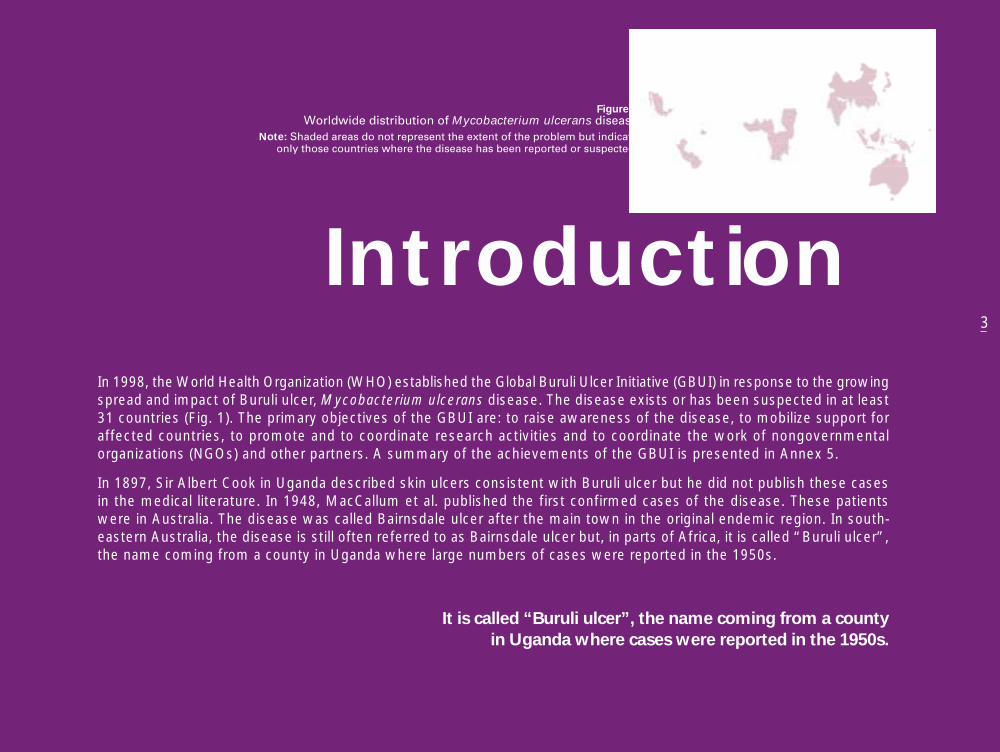
In 1998, the World Health Organization (WHO) established the Global Buruli Ulcer Initiative (GBUI) in response to the growingspread and impact of Buruli ulcer, Mycobacterium ulcerans disease. The disease exists or has been suspected in at least31 countries (Fig. 1). The primary objectives of the GBUI are: to raise awareness of the disease, to mobilize support foraffected countries, to promote and to coordinate research activities and to coordinate the work of nongovernmentalorganizations (NGOs) and other partners. A summary of the achievements of the GBUI is presented in Annex 5.
In 1897, Sir Albert Cook in Uganda described skin ulcers consistent with Buruli ulcer but he did not publish these casesin the medical literature. In 1948, MacCallum et al. published the first confirmed cases of the disease. These patientswere in Australia. The disease was called Bairnsdale ulcer after the main town in the original endemic region. In south-eastern Australia, the disease is still often referred to as Bairnsdale ulcer but, in parts of Africa, it is called “Buruli ulcer”,the name coming from a county in Uganda where large numbers of cases were reported in the 1950s.
It is called “Buruli ulcer”, the name coming from a county in Uganda where cases were reported in the 1950s.
Introduction
Figure 1Worldwide distribution of Mycobacterium ulcerans disease
Note: Shaded areas do not represent the extent of the problem but indicate only those countries where the disease has been reported or suspected.
4
Epidemiology and transmissionAfter tuberculosis and leprosy, Buruli ulcer is the mostcommon mycobacterial infection of humans. It is caused byMycobacterium ulcerans.The disease often occurs in people who live or work closeto rivers and stagnant bodies of water. Changes in theenvironment, such as the construction of irrigation systemsand dams, seem to have played a role in the resurgence ofthe disease.The mode of transmission is not known, but recent evidencesuggests that aquatic insects (Naucoris and Dyplonychusspecies) may be involved. Trauma to contaminated skin sitesappears to be the means by which the organism enters thebody. There is little proven evidence of transmission fromperson to person. No racial or social group is exempt.Infection with the human immunodeficiency virus (HIV) isnot a known risk factor. The disease is more severe in impoverished inhabitants ofremote rural areas. About 70% of those affected are childrenunder the age of 15 years. Mortality due to the disease islow, but morbidity is high. Complications include contracturedeformities, amputation of limbs, and involvement of theeye, breast and genitalia. In some localities 20–25% of thosewith healed lesions are left with disabilities that have a long-term social and economic impact. The current economic andsocial burden imposed by Buruli ulcer is enormous. In Ghana,the average cost of treatment per patient is estimated to beUS$ 780.The prevalence of the disease is not accurately known. InCôte d’Ivoire, over 15 000 cases were recorded between1978 and 1999. Prevalence rates have been estimated at16% in some communities in Côte d’Ivoire and at 22% ina community in Ghana. In Benin, nearly 4 000 cases were
reported between 1989 and 1999. In Ghana, a surveyconducted in 1999 identified over 6 000 cases and showedfor the first time that all 10 regions of the country are affected.Cases have also been reported in Burkina Faso, Togo, Guineaand other West African countries.A few cases have been reported in non-endemic areas inNorth America and Europe as a sequel to internationaltravel. Lack of familiarity with Buruli ulcer has frequentlyresulted in significant delays in the diagnosis and treatmentof these cases.
The causative organismMycobacterium ulcerans is a slow growing environmentalmycobacterium. It is an acid-fast micro-organism that growson common mycobacteriological media, e.g. Löwenstein-Jensen (L-J) medium. It grows best at low temperatures (30–32 °C), at lower thanatmospheric oxygen tension (pO2 < 2.5 kPa) and within a pHrange of 5.4–7.4. A positive culture requires incubation for6 to 8 weeks (or longer) under appropriate conditions.
ToxinA toxin that causes tissue necrosis has been known forsome time. Recently, one such compound—a polyketide-derived macrolide called mycolactone—has been identifiedand its chemical structure established. The toxin has both cytotoxic and local immunosuppressiveproperties. Injection of the purified toxin into experimentalanimals causes changes in subcutaneous fat similar tothose seen in Buruli ulcers. This is the first macrolide known to be produced by a humanpathogen and the only macrolide identified in the genusMycobacterium.

5
PathogenesisOnce introduced into the subcutaneous tissue the organismproliferates and elaborates a toxin that has affinity for fatcells. The resulting necrosis then provides a favourablemilieu for further proliferation of the organism. During thenecrotic phase, there is very little or no cellular immuneresponse and the burulin skin test is negative. By anunknown mechanism, either the toxin may be neutralizedor the organism may cease to proliferate or to producetoxin. Healing seems to begin when the host develops cell-mediated immunity, at which time the burulin skin test maybecome positive. The inflammatory cells then destroy the etiological agent(M. ulcerans) and the disease subsides with scarring. Bonesmay be affected by direct spread from the lesion or as aresult of M. ulcerans bacteraemia. In contrast to otherpathogenic mycobacteria, which are facultative intracellularparasites of macrophages, M. ulcerans occurs primarily asextracellular microcolonies.
Clinical spectrum of the diseaseClinically the disease manifests as papules, nodules, plaques,oedematous forms and ulcers. The disease may be active(ongoing infection) or inactive (previous infection withcharacteristic depressed stellate scars with or without othersequelae). A new case is a patient with no previous historyof or treatment for Buruli ulcer. A recurrent case is a patientpresenting within one year with a further lesion at the sameor a different site. Recurrence rates vary from 16% forpatients presenting early to 28% for patients presenting late.Recurrence at the same site may be due to inadequateexcision. Recurrence at a different site may be due tohaematogenous or lymphatic spread.
DiagnosisClinical: In a known endemic area, an experienced personcan make the diagnosis of Buruli ulcer on clinical grounds. The following clinico-epidemiological features are importantdiagnostic clues: 1) the patient lives in or has travelled to a known endemic area; 2) most patients are children under 15 years of age; 3) about 85% of lesions are on the limbs; 4) lower limb lesions are twice as common as upper limb lesions.
Laboratory: Any two of the following findings are requiredto positively diagnose Buruli ulcers:1) acid-fast bacilli in a smear stained by the Ziehl-Neelsen (ZN)
technique;2) positive culture of M. ulcerans (but this requires 6–8 weeks
or longer); 3) histopathological study of excisional biopsy specimen (result
available rapidly); 4) positive polymerase chain reaction (PCR) for DNA from
M. ulcerans.
TreatmentDrug treatment: Several antimycobacterial agents havein vitro activity against the causative organism but no singleagent has been proven to be regularly useful in thetreatment of the disease. Agents used include rifampicin,rifabutin, clarithromycin, azithromycin, streptomycin andamikacin. Combinations of agents have been used, with apparentlyvarying success. Drug treatment alone, even with combi-nations of drugs, is usually ineffective when there is anestablished, progressing lesion. Research into drugtreatment is a priority.

6
Surgical treatment: This is accepted as the current definitivetreatment. Limiting factors include: 1) inadequate surgical facilities; 2) need for prolonged stay in hospital; 3) high treatment costs; 4) recurrence after surgical treatment (rates of 16% to 28%); 5) the risk of transmission of infections such as HIV.Other adjuncts to treatment include heat and hyperbaricoxygen, which have not been definitively proven and maybe impractical in developing countries.
Control and preventionCommunity control strategies are currently limited by alack of knowledge regarding the source of infection andthe mode of transmission. The current standard treatmentis surgery. Expert opinion is that early surgical managementleads to improved results and resolution that are both costsaving. Early treatment is best promoted by an effectivevillage-based surveillance programme. Current attitudesand beliefs may stigmatize and create fear in the affectedindividuals thereby delaying early and effective treatment.Educational materials should dispel such misinformation
and focus on early detection and surgery. Minor surgery(e.g., nodulectomies) may be performed at the local level.
What you should doThe current control strategy promoted by the Global BuruliUlcer Initiative consists of:• health education and staff training in the communities
most affected;• strengthening the health care capacity in endemic areas
by upgrading surgical facilities, ensuring adequatetreatment supplies and improving laboratories;
• surgical training to enable other health workers (e.g.nurses, medical assistants) to perform effective minorsurgery;
• community-based surveillance to improve early detectionand rapid referral for treatment in collaboration withdisease control programmes such as those for leprosyand dracunculiasis;
• adoption of educational material adapted to the needs ofeach country;
• developing successful motivational strategies; • rehabilitation of those already deformed by the disease.
Key points
1) About 70% of those infected with Buruli ulcer are children under 15 years old.
2) In Ghana the average cost to treat Buruli ulcer is over US$ 780 per person.
3) The accepted current treatment for Buruli ulcer is usually surgery.

Clinical diagnosis
Clinical diagnosis
Non-ulcerative forms I Ulcerative forms I Bone involvement I Complications and sequelae I Differential diagnosis
Chapter 1
Credit: WHO

Clinical diagnosis
8
Clinical diagnosisThis chapter will assist you to recognize different forms of Mycobacterium ulcerans disease and to diagnosethe condition irrespective of the stage at which it presents.
O b j e c t i v e s
Always consider the diagnosis of Mycobacterium ulcerans disease in patients who live in an endemic area. There arebasically two presentations of M. ulcerans disease: non-ulcerative and ulcerative. Non-ulcerative forms present as:
Non-ulcerative forms
• Papule: This is defined as a painless, raised skin lesion, less than 1 cm in diameter. The surrounding skin isreddened (Fig. 2). This form is commonly seen in Australia.
• Nodule: A nodule is a lesion that extends from the skin into the subcutaneous tissue. It is 1–2 cm in diameter.It is usually painless but may be itchy and the surrounding skin may be discoloured compared to adjacent areas(Fig. 3). This form is commonly seen in Africa.
• Plaque: This is a firm, painless, elevated, well-demarcated lesion more than 2 cm in diameter with irregularedges. The skin over the lesion is often reddened or otherwise discoloured (Fig. 4).
• Oedematous form: There is a diffuse, extensive, usually non-pitting swelling. The affected area has ill-definedmargins, is firm and painless and involves part or all of a limb or other part of the body. There may be colourchanges over the affected region (Fig. 5a–b) and the disease may be accompanied by fever.
What you should know
Papule Nodule Plaque Oedematous forms Figure 2 Figure 3 Figure 4 Figure 5a Figure 5b
1

Clinical diagnosis
Ulcerative forms When fully developed, the ulcer has undermined edges and is indurated peripherally. The floor of the ulcer mayhave a white cotton wool-like appearance from the necrotic slough (Fig. 6a–d).
9
The ulcer is usually painless, unless there is secondary bacterial infection. When there is more than one ulcer andthe ulcers are close together, they often communicate beneath intact skin.
Figure 6aHand
Figure 6cBack
Figure 6dForearm
Figure 6bLeg
2

Clinical diagnosis
10
Mycobacterium ulcerans osteomyelitis is initially painless, but subsequently frankly painful, and well localized. There is usually an identifiable area of increased warmth. A swelling then appears and this may progress to a fistula which discharges necrotic material. Incision of the swelling reveals gelatinous tissue and, beneaththis, the bone has a moth-eaten appearance. Unlike open (contiguous) osteitis, the bone is the site of necrosis to a variable extent, similar to that seen in tuberculous osteomyelitis (Fig. 7).
• Reactive osteitis: Reactive (contiguous) osteitis occurs as a consequence of deep destruction of overlying softtissues.Occasionally, the bone is exposed to the point of devascularization, necrosis of cortical bone, sequestration, and osteomyelitis. The macroscopic appearance is then that of white dead bone of almost normal appearanceand texture.
Bone involvement • Osteomyelitis: This is true osteomyelitis. It may be focal or multifocal. The overlying skin is often intact with
no obvious lesion. Osteomyelitis may occur as a primary condition or as a metastatic condition, sometimesat a distance from a cutaneous lesion(s) or after a cutaneous lesion has healed.
Figure 7Osteomyelitis – Leg
3

Clinical diagnosis
11
Figure 8aContracture deformity of the upper limb
Figure 8bContracture deformity of the lower limb
Complications and sequelae• Contractures
Contractures result from scarring caused by lesions over or close to joints (Fig. 8a–b). Ankyloses may follow.
• BleedingThere may be continuous minor bleeding or a sudden major haemorrhage. Care should be taken to avoid largeblood vessels beneath a lesion.
• Secondary infectionSecondary bacterial infection may be caused by organisms such as staphylococci, streptococci, Pseudomonas sp.,Corynebacterium sp., etc. Secondary infection may progress to cellulitis and septicaemia.
• Extension to deep structuresInfection may extend beneath the deep fascia to involve tendon sheaths, muscle, blood vessels, nerves, boneand joints or may destroy periorbital tissue with loss of the eye.
4

Clinical diagnosis
12
• Other sequelaeHypertrophic scars and keloids may develop at infection and surgicalsites including skin graft donor sites (Fig. 9). Squamous cell carcinoma(Marjolin’s ulcer) may appear in an unstable scar or persistent ulcermany years after initial infection with M. ulcerans. (Fig. 10).
Figure 9Hypertrophic scar
Figure 10 Squamous cell carcinoma
Clinical diagnosis
13
Figure 11a Figure 11bLeishmaniasis Tropical phagedenic ulcer
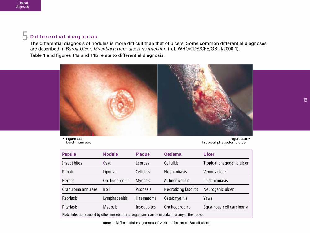
5Differential diagnosisThe differential diagnosis of nodules is more difficult than that of ulcers. Some common differential diagnoses are described in Buruli Ulcer: Mycobacterium ulcerans infection (ref. WHO/CDS/CPE/GBUI/2000.1). Table 1 and figures 11a and 11b relate to differential diagnosis.
Note: Infection caused by other mycobacterial organisms can be mistaken for any of the above.
Table 1 Differential diagnoses of various forms of Buruli ulcer
Papule
Insect bites
Pimple
Herpes
Granuloma annulare
Psoriasis
Pityriasis
Nodule
Cyst
Lipoma
Onchocercoma
Boil
Lymphadenitis
Mycosis
Plaque
Leprosy
Cellulitis
Mycosis
Psoriasis
Haematoma
Insect bites
Oedema
Cellulitis
Elephantiasis
Actinomycosis
Necrotizing fasciitis
Osteomyelitis
Onchocercoma
Ulcer
Tropical phagedenic ulcer
Venous ulcer
Leishmaniasis
Neurogenic ulcer
Yaws
Squamous cell carcinoma

Clinical diagnosis
14
Key points
1) Buruli ulcer disease presents as: papules, nodules, plaques, oedematous forms, ulcersand bone infections.
2) Contractures are easier to prevent than to correct.
3) Osteomyelitis may arise when an ulcer invades bone or when infection is blood-borne.
Notes
______________________________________________________________________________________________________________
______________________________________________________________________________________________________________
______________________________________________________________________________________________________________
______________________________________________________________________________________________________________
______________________________________________________________________________________________________________
______________________________________________________________________________________________________________
______________________________________________________________________________________________________________
______________________________________________________________________________________________________________
______________________________________________________________________________________________________________

15
Plan of management
Plan of management
Collection of specimens I Types of specimens I Storage and transport of specimens I Assessment of the patientNon-surgical treatment I Referral: levels of care
Chapter 2
Credit: WHO

Plan of management
16
Plan of managementThis chapter will assist you to confirm your diagnosis and to develop a plan for managing your patient.O b j e c t i v e s
Laboratory confirmation of diagnosisFor further details of laboratory methods for the diagnosis of Mycobacterium ulcerans disease, refer to the companion manualdealing with laboratory methods for the diagnosis of this disease.
Tissue specimens (swabs or biopsies) from the lesion are examined by the following methods:a) Ziehl-Neelsen (ZN) stainb) Culturec) Histopathologyd) Polymerase chain reaction (PCR)

Plan of management
17
Collection of specimensIt is important to prevent cross-contamination of specimens by the use of separate sterile or disposable instruments.Careful permanent labelling of specimen containers is essential. Never write directly onto the container. Write onan adhesive label.
Types of specimens
• Non-ulcerative forms Specimens for laboratory confirmation from non-ulcerative forms (i.e. papules, nodules, plaques andoedematous forms—see Chapter 1) should be taken from the centre of the surgically excised tissue and shouldinclude the entire thickness of clinically-infected tissue.Especially for non-ulcerative plaques and oedematous forms, the patient or the patient’s relative should be askedto indicate the site at which the lesion first appeared, as this is the most likely site to yield a positive diagnosisbut several further biopsies should be taken from other parts of the lesion. Tissue fragments from the peripheryof a lesion are not recommended for microbiological studies, because M. ulcerans is often not found here butsuch specimens may be most suitable for histopathology.
Materials• Sterile swabs—preferably cotton wool-tipped wooden swabs.• Containers for fresh tissue specimens
(no formalin or other preservatives) (Fig. 12).• Containers for formalin fixation (10% formalin).• Specimen containers of appropriate sizes are required
for tissue fragments obtained from surgical procedures (e.g. sterile tubes for small specimens, sterile containers for large excisional specimens).
Figure 12 Specimen collection containers
1
2

Plan of management
18
• Ulcerative formsMultiple swabs should be taken from different sites, especiallyfrom beneath the undermined edges of lesions (Fig. 13).Do not swab the slough in the centre of an ulcer. Specimensthat include all levels of the skin and subcutaneous tissue aremost suitable for histopathological study.
• BoneDiagnostic procedures to assess bone involvement should only be performed at centres providing intermediateand high-level services. For amputation specimens, the involved bone or curetted samples are required; whenamputation is not necessary, curetted bone samples are appropriate.
Figure 13Swabbing the undermined edges of a Buruli ulcer

Plan of management
19
Storage and transport of specimens
Sample to be stored for immediate analysis—place in a sterile container without any additives.Sample to be transported:
• Analysis within 24 hours—keep the sample cool (ideally at 4ºC), e.g. in an insulated container with a frozencooling block.
• Analysis after 24 hours (specimens may still be culture-positive up to 21 days):– when refrigeration facilities are available, keep at 4ºC—do not freeze;– when refrigeration facilities are not available, transport medium is essential. Liquid Middlebrook 7H9 broth
supplemented with polymyxin B, amphotericin B, nalidixic acid, trimethoprim and azlocillin (PANTA) isrecommended. Supplementation with 0.5% agar achieves a semi-solid medium.
Transport for PCR analysisPCR is best performed directly on fresh tissue specimens prepared as described above. For ulcerative forms, dry cotton wool swabs stored in their plastic containers at ambient temperature are acceptable.
Assessment of the patientA general assessment should include assessment of nutritional state, weight, height, colour of mucousmembranes and recognition of any coexisting diseases.
• Laboratory testsRoutine investigations are haemoglobin (Hb), blood group and sickling test (where indicated). In the presence of super-added infection, send a wound swab for ordinary culture and antimicrobial susceptibility testing.
3
4

Plan of management
20
• Radiological investigationIn cases where bone involvement is suspected, radiological investigation is appropriate (Fig. 14).
• Assessment of the Mycobacterium ulcerans diseaseRecord the type of lesion (see Chapter 1), the site, the extent and the presence or absence of super-addedinfection and of any other complications. You should complete form BU 01 as described in Chapter 8. This should ensure systematic record keeping for every patient and should assist follow-up after discharge.
Figure 14 X-ray of bone involvement in Buruli ulcer. See Figure 7 for physical presentation
Plan of management
21
Non-surgical treatmentAlthough surgery is the mainstay of the treatment of M. ulcerans disease, there are occasions, especially in thepresence of super-added infection, when an ulcer should be cleaned and dressed for a week or more beforesurgery. Elevation of an affected limb and splinting are important (see Chapter 7). When there is secondary bacterial infection, start broad-spectrum antibiotic therapy by administering antibioticssuch as combinations of penicillin, gentamicin and metronidazole. For some oedematous lesions, it is importantto administer an antibiotic combination for 7–10 days prior to surgery.
Referral: levels of care
• Level 1 – Peripheral or local community servicesCases should be identified, dressings applied and, where applicable, limbs should be immobilized in a positionof function to prevent deformities and to allow comfortable transfer.
• Level 2 – Basic surgical servicesAt the district hospital level, excision of nodules, papules, plaques and ulcers as well as skin grafting areappropriately performed by doctors and other trained health care providers. Note, depending on the capacityand expertise at the particular hospital, it may also be appropriate to perform some specialized surgery, e.g. amputation of limbs.
• Level 3 – Specialized surgical servicesPatients with severe extensive lesions and osteo-articular complications and other disabling sequelae should bereferred for specialized surgery.Criteria for referral. The criteria for referral of patients to level 2 and 3 services include: – all ulcers > 2 cm;– all oedematous and plaque forms;– lesions involving deeper structures including bone;– lesions on the head and neck, genitalia, breast and fingers;– difficult diagnoses (clinically and by laboratory methods);– systemically unwell patients.
5
6

Plan of management
22
Key points
1) Specimens for laboratory diagnosis of non-ulcerative formsshould be taken from the centre of the surgically excised tissue.
2) Specimens for laboratory diagnosis of ulcerative forms should be taken from the undermined edges of the ulcer.
3) Make sure transported tissue specimens are not frozen.
Notes
______________________________________________________________________________________________________________
______________________________________________________________________________________________________________
______________________________________________________________________________________________________________
______________________________________________________________________________________________________________
______________________________________________________________________________________________________________
______________________________________________________________________________________________________________
______________________________________________________________________________________________________________
______________________________________________________________________________________________________________

23
Anaesthesia and analgesia
Anaesthetic assessment I Anaesthetic agents I Analgesia
Chapter 3
Anaesthesia and analgesia
Credit: WHO

Anaesthesia and analgesia
24
Anaesthesia and analgesiaThis chapter aims to assist the anaesthetic management of your patients.O b j e c t i v e s
The choice of anaesthesia may depend on the:
• Anaesthetist’s experience• Available equipment and drugs• Age of the patient • Size and location of the lesion• Patient’s preference • Expected duration of surgery
What you should know
Anaesthetic assessmentAll patients should be thoroughly assessed to determine their suitability for local and/or general anaesthesia.The choice of anaesthesia will depend on the anaesthetist’s training and experience, available equipment anddrugs, age of the patient (children may not cooperate with local anaesthesia), size and location of the lesion,patient preference and expected duration of the surgery.The patient’s condition should be stable prior to surgery. Preoperative preparation should be directed to thepatient’s general condition and the area to be operated upon. Appropriate laboratory tests should be ordered(see Chapter 2). Preoperative preparation must include a detailed explanation of the surgery to be performed and discussion of the potential complications and risks associated with the surgery. A signed consent formdetailing the risks of the surgery must be obtained before any premedication or surgical treatment.
1

Anaesthesia and analgesia
25
Anaesthetic agentsPremedication should be offered when indicated. Drugs administered can include morphine, pethidine, diazepam,midazolam, and promethazine.Commonly used local anaesthetic agents include: lignocaine/lidocaine and bupivicaine with or without epinephrine/adrenaline. Local anaesthesia is used for smaller lesions, either by local injection or field blocks. Never inject the anaesthetic agent directly into infected tissue. This is to avoid dissemination of M. ulceransorganisms. Mark the edges of the lesion, then inject around the lesion, not into it. Epinepherine reduces bleeding,but must not be injected into the hand or foot. Regional anaesthetic blocks or spinal anaesthesia are appropriatein the presence of large lesions, otherwise general anaesthesia will be necessary. Some of the general anaestheticagents commonly used are: ketamine, isofluorane, fluothane, and ether. A ketamine/atropine cocktail, with diazepamor midazolam, is an effective way to sedate patients—especially children. Patients must be monitored continuouslyfor airway, breathing and heart rate. Oxygen saturation should also be monitored if the equipment to do so isavailable. Careful observation of the patient during the early post-operative period is also of critical importance(ABC’s).For upper and lower extremity lesions, tourniquets should be applied to minimize bleeding during surgery. Do not apply a tourniquet over a lesion. Do not leave a tourniquet in place for longer than two hours.Exsanguinate affected limbs by elevation alone.
AnalgesiaPain may be a serious issue throughout all stages of M. ulcerans disease. Mild pain may be relieved by simpleagents such as paracetamol and non-steriodal anti-inflammatory agents (e.g. ibuprofen). More severe pain may require narcotics. General anaesthesia may be required for some dressing changes.
2
3
Anaesthesia and analgesia
26
Key points
1) Never inject local anaesthetic directly into an infected tissue.
2) Do not apply a tourniquet over a lesion.
3) Do not leave a tourniquet in place for longer than two hours.
4) Exsanguinate affected limbs by elevation alone.
5) Obtain written consent from patients.
Notes
______________________________________________________________________________________________________________
______________________________________________________________________________________________________________
______________________________________________________________________________________________________________
______________________________________________________________________________________________________________
______________________________________________________________________________________________________________
______________________________________________________________________________________________________________
______________________________________________________________________________________________________________
______________________________________________________________________________________________________________
______________________________________________________________________________________________________________

Surgical treatment
Non-ulcerative forms I Ulcerative forms I Split-skin grafting technique I Complications and sequelae Involvement of bone I Amputation
Surgical treatment
Chapter 4
Credit: WHO

Surgical treatment
This chapter will assist you to perform simple operations and to choose which patients to refer for specializedmanagement.
Non-ulcerative forms
• Papule: The procedure is the same as for a nodule. Depending upon the location of the lesion, the wound maybe difficult to close by suture. If the wound edges cannot be brought together without undue tension, it is betterto stop the bleeding and to leave the wound open. A split-skin graft should then be applied at a later date. Otherwise, the wound should be dressed and the patientreferred to hospital.
28
Surgical treatmentO b j e c t i v e s
Antibiotics Currently, surgery is the only proven effective treatment for M. ulcerans disease. Combination antibiotic therapytargeting M. ulcerans may be a beneficial adjunct to surgery. Note: its efficacy is not proven.Antibiotics may be necessary to control super-infection. These agents may be chosen on the basis of diseasepresentation and the practitioner’s experience. Antibiotic therapy should be subsequently tailored according toculture and sensitivity results.
1

29
• Nodules should be excised only by appropriately trained health care providers. You must remove a nodule witha clear margin of normal tissue (Fig. 15a–d). The line of excision should be parallel to any nearby joint flexioncrease. Remember to send a sample of the excised tissue for laboratory examination. Depending on the location,some lesions may be excised and the wound closed primarily by suture without undue tension. Large surgicalwounds require split-skin grafting. Sutures are removed at 7 to 14 days, depending on the location of the woundand the progress of healing.
Figure 15aNodule
Figure 16aPlaque
Figure 15bExcision of a nodule
Figure 15cSuturing
Figure 15dSuturing
Figure 16bDeveloping plaque
Figure 16cExcision of a plaque with limited excision of healthy tissue
• Plaque: This is a more serious form of the disease which requires extensiveexcision (Fig. 16a–c) followed by split-skin grafting. Skin grafting over a flexioncrease necessitates post-operative splinting and subsequent therapy tominimize flexion contracture.
Surgical treatment

Surgical treatment
The plane of excision usually spares deep fascia but, in some advanced cases extending deep to the fascia,the excision may include deep fascia and even muscle. Wherever possible, a pneumatic tourniquet is applied—but not for longer than two hours.
• Oedematous form: This form of the disease is complex. Urgent referral to a specialized centre is mandatory. Initial management should include elevation of the affected limb. At the specialist centre, an exploratory incisionis made along the long axis of the oedematous area followed by blunt dissection of the affected tissue to reducebleeding (Fig. 17a–d). Electro-cautery is an effective way of reducing blood loss.
30
Figure 17aIncision marked – oedematous form
Figure 17bIncision – oedematous form
Figure 17cDissection – oedematous form
Figure 17dCompleted excision

31
Ulcerative formsThe surgical treatment of small ulcerative lesions is the same as that for nodules and papules (Fig. 18a). Largerlesions require excision (sometimes in stages) and split-skin grafting (Fig. 18b-d). Apply a pneumatic tourniquetwhenever possible. Prior to surgery, secondarily infected lesions should be dressed, affected limbs elevated andappropriate antibiotics administered.
• Technique of excision The required excision may be extensive. Large lesions may require staged excision, one area at a time.The excision must include healthy tissue at the lateral and deep margins. The deep fascia should be preservedif not involved but involved deep fascia must be removed, taking care not to open tendon sheaths or joints andnot to damage important nerves and blood vessels. When diseased tissues have not been removed adequately,repeated excisions may be necessary. It is recommended that extensive lesions should be treated only at leveltwo and level three services.
Surgical treatment
Figure 18cExcision – large ulcer
2
Figure 18dSkin graft application
Box: Test incisiondemonstratesinfected tissue
Figure 18aExcision – small ulcer
Figure 18bIncisions – large ulcer
32
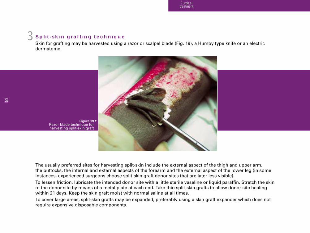
Split-skin grafting techniqueSkin for grafting may be harvested using a razor or scalpel blade (Fig. 19), a Humby type knife or an electricdermatome.
The usually preferred sites for harvesting split-skin include the external aspect of the thigh and upper arm,the buttocks, the internal and external aspects of the forearm and the external aspect of the lower leg (in someinstances, experienced surgeons choose split-skin graft donor sites that are later less visible). To lessen friction, lubricate the intended donor site with a little sterile vaseline or liquid paraffin. Stretch the skinof the donor site by means of a metal plate at each end. Take thin split-skin grafts to allow donor-site healingwithin 21 days. Keep the skin graft moist with normal saline at all times.To cover large areas, split-skin grafts may be expanded, preferably using a skin graft expander which does notrequire expensive disposable components.
Surgical treatment
Figure 19Razor blade technique for harvesting split-skin graft
3

Surgical treatment
33
Split-skin grafts may be secured at their edges and junctions using sutures or staples. Suture fixation of all fullthickness skin grafts is advised. Vaseline gauze is applied, then a further thick absorbent dressing and a bandage.
Materials which may be used to make splints include: plaster of Paris for brief immobilization, aluminium, wood(which may be carved), fibreglass, and polyvinylchloride (PVC).An alternative for the lower and upper limbs is suspension with slings and cords (Fig. 20), thus maintainingthe joints in appropriate positions during the post-operative period. Remove the surgical dressings on the third or fourth post-operative day unless there is haematoma or infection.Thereafter, change the dressings daily or on alternate days.As soon as the graft has taken (at about 10 days post-operatively) commence mobilization.
Figure 20Suspension sling
To prevent joints from becoming stiff, limbs are best splinted with their joints in the following positions: – knee in extension– ankle at a right angle– elbow in extension– wrist in extension– metacarpo-phalangeal joints in flexion – interphalangeal joints in extension

34
Complications and sequelaeComplications such as contractures and loss of body parts, for example, the eye, ear, nose, and breast, alwaysnecessitate early referral to a major hospital for surgery and reconstruction. Specialist teams may visit district or local hospital services to provide treatment, such as the release of contractures.Contractures should be released only as far as is safe, taking into account tension on blood vessels and nerves.Skin defects are then covered by grafts or flaps, including musculo-cutaneous and muscle flaps.
• The genitaliaInvolvement of the genitalia constitutes a serious complication requiring immediate referral for specialized attention (Fig. 21).
• The eye After cleaning and dressing, eyelid and eye involvement necessitate urgent referral to a specialized centre (Fig. 22a and 22b).
Surgical treatment
Figure 21Involvement of the genitalia
4
Figure 22aInvolvement of the eye (before treatment)
Figure 22bInvolvement of the eye (after treatment)

Surgical treatment
35
• The face, neck and breastAfter the initial management, patients withlesions on the face, neck and breast must bereferred to a specialized centre (Fig. 23, 24, 25).
Figure 24Involvement of the neck
Figure 25Involvement of the breast
Figure 23Involvement of the scalp and face

36
Involvement of boneBone involvement occurs by direct extension from a surface lesion into the bone or an adjacent joint or asosteomyelitis. Patients with bone involvement must be referred immediately to a specialized centre.
• Surgical treatment of osteomyelitisAt operation, a limited exposure of the swelling and/or fistula track suffices for drainage of the abscess and fordebridement of the gelatinous infected tissue. Extensive excision is not often required but, in some instances,definitive excision of infected soft tissues may be indicated. Post-operatively (Fig. 26a), the wound may beirrigated with antiseptic solutions (Fig. 26b). A drain is inserted and the wound is partially closed but, at times, an experienced surgeon will close the woundto prevent secondary infection. If present, an open wound should be dressed regularly until satisfactorygranulation tissue develops and split-skin grafting can be performed. Granulating wounds can be sequentiallygrafted as areas become clean enough for grafting.
Surgical treatment
5
Figure 26bIrrigating infected bone
Figure 26aSurgery for bone
involvement

37
Localized epiphyseal lesions (e.g. lesions involving the femoral condyles, the tibial plateau and the small bonesof the hand and foot) often require repeated partial removal of involved bone in order to preserve the adjacentarticular structures. The application of splints, plaster of Paris casts with windows or external fixation is essentialto support the bone and thus to prevent pathological fractures—without interfering with wound care. X-rays atfour to six weekly intervals are recommended to follow bone healing.
• Surgical treatment of reactive osteitisReactive osteitis is a much less serious condition which should be treated conservatively. As retaininghypertrophied periosteum protects the underlying bone, debridement over bone is best limited to curettage,preserving as much periosteum as possible. Daily dressings lessen the risk of progression of bone involvementand promote the growth of granulations.Necrotic cortical bone should be removed only after it has fully demarcated, as manifested by the growthof granulation tissue around and beneath it. Excessive bone removal must be avoided.The ulcer may be excised and then partially grafted while awaiting separation of devitalized cortical bone.Grafting is completed after removal of the sequestrum. In the meanwhile, immobilization in the preferredposition guards against pathological fractures and later contractures.
Surgical treatment
“To avoid unnecessary amputations,do not expose or remove bone widely” .

38
• External fixationWhen there is extensive or circumferential tissue loss over a joint (e.g. elbow, wrist, knee, ankle), externalfixation is applied at the time of the first procedure to keep the joints in the best possible position for function(Fig. 27). As noted above, external fixation facilitates dressings. External fixation is removed when healingis complete.
Surgical treatment
Figure 27External fixation

39
AmputationAmputation should only be performed when reconstructive measures are impossible or have failed. It is rarelynecessary. Ordinarily, the decision to amputate a limb should be taken in consultation with a specialist but severeuncontrollable bleeding may, occasionally, constitute a sufficient indication for an immediate life saving amputation.Steps for amputation are illustrated in figure 28.
Other clear indications for amputation include: – completion of a necrotic auto-amputation;– septicaemia/gangrene that would be life
threatening without amputation;– destruction of the function of a foot;– extensive bone destruction.
Surgical treatment
Figure 28Steps for amputation of limbs
“Amputation should only be performed when reconstructive measures are impossible
or have failed. It is rarely necessary” .
6

Surgical treatment
40
Key points
1) All excisions must include a margin of healthy tissue.
2) Sutures should be removed only when the wound has securely healed.
3) Contractures always need early referral to a major hospital.
4) Exsanguinate affected limbs by elevation alone.
1) Do not prolong dressing regimens when excision is appropriate.2) Do not rush to immediate excision.3) Avoid prolonged non-specific antibiotic therapy.4) Do not rely on specific antibiotics alone.5) Do not perform incisional or punch biopsy of a small lesion. Excisional biopsy is preferable.6) Do not infiltrate a local anaesthetic into any lesion.7) Do not apply an Esmarch bandage over a lesion. Exsanguinate the limb by elevation alone.8) Do not allow excessive blood loss (use a pneumatic tourniquet wherever possible). 9) Do not rely on curettage alone (except for osteomyelitis or when major structures are involved).
10) Do not count on spontaneous healing.11) Do not burn your patient (after autoclaving remember to cool your instruments before use, especially your graft mesher).12) Do not cover possibly infected tissue with a skin flap (flap cover requires special training).
What you should not do!
Nursing care
General principles I Pre-operative wound care I Psychosocial support and education
Nursing care
Chapter 5
Credit: WHO

Nursing care
42
General principlesCertified/registered nurses, other trained health care providers and family members provide nursing care.Professional nursing care, when available, involves the overall assessment of the patient and his or her family and socioeconomic environment. In places where patients often consult traditional healers, the dangers of sometraditional treatments should be tactfully explained to the patients and their families. Depending upon the level of services available, a nurse will need to decide whether to treat or to refer a patient.Nursing care should be provided before, during, and after surgery and may include rehabilitation services.A sympathetic, welcoming reception encourages and comforts the patient and family and thus, improvescompliance and outcome. The patient should be fully informed concerning the treatment plan. Nurses shouldexplain the necessity of taking the medication as prescribed and should confirm compliance.
Attention should be directed to the patient’s diet and personal hygiene. Any nutritional plan should take intoaccount cultural practices and dietary habits. Where specific arrangements are available, register malnourishedpatients for supplementary nutrition. Where such arrangements are not available, encourage the family to preparenutritious foods including local nuts and grains, eggs, fish and meat. Occasionally, when it is available, feeding ofhigh calorie/high protein fluid through a nasogastric tube may be indicated.Measures must be taken to prevent cross-infection, especially by HIV and hepatitis B/C viruses. Gloves should beworn while dressing wounds and must be replaced with a clean set between patients. If they are re-used, glovesmust be cleaned and sterilized. Clean single-use disposable gloves, however, need not be sterilized prior to use.A new set of sterile instruments must be used for each patient.For reasons of economy, where dressings are not readily available, re-use of bandages and even some dressingmaterials may be unavoidable. Bandages and dressings to be re-used are best rinsed in a washing machine.Bleach should then be added. Washing should be at a temperature of 90°C for one hour. The washed materials arethen dried and sterilized.
Nursing careThis chapter will assist your understanding of the principles underpinning appropriate nursing care.O b j e c t i v e s
1

43
In health facilities without washing machines, bandages and dressings should be placed in an antiseptic solutionfor at least one hour before washing by hand. Patients’ relatives responsible for washing bandages and dressingsshould be educated about how to handle the infected materials safely. The patient’s mattress should be protectedby plastic sheeting. The bedsheets should be changed daily.
Pre-operative wound careA complete shower/bath with clean water and soap is recommended before surgical procedures and dressingchanges. Dressings may be removed under a shower or with water which has been boiled and cooled. The woundshould then be washed and new dressings applied.Sterile gauze moistened with saline or an antiseptic solution such as hypochlorite, providone iodine or 2% aceticacid may be used. A 50/50 mixture of liquid paraffin and providone iodine at the time of dressing application willensure a moist dressing and ease of removal—thus lessening bleeding. Several layers of gauze or other absorbentmaterial may be necessary to adequately absorb the fluid exudate. Cover the dressing with a clean bandage.Dressings should be changed frequently—depending upon the amount of discharge from the wound or as advised by the surgeon. An infected wound needs to be dressed more frequently than a clean wound. Rememberto consider oral and parenteral analgesia and, in some cases, general anaesthesia before dressing wounds (see Chapter 3).
Nursing care
“Nurses should explain to patients the necessity of taking the medication as prescribed
and should confirm compliance”...
2

44
Psychosocial support and educationMycobacterium ulcerans disease may be devastating for patients and their families. Reassure the patient andfamily and offer advice about available social services. Educate patients and their families about the diseaseand about the need for early diagnosis and treatment.
Nursing care
“Mycobacterium ulcerans disease may be devastating for the patient and the family. Talk to the patient and family,
reassure them and offer advice about available social services”.
Notes
______________________________________________________________________________________________________________
______________________________________________________________________________________________________________
______________________________________________________________________________________________________________
______________________________________________________________________________________________________________
______________________________________________________________________________________________________________
______________________________________________________________________________________________________________
______________________________________________________________________________________________________________
______________________________________________________________________________________________________________
______________________________________________________________________________________________________________
3

Care in the community
Dressing wounds I Village outreach activities I Extension of specialist services and training I Follow-up I Recurrence
Care in the community
Chapter 6
Credit: WHO

Care in the community
46
Dressing woundsWash the wound with soap and drinking quality tap water or, where this is not available, water that has beenboiled for 15 minutes and then cooled. In some places, community/village health care providers may be trained todress simple wounds. For other more serious lesions, the wound should be cleaned and dressed and the personthen referred to a district hospital.
Care in the communityThis chapter aims to assist health care providers working in local communities to appropriately manageMycobacterium ulcerans disease.
O b j e c t i v e s
Figure 29Community education
1

47
Village outreach activitiesOrganize meetings with community elders and chiefs. Using aids such as educational materials, drama, videosand posters, explain the nature of the disease and stress that it is treatable (Fig. 29). Encourage infected individuals to seek early treatment. Discussion of prognosis and rehabilitation may alsobe appropriate (see Chapter 7).
Extension of specialist services and trainingArrange for specialists from a main hospital to visit local communities and district hospitals to help manage yourcases (Fig. 30). Which cases may be handled at this level will depend on the capacity at your facility. Health careproviders may be trained during these visits. Find out about this from your hospital!
Care in the community
Figure 30Visiting health care
specialists examining people in a local
community
2
3

48
Follow-up Follow-up of patients for several years via outreach visits to villages and hospital outpatient clinics greatly assistsin a better understanding of the disease. These visits are designed to monitor treatment outcomes, for example,the development of scar contractures. Children should be followed up to assess the growth of grafts which maynot keep up with the growth of normal tissues.
RecurrenceFollow-up entails watching for recurrence of infection and for the development of secondary deformities. Patientswith recurrence or disability should be referred early for specialist treatment.
Care in the community
Key points
1) Encourage persons with Buruli ulcer to seek early treatment.
2) Educational materials should be organized with input from community elders and chiefs.
4
5

Rehabilitation
Physiotherapy I Special devices I Occupational therapy and vocational retraining Preventing stigmatization I Education and other assistance
Rehabilitation
Chapter 7
Credit: WHO

Rehabilitation
50
PhysiotherapyThe physiotherapist or a health care provider with special training should teach patients andtheir families how to position a limb to prevent deformities, how to exercise affected joints,and how to use special devices (mostly splints) when they are needed.
• PositioningAfter surgery, a splint is often applied to hold a limb in a position that is good for function.The splint may be made of plaster of Paris, papier-mâché, plastic or wood. A pillow may be bandaged to a limb to hold it straight. After the wound has become stable (which maybe before healing is complete), the patient should begin to move the limb, but it may bebest to apply a splint at night to prevent a contracture. The surgeon will advise.
• ExercisesPassive exercise means that the limb is moved without contraction of its own musclesby the patient, physiotherapist or health care provider. This type of exercise commencesas soon as the positioning splint is removed, while the patient is too weak or has too much pain to move the limb without help. The physiotherapist or health care providershould commence movement slowly and gently to avoid excessive pain and stretching of healing tissues.Active exercise means that the patient moves the limb by contracting the limb’s ownmuscles. This becomes easier as strength improves and pain lessens. Later, exercisesusing weights, such as bags filled with rice, beans and/or sand are appropriate.
RehabilitationThis chapter will assist you to understand the importance and essential features of rehabilitation for patientswho have undergone surgery for Buruli ulcer.
O b j e c t i v e s
Figure 31 Crutches
1

51
Special devices If a prosthesis (artificial limb – Fig. 32) or an orthosis (brace, calliper or splint – Fig. 34) is necessary, the surgeon orthe physiotherapist or health care provider will refer the patient to a centre where the required appliance is made.
If a limb has been amputated, the physiotherapist should teach the patient how to exercise the retained partand how to bandage the stump so that it assumes a shape that fits well into an artificial limb. Proper training touse the prosthesis or orthosis is absolutely essential.Patients not needing (or awaiting) an artificial limb or calliper who have difficulty walking after surgery may needassistance to use a cane or crutches (Fig. 31).Patients with hand deformities may find self-care difficult. A physiotherapist or an occupational therapist mayfashion special devices which help to hold objects such as cups, spoons and combs (Fig. 33).
Rehabilitation
Figure 32Prostheses
Figure 34Splint
Figure 33Cup assist
2

52
Prostheses• Limb prostheses (artificial limbs)
Services to provide limb prostheses (artificial limbs) exist in most countries but often only in major cities.Therefore, these services are often difficult to access from rural and remote areas. Find out where the servicesare available in your area and the procedures for accessing them.
Before referring a person to a prosthetic/orthotic centre, start as early as possible to prepare for the fitting of a prosthesis by initiating exercises to ensure that there are no contractures. Bandage the stump to achievea satisfactory conical shape (see above). Once the wound has healed, the stump is no longer swollen and the limb has no (or minimal) contractures,the person should be referred to the centre where the required artificial limb will be made. An impression ofthe stump is first taken. Trials of the semi-finished prosthesis follow and training in the use of the new limbshould then commence. This normally takes 2–3 weeks but the time taken varies greatly. Much encouragementis needed and many adjustments to the prosthesis are often required.After the patient has returned to his/her community, continued support from the health care provideris essential. The patient often needs to return to the specialist centre for adjustment of the artificial limb.
• Eye prosthesesThese prostheses are individually made using an impression taken from the affected area. This service maynot be available in your area.
Rehabilitation
“It is far easier to prevent a contracture than to correct it”.

53
OrthosesAn orthosis is a device which supports a weakened limb or keeps a limb in a chosen position. Orthoses are referredto as splints, braces or callipers. They are sometimes needed after surgical treatment. Advice should be obtainedfrom an orthopaedic centre. Making an orthosis is often similar to making a prosthesis.
Occupational therapy and vocational retraining The physiotherapist or health care provider should advise persons whose disabilities interfere with their workwhere to go for appropriate training, which may involve the use of special devices.
Preventing stigmatizationIn countries where people suffer rejection because of physical deformities, explain to the patient and family thatothers will not catch the disease and that the patient needs their help to recover and to become active again.A community leader may be able to help the person gain acceptance and thus involvement in social activitiesand work.
Rehabilitation
“Explain to the patient and family that others will not catch the disease and
that the patient needs their help to recover and to become active again”.
3
4

54
Education and other assistanceAdmission to hospital may provide an opportunity to commence or to recommence educational courses.Arrangements should be made with the education sector to ensure that children’s schooling continuesduring hospitalization (Fig. 35). Agencies may support social rehabilitation (e.g. financial assistance orsupport to set up a small business).
Rehabilitation
“Admission to hospital may provide an opportunity to commence or to recommence educational courses”.
Notes
______________________________________________________________________________________________________________
______________________________________________________________________________________________________________
______________________________________________________________________________________________________________
______________________________________________________________________________________________________________
______________________________________________________________________________________________________________
______________________________________________________________________________________________________________
______________________________________________________________________________________________________________
______________________________________________________________________________________________________________
Figure 35Education during hospitalization
5

Recommendations for audit and research
Record-keeping using form BU 01 I Implementation of guidelines in this manual I Indicators
Recommendationsfor audit and research
Chapter 8
Credit: WHO

Recommendations for audit and research
Implementation of the guidelines in this manualThese guidelines may be supported by locally-produced educational materials and workshops.
Indicators
Outcome indicators: average duration of stay in hospital; average treatment costs; recurrence rate; complications and sequelae rate; case fatality rate.
Clinical audit markers: compliance with guidelines in this manual; referral rate to specialized treatment andrehabilitation centres.
The following areas need action:• care provision models (static versus mobile outreach services, community-based detection and referral activities)*,• implementation of these guidelines, • time between patient arrival and first surgery*,• management of various forms of the disease*.
* An economic evaluation should be included
56
Record keeping using form BU 01Form BU 01 (see Annex 9) should be completed for every patient to record demographic characteristics, risk factors,locations of lesions, clinical forms of the disease, laboratory confirmation, treatments and outcomes. This may be useful for clinical audit. Instructions to help you complete this form and diskettes to help you analyzeyour data may be obtained from:
The Global Buruli Ulcer Initiative, Communicable DiseasesWorld Health Organization
20, avenue Appia CH–1211 Geneva 27, Switzerland
Recommendations for audit and research
This chapter deals with forms for recording and reporting Mycobacterium ulcerans disease to assist in datacollection for research.
O b j e c t i v e s
1
2

Annexes
Noma I Prevention of HIV transmission I Blood transfusion and safety I Lists of equipmentWork of WHO on Buruli ulcer I Some research institutions involved in Buruli ulcer activities I Some NGOs and others involved
in Buruli ulcer activities I Members of WHO Advisory Group on Buruli ulcer I Buruli ulcer form (BU 01)
Annexes
Credit: WHO

58
Noma, cancrum oris or orofacial gangrene(from a paper by Marie-Hélène Leclercq, WHO Action Programme Against Noma)
Noma is a serious disease of the mouth and face, of unknown etiology, which is associated with poverty anddeprivation, especially poor nutrition and sanitation (Fig. 36a–c). Noma presently occurs in developing countriesof all continents but previously occurred in Europe and elsewhere.
Children under six years of age are most commonly affected. The course of the disease is different from thatof M. ulcerans but it is an important differential diagnosis.Severe gingivitis is followed by rapidly extending ulceration within the mouth. The infection then spreads throughthe cheek which becomes oedematous and then necrotic. If septicaemia and death do not quickly supervene,a foul smelling purulent discharge precedes massive tissue loss and secondary healing by wound contracture.This often leads to distortion of the face with limitation of jaw motion.Early treatment involves debridement, antibiotics (e.g. high dose penicillin) and improved nutrition. Subsequentrestriction of opening of the jaw may be overcome by inserting sticks of various sizes between the teeth. Severe deformities warrant more complex treatment including speech therapy. Cultural and social factors arehighly relevant to treatment.The WHO noma strategy involves early detection and treatment, education and training of health personnel,integration into health care services, epidemiology and etiological research and support of referral networksfor surgical treatment and rehabilitation.
Annexes
Figures 36 a–cNoma
1

59
Prevention of HIV transmission All body fluids from a person infected with HIV are potentially infectious. In this context, HIV may be transmittedby: (1) needles or sharp instruments contaminated with blood or body fluids; (2) contact between open wounds and broken skin (e.g. dermatitis), or mucous membranes contaminated by blood or body fluids; and (3) transfusion of infected blood or blood products, semen donation, and skin or organ transplantation. Proper sterilization of all surgical instruments and supplies is crucial to preventing HIV transmission.
All viruses, including HIV, are inactivated by steam sterilization (autoclaving) for 20 minutes at 100 kPa aboveatmospheric pressure or by heat treatment in an oven for 2 hours at 170°C.Most of the small number of reported occupational infections of health workers with HIV have resulted from injuryby needles (e.g. during recapping) and other sharp instruments. After use, disposable needles and scalpel bladesshould be put into a puncture-proof receptacle, preferably containing a sodium hypochlorite disinfectant. Needles must never be reused.Surgical gloves protect against transmission of HIV, but there is always the possibility of a glove being punctured.Thick gloves should therefore be worn when sharp instruments are being cleaned. Where HIV infection is prevalentamong patients, instruments should be routinely soaked in a chemical disinfectant for 30 minutes before cleaning.Linen soiled by a patient who is or may be infected with HIV should be handled with gloves and transportedin leak-proof bags. It should be washed with detergent for 25 minutes at a temperature of at least 71°C (or soakedin a hypochlorite disinfectant before washing).Liquid wastes should be carefully poured down a drain connected to a sewer or into a pit latrine or they shouldbe chemically disinfected. Solid wastes should be incinerated or disposed of in a pit latrine; chemical disinfectionmay be a temporary expedient.
Annexes
Several points are particularly relevant to the prevention of transmission of HIV:
• broken skin and open wounds should be protected with watertight dressings;
• gloves should be worn during exposure to blood or body fluids and the hands should be washedwith soap and water afterwards; frequent use of ethanol or other antiseptics on the hands and armsshould be avoided because it may lead to irritated broken skin;
• protective glasses should be worn when blood splashes may occur. If the eyes are inadvertentlysplashed, they should be washed out immediately with copious quantities of water.
2

60
Blood transfusion and safety
• Blood donations– Voluntary unremunerated donors. – Screening for blood-borne pathogens including HIV, hepatitis B and hepatitis C viruses (Fig. 37).– Continuous supply of test kits throughout the year.
• Clinical uses of blood1) Blood and blood products should be used only for conditions with significant
potential for morbidity or mortality and their need should be confirmed by carefulassessment of clinical and laboratory indications.
2) Clinical assessment of anaemia should be based on examination of the tongue,palms, eyes and nailbed.
3) Laboratory assessment should be based on haemoglobin measurement,haematocrit or the haemoglobin colour scale (Fig. 38).
4) Transfusion may be needed before, during or after surgery. Plan carefully forthe blood needs of the patient.
Annexes
Basic requirements in every country:1) a nationally organized coordinated blood transfusion service; 2) a national blood policy;3) legislation controlling the activities of blood transfusion services;4) a specific budget for blood transfusion services to ensure uninterrupted
supply of HIV test kits;5) trained health care providers and the necessary resources;6) a national quality control system.
Figure 37Screened blood ready for transfusion
3
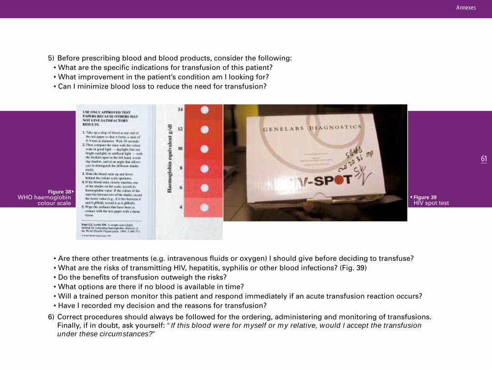
61
5) Before prescribing blood and blood products, consider the following:• What are the specific indications for transfusion of this patient?• What improvement in the patient’s condition am I looking for? • Can I minimize blood loss to reduce the need for transfusion?
• Are there other treatments (e.g. intravenous fluids or oxygen) I should give before deciding to transfuse?• What are the risks of transmitting HIV, hepatitis, syphilis or other blood infections? (Fig. 39) • Do the benefits of transfusion outweigh the risks?• What options are there if no blood is available in time?• Will a trained person monitor this patient and respond immediately if an acute transfusion reaction occurs?• Have I recorded my decision and the reasons for transfusion?
6) Correct procedures should always be followed for the ordering, administering and monitoring of transfusions.Finally, if in doubt, ask yourself: “If this blood were for myself or my relative, would I accept the transfusionunder these circumstances?”
Annexes
Figure 38WHO haemoglobin
colour scaleFigure 39HIV spot test
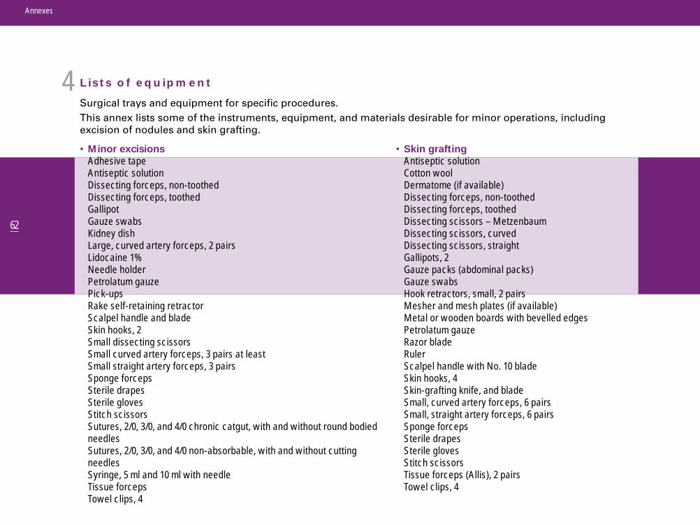
Annexes
62
Lists of equipment
Surgical trays and equipment for specific procedures.This annex lists some of the instruments, equipment, and materials desirable for minor operations, includingexcision of nodules and skin grafting.
• Skin graftingAntiseptic solutionCotton woolDermatome (if available)Dissecting forceps, non-toothedDissecting forceps, toothedDissecting scissors – MetzenbaumDissecting scissors, curvedDissecting scissors, straightGallipots, 2Gauze packs (abdominal packs)Gauze swabsHook retractors, small, 2 pairsMesher and mesh plates (if available)Metal or wooden boards with bevelled edgesPetrolatum gauzeRazor bladeRulerScalpel handle with No. 10 bladeSkin hooks, 4Skin-grafting knife, and bladeSmall, curved artery forceps, 6 pairsSmall, straight artery forceps, 6 pairsSponge forcepsSterile drapesSterile glovesStitch scissorsTissue forceps (Allis), 2 pairsTowel clips, 4
• Minor excisionsAdhesive tapeAntiseptic solutionDissecting forceps, non-toothedDissecting forceps, toothedGallipotGauze swabsKidney dishLarge, curved artery forceps, 2 pairsLidocaine 1%Needle holderPetrolatum gauzePick-upsRake self-retaining retractorScalpel handle and bladeSkin hooks, 2Small dissecting scissorsSmall curved artery forceps, 3 pairs at leastSmall straight artery forceps, 3 pairsSponge forcepsSterile drapesSterile glovesStitch scissorsSutures, 2/0, 3/0, and 4/0 chronic catgut, with and without round bodiedneedlesSutures, 2/0, 3/0, and 4/0 non-absorbable, with and without cuttingneedlesSyringe, 5 ml and 10 ml with needleTissue forcepsTowel clips, 4
4

Annexes
63
Work of WHO on Buruli ulcer
On the advice of world experts, WHO has taken the leadership in coordinating Buruli ulcer control and researchefforts worldwide. This is essential in order to maintain effective function and focus. WHO/GBUI activities sinceearly 1998—when the Initiative was established—include the following:
1) A preliminary meeting of an ad hoc Task Force was held in February 1998. Later, an Advisory Committee of18 experts was established. This Committee includes world authorities on Buruli ulcer and representatives fromendemic countries. Some group members contributed to the recently published monograph and this manual.
2) The first International Conference on Buruli Ulcer Control and Research was held in Yamoussoukro, Côte d’Ivoire,6–8 July 1998. This resulted in an increased awareness of the disease. At this Conference, the YamoussoukroDeclaration on Buruli ulcer was signed by three heads of state and the Director-General of WHO. The report ofthis Conference, in English and in French, is available for distribution.
3) Assessments in Benin, Côte d’Ivoire, Ghana, and Togo were conducted between March and July 1998 with theaim of understanding the problem of Buruli ulcer and discussing the importance of the disease with variousgovernment authorities. As a result, focused programmes have been established in Benin, Côte d’Ivoire, Ghana,Guinea, and Togo. More countries are establishing programmes.
4) Progress has been achieved in raising the awareness of the significance of Buruli ulcer. However, as the diseaseis still unknown to many, more work needs to be done. A newly established website (www.who.int/gtb-buruli)will assist to make available much needed information. The first WHO educational leaflets, in English and inFrench, targeting community workers at district and village levels, have been printed and distributed inendemic countries.
5) Standard case definitions and forms for the surveillance and clinical management of patients, as well asstandard guidelines for treatment and referral of patients, have been developed by the WHO AdvisoryCommittee in consultation with other experts worldwide.
6) A WHO scientific working group, consisting of some 40 world experts in the disease, known as the InternationalMycobacterium ulcerans Study Team (IMuST), has been established in collaboration with A/Prof. John Hayman,from the Monash University, Melbourne, Australia. The IMuST seeks to develop control and research activities,and to help coordinate the world’s efforts against Buruli ulcer.
5

Annexes
64
7) Collaborating centres will be established in some international research institutions to support research andtraining activities.
8) Research in the following priority areas, which have the potential of impacting on the control of the disease,has been advised by the WHO Advisory Group and IMuST:• operational steps in the implementation of adequate control measures;• mode(s) of transmission;• environmental changes that favour emergence of the disease;• surveys to determine the burden of the disease;• chemical structure of the toxin;• rapid methods of diagnosis; and• action of known antimicrobial drugs on M. ulcerans, starting with animal models and continuing
to clinical trials.

Annexes
65
Some research institutions involved in Buruli ulcer activities
• Armed Forces Institute of Pathology (AFIP), Washington DC, USA
• Bactériologie et Hygiène, Faculté de Médecine Pitié-Salpétrière, Paris, France
• Centers for Disease Control and Prevention (CDC), Atlanta, GA, USA
• Department of Anatomy and Cell Biology, Monash University, Melbourne, Australia
• Department of Infectious Diseases, Austin and Repatriation Medical Centre, Melbourne, Australia
• Department of Internal Medicine, University Hospital, Groningen, The Netherlands
• Department of Medicine, Emory University School of Medicine, Atlanta, GA, USA
• Department of Microbiology, Bernhard Nocht Institute for Tropical Medicine, Hamburg, Germany
• Department of Microbiology, Monash University, Victoria, Australia
• Department of Microbiology, University of Tennessee, Knoxville, TN, USA
• Institute of Tropical Medicine, Antwerp, Belgium
• Laboratoire de Bactériologie, Centre Hospitalier Universitaire d’Angers, Angers, France
• Microbiological Research Unit, Royal Children’s Hospital, Melbourne, Australia
• Nagoya University, Graduate School of Medicine, Nagoya, Japan
• Nippon Medical School, Tokyo, Japan
• Noguchi Memorial Institute for Medical Research, Accra, Ghana
• Pasteur Institute of Guiana, Cayenne, French Guiana
• Plastic Surgery & Burns Center, Korle-Bu Teaching Hospital, Accra, Ghana
• St George’s Hospital Medical School, London, England
• Swiss Tropical Institute, Basel, Switzerland
• Unité Génétique Moléculaire Bactérienne, Pasteur Institute, Paris, France
6

Annexes
66
Some nongovernmental organizations and others involved in Buruli ulceractivities
• Acción Sanitaria y Desarrollo Social (ANESVAD), Spain
• Aide aux Lépreux Emmaüs-Suisse (ALES), Switzerland
• American Leprosy Missions (ALM), USA
• Association Française Raoul Follereau (AFRF), France
• Associazione Italiana Amici di Raoul Follereau (AIFO), Italy
• Catriona Hargreaves Charitable Trust (CHCT), England
• Damien Foundation, Belgium
• Directorate General for International Cooperation, Belgium
• Fondation Luxembourgeoise Raoul Follereau (FFL), Luxembourg
• Government of Japan
• Humanitarian Aid Relief Team (HART), USA
• Japan Tissue Engineering Co., Ltd, Japan
• Kobe International University, Japan
• Médecins Sans Frontières (MSF), Luxembourg & Switzerland
• MAP International, West Africa, Côte d’Ivoire
• Pharmaciens Sans Frontières, France
• Projet Humanitaire Afrique Nord Sud (PHANS), France
• Pfizer Pharmaceuticals Inc., USA
• Rotary Club of Milan, Italy
• Sasakawa Memorial Health Foundation, Japan
• The Nippon Foundation, Japan
7

Annexes
67
8Members of the WHO Advisory Group on Buruli ulcer
• Dr George Amofah, Public Health Division, Ministry of Health, P. O. Box M-44, Accra, Ghana
• Dr David Ashford, Meningitis and Special Pathogens Branch, CDC, 1600 Clifton Rd, Atlanta, GA 30333, USA
• Dr John Buntine, Cornell Specialists’ Centre,13 Cornell Street, Camberwell, Victoria, 3124, Australia
• Prof. Jacques Grosset, Bactériologie et Hygiène, Faculté de Médecine Pitié-Salpétrière, 91, boulevard del’Hôpital, 75634 Paris Cedex 13, France
• Dr Augustin Guédénon, Programme National de Lutte contre l’Ulcère de Buruli, Ministère de la Santé publique,06 BP 2572, Cotonou, Benin
• A/Prof. John Hayman, Department of Anatomy and Cell Biology, Monash Univ., Clayton, Melbourne, 3800, Australia
• A/Prof. Paul Johnson, Department of Infectious Diseases, Austin and Repatriation Medical Centre, Heidelberg,3084 Melbourne, Australia
• Sister Joseph, Wewak General Hospital, Private Mailbag, Wewak, East Sepik Province, Papua New Guinea
• Prof. Jean-Marie Kanga, Programme National de Lutte contre l’Ulcère de Buruli, 18 BP 2890, Abidjan 18, Côte d’Ivoire
• Prof. Kenzo Kiikuni, Sasakawa Memorial Health Foundation, 1-2-2 Akasaka, Minato-Ku, Tokyo 107-0052, Japan
• Dr Harold King, Division of Infectious Diseases, Department of Medicine, Emory School of Medicine, 69 Butler Street, S.E., Atlanta, GA 30303, USA
• Dr Wayne M. Meyers, Division of Microbiology, Armed Forces Institute of Pathology, Washington, DC,20306-6000, USA
• Prof. Françoise Portaels, Department of Microbiology, Institute of Tropical Medicine, Nationalestraat 155, B-2000, Antwerp, Belgium
• Dr Roger Pradinaud, Service de Dermatologie, Centre hospitalier général de Cayenne, Cayenne Cedex, GuyaneFrançaise
• Dr G. Battista Priuli, Hôpital Saint-Jean-de-Dieu, BP 7, Tanguiéta, Benin
• Dr Pamela L. Small, Department of Microbiology, 409 Walters Life Sciences, University of Tennessee, Knoxville, TN 37996-0845, USA
• Dr Napo Tignokpa, Programme contre la Lèpre et la Tuberculose, Ministère de la Santé, BP 2271, Lomé, Togo
• Dr Mark Wansbrough-Jones, Division of Infectious Disease, St. George’s Hospital Medical School, Cranmer Terrace, London SW17 0RE, England
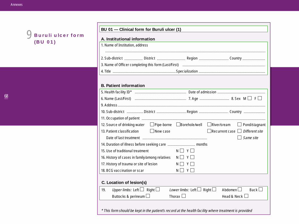
Annexes
68
Buruli ulcer form (BU 01)
5. Health facility ID*
6. Name (Last/First)
9. Address
10. Sub-district District Region Country
11. Occupation of patient
12. Source of drinking water Pipe-borne Borehole/well
13. Patient classification New case Recurrent case Different site
Date of last treatment Same site
14. Duration of illness before seeking care months
15. Use of traditional treatment N Y
16. History of cases in family/among relatives N Y
17. History of trauma or site of lesion N Y
18. BCG vaccination or scar N Y
1. Name of Institution, address
2. Sub-district District Region Country
3. Name of Officer completing this form (Last/First)
4. Title Specialization
BU 01 — Clinical form for Buruli ulcer (1)
B. Patient information
C. Location of lesion(s)
A. Institutional information
19. Upper limbs: Left Right
Buttocks & perineum
Lower limbs: Left Right
Thorax
Abdomen Back
Head & Neck
* This form should be kept in the patient’s record at the health facility where treatment is provided
Date of admission
7. Age 8. Sex M F
River/sream Pond/stagnant
9

69
Annexes
BU 01(continued)

70
Annexes
Notes
______________________________________________________________________________________________________________
______________________________________________________________________________________________________________
______________________________________________________________________________________________________________
______________________________________________________________________________________________________________
______________________________________________________________________________________________________________
______________________________________________________________________________________________________________
______________________________________________________________________________________________________________
______________________________________________________________________________________________________________
______________________________________________________________________________________________________________
______________________________________________________________________________________________________________
______________________________________________________________________________________________________________
______________________________________________________________________________________________________________
______________________________________________________________________________________________________________
______________________________________________________________________________________________________________
______________________________________________________________________________________________________________
______________________________________________________________________________________________________________
______________________________________________________________________________________________________________
______________________________________________________________________________________________________________
______________________________________________________________________________________________________________
______________________________________________________________________________________________________________
______________________________________________________________________________________________________________
______________________________________________________________________________________________________________
71

72
Annexes
Notes
______________________________________________________________________________________________________________
______________________________________________________________________________________________________________
______________________________________________________________________________________________________________
______________________________________________________________________________________________________________
______________________________________________________________________________________________________________
______________________________________________________________________________________________________________
______________________________________________________________________________________________________________
______________________________________________________________________________________________________________
______________________________________________________________________________________________________________
______________________________________________________________________________________________________________
______________________________________________________________________________________________________________
______________________________________________________________________________________________________________
______________________________________________________________________________________________________________
______________________________________________________________________________________________________________
______________________________________________________________________________________________________________
______________________________________________________________________________________________________________
______________________________________________________________________________________________________________
______________________________________________________________________________________________________________
______________________________________________________________________________________________________________
______________________________________________________________________________________________________________
______________________________________________________________________________________________________________
______________________________________________________________________________________________________________

Available further materials
• Buruli ulcer (Mycobacterium ulcerans infection). WHO/CDS/CPE/GBUI/2000.1 (English, French, Spanish)
• Report: 3rd Advisory Buruli ulcer (Mycobacterium ulcerans infection). WHO/CDS/CPE/GBUI/2000.2 (English, French)
• Diagnosis of Mycobacterium ulcerans disease (Buruli ulcer). WHO/CDS/CPE/GBUI/2001.4 (English, French, Spanish)
• Buruli ulcer comic. WHO/CDS/CPE/GBUI/2001.5 (English, French)
• Brochure on Buruli ulcer. WHO/CDS/CPE/SMT/2001.6 (English, French, Spanish)
• Report: 4th Advisory Buruli ulcer (Mycobacterium ulcerans infection). WHO/CDS/CPE/GBUI/2001.6 (English, French)
• Posters and leaflets on Buruli ulcer (English, French)
• 9 minutes 25 seconds video on Buruli ulcer (English, French, Spanish)
• Epi info software for data management (English, French)
• Training video—diagnosis and management of Buruli ulcer (English, French)
For more information, contact:
Global Buruli Ulcer InitiativeCommunicable diseases
World Health Organization1211 Geneva 27 – Switzerland
Tel. (41) 22 791 2803/2498Fax (41) 22 791 4777
E-mail: [email protected]: www.who.int/gtb-buruli

This manual provides an authoritative guide to the diagnosis and clinical management of Buruli ulcer, a mycobacterial diseasethat causes immense suffering and crippling deformities in a growing number of tropical countries. Addressed to district healthcare providers, the manual aims to facilitate a better understanding of this difficult disease, its clinical presentation, and itssurgical management.
A comprehensive protocol, adapted to each form and stage of the disease, is presented together with comments on the levelsof resources and capabilities needed to shorten the length of treatment, to prevent complications, and to minimize undesiredsequelae, and thus to obtain the best possible outcome for each patient.
As surgical treatment is presently the definitive management strategy, the manual includes detailed information on specificsurgical procedures for both non-ulcerative and ulcerative forms of the disease, and for complications and sequelae. Chapterson nursing care, care in the community, and rehabilitation are also included. Throughout, numerous colour photographs andline drawings are used to enhance the manual’s value as a training and reference tool.
Methods for the laboratory diagnosis of Mycobacterium ulcerans disease are covered in a companion volume, Diagnosis of Mycobacterium ulcerans disease: A manual for health care providers.