Corticosteroid-Induced Immunosuppression Ultimately Does Not Compromise the Efficacy of...
8
Corticosteroid-Induced Immunosuppression Ultimately Does Not Compromise the Efficacy of Antibiotherapy in Murine Mycobacterium ulcerans Infection Teresa G. Martins 1,2 , Gabriela Trigo 1,2,3 , Alexandra G. Fraga 1,2 , Jose ´ B. Gama 1,2 , Adhemar Longatto- Filho 1,2,4,5 , Margarida Saraiva 1,2 , Manuel T. Silva { 6 , Anto ´ nio G. Castro 1,2 , Jorge Pedrosa 1,2 * 1 Life and Health Sciences Research Institute, School of Health Sciences, University of Minho, Braga, Portugal, 2 ICVS/3B’s - PT Government Associate Laboratory, Braga/ Guimara ˜es, Portugal, 3 Institute for Biotechnology and Bioengineering, Centre of Biological Engineering, University of Minho, Braga, Portugal, 4 Laboratory of Medical Investigation 14, Faculty of Medicine of University of Sa ˜o Paulo, Sa ˜o Paulo, Brazil, 5 Molecular Oncology Research Center, Barretos, Sa ˜o Paulo, Brazil, 6 Institute for Molecular and Cell Biology, Porto, Portugal Abstract Background: Buruli ulcer (BU) is a necrotizing disease of the skin, subcutaneous tissue and bone caused by Mycobacterium ulcerans. It has been suggested that the immune response developed during the recommended rifampicin/streptomycin (RS) antibiotherapy is protective, contributing to bacterial clearance. On the other hand, paradoxical reactions have been described during or after antibiotherapy, characterized by pathological inflammatory responses. This exacerbated inflammation could be circumvented by immunosuppressive drugs. Therefore, it is important to clarify if the immune system contributes to bacterial clearance during RS antibiotherapy and if immunosuppression hampers the efficacy of the antibiotic regimen. Methodology/Principal Findings: We used the M. ulcerans infection footpad mouse model. Corticosteroid-induced immunosuppression was achieved before experimental infection and maintained during combined RS antibiotherapy by the administration of dexamethasone (DEX). Time-lapsed analyses of macroscopic lesions, bacterial burdens, histology and immunohistochemistry were performed in M. ulcerans-infected footpads. We show here that corticosteroid-immunosup- pressed mice are more susceptible to M. ulcerans, with higher bacterial burdens and earlier ulceration. Despite this, macroscopic lesions remised during combined antibiotic/DEX treatment and no viable bacteria were detected in the footpads after RS administration. This was observed despite a delayed kinetics in bacterial clearance, associated with a local reduction of T cell and neutrophil numbers, when compared with immunocompetent RS-treated mice. In addition, no relapse was observed following an additional 3 month period of DEX administration. Conclusions/Significance: These findings reveal a major role of the RS bactericidal activity for the resolution of M. ulcerans experimental infections even during immunosuppression, and support clinical investigation on the potential use of corticosteroids or other immunosuppressive/anti-inflammatory drugs for the management of BU patients undergoing paradoxical reactions. Citation: Martins TG, Trigo G, Fraga AG, Gama JB, Longatto-Filho A, et al. (2012) Corticosteroid-Induced Immunosuppression Ultimately Does Not Compromise the Efficacy of Antibiotherapy in Murine Mycobacterium ulcerans Infection. PLoS Negl Trop Dis 6(11): e1925. doi:10.1371/journal.pntd.0001925 Editor: Christian Johnson, Fondation Raoul Follereau, France Received July 12, 2012; Accepted October 16, 2012; Published November 29, 2012 Copyright: ß 2012 Martins et al. This is an open-access article distributed under the terms of the Creative Commons Attribution License, which permits unrestricted use, distribution, and reproduction in any medium, provided the original author and source are credited. Funding: This work was supported by a grant from the Health Services of Fundac ¸a ˜o Calouste Gulbenkian, and the Portuguese Science and Technology Foundation (FCT) fellowships SFRH/BD/41598/2007, SFRH/BPD/64032/2009, SFRH/BPD/68547/2010 and SFRH/BD/33573/2009 to TGM, GT, AGF, and JBG, respectively. MS is a Cie ˆncia 2007 fellow. The funders had no role in study design, data collection and analysis, decision to publish, or preparation of the manuscript. Competing Interests: The authors have declared that no competing interests exist. * E-mail: [email protected] { Deceased. Introduction Buruli ulcer (BU) is a necrotizing disease of the skin, subcutaneous tissue and bone [1,2]. The pathogenesis of the disease is associated with local and regional cytotoxic/immuno- suppressive activities of the lipidic toxin mycolactone, produced by the environmental pathogen Mycobacterium ulcerans [3–7]. The clinical forms of BU disease are characterized by an initial nonulcerative lesion, often a nodule or a papule or the more disseminated forms plaques and oedema. Each of these forms can evolve to an ulcer and metastasize with the development of new cutaneous lesions or osteomyelitis [1,2]. Established BU lesions are characterized by extensive necrotic, acellular areas with clumps of extracellular bacilli surrounded by a band of inflammatory cells, usually neutrophils and macrophages [8–10]. Although an extracellular localization of the bacilli is frequently seen in histological sections, M. ulcerans presents an intramacrophage growth phase in its life cycle before shedding to the extracellular compartment, and this supports the observation of intracellular bacilli at the peripheries of necrotic areas [11]. It has also been PLOS Neglected Tropical Diseases | www.plosntds.org 1 November 2012 | Volume 6 | Issue 11 | e1925
Transcript of Corticosteroid-Induced Immunosuppression Ultimately Does Not Compromise the Efficacy of...

Corticosteroid-Induced Immunosuppression UltimatelyDoes Not Compromise the Efficacy of Antibiotherapy inMurine Mycobacterium ulcerans InfectionTeresa G. Martins1,2, Gabriela Trigo1,2,3, Alexandra G. Fraga1,2, Jose B. Gama1,2, Adhemar Longatto-
Filho1,2,4,5, Margarida Saraiva1,2, Manuel T. Silva{6, Antonio G. Castro1,2, Jorge Pedrosa1,2*
1 Life and Health Sciences Research Institute, School of Health Sciences, University of Minho, Braga, Portugal, 2 ICVS/3B’s - PT Government Associate Laboratory, Braga/
Guimaraes, Portugal, 3 Institute for Biotechnology and Bioengineering, Centre of Biological Engineering, University of Minho, Braga, Portugal, 4 Laboratory of Medical
Investigation 14, Faculty of Medicine of University of Sao Paulo, Sao Paulo, Brazil, 5 Molecular Oncology Research Center, Barretos, Sao Paulo, Brazil, 6 Institute for
Molecular and Cell Biology, Porto, Portugal
Abstract
Background: Buruli ulcer (BU) is a necrotizing disease of the skin, subcutaneous tissue and bone caused by Mycobacteriumulcerans. It has been suggested that the immune response developed during the recommended rifampicin/streptomycin(RS) antibiotherapy is protective, contributing to bacterial clearance. On the other hand, paradoxical reactions have beendescribed during or after antibiotherapy, characterized by pathological inflammatory responses. This exacerbatedinflammation could be circumvented by immunosuppressive drugs. Therefore, it is important to clarify if the immunesystem contributes to bacterial clearance during RS antibiotherapy and if immunosuppression hampers the efficacy of theantibiotic regimen.
Methodology/Principal Findings: We used the M. ulcerans infection footpad mouse model. Corticosteroid-inducedimmunosuppression was achieved before experimental infection and maintained during combined RS antibiotherapy bythe administration of dexamethasone (DEX). Time-lapsed analyses of macroscopic lesions, bacterial burdens, histology andimmunohistochemistry were performed in M. ulcerans-infected footpads. We show here that corticosteroid-immunosup-pressed mice are more susceptible to M. ulcerans, with higher bacterial burdens and earlier ulceration. Despite this,macroscopic lesions remised during combined antibiotic/DEX treatment and no viable bacteria were detected in thefootpads after RS administration. This was observed despite a delayed kinetics in bacterial clearance, associated with a localreduction of T cell and neutrophil numbers, when compared with immunocompetent RS-treated mice. In addition, norelapse was observed following an additional 3 month period of DEX administration.
Conclusions/Significance: These findings reveal a major role of the RS bactericidal activity for the resolution of M. ulceransexperimental infections even during immunosuppression, and support clinical investigation on the potential use ofcorticosteroids or other immunosuppressive/anti-inflammatory drugs for the management of BU patients undergoingparadoxical reactions.
Citation: Martins TG, Trigo G, Fraga AG, Gama JB, Longatto-Filho A, et al. (2012) Corticosteroid-Induced Immunosuppression Ultimately Does Not Compromisethe Efficacy of Antibiotherapy in Murine Mycobacterium ulcerans Infection. PLoS Negl Trop Dis 6(11): e1925. doi:10.1371/journal.pntd.0001925
Editor: Christian Johnson, Fondation Raoul Follereau, France
Received July 12, 2012; Accepted October 16, 2012; Published November 29, 2012
Copyright: � 2012 Martins et al. This is an open-access article distributed under the terms of the Creative Commons Attribution License, which permitsunrestricted use, distribution, and reproduction in any medium, provided the original author and source are credited.
Funding: This work was supported by a grant from the Health Services of Fundacao Calouste Gulbenkian, and the Portuguese Science and TechnologyFoundation (FCT) fellowships SFRH/BD/41598/2007, SFRH/BPD/64032/2009, SFRH/BPD/68547/2010 and SFRH/BD/33573/2009 to TGM, GT, AGF, and JBG,respectively. MS is a Ciencia 2007 fellow. The funders had no role in study design, data collection and analysis, decision to publish, or preparation of themanuscript.
Competing Interests: The authors have declared that no competing interests exist.
* E-mail: [email protected]
{ Deceased.
Introduction
Buruli ulcer (BU) is a necrotizing disease of the skin,
subcutaneous tissue and bone [1,2]. The pathogenesis of the
disease is associated with local and regional cytotoxic/immuno-
suppressive activities of the lipidic toxin mycolactone, produced by
the environmental pathogen Mycobacterium ulcerans [3–7]. The
clinical forms of BU disease are characterized by an initial
nonulcerative lesion, often a nodule or a papule or the more
disseminated forms plaques and oedema. Each of these forms can
evolve to an ulcer and metastasize with the development of new
cutaneous lesions or osteomyelitis [1,2]. Established BU lesions are
characterized by extensive necrotic, acellular areas with clumps of
extracellular bacilli surrounded by a band of inflammatory cells,
usually neutrophils and macrophages [8–10]. Although an
extracellular localization of the bacilli is frequently seen in
histological sections, M. ulcerans presents an intramacrophage
growth phase in its life cycle before shedding to the extracellular
compartment, and this supports the observation of intracellular
bacilli at the peripheries of necrotic areas [11]. It has also been
PLOS Neglected Tropical Diseases | www.plosntds.org 1 November 2012 | Volume 6 | Issue 11 | e1925
shown in the mouse model that, in addition to the site of infection,
the draining lymph nodes (DLN) are colonized with bacilli, leading
to extensive cell apoptosis, nodular tissue damage, and conse-
quently depletion of M. ulcerans-specific T cells, further compro-
mising the host immune response [12].
BU is a difficult-to-treat disease, however, improvement in case
management has been achieved with the introduction of combined
antibiotherapy with rifampicin and streptomycin (RS), a regimen
recommended in 2004 by the World Health Organization (WHO)
[13]. Successful results for the treatment of nonulcerative and
small ulcers have been described [14–17], but variation in efficacy
has been reported for advanced and disseminated lesions, for
which surgery is still required in combination with antibiotherapy
to achieve healing [15–19]. Subsequent to RS treatment, both in
humans and in the mouse model, the immunosuppressive state at
the M. ulcerans foci of infection wanes over time, a process
characterized by an increase in inflammatory infiltrates, phago-
cytic activity and development of organized lymphoid structures
[9,20–22], which, in turn, is associated with a rapid decline of
viable bacteria [20,21]. Additionally, during antibiotherapy in
experimental infections it has been shown that the structure of the
DLN is preserved, contributing for the establishment of a cellular
immune response at the site of infection [21]. Together, these
observations implicate the host immune antimicrobial mechanisms
in the process of mycobacterial killing during RS treatment.
Despite the efficacy of the RS antibiotic regimen, acid fast-
bacilli (AFB) persist at the site of infection for extended periods of
time [9,14,20–26]. Although these AFB are non-viable, as
suggested by the non-reactivation of experimental infections after
corticosteroid administration, mice maintain an inflammatory
response with active phagocytes at the site of infection [21]. These
observations in the mouse model, although not related with
apparent pathology, are in line with the descriptions of paradox-
ical reactions occurring in some BU patients submitted to
antibiotherapy. The so-called paradoxical reactions are charac-
terized by exacerbated inflammatory responses and a surplus of
degraded bacteria, which persist at the initial sites of treated lesions
or in new cutaneous lesions [23,26,27]. These inflammatory
responses are associated with a clinical worsening that follows an
initial improvement of the lesion or even the appearance of
fluctuant, erythematous and painful new lesions during or after
antibiotic treatment [17,23,27,28].
The occurrence of paradoxical reactions has also been described
in M. tuberculosis-infected patients undergoing treatment [29–31].
In the case of M. tuberculosis infections, most presentations of
paradoxical reactions are mild and do not require specific
treatment or alteration in the antibiotic regimen [32,33].
However, most severe cases, such as those that involve the central
nervous system and pleural cavity, require treatment [33,34].
Although the treatment of paradoxical reactions is not consensual
[35], in part due to the lack of clinical trials, the use of
corticosteroids seems to improve their resolution and the drug is
usually used by clinicians [29,33,34]. The use of corticosteroids
has already been proposed for BU patients, in order to avoid or
limit the extent of surgical management [27]. Corticosteroids are
potent immunosuppressors and anti-inflammatory compounds,
which act upon leukocyte circulation, function and migration to
the sites of infection and tissue damage [36–38].
Considering the unknown contribution of the host effector
immune mechanisms to the M. ulcerans killing observed during RS
antibiotherapy, and its implications for the possible management
of exacerbated inflammatory responses leading to paradoxical
reactions through immunomodulation, we used the mouse model
of M. ulcerans infection to address the impact of immunosuppres-
sion induced by dexamethasone (DEX) on the efficacy of RS
treatment. For that, we evaluated the macroscopic progression of
the lesions, bacterial burdens, histological alterations and occur-
rence of reactivation of infection after long-term DEX adminis-
tration.
Materials and Methods
Ethics statementThis study was approved by the Portuguese national authority
for animal experimentation Direccao Geral de Veterinaria (ID:
DGV 594 from 1st June 2010). Animals were kept and handled in
accordance with the guidelines for the care and handling of
laboratory animals in the Directive 2010/63/EU of the European
Parliament and of the Council.
AnimalsEight-week-old female Balb/c mice were obtained from Charles
River (Barcelona, Spain) and were housed under specific-
pathogen-free conditions with food and water ad libitum.
M. ulcerans experimental infectionM. ulcerans 98-912 (Institute of Tropical Medicine (ITM)
collection, Antwerp, Belgium), a mycolactone D producing strain,
was isolated in China from a case of ulcer and is highly virulent for
mice, as previously described [6,7,8]. Preparation of the inoculum
was performed as previously described [21]. Mice were inoculated
in the left hind footpad with 0.03 ml of M. ulcerans suspension
containing 5 log10 AFB, determined according to the method
described by Shepard and McRae [39]. The right hind footpad
was used as a control.
Treatment of miceRifampicin and streptomycin (RS) were obtained from Sigma-
Aldrich (USA). The dose and mode of administration were as
Author Summary
Buruli ulcer (BU) is an infectious disease caused by theenvironmental pathogen Mycobacterium ulcerans thataffects the skin, subcutaneous tissue and bone, presentingextensive tissue necrosis. Standard treatment of BUpatients consists of a combination of the antibioticsrifampicin and streptomycin (RS) for 8 weeks. Histologicalanalysis of biopsies taken from the lesions of treatedpatients reveals an augmented inflammation that issuggested to contribute to the antibiotics’ efficacy.However, in some patients, this inflammatory processdeveloped during RS treatment may cause diseaseworsening, the so-called paradoxical reactions. By using amouse model of M. ulcerans footpad infection, we showthat mice co-administered with RS and the immunosup-pressive/anti-inflammatory corticosteroid dexamethasone(DEX) are efficiently cured by the end of antibiotictreatment, although with a slight delay in bacterialclearance, pointing to a contribution of immune effectormechanisms. Additionally, no disease reactivation wasobserved after an additional period of 3 months of DEXadministration. These findings have an important impactfor the management of antibiotic-treated BU patients withparadoxical reactions, since the use of corticosteroids inmouse experimental infection do not cause treatmentfailure or disease reactivation, and therefore represents apotential strategy to control exacerbated immune re-sponses during BU antibiotic treatment.
Corticosteroid/Antibiotherapy in BU Murine Model
PLOS Neglected Tropical Diseases | www.plosntds.org 2 November 2012 | Volume 6 | Issue 11 | e1925

previously described [21,40]. Briefly, rifampicin was given orally
by gavage at a dosage of 10 mg/kg of body weight and
streptomycin was given by subcutaneous injection, at a dosage of
150 mg/kg of body weight. The treatment was initiated at the
second week post-infection and was performed 6 days per week
during 10 weeks. Antibiotic vehicles were given to control mice.
Immunosuppressive treatmentDexamethasone (DEX) (Sigma-Aldrich) was administrated by
intraperitoneal injection at a dosage of 5 mg/kg of body weight, as
previously described [21]. The administration was initiated at day
6 before M. ulcerans infection and lasted for 3 months after the end
of antibiotic treatment, given 6 days per week. DEX vehicle was
given to control antibiotic treated mice. Since DEX induces
atrophy of the lymphoid organs (thymus, spleen and lymph nodes)
in rodents [37], the kinetics of splenocytes was monitored as a
readout of the immunosuppressive state. Approximately a ten to
twenty-fold reduction in the total number of splenocytes was
observed during the entire period of DEX administration to
infected or infected and RS treated mice (Figure 1).
Assessment of footpad swelling and bacterial growthAfter infection, as an index of lesion development, footpad
swelling of infected mice was determined over time, as previously
described [8]. M. ulcerans growth in footpad tissues of infected mice
was evaluated by colony forming units (CFU) at 9, 12, 14, 21, 42,
82 and 168 days post-infection. For the preparation of footpad
suspensions, tissues were homogenized and decontaminated as
previously described [8,21], and serial dilutions were plated on
7H9 agar. CFU’s were counted after 6–8 weeks of incubation at
32uC.
Histological and immunohistochemical studiesMouse footpads were harvested, fixed in buffered formalin and
embedded in paraffin. Light-microscopy studies were performed
on tissue sections stained with haematoxylin and eosin (HE) or
Ziehl Neelsen (ZN), as previously described [8].
For immunohistochemistry, footpad tissue sections were depar-
affinised and hydrated. Antigen retrieval was performed with
EDTA 1 mM pH 8 or Borate buffer 0.02 M pH 7 for 30 min for
the staining of T cells or neutrophils, respectively. Endogenous
peroxidase activity was blocked with 0.3% hydrogen peroxide for
30 min and unspecific binding prevented by fetal bovine serum for
1 h, followed by 30 min blocking of avidin/biotin activity (Avidin/
Biotin Blocking kit, Vector Laboratories, Inc.). Purified rat anti-
CD3 (T cell marker, AbD Serotec) or purified rat anti-Ly-6G
(neutrophil marker, BD Pharmingen) was added to the sections at
a concentration of 1:100 or 1:1000, respectively, and incubated
overnight at 4uC. Rabbit biotinylated anti-rat IgG antibody
(Vector Laboratories, Inc.) was added at a concentration of 1:200
for 1 h at room temperature, followed by 30 min of streptavidin-
peroxidase polymer (Sigma-Aldrich). Staining was performed with
DAB Peroxidase Substrate Kit, 3,39-diaminobenzidine (Vector
Laboratories, Inc.). Tissues were counter stained with haematox-
ylin and images were obtained with an Olympus BX61
microscope. The quantification of CD3+ T cells and Ly-6G+
neutrophils in the tissue sections was determined by counting the
stained cells in the inflammatory area, using the software ImageJ.
The values were represented as the mean cells per mm2 of
inflammatory area of 5 images per section of total of 2 sections per
footpad. Images were taken with a 206 objective lens.
Determination of spleen cell countsSingle cell suspensions of the spleens from the different groups
of mice were obtained and erythrocytes lysed with 0.87%
ammonium chloride solution for 2 min at room temperature.
Cells were counted using a haemocytometer.
Statistical analysisDifferences between the means of experimental groups were
analyzed with the two-tailed Student’s t test, with a 95% level of
significance, using the GraphPad Prism version 5.0 software.
Differences with a P value,0.05 were considered significant.
Results
DEX-induced immunosuppression ultimately does notcompromise M. ulcerans clearance during RSantibiotherapy
To investigate the impact of corticosteroid-induced immuno-
suppression in the effectiveness of antibiotherapy against M.
ulcerans infection, we used the experimental mouse model, treated
or not with RS, in combination with DEX administration. As
previously described [21], emergence of ulceration in the footpad
of mice infected with virulent M. ulcerans 98-912 (control-infected
mice) occurred at day 21 post-infection (Figure 2A), while RS
administration in infected mice (RS mice), starting at day 12 post-
infection, resulted in the continuing reduction of footpad swelling
(Figure 2A) and viable bacteria in the subcutaneous tissue
(Figure 2B), with complete clearance at the end of 10 weeks of
treatment. To assess the protective role of host immunity in the
early control of M. ulcerans proliferation, mice were administered
with DEX from day 6 before infection until the end of the
experimental period. Our results show that immunosuppressed
mice (DEX mice) were more susceptible to infection, with faster
progression of footpad swelling/ulceration (P,0.001 from day 8 to
14 post-infection) and higher bacterial loads (P,0.001) as
compared to control-infected mice (Figure 2A and 2B).
To characterize the anti-M. ulcerans activity of the antibiotics in
immunosuppressed hosts, DEX mice were subjected to the same
antibiotic regimen as RS mice (DEX-RS mice). At the start of RS
treatment, DEX mice presented a higher bacterial load as
compared with control-infected mice (6.2 log10 CFU and 5.1
log10 CFU, respectively) (Figure 2B). During RS treatment, the
progression of footpad swelling in the DEX-RS group followed the
same trend as in RS mice, with a gradual decrease to basal levels,
by the end of the RS administration period (Figure 2A). However,
DEX-RS mice showed a delayed kinetics of bacterial clearance as
Figure 1. Total number of cells in the spleen of mice infectedwith M. ulcerans. Mice were administrated DEX (squares) or vehicle(circles) from day 6 before infection with M. ulcerans 98912 and wereeither left untreated (closed symbols) or treated with RS (open symbols)for 10 weeks. Grey bar represents the period of DEX administration.Striped bar represents the period of RS administration. Data pointsrepresent the mean 6 SEM (n = 3–8).doi:10.1371/journal.pntd.0001925.g001
Corticosteroid/Antibiotherapy in BU Murine Model
PLOS Neglected Tropical Diseases | www.plosntds.org 3 November 2012 | Volume 6 | Issue 11 | e1925

compared to immunocompetent RS treated mice, with 2.6 log10
CFU at 42 days post-infection, time-point when CFU were already
not detectable in the RS group (Figure 2B). Nevertheless, despite
this delay, DEX-RS mice were able to clear the infection after a
10-week period of antibiotic regimen (Figure 2B). Moreover, the
extension of DEX administration for 3 months after the
completion of antibiotherapy did not result in disease reactivation
(Figure 2A) nor in the detection of viable bacilli (Figure 2B),
showing that the RS regimen is effective, even in corticosteroid-
immunosuppressed hosts.
DEX decreases the local inflammatory response to M.ulcerans infection developed during RS treatment
DEX-treated mice showed an increased susceptibility to
infection by M. ulcerans strain 98-912 in terms of bacterial
proliferation and emergence of ulceration. However, DEX-RS
mice were able to clear bacteria, although with a delay, as
compared to RS mice.
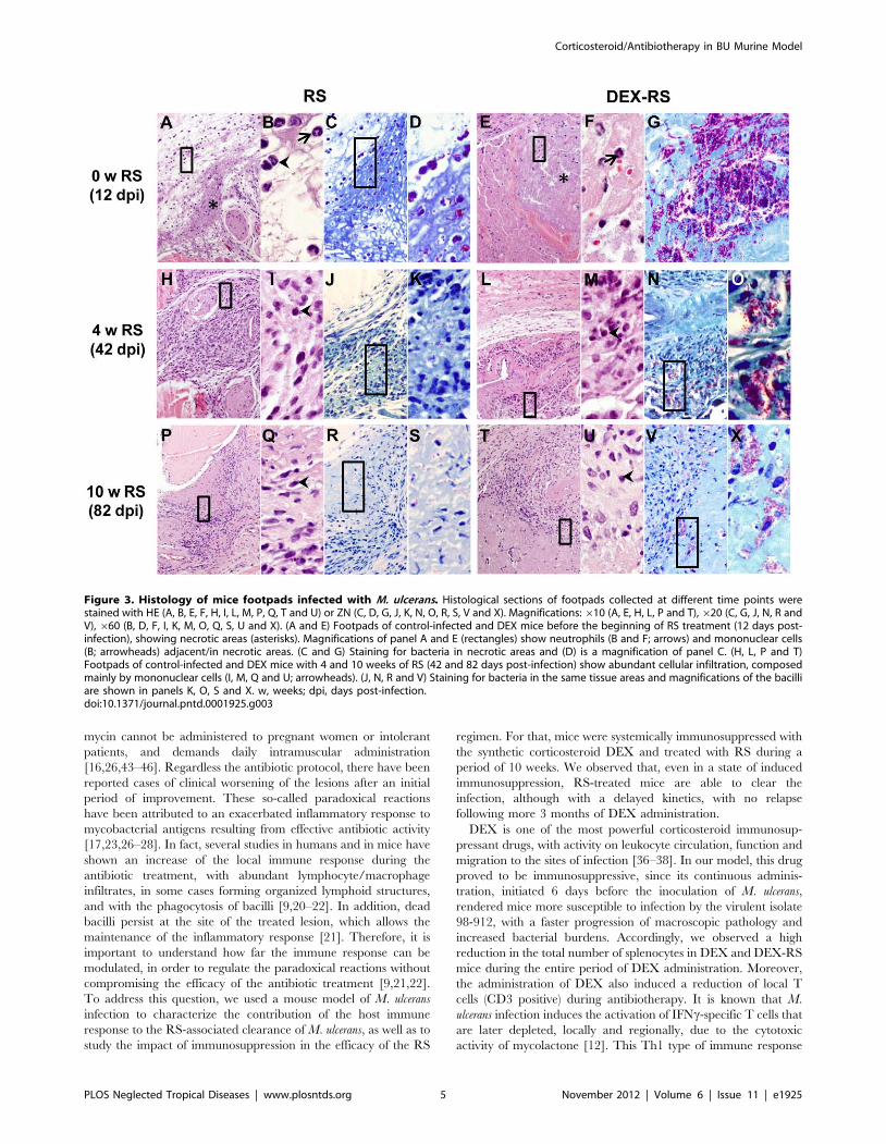
To assess the contribution of immune mechanisms to the
clearance of M. ulcerans, we analyzed the histopathology at the site
of infection in immunocompetent vs. DEX-treated mice. As
previously described [21], at day 12 post-infection the presence of
central necrotic areas with extracellular bacilli surrounded by a
predominantly neutrophilic/macrophagic infiltrate (Figure 3A–D)
are histological features of a progressive subcutaneous infection
with virulent M. ulcerans 98-912. On the other hand, during RS
treatment we observed a switch of the inflammatory profile to
abundant lymphocytic/macrophagic infiltrates, which was main-
tained until the end of the experimental period (10 weeks post-
infection) (Figure 3H–I and 3P–Q).
In comparison to control-infected mice (Figure 3A), footpad
tissue of DEX mice presented widespread necrosis (Figure 3E)
associated with massive clumps of extracellular bacilli (Figure 3G),
which is consistent with the higher bacterial burden (Figure 2B).
The pattern of the inflammatory response in this group of
immunosuppressed mice was similar to control-infected mice, with
neutrophils adjacent and/or in necrotic areas (Figure 3B and 3F).
In immunosuppressed mice submitted to antibiotherapy (DEX-
RS mice) after 4 weeks of RS administration (42 days post-
infection), the increased bacterial burdens, as compared with RS
mice, was reflected in the higher number of clumps of extracellular
bacilli (Figure 3J–K and 3N–O). Despite the higher bacterial
burden, inflammatory infiltrates showed a similar profile to
immunocompetent RS mice (Figure 3H–I and 3L–M), character-
ized by an increase of a predominantly mononuclear infiltrate, as
compared to non-treated mice (Figure 3E–F). This profile was
maintained at the end of treatment (Figure 3P–Q and 3T–U).
Given the known immunosuppressive and anti-inflammatory
properties of DEX, namely the inhibition of inflammatory cell
recruitment, including neutrophils and lymphocytes, to the focus
of infection [37,38,41,42], we next analyzed if there were
differences in these cell populations in infected footpads. We
observed that after 4 weeks of RS treatment (42 days post-
infection), despite the similar amounts of inflammatory infiltrates
observed in slides stained with HE (Figures 3H–I and 3L–M),
DEX-RS mice presented a lower number of T cells stained by
immunohistochemistry (Figure 4B) as compared with RS mice
(Figure 4A). The quantification of T cells confirmed the
histological observations, with a median distribution of 369
cells/mm2 of inflammatory area in RS mice, whereas the DEX-
RS group only showed 110 cells/mm2 (Figure 4E; P,0.001) In
addition, in the DEX-RS group, most of the staining for the
neutrophilic marker Ly-6G was observed in the remaining
necrotic tissue with cell debris, and only few intact cells were
stained in the peripheries of the lesion (Figure 4D), when
compared with RS mice for which intact neutrophils were mainly
found at the peripheral areas (Figure 4C). The quantification of
these cells showed a distribution of 506 vs. 279 cells/mm2 of
inflammatory area in the RS and DEX-RS group of mice,
respectively (Figure 4F; P,0.01).
These data show that corticosteroid-induced immunosuppres-
sion, associated with increased M. ulcerans proliferation, results in
increased necrosis at infection foci. Following RS administration,
bacterial clearance ensues in immunosuppressed mice, although
with slower kinetics, which is associated with lower T cell numbers.
Discussion
The recent regimen with RS, introduced by the WHO in 2004,
has been proven effective in BU patients with nonulcerative or
small ulcers, but variation in efficacy is reported for more
advanced lesions [14–19]. Improvements to this protocol have
been tested, such as the introduction of a fully oral antibiotic
regimen replacing streptomycin by clarithromycin, since strepto-
Figure 2. Lesion progression and bacterial proliferation in the footpad of mice infected with M. ulcerans. Mice were administrated DEX(squares) or vehicle (circles) and were left untreated (closed symbols) or treated with RS (open symbols) for 10 weeks. (A) Lesion progression wasassessed by measurement of footpad swelling (n = 12–20). (B) Bacterial proliferation was assessed by CFU counts (n = 4–8). Asterisks representsignificant differences between control-infected and DEX mice (***, P,0.001). Cardinals represent significant differences between RS and DEX-RSmice (#, P,0.05; ##, P,0.01, ###, P,0.001). Grey bar represents the period of DEX administration. Striped bar represents the period of RSadministration. { Mice were euthanized for ethical reasons after the emergence of ulceration. n.d., not detected for the RS group of mice; n.d. (ingrey), not detected for the DEX-RS group of mice. Data points represent the mean 6 SEM.doi:10.1371/journal.pntd.0001925.g002
Corticosteroid/Antibiotherapy in BU Murine Model
PLOS Neglected Tropical Diseases | www.plosntds.org 4 November 2012 | Volume 6 | Issue 11 | e1925
mycin cannot be administered to pregnant women or intolerant
patients, and demands daily intramuscular administration
[16,26,43–46]. Regardless the antibiotic protocol, there have been
reported cases of clinical worsening of the lesions after an initial
period of improvement. These so-called paradoxical reactions
have been attributed to an exacerbated inflammatory response to
mycobacterial antigens resulting from effective antibiotic activity
[17,23,26–28]. In fact, several studies in humans and in mice have
shown an increase of the local immune response during the
antibiotic treatment, with abundant lymphocyte/macrophage
infiltrates, in some cases forming organized lymphoid structures,
and with the phagocytosis of bacilli [9,20–22]. In addition, dead
bacilli persist at the site of the treated lesion, which allows the
maintenance of the inflammatory response [21]. Therefore, it is
important to understand how far the immune response can be
modulated, in order to regulate the paradoxical reactions without
compromising the efficacy of the antibiotic treatment [9,21,22].
To address this question, we used a mouse model of M. ulcerans
infection to characterize the contribution of the host immune
response to the RS-associated clearance of M. ulcerans, as well as to
study the impact of immunosuppression in the efficacy of the RS
regimen. For that, mice were systemically immunosuppressed with
the synthetic corticosteroid DEX and treated with RS during a
period of 10 weeks. We observed that, even in a state of induced
immunosuppression, RS-treated mice are able to clear the
infection, although with a delayed kinetics, with no relapse
following more 3 months of DEX administration.
DEX is one of the most powerful corticosteroid immunosup-
pressant drugs, with activity on leukocyte circulation, function and
migration to the sites of infection [36–38]. In our model, this drug
proved to be immunosuppressive, since its continuous adminis-
tration, initiated 6 days before the inoculation of M. ulcerans,
rendered mice more susceptible to infection by the virulent isolate
98-912, with a faster progression of macroscopic pathology and
increased bacterial burdens. Accordingly, we observed a high
reduction in the total number of splenocytes in DEX and DEX-RS
mice during the entire period of DEX administration. Moreover,
the administration of DEX also induced a reduction of local T
cells (CD3 positive) during antibiotherapy. It is known that M.
ulcerans infection induces the activation of IFNc-specific T cells that
are later depleted, locally and regionally, due to the cytotoxic
activity of mycolactone [12]. This Th1 type of immune response
Figure 3. Histology of mice footpads infected with M. ulcerans. Histological sections of footpads collected at different time points werestained with HE (A, B, E, F, H, I, L, M, P, Q, T and U) or ZN (C, D, G, J, K, N, O, R, S, V and X). Magnifications:610 (A, E, H, L, P and T),620 (C, G, J, N, R andV), 660 (B, D, F, I, K, M, O, Q, S, U and X). (A and E) Footpads of control-infected and DEX mice before the beginning of RS treatment (12 days post-infection), showing necrotic areas (asterisks). Magnifications of panel A and E (rectangles) show neutrophils (B and F; arrows) and mononuclear cells(B; arrowheads) adjacent/in necrotic areas. (C and G) Staining for bacteria in necrotic areas and (D) is a magnification of panel C. (H, L, P and T)Footpads of control-infected and DEX mice with 4 and 10 weeks of RS (42 and 82 days post-infection) show abundant cellular infiltration, composedmainly by mononuclear cells (I, M, Q and U; arrowheads). (J, N, R and V) Staining for bacteria in the same tissue areas and magnifications of the bacilliare shown in panels K, O, S and X. w, weeks; dpi, days post-infection.doi:10.1371/journal.pntd.0001925.g003
Corticosteroid/Antibiotherapy in BU Murine Model
PLOS Neglected Tropical Diseases | www.plosntds.org 5 November 2012 | Volume 6 | Issue 11 | e1925

was proven to be important for protection against M. ulcerans
strains of lower virulence, as shown by the higher susceptibility of
mice deficient in either T cells or IFNc [7,12]. Therefore, the
lower number of T cells in the footpads of mice treated with DEX
is expected to contribute to the host susceptibility to infection in
the present model. On the other hand, T cell survival is allowed
during RS administration, in association with the decline of viable
bacilli.
The fact that DEX-RS mice presented a delayed clearance of
viable bacteria in the footpad lesions suggests a role of the immune
response in the efficacy of the antibiotic regimen. Such a type of
immune participation is suggested in another experimental model
of antibiotherapy in mice infected with Mycobacterium avium
complex, where treatment with sparfloxacin and ethambutol is
enhanced by combination with an inhibitor of the cortisol receptor
[47]. However, it is also important to stress that, at the beginning
of the RS regimen, immunosuppressed mice already presented a
higher bacterial burden associated with more severe histopathol-
ogy, which may also hamper the diffusion of the antibiotics to the
core of the lesion. Nevertheless, despite the higher bacterial load
and suppressed local inflammatory responses in the footpad of
immunosuppressed mice (with lower numbers of T cells and
neutrophils), the antibiotic regimen was able to clear the infection
after 10 weeks of administration. Indeed, no relapse was observed
after an additional 3 months of DEX administration. This points
out that the bactericidal activity of the drug is the main factor in
the resolution of the infection.
In addition to the analysis of the treatment efficacy in mice
administered with DEX, it would have been interesting to test
specifically if corticosteroids or other immunosuppressive/anti-
inflammatory drugs could control paradoxical reactions during or
after antibiotherapy. However, there is currently no proper animal
model to study paradoxical reactions. Corticosteroids are being
successfully used in the management of other types of paradoxical
reactions, for instance in patients with tuberculosis presenting
severe forms that must be treated, and when surgery of the affected
area is unwanted or difficult/risky to perform, such as in the
central nervous system [29,34]. The fact that the antibiotic
treatment was efficient in our model, even in mice administered
with DEX, suggests that the use of corticosteroids in BU patients
undergoing severe paradoxical reactions may not represent a risk
of reactivation/treatment failure. However, more studies are
needed to address this point, especially when regarding the
management of more severe lesions, where culture positivity is
sometimes detected at the end of antibiotic treatment [16,17]. On
the other hand, monitoring the persistence of AFB in lesions is also
Figure 4. Immunohistochemistry of mice footpads infected with M. ulcerans. Histological sections of footpads of control-infected and DEXmice at 4 weeks of RS treatment (42 days post-infection) were stained for the antigen marker CD3 of T cells (A and B) or Ly-6G of neutrophils (C andD), and with ZN to visualize the bacilli. Magnifications:620. Footpads of RS mice (A) show increased staining for T cells in comparison to DEX-RS mice(B). Staining for Ly-6G in RS mice (C) is located in necrotic areas (arrow) and in cells interspersed in the inflammatory infiltrates. In comparison,footpads of DEX-RS mice (D) show staining for Ly-6G mainly in necrotic areas (arrow), and fewer stained cells appear scattered in the tissue. Thenumber of CD3 (E) and Ly-6G positive cells (F) per mm2 of inflammatory area in the stained tissue sections was quantified by using a 206objectivelens. Asterisks represent significant differences between RS and DEX-RS mice (**, P,0.01; ***, P,0.001). Data points represent the mean 6 SEM of 2different histological sections of each mouse footpad sample, in a total of 3 footpads per group.doi:10.1371/journal.pntd.0001925.g004
Corticosteroid/Antibiotherapy in BU Murine Model
PLOS Neglected Tropical Diseases | www.plosntds.org 6 November 2012 | Volume 6 | Issue 11 | e1925

a feature to be considered for the management of BU patients with
paradoxical reactions submitted to corticotherapy, since the end of
the immunosuppressed-induced state could be followed by an
exacerbated up-regulation of the immune response.
Although the use of corticosteroids in our mouse model does not
compromise the efficacy of antibiotic treatment, we should stress
that in humans the use of these drugs should be considered with
caution. Several side effects are associated with corticosteroids,
such as the development of metabolic alterations like hyperglyce-
mia or adrenal atrophy, or even impaired wound healing, but
these effects are dependent on the type of corticosteroid used,
doses and the time of administration [48,49]. Patients receiving
corticosteroids are also at risk of developing opportunistic or
reactivating infections, like strongyloidiasis, tuberculosis, fungal
infections and cytomegalovirus [48,50]. However, a randomized
placebo-controlled clinical trial in South Africa on the systemic use
of corticosteroids to control paradoxical tuberculosis-associated
immune-reconstitution inflammatory syndrome in HIV-infected
patients receiving antitubercular and antiretroviral therapy,
showed beneficial activity in ameliorating the symptoms with
minimal side-effects, when a low and short-term therapy with
prednisone was used [51]. The authors advise, though, that
excluded diagnosis of multidrug-resistant tuberculosis or Kaposi’s
sarcoma should be performed before starting corticosteroids [51].
Therefore, a possible use of corticosteroids in BU patients or other
alternative management strategies justifies clinical investigation
and deserves consideration, depending on the severity of the case,
potential side effects and evaluation of the risk/benefit ratio. Like
in tuberculosis patients, paradoxical reactions in BU patients are
transient, but in some cases these result in a considerable
enlargement of the lesions and a prolonged period to achieve
healing [28]. A strategy to avoid or improve such outcome during
or after antibiotherapy would be desirable. Although we did not
test other immunosuppressive/anti-inflammatory drugs, our study
may also open possibilities to study the management of more
severe paradoxical reactions with drugs that, for instance, could be
applied locally, thus minimizing systemic effects and avoiding the
need of surgery.
In summary, corticosteroid-induced immunosuppression during
experimental M. ulcerans infection, although delaying bacterial
clearance, does not ultimately compromise the efficacy of the
WHO recommended RS regimen. This observation may be
explained by a major role of the bactericidal activity of RS that
overlaps the activity of the local immune response. This study
justifies future clinical studies on the potential use of corticosteroids
or other immunosuppressive/anti-inflammatory drugs in the
management of BU patients undergoing paradoxical reactions.
Acknowledgments
The authors would like to thank Luis Martins, Deolinda Teixeira and
Miguel Carneiro for laboratory assistance.
Author Contributions
Conceived and designed the experiments: TGM MS MTS AGC JP.
Performed the experiments: TGM GT AGF JBG ALF. Analyzed the data:
TGM ALF MS MTS AGC JP. Contributed reagents/materials/analysis
tools: TGM MS MTS AGC JP. Wrote the paper: TGM AGF ALF MS
MTS AGC JP.
References
1. Portaels F, Silva MT, Meyers WM (2009) Buruli ulcer. Clin Dermatol 27: 291–
305.
2. Walsh DS, Portaels F, Meyers WM (2011) Buruli ulcer: Advances in
understanding Mycobacterium ulcerans infection. Dermatol Clin 29: 1–8.
3. George KM, Pascopella L, Welty DM, Small PL (2000) A Mycobacterium ulcerans
toxin, mycolactone, causes apoptosis in guinea pig ulcers and tissue culture cells.
Infect Immun 68: 877–883.
4. George KM, Chatterjee D, Gunawardana G, Welty D, Hayman J, et al. (1999)
Mycolactone: a polyketide toxin from Mycobacterium ulcerans required for
virulence. Science 283: 854–857.
5. Hong H, Demangel C, Pidot SJ, Leadlay PF, Stinear T (2008) Mycolactones:
immunosuppressive and cytotoxic polyketides produced by aquatic mycobacte-
ria. Nat Prod Rep 25: 447–454.
6. Torrado E, Adusumilli S, Fraga AG, Small PL, Castro AG, et al. (2007)
Mycolactone-mediated inhibition of tumor necrosis factor production by
macrophages infected with Mycobacterium ulcerans has implications for the control
of infection. Infect Immun 75: 3979–3988.
7. Torrado E, Fraga AG, Logarinho E, Martins TG, Carmona JA, et al. (2010)
IFN-gamma-dependent activation of macrophages during experimental infec-
tions by Mycobacterium ulcerans is impaired by the toxin mycolactone. J Immunol
184: 947–955.
8. Oliveira MS, Fraga AG, Torrado E, Castro AG, Pereira JP, et al. (2005)
Infection with Mycobacterium ulcerans induces persistent inflammatory responses in
mice. Infect Immun 73: 6299–6310.
9. Schutte D, Um-Boock A, Mensah-Quainoo E, Itin P, Schmid P, et al. (2007)
Development of highly organized lymphoid structures in Buruli ulcer lesions
after treatment with rifampicin and streptomycin. PLoS Negl Trop Dis
1: e2.
10. Kiszewski AE, Becerril E, Aguilar LD, Kader IT, Myers W, et al. (2006) The
local immune response in ulcerative lesions of Buruli disease. Clin Exp Immunol
143: 445–451.
11. Torrado E, Fraga AG, Castro AG, Stragier P, Meyers WM, et al. (2007)
Evidence for an intramacrophage growth phase of Mycobacterium ulcerans. Infect
Immun 75: 977–987.
12. Fraga AG, Cruz A, Martins TG, Torrado E, Saraiva M, et al. (2011)
Mycobacterium ulcerans triggers T cell immunity followed by local and regional but
not systemic immunosuppression. Infect Immun 79: 421–430.
13. World Health Organization (2004) Provisional guidance on the role of specific
antibiotics in the management of Mycobacterium ulcerans disease (Buruli ulcer).
World Health Organization WHO/CDS/CPE/GBUI/2004.10: 33p
14. Etuaful S, Carbonnelle B, Grosset J, Lucas S, Horsfield C, et al. (2005) Efficacy
of the combination rifampin-streptomycin in preventing growth of Mycobacterium
ulcerans in early lesions of Buruli ulcer in humans. Antimicrob Agents Chemother
49: 3182–3186.
15. Chauty A, Ardant MF, Adeye A, Euverte H, Guedenon A, et al. (2007)
Promising clinical efficacy of streptomycin-rifampin combination for treatment
of Buruli ulcer (Mycobacterium ulcerans disease). Antimicrob Agents Chemother 51:
4029–4035.
16. Nienhuis WA, Stienstra Y, Thompson WA, Awuah PC, Abass KM, et al. (2010)
Antimicrobial treatment for early, limited Mycobacterium ulcerans infection: a
randomised controlled trial. Lancet 375: 664–672.
17. Sarfo FS, Phillips R, Asiedu K, Ampadu E, Bobi N, et al. (2010) Clinical efficacy
of combination of rifampin and streptomycin for treatment of Mycobacterium
ulcerans disease. Antimicrob Agents Chemother 54: 3678–3685.
18. Kibadi K, Boelaert M, Fraga AG, Kayinua M, Longatto-Filho A, et al. (2010)
Response to treatment in a prospective cohort of patients with large ulcerated
lesions suspected to be Buruli ulcer (Mycobacterium ulcerans disease). PLoS Negl
Trop Dis 4: e736.
19. Sopoh GE, Dossou AD, Brun LV, Barogui YT, Houezo JG, et al. (2010) Severe
multifocal form of Buruli ulcer after streptomycin and rifampin treatment: comments
on possible dissemination mechanisms. Am J Trop Med Hyg 83: 307–313.
20. Ruf MT, Schutte D, Chauffour A, Jarlier V, Ji B, et al. (2012) Chemotherapy-
associated changes of histopathological features of Mycobacterium ulcerans lesions in
a Buruli ulcer mouse model. Antimicrob Agents Chemother 56: 687–696.
21. Martins TG, Gama JB, Fraga AG, Saraiva M, Silva MT, et al. (2012) Local and
regional re-establishment of cellular immunity during curative antibiotherapy of
murine Mycobacterium ulcerans infection. PLoS One 7: e32740.
22. Schutte D, Umboock A, Pluschke G (2009) Phagocytosis of Mycobacterium ulcerans
in the course of rifampicin and streptomycin chemotherapy in Buruli ulcer
lesions. Br J Dermatol 160: 273–283.
23. Ruf MT, Chauty A, Adeye A, Ardant MF, Koussemou H, et al. (2011)
Secondary Buruli ulcer skin lesions emerging several months after completion of
chemotherapy: paradoxical reaction or evidence for immune protection? PLoS
Negl Trop Dis 5: e1252.
24. Dega H, Robert J, Bonnafous P, Jarlier V, Grosset J (2000) Activities of several
antimicrobials against Mycobacterium ulcerans infection in mice. Antimicrob Agents
Chemother 44: 2367–2372.
25. Dega H, Bentoucha A, Robert J, Jarlier V, Grosset J (2002) Bactericidal activity
of rifampin-amikacin against Mycobacterium ulcerans in mice. Antimicrob Agents
Chemother 46: 3193–3196.
Corticosteroid/Antibiotherapy in BU Murine Model
PLOS Neglected Tropical Diseases | www.plosntds.org 7 November 2012 | Volume 6 | Issue 11 | e1925

26. Gordon CL, Buntine JA, Hayman JA, Lavender CJ, Fyfe JA, et al. (2010) All-
oral antibiotic treatment for Buruli ulcer: a report of four patients. PLoS NeglTrop Dis 4: e770.
27. O’Brien DP, Robson ME, Callan PP, McDonald AH (2009) ‘‘Paradoxical’’
immune-mediated reactions to Mycobacterium ulcerans during antibiotic treatment:a result of treatment success, not failure. Med J Aust 191: 564–566.
28. Nienhuis WA, Stienstra Y, Abass KM, Tuah W, Thompson WA, et al. (2012)Paradoxical responses after start of antimicrobial treatment in Mycobacterium
ulcerans infection. Clin Infect Dis 54: 519–526.
29. Cheng VC, Ho PL, Lee RA, Chan KS, Chan KK, et al. (2002) Clinicalspectrum of paradoxical deterioration during antituberculosis therapy in non-
HIV-infected patients. Eur J Clin Microbiol Infect Dis 21: 803–809.30. Breen RA, Smith CJ, Bettinson H, Dart S, Bannister B, et al. (2004) Paradoxical
reactions during tuberculosis treatment in patients with and without HIV co-infection. Thorax 59: 704–707.
31. Hawkey CR, Yap T, Pereira J, Moore DA, Davidson RN, et al. (2005)
Characterization and management of paradoxical upgrading reactions in HIV-uninfected patients with lymph node tuberculosis. Clin Infect Dis 40: 1368–
1371.32. Cho OH, Park KH, Kim T, Song EH, Jang EY, et al. (2009) Paradoxical
responses in non-HIV-infected patients with peripheral lymph node tuberculosis.
J Infect 59: 56–61.33. Jung JW, Shin JW, Kim JY, Park IW, Choi BW, et al. (2011) Risk factors for
development of paradoxical response during anti-tuberculosis treatment in HIV-negative patients with pleural tuberculosis. Tohoku J Exp Med 223: 199–204.
34. Nicolls DJ, King M, Holland D, Bala J, del Rio C (2005) Intracranialtuberculomas developing while on therapy for pulmonary tuberculosis. Lancet
Infect Dis 5: 795–801.
35. Garcia Vidal C, Garau J (2005) Systemic steroid treatment of paradoxicalupgrading reaction in patients with lymph node tuberculosis. Clin Infect Dis 41:
915–916; author reply 916-917.36. Boumpas DT, Chrousos GP, Wilder RL, Cupps TR, Balow JE (1993)
Glucocorticoid therapy for immune-mediated diseases: basic and clinical
correlates. Ann Intern Med 119: 1198–1208.37. Parrillo JE, Fauci AS (1979) Mechanisms of glucocorticoid action on immune
processes. Annu Rev Pharmacol Toxicol 19: 179–201.38. Yao LC, Baluk P, Feng J, McDonald DM (2010) Steroid-resistant lymphatic
remodeling in chronically inflamed mouse airways. Am J Pathol 176: 1525–1541.
39. Shepard CC, McRae DH (1968) A method for counting acid-fast bacteria.
Int J Lepr Other Mycobact Dis 36: 78–82.
40. Lefrancois S, Robert J, Chauffour A, Ji B, Jarlier V (2007) Curing Mycobacterium
ulcerans infection in mice with a combination of rifampin-streptomycin or
rifampin-amikacin. Antimicrob Agents Chemother 51: 645–650.
41. Cronstein BN, Kimmel SC, Levin RI, Martiniuk F, Weissmann G (1992) A
mechanism for the antiinflammatory effects of corticosteroids: the glucocorticoid
receptor regulates leukocyte adhesion to endothelial cells and expression of
endothelial-leukocyte adhesion molecule 1 and intercellular adhesion molecule
1. Proc Natl Acad Sci U S A 89: 9991–9995.
42. Pitzalis C, Pipitone N, Bajocchi G, Hall M, Goulding N, et al. (1997)
Corticosteroids inhibit lymphocyte binding to endothelium and intercellular
adhesion: an additional mechanism for their anti-inflammatory and immuno-
suppressive effect. J Immunol 158: 5007–5016.
43. Chauty A, Ardant MF, Marsollier L, Pluschke G, Landier J, et al. (2011) Oral
treatment for Mycobacterium ulcerans infection: results from a pilot study in Benin.
Clin Infect Dis 52: 94–96.
44. Almeida D, Converse PJ, Ahmad Z, Dooley KE, Nuermberger EL, et al. (2011)
Activities of rifampin, Rifapentine and clarithromycin alone and in combination
against Mycobacterium ulcerans disease in mice. PLoS Negl Trop Dis 5: e933.
45. Ji B, Chauffour A, Robert J, Lefrancois S, Jarlier V (2007) Orally administered
combined regimens for treatment of Mycobacterium ulcerans infection in mice.
Antimicrob Agents Chemother 51: 3737–3739.
46. O’Brien DP, McDonald A, Callan P, Robson M, Friedman ND, et al. (2012)
Successful outcomes with oral fluoroquinolones combined with rifampicin in the
treatment of Mycobacterium ulcerans: an observational cohort study. PLoS Negl
Trop Dis 6: e1473.
47. Perronne C, Cohen Y, Truffot-Pernot C, Grosset J, Vilde JL, et al. (1992)
Sparfloxacin, ethambutol, and cortisol receptor inhibitor RU-40 555 treatment
for disseminated Mycobacterium avium complex infection of normal C57BL/6
mice. Antimicrob Agents Chemother 36: 2408–2412.
48. Schacke H, Docke WD, Asadullah K (2002) Mechanisms involved in the side
effects of glucocorticoids. Pharmacol Ther 96: 23–43.
49. Burns JL, Mancoll JS, Phillips LG (2003) Impairments to wound healing. Clin
Plast Surg 30: 47–56.
50. Fardet L, Genereau T, Poirot JL, Guidet B, Kettaneh A, et al. (2007) Severe
strongyloidiasis in corticosteroid-treated patients: case series and literature
review. J Infect 54: 18–27.
51. Meintjes G, Wilkinson RJ, Morroni C, Pepper DJ, Rebe K, et al. (2010)
Randomized placebo-controlled trial of prednisone for paradoxical tuberculosis-
associated immune reconstitution inflammatory syndrome. AIDS 24: 2381–
2390.
Corticosteroid/Antibiotherapy in BU Murine Model
PLOS Neglected Tropical Diseases | www.plosntds.org 8 November 2012 | Volume 6 | Issue 11 | e1925