Low-dose immunosuppression in a rat hind-limb transplantation model
8
Gustavo Perez-Abadia Luis Laurentin-Perez Vijay S. Gorantla Cedric G. Francois Marieke Vossen Pascal C.R. Brouha Haldun I. Orhun Gary L. Anderson Claudio Maldonado Diane J. Pidwell Warren C. Breidenbach John H. Barker Received: 10 April 2002 Revised: 28 August 2002 Accepted: 5 September 2002 Published online: 2 August 2003 0 Springer-Verlag 2003 G. Perez-Abadia . L. Laurentin-Perez V.S. Gorantla . C.G. Francois M. Vossen P.C.R. Brouha . H.I. Orhun C. Maldonado . J.H. Barker (Ixi) Division of Plastic and Reconstructive Surgery, Department of Surgery, University of Louisville, 5 1 1 South Floyd Street, 320 MDR Building, Louisville, KY 40292, USA E-mail: jhbarkOl @louisville.edu Tel.: + 1-502-8520166 Fax: + 1-502-8524675 G.L. Anderson Department of Physiology and Biophysics, University of Louisville, Louisville, Kentucky USA D.J. Pidwell Histopathology Laboratory, Department of Pathology, Jewish Hospital, Louisville, Kentucky USA W.C. Breidenbach Christine M. Kleinert Institute, Louisville, KentuckyUSA low-dose immunosuppression in a rat hind-limb transplantation model Abstract Composite tissue allografts (CTAs) offer an alternative to con- ventional reconstructive methods. However, the toxicity of the drugs that are required to prevent rejection has prevented its widespread clinical application. The purpose of this study was to determine whether a low-dose, corticosteroid-free combi- nation regimen of tacrolimus and mycophenolate mofetil (MMF) would prevent rejection in a rat hind-limb model, with minimal toxic side effects. Three groups were used in this study. In group I, Wistar Furth (WF) rats received a syngeneic WF hind-limb. In groups I1 and 111, WF rats received an ACI hind-limb. The latter were treated with tacroli- mus-MMF. Assessment for rejec- tion, flow cytometry, and mixed lymphocyte reactions was per- formed. Biopsies were taken regu- larly and at the time of killing. Combination therapy with low-dose tacrolimus-MMF effectively pro- longed CTA survival indefinitely, with minimal side effects. Toxicity associated with immunosuppressive drugs can be avoided in a low-dose combination corticosteroid-free regimen. Keywords Composite tissue allografts . Tacrolimus . Mycophenolate mofetil . Combina- tion therapy . Rats Introduction Transplantation of composite tissue allografts (CTAs) from cadaveric donors offers an excellent alternative to conventional reconstructive methods for repairing large tissue defects that result from traumatic injury, tumor extirpation, and congenital birth defects. In spite of its promising potential, composite tissue allotransplanta- tion has not been applied widely in the clinical setting, primarily due to the toxicity associated with the immu- nosuppressive drugs that are needed to prevent graft rejection in these procedures. This toxicity is not necessarily due to the immuno- suppressive drugs per se but rather to the high doses that are required to prevent rejection and ensure long-term survival of the highly immunogenic skin component of CTAs. The risks associated with high-dose immuno- suppression, together with the fact that CTA procedures
-
Upload
independent -
Category
Documents
-
view
0 -
download
0
Transcript of Low-dose immunosuppression in a rat hind-limb transplantation model
Gustavo Perez-Abadia Luis Laurentin-Perez Vijay S. Gorantla Cedric G. Francois Marieke Vossen Pascal C.R. Brouha Haldun I. Orhun Gary L. Anderson Claudio Maldonado Diane J. Pidwell Warren C. Breidenbach John H. Barker
Received: 10 April 2002 Revised: 28 August 2002 Accepted: 5 September 2002 Published online: 2 August 2003 0 Springer-Verlag 2003
G. Perez-Abadia . L. Laurentin-Perez V.S. Gorantla . C.G. Francois M. Vossen P.C.R. Brouha . H.I. Orhun C. Maldonado . J.H. Barker (Ixi) Division of Plastic and Reconstructive Surgery, Department of Surgery, University of Louisville, 5 1 1 South Floyd Street, 320 MDR Building, Louisville, KY 40292, USA E-mail: jhbarkOl @louisville.edu Tel.: + 1-502-8520166 Fax: + 1-502-8524675
G.L. Anderson Department of Physiology and Biophysics, University of Louisville, Louisville, Kentucky USA
D.J. Pidwell Histopathology Laboratory, Department of Pathology, Jewish Hospital, Louisville, Kentucky USA
W.C. Breidenbach Christine M. Kleinert Institute, Louisville, KentuckyUSA
low-dose immunosuppression in a rat hind-limb transplantation model
Abstract Composite tissue allografts (CTAs) offer an alternative to con- ventional reconstructive methods. However, the toxicity of the drugs that are required to prevent rejection has prevented its widespread clinical application. The purpose of this study was to determine whether a low-dose, corticosteroid-free combi- nation regimen of tacrolimus and mycophenolate mofetil (MMF) would prevent rejection in a rat hind-limb model, with minimal toxic side effects. Three groups were used in this study. In group I, Wistar Furth (WF) rats received a syngeneic WF hind-limb. In groups I1 and 111, WF rats received an ACI hind-limb. The latter were treated with tacroli- mus-MMF. Assessment for rejec- tion, flow cytometry, and mixed lymphocyte reactions was per- formed. Biopsies were taken regu- larly and at the time of killing. Combination therapy with low-dose tacrolimus-MMF effectively pro- longed CTA survival indefinitely,
with minimal side effects. Toxicity associated with immunosuppressive drugs can be avoided in a low-dose combination corticosteroid-free regimen.
Keywords Composite tissue allografts . Tacrolimus . Mycophenolate mofetil . Combina- tion therapy . Rats
Introduction
Transplantation of composite tissue allografts (CTAs) from cadaveric donors offers an excellent alternative to conventional reconstructive methods for repairing large tissue defects that result from traumatic injury, tumor extirpation, and congenital birth defects. In spite of its promising potential, composite tissue allotransplanta- tion has not been applied widely in the clinical setting,
primarily due to the toxicity associated with the immu- nosuppressive drugs that are needed to prevent graft rejection in these procedures.
This toxicity is not necessarily due to the immuno- suppressive drugs per se but rather to the high doses that are required to prevent rejection and ensure long-term survival of the highly immunogenic skin component of CTAs. The risks associated with high-dose immuno- suppression, together with the fact that CTA procedures
836
would be used to treat non-life-threatening tissue de- fects, have raised the question “are the risks worth the benefits of these new procedures?” This risk-versus- benefit debate is, perhaps, the primary reason why this promising new reconstructive procedure has not gained widespread clinical application.
The ultimate goal of transplantation research is to replace toxic immunosuppressive drugs with a method of inducing transplantation tolerance [33]. Until trans- plantation tolerance becomes a clinical reality, the reduction of the toxicity of current immunosuppressive regimens is an approach that is worth pursuing. One such approach is the use of combination immunosup- pression therapy, which allows lower doses of individual drugs to be used and, thus, causes less toxicity [18].
In animal composite tissue allotransplantation stud- ies, different combinations of immunosuppressive drugs have been used with varying success. Using a rat hind- limb transplant model, various investigators reported that combinations of tacrolimus and rapamycin [15] or tacrolimus and deoxyspergualin (DSG) [30] prolonged CTA survival. Benhaim et al. demonstrated indefinite limb survival in a fully mismatched rodent model using combination therapy with cyclosporin A (CsA; 1.5 mg/ kg per day) and mycophenolate mofetil (MMF; 15 mg/ kg per day) [7]. However, at these low doses the inves- tigators still reported episodes of rejection in 1 1 % of their animals [7]. Based on the fact that tacrolimus has been demonstrated to have 100 times the immunosup- pressive effect of CsA at equivalent doses [4, 121, it could be expected that the substitution of tacrolimus for CsA in the above-mentioned study could provide improved survival of CTAs in a similar model. The purpose of the present study was to determine in a rat hind-limb CTA model whether low-dose tacrolimus that was adminis- tered in combination with MMF would prevent rejec- tion and minimize toxic side effects.
Materials and methods
Wistar Furth (WF) rats received hind-limbs transplanted from ACI donor rats and were allocated to one of three groups: group I (syngeneic), group I1 (allogeneic, not treated), and group I11 (allogeneic, treated with tacrolimus and MMF combination immunotherapy). Rat limb rejection was assessed daily by visual inspection and by scheduled skin and muscle biopsies. At the end of the study, a histopathological examination was also performed on all tissues. Flow cytometry analysis was performed to detect the presence of donor chimerism and mixed lymphocyte reaction (MLR) for in vitro assessment of tolerance.
Animal care
Animals were kept in separate cages in temperature-controlled (24 “C), light-regulated (12 h/day), and air flow-regulated rooms. They were provided with a balanced rodent diet and water ad libitum. The animals were anesthetized with sodium pentobarbital (50 mg/kg, i.p.) for all surgical procedures, and sterile techniques
were used for all surgery. Upon completion of the experiments, the rats were killed with an overdose of sodium pentobarbital. The study was performed in accordance with the guidelines of the Animal Care and Use Committee of the University of Louisville School of Medicine and the Guide for the Cure and Use of Labo- ratory Animals (Department of Health and Human Services, Publication No. [NIH] 8&23).
Animal model
Strong major histocompatibility complex (MHC) mismatch male rats (weighing 200-250 g) were used in this study. ACI rats (RTIAb) as donors and W F (RTIAU) rats as recipients were pur- chased from Harlan Sprague Dawley (Indianapolis, IN., USA). Twenty rats were used in this study and were allocated to one of three groups: group I (n=4), WF rats received syngeneic hind- limbs from na WF rats; group I1 (n=6), WF rats received alloge- neic hind-limbs from na ACI rats without immunosuppression regimen; and group 111 (n = lo), WF rats received allogeneic hind- limbs from ACI rats and were treated with tacrolimus and MMF.
Donor surgery
A circumferential skin incision was made just proximal to the mid- thigh area. The femoral artery, vein, and nerve were dissected, and the individual muscle groups of the hind-limb were identified and divided as proximally as possible to their tendinous origins. Care was taken not to injure the profunda femoris vein. The sciatic nerve was identified and divided. The femur was exposed and divided transversely at mid-shaft by use of a handle saw. The donor rat was then given the anticoagulant, heparin (50 U), (Elkins-Sinn, Cherry Hill, NJ., USA), which was injected intravenously into the opposite femoral vein. After 10 min, the femoral artery was clamped as proximally as possible and cannulated with a 24-gauge catheter. The limb was flushed with a solution of heparinized Ringer’s lac- tate (1 U of heparin per ml of Ringer’s solution) through the cannulated artery. Vascular flushing was maintained for 10 min until the backflow through the vein was observed to be clear. The femoral vein was ligated and sectioned, as proximally as possible. The dissected limb was isolated and immediately placed in cold Ringer’s lactate, ready for transplantation.
Recipient surgery
The operating procedure to remove the native recipient limb was similar to that performed in the donor, except that the recipient was not given heparin, and all the neurovascular structures were cut as distally as possible to allow for maximum length during the anas- tomosis of the new limb. The bone was fixed with a 2-mm Kirs- chner wire (-1.5 cm in length) inserted intramedullary. The femoral vessels and the nerves were anastomosed by a microsurgical tech- nique (10-0 Nylon). The muscles and tendons were approximated by use of interrupted sutures (5-0 Nylon), and the skin was closed with interrupted absorbable sutures (5-0 Vicryl). The recipient rat was then returned to its cage where it was allowed to recover from anesthesia. For pain relief, ketoprofen (3-5 mg/kg i.m.) was administered twice a day over the first 3 days and thereafter as needed if animals displayed signs of distress. A solution (Butler bitter safe mist, Columbus, Ohio, USA) was sprayed daily (three times) onto the transplant area to prevent automutilation (chewing) of the insensitive, transplanted limb for the first 8 weeks.
Visual assessment of rejection
The transplanted limb was observed daily for signs of rejection (edema, change of color, and necrosis of the skin) and for patency
837
of vessels. Previously described visual scoring criteria were used for the assessment of graft rejection [39]. Time of rejection was defined as the day when either the softened surface of the skin could be wiped away with the gentlest touch or the entire surface was hard and scarified with hair loss.
Histopathology
Using a 2-mm biopsy punch, skin and muscle biopsies from the transplanted limbs were taken at 14 days (except in group I) and monthly after transplantation. Biopsies in group I were taken every 2 days until frank rejection was present. All animals were followed-up for 5 months or until the limb was rejected. Tissues from skin, muscle, spleen, lymph nodes, small bowel, lung, liver, tongue, thymus, bone, and bone marrow were harvested, fixed in 10% neutral buffered formalin, sectioned, and stained with hematoxylin and eosin for microscopic examination. A pathol- ogist read all the slides in a blinded fashion and scored the histological sections based on an established rejection grading scale [lo].
Peripheral blood assays
Five-hundred microliters of blood were collected in two separate vials (EDTA and heparin) for biochemical analysis of blood (CBC, electrolyte, and liver profiles) at the time of killing. The PRO-Trac I1 tacrolimus Elisa kit (DiaSorin) and Date EMIT assay kit were used for the measurement of peripheral blood levels of tacrolimus and MMF, respectively.
Flow cytometry
Fluorescence-activated cell sorter (FACS) analysis was performed after limb transplantation for detection of the levels of donor chi- merism. Briefly, peripheral blood from rats was collected in hepa- rin-containing plastic vials, and aliquots of 100 p1 were stained with purified anti-RTIAU (NR3/3 1, rat IgG2,, Serotec) and biotinylated anti-RTIAab (C3, LOU/Cn IgGZb, Pharmingen) monoclonal anti- body for 30 min. Under a similar procedure, the bone marrow cells (BMCs) from femurs and tibiae in non-transplanted limbs were flushed and analyzed for chimerism, by the use of flow cytometry at the time of killing.
Immunosuppressive treatment
The rats in group 111 were treated with a low-dose combination therapy that consisted of 1 mg/kg per day of tacrolimus diluted in 5% dextrose administered i.p. for 14 consecutive days, followed by 1 mg/kg twice a week thereafter, and MMF powder (15 mg/kg per day) that was reconstituted with saline solution and administered orally. During rejection episodes tacrolimus was administered daily for 7 consecutive days, and, thereafter, the treatment was returned to the bi-weekly regimen.
Statistical analysis
All values are expressed as mean * SEM. Analysis of variance (ANOVA) among groups was performed, and if statistical signifi- cance was found (P < 0.05) we performed a post-hoc unpaired t-test to compare differences between two groups. In all experiments, animal survival times between groups were calculated and com- pared according to the Kaplan-Meier method.
Results
Visual assessment of rejection
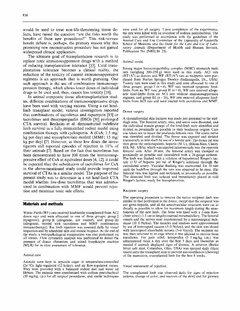
In group I, none of the rats showed any rejection signs, and they were killed at the end of the study at 5 months (Fig. 1). In all animals the postoperative edema disap- peared after 7 days. In group 11, the CTA limbs showed increasing edema postoperatively, up until the point when irreversible acute rejection was established. Skin coloration gradually changed from pinkish to reddish- purple, and finally to purplish-blue (Fig. 2). The mean rejection time of limbs was 5.7 f 1.5 days. In group 111, postoperative edema of the transplanted limb disap- peared completely after 10 days post-transplantation (Fig. 3). Three of ten rats did not complete the study period. They either died or were killed prematurely at 16, 29, and 97 days after transplantation. The cause of death in the first rat (16 days) was not apparent; how- ever, no rejection episodes were observed and no chan- ges in immunosuppressive therapy were made. Automutilation (chewing) of the transplanted limb was the reason why the second rat was killed at 29 days post- transplantation, but no clinical or histological signs of rejection were observed. Only in the third animal (found dead at day 97 post-transplantation) was a rejection episode observed, and the dose of immunosuppressive drugs had to be adjusted. Seven of ten rats survived for the length of the study and were killed at 5 months (Fig. 4). During the follow-up period, one rat had no rejection episodes, four rats had single rejection epi- sodes, and two rats had multiple rejection episodes.
Fig. 1 Transplanted limb in a syngeneic WF animal 150 days after transplantation (group I). Note the healthy appearance of the transplanted limb with normal hair and nail growth. Limb function was normal except for toe contracture

838
- E 'E 40 I a
20 '
Fig. 2 Transplanted allogeneic limb without any treatment (ACI to WF recipients) 10 days after transplantation (group 11). Rejection signs of severe edema with discoloration, formation of vesicles, and hardening of the skin are apparent
I I I I I I I
100 ,
- Syngeneic ---- Allogeneic not treated
Allogeneic treated I
0 I 0 25 50 75 100 125 150
Days post-transplantation
Fig. 3 Percentage of animal survival between groups according to the Kaplan-Meier life-table method. In the syngeneic group, all animals completed the study ( 5 months). In the allogeneic group without treatment all animals rejected their limbs and were killed within 10 days of transplantation. In the allogeneic group with immunosuppressive drugs, seven of ten animals completed the study ( 5 months) and were killed
Histopathology
In group I, none of the animals showed any signs of rejection during the study. Histological analysis showed normal tissue architecture in all solid organs, as well as in muscle and skin from the CTA limb. Two rats pre- sented marginal hyperplasia in the spleen, and in one of these rats a slight portal infiltration was also found. In group 11, the findings from biopsies of skin and muscle
Fig. 4 Transplanted limb from the immunosuppressed group (tacrolimus and MMF) 150 days after transplantation (group 111). Note the healthy limb with normal black hair (from the donor ACI rat) and nail growth. Limb function was normal except for toe contracture
Rejection episodes
Rat 855
Rat 845
Rat 64s
Rat 525 a
E Rat435 ' Rat375
Rat 365
Rat 345
Rat 335
= - Rat505
0 15 30 45 60 75 90 105 120 135 150 Days post-transplantation
Fig. 5 Number of rejection episodes (black bars) in the immuno- suppressed group treated with tacrolimus and MMF (group 111). Seven of the ten animals completed the study ( 5 months) At the time they were killed, four animals showed no clinical or histological signs of rejection of the transplanted limb
from CTA limbs were consistent in all animals. Normal tissue architecture was seen at 2 days after transplanta- tion, moderate rejection (increasing basal cell vacuola- tion and bulla formation in the epidermis) was observed at 4 and 6 days post-transplantation, and severe rejec- tion (edema, vasculitis, complete necrosis and epidermal degeneration, and inflammatory infiltration in the der- mis) was noted at 8 days post-transplantation. In group 111, three of the ten animals did not complete the follow-up period, and at the time of death the trans- planted limbs did not show any histological signs of rejection (Fig. 5). However, in three of the seven
839
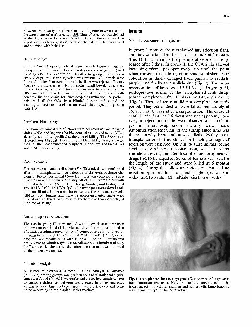
remaining rats that did complete the study, their trans- planted limbs (at the time of killing) showed mononu- clear dermal infiltration compatible with mild rejection (Fig. 6). With the rest of the animals (four rats, 57%), histology was normal, with no signs of rejection. In all seven animals the spleen showed a marginal zone of hyperplasia. The lymph nodes also showed hyperplasia (two rats), atrophy (one rat), congestion (one rat), and normal architecture in the remaining three rats. The liver showed steatosis (two rats), abscess (one rat), abscess with ascending cholangitis (one rat), and cellular infil- tration (one rat); in the remaining two rats the livers were normal. The histopathology of small bowel, lung, and bone showed normal architecture in all seven long- term follow-up animals.
FACS analysis
Only in group I11 was FACS performed on peripheral blood lymphocytes (PBLs) (at 30, 60, 90, and 150 days after transplantation) and from BMCs in the opposite limbs (at the time of killing) to assess the presence of donor chimerism. The levels of donor chimerism in peripheral blood leukocytes (PBLs) ranged between 0.5% and 25%. Similar levels were found in BMCs from the host’s non-transplanted limbs.
Blood analysis
Blood analysis was performed at the end of the study in two animals in the immunosuppressed group. The
Fig. 6 Histological section of donor ACI skin from the trans- planted limb at the end of the study in an immunosuppressed animal (group 111). This animal did not have any rejection episodes during follow-up. At the time it was killed, skin sections showed mild lymphocyte infiltration (H&E, 400x)
analysis showed 30% and 35% of hematocrit (values comparable with the syngeneic group). White blood cells were 4x10³/pl and 6x10³/p1. Platelet counts were 32Ox1O3/pl and 54Ox1O3/p1, and glucose levels were 165 and 200 mg/dl. Drug levels in these two animals were 8.9 and 11.5 ng/ml (for tacrolimus) and 1.6 and 3.8 yg/ml (for MMF).
Discussion
Over the past decade, the concept of the administering of low doses of different immunosuppressive drugs, each acting via different mechanisms, to deliver potent immunosuppression with relatively low toxicity, has gained widespread acceptance in solid-organ transplan- tation. In spite of this knowledge, combination therapy was only recently introduced into clinical composite tissue allotransplantation with several cases of human hand transplantation [13, 16, 24, 321. In those cases tacrolimus, MMF, and corticosteroids were used and found to be effective.
Corticosteroids are associated with several compli- cations, including poor wound and bone healing [2], and opportunistic infections [25], which are particularly rel- evant in CTAs. Accordingly, several new drug regimens that effectively prolong CTA survival without relying on chronic corticosteroid therapy have been or are being investigated. Drugs such as tacrolimus [3], MMF [6], rapamycin [15], or FTY-720 [31] have all been tested in CTA models, either as monotherapy or in a variety of combinations. However, the outcomes of studies that have tested monotherapies in CTA models have been disappointing. For example, rapamycin monotherapy prolonged hind-limb survival for only 9 days in a Brown Norway-to-Lewis rat model [15]. In contrast, when ra- pamycin has been combined with CsA or tacrolimus, limb survival has been significantly increased [ 151. These findings make the strong argument that the combination of calcineurin inhibitors such as CsA or tacrolimus with new drugs [27, 28, 291 that target signaling pathways such as rapamycin, or macrophage-dependent T-cell function such as DSG, or other mechanisms such as Janus kinase inhibitors [22], or FTY-720 Il l] is an effective method of providing corticosteroid-free anti- rejection therapy in CTAs.
Many studies that test the effectiveness of mono- therapy immunosuppression have been performed in the rat hind-limb CTA model. In early studies, limb recipi- ents that were treated with varying doses of azathioprine (AZA), 6-mercaptopurine (6-MP), and prednisone, died from drug-induced side effects before the onset of mac- roscopic signs of rejection [26]. Although, in some cases, investigators reported long-term limb survival using CsA monotherapy [8, 9, 17, 191, others have described early [34] and delayed [6, 201 skin rejection.
840
Another study reported long-term limb survival using a single large dose of tacrolimus, administered on the day of surgery (10 mg/kg) followed by weekly mainte- nance dosing (3 mg/kg). However, most of the animals developed Pneumocystis carinii pneumonia and died [3]. In another report, tacrolimus was given daily for 2 weeks post-transplantation at doses of 0.32-0.64 mg/ kg per day i.m., and while the immunosuppressive effect that was observed was similar to that seen in the CsA studies, this regimen of tacrolimus resulted in early rejection of the skin component of the limb CTA in most of the animals. Using ten times higher doses of tacroli- mus administered orally, Fealy et al. found that tacrol- imus significantly prolonged allograft survival and prevented rejection [ 151. In another series of studies, MMF was shown to prevent [6] and reverse [19] estab- lished acute rejection, although in the former study animals suffered early weight loss and moderate bone marrow toxicity with long-term therapy. Finally, using the same rat hind-limb CTA model, Benhaim et al. [7] reported no significant difference between intermittent immunosuppression with CsA (25 mg/kg) or MMF (30 mg/kg), but found that tacrolimus (2 mg/kg) was significantly superior, in graft survival and lesser toxic- ity, to either CsA or MMF. The implication of this study was that, of the drugs studied, tacrolimus monotherapy was the only agent capable of preventing rejection of the skin component of a CTA. However, tacrolimus monotherapy did not achieve this without inevitable drug toxicity. Combined, these findings suggest that in the case of monotherapy, relatively high doses of drugs are necessary to prevent rejection across major histo- compatibility barriers.
A few studies that tested the effectiveness of combi- nation immunosuppressive therapy have also been con- ducted in the rat hind-limb CTA model. Benhaim et al. reported long-term limb survival and low toxicity in 89% of rats that received a combination of low-dose CsA (1.5 mg/kg) and low-dose MMF (1 5 mg/kg) [7]. In the present study we found that when combined, lower doses of tacrolimus (1 mg/kg per day) and MMF (1 5 mg/kg per day) provided long-term limb survival and minimal toxic side effects. Throughout the duration of this 5-month study, sporadic rejection episodes were observed in these seven immunosuppressed animals. These rejection episodes correlated with sporadic epi- sodes of self-limiting diarrhea, which could have caused erratic absorption of MMF and the resulting rejection episodes. All rejection episodes were effectively con- trolled by administration of tacrolimus for seven con- secutive days and the return to the bi-weekly regimen, without the dose of MMF being changed. Peripheral blood drug level measurements in two animals (in group 111) confirmed that both drugs remained within therapeutic ranges and well below the blood drug levels reported in other comparable studies [3, 71.
In rodent models, MMF is commonly reported to cause dose-dependent aplastic anemia, due to bone marrow toxicity [l, 141, and a wasting syndrome asso- ciated with diarrhea, due to gastrointestinal toxicity [5]. In our study, the only side effect that we observed was episodic diarrhea, which we attributed to MMF. In spite of this, normal weight gain was observed in all animals post-operatively. In rodent models, tacrolimus has been reported to be nephrotoxic and hepatotoxic [18]. How- ever, in this study, at the low dose of tacrolimus we used, we did not observe either of these side effects. At 30, 60, 90, and 150 days after transplantation we performed flow cytometry measurements to detect the presence of donor cells derived from the bone marrow within the CTA hind-limb and found them to be present at our first measurement 30 days post-transplantation. These data confirm previous findings that the bone marrow within a CTA has the capacity to induce chimerism [23, 351. We found that the mean level of chimerism (at the time of killing) in the animals that underwent rejection episodes during the length of the study was 8.6 * 5.6%, whereas the level of chimerism in the animal that experienced no rejection episodes was 2.0%. Previous studies have shown that levels as low as 1% donor cell chimerism resulting from donor stem cell engraftment can confer stable tolerance [21]. In our study, despite the presence of over 1% of donor chimerism in long-surviving ani- mals, we found no relation between presence of donor chimerism and allograft survival. Such a finding in a rat hind-limb model has been previously reported with the use of tacrolimus as monotherapy [37]. We hypothesized that the donor cells that we detected by flow typing in hosts of limb transplants could have been immuno- competent but not tolerized to the host, probably due to long-term immunosuppression [38]. In such an event, quantitative assessment of such circulating “non-toler- ant” donor cells, using flow typing to reflect “engraft- ment” of the donor stem cells, would be misleading. To determine whether flow typing reflected stem-cell engraftment or a mere expansion of donor cell pool derived from the transplanted limb in the presence of immunosuppressive drugs, we examined evidence ob- tained during our experiments. Flow cytometry of re- suspended bone marrow from flushed femurs and tibiae of opposite (non-transplanted) limbs of hosts (at killing) revealed levels of donor chimerism similar to those found in peripheral blood. This finding suggests that the donor stem cells do engraft in hosts that are not con- ditioned with radiation. To enable such allogeneic donor stem cell engraftment, one must first create “geographic niches” [36] in the host bone marrow. This led us to hypothesize that such “niches” could have been created by either of the immunosuppressive drugs (MMF) that were used. We also hypothesized that sustained toler- ance was not achieved, despite chimerism due to dysre- gulation of thymic deletion or peripheral suppression

841
mechanisms secondary to prolonged immunosuppres- sion. We are currently investigating these mechanisms.
In conclusion, the ideal immunosuppressive strategy would be a combination of drugs that are selective and specific in function, synergistically active for maximum effectiveness, and free of toxic side effects. The present study has shown that a combination of tacrolimus and MMF provides effective long-term limb and skin sur- vival. The possibility of reducing the doses of each of these drugs afforded by administering them in combi- nation reduced the incidence of toxic side effects. The fact that we were able to achieve these results without the need for corticosteroids presents a promising thera- peutic alternative for the future of clinical CTAs. In future studies, we plan to use the same low-dose com- bination, corticosteroid-free regimen in a large animal model (swineiprimate) in an attempt to duplicate the same high level of effectiveness and low toxicity.
Based on the findings from this study, we conclude that the toxicity that is normally associated with large doses of immunosuppressive drugs and corticosteroids can be avoided. Combination immunosuppression with
low-dose tacrolimus and MMF prolonged CTA survival indefinitely, without the need for corticosteroids. Fur- ther studies using tacrolimus and MMF in combination with new compounds need to be conducted to further reduce immunosuppression toxicity.
Toxic side effects of immunosuppressive drugs are the reason that prevents the widespread use of composite tissue allotransplantation as a reconstructive procedure. Lowering side effects of the drugs by combining them, and at the same time prolonged survival, is one of the goals in transplantation. Also, successfully eliminating the need for corticosteroids to prevent rejection will diminish important side effects such as impaired wound and bone healing and opportunistic infections.
Acknowledgements The work described here was funded in part by grants from the Jewish Hospital Foundation, Louisville, Kentucky, USA. We would like to thank Lynn Ogden, MD, Mukunda B. Ray, MD, PhD, and Gordon Wang, MD, for their contribution in the preparation and analysis of histological sections, and Anthony Jevans, PhD, for his contribution in the blood analysis. We would also like to thank the animal facility staff for their outstanding animal care.
References
1. Allison AC, Almquist SJ, Muller CD, Eugui EM (1991) In vitro immunosup- pressive effects of mycophenolic acid and an ester pro-drug, RS-61443. Transplant Proc 23 [Suppl 21: 10-14
2. Anstead GM (1998) Steroids, retinoids, and wound healing. Adv Wound Care
3. Arai K, Hotokebuchi T, Miyahara H, Arita C, Mohtai M, Sugioka Y, Kai- bara N (1989) Limb allografts in rats immunosuppressed with FK506. I. Reversal of rejection and indefinite survival. Transplantation 48:782-786
4. Beck Y, Akiyama N (1989) Effect of FK-506 and cyclosporine on human lymphocyte responses in vitro. Trans- plant Proc 21:3464-3467
5. Behrend M (2001) Adverse gastrointes- tinal effects of mycophenolate mofetil: aetiology, incidence and management. Drug Saf 24:645-663
McCalmont TH, Mathes SJ (1993) A long-term study of allogeneic rat hind limb transplants immunosuppressed with RS-61443. Transplantation
111277-285
6. Benhaim P, Anthony JP, Lin LY,
56:911-917 7. Benhaim P, Anthony JP, Ferreira L,
Borsanyi JP, Mathes SJ (1996) Use of combination of low-dose cyclosporine and RS-61443 in a rat hind limb model of composite tissue allotransplantation. Transplantation 6 1527-532
8. Black KS, Hewitt CW, Fraser LA, Howard EB, Martin DC, Achauer BM, Furnas DW (1985) Composite tissue (limb) allografts in rats. 11. Indefinite survival using low-dose cyclosporine. Transplantation 39:365-368
Borger RW, Achauer BM (1988) Dose response of cyclosporine-treated com- posite tissue allografts in a strong his- toincompatible rat model. Transplant Proc 20 [Suppl 2]:266-268
10. Buttemeyer R, Jones NF, Min Z, Rao UR (1996) Rejection of the component tissues of limb allografts in rats immu- nosuppressed with FK-606 and cyclo- sporine. Plast Reconstr Surg 97:139-148
Masubuchi Y, Yanagawa Y, Ohtsuki M, Sasaki S, Fujita T (1996) FTY720, a novel immunosuppressant possessing unique mechanisms. I. Prolongation of skin allograft survival and synergistic effect in combination with cyclosporine in rats. Transplant Proc 28:1056-1059
Calcineurin is a key signaling enzyme in T lymphocyte activation and the target of the immunosuppressive drugs cyclo- sporin A and FK506. Ann N Y Acad Sci 696:20-30
13. Dubernard JM, Owen ER, Lanzetta M, Hakim N (2001) What is happening with hand transplants. Lancet
9. Black KS, Hewitt CW, Hwang JS,
11. Chiba K, Hoshino Y, Suzuki C,
12. Clipstone NA, Crabtree G R (1993)
357:1711-1712
14. Eugui EM, Mirkovich A, Allison AC (1991) Lymphocyte-selective antiprolif- erative and immunosuppressive activity of mycophenolic acid and its morpho- linoethyl ester (RS-61443) in rodents. Transplant Proc 23 [Suppl 21: 15-1 8
15. Fealy MJ, Umansky WS, Bickel KD, Nino JJ, Morris RE, Press BH (1994) Efficacy of rapamycin and FK506 in prolonging rat hind limb allograft survival. Ann Surg 219:88-93
16. Francois CG, Breidenbach WC, Maldonado C, Kakoulidis TP, Hodges A, Dubernard JM, Owen E, Pei G, Ren X, Barker JH (2000) Hand transplan- tation: comparisons and observations of the first four clinical cases. Microsur- gery 20:360-371
17. Fritz WD, Swartz WM, Rose S, Futrell JW, Klein E (1984) Limb allografts in rats immunosuppressed with cyclospo- rin A. Ann Surg 199:211-215
Prabhune KA, Maldonado C, Granger DK (2000) Immunosuppressive agents in transplantation: mechanisms of action and current anti-rejection strate- gies. Microsurgery 20:420429
19. Hewitt CW, Black KS, Fraser LA, Howard EB, Martin DC, Achauer BM, Furnas DW (1985) Composite tissue (limb) allografts in rats. I. Dose-depen- dent increase in survival with cyclo- sporine. Transplantation 39:360-364
18. Gorantla VS, Barker JH, Jones JW,

842
20. Hotokebuchi T, Arai K, Takagishi K, Arita C, Sugioka Y, Kaibara N (1989) Limb allografts in rats immunosup- pressed with cyclosporine: as a whole- joint allograft. Plast Reconstr Surg
21. Ildstad ST, Sachs DH (1984) Reconsti- tution with syngeneic plus allogeneic or xenogeneic bone marrow leads to specific acceptance of allografts or xenografts. Nature 307:168-170
22. Ivashkiv LB (2000) Jak-STAT signaling pathways in cells of the immune system. Rev Immunogenet 2:220-230
M, Olszewski WL, Lukomska B (1999) Fast lymphoid reconstitution after vas- cularized bone marrow transplantation in lethally irradiated rats. Transplanta- tion 68:201-209
24. Jones JW, Gruber SA, Barker JH, Breidenbach WC (2000) Successful hand transplantation. One-year follow- up. Louisville Hand Transplant Team. N Engl J Med 343:468473
25. Kibbler CC (1999) Infections in solid organ transplant recipients. Curr Top Pathol 92:19-35
26. Lance EM, Inglis AE, Figarola F, Veith FJ (1971) Transplantation of the canine hind limb. Surgical technique and methods of immunosuppression for allotransplantation. A preliminary report. J Bone Joint Surg Am
83: 1027-1036
23. Janczewska S, Ziolkowska A, Durlik
53:1137-1149
27. Morris RE, Hoyt EG, Murphy MP, Eugui EM, Allison AC (1990) Myco- phenolic acid morpholinoethyl ester (RS-61443) is a new immunosuppres- sant that prevents and halts heart allo- graft rejection by selective inhibition. Transplant Proc 22: 1659-1662
28. Morris RE, Wu J, Shorthouse R (1990) A study of the contrasting effects of cyclosporine, FK506, and rapamycin on the suppression of allograft rejection. Transplant Proc 22:1638-1641
29. Morris RE, Wu J, Shorthouse R (1990) Comparative immunopharmacologic effects of FK506 and CyA in in vivo models of organ transplantation. Transplant Proc 22:llO-112
30. Muramatsu K, Doi K, Akino T, Shigetomi M, Kawai S (1997) Longer survival of rat limb allograft. Combined immunosuppression of FK-506 and 15-deoxyspergualin. Acta Orthop Scand 68:581-585
31. Muramatsu K, Doi K, Shigetomi M, Kid0 K, Kawai S (1998) A new immu- nosuppressant, FTY720, prolongs limb allograft survival in rats. Ann Plast Surg 40: 16C165
32. Pei G, Gu L, Yu L (2000) A preliminary report of two cases of human hand allograft (in Chinese). Zhonghua Yi Xue Za Zhi 80:417421
33. Prabhune KA, Gorantla VS, Maldona- do C, Perez-Abadia G, Barker JH, Ildstad ST (2000) Mixed allogeneic chimerism and tolerance to composite tissue allografts. Microsurgery 20:441- 447
34. Press BH, Sibley RK, Shons AR (1986) Limb allotransplantation in the rat: extended survival and return of nerve function with continuous cyclosporine/ prednisone immunosuppression. Ann Plast Surg 16:313-321
35. Talmor M, Steinman RM, Codner MA, Chen M, Harper AD, Witmer-Pack MD, Hoffman LA (1995) Bone mar- row-derived chimerism in non-irradi- ated, cyclosporine-treated rats receiving microvascularized limb transplants: evidence for donor-derived dendritic cells in recipient lymphoid tissues. Immunology 86:448455
36. Tavassoli M (1992) The role of condi- tioning regimens in homing of trans- planted hemopoietic cells. Bone Marrow Transplant 10: 15-17
37. Weigao C (1996) Limb allograft in rats: studies on optimal maintenance dose of FK506 and development of graft-ver- sus-host disease (GVHD) (in Japanese). Fukuoka Igaku Zasshi 87:197-205
38. Woo J, Thomson AW, Ildstad ST (1995) Effects of FK 506 on chimerism and the induction of donor-specific unresponsiveness following fully allo- geneic bone marrow transplantation in mice. Transpl Immunol 3:86-90
39. Zdichavsky M, Jones JW, Ustuner ET, Ren X, Edelstein J, Maldonado C, Breidenbach WC, Gruber SA, Ray M, Barker JH (1999) Scoring of skin rejec- tion in a swine composite tissue allo- graft model. J Surg Res 85:l-8