Limb reconstruction after high energy trauma
15
Limb reconstruction after high energy trauma Michael Saleh, Lang Yang and Melanie Sims The Orthopaedic and Traumatic Surgery Research Group, Division of Clinical Sciences, University of Sheffield, Sheffield, UK Limb reconstruction techniques rely on stable external fixation to provide early limb function after major long bone injury. Bone may be generated by callus distraction techniques and internal techniques of moving bone segments used to fill bone defects. Soft tissue defects may be treated by acute shortening, although skin defects will also close spontaneously during bone transport as the leading edge of bone is covered with granulation tissue. External fixation is also used to cross joints permitting rest and repair of the joint. Hinges placed within the bars of the fixation frame may be used to correct deformities in the bone and soft tissue contractures using closed distraction techniques. These techniques are appropriate to metaphyseal fractures and diaphyseal fractures with bone loss. A major advantage is the lack of donor site morbidity, associated with skin flaps and large bone grafts. Acceptance of these techniques is growing whilst the methodology continues to improve. In more complicated cases, specialist training and dedicated hospital units with multidisciplinary support is desirable. Correspondence to Prof M Saleh, Clinical Sciences Centre, Northern General Hospital Trust, Herries Road, Sheffield S5 7AU, UK Severe limb injuries remain a frequent and significant occurrence leading to a reduction in quality of life and employment potential. Despite better safety awareness, faster life styles, busier roads and more active leisure pursuits have resulted in a steady stream of such injuries. Pedestrians, motorcyclists and car occupants remain vulnerable and successful recovery from major visceral injury has resulted in the appearance of some very severe limb injuries. The decision to salvage the severely injured limb must remain objective based on muscle and jomt damage and the potential for neurological and vascular recovery. Skin and underlying soft tissues subjected to severe direct injury are often badly compromised and internal fixation techniques used for low energy fractures are often inappropriate. Stabilisation of the bone is mandatory for initial soft tissue repair and early jomt function. There is increasing evidence that both recovery and final function are significantly improved by early functional rehabilitation. Modern limb reconstruction techniques are largely based on the work of Professor Gavril Ilizarov who pioneered an approach to bone and soft tissue management offering radically different solutions for British Medical Bulletin 1999. 55 (No 4) 870-884 C The British Council 1999 by guest on August 17, 2016 http://bmb.oxfordjournals.org/ Downloaded from
Transcript of Limb reconstruction after high energy trauma

Limb reconstruction after high energy trauma
Michael Saleh, Lang Yang and Melanie SimsThe Orthopaedic and Traumatic Surgery Research Group, Division of Clinical Sciences, Universityof Sheffield, Sheffield, UK
Limb reconstruction techniques rely on stable external fixation to provide earlylimb function after major long bone injury. Bone may be generated by callusdistraction techniques and internal techniques of moving bone segments usedto fill bone defects. Soft tissue defects may be treated by acute shortening,although skin defects will also close spontaneously during bone transport as theleading edge of bone is covered with granulation tissue. External fixation is alsoused to cross joints permitting rest and repair of the joint. Hinges placed withinthe bars of the fixation frame may be used to correct deformities in the boneand soft tissue contractures using closed distraction techniques. Thesetechniques are appropriate to metaphyseal fractures and diaphyseal fractureswith bone loss. A major advantage is the lack of donor site morbidity, associatedwith skin flaps and large bone grafts. Acceptance of these techniques is growingwhilst the methodology continues to improve. In more complicated cases,specialist training and dedicated hospital units with multidisciplinary support isdesirable.
Correspondence to ProfM Saleh, Clinical SciencesCentre, Northern General
Hospital Trust, HerriesRoad, Sheffield
S5 7AU, UK
Severe limb injuries remain a frequent and significant occurrence leadingto a reduction in quality of life and employment potential. Despite bettersafety awareness, faster life styles, busier roads and more active leisurepursuits have resulted in a steady stream of such injuries. Pedestrians,motorcyclists and car occupants remain vulnerable and successfulrecovery from major visceral injury has resulted in the appearance of somevery severe limb injuries. The decision to salvage the severely injured limbmust remain objective based on muscle and jomt damage and thepotential for neurological and vascular recovery. Skin and underlying softtissues subjected to severe direct injury are often badly compromised andinternal fixation techniques used for low energy fractures are ofteninappropriate. Stabilisation of the bone is mandatory for initial soft tissuerepair and early jomt function. There is increasing evidence that bothrecovery and final function are significantly improved by early functionalrehabilitation. Modern limb reconstruction techniques are largely basedon the work of Professor Gavril Ilizarov who pioneered an approach tobone and soft tissue management offering radically different solutions for
British Medical Bulletin 1999. 55 (No 4) 870-884 C The British Council 1999
by guest on August 17, 2016
http://bmb.oxfordjournals.org/
Dow
nloaded from

Limb reconstruction after high energy trauma
the treatment of many traumatic and post-traumatic conditions1. Stableexternal fixation leading to early limb function is a prerequisite, ifnecessary joints may be crossed and protected. Of particular value in highenergy trauma is the ability to deal with bone, joint and soft tissue injurywith minimal donor site morbidity. These techniques are suited tometaphyseal injuries as well as diaphyseal injuries with bone loss. Limbreconstruction may be considered to include one or more of the followingelements: (1) stable external fixation; (li) distraction osteogenesis; (iii) crossjoint fixation; and (iv) progressive deformity correction.
Conventional management approach
Surgical stabilisation of metaphyseal injuries is normally carried out usinginternal fixation techniques which require large skin incisions, soft tissuedissection and internal fixation with plates and screws. These techniqueswere devised for simple, low energy fractures where the skin andunderlying soft tissues are not disrupted. In high energy injuries, openreduction and internal fixation may result in wound breakdown, infectionand bone necrosis. The use of internal fixation for distal tibial fractures isassociated with major infection rates of up to 37%2. The surgeon mustdecide whether to operate and risk complications or delay surgery and,therefore, prejudice accurate fracture reduction. This dilemma is not easilyresolved and the seemingly benign option of temporary external fixationoften produces its own difficult scenario. External fixator pin sites becomecolonised by bacteria and definitive reconstruction may be prejudiced if apin site infection develops. Recognising the risks of immediate internalfixation, some authors claim good results from a staged protocol3'4 inwhich static external fixation is succeeded by internal fixation. In oneseries4 internal fixation was performed between 15 and 49 days andweight bearing delayed for a further 8-12 weeks.
Diaphyseal fractures are less of a problem, since effective stabilisationmay be achieved with closed locked intramedullary nails. In this way,incisions in the traumatised area are usually avoided. Temporary externalfixation has been used, however, the risk of infective complicationsfollowing intramedullary nailing increases5 and delay until the pin sites aredry is recommended6. Most authors, still recommend external fixation formore severe grades of open fracture7 as well as segmental andcomminuted fractures8*9. In more severe limb fractures with bone and/orsoft tissue loss, reconstruction may seem an impossible uphill struggle.Salvage may well require both skin flap cover and massive autogenousbone grafts both of which serve to increase morbidity. Since in these casesthe bone does not contribute to overall stability, more strain is placed onthe fixation system. Failure to achieve stable fixation, slow wound healing
British Medical Bulletin 1999,55 (No 4) 871
by guest on August 17, 2016
http://bmb.oxfordjournals.org/
Dow
nloaded from
Trauma
and joint contractures all delay important rehabilitation milestones andfunctional recovery. The potential for stable external fixation with earlierrehabilitation is probably the single most important contribution thatlimb reconstruction offers over conventional treatment.
Historical perspective - origins liizarov technique
In 1951, Gavril Abramovich liizarov established a general practice inKurgan a small town east of the Urals in Siberia. He rapidly developed aninterest in traumatic limb injuries and evolved a method of treatment witha basic circular external fixation frame1. This consisted of four rings, twoof which were fixed to the bone on either side of the fracture by pairs ofcrossed and tensioned Kirschner wires. The rings were connected togetherby threaded rods. This device provided the even circumferential supportrequired to take a long bone fracture or non-union to full healing. liizarovdescribed four fundamental principles of fracture management: (i) preciseamount of fracture support; (ii) minimal surgical intervention; (iii)immediate weight bearing; and (IV) mobilisation of joints.
The patient was encouraged to mobilise adjacent joints and weight bearas fully as possible. Non-unions were often compressed by shortening theconnecting rods between the rings. On walking, the tensioned Kirschnerwires deformed under load allowing some axial movement. During thehealing phase the stability of the frame could be reduced by removing sup-porting bars, rings or wires further stimulating the fracture to take load.Thus, all the criteria for an 'ideal' fracture environment were fulfilledwithout intruding on the fracture site and producing further surgicaltrauma and bone ischaemia. Even in osteoporosis, cancellous or smallfragments the wire configuration was capable of conferring long-termsupport, if necessary by crossing the adjacent joint. The fixator providedcircumferential support for the bone and, by appropriate use of hingesand connecting rods, was capable of gradual correction of deformity mthe sagittal, coronal and transverse planes as well as lengthening. Crossjoint fixation was used not only to improve fracture support but also torest joints and to prevent and treat contractures.
Distraction osteogenesis was discovered when a patient inadvertentlylengthened the rods and callus formed in the fracture gap. Further studiesresulted in the law of tension stress and a new approach to limb salvage.Dizarov noted that gradual distraction on living tissues created stressesthat stimulated and maintained active regeneration on certain tissuestructures. This concept is perhaps a more specialised statement of Wolff'slaw10 and has been termed the tension stress effect11"13. liizarov noted thatan osteotomised bone segment could be moved axially within the softtissue envelope to close a bone gap leaving a trail of newly generated bone
872 British Medical Bulletin 1999,55 (No 4)
by guest on August 17, 2016
http://bmb.oxfordjournals.org/
Dow
nloaded from

Limb reconstruction after high energy trauma
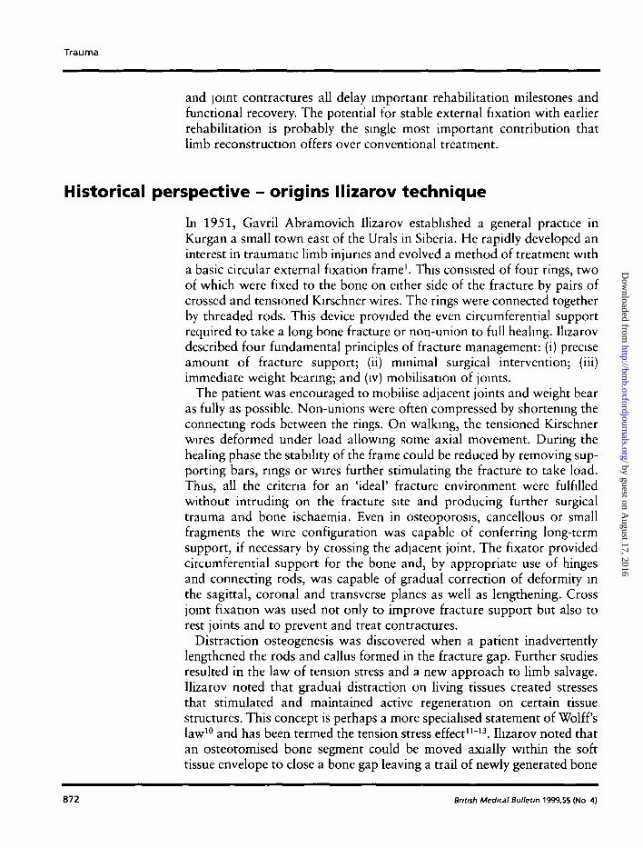
Management scheme for bone loss
BONE LOSS
TRAIIWOBT
COMPRESSIONDtSTRACTtOM
Fig. 1 In bone transport (A), the proximal and distal parts of the bone segment are fixedwith screws maintaining limb length. An osteotomy is performed between the proximaland middle screws and the middle segment of bone slowly moved distally to close anexisting bone segment. New bone forms spontaneously in the proximal distraction gapWhen acute shortening is performed the defect is closed by shortening the limb (B).
in its wake14. This bifocal technique was termed 'bone transport' (Fig. 1A).The additional osteotomy has been shown to increase the vascularity of thelimb segment15. Perhaps more spectacular still was the use of acuteshortening techniques for bone and soft tissue defect. Acute closurepermitted nerve, vascular, muscle and skin repair all of which couldwithstand subsequent slow distraction at the same or a different site (Fig.IB) to restore length. The use of fine tensioned transfixation wires, hingegeometry and soft tissue distraction were new disciplines for us in the West.
Fixation systems for limb reconstruction
hi the early 1980s, Giovanni de Bastiani developed a dynamiseablemonolateral external fixator which could be easily switched from staticto dynamic mode (dynamic axial fixator, DAF; Orthofix, Verona, Italy).Good results were reported in long bone fractures in adults16"18 andchildren19. Surgeons experienced in monolateral external fixationadapted as many of Ilizarov's techniques as they could, when necessaryusing modified devices. Hinged fixators (Fig. 2) were used effectively forpilon fractures20*21, and bifocal lengthening fixators (Fig. 6) for bonetransport22-23. Such devices proved effective in many situations; however,circular fixators proved superior for metaphyseal fixation, jointprotection and deformity correction24. Circular fixators went through asimilar development phase. Problems with diaphyseal fixation and
Bnbsh Medical Bulletin 1999,55 (No 4) 873
by guest on August 17, 2016
http://bmb.oxfordjournals.org/
Dow
nloaded from

Trauma
Fig. 2 Transarticular fixation of a pilon fracture, note the use of a hinge to permit earlymovement
concerns regarding muscle transfixation and pain lead some authors tocombine screws and wires at each ring level25 whilst others dispensedwith wires altogether26. Concern regarding their complexity led tocautious limited acceptance by the orthopaedic community andpromotion mainly in specialist limb reconstruction units27'28. Thesetechniques may well have remained in the hands of specialist units;however, the realisation amongst trauma surgeons that superior fixationmay be achieved in metaphyseal bone led to the development of simplerhybrid designs. Unilateral fixators were adapted to take a ring at oneend29 and popularised particularly for pilon fractures. Such devices areintrinsically less stable than all ring designs with considerable off axismotion30 and retain cantilever loading characteristics common tomonolateral devices31. Circular fixators provide beam loading supportfor the bone, desirable conditions for articular support and reduced pinbone mterface stresses. Havmg realised the importance of both fine wiretransfixation and beam loading characteristics for long termmetaphyseal stability, this author believes that all ring systems arepreferable. The Sheffield hybrid fixator was, therefore, designed tosatisfy the need for a simple, but strong system for trauma surgery32.Two-thirds rings were designed to permit better knee movements andone-third components connected when full rings are required. The ringis strengthened to allow four fully tensioned wires to be applied forstrong metaphyseal fixation. Diaphyseal fixation is achieved with 6 mm
874 British Medical Bulletin 1999,55 (No 4)
by guest on August 17, 2016
http://bmb.oxfordjournals.org/
Dow
nloaded from

Limb reconstruction after high energy trauma
Fig. 3 A four ring Ilizarov fixator and Sheffield hybrid fixator compared.
screws attached using simple clamp designs. Circumferential loadtransfer is achieved via threaded bars or reduction units. Axial elasticityis retained by avoiding the use of screws on the metaphyseal side of thefracture. Mechanical testing demonstrated beam loading characteristicsand similar performance to a four ring Ilizarov construct (Fig. 3)33. Somesurgeons treating articular fractures advocated conventional openreduction and screw fixation with external fixation34. The use, however,of CT scan investigations with less invasive percutaneous approaches toarticular reconstruction permit earlier reconstruction35'36. Circularfixators may be selected in preference to monolateral devices forsegmental fractures and bone transport where strong durable meta-physeal fixation is required.
The application of limb reconstruction techniques inacute trauma
Limb reconstruction techniques may be applied for severe metaphysealfractures, articular fractures, fractures with bone and soft tissue defects,fractures with deformity and joint contractures. Perhaps the simplestexample of the use of these techniques is in metaphyseal fractures. In thiscase (Fig. 4), stable fixation of a comminuted fracture of the proximaltibia permitted immediate weight bearing and return to work within 3months of injury despite an ipsilateral humeral fracture and COntra-
S/vt^ Med;ca/BuWet/n 1999,55 (No 4) 875
by guest on August 17, 2016
http://bmb.oxfordjournals.org/
Dow
nloaded from

Trauma
4A 4B
Fig. 4 A comminuted proximal tibial fracture extending into the diaphysis fixed with a Sheffield hybrid fixator (A),during fixation (B), final result (C).
lateral acetabular reconstruction. If there is articular involvement, thefracture fragments are reduced and fixed using percutaneous cannulatedscrews and wires. Bone graft may be required and percutaneous tech-niques37 reduce donor site morbidity38. External fixation is used tostabilise the limb and interoperative alignment confirmed using animage mtensifier and the axial alignment grid39. Where bone or softtissue damage prohibits adequate metaphyseal fixation, the fixation maybe taken across the joint (Fig. 5). The joint may be bridged until suffi-cient healing has occurred to remove the cross joint element. Threadedbars are attached between the rings spanning the joint and slightdistraction of the joint serves to protect the joint from muscle contractileforces. The use of hinges aligned with the joint allows early movementof the joint, known as articulated distraction.
876 British Medical Bulletin 1999,55 (No 4)
by guest on August 17, 2016
http://bmb.oxfordjournals.org/
Dow
nloaded from

Limb reconstruction after high energy trauma
Fig. 4 (continued) (C) Final result Fig. 5 A tibial plateau fracture stabilised usinghybrid fixation. Joint protection and early weightbearing were facilitated by the temporary additionof a femoral ring.
In this case (Fig. 6), a severe open fracture with bone and soft tissue loss,both muscle and free flaps failed. Temporary external fixation waschanged for more stable fixation using a Limb Reconstruction System(Orthofix, Bussolengo, Italy) spreading the fixation along the wholelength of the tibia. A radical debridement of bone and soft tissue wasperformed. This encourages soft tissue repair, and reduced inflammationpermits some mobilisation of the limb. The bone and soft tissue loss wasrestored by the techniques of slow shortening and simultaneous bonetransport. Granulation tissue following the leading edge of the bone wassoon covered by new epithelium and soft tissue healing was mature andcomplete at 6 weeks. Weight bearing was allowed as soon as the bonedefect was closed and limb length was restored by further lengthening.
British Medical Bulletin 1999,55 (No 4) 877
by guest on August 17, 2016
http://bmb.oxfordjournals.org/
Dow
nloaded from

Trauma
6A
Fig. 6 Severe tibialfracture with necrotic
bone and soft tissuedefect (A), acute
shortening, leavingvital but exposed
bone (B) and stableexternal fixation,
then applied,spontaneous healingof soft tissue defect
at 6 weeks (C), bonedefect closed by a
combination of bonetransport and acute
shortening (E.F).Note new bone
formed bycallotasis (G)
6B
6C
878 British Medical Bulletin 1999,55 (No 4)
by guest on August 17, 2016
http://bmb.oxfordjournals.org/
Dow
nloaded from

Limb reconstruction after high energy trauma
Fig. 6 ContinuedTreatment was complete at 14 months and no bone or skin grafts wererequired.
External fixation may be used to correct a bony deformity or malunion.The fixator is applied with hinges aligned in the same direction as thedeformity. An osteotomy is performed and the hinges slowly altered untilthe deformity is corrected. Slow distraction permits callus to form in theosteotomy gap. In this case (Fig. 7), a valgus malunion of the tibiaprobably contributed to the severity of this Schatzker 6 tibial plateaufracture40. Clearly, fracture repair with residual valgus deformity wouldput unnecessary strain on the joint repair. A three ring hybrid fixator wasapplied and the joint supported by the upper two rings and the malunioncorrected between the middle and lower rings. Soft tissue contracturesmay also be corrected. In this case (Fig. 8), a post-traumatic equinovarusdeformity was corrected by distraction between tibial and hindfoot ringsand between hindfoot and forefoot rings. Initially the joints are distracted(pushing system) and the final correction achieved by altering the framebars and compressing between the tibia and forefoot rings (pullingsystem). Following correction, which normally takes 3-A weeks, the softtissues are neutralised for 6 weeks. The fixator is then removed and
British Medical Bulletin 1999,55 (No 4) 879
by guest on August 17, 2016
http://bmb.oxfordjournals.org/
Dow
nloaded from

Trauma
Fig. 7 The articular fracture is held with cannulated screws and neutralised with a standard two ring fixatormodified by the addition of a further ring and hinges (A). An osteotomy is performed at the level of the previousmalunion and the valgus deformity corrected (B)
orthotic support using a Sheffield splint applied41. This period ofprotection may last for 4—6 months or longer if muscle imbalance issevere.
Fig. 8 A posttraumatic equinovarus
deformity (A), afixator is applied and
slow distraction of thehindfoot and midfoot
joints applied (B).The fixator is
reconfigured and thefinal correction
achieved (C), theappearance after
fixator removed (D).
8A
880 British Medical Bulletin 1999,55 (No 4)
by guest on August 17, 2016
http://bmb.oxfordjournals.org/
Dow
nloaded from

Limb reconstruction after high energy trauma
Fig. 8 Continued
British Medical Bulletin 1999,55 (No 4) 881
by guest on August 17, 2016
http://bmb.oxfordjournals.org/
Dow
nloaded from

Trauma
Discussion
References
Limb reconstruction may be considered as the use of external fixationtechniques to promote bone and soft tissue repair with minimalinvolvement of other body areas. Essential elements are external fixation,internal bone movement, hinges and cross joint fixation. Newer fixatorsrequire less expertise to apply and permit early rehabilitation and weightbearing. Although these fixators are stiff enough to support the limb, axialelasticity afforded by the wires facilitates bone loading and fracturehealing. Much higher levels of activity may be expected with patients infixators doing recreational sport, job retraining and even manual work,thus restoring self respect and independence. Callus distraction techniquesprovide an opportunity for bone and soft tissue repair without donor sitemorbidity. Specialist training is required and, if such techniques areindicated, patients should be referred to specialist units. Patients must becarefully prepared and supported and there is evidence that amultidisciplinary team approach produces better results42. Pinsite infectionremains a problem and the incidence may be reduced by careful operativetechnique, improved fixation devices, improved screw and wire designsand patient involvement in cleaning43. It seems Likely that, with furtherrefinement, these techniques will supercede conventional techniques,improve the threshold for reconstruction and deliver a better functionalresult than amputation. It is always difficult to make such remarks in thecurrent era of evidence-based medicine. Certainly, published comparativestudies have been flawed by the use of historical controls or early ratherthan definitive reconstruction experience. In our own unit, follow-upstudies have indicated that the functional peak is not reached until 2-3years after completion of treatment. No doubt well constructedmulticentre prospective trials will, in time, provide the definitive answer. Inthe meantime, we can only be guided by the small studies that are availableand peer influence. Mason Hohl who has a personal experience of over1500 tibial plateau fractures in his monograph44 cited Uhl et aPscomparative study which concluded that the use of these systems (fine wirehalf ring devices) is leading to better results because of fewer complicationsthan open reduction and plating techniques. In the future, I believe that thesame refinement which has occurred in the management of articularfractures will apply to bone transport and articulated distraction and itsuse in the biological repair of joints.
1 Hizarov GA. Transosseous Osteosynthesis: Theoretical and Clinical Aspects of theRegeneration and Growth of Tissue. Heidelberg: Springer, 1992
2 Teeny SM, Wiss DA Open reduction and internal fixation of tibial plateau fractures: variablescontributing to poor results and complications Clin Orthop 1993; 292: 108—17
882 Bntish Medical Bulletin 1999,55 (No 4)
by guest on August 17, 2016
http://bmb.oxfordjournals.org/
Dow
nloaded from

Limb reconstruction after high energy trauma
3 Sirkin M, Sanders R, DiPasquale T, Herscovici D. A staged protocol for soft tissue managementin the treatment of complex pilon fractures / Orthop Trauma 1999; 13: 78-84
4 Patterson MJ, Cole D Two-staged delayed open reduction and internal fixation of severe pilonfractures. / Orthop Trauma 1999, 13 85-91
5 Maurer MD, Merkow RL, Gustilo RB. Infection after intramedullary nailing of severe opennbial fractures initially treated with external fixation. / Bone Joint Surg Am 1989; 71: 835-8
6 Marshall PD, Saleh M, Douglas DL. Risk of deep infection with intramedullary nailingfollowing the use of external hxators. / R Coll Surg Edmb 1991; 36- 268-71
7 Olson SA Open fractures of the nbial shaft. / Bone Joint Surg Am 1996; 78: 1428-358 Pennig D. External fixation of the tibia. In: Court-Brown C, Pennig D (Eds) Tibia and Fibula
Oxford: Butterworth-Heinemann, 1997; 101-299 Saleh M, Ross ERS. The injured tibia. Int J Orthop Trauma 1994; 4: 90-410 Wolff J. The Law of Bone Remodelling. 1892. Trans Maquet P, Furlong R. Heidelberg:
Springer, 198611 Ilizarov GA. The tension-stress effect on the genesis and growth of tissues. Part I: the influence
of stability of fixation and soft-tissue preservation. Clm Orthop 1989 238; 249-8112 Ilizarov GA. The tension-stress effect on the genesis and growth of tissues. Part II: the influence
of the rate and frequency of distraction. Clm Ortbop 1989- 239; 263-8513 Ilizarov GA. Clinical application of the tension-stress effect for limb lengthening. Clm Orthop
1990: 250; 8-2614 Ilizarov GA, Ledyaev VL The replacement of long tubular defects by lengthening distraction
osteotomy of one of the fragments Vestmk Khururgit 1969; 6: 78. [Reproduced in part andtranslated Clm Orthop 1992; 280: 7-10]
15 Sveshnikov AA, Barabash AP, Chepelenko TA, Smotrova LA, Lanonov AA. Radionuchdestudies of osteogenesis and blood circulation after experimental correction of extensive cruralbone defects. Ortop Traumatol Protez 1984; 11 33-7
16 De Bastiani G, Aldeghen R, Bnvio L R, The treatment of fractures with a dynamic axial fixator/ Bone Joint Surg Br 1984; 66: 538-45
17 Marsh JL, Nepola JV, Wuest TK. Unilateral external fixation until healing with the dynamicaxial fixator for severe open tibial fractures J Orthop Trauma 1991; 5: 341-8
18 Hay S, Rickman M, Saleh M. Fractures of the nbial diaphysis treated by external fixation andthe axial alignment grid. A single surgeon's experience. In/ury 1997; 28: 437—43
19 Sanderson PL, Rickman M, Bell MJ, Saleh M. External fixation of children's fractures; the useof the dynamic axial fixator. / Pediatr Orthop 1997; 6: 203-6
20 Saleh M, Fern D, Shanahan MDG. Intra-articular fractures of the distal tibia. Surgicalmanagement by limited internal fixation and articulated distraction. Injury 1993; 24: 37-40
21 Marsh JL, Bonar S, Nepola JV, Decoster TA Use of an articulated external fixator for fracturesof the nbial plafond. / Bone Joint Surg Am 1995; 77: 1498-509
22 Saleh M, Rees AR. Bifocal surgery for deformity and bone loss — bone transport andcompression distraction compared J Bone Joint Surg Br 1995, 77 429-34
23 Giebel G. Resection debndement of the lower leg with compensatory callus distractionUnfallchirurg 1991; 94: 401-8
24 Saleh M, Royston S. Management of nonunion of fractures by distracnon with correction ofangulanon and shortening. / Bone Joint Surg Br 1996; 78. 105-9
25 Bianchi-Maiocchi A. Instruments and their use. In: Bianchi-Maiocchi A, Aronson J (Eds)Operative Principles of Ilizarov. London- Williams and Wilkins, 1991: 9-32
26 Green SA, Harris L, Wall DM, Ishkanian J, Mannow H. The rancho mounting technique forthe Ilizarov method; a preliminary report. Clm Orthop 1992, 280 104-16
27 Saleh M. The management of bone loss In- Court-Brown C, Pennig D (Eds) Tibia and fibula.Oxford: Butterworth-Heinemann, 1997, 143-59
28 Marsh DR, Shah S, Elliot J, Kurdy N. The Ilizarov method in nonunion, malunion and infecnonof fractures. / Bone Joint Surg. Br 1997; 79. 273-9
29 Bomvento G. Experience with a pen-amcular attachment to the dynamic axial fixator: apreliminary report. Int J Orthop Trauma 1993; 3(Suppl) 52—4
30 Khahly C, Voor MJ, Seligson D. Fracture site motion with Ilizarov and 'hybrid' externalfixation. / Orthop Trauma 1998; 12: 21-6
British Medical Bulletin 1999,55 (No 4) 883
by guest on August 17, 2016
http://bmb.oxfordjournals.org/
Dow
nloaded from

Trauma
31 Saleh M, Yang L, Nayagam S. A biomechanical analysis of the Sheffield hybrid external fixator/ Bone Joint Surg Br 1997, 79 Orthop Proc Suppl IE- 361-2
32 Saleh M The Sheffield hybrid fixator — design considerations and clinical experience OrthopProduct News 1998; May/June: 33-6
33 Saleh M, Yang L, Nayagam S. Can a hybrid fixator perform as well as the Ilizarov fixator? JBone Joint Surg Br 1997; 79 Suppl 462
34 Werner LS, Kelley M, Yang E, Steuer J, Watnick N, Evans M, Bergman M The use ofcombination internal fixation and hybrid external fixation in severe proximal tibial fractures JOrthop Trauma 1995; 3: 244-50
35 Marsh JL, Bonar ST, Do TT. External fixation and limited internal fixation for complexfractures of the tibial plateau. J Bone Joint Surg Am 1995, 77: 661-73
36 Watson JT High-energy fractures of the tibial plateau Orthop Cltn North Am 1994, 25-723-52
37 Saleh M. Bone graft harvesting a percutaneous technique / Bone Joint Surg Br 1991, 73867-8
38 Kreibich DN, Scott IR, Wells JM, Saleh M. Donor site morbidity at the iliac crest: comparisonof percutaneous and open methods. / Bone Joint Surg Br 1994, 76 847-8
39 Saleh M, Harnman P, Edwards DJ. A radiological method for producing precise limbalignment. J Bone Joint Surg Br 1991, 73 515-6
40 Schatzker J, McBroom R, Bruce D. The tibial plateau fracture The Toronto experience ClinOrthop 1979; 138: 94-104
41 Saleh M, Marshall PD, Senior R, MacFarlane A The Sheffield splint for controlled earlymobilisation after rupture of the calcaneal tendon A prospeaive randomised companson withplaster treatment. / Bone Joint Surg Br 1992, 74:206-9
42 Saleh M. The management of bone defects [Editorial] Curr Orthop 1994; 8: 141-343 Sims M Looking After your Pin and Wire Sites, A Patient's Guide. Northern General Hospital:
Intavent Orthofix Ltd, 199844 Hohl M. Tibial Plateau Fractures Philadelphia, PA: Saunders, 1997
884 British Medical Bulletin 1999,55 (No 4)
by guest on August 17, 2016
http://bmb.oxfordjournals.org/
Dow
nloaded from