Occlusal Trauma
11
Trauma from Occlusion Alsan"o Geminiani, DDS, MS Occlusal Trauma Occlusal Trauma: “An injury resulting in tissue changes within the attachment apparatus as a result of excessive occlusal force” Glossary of Periodontic Terms Result in pathological changes to the periodontium Occlusal Trauma Occlusal Trauma: Glossary of Periodontic Terms Primary Occlusal Trauma On a normal periodontium Secondary Occlusal Trauma On a reduced periodontium Excessive Force Normal or Excessive Force
-
Upload
khangminh22 -
Category
Documents
-
view
0 -
download
0
Transcript of Occlusal Trauma

Trauma from Occlusion
Alessan"o Geminiani, DDS, MS
Occlusal Trauma
Occlusal Trauma:
“An injury resulting in tissue changes within the attachment apparatus as a result of excessive
occlusal force”Glossary of Periodontic Terms
Trauma from Occlusion: Periodontal Tissues 355
for the pressure zone at orthodontically moved teeth, with the one difference that the periodontal ligament space at jiggling gradually increased in width on both sides of the tooth. During the phase when the periodontal space gradually increased in width, (1) infl ammatory changes were present in the ligament tissue, (2) active bone resorption occurred, and (3) the tooth displayed signs of gradually increasing (pro-gressive) mobility. When the effect of the forces applied had been compensated for by the increased width of the periodontal ligament space, the liga-ment tissue showed no signs of increased vascularity or exudation. The tooth was hypermobile but the mobility was no longer progressive in character. Dis-tinction should thus be made between progressive and increased tooth mobility.
In jiggling-type trauma experiments, performed on animals with a normal periodontium, the supra-alveolar connective tissue was not infl uenced by the occlusal forces, the reason being that this tissue com-partment is bordered by hard tissue on one side only. This means that a gingiva which was uninfl amed at the start of the experiment remained uninfl amed, but also that an overt infl ammatory lesion residing in the supra-alveolar connective tissue was not aggravated by the jiggling forces.
Healthy periodontium with reduced heightProgressive periodontal disease is characterized by gingival infl ammation and a gradually developing loss of connective tissue attachment and alveolar bone. Treatment of periodontal disease, i.e. removal
a b
c d
Fig. 14-7 Two mandibular premolars with normal periodontal tissues (a) are exposed to jiggling forces (b) as illustrated by the two arrows. The combined tension and pressure zones (encircled areas) are characterized by signs of acute infl ammation including collagen resorption, bone resorption, and cementum resorption. As a result of bone resorption the periodontal ligament space gradually increases in size on both sides of the teeth as well as in the periapical region. (c) When the effect of the force applied has been compensated for by the increased width of the periodontal ligament space, the ligament tissue shows no sign of infl ammation. The supra-alveolar connective tissue is not affected by the jiggling forces and there is no apical downgrowth of the dentogingival epithelium. (d) After occlusal adjustment the width of the periodontal ligament becomes normalized and the teeth are stabilized.
Result in pathological changes to the periodontium
Occlusal TraumaOcclusal Trauma:
Glossary of Periodontic Terms
Trauma from Occlusion: Periodontal Tissues 355
for the pressure zone at orthodontically moved teeth, with the one difference that the periodontal ligament space at jiggling gradually increased in width on both sides of the tooth. During the phase when the periodontal space gradually increased in width, (1) infl ammatory changes were present in the ligament tissue, (2) active bone resorption occurred, and (3) the tooth displayed signs of gradually increasing (pro-gressive) mobility. When the effect of the forces applied had been compensated for by the increased width of the periodontal ligament space, the liga-ment tissue showed no signs of increased vascularity or exudation. The tooth was hypermobile but the mobility was no longer progressive in character. Dis-tinction should thus be made between progressive and increased tooth mobility.
In jiggling-type trauma experiments, performed on animals with a normal periodontium, the supra-alveolar connective tissue was not infl uenced by the occlusal forces, the reason being that this tissue com-partment is bordered by hard tissue on one side only. This means that a gingiva which was uninfl amed at the start of the experiment remained uninfl amed, but also that an overt infl ammatory lesion residing in the supra-alveolar connective tissue was not aggravated by the jiggling forces.
Healthy periodontium with reduced heightProgressive periodontal disease is characterized by gingival infl ammation and a gradually developing loss of connective tissue attachment and alveolar bone. Treatment of periodontal disease, i.e. removal
a b
c d
Fig. 14-7 Two mandibular premolars with normal periodontal tissues (a) are exposed to jiggling forces (b) as illustrated by the two arrows. The combined tension and pressure zones (encircled areas) are characterized by signs of acute infl ammation including collagen resorption, bone resorption, and cementum resorption. As a result of bone resorption the periodontal ligament space gradually increases in size on both sides of the teeth as well as in the periapical region. (c) When the effect of the force applied has been compensated for by the increased width of the periodontal ligament space, the ligament tissue shows no sign of infl ammation. The supra-alveolar connective tissue is not affected by the jiggling forces and there is no apical downgrowth of the dentogingival epithelium. (d) After occlusal adjustment the width of the periodontal ligament becomes normalized and the teeth are stabilized.
Primary Occlusal TraumaOn a normal periodontium
Trauma from Occlusion: Periodontal Tissues 357
a b
c d
Fig. 14-9 (a) Two mandibular premolars are surrounded by a healthy periodontium with reduced height. (b) If such premolars are subjected to traumatizing forces of the jiggling type a series of alterations occurs in the periodontal ligament tissue. (c) These alterations result in a widened periodontal ligament space and in an increased tooth mobility but do not lead to further loss of connective tissue attachment. (d) After occlusal adjustment the width of the periodontal ligament is normalized and the teeth are stabilized.
This problem has also been examined in animal experiments (Ericsson & Lindhe 1977). Destructive periodontal disease was initiated in dogs by allowing the animals to accumulate plaque and calculus for a period of 6 months (Fig. 14-8). When around 50% of the periodontal tissue support had been lost (Fig. 14-8a,b), the progressive disease was subjected to treat-ment by scaling, root planing, and pocket elimination (Fig. 14-8c). During a subsequent 8-month period, the animals were enrolled in a careful plaque-control program. During this period certain premolars were exposed to traumatizing jiggling forces. The peri-odontal tissues in the combined pressure and tension zones reacted to the forces by vascular proliferation, exudation, and thrombosis, as well as by bone resorp-tion. In radiographs, widened periodontal ligaments (Fig. 14-8d) could be found around the traumatized teeth, which displayed signs of progressive tooth mobility at clinical examination. The gradual increase
in the width of the periodontal ligament and the resulting progressive increase in tooth mobility took place during a period of several weeks but eventually terminated. The active bone resorption ceased and the markedly widened periodontal ligament tissue regained its normal composition; healing had occurred (Fig. 14-8e). The teeth were hypermobile but surrounded by periodontal structures which had adapted to the altered functional demands.
During the entire experimental period the supra-alveolar connective tissue remained unaffected by the jiggling forces. There was no further loss of con-nective tissue attachment and no further downgrowth of dentogingival epithelium (Fig. 14-8e). The results from this study clearly reveal that within certain limits a healthy periodontium with reduced height has a capacity similar to that of a periodontium with normal height to adapt to altered functional demands (Fig. 14-9).
Secondary Occlusal TraumaOn a reduced periodontium
Excessive Force Normal or Excessive Force

Signs of Occlusal Trauma
Fremitus
Pulpal and/or Periodontal Sensitivity
Abnormal Tooth Mobility
Widening of the PDL
Enamel Wear
Tooth Fracture
Mobility vs Fremitus
Glossary of Periodontic Terms
Mobility:
Visually perceptible movement of the tooth away from its normal position
when a light force is applied
Fremitus:
Palpable or visible movement of a tooth when subjected to occlusal forces
Occlusal Trauma Research
1901Karolyi
1935Box
1938Stones
2012GHDHS2009
Harrel & NunnNunn & Harrel
2011Branschofsky
1963Glickman
1955GlickmanBhaskar
1976PolsonKantorRosling
1975Meitner
1962Glickman & Smulow
1996McGuire
1992Burgett
1982Perrier
Opinion Based Animal Based Human Based
1980Fleszar
1986Philstrom

Glickman’s Concept
The pathway of the spread of a plaque-associated gingival lesion can be changed if forces of an abnormal magnitudeare acting on teeth harboring subgingival plaque.
Trauma from Occlusion: Periodontal Tissues 351
orientated in a direction parallel to the root surface. The spread of an infl ammatory lesion from the zone of irritation directly down into the periodontal liga-ment (i.e. not via the interdental bone) may hereby be facilitated (Fig. 14-2). This alteration of the “normal” pathway of spread of the plaque-associated infl ammatory lesion results in the development of angular bony defects. Glickman (1967) stated in a review paper that trauma from occlusion is an etiologic factor (co-destructive factor) of importance in situa-tions where angular bony defects combined with infrabony pockets are found at one or several teeth (Fig. 14-3).
Waerhaug’s concept
Waerhaug (1979) examined autopsy specimens (Fig. 14-4) similar to Glickman’s, but in addition measured the distance between the subgingival plaque and (1) the periphery of the associated infl ammatory cell infi ltrate in the gingiva, and (2) the surface of the adjacent alveolar bone. He concluded from his analy-sis that angular bony defects and infrabony pockets occur equally often at periodontal sites of teeth which are not affected by trauma from occlusion as in trau-matized teeth. In other words, he refuted the hypo-thesis that trauma from occlusion played a role in the spread of a gingival lesion into the “zone of co-destruction”. The loss of connective attachment and the resorption of bone around teeth are, according to Waerhaug, exclusively the result of infl ammatory lesions associated with subgingival plaque. Waer-haug concluded that angular bony defects and infrabony pockets occur when the subgingival plaque of one tooth has reached a more apical level than the microbiota on the neighboring tooth, and when the volume of the alveolar bone surrounding the roots is comparatively large. Waerhaug’s observa-tions support fi ndings presented by Prichard (1965) and Manson (1976) which imply that the pattern of loss of supporting structures is the result of an inter-play between the form and volume of the alveolar bone and the apical extension of the microbial plaque on the adjacent root surfaces.
It is obvious, as stated above, that examinations of autopsy material have a limited value when deter-mining “cause–effect” relationships with respect to trauma and progressive periodontitis. As a conse-quence, the conclusions drawn from this fi eld of research have not been generally accepted. A number of authors tend to accept Glickman’s conclusions that trauma from occlusion is an aggravating factor in
Fig. 14-2 The infl ammatory lesion in the zone of irritation can, in teeth not subjected to trauma, propagate into the alveolar bone (open arrow), while in teeth also subjected to trauma from occlusion, the infl ammatory infi ltrate spreads directly into periodontal ligament (fi lled arrow).
bbaa
Fig. 14-3 (a) A radiograph of a mandibular premolar–canine region. Note the angular bony defect at the distal aspect of the premolar. (b) Histologic mesiodistal section of the specimen illustrated in (a). Note the infrabony pocket at the distal aspect of the premolar. From Glickman & Smulow (1965).
Therefore, the periodontium of non-traumatized teethwould present an even destruction (horizontal bone loss),
while traumatized teeth would present angular bony defects.
Glickman & Smulow 1963
Glickman’s Concept
Marginal and Interdental Gingiva
350 Trauma from Occlusion
vertical pocket formation associated with one or a varying number of teeth” (Stones 1938). The experi-ments by Box and Stones, however, have been criticized because they lacked proper controls and because the experimental design of the studies did not justify the conclusions drawn.
The interaction between trauma from occlusion and plaque-associated periodontal disease in humans was frequently discussed in the period 1955–1970 in connection with “report of a case”, “in my opinion” statements, etc. Even if such anecdotal data may have some value in clinical dentistry, it is obvious that conclusions drawn from research fi ndings are much more pertinent. The research-based conclusions are not always indisputable but they invite the reader to a critique which anecdotal data do not. In this chapter, therefore, the presentation will be limited to fi ndings collected from research endeavors involving: (1) human autopsy material, (2) clinical trials, and (3) animal experiments.
Analysis of human autopsy material
Results reported from carefully performed research efforts involving examinations of human autopsy material have been diffi cult to interpret. In the speci-mens examined (1) the histopathology of the lesions in the periodontium have been described, as well as (2) the presence and apical extension of microbial deposits at adjacent root surfaces, (3) the mobility of the teeth involved, and (4) “the occlusion” of the sites under scrutiny. It is obvious that assessments made in specimens from cadavers have a limited to ques-tionable value when “cause–effect” relationships between occlusion, plaque, and periodontal lesions are to be described. It is not surprising, therefore, that conclusions drawn from this type of research can be controversial. This can best be illustrated if “Glickman’s concept” is compared with “Waerhaug’s concept” of what autopsy studies have revealed regarding trauma from occlusion and periodontal disease.
Glickman’s concept
Glickman (1965, 1967) claimed that the pathway of the spread of a plaque-associated gingival lesion can be changed if forces of an abnormal magnitude are acting on teeth harboring subgingival plaque. This would imply that the character of the progres-sive tissue destruction of the periodontium at a “traumatized tooth” will be different from that char-acterizing a “non-traumatized” tooth. Instead of an even destruction of the periodontium and alveolar bone (suprabony pockets and horizontal bone loss), which, according to Glickman, occurs at sites with uncomplicated plaque-associated lesions, sites which are also exposed to abnormal occlusal force will develop angular bony defects and infrabony pockets.
Since Glickman’s concept regarding the effect of trauma from occlusion on the spread of the plaque-associated lesion is often cited, a more detailed presentation of his theory seems pertinent. The peri-odontal structures can be divided into two zones (Fig. 14-1):
1. The zone of irritation2. The zone of co-destruction.
The zone of irritation includes the marginal and interdental gingiva. The soft tissue of this zone is bordered by hard tissue (the tooth) only on one side and is not affected by forces of occlusion. This means that gingival infl ammation cannot be induced by trauma from occlusion but is the result of irritation from microbial plaque. The plaque-associated lesion at a “non-traumatized” tooth propagates in the apical direction by fi rst involving the alveolar bone and only later the periodontal ligament area. The progres-sion of this lesion results in an even (horizontal) bone destruction.
The zone of co-destruction includes the periodontal ligament, the root cementum, and the alveolar bone, and is coronally demarcated by the trans-septal (interdental) and the dento-alveolar collagen fi ber bundles (Fig. 14-1). The tissue in this zone may become the seat of a lesion caused by trauma from occlusion.
The fi ber bundles which separate the zone of co-destruction from the zone of irritation can be affected from two different directions:
1. From the infl ammatory lesion maintained by plaque in the zone of irritation
2. From trauma-induced changes in the zone of co-destruction.
Through this exposure from two different direc-tions the fi ber bundles may become dissolved and/or
Zone ofirritation
Zone ofco-destruction
Fig. 14-1 Schematic drawing of the zone of irritation and the zone of co-destruction according to Glickman.
PDL, Bone, Cementum
Demarcated by the transeptal fibers
Glickman 1967
Glickman’s Concept
Inflammation in the “zone of irritation” is induced by plaque and leads to
horizontal bone loss
The presence of traumatic occlusion extends the lesion into the “zone of co-destruction” leads to the formation of an angular defect
Glickman 1967
Trauma from Occlusion: Periodontal Tissues 351
orientated in a direction parallel to the root surface. The spread of an infl ammatory lesion from the zone of irritation directly down into the periodontal liga-ment (i.e. not via the interdental bone) may hereby be facilitated (Fig. 14-2). This alteration of the “normal” pathway of spread of the plaque-associated infl ammatory lesion results in the development of angular bony defects. Glickman (1967) stated in a review paper that trauma from occlusion is an etiologic factor (co-destructive factor) of importance in situa-tions where angular bony defects combined with infrabony pockets are found at one or several teeth (Fig. 14-3).
Waerhaug’s concept
Waerhaug (1979) examined autopsy specimens (Fig. 14-4) similar to Glickman’s, but in addition measured the distance between the subgingival plaque and (1) the periphery of the associated infl ammatory cell infi ltrate in the gingiva, and (2) the surface of the adjacent alveolar bone. He concluded from his analy-sis that angular bony defects and infrabony pockets occur equally often at periodontal sites of teeth which are not affected by trauma from occlusion as in trau-matized teeth. In other words, he refuted the hypo-thesis that trauma from occlusion played a role in the spread of a gingival lesion into the “zone of co-destruction”. The loss of connective attachment and the resorption of bone around teeth are, according to Waerhaug, exclusively the result of infl ammatory lesions associated with subgingival plaque. Waer-haug concluded that angular bony defects and infrabony pockets occur when the subgingival plaque of one tooth has reached a more apical level than the microbiota on the neighboring tooth, and when the volume of the alveolar bone surrounding the roots is comparatively large. Waerhaug’s observa-tions support fi ndings presented by Prichard (1965) and Manson (1976) which imply that the pattern of loss of supporting structures is the result of an inter-play between the form and volume of the alveolar bone and the apical extension of the microbial plaque on the adjacent root surfaces.
It is obvious, as stated above, that examinations of autopsy material have a limited value when deter-mining “cause–effect” relationships with respect to trauma and progressive periodontitis. As a conse-quence, the conclusions drawn from this fi eld of research have not been generally accepted. A number of authors tend to accept Glickman’s conclusions that trauma from occlusion is an aggravating factor in
Fig. 14-2 The infl ammatory lesion in the zone of irritation can, in teeth not subjected to trauma, propagate into the alveolar bone (open arrow), while in teeth also subjected to trauma from occlusion, the infl ammatory infi ltrate spreads directly into periodontal ligament (fi lled arrow).
bbaa
Fig. 14-3 (a) A radiograph of a mandibular premolar–canine region. Note the angular bony defect at the distal aspect of the premolar. (b) Histologic mesiodistal section of the specimen illustrated in (a). Note the infrabony pocket at the distal aspect of the premolar. From Glickman & Smulow (1965).

Waerhaug’s Concept
Analysis of human autopsy specimens
Measurement of the distance between:Plaque - Inflammatory Infiltrate - Bone Traumatized vs Non-Traumatized Teeth
Conclusions:Angular bony defects occur equally often in traumatized or non-traumatized
Angular bony defects are the result of the progression of plaque
Waerhaug 1979
Waerhaug’s Concept
Waerhaug 1979
352 Trauma from Occlusion
periodontal disease (e.g. Macapanpan & Weinmann 1954; Posselt & Emslie 1959; Glickman & Smulow 1962, 1965) while others accept Waerhaug’s concept, i.e. that there is no relationship between occlusal trauma and the degree of periodontal tissue break-down (e.g. Lovdahl et al. 1959; Belting & Gupta 1961; Baer et al. 1963; Waerhaug 1979).
Clinical trials
In addition to the presence of angular bony defects and infrabony pockets, increased tooth mobility is fre-quently listed as an important sign of occlusal trauma. For details regarding tooth mobility, see Chapter 51. Confl icting data have also been reported regarding the periodontal conditions of mobile teeth. In one clinical study by Rosling et al. (1976) patients with advanced periodontal disease associated with mul-tiple angular bony defects and mobile teeth were exposed to antimicrobial therapy (i.e. subgingival scaling after fl ap elevation). Healing was evaluated by probing attachment level measurements and radiographic monitoring. The authors reported that “the infrabony pocket located at hypermobile teeth exhibited the same degree of healing as those adja-cent to fi rm teeth”. In another study, however, Fleszar et al. (1980) reported on the infl uence of tooth mobil-ity on healing following periodontal therapy includ-
ing both root debridement and occlusal adjustment. They concluded that “pockets of clinically mobile teeth do not respond as well to periodontal treatment as do those of fi rm teeth exhibiting the same disease severity”.
A third study (Pihlstrom et al. 1986) studied the association between trauma from occlusion and peri-odontitis by assessing a series of clinical and radio-graphic features at maxillary fi rst molars. Parameters included in this study were: probing depth, probing attachment level, tooth mobility, wear facets, plaque and calculus, bone height, widened periodontal space, etc. Pihlstrom and his associates concluded from their measurements and examinations that teeth with increased mobility and widened periodontal ligament space had, in fact, deeper pockets, more attachment loss, and less bone support than teeth without these symptoms.
Burgett et al. (1992) studied the effect of occlusal adjustment in the treatment of periodontitis. Fifty subjects with periodontitis were examined at base-line and subsequently treated for their periodontal condition with root debridement ± fl ap surgery. Twenty-two out of the 50 patients, in addition, received comprehensive occlusal therapy. Re-exami-nations performed 2 years later disclosed that probing attachment gain was on average about 0.5 mm larger in patients who received the combined treatment,
bbaa
Fig. 14-4 Microphotographs illustrating two interproximal areas with angular bony defects. “!” denotes a tooth not subjected and “+” denotes a tooth subjected to trauma from occlusion. In categories “!” and “+” the distance between the apical cells of the junctional epithelium and the supporting alveolar bone is about 1–1.5 mm, and the distance between the apical extension of plaque and the apical cells of the junctional epithelium about 1 mm. Since the apical cells of the junctional epithelium and the subgingival plaque are located at different levels on the two adjacent teeth, the outline of the bone crest becomes oblique. A radiograph from such a site would disclose the presence of an angular bony defect at a non-traumatized (“!”) tooth.
Type of Occlusal Trauma
Jiggling Trauma“Chewing” Trauma
Trauma from Occlusion: Periodontal Tissues 355
for the pressure zone at orthodontically moved teeth, with the one difference that the periodontal ligament space at jiggling gradually increased in width on both sides of the tooth. During the phase when the periodontal space gradually increased in width, (1) infl ammatory changes were present in the ligament tissue, (2) active bone resorption occurred, and (3) the tooth displayed signs of gradually increasing (pro-gressive) mobility. When the effect of the forces applied had been compensated for by the increased width of the periodontal ligament space, the liga-ment tissue showed no signs of increased vascularity or exudation. The tooth was hypermobile but the mobility was no longer progressive in character. Dis-tinction should thus be made between progressive and increased tooth mobility.
In jiggling-type trauma experiments, performed on animals with a normal periodontium, the supra-alveolar connective tissue was not infl uenced by the occlusal forces, the reason being that this tissue com-partment is bordered by hard tissue on one side only. This means that a gingiva which was uninfl amed at the start of the experiment remained uninfl amed, but also that an overt infl ammatory lesion residing in the supra-alveolar connective tissue was not aggravated by the jiggling forces.
Healthy periodontium with reduced heightProgressive periodontal disease is characterized by gingival infl ammation and a gradually developing loss of connective tissue attachment and alveolar bone. Treatment of periodontal disease, i.e. removal
a b
c d
Fig. 14-7 Two mandibular premolars with normal periodontal tissues (a) are exposed to jiggling forces (b) as illustrated by the two arrows. The combined tension and pressure zones (encircled areas) are characterized by signs of acute infl ammation including collagen resorption, bone resorption, and cementum resorption. As a result of bone resorption the periodontal ligament space gradually increases in size on both sides of the teeth as well as in the periapical region. (c) When the effect of the force applied has been compensated for by the increased width of the periodontal ligament space, the ligament tissue shows no sign of infl ammation. The supra-alveolar connective tissue is not affected by the jiggling forces and there is no apical downgrowth of the dentogingival epithelium. (d) After occlusal adjustment the width of the periodontal ligament becomes normalized and the teeth are stabilized.
Unidirectional TraumaOrthodontic Trauma
Trauma from Occlusion: Periodontal Tissues 353
i.e. scaling and occlusal adjustment, than in patients in whom the occlusal adjustment was not included.
Nunn and Harrel (2001) and Harrel and Nunn (2001) examined the relationship between occlusal discrepancies and periodontitis in two studies. Their sample included about 90 subjects that had been referred for treatment of periodontal disease and who had at least two (!1 year apart) complete peri-odontal records, including occlusal analysis. The patients were examined with respect to probing pocket depth (PPD), tooth mobility, and furcation involvement (at multirooted teeth). In addition, some occlusal contact relationships were studied such as (1) discrepancies in centric relation and centric occlu-sion, (2) premature occlusal contacts in protrusive movements (lateral and frontal) of the mandible in working and non-working quadrants. A treatment plan, including both periodontal and occlusal mea-sures, was subsequently designed for each patient. About one third of the subjects decided to abstain from treatment, about 20 subjects accepted only a non-surgical approach of therapy (SRP), while about 50% of the patients accepted and received compre-hensive treatment that included surgical pocket elim-ination (surgery) as well as occlusal adjustment (if indicated). Some teeth in the SRP group received occlusal therapy while other teeth with occlusal dis-crepancies were left untreated. It was observed that teeth with occlusal discrepancies had signifi cantly deeper PPD values and higher mobility scores than teeth without occlusal “trauma” and also that teeth exposed to occlusal adjustment responded better (reduction in PPD) to non-surgical periodontal therapy than teeth with remaining occlusal discrepancies.
The fi ndings in some of the clinical studies referred to above lend some support to the concept that trauma from occlusion (and increased tooth mobility) may have a detrimental effect on the periodontium. Neiderud et al. (1992), however, in a beagle dog study demonstrated that tissue alterations which occur at mobile teeth with clinically healthy gingivae (and normal height of the tissue attachment) may reduce the resistance offered by the periodontal tissues to probing. In other words, if the probing depth at two otherwise similar teeth – one non-mobile and one hypermobile – is recorded, the tip of the probe will penetrate 0.5 mm deeper at the mobile than at the non-mobile tooth. This fi nding must be taken into consideration when the above clinical data are interpreted.
Since neither analysis of autopsy material nor data from clinical trials can be used to properly determine the role trauma from occlusion may play in peri-odontal pathology, it is necessary to describe the con-tributions made by means of animal research in this particular fi eld. Results from such experiments, describing the reactions of the normal and subse-quently the diseased periodontium to occlusal forces, are presented below.
Animal experiments
Orthodontic type trauma
The reaction of the periodontal tissues to traumatic forces initiated by occlusion has been studied princi-pally in animal experiments. In early experiments the reaction of the normal periodontium was studied fol-lowing the application of forces which were infl icted on teeth in one direction only. Biopsy specimens, including tooth and periodontium, were harvested after varying experimental time intervals and pre-pared for histologic examinations. Analysis of the tissue sections (Häupl & Psansky 1938; Reitan 1951; Mühlemann & Herzog 1961; Ewen & Stahl 1962; Waerhaug & Hansen 1966; Karring et al. 1982) revealed the following: when a tooth is exposed to unilateral forces of a magnitude, frequency or dura-tion that its periodontal tissues are unable to with-stand and distribute while maintaining the stability of the tooth, certain well defi ned reactions develop in the periodontal ligament, eventually resulting in an adaptation of the periodontal structures to the altered functional demand. If the crown of a tooth is affected by such horizontally directed forces, the tooth tends to tilt (tip) in the direction of the force (Fig. 14-5). This tilting force results in the development of pressure and tension zones within the marginal and apical parts of the periodontium. The tissue reactions which develop in the pressure zone are characterized by increased vascularization, increased vascular perme-ability, vascular thrombosis, and disorganization of cells and collagen fi ber bundles. If the magnitude of forces is within certain limits, allowing the mainte-nance of the vitality of the periodontal ligament cells,
a
P
P
T
T
b
Tipping movement
Fig. 14-5 (a) If the crown of a tooth is exposed to excessive, horizontally directed forces (arrow), pressure (P) and tension (T) zones will develop within the marginal and apical parts of the periodontium. The supra-alveolar connective tissue remains unaffected by force application. Within the pressure and tension zones tissue alterations take place which eventually allow the tooth to tilt in the direction of the force. (b) When the tooth is no longer subjected to the trauma, complete regeneration of the periodontal tissues takes place. There is no apical downgrowth of the dentogingival epithelium.

Type of Occlusal TraumaUnidirectional Trauma
Trauma from Occlusion: Periodontal Tissues 353
i.e. scaling and occlusal adjustment, than in patients in whom the occlusal adjustment was not included.
Nunn and Harrel (2001) and Harrel and Nunn (2001) examined the relationship between occlusal discrepancies and periodontitis in two studies. Their sample included about 90 subjects that had been referred for treatment of periodontal disease and who had at least two (!1 year apart) complete peri-odontal records, including occlusal analysis. The patients were examined with respect to probing pocket depth (PPD), tooth mobility, and furcation involvement (at multirooted teeth). In addition, some occlusal contact relationships were studied such as (1) discrepancies in centric relation and centric occlu-sion, (2) premature occlusal contacts in protrusive movements (lateral and frontal) of the mandible in working and non-working quadrants. A treatment plan, including both periodontal and occlusal mea-sures, was subsequently designed for each patient. About one third of the subjects decided to abstain from treatment, about 20 subjects accepted only a non-surgical approach of therapy (SRP), while about 50% of the patients accepted and received compre-hensive treatment that included surgical pocket elim-ination (surgery) as well as occlusal adjustment (if indicated). Some teeth in the SRP group received occlusal therapy while other teeth with occlusal dis-crepancies were left untreated. It was observed that teeth with occlusal discrepancies had signifi cantly deeper PPD values and higher mobility scores than teeth without occlusal “trauma” and also that teeth exposed to occlusal adjustment responded better (reduction in PPD) to non-surgical periodontal therapy than teeth with remaining occlusal discrepancies.
The fi ndings in some of the clinical studies referred to above lend some support to the concept that trauma from occlusion (and increased tooth mobility) may have a detrimental effect on the periodontium. Neiderud et al. (1992), however, in a beagle dog study demonstrated that tissue alterations which occur at mobile teeth with clinically healthy gingivae (and normal height of the tissue attachment) may reduce the resistance offered by the periodontal tissues to probing. In other words, if the probing depth at two otherwise similar teeth – one non-mobile and one hypermobile – is recorded, the tip of the probe will penetrate 0.5 mm deeper at the mobile than at the non-mobile tooth. This fi nding must be taken into consideration when the above clinical data are interpreted.
Since neither analysis of autopsy material nor data from clinical trials can be used to properly determine the role trauma from occlusion may play in peri-odontal pathology, it is necessary to describe the con-tributions made by means of animal research in this particular fi eld. Results from such experiments, describing the reactions of the normal and subse-quently the diseased periodontium to occlusal forces, are presented below.
Animal experiments
Orthodontic type trauma
The reaction of the periodontal tissues to traumatic forces initiated by occlusion has been studied princi-pally in animal experiments. In early experiments the reaction of the normal periodontium was studied fol-lowing the application of forces which were infl icted on teeth in one direction only. Biopsy specimens, including tooth and periodontium, were harvested after varying experimental time intervals and pre-pared for histologic examinations. Analysis of the tissue sections (Häupl & Psansky 1938; Reitan 1951; Mühlemann & Herzog 1961; Ewen & Stahl 1962; Waerhaug & Hansen 1966; Karring et al. 1982) revealed the following: when a tooth is exposed to unilateral forces of a magnitude, frequency or dura-tion that its periodontal tissues are unable to with-stand and distribute while maintaining the stability of the tooth, certain well defi ned reactions develop in the periodontal ligament, eventually resulting in an adaptation of the periodontal structures to the altered functional demand. If the crown of a tooth is affected by such horizontally directed forces, the tooth tends to tilt (tip) in the direction of the force (Fig. 14-5). This tilting force results in the development of pressure and tension zones within the marginal and apical parts of the periodontium. The tissue reactions which develop in the pressure zone are characterized by increased vascularization, increased vascular perme-ability, vascular thrombosis, and disorganization of cells and collagen fi ber bundles. If the magnitude of forces is within certain limits, allowing the mainte-nance of the vitality of the periodontal ligament cells,
a
P
P
T
T
b
Tipping movement
Fig. 14-5 (a) If the crown of a tooth is exposed to excessive, horizontally directed forces (arrow), pressure (P) and tension (T) zones will develop within the marginal and apical parts of the periodontium. The supra-alveolar connective tissue remains unaffected by force application. Within the pressure and tension zones tissue alterations take place which eventually allow the tooth to tilt in the direction of the force. (b) When the tooth is no longer subjected to the trauma, complete regeneration of the periodontal tissues takes place. There is no apical downgrowth of the dentogingival epithelium.
Karring 1982
Pressure Zone
Limited Force - Direct bone resorptionHigh Force - Indirect bone resorption
Tension Zone
Bone apposition
Type of Occlusal TraumaUnidirectional Trauma
354 Trauma from Occlusion
bone-resorbing osteoclasts soon appear on the bone surface of the alveolus in the pressure zone. A process of bone resorption is initiated. This phenomenon is called “direct bone resorption”.
If the force applied is of higher magnitude, the result may be necrosis of the periodontal ligament tissue in the pressure zone, i.e. decomposition of cells, vessels, matrix, and fi bers (hyalinization). “Direct bone resorption” therefore cannot occur. Instead, osteoclasts appear in marrow spaces within the adja-cent bone tissue where the stress concentration is lower than in the periodontal ligament and a process of undermining or “indirect bone resorption” is initi-ated. Through this reaction the surrounding bone is resorbed until there is a breakthrough to the hyalin-ized tissue within the pressure zone. This breakthrough results in a reduction of the stress in this area, and cells from the neighboring bone or adjacent areas of the periodontal ligament can proliferate into the pres-sure zone and replace the previously hyalinized tissue, thereby re-establishing prerequisites for “direct bone resorption”. Irrespective of whether the bone resorp-tion is of a direct or an indirect nature the tooth moves (tilts) further in the direction of the force.
Concomitant with the tissue alterations in the pres-sure zone, apposition of bone occurs in the tension zone in order to maintain the normal width of the peri-odontal ligament in this area. Because of the tissue reactions in the pressure and tension zones the tooth becomes, temporarily, hypermobile. When the tooth has moved (tilted) to a position where the effect of the forces is nullifi ed, healing of the periodontal tissues takes place in both the pressure and the tension zones and the tooth becomes stable in its new posi-tion. In orthodontic tilting (tipping) movements, neither gingival infl ammation nor loss of connective tissue attachment will occur in a healthy periodon-tium and, as long as the tooth is not moved through the envelope of the alveolar process, there is no apical migration of the dentogingival epithelium. In other words, since the supraalveolar connective tissue is only bordered by hard tissue (the tooth) on one side (in the direction of the force), this structure remains unaffected by this type of force.
These tissue reactions do not differ fundamentally from those which occur as a consequence of bodily tooth movement in orthodontic therapy (Reitan 1951). The main difference is that the pressure and tension zones, depending on the direction of the force, are more extended in an apical–coronal direction along the root surface than in conjunction with tipping movement (Fig. 14-6). The supra-alveolar connective tissue is not affected by the force, either in conjunc-tion with tipping or in conjunction with bodily move-ments of the tooth. Unilateral forces directed to the crown of teeth, therefore, will not induce infl amma-tory reactions in the gingiva or cause loss of connec-tive tissue attachment.
Studies have demonstrated, however, that orth-odontic forces producing bodily (or tipping) move-
ment of teeth may result in gingival recession and loss of connective tissue attachment (Steiner et al. 1981; Wennström et al. 1987). This breakdown of the attachment apparatus occurred at sites with gingivi-tis when, in addition, the tooth was moved through the envelope of the alveolar process. At such sites bone dehiscence becomes established and, if the cov-ering soft tissue is thin (in the direction of the move-ment of the tooth), recession (attachment loss) may occur.
Criticism has been directed, however, at experi-ments in which only unilateral trauma is exerted on teeth (Wentz et al. 1958). It has been suggested that in humans, unlike in the animal experiments described above, the occlusal forces act alternately in one and then in the opposite direction. Such forces have been termed jiggling forces.
Jiggling-type trauma
Healthy periodontium with normal heightExperiments have been reported in which traumatic forces were exerted on the crowns of the teeth, alter-nately in buccal and lingual or mesial and distal directions, and in which the teeth were not allowed to move away from the force (e.g. Wentz et al. 1958; Glickman & Smulow 1968; Svanberg & Lindhe 1973; Meitner 1975; Ericsson & Lindhe 1982). In conjunc-tion with “jiggling-type trauma” no clearcut pressure and tension zones can be identifi ed but rather there is a combination of pressure and tension on both sides of the jiggled tooth (Fig. 14-7).
The tissue reactions in the periodontal ligament provoked by the combined pressure and tension forces were found to be similar, however, to those reported
P T
Bodily movement
Fig. 14-6 When a tooth is exposed to forces which produce “bodily tooth movement”, e.g. in orthodontic therapy, the pressure (P) and tension (T) zones, depending on the direction of the force, are extended over the entire tooth surface. The supra-alveolar connective tissue is not affected in conjunction either with tipping or with bodily movements of teeth. Forces of this kind, therefore, will not induce infl ammatory reactions in the gingiva. No apical downgrowth of the dentogingival epithelium occurs.
Reitan 1951
Orthodontic Movement
Does not produce permanent damages to the PDL as long as there is no inflammation
Wennström et al 1987
Type of Occlusal Trauma
photodr Caton

Type of Occlusal TraumaJiggling Trauma
Healthy Periodontium Healthy but Reduced Periodontium Diseased Periodontium
Trauma from Occlusion: Periodontal Tissues 355
for the pressure zone at orthodontically moved teeth, with the one difference that the periodontal ligament space at jiggling gradually increased in width on both sides of the tooth. During the phase when the periodontal space gradually increased in width, (1) infl ammatory changes were present in the ligament tissue, (2) active bone resorption occurred, and (3) the tooth displayed signs of gradually increasing (pro-gressive) mobility. When the effect of the forces applied had been compensated for by the increased width of the periodontal ligament space, the liga-ment tissue showed no signs of increased vascularity or exudation. The tooth was hypermobile but the mobility was no longer progressive in character. Dis-tinction should thus be made between progressive and increased tooth mobility.
In jiggling-type trauma experiments, performed on animals with a normal periodontium, the supra-alveolar connective tissue was not infl uenced by the occlusal forces, the reason being that this tissue com-partment is bordered by hard tissue on one side only. This means that a gingiva which was uninfl amed at the start of the experiment remained uninfl amed, but also that an overt infl ammatory lesion residing in the supra-alveolar connective tissue was not aggravated by the jiggling forces.
Healthy periodontium with reduced heightProgressive periodontal disease is characterized by gingival infl ammation and a gradually developing loss of connective tissue attachment and alveolar bone. Treatment of periodontal disease, i.e. removal
a b
c d
Fig. 14-7 Two mandibular premolars with normal periodontal tissues (a) are exposed to jiggling forces (b) as illustrated by the two arrows. The combined tension and pressure zones (encircled areas) are characterized by signs of acute infl ammation including collagen resorption, bone resorption, and cementum resorption. As a result of bone resorption the periodontal ligament space gradually increases in size on both sides of the teeth as well as in the periapical region. (c) When the effect of the force applied has been compensated for by the increased width of the periodontal ligament space, the ligament tissue shows no sign of infl ammation. The supra-alveolar connective tissue is not affected by the jiggling forces and there is no apical downgrowth of the dentogingival epithelium. (d) After occlusal adjustment the width of the periodontal ligament becomes normalized and the teeth are stabilized.
Trauma from Occlusion: Periodontal Tissues 357
a b
c d
Fig. 14-9 (a) Two mandibular premolars are surrounded by a healthy periodontium with reduced height. (b) If such premolars are subjected to traumatizing forces of the jiggling type a series of alterations occurs in the periodontal ligament tissue. (c) These alterations result in a widened periodontal ligament space and in an increased tooth mobility but do not lead to further loss of connective tissue attachment. (d) After occlusal adjustment the width of the periodontal ligament is normalized and the teeth are stabilized.
This problem has also been examined in animal experiments (Ericsson & Lindhe 1977). Destructive periodontal disease was initiated in dogs by allowing the animals to accumulate plaque and calculus for a period of 6 months (Fig. 14-8). When around 50% of the periodontal tissue support had been lost (Fig. 14-8a,b), the progressive disease was subjected to treat-ment by scaling, root planing, and pocket elimination (Fig. 14-8c). During a subsequent 8-month period, the animals were enrolled in a careful plaque-control program. During this period certain premolars were exposed to traumatizing jiggling forces. The peri-odontal tissues in the combined pressure and tension zones reacted to the forces by vascular proliferation, exudation, and thrombosis, as well as by bone resorp-tion. In radiographs, widened periodontal ligaments (Fig. 14-8d) could be found around the traumatized teeth, which displayed signs of progressive tooth mobility at clinical examination. The gradual increase
in the width of the periodontal ligament and the resulting progressive increase in tooth mobility took place during a period of several weeks but eventually terminated. The active bone resorption ceased and the markedly widened periodontal ligament tissue regained its normal composition; healing had occurred (Fig. 14-8e). The teeth were hypermobile but surrounded by periodontal structures which had adapted to the altered functional demands.
During the entire experimental period the supra-alveolar connective tissue remained unaffected by the jiggling forces. There was no further loss of con-nective tissue attachment and no further downgrowth of dentogingival epithelium (Fig. 14-8e). The results from this study clearly reveal that within certain limits a healthy periodontium with reduced height has a capacity similar to that of a periodontium with normal height to adapt to altered functional demands (Fig. 14-9).
Trauma from Occlusion: Periodontal Tissues 359
tal ligament will not aggravate a plaque-associated periodontal disease (Fig. 14-11).
If, however, the magnitude and direction of the jiggling forces were such that, during the course of the study (6 months), the tissues in the pressure/tension zones could not become adapted, the injury in the zones of co-destruction had a more permanent character. For several months the periodontal liga-ment in the pressure/tension zones displayed signs of infl ammation (vascular proliferation, exudation, thrombosis, retention of neutrophils and macro-phages, collagen destruction). Osteoclasts residing on the walls of the alveolus maintained the bone-resorptive process, which resulted in a gradual widening of the periodontal ligament in the pres-sure/tension zones (Fig. 14-12). As a consequence, the resulting angular bone destruction was continu-ous and the mobility of the teeth remained progres-
a b
c d
Fig. 14-11 (a) Two mandibular premolars with supra- and subgingival plaque, advanced bone loss and periodontal pockets of a suprabony character. Note the connective tissue infi ltrate (shadowed areas) and the uninfl amed connective tissue between the alveolar bone and the apical portion of the infi ltrate. (b) If these teeth are subjected to traumatizing forces of the jiggling type, pathologic and adaptive alterations occur within the periodontal ligament space. (c) These tissue alterations, which include bone resorption, result in a widened periodontal ligament space and increased tooth mobility but no further loss of connective tissue attachment. (d) Occlusal adjustment results in a reduction of the width of the periodontal ligament and in less mobile teeth.
sive. The plaque-associated lesion in the “zone of irritation” and the infl ammatory lesion in the “zone of co-destruction” merged; the dentogingival epithe-lium proliferated in an apical direction and periodon-tal disease was aggravated (Figs. 14-13, 14-14) (Lindhe & Svanberg 1974).
Similar fi ndings were reported from another experiment in the dog (Ericsson & Lindhe 1982) in which the effect was assessed of a prolonged period of jiggling force application on the rate of progression of plaque-associated, marginal periodontitis. Thus, in dogs with continuing periodontal disease, certain teeth were exposed to jiggling forces during a period of 10 months. Control teeth were not jiggled. Figure 14–15a illustrates the marked periodontal tissue breakdown around a tooth which was exposed to plaque infection combined with jiggling trauma for several months and Fig. 14-15b illustrates a
Occlusal TraumaHealthy Periodontium
Trauma from Occlusion: Periodontal Tissues 355
for the pressure zone at orthodontically moved teeth, with the one difference that the periodontal ligament space at jiggling gradually increased in width on both sides of the tooth. During the phase when the periodontal space gradually increased in width, (1) infl ammatory changes were present in the ligament tissue, (2) active bone resorption occurred, and (3) the tooth displayed signs of gradually increasing (pro-gressive) mobility. When the effect of the forces applied had been compensated for by the increased width of the periodontal ligament space, the liga-ment tissue showed no signs of increased vascularity or exudation. The tooth was hypermobile but the mobility was no longer progressive in character. Dis-tinction should thus be made between progressive and increased tooth mobility.
In jiggling-type trauma experiments, performed on animals with a normal periodontium, the supra-alveolar connective tissue was not infl uenced by the occlusal forces, the reason being that this tissue com-partment is bordered by hard tissue on one side only. This means that a gingiva which was uninfl amed at the start of the experiment remained uninfl amed, but also that an overt infl ammatory lesion residing in the supra-alveolar connective tissue was not aggravated by the jiggling forces.
Healthy periodontium with reduced heightProgressive periodontal disease is characterized by gingival infl ammation and a gradually developing loss of connective tissue attachment and alveolar bone. Treatment of periodontal disease, i.e. removal
a b
c d
Fig. 14-7 Two mandibular premolars with normal periodontal tissues (a) are exposed to jiggling forces (b) as illustrated by the two arrows. The combined tension and pressure zones (encircled areas) are characterized by signs of acute infl ammation including collagen resorption, bone resorption, and cementum resorption. As a result of bone resorption the periodontal ligament space gradually increases in size on both sides of the teeth as well as in the periapical region. (c) When the effect of the force applied has been compensated for by the increased width of the periodontal ligament space, the ligament tissue shows no sign of infl ammation. The supra-alveolar connective tissue is not affected by the jiggling forces and there is no apical downgrowth of the dentogingival epithelium. (d) After occlusal adjustment the width of the periodontal ligament becomes normalized and the teeth are stabilized.
Wentz 1958Glickman 1968
Svanberg & Lindhe 1973Polson 1976
Occlusal TraumaHealthy Periodontium
Meintner 1975TraumaControl
Occlusal TraumaHealthy Periodontium
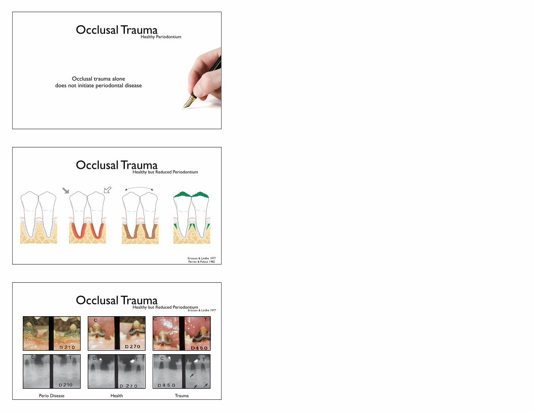
Occlusal trauma alone does not initiate periodontal disease
Occlusal TraumaHealthy but Reduced Periodontium
Trauma from Occlusion: Periodontal Tissues 357
a b
c d
Fig. 14-9 (a) Two mandibular premolars are surrounded by a healthy periodontium with reduced height. (b) If such premolars are subjected to traumatizing forces of the jiggling type a series of alterations occurs in the periodontal ligament tissue. (c) These alterations result in a widened periodontal ligament space and in an increased tooth mobility but do not lead to further loss of connective tissue attachment. (d) After occlusal adjustment the width of the periodontal ligament is normalized and the teeth are stabilized.
This problem has also been examined in animal experiments (Ericsson & Lindhe 1977). Destructive periodontal disease was initiated in dogs by allowing the animals to accumulate plaque and calculus for a period of 6 months (Fig. 14-8). When around 50% of the periodontal tissue support had been lost (Fig. 14-8a,b), the progressive disease was subjected to treat-ment by scaling, root planing, and pocket elimination (Fig. 14-8c). During a subsequent 8-month period, the animals were enrolled in a careful plaque-control program. During this period certain premolars were exposed to traumatizing jiggling forces. The peri-odontal tissues in the combined pressure and tension zones reacted to the forces by vascular proliferation, exudation, and thrombosis, as well as by bone resorp-tion. In radiographs, widened periodontal ligaments (Fig. 14-8d) could be found around the traumatized teeth, which displayed signs of progressive tooth mobility at clinical examination. The gradual increase
in the width of the periodontal ligament and the resulting progressive increase in tooth mobility took place during a period of several weeks but eventually terminated. The active bone resorption ceased and the markedly widened periodontal ligament tissue regained its normal composition; healing had occurred (Fig. 14-8e). The teeth were hypermobile but surrounded by periodontal structures which had adapted to the altered functional demands.
During the entire experimental period the supra-alveolar connective tissue remained unaffected by the jiggling forces. There was no further loss of con-nective tissue attachment and no further downgrowth of dentogingival epithelium (Fig. 14-8e). The results from this study clearly reveal that within certain limits a healthy periodontium with reduced height has a capacity similar to that of a periodontium with normal height to adapt to altered functional demands (Fig. 14-9).
Ericsson & Lindhe 1977Perrier & Polson 1982
Occlusal TraumaHealthy but Reduced Periodontium
Ericsson & Lindhe 1977356 Trauma from Occlusion
gg
aa bb
cc
ee
dd
ff
Fig. 14-8 (a) Dogs were allowed to accumulate plaque and calculus in the mandibular premolar regions over a 210-day period. (b) When around 40–50% of the periodontal tissue support had been lost the animals were treated by scaling, root planing, and pocket elimination. During surgery, a notch was prepared in the root at the level of the bone crest. (c, d) The dogs were subsequently placed on a plaque-control program and 2 months later (day 270) all experimental teeth (the lower fourth premolars; 4P and P4) were surrounded by a healthy periodontium with reduced height. (e) The mandibular left fourth premolar (T) was exposed to jiggling forces. (f) As a consequence, a widened periodontal ligament and increased tooth mobility resulted. (g) This increase in tooth mobility and the development of widened periodontal ligament space did not, however, result in apical downgrowth of the dentogingival epithelium. Arrowheads indicate the apical extension of the junctional epithelium which coincides with the apical border of the notch (N), prepared in the root surface prior to jiggling. C = control tooth; T = test tooth.
of plaque and calculus and elimination of pathologically deepened pockets, will result in the re-establishment of a healthy periodontium but with reduced height. The question is whether a healthy
periodontium with reduced height has a capacity similar to that of the normal periodontium to adapt to traumatizing occlusal forces (secondary occlusal trauma).
356 Trauma from Occlusion
gg
aa bb
cc
ee
dd
ff
Fig. 14-8 (a) Dogs were allowed to accumulate plaque and calculus in the mandibular premolar regions over a 210-day period. (b) When around 40–50% of the periodontal tissue support had been lost the animals were treated by scaling, root planing, and pocket elimination. During surgery, a notch was prepared in the root at the level of the bone crest. (c, d) The dogs were subsequently placed on a plaque-control program and 2 months later (day 270) all experimental teeth (the lower fourth premolars; 4P and P4) were surrounded by a healthy periodontium with reduced height. (e) The mandibular left fourth premolar (T) was exposed to jiggling forces. (f) As a consequence, a widened periodontal ligament and increased tooth mobility resulted. (g) This increase in tooth mobility and the development of widened periodontal ligament space did not, however, result in apical downgrowth of the dentogingival epithelium. Arrowheads indicate the apical extension of the junctional epithelium which coincides with the apical border of the notch (N), prepared in the root surface prior to jiggling. C = control tooth; T = test tooth.
of plaque and calculus and elimination of pathologically deepened pockets, will result in the re-establishment of a healthy periodontium but with reduced height. The question is whether a healthy
periodontium with reduced height has a capacity similar to that of the normal periodontium to adapt to traumatizing occlusal forces (secondary occlusal trauma).
Perio Disease Health Trauma

Occlusal TraumaHealthy but Reduced Periodontium
Ericsson & Lindhe 1977
356 Trauma from Occlusion
gg
aa bb
cc
ee
dd
ff
Fig. 14-8 (a) Dogs were allowed to accumulate plaque and calculus in the mandibular premolar regions over a 210-day period. (b) When around 40–50% of the periodontal tissue support had been lost the animals were treated by scaling, root planing, and pocket elimination. During surgery, a notch was prepared in the root at the level of the bone crest. (c, d) The dogs were subsequently placed on a plaque-control program and 2 months later (day 270) all experimental teeth (the lower fourth premolars; 4P and P4) were surrounded by a healthy periodontium with reduced height. (e) The mandibular left fourth premolar (T) was exposed to jiggling forces. (f) As a consequence, a widened periodontal ligament and increased tooth mobility resulted. (g) This increase in tooth mobility and the development of widened periodontal ligament space did not, however, result in apical downgrowth of the dentogingival epithelium. Arrowheads indicate the apical extension of the junctional epithelium which coincides with the apical border of the notch (N), prepared in the root surface prior to jiggling. C = control tooth; T = test tooth.
of plaque and calculus and elimination of pathologically deepened pockets, will result in the re-establishment of a healthy periodontium but with reduced height. The question is whether a healthy
periodontium with reduced height has a capacity similar to that of the normal periodontium to adapt to traumatizing occlusal forces (secondary occlusal trauma).
TraumaControl
Occlusal Trauma
Healthy reduced periodontium has capacity to adapt to altered functional demands.
Healthy but Reduced Periodontium
Occlusal TraumaDiseased Periodontium
Trauma from Occlusion: Periodontal Tissues 359
tal ligament will not aggravate a plaque-associated periodontal disease (Fig. 14-11).
If, however, the magnitude and direction of the jiggling forces were such that, during the course of the study (6 months), the tissues in the pressure/tension zones could not become adapted, the injury in the zones of co-destruction had a more permanent character. For several months the periodontal liga-ment in the pressure/tension zones displayed signs of infl ammation (vascular proliferation, exudation, thrombosis, retention of neutrophils and macro-phages, collagen destruction). Osteoclasts residing on the walls of the alveolus maintained the bone-resorptive process, which resulted in a gradual widening of the periodontal ligament in the pres-sure/tension zones (Fig. 14-12). As a consequence, the resulting angular bone destruction was continu-ous and the mobility of the teeth remained progres-
a b
c d
Fig. 14-11 (a) Two mandibular premolars with supra- and subgingival plaque, advanced bone loss and periodontal pockets of a suprabony character. Note the connective tissue infi ltrate (shadowed areas) and the uninfl amed connective tissue between the alveolar bone and the apical portion of the infi ltrate. (b) If these teeth are subjected to traumatizing forces of the jiggling type, pathologic and adaptive alterations occur within the periodontal ligament space. (c) These tissue alterations, which include bone resorption, result in a widened periodontal ligament space and increased tooth mobility but no further loss of connective tissue attachment. (d) Occlusal adjustment results in a reduction of the width of the periodontal ligament and in less mobile teeth.
sive. The plaque-associated lesion in the “zone of irritation” and the infl ammatory lesion in the “zone of co-destruction” merged; the dentogingival epithe-lium proliferated in an apical direction and periodon-tal disease was aggravated (Figs. 14-13, 14-14) (Lindhe & Svanberg 1974).
Similar fi ndings were reported from another experiment in the dog (Ericsson & Lindhe 1982) in which the effect was assessed of a prolonged period of jiggling force application on the rate of progression of plaque-associated, marginal periodontitis. Thus, in dogs with continuing periodontal disease, certain teeth were exposed to jiggling forces during a period of 10 months. Control teeth were not jiggled. Figure 14–15a illustrates the marked periodontal tissue breakdown around a tooth which was exposed to plaque infection combined with jiggling trauma for several months and Fig. 14-15b illustrates a
a b
c d
bbaa
Fig. 14-14 (a) Illustration of a tooth where subgingival plaque has mediated the development of an infi ltrated soft tissue (shadowed area) and an infrabony pocket. (b) When trauma from occlusion of the jiggling type is infl icted (arrows) on the crown of this tooth, the associated pathologic alterations occur within a zone of the periodontium which is also occupied by the infl ammatory cell infi ltrate (shadowed area). In this situation the increasing tooth mobility may also be associated with an enhanced loss of connective tissue attachment and further downgrowth of dentogingival epithelium; compare arrows in (c) and (d). Occlusal adjustment will result in a narrowing of the periodontal ligament, less tooth mobility, but no improvement of the attachment level (d) (Lindhe & Ericsson 1982).
Lindhe & Svanberg 1974Meintner 1975
Nyman et al 1978Polson & Zander 1983
Trauma from Occlusion: Periodontal Tissues 359
tal ligament will not aggravate a plaque-associated periodontal disease (Fig. 14-11).
If, however, the magnitude and direction of the jiggling forces were such that, during the course of the study (6 months), the tissues in the pressure/tension zones could not become adapted, the injury in the zones of co-destruction had a more permanent character. For several months the periodontal liga-ment in the pressure/tension zones displayed signs of infl ammation (vascular proliferation, exudation, thrombosis, retention of neutrophils and macro-phages, collagen destruction). Osteoclasts residing on the walls of the alveolus maintained the bone-resorptive process, which resulted in a gradual widening of the periodontal ligament in the pres-sure/tension zones (Fig. 14-12). As a consequence, the resulting angular bone destruction was continu-ous and the mobility of the teeth remained progres-
a b
c d
Fig. 14-11 (a) Two mandibular premolars with supra- and subgingival plaque, advanced bone loss and periodontal pockets of a suprabony character. Note the connective tissue infi ltrate (shadowed areas) and the uninfl amed connective tissue between the alveolar bone and the apical portion of the infi ltrate. (b) If these teeth are subjected to traumatizing forces of the jiggling type, pathologic and adaptive alterations occur within the periodontal ligament space. (c) These tissue alterations, which include bone resorption, result in a widened periodontal ligament space and increased tooth mobility but no further loss of connective tissue attachment. (d) Occlusal adjustment results in a reduction of the width of the periodontal ligament and in less mobile teeth.
sive. The plaque-associated lesion in the “zone of irritation” and the infl ammatory lesion in the “zone of co-destruction” merged; the dentogingival epithe-lium proliferated in an apical direction and periodon-tal disease was aggravated (Figs. 14-13, 14-14) (Lindhe & Svanberg 1974).
Similar fi ndings were reported from another experiment in the dog (Ericsson & Lindhe 1982) in which the effect was assessed of a prolonged period of jiggling force application on the rate of progression of plaque-associated, marginal periodontitis. Thus, in dogs with continuing periodontal disease, certain teeth were exposed to jiggling forces during a period of 10 months. Control teeth were not jiggled. Figure 14–15a illustrates the marked periodontal tissue breakdown around a tooth which was exposed to plaque infection combined with jiggling trauma for several months and Fig. 14-15b illustrates a
Rem
oved
Tra
uma
& In
flam
mat
ion
Rem
oved
Onl
y Tr
aum
a

Occlusal TraumaDiseased Periodontium
Lindhe & Svanberg 1974Meintner 1975
Nyman et al 1978Polson & Zander 1983
Rochester Gothenburg
358 Trauma from Occlusion
Plaque-associated periodontal disease
Experiments carried out on humans and animals have demonstrated that trauma from occlusion cannot induce pathologic alterations in the supra-alveolar connective tissue, i.e. cannot produce infl ammatory lesions in a normal gingiva or aggravate a gingival lesion associated with plaque and cannot induce loss of connective tissue attachment. The question remains if abnormal occlusal forces can infl uence the spread of the plaque-associated lesion and enhance the rate of tissue destruction in periodontal disease. This has been studied in animal experiments (Lindhe & Svanberg 1974; Meitner 1975; Nyman et al. 1978; Ericsson & Lindhe 1982; Polson & Zander 1983). In these experiments progressive and destructive peri-odontal disease was fi rst initiated in dogs or monkeys by allowing the animals to accumulate plaque and calculus. Teeth thus involved in a progressive peri-odontal disease process were also subjected to trauma from occlusion.
“Traumatizing” jiggling forces (Lindhe & Svanberg 1974) were exerted on premolars and were found to induce certain tissue reactions in the com-bined pressure/tension zones. Within a few days of the onset of the jiggling forces, the periodontal ligament tissue in these zones displayed signs of infl amma-
tion, had increased numbers of vessels, showed increased vascular permeability and exudation, thrombosis, as well as retention of neutrophils and macrophages. On the adjacent bone surfaces there were a large number of osteoclasts. Since the teeth could not orthodontically move away from the jiggling forces, the periodontal ligament of both sides of the tooth gradually increased in width, the teeth became hypermobile (progressive tooth mobility) and angular bony defects could be detected in the radiographs. The forces were eventually nulli-fi ed by the increased width of the periodontal ligament.
If the forces applied were of a magnitude to which the periodontal structures could adapt, the progres-sive increase of the tooth mobility terminated within a few weeks. The active bone resorption ceased but the angular bone destruction persisted as well as the increased tooth mobility. The periodontal ligament had an increased width but a normal tissue composi-tion. Histologic examination of biopsy specimens revealed that this adaptation had occurred with no greater apical proliferation of the dentogingival epi-thelium than was caused by the plaque-associated lesion (Fig. 14-10) (Meitner 1975). This means that occlusal forces which allow adaptive alterations to develop in the pressure/tension zones of the periodon-
aa bb
E EEE
E EEE
Fig. 14-10 (a) A composite photomicrograph illustrating the interdental space between two pairs of teeth. The teeth have been subjected to experimental, ligature-induced periodontitis and in (b) also to repetitive mechanical injury. In (b), there is considerable loss of alveolar bone and an angular widening of the periodontal ligament space (arrows). However, the apical downgrowth of the dentogingival epithelium in the two areas (a) and (b) is similar. E indicates the apical level of the dentogingival epithelium. Courtesy of Dr. S.W. Meitner.
Bone loss but not CT attachment loss
360 Trauma from Occlusion
In teeth with progressive, plaque-associated peri-odontal disease, trauma from occlusion may, under certain conditions, enhance the rate of progression of the disease, i.e. act as a co-factor in the destructive process. From a clinical point of view, this knowl-edge strengthens the demand for proper treatment of plaque associated with periodontal disease. This treatment will arrest the destruction of the periodon-tal tissues even if the occlusal trauma persists. Treat-ment directed towards the trauma alone, however, i.e. occlusal adjustment or splinting, may reduce the mobility of the traumatized teeth and result in some regrowth of bone, but it will not arrest the rate of further breakdown of the supporting apparatus caused by plaque. (For a detailed discussion of treat-ment of teeth exhibiting increased mobility, see Chapter 51.)
Fig. 14-13 Microphotographs from one control (C) and one test (T) tooth after 240 days of experimental periodontal tissue breakdown and 180 days of trauma from occlusion of the jiggling type (T). The arrowheads denote the apical position of the dentogingival epithelium. The attachment loss is more pronounced in T than in C. From Lindhe & Svanberg (1974).
Fig. 14-12 Radiographic appearance of one test tooth (T) and one control tooth (C) at the termination of an experiment in which periodontitis was induced by ligature placement and plaque accumulation and in which trauma of the jiggling type was induced. Note angular bone loss particularly around the mesial root of the mandibular premolar (T) and the absence of such a defect at the mandibular premolar (C). From Lindhe & Svanberg (1974).
control tooth which was exposed to plaque infection only.
On the other hand, more short-term experiments in the monkey (Polson & Zander 1983), evaluating the effect of trauma from occlusion on teeth involved in an ongoing process of periodontitis, failed to support the fi ndings by Lindhe and Svanberg (1974) and Ericsson and Lindhe (1982). Polson and Zander (1983) observed that trauma superimposed on peri-odontal lesions associated with angular bony defects (1) caused increased loss of alveolar bone but (2) failed to produce additional loss of connective tissue attachment.
Conclusions
Experiments carried out in humans as well as animals, have produced convincing evidence that neither uni-lateral forces nor jiggling forces, applied to teeth with a healthy periodontium, result in pocket formation or in loss of connective tissue attachment. Trauma from occlusion cannot induce periodontal tissue break-down. Trauma from occlusion does, however, result in resorption of alveolar bone leading to an increased tooth mobility which can be of a transient or perma-nent character. This bone resorption with resulting increased tooth mobility should be regarded as a physiologic adaptation of the periodontal ligament and surrounding alveolar bone to the traumatizing forces, i.e. to altered functional demands.
Fig. 14-15 (a) Periodontal conditions around a tooth which has been exposed to trauma from occlusion (of the jiggling type) for 300 days in combination with plaque-associated experimental periodontitis. (b) Condition of a control tooth from the same dog in which experimental periodontitis but no jiggling trauma had been in operation. Note the difference between (a) and (b) regarding the degree of bone destruction and loss of connective tissue attachment. Note also in (a) the location of the subgingival plaque at the apex of the root. From Ericsson & Lindhe (1982).
!
Bone loss &CT attachment loss
Gothenburg Study Model
Rochester Study Model

Worst outcome during regenerative surgery
Increase mobility is a risk factorfor tooth loss
What Does Clinical Reality Show?
Teeth with initial mobility do not respond to treatment as well as control
Periodontal treatment (SRP and/or surgery) increase mobility
Persson 1981Kerry 1982
Fleszar 1980
McGuire & Nunn 1996
Cortellini et al 2001
Occlusal Trauma
Occlusal trauma cannot induceperiodontal breakdown
However it results in resorption of alveolar boneincreasing tooth mobility
In teeth with progressive plaque accumulationit may enhance progression of disease
and Periodontal Disease
TREAT PERIO DISEASE FIRST
Occlusal Trauma
During initial therapy: Tooth with excessive mobility, especially
when it makes the patient uncomfortable.
During maintenance:if despite initial therapy, mobility is still
bothering the patient or if mobility has increased.
During periodontal regenerative surgery.
When do we adjust occlusion

Occlusal Trauma
If the mobility increases despite occlusal equilibration and periodontal health
If the mobility is uncomfortable for the patient
During periodontal regenerative surgery
When to splint
Thank You!