TRAUMA MANUAL
214
UNIVERSITY OF SASKATCHEWAN T RAUMA MANUAL 2 ND EDITION Department of Surgery
-
Upload
khangminh22 -
Category
Documents
-
view
0 -
download
0
Transcript of TRAUMA MANUAL

University of saskatchewan TRAUMA MANUAL
2nd edition Department of Surgery


Department of Surgery

The Department of Surgery is proud to present the second edition of the Trauma Manual. Under the leadership of Dr. Niroshian Sothilingam, the manual editor, numerous members of the Department of Surgery have contributed to its contents.
The manual provides a comprehensive and practical approach to the resuscitation and acute management of trauma patients. We feel that this manual will be of benefit to our trauma patients and encourage all of us to refresh our memories and our approach to acute trauma.
Although, this manual has focused on the management of acute trauma within the Saskatchewan medical system, the basic trauma management principles and algorithms presented here could be applied anywhere.
We want to thank the faculty and residents that have contributed to the manual and all the members of healthcare institutions across the Province of Saskatchewan for the exemplary care provided to our trauma patients.
- Ivar Mendez MD, PhD, FRCSC, FACS, FCAHS F. H. Wigmore Professor & Provincial Head Department of Surgery University of Saskatchewan | Saskatchewan Health Authority
Foreword

As our Trauma Program evolves and expands, so does our Trauma Manual. In this 2nd edition, we have added chapters dedicated to prehospital/transport, airway management, extremity trauma and updated guidelines.
Thank you to all who contributed their time and knowledge to the education of our trainees and advancing trauma care in Saskatchewan.
- Niroshan S. Sothilingam MD, FRCSC, FACS Medical Director, Trauma Program (Saskatoon) Editor in Chief, Trauma Manual

Evan Barber, BSc, MD, PGY3 (2017) Division of General Surgery, University of Saskatchewan
Richard Bigsby, MD, FRCSC Clinical Associate Professor, Head - Division of Thoracic Surgery, Department of Surgery University of Saskatchewan
Bailey Dyck, MD, PhD , PGY1 (2015) Division of Orthopedic Surgery, Western University
Daryl R. Fourney, MD, FRCSC, FACS Professor, Division of Neurosurgery, Department of Surgery University of Saskatchewan
Suzie Harriman, BSc, MD, FRCSC Assistant Professor, Division of General Surgery, Department of Surgery University of Saskatchewan
Leia Hoffman, MD, PGY3 (2020) Department of Emergency Medicine, University of Saskatchewan
Michael E. Kelly, MD, PhD, FRCSC, FACS Professor, Head - Division of Neurosurgery, Department of Surgery Saskatchewan Clinical Stroke Research Chair Director, Saskatchewan Cerebrovascular Centre University of Saskatchewan
Michael Kindrachuk, MD, PGY2 (2017) Division of Neurosurgery, University of Saskatchewan
Kylie Kvinlaug, MD, FRCSC Assistant Professor, Division of Vascular Surgery, Department of Surgery University of Saskatchewan
Youngseo Lee, MD, PGY2 (2020) Department of Anesthesiology, University of Saskatchewan
Ryan Lett MD, FRCPC Department of Anesthesiology, University of Saskatchewan
Sarah Miller, BSc, BA, MD, FRCSC Assistant Professor, Division of General Surgery, Department of Surgery University of Saskatchewan
U. O. Moodley, MD, FRCP Department of Medicine, University of Saskatchewan
Elliott Pally MD, FRCSC Assistant Professor, Division of Orthopedic Surgery, Department of Surgery University of Saskatchewan
M. Chris Pastor, MD, MSc, PGY2 (2017) Division of General Surgery, University of Saskatchewan
Anokhi Patel, MD, PGY2 (2020) Department of Anesthesiology, Perioperative Medicine and Pain Management University of Saskatchewan
Contributors:

Dallas Pearson MD, CCFP-EM Department of Emergency Medicine, University of Saskatchewan
Sunil Pradhan, MD, PGY3 (2020) Department of Emergency Medicine, University of Saskatchewan
Oksana Prokopchuk-Gauk, MD, FRCPC Transfusion Medicine Consultant & Clinical, Hematologist Department of Pathology & Laboratory Medicine, University of Saskatchewan
Elizabeth Quon, CC4 (2020) College of Medicine, University of Saskatchewan
Zeeshan Rana, MD, MSc, PGY5 (2017) Division of General Surgery, University of Saskatchewan
Kajeandra Ravichandiran, HBSc, MSc, MD, PGY1 (2015) Division of Orthopedic Surgery, Western University
J. M. Shaw, MBBCh, FCS(SA), MMed (surg), FRCSC Clinical Professor, Head - Division of General Surgery, Department of Surgery University of Saskatchewan
Niroshan Sothilingam MD, FRCSC, FACS Assistant Professor, Division of General Surgery, Department of Surgery Medical Director, Trauma Program University of Saskatchewan
Michael Spiess, MD, FRCSC Assistant Professor, Division of Orthopedic Surgery, Department of Surgery University of Saskatchewan
Alena Stirling MD, FRCPC Department of Anesthesiology, Perioperative Medicine and Pain Management University of Saskatchewan
Ian Sunderland, MD FRCSC Assistant Professor, Head - Division of Plastic Surgery, Department of Surgery University of Saskatchewan
Christopher Thomson, MD, FRCSC Assistant Professor, Division of Plastic Surgery, Department of Surgery University of Saskatchewan
Andrew Urmson, BSc, MD, FRCSC Assistant Professor, Division of Orthopedic Surgery, Department of Surgery University of Saskatchewan
Kelly Vogt MD, MSc, FRCSC Trauma & Acute Care Surgeon Division of General Surgery, Western University
Laura Weins, MD, FRCSC Clinical Assistant Professor, Department of Obstetrics and Gynecology University of Saskatchewan
Scott Wilms, MD, PGY4 (2020) Division of Orthopedic Surgery, University of Saskatchewan
Department of Surgery - Communications University of Saskatchewan

SK Medical Transport Prehospital Interfacility Care 10
Initial Resuscitation 14
Airway Management 22
Traumatic Brain Injury 32
Cranio-facial Trauma 44
Neck Trauma 50
Chest Trauma 58
Abdominal Trauma 74
Pelvic Trauma 94
Extremity Trauma 110
Spine Trauma 136
Trauma in Pregnancy 154
Burn Management 166
Hemorrhage Control and Balanced Transfusion in Trauma 174
Beyond the Trauma Bay: VTE Prophylaxis, Tracheostomy,
Enteral Nutrition and Tertiary Surveys 188
Appendix 206
TRAUMA MANUALcontents


Saskatchewan Medical TransportPre-Hospital/Interfacility Care
Dallas Pearson, Leia Hoffman, Sunil Pradhan
Patient TransferIn Saskatchewan there are 3 main methods of patient transport in the pre-hospital or interfacility transfer setting:
Ground Ambulance: Various private or publicly owned companies depending on the region Who they transport: Most stable patients, unstable patients within a short distance of Saskatoon or as a bridge to air resources and any patient when air resources are unavailable
Helicopter: Shock Trauma Air Rescue Society (STARS): Non-profit organization with bases in Saskatoon and Regina focused on rescue and transport of trauma patients; transport patients with a Critical Care Paramedic and an RN Who they transport: Unstable or time-dependent patients Decision to attend a scene call/transfer a patient depends on the specific location, timing, weather and availability of helicopter Considerations: Flying radius 300km without stopping to refuel, can go farther if they stop to refuel (at any airport or fuel cache)
Fixed Wing Plane: Saskatchewan Air Ambulance (SAA): Transport patients with a Critical Care Paramedic and an RN Who they transport: Unstable or time dependent patients coming from a healthcare facility ONLY (these patients will have been assessed prior to their transfer) Decision to transfer a patient depends on specific location and availability of the plane Considerations: The plane needs an airport or landing strip to land but may either: Meet a ground ambulance crew who have picked up the patient from the health care facility Use a ground ambulance (staffed by SAA crew) to pick the patient up from the healthcare facility if specialized skills are required
Role of the Transport Physician
Determine the most efficient, safe, fastest mode of transport for a patient taking into consideration the patient's injuries and the resources available
Required on flight if specific skill set required that the RN or Paramedic are not li-censed/comfortable performing (eg. Chest tube, temporary transvenous pacemaker)
Determine if delivery of a specialist physician to the patient is indicated (eg. Cardiolo-gist to insert a transvenous pacemaker)
UofS TRAUMA MANUAL | PAGE 11

Trauma Team ActivationThe trauma teams receives patients in one of three ways (or a combination of these ways):
Scene call: Health care providers are called to the scene of a trauma (eg. MVC) and are the patients first point of contact with the health care system Ground paramedics or STARS are the only two services which attend scene calls
Interfacility transfer: Patients have seen an MD or RN at a non-tertiary health care facility and are being transferred to a tertiary centre for trauma team activation, assessment and ongoing care Multiple different methods of transfer: STARS, Ground paramedics, SAA or a combination of ground and air services
A patient meeting Level 1 criteria presents to the ED without contact with prehospital services
The information about these patients will be communicated with the TTL/Trauma team in different ways depending on the type of transport
Scene Call: STARS will call the transport physician upon initial assessment who may contact the TTL with the details of the case STARS will send in a patch to triage in the typical fashion to activate the trauma team
Interfacility Transport: The accepting (TTL), sending and transport physicians have a conference call where the sending physician presents the patient to the TTL for transfer. The TTL typically accepts the patient (or gives recommendation for management at home hospital). If the patient is accepted the transport physician will determine the best method of transfer to the tertiary centre for assessment. STARS/SAA will call the transport physician with a bedside assessment /update upon their arrival which will be relayed to the TTL as needed
Paramedic Levels of Training and CapabilitiesIn Saskatchewan there are 6 levels of Paramedic (see table 1 and 2 for a summary of their capabilities):
Emergency Medical Responder (EMR) Emergency Medical Technician (EMT) Primary Care Paramedic (PCP; commonly referred to as “BLS”) Intermediate Care Paramedic (ICP) Advanced Care Paramedic (ACP; commonly referred to as “ACLS”) Have ACLS, ATLS, NRP, PALS, difficult airway course Critical Care Paramedic (STARS/SAA) Usually trained by the company that hires them

UofS TRAUMA MANUAL | PAGE 13


Initial ResuscitationSuzie Harriman, Niroshan Sothilingam
The trauma team responds to all level I traumas
Trauma Activation Criteria – See Appendix
All members of the trauma team must wear personal protective equipment:
Cap, gown, gloves, mask, protective eyewear/shield
The members of the trauma team will include many individuals who all have a very specific role. It is critical for the safety of the team members and the patient that everyone is aware of their roles and where to stand.
Trauma Team Leader (TTL): The most responsible physician (MRP) who runs and controls the room The TTL will stand at the foot of the bed Directs team members in their actions Keeps track of the whole state of the patient Receive and interpret all results of investigations Consult with other specialties Decide on appropriate disposition Will accompany patient out of the trauma bay for all investigations Talk to family members
Learner Trauma Team Leader (TTLST): Senior level trainee under the direct supervision of the TTL Will stand at the foot of the bed with the TTL Role is to oversee trauma patient and critical procedures under direction of the TTL, provision of/or right and left sided procedures.
Airway Management Will stand at the head of the bed Is the Anesthesia resident on-call Role is airway assessment & management Endotracheal intubation if necessary C-spine control Assist with fluid and drug administration Respiratory therapist (RT)/Anesthesia assist (AA) will stand beside them at the head of the bed
Primary Survey Will stand on the right side of the patient Is the General Surgery JR resident on-call Responsible for right sided procedures (e.g. chest tube) Responsible for the primary and secondary survey Role is to oversee trauma patient and critical procedures under direction of the TTL, provision of/or right and left sided procedures.
UofS TRAUMA MANUAL | PAGE 15

Scribe Nurse Role is to record events of trauma resuscitation
Circulating Nurse (x 2) Obtain IV access, attach cardiac monitoring and pulse oximeter on arrival Remove all clothing and keep patient warm with blankets Administer drugs and hang fluids Place foley catheter if necessary Help role the patient Help transport the patient when necessary
Primary Survey
All trauma patients on arrival should have: 2 large bore IV started (14 or 16 gauge) Cardiac monitoring Blood pressure cuff Pulse Oximeter
Assess and establish treatment priorities. Assess vital signs quickly and efficiently. Adhering to the sequence of ABCDEs of trauma.
The primary survey can and should be repeated frequently to reassess any change to patient’s status with subsequent intervention if required. Important to be cognoscente of special populations (i.e. elderly, pediatric, pregnant, athletes) as they will have different ability to compensate, more/less reserve, medication profiles and “normal” vitals.
AN
SN
CN
CNER
PS
TTLSTTTL
AN: Anesthesia Resident On call
PS: Primary Survey (JR General Surgery Resident On Call
ER: Emergency Room Resident
TTL: Trauma Team Leader
TTLST: Trauma Team Leader SR Trainee (SR General Surgery/Emergency/Anesthesia Resident on Trauma Service
SN: Scribe Nurse
CN: Circulating Nurse x 2

UofS TRAUMA MANUAL | PAGE 17
Airway and cervical spine protection: Ascertain patency of airway. Is that patient verbalizing? Significant facial trauma causing possible loss of airway? Is GCS <8? (likely unable to protect airway) Always have suction ready at head of bed when assessing airway. Clear the oropharynx of blood, mucus and foreign bodies. Avoid excessive cervical spine movement while assessing and managing airway Immobilization of cervical spine should be maintained – assume cervical spine injury in all trauma patients with blunt mechanism, multisystem involvement or altered LOC
Breathing and Ventilation: Is there adequate gas exchange – oxygenation and carbon dioxide elimination? Respiratory rate; Oxygenation saturation Expose neck and chest and inspect chest movement and palpate for injury that may compromise ventilation i.e. flail chest, tension pneumothorax and open pneumothorax Percussion may be helpful to identify a pneumothorax or hemothorax, but is very difficult to perform in a noisy resuscitation bay Auscultation for bilateral breath sounds Injuries impairing ventilation should be identified and treated immediately: Pneumo/hemothorax: Chest tube placement Tension pneumothorax: Needle decompression/chest tube Open pneumothorax: Three-sided occlusive dressing Positive pressure ventilation may exacerbate or cause pneumothorax or a tension pneumothorax – ensure frequent re-evaluation.
Circulation (including hemorrhage control): Recognize signs of organ hypoperfusion Level of consciousness Skin color/temperature Pulse rate/character Identify hemorrhage (external vs internal) External – direct pressure, tourniquets if direct pressure fails Internal – chest, abdomen, retroperitoneum, pelvis, long bones Identify through physical exam, CXR, pelvic x-ray, FAST Management: chest tube, pelvic binder, splint application, surgical consult Restore volume Start with crystalloid followed by PRBCs If obvious hypovolvemic shock, may consider initially resuscitating with blood products

May need activation of massive transfusion protocol (MTP) - See Appendix for MTP activation protocol
Disability: Baseline neurologic exam Glasgow coma scale – predictive of patient outcome Pupillary size and reaction Lateralizing signs Spinal cord injury level Objective is to prevent secondary brain injury by ensuring adequate oxygenation and perfusion
Exposure and Environmental Control Fully expose patient for assessment of additional injuries while preventing hypothermia Warm blankets, warming devices, warm IV fluids and room temperature
Adjuncts to Primary Survey Determine occult bleeding and source of shock ECG: Continuous monitoring Dysrhythmias – blunt cardiac injury Pulseless electrical activity – cardiac tamponade, tension pneumothorax and/or hypovolemia Note: hypothermia can cause dysrhythmias Foley catheter and nasogastric tubes Indwelling bladder catheter useful to monitor volume status and renal perfusion Relatively contraindicated if: - Blood at meatus - Perineal ecchymosis - High-riding or high riding prostate Nasogastric tube can reduce stomach contents and distention and decrease risk of aspiration. Can also assess hemorrhage from injury to upper digestive tract Contraindicated if: - Suspected fracture of Cribiform › Orogastric tube can be inserted in this case Arterial blood gas X-Rays AP chest and AP pelvic films (portable) Should not interrupt resuscitation process FAST1
Initial test for detection of occult intra-peritoneal hemorrhage Cardiac tamponade Pneumothorax (E-FAST)

Secondary Survey Begins when primary survey has been completed and resuscitation is succeeding in normalizing vital functions. It is a head to toe evaluation, including a complete history and physical exam. History: AMPLE A: allergies M: medications P: past illnesses/pregnancy L: last meal E: events/environment related to the injury Physical exam Head Scalp Eyes Ears (Blood/CSF leak) Penetrating Injuries Maxillofacial Structures Bony structures, intraoral, and soft tissues Cervical Spine and Neck Dependent on mechanism of injury – may leave immobilized until cervical spine radiological studies performed Examine neck, c-spine tenderness, bruits, subcutaneous emphysema, Chest Palpation of entire chest, auscultation, heart sounds Abdomen Frequent re-evaluations and high index of suspicion. Look for evidence of seat belt sign Avoid excessive manipulation of the pelvis Perineum, Rectum, Vagina Contusions, hematomas, lacerations and urethral bleeding Digital rectal exam – examine for rectal tone, blood, high riding prostate Pelvic fractures can cause vaginal injury Musculoskeletal System Contusions, deformities Palpation of bones for tenderness or abnormal movement with/with out pain Ensure examination includes the back or significant injuries can be missed Neurological System Motor and sensory evaluation of the extremities Re-evaluation of GCS and pupillary size and response Ensure protection of spinal cord at all times
UofS TRAUMA MANUAL | PAGE 19

Adjuncts to Secondary Survey Does the patient require further diagnostic tests? Is the patient’s condition/vitals appropriate for further diagnostic tests? Spinal x-rays CT of the chest, abdomen, and/or spine Extremity x-rays Transesophageal Ultrasound Bronchoscopy Esophagoscopy Spinal Immobilization Transport boards should be used for extrication purposes only, not for transport Not shown to reduce movement of the spine or neurological complications Pressure ulcers can begin 30 minutes after immobilization2
Can affect airway management and breathing3
C-collars and C-spine immobilization Please see Appendix for U of S C-spine clearance protocol Clinical Decision Rules – Radiography? NEXUS vs Canadian C-spine rules In an evaluable patient If C-spine CT normal, can remove collar Canadian C-spine rules4 In an obtunded patient Normal C-spine CT read by a staff radiologist, can remove collar EAST5
Vascular Access6
Percutaneous Peripheral Venous Access Two large bore (16g) IV catheters Central Venous Access If unable to obtain peripheral IV access Similar complication rate in non-emergent situation Internal jugular – carotid artery puncture (most common complication) Subclavian – pneumothorax (most common complication) Femoral – not recommended for intra-abdominal trauma Venous Cutdown If peripheral or central access in contrain dicated or impossible to achieve U/E: cephalic, basilic and median antecubital veins L/E: Greater saphenous vein Interosseous Catheters Most successful in patients less than 5 years of age Tibial tuberosity in pediatrics Proximal to tip of medial malleolus in adults

References
1. ATLS Manual 10th Edition. American College of Surgeons. 2018
2. Hoffman JR, et al. Validity of a set of clinical criteria to rule out injury to the cervical spine in patients with blunt trauma. National Emergency X-Radiography Utilization Study Group. N Engl J Med. 2000;343(2):94
3. Patel MB, et al. Cervical spine collar clearance in the obtunded adult blunt trauma patient: a systematic review and practice management guideline from the Eastern Association for the Surgery of Trauma. J Trauma Acute Care Surg. 2015;78(2):430.
4. Seamon M, Haut et al. An evidence-based approach to patient selection for emergency department thoracotomy: A practice management guideline from the Eastern Association for the Surgery of Trauma. J Trauma Acute Care Surgery; 2015
5. Sparke A et al. The measurement of tissue interface pressures and changes in jugular venous parameters associated with cervical immobilization devices: a systemic reviews. Scand J Trauma Resusc Emerg Med. 2013 Dec;3;21:81.
6. Stengel D et al. Systemic review and meta-analysis of emergency ultrasonography for blunt abdominal trauma. Br J Surg. 2001;Jul;88(7):901-12.
7. Sweeney M. Vascular Access in Trauma: Options, Risk Benefits, and Complications. Trauma. 1999 Mar 17(1)97-106.
8. Totten VYet al. Respiratory Effects of Spinal Immobilization. Prehosp Emerg Care.1999 Oct-Dec;3(4):347-52.
UofS TRAUMA MANUAL | PAGE 21


Airway ManagementAnokhi Patel, Youngseo Lee, Alena Stirling
AssessmentThe initial assessment is an essential first step in the ATLS algorithm. It should:
Determine if the airway is patent and protected, and if not, what type of intervention is necessary Assess for potential difficulty in managing the airway Determine the urgency of any potential intervention
Rapid Assessment
Inspection - facial/neck trauma, cyanosis, oropharynx, breathing pattern Palpate - trachea to ensure it is midline, chest for subcutaneous emphysema C-spine collar if trauma dictates its use An airway is typically patent and protected if a patient is able to talk clearly Reassess frequently as a patent airway can become obstructed rapidly
Assessment for Potential Difficulty
Multiple factors can make airway management challenging:
Environmental Factors
Busy/noisy/unfamiliar environment, ongoing resuscitation with competing goals, time critical, unrehearsed/unfamiliar team, clinician experience, patient cooperation
Physiologic Factors
Uncorrected hypoxemia, hypotension or laboratory abnormalities
Attempt to correct these prior to intubation as worsening of these states can occur with intubation and lead to devastating consequences
Anatomic Factors
Trauma related or pre-existing physical features that make any aspect of airway management challenging
Mnemonics are recommended to aid in assessing the anatomic factors.
For intubation, evaluate for LEMON
L - Look for facial trauma, small jaw, retrognathia E - Evaluate distance between incisor teeth (3 finger breadths), distance from hyoid bone to mentum (3 finger breadths), distance from thyroid notch to floor of mouth (2 finger breadths) M - Mallampati I = soft palate, uvula, fauces, tonsillar pillars entirely visible, II = soft palate, uvula, fauces partially visible, III = soft palate visible, IV hard palate only visible Higher Mallampati score may correlate with more difficult intubation O - Obstruction anywhere along the airway N - Neck mobility range of flexion to extension in patients who do not have C-spine collar on
UofS TRAUMA MANUAL | PAGE 23

For bag valve mask ventilation (BMV) evaluate for BONE
B - Beards can make it difficult to create a mask seal O - Obesity or Obstruction can cause difficulty by soft tissue redundancy in the airway or by foreign objects in the airway - if soft tissue is the issue, it can often be relieved by insertion of an OPA or NPA N - No teeth or edentulous patients often have sunken in cheeks when dentures are out which creates difficulty trying to obtain a mask seal E - Elderly patients may be more difficult to ventilate via bag valve mask ventilation
For supraglottic device (SGD) placement, evaluate for RODS
R - Restricted mouth opening O - Obstruction at or below the larynx D - Disruption or distortion of airway anatomy either above or below the larynx S - Stiff lungs or C- Spine Conditions that increase the amount of pressure required during positive pressure ventilation may limit the effectiveness of a SGD and C - spine precautions must be maintained during LMA placement
For front of neck airway (FONA) evaluate for SHORT
S - Surgery Previous surgery may lead to scar tissue or distorted anatomy H - Hematoma Blood or any other cause of swelling in the neck including subcutaneous emphysema, abscess or infection O - Obesity May make both landmarking and performing the procedure challenging R - Radiation May cause scarring and distorted anatomy T - Tumor Any abnormal or enlarged tissue including thyroid
A difficult airway is defined as difficulty with laryngoscopy and intubation, bag mask ventilation (BMV), supraglottic device ventilation, and with front of neck airway (FONA) access.
Anticipation of difficulty with multiple interventions should prompt the provider to call for additional help and prepare second-line or third-line options for airway management
Maintaining spontaneous ventilation should be considered and awake airway management may be the most appropriate choice in some circumstances

Definitive Airway ManagementA definitive airway is a cuffed tube in the trachea achieved by endotracheal or naso-tracheal intubation, cricothyrotomy or tracheostomy.
Potential Reasons for a Definitive Airway and Associated Features
Trauma Considerations Cervical Spine Injuries
May be associated with edema and cervical hematomas Injuries above C5 level may be associated with diaphragmatic dysfunction resulting in respiratory distress During airway management, the anterior part of the C-Spine collar should be removed and a dedicated individual should provide manual inline stabilization Minimize movement of the neck during all airway manipulation
Laryngotracheal Injury
Signs and symptoms of laryngotracheal injury include stridor, subcutaneous emphysema, crepitus, hoarseness, dysphonia, aphonia, dyspnea, dysphagia, neck or throat pain, large-volume hemoptysis, tracheal deviation, neck hematoma/ lacerations/contusions/trauma, wound bubbling, and palpable laryngeal fracture
UofS TRAUMA MANUAL | PAGE 25

Suspected laryngotracheal injury should be intubated under direct visualization to prevent further damage or creation of false lumen Maintaining spontaneous ventilation is desired - positive pressure ventilation may worsen the subcutaneous emphysema and distort anatomy and should be minimized until the airway is secured Suspicion of trauma to larynx or trachea should initiate a call for help from an experienced airway clinician
Facial Trauma
May increase the difficulty of intubation and bag-mask ventilation because of nasopharyngeal and oropharyngeal edema, bleeding, airway obstruction and jaw dysfunction May have concomitant C-Spine injuries Avoid nasal instrumentation, including nasal intubation or NPA insertion in patients with a suspected basilar skull fracture An awake tracheostomy or awake fiberoptic intubation may be required in certain scenarios and consultations to ENT, general surgery, or anesthesiology should be made
Full Stomach and Aspiration Risk
Most trauma patients are considered to have a “full stomach” and have a higher risk of aspiration, especially with airway manipulation Aspiration can lead to aspiration pneumonitis Cricoid pressure can be considered to reduce aspiration risk by creating direct pressure to the esophagus and mechanically prevent aspiration (however, this may lead to difficulties with intubation) Positioning patients head-up, or reverse Trendelenburg and ensuring functional suction equipment is available are important mitigating factors IV ranitidine can decrease the acidity of gastric contents and decrease severity of potential pneumonitis should aspiration occur Minimizing the time between injection of induction agents and securing a definitive airway also decreases aspiration risk
Preparation for IntubationThe list can be remembered with mnemonic MS MAID. M - Machine Bag-valve mask attached to an oxygen supply Appropriate sized mask to fit the patient Ventilator should also be available post-intubation to connect to the endotracheal tube and ventilate the patient S- Suction Available, reachable, and functioning Yankauer tip on the end of tubing

M- Monitors Blood pressure (NIBP or arterial line) SpO2 ECG End-tidal CO2 (either a colorimetric monitor attached to a bag-valve mask or a quantitative monitor connected to a central monitor) Ensure that the monitors are calibrated and are appropriately sized to the patient and are cycling frequently (e.g. NIBP every 5 minutes or less) A- Airway Oropharyngeal and Nasopharyngeal airway (OPA/NPA) with multiple sizes available Lubricate NPAs if planning on using them to prevent bleeding in nasopharynx Endotracheal tubes (ETTs) Size 7.0 for females and 8.0 for males is typical, but ensure there are multiple smaller sizes available Check the integrity of the cuff of the endotracheal tube by injecting air into the cuff using a 10cc syringe and examining for a leak Stylet Styletting the endotracheal tube provides the tube with stiffness and curvature which can help facilitate the intubation process Direct/video laryngoscope (Glidescope/CMAC) Check the light source of the laryngoscope Ensure the video laryngoscope is plugged in and the blade is connected to the screen Backup airway devices, such as a LMA, fiberoptic bronchoscope and/or bougie should be available when difficulty is anticipated I - Intravenous Reliable IV access available A fluid warmer, blood set, or drug infusion pump may be required de pending on the clinical situation D- Drugs Intubation drugs (see below) Vasopressor agents (such as epinephrine, norepinephrine or vasopressin) Sedative agents for post-intubation
Optimization Prior to Intubation Patient Positioning Position bed in reverse Trendelenburg position Ramp up the patient's head and neck using pillows and/or flannels Put the patient's head and neck in “sniffing position” (neck is flexed and head extended) if no C-Spine precaution is indicated
Preoxygenation Process of denitrogenation of the lung with a high concentration of oxygen to decrease the desaturation that may occur with apnea associated with intubation If feasible, patients should be administered 100% for at least 3 minutes, or 8 vital capacity breaths by BMV prior to intubation
UofS TRAUMA MANUAL | PAGE 27

Airway Maintenance TechniquesIn patients with a decreased level of consciousness, the tongue can fall back into the hypo-pharynx and can cause airway obstruction. This can also occur between the time paralytic is given and an attempt at intubation is performed. Some simple maneuvers can be done to try to create a patent airway, and improve the patient's oxygenation and ventilation.
1. Chin Lift Place digits 3-5 under the patient’s mandible and lift the chin anterior
2. Jaw Thrust Place a finger on either side behind the both angles of the mandible and displace the mandible forward
3. Nasopharyngeal Airway (NPA) Inserted in one nostril and passed into posterior oropharynx Should be well lubricated Generally well tolerated for awake patients Contraindicated in patients with suspected or potential cribriform fracture
4. Oropharyngeal Airway (OPA) Tolerated by obtunded patients Inserted upside down and then rotated 180 degrees at the soft palate before continuing to advance until in place If a patient has an intact gag reflex, inserting an oral airway may cause the patient to vomit and/or aspirate
5. Laryngeal Mask Airway (LMA) LMAs are supraglottic airways that can be used as a rescue airway for ventilating a patient with a difficult airway Inserted blind and does not require much manipulation of the head or neck Not considered a definitive airway
Intubation AgentsDrug assisted intubation goals should include sedation/avoiding awareness, avoiding the sympathetic surge associated with laryngoscopy, and improving intubating conditions while maintaining adequate hemodynamics. Some patients may not require any drugs for intubation, such as those in cardiac arrest. A large portion of trauma patients are intubated with a modified rapid sequence intubation which includes use of one hypnotic agent and a muscle relaxant given rapidly back to back. Caution has to be taken to choose appropriate doses to avoid hemodynamic instability with this technique and plans need to be in place in case of inability to intubate.
Propofol Most commonly used anesthetic agent Onset of action: typically within 30-40 seconds Duration of action: 3-10 minutes if given in a bolus Dose range: 1-2 mg/kg however widely varies on clinical scenario Does not have analgesic properties Causes respiratory depression Causes myocardial depression and vasodilation which can lead to hypotension

Should be avoided or used with extreme caution in patients in shock states
Midazolam Commonly used in conjunction with other anesthetic agents for induction of anesthesia Facilitates amnesia while causing sedation Onset of action: 3-5 minutes Duration of action: 1-2 hours Dose range: 0.5-2 mg Minimal cardiovascular or respiratory effects if used alone
Ketamine Achieves state of unconsciousness by inducing a dissociated state Onset of action: 30 seconds Duration of action: up to 10 minutes Dosing: 1-2 mg/kg however widely varies on clinical scenario Causes stimulation of the heart with increased blood pressure and cardiac output by stimulating presynaptic release of norepinephrine May be beneficial for patients in hypovolemic or cardiogenic shock If a patient is already sympathetically driven and has no more norepinephrine stores available, ketamine acts as a direct myocardial depressant and can lead to hemodynamic compromise Has inherent analgesic properties Causes less respiratory depression compared to propofol
Fentanyl Potent, fast acting opioid used as adjunct for intubation to blunt the sympathetic response to intubation Onset of action: 2-3 minutes Duration: 0.5-1 hour Dosing: 0.5-2mcg/kg but widely varies according to clinical scenario Can cause respiratory depression or apnea
Succinylcholine Depolarizing neuromuscular blocking agent Onset of action: 30 seconds Duration: up to 10 minutes Dosing: 1-2 mg/kg Evaluate for fasciculations after administering the drug, and attempt intubation once fasciculations stop Contraindications: history of malignant hyperthermia, globe rupture, hyperkalemia, skeletal muscle myopathies, 24 hours after a burn, chronic paralysis
Rocuronium Nondepolarizing muscle relaxant Onset of action: 30-90 seconds (faster onset at higher doses) Duration: 30-45 minutes Dosing: 0.6-1.2 mg/kg Remember sedation for patients post intubation if required as muscle paralysis lasts up to 45 minutes
UofS TRAUMA MANUAL | PAGE 29

Sugammadex is a reversal agent for rocuronium and can be used in a can’t ventilate, can’t intubate situation
Indicators of Successful Intubation End tidal CO2 consistently present with each ventilated breath Misting of the ETT Bilateral chest rise Breath sounds heard on both sides of the chest with auscultation Visualization of the ETT going through the vocal cords during laryngoscopy
Visual Aids and ChecklistsChecklists can be a helpful reminder in emergency situations.
Adult Trauma Intubation ChecklistIndication Actual or imminent airway
obstruction Severe/uncontrollable nasal
pharyngeal or upper airway hemorrhage Altered mental status / traumatic
brain injury or aspiration risk (GCSs 8) Spinal cord lesion with insufficient
respiratory mechanics Cardiac arrest Refractory or severe
hypoventilation Refractory or severe hypoxia Hemprrhagic shock with incipient
respiratory failure Severe smoke inhalation, major
thermal or chemical burns Cellular hypoxia/CN or CO
intoxication
Post Intubation Ensure Continuous Capnography Check BP q3min Consider Sedation/Analgesia Consider Ongoing NMBA Assess Ventilator Settings CXR OG tube prn Restraints prn Debrief Documentation
Prepare/Proceed Appropriate airway personnel
present +/- additional staff/advanced airway techniques Roles assigned:
Lead/MILS/BMV/Drugs/Eti/?Cricaid Monitors: PulseOx, BP, ECG,
ETCO2 Positioning: Reverse Trend/
Ramp Dual PreO2: Nasal CAnnula/NRB
Mask/BMV with PEEP/NIV Fluid bolus / Blood available Pressor support ready Airway equipment sized ready:
BVM/ventilator/laryngoscope/videoolaryngascopy/ETTs/stylet/bougie/SGA/surgical equipment/suction Medications: topical and IV Intubation Time out/All Ready
Plan Assess level of difficulty
- BMV/SGA/Laryngoscopy/ Surgical Airway Consider shock index
- HR/sBP > 0.8 Assess for dangerous physiology
- low Sat/low pH/RV strain RSI appropriate vs. Titrated vs.
Awake Prepare/plan for difficulty
- Plan A (ex. DL, VL, fiberoptic) - Plan B (Rescue O2 - 2hand BMV with OPA) - Plan C (Alternate ETI/SGA) - Plan D (Surgical Airway)
Approximate Drug Dose by Wt (kg) and Shock Index(Adult doses based on clinical judgement and have medication ready for hemodynamic support and additional/ongoing sedation)
SI<0.8 (HR<sBP) 50 Kg 75 Kg 100 KgKetaminePropofol
EtomidateRocuronium
Sux
10 - 30 mg 20 - 40 mg 20 - 50 mg10 mg 10 - 20 mg 10 - 20 mg5 mg 5 - 10 mg 5 - 10 mg
50 mg 100 mg 100 mg80 mg 120 mg 150 mg
SI>0.8 (HR>sBP) 50 Kg 75 Kg 100 KgKetaminePropofol
EtomidateRocuronium
Sux
10 - 30 mg 20 - 40 mg 20 - 50 mg10 mg 10 - 20 mg 10 - 20 mg5 mg 5 - 10 mg 5 - 10 mg
50 mg 100 mg 100 mg80 mg 120 mg 150 mg

References
1. Kovacs G, Sowers N. Airway Management in Trauma. Emerg Med Clin North Am [Internet]. 2018 Feb 1 [cited 2020 Apr 27];36(1):61–84. Available from: https://www.sciencedirect.com/science/article/pii/S0733862717300755?via%3Dihub
2. Mayglothling J, Duane TM, Gibbs M, McCunn M, Legome E, Eastman AL,et al. Emergency tracheal intubation immediately following traumatic injury. J Trauma Acute Care Surg [Internet]. 2012 Nov;73:S333–40. Available from: http://content.wkhealth.com/linkback/openurl?sid=WKPTLP:landingpage&an=01586154-201211004-00010
3. Estime SR, Kuza CM. Trauma Airway Management: Induction Agents, Rapid Versus Slower Sequence Intubations, and Special Considerations. Anesthesiol Clin [Internet]. 2019 Mar 1 [cited 2020 Apr 27];37(1):33–50. Available from: https://www.anesthesiolo-gy.theclinics.com/article/S1932-2275(18)30089-2/fulltext#.XqdHc1mTaL4.mendeley
UofS TRAUMA MANUAL | PAGE 31

Traumatic Brain InjuryNiroshan Sothilingam, Michael Kelly
Traumatic brain injury (TBI) is a disruption of brain function due to external force. This is a dynamic process therefore, all injuries and symptoms, regardless of how minor on initial exam, should be taken seriously since injuries may rapidly progress and become life threatening.
Primary Injury: Results from the forces imparted at the time of the event:
Disruption of scalp (laceration) Bone (cranial vault, skull base, facial bones) Vasculature (SDH, EDH, IPH, IVH) Brain parenchyma (Contusion, DAI)
Secondary Injury: After the initial impact and may become more insidious and more difficult to control (failure of autoregulation/ loss of normal hemostasis): Hypoxemia Cerebral edema Ischemia Increased ICP Initial hyperemia Seizures
Etiology:
Falls – 28% Pedestrian impact – 19% MVC – 20% Assault – 11%
Pathophysiology: The intracranial volume (approximately 1500 ml) is equal to the sum of its components: Brain (85-90% of volume) Blood (10%) Cerebrospinal fluid (< 3%)
Monro-Kellie Doctrine: The brain is contained within the rigid and inelastic boundary of the skull. Small increases in volume within the intracranial compartment can be tolerated before pressure within the compartment rises.
With a significant head injury, cerebral edema develops, therefore increases the relative volume of the brain. Pressure within this compartment rises unless some compensatory action occurs, such as a decrease in the volume of one of the other intracranial components The brain has very limited compliance and cannot tolerate significant increases in volume that can result from diffuse cerebral edema or from significant mass lesions such as a hematoma
UofS TRAUMA MANUAL | PAGE 33

Cerebral perfusion pressure (CPP) CPP = MAP – ICP CPP: Net pressure of blood delivery to the brain. Normally, cerebral blood flow (CBF) is constant when mean arterial pressure (MAP) is within the range of 50-150 mmHg. Pressure-Passive Flow When the MAP is less than 50 mm Hg or greater than 150 mm Hg, the arterioles are unable to autoregulate and blood flow becomes entirely dependent on the blood pressure This autoregulation is impaired in TBI: The CBF is no longer constant and is dependent on and proportional to the CPP. Therefore, when the MAP falls below 50 mmHg, the brain is at risk of ischemia due to insufficient blood flow, while a MAP greater than 160 mmHg causes excess CBF that results in increased intracranial pressure (ICP). As a result, pressure-passive flow occurs within and around injured areas.
Assessment:
Management of traumatic brain injury focuses on stabilization of the patient and prevention of secondary neuronal injury to avoid further loss of neurons. The best way to do this is by providing adequate oxygenation and maintenance of blood pressure so that sufficient perfusion of the brain is achieved. Assessment of brain injury depends on evaluation of the GCS and examination of the pupils. GCS less than 9 indicates severe brain injury.Classification of TBI: Mild: GCS 13-15. Awake, usually no focal deficits. Moderate: GCS 9-12. Altered sensorium and may have focal deficits. Severe: GCS < 9. Usually meet criteria of comatose patient
Pupillary Examination:Critical part of the evaluation of patients with TBI, especially in patients with severe injuries.

Pupil size Light response Interpretation
Unilaterally dilated Sluggish or fixed CN III nerve compression secondary to tentorial herniation
Bilaterally dilated Sluggish or fixed Inadequate brain perfusion Bilateral CNIII palsy
Unilaterally dilated or Cross-reactive Optic nerve injuryequal (Marcus-Gunn)
Bilaterally constricted May be difficult to Drugs (opiates) determine Metabolic encephalopathy Pontine lesion
Unilaterally constricted Preserved Injured sympathetic pathway, eg. Carotid sheath injury
UofS TRAUMA MANUAL | PAGE 35
The following should also be noted in the assessment: Check ears and nose for bleeding and/or CSF leakage. Check for signs of basilar skull fracture. Full neurologic exam including cranial nerves, strength, tone and reflexes. Associated injuries.
Acute Management of Severe Traumatic Brain Injury: 1. Protect the airway & oxygenate 2. Ventilate to normocapnia 3. Correct hypovolaemia and hypotension 4. CT Scan when appropriate 5. Early Neurosurgery consultation in patients with moderate or severe head injury 6. Admission to Intensive Care Unit
Hypoxia and hypotension are the greatest threat to functional outcome in brain injury. Rapid sequence intubation (RSI) should be used to secure the airway and maximally oxygenate the patient. Hypovolemia and hypotension must be corrected and take priority over other interventions for the brain injury. Many of the interventions used in the management of intracranial pressure may have a detrimental effect on cardiopulmonary resuscitation, which may have a detrimental effect on cerebral perfusion. Certain measures may be counterproductive when used without adequate monitoring (eg. hyperventilation). These interventions are used without guidance from CT scans or ICP monitoring only when there is evidence of impending brain herniation (unilateral posturing and/or unilateral dilated pupil). CT scan of the head should be obtained when appropriate. Dependent on the presence of other injuries and hemodynamic stability.

This delineates the brain injury and determine whether surgery is indicated to remove an intracranial mass lesion (epidural / subdural hematoma), as well as the degree of diffuse injury and cerebral swelling.
Due to potential ongoing cerebral ischemia, time is critical: No unnecessary investigations or procedures should be undertaken. Damage control techniques should be employed. No spinal or long bone imaging should be ordered prior to CT scan of the head as these investigations will not affect the immediate patient management. The hemodynamically unstable patient should have minimum investigations, control of hemorrhage by the simplest means appropriate, head CT scan and treatment of the brain injury.
Canadian CT Head Guidelines (See Appendix)

UofS TRAUMA MANUAL | PAGE 37
Specific Types of Brain Injury Focal Cerebral Injuries Vs. Diffuse Cerebral Injuries
Focal Cerebral Injures Cerebral Contusion: Injuries to the superficial gray matter of the brain caused by a focal force. Coup Lesion: Ipsilateral to the impact site and can be associated with adjacent calvarial fractures. Countercoup Lesion: Opposite to coup lesions and result from gyral crests of the rebounding brain striking the inner table of the skull. Most common: Temporal and frontal poles. CT findings: Patchy, hyperdense lesions with a hypodense background. Subdural Hemorrhage: Occurs in 10-20% of severely head-injured Originates in the potential space between the dura and arachnoidal meningeal layers Tearing of cortical bridging veins that cross the subdural space and drain into a dural sinus. CT findings: Crescent shaped, cross suture lines, and layer along the tentorium Types: Hyperacute (< 6 hours) Subacute (3 days to 3 weeks) Acute (6 hours to 3 days) Chronic (3 weeks to 3 months) Epidural Hemorrhage: Seen in 1% of all head trauma admissions 4 times more common in males Bleeding from middle meningeal artery Usually occur at site of impact: Lateral convexity of a cerebral hemisphere (70%) Frontal (5-10%) Parieto-occipital (5-10%) Posterior fossa (5-10%) Patients experience lucid interval followed by subsequent neurologic deterioration minutes to hours after the injury. This classic finding is present only 27-50% of cases. Deterioration is caused by expansion of the hematoma until the brain’s compensatory mechanisms fail. CT findings: Hyperdense, biconvex (lenticular) mass adjacent to the inner table of the skull. Bound by cranial suture lines. Subarachnoid Hemorrhage: Seen in 33% of patients with moderate head injury Caused by venous tears in the subarachnoid space Blood pools between the pial and arachnoid membranes. Blood is spread diffusely so does not cause mass effect May predispose to cerebral vasospasm, leading to extensive infarcts

Grade Criteria
I Confused temporarily but does not display any memory changes
II Brief disorientation and anterograde amnesia of less than 5 minutes duration are present
III Retrograde amnesia and loss of consciousness for less than 5 minutes are present, in addition to the 2 criteria for a grade II concussion
IV Similar to a grade III, except the duration of loss of consciousness is 5-10 minutes
V Similar to a grade III, except the loss of consciousness is longer than 10 minutes
Diffuse Cerebral Injuries Diffuse Axonal Injury (DAI): Sudden rotational forces Traumatic axonal stretch injury caused by overlying cerebral cortex and underlying deep brain structures moving at different speeds. Does not require impact, and can be caused by rapid acceleration and deceleration. Small petechial hemorrhages and axonal disruption. 80% of DAI is microscopic and non-hemorrhagic Impaired axonal transport and delayed axonal swelling CT is normal 50-80% of time. Mild Traumatic Brain Injury (Concussion): Alteration of consciousness resulting from non-penetrating injury to the brain. May cause a transient increase in cerebral blood volume due to loss of vascular autoregulation. In some cases, this may cause mild cerebral swelling or hyperemia. In more severe cases, may cause cerebral edema with increase in ICP. CT finding: Normal
Management of Raised ICP IV Fluids Resuscitate to maintain normovolemia Prefer NS Hypertonic Saline: Lower ICP: Acts by establishing an osmotic gradient that reduces brain water content. Will maintain efficacy with repeat dosing even in patients who have stopped responding to Mannitol. (Unlike Mannitol, hypertonic saline does not cause profound osmotic diuresis, therefore the risk of hypovolemia as a complication is decreased)

UofS TRAUMA MANUAL | PAGE 39
Administered as a continuous infusion of 25-50 mL/h of 3% solution. Must monitor serum sodium levels. Elevation of the Head: Elevate head of bed to 30-45° Cause venous outflow from the brain to improved, therefore helping to reduce ICP Contraindications: hypovolemia, spine injury Hyperventilation: Used as a bridge to more definitive therapy Peak effect may be seen as soon as 8 minutes and lasts up to 20 mintues. Maintain PCO2 at 32-35 mmHg Reducing PCO2 cause cerebral vasoconstriction and helps reduce intracranial volume decreased ICP Use in moderation and for limited periods Ventilate to normocapnia and avoid hypocapnia (PCO2 25 – 30 mmHg) Mannitol: Used to reduce ICP: Establishes an osmotic gradient between plasma and parenchymal tissue, resulting in a net reduction in brain water content. Rapid onset of action and maintains its effect for a period of hours. 1 gm/kg IV bolus of 20% solution over 5 minutes. Potential side effects of hyperosmolarity, hypovolemia, electrolyte imbalance, and acute renal failure. More common with chronic or high-dose administration. Serum osmolarity, serum electrolytes, and renal function should be measured at least every six to eight hours. Indications for ICP Monitoring: GCS < 8 with abnormal CT scan GCS < 8 with normal CT scan and 2 of the following: Age > 40 B/P < 90 mmHg systolic Posturing TBI Coagulopathy3: Patients with TBI are at increased risk of developing venous thromboembolic events with their accompanying morbidity and mortality. The risk of developing deep venous thrombosis (DVT) in the absence of prophylaxis is estimated to be 20% after severe TBI. Some evidence supports the use of compression stockings placed for DVT prophylaxis for patients with severe TBI (lower extremity injuries prevent their use) Evidence supports the use of prophylaxis with LMWH for prevention of DVT in patients with severe TBI.

No reliable data can support a recommendation regarding when it is safe to begin pharmacological prophylaxis. No recommendations can be made regarding medication choice or dosing regimen. Outcome of TBI All patients with admitted with head injury should be assessed by a multidisciplinary team including speech/language pathology, occupational therapy, physiotherapy and social work. They should also be referred to the Saskatchewan Acquired Brain Injury Program. Glasgow Outcome Scale (GOS) Widely used outcome grading system Used to interpret and compares the effectiveness of various treatment and common end points. Assessment of general functioning of patient who suffered a head injury 5-level score: 1. Dead 2. Vegetative State (patient is unresponsive, but alive) 3. Severely Disabled (conscious but the patient requires others for daily support due to disability) 4. Moderately Disabled (the patient is independent but disabled) 5. Good Recovery (the patient has resumed most normal activities but may have minor residual problems) The Extended GOS (GOS-E), extends the scale to an 8-level score: 1. Dead 2. Vegetative State 3. Lower Severe Disability 4. Upper Severe Disability 5. Lower Moderate Disability 6. Upper Moderate Disability 7. Lower Good Recovery 8. Upper Good Recovery Disability Rating Scale Used to rate the effects of injury and decide how long recovery might take. The rating system gives insight into the cognitive impairment of patients who suffer from the TBI. Advantages of this scale is that it tracks the patient’s progress over time. Unlike the GOS which is used to determine the extent of a brain injury. A person without disability scores zero. The maximum score a patient can obtain on the DRS is 29 (vegetative state).

UofS TRAUMA MANUAL | PAGE 41
Frequent Sequelae of TBI: Cognition thinking, memory , reasoning Behavior Mental Health Depression, anxiety, personality changes, aggression, acting out, and social inappropriateness Severe TBI is clearly related to long-term cognitive defects and there is suggestive evidence that this is true for moderate TBI as well. TBI is strongly associated with several neurologic disorders 6 months or more after injury4: Seizures 25% of patients with brain contusions or hematomas and 50% of patients with penetrating head injuries will develop seizures with in the first 24 hours of the injury. Seizure prophylaxis should be given at the discretion of the neurosurgery consult service. Prophylactic anticonvulsants are only beneficial for the first 7 days. These immediate seizures do not seem to be linked to the development of post-traumatic epilepsy (recurrent seizures occurring more than 1 week after the initial trauma). After penetrating TBI 32%-53% suffer from seizures. After a closed TBI the seizure risk varies with the initial TBI severity. Compared to a healthy population the risk increases 17-95 times after severe TBI, 3 to 7 times after moderate TBI and doubles in mild TBI resulting in LOC. Neurodegenerative Disorders Dementia of the Alzheimer’s type (DAT) and Parkinsonism are related to mild and moderate TBI. DAT is a progressive disease characterized by dementia, memory loss, and deteriorating cognitive abilities. Parkinsonism may develop years after TBI as a result of damage to the basal ganglia. The association between TBI and parkinsonism has not been studied as extensively as in DAT. However, significant associations between Parkinsonism and TBI have been established. Language and Communication5: Common in TBI patients. Aphasia may occur in 19% Dysarthria in 30% Dysphagia in 17% Some experience difficulty with the more subtle aspects of communication, such as body language and emotional, non-verbal signals called prosodic dysfunction.

References
1. Bazarian JJ, Cernak I, Noble-Haeusslein L, Potolicchio S, Temkin N. 2009. Long-term neurologic outcomes after traumatic brain injury. Journal of Head Trauma Rehabilitation 24:439-451. Papa L et al. Performance of the Canadian CT Head Rule and the New Orleans Criteria for predicting any traumatic intracranial injury on computed tomography in a United States level I trauma center. Acad Emerg Med 2012 Jan; 19:2.
2. Papa L et al. Performance of the Canadian CT Head Rule and the New Orleans Criteria for predicting any traumatic intracranial injury on computed tomography in a United States level I trauma center. Acad Emerg Med 2012 Jan; 19:2.
3. Safaz I, Yasar AR. Tok F, Yilmaz B. 2008. Medical complications, physical function and communication skills in patients with traumatic brain injury: a single centre 5-year experience. Brain Injury 22:733-9.
4. Stiell IG, et al. The Canadian CT Head Rule for Patients with Minor Head Injury. Lancet 2001;357:1391-96.
5. Trauma Brain Foundation Guidelines 2007.

UofS TRAUMA MANUAL | PAGE 43


Cranio-facial TraumaMichael Kinderchuk, Ian Sunderland
IntroductionCraniofacial trauma involves bony and soft tissue injuries of the face and skull. Its severity can range from superficial soft tissue injury to complex injuries of the craniofacial skeleton with significant morbidity and mortality. Motor vehicle collisions, assaults, and falls represent the main etiologies of these injuries, and younger males are disproportionately affected. Given the frequent occurrence of these injuries the trauma team must be competent in their assessment, diagnosis, and acute management.
The goal of this chapter is to provide the trauma team with a framework for identifying, assessing, and diagnosing craniofacial injuries. Pearls for acute management will also be discussed. “What not to miss” will be an important theme.
It is important to remember that patients with craniofacial injuries are TRAUMA PATIENTS FIRST. That means you must employ the ABCDEs of trauma management. These patients can often “look good” but have serious injuries. Greater than 10% of facial injuries are associated with injuries outside of the craniofacial skeleton, and 5% involve neurosurgical injury (brain or cervical spine).
Airway management is a primary concern, and can become difficult in these patients given the proximity of the injuries to the airway. As such, it is important consider securing the airway early if necessary with an ET tube. Bleeding is another significant cause of morbidity and mortality -- getting quick control of bleeding with nasal packing, stapling or suturing of large wounds, gross reduction of unstable fractures, or other maneuvers is critical in the acute setting.
Facial FracturesFamiliarity with the bones comprising the skeletal architecture ofthe face and orbit is essential. These include the frontal bone, zygoma, nasal bone, maxilla, and mandible (the other 4 bones which make up the orbit will not be discussed). Facial fractures may be associated acutely with pain, swelling, ecchymosis, and instability, as well as functional problems such airway concerns, malocclusion or visual disturbance. Untreated or under-treated fractures can result in significant functional impairment and facial deformity.
Facial fractures can be broadly categorized as:
Fronto-basilar fractures Orbito-zygomatic fractures Occlusal fractures (those involving the maxilla and/or mandible) Nasal and NOE fractures.
The relative frequency of these various fractures will depend on demographics and practice location.
UofS TRAUMA MANUAL | PAGE 45

Fronto-basilar Fractures Fractures involving the frontal bone/sinus and skull base Very high force injuries – usually MVCs High association with injuries to the CNS (dura, brain, spinal cord) It is important to rule out C-spine or brain injury Clinical exam is not reliable to rule out CNS injury in a patient with altered GCS, intoxication or a significant distracting injury. GCS score, evaluation for CSF leak is important (tilt test or halo test, or send a fluid sample for beta-2 transferrin). These injuries require combined treatment by neurosurgeon and craniofacial plastic surgeon. Treatment goals involve protecting the dura and brain, re-establishing drainage of the naso-frontal ducts, and restoring forehead aesthetics.
Orbito-zygomatic Fractures The most common fracture pattern is the “OZC” (orbitozygomatic) fracture OZC fractures are typically “tetrapod” fractures, involving fractures at the following anatomic sites:
The lateral orbital wall at the zygomatico-frontal suture The zygomatic arch The zygomaticomaxillary buttress The orbital floor Patients will present with pain, ecchymosis, edema, and often numbness in the V2 nerve distribution (ipsilateral cheek, lateral nose, upper lip/gingiva) as the fracture line is usually through/near the infraorbital foramen Lateral scleral hemorrhage is often seen, and ocular symptoms such as double vision (diplopia) are common. The zygoma makes up a significant part of the lateral orbital wall and orbital floor. By definition, the majority of patients with OZC fractures have an associated fracture of the orbital floor. CT scan is mandatory in all of these patients to allow for complete assessment of the injury. Isolated orbital fractures (typically orbital floor and/or medial wall) are common following blunt trauma to the globe, and isolated fractures of the zygomatic arch are also common following lateral impact. The goal of treatment is to prevent/correct orbital complications and to restore cheek and peri-orbital aesthetics. Untreated of undertreated zygomatic fractures will result in a flat/wide appearance, and also may result in orbital dystopia, enophthalmos and/or persistent visual disturbances. *Remember – Do not forget to examine the patient’s vision, extraocular movements, and globe position. The eye MUST be opened no matter how much swelling is present!! Ophthalmology consultation is warranted if any significant abnormalities are present. *Remember - If there was enough force present to break a facial bone, there was enough force to damage the eye!
UofS TRAUMA MANUAL | PAGE 47
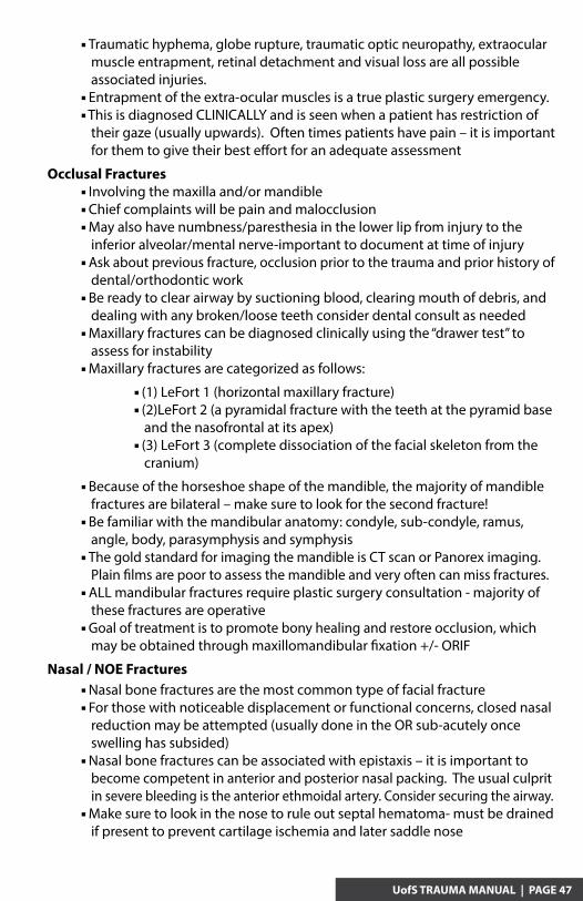
Traumatic hyphema, globe rupture, traumatic optic neuropathy, extraocular muscle entrapment, retinal detachment and visual loss are all possible associated injuries. Entrapment of the extra-ocular muscles is a true plastic surgery emergency. This is diagnosed CLINICALLY and is seen when a patient has restriction of their gaze (usually upwards). Often times patients have pain – it is important for them to give their best effort for an adequate assessment
Occlusal Fractures Involving the maxilla and/or mandible Chief complaints will be pain and malocclusion May also have numbness/paresthesia in the lower lip from injury to the inferior alveolar/mental nerve-important to document at time of injury Ask about previous fracture, occlusion prior to the trauma and prior history of dental/orthodontic work Be ready to clear airway by suctioning blood, clearing mouth of debris, and dealing with any broken/loose teeth consider dental consult as needed Maxillary fractures can be diagnosed clinically using the “drawer test” to assess for instability Maxillary fractures are categorized as follows:
(1) LeFort 1 (horizontal maxillary fracture) (2)LeFort 2 (a pyramidal fracture with the teeth at the pyramid base and the nasofrontal at its apex) (3) LeFort 3 (complete dissociation of the facial skeleton from the cranium)
Because of the horseshoe shape of the mandible, the majority of mandible fractures are bilateral – make sure to look for the second fracture! Be familiar with the mandibular anatomy: condyle, sub-condyle, ramus, angle, body, parasymphysis and symphysis The gold standard for imaging the mandible is CT scan or Panorex imaging. Plain films are poor to assess the mandible and very often can miss fractures. ALL mandibular fractures require plastic surgery consultation - majority of these fractures are operative Goal of treatment is to promote bony healing and restore occlusion, which may be obtained through maxillomandibular fixation +/- ORIF
Nasal / NOE Fractures Nasal bone fractures are the most common type of facial fracture For those with noticeable displacement or functional concerns, closed nasal reduction may be attempted (usually done in the OR sub-acutely once swelling has subsided) Nasal bone fractures can be associated with epistaxis – it is important to become competent in anterior and posterior nasal packing. The usual culprit in severe bleeding is the anterior ethmoidal artery. Consider securing the airway. Make sure to look in the nose to rule out septal hematoma- must be drained if present to prevent cartilage ischemia and later saddle nose

It is important to distinguish nasal bone fracture from the more serious naso-orbital ethmoid (NOE) fracture. The (NOE) complex is the confluence of the frontal sinus, ethmoid sinuses, anterior cranial fossa, orbits, frontal bone, and nasal bones. NOE fractures can be a significant cause of morbidity. Have suspicion if the trauma was high force, there is significant flattening of the nasal bridge or if there is any hint of telecanthus (eyes appearing further apart due to lateral displacement of medial canthal bearing bone). If any of these are present get a CT scan to evaluate.
Radiograpic Imaging in the Craniofacial Trauma PatientCT scan is the gold standard in diagnosing craniofacial injuries. The CT scan should have fine cuts (less than 1mm) and include axial, coronal, and sagittal images. 3D reconstructions of these images are also helpful, but not required. It is important that images go from the vertex of the skull to the mentum to ensure that no injuries are missed. Consider a CT of the cervical spine if indicated.
Soft Tissue Facial Injuries – Pearls Facial lacerations- most of the time there is not any missing tissue! You will be surprised how well things come together Small sutures for facial lacerations– 5-0 or 6-0 sutures (Nylon, Prolene, Novafil, Catgut (FAST absorbing)) 2 layer closure if wound is gapping or there is any tension on the wound– use 5-0 Vicryl or Monocryl for deep layer For lacerations involving the specialized units of the face– eyelid, lip, nose, ear – use marking pen to match up borders prior to injecting local anesthetic Always rule out injuries to the globe, facial nerve, parotid duct For blunt force injuries to the ear, make sure to rule out otohematoma- must be drained if present to prevent future “cauliflower ear” deformity
Discharging the Craniofacial Trauma Patient – Pearls Soft diet for all patients with occlusal injuries or zygoma fractures HOB elevation Avoid nose blowing in all orbital floor fractures Frequent visual checks Temporary eye patch is reasonable for significant diplopia (rarely needed) Mandible Fractures- Clavulin 875mg PO BID until fracture is fixed; Peridex 15 ML PO TID
Final Reminders These patients are trauma patients first – don’t forgot your ABCDEs and remember that many of these patients will have injuries outside of the craniofacial skeleton Always examine the eyes! Do not forget to rule out injury to the brain and cervical spine- when in doubt, get imaging CT is the gold standard for facial fractures Do not neglect a bleeding scalp—life-threatening bleeding can happen surprisingly quickly. Staple or suture closed

References
1. Plastic Surgery: Six Volumes. Third Edition. Volume 3 - Craniofacial, Head and Neck Surgery. Editor: Peter C. Nelgan.
2. Advanced Trauma Life Support (ATLS®): The Ninth Edition ATLS Subcommittee; American College of Surgeons’ Committee on Trauma; International ATLS working group
3. Differences in the Management of Pediatric Facial Trauma. Braun TL, Xue AS, Maricevich RS. Semin Plast Surg. May; 31(2): 118-122. Doi: 10.1055/s-0037-1601380. Review. PMID: 28496392
4. Atlas of Human Anatomy – 6th Edition. Frank H. Netter, M.D.
5. Zygomaticomaxillary complex fractures: diagnosis and treatment. Peretti N, MacLeod S. Curr Opin Otolaryngol Head Neck Surg. 2017 May 25. Doi: 10.1097/MOO.0000000000000372. [Epub]. PMID: 28548998
6. Surgical Treatment of Orbital Blowout Fractures: Complications and Postoperative Care Patterns. Shew M, Carlisle MP, Lu GN, Humphrey C, Kriet JD. Craniomaxillofac Trauma Reconstr. 2016 Nov;9(4):299-304. Epub 2016 Aug 29. PMID: 27833708.
UofS TRAUMA MANUAL | PAGE 49


Neck TraumaNiroshan Sothilingam, Kelly Vogt, Zeeshan Rana (Illustrations)
Neck trauma accounts for only 1% of all injuries however, carries a mortality rate as high as 10%. Multiple vital structures present: Vascular system Carotid, Jugular, subclavian, vertebral, innominate, aortic arch Air passages Pharynx, Larynx, trachea, lungs Upper Gastrointestinal passages Pharynx, esophagus Neurologic system Spinal cord, cranial nerves, peripheral nerves, brachial plexus, sympathetic chain
Borders of the neck: Upper – lower margin of the mandible and the superior nuchal line of the occipital bone. Lower – Suprasternal notch and the upper borders of the clavicles. The sternocleidomastoid separates the neck into anterior and posterior triangles.
Zones of the Neck Zone I Extends from the clavicles to the cricoid cartilage. Includes: The vertebral and proximal common carotid arteries, the subclavian and innominate vessels and the jugular veins. Superior mediastinum, lungs, esophagus, trachea, thoracic duct, and spinal cord.
UofS TRAUMA MANUAL | PAGE 51

Zone II Extends from the cricoid cartilage to the angle of the mandible. Includes The common carotid artery, carotid bifurcation, the vertebral arteries and the jugular veins. Esophagus, trachea, larynx, and spinal cord. Zone III Extends from the angle of the mandible to the mastoid process. Includes: The branches of the external carotid artery, the internal carotid artery, vertebral artery and the internal jugular and facial veins. Pharynx and spinal cord.
Wounds to the posterior triangle require operative management for control of bleeding and wound repair. There are no hidden structures that lead to late complications unlike the anterior triangle.
Principles of Management
Follow ATLS principles: Maintain C-spine precautions if unknown history/mechanism However, make sure to examine with collar off and c-spine immobilized to identify any lacerations or penetrating wounds. Airway May rapidly lose airway due to: Tracheal/laryngeal injury Expanding neck hematoma Significant oropharyngeal trauma Secure if any doubt Endotracheal intubation +/- airway adjuncts. If unsuccessful, a cricothyroidotomy is performed rapidly Injury to the larynx could make cricothyroidotomy difficult/ineffective. (In this case, a tracheostomy may be required.) Control Hemorrhage Most bleeding can be controlled with direct pressure

Penetrating Neck Trauma
Penetrating mechanisms account for most neck injuries.
Penetrating injuries can result in injury to vascular, aero-digestive structures, and nervous structures, therefore all of these structures must be investigated.
Hard signs – Indications for immediate surgical intervention: Vascular10
Expanding hematoma Pulsatile bleeding Shock not explained by other injuries Absent carotid pulse Bruit or palpable thrill Signs of stroke/cerebral ischemia Airway Airway compromise Wound bubbling Esophageal None Nervous None
Soft signs – Indications for further workup/imaging/observation: Vascular Stable hematoma Non-pulsatile bleeding Seatbelt sign History of bleeding at scene Airway Extensive subcutaneous emphysema Stridor Hoarseness Hemoptysis Esophageal Extensive subcutaneous emphysema Hematemesis Odynophagia Dysphagia Nervous Nerve injury CN IX, X, XI and XII Brachial Plexus injury Axillary, musculocutaneous, radial, median and ulnar nerves
97% of patients with hard signs have a vascular injury, as opposed to only 3% of those with soft signs.
UofS TRAUMA MANUAL | PAGE 53

Management Unstable Patient Zone I & II OR Zone III IR Stable Patient Zone I FAST to rule out pericardial fluid CXR to rule out pneumothorax/hemothorax Zone II Hard signs? Yes -> Operative exploration No -> CTA/esophagram/bronchoscopy for high index of suspicion Additional Investigations dictated by physical exam findings Zone III Stable -> CTA/Angiogram head and neck Additional investigations dictated by physical exam findings
All Patients with penetrating trauma require a complete head and neck neurologic exam.
Blunt Neck TraumaBlunt injuries to the neck can cause compression, with fracture of the larynx or trachea. Blunt pharyngeal or esophageal injuries are extremely rare but can result in leakage into the surrounding soft tissues with sepsis if not properly addressed.
Vascular InjuriesBlunt cerebrovascular injuries (BCVIs) involving the carotid arteries commonly result from compression by a seatbelt. The vertebral arteries are vulnerable to severe flexion and extension mechanisms. Stroke secondary to thromboembolism developing from the disrupted vessel wall is a major concern in this type of injury. While rare (1% of blunt trauma patients) these injuries confer a significant risk of morbidity (up to 58%) and mortality (up to 59%). The most common mechanism of blunt carotid injury is hyperextension of the carotid vessels over the lateral articular processes of C1-C3 at the base of the skull, which is typically a result of high-speed collisions. Mechanisms Seatbelt Direct blunt trauma Hyperflexion Hyperextension/rotation Hanging Fracture in proximity to the internal carotid or vertebral artery
As these injuries typically present with no signs/symptoms, CTA is used as the imag-ing modality of choice when there is suspicion for a BCVI.

Blunt carotid arterial injury grading scale3. Grade I: Luminal irregularity or dissection with a 25% narrowing Grade II: Dissection or intraluminal hematoma with > 25% luminal narrowing, intraluminal thrombus, or raised intimal flap. Grade III: Pseudoaneurysm Grade IV: Occlusion Grade V: Transection with free extravasation
EAST Practice Management Guidelines5: Level I: No recommendations Level II: Patients presenting with any neurologic abnormality that is unexplained by a diagnosed injury should be evaluated for BCVI. Blunt trauma patients presenting with epistaxis from a suspected arterial source after trauma should be evaluated for BCVI. Level III: Asymptomatic patients with significant blunt head trauma as defined below are at significantly increased risk for BCVI and screening should be considered. Risk factors are as follows: Glasgow Coma Scale score < 8 Petrous bone fracture Diffuse axonal injury Cervical spine fracture particularly those with: Fracture of C1 to C3 Fracture through the foramen transversarium Cervical spine fracture with subluxation or rotational component Lefort II or III facial fractures Pediatric trauma patients should be evaluated using the same criteria as the adult population.
Approximately 30% of BCVI are still missed following these guidelines.6
ManagementThe mainstay of treatment for BCVI is antithrombotic therapy with either ASA or heparin11. Cochrane review on use of antithrombotic drugs for carotid artery dissections failed to identify a difference in efficacy between ASA and anticoagulants.Patients with pseudoaneurysm or free extravasation may require additional intervention (typically IR).
All patients treated medically should undergo a CTA at 1-3 month follow-up to reeval-uate the injury and determine the need for ongoing therapy.
UofS TRAUMA MANUAL | PAGE 55

Laryngotracheal Injuries Symptoms Hoarseness Pain Bubbling/air through wound Dyspnea* Dysphagia* Aphonia* Odynophagia* *Less commonThe immediate goal of the examination of a patient with suspected laryngeal trauma is to determine the severity of injury and quickly identifying patients who require immediate airway intervention. Airway Evaluation Once the airway is secure, initial evaluation of the larynx should be done in conjunction with ENT and may include a flexible fiberoptic laryngoscopy and CT scan. Radiologic Evaluation Cervical spine injuries must be ruled out in all cases of laryngeal trauma. Chest x-ray to rule out pneumothorax, tracheal deviation, or pneumomediastinum. CT scans diagnose laryngeal fractures and aid in operative planning for the repair and reconstruction of the fractured larynx. It is also important to rule out concomitant head injuries. Small laryngotracheal injuries can be observed safely, however major injuries require surgical repair.
Esophageal InjuryEsophageal and pharyngeal injuries may be difficult to diagnose, but the morbidity and mortality of missed esophageal injuries is high.
Esophageal injury should be suspected in all patients with penetrating neck trauma, and especially where there is a gunshot wound traversing the midline. The incidence of esophageal trauma ranges from 3.9% to 5.4% in penetrating neck injuries1.
Patients may complain of odynophagia or hemoptysis/hematemesis.
Esophagoscopy and gastrograffin swallow are both used for diagnosis. Flexible esophagoscopy has a sensitivity of 96% and specificity of 99%1. A water-soluble (gastrograffin) study has a sensitivity rate of 60% -75%9.Treatment: Minimum contamination and short interval: Primary repair and drain. Contaminated and long interval: Divert and drain.
Principles of Repair of the TracheaMost penetrating injuries of the cervical trachea are straight forward and may include vascular injuries. No debridement is necessary One-layer repair with absorbable suture if small or moderate-size hole Larger injuries may require insertion of tracheostomy tube into the defect, and/or more complex reconstructions
Note: Please see Appendix for SHR C-spine Clearance Protocol

UofS TRAUMA MANUAL | PAGE 57
References
1. Arantes V, Campolina C, Valerio SH, et al. Flexible esophagoscopy as a diagnostic tool for traumatic esophageal injuries. J Trauma. 2009 Jun;66(6): 1677-82
2. Bagheri, SC, Khan HA, Bell RB. Penetrating neck injuries. Oral Maxillofacial Surg Clin N Am 2008. 20:393-414.
3. Biffle WL, Moore EE, Offner PJ, et al. Blunt carotid arterial injuries: Implications of a new grading scale. J Trauma. 1999 Nov;47(5):845-53
4. Bishara RA, Pasch AR, Douglas DD. The necessity of mandatory exploration of penetrating zone II neck injuries. Surgery. 1986;100:655.
5. Bromberg et al. Blunt Cerebrovascular Injury Practice Management Guidelines: The Eastern Association for the Surgery of Trauma. Trauma, Injury, Infection and Critical Care. Vol. 68, 2, Feb 2010.
6. Bruns BR, Tesoriero R, Kufera J, et al. Blunt cerebrovascular injury screening guidelines: what are we willing to miss? J Trauma Acute Care Surg. 2014 Mar;76(3):691-5.
7. Eddy VA. Is routine arteriography mandatory for penetrating injuries to Zone I of the neck? J Trauma. 2000;48:208.
8. Gasparri MG, Lorelli DR, Kralovich KA, et al. Physical examination plus chest radiology in penetrating periclavicular trauma: the appropriate trigger for angiography. J Trauma. 2000;49:1029
9. Ko E, O-Yurvati, AH. Iatrogenic Esophageal Injuries: Evidence-Based manage-ment of Diagnosis and Timing of Contrast studies After Repair. Int Surg. 2012. Jan-Mar;97(1) 1-5
10. Shiroff AM, Gale SC, Niels MD, et al. Penetrating neck trauma: a review of management strategies and discussion of the ‘no zone’ approach. Am Surg 2013;79:23-29.
11. Stein et al. Blunt Cerebrovascular Injuries: Does Treatment Always Matter? J TRAUMA, Injury, Infection, & Critical Care. Jan 2009.


Chest TraumaNiroshan Sothilingam, Kelly Vogt, Richard Bigsby
Chest injuries are common in trauma. All trauma patients should have a chest x-ray in the trauma bay. More complex blunt injuries may require additional investigations such as CT scanning.
Most blunt injuries are managed non-operatively, with simple interventions such as chest tube insertion.
In contrast, penetrating injuries are more likely to require operation, and typically require only a chest x-ray for diagnosis. Patients with penetrating trauma may deterio-rate rapidly, and recover much faster than patients with blunt injury.
Physical examination is the primary tool for diagnosis of acute thoracic trauma. This may be difficult in the busy trauma bay or emergency room, and it is important for the physician to optimize the environment in the room to maximize diagnostic ability.
Physical examination includes: Look Respiratory rate and depth Chest wall asymmetry, penetrating wounds and paradoxical chest wall motion Bruising, seat belt or steering wheel marks Listen Breath sounds on both sides: Apices, axillae and at the back of the chest Feel Tracheal deviation Subcutaneous emphysema Chest wall tenderness
Major threat to life: Tension Pneumothorax Cardiac Tamponade Massive Hemothorax Flail Chest* Open pneumothorax*
* Less common
An initial normal examination does not exclude any of the above, therefore serial examinations and use of diagnostic adjuncts is important.
UofS TRAUMA MANUAL | PAGE 59

Definition Symptoms Diagnosis Trachea
Tension Progressive build up of air Pneumothorax within the pleural space, due Dyspnea, cyanosis, Clinical Away to a lung laceration allowing tachypnea, shock, air to escape into the pleural distended neck veins space but not to return
Simple Air in the potential space Dyspnea, tachypnea &Pneumothorax between the visceral and decreased breath CXR Midline parietal pleura sounds however, many are asymptomatic
Haemothorax Collection of blood in the Dimished breath pleural space that may be sounds, dullness due to CXR Midline caused by blunt or percussion however, penetrating trauma may be asymptomatic
Pulmonary Injury to lung parenchyma, Contusion leading to oedema and blood Asymptomatic. collecting in alveolar spaces May have crackles. CXR Midline and loss of normal lung structure & function
Sucking Chest Defect in the chest wall that Breathing is rapid,Wound allows for equilibration of shallow and labored. CXR Midline intrathoracic and ambient Reduced expansion of pressure the hemithorax, reduced breath sounds, increased percussion.
Injuries identified & treated on secondary survey: Simple Pneumothorax Simple Hemothorax Blunt aortic injury Blunt myocardial injury Pulmonary Contusion Rib Fractures
Investigations Chest X-ray E-FAST Ultrasound CT ECG/Troponin Arterial Blood Gas
The Chest X-Ray is central in the initial workup of trauma patients. It is an inexpensive, quick and safe modality with a high diagnostic yield.
The following injuries should be assessed for on an A/P view16: Rib Fractures Pneumothorax Hemothorax Lung Contusion Subcutaneous/mediastinal emphysema Widening mediastinum Free air under the diaphragm Foreign body

Tube ThoracostomyMost common procedure preformed following chest trauma.
Chest tube insertion carries a complication rate of 2-10%. Most of these are minor, however, some require operative intervention and deaths can still occur.
Absolute Indications Pneumothorax (simple, tension, open) on chest x-ray Hemothorax Blunt Traumatic Arrest (bilateral)
Relative Indications Occult Pneumothorax Profound hypoxia / hypotension & penetrating chest injury Profound hypoxia / hypotension and unilateral signs to a hemothorax Subcutaneous emphysema without obvious pneumothorax
Tube Thoracostomy Placement Chest Tube insertion technique: 1. The area is prepped and draped appropriately 2. An incision is made along the upper border of the rib below the intercostal space behind pectoralis major, 4th-5th intercostal place. The drain track will be directed over the top of the lower rib to avoid the intercostal vessels lying below each rib. The incision should easily accommodate the operator’s finger. 3. Using a curved clamp the track is developed by blunt dissection only. The clamp is inserted into muscle tissue and spread to split the fibers. The track is developed with the operator’s finger. 4. Once the track comes onto the rib, the clamp is angled just over the rib and dissection continued until the pleural is entered. 5. A finger is inserted into the pleural cavity and the area explored for pleural adhesions. Lung is felt to confirm entry into the thoracic cavity. 6. A large-bore (28F) chest tube is mounted on the clamp and passed along the track into the pleural cavity7. Blunt trauma: Drains should be placed anteriorly in the chest. Prevents a tension pneumothorax developing if chest tube is blocked by dependent lung tissue. Normal movement of the lungs will allow drainage of a basal hemothorax through an anterior chest tube. Penetrating trauma: Hemothoraces may be more efficiently drained with a posterior directed drain. 7. The tube is connected to an underwater seal and sutured / secured in place. 8. If desired, a U-stitch is placed for subsequent drain removal (see below). 9. The chest is re-examined to confirm effect. 10. A chest X-ray is taken to confirm placement & position.
UofS TRAUMA MANUAL | PAGE 61

Management of Specific InjuriesPneumothorax
Simple Air in the potential space between the visceral and parietal pleura. Symptoms include dyspnea, tachypnea & decreased breath sounds however, many are asymptomatic. Lung laceration with air leakage is the most common cause of pneumothorax resulting from blunt trauma. Diagnosed by upright CXR Treatment8, 12
Visible on CXR requires chest tube Occult -> Safe to watch Occult and positive pressure ventilation requires chest tube pre-op if no one is available in the OR to decompress Small, asymptomatic pneumothorax may be observed with clinical exam and serial CXR. If the patient is being transported by air, even in a pressurized cabin or if the patient receives general anesthesia/positive pressure ventilation; chest decompression is recommended.
Open “Sucking Chest Wound” Large defect in the chest wall that allows for equilibration of intrathoracic and ambient pressures, therefore causing lung collapse. Clinical Diagnosis Treatment Temporary occlusion of the chest wall defect with a sterile occlusive dressing on three sides to prevent air entering the chest cavity on inspiration and during expiration, the open side allows air to escape from the pleural space. Chest tube insertion to re-expand the lung is typically definitive treatment, however, repair in the OR may be required.
Deep Sulcus Sign Indication of pneumothorax on a supine chest radiograph:
Tension Occurs if pressure of accumulated air in the pleural space exceeds the ambient pressure: Net positive intrathoracic pressure mediastinal shift decreased venous return compression of contralateral lung hemodymanic collapse and severe respiratory distress Symptoms include Dyspenia, cyanosis, tachypnea, shock, distended neck veins, tracheal deviation to opposite side. Diagnosis is made clinically.

UofS TRAUMA MANUAL | PAGE 63
Treatment Needle decompression 14 gauge angiocatheter, 4th or 5th intercostal space, auterior axillary line or 2nd intercostal space midclavicular line Chest tube insertion.Both Tension Pneumothorax and cardiac tamponade have similar signs/symptoms and can be difficult to differentiate.
Tension Pneumothorax however, has the following differences: Hyperresonant note percussion Deviated tracha Absent breath sounds over the affected hemithorax
Hemothorax Collection of blood in the pleural space. Majority are the result of rib fractures, lung parenchymal and minor venous injuries from the chest wall and are self-limiting. Often asymptomatic, however can include tachypnea, dyspnea and hypovolemia. Clinical signs include diminished breath sounds, dullness to percussion Diagnosed by upright CXR. It takes approximately 400-500mls of blood to obliterate the costo-phrenic angle on a chest radiograph. FAST exam can detect smaller hemothoraces, although in the presence of a pneumo thorax or subcutaneous air ultrasound may be difficult radiograph. Treatment The majority of hemothoraces have already stopped bleeding and simple drainage is all that is required. All chest tubes placed for trauma should be of sufficient calibre to drain hemothoraces without clotting.
Indications for Thoracotomy 15% of patients with chest injury require a formal thoracotomy. > 1500 mL blood loss from chest tube insertion. > 200 mL blood loss from chest tube per hour Complications Retained Hemothorax4
Failure of the chest tube to evacuate the hemothorax due to small chest tube caliber, clot in tube, poor placement. Diagnosed on CT If left untreated, these retained hemothorax may become infected and lead to empyema formation. If this collection remain uninfected, the clot will organize and fibrose, causing loss of lung volume. Treatment Intrapleural fibrinolytics (Streptokinase/Urokinase) Surgery Thoracoscopy (< 10 days) Thoracotomy and Decortication (> 10 days)

Empyema Infected hemothorax Present with fever, raised white cell count, air-fluid levels on CT. Treatment involves surgery.
Rib Fractures Most common injury with blunt chest trauma. Tenderness over fracture and pain with A/P compression. Bedside ultrasound may be used to determine the specific location of a rib fracture. Management is directed towards protecting the underlying lung and allowing adequate oxygenation, ventilation and pulmonary toilet. This is to prevent the development of pneumonia, which is the most common complication of rib fractures. Young, healthy patient may be able to manage one or two rib fractures with analgesia alone, however the same injury in an elderly patient is regarded as major and may lead to pneumonia, respiratory failure and even death. Treatment5,6
Oral analgesics PCA Epidural2 Chest Physiotherapy
Flail Chest The fracture of two or more consecutive ribs in at least two locations. It is the result of significant kinetic energy applied to the thorax. Patient with multiple consecutive rib fractures may exhibit the respiratory compromise and pulmonary dysfunction classically associated with flail chest. The associated respiratory failure is due primarily a result of underlying pulmonary contusion, however the biomechanical changes of multiple rib fractures, severe pain, splinting, and atelectasis are contributing factors. Treatment Should be implemented early and aggressive with an emphasis on pain management and pulmonary toilet. Mechanical ventilation may be required in patients with flail chest, even with optimal analgesia and pulmonary toilet. The best method of providing pain relief for these patients is thoracic epidural analgesia, which should be initiated as soon as possible. Intercostal nerve blocks may be useful if long-acting local anesthetics are used. Narcotic use is almost always necessary for analgesia. Operative fixation of rib fractures in flail segments may be considered in a subset of patients9.

UofS TRAUMA MANUAL | PAGE 65
Blunt Cardiac injury (BCI)10,15
Diagnosis: A high index of suspicion is required as there is no pathognomonic mechanism that leads to BCI, along with a proper evaluation of the mechanism of trauma. More than 75% of all patients with BCI will have other associated thoracic injuries, including rib and sternal fractures, pulmonary contusions, pneumothorax, hemothorax, and great vessel injuries. The presence of a sternal fracture does not predict the presence of BCI and therefore does not necessitate monitoring and further evaluation. The most common complaint in patients with BCI is chest pain, which can be difficult to distinguish from pain associated with chest wall injuries such as rib and sternal fractures. The majority of patients with BCI are asymptomatic. All patients with suspicion for blunt cardiac trauma should have an ECG on admission (level 1 recommendation). The most common finding is sinus tachycardia, which is very non specific, as it is commonly found in trauma patients with a multitude of injuries and is often associated with bleeding or pain. The next most common abnormality is premature atrial or ventriular contractions followed by a spectrum of findings, including nonspecific T-wave changes, ST-segment elevation or depression, atrial fibrillation or flutter, ventricular dysrhythmias, conduction delays, bundle branch and heart block, and the presence of Q waves. If the ECG reveals any of these findings or other evidence of ischemia, BCI should be ruled out, and the patient should be admitted for continuous ECG monitoring for 24 to 48 hours (level 2 recommendation). However, if the ECG shows no abnormality, the chances of having a clinically significant BCI requiring treatment are negligible, and pursuit of the diagnosis should be terminated (level 2 recommendation). Those who survive to hospital with more life-threatening types of BCI will present with signs and symptoms of shock. The cause of the shock state needs to be established by differentiating cardiogenic shock secondary to BCI from the more common causes, including tension pneumothorax, neurogenic shock from spinal cord injury, and hypovolemic shock secondary to bleeding.
Treatment Asymptomatic patients should be observed with ECG and Troponin monitored until levels peak. Antiarrhythmics only if symptomatic Inotropic support for those in cardiogenic shock

Suspicion for Blunt Cardiac Injury
Stable
FAST
Normal FAST
Unstable
FAST
ExcludeHemorrhagic shock
Tension pneumothoraxSpinal shock
Positive or inconclusive ECHO
ECHO Normal ECHONo
Pericardial fluid
Yes Pericardial
fluid
Tamponade with cardiac arrest/PEA
ER thoracotomy
ECHO ECG
Troponin I
OR Pericardial
windowPossible
sternotomy
Abnormal rhythm/ ST-segment
changes/PVC’s
Normal sinus rhythm
Age > 55 and/or hx of cardiac disease
OR Repair of
injuryNo evidence of blunt cardiac injury Intensive care unit
Diaphragmatic Injury Traumatic diaphragmatic ruptures occur in 1-7% of patients with significant blunt abdominal trauma and 10-15% in victims of penetrating trauma. Only 25% are diagnosed on the initial chest radiograph. Penetrating trauma is often small compared to blunt. Injuries due to blunt trauma result from high-energy mechanism causing large rents that a usually obvious. 75% of these injuries occur on the left sided as this side is not protected by the large mass of the liver. Diaphragmatic ruptures are often asymptomatic initially Presentation may be masked by concomitant injuries making an immediate diagnosis difficult. In penetrating trauma, any injury to the thoracoabdominal area should raise suspicion for potential injury. Sensitivity of CXR is 27-62% for left-sided injuries and 18-33% for right -sided injuries. CT for blunt injuries is reported as sensitivity of 71-100% and specificity of 75-100%.

UofS TRAUMA MANUAL | PAGE 67
American Association for the Surgery of Trauma Organ Injury Scale for Diaphragmatic Injuries
Grade Description I Contusion II Laceration < 2 cm III Laceration 2-10 cm IV Laceration > 10 cm with tissue loss < 25 cm2
V Laceration with tissue loss > 25 cm2
Treatment Laparoscopy vs. Laparotomy vs. Thoracoscopic Non-absorbable suture Airtight closure Thoracostomy tube after chest washout
Esophageal Injuries Extremely rare injuries Most commonly caused by penetrating injury. Blunt causes are rare, however deadly if unrecognized. Caused by forceful expulsion of gastric content into the esophagus from a severe blow to the upper abdomen. This causes a linear tear in the lower esophagus. Leakage of gastric content into the mediastinum causing mediastinitis and empyema. Diagnosis CXR Patients may present with concomitant pneumothorax/hemothorax/pneumomediastinum Contrast swallow study Start with water-soluble and if no leak is identified, then study can be repeated with barium. Barium is inert and extravasation into the mediastinum will not cause additional harm. Esophagoscopy (flexible) Negative predictive value of 100% Positive predictive value of only 33%.
Treatment Approach an injury to the upper and mid-thoracic esophagus through a right posterolateral thoracotomy in the 4th intercostal space. Lower thoracic esophagus is accessed though a left posterolateral thoracotomy in the 6th or 7th intercostal space. Minimal sepsis, contamination, short interval: Primary repair and reinforced repair +++ sepsis, contamination, long interval: resection +/- reconstruction Unstable: exclusion and drainage

Tracheobronchial Injury Infrequent, however this type of injury has possible life-threatening consequences. The cervical trachea is more commonly injured from penetrating trauma and the distal trachea from blunt trauma. Diagnosis Cervical tracheal injuries may be obvious on physical exam. With penetrating injuries, there may be subcutaneous air, bubbling/fluctuating with the patient’s respirations, air leak, crepitus. In blunt cases, there is often massive subcutaneous emphysema in the neck. Patients may present in respiratory distress and require airway management. Signs of tracheal injury include airway obstruction, subcutaneous air, dysphonia and chest wall contusion. Persistent air leak with chest tube insertion is a common finding in distal trachea/proximal major bronchial injury. Treatment Secure the airway Tube Thoracostomy, second tube for persistent air leak if necessary All patients with suspected injury should undergo bronchoscopy for evaluation. Management options ETT past injury Operative repair Low pressure ventilation and observation
Penetrating Cardiac Injuries Among patients with stab wounds who reach hospital, approximately 35% have an isolated right ventricular injury. The left ventricle is involved in 25%, and in 30% more than one chamber is involved. Other injuries include coronary artery lacerations (most often the left anterior descending and diagonal branches), valve injury, and ventricular septal defects. Management of an unstable patient with penetrating cardiac trauma begins with fluid resuscitation and immediate transfer to the operating room. Pericardial Window 3 to 4 cm skin incision can be made just to the left side of thexiphoid process facilitating passage of a blunt- tipped clamp into the pericardium for immediate decompression. Can be done in OR transdiaphragmatically. The role of FAST14
To determine the presence of pericardial fluid Sensitivity 100% Specificity 97%

UofS TRAUMA MANUAL | PAGE 69
Pericardial Injury False-negative results may occur if the patient is stabbed in the chest and a hole in the pericardial sac decompresses the hemo pericardium into the thoracic cavity. If this is suspected, immediate decompression with tube thoracostomy and repeat ultrasound pericardial windows tube1. This should be considered with a stab wound to the precordium, negative FAST and residual output from a left sided chest tube.1
Emergency department thoracotomy can occasionally salvage trauma patients who arrest.
Resuscitative Thoracotomy (ED thoracotomy) Absolute Indications: Penetrating torso injury and VSA < 15 minutes Blunt injury and VSA < 5 minutes Relative indications: Penetrating non-torso injury VAS < 15 minutes. Survival in penetrating trauma: Cardiac injury 10-20%13
Non-cardiac injury 3-20% Patient Positioning Supine, left side slightly elevated with towels/sheets Arms out/above head Left anterolateral thoracotomy Objectives: Release tamponade Open Cardiac massage Cross clamp descending aorta Technique Left anterolateral thoracotomy 4th/5th intercostal space from lateral border of sternum to the bed. Skin, fat, chest wall muscles – divided with scalpel. Intercostal muscles & pleura divided with heavy scissors. Rib spreader handle towards axilla Pericardiotomy Incise pericardium anterior and parallel to the phrenic nerve. If hemopericardium is present, this should be done with a scalpel and the pericardotomy then extended the entire length using scissors. Evacuate blood clot. Control areas of bleeding then get to an OR Digital pressure Skin stapler Sew with prolene Foley catheter In a non-beating heart, suture the injury, then defibrillate with internal paddles on RA & LA at 10-30 joules +/- epinephrine. Cross Clamp descending aorta

Elevate lung anterior & cephalad Incise mediastinal pleura, inferior pulmonary ligament Bluntly dissect aorta from esophagus (anterior) and prevertebral fascia (posterior), however not circumferentially. Place left hand on the thoracic spine and slide anteriorly. The first structure felt is the aorta. Bluntly dissect anteriorly and posteriorly at one level to avoid injury to additional structures. Cross clamp at the level of the diaphragm with vascular clamp. If inadequate exposure: Clamshell: Extend incision to right anterolateral thoracotomy. Extend incision to right anterolateral Divide sternum with heavy scissors Flip anterior chest wall cephalad onto patients head.
American Association of the Surgery of Trauma Cardiac Injury Scale
Grade Description of InjuryI
II
III
IV
V
VI
Blunt cardiac injury with minor EKG abnormality (non specific ST of T wave changes, premature atrial or ventricular contractions, or persistent sinus tachycardiaBlunt or penetrating pericardial wound without cardiac injury, tamponade, or cardiac herniationBlunt cardiac injury with heart block or ischemic changes without cardiac failurePenetrating tangential cardiac wound, up to but not extending through endocardium, without tamponadeBlunt cardiac injury with sustained or multifocal ventricular contracationsBlunt or penetrating cardiac injury with septal rupture, pulmonary or tricuspid incompetence, papillary muscle dysfunction, or distal coronary artery occlusion without cardiac failureBlunt pericardial laceration with cardiac herniationBlunt cardiac injury with cardiac failurePenetrating tangential myocardial wound, up to but not through endocardium, with tamponadeBlunt or penetrating cardiac injury with septal rupture, pulmonary or tricuspid incompetence, papillary muscle dysfunction, or distal coronary artery occlusion producing cardiac failureBlunt or penetrating cardiac injury with aortic or mitral incompetenceBlunt or penetrating cardiac injury of the right ventricle, right or left atriumBlunt or penetrating cardiac injury with proximal coronary artery occlusionBlunt or penetrating left ventricular perforationStellate injuries, less that 50% tissue loss of the right ventricle, right or left atriumBlunt avulsion of the heart; penetrating wound producing more than 50% tissue loss of a chamber
Mechanism of injury revealed mortality of 84% for gunshot wounds compared with 35% for stab wounds. Grades IV, V, and VI injuries had a mortality of 56%, 76%, and 91%, respectively. Penetrating left atrial and ventricular injuries had a higher mortality rate Between 77% and 80%, while right atrial and right ventricular had mortality rates of 63% and 49%, respectively. Approach to Great Vessel Injury Principles with all vascular injury: Access Exposure Control Proximal left subclavian: Left high (3rd intercostal space) anterolateral thoracotomy. Distal left subclavian: Left Supraclavicular incision. Proximal right subclavian: Median sternotomy Distal right subclavian: Right Supraclavicular incision.

UofS TRAUMA MANUAL | PAGE 71
Thoracic Aortic Injury > 90% of thoracic great vessel injuries are due to penetrating trauma. Blunt injury in most often caused by deceleration injuries. Aortic blunt injury usually involves the proximal descending aorta. 36% to 54% of blunt aortic injuries occur at the aortic isthmus. Approximately 2% to 5% of patients with aortic disruptions survive without operation, or even detection, to form chronic pseudoaneurysms. The patients who develop chronic pseudoaneurysms have fewer associated injuries at the time of the traumatic event. Diagnosis may be suspected on CXR, but definitive diagnosis requires CT or transesophageal echocardiography. Radiographic findings suggestive of a blunt injury to the thoracic aorta: Widened mediastinum > 8 cm at level of aortic knob or mediastinum width to chest ratio > 25% has a sensitivity of 81-100% and specificity of 60%. Obliteration of the aortic knob Depression of the left mainstem bronchus Loss of paravertebral pleural stripe Apical pleural cap Deviation of the NG tube Lateral displacement of the trachea at the T4 level Left hemothorax 1st rib, clavicle or scapula fracture
Treatment TEVAR In large has replaced the need for open repair3,11
“Cardiac Box” Trauma
ABC’s of Resuscitation
Hemodynamic Stability Unstable
2-D Echo (FAST) +/- CT ED Thoracotomy
Equivocal
Subxyphoid Window
Negative
PositiveNegative
Positive
OR Thoracotomy
OR

References
1. Ball CG, Williams BH, Wyrzykowski AD, et al. A caveat to the performance of pericardial ultrasound in patients with penetrating cardiac wounds. J Trauma. 2009 Nov;67(5):1123-4.
2. Carrier FM, Turgeon AF, Nicole PC, et al. Effect of epidural analgesia in patients with traumatic rib fractures: a systematic review and meta-analysis of randomized controlled trials. Can J Anaesth. 2009 Mar;56(3):230-42.
3. Demetriades D, Velmahos GC, Scalea TM, et al. Diagnosis and treatment of blunt thoracic aortic injuries: changing perspectives. J Trauma. 2008 Jun;64(6):1415-8
4. DuBose J, Inaba K, Demetriades DJ, et al. Management of post-traumatic retained hemothorax: a prospective, observational, multicenter AAST study. Trauma Acute Care Surg. 2012 Jan; 72(1):11-22
5. Flagel BT, Luchette FA, Reed RL, et al. Half-a-dozen ribs: the breakpoint for mortal-ity. Surgery. 2005 Oct;138(4):717-23
6. Holcomb JB, McMullin NR, Kozar RA, et al. Morbidity from rib fractures increases after age 45. J Am Coll Surg. 2003 Apr;196(4):549-55.
7. Inaba K, Lustenberger T, Recinos G, et al. Does size matter? A prospective analysis of 28-32 versus 36-40 French chest tube size in trauma. J Trauma Acute Care Surg. 2012 Feb;72(2):422-7
8. Kirkpatrick AW, Rizoli S, Ouellet JF, et al. Occult pneumothoraces in critical care: a prospective multicenter randomized controlled trial of pleural drainage for mechanically ventilated trauma patients with occult pneumothoraces. J Trauma Acute Care Surg. 2013 Mar;74(3):747-54
9. Marasco S Liew S, Edwards E, et al. Analysis of bone healing in flail chest injury: do we need to fix both fractures per rib? J Trauma Acute Care Surg. 2014 Sep;77(3):452-8.
10. Nagy KK, Krosner SM, Roberts RR, et al. Determining which patients require evaluation for blunt cardiac injury following blunt chest trauma. World J Surg. 2001 Jan;25(1):108-11.
11. Ott MC, Stewart TC, Lawlor DK, et al. Management of blunt thoracic aortic inju-ries: endovascular stents versus open repair. J Trauma. 2004 Mar;56(3):565-70.
12. Ouellet JF, Trottier V, Kmet L, et al. The OPTICC trial: a multi-institutional study of occult pneumothoraces in critical care. Am J Surg. 2009 May;197(5):581-6
13. Rhee PM, Acosta J, Bridgeman A, et al. Survival after emergency department thoracotomy: Review of published data from the past 25 years. J Am Coll Surg 2000;190:288-298.
14. Rozycki GS, Feliciano DV, Ochsner MG, et al. The role of ultrasound in patients with possible penetrating cardiac wounds: a prospective multicenter study. J Trauma. 1999 Apr;46(4):543-51.
15. Salim A, Velmahos GC, Jindal A, et al. Clinically significant blunt cardiac trauma: role of se-rum troponin levels combined with electrocardiographic findings. J Trauma. 2001 Feb;50(2):237-43.
16. Tjeerd A, Ludo B, Falco H, Luke L. Initial assessment of chest X-ray in thoracic trauma patients: Awareness of specific injuries. World J Radiol. 2012. Feb 28; 4(2): 48-52

UofS TRAUMA MANUAL | PAGE 73


Abdominal TraumaNiroshan Sothilingam, John Shaw
Indications on History & Physical ExaminationThe abdomen extends from the nipples to the groin crease anteriorly, and the tips of the scapulae to the gluteal skin crease posteriorly.
Even though physical examination remains the most important method to determine the need for exploratory laparotomy, there is little Level I evidence to support this. Several studies have highlighted the inaccuracies of the physical examination in blunt abdominal traumaTrauma.org.
Physical exam can be unreliable due to level of consciousness (drugs, alcohol, head injury), distracting injury and/or spinal cord injury. Look for signs/symptoms of intraperitoneal injury:
Abdominal tenderness, peritoneal irritation Hypotension/hypovolemia Entrance and exit wounds (to determine path) Distention – pneumoperitoneum, gastric dilation, bleeding Left shoulder tip pain can suggest splenic injury (Kehr’s sign) Retroperitoneal hemorrhage Ecchymosis of flanks (Gray-Turners sign) Ecchymosis of umbilicus (Cullen’s sign) “Seat belt sign” – highly correlated with intraperitoneal injuries DRE: Blood, high riding prostate Several factors influence the selection of diagnostic testing:
Capability of hospital - i.e., trauma center vs. peripheral hospital Access to imaging (CT scanner vs. FAST) Surgeon experience with available diagnostic imaging.As facilities evolve, technologies mature and surgeons gain new experience, it is important that any diagnostic strategy constructed be dynamic. Due to the recognized inadequacies of physical examination, there are a number of diagnostic adjuncts: Diagnostic peritoneal lavage (DPL) Computed tomography (CT) Focused abdominal sonography for trauma (FAST) DPL Whitehouse JS, 2009
Largely replaced by FAST and CT DPL is a safe, rapid and accurate method for determining the presence of intraperitoneal blood in blunt abdominal trauma. It is more accurate than CT for the early diagnosis of hollow visceral and mesenteric injuries, but it does not reliably exclude significant injuries to retroperitoneal structures. Hemodynamically stable patients with equivocal results are best managed by additional diagnostic testing to avoid unnecessary laparotomies.
UofS TRAUMA MANUAL | PAGE 75

In blunt trauma, DPL can be used to triage patients who are hemodynamically unstable and have multiple injuries with an equivocal FAST exam. Hypotensive patients should not be transported to the CT scanner. In the absence of CT scanning, DPL is also useful in patients with an unreli able abdominal exam due to altered mental status or spinal cord injury. The only absolute contraindication to DPL is previous abdominal surgery May cause injury an intra-abdominal organ with catheter Difficult introducing a catheter due to adhesions Relative contraindications include preexisting coagulopathy, advanced cirrhosis, and morbid obesity. Relative contraindications to the standard infraumbilical approach include patients with a pelvic fracture or females beyond the 1st trimester of pregnancy. Technique: Sterile prep and draped widely area Open technique uses a vertical infraumbilical incision Attempt to aspirate free peritoneal blood Insert lavage catheter by seldinger or open technique Lavage peritoneal cavity with warm 1 liter saline Positive test requires one of the following: 10 ml gross blood on initial aspiration > 500/mm3 white blood cells > 100,000/mm3 red blood cells Presence of enteric content In the presence of gross blood or enteric matter, immediate laparotomy is indicated in unstable patients. Otherwise, accurate cell counts should be obtained, which takes time. During this time, if the patient’s clinical status deteriorates or signs of peritonitis develop, laparotomy is indicated. The accuracy of DPL has been reported between 92% and 98%EAST. DPL has been shown to be more efficient than CT scan in identifying patients that require surgical exploration. However, a positive DPL does not necessarily mandate immediate laparotomy in the hemodynamically stable patient. The complication rate associated with DPL is low. Rate of complications is lower for open DPL than closed technique. Closed DPL can be performed more rapidly. The false positive rate for DPL is increased in patients with pelvic fractures.

In order to avoid sampling the retroperitoneal hematoma, a supra-umbilical approach has been recommended, theoretically reducing the chances of a false positive result. Cochran W, 1984 Focused
Assessment with Sonography in Trauma (FAST) FAST is noninvasive, may be easily performed and can be done concurrently with resuscitation. Especially useful for detecting intra-abdominal hemorrhage in the multiply injured or pregnant patient. Easily portable and may be easily repeated if necessary. Can be completed within 5 minutes. More cost-effective when compared to DPL or CT. A major limitation of the FAST examination is that a positive examination relies on the presence of free intraperitioneal fluid. Will detect a minimum of 200 mL of fluid (operator dependent) Injuries not associated with hemoperitoneum may not be detected by this modality. Not a reliable method for excluding hollow visceral injury. Cannot be used to reliably grade solid organ injuries. In the hemodynamically stable patient, a follow-up CT scan should be obtained if nonoperative management is contemplated.DPL may also be used as a complementary examination in the hemodynamically stable patient in the presence of an equivocal or negative FAST with strong clinical suspicion of visceral injury.
Overall, FAST has a sensitivity between 73% and 88%, a specificity between 98% and 100% and is 96% to 98% accurateEAST. This level of accuracy is independent of the practitioner performing the study. Surgeons, emergency medicine physicians, ultra-sound technicians and radiologists have equivalent results.
CT ScanCT scanners are available in most trauma centers and, with the advent of helical scan-ners, scan time has been significantly reduced.
Sensitivity between 92% and 97.6% and specificity as high as 98.7% has been reported in patients subjected to emergency CTEAST. Requires a cooperative (may require intubation), hemodynamically stable patient. Patient must be transported out of the trauma resuscitation area to the radiographic suite. Specialized technicians and the availability of a radiologist for interpretation were also viewed as factors which limited the utility of CT for trauma patients. Negative predictive value (99.63%) of CT is sufficiently high to permit safe discharge of blunt abdominal trauma patients following a negative CT scan. Livingston DH et al, 1998
CT is notoriously inadequate for the diagnosis of mesenteric injuries and may also miss hollow visceral injuries.EAST Patient should be admitted to an observation unit with serial abdominal exam preformed.
UofS TRAUMA MANUAL | PAGE 77

Role of Rectal Exam in Trauma AssessmentThe DRE is used to evaluate ATLS
Rectal hemorrhage Rectal mucosal injury or wall defects Loss of anal tone suggesting spinal cord injury Palpable pelvic fractures A high riding prostate suggestive of posterior urethral disruptionTraditionally, DRE has been mandatory in trauma patient.
However, ATLS now recommends reducing the requirement for a DRE from absolute to selective. Kortbeek et al, 2008
DRE changes management in only in 1.2% - 4% of cases. Porter and Ursic’s prospective observational study,
2001, Esposito et al’s prospective study 2005
The sensitivity of the DRE is only 2% for detecting urethral injuries. In patients with posterior urethral disruption, 60% of cases did not have any clinical signs prior to insertion of urinary catheter. Ball et al, 2009
95% of patients with posterior urethral injuries had pelvic fractures. Ball et al, 2009
DRE remains clinically relevant in penetrating trauma for the presence of blood sur-rounding the rectum, pelvic fractures, and spinal cord injuries. Its role in identifying urethral injury via abnormal prostate position is minimal. Clinical signs of urethral disruption: blood at the urethral meatus (20% sensitivity) gross hematuria prior to catheter insertion (17% sensitivity) abnormal prostate position (2% sensitivity) scrotal or perineal echymosis inability to void
Hematuria Hematuria (micro and macroscopic) is the hallmark for injury to the genitourinary system. The amount of hematuria does not correlate to the injury severity. All gross hematuria require evaluation of the genitourinary system. Microscopic hematuria in a trauma setting in the absence of shock does not need immediate evaluation. (Thomason RB, 1989) However, the absence of hematuria does not rule out injury to the genito urinary system. Suspected urethra rupture requires further intervention with retrograde urethrogram to rule out injury before urinary catheter inserted. ATLS
Penetrating Abdominal TraumaDivided into low velocity (Stab wound and hand guns) & high velocity (high caliber rifles).
Most commonly injured organs with stab woundsATLS: Liver (40%) Diaphragm (20%) Small bowel (30%) Colon (15%)

Most commonly injured organs with gunshot woundsATLS: Small bowel (50%) Liver (30%) Colon (40%) Abdominal vascular (25%)
Patients with penetrating abdominal injury fall into 3 categories: Trauma.org
Pulseless Hemodynamically unstable Hemodynamically stable
UofS TRAUMA MANUAL | PAGE 79
Injury TypePresentation Management Priority
Major Vascular Injury
Vascular and/or solid organ injuryAND/ORHemorrhage from other sites
Hollow viscus injuryPancreas or renal
Pulseless
Hemodynamically Unstable
Hemodynamically Normal
Emergency laparotomy, ED thoracotomy (for aortic cross clamp)
Identify & control hemorrhage (with emergent OR laparotomy)
Identify presence of gastrointestinal, diaphragmatic or retroperitoneal injury
Hemodynamically NormalDiagnostic modalities: Serial abdominal examination CT Local Wound Exploration Diagnostic Peritoneal Lavage Laparoscopy
Patients with clinical signs of peritonitis, or with evisceration of bowel should be taken for surgical exploration/laparotomy.
Serial Physical Examination Selective non-operative management of penetrating abdominal injuries has become the standard of care. 55% of anterior abdominal stab wounds can be managed non-operatively. Navsaria PH et al, 2007
Even in the presence of peritoneal violation, a significant number of patients have no major intra-abdominal injury requiring an operation. Admit for observation for 24 hours. Hourly checks of hemodynamic status. Serial examination of abdomen checking for peritonitis. Ideally the same surgeon should examine the patient each time. If this is not possible, during a handover period both surgeons should examine the patient at the same time so they agree on the current status of the abdomen and whether there has been any progression in symptoms. Timing of examination can be at 1, 4, 12 and 24 hours after the initial assessment. Some recommend examination every four hours.

If the patient is well the following day they start a normal diet, and are dis charged once diet is tolerated and they have completed the observation period. Signs of hemodynamic instability or peritonitis during this period of observation warrants a laparoscopy laparotomy. Development of persistent local symptoms of pain and tenderness, fever or tachycardia at 24 hours, however not frank peritonitis should be evaluated with CT Scan, laparoscopy or laparotomy. Requirement admission of patient to monitored bed to evaluate hemodynamic and serial physical examinations. Also require hospital setting with in house surgeons to perform frequent physical examinations.
Local Wound Exploration (LWE) Evaluation of a stab wound under local anaesthesia. The wound is extended under local anaesthesia and the track followed through tissue layers. Penetration of the anterior fascia is considered a positive LWE Penetration of the peritoneum is difficult to identify. A positive LWE leads to another diagnostic test or laparotomy/laparoscopy. When LWE is used alone to determine need for laparotomy, there will be a high non-therapeutic laparotomy rate. When peritoneum is penetrated by a stab wound, many patients will have no intra-peritoneal injury or injuries that does not require surgical intervention i.e. omental laceration, mesenteric laceration or liver tears that have stopped bleeding.
Thoracoabdominal Injury: Any penetrating injury below the nipple should be considered. Must rule out diaphragmatic injury. Radiological studies may miss small diaphragmatic tears, therefore laparos copy / thoracoscopy remains the investigation of choice. Laparoscopy is preferred for left sided and anterior diaphragmatic injuries. Thoracoscopy for posterior diaphragmatic injuries injuries. Diaphragmatic lacerations can be repaired through a laparoscopic or thoracoscopic
Flank or Back Wound: Associated with injuries to retroperitoneal organs. Of these, the colon is the injury most often missed. If suspecting colon injury: Duration of serial physical examination is extended to 72 hrs. Watch for fever or a rise in the white cell count.

Triple-contrast CT scan is an option. If the wound track extends up to the colon, or there is evidence of abnormal bowel wall thickening, laparotomy is indicated.
Wound to Buttock/Perineum: Any penetrating injury to the gluteal region carries the risk of rectal injury. Digital rectal examination is inadequate Proctoscopy and sigmoidoscopy should be performed Look for the presence of blood and/or mucosal tear.
UofS TRAUMA MANUAL | PAGE 81
Anterior Abdominal Stab Wound
ShockPeritonitis
EviscerationOR
YES
Local Wound Exploration*/ FASTD/CNegative
NO
Positive / Equivocal
Admit for Observation (24 Hours): Vital signs q4 hrs; Serial Physical Examination, CBC q8 hrs
PeritonitisHemodynamic Instability
NO
Significant Drop in Hemoglobin (>3 g/dL)Significant Leukocytosis
NO
FEEDD/C
ORYES
CT, DPLor OR
YES
- WEST* Consider CT Scan if patient is morbidly obese or wound track is long and tangential
Blunt Abdominal Trauma Mechanism of Injury: Injury due to broken lower ribs Sudden increase in intraabdominal pressure causing rupture of hollow viscus Crushing of an organ against the spine, pelvis or abdominal wall Deceleration forces

Organs most often injured: ATLS
Spleen (40-55%) Liver (35-45%) Small Bowel (5-10%) Incidence of retroperitoneal hematoma found on laparotomy is 15% The American Association for the Surgery of Trauma Organ Injury Scale Most commonly used system for grading abdominal solid organ injury. Provides a consistent means of comparison from clinical description and decision making with respect to management. Most useful tool for comparison in research. Grade is assigned based on CT findings. However, grade can be changed with intra-operative findings.
Spleen Grade I Hematoma: Subcapsular, <10% surface area Laceration: Capsular tear, < 1 cm. Parenchymal depth Grade II Hematoma: Subcapsular, 10-50% surface area. Intraparenchymal, < 5 cm in diameter Laceration: Capsular tear, 1-3 cm parenchymal depth that does not involve a trabecular vessel Grade III Hematoma: Subcapsular, > 50% surface area or expanding; r uptured subcapsular or parenchymal hematoma; intraparenchymal hematoma > 5 cm or expanding. Laceration: > 3 cm parenchymal depth or involving trabecular vessels Grade IV Laceration: involving segmental or hilar vessels producing major devascularization (> 25% of spleen) Grade V Laceration: Completely shattered spleen Vascular: Hilar vascular injury with devascularized spleen Treatment Splenectomy Unstable patients with grade IV & V will most likely require an operation. Angioembolization (Stable patient) Consider angioembolization in: Stassen NA et al
Patients with a grade >III injury Splenic angioembolization for grade IV and V injuries has a success rate > 80% Haan JM et al, Banerjee A et al
Presence of intravenous contrast extravasation on CT

Moderate haemoperitoneum Evidence of ongoing splenic bleeding Conservative Management Requires patient to be admitted to a monitored bed, serial abdominal exams, frequent evaluation of hemoglobin and repeat CT in 24-48 hrs to rule out splenic pseduoaneurysm. Non-operative management is now becoming the gold standard treatment for minor splenic trauma (Grade I & II). Cirocchi R et al
Non-operative management can be the initial treatment in some cases of severe splenic trauma; however, the decision between operative and non-operative management depends on careful risk-benefit analysis for each patient, as well as on the expertise of the surgeon and of the multidisciplinary hospital team. Cirocchi R et al
Extreme care must be taken with conservatively managing in patients with concurrent head trauma. Any systolic blood pressure drop less than 90 mmHg can cause a significant rise in mortality.
Liver Grade I Hematoma: Subcapsular, <10% surface area Laceration: Capsular tear, < 1 cm. Parenchymal depth Grade II Hematoma: Subcapsular, 10-50% surface area. Intraparenchymal, < 10 cm in diameter Laceration: Capsular tear, 1-3 cm parenchymal depth, < 10 cm in length Grade III Hematoma: Subcapsular, > 50% surface area of ruptured subcapsular or parenchymal hematoma; intraparenchymal hematoma > 10 cm or expanding. Laceration: > 3 cm parenchymal depth Grade IV Laceration: Parenchymal disruption involving 25% - 75% hepatic lob or 1-3 couinaud’s segments Grade V Laceration: Parenchymal disruption involving > 75% of hepatic lobe or > 3 Couinaud’s segmetns within a single lobe Vascular: Juxtahepatic venous injuries; retrohe patic vena cava/ central major hepatic veins Grade VI Vascular: Hepatic avulsion
UofS TRAUMA MANUAL | PAGE 83

Treatment A suspicion of hepatic injury is increased with right uppernquadrant and/or right chest trauma. However, clinical history and physical examination are not sufficiently sensitive or specific for the presence of liver injury. CT scan with IV contrast is the most useful investigation to determine severity of liver injury. Non-operative management is the treatment of choice for hemodynamically stable patients. This requires appropriate patient selection and resources such as monitored bed, blood bank support, immediate operating room availability, and surgeons and interventional angiographers experienced in managing hepatic injury. Patients who are hemodynamically stable with CT findings of extravasation from the liver have higher failure rates with nonoperative management. These patients are candidates for angioembolization followed by continued observation and serial hemoglobin evaluation. Complications: Hemobilia Abscess formation Intrahepatic A/V fistula formation Liver parenchyma necrosis Biloma
Kidney Grade I Contusion: Microscopic or gross hematuria, urologic studies normal Hematoma: Subcapsular, nonexpanding without parenchymal laceration Grade II Hematoma: Nonexpanding perirenal hematoma confirmed to renal retroperitoneum Laceration: < 1.0 cm parenchymal depth of renal cortex without urinary extravasation Grade III Laceration: < 1.0 cm parenchymal depth of renal cortex without collecting system rupture or urinary extravasation. Grade IV Laceration: Parenchymal laceration extending through renal cortex, medulla, and collecting system Vascular: Main renal artery or vein injury with contained hemorrhage Grade V Laceration: Completely shattered kidney Vascular: Avulsion of renal hilum with devascularized kidney

UofS TRAUMA MANUAL | PAGE 85
Treatment The kidney is the most commonly injured genitourinary organ, and is involved in about 1-5% of all trauma patients. Baverstock et al
Non-operative management is the treatment for all patients who are not exsanguinating from the kidney This results in a lower nephrectomy rate without increasing morbidity or mortality. Patients with urinary extravasation and solitary injuries can be managed expectantly, with a resolution rate of more than 90%. Alsikafi, N. F et al
9% of kidney injuries will require surgical exploration, and of these there is on average an 11% nephrectomy rate. Surgery is indicated for: Renal pedicle injury (vascular injury) Shattered kidney Expanding or pulsatile hematoma Shocked polytrauma patient Relative indications for surgery include : A devitalized renal segment in the presence of other abdominal injuries Persistent extravasation Loculated collections Incomplete grading (CT or angiography)
Bladder/Ureter/Urethra
Ureter
The ureter is protected in the retroperitoneum by the bony pelvis, psoas muscle, and vertebrae. Therefore, ureteral injuries due to external trauma are rare. Damage to the ureter usually results from a penetrating injury or significant traumatic event that is almost always associated with collateral injury to other abdominal structures. 25% of ureteral injuries do not present with microscopic hematuria. They are difficult to diagnose on imaging. Most commonly diagnosed intra-operatively during the trauma laparotomy. Grade I Hematoma: Contusion or hematoma without devascularization Grade II Laceration: < 50% transection Grade III Laceration: > 50% transection Grade IV Laceration: Complete transection with < 2 cm devascularization Grade V Avulsion with > 2 cm of devascularization

BladderBladder injuries(Cass AS, 1987): 60%-85% result from blunt trauma 15%-40% are from penetrating injuryThe most common mechanisms of blunt trauma are motor vehicle collision (87%)50-70% of bladder injuries are extraperitoneal bladder perforation while 25%-43% are intraperitoneal. 7%-14% are combined.(Corriere JN, 1996) Brown SL, 1998
Deceleration injuries usually produce both bladder trauma (rupture) and pelvic fractures (which can cause bladder perforation).
Approximately 10% of patients with pelvic fracture also have bladder injury. The propensity of the bladder to sustain injury is positively associated with its degree of distention at the time of trauma. Clinical Presentation Suprapubic pain Hematuria Inability to void Abdominal distension Urine extravasation in scrotum Investigations CT Cystogram Grade I Hematoma: Contusion, intramural hematoma Laceration: Partial thickness Grade II Laceration: Extraperitoneal bladder wall laceration < 2 cm Grade III Laceration: Extraperitoneal (> 2 cm) or intraperitoneal (< 2 cm) bladder wall laceration Grade IV Laceration: Intraperitoneal bladder wall laceration > 2 cm Grade V Laceration: Intraperitoneal or extraperitoneal bladder wall laceration extending into the bladder neck or ureteral orifice (trigone)
Treatment Extraperitoneal bladder rupture Transurethral foley catheter Intraperitoneal bladder rupture Surgical repair

Urethra Clinical Presentation Blood at the urethral meatus Inability to void High riding prostate on rectal exam Urine extravasation in scrotum Investigations Retrograde urethrogram: Place small balloon catheter in urethral meatus Infuse 10-15 cc of high osmolality contrast Obtain A/P x-ray view Grade I Contusion: Blood at urethral meatus; retrography normal Grade II Stretch injury: Elongation of urethra without extravasation on urethrography Grade III Partial disruption: Extravasation of urethrography contrast at injury site with visualization in the bladder Grade IV Complete disruption: Extravasation of urethrography contrast at injury site without visualization in the bladder; < 2 cm of urethra sepeation Grade V Complete disruption: Complete transaction with > 2 cm urethral separation, or extension into the prostate or vagina.
Treatment Avoid transurethral catheterization if suspecting urethral injury. Conservative management: Suprapubic or transurethral catheter (by urology team) placed for 10 – 14 days.
Pancreaticoduodenal
Difficult to assess due to infrequent nature and poorly identified on imaging.
More common in penetrating trauma than blunt abdominal trauma.
In blunt trauma, the mechanism usually involves a direct blow to the epigastric region such as being stuck by a steering wheel or handlebars.
Many pancreaticoduodenal injuries are only identified during exploratory laparotomy.
UofS TRAUMA MANUAL | PAGE 87

Clinical Presentation Epigastric pain Peritonitis Symptoms of acute pancreatitis Investigations Amylase/Lipase Normal level early during the resuscitation phase does not exclude pancreatic trauma. Serial evaluation of blood work if high suspicion. However, amylase can also be elevated for other non-pancreatic sources as well CT scan May be falsely negative Repeat CT in 6 – 8 hrs. if high suspicion Air or fluid in the retroperitoneum should raise suspicion MRCP/ERCP Evaluation of the biliary ducal system
Pancreas Grade I Hematoma: Minor Contusion w/o ductal injury. Laceration: Superficial lac w/o ductal injury Grade II Hematoma: Major contusion w/o ductal injury or tissue loss Laceration: Major lac w/o ductal injury or tissue loss Grade III Laceration: Distal transection or pancreatic parenchymal injury with ductal injury Grade IV Laceration: Proximal transection or pancreatic parenchymal injury involving the ampulla Grade V Laceration: Massive disruption of the pancreatic head
Duodenum Grade I Hematoma: Involving single portion of duodenum Laceration: Partial thickness, no perforation Grade II Hematoma: Involving more than one portion Laceration: Disruption < 50% of circumference Grade III Laceration: Disruption 50 – 75% of circumference of D2 Disruption 50 – 100% of circumference of D1, D3, D4

Grade IV Laceration: Disruption > 75% of circumference of D2 involving ampulla or distal common bile duct Grade V Laceration: Massive disruption of duodenopancreatic complex Vascular: Devascularization of duodenum Treatment Surgical consult Minor pancreatic (grade I/II) injuries shown on CT may be treated conservatively. Patient should be admitted to a monitored bed on surgical floor with serial blood work (amylase) and abdominal exams. Major pancreatic (> grade III) and the majority of duodenal injuries need surgical exploration.
Hollow Viscous Trauma Much more common in penetrating injury rather than blunt. Small bowel injury occurs in 2.7% of all blunt abdominal injuriesNTDB. Colon and rectal injury occur in less than 1% of all blunt traumaNTDB. Due to associated injuries, mortality from blunt colorectal injury is 16.3%Sabiston. Hollow viscous injury associated with blunt trauma is generally caused by rapid deceleration. The small bowel can be crushed between the steering wheel/ seatbelt and the vertebral column. Injury surrounding fixed point of attachment. Mesenteric injuries can cause devascularization of sections of small bowel without direct tissue injury (bucket handle injuries). Many hollow viscous injuries are discovered at the time of laparotomy. Clinical Presentation Abdominal pain/peritonitis Seatbelt sign – Increased risk of bowel injury Chance fracture – Increased risk of bowel injury Investigations CT Scan The accuracy of CT for evaluating bowel injury is 82%, with a sensitivity of 64% and a specificity of 97%. Indications of bowel injury: free intraperitoneal air free intraperitoneal or retroperitoneal fluid focal areas of bowel wall thickening abnormal bowel wall enhancement, bowel wall hematoma (ie, duodenal hematoma) intramural air. The most specific finding is the visualization of oral contrast extravasation and bowel wall disruption.
UofS TRAUMA MANUAL | PAGE 89

Serial abdominal exams Serial blood work Sigmoidoscopy* Gastrograffin enema*** Not if suspicious for extraperitoneal rectal injury
Abdominal Great Vessel InjuriesPenetrating trauma accounts for most abdominal vascular injuries and accounts for 90% of cases in urban centers. 5 -10% of blunt abdominal trauma present with major abdominal vessel injuries. Trauma
The great vessels of the abdomen are located within the retroperitoneum and ab-dominal mesenteries.
Abdominal arterial and venous injuries occur with the same incidence. The most commonly injured abdominal vessel: Rutherfords Inferior vena cava (25%) Aorta (21%) Iliac arteries (20%) Iliac vein (17%) Superior mesenteric artery (10%)
Causes of vascular injury in blunt abdominal trauma: Rutherfords Rapid deceleration: High-speed collision Fall from height Direct anteroposterior crushing: Seat belt injury Direct blow to anterior abdomen Direct laceration of major vessel by bone fragments Clinical Presentation Hypovolemic shock Abdominal pain/Peritonitis Abdominal distention Absence/decrease of lower extremity pulses Asymmetric femoral pulses may indicate iliac vessel injury Investigations FAST Useful to rule out associated cardiac/thoracic injuries Will detect free fluid in abdomen Not helpful for identifying retroperitoneal bleeding CT Scan with IV contrast

UofS TRAUMA MANUAL | PAGE 91

References
1. Alsikafi, N. F., McAninch, J. W., Elliott, S. P. & Garcia, M. Nonoperative management outcomes of isolated urinary extravasation following renal lacerations due to external trauma. J. Urol. 176, 61. 2494-2497 (2006).
2. Anderson SW, Soto JA. Anorectal trauma: the use of computed tomography scan in diagnosis. Semin Ultrasound CT MR. Dec 2008;29(6):472-82.
3. Ball CG, Jafri SM, Kirkpatrick AW, Rajani RR, Rozycki GS, Feliciano DV, Wyrzykowski AD. Traumatic urethral injuries: does the digital rectal examination really help us? Injury. 2009 Sep;40(9):984-6. Epub 2009 Jun 16.
4. Banerjee A, Duane TM, Wilson SP, Haney S, O’Neill PJ, Evans HL, et al. Trauma center variation in splenic artery embolization and spleen salvage: a multicenter analysis. J Trauma Acute Care Surg2013;75:69-74, discussion 74-5
5. Baverstock, R., Simons, R. & McLoughlin, M. Severe blunt renal trauma: a 7-year retrospective review from a provincial trauma centre. Can. J. Urol. 8, 1372-1376 (2001).
6. Brown SL, Persky L, Resnick MI. Intraperitoneal and Extraperitoneal. Atlas of Urol Clin of N Amer. 1998;6:59-70.
7. Cass AS, Luxenberg M. Features of 164 bladder ruptures. J Urol. Oct 1987; 138(4):743-5.
8. Cirocchi R, Boselli C, Corsi A, Farinella E, Listorti C, Trastulli S, et al. Is non-operative management safe and effective for all splenic blunt trauma? A systematic review. Crit Care2013;17:R185
9. Cochran W, Sobat WS: Open versus closed diagnostic peritoneal lavage. A multi-phasic prospective randomized comparison. Ann Surg 200: 24–28, 1984.
10. Corriere JN. Extraperitoneal bladder rupture. In: McAninch JW, ed. Traumatic and Reconstructive Urology. 1996:269-73.
11. Haan JM, Biffl W, Knudson MM, Davis KA, Oka T, Majercik S, et al. Splenic emboli-zation revisited: a multicenter review. J Trauma2004;56:542-7
12. Harbrecht BG, Franklin GA, Miller FB, Richardson JD. Is splenectomy after trauma an endangered species?. Am Surg. May 2008;74(5):410-2.
13. Kirkpatrick AW, Sirois M, Ball CG et al. ‘The hand-held ultrasound examination for penetrating abdominal trauma’. Am J Surg. 2004;187:660-5
14. Livingston DH, Lavery RF, Passannante MR, et al: Admission or observation is not necessary after a negative abdominal computed tomographic scan in patients with suspected blunt abdominal trauma: Results of a prospective, multi-institutional trial. J Trauma 44: 272–282, 1998.

15. Navsaria PH, Berli JU, Edu S, Nicol AJ: Non-operative management of abdominal stab wounds – an analysis of 186 patients. S Afr J Surg 2007, 45:128-32
16. Stassen NA, Bhullar I, Cheng JD, Crandall ML, Friese RS, Guillamondegui OD, et al. Selective nonoperative management of blunt splenic injury: an Eastern Associa-tion for the Surgery of Trauma practice management guideline. J Trauma Acute Care Surg2012;73(5 suppl 4):S294-300
17. Stassen, N, Bhullar, et al. Nonoperative management of blunt hepatic injury: An Eastern Association for the Surgery of Trauma practice management guideline. J Trauma Acute Care Surg 2012; 73:S288.
18. Velmahos GC, Demetriades D, Toutouzas KG et al. ‘Selective nonoperative man-agement in 1,856 patients with abdominal gunshot wounds: should routine laparoto-my still be the standard of care?’ Ann Surg. 2001;234:395-402
19. Whitehouse JS, Weigelt JA: Diagnostic peritoneal lavage: a review of indications, technique, and interpretation. Scandinavian Journal of Trauma, Resuscitation and Emergency Medicine 2009 17-13
UofS TRAUMA MANUAL | PAGE 93


Pelvic TraumaKajeandra Ravichandiran , Niroshan Sothilingam, Kelly Vogt
Updated: Scott Willms, Elliott Pally (2020)
IntroductionPelvic trauma can be a serious, life threatening injury caused by high-energy mechanisms. However, pelvic fractures can also be caused from lower-energy mechanisms, such as falls from standing height, in elderly patients with osteoporotic bone. Thus, pelvic fractures have a bimodal patient distribution with younger patients typically experiencing more severe injuries. Due to the vasculature and organs within the pelvis, these severe injuries that cause pelvic disruption can trigger significant hemorrhage and organ damage with a mortality rate of 6-35% (upwards of 50% if open). Therefore, pelvic ring fractures need to be managed systematically and in a time-critical manner to decrease this mortality rate as much as possible. This requires important treatment decisions in the trauma bay and operating room.
This chapter will discuss pelvic ring injuries sustained in high-energy scenarios rather than in the elderly patient with low-energy trauma. Consideration of these principles should also be given to patients with low-energy injuries as significant hemorrhage and pelvis instability is still possible. Pelvic ring fractures should not be confused with acetabular fractures. Pelvic ring fractures can involve an acetabular fracture, but an ac-etabular fracture alone does not cause pelvic disruption.
Important Anatomic Considerations Bone Ring-like structure comprising 3 bones: sacrum and 2 innominate bones Each innominate bone consists of fusion of the ilium, ischium, and pubis If a rigid ring-like structure is broken in one place, it must be broken in another place as well Can create pelvic instability Multiple processes for origin and insertion of muscles and ligaments
UofS TRAUMA MANUAL | PAGE 95

Ligaments Pelvis stability is a function of the ligaments as it has no inherent bony stability
Pelvic instability can also be created by rupture of ligaments without bony fracture
Transversely placed ligaments resist rotational forces Short posterior SI Iliolumbar Anterior SI Sacrospinous
Vertically placed ligaments resist vertical shear or vertical migration Long posterior SI Lateral lumbosacral Sacrotuberous
Vasculature Abdominal aorta normally bifurcates around L4 into the common iliac arteries which
again bifurcates shortly after into the external and internal iliac arteries External iliac artery courses across iliac fossa just above pelvic brim and passes underneath inguinal ligament to become the common femoral artery Internal iliac artery divides again into a posterior division which gives rise to the superior gluteal artery and an anterior division which becomes the obturator artery Corona mortis is the anastomotic connection between the obturator and femoral artery posterior to the superior pubic rami Inferior vena cava is the confluence of the common iliac veins at the level of about L5 after the common iliac vein is formed by the external and internal iliac veins External iliac vein is formed from the femoral veins and courses with the external iliac artery through the pelvis Internal iliac vein drains the important sacral venous plexus that lies just anterior to the sacrum

UofS TRAUMA MANUAL | PAGE 97
Neurologic Femoral and sacral plexus courses from nerve roots around and through pelvis as it
transitions into peripheral nerves Lower lumbar and sacral nerve roots Lumbosacral trunk, which consist mainly of the L5 nerve root travels across sacral ala 2cm medial to SI joint Peripheral nerves Femoral nerve crosses iliac fossa and passes underneath inguinal ligament Obturator nerve travels around pelvic brim and through obturator foramen Lateral femoral cutaneous nerve (LFCN) crosses iliac fossa superior to femoral nerve and usually passes underneath inguinal ligament just medial to ASIS Sciatic nerve passes through greater sciatic foramen and posterior to acetabulum and femoral head Superior and inferior gluteal nerves pass through greater sciatic foramen and into the gluteal musculature

Pediatrics Most important consideration is the patency of the triradiate cartilage
Prior to closure (girls ~12, boys ~14), children have greater plasticity of bones, thicker cartilage and periosteum, and greater elasticity of the pubic symphysis and SI joints to absorb energy Results in single bone iliac and/or pubic rami fractures more commonly Relative higher energy mechanisms needed to cause fracture versus adults Although a fracture may seem like a lower energy pattern, it can often mask the seriousness of the injury Hemorrhage occurs uncommonly in children as they have smaller vessels with an increased ability to undergo vasoconstriction
Etiology & Mechanisms of Injury Causes of pelvic fractures typically consists of high-energy blunt trauma
Motor vehicle collision: 20-66% Pedestrian vs. Automobile: 14-59% Motorcycle: 5-9% Falls Crush Three different mechanisms of injury or a complex combination thereof account for
all pelvic ring disruption Anterior-posterior force “Open book” injuries caused from an anterior directed force Most likely to cause hemorrhage as this mechanism will increase the volume of the pelvis leading to further bleeding before tamponade effect Diastasis of the pubic symphysis by 3 cm results in doubling of the normal volume of the pelvis Lateral compression force Crush injuries from a lateral force Less risk of hemorrhage as typically decreases the volume of the pelvis Vertical shear Vertical displacement of the hemipelvis typically from a fall from height Most unstable type and can also increase the volume of the pelvis Much higher likelihood of neurologic injury
Associated Injuries SystemicAs the forces to disrupt the pelvic ring are massive, there is a high association with other systemic injuries. This speaks to the importance of ATLS principles to provide a systematic and rapid evaluation and management of trauma patients. A pelvic frac-ture should never be considered in isolation. Head injuries – 40% Spine fractures – 25% Chest injuries – 63% Long bone fractures – 50% Abdominal injuries – 40% Other fractures – 70%

LocalDue to the proximity of many organs to the pelvis, there is also a high rate of local injury to other structures with a pelvic fracture. Each of these organ systems should be evalu-ated with every pelvis fracture and injuries should be ruled out.
Vascular Significant pelvic hemorrhage may occur in up to 75% of patients while 15-30% of
patients with high-energy pelvic fractures will become hemodynamically unstable Major pelvic hemorrhage occurs rapidly as the normal retroperitoneal space can
accumulate up to 4 L of blood prior to venous tamponade occurring As discussed earlier, the pelvic volume can expand with pubic symphysis diastasis Associated with APC type III injuries (see classification section) Sources of bleeding
Venous – responsible for 80-90% of pelvic hemorrhage Most common: posterior sacral and pelvic venous plexuses Less common: Femoral and iliac veins Arterial – responsible for 10-20% Most common: Internal pudendal (27%), superior gluteal (25%), and lateral sacral (23%) arteries Less common: Obturator (16%), inferior gluteal (6%), femoral, and iliac arteries Cancellous bone Typically, does not cause hemodynamic instability although can still bleed a significant amount
Neurological Neurological injury can occur in approximately 10-20% of patients with pelvic disruption Can present as injury to the nerve roots or to the peripheral nerves so careful exam-
ination of the lower extremities must be completed Lower lumbar and sacral nerve roots (~65%) Most likely to injure L5 or S1 nerve roots Can also injure the roots of the cauda equina Most common with Zone 3 and transverse sacral fractures (see classification section) Peripheral nerves (~35%) Superior gluteal and obturator nerves more liable to be injured Iatrogenic injuries also can occur in the setting of SI screws and external fixators LFCN and L5 nerve root are the most common
Skin Open pelvis fractures occur approximately 2-4% of the time with lacerations to the
perineum and groin most commonly Morel-Lavallée lesions (shearing of the dermis from the fascia) are also associated with
pelvis fractures and a thorough exam is needed to make the diagnosis
UofS TRAUMA MANUAL | PAGE 99

Rectal Classifies as open fracture once a laceration to the rectum has occurred
25% of open pelvis fractures have a laceration to the rectum More common with penetrating pelvic trauma than blunt trauma
Genitourinary Can occur in up to 12-20% of patients with pelvic trauma Urological
Ureter, bladder, or urethra injuries Posterior urethral tear is the most common associated injury Stretch injury is more common than laceration More common in males than females Can lead to sexual disfunction in up to 50% of patients with injury Theoretical risk of bladder injury with overtightening of pelvic binder application in lateral compression type pelvis fracture Vaginal
Along with rectal injury, vaginal laceration also classifies as an open fracture Previous study found vaginal lacerations in approximately 4% of pelvis fractures
Classification Pelvis fractures can most simply be classified based on location
Pubic rami – superior or inferior Pubic root Acetabulum – separate classification system (Judet & Letournel) Iliac wing Crescent fracture – unstable iliac wing fracture that enters SI joint Sacrum fractures Denis classification Zone 1 – fracture lateral to foramina Zone 2 – fracture through foramina Zone 3 – fracture medial to foramina into spinal canal Can also have transverse fractures with varying vertical components that are highly unstable (U, H, T, and λ-type) Two other main classification systems exist for pelvic ring disruptions
Tile Classification Categorizes pelvic fractures based on stability Young-Burgess Classification Organises pelvic fractures into the three mechanisms of injury discussed earlier Anterior-posterior compression (APC) I = Symphysis widening of < 2.5 cm (disruption of pubic symphyseal ligaments only) II = Symphysis widening of > 2.5 cm with SI joint widening anteriorly only (pattern I with disruption of sacrotuberous, sacrospinous, and anterior SI ligaments as well)

III = Symphysis widening and SI joint widening anteriorly and posteriorly (pattern II with disruption of posterior SI ligaments as well)
Vertical Shear (VS)
Lateral compression (LC) I = Pubic rami fracture(s) and ipsilateral sacral ala compression fracture II = Pubic rami fracture(s), ipsilateral sacral ala compression fracture and ipsilateral crescent fracture (SI fracture dislocation) III = Ipsilateral LC II with contralateral APC II or III (windswept pelvis)
Pediatric pelvic ring fractures can be classified by Torode/Zieg classification Originally just based upon X-ray Type 1 – Avulsion fractures (not discussed in this chapter) Type 2 – Fractures of the iliac wing Type 3 – Fractures of the ring with no segmental instability Further subdivided into stable (A) vs. unstable (B) with CT imaging Type 4 – Fractures of the ring with segmental instability
UofS TRAUMA MANUAL | PAGE 101

Evaluation & Initial Management Primary SurveyPelvic injuries are initially evaluated during the circulation portion of the primary survey as they lead to hemodynamic instability due to significant blood loss to the retroperitoneal space. The pelvis always needs to be considered in any traumatic patient presenting with hypovolemic shock as there are often very few external clues of an unstable pelvis. In the section below, the specific actions that should be taken for the pelvis are discussed.
Circulation In order to fully assess the pelvis, if a binder has already been placed, this should be
taken down to inspect the surrounding skin and pelvic stability If the pelvis is already considered to be unstable on report, the binder can remain in place until the exposure portion if time is needed to resuscitate the patient The pelvis can first be assessed via inspection of the position of the legs
External rotation of one or both of the legs can indicate external rotation of the ipsilateral hemipelvis Leg length discrepancy can give a clue to a vertical shear mechanism Pelvis stability should be assessed only once by two maneuvers (low sensitivity for
detecting instability) Anterior-posterior compression or external rotation of each hemipelvis Lateral compression or internal rotation of each hemipelvis If the pelvis is felt to be unstable, a pelvic binder should be placed and the hemody-
namics of the patient should be reassessed If the patient is unstable from a hemodynamic standpoint, blood transfusion should be initiated along with fluid resuscitation (if not already underway) Massive transfusion protocol should be considered Pelvic binders can be commercial products or simply a folded sheet with Kelly clamps One advantage of sheets is that holes can be cut to allow for femoral venous access Binder should be placed centered on the greater trochanters of bilateral legs and pulled fairly tight Main goal is to reduce the fracture, provide stability and decrease the volume of the pelvis and tamponade the bleeding May augment with internal rotation of the legs and taping ankles together

Ensure no areas of observed, pulsatile bleeding If observed, direct pressure overlying the wound should occur along with administration of blood products Vascular surgery should be consulted with the plan of proceeding to the operating room as soon as possible Peripheral pulses should be checked in bilateral lower extremities (dorsalis pedis or
posterior tibilais) These pulses can disappear when the systolic blood pressure is below 80 mmHg If absent, ensure proximal arterial hemorrhage is not occurring and revisit this during the secondary survey
Exposure If the binder has not been removed by this point, it should be done now to check the
underlying skin (flanks, perineum, urethra, and scrotum or labia) for bleeding, lacerations, bruising, and swelling Should be done as quickly as possible with the trauma team ready for a potential drop in blood pressure Relaxing the binder can potentially allow the fracture to displace and decrease the tamponade effect or loosen clots If any bleeding or lacerations are found, they should be cleaned and direct pressure should be applied or a pressure dressing placed The posterior sacral area will also need to be examined once the patient is logrolled so the binder will need to be loosened at this point as well A digital rectal exam and vaginal exam (if female) from the lithotomy position assessing for blood can also be considered at this point to limit the amount the binder has to be loosened if the binder is restricting access
Primary AdjunctsAn AP pelvis radiograph should be ordered in all trauma patients to evaluate for a pelvis fracture that may be contributing to hypovolemic shock. Pelvic fractures may have very few external clues and physical manipulation has poor sensitivity to make the diagnosis. A pelvis X-ray is the only way to definitively rule out a pelvic fracture. Important to understand that the pelvis is a ring-like structure
If a fracture is seen in one area of the ring, there will almost certainly be another fracture elsewhere to disrupt the ring This XR can give a lot of early information that is very valuable in the setting of a
pelvis fracture Young-Burgess classification should be applied to give clues about other injuries as well as usefulness of the binder, next management steps and prognostic information APC type – the binder is always useful as it reduces the volume of the pelvis Caution should be used in applying the grade to an APC injury when a binder has been applied If the binder is working, it should be reducing the pelvic fracture and the amount of pubic symphysis widening will be less than at the time of the injury
UofS TRAUMA MANUAL | PAGE 103

LC type – the binder can be harmful as it can further compress and displace the fracture Binder may need to be loosened or even removed in these injuries VS type – the binder is useful but may need to be loosened and traction placed on the ipsilateral leg to help reduce the hemipelvis before re-tightening the binder Femoral skeletal traction can be considered after the secondary survey if the patient is not undergoing an emergent operation If not already previously alerted, orthopedic surgery should be consulted once a pelvis fracture has been diagnosed A urinary catheter will typically be considered at this point
Urethra and perineum should always be inspected prior to insertion of a urinary catheter and a catheter should not be inserted prior to further evaluation by urology in the following settings: Blood at the meatus Perineal ecchymosis High-riding prostate (if male)
Secondary Survey A visual reinspection of the pelvis should be completed, ensuring that no skin
lacerations, bruising, or swelling has been missed If the binder has previously temporarily been removed, it should not be removed again Areas of bruising can be examined for the possibility of a Morel-Lavallée lesion Skin can be fluctuant or more mobile than usual A complete examination of the lower extremity neurovascular status should be
completed (see Extremity Trauma chapter, section Evaluation) If there are hard signs of a vascular injury, the patient should proceed to an operating room with angiography capabilities immediately unless there is an absent pulse with a perfused foot in a stable patient If in the case of the above patient or a stable patient who has soft signs of a vascular injury, then that patient should proceed to the scanner for a CT angiography (CTA) If a neurological injury is suspected or diagnosed, a spinal injury needs to be ruled out The injury should be documented and monitored over time as there is often little that can be done to improve the outcome If the XR shows significant displacement, reduction of the fractures may improve the nerve injury If not completed previously, a digital rectal exam and vaginal exam (if female) should
be completed to rule out an open pelvis fracture Examination should be palpating for tears in the rectal or vaginal walls, sharp bony fragments, or bleeding

Consider consulting general surgery for rigid proctoscopy or flexible sigmoidoscopy for direct visualization of the rectum A gynecology consult should also be considered for a full speculum examination of the vagina
Secondary AdjunctsOnce the secondary survey has been completed, if the patient has a pelvis fracture with hemodynamic instability or hard signs of a vascularinjury, they should proceed to the operating room immediately with the orthopedic and possibly trauma or vascular surgeons. If the patient has a pelvis fracture but is stable, they should undergo a CT scan for further assessment of the fracture and other injuries. A CTA may also be use-ful in a patient who responded to initial management to determine whether bleeding that may be occurring is arterial or venous. Arterial injuries will often show a “blush” sign, while the absence of this sign supports venous bleeding.
In a stable patient with a vertical shear type pelvis fracture, a distal femoral traction pin can be considered to further reduce the pelvis if the patient is not undergoing emergency surgery. Up to 15 kg can safely be applied to skeletal traction to pull the ipsilateral hemipelvis down to realign the SI joint. The pelvic binder would need to be loosened and retightened after applying traction and a post-reduction X-ray should be taken. A distal femoral traction pin can also help gain reduction when undergoing definitive fixation of the pelvis in the operating room. Ensure a femur fracture has been ruled out prior to placing a traction pin.
Further Management
Hemorrhage Control In the trauma bay, three investigations or management strategies should be
performed during the primary survey to determine the best disposition for the pa-tient with an unstable pelvic fracture Fluid resuscitation and blood transfusion Evaluation for other injuries including a FAST scan Pelvic binding If the FAST scan is positive (or there are other injuries that require immediate operation),
the patient should be taken to the operating room for an exploratory laparotomy (or other temporizing procedure) followed by the application of pelvic external fixation Important to place patient on radiolucent table for the laparotomy If the FAST scan is negative and there are no other injuries that require emergency
surgery then… Any patient who is hemodynamically unstable (transient or non-responder) needs emergency surgery to stabilize the pelvis and/or control bleeding Application of an external fixator should be performed If the patient was initially unstable, but stabilized with fluid resuscitation (responder) and binder placement, they can be monitored temporarily and a CT and CTA can be performed A pelvic binder is a temporizing measure and prolonged use results in skin breakdown Timing has not been accurately determined and is variable
UofS TRAUMA MANUAL | PAGE 105

The sooner the binder can be removed and alternative forms of pelvic fixation can be performed safely, the better If the patient stabilizes as per anesthesia after the placement of the external fixator
and other possible surgeries, the patient can be taken to the ICU to undergo further resuscitation and monitoring However, if the patient does not stabilize, consideration needs to be given to the
type of pelvic bleeding that could be occurring as well as to other sources of bleeding Venous bleeding is the most likely source and can be treated further by pelvic packing Usually this is performed in the retroperitoneum through a lower transverse (Pfannenstiel) incision If a laparotomy has been completed, the midline incision can be extended Packing can also be performed inside the peritoneum as well Arterial bleeding is possible and can often be treated with angiography and embolization Since in-suite angiography is not as readily available at RUH, typically this patient will
undergo pelvic packing followed by disposition to the ICU for further resuscitation and monitoring if no other sources of bleeding are found However, if there is high suspicion for an arterial injury, vascular surgery can be consulted and angiography can be performed in the operating room If the patient continues to require blood in the ICU, angiography should be considered
(if not already done)
PositiveFAST
examination Positive
Operating Room: laparotomy, pelvic,
external fixation, pelvic packing
2 U PRBCs in emergency department
trauma bay
Hemodynamically stable
Hemodynamically stableYes Yes
No
No
Yes
ICU
Yes No
ICU
ICUAngiography
Operatong Room: pelvic external
fixation, pelvic packing
Ongoing transfusion requirements
Hemodynamically stable
Angiography
Angiography

Open Fractures Open fractures have a higher mortality rate due to greater soft tissue disruption with
correlates with increased rates of hemorrhage and infection Antibiotics and tetanus need to be administered as soon as possible in the trauma
bay Indication for emergency irrigation and debridement with application of external
fixator regardless of hemodynamic stability If unstable, all steps from above should be followed for any patient that is hemodynamically stable Any potential tamponade effect from controlling pelvic volume can be lost with larger wounds Lower threshold to perform pelvic packing and angiography Wounds can be closed, if possible, or negative pressure wound therapy (NPWT) can be performed via a vacuum dressing with consideration of a plastic surgery consult for large wounds If the wound is located in the perineum, fecal diversion can be considered to decrease infection rates Recent data suggests no difference between infection rates with diverting colostomy leading to increased complications If opening is through the rectum, general surgery should be consulted and the patient should undergo a diverting colostomy followed by direct repair or resection and anastomosis (depending on level) once stable If opening is through the vagina, gynecology should be consulted and an early direct repair should be performed once stable
Urologic Injuries If a urologic injury is suspected, catheter placement should be suspended and urolo-
gy should be consulted Diagnosis and management depend on level the urologic system is injured
Ureter Diagnosis is made via contrast CT scan with delayed films Principles of management include irrigation and debridement followed by a tension-free repair over a stent and retroperitonealization of the ureter If patient is unstable, percutaneous nephrostomy tubes are placed until definitive repair can be provided Bladder Diagnosis is made via retrograde urethrogram Management depends on patient stability and if intraperitoneal or extraperitoneal If the patient is hemodynamically unstable, urinary diversion is required Intraperitoneal rupture warrants debridement and emergent surgical repair in the hemodynamically stable patient Extraperitoneal rupture can be managed conservatively with catheter drainage unless foreign bodies are found in
UofS TRAUMA MANUAL | PAGE 107

References
1. American College of Surgeons Committee on Trauma. Advanced Trauma Life Support. Tenth ed. Chicago, IL: American College of Surgeons; 2018.
2. Banerjee R, Brink P, Cimerman M, Pohlemann T, Tomazevic M. Pelvic Ring - AO Surgery Reference 2015 [cited 2020 April]. Available from: https://surgeryreference.aofoundation.org/orthopedic-trauma/adult-trauma/pelvic-ring.
3. Coccolini F, Stahel PF, Montori G, Biffl W, Horer TM, Catena F, Kluger Y, Moore EE, Peitzman AB, Ivatury R, Coimbra R, Fraga GP, Pereira B, Rizoli S, Kirkpatrick A, Lep-paniemi A, Manfredi R, Magnone S, Chiara O, Solaini L, Ceresoli M, Allievi N, Arvieux C, Velmahos G, Balogh Z, Naidoo N, Weber D, Abu-Zidan F, Sartelli M, Ansaloni L. Pelvic trauma: WSES classification and guidelines. World J Emerg Surg. 2017;12:5.
4. Cothren CC, Osborn PM, Moore EE, Morgan SJ, Johnson JL, Smith WR. Preperiton-al pelvic packing for hemodynamically unstable pelvic fractures: a paradigm shift. J Trauma. 2007 Apr;62(4):834-9; discussion 839-42.
5. Croce MA, Magnotti LJ, Savage SA, Wood GW 2nd, Fabian TC. Emergent pel-vic fixation in patients with exsanguinating pelvic fractures. J Am Coll Surg. 2007 May;204(5):935-9; discussion 940-2.
6. Fitzgerald CA, Moore TJ Jr, Morse BC, Subramanian A, Dente CJ, Patel DC, Reisman WM, Schenker ML, Gelbard RB. The Role of Diverting Colostomy in Traumatic Blunt Open Pelvic Fractures. Am Surg. 2017 Aug 1;83(8):e280-282.
7. Galos D, Doering TA. High-Energy Fractures of the Pelvis and Acetabulum in Pedi-atric Patients. J Am Acad Orthop Surg. 2020 May 1;28(9):353-362.
8. Hak DJ, Smith WR, Suzuki T. Management of hemorrhage in life-threatening pelvic fracture. J Am Acad Orthop Surg. 2009 Jul;17(7):447-57.
9. Holden CP, Holman J, Herman MJ. Pediatric pelvic fractures. J Am Acad Orthop Surg. 2007 Mar;15(3):172-7.
10. Hsu JM, Yadev S, Faraj S. Controlling hemorrhage in exsanguinating pelvic fractures: Utility of extraperitoneal pelvic packing as a damage control procedure. Int J Crit Illn Inj Sci. 2016 Jul-Sep;6(3):148-152.
11. Koraitim MM. Pelvic fracture urethral injuries revisited: A systematic review. Alex-andria Med J. 2011 Sep;47(3):181-4.
12. Langford JR, Burgess AR, Liporace FA, Haidukewych GJ. Pelvic fractures: part 1. Evaluation, classification, and resuscitation. J Am Acad Orthop Surg. 2013 Aug;21(8):448-57.
13. Niemi TA, Norton LW. Vaginal injuries in patients with pelvic fractures. J Trauma. 1985 Jun;25(6):547-51.
14. Reilly MC, Zinar DM, Matta JM. Neurologic Injuries in Pelvic Ring Fractures. Clini-cal Orthopedics and Related Research. 1996;329:28-36.
15. Shaath K, Andras L. Pelvis Fractures - Pediatrics - Orthobullets 2020 [cited 2020 April]. Available from: https://www.orthobullets.com/pediatrics/3000/pelvis-frac-tures--pediatric.

16. hore BJ, Palmer CS, Bevin C, Johnson MB, Torode IP. Pediatric pelvic fracture: a modification of a preexisting classification. J Pediatr Orthop. 2012 Mar;32(2):162-8.
17. Weatherford B. Pelvic Ring Fractures - Trauma - Orthobullets 2020 [cited 2020 April]. Available from: https://www.orthobullets.com/trauma/1030/pelvic-ring-frac-tures.
18. Wu K, Posluszny JA, Branch J, Dray E, Blackwell R, Hannick J, et al. Trauma to the Pelvis: Injuries to the Rectum and Genitourinary Organs. Current Trauma Reports. 2015;1:8-15.
Image References
1. Anatomy Note. Subpubic Angle, Obturator Foramen, Pubic Tubercle Anatomy 2019 [cited 2020 April]. Available from: https://www.anatomynote.com/human-anato-my/skeleton-anatomy/subpubic-angle-obturator-foramen-pubic-tubercle-anatomy/.2. Armis. Pelvic Injury - Orthopedic Knowledges 2016 [cited 2020 April]. Available from: http://profesor-armis.blogspot.com/2016/01/pelvic-injury.html.3. Banerjee R, Brink P, Cimerman M, Pohlemann T, Tomazevic M. Pelvic Ring - AO Surgery Reference 2015 [cited 2020 April]. Available from: https://surgeryreference.aofoundation.org/orthopedic-trauma/adult-trauma/pelvic-ring.4. Chiva LM, Magrina J. Abdominal and Pelvic Anatomy. Principles of Gynecologic Oncology Surgery. Philadelphia, PA: Elsevier Inc.; 2018. p. 3-49.5. Mohr J. Ask a Pro: Pelvic Instability and Emergency Treatment - Dynamic Rescue Systems 2016 [cited 2020 April]. Available from: https://www.dynamicrescue.com/blogs/news/79825863-ask-a-pro-pelvic-instability-and-emergency-treatment.6. Netter FH. Atlas of Human Anatomy - Basicmedical Key. 6 ed. Philedelphia, PA: Elsevier Inc.; 2014.7. Shore BJ, Palmer CS, Bevin C, Johnson MB, Torode IP. Pediatric pelvic fracture: a modification of a preexisting classification. J Pediatr Orthop. 2012 Mar;32(2):162-8.8. Weatherford B. Pelvic Ring Fractures - Trauma - Orthobullets 2020 [cited 2020 April]. Available from: https://www.orthobullets.com/trauma/1030/pelvic-ring-frac-tures.9. Yetter L. Pelvis Fractures - Maimonides Emergency Medicine 2017 [cited 2020 April]. Available from: https://www.maimonidesem.org/blog/171-2.
UofS TRAUMA MANUAL | PAGE 109


Extremity TraumaElizabeth Quon, Scott Willms, Andrew Urmson,
Chris Thomson, Kylie Kvinlaug
IntroductionHalf of hospital admissions for trauma are caused by trauma to the extremities. Extremity trauma can result in loss of limb, life, or cause significant long-term disability including but not limited to amputation, degenerative joint disease, chronic infection, abnormal gait, and chronic pain. Thus, trauma to the extremities is important to consider, manage appropriately and in a timely manner.
Mechanisms and Associated InjuriesThe mechanism of injury is important to consider as it can determine the type of injury sustained, possible complications, and inform management decisions. The two main categories of mechanisms are penetrating trauma and blunt trauma. These can further be divided into life-threatening injuries and limb threatening injuries.
Penetrating Trauma Blunt Trauma Life Threatening Life Threatening Arterial Hemorrhage Traumatic Rhabdomyolysis Limb Threatening Limb Threatening Vascular Injury/ Open Fractures Avascular Limb Traumatic Amputation/ Nerve Injuries Mangled Extremity Traumatic Amputation Compartment Syndrome Other Injuries Other Injuries Intraarticular Wounds Closed Fractures Open Fractures Dislocations Tendon Lacerations Degloving Skin Lacerations Contusions/Hematomas
Life-Threatening Injuries
Arterial HemorrhageAlthough uncommon, if not addressed promptly can lead to significant hemorrhage and should be controlled during “C” of the primary survey. Trauma resulting in arterial hemorrhage can be direct or indirect. Direct: A puncture or laceration directly to the vessel caused by mechanisms associated with penetrating trauma Vessels have limited ability to spasm with sharp, transverse direct injuries and are, therefore, more likely to develop hemorrhage Indirect: A result of a shearing or stretching force applied to the vessel often associated with blunt trauma
UofS TRAUMA MANUAL | PAGE 111

Complete injuries have a much higher likelihood to cause hemorrhage, whereas incomplete injuries result in false aneurysms or intimal tears (dissections) These injuries have a greater chance of spasm or clotting without intervention
Traumatic RhabdomyolysisTraumatic rhabdomyolysis is a severe, life-threatening complication of trauma. It most commonly occurs after a crush injury to a large muscle group, such as the thigh or calf. This prolonged limb entrapment followed by subsequent reperfusion can result in com-partment syndrome as well, and the two diagnoses are closely related. Rhabdomyolysis can occur within 12 hours of the injury and needs to be managed after initial life-saving measures have taken place. Complications of rhabdomyolysis include: Hyperkalemia Hypo/Hypercalcemia Acute Kidney Injury (AKI) Hypovolemia Compartment Syndrome Disseminated Intravascular Coagulation (DIC)
Limb Threatening Injuries
Vascular Injury/Avascular LimbIf not associated with massive hemorrhage, vascular injuries pose significant risk to the limb. Vascular injuries can be transient insults such as vascular spasms or more compli-cated problems such as transections, thromboses, or aneurysms. These are often asso-ciated with extremity injuries such as: Long bone fractures Medial tibial plateau fractures Knee dislocations Scapulothoracic dissociation Supracondylar humerus fractures Distal femur fractures

Traumatic AmputationTraumatic amputation is considered to be a severe type of open fracture or dislocation resulting in complete or near limb loss. The three main etiologies based on mechanism are avulsion, guillotine, and crush, and all can result in complete or partial amputation. Minor upper extremity amputations are the most common (ex. fingers) while major upper extremity amputations are rare. Upper extremity amputations are not as well tolerated as lower extremity amputations.
Nerve InjuryPeripheral nerve injuries associated with trauma are uncommon, occurring in up to 3% of patients sustaining traumatic injury to an extremity. However, they are an important cause of disability and morbidity and thus need to be ruled out in any trauma. Both blunt and penetrating mechanisms can result in a partial or complete nerve injury. These can be classified into the following (Seddon classification): Neuropraxia: Contusion or stretch injury to the nerve as a result of blunt trauma Axonotmesis: Axonal injury resulting from a more intense blunt injury Neurotmesis: Partial or complete transection of a nerve likely from penetrating trauma
The most commonly affected nerves in the upper limb listed in order of incidence are the radial nerve, the ulnar nerve, and the median nerve. In the lower limb they are the sciatic nerve, the peroneal nerve, followed by the tibial nerve. These nerve injuries are often associated with specific injuries as listed in the table below.
Seddon Type
Myelin Intact?
Axon Intact?
Endoneurium Intact?
Wallerian Degeneration?
Reversible
Neuropraxia No Yes Yes No Reversible
Axonotmesis No Yes Yes Yes Reversible
Neurotmesis No Yes No Yes Irreversible
UofS TRAUMA MANUAL | PAGE 113

Nerve Associated Injury
Axillary Proximal humerus fracture Anterior shoulder dislocation
Radial Humerus shaft fracture Extension-type supracondylar humerus (SCH) fracture Prolonged compression while intoxicated
Musculocutaneous Proximal humerus fracture Anterior shoulder dislocation Stab wound near axilla
Posterior Interosseous
Penetrating injury to the posterior forearm Radial head/neck fracture
Median Extension-type SCH fracture Wrist laceration Perilunate dislocation
AnteriorInterosseous
Extension-type SCH fracture Penetrating injury to the anterior forearm
Ulnar Medial epicondylar fracture of the humerus Flexion-type SCH fracture Elbow dislocation
UPPER LIMB
LOWER LIMB
Nerve Associated Injury
Femoral Pelvic fractures
Obturator Pelvic fractures (rami)
Saphenous Saphenous vein cutdown Laceration over distal tibia or medial malleolus
Sciatic Posterior hip dislocation
Tibial Tibia fracture Knee dislocation
Common Peroneal Fibular head fracture Knee dislocation
Superficial Peroneal Fibula fracture Knee dislocation
Deep Peroneal Fibula fracture Knee dislocation
Sural Fibula fracture Fracture of the 5th metatarsal

THIGH LEG
THIGH LEG
Evaluation & Initial ManagementPrimary Survey Detailed examination of the extremities generally occurs in the secondary survey. Due to their graphic nature, extremity injuries can be distracting to medical personal. Therefore, it is imperative to avoid distraction of these injuries and treat patients in a systematic fashion. Extremity injuries can influence the primary survey in the following ways. Circulation Both femurs must be assessed for fracture prior to moving on from circulation as a
patient can lose as much as 1L per side into the soft tissues Femur fractures should be placed under traction, realigned, and splinted to slow blood loss If the fracture is open, the bone should be tucked underneath the skin through the original open wound A snap or similar instrument in combination with traction can be useful to pull the skin over the fracture and aid reduction Extension of the wound with a scalpel can be performed as a last resort under orthopedic direction Hard signs of vascular injury (listed below) should be identified and initially managed
accordingly, along with large-bore IV access in multiple sites running appropriate fluid and blood resuscitation to treat the resulting hypovolemic shock
UofS TRAUMA MANUAL | PAGE 115

Observed, pulsatile hemorrhage Large, expanding, or pulsatile hematoma Thrill palpated over a wound Initial management for the above three hard signs includes direct, external pressure over the area of hemorrhage Can also include external pressure proximal to wound over the supplying artery Tourniquets can be useful if hands are needed elsewhere but should be used with caution as this can possibly lead to chronic damage and amputation if left longer than 4-6 hours In a case of life over limb, a tourniquet would be appropriate Absent distal pulses Palpation of peripheral pulses in all four extremities should be performed (typically includes radial pulses in the upper extremities and either dorsalis pedis or posterior tibialis in the lower extremities) These pulses may also disappear when systolic blood pressure drops below 80 mmHg It should also be noted that dorsalis pedis pulses are congenitally absent in approximately 10% of individuals If a pulse is felt to be absent, ensure that arterial hemorrhage is not occurring proximal in the extremity and revisit this finding during the secondary survey Disability All extremities should be observed for gross movement if possible and attention
should be directed to the spine if bilateral limbs are not moving Exposure Extremities should be exposed, and all surfaces should be checked for open wounds
that could be a cause of hemorrhage Includes removing all splits, binders, and tourniquets
Primary AdjunctsAn AP pelvis radiograph should be obtained in the trauma bay, as it will give important information regarding the stability of the pelvis. Pelvis fracture can be a significant source of blood loss and identifying a pelvis fracture can help with management at this point. From an extremity point of view, the pelvis X-ray can also give information about possible hip dislocations or femoral neck fractures which can be a significant cause of morbidity from avascular necrosis of the femoral head, especially if not managed correctly. If these are identified on the pelvis radiograph, they should be documented and revisited after the secondary survey has been completed.
Secondary SurveyA detailed exam of each extremity should occur during the secondary survey. Each extremity should be inspected for deformities, swelling, lacerations, contusions, or any other abnormalities suggestive of an injury.

Lacerations should be documented, temporarily cleaned, and dressed Should be inspected for depth and examined for possible injuries to underlying structures including nerve injuries, tendon ruptures, and traumatic arthrotomies Long bones and joints should be palpated, put through a range of motion, and stressed to identify areas of pain or deformity which may indicate fractures or dislocations. Further images should be based on the results of this clinical exam These patients often have distracting injuries, so this examination needs to be intense enough to bring attention to the area Vascular status should be revisited including inspection of color and capillary refill, palpation of peripheral pulses and temperature differences (noting any asymmetry), and identifying any soft signs of vascular injury, Soft signs include: Significant hemorrhage on history Diminished pulse compared to contralateral extremity Associated neurological abnormality (paresthesia or paralysis) Proximal penetrating would or bony injury Any extremity that is not normal by these parameters should be worked up further, if timing and patient stability allows If the extremity is deformed (indicating a fracture or dislocation), reduction should be attempted, and exam repeated before moving to further diagnostics Pulse oximetry in all limbs can be completed, noting any differences Doppler ultrasound can be used to demonstrate perfusion when pulses difficult to palpate An ankle-brachial index (ABI) or arterial pressure index (API) are the most sensitive tests and should be completed in any abnormal extremity ABI or API do not define the extent of the injury If either test is below a value of 0.9, a CT angiography (CTA) should be performed but only in a hemodynamically stable patient
UofS TRAUMA MANUAL | PAGE 117

Detailed neurologic status should be examined and documented in any injured extremity (see chart below) If any neurologic deficit is found and the extremity is deformed, reduction should again be attempted, and exam should be repeated Otherwise there is no further acute management for a nerve injury
Normal Physical Examination
Yes
Observation
Change in physical examination
YesNo
No
Hard signs of vascular injury
Soft signs of vascular injury
NoYes
Surgicalintervention
ABI > 0.90
NoYes
Observation
Change in physical examination
Advanced imaging(ie. CT arteriorgraphy)
Normal
NoYes
Observation
Change in physical examination
Surgicalintervention
** Diagnostic Algorithm for vascular injury assciated with extremity trauma ABI = ankle-brachial index
UPPER EXTREMITY NERVE
SENSATION MOTOR
Axillary Lateral deltoid Shoulder abduction
Musculocutaneous Lateral forearm Elbow flexion
Median Volar surface, index finger Thumb abduction
Anterior Interosseous N/A Thumb IP flexion
Radial 1st dorsal webspace Wrist extension
Posterior Interosseous N/A Thumb IP flexion
Ulnar Volar surface, pinky finger Finger abduction

Secondary AdjunctsOnce the secondary survey has been completed, all life and limb threatening injuries should be diagnosed and initially managed. If emergent operative intervention is needed, the patient should proceed to the operating room at this point. Should the need for further investigations arise in the OR, fluoroscopy and angiography can be utilized. However, if the patient has stabilized, further investigations can be ordered to help with further management steps. Prior to transfer to the scanner, diagnosed fractures and dislocations should be realigned or reduced and splinted Stabilizing these injuries helps with pain control for the patient but also prevents further injury during the transfer process Especially important for hip dislocations, knee dislocations, fractures or dislocations with neurovascular compromise, and any severely deformed limb that could be placing pressure on soft tissues Timing to reduction of these fractures or dislocations can greatly affect the morbidity and possible complications of these injuries Investigations typically include a CT scan of the head, cervical spine, chest, abdomen, and pelvis with the possibility of a CTA of certain extremities de pending on the findings of the primary and secondary survey Radiographs of all suspected extremity injuries should be ordered and completed as well X-rays should be centered on the area of suspected injury and should also include the joint above and below If the patient de-stabilizes in the scanner and should need to proceed to the OR immediately, the full body localizing scan can be helpful in diagnosing major limb fractures or dislocations X-rays should always be completed at the next best available moment After the investigations of been completed, disposition to the ward, intensive care unit, or operating room will be decided If fractures or dislocations need re-reduction, this should be completed prior to transfer unless going to the operating room with orthopedics
LOWER EXTREMITY NERVE
SENSATION MOTOR
Femoral Anterior thigh Knee abduction
Obturator Medial thigh Hip abduction
Tibial Plantar surface of foot Ankle plantarflexion
Superficial Peroneal Dorsal surface of foot Ankle dorsiflexion
Deep Peroneal 1st dorsal webspace 1st toe dorsiflexion
Sural Lateral malleolus N/A
Saphenous Medial malleolus N/A
UofS TRAUMA MANUAL | PAGE 119

Lacerations should also be thoroughly cleaned and closed, if possible If required, antibiotics and tetanus should be administered at some point during these adjuncts
Tertiary SurveyA tertiary survey should be performed on all trauma patients 24 hours after the trauma, once the patient is conscious and able to answer questions, and prior to discharge. This is especially important for extremity trauma as many extremity injuries, specifically hand, foot, tendon, and nerve injuries, can be missed during primary and secondary surveys. All extremities should be exposed and assessed such as they were in the secondary survey with the addition of active range of motion for all joints Any area of pain, large contusion, or tenderness should be assessed further, and imaging should be attained if not already done so Neurovascular examination should be repeated in detail on each extremity and the examiner should assess all peripheral nerves Vascular injuries are dynamic and need to be monitored regularly with repeat examinations Any lacerations originally found should be monitored for infection and further evaluation of possible injured structures underlying the laceration should be completed if not already done so Includes specific examination of the tendons and nerves in the area of the laceration
Management of Specific Injuries
Arterial Hemorrhage Bleeding should be managed during the primary survey and vascular surgery consult
should be obtained If bleeding cannot be controlled, proceed emergently to the operating room for repair, bypass, or embolization if the injured vessel can be sacrificed These injuries may require intraoperative angiography An uninjured leg should be prepped and draped for possible saphenous vein graft harvest If bleeding can be controlled and the patient has stabilized, vascular surgery may request a CTA prior to proceeding to the operating room or angiography suite to further characterize and localize the injury Often, this decision depends on the level of injury and the time since injury
Traumatic Rhabdomyolysis Patients with significant crush or ischemic injuries need to be monitored for
development of rhabdomyolysis Classic symptoms and signs include muscle pain, weakness, fatigue, confusion, nausea or vomiting, edema of injured muscles, dark or tea-colored urine, and irregular heartbeat or palpitations

Routine lab work including electrolytes (Na, Cl, K, HCO3, Mg, Ca, and PO4), creatinine, urea, creatinine kinase, and myoglobin are essential for making the diagnosis and monitoring the complications CK > 1000 is often quoted as the number for diagnosis CK > 5000 is associated with acute kidney injury and development of renal damage Urinalysis can also be used to gather supporting evidence If severe, an ECG and possibly telemetry is imperative to monitor for cardiac arrythmias associated with electrolyte abnormalities Management includes aggressive fluid resuscitation as the fundamental first step
Maximizes renal clearance and protects renal function Alkalizes the urine to protect against myoglobinuria and hyperuricosuria Aim for urine output of 100-300 mL/hr by administering normal saline at a rate of 0.5-1.5 L/hr Patients with pre-existing cardiovascular disease should have less aggressive fluid resuscitation due to the risk of fluid overload Sodium bicarbonate can also be used to further alkalize the urine if needed Once improvement in CK and electrolytes are achieved, diuresis can be considered
with mannitol or furosemide Any electrolyte or acid-base abnormalities should be corrected For severe rhabdomyolysis patients with hyperkalemia and acute kidney failure that
does not improve with aggressive fluid resuscitation, hemodialysis should be utilized in the intensive care unit
Vascular Injury/Avascular Limb Ensure that arterial hemorrhage has been ruled out Immediate operative treatment
Absolute indications Any hard signs of vascular injury Avascular limb that persists despite reduction or realignment Reduction and immobilization of the limb if fractured or dislocated should precede any other further management or work-up Vascular surgery consult should be obtained early However, vascular injuries to the forearm or hand usually have concurrent tendon and/or nerve injury and are usually managed by plastic surgeons Time to revascularization is the most important consideration in regard to management Typical time to aim for is within 6 hours Irreversible changes to nerves and muscle can begin after this time Timing and outcome are variable depending on a number of factors including preexisting conditions and collateral flow If patient is stable, timing allows, and localization of the injury is needed, a CTA with lower extremity run-offs should be performed
UofS TRAUMA MANUAL | PAGE 121

If a patient needs to be taken to the operating room emergently, consideration can be given to on-table angiography after vascular control has been achieved in the setting of persistent distal malperfusion Treatment options for vascular injury include primary repair or repair with bypass or patch (usually with great saphenous vein conduit) A temporary shunt may be considered depending on the condition of the patient in the setting of other major injuries If a vascular injury is combined with a fracture or dislocation, stabilization of the area is important to protect the vascular repair If timing and patient stability allows, orthopedic surgery will typically apply an external fixator prior to vascular repair Further diagnostic work-up
Indicated if there is a suspected vascular injury with no hard signs of vascular injury and timing and patient stability allows Typically includes a CTA but conventional angiography or duplex ultrasound can be used If the imaging study documents the presence of extravasation, an acute pulsatile hematoma, early pseudoaneurysm, occlusion, or an AV fistula, emergency surgery would be indicated with options as above Depending on the location of the vascular injury and the condition of the patient, endovascular options such as coil embolization or stenting may be considered as treatment options by the vascular surgeon Some vascular injuries may also be treated non-operatively with monitoring and ongoing clinical exams (depending on the location and in the presence of perfusion with normal clinical findings and a palpable pulse distally) This should be at the discretion of the vascular surgeon If an intimal defect is detected, an ABI should be performed or repeated, as often a palpable pulse may still be appreciated and misleading If the ABI is <0.9 on the affected limb, usually operative intervention is required If nonoperative, thromboprophylaxis can be initiated in the absence of other injuries that would preclude this
Traumatic Amputation/Mangled Extremity Pre-operative management principles include:
May require consultants from orthopaedic surgery, plastic surgery, and vascular surgery depending on the location of the injury Includes hemorrhage control (see above section), debridement of gross contamination, and a coverage with a moist sterile dressing Care must be taken to ensure that the dressing itself does not cause a compartment syndrome Be careful not to scrub or rinse too aggressively as it can loosen clots and cause further hemorrhage If amputated part, rinsing and wrapping the part in saline-soaked, sterile gauze and placing it in a waterproof bag immersed in cool liquid at 4-10OC or on ice

Obtaining an accurate timeline in regard to injury and ischemic time if possible AP and lateral radiographs of the injured extremity If considering replantation, the amputated part should be x-rayed as well Tetanus vaccination and antibiotics (see open fracture section) Definitive management options include limb salvage vs. revision amputation vs.
replantation LEAP (Lower Extremity Assessment Project) study Prospective longitudinal study undertaken at 8 level 1 trauma centers (569 patients) across the USA due to obvious gaps in literature and compared outcome of surgeries between patients who under went reconstruction and those underwent primary amputation Results No significant difference between functional outcomes of amputation vs. reconstruction Increased complications and rehospitalizations associated with reconstruction but increased healthcare costs with amputation No extremity severity score correlates with predicting outcomes Insensate foot not an indication for amputation Smoking associated with increased rates of non-union and infection Risk factors for amputation Gustilo IIIC injuries (see below) Sciatic or tibial nerve, or 2 of the 3 upper extremity nerves, anatomically transected Prolonged ischemia/muscle necrosis Crush or destructive soft tissue injury High degree of contamination Multiple or severely comminuted fractures/segmental bone loss Old age/severe comorbidities Lower vs. upper extremity Apparent futility of revascularization/failed revascularization If traumatic amputation, indications for replantation include: Absolute Relative Thumb amputations at any Single digit amputation distal level to FDS insertion (Zone 1) Multiple digit amputations Ring avulsions Amputations in children Through or above elbow Hand amputations at palm, Guillotine amputations wrist or proximal to wrist
UofS TRAUMA MANUAL | PAGE 123

Contraindications for replantation include: Absolute Relative Severe vascular disorder Smoker Mangled limb/crush injury Disabling psychiatric disorder Segmental amputation Extensive tissue contamination Prolonged ischemic time Single lower extremity Unstable patient amputation Single digit amputation proximal to FDS insertion (Zone 2)
Proximal to carpus Distal to carpus
Prolonged Warm Ischemic Time > 6 hours > 12 hour
Prolonged Cold Ischemic Time > 12 hours > 24 hour

Limb salvage Temporary arterial shunt placement vs. definitive vascular repair depending on the condition of the patient, timing, and concurrent other injuries External skeletal fixation vs. definitive orthopedic fixation of any fractures If timing allows, orthopedic surgery performed prior to vascular surgery to provide stability to limb prior to delicate vascular repair Patient needs to be monitored for a reperfusion injury Compartment fasciotomies should be performed prophylactically Electrolytes and kidney function need to be monitored for hyperkalemia, acidosis, and AKI Telemetry should be considered to monitor for cardiac arrhythmias Primary or revision amputation
The decision to perform a primary, major limb amputation is difficult and it is preferential to be best made by two different surgical services Emergent surgery principles include: Removing contaminated material and ischemic tissue If questioning tissue viability, best to keep and monitor Identifying the appropriate level for amputation while considering soft tissue coverage If level is divisive, it is better to maintain as much length as possible Identifying and tying off major vessels and nerves Return to OR in 48 hours for further exploration and debridement Replantation
Long operations that need to be organized extremely efficiently with multiple consultants to proceed to the operating room as soon as possible Operative sequence begins with vascular shunt (if needed due to longer ischemic time), debridement, bony fixation, arterial repair, venous repair, nerve repair, and finishing with soft tissue repair Closure is dependent on coverage and possible need for fasciotomies
Nerve Injury Initial emergent management is diagnosis and documentation followed by reduction
and immobilization of the associated fracture or dislocation, if necessary Post-reduction neurovascular status should always be examined and documented Emergent operative management includes a plastic surgery consult for surgical
exploration Indications Penetrating trauma resulting in complete peripheral nerve deficit Excluding low energy gunshot injuries Progressive defects If doing surgery for a different reason such as debridement, vascular repair, or open skeletal fixation If there is a clean wound with good vascular supply, adequate soft tissue coverage, and there is sufficient skeletal fixation, then primary nerve repair or reconstruction can be performed
UofS TRAUMA MANUAL | PAGE 125

Repair is possible if there are healthy nerve ends (before or after debridement) and a tension-free repair If repair would be under too much tension, reconstruction can be achieved with conduits for defects <3 cm or autograft nerve for larger defects If autografting is a possibility, ensure to drape out the proposed arm (antebrachial cutaneous) or leg (sural) where the donor nerve will be harvested from If wounds are contaminated or further soft tissue coverage or skeletal fixation needs to be performed, nerve ends should be tagged to healthy tissue to prevent retraction Return within 3 weeks for delayed repair or reconstruction for best results The majority of nerve injuries are monitored for a period of at least 3 months with
nerve conduction studies (NCS) at 6 weeks and 3 months Wrist or foot drop splints should be utilized as well as physiotherapy to ensure no loss of passive range of motion for affected joints Recovery rate of a nerve is about 1-2mm/day If no improvement by 3 months, consideration can be given to nerve exploration depending on the result of the nerve conduction studies
Clinical Examination History
EMG2-4 weeks
Abnormal
Blunt trauma and/or stretch injury(probable lesion in continuity) Laceration injury
Normal or Conductionblock (neurapraxia)
Recovery Delayed repair at 2-4 weeks
Surgery
Contused stumps Nerve sharply divided
Primary repairEMG3 months
Open Fractures An open fracture is any fracture that communicates with a disruption in the skin
Can be subtle differences between superficial abrasions and a true, open fracture Wounds that continually bleed or reveal fat are clues to an open fracture The wound can be probe with a sterile instrument to help make the diagnosis Early antibiotic administration initiated within 3 hours or as soon as possible can
decrease infection rate

Gustilo class I and II Cefazolin 2 g IV q8h* to cover for gram positive bacteria Can use clindamycin if allergic or vancomycin if MRSA+ Gustilo class III Cefazolin in combination with Gentamicin 1.5 mg/kg (dosing body weight) IV q8h* to cover for gram negative bacteria Farm injury, heavy contamination, or possible bowel contamination Add Penicillin G 4 million units IV q4h* to above class to cover for anaerobic bacteria (Clostridium species) Freshwater wounds Add Ciprofloxacin 400 mg IV q8h or 750 mg po q12h* to above class to cover for Aeromonas or Pseudomonas species Saltwater wounds Add Doxycycline 100 mg po q12h* and Ciprofloxacin to above class to cover for Vibrio species
Tetanus prophylaxis is also important to administer according to the chart below
Clean, minor woundType I
Contaminated wound(Type II or III)
Vaccination Complete (≥3 doses)
Give Vaccine only Give Vaccine and Immunoglobulin
Unknown or incompletevaccination history (<3 doses)
Give Vaccine if >10 yearssince last dose
Give Vaccine if >5 yearssince last dose
All open fractures should be rinsed with normal saline to remove gross contamination, followed by placement of a sterile, saline-soaked, loose dressing. The limb should be realigned and splinted Further reduction may need to be performed by orthopedics if there is a fracture dislocation that is not completely reduced after realignment and the patient is not going to the operating room on an emergent basis Operative management
Orthopedics consult, possible plastics consult, if extensive soft tissue loss Typically, orthopedics consults plastics after initial debridement Indicated in all open fractures but timing is controversial Type I and II open fractures should undergo surgery within 24 hours Type III open fractures should be in the operating room within 8 hours If patient is undergoing an operation for other reasons, open fracture should be dealt with in the same operation Principles include extending the wound in line with the extremity in a Z-type fashion, debridement of devitalized tissue (including bone fragments), and low-pressure, saline irrigation followed by external or definitive fixation (depending on patient status and contamination of wound) Care must also be taken to consider the approach that would be required for definitive treatment if a temporizing procedure is being performed
* Recommended dosing for patients without renal disease
UofS TRAUMA MANUAL | PAGE 127

Closure of wound should be performed if possible and if the wound is clean Negative pressure wound therapy (NPWT) can be utilized if unable to close If contaminated, patient should return to the OR within 48 hours for repeat debridement If extensive soft -tissue loss and additional coverage is needed, plastics should be consulted for early soft tissue coverage in keeping with the reconstructive ladder Increased risk for infection if not covered within 7 days
Compartment Syndrome Compartment syndrome is a clinical diagnosis that if missed carries significant
associated morbidity Early presentation consists of pain out of proportion, paresthesia, tense compartments, and pain with passive stretch of compartment affected Late findings include paralysis and loss of peripheral pulse Pediatric presentation consists of increasing anxiety, agitation, and need for analgesia If suspected, orthopedic consultation should be obtained Any cast should be bi-valved or any splint/dressing released completely to skin The affected extremity should be elevated above the level of the heart Diagnosis can often be made on physical findings, however, compartment pressure measurement can be used in the following settings to help Polytrauma patient Comatose, unalert, or unreliable patient Nerve injury or regional anesthesia Inconclusive history and exam findings Compartment pressure measurement is performed using a measurement device with a needle that is inserted into each compartment of the affected limb Measurement numbers are controversial Originally absolute pressure >30 mmHg was thought to be the best diagnostic measurement Now recognized that ΔP >30 mmHg is the better diagnostic measurement ΔP = diastolic blood pressure – intramuscular pressure Both are currently acceptable measurements to aid with diagnosis Once diagnosis has been made, definitive management is emergent fasciotomies of
all compartments within the portion of the limb Should have a low threshold to perform Post-operative wounds should be left open and covered with NPWT or wet- to-dry dressings Wounds should be revisited in 48 hours for debridement and delayed closure If closure not possible, plastics should be consulted for split-thickness skin grafts

Other Injuries Closed Fractures/Dislocations Emergency management includes reduction and immobilization along with
orthopedic consultation The occasional fracture can require skeletal or skin traction for temporary treatment Acetabular fractures with dislocating hip: skeletal traction Pediatric femur fractures: skin traction Definitive management can be non-operative or operative depending on the fracture
Emergency operative indications for closed fractures/dislocations (typically) Fracture/dislocation with compartment syndrome Ex. tibial shaft fracture Fracture/dislocation with vascular injury if not resolved with closed reduction Ex. knee dislocation, supracondylar humerus fracture Fracture/dislocation with skin compromise if not resolved with closed reduction Ex. displaced tongue-type calcaneus fractures Irreducible dislocations or fracture dislocations Ex. hip dislocation All other closed fractures or dislocations can typically be realigned or reduced and splinted if necessary, until patient is stable After patient has stabilized, operative treatment may be necessary Hand fractures are easily missed and are yet very important to manage These injuries must be reduced and splinted appropriately and plastic surgery should be consulted early Damage Control Orthopedics (DCO) refers to delaying definitive orthopedic
management in an unstable patient until physiology improves Reduction and external fixation are typically used in an emergency rather than any definitive fixation
Contusions, Hematomas & Morel-Lavallée Lesion Contusions and hematomas can be managed conservatively and are self-limiting as
long as they are not rapidly expanding Can also signify underlying pathology A Morel-Lavallée Lesion (MLL) develops from a high-energy, shearing injury
Shearing of the subcutaneous fat from the deep fascia Creates a space in between these layers that can fill with blood, lymph, and fatty debris and is eventually replaced with serosanguinous fluid This fluid can serve as a medium to grow bacteria as up to 50% of these lesions can become colonized Has the propensity to cause surgical site infections if surgery occurs near lesion
UofS TRAUMA MANUAL | PAGE 129

Most common locations include overlying the: Greater trochanter/hip (30%) Gluteal region (6%) Thigh (20%) Lumbosacral area (3%) Pelvis (19%) Calf/lower leg (2%) Knee (16%) Abdominal area (1%) Since they are minimally symptomatic and they are often other distracting injuries, they can be easily missed A high index of suspicion is needed to make diagnosis Diagnosis is typically made by physical exam and imaging (CT or US) is used to characterize the size of the lesion to determine management Small lesions (< 50 cm3) that are remote from an operative site can typically be managed conservatively with compression or percutaneous drainage Careful consideration should be used when electing to use percutaneous drainage as smaller studies have found higher rates of recurrence compared with non-operative or operative management Larger (> 50 cm3) likely need operative irrigation and debridement with NPWT or drain insertion as larger lesions have an increased risk of failure with conservative treatment and an increased risk of colonization and infection Although this can be an effective intervention, operative treatment can compromise the subdermal vascular plexus which is the only remaining blood supply to the superficial tissue
Soft Tissue Wounds, Tendon Lacerations, & Traumatic Arthrotomy Soft tissue wounds can be treated non-operatively for the most part via primary
closure, delayed primary closure, or healing by secondary intention Antibiotics and tetanus should be considered Cosmesis should be kept in mind for facial lacerations once the patient is stable If the soft tissue wound is too large for these options, plastic surgery should be consulted once the patient has stabilized for operative coverage in keeping with the reconstructive ladder A high index of suspicion is needed for any wounds over the distal forearm or leg,
the foot, and the hand to possibly diagnose a tendon laceration Tendons in these areas are more superficial and susceptible to injury Diagnosis is made based upon physical exam findings Management includes antibiotics and tetanus with surgical debridement and repair A traumatic arthrotomy should be considered with any periarticular soft tissue wound
Intraarticular communication with the outside environment is high risk for infection Key is to make the diagnosis as it can be difficult and subtle CT Scan Most specific and sensitive test as the scan identifies intraarticular air Saline Load Test Intraarticular injection of saline (with or without methylene blue) to examine for any fluid extravasation from the capsule

Oral antibioticsand appropriate local
wound care
Not as sensitive or specific as CT scan Different volumes needed for each joint (95% sensitivity) Knee = 155 cc; Elbow = 40 cc; Ankle = 30 cc X-ray To ensure that a periarticular fracture is not missed Management includes early antibiotic administration followed by urgent surgical exploration, irrigation, and debridement Typical antibiotic regimen includes cefazolin 2 g IV q8h pre-operatively and 24 hours post-operatively, followed by cephalexin 500 mg po qid x 5 days* Most recent antibiotic regimen suggests piperacillin/tazobactam 3.375 g IV q6h x 48 hours followed by amoxicillin/clavulanate 875 mg PO bid x 5 days*
* Recommended dosing for patients without renal disease
PeriarticularWound
Obvious open joint
Initiate antibiotics and update tetanus
as indicated
Radiographs of involved joint and proximal and
distal bones
Physical examination
Indeterminate open joint
Clean and no associate injury
CT Scan
Negative intra-articular air
Gross contamination or nerve/vessel/tendon injury or concomitant
fracture
Surgical exploration
Consider CT Scan for
surgical planning
Positive intra-articular air
UofS TRAUMA MANUAL | PAGE 131

References
1. American College of Surgeons Committee on Trauma. Advanced Trauma Life Support. Tenth ed. Chicago, IL: American College of Surgeons; 2018.
2. Avis D, Power D. Axillary nerve injury associated with glenohumeral dislocation: A review and algorithm for management. EFORT Open Rev. 2018 Mar;3(3):70-77.
3. Bates T. Peripheral Nerve Injury & Repair - Hand - Orthobullets 2020 [cited 2020 April]. Available from: https://www.orthobullets.com/hand/6066/peripheral-nerve-in-jury-and-repair?expandLeftMenu=true.
4. Bosse MJ, MacKenzie EJ, Kellam JF, Burgess AR, Webb LX, Swiontkowski MF, Sanders RW, Jones AL, McAndrew MP, Patterson BM, McCarthy ML, Travison TG, Castillo RC. An analysis of outcomes of reconstruction or amputation after leg-threatening injuries. N Engl J Med. 2002 Dec 12;347(24):1924-31.
5. Brubacher JW, Grote CW, Tilley MB. Traumatic Arthrotomy. J Am Acad Orthop Surg. 2019 Aug 13.
6. Chen AT, Vallier HA. Noncontiguous and open fractures of the lower extremity: Epidemiology, complications, and unplanned procedures. Injury. 2016 Mar;47(3):742-7.
7. Cronenwett JL, Johnston KW. Rutherford’s Vascular Surgery, 7th edition. Philadelphia: Saunders Elsevier; 2010. Chapter 155.
8. Dawson JR, El Naga A, Atassi O. Trauma, 8th edition. AccessSurgery. McGraw-Hill Medical. 2019. Chapter 40.
9. Desjardins M, Strange B. Pre-hospital treatment of traumatic rhabdomyolysis. Emerg Nurse. 2013 Dec;21(8):28-33.
10. Duwelius PJ. Selected Instructional Course Lectures. The Journal of Bone & Joint Surgery. 2019;89(4).
11. Feliciano DV, Moore FA, Moore EE, West MA, Davis JW, Cocanour CS, Kozar RA, McIntyre RC Jr. Evaluation and management of peripheral vascular injury. Part 1. Western Trauma Association/critical decisions in trauma. J Trauma. 2011 Jun;70(6):1551-6.
12. Gustilo RB, Anderson JT. JSBS classics. Prevention of infection in the treatment of one thousand and twenty-five open fractures of long bones. Retrospective and prospective analyses. J Bone Joint Surg Am. 2002 Apr;84(4):682.
13. Halvorson JJ, Anz A, Langfitt M, Deonanan JK, Scott A, Teasdall RD, Carroll EA. Vascular injury associated with extremity trauma: initial diagnosis and management. J Am Acad Orthop Surg. 2011 Aug;19(8):495-504.
14. Hill RD, Smith RB III. Examination of the Extremities: Pulses, Bruits, and Phlebitis. In: Walker HK, Hall WD, Hurst JW, editors. Clinical Methods: The History, Physical, and Laboratory Examinations. 3rd edition. Boston: Butterworths; 1990. Chapter 30.
15. Karadsheh M. Leg Compartment Syndrome - Trauma - Orthobullets 2020 [cited 2020 April]. Available from: https://www.orthobullets.com/trauma/1001/leg-compartment-syndrome.
16. Kim PH, Leopold SS. Gustilo-Anderson Classification. Clinical Orthopaedics & Related Research. 2019;470(11):3270-5.

17. Lee SK, Wolfe SW. Peripheral nerve injury and repair. J Am Acad Orthop Surg. 2000 Jul-Aug;8(4):243-52.
18. Lieurance R, Benjamin JB, Rappaport WD. Blood loss and transfusion in patients with isolated femur fractures. J Orthop Trauma. 1992;6(2):175-9.
19. Maricevich M, Carlsen B, Mardini S, Moran S. Upper extremity and digital replantation. Hand (N Y). 62011. p. 356-63.
20. Mohler LR, Hanel DP. Closed fractures complicated by peripheral nerve injury. J Am Acad Orthop Surg. 2006 Jan;14(1):32-7.
21. Olson SA, Glasgow RR. Acute compartment syndrome in lower extremity musculoskeletal trauma. J Am Acad Orthop Surg. 2005 Nov;13(7):436-44.
22. Omid R, Stone MA, Zalavras CG, Marecek GS. Gunshot Wounds to the Upper Extremity. J Am Acad Orthop Surg. 2019 Apr 1;27(7):e301-e310.
23. Pasquale MD, Frykberg ER, Tinkoff GH; ACS Committee on Trauma.; Ad Hoc Committee on Outcomes. Management of complex extremity trauma. Bull Am Coll Surg. 2006 Jun;91(6):36-8.
24. Prasarn ML, Ouellette EA. Acute compartment syndrome of the upper extremity. J Am Acad Orthop Surg. 2011 Jan;19(1):49-58.
25. Quan D, Bird SJ. Nerve Conduction Studies and Electromyography in the Evaluation of Peripheral Nerve Injuries. Univ Pa Orthop J. 1999 Spring;12:45-51.
26. Sapega AA, Heppenstall RB, Sokolow DP, Graham TJ, Maris JM, Ghosh AK, Chance B, Osterman AL. The bioenergetics of preservation of limbs before replantation. The rationale for intermediate hypothermia. J Bone Joint Surg Am. 1988 Dec;70(10):1500-13.
27. Scolaro JA, Chao T, Zamorano DP. The Morel-Lavallée Lesion: Diagnosis and Management. J Am Acad Orthop Surg. 2016 Oct;24(10):667-72.
28. Sharareh B, Taylor BC. Morel-Lavalle Lesion - Trauma - Orthobullets 2020 [cited 2020 April]. Available from: https://www.orthobullets.com/trauma/422820/morel-la-valle-lesion.
29. Sharareh B. Open Fractures Management - Trauma - Orthobullets 2020 [cited 2020 April]. Available from: https://www.orthobullets.com/trauma/1004/open-fractures-management.
30. Silverio L. Traumatic Amputation of the Extremity: DynaMed Plus; 2019 [cited 2020 April]
31. Taylor CA, Braza D, Rice JB, Dillingham T. The incidence of peripheral nerve injury in extremity trauma. Am J Phys Med Rehabil. 2008 May;87(5):381-5.
32. Torres PA, Helmstetter JA, Kaye AM, Kaye AD. Rhabdomyolysis: pathogenesis, diagnosis, and treatment. Ochsner J. 2015 Spring;15(1):58-69.
33. Wani ML, Ahangar AG, Ganie FA, Wani SN, Wani NU. Vascular injuries: trends in management. Trauma Mon. 2012 Summer;17(2):266-9.
34. Watts E. Replantation - Hand - Orthobullets 2019 [cited 2020April]. Available from: https://www.orthobullets.com/hand/6063/replantation.
UofS TRAUMA MANUAL | PAGE 133

35. Ziegler-Graham K, MacKenzie EJ, Ephraim PL, Travison TG, Brookmeyer R. Estimating the prevalence of limb loss in the United States: 2005 to 2050. Arch Phys Med Rehabil. 2008 Mar;89(3):422-9.
Image References
1. Anatomy and Physiology Lower Limb Cliffs Notes 2020 [cited 2020 April]. Bones of the Lower Extremity]. Available from: https://www.cliffsnotes.com/study-guides/anat-omy-and-physiology/the-skeletal-system/lower-limb.
2. Biknevicius A. Organization of the Lower Limb 2014 [cited 2020 April]. Available from: https://people.ohio.edu/witmerl/Downloads/2014-08-12_Biknevicius_Organiza-tion_of_Lower_Limb.pdf.
3. Droual R. Blood Vessel Distribution 2016 [cited 2020 April]. Vasculature of the extremities]. Available from: http://droualb.faculty.mjc.edu/Lecture%20Notes/Unit%204/link%20blood_vessel_distribution%20with%20figures.htm.
4. Forearm 2015 [cited 2020 April]. Available from: https://clinicalgate.com/fore-arm-2/.
5. Human arm bones diagram 2019 [cited 2020 April]. Upper extremity bones]. Available from: https://en.wikipedia.org/wiki/File:Human_arm_bones_diagram.svg.
6. Korai P, Karachi A. Surgical Anatomy of Upper Limb Nerves and Plexus 2016 [cited 2020 April]. Available from: https://www.slideshare.net/pirahjalil/surgical-anatomy-of-upper-limb-nerves-and-plexus.
7. LeBrun D. Lower Extremity Innervation MedBullets2020 [cited 2020 April]. Lower Extremity Nerves]. Available from: https://step1.medbullets.com/msk/112016/low-er-extremity-innervation.
8. Singh A. Compartment Syndrome Presentation and Treatment 2016 [cited 2020 April]. Available from: https://boneandspine.com/compartment-syndrome/.

UofS TRAUMA MANUAL | PAGE 115 135


Spine TraumaBailey Dyck, Michael Speiss
PHYSICAL EXAM
Inspection & Palpation: Four-person log roll maintaining C, T & L-precautions Remove from rigid spine board Inspect for signs of blunt trauma, penetrating injury, tracheal deviation, contusions, lacerations Palpate midline/spinous processes for point tenderness, step off deformity, swelling, bruising. Maintain manual in-line immobilization during inspection & palpation, resume rigid immobilization (collar) as soon as possible
Motor Exam: UE C5 – elbow flexion (biceps brachii, brachialis) C6 – wrist extension (extensor carpi radialis) C7 – elbow extension (triceps) C8 – finger flexion of middle finger (flexor digitorum pro fundus) Stabilize PIP, ask patient to flex through DIP T1 – small finger abduction (abductor digiti minimi) Patient keeps fingers fanned out against examiner’s force LE L2 – hip flexion (iliopsoas) L3 – knee extension (quadriceps) L4 – ankle dorsiflexion (tibialis anterior) L5 – long toe extension (extensor halluces longus) S1 – ankle plantarflexion (gastrocnemius, soleus) Perineum S4-S5 – anal sphincter tone Resting tone: as determined by examiner with sphincter relaxed Active tone: with patient instructed to squeeze finger or bear down as in to pass stool
Motor Grading 0 – total paralysis 1 – palpable or visible contraction 2 – active movement, full ROM with gravity eliminated 3 – active movement, full ROM against gravity but no resistance 4 – active movement, full ROM against gravity and moderate resistance 5 – (normal) active movement, full ROM against gravity and full resistance NT – not testable (e.g. immobilization, severe pain, amputation, joint con tracture)
UofS TRAUMA MANUAL | PAGE 137

Sensory Exam: Sensory Grading 0 – absent 1 – altered, either decreased/impaired or hypersensitive 2 – normal NT – not testable
Reflex Exam: UE C5 – biceps C6 – brachialis C7 – triceps LE L4 – patellar S1 - achilles Perineum S3, S4 – bulbocavernosus reflex Pull on foley catheter or penis stimulates anal sphincter tightening S5 – anal wink
Long Tract Testing: for cervical myelopathy/upper motor neuron disease Hoffman test Place long finger PIP in extension, holding proximal and middle phalanx. Flick distal phalanx. Positive exam = involuntary flexion of thumb IP joint and index PIP/DIP joint in same hand. Babinski sign Firmly stroke lateral plantar foot. Positive sign = extension of great toe, splaying of smaller toes. Ankle clonus Rapidly plantar/dorsiflex ankle. Positive test = involuntary repeat contraction of ankle plantar flexion for ≥ 3 beats. Post-Void Residual (PVR)
Patients with concerns for cauda equina syndrome: Place a foley catheter after a proper post-void residual can be obtained. Proper post-void residual Patient is instructed to voluntarily and completely empty their bladder. Immediately after, an in-and-out foley catheterization is performed and volume of any residual urine is recorded. PVR volumes > 100-150 ml considered abnormal. If patient comes in with foley already inserted, it must be removed and above steps performed.Do not use bladder scanner to assess residual volume (Must use in-and-out catheter).
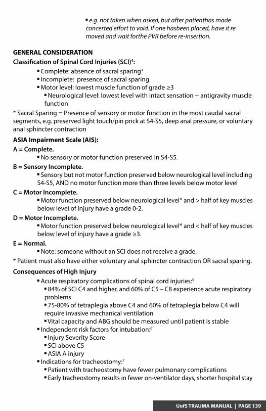
e.g. not taken when asked, but after patienthas made concerted effort to void. If one hasbeen placed, have it re moved and wait forthe PVR before re-insertion.
GENERAL CONSIDERATIONClassification of Spinal Cord Injuries (SCI)4: Complete: absence of sacral sparing* Incomplete: presence of sacral sparing Motor level: lowest muscle function of grade ≥3 Neurological level: lowest level with intact sensation + antigravity muscle function* Sacral Sparing = Presence of sensory or motor function in the most caudal sacral segments, e.g. preserved light touch/pin prick at S4-S5, deep anal pressure, or voluntary anal sphincter contraction
ASIA Impairment Scale (AIS):A = Complete. No sensory or motor function preserved in S4-S5.B = Sensory Incomplete. Sensory but not motor function preserved below neurological level including S4-S5, AND no motor function more than three levels below motor levelC = Motor Incomplete. Motor function preserved below neurological level* and > half of key muscles below level of injury have a grade 0-2.D = Motor Incomplete. Motor function preserved below neurological level* and < half of key muscles below level of injury have a grade ≥3.E = Normal. Note: someone without an SCI does not receive a grade.* Patient must also have either voluntary anal sphincter contraction OR sacral sparing.
Consequences of High Injury Acute respiratory complications of spinal cord injuries:5
84% of SCI C4 and higher, and 60% of C5 – C8 experience acute respiratory problems 75-80% of tetraplegia above C4 and 60% of tetraplegia below C4 will require invasive mechanical ventilation Vital capacity and ABG should be measured until patient is stable Independent risk factors for intubation:6
Injury Severity Score SCI above C5 ASIA A injury Indications for tracheostomy:7
Patient with tracheostomy have fewer pulmonary complications Early tracheostomy results in fewer on-ventilator days, shorter hospital stay
UofS TRAUMA MANUAL | PAGE 139

Predictors of prolonged mechanical ventilation (> 7 days) after cervical CSI: Injury Severity Score > 32 Complete SCI PaO2/FiO2 < 300 3 days after mechanical ventilation initiated Recommended population for early tracheostomy Other variables to consider favoring tracheostomy over extubation:8
FVC < 830 cc Sputum production requiring more than hourly suctioning PaO2/FiO2 < 190
Spinal Shock vs. Neurogenic Shock
Spinal Shock9
Marked reduction or complete loss of motor and reflex function below level of injury. This likely represents an ongoing physiologic continuum consisting of 4 stages This occurs in virtually all patients with severe SCI, beginning within minutes after injury and continuing for up to 12 months. Traditional teaching states patient could not be classified as definitively having a complete SCI unless they were “out of spinal shock”. This was determined by the return of the bulbocavernosus reflex. Practically however, this rarely changes a patients SCI designation and has little prognostic value.
Neurogenic Shock5,9
Hypotension (SBP <90 mmHg supine unrelated to volume depletion, eg. blood loss, dehydration) AND bradycardia after acute SCI Due to a loss of sympathetic nervous system control (disrupted supraspinal control) with intact parasympathetic control (intact vagal nerve), result is loss of vascular tone Can last up to 5 weeks post-injury
Role of Vasopressors in Spinal Cord Perfusion10
Neurogenic shock contributes to spinal cord hypoperfusion and further ischemia Patients with complete SCI, especially cervical (vs. thoracolumbar) and complete motor deficits should be consider for vasopressor support MAP > 85-90 mmHg is the GOAL. Regimen: No consensus on one regimen is superior to another Average duration is typically 5 – 7 days Effective regimens include: Cervical injury: Dobuatmine (5-15 μg/kg per min) and/or dopamine (2-10 μg/kg per min) Dopamine (2.5-5 μg/kg per min) followed by norepinephrine (0.01-0.2 μg/kg per min), if necessary Dopamine, dobutamine, phenylephrine Epinephrine, terbutaline, dobutamine, ephedrine, isoproterenol Norepinephrine alone

Thoracic injury: Dopamine (2.5-5 μg/kg per min) followed by norepinephrine (0.01-0.2 μg/kg per min), if necessary Thoracolumbar injury: Norepinephrine alone
Cervical Spine Injury The Canadian C-Spine Rule11
UofS TRAUMA MANUAL | PAGE 141
1. Any High-Risk Factor Which Mandates Radiography?
Age > 65 years or Dangerous Mechanism * or Paresthesias in extremities
2. Any Low-Risk Factor Which Allows Safe Assessment of Range of Motion?
Simple rear-end motor vehicle collision (MVC) ** or Sitting position in the emergency department or Ambulatory at any time or Delayed onset of neck pain ** or Absence of midline c-spine tenderness
1. Able to Actively Rotate Neck?
45O left and right
No Radiography
Radiography
Rule Not Applicable if: Non-trauma cases Glasgow coma Scale <15 Unstable vital signs Age <16 years Acute paralysis Known vertebral disease Previous C-spine surgery Pregnant
*Dangerous Mechanism
Fall from elevation > 3 feet of stairs Axial load to head, e.g. diving MVC high speed (>100 km/h), rollover ejection Motorized recreational vehicles Bicycle struck or collision
***Delayed
Not immediate onset of neck pain
**Simple Rear-end MVC Excludes
Pushed into oncoming traffic Hit by bus or large truck Rollover Hit by high speed vehicle
Yes
No
Unable
No
Yes
Able

The National Emergency X-Radiography Utilization Study (Nexus)12,13
To determine whether c-spine radiographs can be avoided in the alert, stable trauma patient.C-spine x-ray is indicated for all patients with neck trauma unless they meet ALL of the following criteria: NO posterior midline c-spine tenderness NO evidence of intoxication Alert (GCS=15) NO focal neurological deficits NO painful distracting injuriesCaveats: NEXUS has no age cutoffs (theoretically applied >1 year old); however, lit-erature cautions against use in patients >65 years old as sensitivity decreases signifi-cantly
Special Injury Types
Atlanto-Occipital Dislocation14
A highly unstable craniocervical injury of the occipital-atlanto-axial unit Primarily an injury of widespread ligamentous disruption between the occiput and upper cervical spine (= craniocervical junction), often w/o bony # 3x more common in children vs. adults Caused by any trauma involving large acceleration and deceleration forces, eg. high speed MVC, fall from height Often associated with concomitant cervical injuries below C1 Clinical features: spectrum including lower cranial nerve deficits (abducens, vagus, hypoglossal), unilateral (typically entire side from shoulder to foot) or bilateral weakness including quadriplegia, unconsciousness with respiratory arrest Up to 20% are asymptomatic with a normal neuro exam ± Spinal cord injury: hyperreflexia with clonus, positive Babinski, abnormal sphincter tone ± Autonomic dysregulation, neurogenic shock ± Cerebrovascular injury, esp. vertebral dissection Definitive treatment Halo immobilization followed by internal occipitocervical fixation and fusion Cervical traction should be avoided
Jefferson Fracture (Type II Atlas Fracture)15
Vertical compression of the skull forces the atlas onto the axis Results in rupture (bilateral fractures) of the anterior and posterior arches with splitting of the lateral masses = Jefferson burst fracture (aka Type II atlas fracture) ± Rupture of the transverse ligament essential to prognosis

Radiographic findings C1-C2 joint in frontal x-ray Loss of continuity of a vertical line traced on the lateral margins of the lateral masses of the atlas and of the joint masses of the axis >7 mm combined lateral mass displacement = rupture of the transverse ligament unstable injury Definitive treatment Stable Reduction by cranial traction + immobilization (hard cervical orthosis vs. halo) for 3-4 months Unstable Occipito-cervical arthrodesis (posterior C1-C2 fusion vs. occipitocervical fusion)
Odontoid (Dens) Fractures17,18
Typical mechanism – blow to the vertex of the skull Neurological injury is infrequent Classification Type I: oblique avulsion fracture of the tip of the dens, avulsion of alar ligament Inherently stable, rarely associated with atlantoocipital instability (assess Oc-C1 joints on CT scan, +/- evidence instability on flex-ext views) Type II: fracture at base/body junction (“waist” of dens) Potentially most unstable Highest non-union rate (30-80% in literature) Type III: fracture extends into cancellous body of axis Usually stable Radiographic findings AP, lateral, open mouth odontoid view CT study of choice for fracture delineation, assessing stability of pattern CT angiogram indicated if operative treatment required (location of vertebral arteries) MR indicated if neurological symptoms present Treatment Type I: collar immobilization Type II: Young patients: Halo immobilization vs. operative fixation odontoid screw vs. posterior C1-2 fusion Elderly patients: Cervical orthosis vs. operative fixation
UofS TRAUMA MANUAL | PAGE 143

Type III: collar immobilization
Hangman’s Fracture19
Traumatic spondylolisthesis of C2 Mechanism usually hyperextension and axial loading Five injury patterns described:
Illustrations demonstrating five injury patterns in traumatic spondylolisthesis. A, Type I. B, Type II. C, Type III. D, Type IIA. E, Type III.
Type I: minimally displaced pars interarticularis fracture, translocation <3 mm and no angulation (upright film in collar required) Type IA: atypical, unstable; oblique fracture through one pars and anterior to the pars within the body of the contralateral side Type II: similar to I but displacement >3 mm and/or > 120 angulation (upright film required) Type IIA: more horizontal fracture with no horizontal displacement and kyphosis greater than translation; associated with C2-C3 disk injury and PLL injury Type III: similar to I + dislocation of C2/C3 facet joints; irreducible without surgery Definitive treatment Type I: rigid cervical orthosis x 6 weeks Type IA: same as Type I or halo vest Type II: closed reduction followed by halo immobilization x 12 weeks Surgery if severe displacement or angulation Type IIA: same as Type II Avoid axial traction in IIA, use hyperextension for reduction Type III: surgical intervention Options: C2-3 ACDF Posterior C1-3 fusion Bilateral C2 pars ORIF

Clearing the C-spine with a Normal CTAwake & cooperative patient with no C-spine pain or neurological symptoms and a normal CT c-spine: In an alert patient with a normal CT scan, clinical clearance is required to exclude ligamentous injury Absence of pain and neurological symptoms (e.g. numbness in extremities) on palpation and full range of C-spine movement (rotation, flexion, extension) = clinical clearance, immobilization may be discontinued
Obtunded or altered sensorium patient with a normal CT c-spine:20,21
In an obtunded or unreliable patient, options include: Wait until clinical clearance can be performed Flexion-extension radiography All movements must be voluntary; the patient must move themselves Direct supervision by physician is required Imaging is to be aborted if pain or neurological symptoms arise MR of the c-spine Data is inconclusive on definitive clearance through CT c-spine alone One cohort study concluded safe c-spine clearance based on a normal multi-detector axial CT scan alone Must be multi-detector, axial from occiput to T1, with sagittal and coronal reconstructions Another systematic review revealed 0.7% of patients with a normal CT c-spine went on to have an unstable injury detected on MR c-spine requiring surgical intervention Conclude a continued role for MR c-spine with a normal CT was not necessary if a reliable clinical exam was able to demonstrate intact gross motor function EAST Guidelines 2015: In obtunded blunt trauma patients cervical collar should be removed after a negative high-quality C-spine CT scan. Based on very low-quality evidence Places a strong emphasis on the high negative predictive value of CT imaging in excluding unstable C-spine injury. This recommendation takes into account the high costs of MRI or other additional imaging. Adjunctive imaging after a high-quality CT scan can: Increases the number of low-value diagnoses. Places patients at risk for unnecessary treatment plans. Put patients with multiple injuries at risk by moving them out of the intensive care unit to the resource limited MRI suite. Results in the same clinical action of collar removal.
UofS TRAUMA MANUAL | PAGE 145
(Please see Appendix for UofS C-spine Clearance Protocol

Thoracolumbar InjuryScreening Guidelines & Modalities22,23
Imaging is proposed in any ONE of the following: High-risk injury mechanism MVC >70 kph, fall >3m, ejected from motor vehicle or motorcycle Painful distracting injury Torso, long bone Cognitive impairment GCS<15, any abnormal mentation or clinical intoxication New neurological signs or symptoms/signs of vertebral fracture Back pain, back tenderness, palpable step deformity, midline bruising Known cervical fracture Alert & cooperative patient with reliable clinical exam: Absence of back pain or midline tenderness has 95% negative predictive value for fracture Choice of modality Standard = lateral, AP x-ray If indeterminate, progress to additional modalities If helical CT of chest, abdomen and pelvis are performed, reconstruct (sagittal and coronal) to visualize spine and forgo plain film
Blunt Multi Trauma PatientHigh Force Mechanism
GCS < 15
No Back Pain / Midline TendernessNo Local Signs of TL injury
No Neurological DeficitNo Cervical Spine Fracture
GCS 15TL Imaging
Back Pain / Midline TendernessLocal Signs of TL injury
Neurological DeficitCervical Spine Fracture
Distracting InjuryAlcohol/Drug Intoxication
No Distracting Injury
Alcohol/Drug Intoxication
TL Imaging
TL Imaging Observe
Note: Maintain full spinal precautions until spine cleared Definitions: Blunt Multi-Trauma Patient = victim of blunt trauma necessitating trauma team activation High Force Mechanism = Fall > 3m, MVC or MBC of at least medium velocity, Pedestrian MVC at any velocity Local Signs of TL Injury = palpable step, back bruising/haematoma GCS = Glascow Coma Score Distracting Injury = As decided by treating clinician TL Imaging = x-ray, CT, MRI as appropriate
Special Fracture Types24,25
Injuries described using three-column description Anterior column: ALL; anterior half of vertebral body, disc & anulus

Middle column: posterior half of body, disc & anulus; PLL Posterior column: pedicles, facet joints, ligamentum flavum, lamina, Spinous processes, inter/supraspinous ligament.
UofS TRAUMA MANUAL | PAGE 147
Anterior Column
Middle Column
Posterior Column
T-L spine fractures roughly categorized as: “Major” injuries Compression fractures Burst fractures Flexion-distraction injury (Chance fracture, seat belt-type injury) Fracture-dislocations “Minor” injuries Transverse process fractures Articular process fractures Pars interarticularis fractures Spinal process fractures
The Thoraco-Lumbar Injury Classification and Severity Score (TLICS) Depicts features important in predicting: Spinal stability Future deformity Progressive neurologic compromise Facilitates appropriate treatment recommendations
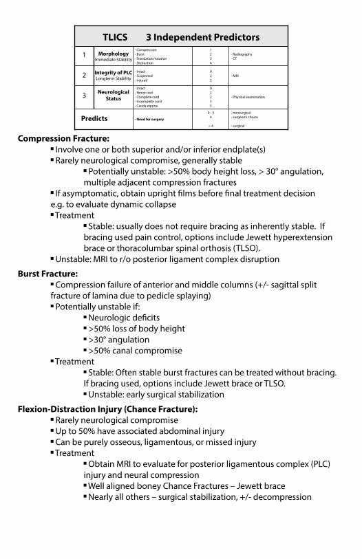
TLICS 3 Independent Predictors
1 MorphologyImmediate Stability
- Compression - Burst - Translation/rotation - Distraction
2
3
Predicts
Integrity of PLCLongterm Stability
Neurological Status
- Intact - Suspected - Injured
- Intact - Nerve root - Complete cord - Incomplete cord - Cauda equina
- Need for surgery
1234
0 - 34
> 4
02233
023
- Radiographs - CT
- MRI
- Physical examination
- nonsurgical - surgeon’s choice
- surgical
Compression Fracture: Involve one or both superior and/or inferior endplate(s) Rarely neurological compromise, generally stable Potentially unstable: >50% body height loss, > 30° angulation, multiple adjacent compression fractures If asymptomatic, obtain upright films before final treatment decision e.g. to evaluate dynamic collapse Treatment Stable: usually does not require bracing as inherently stable. If bracing used pain control, options include Jewett hyperextension brace or thoracolumbar spinal orthosis (TLSO). Unstable: MRI to r/o posterior ligament complex disruption
Burst Fracture: Compression failure of anterior and middle columns (+/- sagittal split fracture of lamina due to pedicle splaying) Potentially unstable if: Neurologic deficits >50% loss of body height >30° angulation >50% canal compromise Treatment Stable: Often stable burst fractures can be treated without bracing. If bracing used, options include Jewett brace or TLSO. Unstable: early surgical stabilization
Flexion-Distraction Injury (Chance Fracture): Rarely neurological compromise Up to 50% have associated abdominal injury Can be purely osseous, ligamentous, or missed injury Treatment Obtain MRI to evaluate for posterior ligamentous complex (PLC) injury and neural compression Well aligned boney Chance Fractures – Jewett brace Nearly all others – surgical stabilization, +/- decompression

UofS TRAUMA MANUAL | PAGE 149
Chane Fracture (flexion-distraction injury)Xray-Lumbar-Lat: Cross-table lateral radiograph of lumbar spine shows fanning of spinous processes (double-headed arrow) and fracture extending through pedicle (between arrowheads) and into L2 vertebral body (single-headed arrow).
Fracture-Dislocation: - 3-column failure - Highly unstable injury almost always requiring surgical intervention Surgery can be performed when patient medically stabilized; neurologic deficit does not require urgent OR - 3 types: Type A: Flexion-rotation 75% have neurological deficits, with 52% having complete SCI Type B: Shear All cases have complete SCI Type C: Flexion-distraction 75% have neurological deficits, all complete SCI
Incomplete Spinal Cord Injuries Anterior cord syndrome: Blunt trauma to the anterior spinal cord and injury of anterior spinal artery (which supplies anterior 2/3 of spinal cord) Loss or impaired ability to sense pain, temperature and touch sensations below their level of injury. Patients retain proprioception, vibration sensation and deep pressure from preservation of the posterior columns. It is possible for some people with this injury to later recover some movement. Posterior Cord Syndrome: Rare syndrome Damage to the dorsal column, resulting in loss of proprioception and vibration sense. Will typically remain with good muscle power, pain and temperature sensation. May experience difficulty in coordinating movement of their limbs. Brown-Sequard Syndrome: Hemitransection of the spinal cord with unilateral damage to the corticospinal tract, spinothalamic tract and dorsal columns.

Ipsilateral deficit: Corticospinal tract Motor function Dorsal columns Proprioception Vibration Contralateral Deficit: Spinothalamic tract Pain Temperature Generally very good prognosis. Central Cord Syndrome: Most common in complete SCI Hyperextension injury Typically in elderly patients with preexisting cervical spondylosis. Motor deficit worse in UE than LE; and in UE hand is affected more than proximal hand Believed to be caused by spinal cord compression and central cord edema with selective destruction of lateral corticospinal tract white matter. It is possible for some recovery from this type of injury, usually starting in the legs, gradually progressing upwards (i.e. bowel/bladder -> proximal UE -> hand function)

UofS TRAUMA MANUAL | PAGE 151

References
1. American College of Surgeons, Committee on Trauma 9th Ed. (2012) Advanced Trauma Life Support (ATLS). Chicago, IL: American College of Surgeons.
2. American Spinal Injury Association. (June 2008) International Standards for the Classification of Spinal Cord Injury – Motor Exam. Retrieved from http://www.asia-spinalinjury.org/elearning/Motor_Exam_Guide.pdf
3. American Spinal Injury Association. (June 2008) International Standards for the Classification of Spinal Cord Injury – Key Sensory Points. Retrieved from http://www.asia-spinalinjury.org/elearning/Key_Sensory_Points.pdf
4. Kirschblum et al. (2011) International standards for neurological classification of spinal cord injury (Revised 2011). Journal of Spinal Cord Medicine, 34(6):535-46.
5. Hagen, E.M. (2015) Acute complications of spinal cord injuries. World Journal of Orthopedics. 6(1):17-23.
6. Berney et al. (2011) The acute respiratory management of cervical spinal cord injury in the first 6 weeks after injury: A systematic review.
7. Leelapattana et al. (2012) Predicting the need for tracheostomy in patients with cervical spinal cord injury. J Trauma Acute Care Surg 73(4):880-4.
8. Berney et al. (2011) A classification and regression tree to assist clinical decision making in airway management for patients with cervical spinal cord injury. Spinal Cord 49:244-250.
9. Krassioukov et al. (2007) Assessment of autonomic dysfunction following spinal cord injury: Rationale for additions to International Standards for Neurological Assessment. Journal of Rehabilitation Research & Development 44(1):103-112.
10. Ploumis et al. (2010) A systematic review of the evidence supporting a role for vasopressor support in acute SCI. Spinal Cord 48:356-362.
11. Stiell et al. (2001) The Canadian C-spine rule for radiography in alert and stable trauma patients. JAMA 286(15):1841-8.
12. Michaleff et al. (2012) Accuracy of the Canadian C-spine rule and NEXUS to screen for clinically important cervical spine injury in patients following blunt trauma: a systematic review. CMAJ 184(16):E867-76.
13. Hoffman et al. (2000) Validity of a set of clinical criteria to rule out injury to the cervical spine in patients with blunt trauma. National Emergency X-Radiography Utilization Study Group. NEJM 343(2):94-9.
14. Hall et al. (2015) Atlanto-occipital dislocation. World Journal of Orthopedics 6(2):236-243.
15. Marcon et al. (2013) Fractures of the cervical spine. Clinics (Sao Paulo) 68(11):1455-61.
16. Moore, D. (Dec 2014) Atlas fractures and transverse ligament injuries. Retrieved from http://www.orthobullets.com/spine/2015/atlas-fracture-and-transverse-ligament-injuries

UofS TRAUMA MANUAL | PAGE 153
16. Moore, D. (Dec 2014) Atlas fractures and transverse ligament injuries. Retrieved from http://www.orthobullets.com/spine/2015/atlas-fracture-and-transverse-ligament-injuries
17. Vaccaro A.R., Madigan L. and Ehrler D.M. (2000) Contemporary management of adult cervical odontoid fractures. Orthopedics 23(10):1109-13.
18. Moore D. (April 2015) Odontoid Fractures (Adult and Pediatric). Available from http://www.orthobullets.com/spine/2016/odontoid-fracture-adult-and-pediatric
19. Bransford et al. (2014) Upper cervical spine trauma. Journal of the American Academy of Orthopaedic Surgeons 22(11):718-29.
20. Raza et al. (2013) Safe cervical spine clearance in adult obtunded blunt trauma patients on the basis of a normal multidetector CT scan – A meta-analysis and cohort study. Injury 44(11):1589-95.
21. James et al. (2014) A systematic review of the need for MRI for the clearance of cervical spine injury in obtunded blunt trauma patients after normal cervical spine CT. J Emerg Trauma Shock 7(4):251-55.
22. O’Connor E. and Walsham J. (2009) Review article: Indications for thoracolumbar imaging in blunt trauma patients: A review of current literature. Emergency Medicine Australasia 21(2):94-101.
23. Hsu J.M., Joseph T., and Ellis A.M. (2003) Thoracolumbar fracture in blunt trauma patients: guidelines for diagnosis and imaging. Injury 34(6):426-33.
24. Aebi M. (2010) Classification of thoracolumbar fractures and dislocations. Eur Spine J 19(Suppl 1):S2-S7.
25. Egol K.A., Koval K.J., and Zuckerman J.D. Handbook of Fractures 4th Ed. Philadelphia: Wolters Kluwer/Lippincott Williams & Wilkins.


Trauma in PregnancyNiroshan Sothilingam, Laura Weins
Every pregnant trauma patient should be questioned regarding domestic/intimate partner violence (SOGC)
7% of pregnancies are complicated by trauma. Trauma is the leading cause of maternal death 46%5.
The leading causes of obstetric trauma are: Motor vehicle crash (49%) Falls (25%) Assaults (18%) Guns (4%) Burns (1%)
The American Congress of Obstetricians and Gynecologists (ACOG) subdivides trauma in pregnancy into three different types: Blunt abdominal trauma Pelvic fractures Penetrating trauma2.
Incidence of trauma increases as pregnancy progresses: 8% in first trimester 40% in second trimester 52% in third trimester
Most maternal deaths are due to head trauma or hemorrhagic shock.
Although many of the assessment and management aspects of obstetric trauma are unique to pregnancy, initial assessment and resuscitation should always be directed at the mother. The main principle guiding therapy must be that resuscitating the mother will resuscitate the fetus. Once maternal stability is established, evaluation of fetal well-being is conducted.
The effect of trauma on pregnancy depends on: Gestational age Type and severity of the trauma Extent of disruption of normal uterine and fetal physiology.
Survival of the fetus depends on adequate uterine perfusion and effective delivery of oxygen through the placenta. Uterine circulation has no autoregulation: This therefore implies that uterine blood flow is directly related to maternal systemic blood pressure, until the mother approaches hypovolemic shock. One the mother shows signs of hypovolemic shock, peripheral vasoconstriction will further compromise uterine perfusion. Once obvious shock develops in the mother, survival rate of the fetus is 20%.
A multidisciplinary team approach is key to optimize outcome in severe trauma.
UofS TRAUMA MANUAL | PAGE 155

Normal Anatomic & Physiologic changes in Pregnancy
Fundal Height is measured in centimeters from the pubic symphysis to the top most portion of the uterus.
Changes in Blood Volume and Composition: 40% increase in blood volume 25% increase in red cell mass Relative anemia (Hct 31-35) ‘Physiologic anemia of pregnancy’ The mother may lose up to 1500 cc of blood without hemodynamic instability However, the fetus may be in shock. White Blood Count elevated in pregnancy (15,000) Fibrinogen and clotting factors increased Decreased albumin level (2.2-2.8) D-dimer often positive, therefore limited use Hemodynamic changes: In the pregnant patient, maternal vitals and perfusion may be preserved at the expense of utero/placental perfusion This delays the signs of hypovolemic shock Cardiac output is increased by 1.0-1.5 liters/minute after the 10th week of pregnancy Hypotension may be due to vena caval compression by the uterus Can cause up to 30% reduction in cardiac output Wedge under right hip can facilitate Heart rate increases 10-15 beats/minute Blood pressure should remain relatively normal compared to a non- pregnant patient. CVP variable Venous hypertension in lower extremities Respiratory changes: Increased oxygen consumption Elevated diaphragm 30-40% increase in tidal volume and minute ventilation Decreased functional residual capacity Decrease respiratory system compliance

Increased airway resistance PaC02= 30-35 mm Hg Intubation may be challenging Due to airway edema, increased breast mass, weight gain In these cases, failed intubation is 8X more likely (SOGC)
Relaxed LES + delayed gastric emptying Increased risk of aspiration Consider early intubation, placement of NG tube, cricoid pressure The fetus is sensitive to maternal hypoxia, therefore maintain maternal O2 sats > 95% (SOGC)
Renal changes: Glomerular Filtration Rate increased BUN and creatinine decrease Glycosuria can be common Mild hydronephrosis is a physiologic response to uterine compression of the ureters Thrombotic Disease and Pregnancy: Pregnancy can induce a hypercoagulable state: Increased activity of clotting factors Decreased fibrinolysis Venous hypertension due to uterine pressure on the inferior vena cava Incidence of DVT of 0.1-0.2% Lower extremity Sequential Compression Devices recommended Heparin and Low Molecular Weight Heparin ok in pregnancy Coumadin contraindicated due to risk of severe fetal malformations Pre-eclampisa/Eclampsia/HELLP Syndrome: Reduced intravascular volume -> more sensitive to anemia and hypovolemia Eclampisa = Seizures Hypertension, proteinuria, hyperreflexia, peripheral edema End organ dysfunction – elevated liver enzymes, hemolysis, thrombocytopenia, stroke, liver capsule hematoma or rupture Eclampsia may mimic head injury in the trauma patient Musculoskeletal: Symphysis pubis widens by 7th month Sacroiliac joint spaces increase This may cause confusion in interpretation of pelvic x-rays.
Imaging the Pregnant Trauma Patient
Even though there is much concern about radiation exposure, a diagnostic modality which is necessary for maternal evaluation should not be withheld on the basis of its potential hazard to the fetus.
In this population of patients, ultrasound is easily accessible in the emergency depart-ment and can provide vital information such as: Approximation of gestational age Placental location Fetal presentation
UofS TRAUMA MANUAL | PAGE 157

Fetal cardiac rate/rhythm Assessment of amniotic fluid volume Confirmation of fetal demise
Placental abruption remains a clinical diagnosis. Ultrasound has been proposed as a method of diagnosis, however the sensitivity is only 24%6.
Ultrasound is a safe and efficient method for detecting intraperitoneal free fluid and intraabdominal injuries. Radiation Exposure : Mettler et al, 2000
Measurement Rad (radiation absorbed dose) Grey (1 rad = 1 centiGy; 100 rads = 1 Gy) Greatest effects of radiation exposure occur between conception and week 25 Radiation injury during weeks 1-3 results in death of the implant or embryo Radiation during weeks 8-25 is more likely to produce CNS effects or growth restrictions: 10 rads may result in decreased IQ 100 rads may result in severe mental retardation After 25 weeks, greatest risk is childhood hematologic malignancy Background incidence is 0.2-0.3% Risk increases to 0.3-0.4% if exposure > 1 Gy Risk increases by 0.06% per 1 Gy of fetal exposure Risk negligible < 5 rads exposure Risk increases > 10-15 rads exposure Most diagnostic procedures have no measurable risk Therapeutic procedures have greatest risk
Benefit of judiciously chosen x-rays far outweighs risks in pregnant trauma patients.
Intermediate exposure (50-100 mGy) roughly equivalent to 3 years of natural background radiation exposure and is associated with no increase in anomalies or growth restriction (Mettler et al, 2000).
A chest x-ray is the equivalent of < 0.01 rads
Abdominal CT scan causes a fetal radiation exposure of up to 3.5 rads (0.035 Gy) per study.
Radiation doses < 5 rads (0.05 Gy) are not associated with an increased risk of anomalies, pregnancy loss, or growth restriction ACOG Guidelines, 2004
Fetal MonitoringWhen the fetus is deemed viable (> 23 weeks GA), continuous fetal monitoring should be initiated as soon as possible, as long as it does not interfere with maternal diagnostic tests or therapy.
The ideal duration for monitoring has not been established, with recommendations ranging from 4-48 hours9. The American Congress of Obstetricians and Gynecologists recommends a minimum of 2-6 hours of electronic fetal monitoring post trauma, in viable pregnancies.

A longer duration of monitoring may detect delayed fetal compromise from a con-cealed abruption.
Fetal monitoring can be discontinued after 4 hours if: Uterine contractions occur less frequently than every 10 minutes. Fetal heart tracing is reassuring. No maternal abdominal pain or vaginal bleeding. Pregnant trauma patients > 23 weeks with signs of labor or abruption such as abdominal/uterine pain, contractions, rupture of membranes, vaginal bleeding or abnormal fetal monitoring, should be admitted for observation for 24 hrs. SOGC
Since placental perfusion and oxygenation depend on maternal cardiopulmonary function, fetal monitoring should continue in cases of: Adult respiratory distress syndrome. Continuous lung injury. Trauma causing maternal cardiac arrhythmia
An abnormality of the fetal heart rate pattern may be the first sign of hemodynamic shock in the mother.
Kleihauer-Betke test (KB)When maternal injury is present, a complete blood cell count, coagulation profile, KB test, and type and screen should be obtained. In Rh-negative mothers, the KB test also allows for calculation of the total required dose of Rh immune globulin needed to prevent alloimmunity. One vial of 300 μg protects against 30 mL of fetal blood (15 mL of fetal red blood cells)2.
The KB test is used in many institutions as a routine component of trauma evaluation. However, the KB test is insensitive and poorly predictive of adverse perinatal outcomes, preterm birth, placental abruption, or fetal distress in minor trauma or in trauma with absent maternal injury4, 14. It’s utility in Rh positive wome is unclear.
In addition to other labs, a coagulation panel including fibrinogen should be obtained.
Emergency Cesarean SectionApproximately 2.4-7.2% of maternal trauma patients need cesarean section shortly after trauma SOGC
Survival of both is dependent on multiple factors:15
Interval between maternal cardiac arrest and delivery. The underlying etiology of the arrest. Where the arrest takes place. The expertise of the team attending to the mother. Gestational age of the fetus
Perimortem cesarean section (PMCS): Cesarean section performed in the face of maternal cardiac arrest, can be life-saving for both mother and fetus.
UofS TRAUMA MANUAL | PAGE 159

Cesarean delivery may be appropriate in the setting of imminent maternal death or after 4 minutes of properly performed cardiopulmonary resuscitation that has failed to revive the mother, as both infant and maternal survival are increased when cesarean delivery is initiated within 4 minutes of maternal cardiac arrest12. Start planning for it right away when the code is called. Start PMCS immediately if the diagnosis is catastrophic, prognosis dismal and maternal survival unlikely. Do not move the patient to do PMCS – only wastes time All that is needed is a scalpel
Although delivery should ideally occur within 4 minutes of failed maternal revival, this standard can rarely be met in actual practice even in ideal situations. Notably, resuscitation efforts may improve following delivery as a result of diminished aortocaval compression and improved volume return to the heart.
Gestational age is less than 24 weeks: Emergency cesarean delivery is usually not performed because the fetus is too small to survive and the birth is unlikely to have much effect on maternal hemodynamics.
Gestational age is greater than 24-25 weeks: Emergency cesarean birth most likely will favorably affect maternal or fetal outcome.
Gestational age of 26 to 32 weeks: External cardiac massage (ECM) is not effective, indicated by: Failure to generate a carotid pulse Inadequate end-tidal CO2 levels Fetal bradycardia. Open cardiac massage (OCM) should be considered before an emergency cesarean section is performed: If OCM proves successful, the delivery may be delayed so that chances of postnatal survival improve. Even slight prolongation of fetal intrauterine life may improve the chances of neonatal survival, especially when gestational age is less than 28 weeks. If OCM proves ineffective, the fetus must be delivered immediately.
Gestation age greater than 32 weeks: ECM is not effective, an emergency cesarean section must be performed immediately. Delivering the infant improves maternal cardiac filling, thereby improving the success of CPR. The longer the delay between the onset of cardiac arrest and delivery, the less are the chances of fetal and maternal survival. If, however, the ECM appears to be effective, ECM may be continued for 5 minutes. If a spontaneous circulation is not restored within 4 minutes, an emergency cesarean delivery must be performed. If this fails to revive the mother, OCM may be considered. Ideally, personnel trained in neonatal resuscitation should be available to attend the infant.

UofS TRAUMA MANUAL | PAGE 161
If preterm labour or preterm prelabour rupture of membrane is suspected, medical management includes: Antibiotics: If Group B streptococcus culture status of patient is unknown Antenatal corticosteroids. Tocolytic agents. MgSO4 for fetal neuro-protection (< 32 weeks) 4 hours of treatment is ideal
Unique Injury Patterns
Blunt TraumaAlthough MVC are the most common cause of serious blunt trauma in pregnancy, assaults, abuse and falls are also frequent.
In addition to maternal mortality from blunt trauma, the fetus is at significant risk, from: Placental abruption Uterine rupture Rupture of membranes
The major risk factor for adverse outcomes during MVC is improper seat belt use: in both front and rear collisions, the impact with the steering wheel can be avoided with proper belt use2.
Placental Abruption(Trauma.org): Occurs in 2 to 4% of minor injuries. Occurs up to 50% of major traumas. Maternal mortality from abruption is less than 1%, but fetal death ranges from 20 to 35%. Separation results as the inelastic placenta shears away from the elastic uterus during sudden deformation of the uterus. Abruption can occur with little or no external signs of injury to the abdominal wall. Abruption is unpredictable on the basis of severity of maternal injury or placental position. Abruption may have associated vaginal bleeding or be concealed, with no external bleeding Clinical indications of abruption: Vaginal bleeding Abdominal pain Uterine tenderness/contractions Amniotic fluid leakage Maternal hypovolemia Uterus larger than normal for the gestational age Change in the fetal heart rate When present after trauma, vaginal bleeding is an ominous sign often indicative of placental separation.

Diagnostic tests: Transabdominal ultrasound: Less than 50% accurate. Cardiotocographic (fetal) monitoring: More sensitive in detecting placental abruption by fetal distress. Ultimately remains a clinical diagnosis
Most cases of abruption become evident within several hours after trauma. Fetal monitoring should be started in the resuscitation room and continued for a minimum of 4 hours, from time of trauma.
Management of placental abruption should not be delayed from imaging if it is clini-cally suspected.
Uterine Rupture Presence of a defect in the gravid uterine wall. 75% of cases involve the uterine fundus. May cause: Serosal hemorrhage. Avulsion of the uterine vasculature with hemorrhage. Complete disruption of the myometrial wall with extrusion of the fetus, placenta, or umbilical cord into the abdominal cavity. Complete uterine avulsion2.
Clinical presentation: Uterine tenderness Vaginal bleeding Non-reassuring fetal heart rate patterns Rapid onset of maternal hypovolemic shock Easily palpable fetal parts on abdominal exam Typical signs of peritoneal irritation on physical examination: Distention Rebound tenderness Guarding Rigidity
Management Urgent laparotomy to control bleeding and facilitate resuscitation
Penetrating TraumaManagement of penetrating injuries depend primarily on the entrance location and the gestational age. Fetal death has been reported in up to 60% of cases of penetrating trauma11.
In terms of mortality, maternal outcome is more favorable during pregnancy than in non-pregnant victims11. This may be due to anatomic changes that take place during pregnancy, such as the superior displacement of the visceral organs by the growinguterus. Therefore, upper abdominal penetrating trauma will likely injure maternal bowel, where lower abdominal penetrating trauma will more likely injure the uterus/fetus.

Even though immediate surgical exploration is the most appropriate management approach in a non-pregnant patient, this approach is not universal in pregnant patients11: This decision to proceed with surgical exploration is dependent on: Location of injury. Uterine size. Maternal and fetal vital signs. Location of possible entrance/exit wounds is key Visceral injuries are less likely when the entry site is anterior and below the uterine fundus Those that do not penetrate beyond the abdominal wall can be managed nonoperatively. Exploratory laparotomy is indicated with any evidence of peritoneal penetration, particularly if intraperitoneal hemorrhage or bowel perforation is suspected. Tetanus prophylaxis is safe in pregnancy, and its indications are similar in both pregnant and nonpregnant states2. Peritoneal lavage can be performed during pregnancy. An open technique is recommended after placement of a nasogastric tube and a Foley catheter13.
Pelvic fractures are not an indication for cesarean delivery. Most women can safely attempt vaginal birth following a pelvic fracture, even those that occur during the third trimester8.
If a thoracostomy tube is required in a pregnancy, it is recommended that it be placed at least 1 or 2 intercostal spaces above the usual landmark of the fifth intercostal space to avoid inadvertent abdominal insertion3.
UofS TRAUMA MANUAL | PAGE 163

References
1. American College of Obstetricians and Gynecologists. Committee on Obstetric Practice. ACOG committee opinion no. 299, September 2004 (replaces no. 158, September 1995): guidelines for diagnostic imaging during pregnancy. Obstet Gynecol 2004;104:647-51.
2. American College of Obstetricians and Gynecologists. ACOG educational bulletin. Obstetric aspects of trauma management. Number 251, September 1998 (replaces Number 151, January 1991, and Number 161, November 1991). Int J Gynaecol Obstet 1999;64:87–94
3. Brown HL. Trauma in pregnancy. Obstet Gynecol 2009;114:147-60.
4. Cahill AG, Bastek JA, Stamilio DM, Odibo AO, Stevens E, Macones GA. Minor trauma in pregnancy–is the evaluation unwarranted? Am J Obstet Gynecol 2008;198:208e1-5.
5. Chames MC, Pearlman MD. Trauma during pregnancy: outcomes and clinical management. Clin Obstet Gynecol 2008;51:398–408
6. Glantz C, Purnell L. Clinical utility of sonography in the diagnosis and treatment of placental abruption. J Ultrasound Med 2002;21: 837-40.
7. Hill DA, Lense JJ. Abdominal trauma in the pregnant patient. Am Fam Physician 1996;53:1269–1274
8. Leggon RE, Wood GC, Indeck MC. Pelvic fractures in pregnancy: factors influencing maternal and fetal outcomes. J Trauma 2002; 53:796-804.
9. Mettler FA, Brent RL, Streffer C, et al. Pregnancy and medical radiation. Ann ICRP 2000;30:1-42.
10. Mirza FG, Devine PC, Gaddipati S. Trauma in pregnancy: a systematic approach. Am J Perinatol 2010;27:579-86.
11. Muench MV, Canterino JC. Trauma in pregnancy. Obstet Gynecol Clin North Am 2007;34:555–583; xiii
12. Katz V, Balderston K, Defreest M. Perimortem cesarean delivery: were our assumptions correct? Am J Obstet Gynecol 2005;192: 1916-20.
13. Nagy KK, Roberts RR, Joseph KT, et al. Experience with over 2500 diagnostic peritoneal lavages. Injury 2000;31:479-82.
14. Pak LL, Reece EA, Chan L. Is adverse pregnancy outcome predictable after blunt abdominal trauma? Am J Obstet Gynecol 1998; 179:1140-4.
15. Society of Obstetricians and Gynaecologists of Canada. Clinical Practice Guideline. Guidelines for the Management of a Pregnant Trauma Patient. J. Obstet Gynaecol Can 2015; 37(6): 553 - 571
16. Thomas R, Sotheran W. Postmortem and perimortem cesarean section. J R Soc Med 2000;93:215-6.

UofS TRAUMA MANUAL | PAGE 165


Burn ManagementEvan Barber, Chris Thomson
1. Pathophysiology Local/systemic inflammatory reaction Mediators disrupt normal capillary barrier Increased vascular permeability Fluid shift to interstitial space Capillaries start to recover at 6 - 12 hrs Without resuscitation, 15-20% TBSA burns = shock “Burn Zones”: Zone of coagulation Nonviable area at epicenter of burn Zone of ischemia/stasis Surrounding coagulated areas Progress to necrosis without resuscitation Zone of hyperemia Peripheral tissues that undergo vasodilatory changes Not injured thermally and remain viable
** Townsend CM, Beauchamp RD, Evers BM, et al, eds. Sabiston Textbook of Surgery. 19th ed. St. Louis: Saunders; 2012.
UofS TRAUMA MANUAL | PAGE 167
Epidermis
Subcutaneous
Dermis
Epidermis
Subcutaneous
Dermis
Degree of Burn 1st degree: Redness, tenderness, pain Superficial damage to epidermis No blistering Sensation normal Ex. sunburns 2nd degree: Partial-thickness Does not extend fully through the dermis Two types: Superficial Epidermis and superficial dermis Blistering Severe pain Deep Into reticular dermis Thicker blisters More pale

3rd degree: Through entire dermis Capillary blood supply destroyed Appears white No sensation 4th degree: Into underlying tissue Estimating BSA: “Rule of 9s” in adults Person’s own palm = ~1% BSA Lund-Browder charts
** Illustration from Anatomy & Physiology, Connexions Web site. http://cnx.org/content/col11496/1.6/
Epidermis
Subcutaneous
Dermis
2. First Aid: Remove patient from source of burn (if safe) Remove charred clothing For chemical burns – irrigate copiously with water Immerse in water 1-5°C Avoid ICE water (frostbite, tissue damage) Use room temperature water - inhibits acidosis/inflammation/ischemia Use COLD water on small burns How long to cool? Small burns (< 9% TBSA) up to 30 minutes or more Large burns – prolonged cooling leads to hypothermia Keep the patient warm, the burn cool
3. Trauma Bay Considerations Airway Fluid resuscitation Large bore IVs NG/foley placement Carboxyhemoglobin levels Elevated levels should get 100% oxygen

All patients should have a non-rebreather mask with high flow O2 on arrival to the trauma bay. This should help correct any elevation of carboxyhemoglobin levels before it become a problem. Tdap
4. Airway Management Inhalation injury occurs 10 - 20% Thermal injury usually supraglottic With steam, upper and lower airway affected Suspect inhalation injury when: Facial burns Singed nasal hair Sooty sputum Tachypnea Hoarseness Stridor Consider intubation early – evidence that airway edema progresses 12-24 hours post-injury5. Fluid Resuscitation Formulas to guide initial fluids: Parkland First 24 h: RL at 4 ml/kg/% TBSA; give half in first 8 h and the remaining over next 16 h. Second 24 h: colloid at 20 - 60% of calculated plasma volume to maintain adequate urinary output Modified Brooke First 24 h: RL at 2 ml/kg/% TBSA burn, one half in the first 8 h and half in the remaining 16 h. Second 24 h: colloid at 0.3–0.5 ml/kg/% TBSA burn + D5W to maintain urine output Only include 2nd degree burns or greater in TBSA; 1st degree do not count No good evidence on which formula to choose Titrate fluids to urine output Goal U/O – 0.5cc/kg/hr for thermal, 1cc/kg/hr for electrical burns Avoid over-resuscitation, may lead to: Pulmonary edema Abdominal compartment syndrome (ACS) Conversion of superficial deep burns Elevated intra-ocular pressure Fasciotomies Increased risk of ACS with IVF 300/hr x 24 hrs6. Role of Antibiotics 2013 Cochrane reviews: Systemic antibiotic prophylaxis in non-surgical patients evaluated in three trials (119 participants)
UofS TRAUMA MANUAL | PAGE 169

No evidence of an effect on rates of burn wound infection Systemic antibiotics reduced PNA but not sepsis Perioperative systemic antibiotic prophylaxis had no effect on outcomes Conclusion: Re: systemic abx – effects unclear, limited by volume/ quality of research Silver sulfadiazine (Flamazine): Use is surgeon-dependent – do not use without involvement of plastics Some evidence it may increase wound infections
7. Wound Care (Saskatchewan Health Authority): Wound and Blister management: Initial Visit (less than 24 hrs. after sustaining burns): If the wound is dirty, clean with soap and tap waterChemical or caustic burns should be well irrigated with tap water or saline Leave small (< 2 cm) blisters alone (serve as biological covering) Open large blisters at one edge but leave otherwise intact Blisters already broken but intact over skin should be left in place Initial Dressing should be multilayer and consist of: Polysporin and Bactigras: Trimmed to cover outside edge of 2nd and 3rd degree area Dry Gauze Kling Subsequent Dressings Remove all layers and on 1st follow up visit Physician to debride all blisters left intact Redress open areas with: Polysporin and Bactigras Dry gauze and kling to cover For facial burns, polysporin to face QID and PRN Follow up in ER q24hrs. Average superficial partial thickness burn will need to be dressed daily for 10 to 12 days Physician to reassess PRN or if signs of infection8. Advanced care, specialist referral Plastic surgery referral for follow-up and further management (for all burns unless very minor) Options include wound excision, autografting, human allografting, porcine xenografting, skin substitute, dermal analogue Circumferential burns may constrict edematous tissue ischemia, respiratory distress Escharotomy indicated Criteria for referrals to Edmonton Burn Centre: 1. Partial or full thickness burns > 20% TBSA in adults 2. Partial or full thickness burns > 10 % TBSA in children, or pediatric burns with other complicating factors 3. Major burns associated with significant inhalation injury 4. Complicated acid/alkali burns

5. Complicated high voltage electrical burns
9. Electrical Burns Review: Resistance – how difficult it is for electrons to pass through a material (ohm) Ohm’s law – Current (I) = Voltage (E)/Resistance (R) Dermis/most internal tissues except bone have low resistance Heat produced proportional to resistance and square of the current Effects of electricity dependent upon: Type of current Amount of current Pathway of current Duration of contact Area of contact Resistance of the body Voltage Current: Alternating current more damaging 3-4x more direct current needed for same effect For 1-second contact time: 1 mA – threshold of perception 10-15 mA – sustained muscular contraction 50-100 mA – respiratory paralysis, ventricular fibrillation 1000 mA – sustained myocardial contractions Arc vs. Internal Current Arc injuries Current external to body Injury more superficial Internal current Path = source entry wound exit wound ground Destruction of deep tissues Low-voltage current – path of least resistance (nerves, blood vessels) High-voltage current – direct path between entrance and ground Most severe damage to tissue occurs at entrance/exit wounds Entry/Exit wounds: Entry = charred, leathery, depressed Exit = “exploded” Hand hand = 60% fatal Hand foot = 20% fatal Complications: MSK – Fractures, dislocations, muscle injury, rhabdomyolysis, periosteal necrosis, limb necrosis, compartment syndrome Vascular – Vascular wall necrosis, bleeding, thrombus Neuro – Nerve injury
UofS TRAUMA MANUAL | PAGE 171

CVS – Vfib, afib, block, arrest Resp – Arrest, effusions, pneumonitis Skin – Burns Nephro – Renal failure GI – N/V, ileus, ulcers, visceral injury
10. Lightning Injuries In Canada, 10 deaths/164 injuries per year Three factors increase risk of strike: Height, isolation, “pointiness” < 5% “direct” strikes < 50% survivors have burn marks Shockwave component Mechanisms of injury: Direct strike (3-5%) Side splash from another object (30%) Contact voltage from object that is struck, ex. plumbing (1-2%) Ground current effect (40-50%) Energy not connected to main lightning channel (15-20%) Blunt trauma/barotraumas Versus electrical burns: Brief contact (4-6 ms), usually no significant tissue damage along path of current Majority of lightning energy flashes around body, vaporizing sweat or rainwater secondary steam burns Rhabdomyolysis, compartment syndromes less likely Blunt, concussive, shrapnel-related injuries Most deaths by cardiac arrest Rupture of the tympanic membranes Minor burns and eye injuries Chronic sequelae – brain injury, chronic pain, neuropsychological Lichtenberg Figure pathognomonic
** From “Lichtenberg Figures Due to a Lightning Strike” by Yves Domart, M.D., Emmanuel Garet, M.D., New England Journal of Medicine, Volume 343:1536, November 23, 2000, Number 21, Images in Clinical Medicine

UofS TRAUMA MANUAL | PAGE 173
References
1. Cooper, M. A. (2016). “Lightning Injuries”. Retrieved from http://emedicine.medscape.com/article/770642-overview
2. Edlich, R. F. (2015). “Thermal Burns”. Retrieved from http://emedicine.medscape.com/article/1278244-overview
3. Edlich, R. F. (2016). “Electrical Burn Injuries”. Retrieved from http://emedicine.medscape.com/article/1277496-overview
4. Sabiston, D. C., & Townsend, C. M. (2012). Sabiston textbook of surgery: The biological basis of modern surgical practice. Philadelphia, PA: Elsevier Saunders.
5. Sheridan, R. L. (2015). “Initial Evaluation and Management of the Burn Patient”. http://emedicine.medscape.com/article/435402-overview


Hemorrhage Control and Balanced Transfusion in Trauma
Oksana Prokopchuk-Gauk, Chris Pastor, Alena Stirling, Ryan Lett
IntroductionMassive hemorrhage requiring massive transfusion is a significant cause of mortality in trauma. Although the phrases massive hemorrhage and massive transfusion imply a degree of bleeding that is life-threatening, a single standard definition has not been established in the literature. Definitions include: Transfusion of blood components equal to, or greater than, one blood volume in 24 hours Replacement of 50% blood volume in 3h Transfusion of 10 or more RBC in 24h or time from ER presentation to ICU transfer Blood loss >150 ml/min Transfusion of 3 U PRBC or more in 1h, in the setting of major bleeding
Algorithms to manage massively bleeding patients are not yet available in all hospitals with emergency departments throughout Saskatchewan. It is important for you to become familiar with policies at your center, and resources for managing bleeding patients – such as lab tests available and stock within your local blood bank. Currently available massive hemorrhage protocols for Saskatchewan sites are located here: https://saskblood.ca/mhp/
Figure 1 shows transfusion activities in Saskatchewan sites, based on the volume of red blood cells transfused per month, and the complexity of lab testing offered. The small blue-drop sites are “transfuse-only” facilities and receive blood as needed for designated patients; however, these sites do not routinely stock red blood cells. Facilities within all other sites carry at least 2 units of uncrossmatched group O negative red blood cells at all times.
A list of blood bank stock information in Saskatchewan is located here: https://saskblood.ca/resources/blood-bank-contact-and-stock-information/
UofS TRAUMA MANUAL | PAGE 175

Physiological CoagulationAn understanding of the physiological mechanisms of hemostasis is essential for the appli-cation of a balanced resuscitation of the massively bleeding patient. The cellular compo-nent of coagulation involves platelets, which are localized to the damaged area and linked via fibrinogen (Figure 2). The enzymatic component involves a serine protease cascade that serves to convert fibrinogen to fibrin and stabilize the clot (Figure 3).
Figure 1: Saskatchewan 'Blood Drop Map'

Figure 3: Enzymatic Component of Coagulation. Several serine proteases form a cascadefor definitive clot stabilization. Intrinisc and extrinsic pathways lead to activation of factor X,
which catalyzes the activation of thrombin resulting in the conversation of fibrinogen to fibrin.
Damaged Surface
XII XIIa
XI XIa
XI IXa VIIIa iTenase
VIII vWF
eTenase Vlla
Trauma
Vll
Thrombin V Prothrombin
Factor II
Va
Fibrin FibrinogenFactor I
XIII Crosslinksthe fibrin clot
There is dysregulation of the thrombotic and antithrombotic mechanisms in the setting of trauma. Thrombin (Factor II) is important in clot formation. In excess, it may pose a risk for systemic thrombosis, such as in the setting of severe blunt trauma. However, in the setting of shock and hypoperfusion, it may lead to hypocoagulation and bleeding. Tissue injury exposes thrombomodulin and release of natural heparins (heparans) from the glycocalyx of endothelial cells. The thrombomodulin, together with thrombin production leads to release of activated Protein C (aPC) The aPC functions as a natural anticoagulant by inhibiting factor Va and Factor VIIIa. Excess activated protein C leads to reduced inhibition of tissue plasminogen activator (tPA), which ultimately degrades fibrinogen (Factor I). Without enough Fibrinogen, a stable clot cannot form, therefore impairing both primary and secondary hemostasis (Duque et al., 2020).
UofS TRAUMA MANUAL | PAGE 177
Figure 2: The cellular component or coagulation in the initial response to tissue damage. Von Willebrand Factor (vWF) localizes platelets to exposed collagen. The localization of platelets results in activation and release of platelet contents including ADP, serotonin, platelet activating factor, vWF, platelet fator 4 and thromboxane A2. The result is recruitment and activation of more platelets and activation of the enzymatic clotting cascade.
Gpllbllla
Gp1b
Exposed Collagen
Fibrinogen
Ca++
ADP
ADP Receptor
Figure 2:
Figure 3:

Trauma Induced Coagulopathy and the “Lethal Triad”In the setting of trauma, the “lethal triad” is comprised of coagulopathy, acidosis, and hypothermia, which is largely the result of significant blood loss. Volume loss decreas-es hemoglobin, which leads to decreased tissue perfusion and an insufficient supply of coagulation factors. Lactic acidosis occurs as cells rely on anaerobic respiration to meet their energy demands. The impaired energy metabolism causes thermal dysreg-ulation and hypothermia, which increases oxygen demand and further exacerbates the problem.
Control of massive hemorrhage is the obvious goal to prevent or reverse the aspects of the “lethal triad.” While this may not be immediately possible, it is critical that the resuscitation team takes measures to prevent exacerbation of the “lethal triad” in the resuscitation bay.
Strategies for avoiding the exacerbation of trauma induced coagulopathy include the following:
Early administration of Tranexamic acid (TXA) –TXA administered within 3 hours of injury has been shown to reduce mortality in trauma victims. Every 15 minute delay in time to TXA administration reduces the survival benefit by 10%. In the landmark CRASH-2 Study, a 1 gram TXA bolus over 10 minutes was given within the first 1 hour (but no more than 3 hours) of injury, followed by a 1 gram infusion over 8 hours (Roberts et al., 2013). Practically speaking, infusion of the second 1 gram is cumbersome, as this requires a dedicated IV line. There is recent evidence supporting the administration of the second 1 gram bolus 1 hour following the initial 1 gram, or a 2 gram bolus of TXA over 20 minutes upfront as at least equivalent in terms of effectiveness to the original TXA dosing strategy demonstrated in trauma (Rowell et al., 2020). Initial bloodwork – A Group & Screen should be sent with initial bloodwork on patient arrival as a means of objectively understanding patient support needs. In some centers, a “Trauma Panel” can be ordered, and should include a CBC, INR, Fibrinogen, Creatinine and ionized Calcium. This panel should be drawn at least every 60 minutes in a trauma patient undergoing active resuscitation and used to guide therapy. Electrolyte balance - If there is a clinical suspicion of hypocalcemia, replace it early (even before results are available). Calcium is a clotting factor (Factor IV), and an essential component of the coagulation cascade. Patients particularly at risk are those who have received large volume transfusion due to dilution binding by citrate, which is used as an anticoagulant within plasma units. The serum ionized Calcium should be maintained above 1.15 mmol/L. Avoid excessive crystalloid resuscitation – Crystalloid can act a volume expander, but it may perpetuate dilutional coagulopathy. In an unstable, actively bleeding patient, crystalloid fluid administration should be minimized, and used only as a temporizing measure until blood products are available. Use of crystalloid fluids with a near physiologic pH, such as PlasmaLyte or Ringers Lactate, is preferred and has been shown to have superior outcomes

UofS TRAUMA MANUAL | PAGE 179
over normal saline due to its acidotic pH. There has been no demonstrated benefit of colloid infusion (ex. albumin) over crystalloid use in the trauma setting. Transfusion support - Replace losses with a balanced transfusion approach, and which is based on results of bloodwork. Remember that informed consent for blood transfusion is required prior to blood administration. It is important to be aware of local policies as they relate to emergency treatment at your center. A summary of transfusion risks appear in Table 1 & 2 at the end of this chapter. Blood components (red blood cells, platelets, plasma) or plasma protein products (fibrinogen concentrate) can be requested as-needed to manage a bleeding patient. A goal-directed approach to transfusion is recommended as follows: Hemoglobin – maintain at 70-90 g/L Platelets ≥ 50 x 109/L (≥ 100 x 109/L in head trauma) INR – goal < 1.8 Fibrinogen level – goal > 1.5 g/L (>2.0 g/L if pregnant) The clinical team should consider providing notification to the transfusion medicine lab to begin thawing plasma in anticipation of a potential Massive Hemorrhage Protocol (MHP) activation. (They can prepare plasma ‘just in case’.) Activation of the MHP should be considered if there is a concern of severe and ongoing blood loss. There are multiple suggested scoring systems to aid in the decision process for MHP activation. (See Appendix A for details) Prevent Hypothermia - Every 1oC drop in body temperature increases blood loss by 20%, due to impaired coagulation enzyme function. Best practice recommendations dictate that the patient core body temperature should be maintained at 36oC or greater at all times, and measured every 30 minutes (or continuously, if available). Ask for warm blankets and/or a Bair Hugger. Warm the patient immediately after completion of the primary survey. The patient will cool quickly when uncovered during the primary survey, and that extremities will inherently be cooler (with greater potential for impaired clotting!) than the body core. Minimize uncovering of patient for adjuncts and secondary survey. X-rays penetrate warm blankets and the patient only needs small areas uncovered for FAST assessment. If possible, warm all fluids during administration Rapid infusers have an incorporated warming mechanism Exception: platelets are stored at room temperature and do not need to be warmed (the cells are damaged if exposed to a warmer). Keep the trauma bay warm. Prevent Acidosis If possible, avoid large volumes of normal saline (pH 5.5) as it can cause metabolic acidosis. A pH balanced fluid is preferred, like

PlasmaLyte (pH 7.4) or Ringer’s Lactate (pH 6.5, although sodium lactate, which is not the same as lactic acid, is converted to bicarbonate upon infusion and improves the pH). Be aware that only normal saline or PlasmaLyte can be co-infused (in the same line) with blood products. Ensure patient can ventilate adequately. There are many reasons for decreased minute volume in a trauma patient (ex. iatrogenic due to administration of opioids). If significant respiratory depression results in respiratory acidosis, consider airway control and mechanical ventilation. Bicarbonate infusion increases CO2, as such a concomitant increase in minute ventilation is required. Furthermore, its use will decrease calcium concentrations lowering myocardial contractility as well as resulting in impaired coagulation. Bicarbonate in trauma resuscitation should only be considered by experienced clinicians.
Balanced Transfusion Strategy Use of a balanced transfusion strategy relies on clinical information gathered from the bedside assessment (which includes a component of gestalt), supplemented with objective laboratory information. Transfusion during an MHP should not be viewed as a binary concept of “all or nothing”. Laboratory data, point-of-care testing (ex. arterial blood gas (ABG) analyzer), and thromboelastography (TEG) help guide appropriate blood components transfusion and administration of hemostatic adjuncts.
A MHP should not be activated before the trauma team is able to assess a patient. The transfusion medicine laboratory may issue up to 2 uncrossmatched RBC Units to have on-hand in the trauma bay upon request prior to patient arrival, if there is a concern of patient instability.
Trauma patient management must include the following: Damage control resuscitation, with highest priority given to treating the source of bleeding. Consideration of the patient underlying condition or medications. Confirm whether the patient has any known significant medical conditions (ex. con genital bleeding disorder, pregnancy, renal failure) or is taking anticoagulant medications, as these may guide dictate earlier use of specific blood products as a part of hemostatic management. Send bloodwork early: Group & Screen and basic bloodwork including the CBC, PT/INR, Fibrinogen, and ionized Calcium). Point-of-care ABG with hemoglobin and metabolites can often return results more quickly than standard lab work and can be used to guide treatment. Thromboelastography (TEG) can be a very useful as a point-of-care assessment of whole blood coagulation ability. TEG is available only at hospitals performing cardiovascular surgery. Its utility is dependent on staff who are familiar with equipment and interpretation of the curves to help guide transfusion decisions.

UofS TRAUMA MANUAL | PAGE 181
Complete a clinical assessment and determine if the patient is rapidly exsanguinating (this may be visible external hemorrhage or massive internal hemorrhage). Suggested criteria for MHP Activation include the presence of one or more of the following, in the setting of ongoing bleeding: Shock index >1.4 (BP divided by the HR) ABC Score 2 or more 1 point each for penetrating injury, blood pressure ≤ 90 mmHg, HR ≥ 120 bpm and positive FAST (Focused Assessment with Sonography for Trauma) on ultrasound Blood loss >150 ml/min Transfusion of 3 U PRBC or more in 1 hour If MHP activation criteria are met, the Trauma Team Lead should clearly state “Activate the Massive Hemorrhage Protocol” to initiate the protocol cascade. The decision to contact Switchboard directly or the Transfusion Medicine Lab to activate a “CODE TRANSFUSION” will be site specific. Confirm the process at your site so you know!! Success of the MHP will be dependent on communication and an understanding of team member roles during the MHP. ONE person should be assigned as the clinical “Team Contact” to serve as the communicator during the MHP. Without effective communication between the bedside and the lab (especially Transfusion Medicine), the MHP process will be suboptimal! Upon MHP activation, a team member must be sent to the Transfusion Medicine lab to pick up the first MHP box. MHP boxes will typically include a standard ratio of blood components, most commonly 2 RBC:1 plasma, until lab results are available to facilitate goal-directed therapy. Pre-thawed plasma is not routinely available from the blood bank, so it may not be included in the first box (about 30 min thaw time, unless the blood bank is given a ‘heads up’ to thaw plasma pre-emptively). Platelets may not be routinely included in the first box, unless the blood bank is told the patient is on an antiplate let medication or known to have a history of a low platelet count. As bloodwork results become available, a transition to goal directed therapy and ‘tailoring’ MHP box contents to suit patient needs is appropriate. Unless the Transfusion Medicine lab is notified otherwise, they will keep preparing MHP Boxes to ensure one is always available for pick-up. Once definitive hemostasis is achieved, the transfusion rate slows and hemodynamic stability is achieved, the Trauma Team Lead can decide to discontinue the MHP. The Team Contact must provide appropriate communication to the Transfusion Medicine Lab and/or Switchboard (depending on local policy) and is responsible for ensuring MHP Boxes are returned and blood is not wasted.

Remember the “60 min rule” – blood components outside of the lab for more than 60 minutes cannot be returned into inventory and are discarded. We must all be good blood stewards! A Transfusion Medicine Physician is available on-call 24/7 throughout Saskatchewan. Call them at any time if you need advice!! Saskatoon and North Saskatchewan – 306-655-1000 Regina and South Saskatchewan – 306-766-4444
Hemostatic AdjunctsThe purpose of a MHP is to provide rapid access to blood components to a massively bleeding patient. Adjunctive clotting factors may be appropriate upfront to manage specific clinical situations or as a part of as a part of goal directed therapy.
Fibrinogen ConcentrateFibrinogen (Factor I) is the first clotting factor depleted in massive bleeding. Its con-centration can be rapidly reduced due to acidosis, dilution, bleeding losses, and re-duced synthesis. Fibrinogen sources include fibrinogen concentrate (FC, brand names RiaSTAP or Fibryga), FFP, and cryoprecipitate. In a recent high-quality trial, 4 g FC was found to be non-inferior to 10 Units of cryoprecipitate in the treatment of acquired hypofibrinogenemia (Callum et al., 2019). From the perspective of safety, FC has the advantage of being virally inactivated (unlike cryoprecipiate) and having a reliable 1 gram dose of fibrinogen per vial. It can also be issued quickly by the Transfusion Med-icine lab and does not require manipulation (a 10 Unit dose of cryoprecipitate takes about 30 minutes to thaw and pool).
Therefore, 4 grams FC is recommended over 10 Units of Cryoprecipitate for treatment of acquired hypofibrinogenemia. A fibrinogen level or evidence of hypofibrinogen-emia should guide the decision for FC administration. The expected fibrinogen rise from this dose is approximately 0.5-1.0 g/L but is dependent on the rate of metabolic consumption in the patient.
Coagulation Factor ConcentratesProthrombin complex concentrates (PCC, brand names Octaplex or Beriplex), contain vitamin K dependent factors II, VII, IX, & X. PCCs can rapidly replace these coagulation factors. PCC is an essential therapy to manage bleeding trauma patients who are on warfarin or factor Xa inhibitors. Warfarin reversal – if the INR is unknown, give PCC 2000 IU IV once. If the INR is known, then dosing is as follows:
INR 1.5-2.9 INR 3.0-5.0 INR 5.1 or greater
PCC Dose 1000 IU 2000 IU 3000 IU
The target INR post-PCC is <1.5 and can be reassessed anytime after PCC infusion is complete. Vitamin K 10 mg IV must be given right after the PCC is given, to ensure a sustained warfarin reversal effect. Missing the Vitamin K
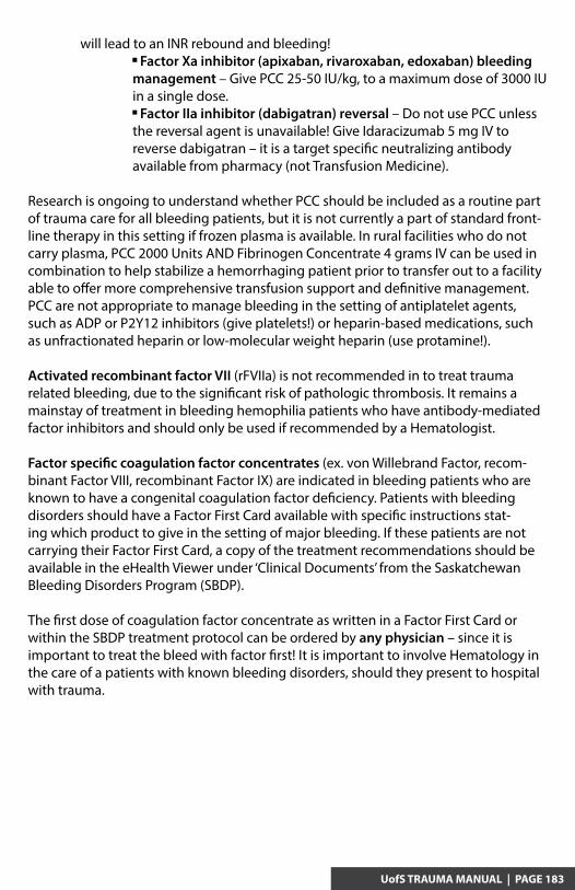
UofS TRAUMA MANUAL | PAGE 183
will lead to an INR rebound and bleeding! Factor Xa inhibitor (apixaban, rivaroxaban, edoxaban) bleeding management – Give PCC 25-50 IU/kg, to a maximum dose of 3000 IU in a single dose. Factor IIa inhibitor (dabigatran) reversal – Do not use PCC unless the reversal agent is unavailable! Give Idaracizumab 5 mg IV to reverse dabigatran – it is a target specific neutralizing antibody available from pharmacy (not Transfusion Medicine).
Research is ongoing to understand whether PCC should be included as a routine part of trauma care for all bleeding patients, but it is not currently a part of standard front-line therapy in this setting if frozen plasma is available. In rural facilities who do not carry plasma, PCC 2000 Units AND Fibrinogen Concentrate 4 grams IV can be used in combination to help stabilize a hemorrhaging patient prior to transfer out to a facility able to offer more comprehensive transfusion support and definitive management. PCC are not appropriate to manage bleeding in the setting of antiplatelet agents, such as ADP or P2Y12 inhibitors (give platelets!) or heparin-based medications, such as unfractionated heparin or low-molecular weight heparin (use protamine!).
Activated recombinant factor VII (rFVIIa) is not recommended in to treat trauma related bleeding, due to the significant risk of pathologic thrombosis. It remains a mainstay of treatment in bleeding hemophilia patients who have antibody-mediated factor inhibitors and should only be used if recommended by a Hematologist.
Factor specific coagulation factor concentrates (ex. von Willebrand Factor, recom-binant Factor VIII, recombinant Factor IX) are indicated in bleeding patients who are known to have a congenital coagulation factor deficiency. Patients with bleeding disorders should have a Factor First Card available with specific instructions stat-ing which product to give in the setting of major bleeding. If these patients are not carrying their Factor First Card, a copy of the treatment recommendations should be available in the eHealth Viewer under ‘Clinical Documents’ from the Saskatchewan Bleeding Disorders Program (SBDP).
The first dose of coagulation factor concentrate as written in a Factor First Card or within the SBDP treatment protocol can be ordered by any physician – since it is important to treat the bleed with factor first! It is important to involve Hematology in the care of a patients with known bleeding disorders, should they present to hospital with trauma.

Further Reading
This manual is by no means a comprehensive review. The following selections are strongly recommended reading. Bloody Easy 4 – Essential reading for every medical student, resident, and attending physician who orders blood & blood products and obtains consent for their administration. An electronic copy for download is available here: https://transfusionontario.org/en/documents/?cat=bloody_easy Treat the Bleed is an educational resource written by Canadian physicians for front-line clinicians with quick-access information on anticoagulation reversal and massive hemorrhage management: https://treatthebleed.org/
The Clinical Guide to Transfusion is an online resource with specific chapters dedicated to transfusion related topics and an excellent resource for front-line clinicians: https://professionaleducation.blood.ca/en/transfusion/clinical-guide-transfusion
Emergency Medicine Cases: Ep 152 The 7 Ts of Massive Hemorrhage Protocols includes an article and video presentation highlighting sentinel management features of the MHP: https://emergencymedicinecases.com/7-ts-massive-hemorrhage-protocols/
The Ontario Blood Coordinating Network has an excellent provincial MHP Toolkit found here: https://transfusionontario.org/en/provincial-massive-hemorrhage-toolkit/
All plasma protein and related factor products have Health Canada approved monographs. These contain very important product-specific information and can easily be found online using your favourite web browser. (Ex. search «drug name product monograph»)
The Circular of Information for blood components is found here : https://www.blood.ca/en/hospital-services/products/component-types/circular-information
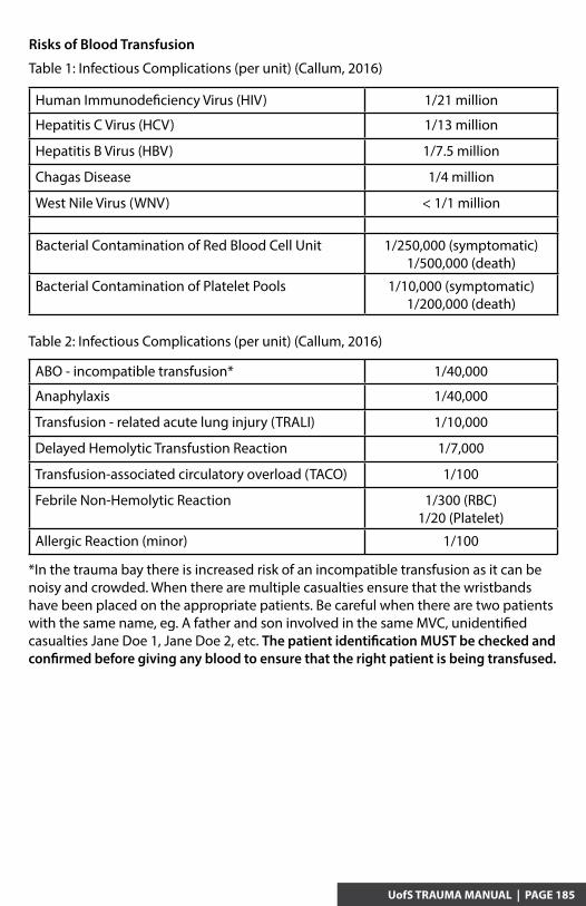
UofS TRAUMA MANUAL | PAGE 185
Risks of Blood Transfusion
Human Immunodeficiency Virus (HIV) 1/21 million
Hepatitis C Virus (HCV) 1/13 million
Hepatitis B Virus (HBV) 1/7.5 million
Chagas Disease 1/4 million
West Nile Virus (WNV) < 1/1 million
Bacterial Contamination of Red Blood Cell Unit 1/250,000 (symptomatic)1/500,000 (death)
Bacterial Contamination of Platelet Pools 1/10,000 (symptomatic)1/200,000 (death)
Table 2: Infectious Complications (per unit) (Callum, 2016)
ABO - incompatible transfusion* 1/40,000
Anaphylaxis 1/40,000
Transfusion - related acute lung injury (TRALI) 1/10,000
Delayed Hemolytic Transfustion Reaction 1/7,000
Transfusion-associated circulatory overload (TACO) 1/100
Febrile Non-Hemolytic Reaction 1/300 (RBC)1/20 (Platelet)
Allergic Reaction (minor) 1/100
*In the trauma bay there is increased risk of an incompatible transfusion as it can be noisy and crowded. When there are multiple casualties ensure that the wristbands have been placed on the appropriate patients. Be careful when there are two patients with the same name, eg. A father and son involved in the same MVC, unidentified casualties Jane Doe 1, Jane Doe 2, etc. The patient identification MUST be checked and confirmed before giving any blood to ensure that the right patient is being transfused.
Table 1: Infectious Complications (per unit) (Callum, 2016)

References
1. Balvers, K., Coppens, M., van Dieren, S., et al. (2015). Effects of a hospital-wide introduction of a massive transfusion protocol on blood product ratio and blood product waste. J Emerg Trauma Shock, 8(4), 199-204. doi:10.4103/0974-2700.166597
2. Callum, J. Farkouh, M. E., Scales, D. C., et al. (2019). Effect of fibrinogen concentrate vs cryoprecipitate in blood component transfusion after cardiac surgery: The FIBRES randomized controlled trial. JAMA. 322(20):1966-1976. doi:10.1001/jama.2019.17312
3. Callum J. L., Yeh C. H., Petrosoniak A., et al. (2019). A regional massive hemorrhage protocol developed through a modified Delphi technique. CMAJ Open. 7(3), E546-E561. http://cmajopen.ca/content/7/3/E546.full
4. Callum, J. L., Pinkerton, P. H., Lima, A., et al. (2016). Bloody Easy 4. Ontario, Canada: Library and Archives Canada Cataloguing in Publication.
5. Cestero, R. F., & Dent, D. L. (2015). Endpoints of resuscitation. Surg Clin North Am, 95(2), 319-336. doi:10.1016/j.suc.2014.10.004
6. Chatrath, V., Khetarpal, R., & Ahuja, J. (2015). Fluid management in patients with trauma: Restrictive versus liberal approach. J Anaesthesiol Clin Pharmacol, 31(3), 308-316. doi:10.4103/0970-9185.161664
7. Connelly, C. R., & Schreiber, M. A. (2015). Endpoints in resuscitation. Curr Opin Crit Care, 21(6), 512-519. doi:10.1097/mcc.0000000000000248
8. D'Alessandro, A., Moore, H. B., Moore, E., et al. (2016). Plasma First Resuscitation Reduces Lactate Acidosis, Enhances Redox Homeostasis, Amino Acid and Purine Catabolism in a Rat Model of Profound Hemorrhagic Shock. Shock. doi:10.1097/shk.0000000000000588
9. Duque, P., Mora, L., Levy, J. H., et al. (2020) Pathophysiological response to trauma-induced coagulopathy: A comprehensive review. Anesth Analg. 130(3):654-64. doi: 10.1213/ANE.0000000000004478.
10. Dzik, W. H., Blajchman, M. A., Fergusson, D., et al. (2011). Clinical review: Canadian National Advisory Committee on Blood and Blood Products--Massive transfusion consensus conference 2011: report of the panel. Crit Care, 15(6), 242. doi:10.1186/cc10498
11. Frantz, T. L., Gaski, G. E., Terry, C., et al. (2016). The effect of pH versus base deficit on organ failure in trauma patients. J Surg Res, 200(1), 260-265. doi:10.1016/j.jss.2015.07.003
12. Gayet-Ageron, A., Prieto-Merino, D., Ker, K., et al. (2018). Effect of treatment delay on the effectiveness and safety of antifibrinolytics in acute severe haemorrhage: a meta-analysis of individual patient-level data from 40 138 bleeding patients. Lancet. 391(10116), 125-32. doi: 10.1016/S0140-6736(17)32455-8.
13. Hunt, B. J., Allard, S., Keeling, D., et al. (2015). A practical guideline for the haematological management of major haemorrhage. Br J Haematol, 170(6), 788-803. doi:10.1111/bjh.13580

14. National Advisory Committee on Blood and Blood Products. NAC statement on fibrinogen concentrate [Internet]. Canada; National Advisory Committee on Blood and Blood Products; 2014 [updated 2021 Mar 10; cited 2021 May 25]. Available from: https://nacblood.ca/resources/index.html.
15. National Advisory Committee on Blood and Blood Products. NAC statement on recombinant factor VIIa. [Internet]. Canada; National Advisory Committee on Blood and Blood Products; 2012 [updated 2019 Feb 4; cited 2021 May 25]. Available from: https://nacblood.ca/resources/index.html.
16. Roberts, I., Shakur, H., Coats, T.,et al. Guerriero, C. (2013). The CRASH-2 trial: a randomised controlled trial and economic evaluation of the effects of tranexamic acid on death, vascular occlusive events and transfusion requirement in bleeding trauma patients. Health Technol Assess, 17(10), 1-79. doi:10.3310/hta17100
17. Rowell, S.E., Meier, E.N., McKnight, B., et al. (2020). Effect of our-of-hosptial tranexamic acid vs placebo on 6-month functional neurologic outcomes in patients with moderate or severe traumatic brain injury. JAMA. 324(10):961-974. doi: 10.1001/jama.2020.8958.
18. Smith, C. A., Gosselin, R. C., Utter, G. H., et al. (2015). Does saline resuscitation affect mechanisms of coagulopathy in critically ill trauma patients? An exploratoryanalysis. Blood Coagul Fibrinolysis, 26(3), 250-254. doi:10.1097/mbc.0000000000000154
19. Stephens, C. T., Gumbert, S., & Holcomb, J. B. (2016). Trauma-associated bleeding: management of massive transfusion. Curr Opin Anaesthesiol, 29(2), 250-255. doi:10.1097/aco.0000000000000306
20. Spahn, D.R., Bouillon, B., Cerny, V. et al. (2019). The European guideline on management of major bleeding and coagulopathy following trauma: fifth edition. Crit Care 23, 98. https://doi.org/10.1186/s13054-019-2347-3
21. Trudeau J. (2017) Ch. 11: Massive hemorrhage and emergency transfusion, in Clinical Guide to Transfusion. https://professionaleducation.blood.ca/en/mas-sive-hemorrhage-and-emergency-transfusion. Accessed May 25, 2021.
22. Tsuei, B. J., & Kearney, P. A. (2004). Hypothermia in the trauma patient. Injury, 35(1), 7-15. doi: 10.1016/s0020-1383(03)00309-7.
23. Yazer, M. H., Waters, J. H., & Spinella, P. C. (2018). Use of uncrossmatched erythrocytes in emergency bleeding situations. Anesthesiology. 128, 650-6. https://doi.org/10.1097/ALN.0000000000002037
UofS TRAUMA MANUAL | PAGE 187


Beyond the Trauma Bay: VTE Prophylaxis, Tracheostomy, Enteral Nutrition and Tertiary Surveys
Sarah Miller, Niroshan Sothilingam
VTE ProphylaxisCause of death amongst trauma patients: Immediate (within the first hour): Blood loss, Traumatic brain injury (TBI) Later: Multi-organ failure, CNS injury, VTE
Trauma patients are at increased risk for VTE General trauma patients 3-5% risk (Chest Guidelines) Higher with spinal trauma, acute spinal cord injury (SCI), and TBI - 8-10% Other risk factors: lower extremity and pelvic fractures, older patient age, prolonged immobility, and increased hospital stay Incidence of DVT 11.8-65% and PE 1.5-20% amongst trauma patients (Cochrane Review)
Pathophysiology of VTE Risk in Trauma Patients Cochrane Review
Virchow’s Triad:
Stasis
Hypercoagulability Endothelial Injury
Injured immobilized reduction in venous return decreased oxygen \ and nutrients to endothelial cells Direct endothelial injury to vessels procoagulation factor amplification thrombosis
UofS TRAUMA MANUAL | PAGE 189

Assessment of VTE risk: Modified Caprini Risk Assessment
Options: Pharmacological LDUH – Low-dose unfractionated heparin Heparin 5,000 units subq BID LMWH – Low molecular weight heparin Enoxaparin 40 mg subq daily Renal impairment: - Enoxaparin 30 mg subq daily if CrCl<30cc/min Bariatric (BMI > 40): - Enoxaparin 40 mg subq BID Tinzaparin 4500 units subq daily Mechanical IPC – Intermittent pneumatic compression Graduated compression stockings Other Inferior vena cava filter
Points Risk Factor1 point - Age 41-60
- Acute MI (<1m)- BMI>25- CHF exacerbation (<1m)- Hx of IBD- Procedure with local anesthesia- Swollen legs of varicose veins- Sepsis (<1m)
- Serious lung dx (e.g. Pneumonia) (<1m)- Abnormal pulmonary function (e.g. COPD)- Pregnancy or postpartum- Hx of unexplained or recurrent spontaneous abortion (> 3)- Oral contraceptives or HRT
2 point - Age 61-74- Arthroscopic sx- Major open surgery (>45 mins)- Laparoscopic surgery (>45 mins)
- Malignancy- Confined to bed (>72 hours)- Immobilizing plaster cast- Central venous access
3 point - Age > 75 years- Hx of CTE- FMHx of VTE (1st degree relative)- F5 Leiden- Prothrombin 20210A
- Lupus anticoagulant- Anticardiolipin antibodies- Elevated serum homocysteins- HIT- Other congenital or acquired thrombophilia
5 point - Stroke (<1m)- Elective arthroplasty- Hip, pelvis, or leg fracture (<1 m)
- Acute spinal cord injury (<1m)- Polytrauma (<1m)
Risk Category Score
Very Low
Estimated VE Risk inAbsence of Prophylaxis (%) Recommendation
0 < 0.5 Early and frequent ambulation
Low 1-2 1.5 Mechanical Prophylaxis
Moderate 3-4 3.0 Pharmacologic Prophylaxis
High > 5 6.0 Mechanical and pharmacologic prophylaxis

Examining the Literature
Cochrane Library: “Prophylaxis was more effective than no prophylaxis, pharmacological prophylaxis than mechanical prophylaxis, and LMWH than UH”. Higher risk of minor bleeding in pharmacological thromboprophylaxis compared to mechanical therapy. Limitations: Small trials, few events, questionable methodology quality.
EAST Guidelines:LDUH Level II: Little evidence to support the benefit of LDUH as sole agent for prophylaxis in trauma patient at high-risk for VTE. Level III: For patients in whom bleeding could exacerbate injuries (e.g. intracranial hemorrhage, incomplete SCI, intraocular injuries, severe pelvic or lower extremity injuries with traumatic hemorrhage, and intra-abdominal solid organ injuries being managed non-operatively), the safety of LDUH has not been established, and an individual decision should be made when considering anticoagulant prophylaxis. LMWH Level II: LMWH can be used for VTE prophylaxis in trauma pts with the following injuries: - 1. Pelvic #s requiring operative fixation or prolonged bed rest (5 days). - 2. Complex lower extremity #s (open fractures or multiple #s in 1 extremity) requiring operative fixation or prolonged bed rest (5 days). - 3. SCI with complete or incomplete motor paralysis. Level III: - 1. Trauma patients with an ISS 9, who can receive anticoagulants, should receive LMWH as their primary mode of VTE prophylaxis. - 2. LMWH has not been sufficiently studied in the head-injured patient with intracranial bleeding to justify its use. - 3. LMWH should not be used when epidural catheters are placed or removed.IVC Filters Please see Appendix for IVC Filter Protocol Level III: Should be considered in very high-risk trauma pts: - Those that cannot receive anticoagulation because of increased bleeding risk, AND - Injury pattern rendering them immobilized for a prolonged period of time, including the following: GCS <8 – severe closed head injury. Incomplete spinal cord injury with paraplegia or quadriplegia. Complex pelvic fractures with associated long bone fractures. Multiple long bone fractures.
UofS TRAUMA MANUAL | PAGE 191

CHEST Guidelines:
Summary of CHEST Guidelines for VTE Prevention in Non-orthopedic Surgical Patients
For major trauma patients:
Use LDUH, LMWH, or mechanical prophylaxis (IPC) over no prophylaxis. If high risk of VTE (acute SCI, TBI, spinal surgery), add mechanical prophylaxis to pharmacologic prophylaxis if not contraindicated by lower extremity injury. If LMWH and LDUH contraindicated, use mechanical prophylaxis (IPC) over none when not otherwise contraindicated. Add LMWH and LDUH when contraindications no longer present. Do not use an IVC filter for primary VTE prevention.
For patients undergoing hip fracture surgery:
Use one of the following for at least 10 to 14 days: LMWH, fondaparinux, LDUH, adjusted-dose vitamin K antagonist (VKA), aspirin, or an IPC. LMWH preferred over other alternatives. Start LMWH either 12h+ preop or postop rather than within 4h or less.
For patients undergoing major orthopedic surgery: Consider extending prophylaxis in the outpatient period for up to 35 days postoperatively (instead of 10 to 14 days). Use dual prophylaxis with an antithrombotic agent and IPC during the hospital stay. Use IPC or no pharmacologic prophylaxis in patients at an increased risk of bleeding. Consider using apixaban or dabigatran (or rivaroxaban or adjusted-dose VKA when the aforementioned are unavailable) in patients who decline or are uncooperative with injections or IPC. Do not use an IVC filter for primary prophylaxis in patients with increased risk of bleeding or with contraindications to pharmacologic/mechanical prophylaxis. Consider no prophylaxis rather than pharmacologic prophylaxis in patients with isolated lower-leg injuries requiring immobilization.
Summary of CHEST Guidelines for VTE Prevention in Non-orthopedic Surgical Patients
Special Situation: TBI and SCI Incidence of DVT among TBI population 54% without prophylaxis (mechanical and pharmacological) (Koehler et al). Assess the risk of on-going intracranial bleeding/haemorrhage. Generally, initiated at 72h. Balance risks of delaying and starting prophylaxis Discuss with your friendly local neurosurgeon. Various studies suggest that early initiation is likely safe: No progression in neurological exam or CT head findings after 24h means that it is likely safe to initiate prophylaxis (Schaible et al). Early initiation (<72h) associated with lower rates of PE and DVT with no increased risk of neurosurgical intervention or death. Bryne et al

UofS TRAUMA MANUAL | PAGE 193
Early VTE prophylaxis (<72h) with enoxaparin not associated with increased progression of intracranial hemorrhagic injury in hemodynamically stable patients. Koehler et al
Summary:
VTE is a major source of morbidity in trauma patients. - Prophylaxis should be initiated on all trauma patients, unless otherwise contraindicated. Daily reassessment thereafter should be performed in order to start as soon as possible.
TracheostomyIndications Airway obstruction Anticipation of prolonged mechanical ventilation requirements from respiratory failure Need for improved pulmonary toilet Inability to protect the airwayBenefits Prevention of laryngeal and upper airway damage secondary to prolonged intubation E.g. Vocal cord edema and damage, laryngeal erosions/scarring/ stenosis, recurrent laryngeal nerve damage Allows for easy and frequent access to lower airway for suctioning and secretion removal Improved patient comfort and increased patient safety Promotion of oral feeding Facilitation of patient mobilization and nursing care Less sedation requirements Decreased risk for ventilator-associated pneumonia Data mixed on this point. Theory: Micro aspiration of oral secretions past the tube cuff may contribute to pneumonia (PNA).

Timing “Prolonged mechanical ventilation anticipated” - Definition of prolonged? Beyond 10-14 days* In severe multi-trauma or head injury with low GCS, tracheostomy within 3-4d of intubation may have some benefit. Durbin et al 2010
- Cochrane 8 RCTs: early tracheostomy < 10d & late > 10d Moderate quality evidence showing lower mortality and decreased time spent on ventilation with early tracheostomy Divergent results on PNA and probability of timing of ICU discharge EAST Guidelines - Level I: No mortality difference between patients receiving early tracheostomy (3-7d) vs late (>7d). - Level II: Early tracheostomy decreases total days of mechanical ventilation and ICU length of stay (LOS) in patients with head injuries. Patients with severe head injuries should receive early tracheostomy. - Level III: Early tracheostomy may decrease total days of mechanical ventilation and ICU LOS in trauma patients without head injuries. Early tracheostomy may decrease rate of PNA in trauma pts. Early tracheostomy should be considered in all trauma patients anticipated to require mechanical ventilation >7d.Risks Morbidity 4-10%, Mortality <1%. Paul et al
Short-term
- Airway loss - Bleeding - Damage to nearby anatomical structures - Tracheal ring rupture - Pneumothorax
Long-term
- Infection - Tracheoinnominate fistula (<1%) May be heralded by minor “sentinel” bleed that later turns to massive haemorrhage. - Tracheomalacia - Tracheal stenosis - Swallowing dysfunction
Method Option for percutaneous or surgical placement Dependent on patient factors as well as operator comfort and preference Prior to performing tracheostomy, optimize conditions when possible: PEEP < 10-15 Correction of coagulopathies and thrombocytopenia INR < 1.5 Platelets > 50,000

UofS TRAUMA MANUAL | PAGE 195
- Decreased: Cost, wound infection rates, scarring - No patient transportation required - Minimal disruption of tissue planes - Avoidance of GA
Surgical
- More appropriate for difficult cases (e.g. anatomy) - Direct and better visualization of tissue during dissection - Ability to place stay sutures
Percutaneous
Disadvantages
Advantages
- Need for bronchoscopy - Lack direct visualization of tissue
- Increased cost - Transportation of patient - Increased time
Cochrane No evidence of different rates in mortality and morbidity related to procedure, major bleeding, tube occlusion/obstruction, accidental decannulation, or difficulty in tube change. Percutaneous tracheostomy showed reduced rates of wound infection and major scarring. 20 trials examined, low quality of evidence. Examining the evidence for percutaneous dilatational tracheostomy (PDT) Freeman et al. 5 RCTs, 236 patients Results: No difference between PDT and open with respect to overall complication rates, days intubated, or death. PDT associated with… - less perioperative bleeding (OR 0.14). - less postoperative bleeding (OR 0.39). - lower infection rate (OR 0.02). - shorter procedure time by 9.84 mins. Bachetta et al. Retrospective study over 3 years with 59 open tracheostomies and 27 PDT Results No significant difference in demographics, medical histories, operations, or complications (excepting more postoperative arrhythmias in open) between both groups. PDT estimated to save $304,000 during a 5-year period. OR time = $$ - Room set up, procedural time, anesthesia cost, room clean up. Delaney et al. 17 RCTs with 1,212 patients Results Reduced wound infections in PDT OR = 0.28, 95% CI, p<0.0005 Minimization of local tissue damage No significant difference in bleeding

No significant difference in overall mortality but possible trend favoring PDT Fewer infections Undue risk of transportation to OR No significant difference in incidence of major complications (pneumothorax, tube malplacement, airway loss) Shorter duration to placement of PDT Decreased duration of sedation Earlier weaning from mechanical ventilation Conclusion: “PDT technique, performed in the ICU, should be considered the technique of choice for critically ill patients who require a tracheostomy”. Further studies supporting use of PDT Higgins et al. Kornblith et al.
Summary: Tracheostomies should generally be considered in patients where intubation is expected beyond 10-14d. - Tracheostomies aid in prevention of upper airway and laryngeal damage from prolonged intubation and increase patient comfort. - Method selection depends on a variety of factors (patient and operator). Both have similar complication rates but PDT is associated with less scarring and fewer wound infections.
Enteral NutritionMetabolic Response to TraumaPostinjury Hypermetabolism Ebb (within first 48h): Hyperglycemia, increased vascular tone, and changes in O2 consumption. Flow: Increased oxygen consumption and delivery, hyperdynamic circulation, and development of insulin resistance. Mobilization of amino acids from lean tissues to support wound healing, immunologic response, and accelerated protein synthesis.Enteral nutrition (EN) Early – Delivery of nutrients into GI lumen within 24-48h of admission/injury. Benefits Nutritional: Fulfill energy requirements via delivery of adequate calories and protein. Non-nutritional: Maintenance of structure and functional integrity of GI tract. Modulation of metabolic response to catabolic stimulus. Support immune system by maintaining GALT. Reduce oxidative stress. Martindale et al

UofS TRAUMA MANUAL | PAGE 197
Metabolic ResponseGI Response
- GI integrity maintenance - Enhance motility - Improve absorptive capacity - Support and maintain commensal bacteria - Reduce virulence of endogenous pathogenic organisms - Promote production of secretory IgA
- Promotion of insulin sensitivity - Reduction of hyperglycemia, muscle and tissue glycosylation - Reduces stress metabolism to enhance more physiologic fuel utilization
Immune Response
- Regular cell modulation to enhance systemic immune function - Promotes anti-inflammatory response - Maintains mucosa associated lymph tissue - Modulates adhesion molecules to decrease migration of macrophages and neutrophils
Martindale et al.
Attenuate oxidative stress Attenuate oxidative stress Systemic Inflammatory Systemic Inflammatory Response Syndrome (SIRS) Response Syndrome (SIRS)
Dominance of anti-inflammatory Th2 over Dominance of anti-inflammatory Th2 over pro-inflammatory Th1 responses pro-inflammatory Th1 responses Modulate adhesion molecules to Modulate adhesion molecules to transendothelial migration of macrophages transendothelial migration of macrophages and neutrophils and neutrophils
Absorptive capacity Absorptive capacity Influence anti-inflammatory receptors Influence anti-inflammatory receptors in GI tract in GI tract Virulence of pathogenic organisms Virulence of pathogenic organisms Motility, contractility Motility, contractility
Reduce gut/lung axis of inflammation Reduce gut/lung axis of inflammation Maintain MALT tissue Maintain MALT tissue Production of Secretory IgA at Production of Secretory IgA at epithelial surfaces epithelial surfaces
Muscle function, mobility, return Muscle function, mobility, return to baseline function to baseline function
Provide micro & macronutrients, antioxidants Provide micro & macronutrients, antioxidants Maintain lean body mass Maintain lean body mass Muscle and tissue glycosylation Muscle and tissue glycosylation Mitochondrial function Mitochondrial function Protein synthesis to meet metabolic demand Protein synthesis to meet metabolic demand
Maintain gut integrity Maintain gut integrity Gut permeability Gut permeability Support commensal bacteria Support commensal bacteria Stimulate oral tolerance Stimulate oral tolerance Butyrate production Butyrate production Promote insulin sensitivity Promote insulin sensitivity hyperglycemia (AGEs) hyperglycemia (AGEs)
McClave et al.
Examining the LiteratureCochrane Library Burn Injuries Insufficient evidence to support or refute effectiveness of early vs. late initiation. Promising evidence that early blunts the hypermetabolic response but insufficient to provide guidelines. Head Injuries Trend towards improved survival and reduced disability and decreased infections with early nutritional support but small trials and unable to come to consensus statement. EAST Guidelines re. Route Level I: Feed pts with blunt and penetrating abdo injuries enterally when feasible. Level II: Feed pts with head injuries preferentially with EN as outcomes similar to parenteral nutrition (PN) but lower cost and complications in EN. PN feeds if EN not possible.

Level III: TPN to be started by day 7 if EN not successful in severely injured patients. Pts who do not tolerate 50% of their EN goals by day 7 should have TPN started. Wean TPN once >50% of EN feeds tolerated. re. Early vs Late Level I: In severely injured blunt/penetrating trauma pts, no outcome advantage to initiating enteral feedings within 24h as compared to 72h. Level II: Burn patients: Enteric feeds should be started ASAP. Delay over 18h associated with gastroparesis and need for PN. Severe head injury: Those that do not tolerate gastric feeds within 48h should be switched to postpyloric feeds if feasible and safe. Level III: Pts incompletely resuscitated should not have direct small bowel feedings. If severely injured and undergoing laparotomy for blunt and penetrating trauma, direct small bowel access should be obtained and enteral feedings begun ASAP.
Severe Trauma
CNS Trauma
Resuscitation Completed Hemodynamically Stable/No Pressors
Blunt/Penatrating Torso Trauma
>20-30% TBSA Burn
Begin Gastric Tube Feeds when Safe
Multiple operativedebridements needed?
Continue Gastric Feeds
Begin NJ* Feeds; Feed Intra-Op
Estimate Energy Needs (Curreri HBEE, etc.), and
provide 2gm/kg/day protein
Consider weekly calorimetry
* NJ = naso-jejunal*PID = Post-Injury Day
N Y
Laparotomy required?
Obtain Access Beyond Lig. of Treitz
& feed when safe
Place NG; √ residual
Y N
PATI>25or
ISS>20?
N Y
StandardFormula
EnhancedFormula
Tolerated?
<300cc/12˚ >300cc/12˚
Begin Gastric Feeds when safe
Begin NJ* feeds
ISS>20?
Y
StandardFormula
N
Y N
Advance to Goal
Start TPN by PID* #7
1.25-2 gm/kg pro; 30-35 kcal/kg(up to 2.4 gm/kg protein for enhanced eternal formulas)
Place NG; √ residual
<300cc/12˚ >300cc/12˚
Begin Gastric Feeds when safe
Begin NJ* feeds
Tolerated? Tolerated?
Advance to Goal
N
Y Y N
TPN
Severe TBI(GCS < 8) Cord Injury
30 kcal/kg1.5-2 gm prot/kg
Quad Para
20-22 kcal/kg 22-24 kcal/kg
EAST Guidelines

UofS TRAUMA MANUAL | PAGE 199
Summary:
Enteral nutrition should be initiated when possible to fulfill the patient’s energy requirements and maintain integrity of GI tract,
support the immune system, and modulate the metabolic response after trauma.
The Tertiary Survey (TS)“The W’s”
What: A repeat comprehensive physical examination with cataloguing of all injuries and a complete review of the mechanism of injury with all pertinent investigations and imaging (films and reports). When: Typically occurs within 24-48 hours after the initial injury. This may vary from site to site. An additional survey may be required at a later time if patient remains unconscious, uncooperative, and immobile. Why: Goal to decrease incidence of missed injuries in trauma patients. Decreased morbidity and avoidance of long-term disability Improved prognosis Medico-legal consequences (Thomson et al) ATLS primary and secondary surveys, although proven to be effective in recognizing life-threatening injuries, has been shown to miss some injuries. Missed injury incidence in literature 1-40% (Hajibandeh et al). Implementation of tertiary surveys decreases this rate. Missed injury rate: injuries that are not identified by tertiary survey. - TS vs. non-TS » Biffl et al. 1.51% vs. 2.37% (p=0.0123) » Keijzers et al. 1.5% vs. 2.4% (p=0.01) Missed injury detection rate: injuries that are not identified by primary and secondary surveys but are diagnosed by tertiary survey. - TS vs. non-TS » Keijzers et al. 7% vs. 3% (p<0.01) » Resler et al. 2.6% vs. 0.34% (p<0.0001) » Ursic et al. 6.16% vs 3.57% (p=0.00999) Contributing factors to missed injuries: Remember the D’s Decreased level of consciousness - Drugs and sedatives - Alcohol - CNS injuries – head trauma
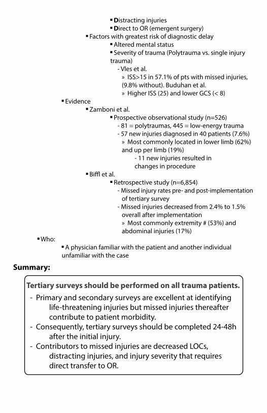
Distracting injuries Direct to OR (emergent surgery) Factors with greatest risk of diagnostic delay Altered mental status Severity of trauma (Polytrauma vs. single injury trauma) - Vles et al. » ISS>15 in 57.1% of pts with missed injuries, (9.8% without). Buduhan et al. » Higher ISS (25) and lower GCS (< 8) Evidence Zamboni et al. Prospective observational study (n=526) - 81 = polytraumas, 445 = low-energy trauma - 57 new injuries diagnosed in 40 patients (7.6%) » Most commonly located in lower limb (62%) and up per limb (19%) - 11 new injuries resulted in changes in procedure Biffl et al. Retrospective study (n=6,854) - Missed injury rates pre- and post-implementation of tertiary survey - Missed injuries decreased from 2.4% to 1.5% overall after implementation » Most commonly extremity # (53%) and abdominal injuries (17%) Who: A physician familiar with the patient and another individual unfamiliar with the case
Summary:
Tertiary surveys should be performed on all trauma patients. - Primary and secondary surveys are excellent at identifying life-threatening injuries but missed injuries thereafter contribute to patient morbidity. - Consequently, tertiary surveys should be completed 24-48h after the initial injury. - Contributors to missed injuries are decreased LOCs, distracting injuries, and injury severity that requires direct transfer to OR.

UofS TRAUMA MANUAL | PAGE 201

References
DVT Prophylaxis: 1. Barrera, L., Perel, P., Ker, K., et al. Thromboprophylaxis for trauma patients. Cochrane Database of Systematic Review 2013;3.
2. Bryne, J., Mason, S., Gomez, D., et al. Timing of pharmacologic venous thromboembolism prophylaxis in severe traumatic brain injury: A propensity-matched cohort study. J Am Coll Surg 2016;223:621-31.
3. Falck-Ytter, Y., Francis, C., Johanson, N., et al. Prevention of VTE in orthopedic surgery patients. Chest 2012;141:278S-325S.
4. Gould, M., Garcia, D., Wren, S., et al. Prevention of VTE in non-orthopedic surgical patients: antithrombotic therapy and prevention of thrombosis, 9th ed: American College of Chest Physicians evidence-based clinical practice guidelines. Chest 2012;141:227S-77S.
5. Koehler, D., Shipman, J., Davidson, M., et al. Is early venous thromboembolism prophylaxis safe in trauma patients with intracranial hemorrhage. J Trauma 2011;70:324-9. 6. Oettgen, P., Shaughnessy, A., Aird, W. Venous thromboembolism (VTE) prophylaxis for medical patients. DYNAmed (Accessed 2016).7. Paydar, S., Sabetien, G., Khalili, H., et al. Management of deep vein thrombosis (DVT) prophylaxis in trauma patients. Bull Emerg Trauma 2016;4:1-7.
8. Rogers, F., Cipolle, M., Velmahos, G., et al. Practice management guidelines for the prevention of venous thromboembolism in trauma patients: The EAST practice management guidelines work group. J Trauma 2002;53:142-64.
9. Schaible, E., Thal, S. Anticoagulation in patients with traumatic brain injury. Curr Opin Anaesthesiol 2013;26:529-34.
Tracheostomy:1. Andriolo, B., et al. Early versus late tracheostomy for critically ill patients. Cochrane Database of Systematic Review 2015;1:1-67.
2. Appleby, I. Tracheostomy. Anaesthesia and Intensive Care Medicine 2005;6:220-2.
3. Bacchetta, M., et al. Comparison of open vs bedside percutaneous dilatational tracheostomy in the cardiothoracic surgical patient: outcomes and financial analysis. Ann Thorac Surg 2005;79:1879-85.
4. Ben-Nun, A., et al. Emergency percutaneous tracheostomy in trauma patients: an early experience. Ann Thorac Surg 2004;77:1045-7.
5. Brass, P., et al. Percutaneous techniques versus surgical techniques for tracheosto-my. Cochrane Database of Systematic Review 2016;7:1-102.
6. Delaney, A. et al. Percutaneous dilatational tracheostomy versus surgical tracheostomy in critically ill patients: a systematic review and meta-analysis. Crit Care 2006;10:R55.
7. Doyle, J. and Scales, D. “Tracheostomy”. Principles of Critical Care. McGraw-Hill (2015).
UofS TRAUMA MANUAL | PAGE 203
8. Durbin, C. Tracheostomy: why, when, and how? Respir Care 2010;55:1056-68.
9. Engels, P., et al. Tracheostomy: from insertion to decannulation. Can J Surg 2009;52:427-33.
10. Freeman, B., et al. A meta-analysis of prospective trials comparing percutaneous and surgical tracheostomy in critically ill patients. Chest 2000;118:1412-8.
11. Higgins, K., et al. Meta-analysis comparison of open versus percutaneous tracheostomy. Laryngoscope 2007;117:447-54.
12. Holevar, M. et al. Practice management guidelines for timing of tracheostomy: the EAST practice management guidelines work group. J Trauma 2009;67:870-4.
13. Kaylie, D. et al. An analysis of time and staff utilization for open versus percutneous tracheostomies. Otolaryngol Head Neck Surg 2003;128:109-14.
14. Kornblith, L., et al. One thousand bedside percutaneous tracheostomies in the surgical intensive care unit: time to change the gold standard. J Am Coll Surg 2011;212:163-70.
15. Lissauer, M. Benefit, timing, and technique of tracheostomy. Curr Probl Surg 2013;50:424-99.
16. Mallick, A. and Bodenham, A. Tracheostomy in critically ill patients. Eur J Anaesthesiol 2010;27:676-82.
17. Paul, S. and Colson, Y. “Tracheostomy”. Adult Chest Surgery. McGraw-Hill (2015)
18. Tobin, M. Principles and Practice of Mechanical Ventilation. McGraw-Hill (2013).
Enteral Nutrition:1. Jacobs, D.G., Jacobs, D.O., Kudsk, K., et al. Practice management guidelines for nutritional support of the trauma patient. J Trauma 2004:57;660-79.
2. Kudsk, K. and Curtis, C. Nutritional support and electrolyte management. Trauma McGraw-Hill (2013).
3. Martindale, R. and Warren, M. Should enteral nutrition be started in the first week of critical illness? Curr Opin Clin Nutr Metab Care 2015;18:202-6.
4. McClave, S. et al. Feeding the Critical Ill Patient. Crit Care Med 2014;42:2600-10.
5. Perel, P., et al. Nutritional support for head-injured patients. Cochrane Database of Systematic Reviews 2006;4.
6. Wasiak J., et al. Early versus delayed enteral nutrition support for burn injuries. Cochrane Database of Systematic Reviews 2006;3.
Tertiary Survey:1. Biffl, W., et al. Implementation of a tertiary trauma survey decreases missed injuries. J trauma 2003;54:38-44.
2. Brooks, A., et al. Missed injury in major trauma patients. Injury 2004;35:407-10.
3. Keijzer, G., et al. The effect of tertiary surveys on missed injuries in trauma. Scand J Trauma Resusc Emerg Med 2012;20:77-86.

4. Hajibandeh, S., et al. Meta-analysis of the effect of tertiary survey on missed injury rate in trauma patients. Injury 2015;46:2474-82.
5. Thomson, C. and Greaves, I. Missed injury and the tertiary trauma survey. Injury 2008;39:107-14.
6. Zamboni, C., et al. Tertiary survey in trauma patients: avoiding neglected injuries. Injury 2014;45S5:S14-7.

UofS TRAUMA MANUAL | PAGE 205

Trauma Triage Guidelines i
Canadian CT Head Guidelines ii
University of Saskatchewan IVC Filter in Trauma Protocol iii
University of Saskatchewan Cervical Spine Clearance Protocol iv
MHP Activation Decision Tools and Scoring Systems v
TRAUMA MANUALappendix

UofS TRAUMA MANUAL | PAGE 207
Pre-Hospital / Scene Call
Level I(Any of the following)
Physiologic · SBP < 90, SBP < 80 (age < 5), SBP < 70 (age <
1)· RR < 10 or > 29 or requiring intubation· GCS 12 or less with visible evidence of
trauma OR clear history of significant trauma
Anatomic· Penetrating Trauma to Head, Neck or Torso· Severe Facial Injury with potential for
airway compromise· Suspected spinal CORD injury (neurological
deficits)· 2 or more long bone fractures· Burns: 20% TBSA (2nd & 3rd Degree)
Mechanism · Pedestrian or Cyclist vs. Car > 30 km/hr· ATV or motorcycle vs. Car > 30km/hr· Ejected from car · Large Animal Trauma (non-MVC)· Fall > 20 feet
Special population meeting Level II criteria, will be upgraded to Level I· Pregnant patients > 20 weeks gestation · Geriatrics (Age > 65)
Level II(Any of the following)
Mechanism· Highway speed MVC· Rollover · Fall from 10-20 feet· Death of same car occupant · Rearward displacement of front axle· Significant passenger compartment
intrusion· Other
Triage to Determine
Level of Trauma
Level 1 **Notify Trauma Team
Lead
Level 2Notify on shift ED
Physician
Intrafacility Transport*
Level 1
Contact Trauma Team Lead (TTL) through SFCC
Level 2Trauma Consult
TTL to discuss case with shift ED Physician at
JPCH
Send directly to Trauma
Service under TTL
Send to JPCH ED under accepting ED physician as Level 2 trauma
ED Physician must contact Trauma Team Lead with update
and reason for upgrade.
Trauma Triage Guidelines
Triage at RUH ED (in JPCH) to follow Activation Guidelines
Paged out information:- Age, Gender, location- Mechanisms- Vitals- ETA
**Notify Trauma team members
** ED Physician on shift is MRP until the Trauma Team Lead arrives and appropriate handover has occurred.
TTL determines level of trauma
The ED Physician may upgrade any Level 2 to a
Level 1 at their discretion at any point during the case of the patient or if the patient deteriorates on transport.
* Includes patients being transported from St. Paul’s Hospital
· Patients who are found unresponsive with no history of trauma and no visible trauma will NOT be considered trauma patients. Simple falls while intoxicated do not qualify as significant trauma.
· If 3 or more Level II patients arrive at the same time, the ER Physician on shift may call the TTL to help manage these patients, at their discretion.
· If any doubt regarding activation, please contact TTL
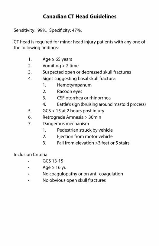
Canadian CT Head Guidelines
Sensitivity: 99%. Specificity: 47%.
CT head is required for minor head injury patients with any one of the following findings:
1. Age ≥ 65 years 2. Vomiting > 2 time 3. Suspected open or depressed skull fractures 4. Signs suggesting basal skull fracture: 1. Hemotympanum 2. Racoon eyes 3. CSF otorrhea or rhinorrhea 4. Battle's sign (bruising around mastoid process) 5. GCS < 15 at 2 hours post injury 6. Retrograde Amnesia > 30min 7. Dangerous mechanism 1. Pedestrian struck by vehicle 2. Ejection from motor vehicle 3. Fall from elevation >3 feet or 5 stairs
Inclusion Criteria • GCS 13-15 • Age ≥ 16 yr. • No coagulopathy or on anti-coagulation • No obvious open skull fractures

UofS TRAUMA MANUAL | PAGE 209
Trauma patients are at high risk of development of venous thromboembolism (VTE) including deep venous thrombosis and pulmonary embolism (PE). PE is thought to be the third major cause of death after trauma in those patients who survive longer than 24 hours after onset of injury.
Placement of a prophylactic IVC filter in a trauma patient could be considered if there are absolute contraindications to prophylactic anticoagulation.
If the ACS/Trauma surgeon feels a patient may benefit from an IVC filter, please consider Hematology Consultation for formal advice and long term follow up.
If the Hematology service agrees that the patient would benefit from a prophylactic IVC filter, then Interventional Radiology will be consulted for placement of the filter.
In ICU patients who are not being followed by the ACS/Trauma surgeon, it is at the discretion of the ICU attending to arrange Hematology Consultation.
In summary, IVC filter placement is a reasonable consideration to mitigate the risk of VTE in the trauma setting. Optimally, IVC filters should be removed as soon as possible as they represent a significant independent risk factor for the development of lower extremity and IVC venous thrombosis. The Hematology Consultation Service is willing and available to inform decisions regarding indications for IVC filter placement and their subse-quent removal.
University of SaskatchewanIVC Filter in Trauma Protocol
Otto Moodley

All patients who meet pre-hospital criteria for a cervical collar placement should undergo cervical spine clearance on arrival at the trauma center. If possible, the cervical spine should be cleared and the collar removed within 24 hours of collar placement.
If cervical collar is required to stay in place for more than 24 hours, stiff extrication collars should be replaced with collars designed for long-term immobilization – ones that provide greater padding and pressure ulcer prevention.
1. Asymptomatic patientsThe cervical spine may be cleared clinically with no need for X-ray films or CT if ALL of the following preconditions are met: • Fully alert and orientated • No head injury • No drugs or alcohol • No neck pain • No midline tenderness, bruising or deformity • No abnormal neurology (e.g. numbness, weakness, radicular pain) • No significant other 'painful distracting' injury (i.e. another injury which may 'distract' the patient from complaining about a possible spinal injury).
2. Symptomatic patientsPatients who do not meet the “asymptomatic” criteria outlined above should be suspected of having a cervical spine injury. Apply Canadian C-spine rules to determine if radiography is necessary (Canadian C-spine rules include additional criteria, including the mechanism of injury, to determine if imaging is indicated).
University of SaskatchewanCervical Spine Clearance Protocol
Daryl Fourney

UofS TRAUMA MANUAL | PAGE 211
It is appropriate to consult the spine surgeon on-call if there is clinical evidence neurological deficit prior to obtaining spine imaging.
For patients in whom C-spine injury is suspected based on clini-cal criteria, or plain radiographs: - The primary screening modality is axial CT from the occiput to T1 with sagittal and coronal reconstructions. - Plain radiographs contribute no additional information and should not be obtained in addition to CT, unless recommended by the spine surgeon (e.g. flexion- extension view, in the appropriate clinical circumstances). - If CT of the C-spine demonstrates injury: Obtain spine consultation. - If there is neurologic deficit (with or without evidence of C-spine injury on x-ray or CT): Obtain spine consultation. - For the neurologically intact awake and alert patient complaining of neck pain with a negative CT: C-spine is considered cleared. Patients should have follow up with a physician (GP, trauma clinic, etc) in 10-14 days. If still having pain during the follow up visit, consider spine consult. - For the obtunded patient with a negative CT c-spine (No injury or degenerative changes) read by a staff radiologist, c-spine can be cleared without further imaging or clinical correlation.

APPENDIX A: MHP Activation Decision Tools and Scoring Systems The easiest scoring tool is the Assessment of Blood Consumption (ABC) Scoring System. One point is assigned for each positive. A score of 2 is 83% sensitive and 78% specific that the patient will require massive transfusion. 1. Tachycardia: HR ≥ 1202. Hypotension: SBP ≤ 903. Penetrating mechanism4. Positive FAST
The McLaughlin scoring system incorporates some laboratory data into the equation. Similar to ABC, there is one point awarded for each criterion. Furthermore, each point has a predictive value of 20% likelihood of massive transfusion. For example, a patient with 2 points has a 40% chance of massive transfusion whereas a patient with 3 points has a 60% likelihood. 1. Tachycardia: HR ≥ 1052. Hypotension: SBP ≤ 1103. Acidosis: pH ≤ 7.254. Hematocrit ≤ 32%
Although there is no gold standard, the Trauma Associated Severe Hemorrhage (TASH) consistently has the highest sensitivity and specificity for massive transfusion. As it is algorithm based, the best application is using a calculator, such as mdcalc.com to perform the number crunching.


Department of Surgery