Signaling-dependent and coordinated regulation of transcription, splicing, and translation resides...
6
Signaling-dependent and coordinated regulation of transcription, splicing, and translation resides in a single coregulator, PCBP1 Qingchang Meng*, Suresh K. Rayala*, Anupama E. Gururaj*, Amjad H. Talukder*, Bert W. O’Malley †‡ , and Rakesh Kumar* †‡ *Molecular and Cellular Oncology, University of Texas M. D. Anderson Cancer Center, Houston, TX 77030; and † Molecular and Cellular Biology, Baylor College of Medicine, Houston, TX 77030 Contributed by Bert W. O’Malley, February 5, 2007 (sent for review December 20, 2006) Transcription, splicing, and translation are potentially coordinately regulatable in a temporospatial-dependent manner, although sup- porting experimental evidence for this notion is scarce. Yeast two-hybrid screening of a mammary gland cDNA library with human p21-activated kinase 1 (Pak1) as bait identified polyC-RNA- binding protein 1 (PCBP1), which controls translation from mRNAs containing the DICE (differentiation control element). Mitogenic stimulation of human cells phosphorylated PCBP1 on threonines 60 and 127 in a Pak1-sensitive manner. Pak1-dependent phosphory- lation of PCBP1 released its binding and translational inhibition from a DICE-minigene. Overexpression of PCBP1 also inhibited the translation of the endogenous L1 cell adhesion molecule mRNA, which contains two DICE motifs in the 3 untranslated region. We also found that Pak1 activation led to an increased nuclear reten- tion of PCBP1, recruitment to the eukaryotic translation initiation factor 4E (eIF4E) promoter, and stimulation of eIF4E expression in a Pak1-sensitive manner. Moreover, mitogenic stimulation pro- moted Pak1- and PCBP1-dependent alternative splicing and exon inclusion from a CD44 minigene. The alternative splicing functions of PCBP1 were in turn mediated by its intrinsic interaction with Caper , a U2 snRNP auxiliary factor-related protein previously implicated in RNA splicing. These findings establish the principle that a single coregulator can function as a signal-dependent and coordinated regulator of transcription, splicing, and translation. differentiation control element eukaryotic translation initiation factor 4E p21-activated kinase 1 T he efficient control of cellular response to various stimuli relies on the coordination of multiple regulatory modules in the eukaryotic genetic architecture, including transcription, pre- cursor mRNA processing, and translation of mature mRNA in the cytoplasm. In addition to inherent selective control at each level, these processes might be regulated in a coordinated manner. For example, steroid hormone receptors activated by cognate ligands or growth factors have been shown to function- ally couple gene transcription activity and exon-content selection of pre-mRNA by recruiting coregulators (1, 2). These coordi- nated responses often involve posttranslational modifications of key components within their regulatory systems. Despite the growing realization of the significance of such delicate and efficient regulatory control of gene expression, the notion of signal-activated and temporospatial-dependent regulation of transcription, splicing, and translation lacks supporting proof- of-principle evidence. In eukaryotic cells, multiple kinases provide obligatory and rapid regulatory switches for controlling mRNA transcription, splicing, and translation (3). P21-activated kinase 1 (Pak1), one of the evolutionarily conserved families of serine/threonine protein kinases, is a downstream effector of the activated Rho GTPases Rac1 and Cdc42, nonreceptor tyrosine kinases lipids, and growth factor receptor-generated mitogenic signals (4, 5). However, to date, Pak1 signaling has not been linked with the functions of RNA-binding proteins. To explore the biologic functions and to discover binding partners of Pak1, we per- formed a yeast two-hybrid screening of a human mammary gland cDNA library. PolyC-binding protein 1 (PCBP1) was identified as an interacting substrate for Pak1. PCBPs are ubiquitously expressed proteins that contain a highly conserved triple repeat of the KH domain, which is a distinctive feature of RNA-binding proteins. PCBPs participate in a variety of mRNA processing steps and have been implicated in posttranscriptional regulatory pathways, including translation silencing (6, 7). Although PCBP1 exists in both phosphorylated and unphosphorylated forms (8), no function of PCBP1 has yet been associated with its phos- phorylation in cells, and no kinase has been shown to phosphor- ylate PCBP1. Our validation of PCBP1 as a Pak1-binding protein warranted further exploration of PCBP1 functions by this reg- ulatory signaling kinase. Here, we investigated the functions and regulation of PCBP1, and identified a series of previously unrecognized roles for PCBP1, establishing the exciting concept that a single coregulator can function as a signal-dependent and coordinated regulator of transcription, splicing, and translation in eukaryotic cells. Results and Discussion To explore the biologic function and to discover binding partners of Pak1, a major signaling nodule in the mammalian cells (4, 5), we performed a yeast two-hybrid screening of a human mam- mary gland cDNA library. We identified several RNA-binding proteins and splicing factors as Pak1-interacting proteins [see supporting information (SI) Table 1]. Because the role of Pak1 signaling in the biology of RNA-binding proteins is relatively unexplored, we decided to focus on PCBP1 (GenBank accession no. NM006196), an evolutionarily conserved RNA-binding protein. PCBPs are ubiquitously expressed and contain a highly conserved triple repeat of the KH domain and participate in a variety of mRNA processing steps (7). The specificity of the Pak1–PCBP1 interaction was verified by cotransfection of Pak1 and PCBP1 plasmids into the yeast cells (Fig. 1A) as well as by the GST-PCBP1 fusion protein pull-down of in vitro translated Pak1 protein (Fig. 1B). Pak1 interacted specifically with amino acids 1– 42 and 139 –220 of PCBP1, regions overlapping with the first two KH domains of PCBP1; whereas PCBP1 interacted with Author contributions: B.W.O. and R.K. designed research; Q.M., S.K.R., A.E.G., and A.H.T. performed research; and R.K. wrote the paper. The authors declare no conflict of interest. Abbreviations: Pak1, p21-activated kinase 1; PCBP1, poly-C-RNA-binding protein 1; eIF4E, eukaryotic translation initiation factor 4E; DICE, differentiation control element. ‡ To whom correspondence may be addressed. E-mail: [email protected] or [email protected]. This article contains supporting information online at www.pnas.org/cgi/content/full/ 0701065104/DC1. © 2007 by The National Academy of Sciences of the USA 5866 –5871 PNAS April 3, 2007 vol. 104 no. 14 www.pnas.orgcgidoi10.1073pnas.0701065104
-
Upload
mdanderson -
Category
Documents
-
view
0 -
download
0
Transcript of Signaling-dependent and coordinated regulation of transcription, splicing, and translation resides...

Signaling-dependent and coordinated regulationof transcription, splicing, and translation residesin a single coregulator, PCBP1Qingchang Meng*, Suresh K. Rayala*, Anupama E. Gururaj*, Amjad H. Talukder*, Bert W. O’Malley†‡,and Rakesh Kumar*†‡
*Molecular and Cellular Oncology, University of Texas M. D. Anderson Cancer Center, Houston, TX 77030; and †Molecular and CellularBiology, Baylor College of Medicine, Houston, TX 77030
Contributed by Bert W. O’Malley, February 5, 2007 (sent for review December 20, 2006)
Transcription, splicing, and translation are potentially coordinatelyregulatable in a temporospatial-dependent manner, although sup-porting experimental evidence for this notion is scarce. Yeasttwo-hybrid screening of a mammary gland cDNA library withhuman p21-activated kinase 1 (Pak1) as bait identified polyC-RNA-binding protein 1 (PCBP1), which controls translation from mRNAscontaining the DICE (differentiation control element). Mitogenicstimulation of human cells phosphorylated PCBP1 on threonines 60and 127 in a Pak1-sensitive manner. Pak1-dependent phosphory-lation of PCBP1 released its binding and translational inhibitionfrom a DICE-minigene. Overexpression of PCBP1 also inhibited thetranslation of the endogenous L1 cell adhesion molecule mRNA,which contains two DICE motifs in the 3� untranslated region. Wealso found that Pak1 activation led to an increased nuclear reten-tion of PCBP1, recruitment to the eukaryotic translation initiationfactor 4E (eIF4E) promoter, and stimulation of eIF4E expression ina Pak1-sensitive manner. Moreover, mitogenic stimulation pro-moted Pak1- and PCBP1-dependent alternative splicing and exoninclusion from a CD44 minigene. The alternative splicing functionsof PCBP1 were in turn mediated by its intrinsic interaction withCaper �, a U2 snRNP auxiliary factor-related protein previouslyimplicated in RNA splicing. These findings establish the principlethat a single coregulator can function as a signal-dependent andcoordinated regulator of transcription, splicing, and translation.
differentiation control element � eukaryotic translation initiationfactor 4E � p21-activated kinase 1
The efficient control of cellular response to various stimulirelies on the coordination of multiple regulatory modules in
the eukaryotic genetic architecture, including transcription, pre-cursor mRNA processing, and translation of mature mRNA inthe cytoplasm. In addition to inherent selective control at eachlevel, these processes might be regulated in a coordinatedmanner. For example, steroid hormone receptors activated bycognate ligands or growth factors have been shown to function-ally couple gene transcription activity and exon-content selectionof pre-mRNA by recruiting coregulators (1, 2). These coordi-nated responses often involve posttranslational modifications ofkey components within their regulatory systems. Despite thegrowing realization of the significance of such delicate andefficient regulatory control of gene expression, the notion ofsignal-activated and temporospatial-dependent regulation oftranscription, splicing, and translation lacks supporting proof-of-principle evidence.
In eukaryotic cells, multiple kinases provide obligatory andrapid regulatory switches for controlling mRNA transcription,splicing, and translation (3). P21-activated kinase 1 (Pak1), oneof the evolutionarily conserved families of serine/threonineprotein kinases, is a downstream effector of the activated RhoGTPases Rac1 and Cdc42, nonreceptor tyrosine kinases lipids,and growth factor receptor-generated mitogenic signals (4, 5).However, to date, Pak1 signaling has not been linked with the
functions of RNA-binding proteins. To explore the biologicfunctions and to discover binding partners of Pak1, we per-formed a yeast two-hybrid screening of a human mammary glandcDNA library. PolyC-binding protein 1 (PCBP1) was identifiedas an interacting substrate for Pak1. PCBPs are ubiquitouslyexpressed proteins that contain a highly conserved triple repeatof the KH domain, which is a distinctive feature of RNA-bindingproteins. PCBPs participate in a variety of mRNA processingsteps and have been implicated in posttranscriptional regulatorypathways, including translation silencing (6, 7). Although PCBP1exists in both phosphorylated and unphosphorylated forms (8),no function of PCBP1 has yet been associated with its phos-phorylation in cells, and no kinase has been shown to phosphor-ylate PCBP1. Our validation of PCBP1 as a Pak1-binding proteinwarranted further exploration of PCBP1 functions by this reg-ulatory signaling kinase. Here, we investigated the functions andregulation of PCBP1, and identified a series of previouslyunrecognized roles for PCBP1, establishing the exciting conceptthat a single coregulator can function as a signal-dependent andcoordinated regulator of transcription, splicing, and translationin eukaryotic cells.
Results and DiscussionTo explore the biologic function and to discover binding partnersof Pak1, a major signaling nodule in the mammalian cells (4, 5),we performed a yeast two-hybrid screening of a human mam-mary gland cDNA library. We identified several RNA-bindingproteins and splicing factors as Pak1-interacting proteins [seesupporting information (SI) Table 1]. Because the role of Pak1signaling in the biology of RNA-binding proteins is relativelyunexplored, we decided to focus on PCBP1 (GenBank accessionno. NM�006196), an evolutionarily conserved RNA-bindingprotein. PCBPs are ubiquitously expressed and contain a highlyconserved triple repeat of the KH domain and participate in avariety of mRNA processing steps (7). The specificity of thePak1–PCBP1 interaction was verified by cotransfection of Pak1and PCBP1 plasmids into the yeast cells (Fig. 1A) as well as bythe GST-PCBP1 fusion protein pull-down of in vitro translatedPak1 protein (Fig. 1B). Pak1 interacted specifically with aminoacids 1–42 and 139–220 of PCBP1, regions overlapping with thefirst two KH domains of PCBP1; whereas PCBP1 interacted with
Author contributions: B.W.O. and R.K. designed research; Q.M., S.K.R., A.E.G., and A.H.T.performed research; and R.K. wrote the paper.
The authors declare no conflict of interest.
Abbreviations: Pak1, p21-activated kinase 1; PCBP1, poly-C-RNA-binding protein 1; eIF4E,eukaryotic translation initiation factor 4E; DICE, differentiation control element.
‡To whom correspondence may be addressed. E-mail: [email protected] [email protected].
This article contains supporting information online at www.pnas.org/cgi/content/full/0701065104/DC1.
© 2007 by The National Academy of Sciences of the USA
5866–5871 � PNAS � April 3, 2007 � vol. 104 � no. 14 www.pnas.org�cgi�doi�10.1073�pnas.0701065104

amino acids 52–75 and 203–270 of Pak1, which contain the PBDand catalytic domains of Pak1 (SI Fig. 6). In vivo association ofthese two proteins was observed after growth factor stimulationof HeLa cells by coimmunoprecipitation of transient transfectedT7-tagged PCBP1 with the endogenous Pak1 (Fig. 1C) and ofendogenous Pak1 and PCBP1 (Fig. 1D). These results indicatethat mitogen signal is sufficient to engage an in vivo associationof Pak1 and PCBP1.
We next determined whether PCBP1 is phosphorylated bymitogenic signals that activate the Pak1 kinase. We found thathuman EGF as well as serum stimulated the phosphorylation ofthe endogenous PCBP1 in HeLa cells (Fig. 1E). Moreover,transfection of Pak1-siRNA abolished EGF-mediated PCBP1phosphorylation (Fig. 1F), suggesting a mediatory role of Pak1signaling in growth factor-mediated phosphorylation of PCBP1.We next tested whether Pak1 can directly phosphorylate PCBP1.Pak1 enzyme efficiently phosphorylated GST-PCBP1 in vitro(Fig. 1G). Pak1 phosphorylation of various PCBP1 deletionconstructs narrowed Pak1 phosphorylation sites to the N-terminal 1–91 and 91–139 aa fragments of PCBP1 (SI Fig. 7).Sequence analysis of the N-terminal PCBP1 revealed Thr 60 and127 as potential Pak1 phosphorylation sites. We next substitutedthese threonines with alanine either individually or together.There was a substantial reduction in the single phosphorylationmutant of PCBP1 (PCBP1-T60A) (Fig. 1H). Pak1 failed tophosphorylate the double mutant PCBP1-T60A-T127A(PCBP1-Mut), thereby confirming that Pak1 phosphorylatesPCBP1 on Thr-60 and Thr-127 (Fig. 1H). To show that Pak1 isalso the enzyme responsible for Thr-60 and Thr-127 phosphor-ylation of PCBP1 in vivo, we next generated pooled MCF-7clones stably expressing T7-PCBP1 or T7-PCBP1-T60A-T127A(PCBP1-Mut) (Fig. 1I). Labeling of these clones with[32P]orthophosphoric acid showed a substantial reduction ofphosphorylation of PCBP1 in T7-PCBP1-Mut exponentially
growing clones (Fig. 1I). These findings suggested that PCBP1 isa physiologic substrate of Pak1.
PCBP1 Phosphorylation Releases Its Translational Inhibition Activityon L1CAM. Because PCBP1 binds to the DICE-containing RNAsequence (9), we examined the influence of Pak1 phosphoryla-tion of PCBP1 on the binding affinity of PCBP1 to the DICEsequences from rabbit 15-lipoxygenase (LOX) mRNA, 4R-DICE (9), by EMSA. Purified PCBP1 was bound to 4R-DICE,and PCBP1 phosphorylation by Pak1 enzyme abolished thisassociation (Fig. 2A, lanes 3 and 9). In contrast, there was nochange in the binding affinity of PCBP1 with mutant Pak1phosphorylation sites to the 4R-DICE sequence (Fig. 2 A, lanes4, 5, 10, and 11). The PCBP1 binding to the 4R-DICE sequencewas supershifted by an anti-PCBP1 antibody (Fig. 2 A, lane 12).Thus, PCBP1 phosphorylation regulates its binding affinity tothe DICE sequence.
To study the biologic consequences of PCBP1 phosphoryla-tion by Pak1, we examined the effects of Pak1 and of WT-PCBP1or its double-phosphorylation mutant on mRNA translation byusing a previously characterized luciferase reporter luc-2R thatcontains two copies of rabbit LOX-DICE sequences in the 3�UTR of the luciferase cDNA (10) (Fig. 2B). We found thattransient expression of PCBP1 in MDA-435 cells inhibited thetranslation of reporter luciferase mRNA (Fig. 2B, lanes 1 and 3).However, EGF treatment of PCBP1-transfected cells was ac-companied by an enhancement of luciferase mRNA translation(Fig. 2B, lanes 3 and 4). The translation-inhibitory effect ofmutant PCBP1 by growth factor was modestly derepressedcompared with that of cells transfected with WT-PCBP1 (Fig.2B, lanes 4 and 6). Because mutant PCBP1 interacted with theDICE-RNA sequence (Fig. 2 A), the mutant PCBP1 also re-pressed the translation of reporter luciferase mRNA (Fig. 2B,lanes 5 and 6). Cotransfection of Pak1, but not kinase-dead Pak1(DN-Pak1), also released the inhibitory translation effect of
Fig. 1. Mitogen-mediated phosphorylation of PCBP1 by Pak1. (A) Yeast two-hybrid identification of PCBP1 as Pak1-interacting partner. Yeast cells werecotransfected with a control GAD vector or GAD-PCBP1 along with a GBD vector, GBD-Pak1 constructs. Growth was recorded after 72 h on selection plates lackingleucine and tryptophan (�LT) or adenosine, histidine, leucine, and tryptophan (�AHLT). (B) In vitro GST-pull down for binding of Pak1 and GST-PCBP1. (C) Invivo interaction of T7-PCBP1 with Pak1 in HeLa cells. EGF, 100 ng/ml for 15 min. (D) Endogenous interaction between PCBP1 and Pak1 in HeLa cells. (E) Mitogenesphosphorylate PCBP1 in HeLa cells. Cell lysates were immunoprecipitated (IP) with goat PCBP1 Ab after [32P]orthophosphoric acid labeling and 10-min treatmentwith mitogens and blotted or autoradiographed as indicated. (F) HeLa cells were transfected with Pak1 or control siRNA, labeled with [32P]orthophosphoric acid,and treated with or without EGF. (G) In vitro phosphorylation of GST-PCBP1 by Pak1. (H) Pak1 phosphorylation of GST-PCBP1 mutant proteins. The two bottomimages show the status of GST proteins by Ponceau staining. (I) Generation of MCF-7 pooled clones expressing T7-PCBP1-WT or T7-PCBP1-Mut. (Upper) Cloneswere metabolically labeled with [32P]orthophosphoric acid, and T7-PCBP1 was IP, and autoradiographed. (Lower) Western blotting of the above blot with ananti-T7 mAb to show equal IP of T7-tagged proteins. IB, immunoblotting.
Meng et al. PNAS � April 3, 2007 � vol. 104 � no. 14 � 5867
CELL
BIO
LOG
Y

PCBP1 (Fig. 2B, lanes 7–12). As expected from the above results,inclusion of Pak1 stimulated the basal reporter activity (comparelanes 11 and 12 with lanes 1 and 2). These findings revealed a rolefor the Pak1-PCBP1 pathway in modulating the DICE sequence-mediated translation.
We next examined the involvement of the Pak1–PCBP1interaction in regulating the translation status of a natural DICEsequence-containing gene, LICAM, which has been predicted tocontain DICE sequences (11) in its 3� UTR (SI Fig. 8). Althoughthere were no differences in the L1CAM transcripts of the clonesexpressing the WT or Mut PCBP1, the L1CAM protein level wasreduced in MCF-7/PCBP1 cells as compared with MCF-7/pcDNA cells (Fig. 2C). The mitogenic stimulation of cellsmodestly induced the level of L1CAM protein MCF-7/pcDNAclone (1.4-fold over unstimulated), whereas the amount ofLICAM protein was 6.6-fold increased in MCF-7/PCBP1 cellscompared with basal level (Fig. 2D). Furthermore, expression ofHA-tagged catalytically active Pak1 under an inducible Tet-onpromoter in MCF-7/DA-Pak1 cells (12) resulted in an increasedlevel of L1CAM expression as compared with DA-Pak1 cellswithout induction of Pak1 expression (Fig. 2E). To demonstratethat the inhibitory effect of PCBP1 on L1CAM translation wasmediated by means of its DICE sequence, we cloned the natural2� DICE sequence from the 3� UTR region of human L1CAM(39 bp, from residues 3822 to 3860 of L1CAM, accession No.M77640) into pBluescript KS�. The DICE sequence was in vitrotranscribed and radiolabeled and tested for its binding affinity toPCBP1 by a Northwestern assay (SI Methods). PCBP1 binds to
L1CAM DICE, and preactivation of PCBP1 by Pak1 enzymesignificantly reduced its association with the L1CAM DICE (Fig.2F). These findings indicate that activation of Pak1, a kinaseimportant in cellular signaling (4), could modulate expressionlevel of L1CAM, an adhesion molecule that plays importantroles in cell migration (13). These results suggest an essential roleof Pak1-PCBP1 signaling in regulating the translation of endog-enous L1CAM RNA, a DICE sequence containing PCBP1target.
Pak1 Signaling Is Required for PCBP1 Localization in the Nucleus.Because PCBP1 contains nuclear localization signals (14), andbecause it has been shown to function as a transcription factorfor mouse m�-opioid receptor gene (15), we hypothesized thatPak1 signaling might be important for PCBP1’s nuclear local-ization and, consequently, its putative nuclear functions. Wefound that transiently expressed T7-PCBP1 was localized in boththe cytoplasmic and nuclear compartments, whereas T7-PCBP1-Mut was predominantly localized in the cytoplasm (SI Fig. 9A).Because some of the mut-PCBP1 appears to be in the nucleus,it is possible that there could be more potential in vivo phos-phorylation sites in PCBP1 other than T60 and T127 that arePak1-sensitive. Transfection of Pak1-siRNA in HeLa cells re-sulted in an increased cytoplasmic PCBP1 (SI Fig. 9B), suggest-ing a role for Pak1 phosphorylation in PCBP1 nuclear localiza-tion. Biochemical fractionation of MCF-7 clones stablyexpressing either T7-PCBP1 or T7-PCBP1-Mut confirmed anincreased accumulation of T7-PCBP1 in the nucleus as com-
Fig. 2. Phosphorylation of PCBP1 by Pak1 relieves translational repression on DICE-containing RNA. (A) EMSA using labeled RNA 4R-DICE with PCBP1-WT andPCBP1-T60A and PCBP1-T60AT127A that were phosphorylated by Pak1 enzyme. *, Shifted band of 4R-DICE by PCBP1; SS, super shift with anti-PCBP1 Ab. (B)MDA-435 cells were transfected with Luc-2R luc reporter and with indicated plasmids. (C) Expression of L1CAM mRNA and protein in MCF-7 clones expressingempty vector or T7-PCBP1. (D) MCF-7 clones expressing pcDNA or T7-PCBP1 were serum stimulated for 12 h. (E) Expression of L1CAM protein in MCF-7/DA-Pak1cells treated with or without doxycycline for 24 h. (F) Membrane with GST, GST-PCBP1, and Pak1 phosphorylated GST, GST-PCBP1, were probed with 32P-DICEof human L1CAM in vitro transcripts (SI Methods).
5868 � www.pnas.org�cgi�doi�10.1073�pnas.0701065104 Meng et al.
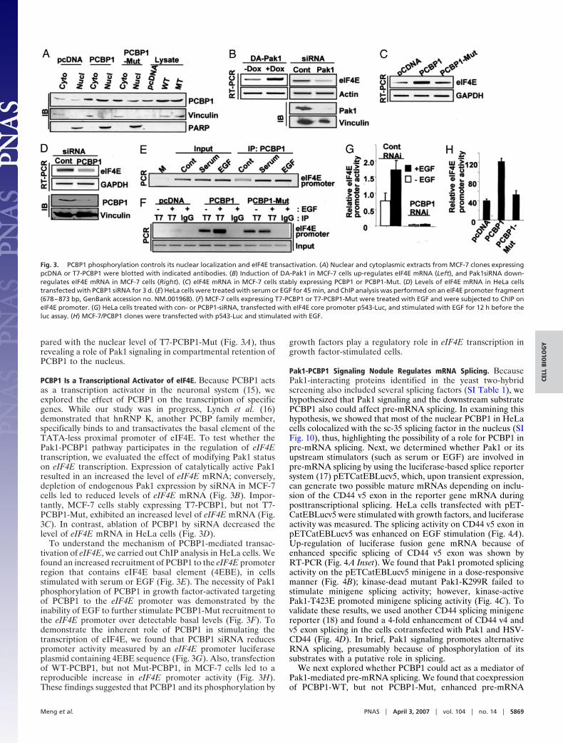
pared with the nuclear level of T7-PCBP1-Mut (Fig. 3A), thusrevealing a role of Pak1 signaling in compartmental retention ofPCBP1 to the nucleus.
PCBP1 Is a Transcriptional Activator of eIF4E. Because PCBP1 actsas a transcription activator in the neuronal system (15), weexplored the effect of PCBP1 on the transcription of specificgenes. While our study was in progress, Lynch et al. (16)demonstrated that hnRNP K, another PCBP family member,specifically binds to and transactivates the basal element of theTATA-less proximal promoter of eIF4E. To test whether thePak1-PCBP1 pathway participates in the regulation of eIF4Etranscription, we evaluated the effect of modifying Pak1 statuson eIF4E transcription. Expression of catalytically active Pak1resulted in an increased the level of eIF4E mRNA; conversely,depletion of endogenous Pak1 expression by siRNA in MCF-7cells led to reduced levels of eIF4E mRNA (Fig. 3B). Impor-tantly, MCF-7 cells stably expressing T7-PCBP1, but not T7-PCBP1-Mut, exhibited an increased level of eIF4E mRNA (Fig.3C). In contrast, ablation of PCBP1 by siRNA decreased thelevel of eIF4E mRNA in HeLa cells (Fig. 3D).
To understand the mechanism of PCBP1-mediated transac-tivation of eIF4E, we carried out ChIP analysis in HeLa cells. Wefound an increased recruitment of PCBP1 to the eIF4E promoterregion that contains eIF4E basal element (4EBE), in cellsstimulated with serum or EGF (Fig. 3E). The necessity of Pak1phosphorylation of PCBP1 in growth factor-activated targetingof PCBP1 to the eIF4E promoter was demonstrated by theinability of EGF to further stimulate PCBP1-Mut recruitment tothe eIF4E promoter over detectable basal levels (Fig. 3F). Todemonstrate the inherent role of PCBP1 in stimulating thetranscription of eIF4E, we found that PCBP1 siRNA reducespromoter activity measured by an eIF4E promoter luciferaseplasmid containing 4EBE sequence (Fig. 3G). Also, transfectionof WT-PCBP1, but not Mut-PCBP1, in MCF-7 cells led to areproducible increase in eIF4E promoter activity (Fig. 3H).These findings suggested that PCBP1 and its phosphorylation by
growth factors play a regulatory role in eIF4E transcription ingrowth factor-stimulated cells.
Pak1-PCBP1 Signaling Nodule Regulates mRNA Splicing. BecausePak1-interacting proteins identified in the yeast two-hybridscreening also included several splicing factors (SI Table 1), wehypothesized that Pak1 signaling and the downstream substratePCBP1 also could affect pre-mRNA splicing. In examining thishypothesis, we showed that most of the nuclear PCBP1 in HeLacells colocalized with the sc-35 splicing factor in the nucleus (SIFig. 10), thus, highlighting the possibility of a role for PCBP1 inpre-mRNA splicing. Next, we determined whether Pak1 or itsupstream stimulators (such as serum or EGF) are involved inpre-mRNA splicing by using the luciferase-based splice reportersystem (17) pETCatEBLucv5, which, upon transient expression,can generate two possible mature mRNAs depending on inclu-sion of the CD44 v5 exon in the reporter gene mRNA duringposttranscriptional splicing. HeLa cells transfected with pET-CatEBLucv5 were stimulated with growth factors, and luciferaseactivity was measured. The splicing activity on CD44 v5 exon inpETCatEBLucv5 was enhanced on EGF stimulation (Fig. 4A).Up-regulation of luciferase fusion gene mRNA because ofenhanced specific splicing of CD44 v5 exon was shown byRT-PCR (Fig. 4A Inset). We found that Pak1 promoted splicingactivity on the pETCatEBLucv5 minigene in a dose-responsivemanner (Fig. 4B); kinase-dead mutant Pak1-K299R failed tostimulate minigene splicing activity; however, kinase-activePak1-T423E promoted minigene splicing activity (Fig. 4C). Tovalidate these results, we used another CD44 splicing minigenereporter (18) and found a 4-fold enhancement of CD44 v4 andv5 exon splicing in the cells cotransfected with Pak1 and HSV-CD44 (Fig. 4D). In brief, Pak1 signaling promotes alternativeRNA splicing, presumably because of phosphorylation of itssubstrates with a putative role in splicing.
We next explored whether PCBP1 could act as a mediator ofPak1-mediated pre-mRNA splicing. We found that coexpressionof PCBP1-WT, but not PCBP1-Mut, enhanced pre-mRNA
Fig. 3. PCBP1 phosphorylation controls its nuclear localization and eIF4E transactivation. (A) Nuclear and cytoplasmic extracts from MCF-7 clones expressingpcDNA or T7-PCBP1 were blotted with indicated antibodies. (B) Induction of DA-Pak1 in MCF-7 cells up-regulates eIF4E mRNA (Left), and Pak1siRNA down-regulates eIF4E mRNA in MCF-7 cells (Right). (C) eIF4E mRNA in MCF-7 cells stably expressing PCBP1 or PCBP1-Mut. (D) Levels of eIF4E mRNA in HeLa cellstransfected with PCBP1 siRNA for 3 d. (E) HeLa cells were treated with serum or EGF for 45 min, and ChIP analysis was performed on an eIF4E promoter fragment(678–873 bp, GenBank accession no. NM�001968). (F) MCF-7 cells expressing T7-PCBP1 or T7-PCBP1-Mut were treated with EGF and were subjected to ChIP oneIF4E promoter. (G) HeLa cells treated with con- or PCBP1-siRNA, transfected with eIF4E core promoter p543-Luc, and stimulated with EGF for 12 h before theluc assay. (H) MCF-7/PCBP1 clones were transfected with p543-Luc and stimulated with EGF.
Meng et al. PNAS � April 3, 2007 � vol. 104 � no. 14 � 5869
CELL
BIO
LOG
Y
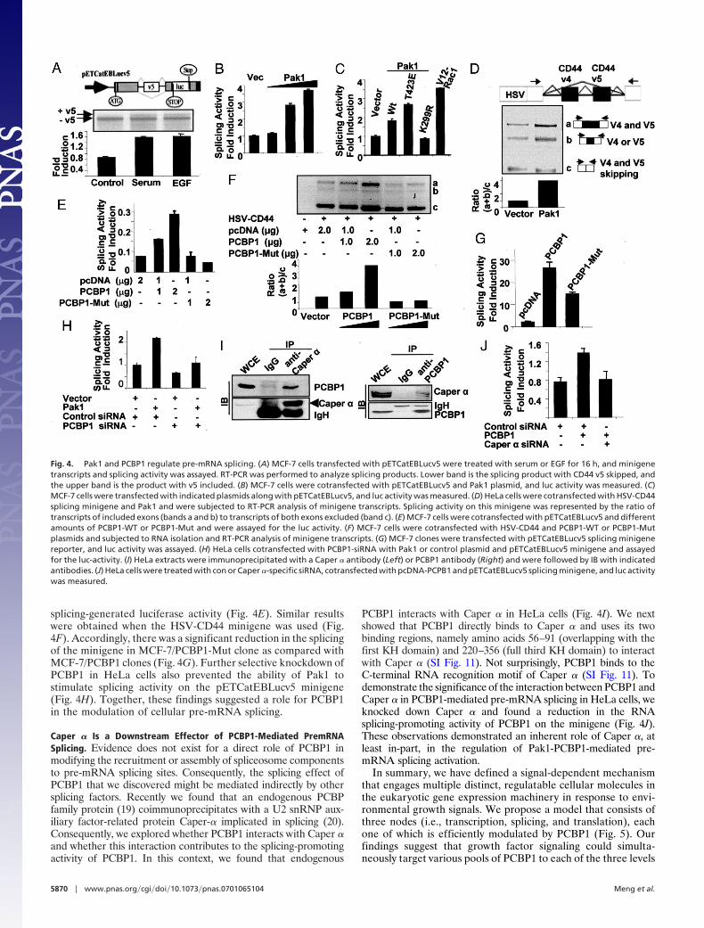
splicing-generated luciferase activity (Fig. 4E). Similar resultswere obtained when the HSV-CD44 minigene was used (Fig.4F). Accordingly, there was a significant reduction in the splicingof the minigene in MCF-7/PCBP1-Mut clone as compared withMCF-7/PCBP1 clones (Fig. 4G). Further selective knockdown ofPCBP1 in HeLa cells also prevented the ability of Pak1 tostimulate splicing activity on the pETCatEBLucv5 minigene(Fig. 4H). Together, these findings suggested a role for PCBP1in the modulation of cellular pre-mRNA splicing.
Caper � Is a Downstream Effector of PCBP1-Mediated PremRNASplicing. Evidence does not exist for a direct role of PCBP1 inmodifying the recruitment or assembly of spliceosome componentsto pre-mRNA splicing sites. Consequently, the splicing effect ofPCBP1 that we discovered might be mediated indirectly by othersplicing factors. Recently we found that an endogenous PCBPfamily protein (19) coimmunoprecipitates with a U2 snRNP aux-iliary factor-related protein Caper-� implicated in splicing (20).Consequently, we explored whether PCBP1 interacts with Caper �and whether this interaction contributes to the splicing-promotingactivity of PCBP1. In this context, we found that endogenous
PCBP1 interacts with Caper � in HeLa cells (Fig. 4I). We nextshowed that PCBP1 directly binds to Caper � and uses its twobinding regions, namely amino acids 56–91 (overlapping with thefirst KH domain) and 220–356 (full third KH domain) to interactwith Caper � (SI Fig. 11). Not surprisingly, PCBP1 binds to theC-terminal RNA recognition motif of Caper � (SI Fig. 11). Todemonstrate the significance of the interaction between PCBP1 andCaper � in PCBP1-mediated pre-mRNA splicing in HeLa cells, weknocked down Caper � and found a reduction in the RNAsplicing-promoting activity of PCBP1 on the minigene (Fig. 4J).These observations demonstrated an inherent role of Caper �, atleast in-part, in the regulation of Pak1-PCBP1-mediated pre-mRNA splicing activation.
In summary, we have defined a signal-dependent mechanismthat engages multiple distinct, regulatable cellular molecules inthe eukaryotic gene expression machinery in response to envi-ronmental growth signals. We propose a model that consists ofthree nodes (i.e., transcription, splicing, and translation), eachone of which is efficiently modulated by PCBP1 (Fig. 5). Ourfindings suggest that growth factor signaling could simulta-neously target various pools of PCBP1 to each of the three levels
Fig. 4. Pak1 and PCBP1 regulate pre-mRNA splicing. (A) MCF-7 cells transfected with pETCatEBLucv5 were treated with serum or EGF for 16 h, and minigenetranscripts and splicing activity was assayed. RT-PCR was performed to analyze splicing products. Lower band is the splicing product with CD44 v5 skipped, andthe upper band is the product with v5 included. (B) MCF-7 cells were cotransfected with pETCatEBLucv5 and Pak1 plasmid, and luc activity was measured. (C)MCF-7 cells were transfected with indicated plasmids along with pETCatEBLucv5, and luc activity was measured. (D) HeLa cells were cotransfected with HSV-CD44splicing minigene and Pak1 and were subjected to RT-PCR analysis of minigene transcripts. Splicing activity on this minigene was represented by the ratio oftranscripts of included exons (bands a and b) to transcripts of both exons excluded (band c). (E) MCF-7 cells were cotransfected with pETCatEBLucv5 and differentamounts of PCBP1-WT or PCBP1-Mut and were assayed for the luc activity. (F) MCF-7 cells were cotransfected with HSV-CD44 and PCBP1-WT or PCBP1-Mutplasmids and subjected to RNA isolation and RT-PCR analysis of minigene transcripts. (G) MCF-7 clones were transfected with pETCatEBLucv5 splicing minigenereporter, and luc activity was assayed. (H) HeLa cells cotransfected with PCBP1-siRNA with Pak1 or control plasmid and pETCatEBLucv5 minigene and assayedfor the luc-activity. (I) HeLa extracts were immunoprecipitated with a Caper � antibody (Left) or PCBP1 antibody (Right) and were followed by IB with indicatedantibodies. (J) HeLa cells were treated with con or Caper �-specific siRNA, cotransfected with pcDNA-PCPB1 and pETCatEBLucv5 splicing minigene, and luc activitywas measured.
5870 � www.pnas.org�cgi�doi�10.1073�pnas.0701065104 Meng et al.
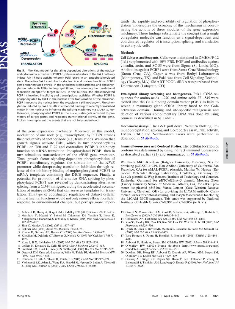
of the gene expression machinery. Moreover, in this model,modulation of one node (e.g., transcription) by PCBP1 attunesthe productivity of another node (e.g., translation). We show thatgrowth signals activate Pak1, which in turn phosphorylatesPCBP1 on T60 and T127 and contradicts PCBP1’s inhibitoryfunction on mRNA translation. Phosphorylated PCBP1 then inturn stimulates transactivation of the eIF4E gene promoter.Thus, growth factor signaling-dependent phosphorylation ofPCBP1 coordinately regulates the stimulation of the eIF4Epromoter while derepressing translation inhibition through re-lease of the inhibitory binding of unphosphorylated PCBP1 tomRNA templates containing the DICE sequence. Finally, apotential for promotion of alternative RNA splicing by phos-phorylated PCBP1 was revealed by demonstrating alternativesplicing from a CD44 minigene, aiding the accelerated accumu-lation of mature mRNAs that can serve as templates for trans-lation. This type of coordinated regulation of distinct cellularcompartmental functions would not only ensure efficient cellularresponse to environmental changes, but perhaps more impor-
tantly, the rapidity and reversibility of regulation of phosphor-ylation underscores the economy of this mechanism in coordi-nating the actions of three modules of the gene expressionmachinery. These findings substantiate the concept that a singlecoregulator molecule can function as a signal-dependent andcoordinated regulator of transcription, splicing, and translationin eukaryotic cells.
MethodsCell Cultures and Reagents. Cells were maintained in DMEM/F-12(1:1) supplemented with 10% FBS. EGF and antibodies againstvinculin, actin, and SC-35 were from Sigma (St. Louis, MO).Antibodies against PCBP1 were from Santa Cruz Biotechnology(Santa Cruz, CA), Caper � was from Bethyl Laboratories(Montgomery, TX), and Pak1 was from Cell Signaling Technol-ogy (Beverly, MA). SMART POOL siRNA was purchased fromDharmacon (Lafayette, CO).
Two-Hybrid Library Screening and Mutagenesis. Pak1 cDNA se-quences for amino acids 1–270 and amino acids 271–545 werecloned into the Gal4-binding domain vector pGBD as baits toscreen a mammary gland cDNA library fused to the Gal4activation domain. Cloning and site-specific mutagenesis anddeletion of various complimentary DNA was done by usingprimers as described in SI Table 2.
Biochemical Assays. The GST pull down, Western blotting, im-munoprecipitation, splicing and luc-reporter assay, Pak1 activity,EMSA, ChIP and Northwestern assays were performed asdescribed in SI Methods.
Immunofluorescence and Confocal Studies. The cellular location ofproteins was determined by using indirect immunofluorescenceas described earlier (21) and summarized in SI Methods.
We thank Mike Kiledjian (Rutgers University, Piscatway, NJ) forproviding pGEX6P-� CP1, Rau Andino (University of California, SanFrancisco, CA) for pMAL-PCBP1 plasmids, Mathias W. Hentze (Eu-ropean Molecular Biology Laboratory, Heidelberg, Germany) forLuc-2R plasmid, S. Weg-Remers (Institute of Toxicology and Genetics,Karlsruhe, Germany) for pETCatEBlucv5 plasmid, Muxiang Zhou(Emory University School of Medicine, Atlanta, GA) for eIF4E pro-moter luc plasmid p543-luc, Vance Lemon (Case Western ReserveUniversity, Cleveland, OH) for providing the L1CAM antibody, Chris-topher Barnes for confocal staining, and Joseph Mascarenhas for cloningthe L1CAM DICE sequence. This study was supported by NationalInstitutes of Health Grants CA90970 and CA80066 (to R.K.).
1. Auboeuf D, Honig A, Berget SM, O’Malley BW (2002) Science 298:416–419.2. Masuhiro Y, Mezaki Y, Sakari M, Takeyama K-i, Yoshida T, Inoue K,
Yanagisawa J, Hanazawa S, O’Malley B, Kato S (2005) Proc Natl Acad Sci USA102:8126–8131.
3. Shin C, Manley JL (2002) Cell 111:407–417.4. Bokoch GM (2003) Annu Rev Biochem 72:743–781.5. Kumar R, Gururaj AE, Barnes CJ (2006) Nat Rev Cancer 6:459–470.6. Kiledjian M, DeMaria CT, Brewer G, Novick K (1997) Mol Cell Biol 17:4870–
4876.7. Kong J, Ji X, Liebhaber SA (2003) Mol Cell Biol 23:1125–1134.8. Leffers H, Dejgaard K, Celis JE (1995) Eur J Biochem 230:447–453.9. Barnhart KM, Kim CG, Banerji SS, Sheffery M (1988) Mol Cell Biol 8:3215–3226.
10. Ostareck DH, Ostareck-Lederer A, Wilm M, Thiele BJ, Mann M, Hentze MW(1997) Cell 89:597–606.
11. Reimann I, Huth A, Thiele H, Thiele BJ (2002) J Mol Biol 315:965–974.12. Vadlamudi RK, Adam L, Wang RA, Mandal M, Nguyen D, Sahin A, Chernoff
J, Hung MC, Kumar R (2000) J Biol Chem 275:36238–36240.
13. Gavert N, Conacci-Sorrel M, Gast D, Schneider A, Altevogt P, Brabletz T,Ben-Ze’ev A (2005) J Cell Biol 168:633–642.
14. Chkheidze AN, Liebhaber SA (2003) Mol Cell Biol 23:8405–8415.15. Kim SS, Pandey KK, Choi HS, Kim SY, Law PY, Wei LN, Loh HH (2005) Mol
Pharmacol 68:729–736.16. Lynch M, Chen L, Ravitz MJ, Mehtani S, Lorenblat K, Pazin MJ, Schmidt EV
(2005) Mol Cell Biol 25:6436–6453.17. Weg-Remers S, Ponta H, Herrlich P, Konig H (2001) EMBO J 20:4194–
4203.18. Auboeuf D, Honig A, Berget SM, O’Malley BW (2002) Science 298:416–419.19. O’Malley BW (2003) Nursa database http://www.nursa.org/corip.
cfm?detail�assay&dataset�25&ex�no�25.1.20. Dowhan DH, Hong EP, Auboeuf D, Dennis AP, Wilson MM, Berget SM,
O’Malley BW (2005) Mol Cell 17:429–439.21. Gururaj AE, Singh RR, Rayala SK, Holm C, den Hollander P, Zhang H,
Balasenthil S, Talukder AH, Landberg G, Kumar R (2006) Proc Natl Acad Sci103:6670–6675.
Fig. 5. Working model for signaling-dependent alterations of the nuclearand cytoplasmic activities of PCBP1. Upstream activators of the Pak1 pathwayinduce Pak1 kinase activity wherein Pak1 exists in an autophosphorylatedstate. The active Pak1 exerts both cytoplasmic and nuclear functions. PCBP1gets phosphorylated by Pak1 in the cytoplasmic compartment, and phosphor-ylation reduces its RNA-binding capabilities, thus releasing the translationalrepression on specific target mRNAs. In the nucleus, the phosphorylatedPCBP1 is involved in splicing and transcriptional activities. Whether PCBP1 isphosphorylated by Pak1 in the nucleus after translocation or the phosphor-PCBP1 moves to the nucleus from the cytoplasm is still not known. Phosphor-ylation induced by Pak1 results in enhanced binding to recently transcribedmRNA in the nucleus to influence the splicing machinery via CAPER �. Fur-thermore, phosphorylated PCBP1 in the nucleus also gets recruited to pro-moters of target genes and regulates transcriptional activity of the gene.Broken lines represent the events that are not fully understood.
Meng et al. PNAS � April 3, 2007 � vol. 104 � no. 14 � 5871
CELL
BIO
LOG
Y