
Coordinated Post-translational Responses of Aquaporins to ...
13
HAL Id: hal-00919768 https://hal.archives-ouvertes.fr/hal-00919768 Submitted on 8 Jun 2021 HAL is a multi-disciplinary open access archive for the deposit and dissemination of sci- entific research documents, whether they are pub- lished or not. The documents may come from teaching and research institutions in France or abroad, or from public or private research centers. L’archive ouverte pluridisciplinaire HAL, est destinée au dépôt et à la diffusion de documents scientifiques de niveau recherche, publiés ou non, émanant des établissements d’enseignement et de recherche français ou étrangers, des laboratoires publics ou privés. Distributed under a Creative Commons Attribution| 4.0 International License Coordinated Post-translational Responses of Aquaporins to Abiotic and Nutritional Stimuli in Arabidopsis Roots. Magali Di Pietro, Jérôme Vialaret, Guo-Wei Li, Sonia Hem, Karine Prado, Michel Rossignol, Christophe Maurel, Véronique Santoni To cite this version: Magali Di Pietro, Jérôme Vialaret, Guo-Wei Li, Sonia Hem, Karine Prado, et al.. Coordinated Post-translational Responses of Aquaporins to Abiotic and Nutritional Stimuli in Arabidopsis Roots.. Molecular and Cellular Proteomics, American Society for Biochemistry and Molecular Biology, 2013, 12 (12), pp.3886-97. 10.1074/mcp.M113.028241. hal-00919768
-
Upload
khangminh22 -
Category
Documents
-
view
0 -
download
0
Transcript of Coordinated Post-translational Responses of Aquaporins to ...
HAL Id: hal-00919768https://hal.archives-ouvertes.fr/hal-00919768
Submitted on 8 Jun 2021
HAL is a multi-disciplinary open accessarchive for the deposit and dissemination of sci-entific research documents, whether they are pub-lished or not. The documents may come fromteaching and research institutions in France orabroad, or from public or private research centers.
L’archive ouverte pluridisciplinaire HAL, estdestinée au dépôt et à la diffusion de documentsscientifiques de niveau recherche, publiés ou non,émanant des établissements d’enseignement et derecherche français ou étrangers, des laboratoirespublics ou privés.
Distributed under a Creative Commons Attribution| 4.0 International License
Coordinated Post-translational Responses of Aquaporinsto Abiotic and Nutritional Stimuli in Arabidopsis Roots.
Magali Di Pietro, Jérôme Vialaret, Guo-Wei Li, Sonia Hem, Karine Prado,Michel Rossignol, Christophe Maurel, Véronique Santoni
To cite this version:Magali Di Pietro, Jérôme Vialaret, Guo-Wei Li, Sonia Hem, Karine Prado, et al.. CoordinatedPost-translational Responses of Aquaporins to Abiotic and Nutritional Stimuli in Arabidopsis Roots..Molecular and Cellular Proteomics, American Society for Biochemistry and Molecular Biology, 2013,12 (12), pp.3886-97. �10.1074/mcp.M113.028241�. �hal-00919768�

Coordinated Post-translational Responses ofAquaporins to Abiotic and Nutritional Stimuli inArabidopsis Roots*□S
Magali di Pietro‡, Jerome Vialaret§¶, Guo-Wei Li‡, Sonia Hem‡¶, Karine Prado‡,Michel Rossignol¶, Christophe Maurel‡, and Veronique Santoni‡�
In plants, aquaporins play a crucial role in regulating rootwater transport in response to environmental and physi-ological cues. Controls achieved at the post-translationallevel are thought to be of critical importance for regulatingaquaporin function. To investigate the general molecularmechanisms involved, we performed, using the modelspecies Arabidopsis, a comprehensive proteomic analysisof root aquaporins in a large set of physiological contexts.We identified nine physiological treatments that modulateroot hydraulics in time frames of minutes (NO and H2O2
treatments), hours (mannitol and NaCl treatments, expo-sure to darkness and reversal with sucrose, phosphatesupply to phosphate-starved roots), or days (phosphate ornitrogen starvation). All treatments induced inhibition ofroot water transport except for sucrose supply to dark-grown plants and phosphate resupply to phosphate-starved plants, which had opposing effects. Using a ro-bust label-free quantitative proteomic methodology, weidentified 12 of 13 plasma membrane intrinsic protein(PIP) aquaporin isoforms, 4 of the 10 tonoplast intrinsicprotein isoforms, and a diversity of post-translationalmodifications including phosphorylation, methylation,deamidation, and acetylation. A total of 55 aquaporin pep-tides displayed significant changes after treatments andenabled the identification of specific and as yet unknownpatterns of response to stimuli. The data show that theregulation of PIP and tonoplast intrinsic protein abun-dance was involved in response to a few treatments (i.e.NaCl, NO, and nitrate starvation), whereas changes in thephosphorylation status of PIP aquaporins were positivelycorrelated to changes in root hydraulic conductivity in thewhole set of treatments. The identification of in vivodeamidated forms of aquaporins and their stimulus-in-duced changes in abundance may reflect a new mecha-nism of aquaporin regulation. The overall work providesdeep insights into the in vivo post-translational events
triggered by environmental constraints and their possi-ble role in regulating plant water status. Molecular &Cellular Proteomics 12: 10.1074/mcp.M113.028241, 3886–3897, 2013.
The absorption of soil water by roots is crucial in order forplants to maintain their water status. Studies in various plantspecies have shown that the root water permeability (roothydraulic conductivity Lpr) is constantly adjusted dependingon the developmental stage of the plant, its nutritional orhormonal status, or multiple environmental stimuli (1). Despitetheir importance in plant growth and adaptation, these multi-ple responses have not been investigated in a single plantspecies yet. Aquaporins form a large class of channel proteinsthat facilitate the diffusion of water and small neutral solutesacross cell membranes and, among many other functions,contribute to root water uptake (2, 3). Aquaporins are 25- to30-kDa proteins with six membrane-spanning domains andfive connecting loops (A–E), with N- and C-terminal tails ex-posed to the cytosol (4, 5). Plant aquaporins show a highmultiplicity of isoforms. Thirty-five homologs belonging to fourhomology subclasses have been identified in Arabidopsis.The plasma membrane intrinsic proteins (PIPs)1 (with 13 iso-forms further subdivided into the PIP1 and PIP2 subgroups)and the tonoplast intrinsic proteins (TIPs) with 10 homologsare the most abundant aquaporins in the plasma membraneand the tonoplast, respectively (6, 7). Two other families in-clude nine homologs of the nodulin-26-like proteins and threehomologs of small basic intrinsic proteins (6–8). Several linesof functional analysis, including the use of aquaporin chemicalblockers or phenotypic analysis of transgenic plants withantisense inhibition or insertional mutagenesis of PIP genes,have demonstrated that PIP aquaporins mediate a significantpart of root water uptake (9–12). However, the mechanisms
From ‡Biochimie et Physiologie Moleculaire des Plantes, SupAgro/INRA/CNRS/UMII/UMR 5004, 2 Place Viala, 34060 F-Montpellier cedex1, France; ¶Laboratoire de Proteomique Fonctionnelle, UR 1199, 1Place Viala, 34060 F-Montpellier Cedex 1, France
Received February 6, 2013, and in revised form, September 15,2013
Published, MCP Papers in Press, September 20, 2013, DOI10.1074/mcp.M113.028241
1 The abbreviations used are: DEA-NO, diethylamine NONOate;DW, dry weight; Jv, sap flow; Lpr, root hydraulic conductivity;mantmax, treatment with 200 mM mannitol for 4 h; NOtmax, treatmentwith 125 �M diethylamine NONOate for 18 min; Nstarv, starved innitrate for 6 days; PIP, plasma membrane intrinsic protein; Presup,phosphate resupply to plants starved in phosphate for 6 days; PTM,post-translational modification; TIP, tonoplast intrinsic protein.
Research© 2013 by The American Society for Biochemistry and Molecular Biology, Inc.This paper is available on line at http://www.mcponline.org
3886 Molecular & Cellular Proteomics 12.12
This is an Open Access article under the CC BY license.
that underlie PIP regulation in roots under abiotic constraintsappear composite and remain partially understood. In addi-tion to the long-term transcriptional control of aquaporin func-tions, multiple post-translational mechanisms that affectaquaporin activity, targeting to their destination compartment,or stability seem to be involved in the response of plant rootsto environmental and hormonal stimuli.
Post-translational modifications (PTMs) are central for reg-ulating protein structure and function and thereby modulatingprotein catalytic activity, subcellular localization, stability, andinteraction with other partners. Protein phosphorylation is oneof most important and best characterized PTMs. Virtually allcellular processes are regulated in one or multiple ways byreversible phosphorylation. PIPs are no exception and show aconserved phosphorylation site in their first cytosolic loop(loop B) and multiple phosphorylations in adjacent sites oftheir C-terminal tail (13–15), as listed in the PhosPhAt data-base. Aquaporin phosphorylation seems to be a significanttarget in plants under stress: following exposure of Arabidop-sis roots to salt (NaCl) or hydrogen peroxide (H2O2), AtPIP2;1phosphorylation is decreased and increased, respectively(15). Abscisic acid treatment of Arabidopsis seedlings alsodecreases the phosphorylation of several AtPIPs (16). A tran-sient increase in phosphorylation of AtPIP2;6 was observedupon sucrose resupply to starved Arabidopsis plantlets (17),whereas nitrogen resupply to starved plants results in a tran-sient dephosphorylation of AtPIP2;6 and AtPIP2;7 aquaporins(18). The phosphorylation of PIPs is thought to act on bothgating and subcellular localization (5, 15). In particular, theatomic structure of spinach SoPIP2;1 indicates that the phos-phorylation of Ser115 in loop B directly contributes to openingof the pore (5). In addition, phosphorylation of C-terminalSer274, a residue homologous to Ser280 of AtPIP2;1, acts onan adjacent SoPIP2;1 monomer to prevent its transition to aclosed-pore conformation (5). Finally, phosphorylation ofSer283 was shown to be necessary for targeting of newlysynthesized AtPIP2;1 to the plasma membrane and interferedwith its internalization in response to salt stress (15). Plantaquaporins are subjected to many other PTMs. Methylationhas been described in AtPIP2;1, but its functional significanceis as yet unknown (1, 19, 20). Drought-induced PIP ubiquiti-nation in the endoplasmic reticulum was shown to prevent itstrafficking to the plasma membrane and triggered its degra-dation (21).
Qualitative and quantitative information about PTMs andtheir dynamic changes is now critically needed in order toaddress the general principles and complexity of aquaporinregulation during plant responses to stresses. To this end, weinvestigated in Arabidopsis a set of representative environ-mental stimuli, including water, osmotic, ionic, oxidativestress, and nutritional constraints. The effects of these stimulion Lpr were characterized. The results provided a well-de-fined time frame for investigating the dynamics of aquaporinPTMs using dedicated quantitative approaches. The overall
study points to multiple regulatory processes that can beshared between treatments. In particular, coordinated phos-phorylation events and a putative role for deamidation wereuncovered.
EXPERIMENTAL PROCEDURES
Plant Culture—Arabidopsis thaliana plants, ecotype Columbia-0(Col-0), were grown in hydroponic conditions as described previously(22). Briefly, sterile seeds were sown on an MS/2 medium (23) com-plemented with 1% sucrose, 0.05% MES, and 7 g l�1 agar. Seedswere kept at 4 °C for 48 h and cultivated in vitro for 10 days (16 h oflight, 20 °C). Seedlings were then transferred to hydroponic culturesolution (1.25 mM KNO3, 1.50 mM Ca(NO3)2, 0.75 mM MgSO4, 0.50mM KH2PO4, 50 �M H3BO3, 12 �M MnSO4, 0.70 �M CuSO4, 1 �M
ZnSO4, 0.24 �M Na2MoO4, 50 �M Fe-EDTA, 100 �M Na2SiO3) andgrown under long-day conditions (16 h of light (250 �mol photons/m2/s) at 22 °C and 8 h of dark at 21 °C) for 11 days. Culture solutionwas replaced weekly.
Plant Treatments—Short treatments (up to 6 h) were applied byexposing 21-day-old plants to modified hydroponic solutions. Theeffect of salt was studied by complementing the hydroponic solutionwith 100 mM NaCl over 45 min or 2 h. For mannitol treatment, plantswere grown in a hydroponic solution containing 200 mM mannitol over45 min to 6 h. Plants were treated with 500 �M H2O2 over 8 or 20 min.Nitric oxide (NO) treatment was applied by using diethylamine NONOate(DEA-NO), an NO donor, as previously described (24). Briefly,DEA-NO was dissolved in 0.01 M NaOH as a stock solution (500 mM).The solution was diluted in a phosphate buffer, pH 7.2, to initiate NOrelease. A concentration of 50 �M to 500 �M DEA-NO was applied ina hydroponic solution without Fe-EDTA to avoid metal–NO com-plexes. NO specificity was tested by using 0.1 mM carboxy-2-phenyl-4,4,5,5,-tetramethylimidazoline-1-oxyl 3-oxide (cPTIO) as a specificNO scavenger (25). For sucrose treatment, plants were kept in dark-ness for 24 to 48 h and then supplied with 30 mM sucrose or mannitolfor 2 or 4 h. Nitrate starvation was applied by transferring 15-day-oldplants to a modified hydroponic medium containing 0.1 mM NO3 intotal for 6 days, where KNO3 and Ca(NO3)2 were replaced by K2SO4
and CaCl2, respectively (0.03 mM KNO3, 0.035 mM Ca(NO3)2, 0.61 mM
K2SO4, 1.465 mM CaCl2). Phosphate starvation was applied by trans-ferring 15-day-old plants to a PO4
3�-free hydroponic solution for 6days. Nitrate or phosphate resupply was performed by transferringplants to standard media for 2 to 24 h.
Water Transport Measurements—Measurements were performedessentially as described elsewhere (22). Briefly, the root system offreshly detopped 21-day-old Arabidopsis plants was inserted into apressure chamber filled with the culture solution. The hypocotylwas carefully threaded through the soft plastic washer of the metallid and connected to a flow meter using a low-viscosity dental paste(PRESIDENT microSystem, Coltene, Altstatten, Switzerland). Pres-sure was then slowly applied to the chamber using nitrogen gas. Tenminutes of pre-pressurization were required in order to achieve astabilized sap flow (Jv), which was then determined over 20-minperiods for imposed hydrostatic pressures between 0.16 and 0.72MPa. Root and shoot dry weight (DW) was measured after 48 h ofdrying. The hydraulic conductivity (Lpr, ml g�1 h�1 MPa�1) of eachindividual root system was calculated from the slope of a plot of Jv
versus driving pressure, divided by the root DW.Microsomal Protein Purification—The whole procedure was per-
formed at 4 °C. Roots of 21-day-old plants were grinded with a rollmill (conception by C. Fauvel, INRA Avignon, France) in 4 ml/g freshweight of grinding buffer (500 mM sucrose, 10% glycerol, 20 mM
EDTA, 20 mM EGTA, 50 mM NaF, 5 mM �-glycerophosphate, 1 mM
phenantroline, 0.6% polyvinylpyrrolidone (PVP), 10 mM ascorbic acid,
Coordination of Aquaporin Post-translational Modifications
Molecular & Cellular Proteomics 12.12 3887
50 mM Tris, pH 8, with 1 M MES, 1 �M leupeptin, 5 mM DTT, 1 mM
stabilized vanadate, 1 mM PMSF) and centrifuged at 800g for 2 min.The supernatant was then centrifuged at 9000g for 12 min, and theresulting supernatant was centrifuged at 50,000g for 12 min. Theresulting pellet was suspended in a minimal volume of conservationbuffer (10 mM borate, 300 mM sucrose, 9 mM KCl, 5 mM EDTA, 5 mM
EGTA, 50 mM NaF, 4.2 �M leupeptine, 1 mM PMSF, 5 mM DTT, 10 mM
Tris, pH 8.3) and stored at �80 °C. The protein concentration wasestimated using a modified Bradford procedure (26). Microsomeswere stripped by incubation in 4 M urea, pH 11, and 20 mM NaOHaccording to a previously described procedure (27). Proteins werethen solubilized in 2� Laemmli buffer (2% SDS, 10% glycerol, 100mm DTT, 0.001% bromphenol blue, 50 mM Tris, pH 6.8) for 3 h undershaking. After centrifugation (16,000g for 2 min), the supernatantcontaining solubilized proteins was stored at �20 °C.
ELISA Assay—ELISA assays were performed according to Ref. 22to obtain a relative quantification of aquaporins in microsomal ex-tracts. An antibody recognizing four out of five PIP1s (PIP1;1, PIP1;2,PIP1;3, and PIP1;4) and another antibody recognizing three out ofeight PIP2s (PIP2;1, PIP2;2, and PIP2;3) were used at a 1:2000dilution (27).
Protein Digestion and Phosphopeptide Enrichment—In-solution re-ductions/alkylations were performed simultaneously with detergentremoval via the filter-aided sample preparation protocol (28). Thesesteps were followed by two types of double digestions. One of thesesubsequently used Lys-C (Roche Applied Science) for 4 h at 37 °Cand trypsin (Sequencing Grade Modified, Promega, Madison, WI)overnight at 37 °C. The other used chymotrypsin (Sequencing GradeModified, Promega) for 5 h at 25 °C and then AspN (SequencingGrade Modified, Promega) overnight at 25 °C. Peptides were elutedvia step elutions with 50 mM ammonium bicarbonate followed by 50%acetonitrile and then 0.5 m NaCl. Peptides were desalted on C18columns (Sep-Pak® Vac tC18 cartridge, 3 cc, Waters, Guyancourt,France) before strong cation exchange separation. Dried peptideswere resuspended in 10 mM ammonium formate with 30% acetonitrileat pH 2.7 and loaded on a Famos-Ultimate HPLC (Dionex, Courta-boeuf, France) at 200 �l min�1 on a column (PL-SCX 1000A, 5 �m,50 � 2.1 mm, Varian, Agilent, Massy, France). A salt gradient (10 mM
ammonium formate, 800 mM NH4Cl, 30% acetonitrile, pH 2.7) allowedus to obtain nine fractions. In the case of chymotrypsin/AspN diges-tion, two peptide fractions were obtained and quantified. In the caseof Lys-C/trypsin digestion, nine fractions were obtained and treatedas follows: 10% of the volume of fractions 4 (F4) to 9 (F9) were keptout to direct LC-MS/MS analyses. Fractions F2, F3, and F4 andfractions F5, F6, and F7 were pooled before the phosphopeptideenrichment step. The F1 fraction was set aside. After solvent evapo-ration, peptides were resuspended in a loading buffer (25% lacticacid, 60% acetonitrile, 0.75% trifluoroacetic acid (TFA)) for titaniumdioxide (TiO2) enrichment according to Refs. 29 and 30. Briefly, a C8plug was packed on-tip with metal oxide bulk beads and equilibratedwith 1% TFA and 80% acetonitrile including 25% lactic acid asselectivity chelating enhancers (buffer A). Two successive columnswere built with 1 to 2 mm and 4 to 5 mm of TiO2 beads, respectively.After loading, washes were performed with buffer A and buffer B (1%TFA in 80% acetonitrile) and peptides were eluted twice with NH4OHat 0.5% and then 5%. Eluates were pooled and dried.
Identification of Peptides and Modified Peptides—Digested pep-tides were concentrated with a pre-column (Dionex, C18 PepMap100,300 �m � 5 mm, 5 �m, 100 A) and separated with a reversed-phasecapillary column (Dionex, C18 PepMap100, 75 �m � 250 mm, 3 �m,100 A) over a 140-min gradient. Peptide fragmentation was carriedout with a Q-TOF-MS/MS system equipped with a nano-electrosprayionization source (Maxis, Bruker Daltonics, Bremen, Germany) andperformed in the positive ion mode. The corresponding data were
interrogated with Mascot 2.2.07 (Matrix Science) via ProteinScapesoftware (Bruker Daltonics) against the Arabidopsis proteome (Tair10,31,960 entries). Parameters of interrogation accepted one putativemissed cleavage and 15-ppm and 0.05-Da mass ranges for theparent peptide and the MS/MS fragment, respectively. The selectedenzymes were trypsin or chymotrypsin/AspN. Because proteins werereduced and alkylated, carbamidomethylation was taken as a fixedmodification. Variable modifications were N-terminal acetylation;deamidation of asparagines and glutamines; methionine oxidation;phosphorylation of serines, threonines, and tyrosines; and methylationof aspartic and glutamic acids and of serines. Using the above criteriafor protein identification, the rate of false peptide sequence assignment(false discovery rate) as determined by the “decoy database” functionimplemented in Mascot version 2.2.07 was 1%. All modified aquaporinpeptides were checked manually via comparison of experimentallyidentified masses with the theoretically accepted masses for the frag-ments. Modified peptides were accepted if the PTM score was �20and/or site-determining b or y ions were present.
Label-free Peptide Quantification and Statistical Analysis—Micro-somal samples prepared from three independent plant cultures wereall analyzed with two technical replicates corresponding to indepen-dent injections in the mass spectrometer. Data corresponding to peakintensity were computed using the ProfileAnalysis software (BrukerDaltonics). Fractionated peptides obtained after digestion with Lys-C/trypsin or chymotrypsin/AspN were quantified with a label-freeapproach using a direct comparison of the peak intensity of eachpeptide ion in multiple LC-MS. Bruker software (DataAnalysis, Pro-fileAnalysis) provided a workflow including peak detection, peakmatching/alignment, normalization, detection of differential peptides,and statistical analyses of data. In particular, the technical variabilitywas reduced via the “quantile” method (Bruker Daltonics), the goal ofwhich is to homogenize the distribution of compound intensities foreach LC-MS run in a common data set containing all acquiredLC-MS runs. ProfileAnalysis (Bruker Daltonics) was used to dis-criminate peptides with quantitative variations, which were furtheridentified via collision-induced dissociation. Three independentbiological experiments and two technical repeats by experimentwere used to calculate quantitative ratios and corresponding pvalues. An aquaporin peptide was considered for data interpreta-tion if a quantitative ratio could be calculated in at least half of thestudied treatments (i.e. seven) with a p value below 0.1 for at leastthree of the treatments. When a peptide met these criteria, itsquantitative variations were considered in all treatments. For propercomparison of all treatments, quantitative data of each selectedpeptide were centered and reduced. Hierarchical clustering wasperformed using Multi-Experiment-Viewer 4.0. Euclidian was usedas a distance matrix.
RESULTS
Modulation of Lpr by Multiple Abiotic Treatments—In aprevious study, we showed that exposure of Arabidopsisroots to 100 mM NaCl induces a rapid (half-time: 45 min) andmarked (�70%) decrease in Lpr (22). In the present study,plants were treated for up to 6 h with an equivalent hyperos-motic solution containing 200 mM mannitol (Fig. 1A). Thistreatment induced a decrease in Lpr by up to 63%, with ahalf-time of 60 min. The application of H2O2 to excised Ara-bidopsis root systems was previously shown to induce a time-and dose-dependent decrease in pressure-induced sap flow(Jv), an indicator of Lpr in these conditions (31). In particular,0.5 mM H2O2 induced a 40% inhibition of Jv after 15 min (31).Here, we probed the effects on Jv of NO, another oxidative
Coordination of Aquaporin Post-translational Modifications
3888 Molecular & Cellular Proteomics 12.12
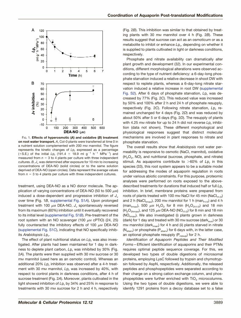
treatment, using DEA-NO as a NO donor molecule. The ap-plication of varying concentrations of DEA-NO (50 to 500 �M)induced a dose-dependent and progressive inhibition of Jv
over time (Fig. 1B, supplemental Fig. S1A). Upon prolongedtreatment with 100 �M DEA-NO, Jv spontaneously reversedfrom its maximum (60%) inhibition until it eventually recoveredto its initial level (supplemental Fig. S1B). Pre-treatment of theroot system with an NO scavenger (100 �M cPTIO) (24, 25)fully counteracted the inhibitory effects of 100 �M DEA-NO(supplemental Fig. S1C), indicating that NO specifically inhib-its Arabidopsis Lpr.
The effect of plant nutritional status on Lpr was also inves-tigated. After plants had been maintained for 1 day in dark-ness to deplete plant carbon, Lpr was inhibited by 30% (Fig.2A). The plants were then supplied with 30 mM sucrose or 30mM mannitol (used here as an osmotic control). Whereas anadditional 20% Lpr inhibition was observed after a 4-h treat-ment with 30 mM mannitol, Lpr was increased by 40%, withrespect to control plants in darkness conditions, after 4 h ofsucrose treatment (Fig. 2A). Moreover, plants cultivated in thelight showed inhibition of Lpr by 34% and 25% in response totreatments with 30 mM sucrose for 2 h and 4 h, respectively
(Fig. 2B). This inhibition was similar to that obtained by treat-ing plants with 30 mM mannitol over 4 h (Fig. 2B). Theseresults suggest that sucrose can act as an osmoticum or as ametabolite to inhibit or enhance Lpr, depending on whether itis supplied to plants cultivated in light or darkness conditions,respectively.
Phosphate and nitrate availability can dramatically alterplant growth and development (32). In our experimental con-ditions, different morphological alterations were observed ac-cording to the type of nutrient deficiency: a 6-day-long phos-phate starvation induced a relative decrease in shoot DW withrespect to replete plants, whereas a 6-day-long nitrate star-vation induced a relative increase in root DW (supplementalFig. S2). After 6 days of phosphate starvation, Lpr was de-creased by 77% (Fig. 2C). This reduced value was increasedby 50% and 100% after 2 h and 24 h of phosphate resupply,respectively (Fig. 2C). Following nitrate starvation, Lpr re-mained unchanged for 4 days (Fig. 2D) and was reduced byabout 50% after 5 or 6 days (Fig. 2D). The resupply of plantswith 4.25 mM nitrate for up to 24 h did not reverse Lpr inhibi-tion (data not shown). These different morphological andphysiological responses suggest that distinct molecularmechanisms are involved in plant responses to nitrate andphosphate starvation.
The overall results show that Arabidopsis root water per-meability is responsive to osmotic (NaCl, mannitol), oxidative(H2O2, NO), and nutritional (sucrose, phosphate, and nitrate)stimuli. As aquaporins contribute to �60% of Lpr in thisspecies (33), this root system appears to be a suitable modelfor addressing the modes of aquaporin regulation in rootsunder various abiotic constraints. For this purpose, proteomicanalyses were performed on roots exposed to the above-described treatments for durations that induced half or full Lpr
inhibition. In brief, membrane proteins were prepared fromroots of plants treated with 100 mM NaCl for 45 min (NaClt1/2)and 2 h (NaCltmax), 200 mM mannitol for 1 h (mant1/2) and 4 h(mantmax), 500 �M H2O2 for 8 min (H2O2t1/2) and 18 min(H2O2tmax), and 125 �M DEA-NO (NOt1/2) for 8 min and 18 min(NOtmax). We also investigated (i) plants grown in darkness(dark) for 1 day and treated with 30 mM sucrose (darksuc) or 30mM mannitol (darkman) for 4 h and (ii) plants starved in nitrate(Nstarv) or phosphate (Pstarv) for 6 days with, in the latter case,an optional phosphate resupply (Presup) for 2 h.
Identification of Aquaporin Peptides and Their ModifiedForms—Efficient identification of aquaporins and their PTMsrequires optimal peptide sequence coverage. For this, wedeveloped two types of double digestions of microsomalproteins, employing LysC followed by trypsin and chymotryp-sin followed by AspN, respectively. Additionally, the releasedpeptides and phosphopeptides were separated according totheir charge on a strong cation exchange column, and phos-phopeptides were further enriched with TiO2 microcolumns.Using the two types of double digestions, we were able toidentify 1291 proteins from a decoy database set to a false
FIG. 1. Effects of hyperosmotic (A) and oxidative (B) treatmenton root water transport. A, Col-0 plants were transferred at time 0 ina nutrient solution complemented with 200 mM mannitol. The figurerepresents the kinetic changes of Lpr expressed as a percentage(�S.E.) of the initial Lpr (191.4 � 18.9 ml g�1 h�1 MPa�1) andmeasured from n � 3 to 4 plants per culture with three independentcultures. B, Jv was determined after exposure for 10 min to increasingconcentrations of DEA-NO (solid circles) or to the same solutiondeprived of DEA-NO (open circles). Data represent the average valuesfrom n � 3 to 4 plants per culture with three independent cultures.
Coordination of Aquaporin Post-translational Modifications
Molecular & Cellular Proteomics 12.12 3889

discovery rate of 1% with a 95% confidence level (supple-mental Table S1). 1251 proteins were identified with LysC/trypsin digestion and 162 proteins with chymotrypsin/AspNdigestion, with 40 of the 162 proteins being specific for thelatter digestion. In addition, 32% and 16% of proteins showedat least one phosphorylation and one deamidation event,respectively (supplemental Table S1). This global analysisrevealed the presence of 12 PIP isoforms out of a total of 13(supplemental Table S1). The only lacking PIP isoform (PIP2;6)was detected and shown to play a role in leaves (34). Inaddition, we detected 4 out of the 10 TIP isoforms (TIP1;1,TIP1;2, TIP2;2, TIP2;3), with 2 of them (TIP1;1 and TIP1;2)specifically identified through chymotrypsin/AspN digestion.Consistent with a lack of detection in roots, TIP3;1 and TIP3;2are known to be specifically expressed in seeds, and TIP1;3and TIP5;1 in pollen (35, 36). MS analyses also showedthat numerous PIP isoforms were modified by acetylation,phosphorylation, methylation, or deamidation (Table I,supplemental Fig. S3). Among the 14 phosphorylation sitesidentified in this study, 4 were located in the N-terminal tail ofPIP1;1/PIP1;2 (Ser24, Ser27), PIP2;2 (Thr22), and PIP2;4
(Ser10) and were novel according to the PhosphAt database.The mass variation induced by deamidation is 0.98 Da. Thus,the monoisotopic ion was carefully examined to avoid anymistake in the selection of the parent (supplemental Fig. S4).Deamidation of Asn and Gln residues (Table I) was detected at12 sites in the second extracellular loop (loop C) and in the N-and C-terminal tails of several PIPs (Table I, supplemental Fig.S3). We previously showed that Lys (Lys4) and Glu (Glu6)residues can be methylated in the N-terminal tail of PIP2;1(20). Although these modifications were not observed in thisstudy’s samples, methylation of a Glu residue was observedat another position in the N-terminal tail of PIP2;2 (Table I,supplemental Fig. S3). Combinatorial deamidation and phos-phorylation was also observed in some peptides. This re-vealed a particularly complex modification profile in the N-ter-minal tail of PIP1;1–1;2 and in the C-terminal tail of PIP2;1–2;3and PIP2;7 (Table I, supplemental Fig. S3). Although not ex-haustive, these analyses establish that PIP aquaporins arehighly modified proteins and point to a multiplicity of post-translational regulation mechanisms acting on a same protein(Fig. 3).
FIG. 2. Lpr response to light and carbon (A, B), phosphate (C), or nitrate (D) availability. A, Lpr was measured in plants grown in the light(light), exposed to a 24-h period of darkness (control), or exposed to a 24-h period of darkness followed by a treatment with 30 mM sucrose(S2h and S4h) or 30 mM mannitol (M2h and M4h) for 2 h (S2h and M2h) or 4 h (S4h and M4h). Data represent the average value (�S.E.) from n �3 to 20 plants from three independent cultures. B, Lpr was measured in plants grown in light without any addition of sucrose or mannitol(control) or after the addition of 30 mM sucrose for 2 h (S2h) or 4 h (S4h) or 30 mM mannitol for 4 h (M4h). Data represent the average value (�S.E.)of n � 3 to 20 plants from three independent cultures. Different letters indicate statistically significant differences at p � 0.05. C, Lpr wasmeasured in plants grown in control conditions (control), after 6 days of phosphate starvation (Pstarv), or after phosphate starvation followedby 1 h, 2 h, and 24 h of 0.5 mM phosphate resupply (Presup). Data represent the average value (�S.E.) from n � 3 to 13 plants from threeindependent cultures. D, Lpr was measured in plants subjected to 4, 5, or 6 days of nitrate starvation (gray bars) and in plants of the same agebut grown in replete conditions (black bars). Data represent the average value (�S.E.) of n � 4 plants from at least two independent cultures.
Coordination of Aquaporin Post-translational Modifications
3890 Molecular & Cellular Proteomics 12.12

Classification of Aquaporin Peptides and Their ModifiedForms—A label-free MS approach was developed for com-parative quantification of aquaporin peptides between treat-ments. Hierarchical clustering was then used to classify bothunmodified and modified PIP peptides according to treat-ments (Fig. 4). We considered 55 aquaporin peptides, thequantitative behavior of which was significant in at least 3treatments (i.e. with p � 0.1) and quantified in at least 7treatments out of 14 so as to avoid misinterpretation due tomissing data (Fig. 4, supplemental Table S3). For mutualcomparison of all treatments, quantitative ratios of peptideabundance referring to specific control conditions were nor-malized to the general control condition. This overall analysisindicated that all treatments clustered with complementarykinetic points of the same or related stimuli (Fig. 4), with theexception of the mantmax treatment, which was surprisinglyrelated to the H2O2 treatments. The good biological consis-tency of this clustering attests to the quality of the overallproteomic data. The data yielded, however, a very intricateclassification of the 55 peptides. A cluster of phosphorylatedpeptides including the phosphorylation of the N-terminal tail
of PIP1 and of the C-terminal tail of PIP2;1–2;3 and PIP2;7could be distinguished (Fig. 4). The phosphorylation of thesepeptides increased in darkness, H2O2t1/2, H2O2tmax, andmantmax treatments and decreased in all other treatmentsexcept the Nstarv treatment, which showed contrasted phos-phorylation behavior (Fig. 4). These data extend previous workshowing the role of PIP2;1 phosphorylation during salt andH2O2 responses (15). A general relationship was tentativelyestablished between Lpr and aquaporin phosphorylation (Fig.5). For each treatment, the mean relative abundance of allphosphorylated PIP peptides was reported to the normalizedLpr value (Fig. 5A). A significant correlation could be estab-lished (p � 0.007; Fig. 5A), suggesting that aquaporin phos-phorylation positively contributes to Lpr variations.
Aquaporin Abundance According to Treatment—Becausevariations of Lpr could also be due to primary changes inaquaporin abundance, we then focused our analysis on PIPand TIP expression levels in plants exposed to the osmotic,oxidative, or nutritional constraints described above. For this,we considered the abundance of proteotypic aquaporin pep-tides that can be considered as indicators of the abundance
TABLE IModified aquaporin peptides
Name Aquaporin Peptide sequence PTM PTM positiona
PIP11–14 Acet PIP1;1–1;2–1;3–1;4 acetMEGKEEDVR14 Acet M1PIP11 12 (15–29) 1P PIP1;1–1;2 15FPERQPIGTpSAQpSDK29 1P S24, S27PIP11 12 (15–29) 1P 1Dea PIP1;1–1;2 15FPERdQPIGTpSAQpSDK29 1P, 1Dea 1P: S24, S27; 1Dea: Q19PIP11 12 (19–29) 1P PIP1;1–1;2 19QPIGTSAQpSDK29 1P S27PIP21 (4–16) 1Dea PIP2;1 4DVEAVPGEGFdQTR16 1Dea Q14PIP22 (4–14) 1Dea PIP2;2 4DVEGPEGFdQTR14 1Dea Q12PIP24 (4–16) 1Dea PIP2;4 4DLDVdNESGPPAAR16 1Dea N8PIP24 (4–19) 1P PIP2;4 4DLDVNEpSGPPAARDYK19 1P S10PIP21 (17–33) 1Dea PIP2;1 17DYdQDPPPAPFIDGAELK33 1Dea Q19PIP22 (15–32) 1P PIP2;2 15DYEDPPPpTPFFDADELTK32 1P T22PIP22 (15–32) 1Met PIP2;2 15DYmEDPPPTPFFDADELTK32 1Met E17PIP12 (loop C) 1Dea PIP1;2 157QYQALGGGAdNTIAHGYTK174 1Dea N166PIP15 (loop C) 1Dea PIP1;5 153GFdQPGLYQTdNGGGAdNVVAHGYTK175 1Dea Q155, N162, N167PIP24 (loop C) 1Dea PIP2;4 154YGGGAdNELADGYNK167 1Dea N159PIP27 (loop C) 1Dea PIP2;7 142TPYNTLGGGAdNTVADGYSK160 1Dea N152PIP21 (272–282) 1P PIP2;1–2;2–2;3 272ASGSKSLGpSFR282 1P S280PIP21 (277–282) 1P PIP2;1–2;2–2;3 277SLGpSFR282 1P S280PIP21 (277–287) 1P PIP2;1–2;2–2;3 277SLGpSFRpSAANV287 1P S280, S283PIP21 (277–287) 1P 1Dea PIP2;1–2;2–2;3 277SLGpSFRpSAAdNV287 1P and 1Dea 1P: S280, S283; 1Dea: N286PIP21 (277–287) 2P PIP2;1–2;2–2;3 277SLGpSFRpSAANV287 2P S280 � S283PIP21 (277–287) 2P 1Dea PIP2;1–2;2–2;3 277SLGpSFRpSAAdNV287 2P and 1Dea 2P: S280 � S283; 1Dea: N286PIP24 (276–287) 1P PIP2;4 277ALGpSFGpSFGpSFR288 1P S280, S283, S286PIP25 (276–286) 1P PIP2;5 276ALGpSFRSQPHV286 1P S279PIP27 (270–280) 1P PIP2;7 270ALGpSFRSNATN280 1P S273PIP27 (270–280) 2P PIP2;7 270ALGpSFRpSNATN280 2P S273 � S276, S276 � T279PIP27 (270–280) 1P 1Dea PIP2;7 270ALGpSFRSdNATN280 1P and 1Dea 1P: S273; 1Dea: N277PIP27 (270–280) 2P 1Dea PIP2;7 270ALGpSFRpSdNATN280 2P and 1Dea 2P: S273 � S276; 1Dea: N277PIP28 (268–278) 1P PIP2;8 268ALApSFRSNPTN278 1P S271
This table lists all aquaporin peptides harboring PTMs. The first column indicates the name of the peptide, as referred to in Fig. 4. The secondand third columns contain the name of the aquaporin and the peptide sequence, respectively. The last two columns indicate the type andnumber of PTMs and the position of the modified residue(s). The fragmentations of peptides are detailed in supplemental Fig. S3. Acet,acetylation; Dea, deamidation; Met, methylation; P, phosphorylation; PIP1;1–1;2–1;3–1;4, PIP1;1 and/or PIP1;2 and/or PIP1;3 and/or PIP1;4;PIP1;1–1;2, PIP1;1 and/or PIP1;2; PIP2;1–2;2–2;3, PIP2;1 and/or PIP2;2 and/or PIP2;3.
a A comma indicates the coexistence of different phosphorylation or deamidation sites within a peptide (supplemental Fig. S3).
Coordination of Aquaporin Post-translational Modifications
Molecular & Cellular Proteomics 12.12 3891

of the entire protein, provided that they are not modifiable.Five PIP1, five PIP2, and eight TIPs could be selected accord-ing to these criteria (supplemental Fig. S5). Because not allPIP aquaporins could be quantified through this approach, wealso made use of an additional quantification method (i.e.ELISA) using anti-PIP1 antibody that recognized PIP1;1 to-1;4 isoforms and anti-PIP2 antibody that recognized PIP2;1to -2;3 isoforms (27). Supplemental Fig. S5A indicates that thePIP1 peptides showed decreased abundance after NOtmax
treatment, and that this decrease was not as complete afterNOt1/2. This result was confirmed using an ELISA assay withanti-PIP1 antibodies (supplemental Fig. S5C). All PIP1 pep-tides also showed decreased abundance in response to aNatmax treatment (supplemental Fig. S5A), as already shownby ELISA measurements (22). We also noticed a global de-crease of PIP2 peptides in these conditions (supplementalFig. S5B). Regarding nutritional constraints, the decrease inPIP2;1, PIP2;2, PIP2;4, PIP1;2, and PIP1;3 abundance in re-sponse to Nstarv and Presup treatments was confirmed byELISA assays (supplemental Fig. S5C). Interestingly, theabundance of TIP1;1 and TIP1;2 showed marked variation inresponse to several treatments. The abundance of all de-tected TIP1;1 and TIP1;2 decreased in response to Natmax,dark, and Nstarv treatments (supplemental Fig. S5D). In con-trast, all TIP1;1 and TIP1;2 peptides showed increased abun-dance upon Presup (supplemental Fig. S5D). In a global anal-ysis across all treatments, we considered normalized Lpr
values and the mean relative abundance of peptides proteo-typic for PIP1 (Fig. 5B), PIP2 (Fig. 5C), or TIPs (Fig. 5D). Theseanalyses did not reveal any significant correlation between Lpr
and PIP1 or TIP abundance (Figs. 5B, 5D). In contrast, a
FIG. 3. Schematic representation of post-translational modifi-cations identified in PIP1;2 (A) and PIP2;2 (B). Acet, acetylation;Dea, deamidation; P, phosphorylation; Met, methylation. The crossindicates the cleavage of the initiating methionine. The correspondingmodified peptides are described in Table I.
FIG. 4. Heat map representation of aquaporin peptide abun-dance according to treatment. Aquaporin peptides that were un-modified or carrying phosphorylation (P) or deamidation (Dea), aloneor in combination, were quantified via label-free MS and clusteredusing a Euclidian correlation distance metric. Green and red colorsindicate down- and up-regulation, respectively (see scale). Gray indi-cates missing data. Corresponding peptide sequences are describedin Table I and supplemental Table S2. Quantitative ratios and corre-sponding p values before centering and reduction of ratio values areindicated in supplemental Table S3. A cluster of phosphorylatedpeptides is framed.
Coordination of Aquaporin Post-translational Modifications
3892 Molecular & Cellular Proteomics 12.12
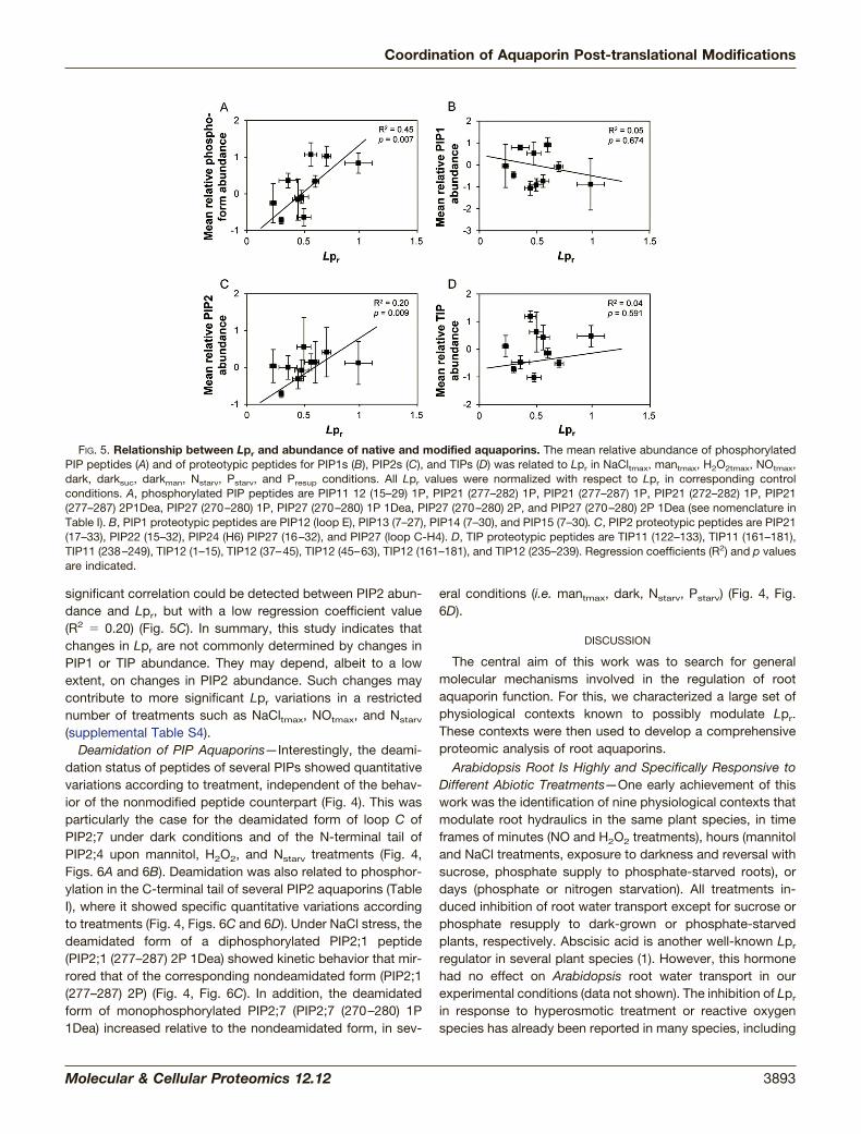
significant correlation could be detected between PIP2 abun-dance and Lpr, but with a low regression coefficient value(R2 � 0.20) (Fig. 5C). In summary, this study indicates thatchanges in Lpr are not commonly determined by changes inPIP1 or TIP abundance. They may depend, albeit to a lowextent, on changes in PIP2 abundance. Such changes maycontribute to more significant Lpr variations in a restrictednumber of treatments such as NaCltmax, NOtmax, and Nstarv
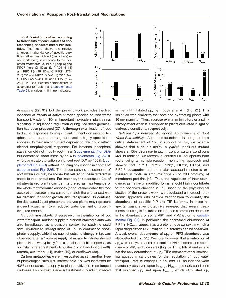
(supplemental Table S4).Deamidation of PIP Aquaporins—Interestingly, the deami-
dation status of peptides of several PIPs showed quantitativevariations according to treatment, independent of the behav-ior of the nonmodified peptide counterpart (Fig. 4). This wasparticularly the case for the deamidated form of loop C ofPIP2;7 under dark conditions and of the N-terminal tail ofPIP2;4 upon mannitol, H2O2, and Nstarv treatments (Fig. 4,Figs. 6A and 6B). Deamidation was also related to phosphor-ylation in the C-terminal tail of several PIP2 aquaporins (TableI), where it showed specific quantitative variations accordingto treatments (Fig. 4, Figs. 6C and 6D). Under NaCl stress, thedeamidated form of a diphosphorylated PIP2;1 peptide(PIP2;1 (277–287) 2P 1Dea) showed kinetic behavior that mir-rored that of the corresponding nondeamidated form (PIP2;1(277–287) 2P) (Fig. 4, Fig. 6C). In addition, the deamidatedform of monophosphorylated PIP2;7 (PIP2;7 (270–280) 1P1Dea) increased relative to the nondeamidated form, in sev-
eral conditions (i.e. mantmax, dark, Nstarv, Pstarv) (Fig. 4, Fig.6D).
DISCUSSION
The central aim of this work was to search for generalmolecular mechanisms involved in the regulation of rootaquaporin function. For this, we characterized a large set ofphysiological contexts known to possibly modulate Lpr.These contexts were then used to develop a comprehensiveproteomic analysis of root aquaporins.
Arabidopsis Root Is Highly and Specifically Responsive toDifferent Abiotic Treatments—One early achievement of thiswork was the identification of nine physiological contexts thatmodulate root hydraulics in the same plant species, in timeframes of minutes (NO and H2O2 treatments), hours (mannitoland NaCl treatments, exposure to darkness and reversal withsucrose, phosphate supply to phosphate-starved roots), ordays (phosphate or nitrogen starvation). All treatments in-duced inhibition of root water transport except for sucrose orphosphate resupply to dark-grown or phosphate-starvedplants, respectively. Abscisic acid is another well-known Lpr
regulator in several plant species (1). However, this hormonehad no effect on Arabidopsis root water transport in ourexperimental conditions (data not shown). The inhibition of Lpr
in response to hyperosmotic treatment or reactive oxygenspecies has already been reported in many species, including
FIG. 5. Relationship between Lpr and abundance of native and modified aquaporins. The mean relative abundance of phosphorylatedPIP peptides (A) and of proteotypic peptides for PIP1s (B), PIP2s (C), and TIPs (D) was related to Lpr in NaCltmax, mantmax, H2O2tmax, NOtmax,dark, darksuc, darkman, Nstarv, Pstarv, and Presup conditions. All Lpr values were normalized with respect to Lpr in corresponding controlconditions. A, phosphorylated PIP peptides are PIP11 12 (15–29) 1P, PIP21 (277–282) 1P, PIP21 (277–287) 1P, PIP21 (272–282) 1P, PIP21(277–287) 2P1Dea, PIP27 (270–280) 1P, PIP27 (270–280) 1P 1Dea, PIP27 (270–280) 2P, and PIP27 (270–280) 2P 1Dea (see nomenclature inTable I). B, PIP1 proteotypic peptides are PIP12 (loop E), PIP13 (7–27), PIP14 (7–30), and PIP15 (7–30). C, PIP2 proteotypic peptides are PIP21(17–33), PIP22 (15–32), PIP24 (H6) PIP27 (16–32), and PIP27 (loop C-H4). D, TIP proteotypic peptides are TIP11 (122–133), TIP11 (161–181),TIP11 (238–249), TIP12 (1–15), TIP12 (37–45), TIP12 (45–63), TIP12 (161–181), and TIP12 (235–239). Regression coefficients (R2) and p valuesare indicated.
Coordination of Aquaporin Post-translational Modifications
Molecular & Cellular Proteomics 12.12 3893
Arabidopis (22, 31), but the present work provides the firstevidence of effects of active nitrogen species on root watertransport. A role for NO, an important molecule in plant stresssignaling, in aquaporin regulation during rice seed germina-tion has been proposed (37). A thorough examination of roothydraulic responses to major plant nutrients or metabolites(phosphate, nitrate, and sugars) revealed highly specific re-sponses. In the case of nutrient deprivation, this could reflectdistinct morphological responses. For instance, phosphatestarvation did not modify root mass (supplemental Fig. S2A)but decreased shoot mass by 55% (supplemental Fig. S2B),whereas nitrate starvation enhanced root DW by 100% (sup-plemental Fig. S2D) without inducing any change in shoot DW(supplemental Fig. S2E). The accompanying adjustments ofroot hydraulics may be somewhat related to these differentialshoot-to-root alterations. For instance, the decreased Lpr ofnitrate-starved plants can be interpreted as maintenance ofthe whole root hydraulic capacity (conductance) while the rootabsorption surface is increased to match the unchanged wa-ter demand for shoot growth and transpiration. In contrast,the decreased Lpr of phosphate-starved plants may representa direct adjustment to a reduced water demand of growth-inhibited shoots.
Although most abiotic stresses result in the inhibition of rootwater transport, nutrient supply to nutrient-starved plants wasalso investigated as a possible context for studying rapidstimulus-induced up-regulation of Lpr. In contrast to phos-phate resupply, which had such effects, no change in Lpr wasobserved after a 1-day resupply of nitrate to nitrate-starvedplants. Here, we typically face a species-specific response, asa similar nitrate treatment stimulates Lpr in birdsfoot (38–40),tomato, cucumber (41), maize (40), or sunflower (38).
Carbon metabolites were investigated as still another typeof physiological stimulus. Interestingly, Lpr was increased by40% after sucrose resupply to plants cultivated in prolongeddarkness. By contrast, a similar treatment in plants cultivated
in the light inhibited Lpr by �30% after 4 h (Fig. 2B). Thisinhibition was similar to that obtained by treating plants with30 mM mannitol. Thus, sucrose exerts an inhibitory or a stim-ulatory effect when it is supplied to plants cultivated in light ordarkness conditions, respectively.
Relationships between Aquaporin Abundance and RootWater Permeability—Aquaporin abundance is thought to be acritical determinant of Lpr. In support of this, we recentlyshowed that a double pip2;1 � pip2;2 knock-out mutantshows a 40% decrease in Lpr in control culture conditions(42). In addition, we recently quantified PIP aquaporins fromroots using a multiple-reaction monitoring approach andshowed that PIP1;1, PIP1;2, PIP2;1, PIP2;2, PIP2;4, andPIP2;7 aquaporins are the major aquaporin isoforms ex-pressed in roots, in amounts from 70 to 280 pmol/mg ofmembrane proteins (43). Thus, the regulation of their abun-dance, as native or modified forms, should highly contributeto the observed changes in Lpr. Based on the physiologicalstudies of the present work, we developed a thorough pro-teomic approach with peptide fractionation to quantify theabundance of specific PIP and TIP isoforms. In these re-spects, quantitative proteomics revealed that several treat-ments resulting in Lpr inhibition induced a prominent decreasein the abundance of some PIP1 and PIP2 isoforms (supple-mental Fig. S5). In particular, the decreased abundance ofPIP1 in NOtmax appears as a pretty unique context in which arapid degradation (�20 min) of PIP isoforms can be observed.A weak overall dependence of Lpr on PIP2 abundance wasalso detected (Fig. 5C). We note, however, that an inhibition ofLpr was not systematically associated with a decreased abun-dance of PIP, and vice versa (Fig. 5). Thus, PIP abundance isnot the only determinant of Lpr. TIPs represent other interest-ing aquaporin candidates for the regulation of root watertransport. Parallel changes in Lpr and TIP abundance werepunctually observed upon Natmax, Nstarv, and dark conditionsthat inhibited Lpr and upon Presup, which stimulated Lpr
FIG. 6. Variation profiles accordingto treatments of deamidated and cor-responding nondeamidated PIP pep-tides. The figure shows the relativechanges in abundance of specific pep-tides, either deamidated (black bars) ornot (white bars), in response to the indi-cated treatments. A, PIP27 (loop C) andPIP27 (loop C) 1Dea. B, PIP24 (4–16)and PIP2;4 (4–16) 1Dea. C, PIP21 (277–287) 2P and PIP21 (277–287) 2P 1Dea.D, PIP27 (277–280) 1P and PIP27 (277–280) 1P 1Dea. Peptide nomenclature isaccording to Table I and supplementalTable S1. p values � 0.1 are indicated.
Coordination of Aquaporin Post-translational Modifications
3894 Molecular & Cellular Proteomics 12.12

(supplemental Fig. S5D). TIPs may contribute to transcellularwater transport or, alternatively, cytoplasm osmoregulation inconcert with PIPs. Although genetic evidence supporting arole of TIPs in root water transport is lacking, a few studieshave suggested a role for TIPs in salt stress responses. Forinstance, the overexpression of a soybean TIP2 isoform intransgenic Arabidopsis resulted in an increased plant sensi-tivity to salt stress (44). By contrast, Arabidopsis plants over-expressing a ginger TIP1 isoform became able to germinateand grow at high NaCl concentrations (150 mM) (45). However,the present study indicated that TIP abundance is not a majordeterminant of Lpr (Fig. 5D).
In summary, our study indicates that the modulation of Lpr
cannot be accounted for exclusively by modulations in PIP orTIP abundance. In view of the sensitivity of our MS analysis, itis very unlikely that an aquaporin isoform that would play amajor role in root water transport was missed in our study.These results prompted us to investigate PTMs as anothermode of aquaporin regulation in roots under abiotic and nu-tritional constraints.
Coordinated Post-translational Events to Regulate Aqua-porin Function—In line with previous studies (13–17), thepresent work highlights the diversity of aquaporin phosphor-ylated forms. It also points to combinations of various PTMswithin a single aquaporin, including phosphorylation, methyl-ation, deamidation, and acetylation (Fig. 3). The hierarchicalclustering of PIP peptides shown in Fig. 4 indicated thatmodified peptides showed variations profiles that were some-what more coordinated than those of their unmodified coun-terparts. A previous study using an absolute quantification MSmethodology showed that 2- or 4-h treatments with 100 mM
NaCl induced an increased abundance of the (PIP21 (277–287) 2P) peptide form, whereas a 15-min treatment with 2 mM
H2O2 induced a decrease of the (PIP21 (277–287) 1P) form(15). The present label-free quantification data are in fullagreement with this previous study (15), which validates thelabel-free strategy used here.
The present work further revealed coordinated changes inphosphorylation in the N-terminal tail of PIP1;1–1;2 and in theC-terminal tail of five PIP2 aquaporins (PIP2;1–PIP2;4 andPIP2;7) and showed that, in contrast to aquaporin cellularabundance, the extent of aquaporin phosphorylation is a pre-dominant parameter for controlling Lpr (Fig. 5A). Sequencealignment showed that two phosphorylation sites were con-served among the five PIP2 aquaporins (supplemental Fig.S6A). The first site, corresponding to Ser280 in AtPIP2;1, ishomologous to Ser274 of SoPIP2;1, the phosphorylation ofwhich enhances the opening of the aquaporin pore (5, 13).Phosphorylation of the second site, Ser283 in AtPIP2;1, wasshown to promote targeting of this aquaporin to the plasmamembrane and to interfere with its intracellular sorting afterstress-induced internalization (15). Therefore, we proposethat a decrease in Lpr associated with decreased PIP2 phos-phorylation involves both gating and regulated trafficking of
PIPs. Conversely, the increased phosphorylation of the sametwo sites may contribute to the Lpr increase induced bysucrose resupply to plants cultivated in prolonged darkness.We note, however, that Ser280 and Ser283 phosphorylationand Lpr showed opposite variations under some other treat-ments (mantmax, H2O2 treatments, dark, darkman, Nstarv, Pstarvation). This suggests the involvement of other post-trans-lational regulatory mechanisms.
Deamidation appeared as another prominent PTM in Ara-bidopsis root aquaporins. This modification was also identi-fied in 16% of microsomal proteins (i.e. 206 out of 1291proteins; supplemental Table S1). Given that 8 out of 12 PIPswere found to be deamidated, this modification seems tomore frequently affect PIP aquaporins than other membraneproteins. Deamidation possibly interferes with protein stabil-ity, protein activation, and protein–protein interactions (46–51). In mammalian AQP0, several sites of N-terminal trunca-tion have been identified as sites of Asn deamidation (52, 53).This age-related PTM, together with other PTMs, includingphosphorylation, is supposed to serve as a molecular clock(54) to regulate AQP0 function. Nonenzymatic deamidation ofAsn (N) and Gln (Q) residues may also occur spontaneouslyduring sample preparation (55). In this case, Asn deamidationtends to preferentially occur at NG, NS, and ND sequences,whereas Gln deamidation occurs at QV, QL, QG, and, to alesser extent, QA and QE sequences (56). Among the deami-dated Asn residues identified in this study, only one corre-sponded to an NG sequence (Table I). In addition, the abun-dance of the identified deamidated peptides showedquantitative variation according to the treatment (Figs. 4 and6), reinforcing the notion that the deamidated residues ob-served in PIPs reflect genuine PTMs occurring in planta.
Interestingly, we observed that deamidation was closelyassociated with phosphorylation in C-terminal PIP2 peptides.This observation may provide a clue to explain how an ap-parent increase in PIP2 C-terminal phosphorylation could beassociated with a decrease in Lpr (Fig. 4). A typical case isPIP2;7, for which the abundance of the monophosphorylatedform ((PIP27 (270–280) 1P) and that of the same form with anadditional deamidation ((PIP27 (270–280) 1P1Dea) showedopposite behaviors. It remains unknown whether or howneighboring deamidation and phosphorylation could interfereto alter protein function (57). Deamidation was also identifiedat a conserved Asn residue in loop C of several PIPs (Table I,supplemental Fig. S6B). In the case of AtPIP2;7, molecularmodeling according to the previously described SoPIP2;1structure (5) indicated that the deamidated Asn152 residue islocated at the entrance of the pore, close (�5 Å) to Arg224(supplemental Fig. S7). The latter residue contributes to one ofthe main pore constrictions (aromatic residue/Arg) involved insubstrate specificity. Deamidation—that is, the replacementof an amide with a carboxyl group—introduces a negativecharge at physiological pH and may thereby interfere withaquaporin action.
Coordination of Aquaporin Post-translational Modifications
Molecular & Cellular Proteomics 12.12 3895
CONCLUSION
In the present study, we used a large panel of abiotic andnutritional stimuli to search for aquaporin peptides that showquantitative variations consistent with the regulation of rootwater transport. We describe a high diversity of PIP responseprofiles concerning both aquaporin abundance and PTMs.Thus, PIP and TIP aquaporins undergo a combination ofseveral regulatory mechanisms, including (i) the regulation ofprotein abundance, mainly involved in NaCl, NO, and N star-vation treatments; (ii) the modulation of the phosphorylationstate of the N-terminal tail of PIP1;1–1;2 and the C-terminaltail of PIP2;1–2;3 and PIP2;7, which appears to be a predom-inant factor controlling Lpr; and (iii) the deamidation of Asnand Gln residues. The possible effects of deamidation onaquaporin function, which clearly deserve further experimen-tal work, may provide new perspectives in membrane proteinresearch.
Acknowledgments—We thank Dr. Lionel Verdoucq for help inAtPIP2;7 structural modeling and Cecile Fizames for help in analyzingMS data.
* This research work was supported by a grant from the AgenceNationale de la Recherche (PhosphoStim, ANR-08-GENM-013).
□S This article contains supplemental material.� To whom correspondence should be addressed: Veronique
Santoni, Tel.: 33-0-4-99-61-20-20, Fax: 33-0-4-67-52-57-37,E-mail: [email protected].
§ Present address: Proteomique Clinique – CHU Montpellier, Insti-tut de Recherches en Biotherapie, 80 av. A. Fliche, F-34295 Mont-pellier cedex 5, France.
REFERENCES
1. Maurel, C. (2008) Plant aquaporins: novel functions and regulation proper-ties. FEBS Lett. 581, 2227–2236
2. Kaldenhoff, R., Ribas-Carbo, M., Flexas, J., Lovisolo, C., Heckwolf, M., andUehlein, N. (2008) Aquaporins and plant water balance. Plant Cell Envi-ron. 31, 658–666
3. Maurel, C., Verdoucq, L., Luu, D.-T., and Santoni, V. (2008) Plant aqua-porins: membrane channels with multiple integrated functions. Annu.Rev. Plant Biol. 59, 595–624
4. Fujiyoshi, Y., Mitsuoka, K., de Groot, B. L., Philippsen, A., Grubmuller, H.,Agre, P., and Engel, A. (2002) Structure and function of water channels.Curr. Opin. Struct. Biol. 12, 509–515
5. Tornroth-Horsefield, S., Wang, Y., Hedfalk, K., Johanson, U., Karlsson, M.,Tajkhorshid, E., Neutze, R., and Kjellbom, P. (2006) Structural mecha-nism of plant aquaporin gating. Nature 439, 688–694
6. Johanson, U., Karlsson, M., Johansson, I., Gustavsson, S., Sjovall, S.,Fraysse, L., Weig, A. R., and Kjellbom, P. (2001) The complete set ofgenes encoding major intrinsic proteins in Arabidopsis provides a frame-work for a new nomenclature for major intrinsic proteins in plants. PlantPhysiol. 126, 1–12
7. Quigley, F., Rosenberg, J. M., Shachar-Hill, Y., and Bohnert, H. J. (2002)From genome to function: the Arabidopsis aquaporins. Genome Biol. 3,1–17
8. Ishikawa, F., Suga, S., Uemura, T., Sato, M. H., and Maeshima, M. (2005)Novel type aquaporins SIPs are mainly localized to the ER membraneand show cell-specific expression in Arabidopsis thaliana. FEBS Lett.579, 5814–5820
9. Javot, H., Lauvergeat, V., Santoni, V., Martin, F., Guclu, J., Vinh, J., Heyes,J., Franck, K. I., Schaffner, A. R., Bouchez, D., and Maurel, C. (2003) Rolefor a single aquaporin isoform in root water uptake. Plant Cell 15,509–522
10. Javot, H., and Maurel, C. (2002) The role of aquaporins in root water uptake.
Ann. Bot. 90, 301–31311. Martre, P., Morillon, R., Barrieu, F., North, G. B., Nobel, P. S., and
Chrispeels, M. J. (2002) Plasma membrane aquaporins play a significantrole during recovery from water deficit. Plant Physiol. 130, 2101–2110
12. Siefritz, F., Tyree, M. T., Lovisolo, C., Schubert, A., and Kaldenhoff, R.(2002) PIP1 plasma membrane aquaporins in tobacco: from cellulareffects to functions in plants. Plant Cell 14, 869–876
13. Johansson, I., Karlsson, M., Shukla, V. K., Chrispeels, M. J., Larsson, C.,and Kjellbom, P. (1998) Water transport activity of the plasma membraneaquaporin PM28A is regulated by phosphorylation. Plant Cell 10,451–459
14. Johansson, I., Larsson, C., Ek, B., and Kjellbom, P. (1996) The majorintegral proteins of spinach leaf plasma membranes are putative aqua-porins and are phosphorylated in response to Ca2� and apoplastic waterpotential. Plant Cell 8, 1181–1191
15. Prak, S., Hem, S., Boudet, J., Viennois, J., Sommerer, N., Rossignol, R.,Maurel, C., and Santoni, V. (2008) Multiple phosphorylations in the C-terminal tail of plant plasma membrane aquaporins. Role in sub-cellulartrafficking of AtPIP2;1 in response to salt stress. Mol. Cell. Proteomics 7,1019–1030
16. Kline, K. G., Barrett-Wilt, G. A., and Sussman, M. R. (2010) In plantachanges in protein phosphorylation induced by the plant hormone ab-scisic acid. Proc. Natl. Acad. Sci. U.S.A. 107:15986–15991
17. Niittyla, T., Fuglsang, A. T., Palmgren, M. G., Frommer, W. B., and Schulze,W. X. (2007) Temporal analysis of sucrose-induced phosphorylationchanges in plasma membrane proteins of Arabidopsis. Mol. Cell. Pro-teomics 6, 1711–1726
18. Engelsberger, W. R., and Schulze, W. X. (2012) Nitrate and ammonium leadto distinct global dynamic phosphorylation patterns when resupplied tonitrogen-starved Arabidopsis seedlings. Plant J. 69, 978–995
19. Sahr, T., Adam, T., Fizames, C., Maurel, C., and Santoni, V. (2010) O-carboxyl- and N-methyltransferases active on plant aquaporins. PlantCell Physiol. 51, 2092–2104
20. Santoni, V., Verdoucq, L., Sommerer, N., Vinh, J., Pflieger, D., and Maurel,C. (2006) Methylation of aquaporins in plant plasma membrane.Biochem. J. 400, 189–197
21. Lee, H. K., Cho, S. K., Son, O., Xu, Z., Hwang, I., and Kim, W. T. (2009)Drought stress-induced Rma1H1, a RING membrane-anchor E3 ubiqui-tin ligase homolog, regulates aquaporin levels via ubiquitination in trans-genic arabidopsis plants. Plant Cell 21, 622–641
22. Boursiac, Y., Chen, S., Luu, D.-T., Sorieul, M., van den Dries, N., andMaurel, C. (2005) Early effects of salinity on water transport in Arabidop-sis roots. Molecular and cellular features of aquaporin expression. PlantPhysiol. 139, 790–805
23. Murashige, T., and Skoog, F. (1962) A revised medium for rapid growth andbioassays with tobacco tissue cultures. Physiol. Plant. 15, 473–497
24. Besson-Bard, A., Griveau, S., Bedioui, F., and Wendehenne, D. (2008)Real-time electrochemical detection of extracellular nitric oxide in to-bacco cells exposed to cryptogein, an elicitor of defence responses. J.Exp. Bot. 59, 3407–3414
25. Goldstein, S., Russo, A., and Samuni, A. (2003) Reactions of PTIO andcarboxy-PTIO with (NO)-N-center dot, (NO2)-N-center dot, and O-2(cen-ter dot). J. Biol. Chem. 278, 50949–50955
26. Stoscheck, C. M. (1990) Quantitation of proteins. Methods Enzymol. 182,50–68
27. Santoni, V., Vinh, J., Pflieger, D., Sommerer, N., and Maurel, C. (2003) Aproteomic study reveals novel insights into the diversity of aquaporinforms expressed in the plasma membrane of plant roots. Biochem. J.373, 289–296
28. Wiœniewski, J. R., Zougman, A., Nagaraj, N., and Mann, M. (2009) Univer-sal sample preparation method for proteome analysis. Nat. Methods 6,359–362
29. Kyono, Y., Sugiyama, N., Imami, K., Tomita, M., and Ishihama, Y. (2008)Successive and selective release of phosphorylated peptides capturedby hydroxy acid-modified metal oxide chromatography. J. ProteomeRes. 7, 4585–4593
30. Li, Q. R., Ning, Z. B., Tang, J. S., Nie, S., and Zeng, R. (2009) Effect ofpeptide-to-TiO2 beads ratio on phosphopeptide enrichment selectivity.J. Proteome Res. 8, 5375–5381
31. Boursiac, Y., Prak, S., Boudet, J., Postaire, O., Luu, D.-T., Tournaire-Roux,C., Santoni, V., and Maurel, C. (2008) The response of Arabidopsis root
Coordination of Aquaporin Post-translational Modifications
3896 Molecular & Cellular Proteomics 12.12

water transport to a challenging environment implicates reactive oxygenspecies- and phosphorylation-dependent internalization of aquaporins.Plant Signal. Behav. 3, 1096–1098
32. Lopez-Bucio, J., Cruz-Ramirez, A., and Herrera-Estrella, L. (2003) The roleof nutrient availability in regulating root architecture. Curr. Opin. PlantBiol. 6, 280–287
33. Sutka, M., Li, G. W., Boudet, J., Boursiac, Y., Doumas, P., and Maurel, C.(2011) Natural variation of root hydraulics in Arabidopsis grown in normaland salt-stressed conditions. Plant Physiol. 155, 1264–1276
34. Prado, K., Boursiac, Y., Tournaire-Roux, C., Monneuse, J. M., Postaire, O.,Da Ines, O., Schaffner, A. R., Hem, S., Santoni, V., and Maurel, C. (2013)Regulation of Arabidopsis leaf hydraulics involves light-dependent phos-phorylation of aquaporins in veins. Plant Cell 25, 1029–1039
35. Hofte, H., Hubbard, L., Reizer, J., Ludevid, D., Herman, E. M., andChrispeels, M. J. (1992) Vegetative and seed-specific forms of tonoplastintrinsic protein in the vacuolar membrane of Arabidopsis thaliana. PlantPhysiol. 99, 561–570
36. Soto, G., Alleva, K., Mazzella, M. A., Amodeo, G., and Muschietti, J. P.(2008) AtTIP1;3 and AtTIP5;1, the only highly expressed Arabidopsispollen-specific aquaporins, transport water and urea. FEBS Lett. 582,4077–4082
37. Liu, H. Y., Yu, X., Cui, D. Y., Sun, M. H., Sun, W. N., Tang, Z. C., Kwak, S. S.,and Su, W. A. (2007) The role of water channel proteins and nitric oxidesignaling in rice seed germination. Cell Res. 17, 638–349
38. Clarkson, D. T., Carvajal, M., Henzler, T., Waterhouse, R. N., Smyth, A. J.,Cooke, D. T., and Steudle, E. (2000) Root hydraulic conductance: diurnalaquaporin expression and the effects of nutrient stress. J. Exp. Bot. 51,61–70
39. Gorska, A., Ye, Q., Holbrook, N. M., and Zwieniecki, M. A. (2008) Nitratecontrol of root hydraulic properties in plants: translating local informationto whole plant response. Plant Physiol. 148, 1159–1167
40. Prosser, I. M., Massonneau, A., Smyth, A. J., Waterhouse, R. N., Forde,B. G., and Clarkson, D. T. (2006) Nitrate assimilation in the forage legumeLotus japonieus L. Planta 223, 821–834
41. Gloser, V., Zwieniecki, M. A., Orians, C. M., and Holbrook, N. M. (2007)Dynamic changes in root hydraulic properties in response to nitrateavailability. J. Exp. Bot. 10, 2409–2415
42. Peret, B., Li, G. W., Zhao, J., Band, L. R., Voss, U., Postaire, O., Luu, D. T.,Da Ines, O., Casimiro, I., Lucas, M., Wells, D. M., Lazzerini, L., Nacry, P.,King, J. R., Jensen, O. E., Schaffner, A. R., Maurel, C., and Bennett, M. J.(2012) Auxin regulates aquaporin function to facilitate lateral root emer-gence. Nat. Cell Biol. 14, 991–998
43. Monneuse, J. M., Sugano, M., Becue, T., Santoni, V., Hem, S., and Ros-signol, M. (2011) Towards the profiling of the Arabidopsis thalianaplasma membrane transportome by targeted proteomics. Proteomics11, 1789–1797
44. Wang, X., Li, Y., Ji, W., Bai, X., Cai, H., Zhu, D., Sun, X. L., Chen, L. J., andZhu, Y. M. (2011) A novel glycine soja tonoplast intrinsic protein generesponds to abiotic stress and depresses salt and dehydration tolerancein transgenic Arabidopsis thaliana. J. Plant Physiol. 168, 1241–1248
45. Peng, Y., Lin, W., Cai, W., and Arora, R. (2007) Overexpression of a Panaxginseng tonoplast aquaporin alters salt tolerance, drought tolerance andcold acclimation ability in transgenic Arabidopsis plants. Planta226:729–740
46. Flatau, G., Lemichez, E., Gauthier, M., Chardin, P., Paris, S., Fiorentini, C.,and Boquet, P. (1997) Toxin-induced activation of the G protein p21 Rhoby deamidation of glutamine. Nature 387, 729–733
47. Geiger, T., and Clarke, S. (1987) Deamidation, isomerization, and racemi-zation at asparaginyl and aspartyl residues in peptides—succimide-linked reactions that contribute to protein-degradation. J. Biol. Chem.262, 785–794
48. Lerm, M., Selzer, J., Hoffmeyer, A., Rapp, U. R., Aktories, K., and Schmidt,G. (1999) Deamidation of Cdc42 and Rac by Escherichia coli cytotoxicnecrotizing factor 1: activation of cJun N-terminal kinase in HeLa cells.Infect. Immun. 67, 496–503
49. Schmidt, G., Sehr, P., Wilm, M., Selzer, J., Mann, M., and Aktories, K.(1997) Gln 63 of Rho is deamidated by Escherichia coli cytotoxic necro-tizing factor-1. Nature 387, 725–729
50. Visvikis, O., Maddugoda, M. P., and Lemichez, E. (2010) Direct modifica-tions of Rho proteins: deconstructing GTPase regulation. Biol. Cell 102,377–389
51. Curnis, F., Longhi, R., Crippa, L., Cattaneo, A., Dondossola, E., Bachi, A.,and Corti, A. (2006) Spontaneous formation of L-isoaspartate and gain offunction in fibronectin. J. Biol. Chem. 281, 36466–36476
52. Ball, L. E., Garland, D. L., Crouch, R. K., and Schey, K. L. (2004) Post-translational modifications of aquaporin-0 (AQP0) in the normal humanlens: spatial and temporal occurrence. Biochemistry 43, 9856–9865
53. Schey, K. L., Little, M., Fowler, J. G., and Crouch, R. K. (2000) Character-ization of human lens major intrinsic protein structure. Invest. Ophthal-mol. Vis. Sci. 41, 175–182
54. Robinson, N. E., and Robinson, A. B. (2001) Deamidation of human pro-teins. Proc. Natl. Acad. Sci. U.S.A. 98, 12409–12413
55. Li, X. J., Cournoyer, J. J., Lin, C., and O’Cormora, P. B. (2008) Use of O-18labels to monitor deamidation during protein and peptide sample pro-cessing. J. Am. Soc. Mass Spectrom. 19, 855–864
56. Hao, P. L., Ren, Y., Alpert, A. J., and Sze, S. K. (2011) Detection, evaluationand minimization of nonenzymatic deamidation in proteomic samplepreparation. Mol. Cell. Proteomics 10 M0111.009381
57. Tholey, A., Pipkorn, R., Bossemeyer, D., Kinzel, V., and Reed, J. (2001)Influence of myristoylation, phosphorylation, and deamidation on thestructural behavior of the N-terminus of the catalytic subunit of CAMP-dependent protein kinase. Biochemistry 40, 225–231
Coordination of Aquaporin Post-translational Modifications
Molecular & Cellular Proteomics 12.12 3897