The Comparative Survey of Coordinated Regulation of ... - MDPI
17
Citation: Yang, F.; Wang, Y.; Lu, W.; Zong, W.; Zhu, Q.; Cheng, J. The Comparative Survey of Coordinated Regulation of Steroidogenic Pathway in Japanese Flounder (Paralichthys olivaceus) and Chinese Tongue Sole (Cynoglossus semilaevis). Int. J. Mol. Sci. 2022, 23, 5520. https://doi.org/10.3390/ ijms23105520 Academic Editors: Isabel Navarro and Daniel Garcia de la Serrana Received: 8 April 2022 Accepted: 12 May 2022 Published: 15 May 2022 Publisher’s Note: MDPI stays neutral with regard to jurisdictional claims in published maps and institutional affil- iations. Copyright: © 2022 by the authors. Licensee MDPI, Basel, Switzerland. This article is an open access article distributed under the terms and conditions of the Creative Commons Attribution (CC BY) license (https:// creativecommons.org/licenses/by/ 4.0/). International Journal of Molecular Sciences Article The Comparative Survey of Coordinated Regulation of Steroidogenic Pathway in Japanese Flounder (Paralichthys olivaceus) and Chinese Tongue Sole (Cynoglossus semilaevis) Fan Yang 1 , Yapeng Wang 1 , Wei Lu 1 , Wenyu Zong 1 , Qing Zhu 1,2 and Jie Cheng 1,2,3, * 1 Key Laboratory of Marine Genetics and Breeding (Ocean University of China), Ministry of Education, 5 Yushan Road, Qingdao 266003, China; [email protected] (F.Y.); [email protected] (Y.W.); [email protected] (W.L.); [email protected] (W.Z.); [email protected] (Q.Z.) 2 Key Laboratory of Tropical Aquatic Germplasm of Hainan Province, Sanya Oceanographic Institution, Ocean University of China, Sanya 572024, China 3 Laboratory for Marine Fisheries Science and Food Production Processes, Pilot National Laboratory for Marine Science and Technology (Qingdao), 1 Wenhai Road, Qingdao 266237, China * Correspondence: [email protected]; Tel.: +86-532-82031986 Abstract: Steroidogenesis controls the conversion of cholesterol into steroid hormones through the complex cascade reaction of various enzymes, which play essential roles in sexual differentiation and gonadal development in vertebrates, including teleosts. Japanese flounder (Paralichthys olivaceus) and Chinese tongue sole (Cynoglossus semilaevis) are important marine cultured fishes in China and have remarkable sexual dimorphism with bigger females and sex reversal scenarios from female to neo-male. Several steroidogenic genes have been analyzed individually in the two species, but there is a lack of information on the coordinated interaction of steroidogenic gene regulation. Therefore, in this study, through genomic and transcriptomic analysis, 39 and 42 steroidogenic genes were systematically characterized in P. olivaceus and C. semilaevis genomes, respectively. Phylogenetic and synteny analysis suggested a teleost specific genome duplication origin for cyp19a1a/cyp19a1b, hsd17b12a/hsd17b12b, ara/arb and esr2a/esr2b but not for star/star2 and cyp17a1/cyp17a2. Comparative transcriptome analysis revealed conserved expression patterns for steroidogenic genes in P. olivaceus and C. smilaevis gonads; star/star2, cyp11a/cyp11c, cyp17a1/cyp17a2, cyp21a, hsd3b1, hsd11b and hsd20b were strongly expressed in testis, while cyp19a1a and hsd17b genes were highly expressed in ovaries. Only a few genes were differentially expressed between male and neo-male testis of both P. olivaceus and C. semilaevis, and even fewer genes were differentially regulated in the brains of both species. Network analysis indicated that cyp11c, cyp17a1 and hsd3b1 actively interacted with other steroidogenic genes in P. olivaceus and C. semilaevis, and may play a more sophisticated role in the steroid hormone biosynthesis cascade. The coordinated interaction of steroidogenic genes provided comprehensive insights into steroidogenic pathway regulation with a global biological impact, as well as sexual development in teleost species. Keywords: steroidogenesis; hypothalamic-pituitary-gonadal axis; regulatory network; Paralichthys olivaceus; Cynoglossus semilaevis 1. Introduction Steroidogenesis is the conversion of cholesterol into biologically active steroid hormones by the complex cascade reaction of various enzymes, which plays essential roles during many physiological processes including sexual differentiation, gonadal development, growth and maturation [1]. Steroidogenesis occurs primarily in tissues, such as the gonads, interrenal gland and brain, which is, in general, controlled by the hypothalamus–pituitary–interrenal Int. J. Mol. Sci. 2022, 23, 5520. https://doi.org/10.3390/ijms23105520 https://www.mdpi.com/journal/ijms
-
Upload
khangminh22 -
Category
Documents
-
view
3 -
download
0
Transcript of The Comparative Survey of Coordinated Regulation of ... - MDPI
Citation: Yang, F.; Wang, Y.; Lu, W.;
Zong, W.; Zhu, Q.; Cheng, J.
The Comparative Survey of
Coordinated Regulation of
Steroidogenic Pathway in Japanese
Flounder (Paralichthys olivaceus) and
Chinese Tongue Sole (Cynoglossus
semilaevis). Int. J. Mol. Sci. 2022, 23,
5520. https://doi.org/10.3390/
ijms23105520
Academic Editors: Isabel Navarro
and Daniel Garcia de la Serrana
Received: 8 April 2022
Accepted: 12 May 2022
Published: 15 May 2022
Publisher’s Note: MDPI stays neutral
with regard to jurisdictional claims in
published maps and institutional affil-
iations.
Copyright: © 2022 by the authors.
Licensee MDPI, Basel, Switzerland.
This article is an open access article
distributed under the terms and
conditions of the Creative Commons
Attribution (CC BY) license (https://
creativecommons.org/licenses/by/
4.0/).
International Journal of
Molecular Sciences
Article
The Comparative Survey of Coordinated Regulation ofSteroidogenic Pathway in Japanese Flounder(Paralichthys olivaceus) and Chinese Tongue Sole(Cynoglossus semilaevis)Fan Yang 1, Yapeng Wang 1, Wei Lu 1, Wenyu Zong 1, Qing Zhu 1,2 and Jie Cheng 1,2,3,*
1 Key Laboratory of Marine Genetics and Breeding (Ocean University of China), Ministry of Education,5 Yushan Road, Qingdao 266003, China; [email protected] (F.Y.);[email protected] (Y.W.); [email protected] (W.L.); [email protected] (W.Z.);[email protected] (Q.Z.)
2 Key Laboratory of Tropical Aquatic Germplasm of Hainan Province, Sanya Oceanographic Institution,Ocean University of China, Sanya 572024, China
3 Laboratory for Marine Fisheries Science and Food Production Processes, Pilot National Laboratory for MarineScience and Technology (Qingdao), 1 Wenhai Road, Qingdao 266237, China
* Correspondence: [email protected]; Tel.: +86-532-82031986
Abstract: Steroidogenesis controls the conversion of cholesterol into steroid hormones through thecomplex cascade reaction of various enzymes, which play essential roles in sexual differentiation andgonadal development in vertebrates, including teleosts. Japanese flounder (Paralichthys olivaceus)and Chinese tongue sole (Cynoglossus semilaevis) are important marine cultured fishes in China andhave remarkable sexual dimorphism with bigger females and sex reversal scenarios from female toneo-male. Several steroidogenic genes have been analyzed individually in the two species, but thereis a lack of information on the coordinated interaction of steroidogenic gene regulation. Therefore,in this study, through genomic and transcriptomic analysis, 39 and 42 steroidogenic genes weresystematically characterized in P. olivaceus and C. semilaevis genomes, respectively. Phylogeneticand synteny analysis suggested a teleost specific genome duplication origin for cyp19a1a/cyp19a1b,hsd17b12a/hsd17b12b, ara/arb and esr2a/esr2b but not for star/star2 and cyp17a1/cyp17a2. Comparativetranscriptome analysis revealed conserved expression patterns for steroidogenic genes in P. olivaceusand C. smilaevis gonads; star/star2, cyp11a/cyp11c, cyp17a1/cyp17a2, cyp21a, hsd3b1, hsd11b andhsd20b were strongly expressed in testis, while cyp19a1a and hsd17b genes were highly expressed inovaries. Only a few genes were differentially expressed between male and neo-male testis of bothP. olivaceus and C. semilaevis, and even fewer genes were differentially regulated in the brains ofboth species. Network analysis indicated that cyp11c, cyp17a1 and hsd3b1 actively interacted withother steroidogenic genes in P. olivaceus and C. semilaevis, and may play a more sophisticated rolein the steroid hormone biosynthesis cascade. The coordinated interaction of steroidogenic genesprovided comprehensive insights into steroidogenic pathway regulation with a global biologicalimpact, as well as sexual development in teleost species.
Keywords: steroidogenesis; hypothalamic-pituitary-gonadal axis; regulatory network; Paralichthysolivaceus; Cynoglossus semilaevis
1. Introduction
Steroidogenesis is the conversion of cholesterol into biologically active steroid hormonesby the complex cascade reaction of various enzymes, which plays essential roles during manyphysiological processes including sexual differentiation, gonadal development, growth andmaturation [1]. Steroidogenesis occurs primarily in tissues, such as the gonads, interrenalgland and brain, which is, in general, controlled by the hypothalamus–pituitary–interrenal
Int. J. Mol. Sci. 2022, 23, 5520. https://doi.org/10.3390/ijms23105520 https://www.mdpi.com/journal/ijms
Int. J. Mol. Sci. 2022, 23, 5520 2 of 17
(HPI) and hypothalamus–pituitary–gonadal (HPG) axes and has a considerable impact onregulating complex steroidogenic process in vertebrates, and further affects reproduction [2,3].
In vertebrates, including teleosts, steroidogenesis is triggered by the mobilization ofcholesterol by steroidogenic acute regulatory protein (star) from the outer to the inner mito-chondrial membrane [4,5]. The following sophisticated process recruits diverse enzymes,such as cytochrome P450 and hydroxysteroid dehydrogenase, for the synthesis of C18estradiol, C19 testosterone and C21 cortisol [6]. The cytochrome P450 (cyp) is a multigenefamily that is involved in catalyzing the production of sexual steroids [7,8]. For exam-ple, cyp11a is the enzyme that catalyzes the generation of pregnenolone, which is theprimary step in the initiation of steroid hormone biosynthesis [6,9]. Cyp11b/c can promotethe production of corticosterone, cortisol and 11β-OH-testosterone [10,11], which has agreat impact on male ontogenesis. Cyp17a1 and cyp17a2 are two isoforms of steroid17α-hydroxylase/17,20-lyase [12], with cyp17a1 possessing both 17α-hydroxylase and17,20-lyase activities, while cyp17a2 has 17α-hydroxylase activity. Moreover, cyp17a1 wasproven to participate in generating ovary estradiol, while the generation of C21 steroids,such as cortisol, in the head kidney is associated with cyp17a2 [13,14]. Aromatase (cyp19a1)is responsible for the transformation of androstenedione into estrone, and testosterone intoestradiol, in which the up- and down-regulation of cyp19a1 are crucial for ovarian andtesticular differentiation [15]. In addition, Hydroxysteroid dehydrogenases (hsds) are alsoinvolved in the regulation of steroid hormone biosynthesis [16]. 3-beta-hydroxysteroiddehydrogenase (hsd3b) is important in the production of progesterone and testosterone [17].Corticosteroid 11-beta-dehydrogenase isozyme 2 (hsd11b2) was found to exert a major in-fluence on the testicular differentiation of catfish (Clarias gariepinus) [18]. In zebrafish (Daniorerio), hsd20b2 is able to synthesize 20β-dihydrocortisone from cortisone [19], and hsd17b3is highly expressed in the ovary [20]. In addition, sex hormone receptors, such as estro-gen (esr) and androgen (ar) receptors, are also important for the regulation of sex steroidhormones [6,21,22], which regulate reproductive activities by combining with esr andar [21].
Teleost species exhibit a wide range of reproductive strategies for sex determinationand differentiation, and steroids are required during all stages of the reproductive cyclein teleosts which include sex differentiation, maturation, growth and sexual behavior [5].Japanese flounder (Paralichthys olivaceus) and Chinese tongue sole (Cynoglossus semilaevis),both belonging to Pleuronectiformes, are important maricultured fish species in China,Japan and Korea [23,24]. P. olivaceus and C. semilaevis have remarkable sexual dimorphism,with females growing faster and bigger than males; they also present a sex reversal scenariofrom female to neo-male during the key period of sex differentiation [25,26]. Therefore,it makes the sex control for all female production more economic and beneficial in aqua-culture [25]. The steroidogenic pathway, important for sex hormone homeostasis, hasbeen proven to play essential roles in gonadal development and sex differentiation inP. olivaceus and C. semilaevis, especially for the sex reversal of both species [26–28]. Manysteroidogenic genes, such as star [29], cyp11 [9,10], cyp17a [30], cyp19a1 [28], as well as thehsd family [24] and sex hormone receptors [31], have been characterized in P. olivaceus andC. semilaevis. However, most of these studies are only focused on a limited number ofsteroidogenic genes rather than the full complement of the steroidogenic pathways thatcomprise the coordinated cascade reaction in steroid biosynthesis. Therefore, in this study,the steroidogenic genes in P. olivaceus and C. semilaevis were characterized, and their evolu-tion, expression and coordinated interaction were compared through molecular evolutionand transcriptome analysis, which will provide a comprehensive understanding of therelationships between the regulation of steroidogenic pathways and sexual development inteleost species.
Int. J. Mol. Sci. 2022, 23, 5520 3 of 17
2. Results and Discussion2.1. Genomic Landscape, Functional Domain and Phylogeny of Steroidogenic Genes in P. olivaceusand C. semilaevis
Through a genome-wide screen, 39 and 42 genes participating in steroid biosynthesiswere identified in the P. olivaceus and C. semilaevis genomes, respectively (Figure 1 andTable S1), including star, cyp, hsd genes, as well as steroid receptors genes esr and ar (Figure 1and Table S2). The copy number of these steroidogenic genes between tetrapod andteleost lineages was generally conserved with several gene duplication events observedin the teleost lineage (Table S1), most likely originating from the teleost specific genomeduplication (TSGD).
Int. J. Mol. Sci. 2022, 23, x FOR PEER REVIEW 3 of 17
2. Results and Discussion 2.1. Genomic Landscape, Functional Domain and Phylogeny of Steroidogenic Genes in P. olivaceus and C. semilaevis
Through a genome-wide screen, 39 and 42 genes participating in steroid biosynthesis were identified in the P. olivaceus and C. semilaevis genomes, respectively (Figure 1 and Table S1), including star, cyp, hsd genes, as well as steroid receptors genes esr and ar (Figure 1 and Table S2). The copy number of these steroidogenic genes between tetrapod and tel-eost lineages was generally conserved with several gene duplication events observed in the teleost lineage (Table S1), most likely originating from the teleost specific genome du-plication (TSGD).
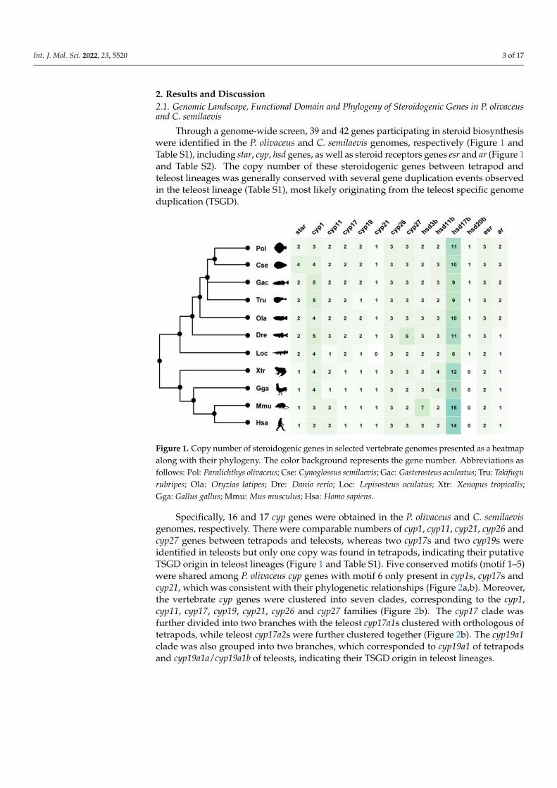
Figure 1. Copy number of steroidogenic genes in selected vertebrate genomes presented as a heatmap along with their phylogeny. The color background represents the gene number. Abbrevi-ations as follows: Pol: Paralichthys olivaceus; Cse: Cynoglossus semilaevis; Gac: Gasterosteus aculeatus; Tru: Takifugu rubripes; Ola: Oryzias latipes; Dre: Danio rerio; Loc: Lepisosteus oculatus; Xtr: Xenopus tropicalis; Gga: Gallus gallus; Mmu: Mus musculus; Hsa: Homo sapiens.
Specifically, 16 and 17 cyp genes were obtained in the P. olivaceus and C. semilaevis genomes, respectively. There were comparable numbers of cyp1, cyp11, cyp21, cyp26 and cyp27 genes between tetrapods and teleosts, whereas two cyp17s and two cyp19s were identified in teleosts but only one copy was found in tetrapods, indicating their putative TSGD origin in teleost lineages (Figure 1 and Table S1). Five conserved motifs (motif 1–5) were shared among P. olivaceus cyp genes with motif 6 only present in cyp1s, cyp17s and cyp21, which was consistent with their phylogenetic relationships (Figure 2a, b). Moreo-ver, the vertebrate cyp genes were clustered into seven clades, corresponding to the cyp1, cyp11, cyp17, cyp19, cyp21, cyp26 and cyp27 families (Figure 2b). The cyp17 clade was fur-ther divided into two branches with the teleost cyp17a1s clustered with orthologous of tetrapods, while teleost cyp17a2s were further clustered together (Figure 2b). The cyp19a1 clade was also grouped into two branches, which corresponded to cyp19a1 of tetrapods and cyp19a1a/cyp19a1b of teleosts, indicating their TSGD origin in teleost lineages.
Figure 1. Copy number of steroidogenic genes in selected vertebrate genomes presented as a heatmapalong with their phylogeny. The color background represents the gene number. Abbreviations asfollows: Pol: Paralichthys olivaceus; Cse: Cynoglossus semilaevis; Gac: Gasterosteus aculeatus; Tru: Takifugurubripes; Ola: Oryzias latipes; Dre: Danio rerio; Loc: Lepisosteus oculatus; Xtr: Xenopus tropicalis;Gga: Gallus gallus; Mmu: Mus musculus; Hsa: Homo sapiens.
Specifically, 16 and 17 cyp genes were obtained in the P. olivaceus and C. semilaevisgenomes, respectively. There were comparable numbers of cyp1, cyp11, cyp21, cyp26 andcyp27 genes between tetrapods and teleosts, whereas two cyp17s and two cyp19s wereidentified in teleosts but only one copy was found in tetrapods, indicating their putativeTSGD origin in teleost lineages (Figure 1 and Table S1). Five conserved motifs (motif 1–5)were shared among P. olivaceus cyp genes with motif 6 only present in cyp1s, cyp17s andcyp21, which was consistent with their phylogenetic relationships (Figure 2a,b). Moreover,the vertebrate cyp genes were clustered into seven clades, corresponding to the cyp1,cyp11, cyp17, cyp19, cyp21, cyp26 and cyp27 families (Figure 2b). The cyp17 clade wasfurther divided into two branches with the teleost cyp17a1s clustered with orthologous oftetrapods, while teleost cyp17a2s were further clustered together (Figure 2b). The cyp19a1clade was also grouped into two branches, which corresponded to cyp19a1 of tetrapodsand cyp19a1a/cyp19a1b of teleosts, indicating their TSGD origin in teleost lineages.

Int. J. Mol. Sci. 2022, 23, 5520 4 of 17Int. J. Mol. Sci. 2022, 23, x FOR PEER REVIEW 4 of 17
Figure 2. Characterization of steroidogenic proteins. The conserved motif and structure of (a) cyp, (c) hsd, (e) star, esr and ar in P. olivaceus, and the phylogeny of (b) cyp, (d) hsd, (f) star, esr and ar in selected vertebrates. The phylogenic trees were constructed with a bootstrap of 1,000 times. The sub-families of cyp and hsd were labeled with different background colors, and the P. olivaceus proteins were highlighted in red.
Figure 2. Characterization of steroidogenic proteins. The conserved motif and structure of (a) cyp,(c) hsd, (e) star, esr and ar in P. olivaceus, and the phylogeny of (b) cyp, (d) hsd, (f) star, esr andar in selected vertebrates. The phylogenic trees were constructed with a bootstrap of 1000 times.The sub-families of cyp and hsd were labeled with different background colors, and the P. olivaceusproteins were highlighted in red.
Int. J. Mol. Sci. 2022, 23, 5520 5 of 17
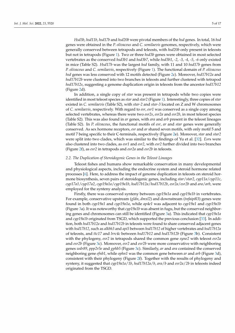
Hsd3b, hsd11b, hsd17b and hsd20b were pivotal members of the hsd genes. In total, 16 hsdgenes were obtained in the P. olivaceus and C. semilaevis genomes, respectively, which weregenerally conserved between tetrapods and teleosts, with hsd20b only present in teleostsbut not in tetrapods (Figure 1). Two or three hsd3b genes were obtained in most selectedvertebrates as the conserved hsd3b1 and hsd3b7, while hsd3b1, -2, -3, -4, -5, -6 only existedin mice (Table S2). Hsd17b was the largest hsd family, with 11 and 10 hsd17b genes fromP. olivaceus and C. semilaevis, respectively (Figure 1). The functional domain of P. olivaceushsd genes was less conserved with 12 motifs detected (Figure 2c). Moreover, hsd17b12a andhsd17b12b were clustered into two branches in teleosts and further clustered with tetrapodhsd17b12s, suggesting a genome duplication origin in teleosts from the ancestor hsd17b12(Figure 2d).
In addition, a single copy of star was present in tetrapods while two copies wereidentified in most teleost species as star and star2 (Figure 1). Interestingly, three copies of starexisted in C. semilaevis (Table S2), with star-2 and star-3 located on Z and W chromosomesof C. semilaevis, respectively. With regard to esr, esr1 was conserved as a single copy amongselected vertebrates, whereas there were two esr2s, esr2a and esr2b, in most teleost species(Table S2). This was also found in ar genes, with ara and arb present in the teleost lineages(Table S2). In P. olivaceus, the functional motifs of esr, ar and star genes were generallyconserved. As sex hormone receptors, esr and ar shared seven motifs, with only motif 5 andmotif 7 being specific to their C-terminals, respectively (Figure 2e). Moreover, star and star2were split into two clades, which was similar to the findings of Yu et al. [32]. Esrs werealso clustered into two clades, as esr1 and esr2, with esr2 further divided into two branches(Figure 2f), as esr2 in tetrapods and esr2a and esr2b in teleosts.
2.2. The Duplication of Steroidogenic Genes in the Teleost Lineages
Teleost fishes and humans show remarkable conservation in many developmentaland physiological aspects, including the endocrine system and steroid hormone relatedprocesses [6]. Here, to address the impact of genome duplication in teleosts on steroid hor-mone biosynthesis, seven pairs of steroidogenic genes, including star/star2, cyp11a/cyp11c,cyp17a1/cyp17a2, cyp19a1a/cyp19a1b, hsd17b12a/hsd17b12b, esr2a/esr2b and ara/arb, wereemployed for the synteny analysis.
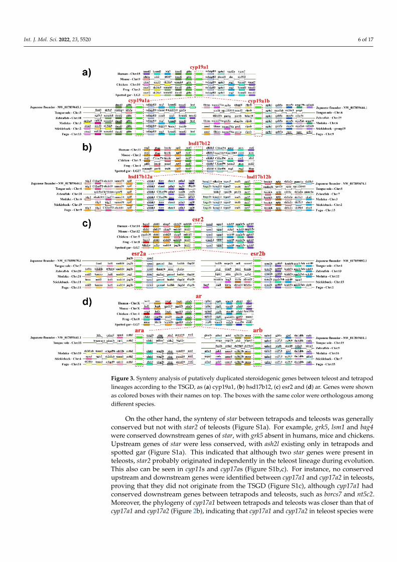
Firstly, there was conserved synteny between cyp19a1a and cyp19a1b in vertebrates.For example, conservative upstream (gldn, dmxl2) and downstream (tnfaip8l3) genes werefound in both cyp19a1 and cyp19a1a, while ap4e1 was adjacent to cyp19a1 and cyp19a1b(Figure 3a). It was noteworthy that cyp19a1b was absent in fugu, but the conserved neighbor-ing genes and chromosomes can still be identified (Figure 3a). This indicated that cyp19a1aand cyp19a1b originated from TSGD, which supported the previous conclusion [33]. In addi-tion, both hsd17b12a and hsd17b12b in teleosts were found to share conserved adjacent geneswith hsd17b12, such as alkbh3 and api5 between hsd17b12 of higher vertebrates and hsd17b12aof teleosts, and ttc17 and lrrc4c between hsd17b12 and hsd17b12b (Figure 3b). Consistentwith the phylogeny, esr2 in tetrapods shared the common gene syne2 with teleost esr2aand esr2b (Figure 3c). Moreover, esr2 and esr2b were more conservative with neighboringgenes wdr89, ppp2r5e and gphb5 (Figure 3c). Similarly, ar and ara contained the conservedneighboring gene efnb1, while ophn1 was the common gene between ar and arb (Figure 3d),consistent with their phylogeny (Figure 2f). Together with the results of phylogeny andsynteny, it suggested that cyp19a1a/1b, hsd17b12a/b, ara/b and esr2a/2b in teleosts indeedoriginated from the TSGD.
Int. J. Mol. Sci. 2022, 23, 5520 6 of 17Int. J. Mol. Sci. 2022, 23, x FOR PEER REVIEW 6 of 17
Figure 3. Synteny analysis of putatively duplicated steroidogenic genes between teleost and tetra-pod lineages according to the TSGD, as (a) cyp19a1, (b) hsd17b12, (c) esr2 and (d) ar. Genes were shown as colored boxes with their names on top. The boxes with the same color were orthologous among different species.
On the other hand, the synteny of star between tetrapods and teleosts was generally conserved but not with star2 of teleosts (Figure S1a). For example, grk5, lsm1 and bag4 were conserved downstream genes of star, with grk5 absent in humans, mice and chickens. Up-stream genes of star were less conserved, with ash2l existing only in tetrapods and spotted gar (Figure S1a). This indicated that although two star genes were present in teleosts, star2 probably originated independently in the teleost lineage during evolution. This also can be seen in cyp11s and cyp17as (Figure S1b, c). For instance, no conserved upstream and downstream genes were identified between cyp17a1 and cyp17a2 in teleosts, proving that they did not originate from the TSGD (Figure S1c), although cyp17a1 had conserved down-stream genes between tetrapods and teleosts, such as borcs7 and nt5c2. Moreover, the phy-logeny of cyp17a1 between tetrapods and teleosts was closer than that of cyp17a1 and cyp17a2 (Figure 2b), indicating that cyp17a1 and cyp17a2 in teleost species were probably gained independently during evolution. Therefore, it revealed that star/star2, cyp11a/11c and cyp17a1/17a2 did not originate from TSGD as previously assumed [6].
Figure 3. Synteny analysis of putatively duplicated steroidogenic genes between teleost and tetrapodlineages according to the TSGD, as (a) cyp19a1, (b) hsd17b12, (c) esr2 and (d) ar. Genes were shownas colored boxes with their names on top. The boxes with the same color were orthologous amongdifferent species.
On the other hand, the synteny of star between tetrapods and teleosts was generallyconserved but not with star2 of teleosts (Figure S1a). For example, grk5, lsm1 and bag4were conserved downstream genes of star, with grk5 absent in humans, mice and chickens.Upstream genes of star were less conserved, with ash2l existing only in tetrapods andspotted gar (Figure S1a). This indicated that although two star genes were present inteleosts, star2 probably originated independently in the teleost lineage during evolution.This also can be seen in cyp11s and cyp17as (Figure S1b,c). For instance, no conservedupstream and downstream genes were identified between cyp17a1 and cyp17a2 in teleosts,proving that they did not originate from the TSGD (Figure S1c), although cyp17a1 hadconserved downstream genes between tetrapods and teleosts, such as borcs7 and nt5c2.Moreover, the phylogeny of cyp17a1 between tetrapods and teleosts was closer than that ofcyp17a1 and cyp17a2 (Figure 2b), indicating that cyp17a1 and cyp17a2 in teleost species were
Int. J. Mol. Sci. 2022, 23, 5520 7 of 17
probably gained independently during evolution. Therefore, it revealed that star/star2,cyp11a/11c and cyp17a1/17a2 did not originate from TSGD as previously assumed [6].
2.3. Evolutionary Dynamics of Steroidogenic Genes in Teleost Lineages
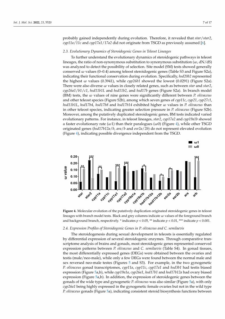
To further understand the evolutionary dynamics of steroidogenic pathways in teleostlineages, the ratio of non-synonymous substitution to synonymous substitution (ω, dN/dS)was analyzed to detect the possibility of selection. Site model (SM) tests showed generallyconservedω values (0–0.4) among teleost steroidogenic genes (Table S3 and Figure S2a),indicating their functional conservation during evolution. Specifically, hsd20b2 representedthe highest ω values (0.3941), while cyp26b1 showed the lowest (0.0291) (Figure S2a).There were also diverseω values in closely related genes, such as between star and star2,cyp26a1/b1/c1, hsd11b1L and hsd11b2, and hsd17b genes (Figure S2a). In branch model(BM) tests, the ω values of nine genes were significantly different between P. olivaceusand other teleost species (Figure S2b), among which seven genes of cyp11c, cyp21, cyp27c1,hsd11b1L, hsd17b4, hsd17b8 and hsd17b14 exhibited higher ω values in P. olivaceus thanin other teleost species, indicating greater selection pressure in P. olivaceus (Figure S2b).Moreover, among the putatively duplicated steroidogenic genes, BM tests indicated variedevolutionary patterns. For instance, in teleost lineages, star2, cyp17a2 and cyp19a1b showeda faster evolutionary rate (ω1) than their paralogues (ω0) (Figure 4), while other TSGDoriginated genes (hsd17b12a/b, ara/b and esr2a/2b) do not represent elevated evolution(Figure 4), indicating possible divergence independent from the TSGD.
Int. J. Mol. Sci. 2022, 23, x FOR PEER REVIEW 7 of 17
2.3. Evolutionary Dynamics of Steroidogenic Genes in Teleost Lineages To further understand the evolutionary dynamics of steroidogenic pathways in tele-
ost lineages, the ratio of non-synonymous substitution to synonymous substitution (ω, dN/dS) was analyzed to detect the possibility of selection. Site model (SM) tests showed generally conserved ω values (0–0.4) among teleost steroidogenic genes (Table S3 and Fig-ure S2a), indicating their functional conservation during evolution. Specifically, hsd20b2 represented the highest ω values (0.3941), while cyp26b1 showed the lowest (0.0291) (Fig-ure S2a). There were also diverse ω values in closely related genes, such as between star and star2, cyp26a1/b1/c1, hsd11b1L and hsd11b2, and hsd17b genes (Figure S2a). In branch model (BM) tests, the ω values of nine genes were significantly different between P. oliva-ceus and other teleost species (Figure S2b), among which seven genes of cyp11c, cyp21, cyp27c1, hsd11b1L, hsd17b4, hsd17b8 and hsd17b14 exhibited higher ω values in P. olivaceus than in other teleost species, indicating greater selection pressure in P. olivaceus (Figure S2b). Moreover, among the putatively duplicated steroidogenic genes, BM tests indicated varied evolutionary patterns. For instance, in teleost lineages, star2, cyp17a2 and cyp19a1b showed a faster evolutionary rate (ω1) than their paralogues (ω0) (Figure 4), while other TSGD originated genes (hsd17b12a/b, ara/b and esr2a/2b) do not represent elevated evolu-tion (Figure 4), indicating possible divergence independent from the TSGD.
Figure 4. Molecular evolution of the putatively duplication originated steroidogenic genes in teleost lineages with branch model tests. Black and grey columns indicate ω values of the foreground branch and background branch, respectively. * indicates p < 0.05, ** indicate p < 0.01, *** indicate p < 0.001.
2.4. Expression Profiles of Steroidogenic Genes in P. olivaceus and C. semilaevis The steroidogenesis during sexual development in teleosts is essentially regulated by
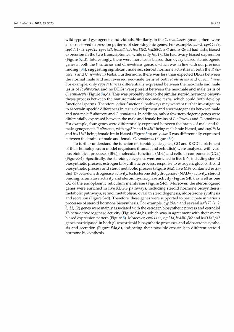
differential expression of several steroidogenic enzymes. Through comparative transcrip-tome analysis of brains and gonads, most steroidogenic genes represented conserved ex-pression patterns between P. olivaceus and C. semilaevis (Table S4). In gonad tissues, the most differentially expressed genes (DEGs) were obtained between the ovaries and testis (male/neo-male), while only a few DEGs were found between the normal male and sex reversed neo-male testes (Figures 5 and S3). For example, in the two gynogenetic P. oliva-ceus gonad transcriptomes, cyp11a, cyp11c, cyp17a1 and hsd3b1 had testis biased expression (Figure 5a, b), while cyp19a1a, cyp26a1, hsd17b1 and hsd17b12a had ovary biased expression (Figure 5a, b). In addition, the expression of steroidogenic genes between the gonads of the wide type and gynogenetic P. olivaceus was also similar (Figure 5a), with only cyp26a1 being highly expressed in the gynogenetic female ovaries but not in the wild type P. oli-vaceus gonads (Figure 5a), indicating consistent steroid biosynthesis functions between wild type and gynogenetic individuals. Similarly, in the C. semilaevis gonads, there were
Figure 4. Molecular evolution of the putatively duplication originated steroidogenic genes in teleostlineages with branch model tests. Black and grey columns indicateω values of the foreground branchand background branch, respectively. * indicates p < 0.05, ** indicate p < 0.01, *** indicate p < 0.001.
2.4. Expression Profiles of Steroidogenic Genes in P. olivaceus and C. semilaevis
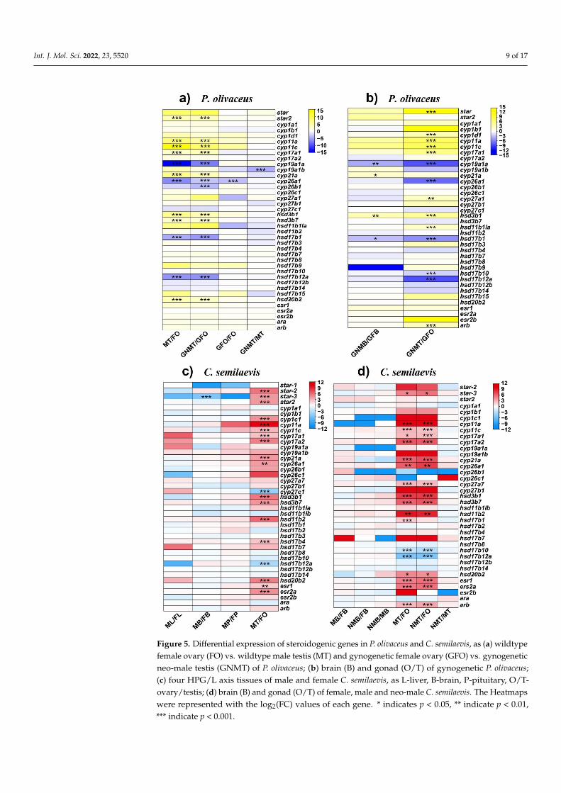
The steroidogenesis during sexual development in teleosts is essentially regulatedby differential expression of several steroidogenic enzymes. Through comparative tran-scriptome analysis of brains and gonads, most steroidogenic genes represented conservedexpression patterns between P. olivaceus and C. semilaevis (Table S4). In gonad tissues,the most differentially expressed genes (DEGs) were obtained between the ovaries andtestis (male/neo-male), while only a few DEGs were found between the normal male andsex reversed neo-male testes (Figures 5 and S3). For example, in the two gynogeneticP. olivaceus gonad transcriptomes, cyp11a, cyp11c, cyp17a1 and hsd3b1 had testis biasedexpression (Figure 5a,b), while cyp19a1a, cyp26a1, hsd17b1 and hsd17b12a had ovary biasedexpression (Figure 5a,b). In addition, the expression of steroidogenic genes between thegonads of the wide type and gynogenetic P. olivaceus was also similar (Figure 5a), with onlycyp26a1 being highly expressed in the gynogenetic female ovaries but not in the wild typeP. olivaceus gonads (Figure 5a), indicating consistent steroid biosynthesis functions between
Int. J. Mol. Sci. 2022, 23, 5520 8 of 17
wild type and gynogenetic individuals. Similarly, in the C. semilaevis gonads, there werealso conserved expression patterns of steroidogenic genes. For example, star-3, cyp11a/c,cyp17a1/a2, cyp21a, cyp26a1, hsd3b1/b7, hsd11b2, hsd20b2, esr1 and esr2a all had testis biasedexpression in the two transcriptomes, while only hsd17b12a had ovary biased expression(Figure 5c,d). Interestingly, there were more testis biased than ovary biased steroidogenicgenes in both the P. olivaceus and C. semilaevis gonads, which was in line with our previousfinding [34], suggesting significant male sex steroid hormone activities in both the P. oli-vaceus and C. semilaevis testis. Furthermore, there was less than expected DEGs betweenthe normal male and sex reversed neo-male testis of both P. olivaceus and C. semilaevis.For example, only cyp19a1b was differentially expressed between the neo-male and maletestis of P. olivaceus, and no DEGs were present between the neo-male and male testis ofC. semilaevis (Figure 5a,d). This was probably due to the similar steroid hormone biosyn-thesis process between the mature male and neo-male testis, which could both developfunctional sperms. Therefore, other functional pathways may warrant further investigationto ascertain specific differences in testis development and spermatogenesis between maleand neo-male P. olivaceus and C. semilaevis. In addition, only a few steroidogenic genes weredifferentially expressed between the male and female brains of P. olivaceus and C. semilaevis.For example, four genes were differentially expressed between the brains of male and fe-male gynogenetic P. olivaceus, with cyp21a and hsd3b1 being male brain biased, and cyp19a1aand hsd17b1 being female brain biased (Figure 5b); only star-3 was differentially expressedbetween the brains of male and female C. semilaevis (Figure 5c).
To further understand the function of steroidogenic genes, GO and KEGG enrichmentof their homologous in model organisms (human and zebrafish) were analyzed with vari-ous biological processes (BPs), molecular functions (MFs) and cellular components (CCs)(Figure S4). Specifically, the steroidogenic genes were enriched in five BPs, including steroidbiosynthetic process, estrogen biosynthetic process, response to estrogen, glucocorticoidbiosynthetic process and sterol metabolic process (Figure S4a); five MFs contained estra-diol 17-beta-dehydrogenase activity, testosterone dehydrogenase (NAD+) activity, steroidbinding, aromatase activity and steroid hydroxylase activity (Figure S4b), as well as oneCC of the endoplasmic reticulum membrane (Figure S4c). Moreover, the steroidogenicgenes were enriched in five KEGG pathways, including steroid hormone biosynthesis,metabolic pathways, retinol metabolism, ovarian steroidogenesis, aldosterone synthesisand secretion (Figure S4d). Therefore, these genes were supported to participate in variousprocesses of steroid hormone biosynthesis. For example, cyp19a1a and several hsd17b (1, 2,8, 11, 12) genes were mainly associated with the estrogen biosynthetic process and estradiol17-beta-dehydrogenase activity (Figure S4a,b), which was in agreement with their ovarybiased expression pattern (Figure 5). Moreover, cyp11a/c, cyp21a, hsd3b1/b2 and hsd11b1/b2genes participated in both glucocorticoid biosynthetic processes and aldosterone synthe-sis and secretion (Figure S4a,d), indicating their possible crosstalk in different steroidhormone biosynthesis.
Int. J. Mol. Sci. 2022, 23, 5520 9 of 17Int. J. Mol. Sci. 2022, 23, x FOR PEER REVIEW 9 of 17
Figure 5. Differential expression of steroidogenic genes in P. olivaceus and C. semilaevis, as (a) wildtype female ovary (FO) vs. wildtype male testis (MT) and gynogenetic female ovary (GFO) vs. gynogenetic neo-male testis (GNMT) of P. olivaceus; (b) brain (B) and gonad (O/T) of gynogenetic P. olivaceus; (c) four HPG/L axis tissues of male and female C. semilaevis, as L-liver, B-brain, P-pituitary, O/T-ovary/testis; (d) brain (B) and gonad (O/T) of female, male and neo-male C. semilaevis. The Heatmaps were represented with the log2(FC) values of each gene. * indicates p < 0.05, ** indicate p < 0.01, *** indicate p < 0.001.
Figure 5. Differential expression of steroidogenic genes in P. olivaceus and C. semilaevis, as (a) wildtypefemale ovary (FO) vs. wildtype male testis (MT) and gynogenetic female ovary (GFO) vs. gynogeneticneo-male testis (GNMT) of P. olivaceus; (b) brain (B) and gonad (O/T) of gynogenetic P. olivaceus;(c) four HPG/L axis tissues of male and female C. semilaevis, as L-liver, B-brain, P-pituitary, O/T-ovary/testis; (d) brain (B) and gonad (O/T) of female, male and neo-male C. semilaevis. The Heatmapswere represented with the log2(FC) values of each gene. * indicates p < 0.05, ** indicate p < 0.01,*** indicate p < 0.001.

Int. J. Mol. Sci. 2022, 23, 5520 10 of 17
2.5. Coordinated Interaction of Steroidogenic Pathway in P. olivaceus and C. semilaevis
To investigate the possible cascade regulation of steroidogenic genes, the STRINGdatabase was employed to show that the steroid biosynthesis proteins could interact directlywith each other in model organisms (Figure S5). For example, cyp1a, cyp11b/c, cyp17a1,cyp19a1, hsd3b1 and several hsd17b genes were highly interactive with other steroidogenicgenes in zebrafish and humans (Figure S5). In addition, with PCC > 0.8 as highly correlation,interaction networks revealed a high level of coordinated regulation among steroidogenicgenes of P. olivaceus and C. semilaevis, with more positive correlation than negative cor-relation (Table S5). For instance, in P. olivaceus, cyp11c, cyp17a1, cyp19a1a, hsd3b1, hsd20band hsd17b genes appeared to interact greatly with other steroidogenic genes (Figure 6a,b),while cyp11a/c, cyp17a1/a2, cyp21a, hsd3b1, hsd11b2 and hsd20b2 were the most interactivegenes in C. semilaevis (Figure 6c,d). Most of these genes represented testis biased expression(Figure 5), indicating their possible essential coordinated roles in testicular developmentand spermatogenesis. Moreover, in P. olivaceus and C. semilaevis, the sex hormone receptorgenes, esr and ar, also represented high connectivity with other genes (Figure 6), which wasin accordance with their transcription factor function. For example, in the steroidogenicpathway, both esr and ar may have direct interactions with cyp19a1 genes, and therefore,participate in the regulation of the transcriptional activity of cyp19a1 [31].
Int. J. Mol. Sci. 2022, 23, x FOR PEER REVIEW 10 of 17
2.5. Coordinated Interaction of Steroidogenic Pathway in P. olivaceus and C. semilaevis To investigate the possible cascade regulation of steroidogenic genes, the STRING
database was employed to show that the steroid biosynthesis proteins could interact di-rectly with each other in model organisms (Figure S5). For example, cyp1a, cyp11b/c, cyp17a1, cyp19a1, hsd3b1 and several hsd17b genes were highly interactive with other steroidogenic genes in zebrafish and humans (Figure S5). In addition, with PCC > 0.8 as highly correlation, interaction networks revealed a high level of coordinated regulation among steroidogenic genes of P. olivaceus and C. semilaevis, with more positive correlation than negative correlation (Table S5). For instance, in P. olivaceus, cyp11c, cyp17a1, cyp19a1a, hsd3b1, hsd20b and hsd17b genes appeared to interact greatly with other steroidogenic genes (Figure 6a,b), while cyp11a/c, cyp17a1/a2, cyp21a, hsd3b1, hsd11b2 and hsd20b2 were the most interactive genes in C. semilaevis (Figure 6c, d). Most of these genes represented testis biased expression (Figure 5), indicating their possible essential coordinated roles in testicular development and spermatogenesis. Moreover, in P. olivaceus and C. semilaevis, the sex hormone receptor genes, esr and ar, also represented high connectivity with other genes (Figure 6), which was in accordance with their transcription factor function. For example, in the steroidogenic pathway, both esr and ar may have direct interactions with cyp19a1 genes, and therefore, participate in the regulation of the transcriptional activity of cyp19a1 [31].
Figure 6. Network analysis of steroidogenic enzymes in P. olivaceus and C. semilaevis with Pearson’s correlation coefficient (PCC) of FPKM values. (a, b) represent networks in P. olivaceus corresponding to Figure 5a,b; (c, d) represent networks in C. semilaevis corresponding to Figure 5c,d. Red lines in-dicate positive correlation of PCC > 0.8 and green lines indicate negative correlation of PCC < −0.8.
In the sophisticated reaction cascade of steroid hormone biosynthesis in the gonads, star assists the transport of precursor cholesterol from the outer to the inner mitochondrial membrane (Figure 7) [4]. In P. olivaceus and C. semilaevis, star (star-2/3) and star2 were highly expressed in the testis of both species (Figures 5 and 7), indicating their functional conservation in the production of male-specific steroid hormones. In Nile tilapia (Oreo-chromis niloticus), androgen generation during spermatogenesis may require the involve-ment of star, which was proven to be primarily expressed in the testis [32]. Interestingly, even with star-1 not expressed in the gonads of C. semilaevis, star-2 and star-3 were highly expressed in the testis of C. semilaevis (Figure 5c, d), suggesting their diverged function in C. semilaevis testis development and spermatogenesis. In mitochondria, cyp11a is respon-sible for subsequently catalyzing the side-chain hydroxylation and cleavage of cholesterol
Figure 6. Network analysis of steroidogenic enzymes in P. olivaceus and C. semilaevis with Pearson’scorrelation coefficient (PCC) of FPKM values. (a,b) represent networks in P. olivaceus corresponding toFigure 5a,b; (c,d) represent networks in C. semilaevis corresponding to Figure 5c,d. Red lines indicatepositive correlation of PCC > 0.8 and green lines indicate negative correlation of PCC < −0.8.
In the sophisticated reaction cascade of steroid hormone biosynthesis in the gonads,star assists the transport of precursor cholesterol from the outer to the inner mitochondrialmembrane (Figure 7) [4]. In P. olivaceus and C. semilaevis, star (star-2/3) and star2 were highlyexpressed in the testis of both species (Figures 5 and 7), indicating their functional conser-vation in the production of male-specific steroid hormones. In Nile tilapia (Oreochromisniloticus), androgen generation during spermatogenesis may require the involvement ofstar, which was proven to be primarily expressed in the testis [32]. Interestingly, even withstar-1 not expressed in the gonads of C. semilaevis, star-2 and star-3 were highly expressedin the testis of C. semilaevis (Figure 5c,d), suggesting their diverged function in C. semi-laevis testis development and spermatogenesis. In mitochondria, cyp11a is responsiblefor subsequently catalyzing the side-chain hydroxylation and cleavage of cholesterol topregnenolone (Figure 7) [35], which together with star for cholesterol mobilization, controlsthe rate of steroid hormone biosynthesis [22]. Cyp11a was greatly expressed in both themale and neo-male testes of P. olivaceus and C. semilaevis (Figures 5 and 7), and it interacted

Int. J. Mol. Sci. 2022, 23, 5520 11 of 17
with star in C. semilaevis network (Figure 6c), indicating that the steroidogenesis is officiallyinitiated under the action of star and cyp11a, which may be more closely related to testiculardevelopment in teleost species [36].
Int. J. Mol. Sci. 2022, 23, x FOR PEER REVIEW 11 of 17
to pregnenolone (Figure 7) [35], which together with star for cholesterol mobilization, con-trols the rate of steroid hormone biosynthesis [22]. Cyp11a was greatly expressed in both the male and neo-male testes of P. olivaceus and C. semilaevis (Figures 5 and 7), and it in-teracted with star in C. semilaevis network (Figure 6c), indicating that the steroidogenesis is officially initiated under the action of star and cyp11a, which may be more closely related to testicular development in teleost species [36].
Figure 7. Putative steroid hormone biosynthesis pathway in P. olivaceus and C. semilaevis. Genes with green background represent higher expression in testis, genes with red background represent higher expression in ovaries, and controversial expression genes were represented with yellow background.
Cyp17a and hsd3b are important enzymes as the second step of steroidogenesis in-volving the conversion of pregnenolone into 17α-OH-pregnenolone and progesterone, and later to androstenedione [12,14,37] (Figure 7). Cyp17a1 and cyp17a2 were reported to be highly expressed in the P. olivaceus testis and ovary [30], and cyp17a1 and hsd3b1/b7 were all strongly expressed in the testis of both P. olivaceus and C. semilaevis (Figure 5). Here, in the networks, cyp17a1/a2 and hsd3b1 were closely associated with each other (Fig-ure 6), which corresponded to their catalytic function of transforming pregnenolone into androstenedione (Figure 7). Moreover, cyp17a1/a2 and hsd3b1/b7 also strongly interacted with testis biased cyp11a/c, cyp21a, hsd20b, as well as negatively correlated with ovary bi-ased cyp19a1a and hsd17b genes (Figure 6), indicating their essential regulatory interaction in the steroidogenesis cascade. For example, hsd20b has been demonstrated to be associ-ated with 17α, and 20β-DP synthesis [38] as well as testicular development [39]. Here hsd20b was dominant in testicular expression in both P. olivaceus and C. semilaevis (Figure 5) and has a similar interaction pattern as hsd3b1 with other steroidogenic genes (Figure 6), corresponding to the sequential interaction of hsd3b1 and hsd20b along with the biosyn-thesis of the C21 steroid (Figure 7).
Figure 7. Putative steroid hormone biosynthesis pathway in P. olivaceus and C. semilaevis. Genes withgreen background represent higher expression in testis, genes with red background represent higherexpression in ovaries, and controversial expression genes were represented with yellow background.
Cyp17a and hsd3b are important enzymes as the second step of steroidogenesisinvolving the conversion of pregnenolone into 17α-OH-pregnenolone and progesterone,and later to androstenedione [12,14,37] (Figure 7). Cyp17a1 and cyp17a2 were reported tobe highly expressed in the P. olivaceus testis and ovary [30], and cyp17a1 and hsd3b1/b7were all strongly expressed in the testis of both P. olivaceus and C. semilaevis (Figure 5).Here, in the networks, cyp17a1/a2 and hsd3b1 were closely associated with each other(Figure 6), which corresponded to their catalytic function of transforming pregnenolone intoandrostenedione (Figure 7). Moreover, cyp17a1/a2 and hsd3b1/b7 also strongly interactedwith testis biased cyp11a/c, cyp21a, hsd20b, as well as negatively correlated with ovarybiased cyp19a1a and hsd17b genes (Figure 6), indicating their essential regulatory interactionin the steroidogenesis cascade. For example, hsd20b has been demonstrated to be associatedwith 17α, and 20β-DP synthesis [38] as well as testicular development [39]. Here hsd20bwas dominant in testicular expression in both P. olivaceus and C. semilaevis (Figure 5)and has a similar interaction pattern as hsd3b1 with other steroidogenic genes (Figure 6),corresponding to the sequential interaction of hsd3b1 and hsd20b along with the biosynthesisof the C21 steroid (Figure 7).
Subsequently, cyp11c and hsd11b participate in the production of 11-ketotestosterone(11-KT) precursors as well as the biosynthesis of cortisol in teleosts (Figure 7); therefore,they could function essentially in the crosstalk between the HPG and HPI axes [40,41].In addition, steroid 21-hydroxylase (cyp21a) has a crucial role in the formation of 11-deoxycorticosterone and 11-deoxycortisol [6]. Together with cyp11c, hsd11b was proven tosynthesize 11-KT from testosterone (T) and interact with cyp21a to promote the formation ofcortisol and corticosterone (Figure 7) [42], which was consistent with the reaction network
Int. J. Mol. Sci. 2022, 23, 5520 12 of 17
results in P. olivaceus and C. semilaevis that cyp11c was strongly associated with hsd11b andcyp21a, respectively (Figure 6). All these three genes were strongly expressed in the maleand neo-male testis of P. olivaceus and C. semilaevis (Figure 5), indicating their essential rolesin androgen related hormones biosynthesis. Interestingly, hsd11b1la was highly expressed inthe P. olivaceus testis but not in the C. semilaevis testis, while hsd11b2 was greatly expressed inthe C. semilaevis testis but not in the P. olivaceus testis (Figure 5), suggesting their putativelylineage-specific function in different teleost species.
On the other side, aromatase (cyp19a1a) catalyzes the production of aromatic C18estrogens from C19 androgens, for instance, from androstenedione into estrone (E1) andfrom T into estradiol (E2) (Figure 7) [28,43]. The expression of cyp19a1a was higher in theovary than in the testis, while cyp19a1b was notably highly expressed in the brain thanin the gonads as well as highly expressed in neo-male brains of P. olivaceus and C. semi-laevis (Table S4). A similar cyp19a1 expression pattern was also reported in other teleostspecies, such as in Clupeocephala and zebrafish [44,45], which suggested that cyp19a1b isrelevant to brain development and repair in teleost lineages [46]. Together with cyp19a1a,hsd17b1 is responsible for the generation of E2 [47], and the network results were consis-tent with this collaboration, showing a strong correlation between cyp19a1a and hsd17bgenes (Figure 6a,c,d). Hsd17b1 was proven to be highly expressed in the ovary of P. oli-vaceus [24], but over-expressed in the testis of C. semilaevis (Figure 5). Hsd17b3 is involvedin T production [20], with the expression of hsd17b3 higher in the testis of O. niloticus and P.olivaceus [24,48]. In this study, hsd17b3 was not differentially expressed in either P. olivaceusor C. semilaevis gonads (Figure 5), which requires further experimental verification. More-over, hsd17b12a and hsd17b12b can accelerate the conversion of E1 into E2 and promoteestrogen synthesis [49]. Only hsd17b12a but not hsd17b12b was highly expressed in theovary of P. olivaceus and C. semilaevis (Figure 5), suggesting their functional divergence.This was in line with the previous report that hsd17b12 was distinctly expressed in the P.olivaceus ovary [24]. All in all, these interactive networks indicate the cascading effect of keysteroidogenic enzymes and warrant further investigation with a global biological impact inteleost lineages.
3. Materials and Methods3.1. Characterization of Steroidogenic Genes in P. olivaceus and C. semilaevis
The coding sequences (cds) and amino acid (aa) sequences of steroidogenic genes fromselected vertebrates, including tetrapods human (Homo sapiens), mouse (Mus musculus),chicken (Gallus gallus), frog (Xenopus tropicalis) and fishes spotted gar (Lepisosteus oculatus),zebrafish (Danio rerio), stickleback (Gasterosteus aculeatus), medaka (Oryzias latipes), fugu(Takifugu rubripes), were retrieved from NCBI (http://www.ncbi.nlm.nih.gov (accessed on15 May 2021)) and Ensembl (http://asia.ensembl.org (accessed on 15 May 2021)) database.These sequences were used as queries to search for their homologous genes in the genomesof P. olivaceus and C. semilaevis by BLASTN (e-value = 1 × 10⁻⁵), respectively.
Multiple Em for Motif Elicitation (MEME, https://meme-suite.org/meme (accessedon 7 July 2021)) program was applied to evaluate the conserved motifs with the multiplesequence alignment of the aa sequences, with the parameters of any number of repetitions,optimum width of motifs from 6 to 200 with 6 motifs for cyp genes, 12 motifs for hsd genes,12 motifs for star, ar and esr, respectively. The Gene Structure Display Sever (GSDS 2.0,http://gsds.gao-lab.org (accessed on 7 July 2021)) was used to visualize the exon-intronstructure of steroidogenic genes. The gene structure and conserved motifs were visualizedusing TBtools V1.09 [50].
3.2. Phylogenetic Analysis of Steroidogenic Genes
The multiple sequence alignment was performed using MUSCLE [51] with the aasequences of cyp, hsd, star, ar and esr genes, respectively. The Maximum Likelihood(ML) phylogenetic trees were generated using the IQ-Tree (http://iqtree.cibiv.univie.ac.at/(accessed on 4 June 2021 )) with a bootstrap of 1000 replicates and were visualized by

Int. J. Mol. Sci. 2022, 23, 5520 13 of 17
the iTOL (https://itol.embl.de/ (accessed on 6 June 2021)). The best models selected forphylogeny construction were the JTT + F + I + G4 model for cyp genes, the VT + F + G4model for hsd genes, the JTT + G4 model for star, the JTT + F + I + G4 model for ar, and theJTT + F + I + G4 model for esr.
3.3. Synteny Analysis of Steroidogenic Genes
Seven pairs of steroidogenic genes, which were putatively originated from the teleostspecific genome duplication (TSGD) event, were selected for synteny analysis, includingstar/star2, cyp11a/cyp11b(c), cyp17a1/cyp17a2, cyp19a1a/cyp19a1b, hsd17b12a/hsd17b12b,esr2a/esr2b and ara/arb. With the Ensembl and Genomicus (https://www.genomicus.bio.ens.psl.eu/genomicus-105.01/cgi-bin/search.pl (accessed on 25 July 2021)) database,the genomic locations of upstream and downstream genes from 10 selected vertebrates,including human (H. sapiens), mouse (M. musculus), chicken (G. gallus), frog (X. tropicalis),spotted gar (L. oculatus), Chinese tongue sole (C. semilaevis), zebrafish (D. rerio), medaka(O. latipes), stickleback (G. aculeatus) and fugu (T. rubripes), were collected and sorted.The genomic structure of P. olivaceus genes was obtained from the NCBI database.
3.4. Molecular Evolution Analysis
Molecular evolution analysis was performed with PAML [52] in EasyCodeML V1.21 [53]to compare the evolution rates of steroidogenic genes in teleost lineages, including 10 teleostspecies as Japanese flounder (P. olivaceus), Chinese tongue sole (C. semilaevis), zebrafish(D. rerio), stickleback (G. aculeatus), medaka (O. latipes), fugu (T. rubripes), Asian sea bass(Lates calcarifer), platyfish (Xiphophorus maculatus), tilapia (Oreochromis niloticus) and turbot(Scophthalmus maximus). Firstly, the ratio of non-synonymous to synonymous substitutions(dN/dS, ω) and the likelihood ratio tests (LRTs) was employed to evaluate the selectivepressure in each gene. Six site model (SM) tests were used to evaluate the positive selectionin each codon: M0 assumes to have the same ω for all codons, M3 assumes the ω of allcodons showing a simple discrete division trend; M1a assumes only conservative sites(0 <ω < 1) and neutral sites (ω = 1) for all codons, while M2a is considered to increase theexistence of positive sites (ω > 1) for all codons on the basis of M1a; the ω of all codonsin M7 are assumed to belong to the matrix (0, 1) with a beta distribution, while M8 addsanother type ofω (ω > 1) on the basis of M7. LRTs were used to judge whether the pairedmodels (M0 vs. M3; M1a vs. M2a; M7 vs. M8) are significantly different [52], and toestimate whether there are positively selected sites (PSSs) (M2a vs. M1a and M8 vs. M7)under the premise of a significant p-value (p < 0.05). For branch model (BM) tests, firstly,the P. olivaceus branch was labeled as the foreground branch (ω1) and the other teleostsas the background branch (ω0) to compare the evolution rates of steroidogenic genes inP. olivaceus against other teleosts; secondly, for the putative duplication originated genes,each gene cluster was labeled as the foreground branch (ω1) to compare it with the otherbranch (ω0) in teleost lineages.
3.5. Expression of Steroidogenic Genes through Transcriptomic Analysis
To investigate the expression profiles of steroidogenic genes, four RNA-seq datasetsfrom P. olivaceus and C. semilaevis were obtained from NCBI. For P. olivaceus, one datasetwas from the gonad tissues of 1.5-year-old wildtype female ovary (FO), wildtype maletestis (MT), gynogenetic female ovary (GFO), gynogenetic neo-male testis (GNMT) (PR-JNA639001) [54], and the other dataset was from the brain and gonad tissues of 1.5-year-oldgynogenetic P. olivaceus (PRJNA764760) [34]. Two transcriptomes for C. semilaevis were alsoemployed, such as liver, brain, pituitary and gonad tissues of 2-year-old male and femaleindividuals (PRJNA413516) [55], as well as the brain and gonad tissues of 1-year-old male,female and pseudo-male individuals (PRJNA480118) [27].
For transcriptome analysis, the fastq files were first optimized by trimmomatic [56], toremove adapters, low-quality reads, and the reads with less base numbers. Next, the readmapping and quantification were performed with the Hisat and StringTie pipeline [57], and

Int. J. Mol. Sci. 2022, 23, 5520 14 of 17
the Fragments Per Kilobase of exon model per Million mapped fragments (FPKM) valueswere extracted for each steroidogenic gene. Differentially expressed genes (DEGs) betweengroups were determined by edgeR [58]. Any pair-wise genes with log2|fold change|(log2FC) > 2, adjusted p-value < 0.05 and at least one gene with FPKM >1 was identified asthe DEG. TBtools were utilized to draw heat maps with both log2(FPKM + 1) and log2FCvalues. GO and KEGG annotation of steroidogenic genes were obtained from DAVID(https://david.ncifcrf.gov/ (accessed on 8 September 2021)) for humans and zebrafish.Redundancy (semantically similar terms) and pathways involving a low number of genes(<3) were removed.
3.6. Protein-Protein Interaction and Co-Expression Analysis
Protein-protein interaction (PPI) networks of steroidogenic genes in humans and ze-brafish were analyzed using the online site STRING 11.5 (https://string-db.org/ (accessedon 14 August 2021)) with the parameter, minimum required interaction score, set to 0.7.For P. olivaceus and C. semilaevis, OmicShare (https://www.omicshare.com/ (accessed on17 August 2021)) was employed for the Pearson’s correlation coefficient (PCC) analysis ofFPKM values between steroidogenic genes with a PCC threshold of 0.8 from the transcrip-tome analysis. Cytoscape [59] was employed to illustrate the networks according to thePCC values.
4. Conclusions
In summary, steroidogenic genes were comprehensively characterized in P. olivaceusand C. semilaevis, with cyp19a1a/cyp19a1b, hsd17b12a/hsd17b12b, ara/arb and esr2a/esr2boriginating from the TSGD, and star/star2 and cyp17a1/cyp17a2 putatively evolving inde-pendently. The expression profile and network analysis indicated the key factors function-ing essentially during the steroidogenesis of P. olivaceus and C. semilaevis. Specifically, star,cyp11a/c, cyp17a1/a2, cyp21a, hsd3b1/b7, hsd11b and hsd20b were primarily expressed inthe testis of P. olivaceus and C. semilaevis, whereas cyp19a1a and hsd17b genes were highlyexpressed in the ovaries, which constitute the coordinated regulatory network during thesteroidogenic process. Interestingly, only a few steroidogenic genes were differentiallyexpressed between male and neo-male testis, which warrants more functional verification.Investigation of other functional pathways, such as the piRNA pathway [60], may help toexplain the specific difference in testis development and spermatogenesis between maleand neo-male P. olivaceus and C. semilaevis in the future. All these findings will providesignificant insights into the steroidogenic enzyme cascade of the teleost lineages and maycontribute to reproductive manipulation in aquaculture.
Supplementary Materials: The following supporting information can be downloaded at: https://www.mdpi.com/article/10.3390/ijms23105520/s1.
Author Contributions: J.C. designed the study, F.Y., Y.W., W.L., Q.Z. and W.Z. analyzed the data.J.C. and F.Y. conducted the manuscript writing. All authors have read and agreed to the publishedversion of the manuscript.
Funding: This research was funded by the National Natural Science Foundation of China (31702331),the China Postdoctoral Science Foundation (2017M622272), and the National Marine Genetic ResourceCenter, China.
Institutional Review Board Statement: This study was conducted in accordance with the Institu-tional Animal Care and Use Committee of Ocean University of China (OUC-IACUC), and it does notcontain any studies with human participants.
Informed Consent Statement: Not applicable.
Data Availability Statement: The transcriptome datasets used in this study can be found in theNCBI Sequence Read Archive (SRA) BioProject as PRJNA639001 and PRJNA764760 for P. olivaceus,and PRJNA413516 and PRJNA480118 for C. semilaevis.
Conflicts of Interest: The authors declare no conflict of interest.
Int. J. Mol. Sci. 2022, 23, 5520 15 of 17
References1. Bhat, I.A.; Dar, J.Y.; Ahmad, I.; Mir, I.N.; Bhat, H.; Bhat, R.A.H.; Ganie, P.A.; Sharma, R. Testicular development and spermatogen-
esis in fish: Insights into molecular aspects and regulation of gene expression by different exogenous factors. Rev. Aquac. 2021, 13,2142–2168. [CrossRef]
2. Bliss, S.P.; Navratil, A.M.; Xie, J.; Roberson, M.S. GnRH signaling, the gonadotrope and endocrine control of fertility. Front.Neuroendocrinol. 2010, 31, 322–340. [CrossRef] [PubMed]
3. Kim, N.N.; Shin, H.S.; Choi, Y.J.; Choi, C.Y. Kisspeptin regulates the hypothalamus-pituitary-gonad axis gene expression duringsexual maturation in the cinnamon clownfish, Amphiprion melanopus. Comp. Biochem. Physiol. B Biochem. Mol. Biol. 2014, 168,19–32. [CrossRef] [PubMed]
4. Stocco, D.M. The role of the StAR protein in steroidogenesis: Challenges for the future. J. Endocrinol. 2000, 164, 247–253. [CrossRef]5. Tenugu, S.; Pranoty, A.; Mamta, S.K.; Senthilkumaran, B. Development and organization of gonadal steroidogenesis in bony
fishes—A review. Aquac. Fish. 2021, 6, 223–246. [CrossRef]6. Tokarz, J.; Möller, G.; de Angelis, M.H.; Adamski, J. Steroids in teleost fishes: A functional point of view. Steroids 2015, 103,
123–144. [CrossRef]7. Godard, C.A.; Leaver, M.J.; Said, M.R.; Dickerson, R.L.; George, S.; Stegeman, J.J. Identification of cytochrome P450 1B-like
sequences in two teleost fish species (scup, Stenotomus chrysops and plaice, Pleuronectes platessa) and in a cetacean (striped dolphin,Stenella coeruleo). Mar. Environ. Res. 2000, 50, 7–10. [CrossRef]
8. McArthur, A.G.; Hegelund, T.; Cox, R.L.; Stegeman, J.J.; Liljenberg, M.; Olsson, U.; Sundberg, P.; Celander, M.C. Phylogeneticanalysis of the cytochrome P450 3 (CYP3) gene family. J. Mol. Evol. 2003, 57, 200–211. [CrossRef]
9. Liang, D.; Fan, Z.; Zou, Y.; Tan, X.; Wu, Z.; Jiao, S.; Li, J.; Zhang, P.; You, F. Characteristics of Cyp11a during Gonad Differentiationof the Olive Flounder Paralichthys olivaceus. Int. J. Mol. Sci. 2018, 19, 2641. [CrossRef]
10. Meng, L.; Yu, H.; Ni, F.; Niu, J.; Liu, X.; Wang, X. Roles of two cyp11 genes in sex hormone biosynthesis in Japanese flounder(Paralichthys olivaceus). Mol. Reprod. Dev. 2020, 87, 53–65. [CrossRef]
11. Sun, M.; Jiang, S.; Song, W.; Qu, J.; Qi, J. Molecular characterization and functional analysis of cyp11a and cyp11b in black rockfish(Sebastes schlegelii). J. Fish Biol. 2021, 99, 9–17. [CrossRef] [PubMed]
12. Zhou, L.Y.; Wang, D.S.; Shibata, Y.; Paul-Prasanth, B.; Suzuki, A.; Nagahama, Y. Characterization, expression and transcriptional reg-ulation of P450c17-I and -II in the medaka, Oryzias latipes. Biochem. Biophys. Res. Commun. 2007, 362, 619–625. [CrossRef] [PubMed]
13. Miller, W.L.; Auchus, R.J.; Geller, D.H. The regulation of 17,20 lyase activity. Steroids 1997, 62, 133–142. [CrossRef]14. Zhou, L.Y.; Wang, D.S.; Kobayashi, T.; Yano, A.; Paul-Prasanth, B.; Suzuki, A.; Sakai, F.; Nagahama, Y. A novel type of P450c17
lacking the lyase activity is responsible for C21-steroid biosynthesis in the fish ovary and head kidney. Endocrinology 2007, 148,4282–4291. [CrossRef]
15. Guiguen, Y.; Fostier, A.; Piferrer, F.; Chang, C.F. Ovarian aromatase and estrogens: A pivotal role for gonadal sex differentiationand sex change in fish. Gen. Comp. Endocrinol. 2010, 165, 352–366. [CrossRef]
16. Adamski, J.; Jakob, F.J. A guide to 17beta-hydroxysteroid dehydrogenases. Mol. Cell. Endocrinol. 2001, 171, 1–4. [CrossRef]17. Rasmussen, M.K.; Ekstrand, B.; Zamaratskaia, G. Regulation of 3β-hydroxysteroid dehydrogenase/∆5-∆4 isomerase: A review.
Int. J. Mol. Sci. 2013, 14, 17926–17942. [CrossRef]18. Rasheeda, M.K.; Kagawa, H.; Kirubagaran, R.; Dutta-Gupta, A.; Senthilkumaran, B. Cloning, expression and enzyme activity
analysis of testicular 11beta-hydroxysteroid dehydrogenase during seasonal cycle and after hCG induction in air-breathing catfishClarias gariepinus. J. Steroid Biochem. Mol. Biol. 2010, 120, 1–10. [CrossRef]
19. Tokarz, J.; Lintelmann, J.; Möller, G.; Adamski, J. Substrate multispecificity among 20β-hydroxysteroid dehydrogenase type 2members. Mol. Cell. Endocrinol. 2020, 510, 110822. [CrossRef]
20. Mindnich, R.; Haller, F.; Halbach, F.; Moeller, G.; Hrabé de Angelis, M.; Adamski, J. Androgen metabolism via 17beta-hydroxysteroid dehydrogenase type 3 in mammalian and non-mammalian vertebrates: Comparison of the human and thezebrafish enzyme. J. Mol. Endocrinol. 2005, 35, 305–316. [CrossRef]
21. Ogino, Y.; Tohyama, S.; Kohno, S.; Toyota, K.; Yamada, G.; Yatsu, R.; Kobayashi, T.; Tatarazako, N.; Sato, T.; Matsubara, H.; et al.Functional distinctions associated with the diversity of sex steroid hormone receptors ESR and AR. J. Steroid Biochem. Mol. Biol.2018, 184, 38–46. [CrossRef] [PubMed]
22. Rajakumar, A.; Senthilkumaran, B. Steroidogenesis and its regulation in teleost-a review. Fish Physiol. Biochem. 2020, 46, 803–818.[CrossRef] [PubMed]
23. Lin, G.; Gao, D.; Lu, J.; Sun, X. Transcriptome Profiling Reveals the Sexual Dimorphism of Gene Expression Patterns during GonadDifferentiation in the Half-Smooth Tongue Sole (Cynoglossus semilaevis). Mar. Biotechnol. 2021, 23, 18–30. [CrossRef] [PubMed]
24. Zou, C.; Wang, L.; Zou, Y.; Wu, Z.; Wang, W.; Liang, S.; Wang, L.; You, F. Characteristics and sex dimorphism of 17β-hydroxysteroiddehydrogenase family genes in the olive flounder Paralichthys olivaceus. J. Steroid Biochem. Mol. Biol. 2020, 199, 105597. [CrossRef][PubMed]
25. Cui, Y.; Wang, W.; Ma, L.; Jie, J.; Zhang, Y.; Wang, H.; Li, H. New locus reveals the genetic architecture of sex reversal in theChinese tongue sole (Cynoglossus semilaevis). Heredity 2018, 121, 319–326. [CrossRef]
26. Ye, Z.; Wang, W.; Zhang, Y.; Wang, L.; Cui, Y.; Li, H. Integrative analysis reveals pathways associated with sex reversal inCynoglossus semilaevis. PeerJ 2020, 8, e8801. [CrossRef]
Int. J. Mol. Sci. 2022, 23, 5520 16 of 17
27. Liu, J.; Liu, X.; Jin, C.; Du, X.; He, Y.; Zhang, Q. Transcriptome Profiling Insights the Feature of Sex Reversal Induced by HighTemperature in Tongue Sole Cynoglossus semilaevis. Front. Genet. 2019, 10, 522. [CrossRef]
28. Si, Y.; Ding, Y.; He, F.; Wen, H.; Li, J.; Zhao, J.; Huang, Z. DNA methylation level of cyp19a1a and foxl2 gene related to theirexpression patterns and reproduction traits during ovary development stages of Japanese flounder (Paralichthys olivaceus). Gene2016, 575, 321–330. [CrossRef]
29. Liang, D.; Fan, Z.; Weng, S.; Jiao, S.; Wu, Z.; Zou, Y.; Tan, X.; Li, J.; Zhang, P.; You, F. Characterization and expression of StAR2aand StAR2b in the olive flounder Paralichthys olivaceus. Gene 2017, 626, 1–8. [CrossRef]
30. Meng, L.; Yu, L.; Qu, J.; Niu, J.; Ni, F.; Han, P.; Yu, H.; Wang, X. Two cyp17 genes perform different functions in the sex hormonebiosynthesis and gonadal differentiation in Japanese flounder (Paralichthys olivaceus). Gene 2019, 702, 17–26. [CrossRef]
31. Zou, Y.; Peng, L.; Weng, S.; Liang, D.; Fan, Z.; Wu, Z.; Tan, X.; Jiao, S.; You, F. Characterization and expression of androgenreceptors in olive flounder. Gene 2019, 683, 184–194. [CrossRef] [PubMed]
32. Yu, X.; Wu, L.; Xie, L.; Yang, S.; Charkraborty, T.; Shi, H.; Wang, D.; Zhou, L. Characterization of two paralogous StAR genes in ateleost, Nile tilapia (Oreochromis niloticus). Mol. Cell. Endocrinol. 2014, 392, 152–162. [CrossRef] [PubMed]
33. Chiang, E.F.; Yan, Y.L.; Guiguen, Y.; Postlethwait, J.; Chung, B. Two Cyp19 (P450 aromatase) genes on duplicated zebrafishchromosomes are expressed in ovary or brain. Mol. Biol. Evol. 2001, 18, 542–550. [CrossRef]
34. Cheng, J.; Yang, F.; Liu, S.; Zhao, H.; Lu, W.; Zhang, Q. Transcriptomic Analysis Reveals Functional Interaction of mRNA–lncRNA–miRNA in Steroidogenesis and Spermatogenesis of Gynogenetic Japanese Flounder (Paralichthys olivaceus). Biology 2022, 11, 213.[CrossRef]
35. Strushkevich, N.; MacKenzie, F.; Cherkesova, T.; Grabovec, I.; Usanov, S.; Park, H.W. Structural basis for pregnenolonebiosynthesis by the mitochondrial monooxygenase system. Proc. Natl. Acad. Sci. USA 2011, 108, 10139–10143. [CrossRef]
36. Rajakumar, A.; Senthilkumaran, B. Expression analysis of cyp11a1 during gonadal development, recrudescence and after hCGinduction and sex steroid analog treatment in the catfish, Clarias batrachus. Comp. Biochem. Physiol. B Biochem. Mol. Biol. 2014, 176,42–47. [CrossRef]
37. Dumont, M.; Luu-The, V.; Dupont, E.; Pelletier, G.; Labrie, F. Characterization, expression, and immunohistochemical localizationof 3 beta-hydroxysteroid dehydrogenase/delta 5-delta 4 isomerase in human skin. J. Invest. Dermatol. 1992, 99, 415–421.[CrossRef] [PubMed]
38. Sreenivasulu, G.; Senthilkumaran, B.; Sridevi, P.; Rajakumar, A.; Rasheeda, M.K. Expression and immunolocalization of 20β-hydroxysteroid dehydrogenase during testicular cycle and after hCG induction, in vivo in the catfish, Clarias gariepinus. Gen.Comp. Endocrinol. 2012, 175, 48–54. [CrossRef]
39. Miura, T.; Yamauchi, K.; Takahashi, H.; Nagahama, Y. The role of hormones in the acquisition of sperm motility in salmonid fish.J. Exp. Zool. 1992, 261, 359–363. [CrossRef]
40. Jiang, J.Q.; Kobayashi, T.; Ge, W.; Kobayashi, H.; Tanaka, M.; Okamoto, M.; Nonaka, Y.; Nagahama, Y. Fish testicular 11beta-hydroxylase: cDNA cloning and mRNA expression during spermatogenesis. FEBS Lett. 1996, 397, 250–252. [CrossRef]
41. Jiang, J.Q.; Young, G.; Kobayashi, T.; Nagahama, Y. Eel (Anguilla japonica) testis 11beta-hydroxylase gene is expressed in interrenaltissue and its product lacks aldosterone synthesizing activity. Mol. Cell. Endocrinol. 1998, 146, 207–211. [CrossRef]
42. Xiao, L.; Guo, Y.; Wang, D.; Zhao, M.; Hou, X.; Li, S.; Lin, H.; Zhang, Y. Beta-Hydroxysteroid Dehydrogenase Genes in Orange-Spotted Grouper (Epinephelus coioides): Genome-Wide Identification and Expression Analysis During Sex Reversal. Front. Genet.2020, 11, 161. [CrossRef] [PubMed]
43. Mills, L.J.; Gutjahr-Gobell, R.E.; Zaroogian, G.E.; Horowitz, D.B.; Laws, S.C. Modulation of aromatase activity as a mode of actionfor endocrine disrupting chemicals in a marine fish. Aquat. Toxicol. 2014, 147, 140–150. [CrossRef]
44. Lynch, M.; Force, A. The probability of duplicate gene preservation by subfunctionalization. Genetics 2000, 154, 459–473.[CrossRef]
45. Goldstone, J.V.; McArthur, A.G.; Kubota, A.; Zanette, J.; Parente, T.; Jönsson, M.E.; Nelson, D.R.; Stegeman, J.J. Identificationand developmental expression of the full complement of Cytochrome P450 genes in Zebrafish. BMC Genom. 2010, 11, 643.[CrossRef] [PubMed]
46. Diotel, N.; Vaillant, C.; Gabbero, C.; Mironov, S.; Fostier, A.; Gueguen, M.M.; Anglade, I.; Kah, O.; Pellegrini, E. Effects of estradiolin adult neurogenesis and brain repair in zebrafish. Horm. Behav. 2013, 63, 193–207. [CrossRef] [PubMed]
47. Zhou, L.Y.; Wang, D.S.; Senthilkumaran, B.; Yoshikuni, M.; Shibata, Y.; Kobayashi, T.; Sudhakumari, C.C.; Nagahama, Y. Cloning,expression and characterization of three types of 17beta-hydroxysteroid dehydrogenases from the Nile tilapia, Oreochromisniloticus. J. Mol. Endocrinol. 2005, 35, 103–116. [CrossRef] [PubMed]
48. Zhou, L. Molecular Cloning, Gene Expression and Enzyme Activity Characterization of Three Types 17β-hydroxysteroidDehydrogenases (17β-HSD1, 17β-HSD3, 17β-HSD8) from Nile Tilapia, Oreochromis niloticu. Master’s Thesis, Southwest ChinaNormal University, Chongqing, China, 2004.
49. Mindnich, R.; Möller, G.; Adamski, J. The role of 17 beta-hydroxysteroid dehydrogenases. Mol. Cell. Endocrinol. 2004, 218, 7–20.[CrossRef]
50. Chen, C.; Chen, H.; Zhang, Y.; Thomas, H.R.; Frank, M.H.; He, Y.; Xia, R. TBtools: An Integrative Toolkit Developed for InteractiveAnalyses of Big Biological Data. Mol. Plant 2020, 13, 1194–1202. [CrossRef]
51. Edgar, R.C. MUSCLE: Multiple sequence alignment with high accuracy and high throughput. Nucleic Acids Res. 2004, 32,1792–1797. [CrossRef]

Int. J. Mol. Sci. 2022, 23, 5520 17 of 17
52. Yang, Z. PAML 4: Phylogenetic analysis by maximum likelihood. Mol. Biol. Evol. 2007, 24, 1586–1591. [CrossRef] [PubMed]53. Gao, F.; Chen, C.; Arab, D.A.; Du, Z.; He, Y.; Ho, S.Y.W. EasyCodeML: A visual tool for analysis of selection using CodeML. Ecol.
Evol. 2019, 9, 3891–3898. [CrossRef] [PubMed]54. Wang, L.; Wu, Z.; Zou, C.; Liang, S.; Zou, Y.; Liu, Y.; You, F. Sex-Dependent RNA Editing and N6-adenosine RNA Methylation
Profiling in the Gonads of a Fish, the Olive Flounder (Paralichthys olivaceus). Front. Cell Dev. Biol. 2020, 8, 751. [CrossRef] [PubMed]55. Wang, N.; Wang, R.; Wang, R.; Chen, S. Transcriptomics analysis revealing candidate networks and genes for the body size sexual
dimorphism of Chinese tongue sole (Cynoglossus semilaevis). Funct. Integr. Genom. 2018, 18, 327–339. [CrossRef] [PubMed]56. Bolger, A.M.; Lohse, M.; Usadel, B. Trimmomatic: A flexible trimmer for Illumina sequence data. Bioinformatics 2014, 30, 2114–2120.
[CrossRef]57. Pertea, M.; Kim, D.; Pertea, G.M.; Leek, J.T.; Salzberg, S.L. Transcript-level expression analysis of RNA-seq experiments with
HISAT, StringTie and Ballgown. Nat. Protoc. 2016, 11, 1650–1667. [CrossRef]58. Robinson, M.D.; McCarthy, D.J.; Smyth, G.K. EdgeR: A Bioconductor package for differential expression analysis of digital gene
expression data. Bioinformatics 2010, 26, 139–140. [CrossRef]59. Shannon, P.; Markiel, A.; Ozier, O.; Baliga, N.S.; Wang, J.T.; Ramage, D.; Amin, N.; Schwikowski, B.; Ideker, T. Cytoscape:
A software environment for integrated models of biomolecular interaction networks. Genome Res. 2003, 13, 2498–2504. [CrossRef]60. Wang, B.; Wang, H.; Song, H.; Jin, C.; Peng, M.; Gao, C.; Yang, F.; Du, X.; Qi, J.; Zhang, Q.; et al. Evolutionary significance and
regulated expression of Tdrd family genes in gynogenetic Japanese flounder (Paralichthys olivaceus). Comp. Biochem. Physiol. DGenom. Proteom. 2019, 31, 100593. [CrossRef]