Coordinated regulation of hormone-sensitive lipase and lipoprotein lipase in human adipose tissue in...
16
Pergamon Advan. Enzyme Regsd., Vol. 35, pp. 163-178, 1995 cOpyr@t @ 1995 Elsevier SfienceLtd Printed in Great Britain. All rights reserved 0065-2571/9%?9.00 0065-2571(94)ooo11-5 COORDINATED REGULATION OF HORMONE-SENSITIVE LIPASE AND LIPOPROTEIN LIPASE IN HUMAN ADIPOSE TISSUE IN VIVO: IMPLICATIONS FOR THE CONTROL OF FAT STORAGE AND FAT MOBILIZATION KEITH N. FRAYN, SIMON W. COPPACK’, BARBARA A. FIELDING and SANDY M. HUMPHREYS Oxford Lipid Metabolism Group, Nuffield Department of Clinical Medicine, Radcliffe Infirmary, Oxford OX2 6HE, U.K. INTRODUCTION The first law of thermodynamics may be applied to the human body as to any other system, and dictates that energy intake will equal energy expenditure plus energy storage. Thus, a consistent excessof energy intake over expenditure leads to energy storage, and the opposite situation leads to depletion of energy stores. In adult humans by far the greatest store of energy is in the form of triacylglycerol (TAG) in white adipocytes. Thus, the net balance between fat mobilization from, and fat deposition in, adipose tissue must be matched in some quantitative way to the integrated imbalance between energy intake and energy expenditure. An understanding of the manner in which this matching is brought about would be of great interest to those studying human body weight regulation. It poses a challenge in terms of enzyme regulation, since it implies that enzymes within one particular tissue (adipose tissue) are regulated in a way which is quantitatively linked with whole-body energy balance. In humans, at least on a typical western diet, net de nuvo lipogenesis is rarely observed (1, 2). Fat storage in white adipose tissue occurs almost entirely by the uptake of pre-formed fatty acids from circulating lipoprotein-TAG. The key enzymes responsible for fat deposition and fat mobilization in white adipose tissue are therefore, respectively, lipoprotein * Present address: Academic Division of Medicine, University College London Medical School, Whittington Hospital, London N19 3UA, U.K. 163
-
Upload
independent -
Category
Documents
-
view
2 -
download
0
Transcript of Coordinated regulation of hormone-sensitive lipase and lipoprotein lipase in human adipose tissue in...
Pergamon Advan. Enzyme Regsd., Vol. 35, pp. 163-178, 1995
cOpyr@t @ 1995 Elsevier Sfience Ltd Printed in Great Britain. All rights reserved
0065-2571/9%?9.00
0065-2571(94)ooo11-5
COORDINATED REGULATION OF HORMONE-SENSITIVE LIPASE AND LIPOPROTEIN LIPASE IN
HUMAN ADIPOSE TISSUE IN VIVO: IMPLICATIONS FOR THE CONTROL
OF FAT STORAGE AND FAT MOBILIZATION
KEITH N. FRAYN, SIMON W. COPPACK’, BARBARA A. FIELDING and SANDY M. HUMPHREYS
Oxford Lipid Metabolism Group, Nuffield Department of Clinical Medicine, Radcliffe Infirmary, Oxford OX2 6HE, U.K.
INTRODUCTION
The first law of thermodynamics may be applied to the human body as to any other system, and dictates that energy intake will equal energy expenditure plus energy storage. Thus, a consistent excess of energy intake over expenditure leads to energy storage, and the opposite situation leads to depletion of energy stores. In adult humans by far the greatest store of energy is in the form of triacylglycerol (TAG) in white adipocytes. Thus, the net balance between fat mobilization from, and fat deposition in, adipose tissue must be matched in some quantitative way to the integrated imbalance between energy intake and energy expenditure. An understanding of the manner in which this matching is brought about would be of great interest to those studying human body weight regulation. It poses a challenge in terms of enzyme regulation, since it implies that enzymes within one particular tissue (adipose tissue) are regulated in a way which is quantitatively linked with whole-body energy balance.
In humans, at least on a typical western diet, net de nuvo lipogenesis is rarely observed (1, 2). Fat storage in white adipose tissue occurs almost entirely by the uptake of pre-formed fatty acids from circulating lipoprotein-TAG. The key enzymes responsible for fat deposition and fat mobilization in white adipose tissue are therefore, respectively, lipoprotein
* Present address: Academic Division of Medicine, University College London Medical School, Whittington Hospital, London N19 3UA, U.K.
163
164 K. N. FRAYN et al.
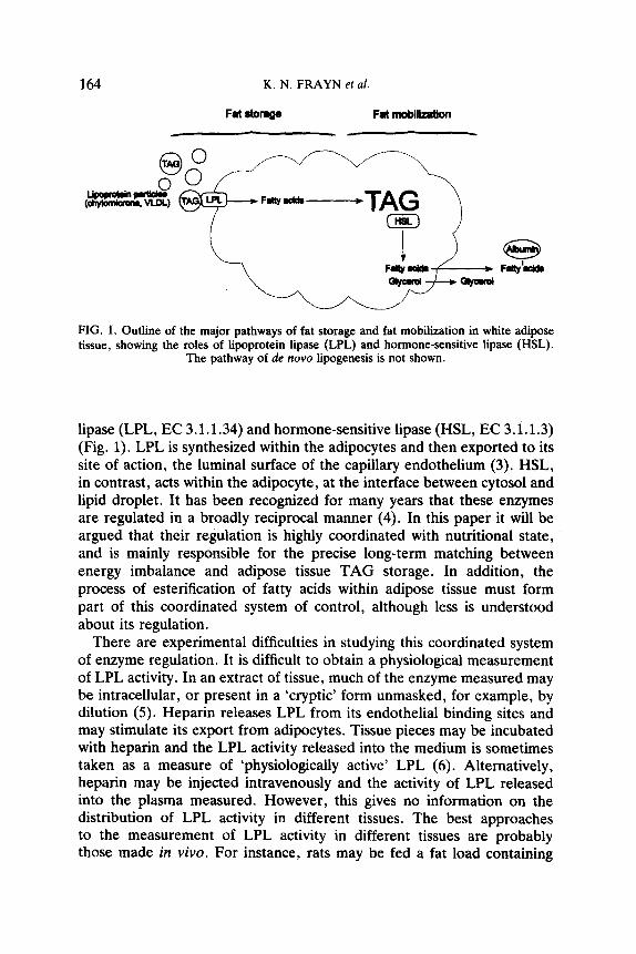
FIG. 1. Outline of the major pathways of fat storage and fat mobilization in white adipose tissue, showing the roles of lipoprotein lipase (LPL) and hormone-sensitive lipase (HSL).
The pathway of de nova lipogenesis is not shown.
lipase (LPL, EC 3.1.1.34) and hormone-sensitive lipase (HSL, EC 3.1.1.3) (Fig. 1). LPL is synthesized within the adipocytes and then exported to its site of action, the luminal surface of the capillary endothelium (3). HSL, in contrast, acts within the adipocyte, at the interface between cytosol and lipid droplet. It has been recognized for many years that these enzymes are regulated in a broadly reciprocal manner (4). In this paper it will be argued that their regulation is highly coordinated with nutritional state, and is mainly responsible for the precise long-term matching between energy imbalance and adipose tissue TAG storage. In addition, the process of esterification of fatty acids within adipose tissue must form part of this coordinated system of control, although less is understood about its regulation.
There are experimental difficulties in studying this coordinated system of enzyme regulation. It is difficult to obtain a physiological measurement of LPL activity. In an extract of tissue, much of the enzyme measured may be intracellular, or present in a ‘cryptic’ form unmasked, for example, by dilution (5). Heparin releases LPL from its endothelial binding sites and may stimulate its export from adipocytes. Tissue pieces may be incubated with heparin and the LPL activity released into the medium is sometimes taken as a measure of ‘physiologically active’ LPL (6). Alternatively, heparin may be injected intravenously and the activity of LPL released into the plasma measured. However, this gives no information on the distribution of LPL activity in different tissues. The best approaches to the measurement of LPL activity in different tissues are probably those made in vivo. For instance, rats may be fed a fat load containing
REGULATION OF ADIPOSE TISSUE LIPASES 165
radioactively labelled triolein and the uptake of activity by different tissues measured (7).
The activity of HSL is also difficult to assess. The major form of acute regulation of this enzyme is reversible phosphorylation, but no satisfactory methods exist for measurement of the proportion in active form (as can be done, for example, with pyruvate dehydrogenase). The phosphorylated and dephosphorylated forms have relatively similar activities towards the artificial lipid emulsions used as substrates for in vitro assay (8). Thus, measurement of HSL activity in a tissue extract is effectively a measure of total enzyme present.
The studies described below are based on measurement of arteriovenous differences across a subcutaneous adipose depot in humans, which represents one approach for the study of these enzyme systems under physiological conditions.
EXPERIMENTAL METHODS
Subjects and clinical methods. The studies were carried out in normal subjects. (Their characteristics are given later.) The subjects reported to the laboratory after an overnight fast. A cannula was placed in either a radial artery or an arterialized hand vein; for simplicity, blood samples obtained from this site will be referred to as ‘arterial’. A further cannula (Secalon Hydrocath, 22 gauge x 10 cm; Viggo Spectramed, Swindon, UK) was threaded down a vein on the anterior abdominal wall until its tip lay just superior to the inguinal ligament (9). Considerable biochemical evidence suggests that the blood collected from this site represents almost pure drainage from subcutaneous adipose tissue (10).
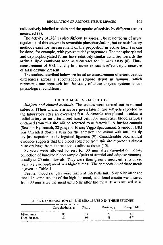
Subjects were allowed to rest for 30 min after cannulation before collection of baseline blood sample (pairs of arterial and adipose-venous), usually at 20 min intervals. They were then given a meal, either a mixed (relatively normal) meal or a high-fat meal. The composition of these meals is given in Table 1.
Further blood samples were taken at intervals until 5 or 6 hr after the meal. In some studies of the high-fat meal, additional insulin was infused from 30 min after the meal until 5 hr after the meal. It was infused at 40
TABLE 1. COMPOSITION OF THE MEALS USED IN THESE STUDIES
Mixed meal High-fat meal
Carbohydrate, g Fat. g
93 33 80 80
Protein, g
22 18
Energy, MJ
3.1 4.7

166 K. N. FRAYN er al.
mU.m-*.min-1, and the plasma glucose was ‘clamped‘ at around 6 mmol/l (11) by infusion of glucose at a variable rate. These studies will be referred to as the ‘clamped’ experiments.
Adipose tissue blood flow was determined immediately after each sample by the clearance of 133Xe (12).
Analytical methods. A portion of each blood sample was rapidly deproteinized in perchloric acid. Plasma was prepared rapidly in the cold to minimize in vitro hydrolysis of TAG. Concentrations of TAG and non-esterified fatty acids (NEFA) in plasma, and of glycerol in whole blood, were measured by enzymatic methods adapted to a centrifugal analyzer (Multistat III or Monarch, IL, Warrington, UK). Hormones were measured by double-antibody radioimmunoassay.
Calculations and assumptions. The flux of a metabolite across adipose tissue was taken as its arteriovenous difference multiplied by the blood or plasma Aow as appropriate. Fractional extraction of TAG was assessed as (arterial - adipose venous concentration)/arterial concentration.
Total re-esterification of fatty acids within the tissue was calculated from the extent to which the ratio of NEFA to glycerol release differed from 3:l.
The rates of action of LPL and HSL were calculated as follows. Total glycerol release was taken to represent the sum of the rates of action of LPL and HSL. TAG extraction was assumed to reflect LPL action. HSL action was calculated by subtracting from total glycerol release the glycerol attributable to LPL action.
The fate of LPL-derived fatty acids and the net transcapillary flux of fatty acids (see Fig. 2) were determined as described in detail elsewhere (13). Calculation of the fate of LPL-derived fatty acids requires an assumption to be made about the fate of HSL-derived fatty acids (esterification within the tissue vs escape to venous plasma). As described elsewhere (13), consistently plausible results are obtained on the assumption of equal proportional re-esterification of HSL- and LPL-derived fatty acids. The proportion of LPL-derived fatty acids retained in the tissue is then the same as the overall re-esterification of fatty acids in the tissue calculated from the ratio of fatty acid to glycerol release. For simplicity of presentation, this assumption will be used for postprandial results in the present work.
The major assumptions behind these calculations have been discussed previously (13, 14). They are: the absence of significant glycerol re- utilisation in the tissue, the lack of monoacylglycerol esterification in the tissue, and the lack of significant fatty acid oxidation in the tissue.
REGULATION OF ADIPOSE TISSUE LIPASES
(c) FATE OF LPL-DERIVED FATTY ACIDS
‘Escape’ to venous plasma
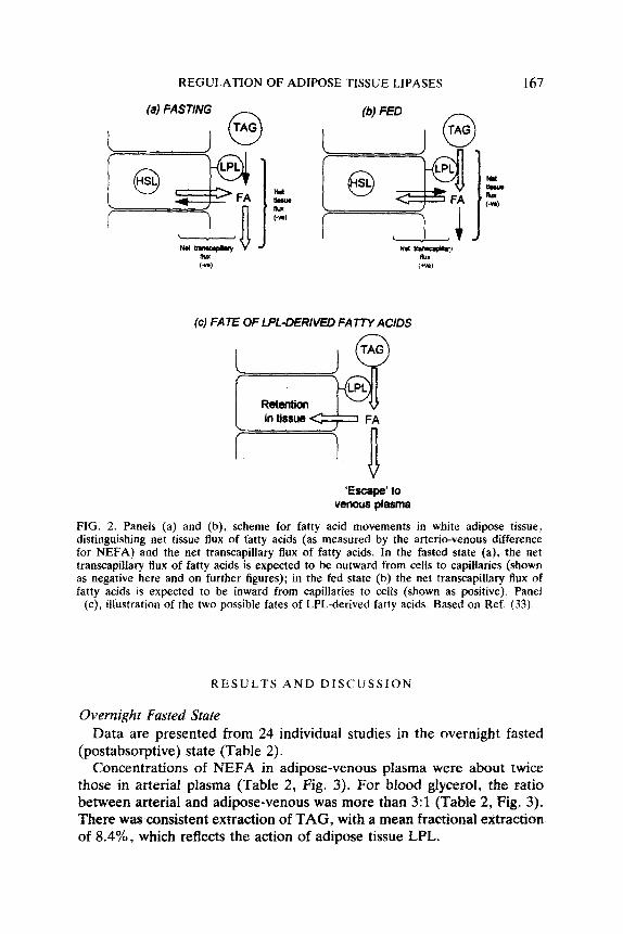
FIG. 2. Panels (a) and (b), scheme for fatty acid movements in white adipose tissue, distinguishing net tissue flux of fatty acids (as measured by the arterio-venous difference for NEFA) and the net transcapillary flux of fatty acids. In the fasted state (a), the net transcapiilary flux of fatty acids is expected to be outward from celis to capillaries (shown as negative here and on further figures); in the fed state (b) the net transcapillary flux of fatty acids is expected to be inward from capillaries to cells (shown as positive). Panel
(c), illustration of the two possible fates of LPL-derived fatty acids. Based on Ref. (33).
RESULTS AND DISCUSSION
Overnight Fasted State Data are presented from 24 individual studies in the overnight fasted
(postabsorptive) state (Table 2). Concentrations of NEFA in adipose-venous plasma were about twice
those in arterial plasma (Table 2, Fig. 3). For blood glycerol, the ratio between arterial and adipose-venous was more than 3:l (Table 2, Fig. 3). There was consistent extraction of TAG, with a mean fractional extraction of 8.4%, which reflects the action of adipose tissue LPL.
168 K. N. FRAYN et al.
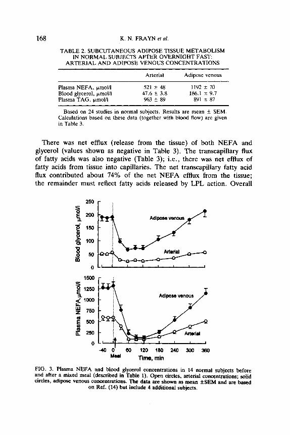
TABLE 2. SUBCUTANEOUS ADIPOSE TISSUE METABOLISM IN NORMAL SUBJECTS AFTER OVERNIGHT FAST:
ARTERIAL AND ADIPOSE VENOUS CONCENTRATIONS
Arterial Adipose venous
Plasma NEFA, ~moV1 521 T!Z 48 1192 + 70 Blood glycerol, )Lmol/l 47.6 + 3.8 186.1 3~ 9.7 Plasma TAG, &moUl 963 zk 89 891 f 87
Based on 24 studies in normal subjects. Results are mean + SEM. Calculations based on these data (together with blood flow) are given in Table 3.
There was net efflux (release from the tissue) of both NEFA and glycerol (values shown as negative in Table 3). The transcapillary flux of fatty acids was also negative (Table 3); i.e., there was net efflux of fatty acids from tissue into capillaries. The net transcapillary fatty acid flux contributed about 74% of the net NEFA efflux from the tissue; the remainder must reflect fatty acids released by LPL action. Overall
250 r :
-400’ 60 120 (80 240 300 360 Meal Time, min
FIG. 3. Plasma NEFA and blood glycerol concentrations in 14 normal subjects before and after a mixed meal (described in Table 1). Open circles, arterial concentrations; solid circles, adipose venous concentrations. The data are shown as mean fSEM and are based
on Ref. (14) but include 4 additional subjects.

REGULATION OF ADIPOSE TISSUE LIPASES 169
TABLE 3. SUBCUTANEOUS ADIPOSE TISSUE METABOLISM IN NORMAL SUBJECTS AFTER OVERNIGHT FAST: METABOLIC FEATURES
Parameter Mean 95% confidence limits
Net NEFA efflux, nmol. 100 g-t .min-t Net transcapillary fatty acid flux, nmol. 100 g-t.min-i Net glycerol effIux, nmol. 100 g-l.min-1 Fractional extraction of TAG, % Overall fatty acid re-esterification, % HSL action, nmol glycerol. 100 gr.min-* LPL action q nmol glycerol. 100 gl.min-1 Ratio HSULPL action FateofLPL-derivedfattyacids(percentageleavingin venous plasma), on the assumption: (1) 100% re-esterification of HSL-derived fatty acids (2) 0% re-esterification of HSL-derived fatty acids (3) equal re-esterification of HSL- and LPL-derived fatty acids
-1356 -1600 to -1113
-1006 -1244 to -768
-452 -526 to -378
8.4 6.4 - 10.5 3.4 -1.9 - 8.8 335 267 - 403
117 93 - 141
3.2 2.0 - 4.5
(371)* (238 - 505)*
87 58 - 107
98 91 - 102
Based on 24 studies in normal subjects, raw data from which are given in Table 2. Methods of calculation described in the text. In the 10 studies described in Ref. (13) blood flow was measured in arbitrary units. To allow combination with other studies, the mean blood flow was adjusted so that mean NEFA effhrx for these 10 studies was equal to that for the other 14 studies.
*Impossible values; discussed in the text.
fatty acid re-esterification was low, at around 3% of total fatty acids released. This figure in vivo is lower than usual estimates for fatty acid re-esterification in human adipocytes in vitro (15). We find that the proportion of fatty acids re-esterified is highly dependent upon nutritional state and falls to close to zero after 12-15 hr fasting, although it is higher in the fed state as discussed below. Hammond and Johnston (15) found that re-esterification was extremely variable, perhaps reflecting lack of standardization of nutritional state in the surgical patients from whom they obtained biopsies.
The rate of action of HSL was high relative to that of LPL (Table 3, Fig. 4). Nevertheless, LPL was responsible for around 25% of the glycerol released from adipose tissue.
The low overall re-esterification implies that LPLderived fatty acids (as well as HSL-derived fatty acids) must have been largely lost from the tissue, and this was borne out by the calculations given in Table 3. The assumption that 100% of HSL-derived fatty acids were re-esterified in the overnight
170 K. N. FRAYN et al.
I I L 1 I I I 1-
-40 0 60 120 160 240 300 360 Time, min
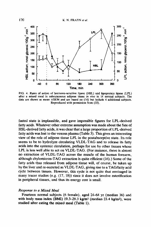
FIG. 4. Rates of action of hormone-sensitive lipase (HSL) and lipoprotein lipase (LPL) after a mixed meal in subcutaneous adipose tissue in viva in 14 normal subjects. The data are shown as mean fSEM and are based on (14) but include 4 additional subjects.
Reproduced with permission from (33).
fasted state is implausible, and gave impossible figures for LPL-derived fatty acids. Whatever other extreme assumption was made about the fate of HSL-derived fatty acids, it was clear that a large proportion of LPL-derived fatty acids was lost to the venous plasma (Table 3). This gives an interesting view of the role of adipose tissue LPL in the postabsorptive state. Its role seems to be to hydrolyze circulating VLDL-TAG and to release its fatty acids into the systemic circulation, perhaps for use by other tissues whose LPL is less well able to act on VLDL-TAG. (For instance, there is almost no extraction of VLDL-TAG across the muscle of the human forearm, although chylomicron-TAG extraction is quite efficient (16) .) Some of the fatty acids thus released from adipose tissue will, of course, be taken up by the liver and re-secreted as VLDL-TAG, giving rise to a TAG/fatty acid cycle between tissues. However, this cycle is not quite that envisaged in many tracer studies (e.g. (17, 18)) since it does not involve esterification in peripheral tissues, and thus its energy cost is small.
Response to a Mixed Meal Fourteen normal subjects (6 female), aged 24-64 yr (median 36) and
with body mass index (BMI) 19.3-29.3 kg/m2 (median 23.4 kg/m*), were studied after eating the mixed meal (Table 1).
REGULATION OF ADIPOSE TISSUE LIPASES 171
Plasma NEFA and blood glycerol concentrations before and after this meal are shown in Figure 3. Arterial plasma NEFA concentrations, around 600 kmolil after overnight fast, fell markedly after the meal and then returned gradually to baseline levels over the next 6 hr. Adipose-venous plasma NEFA concentrations were considerably higher than arterial after overnight fast. The arterio-venous difference narrowed markedly after the meal, and was close to zero from around l-2 hr after the meal. Blood glycerol concentrations followed a similar pattern, although the ratio between arterial and adipose-venous concentrations was even greater. The suppression of glycerol release from adipose tissue after the meal was less marked than that for NEFA.
HSL action was rapidly suppressed after the meal (Fig. 4), and stayed so until around 5 hr after the meal. The rate of LPL action, in contrast, rose steadily to peak at about 4-5 after the meal. The net efflux of fatty acids from adipose tissue fell to low levels during the period from 1-2 hr after the meal, although efflux of fatty acids always occurred (Fig. 5). (We have not observed a net uptake of NEFA across adipose tissue - i.e. a positive arterio-venous difference for NEFA - in these or any other experiments.) The transcapillary flux of fatty acids (Fig. 5a), in contrast, which was negative (outward flow of fatty acids from cells to capillaries) after overnight fast, changed within the first hour to a positive value (i.e. net inflow of fatty acids from capillaries to cells, and presumably storage as TAG). It remained positive until 5 hr after the meal, and then returned to a negative value.
Overall re-esterification of fatty acids increased after the meal. On the assumption of equal re-esterification of HSL- and LPL-derived fatty acids, the retention in adipose tissue of LPL-derived fatty acids is the same. It was therefore low after overnight fast, and increased after the meal (Fig. 6). Nevertheless, at the time of peak LPL action (4-5 hr after the meal) only 50% of LPL-derived fatty acids were retained in the tissue (Fig. 6).
Response to a High-Fat Meal Eleven studies were carried out in 7 subjects (4 subjects took part in
both ‘unclamped’ and ‘clamped’ protocols). There was one female; the subjects’ ages were 26-45 yr (median 34) and BMI 19.1-32.2 kg/m2. In 6 studies, no further intervention was made after the meal (‘unclamped’ studies). In the other 5 studies exogenous insulin was infused (‘clamped’ studies).
In both unclamped and clamped studies, the rate of action of HSL decreased after the meal until it was close to zero by 5 hr, whilst the rate of action of LPL increased and was highest at 5 hr after the meal (results given in (13)). There were no differences in HSL and LPL action
132 K. N. FRAYN et al.
-2.0 l-1 ' ' ' ' ' ' 0 60 120 180 240 300 360
Time after meal, min
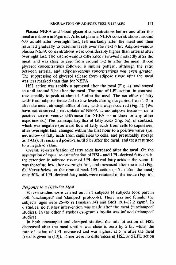
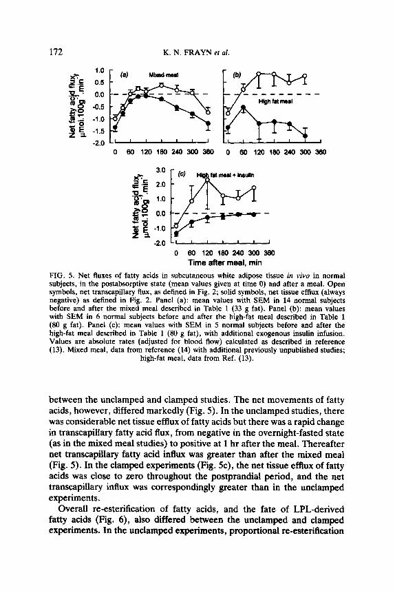
FIG. 5. Net fluxes of fatty acids in subcutaneous white adipose tissue in vivo in normal subjects, in the postabsorptive state (mean values given at time 0) and after a meal. Open symbols, net transcapillary tlux, as defined in Fig. 2; solid symbols, net tissue efthtx (always negative) as defined in Fig. 2. Panel (a): mean values with SEM in 14 normal subjects before and after the mixed meal described in Table 1 (33 g fat). Panel (b): mean values with SEM in 6 normal subjects before and after the high-fat meal described in Table 1 (80 g fat). Panel (c): mean values with SEM in 5 normal subjects before and after the high-fat meal described in Table 1 (80 g fat), with additional exogenous insulin infusion. Values are absolute rates (adjusted for blood flow) calculated as described in reference (13). Mixed meal, data from reference (14) with additional previously unpublished studies;
high-fat meal, data from Ref. (13).
between the unclamped and clamped studies. The net movements of fatty acids, however, differed markedly (Fig. 5). In the unclamped studies, there was considerable net tissue efflux of fatty acids but there was a rapid change in transcapillary fatty acid flux, from negative in the overnight-fasted state (as in the mixed meal studies) to positive at 1 hr after the meal. Thereafter net transcapillary fatty acid influx was greater than after the mixed meal (Fig. 5). In the clamped experiments (Fig. 5c), the net tissue efflux of fatty acids was close to zero throughout the postprandial period, and the net transcapillary influx was correspondingly greater than in the unclamped experiments.
Overall re-esterification of fatty acids, and the fate of LPL-derived fatty acids (Fig. 6), also differed between the unclamped and clamped experiments. In the unclamped experiments, proportional re-esterification

REGULATION OF ADIPOSE TISSUE LIPASES 173
-
0 80 120 180 240
Time after meal, min
300 380
FIG. 6. Percentage retention in white adipose tissue in viva of the fatty acids released by LPL action on circulating triacylglycerol. The values are calculated on the assumption of equal re-esterification of fatty acids derived from HSL- and from LPL-action. as discussed in the text and in Ref. (13). Results are shown for normal subjects, before (time 0) and after eating a meal: solid triangles, the mixed meal described in Table 1 (n = 14); open circles, the high-fat meal described in Table 1 (n = 6); solid circles, the high-fat meal
with additional exogenous insulin infusion (n = 5). Source of the data as for Fig. 5.
of fatty acids was similar to that observed after the mixed meal, although at the time of peak LPL action (4-5 hr after the meal) the proportion of LPL-derived fatty acids retained in adipose tissue was somewhat lower (20-30%). In the clamped experiments, however, there was considerably greater fatty acid re-esterification (and thus retention of LPL-derived fatty acids) in the postprandial period, averaging around 90% (Fig. 6).
Partial Acylglycerol Production by LPL An essential assumption underlying these studies is that LPL liberates
glycerol mole for mole with hydrolysis of TAG. This might be questioned since LPL acting in vitro may, depending upon the conditions, generate a large proportion of monoacylglycerol (MAG) (19,20). This arises because LPL action is specific for the sn-l(3) ester links in an acyiglycerol (19). Its initiai action gives rise to sn-2-MAG, which can then isomerize non-enzymatically to form sn-l(3)-MAG, which is again a substrate for LPL hydrolysis. We have measured MAG concentrations in arterial and adipose venous plasma in these experiments, and found absolutely no accumulation in the plasma of MAG during high rates of LPL
174 K. N. FRAYN etal.
action (as after the high-fat meal) (21; Fielding B. A., Frayn, K. N. et al., unpublished observations). However, it is possible that MAG is removed from the plasma compartment. It has been suggested, on the basis of electron micrographic studies, that MAG is hydrolyzed in the sub-endothelial space (22). Other authors have suggested that MAG is taken up by adipocytes (8), but even in this case it is highly likely to be hydrolyzed by the very active MAG-lipase (EC 3.1.1.23) in adipose tissue. Although the MAG-esterification pathway can be demonstrated in white adipose tissue, its activity is not high and special conditions are needed (e.g., the use of ether-linked substrate which is not a substrate for the MAG-lipase) (23). Studies of the tissue distribution of label after administration of labelled TAG in animals show that the glycerol moiety is not found in adipose tissue [reviewed in (24)]. Provided the MAG generated by LPL is hydrolyzed without direct incorporation into TAG, the glycerol will be released into the venous plasma and the assumptions made for these studies will not be invalidated.
Regulation of Fatty Acid Movement in Adipose Tissue The results of these studies seem entirely consistent with the hypothesis
that the net movement of fatty acids into and out of adipose tissue is governed by concentration gradients. In the postabsorptive state, LPL is relatively inactive, whilst HSL is more active and re-esterification of fatty acids is low. Therefore, the net flow of fatty acids will be from cells to capillaries, as observed, in keeping with adipose tissue’s major role as a supplier of fuel to other tissues in the fasting state. In the fed state, HSL is suppressed, esterification of fatty acids is stimulated and presumably the intracellular concentration of non-esterified fatty acids is low; as LPL activity increases, fatty acids generated by LPL action will be drawn from capillaries into cells down a concentration gradient. When additional insulin was given in the clamped experiments, fatty acid esterification was marked and LPL-derived fatty acids were more effectively trapped in the tissue. There is evidence that the cell-membrane-associated fatty acid transporter in adipocytes is subject to hormonal regulation (259, and this may add a further degree of precision of control.
The most consistently plausible results are found in these studies when the esterification of HSL- and LPL-derived fatty acids is assumed to be equal. This might suggest (but does not prove) that they form a common pool. This pool is unlikely to be intravascular, but may well be extracellular since studies of fatty acid re-esterification in vitro suggest that HSL-derived fatty acids follow an extracellular route (26). Other studies in cultured adipocytes have shown that fatty acids released by intracellular lipolysis dilute fatty acids taken up for esterification to only a small extent (27).
REGULATION OF ADIPOSE TISSUE LIPASES 17s
Again, these findings might be consistent with mixing in the extracellular rather than the intracellular environment.
The stimulation of esterification by insulin in the clamped experiments conlirms long standing observations that insulin causes such an effect (28). Nevertheless, insulin is usually considered to produce this effect mainly by stimulation of glucose uptake, glycolysis and thus provision of glycerol 3-phosphate for esterification (28). In the present studies, although glucose uptake by the tissue was stimulated in the postprandial period, it was not further increased by insulin (13). This suggests that insulin may directly stimulate some step in the re-esterification process, although which step is currently not known. Alternatively, stimulation or activation of the so-called acylation stimulating protein (29, 30). thought to be released by adipocytes (31), may be involved.
The rate of action of LPL in these and other acute studies seems to fluctuate less than that of HSL, which goes through swings of 4- to S-fold in activity (e.g., after the mixed meal, Fig. 4). This suggests a model for metabolic regulation in which LPL generates a pool of fatty acids (probably somewhere in the extracellular space, as discussed above) whose fate is determined mainly by intracellular events. Even after a high-fat meal a considerable proportion of the LPL-derived fatty acids is lost directly to the venous plasma. The diversion of LPL-derived fatty acids into the tissue may thus be seen as a metabolic branch-point (32), giving a high degree of control to the process of net fat storage, as seems necessary for a process whose quantitative regulation is so important to the body, as discussed in the Introduction. Net fat storage would be favored by increased concentrations in the plasma of TAG (increasing LPL action by a mass-action effect), glucose or insulin (increasing esterification and suppressing HSL action), and perhaps by other factors. Thus, we begin to see how the presence of an ‘energy surplus’ in the circulation may be coupled in a quantitative manner with net fat deposition.
SUMMARY
The enzymes lipoprotein lipase (LPL, EC 3.1.1.34) and hormone- sensitive lipase (HSL, EC 3.1.1.3) apparently catalyze opposing functions in white adipose tissue: the former is concerned with fat storage, the latter with fat mobilization. We have studied their regulation in vivo in normal subjects in the postabsorptive state and after eating meals of different compositions, by measurement of arteriovenous concentration differences for triacylglycerol, non-esterified fatty acids and glycerol across a subcutaneous adipose depot. The two enzymes are regulated in a broadly reciprocal manner: in the overnight-fasted state, HSL is more active, but after a meal HSL is suppressed whilst LPL is activated. The movement
176 K. N. FRAYN et al.
of fatty acids in and out of adipose tissue appears to be driven by concentration gradients generated by regulation of these two enzymes, and also by activation, in the postprandial period, of the process of fatty acid esterification.
The results show some interesting and perhaps unexpected features of metabolic regulation. Of the fatty acids generated by the action of LPL on circulating TAG, a large proportion is released directly into the venous plasma: close to 100% in the overnight-fasted state, and 50% or more at the peak of LPL action after a meal, making what appear reasonable assumptions. We suggest that this apparent ‘inefficiency’ of fat storage reflects the energetic cost of maintaining precise control over such a fundamental process. Although LPL is usually thought of as the enzyme regulating fat deposition, in fact the fatty acids and glycerol it releases from circulating TAG represent a substantial proportion of those released from adipose tissue, especially in the postprandial state. In addition, although HSL is considered the enzyme responsible for fat mobilization, suppression of its activity is essential to normal regulation of fat deposition.
Thus, fat storage and fat mobilization during normal daily life are controlled by coordinated regulation of a number of enzymatic processes in white adipose tissue.
ACKNOWLEDGEMENTS
Our own work on this topic has been supported by the British Heart Foundation and the Oxford Diabetes Trust. SWC held a Medical Research Council, U.K., Training Fellowship for some of the duration of these studies. We thank other members of the Oxford Lipid Metabolism Group who have contributed to these studies.
1.
2.
3.
4.
5.
6.
REFERENCES K. J. ACHESON, Y. SCHUTZ, T. BESSARD, E. RAVUSSIN, E. JBQUIER and J. P. FLAlT, Nutritional influences on lipogenesis and thermogenesis after a carbohydrate meal, Am. J. Physiol. 246, E62-E70 (1984). M. K. HELLERSTEIN, M. CHRISTIANSEN, S. KAEMPFER, S. KLETKE, K. WU, J. S. REID, K. MULLIGAN, N. S. HELLERSTEIN and C. H. L. SHACKLETON, Measurement of de nova lipogenesis in humans using stable isotopes, J. Clin. Invest. 87, 1841-1852 (1991). R. H. ECKEL, Lipoprotein lipase. A multifunctional enzyme relevant to common metabolic diseases, New En@. J. Med. 320, 1060-1068 (1989). R. L. PAlTEN, The reciprocal regulation of lipoprotein lipase activity and hormone- sensitive lipase activity in rat adipocytes, J. Biol. Chem. 245, 5577-5584 (1970). A. PRADINES-FIGURES, C. VANNIER and G. AILHAUD, Lipoprotein lipase stored in adipocytes and muscle cells is a cryptic enzyme, .I. Lipid Res. 31, 1467-1476 (1990). M.-R. TASKINEN, E. A. NIKKILA, J. K. HUITUNEN and H. HILDEN, A micromethod for assay of lipoprotein lipase activity in needle biopsy samples of human adipose tissue and skeletal muscle, Clin. Chim. Acta 104, 107-117 (1980).
REGULATION OF ADIPOSE TISSUE LIPASES 177
7. T. H. M. DA COSTA and D. H. WILLIAMSON, Effects of exogenous in&n or van&date on disposal of dietary triacylglycerols betweez~ mammary glmtd and adipose tissue in the lactating rat: insulin resistance in white adipose tissue, Eiochem. J. zfwt, 557-561 (1993).
8. P. BELFRAGE, G. FREDRIKSON, P. STRALFORS and H. TORNQVIST, Adipose tissue lipases, pp. 365-416 in Lipases. (B. BORGSTROM and H. L. BROCKMAN, eds.) Elsevier, Amsterdam (1984).
9. K. N. FRAYN, S. W. COPPACK, S. M. HUMPHREYS and P. L. WHYTE, Metabolic characteristics of human adipose tissue in viva, Clin. Sci. 76, W-516 (1989).
10. K. N. FRAYN, S. W. COPPACK and S. M. HUMPHREY& Subcutaneous adipose tissue metabolism studied by local catheterization, Inr. 1. Obesity 17 Supplement 3, S&S21 (1993).
11. R. A. DEFRONZO, J. TOBIN and R. ANDRES, Glucose clamp technique: a method for quantifying insulin secretion and resistance, Am. J. Physior’. 237, E214-E223 (1979).
12. 0. A. LARSEN, N. A. LASSEN and F. QUAADE, Blood flow through human adipose tissue determined with radioactive xenon, Acru Physiof. &and. 66, 337-345 (1966).
13. K. N. FRAYN, S. SHADID, R. HAMLANI, S. M. HUMPHREY& M. L. CLARK, B. A. FIELDING, 0. BOLAND and S. W. COPPACK, Regulation of fatty acid movement in human adipose tissue in the postabsorptive-to-postprandial transition. Am. J. Physiol. 266, E308-E317 (1994).
14. S. W. COPPACK, R. D. EVANS, R. M. FISHER, K. N. FRAYN, G. F. GIBBONS, S. M. HUMPHREYS, M. J. KIRK, J. L. POTTS and T. D. R. HOCKADAY, Adipose tissue metabolism in obesity: lipase action in viva before and after a mixed meal, Metabolism 41, 264-272 (1992).
15. V. A. HAMMOND and D. G. JOHNSTON, Substrate cycling between triglyceride and fatty acid in human adipocytes, Metabolism 36, 308-313 (1987).
16. J. L. POTTS, R. M. FISHER, S. M. HUMPHREYS, S. W. COPPACK, G. F. GIBBONS and K. N. FRAYN, Peripheral triacylglycerol extraction in the fasting and post-prandial states, Clin. Sci. 81, 621-626 (1991).
17. M. ELIA. C. ZED, G. NEALE and G. LIVESEY, The energy cost of triglyceride- fatty acid recycling in nonobese subjects after an overnight fast and four days of starvation. Metabolism 36, 251-255 (1987).
18. R. R. WOLFE, S. KLEIN, F. CARRARO and J.-M. WEHER, Role of triglyceride- fatty acid cycle in controlling fat metabolism in humans during and after exercise, Am. J. Physiol. 258, E382-E389 (1990).
19. P. NILSSON-EHLE, T. EGELRUD, P. BELFRAGE, T. OLIVECRONA and B. BORGSTRGM, Positional specificity of purified milk lipoprotein lipase, J. Biol. Chem. 248, 6734-6737 (1973).
20. R. 0. SCOW and T. OLIVECRONA, Effect of albumin on products formed from chyiomicron triacylglycerol by lipoprotein iipase in vitro, Biochim. Biophys. Acfa 487, 472-486 (1977).
21. B. A. FIELDING, S. M. HUMPHREYS, R. F. C. ALLMAN and K. N. FRAYN. Mono-, di- and triacylglycerol concentrations in human plasma: effects of heparin injection and of a high-fat meal, Clin. Chim. Acra 216, 167-173 (1993).
22. R. 0. SCOW, E. J. BLANCHETTE-MACKIE and L. C. SMITH, Role of capillary endothelium in the clearance of chylomicrons. A model for lipid transport from blood by lateral diffusion in cell membranes, Circ. Res. 39, 149-162 (1976).
23. F. SCHULTZ and J. JOHNSTON, The synthesis of higher glycerides via the monoglyceride pathway in hamster adipose tissue, J. Lipid Res. 12, 132-138 (1971).
24. R. 0. SCOW, Metabolism of chylomicrons in perfused adipose and mammary tissue of the rat, Fed. Proc. 36, 182-185 (1977).
25. N. A. ABUMRAD, C. M. HARMON, U. S. BARNELA and R. R. WHITESELL, Insulin antagonism of catechoiamine stimulation of fatty acid transport in the adipocyte. Studies on its mechanism of action, J. Biol. Chem. 263, 14678-14683 (1988).
178 K. N. FRAYN et al.
26. N. K. EDENS, R. L. LEIBEL and J. HIRSCH, Mechanism of free fatty acid re-esterification in human adipocytes in vitro, 1. Lipid Res. 31, 1423-1431 (1990).
27. N. A. ABUMRAD, C. FOREST, D. M. REGEN, U. S. BARNELLA and S. A. MELKI, Metabolism of oleic acid in differentiating BFC-1 preadipose cells, Am. J. Physiol. 261, E76-ES6 (1991).
28. B. LEBOEUF, Regulation of fatty acid esterification in adipose tissue incubated in vitro, pp. 385-391 in Handbook of Physiology Section 5: Adipose Tissue. (A. E. RENOLD and G. F. CAHILL, eds.) American Physiological Society, Washington DC (1965).
29. K. CIANFLONE, H. VU, M. WALSH, A. BALD0 and A. SNIDERMAN, Metabolic response of acylation stimulating protein to an oral fat load, J. Lipid Res. 30, 1727-1733 (1989).
30. A. SNIDERMAN, A. BALD0 and K. CAINFLONE, The potential role of acylation stimulating protein as a determinant of plasma triglyceride clearance and intracellular triglyceride synthesis, Curr. Opin. Lipidbl. 3, 202-267 (1992).
31. A. BALDO. A. D. SNIDERMAN. S. ST-LUCE. R. K. AVRAMOGLU. M. MASLOWSKA, B. HOANG, J. C: MONGE, A.’ BELL, S. MULAY and K. CAINFLONE, The adipsin-acylation stimulating protein system and regulation of intracellular triglyceride synthesis, J. Clin. Invest. 92, 1543-1547 (1993).
32. E. A. NEWSHOLME, B. CRABTREE and M. PARRY-BILLINGS, The energetic cost of regulation: an analysis based on the principles of metabolic-control-logic, pp. 447493 in Energy Metabolism: Tissue Determinants and Cellular Corollaries. (J. M. KINNEY and H. N. TUCKER, eds.), Raven Press, New York (1992).
33. K. N. FRAYN, S. M. HUMPHREYS and S. W. COPPACK, Fuel selection in white adipose tissue, Proc. Nutr. Sot. 54 (in press).