Catchin, a novel protein in molluscan catch muscles, is produced by alternative splicing from the...
10
Catchin, a Novel Protein in Molluscan Catch Muscles, is Produced by Alternative Splicing from the Myosin Heavy Chain Gene Akira Yamada 1 *, Maki Yoshio 1 , Kazuhiro Oiwa 1 and La ´ szlo ´ Nyitray 2 1 Kansai Advanced Research Center, Communications Research Laboratory, Kobe 651-2492, Japan 2 Department of Biochemistry Eo ¨tvo ¨s University, Budapest H- 1088, Hungary Molluscan catch muscles contain polypeptides of 110-120 kDa in size which have the same partial amino acid sequences as those of the myosin heavy chain (MHC). Here we provide evidence that these polypeptides are major components only of the catch-type muscles (their estimated molar ratio to MHC is approximately 1:1) and they are alternative pro- ducts of the MHC gene. Northern blot analysis of total RNA from Myti- lus galloprovincialis catch muscles was carried out with fragments from the 3 0 -end of the MHC cDNA as probes. We detected two bands of 6.5 kb and 3.5 kb. The former corresponds to the MHC mRNA, and the latter is an mRNA coding for catchin, a novel myosin rod-like protein. By using a 5 0 -rapid amplification of cDNA ends (RACE) PCR method, the full-length cDNA of Mytilus catchin was cloned. It codes for a protein with a unique N-terminal domain of 156 residues (rich in serine, threo- nine, and proline), which includes a phosphorylatable peptide sequence. The rest of the sequence is identical with the C-terminal 830 residues of the MHC. We also analyzed Mytilus and scallop (Argopecten irradians) genomic DNAs and found that the 5 0 -end of the cDNA sequence was located in a large intron of the MHC gene in both species. Since catchin is abundantly expressed only in catch muscles and it is phosphorylatable, we suggest that it may play an important role in the catch contraction of molluscan smooth muscles. # 2000 Academic Press Keywords: molluscan catch muscle; myosin, alternative splicing; thick filament; phosphorylation *Corresponding author Introduction Molluscan smooth muscles have large thick fila- ments that consist of a core of paramyosin covered with a layer of myosin (Szent-Gyo ¨ rgyi et al., 1971; Cohen, 1982). Some of these muscles have a unique physiological function known as catch. After the active contraction, these muscles can enter the catch state, under which they maintain high ten- sion without much expenditure of energy at a rest- ing level of intracellular Ca 2 concentration. A similar state exists in vertebrate tonic smooth muscles termed ‘‘latch’’. In both cases the cross- bridges somehow remain attached to the actin filament and cycle at a very slow rate (for reviews, see Twarog, 1979; Chantler, 1991). The most exten- sively studied catch muscle is the anterior byssus retractor muscle (ABRM) of the common mussel Mytilus edulis. As in all molluscan muscles Ca 2 induces active contraction by binding directly to myosin (Kendrick-Jones et al., 1970). In ABRMs, catch subsequently occurs after decrease of Ca 2 to resting level (Astumi & Sugi, 1976; Ishii et al., 1989). Catch contraction is regulated by phos- phorylation. Release of catch is mediated by a cAMP-dependent protein kinase (Cole & Twarog, 1972). It has been recently shown that the major target of the kinase is twitchin/mini-titin, a giant protein associated with thick filaments (Siegman et al., 1997, 1998). Interestingly, twitchin phos- phorylation also effects force output of Mytilus E-mail address of the corresponding author: [email protected] Abbreviations used: ABRM, anterior byssus retractor muscle; PBRM, posterior byssus retractor muscle; PAM, posterior adductor muscle; PRM, pedal retractor muscle; MHC, myosin heavy chain; RLC, regulatory light chain; EGTA, ethyleneglycol-bis-(2-aminoethylether)-N,N,N 0 ,N 0 - tetraacetic acid; DIG, digoxigenine; PAGE, polyacrylamide gel electrophoresis; RACE, rapid amplification of cDNA ends. Article No. jmbi.1999.3349 available online at http://www.idealibrary.com on J. Mol. Biol. (2000) 295, 169–178 0022-2836/00/020169–10 $35.00/0 # 2000 Academic Press
Transcript of Catchin, a novel protein in molluscan catch muscles, is produced by alternative splicing from the...

Article No. jmbi.1999.3349 available online at http://www.idealibrary.com on J. Mol. Biol. (2000) 295, 169±178
Catchin, a Novel Protein in Molluscan Catch Muscles,is Produced by Alternative Splicing from the MyosinHeavy Chain Gene
Akira Yamada1*, Maki Yoshio1, Kazuhiro Oiwa1 and LaÂszlo Nyitray2
1Kansai Advanced ResearchCenter, CommunicationsResearch Laboratory, Kobe651-2492, Japan2Department of BiochemistryEoÈtvoÈs University, Budapest H-1088, Hungary
E-mail address of the [email protected]
Abbreviations used: ABRM, antermuscle; PBRM, posterior byssus retposterior adductor muscle; PRM, peMHC, myosin heavy chain; RLC, reEGTA, ethyleneglycol-bis-(2-aminoetetraacetic acid; DIG, digoxigenine;polyacrylamide gel electrophoresis;ampli®cation of cDNA ends.
0022-2836/00/020169±10 $35.00/0
Molluscan catch muscles contain polypeptides of 110-120 kDa in sizewhich have the same partial amino acid sequences as those of the myosinheavy chain (MHC). Here we provide evidence that these polypeptidesare major components only of the catch-type muscles (their estimatedmolar ratio to MHC is approximately 1:1) and they are alternative pro-ducts of the MHC gene. Northern blot analysis of total RNA from Myti-lus galloprovincialis catch muscles was carried out with fragments fromthe 30-end of the MHC cDNA as probes. We detected two bands of6.5 kb and 3.5 kb. The former corresponds to the MHC mRNA, and thelatter is an mRNA coding for catchin, a novel myosin rod-like protein.By using a 50-rapid ampli®cation of cDNA ends (RACE) PCR method,the full-length cDNA of Mytilus catchin was cloned. It codes for a proteinwith a unique N-terminal domain of 156 residues (rich in serine, threo-nine, and proline), which includes a phosphorylatable peptide sequence.The rest of the sequence is identical with the C-terminal 830 residues ofthe MHC. We also analyzed Mytilus and scallop (Argopecten irradians)genomic DNAs and found that the 50-end of the cDNA sequence waslocated in a large intron of the MHC gene in both species. Since catchinis abundantly expressed only in catch muscles and it is phosphorylatable,we suggest that it may play an important role in the catch contraction ofmolluscan smooth muscles.
# 2000 Academic Press
Keywords: molluscan catch muscle; myosin, alternative splicing; thick®lament; phosphorylation
*Corresponding authorIntroduction
Molluscan smooth muscles have large thick ®la-ments that consist of a core of paramyosin coveredwith a layer of myosin (Szent-GyoÈrgyi et al., 1971;Cohen, 1982). Some of these muscles have a uniquephysiological function known as catch. After theactive contraction, these muscles can enter thecatch state, under which they maintain high ten-sion without much expenditure of energy at a rest-
ing author:
ior byssus retractorractor muscle; PAM,dal retractor muscle;gulatory light chain;thylether)-N,N,N0,N0-PAGE,RACE, rapid
ing level of intracellular Ca2� concentration. Asimilar state exists in vertebrate tonic smoothmuscles termed ``latch''. In both cases the cross-bridges somehow remain attached to the actin®lament and cycle at a very slow rate (for reviews,see Twarog, 1979; Chantler, 1991). The most exten-sively studied catch muscle is the anterior byssusretractor muscle (ABRM) of the common musselMytilus edulis. As in all molluscan muscles Ca2�
induces active contraction by binding directly tomyosin (Kendrick-Jones et al., 1970). In ABRMs,catch subsequently occurs after decrease of Ca2� toresting level (Astumi & Sugi, 1976; Ishii et al.,1989). Catch contraction is regulated by phos-phorylation. Release of catch is mediated by acAMP-dependent protein kinase (Cole & Twarog,1972). It has been recently shown that the majortarget of the kinase is twitchin/mini-titin, a giantprotein associated with thick ®laments (Siegmanet al., 1997, 1998). Interestingly, twitchin phos-phorylation also effects force output of Mytilus
# 2000 Academic Press

170 A Novel Protein in Molluscan Catch Muscles
ABRM during calcium-mediated submaximumcontractions (Butler et al., 1998). The catch staterequires the twitchin to be dephosphorylated,probably through the action of a calcium-activatedphosphatase (Castellani & Cohen, 1992). Otherthick ®lament components were also shown tobe phosphorylatable in vitro by an endogenouskinase - paramyosin (Cooley et al., 1979; Achazi,1979; Cohen, 1982), myosin heavy chain (MHC)(Castellani & Cohen, 1987) and myosin regulatorylight chain (RLC) (Sohma et al., 1985); their poten-tial role in the regulation of the catch state is morecontroversial. The mechanism of catch contractionhas been much discussed. It was proposed that theslow cycling cross-bridges during catch state aresomehow related to the unusual organization ofthe thick ®laments where critical changes ofprotein-protein interactions are brought about byphosphorylation (Cohen, 1982).
Thick ®laments of catch muscles compared withthose of non-catch muscles, besides their muchhigher paramyosin content, possess different myo-sin isoforms with different enzymatic properties. Inparticular, catch muscle myosins have considerablylower steady-state ATPase activity than myosinsfrom phasic muscles. Catch and striated musclespeci®c RLC isoforms were identi®ed in Patinopec-ten yessoensis and Placopecten magellanicus (Kondo& Morita, 1981; Perreault-Micale et al., 1996a).However, it was shown that they are not respon-sible for the muscle-speci®c differences in enzy-matic activities. Instead, those differences areattributable to MHC isoforms (Perreault-Micaleet al., 1996b). In the Pectinidae family, a singleMHC gene generates catch and striated musclespeci®c MHC isoforms by alternative RNA splicing(Nyitray et al., 1994; Perreault-Micale et al., 1996b).Among the three alternative exons in the motordomain of MHC, sequence variations within theexon forming the ¯exible surface loop I at the ATPbinding pocket modulate ADP af®nity, ATPaseactivity and in vitro motility of scallop catch myo-sin (Kurzawa-Goertz et al., 1998). Much less isknown about the role of the two alternativesequences in the middle of the coiled-coil rodregion and at the C-terminal non-helical tailpiece(Nyitray et al., 1994).
It has previously been noted that the crude myo-sin or thick ®lament preparations from MytilusABRM contain considerable amounts of anadditional protein whose apparent molecular massis around 110-140 kDa (Castellani et al., 1988;Yamada et al., 1989). Originally it was thought tobe a degradation product of the myosin rod,however later studies suggested that the myosinrod-like protein is produced in the muscle cells(Yamada et al., 1997). Here we provide evidencethat the ABRM myosin rod-like protein, namedcatchin, is an alternative product of the MHC gene.Since it is abundantly expressed only in molluscancatch muscles, we suggest that it may contribute tothe force-maintaining structures during the catchstate.
Preliminary results of this work have been pub-lished elsewhere (Yamada et al., 1999).
Results
A new protein component in bivalve muscles
Recently, two new polypeptides (120 kDa and112 kDa) were identi®ed in Mytilus catch musclesas myosin rod-like proteins. Their partial aminoacid sequences were found to be identical withthose of the MHC from the same muscle (Yamadaet al., 1997). In this report we analyzed proteincompositions of various muscles from Mytilis gallo-provincialis, Crassostrea sp. and several species ofthe Pectinidae family (P. yessoensis, Argopectenirradians, P. magellanicus, Pecten maximus) by SDS-polyacrylamide gel electrophoresis (PAGE), to seewhether they contain similar components(Figure 1). Anterior byssus retractor muscle(ABRM), posterior byssus retractor muscle(PBRM), and posterior adductor muscle (PAM) ofMytilus contained considerable amount of the rod-like proteins (Figure 1, lanes 1±3). The stainingintensity ratios of the two new bands to MHC andparamyosin were 1:2 and 1:5, respectively. Themolar ratio of the new protein to MHC is approxi-mately 1:1, assuming that the speci®c staining issimilar between them. In contrast, pedal retractormuscle (PRM), which is not a catch muscle (Ishii &Takahashi, 1981), contained only a small amountof the rod-like proteins (Figure 1, lane 4; the stain-ing intensity ratio to MHC was 1:25). The rod-likeprotein was found to be heat-stable, and couldeasily be puri®ed by heat treatment (compareFigure 1, lanes 5 and 6). Therefore, we identi®edthe corresponding protein in various muscles bythis simple method (Figure 1, lanes 8-15). In oyster,a heat-stable component of about 120 kDa wasfound abundantly in the white adductor catchmuscle, but only in small amount in the translu-cent non-catch adductor muscle (Figure 1, lanes 12-15). In the catch adductor muscle of Patinopecten,there was a component of about 120 kDa and itwas heat stable (Figure 1, lanes 8 and 10). In con-trast, there was no such a component in thestriated adductor muscle (Figure 1, lanes 9 and 11).The same results were obtained with the threeother scallop species (results not shown). Thus, theheat-stable proteins are abundant only in catchmuscles, and the name ``catchin'' was coined tothem.
Cloning of the rod portion of MHC fromMytilus catch muscles
It was suggested that the 120 kDa and the112 kDa myosin rod-like polypeptides of Mytiluscatch muscles had been produced in the musclecells, rather than by proteolytic degradation ofMHC during the isolation procedure after themuscles were dissected from the animals (Yamadaet al., 1997). If catchin is a genuine muscle protein,

Figure 2. Schematic view of the MHC and positionsof the various cDNA fragments and probes used in thisstudy. Cloned and sequenced cDNA fragments of theMHC of (a) catch muscles (ABRM, PBRM and PAM)and (b) of PRM from M. galloprovincialis. (c) Probes forNorthern blotting analysis. (d) cDNA fragments ofcatchin obtained by 50-RACE PCR experiments. Dottedlines show the N-terminal unique part of catchin. SeeMaterials and Methods for details.
Figure 1. Protein compositions ofmuscles from different bivalves(Mytilus, Patinopecten and Crassos-trea). Protein compositions of wholemuscle homogenates (lanes 1-4, 8,9, 12 and 13) and their heat-stablefractions (lanes 6, 7, 10, 11, 14 and15) are shown on SDS-7.5 % PAGE.Samples were prepared fromABRM (lanes 1 and 6), PBRM (lane2), PAM (lane 3) and PRM (lanes 4and 7) of M. galloprovincialis, catchadductor (lanes 8 and 10) andstriated adductor (lanes 9 and 11)of P. yessoensis, and catch adductor(lanes 12 and 14) and translucent
cross-striated adductor (lanes 13 and 15) of a Crassostrea sp. Catchin prepared from M. galloprovincialis catch musclesaccording to Yamada et al. (1997) was also analyzed (lane 5). Note that the heat-stable components of about 120 kDain size (catchin) are abundant only in catch muscles. Lane 16 shows a molecular mass protein markers (Sigma, SDS-6H) with molecular masses (kDa) on the right.
A Novel Protein in Molluscan Catch Muscles 171
the cells must contain an mRNA which is consider-ably smaller than that of the MHC. Moreover,since the catchin polypeptides have, at least par-tially, identical amino acid sequences to that of theMHC rod, it is likely that their mRNA sequence isalso partially identical and that catchin is producedby alternative RNA splicing from the MHC gene.In order to con®rm this possibility, Northern blotexperiments and genomic sequence analysis (seebelow) were carried out.
We ®rst cloned and sequenced cDNA fragmentscoding for the MHC from M. galloprovincialis catchmuscles as outlined in Materials and Methods(Figure 2(a); GenBank accession no. AJ249991). InFigure 3, the deduced amino acid sequence ofmyosin rod is shown aligned with the sequences ofmyosin rods from M. galloprovincialis PRM (seebelow) and from A. irradians catch adductormuscle (Nyitray et al., 1991, 1994). Peptidesequences of Mytilus rod which were previouslyreported are underlined (sequences A and B weredescribed by Yamada et al., 1997; sequence C wasby Castellani et al., 1988). The fourth residue ofsequence A is Tyr instead of Val as reported pre-viously. This could be explained by a modi®cationof the Tyr side-chain during the o-iodosobenzoicacid treatment and the erroneous identi®cation ofthe modi®ed residue as Val (Yamada et al., 1997).The C-terminal residue in sequence C is an Aspresidue instead of a Glu residue. This phosphory-lated peptide, however, was isolated from ABRMof M. edulis. Comparison of the sequence of a 1 kbfragment from M. edulis MHC cDNA (L.N., unpub-lished results; Genbank accession no. U40036) withthe rod sequence reported here, shows the sameGlu-Asp change at the C-terminal residue. The restof the differences are at either silent codon pos-itions or within the 30-non-translated region (99 %identity in the coding and 97.6 % in the non-codingregion). The very high degree of conservationre¯ects the fact that M. galloprovincialis and M. edu-lis belong to one species complex.
Northern blotting analysis of four musclesfrom M. galloprovincialis
Since the 120 kDa and the 112 kDa polypeptideshave partially identical amino acid sequences tothose of the MHC rod, putative mRNAs for thesepolypeptides would hybridize with probes for themyosin rod cDNA. Two probes were prepared (seeFigure 2(c)) and hybridized to total RNAs fromfour muscles of M. galloprovincialis (Figure 4).Hybridization of ABRM, PBRM, and PAM RNAwith probe 1 shows two bands of 6.5 kb (indicatedby an arrowhead) and 3.5 kb (indicated by a tan-dem arrowhead). The 6.5 kb band is the mRNA ofMHC. The presence of a 3.5 kb band indicates that

Figure 3 (legend opposite)
172 A Novel Protein in Molluscan Catch Muscles

Figure 4. Northern blotting analyses of Mytilusmuscles. Total RNAs prepared from (1) ABRM, (2)PBRM, (3) PAM and (4) PRM were electrophoresed andtransferred to a nylon membrane. The membrane wasstained with either (a) ethidium bromide or (b) probe 1.In addition to the bands of mRNA for MHC of 6.5 kb(arrowhead), bands of 3.5 kb (tandem arrowhead) weredetected in RNAs from ABRM, PBRM and PAM. (c)When we used a probe for Mytilus tropomyosintogether with probe 1, additional bands of tropomyosinmRNAs (arrows) were detected in all RNA prep-arations.
A Novel Protein in Molluscan Catch Muscles 173
a truncated mRNA, presumably coding for catchin,was also transcribed in these muscles. It is notablethat the 3.5 kb RNA is abundant only in catchmuscles (ABRM, PBRM and PAM), similarly to thetwo polypeptides of the rod-like protein (Figure 1).When the Northern blotting experiment was car-ried out with a mixture of probes for myosin rodand for tropomyosin (obtained from a M. edulistropomyosin clone, L.N., unpublished results, Gen-bank accession no. U40035), two additional bandsof about 2.3 kb and 1.3 kb were detected in allmuscles (Figure 4(c)). Thus the fact that the 6.5 kband 3.5 kb bands were rather faint in the PRMsample (Figure 4(b), lane 4) is explained by themuch lower expression level of these mRNAs inPRM and not by degradation of RNA. The sameresults were obtained by using probe 2 (data notshown). These results suggest that the sequence ofMHC in PRM is considerably different from that ofthe MHC of catch muscles. Indeed, sequencedcDNA fragments of MHC from PRM (clones 7 and8) are different from that of the catch muscle MHC(identity 75 %; GenBank accession no. AJ249992).
Figure 3. Amino acid sequences of the rod region of MHChighly conserved Pro residue, marking the head rod junctiovincialis, and (c) the catch adductor muscle of A. irradians (identical amino acid residue to that of the Mytilus catch musunderlined (A-C). Sequences A and B were reported by Ya(1988). The boundary between the MHC-speci®c sequence acated by an arrow. A phosphorylatable Ser residue is indicat
The deduced partial amino-acid sequence of PRMMHC shows 78 % identity (84 % similarity) withthat of the catch muscle MHC (Figure 3).
N-terminal unique domain of catchin
In order to determine the N-terminal region ofcatchin polypeptides, we performed 50-rapidampli®cation of cDNA ends (RACE) PCR exper-iments using speci®c primers based on the MHCcDNA sequence coding for sequence A (Figure 3(a))which is common in catchin and MHC. Four inde-pendent cDNA clones from the 50-end of the 3.5 kbRNA were sequenced (Figure 2(d); GenBank acces-sion no. AJ249993). The sequence obtained encodesa 156 residues stretch of peptide, representing aunique N-terminal domain of catchin (Figure 5(a)).The corresponding sequence of Argopecten, whichwas obtained by analysis of the MHC gene (seebelow), is also shown (Figure 5(b)). The two hom-ologous sequences show rather low similarity(42 % similarity; 33 % identity). The sequence in thesecond half of the unique domain is more conser-vative than in the ®rst half (68 % similarity, 53 %identity), suggesting that this region is functionallymore important. The N-terminal domain of catchindoes not seem to form a coiled-coil structure (asanalyzed by COILS; Lupas et al., 1991) and it doesnot show strong propensity for any secondarystructure element (results not shown). It is indeeda unique sequence: No similarity was found withany sequences in the databases. Within the uniqueN-terminal domain, we identi®ed one of the phos-phorylatable peptides (underlined in Figure 5)reported by Castellani et al. (1988). They assumedthis peptide was in the C-terminal part of theMHC. However, our ®nding indicates it was actu-ally in the N-terminal part of catchin.
Although there are two different polypeptides of120 kDa and 112 kDa in Mytilus catch muscles,only a single sequence was obtained among the50-RACE products. Since there is an internal Metresidue in the N-terminal domain of the Mytilussequence (Met67), it is possible that translation alsobegins at this internal ATG triplet resulting in the112 kDa polypeptide. In contrast, there is nosecond Met residue in the scallop sequence, inaccord with the result that only a single main bandwas found in the heat-stable fraction of the scallopcatch muscles (Figure 1, lane 10).
To get information from the C-terminal region ofthe catchin sequence, a 30-RACE PCR experimentwas performed using a catchin-speci®c 50-primer
s. Deduced amino acid sequences of the MHCs from then, of (a) catch muscles and (b) of PRM from M. gallopro-Nyitray et al., 1991, 1994) are shown. Dash indicates ancle MHC. Amino acid sequences reported previously aremada et al. (1997), and sequence C by Castellani et al.nd the sequence common to MHC and catchin is indi-
ed by an arrowhead (Castellani et al., 1988).
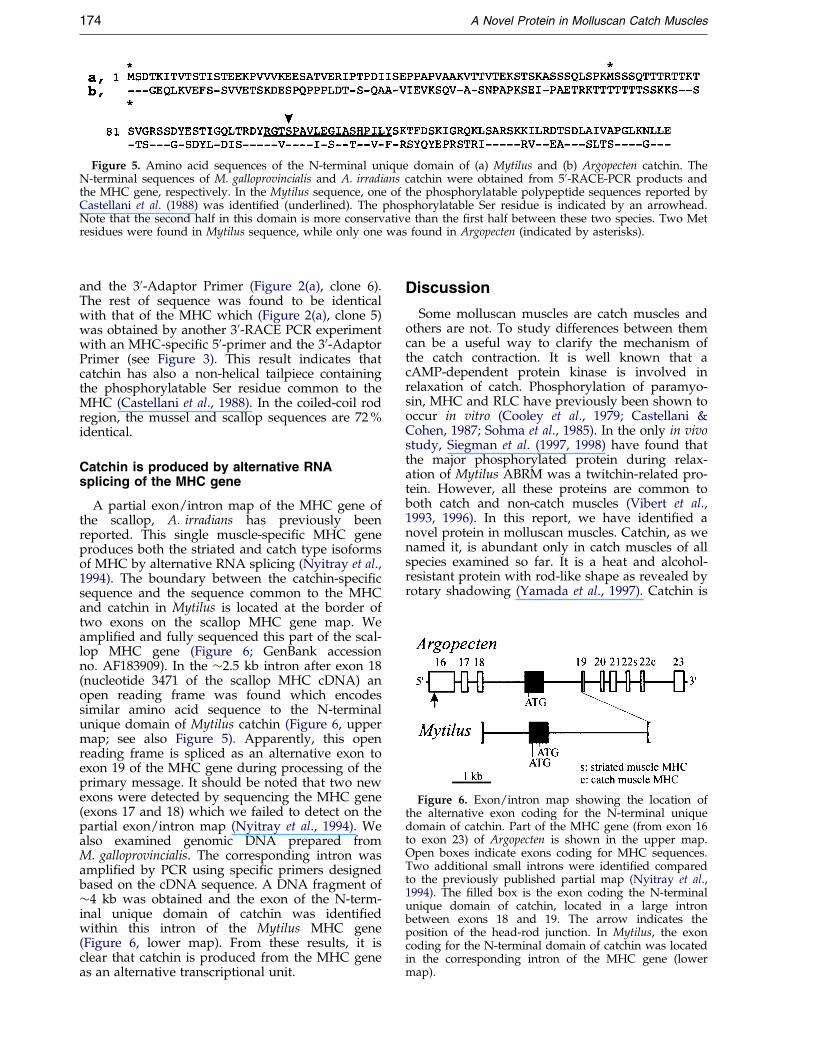
Figure 5. Amino acid sequences of the N-terminal unique domain of (a) Mytilus and (b) Argopecten catchin. TheN-terminal sequences of M. galloprovincialis and A. irradians catchin were obtained from 50-RACE-PCR products andthe MHC gene, respectively. In the Mytilus sequence, one of the phosphorylatable polypeptide sequences reported byCastellani et al. (1988) was identi®ed (underlined). The phosphorylatable Ser residue is indicated by an arrowhead.Note that the second half in this domain is more conservative than the ®rst half between these two species. Two Metresidues were found in Mytilus sequence, while only one was found in Argopecten (indicated by asterisks).
Figure 6. Exon/intron map showing the location ofthe alternative exon coding for the N-terminal uniquedomain of catchin. Part of the MHC gene (from exon 16to exon 23) of Argopecten is shown in the upper map.Open boxes indicate exons coding for MHC sequences.Two additional small introns were identi®ed comparedto the previously published partial map (Nyitray et al.,1994). The ®lled box is the exon coding the N-terminalunique domain of catchin, located in a large intronbetween exons 18 and 19. The arrow indicates theposition of the head-rod junction. In Mytilus, the exoncoding for the N-terminal domain of catchin was locatedin the corresponding intron of the MHC gene (lowermap).
174 A Novel Protein in Molluscan Catch Muscles
and the 30-Adaptor Primer (Figure 2(a), clone 6).The rest of sequence was found to be identicalwith that of the MHC which (Figure 2(a), clone 5)was obtained by another 30-RACE PCR experimentwith an MHC-speci®c 50-primer and the 30-AdaptorPrimer (see Figure 3). This result indicates thatcatchin has also a non-helical tailpiece containingthe phosphorylatable Ser residue common to theMHC (Castellani et al., 1988). In the coiled-coil rodregion, the mussel and scallop sequences are 72 %identical.
Catchin is produced by alternative RNAsplicing of the MHC gene
A partial exon/intron map of the MHC gene ofthe scallop, A. irradians has previously beenreported. This single muscle-speci®c MHC geneproduces both the striated and catch type isoformsof MHC by alternative RNA splicing (Nyitray et al.,1994). The boundary between the catchin-speci®csequence and the sequence common to the MHCand catchin in Mytilus is located at the border oftwo exons on the scallop MHC gene map. Weampli®ed and fully sequenced this part of the scal-lop MHC gene (Figure 6; GenBank accessionno. AF183909). In the �2.5 kb intron after exon 18(nucleotide 3471 of the scallop MHC cDNA) anopen reading frame was found which encodessimilar amino acid sequence to the N-terminalunique domain of Mytilus catchin (Figure 6, uppermap; see also Figure 5). Apparently, this openreading frame is spliced as an alternative exon toexon 19 of the MHC gene during processing of theprimary message. It should be noted that two newexons were detected by sequencing the MHC gene(exons 17 and 18) which we failed to detect on thepartial exon/intron map (Nyitray et al., 1994). Wealso examined genomic DNA prepared fromM. galloprovincialis. The corresponding intron wasampli®ed by PCR using speci®c primers designedbased on the cDNA sequence. A DNA fragment of�4 kb was obtained and the exon of the N-term-inal unique domain of catchin was identi®edwithin this intron of the Mytilus MHC gene(Figure 6, lower map). From these results, it isclear that catchin is produced from the MHC geneas an alternative transcriptional unit.
Discussion
Some molluscan muscles are catch muscles andothers are not. To study differences between themcan be a useful way to clarify the mechanism ofthe catch contraction. It is well known that acAMP-dependent protein kinase is involved inrelaxation of catch. Phosphorylation of paramyo-sin, MHC and RLC have previously been shown tooccur in vitro (Cooley et al., 1979; Castellani &Cohen, 1987; Sohma et al., 1985). In the only in vivostudy, Siegman et al. (1997, 1998) have found thatthe major phosphorylated protein during relax-ation of Mytilus ABRM was a twitchin-related pro-tein. However, all these proteins are common toboth catch and non-catch muscles (Vibert et al.,1993, 1996). In this report, we have identi®ed anovel protein in molluscan muscles. Catchin, as wenamed it, is abundant only in catch muscles of allspecies examined so far. It is a heat and alcohol-resistant protein with rod-like shape as revealed byrotary shadowing (Yamada et al., 1997). Catchin is

A Novel Protein in Molluscan Catch Muscles 175
probably identical to a contaminating protein ofABRM myosin preparations, previously thought tobe a degradation product of the MHC (Castellani& Cohen, 1987; Castellani et al., 1988); to a heat-stable catch muscle component, tentatively ident-i®ed as an invertebrate caldesmon-like protein(Bartegi et al., 1989; Csizmadia et al., 1994); and toan unknown heat-stable protein in molluscan catchmuscles reported by others in preliminary form(Shelud'ko et al., 1998; Kumeiko et al., 1999). Onecan assume that catchin, as a catch muscle speci®cprotein, plays some, perhaps a key role in thecatch contraction of molluscan muscles.
The full-length cDNA of M. galloprovincialiscatchin was cloned and sequenced. The encodedprotein has three domains: the N-terminal domain,consisting of 156 amino acid residues, is unique tocatchin and apparently does not form a coiled-coilstructure; while the rest of the molecule is identicalwith the C-terminal two-thirds of the rod region ofthe MHC, consisting of an a-helical coiled-coildomain of 809 residues and a non-helical tailpiecedomain of 21 residues. Because of the presence ofthe myosin rod sequence the catchin polypeptidesmust form dimeric coiled-coil protein as the MHCsdo. Puri®ed catchin forms ®laments and it co-aggregates with rabbit skeletal muscle myosin ®la-ments indicating that it is a component of the thick®lament (Yamada et al., 1997). Densitometry anal-ysis shows that the estimated molar ratio of catchinto MHC in all catch muscle is as much as 1:1. Itsprecise location and distribution within the thick®lament is being investigated.
The N-terminal domain may have some uniquefunctions, since this domain is unique to catchinand does not show high similarity to any otherknown proteins. Close to the border of the uniqueN-terminal domain and the rod portion we haveidenti®ed a peptide sequence which has previouslybeen reported by Castellani et al. (1988) to containa phosphorylatable Ser residue. In their report, thissequence was tentatively located in the non-helicaltailpiece of MHC together with an additional phos-phorylated peptide. Now it is clear that the ®rstpeptide comes from the N-terminal non-helicaldomain of catchin, and that the second peptiderepresents the C-terminal end of both catchin andthe MHC. Thus, catchin has at least two phosphor-ylatable Ser residues, one in the N-terminal uniquedomain and the other in the C-terminal non-helicaltailpiece domain common to the MHC. Phos-phorylation of these Ser residues by an endogen-ous kinase must be some important but yetunknown functions.
It was noted earlier that in the A. irradians MHCgene some of the large ``intervening'' sequencesmay contain additional alternatively spliced exons(Nyitray et al., 1994). Indeed, catchin is an alterna-tive product of the MHC gene. The N-terminalunique domain is encoded within one of the largeintrons of the MHC gene (after exon 18) andspliced to the rod coding exon 19. The rodsequence common with the MHC starts near the
subfragment-2 hinge region within the rod(Ala1110 in the A. irradians MHC sequence). Inter-estingly, a myosin rod protein was also identi®edas an alternative product of the single Drosophilamuscle MHC gene. This protein of 155 kDa in sizewas expressed abundantly in various muscles andin testis (Miedema, 1995; Standiford et al., 1997). Ithas a unique 77 amino acid residue N-terminaldomain that exactly replaces the MHC motordomain. Thus it has a full-length rod domaincompared with the molluscan catchin in which theN-terminal domain is connected to the rod nearthe subfragment-2 hinge within the rod. There isno signi®cant similarity between the amino acidsequences of the unique N-terminal domainsexcept that both are rich in Pro, Ser and Thrresidues. Another difference between the twointernal transcripts is that the exon coding for theN-terminal domain in Drosophila rod protein iscontinuous in frame into the rod coding exon,while there is an additional intron in the molluscangenes. The presence of alternative transcriptionalunits in genes of thick ®lament proteins is notrestricted to the MHC: Drosophila paramyosin geneencodes internally a truncated form, mini-paramyosin which also has a unique N-terminalnon-helical domain (Maroto et al., 1995, 1996).
What is the function of catchin and its uniqueN-terminal domain? One possibility is that theN-terminal domain contributes to the ``setting'' ofthe acto-myosin crossbridges during the catch stateby either directly or by interacting with other thick®lament associated proteins, particularly withtwitchin. It was reported that twitchin wasinvolved in regulation of catch contraction(Siegman et al., 1997, 1998). It would be interestingto see whether catchin is able to interact withtwitchin by a phosphorylation dependent manner.If the N-terminal unique domain of catchin has anextended structure, it might tether actin to thethick ®lament in the catch state. Standiford et al.(1997) noted some relatedness between theN-terminal domain of the Drosophila rod proteinand the N-terminal extensions of certain myosinlight chains which might serve to tether the lightchains to actin and modulate the contractile func-tion. A similar function for the N-terminal domainof each myosin rod protein is conceivable. The roleof phosphorylation of catchin might also be relatedto its putative role in the catch contraction or, alter-natively it could be involved in the myo®brillo-genesis of catch muscles. Catchin may also have arole in the formation of the peculiar structure ofthe large thick ®laments of catch muscles com-pared to smaller thick ®laments of more commonnon-catch muscles. Considering the organization ofthe thick ®lament it is worth to note that thecoiled-coil portion of catchin has the same length(�125 nm) as that of paramyosin.
In summary, we have discovered a novel myosinrod-like protein, catchin, in molluscan catchmuscles which is an alternative transcriptional pro-duct of the MHC gene. Apparently, catchin is a

176 A Novel Protein in Molluscan Catch Muscles
major component of the thick ®laments, and phos-phorylatable by an endogenous kinase both in theunique N-terminal non-helical domain and in theC-terminal non-helical tailpiece domain. Now thatthe presence of catchin in catch muscles is evident,although its function is unclear yet, one has to takeit into account as a ``new player'' for proposingmodels for the structure of catch muscle thick®laments and for the molecular basis of catchcontraction.
Materials and Methods
Animals and muscle proteins
Blue mussels, M. galloprovincialis, and oysters, Crassos-trea sp., were collected in the Inland Sea of Japan. Livescallops, P. yessoensis were purchased from a marketnear the Kansai Advanced Research Center. A. irradiansand P. magellanicus muscles were gift from Dr A.G.Szent-GyoÈrgyi (Brandeis University, USA). Heat-stableprotein fractions were prepared from these muscles by®rst homogenizing them in six volumes of 0.3 M KCl,1 mM ethyleneglycol-bis-(2-aminoethylether)-N,N,N0,N0-tetraacetic acid (EGTA), 0.5 mM MgCl2, 50 mM imida-zole (pH 7.0), then heated at 85 �C for four minutes.After cooling, the homogenate was centrifuged at 15,000g for 30 minutes and the supernatant was dialyzed over-night against 1 mM EGTA, 1 mM dithiothreitol (DTT),20 mM Tris-HCl (pH 7.5). The dialysate was centrifugedand the precipitate was analyzed by SDS-PAGE, togetherwith total homogenates of the muscles. The gels werestained with Coomassie brilliant blue R-250, and densi-tometry was carried out with a Shimadzu CS-9300densitometer.
Cloning experiments
Total RNA of M. galloprovincialis muscles was pre-pared essentially according to Chomczynski & Sacchi(1987). Complementary DNA was synthesized with aSuperScript II reverse transcriptase (Gibco BRL, LifeTechnologies, Inc., USA) using either an Oligo (dT) pri-mer (for clones 1-3, 7 in Figure 2) or an Oligo (dT)-Adaptor Primer (TaKaRa Shuzo Co. Ltd., Japan; forclones 4-6, 8 in Figure 2). PCR experiments were per-formed with either a TaKaRa Ex Taq or a TaKaRa LATaq DNA polymerase. The products were cloned into apCR 2.1 cloning vector (Invitrogen) and sequenced withan automatic DNA sequencer (ABI PRISM 377 DNASequencer, Perkin Elmer). Clone 1 of M. galloprovincialiscatch muscle MHC (Figure 2(a)) was ampli®ed using adegenerate 50-primer based on the partial amino acidsequence of the 120 kDa and 112 kDa proteins (Yamadaet al., 1997) and a speci®c 30-primer designed based on apartial cDNA sequence of M. edulis MHC (L.N., unpub-lished results; GenBank accession no. U40036). Clone 2was ampli®ed with a degenerate 50-primer designedaccording to a highly conservative amino acid sequencein the myosin head and a speci®c 30-primer based on thesequence of clone 1. Clone 3 was constructed the sameway. We carried out 30-RACE PCR experiments to makeclones 4-6 using speci®c 50-primers and a 30-AdaptorPrimer. An MHC cDNA fragment from PRM was alsocloned (Figure 2(a), clone 7) using the same primers asused for clone 3. The 30-end of the PRM MHC cDNA(clone 8) was ampli®ed by a 30-RACE PCR experiment
with a speci®c 50-primer designed based on the sequenceof clone 7 and the 30-Adaptor Primer.
In order to get the 50-end of the catchin cDNA, weperformed a 50-RACE PCR experiment using a TaKaRa50-Full RACE Core Set and a SuperScript II reversetranscriptase (Figure 2(d)), essentially according to themanufacturer's instructions. The product was cloned andsequenced as described above.
A genomic fragment of the scallop (Argopecten) MHCgene was obtained by PCR ampli®cation using cDNAspeci®c primers annealing to the sequences 3200-3222and 3577-3557 (Nitray et al., 1991, 1994; GenBank acces-sion no. X55714).
Northern blotting
Digoxigenine (DIG)-labeled RNA probes (Figure 2(c))for Northern blot analysis were synthesized with a DIGRNA Labeling Kit (SP6/T7) (Boehringer Mannheim).Total RNAs from Mytilus muscles were electrophoresedand transferred to a Hybond-N� nylon membrane(Amersham Life Science Ltd., UK) essentially asdescribed by Sambrook et al. (1989). Hybridization of thelabeled probes and conjugation with an anti-DIG anti-body-peroxidase complex were carried out with a DIGNucleic Acid Detection Kit (Boehringer Mannheim)according to the instruction manual. The peroxidasecomplex on the nylon membrane was visualized withELC Western blotting detection reagents (Amersham).
Nucleotide sequence accession numbers
The nucleotide sequences reported here have beensubmitted to the GenBank Nucleotide SequenceDatabase under accession numbers AJ249991 (M. gallo-provincialis catch muscle MHC cDNA), AJ249992 (M. gal-loprovincialis PRM MHC cDNA), AJ249993(M. galloprovincialis catchin cDNA), and AF183909(A. irradians MHC/catchin genomic DNA).
Acknowledgements
We thank Dr Y. Hiraoka, Ms R. Kurokawa, Y. Tomita,C. Tsutsumi and K. Recska for technical support. We arealso grateful to Drs L. Castellani and A.G. Szent-GyoÈrgyifor reading the manuscript, and Drs A. Yamamoto andA. Nabetani for useful discussions. The work was sup-ported by a grant from Japan Science and TechnologyCorporation (Cooperative System for Supporting PriorityResearch), the program of Special Coordination FoundsPromoting for Science and Technology to K.O.s andOTKA (Hungarian Scienti®c Research Fund) T023618and FKFP B-05 to L.N.
References
Achazi, R. K. (1979). Phosphorylation of molluscan para-myosin. P¯uÈ gers Arch. 379, 197-201.
Atsumi, S. & Sugi, H. (1976). Localization of calcium-accumulating structures in the anterior byssalretractor muscle of Mytilus edulis and their role inthe regulation of active and catch contractions.J. Physiol. 257, 549-560.
Bartegi, A., Fattoum, A., Dagorn, C., Gabrion, J. &Kassab, R. (1989). Isolation, characterization, and
A Novel Protein in Molluscan Catch Muscles 177
immunocytochemical localization of caldesmon-likeprotein from molluscan striated muscle. Eur. J. Bio-chem. 185, 589-595.
Butler, T. M., Moores, S. U., Li, C., Narayan, S. &Siegman, M. J. (1998). Regulation of catch muscleby twitchin phosphorylation: effects on force,ATPase, and shortening. Biophys. J. 75, 1904-1914.
Castellani, L. & Cohen, C. (1987). Myosin rod phos-phorylation and the catch state of molluscanmuscles. Science, 235, 334-337.
Castellani, L. & Cohen, C. (1992). A calcineurin-likephosphatase is required for catch contraction. FEBSLetters, 309, 321-326.
Castellani, L., Elliot, B. W., Jr & Cohen, C. (1988). Phos-phorylatable serine residues are located in a non-helical tailpiece of a catch muscle myosin. J. MuscleRes. Cell Motil. 9, 533-540.
Chantler, P. D. (1991). The structure and function ofscallop adductor muscles. In Scallops: Biology, Ecol-ogy and Aquaculture (Shumway, S. E., ed.), pp. 225-305, Elsevier, Amsterdam.
Chomczysnki, P. & Sacchi, N. (1987). Single-step methodof RNA isolation by acid guanidium thiocyanate-phenol-chloroform extraction. Anal. Biochem. 162,156-159.
Cohen, C. (1982). Matching molecules in the catch mech-anism. Proc. Natl Acad. Sci. USA, 79, 3176-3178.
Cole, R. A. & Twarog, B. M. (1972). Relaxation of catchin a molluscan smooth muscle. I. Effects of drugswhich act on the adenyl cyclase system. Comp. Bio-chem. Physiol. sect. A, 43, 321-330.
Cooley, L. B., Johnson, W. H. & Krause, S. (1979). Phos-phorylation of paramyosin and its possible role inthe catch mechanism. J. Biol. Chem. 254, 2195-2198.
Csizmadia, A. M., Bonet-Kerrache, A., Nyitray, L. &Mornet, D. (1994). Puri®cation and properties ofcaldesmon-like protein from molluscan smoothmuscle. Comp. Biochem. Physiol. sect. B, 108, 59-63.
Ishii, N. & Takahashi, K. (1981). Mechanical propertiesand ®ne structure of the pedal retractor muscle ofMytilus edulis. Comp. Biochem. Physiol. sect. A, 70,275-283.
Ishii, N., Simpson, A. W. M. & Ashley, C. C. (1989).Free calcium at rest during ``catch'' in single smoothmuscle cells. Science, 243, 1367-1368.
Kendrick-Jones, J., Lehman, W. & Szent-GyoÈrgyi, A. G.(1970). Regulation in molluscan muscles. J. Mol.Biol. 54, 313-326.
Kondo, S. & Morita, F. (1981). Smooth muscle of scallopadductor contains at least two kinds of myosin.J. Biochem. 90, 673-681.
Kumeiko, V. V., Plotnikov, S. V., Moroz, E. S. &Korchagin, V. P. (1999). A new heat stable regulat-ory protein of scallop catch muscle that interactswith thick ®laments. J. Muscle Res. Cell Motil. 20, 73.
Kurzawa-Goertz, S. E., Perreault-Micale, C. L., Trybus,K. M., Szent-GyoÈrgyi, A. G. & Geeves, M. A. (1998).Loop I can modulate ADP af®nity, ATPase activity,and motility of different scallop myosins. Transientkinetic analysis of S1 isoforms. Biochemistry, 37,7517-7525.
Lupas, A., Van Dyke, M. & Stock, J. (1991). Predictingcoiled coils from protein sequences. Science, 252,1162-1164.
Maroto, M., Arredondo, J. J., San Roman, M., Marco, R.& Cervera, M. (1995). Analysis of the paramyosin/miniparamyosin gene. Miniparamyosin is an inde-pendently transcribed, distinct paramyosin isoform,
widely distributed in invertebrates. J. Biol. Chem.270, 4375-4382.
Maroto, M., Arredondo, J. J., Goulding, D., Marco, R.,Bullard, B. & Cervera, M. (1996). Drosophila para-myosin/miniparamyosin gene products show alarge diversity in quantity, localization, and isoformpattern: a possible role in muscle maturation andfunction. J. Cell Biol. 134, 81-92.
Miedema, K., Hanske, M., Akhmanova, A., Bindels, P.& Hennig, W. (1995). Minor-myosin, a novel myo-sin isoform synthesized preferentially in Drosophilatestis is encoded by the muscle myosin heavy chaingene. Mech. Dev. 51, 67-81.
Nyitray, L., Goodwin, E. B. & Szent-GyoÈrgyi, A. G.(1991). Complete primary structure of a scallopstriated muscle myosin heavy chain. J. Biol. Chem.266, 18469-18476.
Nyitray, L., Jancso , A . , Ochiai, Y., GraÂf, L. & Szent-GyoÈrgyi, A. G. (1994). Scallop striated and smoothmuscle myosin heavy-chain isoforms are producedby alternative RNA splicing from a single gene.Proc. Natl Acad. Sci. USA, 91, 12686-12690.
Perreault-Micale, C. L., Jancso , A . & Szent-GyoÈrgyi, A. G.(1996a). Isoforms of the essential and regulatorylight chains of Placopecten striated and catch musclemyosins. J. Muscle Res. Cell Motil. 17, 533-542.
Perreault-Micale, C. L., Kalabokins, V. N., Nyitray, L. &Szent-GyoÈrgyi, A. G. (1996b). Sequence variationsin the surface loop near the nucleotide binding sitemodulate the ATP turnover rates of molluscanmyosins. J. Muscle Res. Cell Motil. 17, 543-553.
Sambrook, J., Fritsch, E. F. & Maniatis, T. (1989). Molecu-lar Cloning, 2nd edit., Cold Spring Harbor Labora-tory Press, Cold Spring Harbor NY.
Shelud'ko, N. S., Tuturova, K. P., Permyakova, T. V. &Orlova, A. A. (1998). Thick ®laments in smoothmuscles of bivalve molluscs contain an unknownprotein. Biophys. J. 74, A262.
Siegman, M. J., Mooers, S. U., Li, C., Narayan, S.,Trinkle-Mulcahy, L., Watabe, S., Hartshone, D. J. &Butler, T. M. (1997). Phosphorylation of a high mol-ecular weight (�600 kDa) protein regulates catch ininvertebrate smooth muscle. J. Muscle Res. CellMotil. 18, 655-670.
Siegman, M. J., Funabara, D., Kinoshita, S., Watabe, S.,Hartshorne, D. J. & Butler, T. M. (1998). Phos-phorylation of a twitchin-related protein controlscatch and calcium sensitivity of force production ininvertebrate smooth muscle. Proc. Natl Acad. Sci.USA, 95, 5383-5388.
Sohma, H., Yazawa, M. & Morita, F. (1985). Phosphoryl-ation of regulatory light chain a (RLC-a) in smoothmuscle myosin of scallop, Patinopecten yessoensis.J. Biochem. 98, 569-572.
Standiford, D. M., Davis, M. B., Miedema, K., Franzini-Armstrong, C. & Emerson, C. P., Jr (1997). Myosinrod protein: a novel thick ®lament component ofDrosophila muscle. J. Mol. Biol. 265, 40-55.
Szent-GyoÈrgyi, A. G., Cohen, C. & Kendrick-Jones, J.(1971). Paramyosin and the ®laments of molluscan``catch'' muscles. II. Native ®laments: isolation andcharacterization. J. Mol. Biol. 56, 239-258.
Twarog, B. M. (1979). The nature of catch and its con-trol. In Motility in Cell Function (Pepe, F. A., Sanger,J. W. & Nachmias, V. T., eds), pp. 231-241,Academic Press, New York.
Vibert, P., Edelstein, S. M., Castellani, L. & Elliot, B. W.,Jr (1993). Mini-titins in striated and smooth mollus-

178 A Novel Protein in Molluscan Catch Muscles
can muscles: structure, location, and immunologicalcrossreactivity. J. Muscle Res. Cell Motil. 14, 598-670.
Vibert, P., York, M. L., Castellani, L., Edelstein, S., Elliot,B. & Nyitray, L. (1996). Structure and distributionof mini-titins. Advan. Biophys. 33, 199-209.
Yamada, A., Ishii, N., Shimmen, T. & Takahashi, K.(1989). Mg-ATPase activity and motility of nativethick ®laments isolated from the anterior byssusretractor muscle of Mytilus edulis. J. Muscle Res. CellMotil. 10, 124-134.
Yamada, A., Yoshio, M. & Nakayama, H. (1997). Bi-directional movement of actin ®laments along longbipolar tracks of oriented rabbit skeletal musclemyosin molecules. FEBS Letters, 409, 380-384.
Yamada, A., Yoshio, M. & Nyitray, L. (1999). Catchin, anovel contractile protein in molluscan catchmuscles, is produced from an alternative transcrip-tional unit of the myosin heavy chain gene. J. MuscleRes. Cell Motil. 20, 108.
Edited by J. Karn
(Received 30 September 1999; received in revised form 5 November 1999; accepted 5 November 1999)