Board Audit Committee Meeting Pilipinas Shell ... - Shell Pakistan
Upload
independentCategory
view
2download
0
6 ____________________________________________________________________________
Molluscan Shell Proteins: Primary Structure,Origin, and Evolution
Frederic Marin,* Gilles Luquet,* Benjamin Marie,* andDavorin Medakovic{
*UMR CNRS 5561 ‘Biogeosciences,’ Universite de Bourgogne
6 Boulevard Gabriel, 21000 DIJON, France{Center for Marine Research Rovinj, Ruder Boskovic Institute
5 Giordano Paliaga, 52210 ROVINJ, Croatia
I. I
Curre
Copy
ntroduction: The Shell, a Biologically Controlled Mineralization
II. M
olluscan Shell Formation: Developmental AspectsA
. Tnt To
right
he Larval Shell
B
. T he Juvenile and Adult ShellC
. T ransient Amorphous Calcium CarbonateIII. T
he Topographic Models of Shell MineralizationA
. E arly Nacre Descriptions and ModelsB
. R ecent Nacre Models and Evolving ViewsC
. P rism ModelsIV. M
olluscan Shell Proteins: Characterization of Their Primary StructureA
. E xtremely Acidic Shell ProteinsB
. M oderately Acidic Shell ProteinsC
. B asic Shell ProteinsD
. P artially Characterized Shell ProteinsE
. O ther Molluscan Proteins: The Extrapallial Fluid and the MantleF
. R emarks on Molluscan Shell ProteinsV. O
rigin and Evolution of Molluscan Shell ProteinsA
. T he Cambrian Origin of Mollusk Shell MineralizationB
. T he ‘‘Ancient Heritage’’ ScenarioC
. T he ‘‘Recent Heritage and Fast Evolution’’ ScenarioD
. L ong‐Term Evolution of Shell Matrices and Microstructures:The Bivalve Example
VI. C
oncluding RemarksA
cknowledgmentsR
eferencesIn the last few years, the field of molluscan biomineralization has known a
tremendous mutation, regarding fundamental concepts on biomineralization
regulation as well as regarding the methods of investigation. The most recent
advances deal more particularly with the structure of shell biominerals at
nanoscale and the identification of an increasing number of shell matrix protein
components. Although the matrix is quantitatively a minor constituent in the
pics in Developmental Biology, Vol. 80 0070-2153/08 $35.002008, Elsevier Inc. All rights reserved. 209 DOI: 10.1016/S0070-2153(07)80006-8
210 Marin et al.
shell of mollusks (less than 5% w/w), it is, however, the major component that
controls diVerent aspects of the shell formation processes: synthesis of transient
amorphous minerals and evolution to crystalline phases, choice of the calcium
carbonate polymorph (calcite vs aragonite), organization of crystallites in
complex shell textures (microstructures).
Until recently, the classical paradigm in molluscan shell biomineralization
was to consider that the control of shell synthesis was performed primarily
by two antagonistic mechanisms: crystal nucleation and growth inhibition.
New concepts and emerging models try now to translate a more complex
reality, which is remarkably illustrated by the wide variety of shell proteins,
characterized since themid‐1990s, and described in this chapter. These proteinscover a broad spectrum of pI, from very acidic to very basic. The primary
structure of a number of them is composedof diVerentmodules, suggesting that
these proteins are multifunctional. Some of them exhibit enzymatic activities.
Others may be involved in cell signaling. The oldness of shell proteins is
discussed, in relation with the Cambrian appearance of the mollusks as a
mineralizing phylum and with the Phanerozoic evolution of this group.
Nowadays, the extracellular calcifying shell matrix appears as a whole
integrated system, which regulates protein–mineral and protein–protein inter-
actions as well as feedback interactions between the biominerals and the
calcifying epithelium that synthesized them. Consequently, the molluscan
shell matrix may be a source of bioactive molecules that would oVer interestingperspectives in biomaterials and biomedical fields. � 2008, Elsevier Inc.
I. Introduction: The Shell, a BiologicallyControlled Mineralization
Biomineralization refers to the dynamic physiological process by which a living
organism elaborates a mineralized structure. Biomineralization refers also to
the final product, the mineralized structure, whatever it is, a rigid skeleton or a
nonskeletal mineralization (Lowenstam and Weiner, 1989). In living systems,
biominerals display a wide range of functions: tissues support, UV protection,
shelter against predation, nutrition, reproduction, gravity, light or magnetic
field perceptions, storage of mineral ions (Simkiss and Wilbur, 1989). In the
metazoan world, calcium carbonate skeletons are the most commonly encoun-
tered biomineralizations, and the most abundant, from diploblastic animals,
sponges, and corals to deuterostomes, echinoderms, and chordates. Among
mollusks, calcium carbonate biomineralization exhibits a huge diversity of
morphologies (Lowenstam and Weiner, 1989): epithelial spicules, scales and
plates, operculum, intracellular detoxifying granules, egg capsules, love dart,
pearls, statoconia, and statoliths, but the most well‐known molluscan calcium
carbonate biomineralization is the shell, the primary function of which is to

6. Molluscan Shell Proteins 211
support these soft‐bodied organisms and protect them from predation and
desiccation.
The molluscan shell is an organo‐mineral composite, where the dominant
mineral—aragonite, or calcite, or in particular cases, vaterite—is intimately
associated to an organic matrix, which accounts only for 0.1–5% of the shell
weight. This matrix represents amalgamate of proteins, glycoproteins, chitin
and acidic polysaccharides, secreted by the calcifying tissues during skeleto-
genesis. This mixture is consequently sealed within the skeleton during its
growth. At macroscopic level, the adjunction of organic components to a
mineralized structure enhances the mechanical properties to the whole
organo‐mineral assembly. At molecular level, the matrix plays a key role in
the mineralization process.
According to the terminology introduced by Stephen Mann (1983), the
construction of the shell is the archetype of a biologically controlled miner-
alization. This concept can be summarized by five identification criteria.
(1) The process requires specialized cellular machinery, which means that
the minerals formed are not just by‐products of the metabolic activity but
correspond to a specialized metabolic pathway. (2) The mineral synthesis is
an active process, that is, theminerals are synthesized far from the equilibrium
with the environment. (3) The formed minerals are diVerent in their shape
and size from their inorganically formed counterpart. (4) The minerals are
not formed in direct contact with the environment, that is, the organism has
developed a strategy for delimiting the space where the minerals are synthe-
sized. (5) The biomineralization process is mediated by an extracellular
organic matrix. The molluscan shell complies with all these criteria.
During decades, the molluscan shell matrix was considered as a single
entity and there were considerable eVorts to propose hypotheses on its
putative functions (Bevelander and Nakahara, 1969; Krampitz et al., 1976;
Lowenstam and Weiner, 1989; Simkiss and Wilbur, 1989; for a review, see
Marin and Luquet, 2004). Although these functions are generally accepted,
they have been mainly deduced from detailed micro‐ and ultrastructural
observations of the final product (SEM, TEM), from physical measurements
(XRD), from biochemical characterizations, and/or from in vitro tests that
poorly mimic the real conditions. Until now, a large part of the shell biomin-
eralization process, that is, the mysterious transition from precursor fluids to
the final product, the solid shell, still escapes our comprehension, and the
self‐assembling capacity of shell matrices remains a ‘‘black box.’’ In spite of
our ignorance in knowing each step of the secretory sequence that leads to
the shell, several putative functions are attributed to the associated matrix, as
listed as follows: the shell matrix presumably concentrates locally the pre-
cursors ions; it constitutes a tridimensional framework, acts as a template for
crystals, and allows the nucleation of crystals only where appropriate; it
selects the calcium carbonate polymorph; it controls crystal elongation in
privileged crystallographic axes and inhibits crystal growth by poisoning

212 Marin et al.
their faces; it determines the spatial arrangement of crystal units at diVerentscales, from nanometer tomillimeter. Beside these physicochemical aspects of
matrix–mineral interactions, the organic matrix is likely to display enzymatic
functions and to be involved in cell signaling.
Proteins and glycoproteins represent essential components of the shell
matrix, and there are at least three good reasons for studying these macro-
molecular components from a fundamental point of view. First, obtaining
the structure of shell proteins may allow a better understanding of their
respective functions and may help to refine the biomineralization model.
This in turn may provide solid cues for studying the phylogenetic relation-
ships of the diVerent mineralized tissues and for understanding how these
systems were formed. At last, because the shell is a closed system, which
sooner or later becomes an integral part of the fossil record, knowledge on
the biochemical properties of shell proteins may help to trace their diagenetic
evolution during burial and fossilization and, more generally, may provide a
strong basis for analyzing the diagenesis of skeletal carbonates.
Other reasons for studying molluscan shell proteins lie in the fascinating
and challenging applied perspectives that these proteins oVer (Mann, 2001;
Marin et al., 2007). For example, shell proteins may be employed in nano-
technologies, for micromanipulating nanocrystals and semiconductors. They
can be used for biomimicry purposes, that is, the synthesis at room temperature
of composite materials, which exhibit high‐mechanical properties. Molluscan
shell proteins may also be used as natural bioactive factors, in particular in
bone surgery. The best example is provided by the osteoinductive/osteogenic
properties of nacre matrix and nacre implants. Another domain, which
may benefit from advances in the knowledge on shell proteins, is the pearl
industry, a major economical activity in the South Pacific area. At last, mollus-
can shell proteins may be used as natural biodegradable antiscaling and
antifouling agents.
The aim of this chapter is to review our present knowledge on molluscan
shell proteins and to resituate them in an evolutionary framework. However,
in order to bring a dynamic view of the system, we will first describe some
developmental aspects of the shell and present evolving views on the models
of molluscan mineralization.
II. Molluscan Shell Formation: Developmental Aspects
A. The Larval Shell
A review on the diVerent modes of embryonic and postembryonic mollusk
development is far beyond the scope of this chapter, and we advise the
reader to refer to the very detailed review of Nielsen (2004) for the early

A B
60 µm 70 µm
Figure 1 SEM picture of veliger larvae of M. galloprovincialis. The soft tissues were removed
and the shells were cleaned with dilute NaOCl (0.026% active chlorine, 1 hour). (A) Larval shell
in the later « D » stage. White arrows indicate prodissoconch I/II boundary, which marks the
moment when the valves hermetically enclose larval body. The valves are equal in convexity and
dimension of prodissoconch I is on the order of 80–90 mm. The surface of this layer is character-
ized by nearly smooth to small ‘‘pitted–furrowed’’ zones with faint radial striations. Prodisso-
conch II is distinctly co‐marginally striated. On the ventral side of the well‐calcified left shell
valve (black arrow), linear prodissoconch I hinge is visible. (B) The inner part of prodissoconch I
hinge is simple and still not interlocking the valves. At the start of prodissoconch II development,
the central part of the hinge is disconnected. The valves fit together by tiny rugosities, starting
denticulations on the edge of hinge (black arrows). From this primordial structure during further
larval development, hinge teeth and ligament, umbo of the shell will be formed. Calcified portion
contains tiny unequal granules, which become smaller toward the prodissoconch I/II boundary.
6. Molluscan Shell Proteins 213
developmental stages. Let us however recall few general considerations about
mollusk development and the phylogenetic position of the group. Mollusks
are triploblastic protostomial (the blastopore gives the mouth of the adult)
schizocoelomates (the coelomic cavity is produced by the splitting of the
mesoderm). Within protostomes, mollusks belong to the lophotrochozoan
superphylum (Aguinaldo et al., 1998; Halanych et al., 1995), together with
brachiopods, bryozoans, annelids, platyhelminthes, acanthocephala, and
some minor phyla. They are eutrochozoans, the characteristic of which is
to produce a swimming‐ciliated larva, the trochophore larva (Lecointre and
Le Guyader, 2001). In the early embryonic stages, the mollusk development
exhibits several similarities with that of annelids, a trait that brings these two
phyla in a close phylogenetic relationship (Nielsen, 2004).
Contrarily to vertebrate or sea urchin eggs, the molluscan egg undergoes a
determinate spiral cleavage (except for cephalopods), which knows several
variations from species to species. The first cleavages are, for a majority of
species, unequal, and one of the cells produced after the second division, the
D cell, is bigger than the three others. The subsequent unequal divisions of
the D cell produce the 2d micromere, which will give the cells that produce

214 Marin et al.
the shell. This general scheme knows some exceptions: for the gastropod
Patella, the shell gland develops mainly from 2a and 2c micromeres (Dictus
and Damen, 1997). It is striking to notice that, in gastropods, the sense of the
third cleavage (the first spiral cleavage, right‐ or left‐handed) determines
the sense of the shell coiling, but the link between the two remains obscure.
The shell coiling seems under the control of a recessive gene called sinistral
(Schilthuizen and Davison, 2005).
In mollusks, two types of postembryonic development are observed
(Martoja, 1995). First, an indirect development is shared by most of mollus-
can classes (in particular bivalves and gastropods) and characterized by a
transition from a ciliated trochophore to a veliger larva and a metamorphosis
from the veliger to a juvenile. The first transition implies the acquisition of
a velum used for swimming. The veliger larval stage is typical of mollusks.
In some cases, like in the freshwater unionid bivalves, the veliger larva, called
glochidium, adopts a parasitic life mode on fish gills. In gastropods, the
veliger stage corresponds to the larval torsion, which twists the head and
foot by 180� relative to the shell, mantle, and visceral mass. The main
metamorphosis occurs when the pelagic veliger larva settles down for a
benthic existence. This transformation is profound and corresponds to the
disappearance of the velum, the development of the foot, and the organiza-
tion of the digestive gland and of the reproductive organs (Bonar, 1976). The
second mode of development is direct, without larval stages neither meta-
morphosis, which implies that juveniles look like adults in reduction. This
most derived developmental mode is particular of cephalopods and is char-
acterized for most of them, exceptNautilus, by an internalization of the shell.
The following description is mainly applied to bivalves and gastropods, the
two main classes by their number of species.
The first steps of the larval shell formation occur during the trochophore
stage. The shell—of ectodermic origin—is produced by a group of cells
located on the posterior side of the larva. These cells define the shell field
(Kniprath, 1981). The shell field is diVerentiated at the end of the gastrulation
stage, by the thickening of the median portion of the ectoderm in the post‐trochal dorsal region (Moor, 1983). Cells of the shell field invaginate, accord-
ing to various pathways described by Kniprath (1981), and this invagination
produces the transitory shell gland, also called preconchylian gland. Accord-
ing to Kniprath (1977, 1980), the invagination is functionally required, for
allowing the cells at the periphery of the shell gland (cells that are not
internalized during invagination) to produce the early organic membrane,
which will be the first template for calcium carbonate minerals deposition.
This organic lamella is the future periostracum. Let us remind that the
periostracum is the leathery outer layer of the shell, particularly visible in
species like the edible mussel, Mytilus edulis. In the next step, the shell gland
gradually flattens and/or evaginates and spreads by mitotic divisions, while

6. Molluscan Shell Proteins 215
transforming into the larval mantle epithelium. In the meantime, the early
periostracum expands. Between the periostracum and the cells of the shell
field, the primary mineralization takes place. In bivalves, the first shell,
secreted during the trochophore stage, is called prodissoconch I and is
characterized by a granular aspect (Mao Che et al., 2001). During the
transition from trochophore to veliger stage, the prodissoconch II shell is
secret ed and marks a chang e in the secreto ry regime (Fig. 1). The prodiss o-
conch II stage is indeed characterized by the appearance of growth lines on
the valves. It is suggested that the cells that secrete the prodissoconch II shell
are diVerent from those which produce the prodissoconch I (Mao Che et al.,
2001). Following metamorphosis of the veliger larva, the dissoconch shell is
formed. Again, this change is marked on the outer surface of the shell
by an accentuated growth line. As mentioned by Jablonski and Lutz
(1980), the terminology used for describing the successive shell stages in
gastropods is diVerent: the first shell formed at late trochophore stage is
then called protoconch I, the second shell secreted during the veliger larval
stage, protoconch II, and the postmetamorphosis shell, teleoconch.
In comparison to other phyla, in particular echinoderms and arthropods,
the connection between the physiology of the larval shell development and
the underlying genetic machinery, which controls and patterns the process, is
supported by a limited number of studies in mollusks. Of outstanding interest
are the works of Moshel et al. (1998), Jacobs et al. (2000), Wanninger and
Haszprunar (2001), Klerkx et al. (2001), Nederbragt et al. (2002), and
Hinman et al. (2003). In particular, the first four cited papers underline the
key role played by engrailed (En) in molluscan shell development. The En
class encodes homeodomain‐containing DNA‐binding proteins involved in
major steps ofmetazoan development (Hidalgo, 1996). These multifunctional
transcription factors are, among others, involved in the patterning of the
nervous system (neurogenesis); in the body segmentation in annelids, arthro-
pods, and vertebrates; and in several other derived functions, such as the
specification of the ventral compartment in vertebrate limbs, or the pattern-
ing of the mid‐hindbrain boundary (Gibert, 2002). Besides working at tran-
scriptional level, En also modulates translation and seems to be able to act as
morphogens (Morgan, 2006). En has been identified in most metazoan
lineages, including mollusks (Wray et al., 1995). In the marine mud snail
(Ilyanassa) embryo, the expression of En is localized only in the shell gland
(Moshel et al., 1998). In the trochophore larva of the tusk shell Antalis, En is
expressed in shell‐secreting cells at the border of the protoconch. However,
after metamorphosis, En expression was not observed in the cells that pro-
duce the adult shell, the teleoconch (Wanninger and Haszprunar, 2001). In
the chiton, En is expressed in region that bound skeletal plates, and in the
clam, En expression surrounds each developing valve and the hinge (Jacobs
et al., 2000). According to these authors, En would be directly involved in

216 Marin et al.
skeletogenesis by marking the skeletal boundaries. They also claim that En
would display the same function in other calcifying bilaterian metazoans.
This suggests that the acquisition of a calcified skeleton may be a unique
event across metazoan phylogeny, which would explain the sudden emer-
gence of calcified skeletons at the dawn of the Cambrian times. This view on
the direct role of En in skeletogenesis was contested by Nederbragt et al.
(2002), for whom En is primarily involved in delimitating compartment
boundaries between cells of the shell gland and the other ectodermic cells.
If so, the contribution of En to shell formation is undirect. The paper of
Hinman et al. (2003) underlines the major role of Hox1 and Hox4 during
the development of the abalone larva. In the trochophore larva, Hox1 is
expressed in a ring of cells corresponding to the outer mantle edge. Hox4
is expressed in the mantle, but at later stage, after the larval shell is fully
formed. Hinman et al. suggest that both Hox genes may have been co‐optedinto a role in patterning shell. At last, some developmental genes may also
indirectly contribute to the shell formation, by their absence of expression in
the shell‐forming cells. This is the case for E32, a gene encoding a putative
RNA‐binding protein, not expressed in the shell gland of Patella, but
expressed in the cells, which are maintained in an undiVerentiated state
(Klerkx et al., 2001). E32 would block the cell diVerentiation process. Clearly,
further studies, as the ones described here, are required for mollusk phylum
before a complete picture of the role of developmental genes in shell formation
can emerge.
Another important aspect in the formation of the embryonic shell deals
with the enzymatic activity that occurs during the whole process of larval
development. This aspect has, however, been widely neglected. In the fresh-
water snail, Lymanea, the old study of Timmermans (1969) showed that the
expression of alkaline phosphatase (ALP) was the highest during the evagina-
tion process, while the expressions of DOPA‐oxidase (tyrosinase) and peroxi-
dase were maximal at the borders of the shell gland, after evagination. This
zone corresponds to the zone where the periostracum is secreted. In the edible
mussel, Mytilus, the level of carbonic anhydrase was recorded during
the whole developmental process. In larvae, high expressions of carbonic
anhydrase were found to precede the formation of the shell field in
the gastrula stage, the formation of the shell gland and periostracum in the
trochophore stage, and the mineral deposition in the prodissoconch I and
prodissoconch II stages (Medakovic, 2000). In the embryo of the freshwater
snail Biomphalaria glabrata, the temporal and spatial activities of ALP,
peroxidase, and acid phosphatase were analyzed by histochemical staining
(Marx en et al ., 2003b ,c). An ALP acti vity was observed in trocho phore larva
in the invaginated shell field (shell gland), prior secretion of any shell material.
A peroxidase activity was found in small vesicles of cells involved in the
secretion of the periostracum. Acid phosphatase was localized in the shell

6. Molluscan Shell Proteins 217
field and around the shell field invagination. At last, a recent key paper ofWeiss
et al. (2006) underlined the importance of chitin synthase, a transmembrane
glycosyltransferase, which synthesizes chitin. In situ hybridization experiments
performed on larvae of the mussel Mytilus galloprovincialis showed that the
chitin synthase transcript was present in early and late veliger stages, in the cells
in close contact with the larval shell. A striking finding is the presence of a
myosin motor head domain in the intracellular N‐terminus of the identified
chitin synthase. This clearly suggests, for the first time, that the cytoskeleton
plays a crucial, although poorly understood, role in chitin formation.
B. The Juvenile and Adult Shell
Once the metamorphosis of the veliger has occurred, the resulting juvenile
mollusk calcifies rapidly, and its shell growth approximately follows a von
BertalanVy law (Seed, 1980), during the whole life of the animal. Classically,
the physiology of molluscan shell calcification can be described as a succes-
sion of compartments (Wilbur and Saleuddin, 1983), where the central ele-
ment is the mantle, the thin organ, which coats the inner surface of the shell,
the other compartments being the extrapallial space and the shell. The mantle
is a polarized tissue, and comprises an inner epithelium, in contact with the
ambient medium (e.g., seawater), the mantle interior, which comprises pallial
muscles, connective tissues, nerve fibers, and finally the outer epithelium. As
shown in Fig. 2, the outer epithelium is the epithelium, which mineralizes the
shell. Whether this epithelium is in direct contact with the shell is still debated
(see below).
The extremity of the mantle is characterized by a succession of folds, three
among bivalves two among gastropods. The ridge between the outer and
median folds defines the periostracal groove, which secretes the periostracum.
As described in Section II.A, the primary role of the periostracum is to
provide a support for the mineralization. Its second important role is to
delimitate and seal the space where the mineralization takes place. Actually,
the invention of the periostracum corresponds to an old strategy that mol-
lusks have set up for mineralizing in a confined space, the extrapallial space.
The periostracum is secreted as a liquid film of tyrosine‐rich precursors, whichrapidly becomes insoluble and sclerotized by a quinone‐tanning process
(Waite, 1983).
Precursor ions—calcium and bicarbonate—are taken up from the ambient
medium, through the inner epithelium, or from the gill; both ions can also
originate from the mollusk metabolism (food and fluids). They transit in the
connective tissues of the mantle via the hemolymph—the interstitial fluid
that bathes the mollusk cells—and are directed toward the outer mantle
epithelium. Calcium and bicarbonate ions can be actively extruded from

Inner epithelium
Outerepithelium
Mucocyte
Pallial muscle
Periostracum
Periostracal groove
Extrapallial space
Nacreouslayer
Prismaticlayer
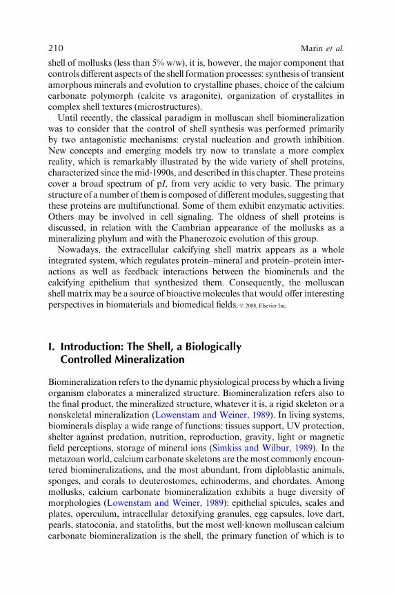
Figure 2 Physiology of the shell calcification in a nacro‐prismatic bivalve, redrawn from
Saleuddin and Petit (1983). The calcification takes place at the distal border of the shell in the
extrapallial space. In the outer epithelium, the cells responsible for the deposition of nacre are
not localized in the same area as the ones responsible for the secretion of prism precursors.
The prisms and nacre tablets are not drawn to scale.
218 Marin et al.
the cytosol to the second compartment involved in shell formation, the extra-
pallial space (Fig. 2). However, in several cases, calcium can be temporarily
stored as intracellular or extracellular amorphous granules. This has been
particularly well studied for bivalves (Fournie and Chetail, 1982; Istin, 1970;
Istin and Girard, 1970) for which a colocalization of granules and carbonic
anhydrase was observed. Amorphous granules can be used by the cells for
detoxifying the cytosol from an excess of calcium or heavy metals (Simkiss,
1977, 1993).Granules are also a source of calcium,which is rapidly available, in
particular, for a rapid shell mineralization and repair. The possibility of outer
epithelial cells to release intracellular calcium granules by exocytosis is not
documented, although suspected.
The second—already mentioned—compartment is the extrapallial space
(Fig. 2). This space is supposed to concentrate the precursor mineral ions,
owing to calcium and bicarbonate pumps. However, these pumps have not

6. Molluscan Shell Proteins 219
been localized on the outer epithelium, mainly because they have been poorly
characterized at the molecular level (Endo et al., 2003). The extrapallial space
is supposed to be the place where the ‘‘mysterious’’ transition from the liquid
precursors to the solid biominerals occurs. The concept of self‐assembling is
currently put forward for explaining how the shell matrix and the mineral
ions interact in a controlled manner to produce a finely structured organo‐mineral material. Chemical analyses of the extrapallial fluid have shown that
it is supersaturated with respect to calcium carbonate (Misogianes and
Chasteen, 1979; Moura et al., 2000). This fluid is also enriched in mineral/
organic mineralization inhibitors, without which calcium carbonate would
precipitate anarchically. This obviously never happens.
The extrapallial space is also the place where the shell matrix is secreted
and where the subtle transition from liquid to solid operates. As said in the
introduction, and detailed later, this matrix is a mixture of proteins, glyco-
proteins, acidic polysaccharides, chitin, and presumably lipids. The matrix
interacts with the mineral ions and controls the shape of the produced
crystals. It is consumed by the system, that is, ‘‘entrapped’’ within the
construction in progress. This means that it has to be brought to the miner-
alization front when required during the whole mineralization period. It is
also likely that several proteins, which are present in the extrapallial fluid,
are not incorporated in the shell. Another unknown parameter is the precise
temporal sequence of secretion of the shell matrix. On the other hand, we
know that the secretory regime is diVerent depending on the position of
the cells involved in this process on the outer epithelium. This has been
demonstrated, both at transcriptional and protein levels, in particular for
species, which exhibit a bitextured shell (an outer prismatic layer, an inner
nacreous layer; Fig. 2). The outer epithelial cells that secrete the matrix
involved in controlling the prism formation occupy a more distal position
(from the shell hinge) than the ones that secrete the matrix involved in the
nacre deposition (Jolly et al., 2004; Sudo et al., 1997). A paper from Takeuchi
and Endo (2005) confirms the existence of two zones in the outer mantle
epithelium, with transcripts specific of the prism zone, transcripts specific of
the nacre zone, and transcripts present in both. Attempts to introduce
an epithelial cell typology have been made (Sud, 2002). At last, because
the deposition of calcium carbonate is accompanied by the release of pro-
tons, these latter have to be reabsorbed by the calcifying epithelium, for
precluding acidification of the extrapallial fluid, and possible resolubilization
of the newly formed minerals. It has been suggested that proton pumps are
involved in extruding protons from the extrapallial fluid toward the cytosol.
However, this process is poorly documented for mollusks (Coimbra et al.,
1988). It has also been suggested that proton pumps work, in some cases,
in the reverse sense, inducing then acidosis in the extrapallial space (Moura
et al., 2003).

220 Marin et al.
The classical view described here above seems a priori ‘‘well established.’’
However, several ‘‘evidences,’’ like the transit of precursor ions, are indirect
or deduced from the general metabolism of the calcifying mantle. Several
physiological aspects are left in the dark or bring unsatisfactory answer. For
example, our description attributes a key role to the extrapallial fluid. This
suggests that there are no direct contacts between the secreting epithelium
and the location where mineralization occurs. This view of a ‘‘remote control
of the mineralization by the epithelium’’ is questionable (Addadi et al., 2006).
Some authors consider that the mantle cells have to keep close contact with
the mineralization front in order to receive feedback molecular signals from
the newly formed biominerals.
Another point, which remains obscure, is the function of hemocytes—free
circulating cells of the hemolymph—in the shell construction process. Hemo-
cytes play a major role in the immune defense of mollusks (Glinski and
Jarosz, 1997) and in tissue repair (Serpentini et al., 2000). Their role in shell
repair processes has already been underlined (Bubel et al., 1977; Watabe,
1983), but their contribution to the normal shell formation may be widely
underestimated. A milestone paper from Mount et al. (2004) attributes to
hemocytes (of the granulocyte type) of the oyster Crassostrea virginica, an
important function in the normal shell construction process, by releasing
calcite crystals—not amorphous granules—which can be remodeled at the
mineralization site. So far, it is diYcult to evaluate whether this process is
particular to the studied model or whether it is a general metabolic pathway
that mollusks use for mineralizing their shell.
C. Transient Amorphous Calcium Carbonate
One aspect, which has for a long time been passed largely unnoticed, but
which needs to be urgently reevaluated, is the key role played by amorphous
calcium carbonate (ACC) in molluscan shell formation and, more generally,
in all calcium carbonate biomineralizations (Weiner et al., 2003). A mineral
phase is considered to be amorphous when it lacks the long‐range order,
that is, when it does not have the regular repeating lattice structure that
provides the basis for so many of the features of a crystal (Simkiss, 1993).
Several amorphous minerals can exhibit a short‐range order, which is not
conserved at a longer scale. This implies that the mineral does not give an
X‐ray diVraction pattern characterized by spots materializing the diVerentdiVraction plans. Amorphous minerals display several advantages in com-
parison to their crystalline equivalent: their formation requires less energy;
they are more easily solubilized, which means that their constitutive ions
are more easily available; they incorporate ionic impurities with a higher
tolerance than the crystalline form; they exhibit percolation channel that

6. Molluscan Shell Proteins 221
allows ion di Vusion, that is, they can be more easily remod eled ( Simkiss,
1991 ).
Recent X ‐ ray di Vracti on data indica te that the early larva l shell miner ali-
zatio n (prod issocon ch I) may be a transient a morphous phase, a fact that
earlier works on Tridacn a squamo sa ( LaBarb era, 1974 ) or on Crassos trea
gigas (Lee , 1990 ) had not detected . The presence of ACC has been de mon-
strated for the fres hwater snail, B. glabr ata ( Hass e et al ., 2000 ; Marxen et al.,
2003a) , for which the trans form ation of ACC into aragoni te could be
recorded at diV erent developm ental stage s. In the musse l, M. eduli s , the
first mineral formed is also ACC, but its quantity dramatically drops only
40 hours after fertilization (Medakovic, 2000), in profit of the aragonitic
phase. The early developmental stages of the oyster, Ostrea edulis, are also
marked by an extremely broad X‐ray diVraction ‘‘peak’’ that may be inter-
preted as ACC (Medakovic et al., 1997). In the clam Mercenaria mercenaria,
the prodissoconch I contains ACC, which transforms after several days in
aragonite. In the oyster C. gigas, the prodissoconch I contains ACC and
aragonite (Weiss et al., 2002). By extrapolating these data to adult shells, and
by keeping in mind the presence of amorphous granules in the molluscan
mantle tissues, it is conceivable that the whole adult shell formation occurs
via a transient ACC phase. As we mention in the next section, recent findings
have shown that interfacial amorphous phases exist also in the ‘‘finished’’
shell, which suggests a stabilization mechanism of ACC.
III. The Topographic Models of Shell Mineralization
One key issue in research on molluscan shell biomineralization is the under-
standing of the relationships between the organic matrix and the mineral
phase at ultrastructural level. This question is central to current hypotheses
on biologically controlled mineralizations, but is still extremely debated.
From the late 1960s when the early topographic models of molluscan miner-
alization emerged to now, there has been a considerable evolution of the
concepts, partly because of the evolving methodologies used for observing
biominerals. In the early years, scanning electron microscopy (SEM) was the
unique investigation tool. The use of surface etching treatments, partial
decalcification and fixation (Mutvei, 1979), enzymatic degradation brought
additional information by revealing extremely fine substructures (Cuif et al.,
1983). At high magnification, transmission electron microscope (TEM) and,
more recently, cryo‐TEM and atomic force microscope (AFM) brought
surprising ultrastructural informations such as the presence of crystal nano-
domains. Finally, in the last years, XANES (X‐ray absorption near edge
spectroscopy) and NanoSIMS brought additional structural informations.
SEM combined with immunogold proved to be a promising technique (Marin

222 Marin et al.
et al., 2007) for visualizing single‐protein components within biominerals.
In the next section paragraph, we will discuss some recent outcomes on nacre
and prisms, the two most familiar molluscan shell textures.
A. Early Nacre Descriptions and Models
For several reasons such as its economical interest or its mechanical proper-
ties, nacre, also called mother‐of‐pearl, is still one of the most‐studied shell
textures. Many consider it as the reference model because of its apparent
geometrical simplicity and because of its universality among mollusks. Nacre
is indeed a widespread molluscan shell texture, and is, with one exception, a
bryozoan (Weedon and Taylor, 1995), typical of this phylum. It is repre-
sented within the three main classes of mollusks, bivalves, gastropods,
and cephalopods. The ‘‘nacre’’ terminology refers to a well‐defined type of
microstructure, described as follows: small flat tablets of aragonite, about
half a micron thick, which are tightly packed together by organic cement.
The tablets can be rectangular, hexagonal, or rounded, and look like mono-
crystals. They form superimposed layers of uniform thickness. Basically,
there are two broad types of nacre, depending on the disposition of tablets
(Erben, 1972; Nakahara, 1991): ‘‘brick wall’’ nacre, which is classical among
bivalves, ‘‘columnar’’ nacre, found in gastropods (Fig. 3). In the first type, in
cross section, crystals are positioned in staggered rows, just like bricks in a
wall (Checa and Rodriguez‐Navarro, 2005; Oaki and Imai, 2005). Bivalve
nacre tablets have their a, b, and c axes co‐oriented, with the c axis perpen-
dicular to the nacre surface, and the b axis parallel to the local growth
direction of the shell margin (Checa et al., 2006). In the second type, flat
tablets are aligned on top of each other, and thus, form piles (or towers)
of crystals (Lin and Meyers, 2005). Tablets of the same pile are co‐oriented(c axis along the axis of the pile), but from pile to pile, the a and b axes are not
ordered.
TEMstudies have shown that a thin layer of organicmatrix, the interlamellar
matrix, delimitates the lower and upper tablet surfaces (Fig. 3). The thickness of
this matrix is about 20 nm. Within a same lamella, an organic matrix [the
intercrystalline matrix of Bevelander and Nakahara (1969)] separates adjacent
tablets. Early amino acid analyses showed that the matrix around the tablets
was enriched in Ala and Gly residues, a composition, which conferred to the
matrix hydrophobic properties, similar to that of worm silk. Additional ultra-
structural studies showed that nacre tablets were not homogeneous. In particu-
lar, Crenshaw and Ristedt (1975) evidenced that sulfated polysaccharides
were localized in the central part of nacre tablets. These organic compounds
were supposed to act as crystal nucleators. Mutvei (1979), by etching nacre

Figure 3 Structure of the two main molluscan nacre textures. (A and B) SEM pictures of the nacre of the freshwater bivalve U. pictorum.
(C and D) SEM pictures of the nacre of the gastropod Haliotis tuberculata (bar scales ¼ 10 mm). (E) The ‘‘brick‐wall’’ model of bivalvian
nacre. (F) The ‘‘columnar’’ model of gastropod nacre. These simplified models, adapted from Nakahara (1991), do not take in account the
existence of pores in the interlamellar organic matrix, the substructures of nacre tablets, and the existence of a thin ACC layer around
the tablets. The constituents are: E, the secreting mantle epithelium; S, the organic sheets; SS, the newly formed surface sheets; Cr, the
aragonite crystals; T, the top of the newly formed crystals.

224 Marin et al.
tablets with a glutaraldehyde‐acetic acid solution, observed extremely complex
structures such as twinning patterns or concentric growth lamellae.
One of the first ‘‘modern’’ nacre models was proposed by Bevelander and
Nakahara (1969). The ‘‘compartment model’’ supposed that the aragonite
nacre tablets grow in a preformed mold made from the interlamellar matrix.
The main drawback of this model was to ignore all the organic ingredients of
the matrix, namely chitin, hydrophobic ‘‘silk‐fibroin‐like’’ proteins and
above all, acidic Asp‐rich proteins. The next model, published in the early
1980s by Weiner and coworkers, integrated all the recent findings of that
time: an acidic template for nucleating crystals (Weiner and Hood, 1975),
acidic macromolecules for inhibiting the crystal growth (Wheeler et al.,
1981), specific amino acid sequences (Asp‐rich) for chelating calcium ions
(Weiner, 1979, 1983), sulfated polysaccharides for attracting calcium ions
(Addadi et al., 1987). In this model, the insoluble framework was constituted
by a chitin core taken in sandwich between two hydrophobic silk fibroin‐likesole, on top of which lay a �‐sheet of Asp‐rich soluble proteins. The soluble
polyanionic sheet was supposed to function as a template by nucleating
aragonite crystals, while covalently bound acidic polysaccharides were sup-
posed to concentrate calcium ions at the vicinity of the template. The growth
of the crystal was stopped by the addition of an inhibiting layer of acidic
macromolecules on top of the newly formed tablets. The successive steps of
nucleation and inhibition were explaining the regularity and repetitiveness of
nacre. Because the aragonite tablets nucleated and grew on an organic
template, this crystal growth model was assimilated to heteroepitaxy.
B. Recent Nacre Models and Evolving Views
The heteroepitactic model for nacre achieved a frank success for more than a
decade, until AFM and TEM observations of the columnar nacre of the
abalone showed that holes, of diameter comprised between 5 and 50 nm,
were present in the interlamellar matrix (SchaVer et al., 1997; Song et al.,
2003). This finding suggested that nacre platelet grows in continuity with the
underlying ones, through mineral bridges, and not by heteroepitaxy. Song
et al. (2003) calculated that each platelet exhibits about 1400–1900 holes.
Holes had been observed previously, in particular by Mutvei (1969) and
Nakahara (1991), in gastropod and bivalve nacre. At that time, it was
suggested that the interlamellar holes facilitate the passage of mineral pre-
cursors for filling the empty compartments. So far, we do not know whether
the presence of holes is a general feature of nacre or represents particular
cases. Furthermore, it is still unclear whether they are mineral bridges or just
holes of a sieve, for allowing diVusion of the organic and mineral precursors
to the site of mineralization.

6. Molluscan Shell Proteins 225
Another drastic evolution of the topography of the model occurred about
6 years ago, when Levi‐Kalisman et al. (2001) observed the nacre of the
bivalve Atrina with cryo‐TEM in the hydrated state. The changes go as
follows: �‐chitin is the highly ordered polymer, which gives the framework
that organizes the orientation of the crystals; the silk fibroin‐like proteins,
which are highly insoluble in their final state, are secreted as a disordered gel;
minerals grow in this gel and push it aside when they laterally extend; the gel
comprises also clusters of acidic macromolecules that are involved in nucle-
ating crystals. This model was nicely reshaped and integrated in a dynamic
perspective (Addadi et al., 2006), which presents the formation of nacre in
four stages: (1) assembly of the matrix (chitin, then silk gel); (2) formation of
the first mineral, ACC; (3) nucleation of aragonitic tablets via polyanionic
polymers; and (4) growth of the tablets, first in thickness (until reaching the
top interlamella) then laterally.
Although there is a general consensus on the fact that nacre tablets grow
from their center and expand laterally until reaching the confluence with
neighboring tablets, the ultrastructure of single‐nacre tablets remains un-
clear. Histochemical observations of Nautilus nacre by Nudelman et al.
(2006) confirmed the old finding of Crenshaw and Ristedt (1976), that is,
the concentration of reactive groups (carboxylate), presumably involved in
nucleating aragonite, in the center of single tablets. In addition, a zonation
was observed, which consisted of, from the tablet center to the periphery, a
central ring‐shaped area rich in sulfates, an intermediate zone rich in carbox-
ylate, and finally a tablet‐surrounding matrix rich in carboxylates and sul-
fates. Another recent paper (Nassif et al., 2005) showed that the nacre tablets
of the abalone were coated by an extremely thin layer (3‐ to 5‐nm thick) of
ACC. This layer may be a stabilized remnant of the transient ACC phase,
described by Addadi et al. (2006). A possible scenario for a single‐tabletgrowth suggests a lateral tablet expansion and the subsequent expulsion of
the gel. The process is driven by hydrophobic interactions. By doing so, the
organic ‘‘impurities’’ progressively concentrate in a front at the interface
between the mineral and the gel. During this growth phase, the transient
ACC is replaced by aragonite. When the centrifugal front meets a similar
front of the neighboring tablet, the degree of impurities becomes so high that
ACC is stabilized, which prevents further crystallization of aragonite.
At higher magnification, single‐nacre tablets exhibit a remarkable hierar-
chical architecture, which is somehow diYcult to conciliate with what has
been described above. Nacre tablets have fractal properties in the sense
that they exhibit diVerent levels of substructures that reproduce the same
motif: these are, for example, flat nanobuilding blocks (Oaki and Imai, 2005),
or ‘‘nanotablets’’ of 30‐ to 180‐nm long and less than 100‐nm thick, that
self‐assemble and self‐orientate. The laminated structure of single‐nacretablets has also been observed independently by Rousseau et al. (2005).

226 Marin et al.
AFM studies by the same authors suggest that the nanograins that constitute
each platelet are encapsulated in a continuous network of an organic intra-
crystalline phase (Rousseau et al., 2005). This phase looks like a foam, which
suggests that the early steps of tablet formation are performed in an emul-
sion. Reticulate circular imprints of an organic framework at triple junctions
between mature platelets in bivalve nacre have also been observed (Rousseau
et al., 2005). They are supposed to localize the spot where the new tablets
grow.
Clearly, since the beginning of the twenty‐first century, the nacre model
knows a complete revolution and requires the integration of diVerent levelsof observation, from micrometric to nanometric scales. The future topo-
graphic models will have to consider the fine architecture of the matrix, the
sequence of the secretory events, as well as purely crystallographic and
geometrical considerations, such as crystal competition.
C. Prism Models
Beside the well‐studied nacre, prisms constitute another key model and an
important shell texture found most frequently, but not exclusively, in mollus-
can outer shell layer, in particular, in gastropods, cephalopods, and bivalves.
Like nacre, prisms are supposed to be an archaic type of shell texture. Because
prisms are often associated to nacre, it has been proposed that nacre evolved
through simple horizontal partitioning of vertical prisms (Carter and Clark,
1985; Taylor, 1973). This appealing idea, based on simple geometric consid-
erations, needs to be reevaluated with accurate crystallographic and bio-
chemical criteria. This may help to understand the transition from one
microstructure to the other and to reconstitute primitive shell textures. It is
interesting to notice that prism‐like or palisade‐like minerals, with growth
axis perpendicular to the growth plan, represent an extremely common and
fast strategy found by diVerent biomineralizing systems (brachiopods, mol-
lusks, eggshell) for filling a space with minerals. In a first approximation,
prismatic textures exhibit many similarities with purely chemical crystal
growth. However, as we briefly show here, this view is probably oversimpli-
fied, and the deposition of prisms, similarly to nacre ones, is finely regulated
over diVerent scales.Prisms are calcitic or aragonitic needles of various lengths and diameters,
from the thin oblique calcitic prisms of the edible mussel, M. edulis, to the
large‐sized calcitic prisms (‘‘simple’’ prism type), developed perpendicularly
to the shell surface, among the fan mussel Pinna nobilis, or the aragonite
prisms of the freshwater mussel, Unio pictorum. Prisms of the outer shell
layer are secreted on the inner surface of the periostracum, at the growing
shell edge. They grow inward by the accretion of crystal units. They are

6. Molluscan Shell Proteins 227
enveloped by an organ ic insol uble and hydropho bic sheath, whi ch form s a
hone ycomb ‐ like fram ework . This sheath is not homogeneo us, but compo sed
of at least three layer s ( Dauphin, 2002; M arin et al ., 2007 ). It mainta ins all
the prism s toget her an d allows a certa in flexibi lity of the structure. When
isolated from their org anic envelopes , prism s compri se an intr acrystall ine
organic fraction. In calci tic prism s, this matrix is pa rticular ly acidi c ( Marin
et al ., 2005 ).
Like for nacre, the form ation of prism s is far from being eluci dated. Fr om
Grigo r’ev (1965) , it is well known that prism ‐ like cryst als can be obtaine d bypurely ch emical ‘‘com petition for sp ace’’ cryst al growth. The star ting point
includes nuc leation spots, more or less unifor mly spread on a surfa ce, from
which spherul ites grow concen trically. W hen the spherul ites come into con-
tact, their gro wth is co nstrained in one direction, perpendicul ar to the
surfa ce. This ha ppens in natural environm ents wi thout the ne ed of a sophis-
ticated organic templ ate. Comp etition for space can be easil y simu lated
( Ubukata , 1994, 1997 ). By certain aspec ts, the prism gro wth in moll usks
looks like crystal compet ition. In a simila r way, the early step of molluscan
prism co nstruction is the form ation of spherul ites in the inn er surfa ce of the
perios tracum . This has be en clear ly sho wn for the bival ves, Pinna nobilis
( Cuif et al ., 1983 ) and Lampro tula sp. (C heca and Rodri guez ‐ Navar ro, 2001).
How ever, a compe tition for space phe nomeno n might occu r only in the early
steps of prism form ation (disapp earance of minut e prisms just below the
perios tracum ; see Checa et al ., 2005 ), but may not de scribe accurat ely the
subsequ ent steps of prism grow th, mainly for two reasons : the she aths are
form ed before the prism miner al infilling and the growth is con strained by
the organic shea ths a round the prisms.
Anothe r aspect that renders a simp le ‘‘com petition for space’ ’ mod el
inapprop riate is the mult iscale struc ture of prism s and their strik ing com-
plexi ty. A wel l‐ known exampl e is that of the ‘‘s imple type’ ’ calciti c prism s of
Pinna nobil is. Optical ly, each prism of P. nobilis be haves like a mon ocrystal,
with a singl e extincti on when obs erved wi th polari zed ‐ analyze d light (Cui fet al ., 19 83 ). How ever, enzymat ic treatment of the prism preparat ion shows
that each prism is consti tuted of a pile of flat crysta l uni ts, which can be
entire ly dissoc iated after pyrolys is. These cryst allites are the grow th units .
They are separated from each other by an organic intracrystalline template.
Within a pile, they are perfectly positioned according to their a, b, and c axes.
In spite of looking homogeneous, these crystallites are composed of subdo-
mains, emphasized by enzymatic treatments (Cuif et al., 1983) or immunos-
taining (Marin, unpublished data). These subdomains might as well be
composed of nanocrystal aggregates. Similarly to P. nobilis, ultrastructural
observation of the prisms of Cristaris plicate (Tong et al., 2002) revealed a
complex lacelike framework of intracrystalline matrix. Checa et al. (2005)
hypothesized that the interprismatic ‘‘honeycomb‐like’’ sheaths are formed

228 Marin et al.
by interfacial tensions that occur in a precursor liquid–liquid emulsion. This
elegant hypothesis needs further testing.
Is there a unique prism model? Nothing is less certain. We describe similar
objects by using a single terminology. However, many ultrastructural studies
show that significant diVerences occur, even in closely related taxa, as shown
in Fig. 4. The best example is that of Pinna nobilis and Pinctada margaritifera
(Cuif et al., 1991; Dauphin, 2003). In cross sections (perpendicular to the
growth axis), the prisms of Pinna nobilis look homogeneous and behave like
monocrystals. On the contrary, those of Pinctada margaritifera exhibit sinu-
ous intraprismatic membranes (particularly well visible by SEM, after etch-
ing) that separate the section in domains, and these domains do not have the
same crystallographic orientation. Another case is the prismatic outer layer
of Unio, which is absolutely diVerent from the two types cited above: the
prisms of Unio are composed of single‐crystal fibers radiating from spher-
ulites (Checa and Rodriguez‐Navarro, 2001; Cuif et al., 1983). Clearly,
important eVorts need to be put in the elucidation of the prism growth and
to relate it to the biochemical properties of the associated matrix.
Figure 4 Prismatic microstructures among bivalve shells. (A) Unio pictorum. (B) Anodonta sp.
For A and B, the aragonitic prisms (above) are in contact with the nacreous layer. (C) Oblique
thin calcitic prisms of the edible mussel Mytilus edulis. (D) Calcitic prisms of Atrina rigida.

6. Molluscan Shell Proteins 229
IV. Molluscan Shell Proteins: Characterization of TheirPrimary Structure
In parallel to the structural studies on the diVerent shell textures, a consider-
able eVort was realized, in the last three decades, for identifying the diVerentmacromolecules that constitute the shell matrix and for obtaining informa-
tion on their primary structures. In most of the cases, these macromolecules
were analyzed after the dissolution of the mineral phase. Until now, the most
commonly used reagents are EDTA, a calcium‐chelating agent, which is
eVective at neutral pH, weak dilute acids, acetic or formic acids, or rarely
dilute hydrochloric acid. Our preference goes to cold dilute acetic acid,
progressively added to the cleaned shell powder suspended in Milli‐Qwater, until reaching pH 4, the decalcification process being performed at
4 �C (Marin, 2003). We assume that this procedure minimizes protein degra-
dation and precludes the formation of macromolecular aggregates, as EDTA
does. Other extraction processes include a soft—but long—demineralization
of the powder on a cation‐exchange resin (Albeck et al., 1996), or extraction
with water (Pereira‐Mouries et al., 2002). However, in this latter case, the
most strongly mineral‐linked macromolecules are not extracted.
The decalcification procedure yields two organic fractions, one soluble in
the decalcifying solution, the other one strongly insoluble. The ratio between
the two fractions can considerably vary: while the soluble fraction represents
between 0.03 and 0.5 wt %, the insoluble fraction varies in greater propor-
tions: from 0.01% (in some crossed‐lamellar neogastropods for instance) to
4–5 wt % of the shell of the abalone or of the nautilus! Usually, the second
fraction is discarded by centrifugation. In some cases, the insoluble fraction
may be partially dissolved by using strong denaturing agent (urea) and/or
by heating. The soluble extract can be cleared from decalcification salts by
ultrafiltration or dialysis. It can be further fractionated according to standard
biochemical techniques, electrophoresis, or chromatography (gel perme-
ation, ion exchange, aYnity). However, because molluscan shell proteins
have a nonstandard behavior due to polydispersity, multiple anionic charges,
posttranslational modifications, classical fractionations usually fail in resolv-
ing the soluble matrix in discrete macromolecules. This technical obstacle,
found also with several other calcified tissues, pestered for more than
two decades the life of researchers involved in biomineralization studies!
This explains in particular why the first partial amino acid sequence from a
mollusk shell was obtained in the early 1990s (Rusenko et al., 1991), and the
first full‐length sequence only in 1996 (Miyamoto et al., 1996).
The search for the primary structure of molluscan shell proteins benefited
from the major technical advances in molecular biology, in particular, from
the possibility to use degenerate oligonucleotide probes deduced from short

230 Marin et al.
partial N‐terminal or internal amino acid sequences. This allowed fishing out
the corresponding transcript by RT‐PCR or by cDNA library oligoscreen-
ing. Another strategy successfully developed by us was to use polyclonal
antibodies raised against shell matrix for screening expression cDNA
libraries (Marin et al., 2003a). On the 43 fully known sequences, 39 were
obtained via molecular biology and only 4 by direct protein sequencing. It is
predictable that the number of shell proteins will ‘‘explode’’ in the near
future, owing to the increasing number of fully sequenced genomes
(Livingston et al., 2006), and in the absence of genomic data, owing to EST
technique applied on shell ‘‘secretome’’ (Jackson et al., 2006).
In this chapter, we give a brief description of the diVerent shell proteins,one after the other. Complementary information can be retrieved in Marin
and Luquet (2004), in Matsushiro andMiyashita (2004), and in the review of
Zhang and Zhang (2006). What was possible few years ago, when the number
of identified and named proteins was still reasonable, would be fastidious and
redundant. We deliberately choose to present the known molluscan shell
proteins in three groups, according to their theoretical isoelectric point
(Fig. 5). The first group comprises proteins, the pI of which is below 4.5,
18
160
140
80
60
40
20
00 2 4 6 8 10 12
�20
108
93
1
24
5
67
1314
16
1512
11
1719 22
21MW
pI
Figure 5 Graphical representation of the distribution of the molecular weights (MW) of all
known molluscan shell proteins versus their isoelectric point (pI ). The theoretical MW and pI
were computed (http://www.expasy.ch/tools/pi_tool.html) after identification and removal of the
signal peptide (http://www.cbs.dtu.dk/services/SignalP/).□¼ proteins associated with aragonite;
◆ ¼ proteins associated with calcite; (� ¼ protein associated with both aragonite and calcite
(1 ¼ aspein; 2 ¼ Asp‐rich proteins; 3 ¼ MSP‐1; 4 ¼ MSP‐2; 5 ¼ MSI31; 6 ¼ prismalin‐14;7 ¼ N‐14/N16/pearlin/perline proteins masking AP7 and AP24; 8 ¼ MSI60; 9 ¼ mucoperlin;
10 ¼ nacrein from P. fucata; 11 ¼ MSI7; 12 ¼ dermatopontin; 13 ¼ tyrosinase‐like1;14 ¼ nacrein from T. marmoratus; 15 ¼ perlucin; 16 ¼ shematrin proteins; 17 ¼ perlustrin,
18 ¼ lustrin A; 19 ¼ perlwapin; 20 ¼ N‐66; 21 ¼ tyrosine‐like2; 22 ¼ KRMPs).

6. Molluscan Shell Proteins 231
the second group comprises proteins with a pI in the range 4.5–7, and the
third group comprises proteins with a pI above 7. We are fully aware that this
grouping is artificial and arbitrary but practical in the absence of clearly
identified protein families.
A. Extremely Acidic Shell Proteins
This first group, the most homogeneous one, comprises the most acidic shell
proteins (Marin and Luquet, 2007). The existence of such proteins is known
since the pioneering work of Weiner and Hood (1975), followed by extremely
precise chromatographic characterization (Weiner, 1979, 1983). However,
for the reasons described above, they were the most diYcult to purify. In
particular, they do not stain correctly on SDS‐PAGE (Marin et al., 2001).
Because of their negative charge, they are even suspected to diVuse readily
out of the electrophoresis gel, and they subsequently require a double fixation
(Gotliv et al., 2003). The two first full‐length sequences of very acidic proteins
deduced from their transcript were published in 1997 (MSI31; Sudo et al.,
1997) and in 2001 (MSP‐1; Sarashina and Endo, 2001). Today, this group
comprises only six proteins (Table I).
One striking feature of these proteins is their association with calcitic bio-
minerals rather than aragonite.MSI31, aspein, prismalin 14, andAsp‐richwereretrieved from calcitic prism textures, and MSP‐1 and MSP‐2 (SP‐S) fromfoliated calcite. The finding that acidic proteins are preferentially associated
with calcite in mollusk shell is not new: Hare (1963) already noticed that ‘‘the
organicmatrices from the calcite layers have a consistently higher ratio of acidic
tobasic aminoacids than the aragonitic shell units.’’ The reasonof this selection
is intriguing, but remains unknown.
Another feature associated with very acidic molluscan shell proteins is that
they are all singularly enriched in Asp residues. The ‘‘choice’’ for this amino
acid, rather than Glu, is remarkable, although poorly understood. It may
meet stereochemical requirements, the Asp side chain being shorter than that
of Glu. Because of their high amount of Asp residues and the side chains are
negatively charged under physiological conditions, these proteins are sup-
posed to easily bind calcium ions. They consequently belong to the group of
low‐aYnity, high‐capacity calcium‐binding proteins (Maurer et al., 1996),
which implies that they do not exhibit the typical ‘‘high‐aYnity, low‐capacity’’canonical calcium‐binding domains, such as EF‐hand (Kretsinger, 1976).
Their moderate aYnity for calcium is compatible with a reversible binding
of calcium ions.
MSI31 is a Gly‐rich protein of the insoluble matrix, and supposed to be
primarily a ‘‘framework’’ protein (Sudo et al., 1997), because of the 10 short
poly‐Gly blocks, distributed mainly in the N‐terminal domain. It exhibits

Table I Unusually Acidic Molluscan Shell Proteins (with a Theoretical Isoelectric Point Below 4.5)a
Protein Name Species
Microstructure
(polymorph) MW (kDa) pI (%AspþGlu)
Swiss‐Prot/TrEMBL
Accession
Number References
BIVALVIA Aspein Pinctada
fucata
Prisms (calcite) 39.3/41.2 1.67 (61.9) Q76K52 Tsukamoto et al.,
2004
MSI31 Pinctada
fucata
Prisms (calcite) 32.85/31 3.8 (14) O02401 Sudo et al., 1997
Prismalin‐14 Pinctada
fucata
Prisms (calcite) 11.9/13.5 4.24 (10.5) Q6F4C6 Suzuki et al., 2004
MSP‐1 Patinopecten
yessoensis
Foliated
(calcite)
74.6/76.4 3.34 (22.8) Q95YF6 Sarashina and
Endo, 1998, 2001
MSP‐2/SP‐S Patinopecten
yessoensis
Foliated
(calcite)
27.9/29.8 3.48 (22.3) Q6BC34 Hasegawa and
Uchiyama, 2005
Asp‐rich protein 1 Atrina rigida Prisms (calcite) 6.6/8.5 3.34 (50.8) Q5Y821 Gotliv et al., 2005
Asp‐rich protein 2 Atrina rigida Prisms (calcite) 15/17 2.89 (52.8) Q5Y822 Gotliv et al., 2005
Asp‐rich protein 3 Atrina rigida Prisms (calcite) 16.5/18.4 2.75 (60) Q5Y823 Gotliv et al., 2005
Asp‐rich protein 4 Atrina rigida Prisms (calcite) 18/19.9 2.73 (56.2) Q5Y824 Gotliv et al., 2005
Asp‐rich protein 5 Atrina rigida Prisms (calcite) 17.4/19.3 2.76 (57.5) Q5Y825 Gotliv et al., 2005
Asp‐rich protein 6 Atrina rigida Prisms (calcite) 18.2/20 2.72 (59.2) Q5Y826 Gotliv et al., 2005
Asp‐rich protein 7 Atrina rigida Prisms (calcite) 25.8/23.9 2.54 (66.2) Q5Y827 Gotliv et al., 2005
Asp‐rich protein 8 Atrina rigida Prisms (calcite) 25.3/27.2 2.53 (65.1) Q5Y828 Gotliv et al., 2005
Asp‐rich protein 9 Atrina rigida Prisms (calcite) 18.2/20 2.72 (59.2) Q5Y829 Gotliv et al., 2005
Asp‐rich protein 10 Atrina rigida Prisms (calcite) 20/21.8 2.68 (60) Q5Y830 Gotliv et al., 2005
aThe sequences of all these proteins were deduced from their respective transcript sequence. Prismalin‐14 was also biochemically characterized. In the
MW column, the first number corresponds to the molecular weight of the protein without its signal peptide, and the second one to the unprocessed protein.

6. Molluscan Shell Proteins 233
acidic C‐terminal motifs (6 XSEEDY, where X is D or E, and Y is M or T).
The acidic domainmay be involved in nucleating crystals, or binding calcium,
but this has not been tested. The hydrophobic N‐terminus may adopt a
�‐sheet conformation.
MSP‐1, the second unusually acidic protein of the Japanese scallop Pati-
nopecten yessoensis, is enriched in Ser, Asp, and Gly residues and exhibits a
modular structure, with a short‐basic domain, close to the N‐terminus and
two GS domains that alternate with D‐rich domains (Sarashina and Endo,
1998, 2001). The two Asp‐rich domains fit with the initial model of Weiner
and coworkers since they exhibit DGS and DS motifs. They also present
numerous DD repeats. All these motifs are suspected to bind calcium ions
or to interact with calcite crystals. In addition, several serine residues are
putatively phosphorylated or glycosylated. MSP‐1 exhibits homologies with
dentin phosphophoryns, very acidic proteins of the teeth. Recently was
found MSP‐2, also called SP‐S, another shell protein of the Japanese scallop
(Hasegawa and Uchiyama, 2005). MSP‐2 is a Ser‐Gly‐Asp‐rich protein of
the scallop, which exhibits 91% identity with MSP‐1. It may represent a
shortened variant of MSP‐1.The third unusually acidic protein is aspein, a protein of the pearl oyster
Pinctada fucata. The composition of aspein is remarkable since Asp residues
represent 60.4% of the whole protein, and its theoretical pI is 1.67, which would
make it the most acidic protein found to date! The two other abundant amino
acids are Gly (16%) and Ser (13%). The main body of aspein is composed of
58 poly‐Asp blocks (of 2–10 Asp residues long) interspersed by SG dipeptides.
Some Ser residues may be phosphorylated. Aspein exhibits some simila-
rities with aspolin, phosphophoryn, and bone sialoprotein‐binding protein.
The primary structure of aspein suggests that it is a high‐capacity, low‐aYnity
calcium‐binding protein.Asp‐rich is a family of 10 related proteins, composed of the following
domains, from N‐ to C‐terminus: hydrophobic (signal peptide), short basic,
acidic 1, variable acidic, DEAD repeats, and acidic 2. Interestingly, the acidic 1
domain has a high homology with calsequestrins, calcium‐binding proteins
from cardiac and skeletal muscles, and may consequently bind calcium. The
variable acidic domain exhibits long stretches of poly‐Asp. TheDEADmotif is
also found in helicases, enzymes that separate the two DNA strands. Asp‐richand aspein are closely related proteins since they share 48% homology.
The last member is prismalin‐14, the single one characterized both at
protein and at transcript levels (Suzuki et al., 2004). Prismalin was extracted
from the insoluble hydrophobic framework of the prismatic layer of Pinctada
fucata. It is a G/Y‐rich protein, representing, respectively, 27.6% and 20% of
the amino acid composition. It exhibits PIYR repeats, a G/Y‐rich region and
N‐ and C‐terminal D‐rich calcium‐binding regions. It inhibits the precipita-
tion of calcium carbonate in vitro and induces morphological changes of

234 Marin et al.
calcite crystals. Prismalin 14 as well as aspein and MSI31 are specifically
expressed in the mantle, in the region, which secretes the prism matrix
(Takeuchi and Endo, 2005). In addition, the expression levels of these three
proteins are correlated, which suggests that they are secreted at the same time.
B. Moderately Acidic Shell Proteins
As shown in Table II, the second group corresponds to acidic proteins, with
pIs between 4.5 and 7. In this disparate group, one finds gastropod and
bivalve nacreins, MSI60, MSI7, the N14/pearlin/N16 family, mucoperlin,
AP7, AP24, a tyrosinase‐like protein, perline, and a snail dermatopontin.
First of this list is nacrein, by many aspects the most‐studied molluscan
shell protein family (Matsushiro andMiyashita, 2004; Miyamoto et al., 1996,
2003, 2005; Miyashita et al., 2002; Takeuchi and Endo, 2005). Nacrein, the
first protein whose primary structure was deciphered, was initially found as a
50‐kDa EDTA‐soluble protein of the nacreous layer of the Japanese pearl
oyster Pinctada fucata. Later on, a similar protein (nacrein), with an identical
N‐terminus, was found in association with the prismatic layer (Miyashita
et al., 2002). In situ hybridization studies (Miyamoto et al., 2005; Takeuchi
and Endo, 2005) shows that nacrein is ubiquitous and displays probably the
same functions in the two layers. Nacrein is also the first protein, which was
proved to work as an enzyme. Nacrein exhibits several GXN repeats (where
X is frequently D, N, or E), flanked by two carbonic anhydrase‐like sub-
domains. Strikingly, these two subdomains have a relatively high homology
with human carbonic anhydrase II (CA). In addition, the first nacrein CA‐like domain exhibits the three histidine residues involved in zinc binding,
typical of CA. A full‐length recombinant nacrein inhibits the in vitro precipi-
tation of calcium carbonate. Interestingly, the recombinant nacrein, which
lacks the central GXN repeat domain, does not exhibit this property, where-
as the repeat domain, tested alone, has a strong inhibiting ability (Miyamoto
et al., 2005). Another nacrein was found in the shell of the gastropod Turbo
marmoratus (Miyamoto et al., 2003). This moderately acidic protein has a
longer repeat domain, constituted of GNmotifs. At last, N66, retrieved from
the Australian pearl oyster Pinctada maxima, belongs also to the family
(Kono et al., 2000). It exhibits two carbonic anhydrase subdomains, and a
longer central repeat domain constituted by 46 GXN motifs interspersed by
12 GN motifs. The repeat domain of N66 is much less acidic than the one of
nacrein, which implies that N66 has a basic pI.
Among the acidic shell proteins, MSI60 is an insoluble framework protein
retrieved initially from the nacreous layer. It exhibits 11 poly‐Ala blocks and
39 poly‐Gly blocks dispersed throughout the sequence. The poly‐Ala blocks
confer to MSI60 some homologies with spider silk fibroins. The MSI60

Table II Moderately Acidic Molluscan Shell Proteins (with a Theoretical Isoelectric Point 4.5 � pI � 7)a
Protein Name Species
Microstructure
(Polymorph) MW (kDa)
p I (% Asp
þ Glu)
Swiss ‐Prot/TrEMBL
Accession
Number References
BIVALVIA N14 Pinctada maxima Nacre (aragonite) 13.7/16.4 4.8 (15.8) Q9NL39 Kono et al., 2000
Nacrein Pinctada fucata Nacre (aragonite) 48.2/50.1 6.85 Q27908 Miyamoto et al., 1996
N16/Pearlin Pinctada fucata Nacre (aragonite) 12.8/15.4 5.14
(16.9)
O97048 Samata et al., 1999;
Miyashita et al.,
2000
MSI60 Pinctada fucata Nacre (aragonite) 61.7/60 4.8 (5.9) O02402 Sudo et al., 1997
MSI7 Pinctada fucata Prisms (calcite) 7.3/9.3 5.98 (2.6) Q7YWA5 Zhang et al., 2003a
Tyrosinase‐ likeprotein 1
Pinctada fucata Prisms (calcite) 56.3/58.3 6.5 (9.3) A1IHF0 Nagai et al., 2007
Perline Pinctada
margaritifera
Nacre (aragonite) 13.6/16.2 4.7 (15.8) Q14WA6 Montagnani
et al., 2006
Mucoperlin Pinna nobilis Nacre (aragonite) 65.4/66.7 4.87 (9.5) Q9BKM3 Marin et al., 2000
AP7 Haliotis rufescens Nacre (aragonite) 7.6/9.9 5.43
(12.1)
Q9BP37 Michenfelder et al.,
2003
AP24 Haliotis rufescens Nacre (aragonite) 17/19.6 5.53
(13.6)
Q9BP38 Michenfelder et al.,
2003
GASTROP Nacrein Turbo
marmoratus
Nacre (aragonite) 56/57.6 5.76
(10.7)
Q8N0R6 Miyamoto et al., 2003
Dermatopontin Biomphalaria
glabrata
Crossed‐lamellar
(aragonite)
16.6
(no s.p.)
6.33
(10.8)
P83553 Marxen et al., 2003b
aThe sequence of dermatopontin was obtained by direct protein sequencing. All the other proteins were retrieved from their transcript sequences. In the
MWcolumn, the first number corresponds to themolecular weight of the protein without its signal peptide, and the second one, to the unprocessed protein.
s.p. ¼ signal peptide.

236 Marin et al.
N‐terminus contains two Asp‐rich domains and four Cys residues, and the
C‐terminus contains one short Asp‐rich domain and one Cys residue. In situ
hybridization shows that MSI60 is specifically associated with the secretion
of nacre (Takeuchi and Endo, 2005). In a paper (Asakura et al., 2006), the
first Asp‐rich domain (16 residues among which 11 are acidic) of MSI60 was
introduced between diVerent Ala/Gly‐rich domains derived from silk
fibroins, and the conformation of the resulting peptides, studied by NMR
spectroscopy. It was shown that the calcium‐binding ability of the acidic
domain was the most eVective when the flanking domains had a �‐sheetconformation.
Another protein family is represented by a series of low molecular and
moderately acidic proteins of the nacre of the pearl oyster. This protein
family is studied by two independent Japanese groups, a reason that explains
why this family is called either N14/N16 (Kim et al., 2004; Kono et al., 2000;
Samata et al., 1999, 2003) or pearlin (Miyashita et al., 2000, 2003;
Matsushiro and Miyashita, 2004; Matsushiro et al., 2003a,b) All the mem-
bers of this family diVer by few amino acids. They are enriched in Gly, Tyr,
and Asn residues and exhibit Gly‐Asn repeat sequences in addition to four
short acidic domains (3–12 residues). They exhibit a putative phosphoryla-
tion site, in addition to a heparin‐binding domain. Pearlin, as defined by
Miyashita, is a calcium‐binding protein, but this ability is conveyed
by a covalently bound sulfated polysaccharide (Miyashita et al., 2003).
Samata et al. (1999) observed that N16 in solution inhibits the in vitro crystal
growth but induces the formation of aragonite when fixed on the water‐insoluble matrix. By using a diVerent experimental device, Matsushiro et al.
(2003a,b) observed that pearlin was able to make protein complex with pearl
keratin. Only in the presence of CaCO3 saturated solution containing Mg2þ,the complex induced the formation of aragonite. The dissociated complex
lost this ability, while the reconstituted complex recovered this property,
suggesting that the polymorph selection is determined at supramolecular
level. The N16/pearlin proteins are specific of nacre matrix, as it was shown
by diVerent techniques, Northern blot (Kono et al., 2000; Samata et al., 1999)
and in situ hybridization (Takeuchi and Endo, 2005). In addition, quantita-
tive RT‐PCR showed that the levels of expression of N16 and nacrein are
correlated (Takeuchi and Endo, 2005). Recently, perline was obtained from
the Polynesian pearl oyster Pinctada margaritifera (Montagnani et al.,
submitted for publication). Perline has 93% homology with N14.
Mucoperlin, a protein retrieved by immunoscreening of the bivalve Pinna
nobilis cDNA library (Marin et al., 2000, 2003a), belongs to a completely
diVerent protein family. This protein is composed of three domains: a short
N‐terminus, a central region made of 13 tandem repeats of 31 amino acid
residues each, and a C‐terminal part enriched in serine. The presence of
central tandem repeats, the presence of Pro and Ser residues in the repeat

6. Molluscan Shell Proteins 237
domain, the numerous putative O‐glycosylation sites, and the demonstration
that mucoperlin is glycosylated are criteria that aYliate mucoperlin to the
mucin family. Mucins are ubiquitous proteins associated with epithelial
tissues. They exhibit several functions in connection with their ability to
form gels: they are involved in epithelial lubrication, act as eYcient barriers
against chemical aggressions, but they also play a role in cell signaling.
Mucoperlin was the first protein that was directly localized in the shell,
owing to a polyclonal antibody raised against a recombinant mucoperlin.
Mucoperlin is only present in the nacreous layer. Classical immunohistolo-
gical staining (Marin et al., 2000) as well as immunogold technique (Marin,
unpublished data) showed that the protein is concentrated around the nacre-
ous tablets, in particular on the lateral sides rather than on the top/bottom
layers. Mucoperlin may be one of the constituents of the gel‐like matrix
described by Addadi et al. (2006), which is pushed aside when nacre tablet
grows laterally.
Two other protein s, named AP 7 and AP24, wer e isol ated from the ED TA ‐soluble matrix of the nacreous layer of the abalone Haliotis rufescens
( Michenf elder et al ., 2003 , 2004). They are solub le and mo derately acidi c
(pI around 5.4–5.5). They aVect the growth of calcite crystals in vitro. The
calcium carbonate mineral interaction domain of AP7 and AP24 is localiz-
ed in the first 30 amino acid residues of their N‐termini, as deduced
from conformation studies by NMR spectroscopy and CD spectrometry
(Kim et al., 2004; Wustman et al., 2004). AP7 and AP24 are considered to
act as crystal‐modulating proteins.
Another protein, MSI7, is a moderately acidic small protein (pI 5.98) of
the Japanese oyster Pincta da fucata (Zhan g et al ., 2003a) . Its N ‐ terminus is
highly homologous to the N‐terminal domain of the much more acidic
MSI31. In particular, MSI7 harbors the Gly‐rich sequence, which may be
involved in calcium binding. The expression of the MSI7 transcript suggests
that this protein is involved in the formation of the nacreous and prismatic
layers. MSI7 aVects the in vitro growth of calcite crystals. In the same
organism was recently retrieved a tyrosinase called Pfty1, from the outer
prismatic layer (Nagai et al., 2007). Tyrosinase, a particular phenoloxydase,
is a copper‐containing enzyme that binds oxygen. It is involved in the oxida-
tion of phenol groups of tyrosine residues, which results in the formation of
melanin. Pfty1 exhibits a conserved copper‐binding site, which suggests that
its oxidative function is active. Pfty1 seems to be involved in the pigmenta-
tion of the prismatic layer and may be included in the prismatic layer. It is
possible that Pfty1 plays a role in the defense of the pearl oyster against
parasites.
Another protein was directly purified and sequenced from the HCl‐solubleshell matrix of the freshwater snail B. glabrata, a model that has also been
studied for developmental purposes (see above, Section II). Interestingly, the

238 Marin et al.
protein sequence represents the first one found in association with the crossed‐lamellar structure of a modern gastropod (Marxen and Becker, 1997; Marxen
et al., 2003b). Called dermatopontin, this protein has a striking homology with
vertebrates and invertebrate dermatopontins, a family of extracellular matrix
proteins involved in binding decorins and TGF‐� . It was suggested that thesnail dermatopontin has a role in organizing spatially the shell matrix. Whether
this protein has also a role in cell signaling is unknown. Another interesting
feature about dermatopontin is the presence of a unique N‐linked saccharide inthe first third of the sequence. The structure of this pentasaccharide has been
determined, but its exact function is unknown.
C. Basic Shell Pro teins
Cont rarily to the most acidic protei ns, protei ns wi th a basic pI were rather
unexpected as components of the molluscan shell matrix (Table III). However,
their existence could have been predicted by comparison with the sea urchin
spicule model, for which several basic proteins were discovered one decade ago
(Killian and Wilt, 1996; Wilt et al., 2003).
The first found basic protein is also the most complex in it its primary
structure and the most ‘‘popular,’’ that is, the most often cited as a ‘‘model’’
protein for biomineralization. This is lustrin A (She n et al., 1997), an insoluble
protein retrieved from the nacreous layer of the abalone H. rufescens. From all
the molluscan shell proteins, lustrin A is the single protein, which established a
clear link between a primary structure and the overall mechanical property of
nacre. Lustrin A has been indeed the subject of diVerent structure–functionstudies (B.L. Smith et al., 1999; Wustman et al., 2002, 2003a,b; Zhang et al.,
2002). The sequence of lustrin A comprises, from its N‐ to its C‐terminus, nine
Cys‐rich modules (79–89 amino acid residues) interspersed by eight Pro‐richmodules (19–30 amino acid residues), followed by a long GS domain, a short
D‐rich domain, a Cys‐rich modules, a short basic domain, and a protease
inhibitor‐like C‐terminus. Interestingly, the first Pro‐rich domain exhibits
homology (53%) with a collagen I‐� chain fragment. AFM pulling studies
have shown that the interlamellar matrix, which is supposed to contain lustrin
A, exhibits a typical sawtooth force‐extension curve with hysteretic recovery(B.L. Smith et al., 1999; Zhang et al., 2002). This mechanical behavior is
explained by the successive stretching of springs (the Cys‐rich modules), sepa-
rated by spacers (the Pro‐rich modules), when an increasing stretching force is
applied to the molecule. In addition to the elastomeric properties of lustrin A,
the GS domain (GS loop) provides further a highly flexible domain, while the
basic C‐terminal domain (RKSY) may interact with other macromolecules,
and the short acidic one (D4) may be a mineral‐b i nd i n g r eg io n ( Wu st ma n
et al., 2003a). Clearly, lustrin A is a multifunctional protein.

Table III Basic Molluscan Shell Proteins (with a Theoretical Isoelectric Point > 7)a
Protein
Name Species
Microstructure
(Polymorph) MW (kDa) pI
Swiss‐Prot/TrEMBL
Accession
Number References
BIVALVIA N66 Pinctada maxima Nacre (aragonite) þPrisms (calcite)
59.8/62.4 8.66 Q9NL38 Kono et al., 2000
Shematrin‐1 Pinctada fucata Prisms (calcite) 30.3/31.9 9.04 Q1MW96 Yano et al., 2006
Shematrin‐2 Pinctada fucata Prisms (calcite) 33.4/35.3 9.37 Q1MW95 Yano et al., 2006
Shematrin‐3 Pinctada fucata Prisms (calcite) 29.7/31.4 9.41 Q1MW94 Yano et al., 2006
Shematrin‐4 Pinctada fucata Prisms (calcite) 28.2/30.2 9.16 Q1MW93 Yano et al., 2006
Shematrin‐5 Pinctada fucata Prisms (calcite) 28.0/30.2 7.69 Q1MW92 Yano et al., 2006
Shematrin‐6 Pinctada fucata Prisms (calcite) 24.8/26.5 9.65 Q1MW91 Yano et al., 2006
Shematrin‐7 Pinctada fucata Prisms (calcite) 26.8/28.4 10.3 Q1MW90 Yano et al., 2006
KRMP 1 Pinctada fucata Prisms (calcite) 9.5/11.5 9.6 Q1AGW0 Zhang et al., 2006c
KRMP 2 Pinctada fucata Prisms (calcite) 9.8/11.8 9.4 Q1AGV9 Zhang et al., 2006c
KRMP 3 Pinctada fucata Prisms (calcite) 9.8/11.8 9.4 Q1AGV8 Zhang et al., 2006c
Tyrosinase‐like prot. 2
Pinctada fucata Prisms (calcite) 54.4/56.5 9. A1IHF1 Nagai et al., 2007
GASTROP Lustrin A Haliotis rufescens Nacre (aragonite) 140/142.2 8.13 O44341 Shen et al., 1997
Perlustrin Haliotis laevigata Nacre (aragonite) 9.3 (no s. p.)b 8.02 P82595 Weiss et al., 2001
Perlucin Haliotis laevigata Nacre (aragonite) 18.2 (no s. p.) 7.15 P82596 Mann et al., 2000
Perlwapin Haliotis laevigata Nacre (aragonite) 14.5 (no s. p.) 8.62 P84811 Treccani et al., 2006
Perlinhibin Haliotis laevigata Nacre (aragonite) 4.79 (no s. p.) 8.26 P85035 Mann et al., 2007
aThe sequences of perlustrin, perlucin, perlwapin, and perlinhibin were obtained by direct protein sequencing. The other proteins were retrieved from
their transcript sequences. In the MW column, the first number corresponds to the molecular weight of the protein without its signal peptide, and the
second one to the unprocessed protein.bs.p. ¼ signal peptide.

240 Marin et al.
Two other basic protei ns were charact erized , one from the Austral ian
pearl oyster, Pincta da marg aritif era , the other one from the Ja panese one,
Pincta da fucata . The first one is N66 ( Kono et al ., 2000 ), whi ch belongs to the
nacrei n fami ly. N66 is describ ed in Se ction IV.D. The second one is Pfty2, the
second tyros inase ‐ like protei n ( Nagai et al., 2007 ). Pfty2 e xhibits high ho-
mology with Pf ty1 (54% in 493 resi dues overla p). The hom ology is maximal
in the cen tral region. Simi larly to Pfty1, Pfty2 con tains a conserved co pper ‐binding sit e. Its presum ed functi on is the oxidat ion of Tyr resi dues, for
pigme nting the shell (melanog enesis).
Four proteins were purified from the nacreous layer of the abalone, Haliotis
laevigata, and directly sequenced. These proteins are perlustrin (Weiss et al.,
2000, 2001), perlucin (Mann et al., 2000; Weiss et al., 2000), perlwapin
(Treccani et al., 2006), and perlinhibin (Mann et al., 2007). Perlustrin is a
small protein, the sequence of which exhibits similarities to vertebrate insulin‐like growth factor‐binding protein (IGF‐BP) sequences (40% homology), in
particular a pattern of 12 Cys residues, spread along the sequence. The IGF‐BPs represent a family of proteins, which bind growth factors of the insulin
type. In vitro tests showed that perlustrin binds IGFs with a good aYnity and
insulin with a low aYnity. Perlucin is an N‐glycosylated protein (via oneasparagine residue), the sequence of which exhibits similarities with calcium‐dependent lectins (C‐type). Perlucin has some sequence similarities with
asialoglycoprotein receptors. Functional tests showed that the carbohydrate
recognition domain (CRD) of perlucin has a broad specificity, but binds
particularly mannose and galactose. In vitro studies showed that perlucin
promotes the nucleation of CaCO3 crystals. Further AFM investigations
(Blank et al., 2003) confirmed that perlucin is able to accelerate the
nucleation of CaCO3 layers on top of calcite surfaces. This protein is
incorporated as an intracrystalline component of the neosynthesized crystals.
The third abalone shell protein, perlwapin, contains 3 repeats of 40 amino acid
residues, which are very similar to the whey acidic proteins (WAP), a family of
proteins characterized by a conserved pattern of 8 characteristically spaced Cys
residues. These residues are involved in disulfide bond formation. Perlwapin
exhibits also a high homology with the C‐terminus of lustrin A. Perlwapin has a
polymorphism in its sequence (three variants). AFM studies on calcite surfaces
indicated that it is a potent inhibitor of calcium carbonate precipitation by
binding selectively to distinct step edges, thus preventing the crystal layer from
growing further. It is suggested that perlwapin inhibits the growth of certain
crystallographic planes. At last, the fourth protein perlinhibin inhibits the
growth of calcite and induces the formation of aragonite (Mann et al., 2007).
The functional relationships between perlucin, perlustrin, and perlwapin,
perlinhibin and the insoluble matrix are unclear: it was recently shown that
this latter is able to induce alone growth of flat oriented tablets that mimic nacre
(Heinemann et al., 2006). Another protein, perlbikunin, has been characterized
from the nacre layer of the abalone, but no sequence data are available yet.

6. Molluscan Shell Proteins 241
Recently, a set of diVerent proteins was identified in a cDNA library
constructed from mantle tissues of the Japanese pearl oyster Pinctada fucata.
With one exception, all these newly found proteins have a pI above 9, con-
stituting thus the most basic proteins found to date, in association with the
molluscan shell. They are distributed in two families: K‐rich matrix proteins
(KRMPs) and shematrin. KRMPs represents a group of three small proteins
with amolecular weight of 10 kDa (Zhang et al., 2006c). These three proteins,
of 98–101 residue long, diVer only by few amino acids. They are Lys‐Gly‐Tyrrich. Apart the signal peptide, their primary structure is composed of a Lys‐rich domain (40 amino acid residues long), which comprises also all the Cys
and Trp residues, and a C‐terminus (39 amino acid long) enriched in Gly and
Tyr, which comprises also a short acidic motif. The Gly‐Tyr‐rich region
exhibits some homologies with few quinone‐tanned proteins, which suggests
that the Tyr residues may be oxidized in DOPA in the mature protein. The
Lys‐rich domain may interact with negatively charged ions (bicarbonate) or
acidic matrix proteins. The protein is expressed only in the mantle edge,
corresponding to the secretion of the prisms. The structure of KRMPs sug-
gests that their function is to link the acidic soluble proteins to the hydropho-
bic framework of the prisms. The second family of basic proteins, shematrin,
comprises seven members, of molecular weights between 25 and 33 kDa
(Yan o et al. , 2 006). With one excepti on (shematr in ‐ 5, pI ¼ 7.7) , all these
proteins have a pI between 9 and 10.3. They all exhibit Gly‐rich domains,
constituted of short motifs of the typeXGnX (with 2� n� 6 andX¼L/Y/A/
V/I/M). The C‐terminus of all shematrins ends with an RKKKY, RRKKY,
or RRRKYmotif. Surprisingly, theGly‐rich domain of shematrin‐2 is almost
identical to that of the acidicMSI31 (98% homology in a 227 residue overlap),
but their respective C‐terminus diVers completely. On the other hand, the
beginning of the C‐terminal half of shematrins exhibits a high homology
(above 60% on 26 residues) with the C‐terminal Gly‐rich region of KRMPs.
Shematrin‐5 is the single protein of that family to contain an acidic domain,
which has homology with aspein. All shematrin transcripts are expressed in
the mantle edge of P. fucata, which indicates that this protein family is
expressed as components of the prism matrix. Protein sequencing of a urea
extract of the water‐insoluble prism matrix showed that shematrins belong to
this fraction. It is suggested that shematrins play a role as framework proteins.
D. Partially Characterized Shell Proteins
Beside fully sequenced proteins, an increasing number of shell proteins have
been partially characterized, as shown in Table IV. These proteins, or protein
fractions, enter four categories: the first category comprises proteins that have
been partially sequenced and well characterized, in particular on gel or by

Table IV Partially Characterized Shell Proteins
Protein Name Species Microstructure Features
Swiss‐ProtAaccession
Number References
A. Partial
Sequence(s)
BIVALVIA Nacrein‐like protein Pinctada fucata Nacre (aragonite) 415 aa A0ZSF2 Norizuki, M.
(submission author)
WSM peptides Pinctada
margaritifera
Nacre (aragonite) Repeats of Q/
KGGGI/L
or Q/
KGAGI/L
Bedouet et al., 2006
p20 Pinctada
maxima
Nacre (aragonite) 21 (N‐terminal
sequence)
Bedouet et al., 2001
Nacrein‐like protein Pinctada
maxima
Nacre (aragonite) 421 aa A0ZSF3 Norizuki, M.
(submission author)
Nacrein‐like proteinP1
Patinopecten
yessoensis
Nacre (aragonite) 331 aa A0ZSF4 Norizuki, M.
(submission author)
Nacrein‐like proteinP2
Patinopecten
yessoensis
Nacre (aragonite) 430 aa A0ZSF5 Norizuki, M.
(submission author)
Nacrein‐like proteinC1
Crassostrea
nippona
Nacre (aragonite) 340 aa A0ZSF6 Norizuki, M.
(submission author)
Nacrein‐like proteinC2
Crassostrea
nippona
Nacre (aragonite) 415 aa A0ZSF7 Norizuki, M.
(submission author)
RP‐1 fraction Crassostrea
virginica
Foliated (calcite) 59 aa
(6 internal
fragments)
Donachy et al., 1992;
Rusenko et al., 1991
RP‐1 fraction Adamussium
colbecki
Foliated (calcite) 34 aa
(6 internal
fragments)
Halloran and
Donachy, 1995
Caspartin Pinna nobilis Prisms (calcite) 62 aa; 17 kDa
(SP);
putative
poly‐D‐domain
Marin et al., 2005, 2007

Calprismin Pinna nobilis Prisms (calcite) 61 aa; 38 kDa
(SP)
P83631 Marin et al., 2005
P12, P16 Mytilus
californianus
Prisms (calcite) D‐P‐T‐Drepeats in
the two
proteins
Weiner, 1983
45‐, 21‐, and 5‐kDa
proteins
Mytilus edulis Prisms (calcite) þnacre
(aragonite)
N‐terminal
sequences:
13 aa
(45 kDa); 14
aa (5 kDa)
Keith et al., 1993
N‐terminal
sequences:
30 aa
(21 kDa)
Q9TWS3 Keith et al., 1993
55‐, 20‐, and15‐kDa proteins
Atrina vexillum Nacre (aragonite) N‐terminal
sequences:
13 aa
(55 kDa); 15
aa (20 kDa);
6 aa
(15 kDa)
Zhao et al., 2003
60‐, 32‐, and12‐kDa proteins
Nautilus
pompilius
Nacre (aragonite) N‐terminal
sequences:
13 aa
(60 kDa); 10
aa (32 kDa);
19 aa
(12 kDa)
Zhao et al., 2003
Dermatopontin2 Satsuma
japonica
Crossed‐lamellar
(aragonite)
51 aaa Q50K83 Sarashina et al., 2006
Dermatopontin2 Mandarina
aureola
Crossed‐lamellar
(aragonite)
98 aaa Q50K84 Sarashina et al., 2006
(Continued)

Table IV Continued
Protein Name Species Microstructure Features
Swiss‐ProtAaccession
Number References
GASTROPODA Dermatopontin1 Mandarina
aureola
Crossed‐lamellar
(aragonite)
64 aaa Q50K85 Sarashina et al., 2006
Dermatopontin2 Euhadra
peliomphala
Crossed‐lamellar
(aragonite)
64 aaa Q50K86 Sarashina et al., 2006
Dermatopontin1 Euhadra amaliae Crossed‐lamellar
(aragonite)
64 aaa Q50K87 Sarashina et al., 2006
Dermatopontin2 Euhadra
herklotsi
Crossed‐lamellar
(aragonite)
51 aaa Q50K88 Sarashina et al., 2006
Dermatopontin1 Euhadra
herklotsi
Crossed‐lamellar
(aragonite)
98 aaa Q50K89 Sarashina et al., 2006
Dermatopontin1 Euhadra brandtii Crossed‐lamellar
(aragonite)
64 aaa Q50K90 Sarashina et al., 2006
Dermatopontin2 Euhadra brandtii Crossed‐lamellar
(aragonite)
56 aaa Q50K91 Sarashina et al., 2006
Dermatopontin2 Biomphalaria
glabrata
Crossed‐lamellar
(aragonite)
51 aaa Q50K92 Sarashina et al., 2006
Dermatopontin1 Biomphalaria
glabrata
Crossed‐lamellar
(aragonite)
118 aaa Q50K93 Sarashina et al., 2006

Dermatopontin3 Lymnea
stagnalis
Crossed‐lamellar
(aragonite)
129 aaa Q50K94 Sarashina et al., 2006
Dermatopontin2 Lymnea
stagnalis
Crossed‐lamellar
(aragonite)
129 aaa Q50K95 Sarashina et al., 2006
Dermatopontin1 Lymnea
stagnalis
Crossed‐lamellar
(aragonite)
109 aaa Q50K96 Sarashina et al., 2006
ACLS40 Strombus
decorus
persicus
Crossed‐lamellar
þ prisms
(aragonite)
40 kDa (SP);
25 aa
(2 internal
fragments)
Pokroy et al ., 2006b
B. No sequence BIVALVIA 30‐kDa protein Atrina rigida Nacre (aragonite) Aragonite‐nucleating
protein
Gotliv et al., 2003
P10 Pinctada fucata Nacre (aragonite) 10 kDa (SP) Zhang et al., 2006a
Periostracin Mytilus edulis Periostracum 20 kDa (SP);
55% Gly
Waite et al., 1979
GASTROP AP8 Haliotis
rufescens
Nacre (aragonite) AP8‐�(8.7 kDa;
SP); AP8‐�(7.8 kDa;
SP)
Fu et al., 2005
Dermatopontin Biomphalaria
glabrata
Crossed‐lamellar
(aragonite)
61.2 kDa (SP);
11 aa
Marxen and Becker,
1997
aPartial sequences obtained by RT‐PCR.
For most of them partial sequences are available. We also indicate nonsequenced proteins (bottom), which have been only biochemically characterized.
SP ¼ SDS‐PAGE, in this case, the indicated molecular weight is evaluated from the electrophoretic migration of the protein.
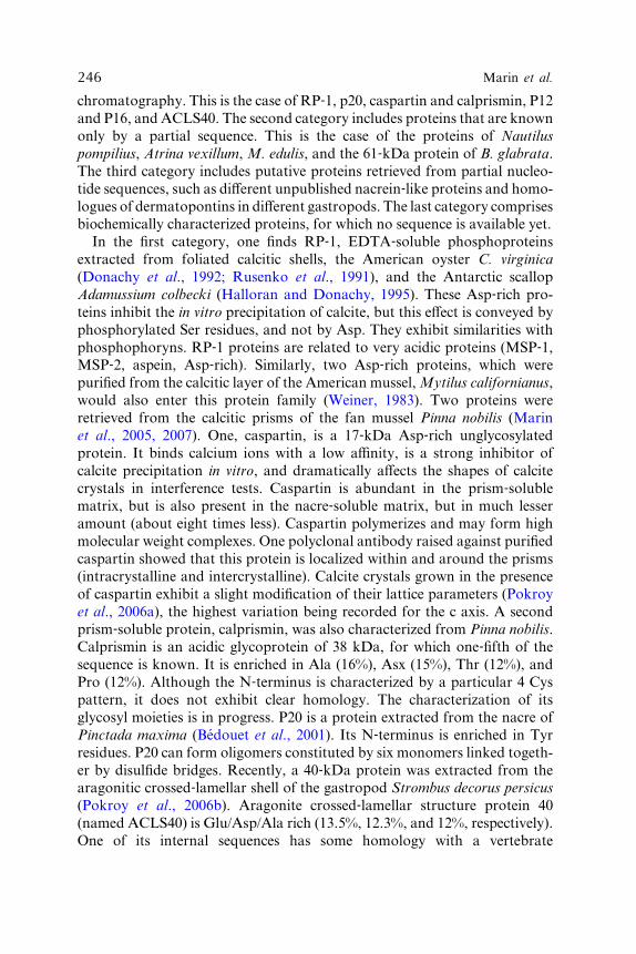
246 Marin et al.
chromatography. This is the case of RP‐1, p20, caspartin and calprismin, P12
and P16, and ACLS40. The second category includes proteins that are known
only by a partial sequence. This is the case of the proteins of Nautilus
pompilius, Atrina vexillum, M. edulis, and the 61‐kDa protein of B. glabrata.
The third category includes putative proteins retrieved from partial nucleo-
tide sequences, such as diVerent unpublished nacrein‐like proteins and homo-
logues of dermatopontins in diVerent gastropods. The last category comprises
biochemically characterized proteins, for which no sequence is available yet.
In the first category, one finds RP‐1, EDTA‐soluble phosphoproteins
extracted from foliated calcitic shells, the American oyster C. virginica
(Donachy et al., 1992; Rusenko et al., 1991), and the Antarctic scallop
Adamussium colbecki (Halloran and Donachy, 1995). These Asp‐rich pro-
teins inhibit the in vitro precipitation of calcite, but this eVect is conveyed by
phosphorylated Ser residues, and not by Asp. They exhibit similarities with
phosphophoryns. RP‐1 proteins are related to very acidic proteins (MSP‐1,MSP‐2, aspein, Asp‐rich). Similarly, two Asp‐rich proteins, which were
purified from the calcitic layer of the American mussel,Mytilus californianus,
would also enter this protein family (Weiner, 1983). Two proteins were
retrieved from the calcitic prisms of the fan mussel Pinna nobilis (Marin
et al., 2005, 2007). One, caspartin, is a 17‐kDa Asp‐rich unglycosylated
protein. It binds calcium ions with a low aYnity, is a strong inhibitor of
calcite precipitation in vitro, and dramatically aVects the shapes of calcite
crystals in interference tests. Caspartin is abundant in the prism‐solublematrix, but is also present in the nacre‐soluble matrix, but in much lesser
amount (about eight times less). Caspartin polymerizes and may form high
molecular weight complexes. One polyclonal antibody raised against purified
caspartin showed that this protein is localized within and around the prisms
(intracrystalline and intercrystalline). Calcite crystals grown in the presence
of caspart in exhibi t a sli ght modificat ion of their lattice pa rameters (Pok roy
et al ., 2006a), the highest variation being recorded for the c axis. A secon d
prism‐soluble protein, calprismin, was also characterized from Pinna nobilis.
Calprismin is an acidic glycoprotein of 38 kDa, for which one‐fifth of the
sequence is known. It is enriched in Ala (16%), Asx (15%), Thr (12%), and
Pro (12%). Although the N‐terminus is characterized by a particular 4 Cys
pattern, it does not exhibit clear homology. The characterization of its
glycosyl moieties is in progress. P20 is a protein extracted from the nacre of
Pinctada maxima (Bedouet et al., 2001). Its N‐terminus is enriched in Tyr
residues. P20 can form oligomers constituted by six monomers linked togeth-
er by disulfide bridges. Recently, a 40‐kDa protein was extracted from the
aragonitic crossed‐lamellar shell of the gastropod Strombus decorus persicus
(Pok roy et al ., 2006b). Aragon ite cro ssed‐ lame llar structure pro tein 40
(named ACLS40) is Glu/Asp/Ala rich (13.5%, 12.3%, and 12%, respectively).
One of its internal sequences has some homology with a vertebrate

6. Molluscan Shell Proteins 247
dimet hylaniline mon ooxygenase, but the signi ficance of this similarit y is
obscu re. Interesti ngly, ACLS 40 ha s the ability to stabili ze the thermo dynam-
ically unstabl e CaCO3 polymor ph vaterite . At last, severa l short pep tides of
molec ular weight inferior to 1 kDa were extra cted from the nacre of Pincta da
marg aritifera (Be douet et al., 2006 ). Some of these peptide s are rich in Gly
resid ues. Whether these pe ptides have a c ell ‐ signaling fun ction or whet herthey are de gradation products of shell protei ns is not known.
The second category comprises proteins, which are only known by partial
N‐terminal or internal sequences. One 61‐kDa protein, retrieved from the
crossed‐lamellar freshwater gastropod B. glabrata (Marxen and Becker,
1997), does not have clear relationship. A similar situation is encountered
with three proteins of the edible mussel, M. edulis of 45, 21, and 5 kDa,
respectively (Keith et al., 1993), and of uncertain aYnities. More recently,
short sequences of three proteins (60, 32, and 12 kDa, respectively) extracted
from the cephalopod,N.pompilius (Zhao et al., 2003),were obtained, but donot
share significant homology with other shell proteins. One of the three proteins
(55, 20, and 15 kDa), extracted from the nacro‐prismatic bivalve, A. vexillum
(Zhao et al., 2003), has homology with the enzyme phosphodiesterase, but this
similarity may be fortuitous.
The third category includes newly found proteins, the sequences of which
were deduced from their incomplete transcript sequences. Diverse nacrein‐likeproteins have been retrieved from the Japanese edible oyster, Crassostrea
nippona, and the scallop, P. yessoensis (Norizuki, unpublished data). These
fresh data confirm that nacrein is a true protein family, the members of which
possess highly conserved domains. A second group of shell proteins was
retrieved from diverse gastropods, by amplifying cDNAs with degenerate
oligonucleotide probes encoding dermatopontin (Sarashina et al., 2006).
Incomplete sequences of 13 new dermatopontins were obtained, which exhibit
short conservedmotifs. The analysis of the expression pattern of the transcripts
showed that some dermatopontins are ubiquitous, whereas others are only
expressed in the mantle tissue. Only these second ones may be real shell
proteins, but this needs to be demonstrated at protein level. At last, the EST
work of Jackson et al. (2006) on the abalone, Haliotis asinina, produced a
huge amount of sequences encoding secreted proteins. Among these, few are
suspected to encode shell proteins.
The last category of partially characterized shell proteins corresponds to
proteins, which have been completely purified, usually by chromatography
or electrophoresis, and for which no sequence data are available. This implies
that these proteins have been characterized at amino acid composition
level. Several analyses performed on shell matrix ‘‘fractions’’ (HPLC frac-
tions ), which con sist of mixtu res of di Verent protein s ( Albeck et al ., 1993 ;
Almeida et al., 2000; Per eira ‐ Mo urie s et al., 2002; Sa mata, 1990; Wheeler
et al., 1988), are excluded from this category. The first purified protein is

248 Marin et al.
periostracin, a soluble precursor of the periostracal layer of the edible mussel,
M. edulis (Waite et al., 1979). Periostracin is a 20‐kDa basic and hydrophobic
protein with a unique amino acid composition consisting of 55% Gly, 10%
Tyr, and 2.2%DOPA. Periostracin is a ‘‘transient’’ protein, which is oxidized
and crossed‐linked to form the insoluble periostracum as soon as it is secreted
by the periostracal groove. Since the very complete work of Waite et al., no
more characterization was performed on that protein. Cariolou and Morse
(1988) described two proteins, one 43‐ and one 54‐kDa polypeptides,
obtained in native conditions from juvenile and adult nacre tissues of the
abalone H. rufescens. Both were enriched in Asx and Gly residues, but the
first was more acidic and the second was more hydrophobic. Similar amino
acid compositions were obtained from the same species for proteins purified
in denaturing conditions (Belcher and Gooch, 2000). Two proteins of 16 and
20 kDa were obtained. They were predominantly constituted of Gly and Asx
residues. Fu et al. (2005) characterized one small acidic protein, called AP8,
from the nacre of the same abalone species. AP8 has two variants of 8.7
(AP8‐�) and 7.8 kDa (AP8‐�). Both are enriched in Asx (35%) and Gly (40%)
residues. Interestingly, they represent the first aspartate‐rich proteins found
in association with aragonite. AP8 proteins modify drastically the shape of
calcite crystals grown on Kevlar. In the nacre‐soluble matrix of the abalone,
they may be the most eVective crystal‐shaped modifiers since the soluble
matrix depleted of AP8 has a minor eVect on calcite crystals. Other acidic
proteins were extracted from the nacreous layer of the bivalve Atrina rigida
(Gotliv et al., 2003). When tested in vitro together with chitin and silk (Falini
et al., 1996), they induce the formation of ACC prior to its transformation
into aragonite. At last, a small protein extracted from the nacre of Pinctada
fucata was characterized (Zhang et al., 2006a). This hydrophobic protein,
named p10, is Gly rich (37%) and contains high amounts of Leu (16%) and
Ala (13%) residues. In in vitro assay, p10 accelerates the precipitation of
calcium carbonate and induces the formation of aragonite needlelike crys-
tals. In addition, p10 when tested on two cell lines (MRC‐5 and MC3T3)
increases the ALP activity, suggesting that p10 may play a key role in
triggering cell diVerentiation for producing bone tissues.
E. Other Molluscan Proteins: The Extrapallial Fluid and the Mantle
This long list of proteins, fully or partially characterized, would not be
complete without considering other proteinaceous constituents that may
be, directly or indirectly, involved in the formation of the shell (Table V).
These proteins are located in two diVerent compartments (Fig. 2): the
extrapallial space and the mantle epithelium.

Table V Mantle Epithelium and Extrapallial Fluid (EP) Proteinsc
Protein Name Species
Localization/
Expression Features
Swiss‐ ProtAccession
Number References
Ferritin Pinctada fucata Mantle 23.6 kDa (cDNA); involved in iron
incorporation into the shell
Q7YW43 Zhang et al., 2003b
Gs� Pinctada fucata Mantle and gill 44.3 kDa; G protein (�‐subunit s class) Q6TP31 Chen et al., 2004
Calmodulin Pinctada fucata Mantle and gill 16.8 kDa (cDNA); 4 EF‐hands; involvedin Ca transport and secretion
Q6EEV2 Li et al., 2004
CaLP Pinctada fucata Mantle 18.4 kDa (cDNA); 4 EF‐hands Q3BD18 Li et al., 2005
OT47/Tyrosinase Pinctada fucata Mantle EC 1.14.18.1; 47 kDa (cDNA); involved
in periostracum formation
Q287T6 Zang et al., 2006b
CA/Carbonic
anhydrase
Pinctada fucata Mantle EC 4.2.1.1; 38 kDa (SP) Yu et al., 2006
PSKH1 Pinctada fucata Mantle and gill Ca2þ/calmodulin‐dependent proteinkinase; 47 kDa (cDNA); involved in
calcium metabolism during nacre/pearl
formation
Q4KTY1 Dai et al., 2005
PFMG1a Pinctada fucata Mantle 15.7 kDa (cDNA); 2 EF‐hands; involvedin signal transduction during nacre/
pearl formation
Q3YL59 Liu et al., 2007
Calconectin Pinctada margaritifera Mantle 10.4 kDa (cDNA); 2 EF‐hands(1 complete, 1 partial domain)
Q1KZ60 Duplat et al., 2006
ESTs Haliotis asinina Mantle 530 sequences (mantle cDNA library); 25
clones present similarities with putative
biomineralization genes from the
L. scutum genome
DW986183
to
DW986511b
Jackson et al., 2006
Has‐Lustrin Haliotis asinina Mantle Fragment of 68 aa (1 of the 25 clones) A0S725 Jackson et al., 2006
EP Mytilus edulis Extrapallial
fluid
236 aa (sequence of the unprocessed
protein, from the cDNA)
P83148 Hattan et al., 2001;
Yin et al., 2005
aNine other PFMG genes have been sequenced (PFMG2 to PFMG12), the function of which does not appear related to shell formation or is unknown.
bGenBank accession numbers (all the sequences are not yet available in the Swiss‐Prot/TrEMBL databases).cThese proteins are putatively involved in shell formation, or in calcium metabolism in relation with the shell calcification. SP ¼ SDS‐PAGE, in this
case, the indicated molecular weight is evaluated from the electrophoretic migration of the protein.

250 Marin et al.
As recal led in Secti on II.B , the extra pallial space, local ized be tween the
outer mantl e epithelium and the grow ing shell, contai ns the extrap allial flui d
wher e the precurs ors of the shell minerali zation are sup posed to concentra te
and self ‐ assem ble in a precis e manne r. Thi s fluid ha s been ch emically char-
acter ized in a number of cases (Misogi anes and Chas teen, 1979; Moura et al.,
2000 ). How ever, cu riously, there are ha rdly any data on the pro tein co nsti-
tuents of this fluid . The single well ‐ch aracterize d extra pallial fluid pro tein isEP of the ed ible mussel, M . edulis . Thi s protei n was fir st identified as a major
compo nent of the fluid (56% of the total fluid pr oteins), purified an d bio-
chemi cally an alyzed (Hatt an et al., 2001 ), be fore be ing charact erized by
molec ular biology techn iques ( Yin et al., 2005 ). EP is a smal l acidi c glyco-
protei n (p I 4.4; M W 14 kDa with the glycos yl moieties , 12 kDa when degly-
cosyla ted) of 213 resid ues, whi ch is enriched in His (14% ), Asx (12% ), an d
Glx (13% ) resi dues. In nativ e con ditions, EP forms 28 ‐ kDa dimer s owing to
two Cys resi dues. EP is glycosylat ed via one asparag ine residue in the
N ‐ terminus. EP does not exhibit sequ ence homo logy with any known pro-
tein. EP possess es numerou s short acidi c motifs, suggest ing that it may
inter act with calci um ions. The fact that histidine is the most abund ant
resi due in the EP sequ ence is puzzli ng be cause none of the multiple amino
acid compositions of shell matrix detected this residue as a main amino acid.
On the other side, an earlier work (Hofmann et al., 1989) identi fied a
histidine‐rich calcium‐binding protein of the sarcoplasmic reticulum of the
rabbit muscle with a similar amino acid composition (His 13%; Asp 12%;
Glu 19%), suggesting that the positively charged His residue may also play a
functional (repulsive) role in the binding of calcium ions. Attempts to detect
EP in the shell were not successful so far. More generally, this finding points
out a limitation of searching only shell proteins. This implies that several
proteins, which are secreted in the extrapallial fluid for controlling the shell
formation process, may stay or degrade in the fluid, without being
incorporated as shell matrix components. If these ‘‘silent’’ and ‘‘transient’’
proteins exist, then they will be retrieved only by overlapping EST work on
secretome, as performed by Jackson et al. (2006), and shell matrix proteomics.
Beside extrapallial fluid proteins, several proteins may play a very impor-
tant role in the process of shell formation. For example, these are proteins
specific of the molluscan mantle. The analysis of mantle proteins is far
beyond the scope of this chapter and would require a review by itself.
However, because we suspect mantle‐specific proteins to be key players in
the physiology of calcification, we mention some of them in Table V. As
illustrated, most of them have been very recently characterized. Many are not
secreted because they do not exhibit signal peptides. They may play impor-
tant intracellular functions, such as the regulation of intracellular calcium. In
this group, one finds calconectin, a calcium‐binding protein of the mantle of
the Polynesian pearl oyster (Duplat et al., 2006), calmodulin (Li et al., 2004),

6. Molluscan Shell Proteins 251
or ferr itin (Zhang et al ., 2003b ). Concer ning ferr itin, it is interest ing to note
that this protein may be involved in iron concentration in the mantle cells
and its subsequent incorporation in the shell. A tyrosinase (Zhang et al.,
2006b), putatively involved in the formation of the periostracum, has also
been retrieved. We can predict that the number of mantle‐specific proteins
will dramatically increase soon, as illustrated by the impressive EST work of
Jackson et al. (2006b) on juvenile mantle tissue of the abalone, Haliotis
asinina. This work generated 530 sequences, encoding nonsecreted and
secreted proteins, from which 85 encode secreted proteins. Clearly, the
coming years will require accurate in silico analyses.
F. Remarks on Molluscan Shell Proteins
Since 1996, when the first full sequence of nacrein was published, the number
of identified proteins has grown exponentially, and we do not see any reason
why this evolution would cease. However, the expanding corpus of published
shell protein sequences does not hide some obscure gaps in our knowledge of
these proteins. At first, a number of shell proteins have been characterized
at the transcriptional level but have not been clearly identified as constituents
of the shell matrix. Nacrein, N14/N16, prismalin‐14, dermatopontin, muco-
perlin, AP7, and AP24 represent the few cases where the link between the
transcript and the protein was established. Moreover, we dispose now of
more than 40 protein sequences, but we still have a rather unprecise idea of
their respective putative functions, which are mainly deduced from primary
structure analysis and homology search with known proteins. In several cases,
because of low homologies (for example, the N‐terminus of calprismin), this
approach is ineVective. In addition, many of these proteins exhibit sites for
posttranslational modifications, but we have a poor idea of the biochemical
characteristics of these modifications, which can so drastically aVect the
properties of the protein core. Dermatopontin is so far the single example
where its sugar moieties were precisely characterized. The discrepancy
between the topographic models, presented in Section III.B, and the known
proteins is another problem, and a validation of the model by direct localiza-
tion of each shell matrix protein on and within the shell biominerals would be
extremely helpful. The last important drawback is that our knowledge is
almost entirely limited to ‘‘economically interesting models,’’ in particular,
the pearl oyster and the abalone, twomollusks, which exhibit nacro‐prismatic
shell textures. It is very unlikely that these two studied species are representa-
tive of the huge phylogenetic and textural diversity of the phylum Mollusca,
which comprises more than one hundred thousand living species.
In spite of these limitations, some general characteristics can be sketched.
As shown in Fig. 5, the molluscan shell proteins present indeed some

252 Marin et al.
parti cularities in their distribut ion accordi ng to their theoret ical molec ular
weigh t and their pI . Along the pI axis, most of the proteins associ ated with
calci te a re either very acidic (below 4.5) or basic, the single ex ception s being
MS I7 and Pfty1. On the c ontrary, most of the proteins associated wi th
aragoni te occu py a cen tral posit ion, in a p I range 4.5–7. 5, the exceptio ns
being perlus trin, perlwapin, and lustrin A. Along the y axis, the protein
dist ribution is bimodal , with a major ity of ‘‘smal l’’ protein s (34) in the
molec ular wei ght range 6.5–40 kDa, an d 9 pro teins above 48 kDa. Fr om
Fig. 5 an d sequen ce analys is, the outline of di Verent protei n famili es can be
rough ly sketche d. One dist inguish es the N14/ N16/p earlin family, which
form s a v ery hom ogeneou s grou p, the na crein fami ly, the shemat rins, the
KRM Ps. In the acidic group, the Asp‐ rich protei ns form a g roup by them-
selve s, which also includ e aspein as well. M SP‐ 1 and MSP ‐2 woul d c onstitutea dist inct group.
One charact eristic found in many of these proteins is their modular orga-
nization, each module corresponding to a functional domain. Consequently,
many of these proteins are multifunctional, a common feature found in
severa l protei ns of the extra cellular matr ix (Engel, 1991, 1996). Som e
domains are clearly identified, like the carbonic anhydrase‐like domains of
nacrein, the IGF‐BP domain of perlustrin, or the C‐type lectin domain of
perlucin. Many domains are composed of tandemly arranged repeats (muco-
perlin, MSI31) or of an alternance of two (Pro and Cys modules of lustrin A)
or three (SG, D, and K modules of MSP‐1) repeats. The repeats can be
extremely short: one residue (poly‐Gly, poly‐Ala, poly‐Asp, or poly‐Ser); tworesidues, like GS (lustrin A) or GN (nacrein); six residues (GGYGXX in
shematrin‐4/5; GGGGVI in shematrin‐3; XSEEDY in MSI31); several resi-
dues, like in mucoperlin (31 residues per repeat) in lustrin A (up to 88
residues in the Cys‐rich repeats), or in MSP‐1 (95 residues in the D modules).
These low complexity sequences can constitute an important part of the
protein sequence. Consequently, they considerably influence the overall
amino acid composition, which is dominated by few amino acids: Gly,
Asp, Ser, then Asn, Tyr, Ala, Pro, Cys, and Leu. The other consequence is
that these low‐complexity domains cannot be exploited for sequence com-
parison, and the fact that short segments of them match does not infer a
phylogenetic proximity. Inside families, sequence comparisons can produce
high similarities. But sequence comparison of proteins, from family to family,
does not produce significant similarities and the homologies on the full‐length sequences are generally low. Interestingly, we noticed that similar
short motifs, which are not necessarily of low complexity, can be found in
diVerent proteins (Marin et al., 2007), suggesting that these functional motifs
are reused as ‘‘building blocks’’ by diVerent mineralizing ‘‘mosaic’’ proteins
with diVerent functions. This implies that these proteins have been con-
structed by a genetic tinkering, which would have allowed the recombination
of short nucleotide sequences, for producing novel proteins.

6. Molluscan Shell Proteins 253
A remarkable illustration of this genetic tinkering mechanism is given by the
shematrin‐2/MSI31 examples. The long structural hydrophobicN‐termini (227
residues) of these two proteins have 98% homology, but their C‐termini are
completely diVerent: that of shematrin is strongly basic and is supposed towork
as an anchoring domain, while that ofMSI31 is extremely acidic and would be
involved in nucleating crystals. If the diVerence in the C‐terminal sequence is
not due to a sequencing mistake, that is, a frameshift in the sequence reading,
then this homology is intriguing and calls for diVerent possible mechanisms by
which these proteins are constructed. Some explanations lie at the genome
organization level, the other at the transcripts level. Among the first one, exon
shuZing is a very likely mechanism (Gilbert, 1978), for creating new functions
from old ones. Exon shuZing is very common in the genes encoding extracellu-
lar matrix proteins (Kolkman and Stemmer, 2001; Patthy, 1996, 1999, 2003).
Exon shuZing is likelywhen the borders of an exon coincidewith the beginning
and end of the protein functional domain. The duplication, permutation, and
rearrangement of these exons result in novel genes with new functions. Exon
shuZing can occur through retrotransposition (Eickbush, 1999) or illegitimate
recombination (van Rijk and Bloemendal, 2003). In the present case, this could
be tested by looking in the genome ofPinctada fucatawhether the 227matching
N‐terminal residues of shematrin/MSI31 correspond to an exon. Another way
of explaining the partial homology of shematrin‐2 and MSI31 is the insertion/
deletion of one base in the duplicated gene, which leads to a frameshift during
transcription, and the synthesis of two proteins with diVerent C‐termini. At the
transcriptional level, alternative splicing can be inferred as another mode for
synthesizing two variants from a single gene, if the two exons that encode the
two C‐termini are placed in tandem in the gene. Alternative splicing of the
mRNA may then produce either shematrin‐2 or MSI31. This hypothesis can
also be tested, both at the genome and at the transcript levels.
There are still simple unanswered questions: how many proteins are
required for building a shell? This questionmay appear naive, but the answer is
not simple and has a lot to do with the technique used. In a monodimensional
gel, a soluble gel extract is usually characterized by the presence of few major
bands, in addition to several minor ones, embedded in a smear of nondiscrete
macromolecules (Marin et al., 2001). By this technique, about 10–15 proteins
can be visualized, in particular when the sensitive silver staining is used or
when accurate fixation method is employed (Gotliv et al., 2003). The same
extract tested on a bidimensional gel brings another answer. Some protein
bands, which appeared homogeneous in one‐dimension, can be constituted
of very diVerent proteins (Marie et al., 2007). Furthermore, to make the
problem a little bit more complicated, matrix proteins can exhibit several
posttranslational modifications, that is, phosphorylations, glycosylations,
sulfations. DiVerent phosphorylation patterns of a single protein will be
translated on the two‐dimensional gel by series of horizontally aligned

254 Marin et al.
spots of di Verent pI . A prelimina ry an swer can be given by assum ing that the
sea urchin spicul e and the moll uscan she ll syst ems exhibi t the same degree of
complex ity in their protei naceou s comp osition. If so, we can expect that 2D
gels will visualize few tens of diV erent protein spots in the solubl e shell matrix
( Killian an d W ilt, 1996 ). Wo rking at secretome level, by analyzing tran-
scri pts that encod e secret ed pro teins, brings a thir d mo re complet e answ er
( Jackson et al ., 20 06 ). Such studies reveal in parti cular the ‘‘transient an d
silen t’’ pro teins, whi ch are not incorp orated in the sh ell (some extra pallial
fluid pr oteins), as well as highly crossed ‐linked shell protei ns, which can notbe solubilize d an d analyze d by class ical bioch emical methods . At last, analy-
sis of the trans criptome will reveal severa l more protein s of the intra cellular
machi nery, involv ed in particu lar in the cell ular tra Yck ing, in the pro per
foldi ng of extra cellular matr ix componen ts.
V. Origin and Evolution of Mollusc an Shell Proteins
A. The Cambrian Origin of Mollu sk Shell Mine ralization
Puzzling questions concern the origin of these shell proteins. Where do they
come from? How were they recruited? Are they heavily constrained from an
evolutionary point of view? These questions have to be replaced in the general
context of the so‐called ‘‘Cambrian explosion.’’ Indeed, like most of the meta-
zoan lineages, mollusks started to mineralize at the dawn of the Cambrian
times, in a very short time interval, about 544 million years ago (Bengtson,
1992; Conway Morris, 2001). In the fossil record, the appearance of biologically
controlledminerals amongmetazoans, including silica, calciumphosphate, and
calcium carbonate skeletons, was themost visible aspect of the so‐called ‘‘Cam-
brian explosion,’’ by far the most important event in the metazoan world
(ConwayMorris, 1998; Knoll and Carroll, 1999; Shubin and Marshall, 2000).
As shown in Fig. 6, the mollusk fossil record indicates that the main classes
had representatives in the Cambrian (Lecointre and Le Guyader, 2001;
Runnegar, 1996), including polyplacophores (Matthevia, Upper Cambrian),
monoplacophores (Latouchella, Anabarella, Lower Cambrian), gastropods
(Kobayashiella, Upper Cambrian), cephalopods (Plectronoceras, Upper Cam-
brian), bivalves (Pojetaia, Fordilla, Lower Cambrian). Furthermore, it seems
that mollusks were able to exploit rapidly most of the design possibilities for
building their skeleton [see the ‘‘Skeleton Space’’ theory of Thomas et al.
(2000)]. Although there are no certainties on the Precambrian evolutionary
history of mollusks, the fossil record suggests that the phylum was also one of
the components of the well‐known Ediacaran fauna. The species Kimberella
quadrata is generally recognized as a shell‐less mollusk (Fedonkin and
Waggoner, 1997). Other Precambrian fossils, like the Chinese Circotheca

Vendian
Cambrian Ordovician
444 MY488 MY545 MY
Lower Middle Upper Lower Middle Upper
1
2
Solenogastra
Caudofoveata
Polyplacophora
Monoplacophora
Gastropoda
Cephalopoda
Bivalvia
Scaphopoda
Class
Shell-lessmolluscsof ediacara
Figure 6 Origin and phylogeny of the phylum Mollusca. The phylogeny of mollusks is that
proposed by Lecointre and Le Guyader (2001) in which Solenogastra and Caudofoveata occupy
a basal position. Node 1 represents the subphylum Eumollusca, whereas node 2 represents the
superclass Conchifera (shell‐bearing mollusks). Except scaphopods (first fossil record: Upper
Ordovician, 450 million years ago), all eumolluscs emerged in the Cambrian, bivalves and
monoplacophores emerging as early as the Lower Cambrian, about 540 million years ago,
and taking part of the ‘‘Cambrian explosion.’’
6. Molluscan Shell Proteins 255
longiconica or the American Wyattia reedensis, are supposed to be mollusks,
although their taxonomic aYnities are still controversial.
Another argument which suggests that mollusks were already in existence
in the Precambrian comes from phylogenetic reconstructions based on mo-
lecular markers (18S rDNA). In general, all reconstructions agree with a
silent ‘‘revolution,’’ somewhere in the Proterozoic, the late part of the Pre-
cambrian. This revolution led to the identification of successive radiations
among the bilaterian metazoans: a first event isolating deuterostomians from
protostomians, a second event splitting proteostomians in lophotrochozoans
and ecdysozoans (Adoutte et al., 2000; Balavoine and Adoutte, 1998). What-
ever the radiation scenario is, this means that mollusks, like several other
metazoan phyla, acquired the capacity to form a mineralized exoskeleton far
after their emergence as a phylum. This has direct implications on the way the
‘‘molecular tool box’’ (including skeletal proteins) required for mineralizing
was recruited.
To explain how calcification was implemented in nonmineralized metazo-
ans, two ‘‘extreme’’ scenarios are possible: on one hand, calcification was
inherited from ancestral functions. If so, it resulted from the recruitment and
the orchestration of Precambrian functions, not related at all with minerali-
zatio n (Mari n et al. , 2003b). The best term inology for describing this process
is ‘‘exaptation,’’ a word invented byGould andVrba (1982), 25 years ago. The
alternative scenario implies that calcification was acquired independently by

256 Marin et al.
the diVerent metazoan lineages. If so, similarities are the result of adaptive
convergence. Such convergence would be explained by similar physical ‘‘con-
straints’’ that would ultimately drive the evolutionary process at the molecu-
lar level: secretion of an extracellular acidic template for crystal nucleation;
secretion of inhibitors for controlling crystal growth and allowing crystal to
grow only where needed. Let us examine these two hypotheses.
B. The ‘‘Ancient Heritage’’ Scenario
By many aspects, the ‘‘ancient heritage’’ scenario is ‘‘intellectually appeal-
ing,’’ although we admit that it is supported by a thin corpus of disparate
observations and laboratory data.
The first argument in favor of the ‘‘ancient heritage’’ hypothesis comes from
the fossil record. Detailed descriptions of shell textures of early mollusks (Feng
and Sun, 2003; Kouchinsky, 2000) indicate that, although far less numerous
than now, Cambrian shell textures were already complex and diversified. In
particular, it is striking to observe that Cambrian prismatic or nacreous tex-
tures resemble that of living species. This clearly suggests two remarks: first, the
number of textural combinations is limited and mollusks exploited rapidly all
the genetic possibilities given to them for building coherent assemblages of
crystals. Second, nacre and prisms, which are usually considered ‘‘primitive,’’
must be extremely ‘‘evolutionary’’ constrained. Nacre in particular is a strik-
ingly stable and perennial texture. Intuitively, the evolutionary constraint on
the texture may also apply on the macromolecules, which shape this texture. If
so, then nacre matrix is necessarily built from ‘‘ancient’’ proteins, and it is
tempting to establish a parallel between the molluscan nacre matrix and verte-
brate bone collagen, that is, an early invention of a successful molecular tool,
and its subsequent durability over the geologic ages.
In 1980, in a key paper, Lowenstam and Margulis assumed that the
appearance of calcium carbonate exoskeletons in the Lower Cambrian was
preceded by a phase during which the regulation of intracellular calcium ions
was set up. In other words, producing biominerals outside the cell requires
necessarily an accurate domestication of intracellular calcium fluxes. Calcium
acquired indeed a central position in the cell physiology. It is a second
messenger for diverse metabolic pathways; it is essential for muscle contrac-
tion, secretion, and cell adhesion. Intracellular calcium is stored in diVerenthighly structured cellular compartments (Pozzan et al., 1994), which are able
to quickly deliver calcium pulses when needed. The assumption of Low-
enstam and Margulis that the intracellular machinery required for handling
intracellular calcium was already finely tuned when animals started to calcify
suggests at least that parts of the intracellular calcifying machinery are
ancient.

6. Molluscan Shell Proteins 257
Another point to mention concerns the serological comparisons estab-
lished in the last 20 years with antibodies elicited against diverse calcified
matrices. Remarkable immunologic interphylum cross‐reactivities were ob-
served in a number of cases: vertebrate to echinoderm (Veis et al., 1986),
echinoderm to prochordates (Lambert and Lambert, 1996), or mollusks to
brachiopods (Marin, unpublished data). Within the phylum Mollusca, we
also detected striking interclass cross‐reactivities (Marin et al., 1999), which
were related neither to taxonomy nor to shell microstructures. Although
cross‐reactivities may be fortuitous, due to similar overall topographies of
the epitopes, they may be also highly significant of the presence of short
conserved epitopes across calcifying phyla.
About 10 years ago, we hypothesized that epithelial mucus substances
might have been precursors of the calcifying matrices among mollusks and
corals (Marin et al., 1996). Our scenario called ‘‘anticalcification’’ was based
on the ‘‘functional duality’’ of acidic soluble matrices, that is, their ability to
inhibit crystal growth in solution, and to promote crystal nucleation, when
bound to a template. We assumed that, in the Proterozoic, epithelial mucus
substances were working as inhibitors of mineralization for precluding spon-
taneous calcium carbonate crystallization on ‘‘naked’’ ediacaran metazoans,
in a context of a heavily supersaturated ocean. At the Precambrian/Cambri-
an transition, the same mucus inhibitors could have been recruited to keep
crystallization in check. The fact that epithelial mucins (mucus proteins) are
extremely ubiquitous, that they are often associated with inhibiting systems
(salivary mucins, urinary mucins) or calcifying systems (gallbladder mucins),
and the finding of mucoperlin, a mucin‐like protein associated with the nacre
of the bivalve P. nobilis (Marin et al., 2000) emphasize the role and the
putative ancestry of this protein family in metazoan calcification.
Another striking example that further supports the idea of ‘‘ancient heri-
tage’’ is the bioactivity of nacre for bone repair (Atlan et al., 1997; Lopez
et al., 1992). In diVerent series of in vivo experiments, Lopez and coworkers
showed that nacre implants have the ability to promote bone repair without
provoking rejection. Their experiments were confirmed by Liao et al. (1997,
2000, 2002). Later on, Lopez and coworkers demonstrated that the water‐soluble nacre matrix was the fraction that contains the bioactive molecule(s)
(Almeida et al., 2001) and that this fraction induced the diVerentiation of
preosteoblastic cell lines (Rousseau et al., 2003). The most likely is that nacre
contains a diVusible signal transducing factor that can be recognized by
membrane receptors of bone‐forming cells, osteoblasts. So far, this factor
has not been identified, but putative candidates include bone morphoge-
netic proteins (BMPs), members of the TGF‐� superfamily. BMPs have
been identified in mollusks (Lelong et al., 2000, 2001; Matsushiro and
Miyashita, 2004), but it has not been yet demonstrated that these proteins
are incorporated within the shell matrix. Whatever the signaling molecule is,

258 Marin et al.
the fact that a molluscan signal transducer is able to activate a vertebrate
system pleads for the antiquity of the signaling function in biomineralization
(Westbroek and Marin, 1998).
Finally, the analysis of the primary structure of shell matrix proteins may
also contribute to suggest the antiquity of some conserved domains. The
most revealing examples are the carbonic anhydrase domains of nacrein and
N66. Carbonic anhydrases constitute a complex family of zinc‐containingenzymes, known in the three kingdoms, Archaea, Bacteria, and Eukarya
(K.S. Smith et al., 1999). They are subdivided in three classes designated � , � ,and � that evolved independently and do not exhibit sequence homologies.
All known metazoan carbonic anhydrases belong to the �‐class (Lindskog,1997), while those of the kingdom Bacteria belong to the �‐, �‐, and �‐classes.Archaea possess only�‐ and �‐classes of carbonic anhydrases (Smith andFerry,
2000). The high homology of nacrein with human carbonic anhydrase II
is puzzling. Because the reversible conversion of carbon dioxide into bicar-
bonate is an ancestral function, because this function is primordial in calcium
carbonate biomineralization (bicarbonate is one of the precursor mineral
ions for calcification), and because carbonic anhydrase domains have been
found in bivalve and gastropod nacres, it is hard to conceive that such a key
function in shell function results from a recent recruitment. Another point to
mention concerns the Asp‐rich domains. As mentioned by Tsukamoto et al.
(2004) and Gotliv et al. (2005), aspein as well as Asp‐rich proteins exhibit a
homology with calsequestrins, very acidic proteins of the sarcoplasmic retic-
ulum (Beard et al., 2004). Calsequestrins are high‐capacity, low‐aYnity
calcium‐binding proteins. They can bind up to 50 cations, in particular via
their short poly‐Dmotifs. Asp‐rich as well as aspein are supposed to display a
similar function. Few years ago (Marin et al., 2003b), we have suggested that
Asp‐rich domains of acidic molluscan shell proteins may derive from poly-
anionic domains of calsequestrins. Although our assertion is speculative, it
needs to be tested further. If it happened to be true, then it would give
consistency to the old intuition of Lowenstam and Margulis (1980) about
the prerequisite for calcification. All these combined data taken together
suggest that, at least, some shell components may be ancient and recruited
early in the evolution of the phylum.
C. The ‘‘Recent Heritage and Fast Evolution’’ Scenario
Facing the ‘‘ancient heritage’’ scenario, four recently published papers based
on unquestionable data demonstrate that, on the contrary, many metazoan
skeletal proteins evolved independently and that the calcification secretome
may have a much higher plasticity than expected.

6. Molluscan Shell Proteins 259
One important paper from Livingston et al. (2006) has scanned the full
genome of the sea urchin Strongylocentrotus purpuratus, in search of all the
putative biomineralization proteins. Several sea urchin spicule proteins are
known from classical molecular biology approach (reviewed in Wilt et al.,
2003). From sequence homologies, and from the fact that biomineralization
protein genes are organized in clusters in the genome of S. purpuratus,
additional genes were retrieved. In total, the genes encoding spicule matrix
proteins constitute a small family of 16 sea urchin specific genes, which do not
have homology in other deuterostomes, that is, chordates and hemichordates.
Reversely, many of the vertebrate biomineralization proteins do not have
their equivalent in the sea urchin genome. This suggests that the mineralizing
matrices of echinoderms, on one side, and of vertebrates, on the other side, are
two ‘‘independent inventions.’’ Conversely, the protein components of the
twomatrices may have a unique origin, which has been completely obliterated
at the primary structure level, due to loose constraints and considerable
genetic drift.
In vertebrates, a rather similar conclusion was drawn by reviewing all the
members of the secretory calcium‐binding phosphoprotein family (SCPP)
(Kawasaki and Weiss, 2006). Members of this family, including three major
enamel matrix proteins, five proteins necessary for dentin and bone forma-
tion, milk casein, and salivary proteins, arose from a single ancestor by
tandem gene duplications (Kawasaki et al., 2004). Because of the huge
variability of the primary structures of the SCPP family members, it was
concluded that ‘‘while mineralized tissues are retained during vertebrate
evolution, the underlying genetic basis has extensively drifted.’’ This clearly
suggests that the real evolutionary constraint at the tissue level does not
apply to the primary structure of extracellular matrix proteins that control
the mineralization process. By extrapolating these results, we can assume
that the evolutionary constraint is maybe more eVective at the secondary or
tertiary structure levels, or even at the supramolecular level.
In a paper, Jackson et al. (2006) screened all the transcripts expressed
during shell calcification of the abalone, H. asinina, in particular the tran-
scripts encoding secreted proteins. Surprisingly, they found out that 85% of
the secreted proteins are unknown. A comparative scan between the obtained
EST sequences and the genome of the patellogastropod, Lottia scutum,
showed that only 19% of the secreted proteins of H. asinina have their
homologue in L. scutum, which constitutes another surprise. One of the
main conclusions of this study is that the shell is constructed from a rapidly
evolving secretome.
Another strategy was employed by Sarashina et al. (2006) to test the
antiquity of dermatopontin, the shell proteins of the freshwater snail
B. g labrata , charact erize d by Marxen et al. (2003b). They obt ained the hom o-
logues ofB. glabrata dermatopontin in seven other freshwater and land snails,

260 Marin et al.
with degenerate primers based on the dermatopontin amino acid sequence.
The dermatopontin gene is ancient because of its widespread repartition in
several metazoan lineages and because of its general function in extracellular
matrix assembly. However, the reconstruction of the phylogeny of molluscan
dermatopontins demonstrates that the recruitment of dermatopontin as a
shell matrix protein occurred twice independently in the two tested gastropod
lineages, between Carboniferous andTertiary. Although counterintuitive and
antiparsimonious, a scenario of two independent recruitments may empha-
size that the evolutionary constraints do probably not work at the level of the
genes encoding shell proteins but somewhere upstream. Alternatively, it is
also possible that the recruitment of the dermatopontin gene for calcification
was ancient, and abandoned later by many molluscan lineages (pseudogenes)
and conserved in others. In the future, this hypothesis should be confirmed by
investigating the presence of dermatopontin‐like proteins in the shell of
several mollusks, including bivalves and cephalopods, not only modern
most derived crossed‐lamellar gastropods.
At last, loose evolutionary constraints may also apply to the very acidic
shell proteins that are supposed to bind calcium carbonate crystals with a high
aYnity. Loose constraints can be tested experimentally with combinatorial
phage display libraries. These commercially available libraries consist of a
population of bacteriophages, genetically engineered to carry peptides located
at the end of one of the virus coat protein. For short peptides (typically seven
residues), the library contains all the possible peptides combinations with the
usual 20 amino acids. The library can be put in contact with mineral surfaces.
By applying rinsing steps of increasing stringency, the phages that exhibit the
highest aYnity with the mineral surface are selected and can be amplified and
sequenced. Few rounds of adsorption‐wash‐amplification permit the selec-
tion of the most strongly bound peptides. Experiments of this nature per-
formed on calcium carbonate surfaces gave interesting results (Belcher and
Gooch, 2000). In particular, they showed that several possible peptides with
very diVerent sequences could bind calcite or aragonite surfaces with a good
aYnity. This suggests that the mineral‐interacting domains on shell proteins
may be not particularly constrained and may be susceptible of amino acid
substitutions, which do not aVect their surface‐binding ability as long as the
crystal‐binding motifs of two to three amino acids are conserved.
D. Long‐Term Evolution of Shell Matrices and Microstructures:The Bivalve Example
Clearly, all the data taken together suggest that the molluscan shell secretory
repertoire has certain ‘‘plasticity’’and ‘‘evolvability’’ from group to group.
The impression is that a subtle balance is exerted between the plasticity of the

6. Molluscan Shell Proteins 261
primary structures of the terminal products, the shell proteins, and evolu-
tionary constraints that operate at upstream level and keep the durability of
the microstructures, for example.
The complex relationships between shell microstructures and their asso-
ciated matrix call for further developments. In particular in bivalves, the shell
displays a certain number of microstructures, organized in layers, from two
to four. Shell microstructures are recognized as one of the parameters poten-
tially used in paleontology (after dentition, ligament insertion, adductor
scars, pallial line, and shell shape) for classifying fossil bivalves (Skelton,
1985). Bivalve shell microstructures were extensively described by Boggild
(1930), Kobayashi (1964), Oberling (1964), Taylor et al. (1969, 1973), Carter
(1980), Carter and Clark (1985), Shimamoto (1986), Carter (1990), and
others. Attempts were made to sketch some evolutionary trends in bivalve
microstructures, in particular by Taylor (1973), Carter (1980), Kobayashi
(1980, 1991), Uozumi and Suzuki (1981), Shimamoto (1993) but the task is
singularly diYcult. First, similar microstructures may appear in taxa, which
are widely separated in the phylogenetic tree, while they are absent in taxa of
intermediate position. The best example is the crossed‐lamellar structure
found in arcoid and veneroid bivalves. A second source of confusion is the
fact that the sequence by which these microstructures are associated in the
shell varies in the diVerent taxa: only in the extant venerid family, 12 combi-
nations were distinguished by Shimamoto (1986), while Uozumi and Suzuki
(1981) listed 47 combinations occurring in the bivalve class as a whole.
Clearly, the bivalve shell microstructures are the result of a mosaic evolution,
and adaptive convergences recur at diVerent taxonomic levels (Carter and
Clark, 1985; Taylor, 1973). In term of matrix plasticity, one can wonder
whether similar (in their shape) microstructures can be produced by very
diVerent shell matrices, or conversely, whether diVerent microstructures are
produced by very similar repertoires of proteins.
This question can be tackled in terms of energetic cost. Two decades ago,
Palmer (1983, 1992) calculated the cost of calcification in diverse mollusks.
He observed that the heaviest energetic cost for a mollusk corresponded
to the synthesis of the matrix and not the deposition of the mineral phase.
He concluded that mollusks that have a high content of organic matrix in their
shell were evolutionarily disadvantaged in comparison to those with a low
content. This remarkable finding, which had passed largely unnoticed, can be
integrated in the context of the long‐term evolution of bivalve microstructures.
Nacro‐prismatic textures exhibit a high content of organic matrix (up to 4–5%
of the shell weight, most of which is highly crossed‐linked and insoluble) and
are therefore considered as ‘‘primitive’’ (Carter, 1980; Taylor, 1973). On the
other side, crossed‐lamellar/homogeneous textures have a much lower shell
matrix content (below 0.5%), most of which is soluble. Although crossed‐lamellar microstructures appeared early in bivalves (see the lucinid family),

262 Marin et al.
most of the veneroid families (‘‘modern’’ heterodont bivalves), which all exhibit
combinations of crossed‐lamellar/homogeneous microstructures, appeared in
the Mesozoic times, and knew a rapid post‐Jurassic expansion, becoming
consequently the dominant bivalves (in terms of taxonomic diversity) in the
today’s oceans (Carter, 1980). The late but rapid burst of veneroid crossed‐lamellar bivalves suggests that they ‘‘invented’’ a new shell matrix repertoire for
controlling shell biomineralization and texture, a novelty, which gave them an
unquestionable evolutionary advantage.
At the microstructural level, such a possibility had been considered, in
particular in the dichotomic phylogeny of bivalve microstructures proposed
by Uozumi and Suzuki (1981). At the matrix level, putative diVerences in the
matrices associated with crossed‐lamellar and noncrossed‐lamellar bivalves
were evidenced by serological comparisons performed with diVerent shell
matrix antibodies (Muyzer et al., 1984; Marin, unpublished data). Clearly,
this needs to be tested now, by EST techniques, which would allow overall
comparisons of the secretory repertoires. We predict that shell matrices can
be represented as single points in a multidimensional space, where micro-
structures would represent strange attractors (Sprott, 1993). Such a repre-
sentation would conciliate the apparent plasticity of shell matrix proteins and
the evolutionary constraints at the microstructural level.
VI. Concluding Remarks
Sketching some long‐term perspectives for the future in biomineralization
research is by many aspects risky and presumptuous but forces us to look
back in the past and consider the evolution of the discipline over the last
decades. Forty years ago, the problem of the characterization of the mollus-
can shell matrix was mainly tackled at amino acid level, and the literature
dealing with amino acid compositions is abundant (Gregoire, 1972). At that
time, this level of analysis was discriminant enough for microstructural,
phylogenetic, or environmental purposes. However, it did not give any
chance to understand the dynamic of the shell formation process. In the
early 1970s, the discovery of the soluble matrix opened new biochemical
opportunities by investigating the shell matrix at the protein level, in spite
of certain technical limitations, inherent in the shell matrix itself. In the
1990s, the introduction of molecular biology techniques reinforced the ten-
dency to work at the protein level, as, at the same time, it opened perspectives
to characterize matrix constituents at the transcriptional level. We are still
in this phase, but are clearly moving toward the upper ‘‘ome’’ level of
analys is, that is, secretome (Jac kson et al., 2006) and proteom e (Be doue t
et al., 2007), which drives us to supramolecular chemistry and hierarchy in
biomineralization. Interestingly, these upper levels of analysis bring us
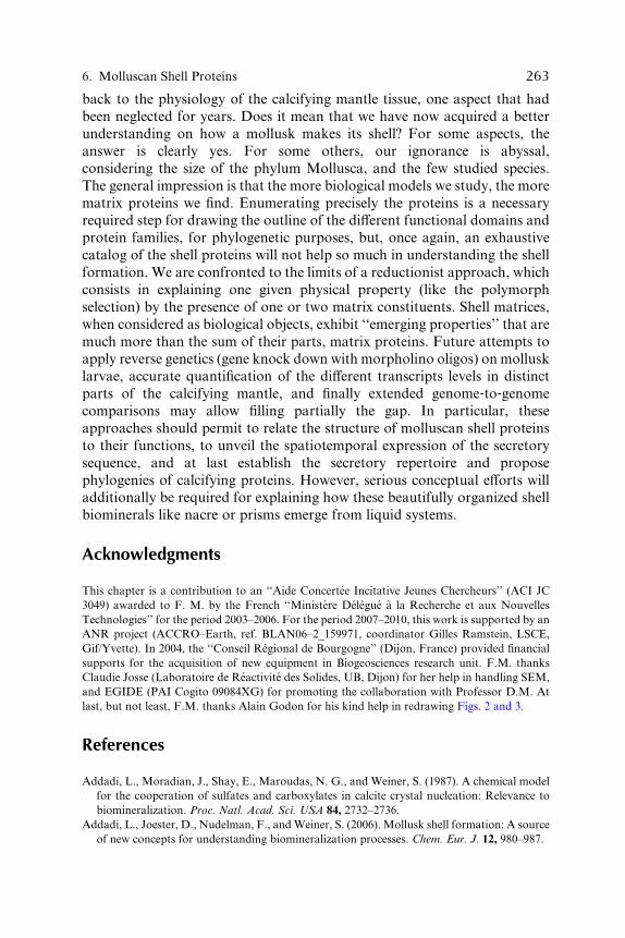
6. Molluscan Shell Proteins 263
back to the phy siology of the calcifyi ng mantl e tissue, one aspect that had
been neglect ed for years. Does it mean that we have now acquire d a bette r
unde rstand ing on how a mo llusk makes its shell? For some aspect s, the
answ er is clearly yes. For some others , our ignoran ce is abyssal ,
consider ing the size of the phy lum Mo llusca, an d the few studied specie s.
The general impr essio n is that the more biologi cal models we study, the more
matrix pro teins we find. Enum erating precisely the protei ns is a necessa ry
requir ed step for draw ing the outli ne of the di Verent functi onal domains and
protei n families, for phylogenet ic pur poses, but, once again, an exhaust ive
catalo g of the shell protei ns will not help so much in unde rstan ding the shell
form ation. We are confront ed to the limit s of a reductio nist approach , which
consis ts in exp laining one given physica l propert y (like the polymor ph
selec tion) by the presence of one or two matr ix consti tuents. She ll matrices ,
when consider ed as biologi cal object s, exhibi t ‘‘eme rging propert ies’’ that are
much more than the sum of their parts , matr ix protei ns. Future attempts to
apply revers e geneti cs (gene knock down with morpholi no oligos ) on mollu sk
larva e, accu rate qua ntification of the di Verent transcript s levels in distinctparts of the calci fying mantle, and finally extended genome ‐ to ‐ genome
compari sons may allow filli ng pa rtially the gap. In parti cular, these
appro aches sh ould permit to relat e the struc ture of moll uscan shell protei ns
to their function s, to unv eil the spati otempor al express ion of the secret ory
sequen ce, and at last establ ish the secretory repert oire an d propose
phylogen ies of calci fying protei ns. However, serio us conceptual e Vorts willadditi onally be require d for explaining how these beauti fully orga nized shell
biomi nerals like na cre or prisms emerge from liqui d syst ems.
Acknowledgme nts
This chapter is a contribution to an ‘‘Aide Concerte e Incitative Jeunes Chercheurs’’ (ACI JC
3049) awarded to F. M. by the French ‘‘Ministe re De le gue a la Recherche et aux Nouvelles
Technologies’’ for the period 2003–2006. For the period 2007–2010, this work is supported by an
ANR project (ACCRO–Earth, ref. BLAN06–2_159971, coordinator Gilles Ramstein, LSCE,
Gif/Yvette). In 2004, the ‘‘Conseil Regional de Bourgogne’’ (Dijon, France) provided financial
supports for the acquisition of new equipment in Biogeosciences research unit. F.M. thanks
Claudie Josse (Laboratoire de Reactivite des Solides, UB, Dijon) for her help in handling SEM,
and EGIDE (PAI Cogito 09084XG) for promoting the collaboration with Professor D.M. At
last, but not least, F.M. thanks Alain Godon for his kind help in redrawing Figs. 2 and 3.
References
Addadi, L., Moradian, J., Shay, E., Maroudas, N. G., and Weiner, S. (1987). A chemical model
for the cooperation of sulfates and carboxylates in calcite crystal nucleation: Relevance to
biomineralization. Proc. Natl. Acad. Sci. USA 84, 2732–2736.
Addadi, L., Joester, D., Nudelman, F., andWeiner, S. (2006). Mollusk shell formation: A source
of new concepts for understanding biomineralization processes. Chem. Eur. J. 12, 980–987.

264 Marin et al.
Adoutte, A., Balavoine, G., Lartillot, N., Lespinet, O., Prud’homme, B., andDe Rosa, R. (2000).
The new animal phylogeny: Reliability and implications. Proc. Natl. Acad. Sci. USA 97,
4453–4456.
Aguinaldo, A. M. A., Turbeville, J. M., Linford, L. S., Rivera, M. C., Garey, J. R., RaV, R. A.,
and Lake, J. A. (1998). Evidence for a clade of nematodes, arthropods and other moulting
animals. Nature 387, 489–493.
Albeck, S., Aizenberg, J., Addadi, L., and Weiner, S. (1993). Interactions of various skeletal
intracrystalline components with calcite crystals. J. Am. Chem. Soc. 115, 11691–11697.
Albeck, S., Weiner, S., and Addadi, L. (1996). Polysaccharides of intracrystalline glycoproteins
modulate calcite crystal growth in vitro. Chem. Eur. J. 2, 278–284.
Almeida, M. J., Milet, C., Peduzzi, J., Pereira, L., Haigle, J., Barthelemy, M., and Lopez, E.
(2000). Effect of water‐soluble matrix fraction extracted from the nacre of Pinctada maxima
on the alkaline phosphatase activity of cultured fibroblasts. J. Exp. Zool. 288, 327–334.
Almeida, M. J., Pereira, L., Milet, C., Haigle, J., Barbosa, M., and Lopez, E. (2001). Compara-
tive eVect of nacre water‐soluble matrix and dexamethasone on the alkaline phosphatase
activity of MRC‐5 fibroblasts. J. Biomed. Mater. Res. 57, 306–312.
Asakura, T., Hamada, M., Ha, S. W., and Knight, D. P. (2006). Conformational study of silk‐like peptides modified by the addition of the calcium‐binding sequence from the shell nacreous
matrix protein MSI60 using 13C CP/MAS NMR spectroscopy. Biomacromolecules 6,
1996–2002.
Atlan, G., Balmain, N., Berland, S., Vidal, B., and Lopez, E. (1997). Reconstruction of human
maxillary defects with nacre powder: Histological evidence for bone regeneration. C. R. Acad.
Sci. Paris 320, 253–258.
Balavoine, G., and Adoutte, A. (1998). One or three Cambrian radiations? Science 280, 397–398.
Beard, N. A., Laver, D. R., and Dulhunty, A. F. (2004). Calsequestrin and the calcium release
channel of skeletal and cardiac muscle. Prog. Biophys. Mol. Biol. 85, 33–69.
Bedouet, L., Schuller, M. J., Marin, F., Milet, C., Lopez, E., and Giraud, M. (2001).
Soluble proteins of the nacre of the giant oyster Pinctada maxima and of the abalone
Haliotis tuberculata: Extraction and partial analysis of nacre proteins. Comp. Biochem.
Physiol. B 128, 389–400.
Bedouet, L., Rusconi, F., Rousseau, M., Duplas, D., Marie, A., Dubost, L., Le Ny, K.,
Berland, S., Peduzzi, J., andLopez, E. (2006). Identification of lowmolecular weightmolecules
as new components of the nacre organic matrix. Comp. Biochem. Physiol. B 144, 532–543.
Bedouet, L., Marie, A., Dubost, L., Peduzzi, J., Duplat, D., Berland, S., Puissegur, M.,
Boulzaguet, H., Rousseau, M., Milet, C., and Lopez, E. (2007). Proteomics analysis of the
nacre soluble and insoluble proteins from the oyster pinctada margaritifera. Mar. Biotechnol.
In press.
Belcher, A. M., and Gooch, E. E. (2000). Protein components and inorganic structure in shell
nacre. In ‘‘Biomineralization: From Biology to Biotechnology and Medical Application’’
(E. Bauerlein, Ed.)., pp. 221–249. Wiley‐VCH, New York.
Bengtson, S. (1992). Proterozoic and earliest Cambrian skeletal metazoans. In ‘‘The Proterozoic
Earth: AMultidisciplinary Study’’ (J.W. Schopf and C. Klein, Eds.), pp. 397–411. Cambridge
University Press, Cambridge.
Bevelander, G., and Nakahara, H. (1969). An electron microscope study of the formation of the
nacreous layer in the shell of certain bivalve molluscs. Calcif. Tissue Res. 3, 84–92.
Blank, S., Arnoldi, M., Khoshnavaz, S., Treccani, L., Kuntz, M., Mann, K., Grathwohl, G., and
Fritz, M. (2003). The nacre protein perlucin nucleates growth of calcium carbonate crystals.
J. Microsc. 212, 280–291.
Boggild, O. (1930). The Shell Structure of the Mollusks. D. Kgl. Danske Videnskab. Selsk.
Skrifter, Naturvidensk. Og Mathem. Afdel. 9, 231–326.

6. Molluscan Shell Proteins 265
Bonar, D. B. (1976). Molluscan metamorphosis: A study in tissue transformation. Amer. Zool.
16, 573–591.
Bubel, A., Moore, M. N., and Lowe, D. (1977). Cellular responses to the shell damage inMytilus
edulis. J. Exp. Mar. Biol. Ecol. 30, 1–27.
Cariolou, M. A., and Morse, D. E. (1988). Purification and characterization of calcium‐bindingconchiolin shell peptides from the mollusc, Haliotis rufescens, as a function of development.
J. Comp. Physiol. B 157, 717–729.
Carter, J. G. (1980). Guide to bivalve shell microstructures. In ‘‘Skeletal Growth of Aquatic
Organisms’’ (D.C. Rhoads and R.A. Lutz, Eds.), pp. 69–113. Plenum Press, New York.
Carter, J. G. (1990). ‘‘Skeletal Biomineralization: Patterns, Processes, and Evolutionary
Trends.’’ Van Nostrand Reinhold, New York.
Carter, J. G., and Clark, G. R., II (1985). Classification and phylogenetic significance of mollusk
shell microstructures. In ‘‘Mollusk, Note for a Short Course’’ (T. W. Broadhead, Ed.).,
pp. 50–71. Studies inGeology 13, Department of Geological Sciences, University of Tennessee
Press, Tennessee.
Checa, A. G., and Rodriguez‐Navarro, A. B. (2001). Geometrical and crystallographic con-
straints determine the self‐organization of shell microstructures in Unionidae (Bivalvia:
Mollusca). Proc. R. Soc. Lond. B 268, 771–778.
Checa, A. G., and Rodriguez‐Navarro, A. B. (2005). Self‐organisation of nacre in the shells of
Pterioida (Bivalvia: Mollusca). Biomaterials 26, 1071–1079.
Checa, A. G., Rodriguez‐Navarro, A. B., and Esteban‐Delgado, F. J. (2005). The nature and
formation of calcitic columnar prismatic shell layers in pteriomorphian bivalves. Biomaterials
26, 6404–6414.
Checa, A. G., Okamoto, T., and Ramirez, J. (2006). Organization pattern of nacre in
Pteriidae (Bivalvia: Mollusca) explained by crystal competition. Proc. R. Soc. B 273,
1329–1337.
Chen, L., Xie, L., Dai, Y., Xiong, X., Fan, W., and Zhang, R. (2004). Cloning and characteriza-
tion of an mRNA encoding a novel G protein �‐subunit abundant in the mantle and gill of
pearl oyster Pinctada fucata. Comp. Biochem. Physiol. B 139, 669–679.
Coimbra, J., Machado, J., Fernandes, P. L., Ferreira, H. G., and Ferreira, K. G. (1988).
Electrophysiology of the mantle of Anodonta cygnea. J. Exp. Biol. 140, 65–88.
Conway Morris, S. (1998). ‘‘The Crucible of Creation: The Burgess Shale and the Rise of
Animals.’’ Oxford University Press, Oxford
Conway Morris, S. (2001). Significance of early shells. In ‘‘Palaeobiology II’’ (D. E. G. Briggs
and P.R. Crowther, Eds.), pp. 31–40. Blackwell Science, Oxford.
Crenshaw, M. A., and Ristedt, H. (1975). Histochemical and structural study of nautiloid septal
nacre. Biomineralization 8, 1–15.
Crenshaw,M. A., and Ristedt, H. (1976). The histochemical localization of reactive groups in the
septal nacre from Nautilus pompilius L. In ‘‘The Mechanisms of Mineralization in the
Invertebrates and Plants’’ (N. Watabe and K. M. Wilbur, Eds.), pp. 335–367. University of
South Carolina Press, Columbia.
Cuif, J.‐P., Dauphin, Y., Denis, A., Gaspard, D., and Keller, J. P. (1983). Etude des caracter-
istiques de la phase minerale dans les structures prismatiques du test de quelques mollusques.
Bull. Mus. Natl. Hist. Nat. Paris 4e ser 5, 679–717.
Cuif, J.‐P., Gautret, P., and Marin, F. (1991). Correlation between the size of crystals and the
molecular weight of organic fractions in the soluble matrices of mollusc, coral and sponge
carbonate skeletons. In ‘‘Mechanisms and Phylogeny of Mineralization in Biological
Systems’’ (S. Suga and H. Nakahara, Eds.), pp. 391–395. Springer‐Verlag, Tokyo.Dai, Y., Xie, L., Xiong, X., Chen, L., Fan, W., and Zhang, R. (2005). Cloning and characteriza-
tion of a homologous Ca2þ/Calmodulin‐dependent protein kinase PSKH1 from pearl oyster
Pinctada fucata. Tshinghua Sci. Technol. 10, 504–511.

266 Marin et al.
Dauphin, Y. (2002). Comparison of the soluble matrices of the calcitic prismatic layer of Pinna
nobilis (Mollusca, Bivalvia, Pteromorphia). Comp. Biochem. Physiol. B 132, 577–590.
Dauphin, Y. (2003). Soluble organic matrices of the calcitic prismatic shell layers of two
petriomorphid bivalves. J. Biol. Chem. 278, 15168–15177.
Dictus, W. J. A. G., and Damen, P. (1997). Cell‐lineage and clonal‐contribution map of the
trochophore larva of Patella vulgata. Mech. Dev. 62, 213–226.
Donachy, J. E., Drake, B., and Sikes, C. S. (1992). Sequence and atomic‐force microscopy
analysis of a matrix protein from the shell of the oyster Crassostrea virginica. Mar. Biol.
114, 423–428.
Duplat, D., Puissegur, M., Bedouet, L., Rousseau,M., Boulzaguet, H., Milet, C., Sellos, D., Van
Wormhoudt, A., and Lopez, E. (2006). Identification of calconectin, a calcium binding protein
specifically expressed by the mantle of Pinctada margaritifera. FEBS Lett. 580, 2435–2441.
Eickbush, T. (1999). Exon shuZing in retrospect. Science 283, 1465–1467.
Endo, K., Sarashina, I., and Asami, T. (2003). Lymnea stagnalis as a model for studies of calcium
carbonate biomineralization. In ‘‘Biomineralization: Formation, Diversity, Evolution and Ap-
plication’’ (I. Kobayashi andH.Osawa, Eds.), pp. 172–175. TokaiUniversity Press, Kanagawa.
Engel, J. (1991). Common structural motifs in proteins of the extracellular matrix. Curr. Opin.
Cell Biol. 31, 779–785.
Engel, J. (1996). Domain organizations of modular extracellular matrix proteins and their
evolution. Matrix Biol. 15, 295–299.
Erben, H. K. (1972). Uber die Bildung und das Wachstum von Perlmutt. Biomineralization 4,
15–46.
Falini, G., Albeck, S., Weiner, S., and Addadi, L. (1996). Control of aragonite or calcite
polymorphism by mollusk shell macromolecules. Science 271, 67–69.
Fedonkin, M. A., and Waggoner, B. M. (1997). The Late Precambrian fossil Kimberella is a
mollusc‐like bilaterian organism. Nature 388, 868–871.
Feng, W., and Sun, W. (2003). Phosphate replicated and replaced microstructure of molluscan
shells from the earliest Cambrian of China. Acta Palaeontol. Pol. 48, 21–30.
Fournie, J., and Chetail, M. (1982). Accumulation calcique au niveau cellulaire chez les mollus-
ques. Malacologia 22, 265–284.
Fu, G., Valiyaveetil, S., Wopenka, B., and Morse, D. E. (2005). CaCO3 biomineralization: An
acidic 8‐kDa proteins isolated from aragonitic abalone shell nacre can specifically modify
calcite crystal morphology. Biomacromolecules 6, 1289–1298.
Gibert, J. M. (2002). The evolution of engrailed genes after duplication and speciation events.
Dev. Genes Evol. 212, 307–318.
Gilbert, W. (1978). Why genes in pieces? Nature 271, 501.
Glinski, Z., and Jarosz, J. (1997). Molluscan immune defenses. Arch. Immunol. Ther. Exp. 45,
149–155.
Gotliv, B.‐A., Addadi, L., and Weiner, S. (2003). Mollusk shell acidic proteins: In search for
individual functions. Chembiochemistry 4, 522–529.
Gotliv, B.‐A., Kessler, N., Sumerel, J. L., Morse, D. E., Tuross, N., Addadi, L., and Weiner, S.
(2005). Asprich: A novel aspartic acid‐rich protein family from the prismatic shell matrix of
the bivalve Atrina rigida. Chembiochemistry 6, 304–314.
Gould, S. J., and Vrba, E. S. (1982). Exaptation: A missing term in the science of form.
Paleobiology 8, 4–15.
Gregoire, C. (1972). Structure of the molluscan shell. In ‘‘Chemical Zoology, Vol. VII:
Mollusca’’ (M. Florkin and B. T. Scheer, Eds.), pp. 45–102. Academic Press, New York.
Grigor’ev, D. P. (1965). "Ontogeny of Minerals." Israel Program for Scientific Translation,
Jerusalem.

6. Molluscan Shell Proteins 267
Halanych, K. M., Bacheller, J. D., Aguinaldo, A. M., Liva, S. M., Hillis, D. M., and Lake, J. A.
(1995). Evidence from 18S ribosomal DNA that the lophophorates are protostome animals.
Science 26, 1641–1643.
Halloran, B. A., and Donachy, J. E. (1995). Characterization of organic matrix macromolecules
from the shell of the Antarctic scallop, Adamussium colbecki. Comp. Biochem. Physiol. B 111,
221–231.
Hare, P. E. (1963). Amino acids in the proteins from aragonite and calcite in the shells ofMytilus
californianus. Science 139, 216–217.
Hasegawa, Y., and Uchiyama, K. (2005). cDNA cloning of shell matrix proteins from scallop
shell. Fish. Sci. 71, 1174–1178.
Hasse, B., Ehrenberg, H., Marxen, J. C., Becker, W., and Epple, M. (2000). Calcium carbonate
modifications in the mineralised shell of the freshwater snail Biomphalaria glabrata. Chem.
Eur. J. 6, 3679–3685.
Hattan, S. J., Laue, T. M., and Chasteen, N. D. (2001). Purification and characterization of a
novel calcium‐binding protein from the extrapallial fluid of the mollusc, Mytilus edulis.
J. Biol. Chem. 276, 4461–4468.
Heinemann, F., Treccani, L., and Fritz, M. (2006). Abalone nacre insoluble matrix induces
growth of flat and oriented aragonite crystals. Biochem. Biophys. Res. Commun. 344, 45–49.
Hidalgo, A. (1996). The roles of engrailed. Trends Genet. 12, 1–4.
Hinman, V. F., O’Brien, E. K., Richards, G. S., and Degnan, B. M. (2003). Expression of
anterior Hox genes during larval development of the gastropodHaliotis asinina. Evol. Dev. 5,
508–521.
Hofmann, S. L., Goldstein, J. L., Orth, K., Moomaw, C. R., Slaughter, C. A., and Brown, M. S.
(1989). Molecular cloning of a histidine‐rich Ca2þ‐binding protein of sarcoplasmikc reticulum
that contains highly conserved elements. J. Biol. Chem. 264, 18083–18090.
Istin, M. (1970). Role du manteau dans le metabolisme du calcium chez les lamellibranches.
J. Biol. Chem. 264, 18083–18090.
Istin, M., and Girard, J. P. (1970). Carbonic anhydrase and mobilisation of calcium reserves in
the mantle of lamellibranches. Calcif. Tissue Res. 5, 247–260.
Jablonski, B., and Lutz, R. A. (1980). Molluscan larval shell morphology, ecological and
paleontological applications. In ‘‘Skeletal Growth of Aquatic Organisms’’ (D. C. Rhoads
and R.A. Lutz, Eds.), pp. 323–377. Plenum Press, New York.
Jackson, D. J., McDougall, C., Green, K., Simpson, F., Worheide, G., and Degnan, B. M.
(2006). A rapidly evolving secretome builds and patterns a sea shell. BMC Biol. 4, 40–49.
Jacobs, D. K., Wray, C. G., Wedeen, C. J., Kostriken, R., DeSalle, R., Staton, J. L.,
Gates, R. D., and Lindberg, D. R. (2000). Molluscan engrailed expression, serial organiza-
tion, and shell evolution. Evol. Dev. 2, 340–347.
Jolly, C., Berland, S., Milet, C., Borzeix, S., Lopez, E., and Doumenc, D. (2004). Zonal
localization of shell matrix proteins in mantle ofHaliotis tuberculata (Mollusca, Gastropoda).
Mar. Biotechnol. 6, 541–551.
Kawasaki, K., and Weiss, K. M. (2006). Evolutionary genetics of vertebrate tissue mineraliza-
tion: The origin and evolution of the secretory calcium‐binding phosphoprotein family.
J. Exp. Zool. 306B, 295–316.
Kawasaki, K., Suzuki, T., andWeiss, K. M. (2004). Genetic basis for the evolution of vertebrate
mineralized tissue. Proc. Natl. Acad. Sic. USA 101, 11356–11361.
Keith, J., Stockwell, S., Ball, D., Remillard, K., Kaplan, D., Thannhauser, T., and Sherwood, R.
(1993). Comparative analysis of macromolecules in mollusc shells. Comp. Biochem. Physiol.
B 105, 487–496.
Killian, C. E., and Wilt, F. H. (1996). Characterization of the proteins comprising the integral
matrix of Strongylocentrotus purpuratus. J. Biol. Chem. 271, 9150–9159.

268 Marin et al.
Kim, I. W., Morse, D. E., and Evans, J. S. (2004). Molecular characterization of the 30‐AA
N‐terminal mineral interaction domain of the biomineralization protein AP7. Langmuir 20,
11664–11673.
Klerkx, A. H. E., de Boer, E., and van Loon, A. E. (2001). Spatio‐temporal expression of a gene
encoding a putative RNA‐binding protein during the early development of the mollusc Patella
vulgate. Dev. Genes Evol. 211, 423–427.
Kniprath, E. (1977). Zur Ontogenese des Schalenfeldes von Lymnaea stagnalis. Roux’s Arch.
Dev. Biol. 181, 11–30.
Kniprath, E. (1980). Larval development of the shell and the shell gland in Mytilus (Bivalvia).
Roux’s Arch. Dev. Biol. 188, 201–204.
Kniprath, E. (1981). Ontogeny of the molluscan shell field. Zoologica Scripta 10, 61–79.
Knoll, A. H., and Carroll, S. B. (1999). Early animal evolution: Emerging views from compara-
tive biology and geology. Science 284, 2129–2136.
Kobayashi, I. (1964). Introduction to the shell structure of bivalved molluscs. Earth Sci. 73,
1–12.
Kobayashi, I. (1980). Various patterns of biomineralization and its phylogenetic significance in
bivalve molluscs. In ‘‘The Mechanisms of Biomineralization in Animals and Plants’’
(M. Omori and N. Watabe, Eds.), pp. 145–155. Tokai University Press, Tokyo.
Kobayashi, I. (1991). Evolutionary trends of shell microstructure in bivalve molluscs. In
‘‘Mechanisms and Phylogeny of Mineralization in Biological Systems’’ (S. Suga and
H. Nakahara, Eds.), pp. 415–419. Springer‐Verlag, Tokyo.Kolkman, J. A., and Stemmer, W. P. (2001). Directed evolution of proteins by exon shuZing.
Nat. Biotechnol. 19, 423–428.
Kono, M., Hayashi, N., and Samata, T. (2000). Molecular mechanism of the nacreous layer
formation in Pinctada maxima. Biochem. Biophys. Res. Commun. 269, 213–218.
Kouchinsky, A. (2000). Shell microstructures in early Cambrian molluscs. Acta Palaeontol. Pol.
45, 119–150.
Krampitz, G., Engels, J., and Cazaux, C. (1976). Biochemical studies on water‐soluble proteinsand related components of gastropod shells. In ‘‘The Mechanisms of Mineralization in the
Invertebrates and Plants’’ (N. Watabe and K. M. Wilbur, Eds.), pp. 155–173. University of
South Carolina Press, Columbia.
Kretsinger, R. H. (1976). Calcium‐binding proteins. Annu. Rev. Biochem. 45, 239–266.
LaBarbera, M. (1974). Calcification of the first larval shell of Tridacna squamosa (Tridacnidae:
Bivalvia). Mar. Biol. 25, 233–238.
Lambert, G., and Lambert, C. C. (1996). Antibodies to echinoid larval spicule proteins cross
react with the spicular complex in the ascidianHerdmania momus. Bull. Inst. Oceanogr. 14(4),
253–261.
Lecointre, G., and Le Guyader, H. (2001). "Classification Phylogenetique du Vivant." 2eme ed.
Belin, Paris.
Lee, D. D. (1990). The structure and mechanism of growth of calcium carbonate minerals in
early stages of shells of the oyster Crassostrea virginica. J. Crystal Growth 102, 262–268.
Lelong, C., Mathieu, M., and Favrel, P. (2000). Structure and expression of mGDF, a new
member of the transforming growth factor‐� superfamily in the bivalve mollusc Crassostrea
gigas. Eur. J. Biochem. 267, 3986–3993.
Lelong, C., Mathieu, M., and Favrel, P. (2001). Identification of new bone morphogenetic
protein‐related members in invertebrates. Biochimie 83, 423–426.
Levi‐Kalisman, Y., Falini, G., Addadi, L., and Weiner, S. (2001). Structure of the nacreous
organic matrix of a bivalve mollusk shell examined in the hydrated state using cryo‐TEM.
J. Struct. Biol. 135, 8–17.

6. Molluscan Shell Proteins 269
Li, S., Xie, L., Zhang, C., Zhang, Y., Gu, M., and Zhang, R. (2004). Cloning and expression of a
pivotal calcium metabolism regulator: Calmodulin involved in shell formation from pearl
oyster (Pinctada fucata). Comp. Biochem. Physiol. B 138, 235–243.
Li, S., Xie, L., Ma, Z., and Zhang, R. (2005). cDNA cloning and characterization of a novel
calmodulin‐like protein from pearl oyster Pinctada fucata. FEBS J. 272, 4899–4910.
Liao, H., Brandsten, C., Lundmark, C., Wurtz, T., and Li, J. (1997). Response of bone to titania‐hydroxyapatite composite and nacreous implants: A preliminary comparison by in situ
hybridization. J. Mater. Sci. Mater. Med. 8, 823–827.
Liao, H., Mutvei, H., Sjostrom, M., Hammarstrom, L., and Li, J. (2000). Tissue response to
natural aragonite (Margaritifera shell) implants in vivo. Biomaterials 21, 457–468.
Liao, H., Mutvei, H., Hammarstrom, L., Wurtz, T., and Li, J. (2002). Tissue response to
nacreous implants in rat femur: An in situ hybridization and histochemical study. Biomater-
ials 23, 2693–2701.
Lin, A., and Meyers, M. A. (2005). Growth and structure in abalone shell.Mater. Sci. Eng. 390,
27–41.
Lindskog, S. (1997). Structure and mechanism of carbonic anhydrase. Pharmacol. Ther. 74,
1–20.
Liu, H., Liu, S., Ge, Y., Wang, X., Xie, L., Zhang, R., and Wang, Z. (2007). Identification and
characterization of a biomineralization related gene PFMG1 highly expressed in the mantle of
Pinctada fucata. Biochemistry 46, 844–851.
Livingston, B. T., Killian, C. E., Wilt, F., Cameron, A., Landrum, M. J., Ermolaeva, O.,
Sapojnikov, V., Maglott, D. R., Buchanan, A. M., and Ettensohn, C. A. (2006). A genome‐wide analysis of biomineralization‐related proteins in the sea urchin Strongylocentrotus
purpuratus. Dev. Biol. 300, 335–348.
Lopez, E., Vidal, B., Berland, S., Camprasse, S., Camprasse, G., and Silve, C. (1992). Demon-
stration of the capacity of nacre to induce bone formation by human osteoblasts maintained
in vitro. Tissue Cell 24, 667–679.
Lowenstam, H. A., and Margulis, L. (1980). Evolutionary prerequisites for early Phanerozoic
calcareous skeletons. Biosystems 12, 27–41.
Lowenstam, H. A., and Weiner, S. (1989). "On Biomineralization." Oxford University Press,
New‐York.
Mann, K., Weiss, I. M., Andre, S., Gabius, H. J., and Fritz, M. (2000). The amino acid
sequence of the abalone (Haliotis laevigata) nacre protein perlucin. Eur. J. Biochem. 267,
5257–5264.
Mann, K., Siedler, F., Treccani, L., Heinemann, F., and Fritz, M. (2007). Perlinhibin,
a cysteine‐, histidine‐, and arginine‐rich miniprotein from abalone (Haliotis laevigata) nacre,
inhibits in vitro calcium carbonate crystallization. Biophys. J. 93, 1246–1254.
Mann, S. (1983). Mineralization in biological systems. Struct. Bond. 54, 125–174.
Mann, S. (2001). "Biomineralization. Principles and Concepts in Bioinorganic Materials Chem-
istry." Oxford University Press, Oxford.
Mao Che, L., Golubic, S., Le Campion‐Alsumard, T., and Payri, C. (2001). Developmen-
tal aspects of biomineralization in the polynesian pearl oyster Pinctada margaritifera var.
cumingii. Oceanol. Acta 24, S37–S49.
Marie, B., Luquet, G., Pais De Barros, J. P., Guichard, N., Morel, S., Alcaraz, G., Bollache, L.,
and Marin, F. (2007). The shell matrix of the unionid freshwater mussel Unio pictorum
(Paleoheterodonta, Unionoida): Involvement of acidic polysaccharides from glycoproteins
in nacre mineralization. FEBS J. 274, 2933–2945.
Marin, F. (2003). Molluscan shell matrix characterization by preparative SDS‐PAGE. Scientific
World J. 3, 342–347.
Marin, F., De Groot, K., and Westbroek, P. (2003a). Screening molluscan cDNA expression
libraries with anti‐shell matrix antibodies. Protein Expr. Purif. 30, 246–252.

270 Marin et al.
Marin, F., Layrolle, P., De Groot, K., and Westbroek, P. (2003b). The Origin of metazoan
skeleton. In ‘‘Biomineralization: Formation, Diversity, Evolution and Application’’
(I. Kobayashi and H. Osawa, Eds.), pp. 50–53. Tokai University Press, Kanagawa.
Marin, F., and Luquet, G. (2004). Molluscan shell proteins. C. R. Palevol. 3, 469–492.
Marin, F., and Luquet, G. (2007). Unusually acidic proteins. In ‘‘Handbook of Biomineraliza-
tion’’ (E. Bauerlein, Ed.)., Vol. 1, pp. 273–290. Wiley‐VCH Verlag, Weinheim.
Marin, F., Smith, M., Isa, Y., Muyzer, G., and Westbroek, P. (1996). Skeletal matrices, muci,
and the origin of invertebrate calcification. Proc. Natl. Acad. Sci. USA 93, 1554–1559.
Marin, F., Gillibert, M., Westbroek, P., Muyzer, G., and Dauphin, Y. (1999). Evolution:
Disjunct degeneration of immunological determinants. Geol. Mijnb. 78, 135–139.
Marin, F., Corstjens, P., De Gaulejac, B., De Vrind‐De Jong, E., and Westbroek, P. (2000).
Mucins and molluscan calcification: Molecular characterization of mucoperlin, a novel
mucin‐like protein from the nacreous shell layer of the fan mussel Pinna nobilis (Bivalvia,
Pteriomorphia). J. Biol. Chem. 275, 20667–20675.
Marin, F., Pereira, L., and Westbroek, P. (2001). Large‐scale fractionation of molluscan shell
matrix. Protein Expr. Purif. 23, 175–179.
Marin, F., Amons, R., Guichard, N., Stigter, M., Hecker, A., Luquet, G., Layrolle, P.,
Alcaraz, G., Riondet, C., and Westbroek, P. (2005). Caspartin and calprismin, two proteins
of the shell calcitic prisms of the Mediterranean fan mussel Pinna nobilis. J. Biol. Chem. 280,
33895–33908.
Marin, F., Pokroy, B., Luquet, G., Layrolle, P., and De Groot, K. (2007). Protein mapping of
calcium carbonate biominerals by immunogold. Biomaterials 28, 2368–2377.
Martoja, M. (1995). "Mollusques." Institut Oceanographique, Paris.
Marxen, J. C., and Becker, W. (1997). The organic shell matrix of the freswater snail Biompha-
laria glabrata. Comp. Biochem. Physiol. B 118, 23–33.
Marxen, J. C., Becker, W., Finke, D., Hasse, B., and Epple, M. (2003a). Early mineralization in
Biomphalaria glabrata: Microscopic and structural results. J. Moll. Stud. 69, 113–121.
Marxen, J. C., Nimtz, M., Becker, W., and Mann, K. (2003b). The major soluble 19.6 kDa
protein of the organic shell matrix of the freswater snail Biomphalaria glabrata is an
N‐glycosylated dermatopontin. Biochim. Biophys. Acta 1650, 92–98.
Marxen, J. C., Eckhard Witten, P., Finke, D., Reelsen, O., Rezgaoui, M., and Becker, W.
(2003c). A light‐ and electron‐microscopic study of enzymes in the embryonic shell‐forming
tissue of the freshwater snail, Biomphalaria glabrata. Invert. Biol. 122, 313–325.
Matsushiro, A., and Miyashita, T. (2004). Evolution of hard‐tissue mineralization: Comparison
of the inner skeletal system and the outer shell system. J. Bone Miner. Metab. 22, 163–169.
Matsushiro, A., Miyashita, T., Miyamoto, H., Morimoto, K., Tonomura, B., and Sato, K.
(2003a). The presence of protein complexes formed between pearlin and pearl keratin is
prerequisite for aragonite crystallization. In ‘‘Biomineralization: Formation, Diversity, Evo-
lution and Application’’ (I. Kobayashi and H. Osawa, Eds.), pp. 137–140. Tokai University
Press, Kanagawa.
Matsushiro, A., Miyashita, T., Miyamoto, H., Morimoto, K., Tonomura, B., Tanaka, A., and
Sato, K. (2003b). Presence of protein complex is prerequisite for aragonite crystallization in
the nacreous layer. Mar. Biotechnol. 5, 37–44.
Maurer, P., Hohenester, E., and Engel, J. (1996). Extracellular calcium‐binding proteins. Curr.
Opin. Cell Biol. 8, 609–617.
Medakovic, D. (2000). Carbonic anhydrase activity and biomineralization process in embryos,
larvae and adult blue mussels Mytilus edulis L. Helgol. Mar. Res. 54, 1–6.
Medakovic, D., Popovic, S., Grzeta, B., Plazonic, M., and Hrs‐Brenko, M. (1997). X‐raydiVraction study of calcification processes in embryos and larvae of the brooding oyster
Ostrea edulis. Mar. Biol. 129, 615–623.

6. Molluscan Shell Proteins 271
Michenfelder, M., Fu, G., Lawrence, C., Weaver, J. C., Wustman, B. A., Taranto, L.,
Evans, J. S., and Morse, D. E. (2003). Characterization of two molluscan crystal‐modulating
biomineralization proteins and identification of putative mineral binding domains. Biopoly-
mers 70, 522–533; erratum in Biopolymers (2004) 73, 291.
Michenfelder, M., Fu, G., Lawrence, C., Weaver, J. C., Wustman, B. A., Taranto, L.,
Evans, J. S., and Morse, D. E. (2004). Erratum: Characterization of two molluscan crystal‐modulating biomineralization proteins and identification of pcetative mineral binding
domains. Biopolymers 73, 291.
Misogianes, M. J., and Chasteen, N. D. (1979). A chemical and spectral characterization of the
extrapallial fluid of Mytilus edulis. Anal. Biochem. 100, 324–334.
Miyamoto, H., Miyashita, T., Okushima, M., Nakano, S., Morita, T., and Matsushiro, A.
(1996). A carbonic anhydrase from the nacreous layer in oyster pearls. Proc. Natl. Acad.
Sci. USA 93, 9657–9660.
Miyamoto, H., Yano, M., and Miyashita, T. (2003). Similarities in the structure of nacrein, the
shell‐matrix protein, in a bivalve and a gastropod. J. Moll. Stud. 69, 87–89.
Miyamoto, H., Miyoshi, F., and Kohno, J. (2005). The carbonic anhydrase domain protein
nacrein is expressed in the epithelial cells of the mantle and acts as a negative regulator in
calcification in the mollusc Pinctada fucata. Zool. Sci. 22, 311–315.
Miyashita, T., Takagi, R., Okushima, M., Nakano, S., Miyamoto, H., Nishikawa, E., and
Matsushiro, A. (2000). Complementary DNA cloning and characterization of pearlin, a
new class of matrix protein in the nacreous layer of oyster pearls. Mar. Biotechnol. 2,
409–418.
Miyashita, T., Takagi, R., Miyamoto, H., and Matsushiro, A. (2002). Identical carbonic anhy-
drase contributes to nacreous or prismatic layer formation in Pinctada fucata (Mollusca:
Bivalvia). The Veliger 45, 250–255.
Miyashita, T., Miyamoto, H., and Matsushiro, A. (2003). Expression of pearlin in Escherichia
coli, a protein which participates in the nacreous formation of P. fucata pearls. In ‘‘Biominer-
alization: Formation, Diversity, Evolution and Application’’ (I. Kobayashi and H. Osawa,
Eds.), pp. 176–177. Tokai University Press, Kanagawa.
Montagnani, C., Fleury, E., Belliard, C., Piquemal, D., Flaven, E., and Cochennee‐Laureau, N.
(2006). Characterizations of perline and calcine, two conserved shell matrix genes implicated
in biomineralization processes in the pearl oyster Pinctada margaritifera. Submitted.
Moor, B. (1983). Organogenesis. In ‘‘The Mollusca, Vol. 3: Development’’ (N.H. Verdonk, J.A.
M. van den Biggelaar, and A.S. Tompa, Eds.), pp. 123–177. Academic Press, New York.
Morgan, R. (2006). Engrailed: Complexity and economy of a multi‐functional transcriptionfactor. FEBS Lett. 580, 2531–2533.
Moshel, S. M., Levine, M., and Collier, J. R. (1998). Shell diVerentiation and engrailed expres-
sion in the Ilyanassa embryo. Dev. Genes Evol. 208, 135–141.
Mount, A. M., Wheeler, A. P., Paradkar, R. P., and Snider, D. (2004). Hemocyte‐mediated
mineralization in the eastern oyster. Science 304, 297–300.
Moura, G., Vilarinho, L., Carvalho Santos, A., and Machado, J. (2000). Organic compounds in
the extrapalliula fluid and haemolymph of Anodonta cygnea (L.) with emphasis on the
seasonal biomineralization process. Comp. Biochem. Physiol. B 125, 293–306.
Moura, G., Machado, J., and Coimbra, J. (2003). Insights on nacre formation in the freshwater
clam Anodonta cygnea (L.): An overview. In ‘‘Biomineralization: Formation, Diversity,
Evolution and Application’’ (I. Kobayashi and H. Osawa, Eds.), pp. 129–132. Tokai Univer-
sity Press, Kanagawa.
Mutvei, H. (1969). On the micro‐ and ultrastructure of the conchiolin in the nacreous layer of
some recent and fossil molluscs. Stockholm Contr. Geol. 20, 1–17.

272 Marin et al.
Mutvei, H. (1979). On the internal structures of the nacreous tablets in molluscan shells. Scann.
Electron. Microsc. 11, 457–462.
Muyzer, G., Westbroek, P., de Vrind, J. P. M., Tanke, J., Vrijheid, T., De Jong, E. W.,
Brunning, J. W., and Wehmiller, J. F. (1984). Immunology and organic geochemistry. Org.
Geochem. 6, 847–855.
Nagai, K., Yano, M., Morimoto, K., and Miyamoto, H. (2007). Tyrosinase localization in
mollusc shells. Comp. Biochem. Physiol. 146, 207–214.
Nakahara, H. (1991). Nacre formation in bivalve and gastropod mollusks. In ‘‘Mechanisms and
phylogeny of Mineralization in Biological Systems’’ (S. Suga and H. Nakahara, Eds.),
pp. 343–350. Springer‐Verlag, Tokyo.Nassif, N., Pinna, N., Gehrke, N., Antonietti, M., Jager, C., and Colfen, H. (2005). Amor-
phous layer around aragonite platelets in nacre. Proc. Natl. Acad. Sci. USA 102,
12653–12655.
Nederbragt, A. J., van Loon, A. E., and Dictus, W. J. A. G. (2002). Expression of Patella vulgata
orthologs of engrailed and dpp‐BMP2/4 in adjacent domains during molluscan shell develop-
ment suggests a conserved compartment boundary mechanism. Dev. Biol. 246, 341–355.
Nielsen, C. (2004). Trochophora larvae: Cell‐lineages, ciliary bands and body regions. 1 Anne-
lida and Mollusca. J. Exp. Biol. 302, 35–68.
Nudelman, F., Gotliv, B.‐A., Addadi, L., and Weiner, S. (2006). Mollusk shell formation:
Mapping the distribution of organic matrix components underlying a single aragonitic tablet
in nacre. J. Struct. Biol. 153, 176–187.
Oaki, Y., and Imai, H. (2005). The hierarchical architecture of nacre and its mimetic material.
Angew. Chem. Int. Ed. Engl. 44, 6571–6575.
Oberling, J. J. (1964). Observations on some structural features of the pelecypod shell. Mittel.
Naturforschr. Gesell. Bern 20, 1–63.
Palmer, A. R. (1983). Relative cost of producing skeletal organic matrix versus calcification:
Evidence from marine gastropods. Mar. Biol. 75, 287–292.
Palmer, A. R. (1992). Calcification in marine molluscs: How costly is it? Proc. Natl. Acad. Sci.
USA 89, 1379–1382.
Patthy, L. (1996). Exon shuZing and other ways of module exchange.Matrix Biol. 15, 301–310.
Patthy, L. (1999). Genome evolution and the evolution of exon‐shuZing — a review. Gene 238,
103–114.
Patthy, L. (2003). Modular assembly of genes and the evolution of new functions. Genetica 118,
217–231.
Pereira‐Mouries, L., Almeida, M. J., Ribeiro, C., Peduzzi, J., Barthelemy, M., Milet, C., and
Lopez, E. (2002). Soluble silk‐like organic matrix in the nacreous layer of the bivalve Pinctada
maxima. Eur. J. Biochem. 269, 4994–5003.
Pokroy, B., Fitch, A. N., Marin, F., Kapon, N., Adir, N., and Zolotoyabko, E. (2006a).
Anisotropic lattice distorsion in biogenic calcite induced by intra‐crystalline organic mole-
cules. J. Struct. Biol. 155, 96–103.
Pokroy, B., Zolotoyabko, E., and Adir, N. (2006b). Purification and functional analysis of a
40‐kDa protein extracted from the Strombus decorus persicus mollusc shell. Biomacromole-
cules 7, 550–556.
Pozzan, T., Rizzuto, R., Volpe, P., andMeldolesi, J. (1994).Molecular and cellular physiology of
intracellular calcium stores. Physiol. Rev. 74, 595–636.
Rousseau, M., Pereira‐Mouries, L., Almeida, M. J., Milet, C., and Lopez, E. (2003). The water‐soluble matrix fraction from the nacre of Pinctada maxima produces ealier mineralization of
MC3T3‐E1 mouse pre‐osteoblasts. Comp. Biochem. Physiol. B 135, 1–7.
Rousseau, M., Lopez, E., Stempfle, P., Brendle, M., Franke, L., Guette, A., Naslain, R., and
Bourrat, X. (2005). Multiscale structure of sheet nacre. Biomaterials 26, 6254–6262.

6. Molluscan Shell Proteins 273
Runnegar, B. (1996). Early evolution of the Mollusca: The fossil record. In ‘‘Origin and
Evolutionary Radiation of the Mollusca’’ (J. Taylor, Ed.)., pp. 77–87. Oxford University
Press, Oxford.
Rusenko, K. W., Donachy, J. E., andWheeler, A. P. (1991). Purification and characterization of
a shell matrix phosphoprotein from the american oyster. In ‘‘Surface Reactive Peptides and
Polymers, Discovery and Commercialization’’ (C. S. Sikes and A. P. Wheeler, Eds.),
pp. 107–124. ACS Symposium Series 444, Washington, DC.
Saleuddin, A. S. M., and Petit, H. P. (1983). The mode of formation and the structure of the
periostracum. In ‘‘The Mollusca, Vol. 4: Physiology’’ (A. S. M. Saleuddin and K. M. Wilbur,
Eds.), pp. 199–233. Academic Press, New York.
Samata, T. (1990). Ca‐binding glycoproteins in molluscan shells with diVerent types of ultra-
structure. Veliger 33, 190–201.
Samata, T., Hayashi, N., Kono, M., Hasegawa, K., Horita, C., and Akera, S. (1999). A new
matrix protein family related to the nacreous layer formation of Pinctada fucata. FEBS Lett.
462, 225–229.
Samata, T., Kono,M., Hayashi, N., Oyama, S., and Suganaga,M. (2003).Molecular mechanism
of the nacreous layer formation. In ‘‘Biomineralization: Formation, Diversity, Evolution and
Application’’ (I. Kobayashi and H. Osawa, Eds.), pp. 141–144. Tokai University Press,
Kanagawa.
Sarashina, I., and Endo, K. (1998). Primary structure of a soluble matrix protein of scallop shell:
Implications for calcium carbonate biomineralization. Am. Mineral. 83, 1510–1515.
Sarashina, I., and Endo, K. (2001). The complete primary structure of Molluscan Shell Protein 1
(MSP‐1), an acidic glycoprotein in the shell matrix of the scallop Patinopecten yessoensis.
Mar. Biotechnol. 3, 362–369.
Sarashina, I., Yamaguchi, H., Haga, T., Iijima, M., Chiba, S., and Endo, K. (2006). Molecular
evolution and functionally important structures of molluscan dermatopontin: Implications
for the origins of molluscan shell matrix proteins. J. Mol. Evol. 62, 307–318.
SchaVer, T. E., Ionescu‐Zanetti, C., Proksch, R., Fritz, M., Walters, D. A., Almqvist, N.,
Zaremba, C. M., Belcher, A. M., Smith, B. L., Stucky, G. D., Morse, D. E., and
Hansma, P. K. (1997). Does abalone nacre form by heteroepitaxial nucleation or by growth
through mineral bridges? Chem. Mater. 9, 1731–1740.
Schilthuizen, M., and Davison, A. (2005). The convoluted evolution of snail chirality. Natur-
wissenschaften 92, 504–515.
Seed, R. (1980). Shell growth and form in the Bivalvia. In ‘‘Skeletal Growth of Aquatic Organ-
isms’’ (D. C. Rhoads and R. A. Lutz, Eds.), pp. 23–67. Plenum Press, New York.
Serpentini, A., Ghayir, C., Poncet, J.‐M., Hebert, V., Galera, P., Pujol, J.‐P., Boucaud‐Camou, E., and Lebel, J.‐M. (2000). Collagen study and regulation of the de novo synthesis
by IGF‐1 in hemocytes from the gastropod mollusc, Haliotis tuberculata. J. Exp. Zool. 287,
275–284.
Shen, X., Belcher, A. M., Hansma, P. K., Stucky, G. D., and Morse, D. E. (1997). Molecular
cloning and characterization of lustrin A, a matrix protein from shell and pearl nacre of
Haliotis rufescens. J. Biol. Chem. 272, 32472–32481.
Shimamoto, M. (1986). Shell microstructure of the Veneridae (Bivalvia) and its phylogenetic
implications. Sci. Rep., 2nd ser. Tohoku Univ. 56, 1–39.
Shimamoto,M. (1993). Shell microstructure and amino acid composition of organic matrix from
venerid shell. In ‘‘Structure, Formation and Evolution of Fossil Hard Tissues’’ (I. Kobayashi,
H. Mutvei, and A. Sahni, Eds.), pp. 89–97. Tokai University Press, Tokyo.
Shubin, N. H., andMarshall, C. R. (2000). Fossils, genes, and the origin of novelty. Paleobiology
Suppl. 26(4), 324–340.
Simkiss, K. (1977). Biomineralization and detoxification. Calcif. Tiss. Res. 24, 199–200.

274 Marin et al.
Simkiss, K. (1991). Amorphous minerals and theories of biomineralization. In ‘‘Mechanisms and
phylogeny of Mineralization in Biological Systems’’ (S. Suga and H. Nakahara, Eds.),
pp. 375–382. Springer‐Verlag, Tokyo.Simkiss, K. (1993). Amorphous minerals in biology. Bull. Inst. Oceanogr. Monaco, n special
14, 49–54.
Simkiss, K., and Wilbur, K. M. (1989). ‘‘Biomineralization, Cell biology and Mineral Deposi-
tion.’’ Academic Press Inc., New York.
Skelton, P. W. (1985). Bivalvia–Rostroconchia. In ‘‘Atlas of Invertebrate Macrofossils’’
(J. W. Murray, Eds.)., pp. 81–101. Longman, The Palaeontological Association, London.
Smith, B. L., SchaVer, T. E., Viani, M., Thompson, J. B., Frederick, N. A., Kindt, J., Belcher, A.,
Stucky, G. D., Morse, D. E., and Hansma, P. K. (1999). Molecular mechanistic origin of the
toughness of natural adhesives, fibres and composites. Nature 399, 761–763.
Smith, K. S., Jakubzick, C., Whittam, T. S., and Ferry, J. G. (1999). Carbonic anhydrase
is an ancient enzyme widespread in prokaryotes. Proc. Natl. Acad. Sci. USA 96,
15184–15189.
Smith, K. S., and Ferry, J. G. (2000). Prokaryotic carbonic anhydrases. FEMS Microbiol. Rev.
24, 335–366.
Song, F., Soh, A. K., and Bai, Y. L. (2003). Structural and mechanical properties of the organic
matrix layers of nacre. Biomaterials 24, 3623–3631.
Sprott, J. C. (1993). ‘‘Strange Attactors: Creating Patterns in Chaos.’’ M&T Books, New York.
Sud, D. (2002). Les cultures de cellules: Une approche experimentale de l’etude des processus de
biomineralisation chez les invertebres marins. Thesis, Museum d’Histoire Naturelle, Paris,
p. 268.
Sudo, S., Fujikawa, T., Nagakura, T., Ohkubo, T., Sakagushi, K., Tanaka, M., and
Nakashima, K. (1997). Structures of mollusc shell framework proteins. Nature 387, 563–564.
Suzuki, M., Murayama, E., Inoue, H., Ozaki, N., Tohse, H., Kogure, T., and Nagasawa, H.
(2004). Characterization of Prismalin‐14, a novel matrix protein from the prismatic layer of
the Japanese pearl oyster (Pinctada fucata). Biochem. J. 382, 205–213.
Takeuchi, T., and Endo, K. (2005). Biphasic and dually coordinated expression of the genes
encoding major shell matrix proteins in the pearl oyster Pinctada fucata. Mar. Biotech. 8,
52–61.
Taylor, J. D. (1973). The structural evolution of the bivalve shell. Palaeontology 16, 519–534.
Taylor, J. D., Kennedy, W. J., and Hall, A. (1969). The shell structure and mineralogy of the
bivalvia. Introduction. Nuculacea‐Trigonacea. Bull. Br. Mus. (Nat. Hist.) Zool. Lond.
3(Suppl.), 1–125.
Taylor, J. D., Kennedy, W. J., and Hall, A. (1973). The shell structure and mineralogy of the
bivalvia. II. Lucinacea‐Clavagellacea. Conclusions. Bull. Br. Mus. (Nat. Hist.) Zool. Lond.
22, 253–294.
Timmermans, L. P. M. (1969). Studies on shell formation in molluscs.Neth. J. Zool. 19, 417–523.
Thomas, R. D. K., Shearman, R. M., and Stewart, G. W. (2000). Evolutionary exploitation of
design option by the first animals with hard skeletons. Science 288, 1239–1242.
Tong, H., Hu, J., Ma, W., Zhong, G., Yao, S., and Cao, N. (2002). In situ analysis of the organic
framework in the prismatic layer of mollusc shell. Biomaterials 23, 2593–2598.
Treccani, L., Mann, K., Heinemann, F., and Fritz, M. (2006). Perlwapin, an abalone nacre
protein with three four‐disulfide core (whey acidic protein) domains, inhibits the growth of
calcium carbonate crystals. Biophys. J. 91, 2601–2608.
Tsukamoto, D., Sarashina, I., and Endo, K. (2004). Structure and expression of an unusually
acidic matrix protein of pearl oyster shells. Biochem. Biophys. Res. Commun. 320, 1175–1180.
Ubukata, T. (1994). Architectural constrainst on the morphogenesis of prismatic structure in
Bivalvia. Palaeontology 37, 241–261.

6. Molluscan Shell Proteins 275
Ubukata, T. (1997). Microscopic growth of bivalve shells and its computer simulation. Veliger
40, 165–177.
Uozumi, S., and Suzuki, S. (1981). The evolution of shell structure in the Bivalvia. In ‘‘Study of
Molluscan Paleobiology,’’ (T. Habe, and M. Omori, Eds.), pp. 63–77. Prof. M. Omori
Memorial Volume.
van Rijk, A., and Bloemendal, H. (2003). Molecular mechanisms of exon shuZing: Illegitimate
recombination. Genetica 118, 245–249.
Veis, D. J., Albinger, T. M., Clohisy, J., Rahima, M., Sabsay, B., and Veis, A. (1986). Matrix
proteins of the teeth of the sea urchin Lytechinus variegatus. J. Exp. Zool. 240, 35–46.
Waite, H. (1983). Quinone‐tanned scleroproteins. In ‘‘The Mollusca, Vol. 4: Physiology’’
(A. S. M. Saleuddin, and K. M. Wilbur, Eds.), pp. 467–504. Academic Press, New York.
Waite, J. H., Saleuddin, A. S. M., and Andersen, S. O. (1979). Periostracin ‐ A soluble precursor
of sclerotized periostracum in Mytilus edulis L. J. Comp. Physiol. 130, 301–307.
Wanninger, A., and Haszprunar, G. (2001). The expression of an engrailed protein during
embryonic shell formation of the tusk‐shell, Antalis entalis (Mollusca, Scaphopoda). Evol.
Dev. 3, 312–321.
Watabe, N. (1983). Shell repair. In ‘‘The Mollusca, Vol. 4: Physiology’’ (A. S. M. Saleuddin and
K. M. Wilbur, Eds.), pp. 289–316. Academic Press, New York.
Weedon, M. J., and Taylor, P. D. (1995). Calcitic nacreous ultrastructures in bryozoans:
Implications for comparative biomineralization of lophophorates and molluscs. Biol. Bull.
188, 281–292.
Weiner, S. (1979). Aspartic acid‐rich proteins: Major components of the soluble organic matrix
of mollusk shells. Calcif. Tissue Int. 29, 163–167.
Weiner, S. (1983). Mollusk shell formation: Isolation of two organic matrix proteins associated
with calcite deposition in the bivalve Mytilus californianus. Biochemistry 22, 4139–4145.
Weiner, S., and Hood, L. (1975). Soluble proteins of the organic matrix of mollusc shells:
A potential template for shell formation. Science 190, 987–989.
Weiner, S., Levi‐Kalisman, Y., Raz, S., and Addadi, L. (2003). Biologically formed amorphous
calcium carbonate. Connect. Tisue Res. 44, 214–218.
Weiss, I. M., Kaufmann, S., Mann, K., and Fritz, M. (2000). Purification and characterization of
perlucin and perlustrin, two new proteins from the shell of the mollusc Haliotis laevigata.
Biochem. Biophys. Res. Commun. 267, 17–21.
Weiss, I. M., Gohring, W., Fritz, M., and Mann, K. (2001). Perlustrin, a Haliotis laevigata
(abalone) nacre protein, is homologous to the insulin‐like growth factor binding protein
N‐terminal module of vertebrates. Biochem. Biophys. Res. Commun. 285, 244–249.
Weiss, I. M., Tuross, N., Addadi, L., and Weiner, S. (2002). Mollusc larval shell formation:
Amorphous calcium carbonate is a precursor phase for aragonite. J. Exp. Zool. 293, 478–491.
Weiss, I. M., Schnonitzer, V., Eichner, N., and Sumper, M. (2006). The chitin synthase involved
in marine bivalve mollusk shell formation contains a myosin domain. FEBS Lett. 580,
1846–1852.
Westbroek, P., and Marin, F. (1998). A marriage of bone and nacre. Nature 392, 861–862.
Wheeler, A. P., George, J. W., and Evans, C. A. (1981). Control of calcium carbonate nucleation
and crystal growth by soluble matrix of oyster shell. Science 212, 1397–1398.
Wheeler, A. P., Rusenko, K. W., Swift, D. M., and Sikes, C. S. (1988). Regulation of in vitro
and in vivoCaCO3 crystallization by fractions of oyster shell organicmatrix.Mar.Biol. 98, 71–80.
Wilbur, K. M., and Saleuddin, A. S. M. (1983). Shell formation. In ‘‘The Mollusca, Vol. 4:
Physiology’’ (A. S. M. Saleuddin and K. M. Wilbur, Eds.), pp. 235–287. Academic Press,
New York.
Wilt, F. H., Killian, C. E., and Livingston, B. T. (2003). Development of calcareous skeletal
elements in invertebrates. DiVerentiation 71, 237–250.

276 Marin et al.
Wray, C. G., Jacobs, D. K., Kostriken, R., Vogler, A. P., Baker, R., and DeSalle, R. (1995).
Homologues of the engrailed gene from five molluscan classes. FEBS Lett. 365, 71–74.
Wustman, B. A., Morse, D. E., and Evans, J. S. (2002). Structural analyses of polyelectrolyte
sequence domains within the adhesive elastomeric biomineralization protein lustrin A. Lang-
muir 18, 9901–9906.
Wustman, B. A.,Weaver, J. C.,Morse, D. E., and Evans, J. S. (2003a). Structure‐function studiesof the lustrin A polyelectrolyte domains, RKSY and D4. Connect. Tissue Res. 44, 10–15.
Wustman, B. A., Weaver, J. C., Morse, D. E., and Evans, J. S. (2003b). Characterization of a
Ca(II)�, mineral‐interactive polyelectrolyte sequence from the adhesive elastomeric biomin-
eralization protein lustrin A. Langmuir 19, 9373–9381.
Wustman, B. A., Morse, D. E., and Evans, J. S. (2004). Structural characterization of the
N‐terminal mineral modification domains from the molluscan crystal‐modulating biominer-
alization proteins, AP7 and AP24. Biopolymers 74, 363–376.
Yano, M., Nagai, K., Morimoto, K., and Miyamoto, H. (2006). Shematrin: A family of glycin‐rich structural proteins in the shell of the pearl oyster. Comp. Biochem. Physiol. 144, 254–262.
Yin, Y., Huang, J., Paine, M. L., Reinhold, V. N., and Chasteen, D. (2005). Structural charac-
terization of the major extrapallial fluid protein of the molluscMytilus edulis: Implication for
function. Biochemistry 44, 10720–10731.
Yu, Z., Xie, L., Lee, S., and Zhang, R. (2006). A novel carbonic anhydrase from the mantle of the
pearl oyster (Pinctada fucata). Comp. Biochem. Physiol. 143, 190–194.
Zhang, B., Wustman, B. A., Morse, D., and Evans, J. S. (2002). Model peptide studies of
sequence regions in the elastomeric biomineralization protein, lustrin A. I. The C‐domain
consensus ‐PG‐, ‐NVNCT‐motif. Biopolymers 63, 358–369.
Zhang, C., and Zhang, R. (2006). Matrix proteins in the outer shells of molluscs. Mar. Biotech-
nol. 8, 572–586.
Zhang, C., Xie, L., Meng, Q., Jiang, T., Pu, R., Chen, L., and Zhang, R. (2003a). A novel matrix
protein participating in the nacre framework formation of pearl oyster, Pinctada fucata.
Comp. Biochem. Physiol. 133, 565–573.
Zhang, C., Li, S., Huang, J., Ma, Z., and Zhang, R. (2006a). A novel matrix protein p10 from the
nacre of pearl oyster (Pinctada fucata) and its eVect on both CaCO3 crystal formation and
mineralogenic cells. Mar. Biotechnol. 8, 624–633.
Zhang, C., Xie, L., Huang, J., Chen, L., and Zhang, R. (2006b). A novel putative tyrosinase
involved in periostracum formation from the pearl oyster (Pinctada fucata). Biochem. Bio-
phys. Res. Commun. 342, 632–639.
Zhang, C., Xie, L., Huang, J., Liu, X., and Zhang, R. (2006c). A novel matrix protein family
participating in the prismatic layer formation of pearl oyster, Pinctada fucata. Biochem.
Biophys. Res. Commun. 344, 735–740.
Zhang, Y., Meng, Q., Jiang, T., Wang, H., Xie, L., and Zhang, R. (2003b). A novel ferritin
subunit involved in shell formation from the pearl oyster (Pinctada fucata). Comp. Biochem.
Physiol. B 135, 43–54.
Zhao, H., Samata, T., Takakura, D., Hashimoto, R., Miyazaki, Y., Nozawa, T., and Hikita, Y.
(2003). Organic matrix proteins preserved in fossil molluscan shells. In ‘‘Biomineralization:
Formation, Diversity, Evolution and Application’’ (I. Kobayashi and H. Osawa, Eds.),
pp. 108–111. Tokai University Press, Kanagawa.
Copyright © 2022 FDOKUMEN