Liver disease in pregnancy
12
Review 594 www.thelancet.com Vol 375 February 13, 2010 Liver disease in pregnancy Deepak Joshi, Andra James, Alberto Quaglia, Rachel H Westbrook, Michael A Heneghan Severe liver disease in pregnancy is rare. Pregnancy-related liver disease is the most frequent cause of liver dysfunction in pregnancy and provides a real threat to fetal and maternal survival. A rapid diagnosis differentiating between liver disease related and unrelated to pregnancy is required in women who present with liver dysfunction during pregnancy. Research has improved our understanding of the pathogenesis of pregnancy-related liver disease, which has translated into improved maternal and fetal outcomes. Here, we provide an overview of liver diseases that occur in pregnancy, an update on the key mechanisms involved in their pathogenesis, and assessment of available treatment options. Introduction Alterations in normal physiological and hormonal profiles occur throughout pregnancy. Moreover, changes in liver biochemical profile are normal in pregnancy. However, up to 3% of all pregnancies are complicated by liver disorders. 1 Severe liver disease, although rare, can occur and leads to increased morbidity and mortality for both mother and newborn infant. Liver disorders were once thought to be trimester specific, but this is not always the case. As such, liver disease in pregnancy can be related or unrelated to pregnancy. Liver disease unrelated to pregnancy can be further classified into pre- existing disorders that might become active during pregnancy and those co-incident with pregnancy. Panel 1 lists these liver diseases. Normal physiological changes in pregnancy A rise in maternal heart rate, cardiac output, together with a fall in blood pressure and systemic vascular resistance, all occur during pregnancy. These alterations mimic physiological changes in patients with decompensated chronic liver disease. Blood volume increases by about 50%, peaking in the second trimester. However, blood flow to the liver remains constant and the liver usually remains impalpable during pregnancy. Telangiectasia or spider angiomas and palmar erythema are normal findings in pregnancy and are caused by the hyperoestrogenic state. Gall bladder motility is decreased, which increases the lithogenicity of the bile. During a normal pregnancy, serum albumin con- centration falls due to the expansion in plasma volume, and the alkaline phosphatase activity increases due to added placental secretion (table 1). In general, amino- transferase concentrations (alanine aminotransferase and aspartate aminotransferase), bilirubin, and gamma- glutamyl transpeptidase all remain normal throughout pregnancy, and their change should be further investigated. On light microscopy, the liver appears normal or near normal. 2 Ultrasonography remains the safest imaging modality to visualise the liver during pregnancy. However, if further detailed imaging is needed, MRI without contrast is safe. Gadolinium-enhanced MRI should be avoided because of transplacental transfer and unknown effects on the fetus. 3 Pregnancy-related liver diseases Hyperemesis gravidarum Nausea and vomiting are not uncommon in pregnancy. Hyperemesis gravidarum occurs in 0·3–2·0% of all pregnancies usually within the first trimester. 4,5 Under- reporting of symptoms of this condition might account for the variation of the reported incidence in published reports. One of the most frequently used definitions of hyperemesis gravidarum is that of intractable vomiting, resulting in dehydration, ketosis, and weight loss of 5% or more. The cause remains unclear but abnormal gastric motility, hormonal factors, and changes in the autonomic nervous system are all probably involved. Risk factors include increased body-mass index (BMI), psychiatric illness, molar pregnancy, pre-existing diabetes, and multiple pregnancies. 6,7 Hyperthyroidism is seen in an estimated 60% of cases of hyperemesis gravidarum 8,9 and might occur because of high serum concentrations of human chorionic gonadotropin, which has increased thyroid-stimulating activity during pregnancy. 9 Hyperemesis gravidarum can start as early as week 4 of gestation and typically resolves by week 18. Serum aminotransferases can be raised by as much as 20 times the upper limit of normal, but jaundice is rare (table 2). 10 Other biochemical abnormalities include raised serum urea and creatinine concentrations, hypo- phosphataemia, hypomagnesiumia, and hypokalaemia. Biochemical abnormalities resolve on resolution of vomiting. Persistent abnormalities of the liver should alert the physician to alternative diagnoses (ie, viral hepatitis). Liver biopsy is not indicated, but when done, it shows non-specific changes including mild steatosis Search strategy and selection criteria We searched Medline (from January, 1966, to present) for publications containing the terms “liver disease in pregnancy” in combination with “jaundice” and “transplantation”. We selected publications mainly from the past 5 years, but did not exclude seminal older publications. We also reviewed reference lists of publications identified by this search strategy and selected those we judged relevant. Our reference list was modified on the basis of comments from peer reviewers. Lancet 2010; 375: 594–605 Institute of Liver Studies, King’s College Hospital, London, UK (D Joshi MRCP, A Quaglia FRCPath, R H Westbrook MRCP, M A Heneghan MD); and Department of Maternal-Fetal Medicine/Obstetrics and Gynaecology, Duke University Medical Centre, Durham, NC, USA (A James MD) Correspondence to: Dr Michael A Heneghan, Institute of Liver Studies, King’s College Hospital, Denmark Hill, London SE5 9RS, UK [email protected]
-
Upload
independent -
Category
Documents
-
view
3 -
download
0
Transcript of Liver disease in pregnancy

Review
594 www.thelancet.com Vol 375 February 13, 2010
Liver disease in pregnancyDeepak Joshi, Andra James, Alberto Quaglia, Rachel H Westbrook, Michael A Heneghan
Severe liver disease in pregnancy is rare. Pregnancy-related liver disease is the most frequent cause of liver dysfunction in pregnancy and provides a real threat to fetal and maternal survival. A rapid diagnosis diff erentiating between liver disease related and unrelated to pregnancy is required in women who present with liver dysfunction during pregnancy. Research has improved our understanding of the pathogenesis of pregnancy-related liver disease, which has translated into improved maternal and fetal outcomes. Here, we provide an overview of liver diseases that occur in pregnancy, an update on the key mechanisms involved in their pathogenesis, and assessment of available treatment options.
IntroductionAlterations in normal physiological and hormonal profi les occur throughout pregnancy. Moreover, changes in liver biochemical profi le are normal in pregnancy. However, up to 3% of all pregnancies are complicated by liver disorders.1 Severe liver disease, although rare, can occur and leads to increased morbidity and mortality for both mother and newborn infant. Liver disorders were once thought to be trimester specifi c, but this is not always the case. As such, liver disease in pregnancy can be related or unrelated to pregnancy. Liver disease unrelated to pregnancy can be further classifi ed into pre-existing disorders that might become active during pregnancy and those co-incident with pregnancy. Panel 1 lists these liver diseases.
Normal physiological changes in pregnancy A rise in maternal heart rate, cardiac output, together with a fall in blood pressure and systemic vascular resistance, all occur during pregnancy. These alterations mimic physiological changes in patients with decompensated chronic liver disease. Blood volume increases by about 50%, peaking in the second trimester. However, blood fl ow to the liver remains constant and the liver usually remains impalpable during pregnancy. Telangiectasia or spider angiomas and palmar erythema are normal fi ndings in pregnancy and are caused by the hyperoestrogenic state. Gall bladder motility is decreased, which increases the lithogenicity of the bile.
During a normal pregnancy, serum albumin con-centration falls due to the expansion in plasma volume, and the alkaline phosphatase activity increases due to added placental secretion (table 1). In general, amino-transferase concentrations (alanine amino transferase and aspartate aminotransferase), bilirubin, and gamma-glutamyl transpeptidase all remain normal throughout pregnancy, and their change should be further investigated. On light microscopy, the liver appears normal or near normal.2
Ultrasonography remains the safest imaging modality to visualise the liver during pregnancy. However, if further detailed imaging is needed, MRI without contrast is safe. Gadolinium-enhanced MRI should be avoided because of transplacental transfer and unknown eff ects on the fetus.3
Pregnancy-related liver diseases Hyperemesis gravidarumNausea and vomiting are not uncommon in pregnancy. Hyperemesis gravidarum occurs in 0·3–2·0% of all pregnancies usually within the fi rst trimester.4,5 Under-reporting of symptoms of this condition might account for the variation of the reported incidence in published reports. One of the most frequently used defi nitions of hyperemesis gravidarum is that of intractable vomiting, resulting in dehydration, ketosis, and weight loss of 5% or more. The cause remains unclear but abnormal gastric motility, hormonal factors, and changes in the autonomic nervous system are all probably involved. Risk factors include increased body-mass index (BMI), psychiatric illness, molar pregnancy, pre-existing diabetes, and multiple pregnancies.6,7 Hyperthyroidism is seen in an estimated 60% of cases of hyperemesis gravidarum8,9 and might occur because of high serum concentrations of human chorionic gonadotropin, which has increased thyroid-stimulating activity during pregnancy.9
Hyperemesis gravidarum can start as early as week 4 of gestation and typically resolves by week 18. Serum aminotransferases can be raised by as much as 20 times the upper limit of normal, but jaundice is rare (table 2).10 Other biochemical abnormalities include raised serum urea and creatinine concentrations, hypo-phosphataemia, hypomagnesiumia, and hypokalaemia. Biochemical abnormalities resolve on resolution of vomiting. Persistent abnormalities of the liver should alert the physician to alternative diagnoses (ie, viral hepatitis). Liver biopsy is not indicated, but when done, it shows non-specifi c changes including mild steatosis
Search strategy and selection criteria
We searched Medline (from January, 1966, to present) for publications containing the terms “liver disease in pregnancy” in combination with “jaundice” and “transplantation”. We selected publications mainly from the past 5 years, but did not exclude seminal older publications. We also reviewed reference lists of publications identifi ed by this search strategy and selected those we judged relevant. Our reference list was modifi ed on the basis of comments from peer reviewers.
Lancet 2010; 375: 594–605
Institute of Liver Studies, King’s College Hospital,
London, UK (D Joshi MRCP, A Quaglia FRCPath,
R H Westbrook MRCP, M A Heneghan MD); and
Department of Maternal-Fetal Medicine/Obstetrics and
Gynaecology, Duke University Medical Centre, Durham, NC,
USA (A James MD)
Correspondence to:Dr Michael A Heneghan, Institute
of Liver Studies, King’s College Hospital, Denmark Hill,
London SE5 9RS, [email protected]
Review
www.thelancet.com Vol 375 February 13, 2010 595
and cholestasis.2 Persistent symptoms beyond week 18 should warrant consideration of a gastroscopy to exclude mechanical obstruction.
Treatment of hyperemesis gravidarum is supportive and includes intravenous rehydration, antiemetics, and gradual reintroduction of oral intake. Vitamin supplementation, especially thiamine, is mandatory to prevent Wernicke’s encephalopathy. Most patients will need 5–8 days of hospital admission, but relapse is common. No benefi t in outcomes is seen with the use of steroids.11 Recurrence in subsequent pregnancies is common.
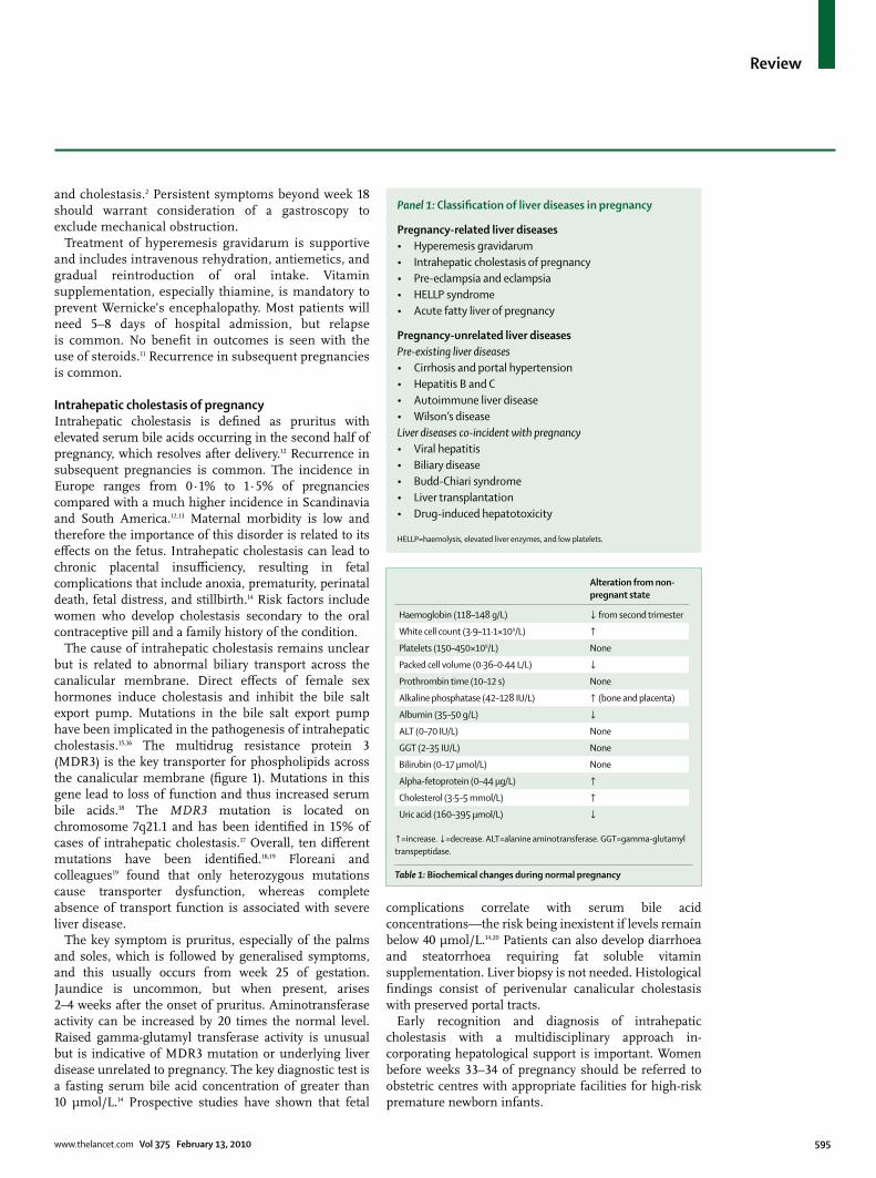
Intrahepatic cholestasis of pregnancy Intrahepatic cholestasis is defi ned as pruritus with elevated serum bile acids occurring in the second half of pregnancy, which resolves after delivery.12 Recurrence in subsequent pregnancies is common. The incidence in Europe ranges from 0·1% to 1·5% of pregnancies compared with a much higher incidence in Scandinavia and South America.12,13 Maternal morbidity is low and therefore the importance of this disorder is related to its eff ects on the fetus. Intrahepatic cholestasis can lead to chronic placental insuffi ciency, resulting in fetal complications that include anoxia, prematurity, perinatal death, fetal distress, and stillbirth.14 Risk factors include women who develop cholestasis secondary to the oral contraceptive pill and a family history of the condition.
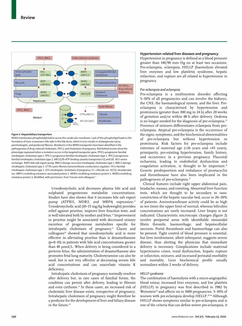
The cause of intrahepatic cholestasis remains unclear but is related to abnormal biliary transport across the canalicular membrane. Direct eff ects of female sex hormones induce cholestasis and inhibit the bile salt export pump. Mutations in the bile salt export pump have been implicated in the pathogenesis of intrahepatic cholestasis.15,16 The multidrug resistance protein 3 (MDR3) is the key transporter for phospholipids across the canalicular membrane (fi gure 1). Mutations in this gene lead to loss of function and thus increased serum bile acids.18 The MDR3 mutation is located on chromosome 7q21.1 and has been identifi ed in 15% of cases of intrahepatic cholestasis.17 Overall, ten diff erent mutations have been identifi ed.18,19 Floreani and colleagues19 found that only heterozygous mutations cause transporter dysfunction, whereas complete absence of transport function is associated with severe liver disease.
The key symptom is pruritus, especially of the palms and soles, which is followed by generalised symptoms, and this usually occurs from week 25 of gestation. Jaundice is uncommon, but when present, arises 2–4 weeks after the onset of pruritus. Aminotransferase activity can be increased by 20 times the normal level. Raised gamma-glutamyl transferase activity is unusual but is indicative of MDR3 mutation or underlying liver disease unrelated to pregnancy. The key diagnostic test is a fasting serum bile acid concentration of greater than 10 μmol/L.14 Prospective studies have shown that fetal
complications correlate with serum bile acid concentrations—the risk being inexistent if levels remain below 40 μmol/L.14,20 Patients can also develop diarrhoea and steatorrhoea requiring fat soluble vitamin supplementation. Liver biopsy is not needed. Histological fi ndings consist of perivenular canalicular cholestasis with preserved portal tracts.
Early recognition and diagnosis of intrahepatic cholestasis with a multidisciplinary approach in-corporating hepatological support is important. Women before weeks 33–34 of pregnancy should be referred to obstetric centres with appropriate facilities for high-risk premature newborn infants.
Alteration from non-pregnant state
Haemoglobin (118–148 g/L) ↓ from second trimester
White cell count (3·9–11·1×109/L) ↑
Platelets (150–450×109/L) None
Packed cell volume (0·36–0·44 L/L) ↓
Prothrombin time (10–12 s) None
Alkaline phosphatase (42–128 IU/L) ↑ (bone and placenta)
Albumin (35–50 g/L) ↓
ALT (0–70 IU/L) None
GGT (2–35 IU/L) None
Bilirubin (0–17 μmol/L) None
Alpha-fetoprotein (0–44 μg/L) ↑
Cholesterol (3·5–5 mmol/L) ↑
Uric acid (160–395 μmol/L) ↓
↑=increase. ↓=decrease. ALT=alanine aminotransferase. GGT=gamma-glutamyl transpeptidase.
Table 1: Biochemical changes during normal pregnancy
Panel 1: Classifi cation of liver diseases in pregnancy
Pregnancy-related liver diseases• Hyperemesis gravidarum• Intrahepatic cholestasis of pregnancy• Pre-eclampsia and eclampsia• HELLP syndrome• Acute fatty liver of pregnancy
Pregnancy-unrelated liver diseasesPre-existing liver diseases• Cirrhosis and portal hypertension• Hepatitis B and C• Autoimmune liver disease• Wilson’s diseaseLiver diseases co-incident with pregnancy• Viral hepatitis• Biliary disease• Budd-Chiari syndrome• Liver transplantation• Drug-induced hepatotoxicity
HELLP=haemolysis, elevated liver enzymes, and low platelets.
Review
596 www.thelancet.com Vol 375 February 13, 2010
Ursodeoxycholic acid decreases plasma bile acid and sulphated progesterone metabolite concentrations. Studies have also shown that it increases bile salt export pump (ATPB11, MDR3, and MRP4) expression.21 Ursodeoxycholic acid (10–15 mg/kg bodyweight) provides relief against pruritus, improve liver function tests, and is well tolerated both by mother and fetus.22 Improvement in pruritus might be associated with decreased urinary excretion of progesterone metabolites specifi c for intrahepatic cholestasis of pregnancy.23 Glantz and colleagues20 showed that ursodeoxycholic acid is more eff ective in alleviating pruritus than is dexamethasone (p=0·01) in patients with bile acid concentrations greater than 40 μmol/L. When delivery is being considered in a preterm fetus, the administration of dexamethasone also promotes fetal lung maturity. Cholestyramine can also be used, but is not very eff ective at decreasing serum bile acid concentrations and can exacerbate vitamin K defi ciency.
Intrahepatic cholestasis of pregnancy normally resolves after delivery but, in rare cases of familial forms, the condition can persist after delivery, leading to fi brosis and even cirrhosis.24 In these cases, an increased risk of cholestatic liver disease exists, irrespective of pregnancy. Intrahepatic cholestasis of pregnancy might therefore be a predictor for the development of liver and biliary disease in the future.24
Hypertension-related liver diseases and pregnancyHypertension in pregnancy is defi ned as a blood pressure greater than 140/90 mm Hg on at least two occasions. Pre-eclampsia, eclampsia, HELLP (haemolysis elevated liver enzymes and low platelets) syndrome, hepatic infarction, and rupture are all related to hypertension in pregnancy.
Pre-eclampsia and eclampsiaPre-eclampsia is a multisystem disorder aff ecting 5–10% of all pregnancies and can involve the kidneys, the CNS, the haematological system, and the liver. Pre-eclampsia is characterised by hypertension and proteinuria (greater than 300 mg in 24 h) after 20 weeks of gestation and/or within 48 h after delivery. Oedema is no longer needed for the diagnosis of pre-eclampsia.25 Presence of seizures diff erentiates eclampsia from pre-eclampsia. Atypical pre-eclampsia is the occurrence of the signs, symptoms, and the biochemical abnormalities of pre-eclampsia but without hypertension or proteinuria. Risk factors for pre-eclampsia include extremes of maternal age (<16 years and >45 years), primiparity, pre-existing hypertension, family history, and occurrence in a previous pregnancy. Placental ischaemia, leading to endothelial dysfunction and coagulation activation, is thought to be important.26 Genetic predisposition and imbalance of prostacyclin and thromboxane have also been implicated in the pathogenesis of pre-eclampsia.27
Clinical features include right upper abdominal pain, headache, nausea, and vomiting. Abnormal liver function tests, which are thought to be secondary to vaso-constriction of the hepatic vascular bed, occur in 20–30% of patients. Aminotransferase activity could be as high as ten times the upper limit of normal, whereas bilirubin concentrations are rarely increased. Liver biopsy is not indicated. Characteristic microscopic changes (fi gure 2) involve periportal areas with identifi able sinusoidal fi brin thrombi, haemorrhage, and hepatocellular necrosis. Portal thrombosis and haemorrhage can also be present. Tight control of blood pressure is essential, but liver involvement, albeit infrequent, suggests severe disease, thus alerting the physician that immediate delivery is necessary. Complications include maternal hypertensive crises, renal dysfunction, hepatic rupture or infarction, seizures, and increased perinatal morbidity and mortality. Liver biochemical profi le usually normalises within 2 weeks of delivery.
HELLP syndromeThe combination of haemolysis with a micro-angiopathic blood smear, increased liver enzymes, and low platelets (HELLP) in pregnancy was fi rst described in 1982 by Weinstein28 and aff ects six in 1000 pregnancies. 5–10% of women with pre-eclampsia develop HELLP.29,30 Although HELLP shows symptoms similar to pre-eclampsia and is one of the criteria that can defi ne severe pre-eclampsia, it
Amino phospho- lipids
Conjugated bilirubin Glutathione
Phosphatidylcholine
Sitosterol Cholesterol
Cystic fibrosis
Chloride
Bicarbonate
CFTR (ABCC7)
AE2 (SLC4A2) Bileduct
Hepatocyte
PFIC2, BRIC2
HCO3–
Cl–
Cl–
Bile acids
Drugs MDR1 (ABCB1)
BSEP (ABCB11)
FIC1 (ATP 8B1)
MRP2 (ABCC2)
MDR3 (ABCB4)
ABCG5/8
ICP, PFIC3,drug-induced cholestasis
PFIC1, BRIC1 Dubin-Johnson syndrome
Figure 1: Hepatobiliary transportersMDR3 translocates phosphatidylcholine across the canalicular membrane. Lack of this phospholipid leads to the formation of toxic monomeric bile salts in the bile ducts, which in turn results in cholangiocyte injury, pericholangitis, and periductal fi brosis. Mutations in the MDR3 transporter have been identifi ed in the pathogenesis of drug-induced cholestasis, PFIC3, and cholestasis of pregnancy. Red dotted arrows show the phenotype expressed when a mutation occurs in the targeted transporter gene. PFIC1=progressive familial intrahepatic cholestasis type 1. PFIC2=progressive familial intrahepatic cholestasis type 2. PFIC3=progressive familial intrahepatic cholestasis type 3. ABCG5/8=ATP binding cassette transporters G5 and G8. AE2=anion exchanger. BSEP=bile salt export pump. BRIC1=benign recurrent intrahepatic cholestasis type 1. BRIC2=benign intrahepatic cholestasis type 2. CFTR=cystic fi brosis transmembrane conductance regulator. FIC1=familial intrahepatic cholestasis type 1. ICP=intrahepatic cholestasis of pregnancy. Cl–= chloride ion. HCO3–=bicarbonate ion. MRP2=multidrug resistance-associated protein 2. MDR1=multidrug resistance protein 1. MDR3=multidrug resistance protein 3. Modifi ed, with permission, from Trauner and colleagues.17

Review
www.thelancet.com Vol 375 February 13, 2010 597
can develop in women who might not have any other signs or symptoms of pre-eclampsia. A perinatal infant mortality rate of 6–70% has been reported due to prematurity, or secondary to maternal complications.31 HELLP usually arises in the second or third trimester, but can also develop after delivery. Risk factors include advanced maternal age, multiparity, and white ethnic origin.
Angiogenic markers have also been identifi ed, which might help to confi rm the diagnosis of pre-eclampsia in women without hypertension or proteinuria. They include decreased placental growth factor, increased serum soluble endoglin, and increased soluble fms-like tyrosine kinase-1 (VEGF) receptor.32–36
Patients with HELLP syndrome might be asymptomatic or present with right upper quadrant and epigastric pain, nausea, vomiting, and malaise. Hypertension and proteinuria is evident in up to 85% of cases. Liver injury is precipitated by intravascular fi brin deposition, hypo-volaemia, and increased sinusoidal pressure resulting in mild-to-moderate increase of aminotrans ferases and mild elevation of bilirubin. Recognised classifi cation systems of HELLP include the Tennessee and the Mississippi systems (panel 2).37 In the Tennessee system classifi cation, the result can be complete (ie, demonstration of haemolysis [raised lactate dehydro-genase, decreased haptoglobin, raised unconjugated bilirubin], thrombo cytopenia [secondary to vascular endothelial damage and fi brin deposition in vascular walls], and elevated aminotransferases) or partial (ie, encompassing one or two components). The prothrombin time or international normalised ratio remains normal unless there is evidence of disseminated intravascular coagulation or severe liver injury. A serum uric acid of more than 464 μmol/L is associated with increased maternal and fetal morbidity and mortality.20,31 Liver biopsy remains a high-risk procedure because of the thrombocytopenia. Microscopic fi ndings may be non-specifi c or similar to those of pre-eclampsia (fi gure 2).
Hepatic haematoma, infarction, and ruptureCT or MRI of the liver could identify hepatic infarction and rupture, haemorrhage, or subcapsular haematoma. The diff erential diagnosis includes acute fatty liver of pregnancy, thrombotic thrombocytopenic purpura, and haemolytic uraemic syndrome. Hepatic haematoma, infarction, and rupture occur in a minority of women with established pre-eclampsia or HELLP syndrome. 50% maternal mortality has been reported for this complication of disease, with prevalence of hepatic rupture being higher with severe thrombocytopenia.38 Hepatic adenoma, hepatocellular carcinoma, and haemangiomas might also rupture during pregnancy.
Risk factors for rupture include advanced maternal age, multiparity, and pre-eclampsia. Patients with hepatic haematoma secondary to a ruptured liver
capsule typically present in the third trimester with severe right upper quadrant pain and pyrexia, although the presentation can also be early after delivery. Increased aminotransferase concentrations in excess of 3000 U/L, leucocytosis, pyrexia, and anaemia are frequently seen. Acute complications include acute respiratory distress syndrome, acute kidney injury, and hypovolaemic shock. CT and MRI help to identify these pathologies (fi gure 3). Contained haematomas should be managed conservatively with blood transfusion and supportive measures for the mother. Infection can occur within areas of hepatic infarction. Haemodynamic instability suggests persistent active bleeding and should prompt hepatic angiography, and when required, invasive haemostatic measures by arterial embolisation of the hepatic artery or surgical exploration. Surgical options include packing, hepatic artery ligation, or
Panel 2: Classifi cation systems used in HELLP syndrome
Tennessee system• AST >70 IU/L• LDH >600 IU/L• Platelets <100×109/L
Mississippi system AST >40 IU/L and LDH >600 IU/L and:• Class I: platelets <50×109/L• Class II: platelets 50–100×109/L • Class III: platelets 100–150×109/L
HELLP=haemolysis, elevated liver enzymes, and low platelets. AST=aspartate aminotransferase. LDH=lactate dehydrogenase.
Figure 2: Liver biopsy from a young woman with eclampsiaHaematoxylin and eosin staining. Liver biopsy shows an area of coagulative necrosis (marked by the arrows), which involves perivenular and midzonal hepatocytes. The arrowhead indicates a portal tract.

Review
598 www.thelancet.com Vol 375 February 13, 2010
resection of the aff ected liver. No long-term maternal complications have been reported.
Women with HELLP syndrome might need a high-dependency unit or an intensive care setting because of the potential complications of hepatic encephalopathy, acute renal dysfunction, hepatic rupture, and bleeding. The cornerstone of management is delivery. Prompt delivery should be undertaken if pregnancy is over 34 weeks of gestation, if fetal distress is present, or if evidence exists of maternal end-organ disease (ie, disseminated intravascular coagulation, renal failure, or abruptia placenta).39 Management of hypertension involves the use of labetalol, hydralazine, and nifedipine. Diuretics are not recommended in patients with HELLP,
because they can cause utero–placental hypoperfusion.39 Intravenous magnesium sulphate with platelet, coagulation support, or both, are recommended, especially in the presence of bleeding. If gestation is less than 34 weeks, corticosteroids should be administered to promote fetal lung maturity only, because they do not provide any maternal benefi t.40
Women with atypical pre-eclampsia have a more diffi cult diagnostic and management conundrum. Physicians should therefore be vigilant to atypical presentations of pre-eclampsia with careful assessment of maternal risk factors, laboratory fi ndings, and timing in the course of pregnancy.
The risk of recurrence of HELLP syndrome in subsequent pregnancies is increased.41,42 HELLP syndrome usually resolves rapidly after delivery. Laboratory values, however, might worsen after delivery. Hepatic or renal failure necessitates admission to intensive care. Indications for liver transplantation include persistent bleeding from haematoma, hepatic rupture, or liver failure.43 88% of these patients survive 5 years after liver transplant.44
Acute fatty liver of pregnancy First described in 1934 by Stander and Cadden45 as “acute yellow atrophy of the liver”, acute fatty liver of pregnancy remains a medical and obstetric emergency. This condition is defi ned as microvesicular fatty infi ltra tion of hepatocytes during the second half of pregnancy (usually third trimester), and it remains a common cause of liver failure in pregnancy. Maternal and fetal mortality rates are signifi cantly increased and range between 1% and 20%.46
Acute fatty liver of pregnancy is a rare disorder aff ecting from one in 7000 to one in 16 000 pregnancies,47,48 therefore making it diffi cult to study. A recent UK-based prospective study involving 229 centres identifi ed 57 confi rmed cases in a total of 1 132 964 pregnancies, giving an incidence of fi ve in 100 000 pregnancies.49 74% of cases were identifi ed at a median gestation age of 36 weeks, with 60% of cases delivered within 24 h of diagnosis.49 The caesarean section rate was 74%.
Acute fatty liver of pregnancy is one of the mitochondrial cytopathies, which include Reye’s syndrome and other drug-related liver diseases. Common characteristics of these disorders include vomiting, hypoglyacaemia, lactic acidosis, hyperammonaemia, and microvesicular fat deposition in organs. Abnormality in mitochondrial β oxidation is recognised as the cause of this condition.48 Mitochondrial β oxidation of fatty acids is a complex process and is an important energy source for skeletal muscle and myocardial tissue. The enzyme long-chain 3-hydoxyacyl coenzyme A dehydrogenase is part of the mitochondrial trifunctional protein (MTP), which is an important complex associated with the inner mitochondrial membrane.50,51 MTP is an hetero-octamer consisting of four α subunits and four β subunits52 (fi gure 4).
Figure 3: Abdominal CT showing a subcapsular haematoma in a woman with HELLP syndrome
3-ketoacyl-CoA thiolase
Shortened acyl-CoA
3-ketoacyl-CoA
3-hydroxyacyl-CoA
Trifunctional protein
2,3-enoyl-CoA
Acyl-CoA
Acyl-CoA dehydrogenase
2,3-enoyl-CoA hydratase
3-hydroxyacyl-CoAdehydrogenase
LCHAD deficiency
Tri-functionalproteindeficiency
Figure 4: Cycle of mitochondrial oxidation Long-chain 3-hydroxyacyl-CoA dehydrogenase (LCHAD) catalyses the third step in the β oxidation of fatty acids in mitochondria (the formation of 3-ketoacyl-CoA from 3-hydroxyacyl-CoA). The accumulation of long-chain 3-hydroxyacyl metabolites produced by the fetus or placenta is toxic to the liver. LCHAD defi ciency in infants can lead to non-ketotic hypoglycaemia, hepatic encephalopathy, cardiomyopathy, peripheral neuropathy, myopathy, and sudden death. Modifi ed, with permission, from Ibdah and colleagues.53
Review
www.thelancet.com Vol 375 February 13, 2010 599
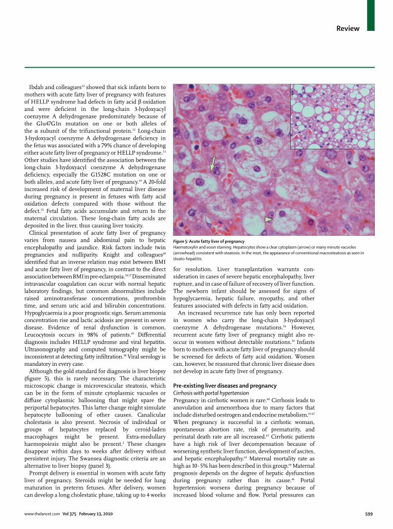
Ibdah and colleagues53 showed that sick infants born to mothers with acute fatty liver of pregnancy with features of HELLP syndrome had defects in fatty acid β oxidation and were defi cient in the long-chain 3-hydoxyacyl coenzyme A dehydrogenase predominately because of the Glu47G1n mutation on one or both alleles of the α subunit of the trifunctional protein.53 Long-chain 3-hydoxyacyl coenzyme A dehydrogenase defi ciency in the fetus was associated with a 79% chance of developing either acute fatty liver of pregnancy or HELLP syndrome.53 Other studies have identifi ed the association between the long-chain 3-hydoxyacyl coenzyme A dehydrogenase defi ciency, especially the G1528C mutation on one or both alleles, and acute fatty liver of pregnancy.54 A 20-fold increased risk of development of maternal liver disease during pregnancy is present in fetuses with fatty acid oxidation defects compared with those without the defect.55 Fetal fatty acids accumulate and return to the maternal circulation. These long-chain fatty acids are deposited in the liver, thus causing liver toxicity.
Clinical presentation of acute fatty liver of pregnancy varies from nausea and abdominal pain to hepatic encephalopathy and jaundice. Risk factors include twin pregnancies and nulliparity. Knight and colleagues49 identifi ed that an inverse relation may exist between BMI and acute fatty liver of pregnancy, in contrast to the direct association between BMI in pre-eclampsia.56,57 Disseminated intravascular coagulation can occur with normal hepatic laboratory fi ndings, but common abnormalities include raised aminotransferase concentrations, prothrombin time, and serum uric acid and bilirubin concentrations. Hypoglycaemia is a poor prognostic sign. Serum ammonia concentration rise and lactic acidosis are present in severe disease. Evidence of renal dysfunction is common. Leucocytosis occurs in 98% of patients.49 Diff erential diagnosis includes HELLP syndrome and viral hepatitis. Ultrasonography and computed tomography might be inconsistent at detecting fatty infi ltration.58 Viral serology is mandatory in every case.
Although the gold standard for diagnosis is liver biopsy (fi gure 5), this is rarely necessary. The characteristic microscopic change is microvescicular steatosis, which can be in the form of minute cytoplasmic vacuoles or diff use cytoplasmic ballooning that might spare the periportal hepatocytes. This latter change might simulate hepatocyte ballooning of other causes. Canalicular cholestasis is also present. Necrosis of individual or groups of hepatocytes replaced by ceroid-laden macrophages might be present. Extra-medullary haemopoiesis might also be present.2 These changes disappear within days to weeks after delivery without persistent injury. The Swansea diagnostic criteria are an alternative to liver biopsy (panel 3).
Prompt delivery is essential in women with acute fatty liver of pregnancy. Steroids might be needed for lung maturation in preterm fetuses. After delivery, women can develop a long cholestatic phase, taking up to 4 weeks
for resolution. Liver transplantation warrants con-sideration in cases of severe hepatic encephalopathy, liver rupture, and in case of failure of recovery of liver function. The newborn infant should be assessed for signs of hypoglycaemia, hepatic failure, myopathy, and other features associated with defects in fatty acid oxidation.
An increased recurrence rate has only been reported in women who carry the long-chain 3-hydoxyacyl coenzyme A dehydrogenase mutations.54 However, recurrent acute fatty liver of pregnancy might also re-occur in women without detectable mutations.59 Infants born to mothers with acute fatty liver of pregnancy should be screened for defects of fatty acid oxidation. Women can, however, be reassured that chronic liver disease does not develop in acute fatty liver of pregnancy.
Pre-existing liver diseases and pregnancy Cirrhosis with portal hypertensionPregnancy in cirrhotic women is rare.60 Cirrhosis leads to anovulation and amenorrhoea due to many factors that include disturbed oestrogen and endocrine metabolism.61,62 When pregnancy is successful in a cirrhotic woman, spontaneous abortion rate, risk of prematurity, and perinatal death rate are all increased.63 Cirrhotic patients have a high risk of liver decompensation because of worsening synthetic liver function, development of ascites, and hepatic encephalopathy.63 Maternal mortality rate as high as 10·5% has been described in this group.64 Maternal prognosis depends on the degree of hepatic dysfunction during pregnancy rather than its cause.46 Portal hypertension worsens during pregnancy because of increased blood volume and fl ow. Portal pressures can
Figure 5: Acute fatty liver of pregnancy Haematoxylin and eosin staining. Hepatocytes show a clear cytoplasm (arrow) or many minute vacuoles (arrowhead) consistent with steatosis. In the inset, the appearance of conventional macrosteatosis as seen in steato-hepatitis.

Review
600 www.thelancet.com Vol 375 February 13, 2010
also increase because of an increased vascular resistance due to external compression of the inferior vena cava by the gravid uterus. Up to 25% of patients with varices have a bleeding episode during pregnancy.65 The greatest risk is seen in the second trimester, when portal pressures peak, and during delivery because of the repeated use of the valsalva manoeuvre to help to expel the fetus.64 Rupture of splenic artery aneurysm, although uncommon, could also present in pregnant women with portal hypertension.
All cirrhotic patients should undergo variceal screening. Banding before pregnancy, although not proven, is appropriate for high-risk varices. Propranolol has also been used safely in pregnancy but side-eff ects include fetal growth retardation, neonatal bradycardia, and hypoglycaemia. Terlipressin has not been studied in pregnancy but concerns have been raised about decreased placental perfusion and increased risk of placental abruption. The use of a transjugular intrahepatic porto-systemic shunt in extreme cases of variceal bleeding can be considered but has the risk of radiation exposure to the fetus.
Hepatitis B and hepatitis C infectionsIn many developed countries, pregnant women are routinely screened for hepatitis B virus (HBV) at the initial booking visit.66 HBV vaccine can be given safely during pregnancy if needed.67 Women who are not cirrhotic but HBV-positive are at risk of transmitting the virus to the fetus. Vertical transmission remains the most common way of transmission of HBV in endemic areas and accounts for most HBV infection worldwide. Chronic HBV infection is more likely in the newborn infant when the mother is positive to both hepatitis B surface antigen and hepatitis B e antigen, and also has a high HBV viral
load. HBV viral load is a key factor in transmission, with high viral load being associated with 80–90% risk of transmission compared with 10–30% transmission rates in patients with undetectable viral load.68,69 Transmission can occur directly via the placenta (intrauterine), during breastfeeding, or during delivery. Mode of delivery does not aff ect the risk of transmission, with similar rates seen with normal vaginal delivery and caesarean section.70 Transmission can be reduced further by administration of hepatitis B immunoglobulin to the neonate within 12 h of birth.71 HBV vaccine should also be administered, with three doses being given to the infant within the fi rst 6 months.
Use of lamivudine and other antiviral drugs during the third trimester to reduce HBV viral load,72 and thus decrease the risk of transmission to the fetus, is a source of debate. Use of lamivudine monotherapy could predispose to viral mutations, thus rendering the patient susceptible to viral resistance to both lamivudine and other antiviral drugs long term. Lamivudine, which has been classifi ed by the US Food and Drug Administration (FDA) as a category C drug in pregnancy, has been used successfully in patients with both HBV and HIV infection during pregnancy without substantial risk to either mother or fetus. Entecavir, a more potent nucleoside analogue with a better long-term viral resistance profi le than lamivudine, and designated as a category C drug in pregnancy by the FDA, also shows promise, whereas tenofovir, widely used in HIV-positive pregnant patients, seems to have a better safety profi le than both entecavir and lamivudine, and is regarded as a category B drug.
At present, no guidelines exist for the use of lamivudine or any other nucleoside in HBV-positive pregnant women, and therefore any decisions should be made on an individual basis. We advise the use of either lamivudine or tenofovir after week 32 of gestation in patients with a high HBV viral load (greater than 10⁶ copies per mL), especially in mothers who might have already infected their child during their previous pregnancy. However, the duration of therapy needs to be considered carefully. Moreover, the use of antiviral agents should not be a substitute to appropriate vaccination.
Pregnancy in patients with hepatitis C virus (HCV) is usually uneventful. Risk of vertical transmission of HCV remains low, except when the fetus is exposed to large volumes of mother’s blood and vaginal fl uid during delivery or if the mother is co-infected with HIV.73,74 Patients with genotypes 1 or 3 and with HIV co-infection are more likely to transmit HCV vertically.75 Ribavirin is teratogenic and the use of pegylated interferon in combination with ribavirin is contra-indicated during pregnancy, but not during breastfeeding.
Autoimmune liver diseaseSuccessful pregnancies are achievable in women with autoimmune hepatitis. Some studies have shown that
Panel 3: Swansea diagnostic criteria for diagnosis of acute fatty liver of pregnancy1
Six or more of the following features in the absence of another explanation • Vomiting• Abdominal pain• Polydipsia/polyuria• Encephalopathy• High bilirubin (>14 μmol/L)• Hypoglycaemia (<4 mmol/L)• High uric acid (>340 μmol/L)• Leucocytosis (>11×106/L)• Ascites or bright liver on ultrasound scan• High AST/ALT (>42 IU/L)• High ammonia (>47 μmol/L)• Renal impairment (creatinine >150 μmol/L)• Coagulopathy (PT >14 s or APTT >34 s)• Microvesicular steatosis on liver biopsy
ALT=Alanine aminotransferase. AST=aspartate aminotransferase. PT=prothrombin time. APTT=activated partial thromboplastin time.

Review
www.thelancet.com Vol 375 February 13, 2010 601
fl ares in disease activity are more likely to occur in the fi rst 3 months after delivery, although autoimmune hepatitis might present for the fi rst time during pregnancy.76,77 Women with autoimmune hepatitis need stable immunosuppression throughout pregnancy. Although azathioprine is teratogenic in animal models, teratogenic eff ects in human beings have not been described. Two separate studies failed to show toxic eff ects of azathioprine or its metabolites during pregnancy.76,77 Fetal side-eff ects have been reported, however, and include lymphopoenia, hypogam-maglobulinaemia, and thymic hypolasia. If fl ares occur, then they should be managed in the conventional manner—that is, administration of steroids or increase in steroid dose. If an immunosuppressant is needed, azathioprine remains the safest choice. A study76 described the presence of antibodies to SLA/LP and Ro(SSA) as risk factors for adverse outcomes in pregnancy.
Primary biliary cirrhosis is a chronic cholestatic disease that leads to destruction of intrahepatic bile ducts. Deterioration in synthetic liver function can occur during pregnancy.78 Primary biliary cirrhosis might also present for the fi rst time after delivery with protracted pruritus. Ursodeoxycholic acid can be used safely in pregnant women with primary biliary cirrhosis.78
Wilson’s diseaseWilson’s disease is a rare autosomal recessive disease of defective biliary copper excretion, which leads to copper deposition in the liver, brain, and kidneys. Patients usually present with hyperbilirubinaemia, high con-centrations of aminotransferases, Coombes-negative haemolytic anaemia, and low serum alkaline phosphatase. In pregnant women with undiagnosed Wilson’s disease, evidence of acute liver failure and haemolysis could be misinterpreted as HELLP syndrome.
In women with known Wilson’s disease, serum copper levels and caeruloplasmin can rise during pregnancy79 without treatment, leading to a fl are in patient symptoms. Zinc can be continued in pregnancy without causing harm to the fetus.80
Liver diseases co-incident with pregnancyAcute viral hepatitisHepatitis A virus (HAV) infection in pregnancy has a clinical course similar to that in the non-pregnant population.81 A recent study has identifi ed the presence of ascites and hypertension as factors that help to diff erentiate liver disease specifi c to pregnancy from viral hepatitis.82 Increased severity of disease is associated with advanced maternal age, with severe infection in the third trimester associated with an increased risk of prematurity.83 Treatment is supportive.
Pregnant women are more vulnerable to hepatitis E virus (HEV) infection than to HAV, HBV, and HCV.84 HEV remains the most prevalent viral cause of acute
liver failure in pregnancy.85 Endemic in parts of Asia and Africa, HEV-related hepatitis usually follows a more severe course in pregnancy, especially in the Indian subcontinent,86–88 with these patients more likely to develop fulminant hepatic failure.89 In-utero trans-mission of HEV to the fetus might add further toxic metabolites to the maternal circulation,90,91 resulting in increased maternal morbidity and mortality.92 Pregnant women are more likely to acquire HEV in the second or third trimester, with a median gestational age of 28 weeks.86 Reported maternal mortality from fulminant hepatic failure secondary to HEV in pregnancy is
Trimester Diagnostics
HG 1, 2 ↑ Bilirubin (×4 ULN), ↑ ALT/AST (×2–4 ULN)
ICP 1, 2, 3 ↑ Bilirubin (×6 ULN), ↑ ALT/AST (×6 ULN), ↑ bile acids
Pre-eclampsia 2, 3 ↑ Bilirubin (×2–5 ULN), ↑ ALT/AST (×10–50 ULN), ↓ platelets
HELLP 2, 3 ↑ ALT/AST (×10–20 ULN), ↑ LDH, ↓ platelets, ↑ uric acid
AFLP 2, 3 ↑ Bilirubin (×6-8 ULN), ↑ ALT/AST (×5–10 ULN)—rarely >20
↑=increase. ↓=decrease. HG=hyperemesis gravidarum. ICP=intrahepatic cholestasis of pregnancy. HELLP=haemolysis, elevated liver enzymes, and low platelets. AFLP=acute fatty liver of pregnancy. ALT=alanine aminotransferase. AST=aspartate aminotransferase. LDH=lactate dehydrogenase. ULN=upper limit of normal.
Table 2: Characteristic timings and diagnostic laboratory features of liver diseases related to pregnancy
Figure 6: Percutaneous transhepatic cholangiogram showing large choledochal cystsTwo large choledochal cysts (arrows) in a patient who had previously undergone biliary reconstruction due to repeated bouts of biliary sepsis.

Review
602 www.thelancet.com Vol 375 February 13, 2010
41–54%,82,93 with a fetal mortality rate of 69%.75 Poor outcomes are associated with the development of grade III or IV hepatic encephalopathy,75 irrespective of which trimester the infection was contracted. Manage-ment is supportive, ideally including intensive care unit input. Remarkably, delivery does not aff ect maternal outcome.
Herpes simplex virus (HSV) hepatitis is a rare condition, occurring predominantly in immuno-compromised individuals or children.94,95 Pregnant women, however, are more susceptible than the general population to HSV hepatitis,96 with a 39% maternal mortality. HSV sero types 1 and 2 have been seen in pregnancy and are caused by primary or latent disease. Raised amino transferases, thrombocytopenia, leuco-penia, and coagulopathy with a normal concentration of serum bilirubin are common laboratory fi ndings. Mucocutan eous lesions associated with HSV infection might be present, but only in 50% of cases.97 HSV hepatitis should be considered in any pregnant woman with hepatic failure. Liver biopsy provides the defi nitive evidence of HSV hepatitis, but computed tomography could also be useful, showing multiple low-density areas of necrosis within the liver. Treatment with intravenous aciclovir should not be delayed until confi rmatory results are available, and should therefore be commenced if clinical suspicion is high. Treatment with aciclovir is associated with improved survival rate.96 Published reports of this rare disease are unclear as to whether delivery improves neonatal survival.
Biliary diseaseGallstones are more common in pregnancy because of increased cholesterol secretion in the second and third trimester, increased lithogenicity of the bile, and decreased gallbladder motility.98 About 10% of pregnant women develop either gallstones or viscus biliary sludge.99 Open or laparoscopic cholecystectomy can be done safely and successfully during the second trimester. Endoscopic retrograde cholangio pan creatography and
sphincterotomy can be done if needed, with little added risk compared with that in non-pregnant women.100 Choledochal anomalies (fi gure 6) might also present in pregnancy, as a consequence of bile stasis and stone formation. Septic episodes can ensue.
Budd-Chiari syndromeBudd-Chiari syndrome is defi ned as outfl ow obstruction of the hepatic veins. It is commonly associated with myeloproliferative disorders. Up to 20% of cases of Budd-Chiari syndrome occur in women who are on the oral contraceptive pill, are pregnant, or have delivered in the previous 2 months.101 Pregnancy itself represents a prothrombotic state; a physiological decrease of protein S concentration is seen,102 which might account for the increased incidence of Budd-Chiari syndrome in pregnancy.103 Patients with known Budd-Chiari syndrome are at risk of developing an exacerbation during pregnancy because of the increased concentrations of female sex hormones.80
Clinical features include right upper quadrant pain, jaundice, and ascites. Doppler ultrasound is very important in diagnosis. The treatment is anticoagulation at the onset, identifi cation of procoagulant causes, and shunting, or liver transplantation in extreme cases.
Liver transplantation Because of the success of liver transplantation, young patients who have been successfully grafted can become pregnant. Pregnancy should be deferred for 1 year after liver transplantation, which allows lower doses of immunosuppression and more stable graft function. Pregnancy in this group should be managed in specialised centres. 70% of women after liver transplantation will deliver a healthy baby,104 although pregnancy might pose problems, such as an increased risk of gestational diabetes and pre-eclampsia.104,105 Caesarean deliveries are also more likely in this group: 35–63% versus 23% in the general population within the UK.106–108 Commonly used drugs such as tacrolimus, mycophenalate mofetil, prednisolone, azathioprine, and ciclosporin all carry a risk of teratogenicity. Prematurity and low birthweights (<2500 g) occur more frequently in women who have previously undergone liver transplantation. Table 3 shows the side-eff ects of commonly used immunosuppressants. Mycophenalate mofetil should be stopped in all women wishing to become pregnant, but tacrolimus and ciclosporin can be continued. Risk profi les of prednisolone and azathioprine, however, are acceptable and should not be withheld. Breastfeeding is not advised while women are taking immunosuppressants because of the uncertain eff ects on the newborn infant.
Conclusion Hepatic disorders in pregnancy are rare, but remain clin-ically important because of serious adverse eff ects on both
Side-eff ects FDA category
Azathioprine Lymphopenia, hypogammaglobulinaemia, thymic hypoplasia D
Ciclosporin A Premature labour, low birthweight, neonatal hyperkalaemia, renal dysfunction
C
Mycophenalate mofetil
First trimester loss, microtia. Increased risk of congenital malformations D
Prednisolone Cleft palate, intrauterine growth retardation, premature rupture of membranes, fetal adrenal hypoplasia
C
Tacrolimus Similar side-eff ects to ciclosporin. Neonatal malformation rates of 4% C
FDA=US Food and Drug Administration. Pregnancy category C: animal reproduction studies have shown an adverse effect on the fetus, but no adequate and well controlled studies in human beings exist. Potential benefits might warrant use of the drug in pregnant women despite potential risks. Pregnancy category D: positive evidence of human fetal risk based on adverse reaction data from investigational or marketing experience or studies in human beings. However, potential benefits might warrant use of the drug in pregnant women despite potential risks.
Table 3: Common side-eff ects of immunosuppressants
Review
www.thelancet.com Vol 375 February 13, 2010 603
mother and fetus. Liver disease in pregnancy can present with subtle changes in liver biochemical profi le or with ful-minant hepatic failure. These disorders are complex and might need to be managed by experienced physicians in specialised centres. Maternal and fetal survival has im-proved because of better understanding of the pathogene-sis of these disorders and higher standards of clinical care.
ContributorsDJ wrote and edited the report. AJ provided obstetric expertise, and
reviewed and edited the report. RHW reviewed and edited the report.
AQ reviewed and edited the report, and provided histopathological
expertise. MAH wrote, edited, and helped to supervise the preparation of
the report.
Confl icts of interestWe declare that we have no confl icts of interest.
References1 Ch’ng CL, Morgan M, Hainsworth I, Kingham JG. Prospective
study of liver dysfunction in pregnancy in Southwest Wales. Gut 2002; 51: 876–80.
2 Rolfes DB, Ishak KG. Liver disease in pregnancy. Histopathology 1986; 10: 555–70.
3 Shellock FG, Kanal E. Safety of magnetic resonance imaging contrast agents. J Magn Reson Imaging 1999; 10: 477–84.
4 Fairweather DV. Nausea and vomiting during pregnancy. Obstet Gynecol Annu 1978; 7: 91–105.
5 Kallen B. Hyperemesis during pregnancy and delivery outcome: a registry study. Eur J Obstet Gynecol Reprod Biol 1987; 26: 291–302.
6 Kuscu NK, Koyuncu F. Hyperemesis gravidarum: current concepts and management. Postgrad Med J 2002; 78: 76–79.
7 Fell DB, Dodds L, Joseph KS, Allen VM, Butler B. Risk factors for hyperemesis gravidarum requiring hospital admission during pregnancy. Obstet Gynecol 2006; 107: 277–84.
8 Colin JF, Mathurin P, Durand F, et al. Hyperthyroidism: a possible factor of cholestasis associated with hyperemesis gravidarum of prolonged evolution. Gastroenterol Clin Biol 1994; 18: 378–80.
9 Goodwin TM, Montoro M, Mestman JH. Transient hyperthyroidism and hyperemesis gravidarum: clinical aspects. Am J Obstet Gynecol 1992; 167: 648–52.
10 Conchillo JM, Pijnenborg JM, Peeters P, Stockbrugger RW, Fevery J, Koek GH. Liver enzyme elevation induced by hyperemesis gravidarum: aetiology, diagnosis and treatment. Neth J Med 2002; 60: 374–78.
11 Yost NP, McIntire DD, Wians FH Jr, Ramin SM, Balko JA, Leveno KJ. A randomized, placebo-controlled trial of corticosteroids for hyperemesis due to pregnancy. Obstet Gynecol 2003; 102: 1250–54.
12 Reyes H. The enigma of intrahepatic cholestasis of pregnancy: lessons from Chile. Hepatology 1982; 2: 87–96.
13 Schneider G, Paus TC, Kullak-Ublick GA, et al. Linkage between a new splicing site mutation in the MDR3 alias ABCB4 gene and intrahepatic cholestasis of pregnancy. Hepatology 2007; 45: 150–58.
14 Glantz A, Marschall HU, Mattsson LA. Intrahepatic cholestasis of pregnancy: Relationships between bile acid levels and fetal complication rates. Hepatology 2004; 40: 467–74.
15 Eloranta ML, Hakli T, Hiltunen M, Helisalmi S, Punnonen K, Heinonen S. Association of single nucleotide polymorphisms of the bile salt export pump gene with intrahepatic cholestasis of pregnancy. Scand J Gastroenterol 2003; 38: 648–52.
16 Dixon PH, van Mil SW, Chambers J, et al. Contribution of variant alleles of ABCB11 to susceptibility to intrahepatic cholestasis of pregnancy. Gut 2009; 58: 537–44.
17 Trauner M, Fickert P, Wagner M. MDR3 (ABCB4) defects: a paradigm for the genetics of adult cholestatic syndromes. Semin Liver Dis 2007; 27: 77–98.
18 Keitel V, Vogt C, Haussinger D, Kubitz R. Combined mutations of canalicular transporter proteins cause severe intrahepatic cholestasis of pregnancy. Gastroenterology 2006; 131: 624–29.
19 Floreani A, Carderi I, Paternoster D, et al. Intrahepatic cholestasis of pregnancy: three novel MDR3 gene mutations. Aliment Pharmacol Ther 2006; 23: 1649–53.
20 Glantz A, Marschall HU, Lammert F, Mattsson LA. Intrahepatic cholestasis of pregnancy: a randomized controlled trial comparing dexamethasone and ursodeoxycholic acid. Hepatology 2005; 42: 1399–405.
21 Marschall HU, Wagner M, Zollner G, et al. Complementary stimulation of hepatobiliary transport and detoxifi cation systems by rifampicin and ursodeoxycholic acid in humans. Gastroenterology 2005; 129: 476–85.
22 Mazzella G, Rizzo N, Azzaroli F, et al. Ursodeoxycholic acid administration in patients with cholestasis of pregnancy: eff ects on primary bile acids in babies and mothers. Hepatology 2001; 33: 504–08.
23 Glantz A, Reilly SJ, Benthin L, Lammert F, Mattsson LA, Marschall HU. Intrahepatic cholestasis of pregnancy: amelioration of pruritus by UDCA is associated with decreased progesterone disulphates in urine. Hepatology 2008; 47: 544–51.
24 Ropponen A, Sund R, Riikonen S, Ylikorkala O, Aittomaki K. Intrahepatic cholestasis of pregnancy as an indicator of liver and biliary diease: a population-based study. Hepatology 2006; 43: 723–28.
25 Sibai BM. Diagnosis and management of gestational hypertension and pre-eclampsia. Obstet Gynaecol 2003; 102: 181–92.
26 Caniggia I, Winter J, Lye SJ, Post M. Oxygen and placental development during the fi rst trimester: implications for the pathophysiology of pre-eclampsia. Placenta 2000; 21 (suppl A): S25–30.
27 Walsh SW, Vaughan JE, Wang Y, Roberts LJ. Placental isoprostane is signifi cantly increased in preeclampsia. FASEB J 2000; 14: 1289–96.
28 Weinstein L. Syndrome of hemolysis, elevated liver enzymes, and low platelet count: a severe consequence of hypertension in pregnancy. Am J Obstet Gynecol 1982; 142: 159–67.
29 Egerman RS, Sibai BM. HELLP syndrome. Clin Obstet Gynecol 1999; 42: 381–89.
30 Martin JN Jr, Rose CH, Briery CM. Understanding and managing HELLP syndrome: the integral role of aggressive glucocorticoids for mother and child. Am J Obstet Gynecol 2006; 195: 914–34.
31 Mihu D, Costin N, Mihu CM, Seicean A, Ciortea R. HELLP syndrome—a multisystemic disorder. J Gastrointestin Liver Dis 2007; 16: 419–24.
32 Masuyama H, Suwaki N, Nakatsukasa H, et al. Circulating angiogenic factors in preeclampsia superimposed on chronic glomerulonephritis. Am J Obstet Gynecol 2006; 194: 551–56.
33 Levine RJ, Lam C, Qian C. Soluble endoglin and other circulating angiogenic factors in preeclampsia. N Engl J Med 2006; 355: 992–1005.
34 Levine RJ, Maynard SE, Qian C, et al. Circulating angiogenic factors and the risk for preeclampsia. N Engl J Med 2004; 350: 672–83.
35 Salahuddin S, Lee Y, Vadnais M. Diagnostic utility of soluble fms-like tyrosine kinase 1 and soluble endoglin in hypertensive diseases of pregnancy. Am J Obstet Gynecol 2007; 197: 28.e1–6.
36 Robinson CJ, Johnson DJ. Soluble endoglin as a second-trimester marker for preeclampsia. Am J Obstet Gynecol 2007; 197: 174.e1–6.
37 Magann EF, Martin JN Jr. Twelve steps to optimal management of HELLP syndrome. Clin Obstet Gynecol 1999; 42: 532–50.
38 Sibai BM, Ramadan MK, Usta I, Salama M, Mercer BM, Friedman SA. Maternal morbidity and mortality in 442 pregnancies with hemolysis, elevated liver enzymes, and low platelets (HELLP syndrome). Am J Obstet Gynecol 1993; 169: 1000–06.
39 Sibai BM, Caritis S, Hauth J. What we have learned about preeclampsia. Semin Perinatol 2003; 27: 239–46.
40 Fonseca J, Mendez F, Catono C, Arias F. Dexamethasone treatment does not improve the outcome of women with HELLP syndrome: a double-blind placebo-controlled, randomized clinical trial. Am J Obstet Gynecol 2005; 193: 1591–98.

Review
604 www.thelancet.com Vol 375 February 13, 2010
41 Sibai BM, Ramadan MK, Chari RS, Friedman SA. Pregnancies complicated by HELLP syndrome (hemolysis, elevated liver enzymes, and low platelets): subsequent pregnancy outcome and long-term prognosis. Am J Obstet Gynecol 1995; 172: 125–29.
42 Sullivan CA, Magann EF, Perry KG Jr, Roberts WE, Blake PG, Martin JN Jr. The recurrence risk of the syndrome of hemolysis, elevated liver enzymes, and low platelets (HELLP) in subsequent gestations. Am J Obstet Gynecol 1994; 171: 940–43.
43 Shames BD, Fernandez LA, Sollinger HW, et al. Liver transplantation for HELLP syndrome. Liver Transpl 2005; 11: 224–28.
44 Zarrinpar A, Farmer DG, Ghobrial RM, et al. Liver transplantation for HELLP syndrome. Am Surg 2007; 73: 1013–16.
45 Stander H, Cadden B. Acute yellow atrophy of the liver in pregnancy. Am J Obstet Gynecol 1934; 28: 61–69.
46 Fesenmeier MF, Coppage KH, Lambers DS, Barton JR, Sibai BM. Acute fatty liver of pregnancy in 3 tertiary care centers. Am J Obstet Gynecol 2005; 192: 1416–19.
47 Reyes H, Sandoval L, Wainstein A, et al. Acute fatty liver of pregnancy: a clinical study of 12 episodes in 11 patients. Gut 1994; 35: 101–06.
48 Castro MA, Fassett MJ, Reynolds TB, Shaw KJ, Goodwin TM. Reversible peripartum liver failure: a new perspective on the diagnosis, treatment, and cause of acute fatty liver of pregnancy, based on 28 consecutive cases. Am J Obstet Gynecol 1999; 181: 389–95.
49 Knight M, Nelson-Piercy C, Kurinczuk JJ, Spark P, Brocklehurst P. A prospective national study of acute fatty liver of pregnancy in the UK. Gut 2008; 57: 951–56.
50 Uchida Y, Izai K, Orii T, Hashimoto T. Novel fatty acid beta-oxidation enzymes in rat liver mitochondria. II. Purifi cation and properties of enoyl-coenzyme A (CoA) hydratase/3-hydroxyacyl-CoA dehydrogenase/3-ketoacyl-CoA thiolase trifunctional protein. J Biol Chem 1992; 267: 1034–41.
51 Jackson S, Kler RS, Bartlett K, et al. Combined enzyme defect of mitochondrial fatty acid oxidation. J Clin Invest 1992; 90: 1219–25.
52 Ibdah JA. Role of genetic screening in identifying susceptibility to acute fatty liver of pregnancy. Nat Clin Pract Gastroenterol Hepatol 2005; 2: 494–95.
53 Ibdah JA, Bennett MJ, Rinaldo P, et al. A fetal fatty-acid oxidation disorder as a cause of liver disease in pregnant women. N Engl J Med 1999; 340: 1723–31.
54 Yang Z, Zhao Y, Bennett MJ, Strauss AW, Ibdah JA. Fetal genotypes and pregnancy outcomes in 35 families with mitochondrial trifunctional protein mutations. Am J Obstet Gynecol 2002; 187: 715–20.
55 Browning MF, Levy HL, Wilkins-Haug LE, Larson C, Shih VE. Fetal fatty acid oxidation defects and maternal liver disease in pregnancy. Obstet Gynecol 2006; 107: 115–20.
56 Sebire NJ, Jolly M, Harris J, Regan L, Robinson S. Is maternal underweight really a risk factor for adverse pregnancy outcome? A population-based study in London. BJOG 2001; 108: 61–66.
57 Sebire NJ, Jolly M, Harris JP, et al. Maternal obesity and pregnancy outcome: a study of 287,213 pregnancies in London. Int J Obes Relat Metab Disord 2001; 25: 1175–82.
58 Castro MA, Ouzounian JG, Colletti PM, Shaw KJ, Stein SM, Goodwin TM. Radiologic studies in acute fatty liver of pregnancy. A review of the literature and 19 new cases. J Reprod Med 1996; 41: 839–43.
59 Bacq Y, Assor P, Gendrot C, Perrotin F, Scotto B, Andres C. Recurrent acute fatty liver of pregnancy. Gastroenterol Clin Biol 2007; 31: 1135–38.
60 Steven MM, Buckley JD, Mackay IR. Pregnancy in chronic active hepatitis. Q J Med 1979; 48: 519–31.
61 Russell MA, Craigo SD. Cirrhosis and portal hypertension in pregnancy. Semin Perinatol 1998; 22: 156–65.
62 Brunt PW, Kew MC, Scheuer PJ, Sherlock S. Studies in alcoholic liver disease in Britain. I. Clinical and pathological patterns related to natural history. Gut 1974; 15: 52–58.
63 Aggarwal N, Sawnhey H, Suril V, Vasishta K, Jha M, Dhiman RK. Pregnancy and cirrhosis of the liver. Aust N Z J Obstet Gynaecol 1999; 39: 503–06.
64 Steven MM. Pregnancy and liver disease. Gut 1981; 22: 592–614.
65 Hay JE. Liver disease in pregnancy. Hepatology 2008; 47:1067–76.
66 National Institute for Clinical Excellence. Antenatal care: routine care for the healthy pregnant woman. National Collaborating Centre for Women’s and Children’s Health. London: NICE Clinical Guideline, 2003.
67 US Food and Drug Administration. FDA ,2009. http://www.fda.gov.Drugs/DrugsSafety/PostmarketDrugSafetyInformationforPatientsandProviders/ucm111085.htm (accessed April 4, 2009).
68 Lee C, Gong Y, Brok J, Boxall EH, Gluud C. Hepatitis B immunisation for newborn infants of hepatitis B surface antigen-positive mothers. Cochrane Database Syst Rev 2006; 2: CD004790.
69 Schaefer E, Koeppen H, Wirth S. Low level virus replication in infants with vertically transmitted fulminant hepatitis and their anti-HBe positive mothers. Eur J Pediatr 1993; 152: 581–84.
70 Wang J, Zhu Q, Zhang X. Eff ect of delivery mode on maternal-infant transmission of hepatitis B virus by immunoprophylaxis. Chin Med J (Engl) 2002; 115: 1510–12.
71 Bhattacharya S, O’Donnell K, Dudley T, et al. Ante-natal screening and post-natal follow-up of hepatitis B in the West Midlands of England. Q JM 2008; 101: 307–12.
72 Sookoian S. Eff ect of pregnancy on pre-existing liver disease: chronic viral hepatitis. Ann Hepatol 2006; 5: 190–97.
73 Butt AA, Justice AC, Skanderson M, Good C, Kwoh CK. Rates and predictors of hepatitis C virus treatment in HCV-HIV-coinfected subjects. Aliment Pharmacol Ther 2006; 24: 585–91.
74 Zinkernagel AS, von Wyl V, Ledergerber B, et al. Eligibility for and outcome of hepatitis C treatment of HIV-coinfected individuals in clinical practice: the Swiss HIV cohort study. Antivir Ther 2006; 11: 131–42
75 Khuroo MS, Kamili S. Aetiology, clinical course and outcome of sporadic acute viral hepatitis in pregnancy. J Viral Hepat 2003; 10: 61–69.
76 Schramm C, Herkel J, Beuers U, Kanzler S, Galle PR, Lohse AW. Pregnancy in autoimmune hepatitis: outcome and risk factors. Am J Gastroenterol 2006; 101: 556–60.
77 Heneghan MA, Norris SM, O’Grady JG, Harrison PM, McFarlane IG. Management and outcome of pregnancy in autoimmune hepatitis. Gut 2001; 48: 97–102.
78 Poupon R, Chretien Y, Chazouilleres O, Poupon RE. Pregnancy in women with ursodeoxycholic acid-treated primary biliary cirrhosis. J Hepatol 2005; 42: 418–19.
79 Walshe JM. The management of pregnancy in Wilson’s disease treated with trientine. Q J Med 1986; 58: 81–87.
80 Brewer GJ, Johnson VD, Dick RD, Hedera P, Fink JK, Kluin KJ. Treatment of Wilson’s disease with zinc. XVII: treatment during pregnancy. Hepatology 2000; 31: 364–70.
81 Elinav E, Ben-Dov IZ, Shapira Y, et al. Acute hepatitis A infection in pregnancy is associated with high rates of gestational complications and preterm labor. Gastroenterology 2006; 130: 1129–34.
82 Devarbhavi H, Kremers WK, Dierkhising R, Padmanabhan L. Pregnancy-associated acute liver disease and acute viral hepatitis: diff erentiation, course and outcome. J Hepatol 2008; 49: 930–35.
83 Willner IR, Uhl MD, Howard SC, Williams EQ, Riely CA, Waters B. Serious hepatitis A: an analysis of patients hospitalized during an urban epidemic in the United States. Ann Intern Med 1998; 128: 111–14.
84 Bhatia V, Singhal A, Panda SK, Acharya SK. A 20-year single-center experience with acute liver failure during pregnancy: is the prognosis really worse? Hepatology 2008; 48: 1577–85.
85 Mushahwar IK. Hepatitis E virus: molecular virology, clinical features, diagnosis, transmission, epidemiology, and prevention. J Med Virol 2008; 80: 646–58.
86 Sreenivasan MA, Banerjee K, Pandya PG, et al. Epidemiological investigations of an outbreak of infectious hepatitis in Ahmedabad city during 1975–76. Indian J Med Res 1978; 67: 197–206.
87 Tandon BN, Joshi YK, Jain SK, Gandhi BM, Mathiesan LR, Tandon HD. An epidemic of non-A, non-B hepatitis in north India. Indian J Med Res 1982; 75: 739–44.
88 Khuroo MS, Teli MR, Skidmore S, Sofi MA, Khuroo MI. Incidence and severity of viral hepatitis in pregnancy. Am J Med 1981; 70: 252–55.

Review
www.thelancet.com Vol 375 February 13, 2010 605
89 Banait VS, Sandur V, Parikh F, et al. Outcome of acute liver failure due to acute hepatitis E in pregnant women. Indian J Gastroenterol 2007; 26: 6–10.
90 Singh S, Mohanty A, Joshi YK, Deka D, Mohanty S, Panda SK. Mother-to-child transmission of hepatitis E virus infection. Indian J Pediatr 2003; 70: 37–39.
91 Kumar RM, Uduman S, Rana S, Kochiyil JK, Usmani A, Thomas L. Sero-prevalence and mother-to-infant transmission of hepatitis E virus among pregnant women in the United Arab Emirates. Eur J Obstet Gynecol Reprod Biol 2001; 100: 9–15.
92 Tsega E, Hansson BG, Krawczynski K, Nordenfelt E. Acute sporadic viral hepatitis in Ethiopia: causes, risk factors, and eff ects on pregnancy. Clin Infect Dis 1992; 14: 961–65.
93 Navaneethan U, Al MM, Shata MT. Hepatitis E and pregnancy: understanding the pathogenesis. Liver Int 2008; 28: 1190–99.
94 Farr RW, Short S, Weissman D. Fulminant hepatitis during herpes simplex virus infection in apparently immunocompetent adults: report of two cases and review of the literature. Clin Infect Dis 1997; 24: 1191–94.
95 Pinna AD, Rakela J, Demetris AJ, Fung JJ. Five cases of fulminant hepatitis due to herpes simplex virus in adults. Dig Dis Sci 2002; 47: 750–54.
96 Klein NA, Mabie WC, Shaver DC, et al. Herpes simplex virus hepatitis in pregnancy. Two patients successfully treated with acyclovir. Gastroenterology 1991; 100: 239–44.
97 Kang AH, Graves CR. Herpes simplex hepatitis in pregnancy: a case report and review of the literature. Obstet Gynecol Surv 1999; 54: 463–68.
98 Lindseth G, Bird-Baker MY. Risk factors for cholelithiasis in pregnancy. Res Nurs Health 2004; 27: 382–91.
99 Ko CW, Beresford SA, Schulte SJ, Matsumoto AM, Lee SP. Incidence, natural history, and risk factors for biliary sludge and stones during pregnancy. Hepatology 2005; 41: 359–65.
100 Mendez-Sanchez N, Chavez-Tapia NC, Uribe M. Pregnancy and gallbladder disease. Ann Hepatol 2006; 5: 227–30.
101 Khuroo MS, Datta DV. Budd-Chiari syndrome following pregnancy. Report of 16 cases, with roentgenologic, hemodynamic and histologic studies of the hepatic outfl ow tract. Am J Med 1980; 68: 113–21.
102 Lim W, Eikelboom JW, Ginsberg JS. Inherited thrombophilia and pregnancy associated venous thromboembolism. BMJ 2007; 334: 1318–21.
103 Rautou PE, Plessier A, Bernuau J, Denninger MH, Moucari R, Valla D. Pregnancy: a risk factor for Budd-Chiari syndrome? Gut 2009; 58: 606–08.
104 Christopher V, Al-Chalabi T, Richardson PD, et al. Pregnancy outcome after liver transplantation: a single-center experience of 71 pregnancies in 45 recipients. Liver Transpl 2006; 12: 1138–43.
105 Armenti VT, Radomski JS, Moritz MJ, et al. Report from the National Transplantation Pregnancy Registry (NTPR): outcomes of pregnancy after transplantation. Clin Transpl 2005; 69–83.
106 Kainz A, Harabacz I, Cowlrick IS, Gadgil SD, Hagiwara D. Review of the course and outcome of 100 pregnancies in 84 women treated with tacrolimus. Transplantation 2000; 70: 1718–21.
107 Nagy S, Bush MC, Berkowitz R, Fishbein TM, Gomez-Lobo V. Pregnancy outcome in liver transplant recipients. Obstet Gynecol 2003; 102: 121–28.
108 Jain AB, Reyes J, Marcos A, et al. Pregnancy after liver transplantation with tacrolimus immunosuppression: a single center’s experience update at 13 years. Transplantation 2003; 76: 827–32.