A proteomic strategy to identify novel serum biomarkers for liver cirrhosis and hepatocellular...
11
BioMed Central Page 1 of 11 (page number not for citation purposes) BMC Cancer Open Access Research article A proteomic strategy to identify novel serum biomarkers for liver cirrhosis and hepatocellular cancer in individuals with fatty liver disease Joe Gray 1 , Dipankar Chattopadhyay 2 , Gary S Beale 2 , Gillian L Patman 2 , Luca Miele 2,3 , Barry P King 2 , Stephen Stewart 4 , Mark Hudson 4 , Christopher P Day 4,5 , Derek M Manas 4 and Helen L Reeves* 2,4 Address: 1 Pinnacle Proteomics Laboratory, The Medical School, Newcastle University, Newcastle-upon-Tyne, UK, 2 Northern Institute for Cancer Research, The Medical School, Newcastle University, Newcastle-upon-Tyne, UK, 3 Department of Internal Medicine, Policlinico Gemelli Hospital and Catholic University of the Sacred Heart, Rome, Italy, 4 The Liver Unit, Freeman Hospital, Newcastle-upon-Tyne, UK and 5 Institute of Cellular Medicine, The Medical School, Newcastle University, Newcastle-upon-Tyne, UK Email: Joe Gray - [email protected]; Dipankar Chattopadhyay - [email protected]; Gary S Beale - [email protected]; Gillian L Patman - [email protected]; Luca Miele - [email protected]; Barry P King - [email protected]; Stephen Stewart - [email protected]; Mark Hudson - [email protected]; Christopher P Day - [email protected]; Derek M Manas - [email protected]; Helen L Reeves* - [email protected] * Corresponding author Abstract Background: Non-alcoholic fatty liver disease (NAFLD) has a prevalence of over 20% in Western societies. Affected individuals are at risk of developing both cirrhosis and hepatocellular cancer (HCC). Presently there is no cost effective population based means of identifying cirrhotic individuals and even if there were, our ability to perform HCC surveillance in the at risk group is inadequate. We have performed a pilot proteomic study to assess this as a strategy for serum biomarker detection. Methods: 2D Gel electrophoresis was performed on immune depleted sera from 3 groups of patients, namely those with (1) pre-cirrhotic NAFLD (2) cirrhotic NAFLD and (3) cirrhotic NAFLD with co-existing HCC. Five spots differentiating at least one of these three groups were characterised by mass spectroscopy. An ELISA assay was optimised and a cross sectional study assessing one of these serum spots was performed on serum from 45 patients with steatohepatitis related cirrhosis and HCC and compared to 77 patients with histologically staged steatohepatitis. Results: Four of the spots identified were apolipoprotein isoforms, the pattern of which was able to differentiate the three groups. The 5 th spot, seen in the serum of cirrhotic individuals and more markedly in those with HCC, was identified as CD5 antigen like (CD5L). By ELISA assay, although CD5L was markedly elevated in a number of cirrhotic individuals with HCC, its overall ability to distinguish non- cancer from cancer individuals as determined by AUC ROC analysis was poor. However, serum CD5L was dramatically increased, independently of age, sex, and the presence of necroinflammation, in the serum of individuals with NAFLD cirrhosis relative to those with pre-cirrhotic disease. Conclusion: This novel proteomic strategy has identified a number of candidate biomarkers which may have benefit in the surveillance and diagnosis of individuals with chronic liver disease and/or HCC. Published: 5 August 2009 BMC Cancer 2009, 9:271 doi:10.1186/1471-2407-9-271 Received: 7 January 2009 Accepted: 5 August 2009 This article is available from: http://www.biomedcentral.com/1471-2407/9/271 © 2009 Gray et al; licensee BioMed Central Ltd. This is an Open Access article distributed under the terms of the Creative Commons Attribution License (http://creativecommons.org/licenses/by/2.0 ), which permits unrestricted use, distribution, and reproduction in any medium, provided the original work is properly cited.
Transcript of A proteomic strategy to identify novel serum biomarkers for liver cirrhosis and hepatocellular...

BioMed CentralBMC Cancer
ss
Open AcceResearch articleA proteomic strategy to identify novel serum biomarkers for liver cirrhosis and hepatocellular cancer in individuals with fatty liver diseaseJoe Gray1, Dipankar Chattopadhyay2, Gary S Beale2, Gillian L Patman2, Luca Miele2,3, Barry P King2, Stephen Stewart4, Mark Hudson4, Christopher P Day4,5, Derek M Manas4 and Helen L Reeves*2,4Address: 1Pinnacle Proteomics Laboratory, The Medical School, Newcastle University, Newcastle-upon-Tyne, UK, 2Northern Institute for Cancer Research, The Medical School, Newcastle University, Newcastle-upon-Tyne, UK, 3Department of Internal Medicine, Policlinico Gemelli Hospital and Catholic University of the Sacred Heart, Rome, Italy, 4The Liver Unit, Freeman Hospital, Newcastle-upon-Tyne, UK and 5Institute of Cellular Medicine, The Medical School, Newcastle University, Newcastle-upon-Tyne, UK
Email: Joe Gray - [email protected]; Dipankar Chattopadhyay - [email protected]; Gary S Beale - [email protected]; Gillian L Patman - [email protected]; Luca Miele - [email protected]; Barry P King - [email protected]; Stephen Stewart - [email protected]; Mark Hudson - [email protected]; Christopher P Day - [email protected]; Derek M Manas - [email protected]; Helen L Reeves* - [email protected]
* Corresponding author
AbstractBackground: Non-alcoholic fatty liver disease (NAFLD) has a prevalence of over 20% in Westernsocieties. Affected individuals are at risk of developing both cirrhosis and hepatocellular cancer (HCC).Presently there is no cost effective population based means of identifying cirrhotic individuals and even ifthere were, our ability to perform HCC surveillance in the at risk group is inadequate. We have performeda pilot proteomic study to assess this as a strategy for serum biomarker detection.
Methods: 2D Gel electrophoresis was performed on immune depleted sera from 3 groups of patients,namely those with (1) pre-cirrhotic NAFLD (2) cirrhotic NAFLD and (3) cirrhotic NAFLD with co-existingHCC. Five spots differentiating at least one of these three groups were characterised by massspectroscopy. An ELISA assay was optimised and a cross sectional study assessing one of these serumspots was performed on serum from 45 patients with steatohepatitis related cirrhosis and HCC andcompared to 77 patients with histologically staged steatohepatitis.
Results: Four of the spots identified were apolipoprotein isoforms, the pattern of which was able todifferentiate the three groups. The 5th spot, seen in the serum of cirrhotic individuals and more markedlyin those with HCC, was identified as CD5 antigen like (CD5L). By ELISA assay, although CD5L wasmarkedly elevated in a number of cirrhotic individuals with HCC, its overall ability to distinguish non-cancer from cancer individuals as determined by AUC ROC analysis was poor. However, serum CD5Lwas dramatically increased, independently of age, sex, and the presence of necroinflammation, in the serumof individuals with NAFLD cirrhosis relative to those with pre-cirrhotic disease.
Conclusion: This novel proteomic strategy has identified a number of candidate biomarkers which mayhave benefit in the surveillance and diagnosis of individuals with chronic liver disease and/or HCC.
Published: 5 August 2009
BMC Cancer 2009, 9:271 doi:10.1186/1471-2407-9-271
Received: 7 January 2009Accepted: 5 August 2009
This article is available from: http://www.biomedcentral.com/1471-2407/9/271
© 2009 Gray et al; licensee BioMed Central Ltd. This is an Open Access article distributed under the terms of the Creative Commons Attribution License (http://creativecommons.org/licenses/by/2.0), which permits unrestricted use, distribution, and reproduction in any medium, provided the original work is properly cited.
Page 1 of 11(page number not for citation purposes)

BMC Cancer 2009, 9:271 http://www.biomedcentral.com/1471-2407/9/271
BackgroundGlobally, viral infections such as Hepatitis B (HBV) andHepatitis C (HCV) are the principal causes of chronic liverinjury, while in western nations steatohepatitis secondaryto alcoholic liver disease (ALD) or non-alcoholic fattyliver disease (NAFLD) contribute significantly. NAFLD isthe liver manifestation of the metabolic syndrome, char-acterised by central obesity, insulin resistance, hyperten-sion and atherogenic dyslipidaemia. It is now thecommonest cause of chronic liver disease in westerncountries.[1] Whatever the insult, chronic injury generatesa persistent wound healing response associated with achanging extracellular matrix (ECM) and the accumula-tion of fibrous, type I collagen rich, scar tissue. Cirrhosisdescribes the end stages of this process and is character-ised by the disruption of normal liver architecture by bothfibrotic bands and disorganised nodules of regeneratinghepatocytes. There is presently no medical treatment toreverse the changes of cirrhosis, although it is hoped thatimproved therapies (eg. antiviral therapy or those target-ing the metabolic syndrome and/or NAFLD) introducedat lesser stages of disease may have an impact on their rateof progression to cirrhosis.
As well as deteriorating liver function and significant clin-ical morbidity, cirrhosis is associated with a markedlyincreased risk of developing hepatocellular carcinoma(HCC). With more than 500,000 cases diagnosed annu-ally, HCC itself is a major health problem.[2] It is fre-quently detected at an advanced, incurable stage [3] andthe survival of those affected has not altered significantlyin the last two decades.[2,4,5] It was hoped that surveil-lance of cirrhotic individuals would facilitate early diag-nosis of HCC and improve survival. Surveillance usingliver imaging with abdominal ultrasound (USS) in combi-nation with serum alpha fetoprotein (AFP) measurementis performed 6 monthly in many centres. This strategy,however, has limited value. There is no safe and cost-effec-tive population-based means to identify the at risk cir-rhotic population requiring surveillance. Liver biopsy ispresently the best means of diagnosing cirrhosis, but itcarries a significant risk and has well recognised limita-tions such as sampling error. It is only performed if thereis a clinical indication. There are panels of serum-basedtests,[6] some in conjunction with clinical parameters,[7,8]now proposed as useful in diagnosing cirrhosis.These too are largely aimed at individuals in the clinicalsetting, rather than being advocated as population-basedscreening tools. Despite the prevalence of NAFLD being2025% of the population,[9] the reality is that many ofthose who progress and develop cirrhosis remain unawareof their disease until a complication, such as an HCC,develops. Secondly, and even more disheartening forthose with known cirrhosis, the diagnostic performanceof AFP for HCC detection is inadequate [10] as it is only
elevated in 4060% of positive cases. Abdominal USS is lit-tle better as it is difficult in cirrhotic nodular livers and isnotoriously user dependent.[11] USS in NAFLD patientsis particularly difficult owing to the frequent associationwith central obesity.
Improved non-invasive means of detecting both cirrhosisand HCC are urgently required if we are to have an impacton the survival of the increasing numbers of individualsaffected by these diseases. Although a number of alterna-tive biomarkers for HCC have been proposed, largely inindividuals with HCV and some in combination withAFP, none has yet had an impact on clinical practice. Herewe report our own pilot study in individuals with eitherALD or NAFLD. We have used immune depleted and fil-tered serum from individuals with liver disease, with andwithout cirrhosis, and from cirrhotic individuals with andwithout cancer. This serum has been studied using gel-based proteomic techniques in conjunction with massspectroscopy to identify novel biomarkers distinguishingthe three conditions. We have identified 5 candidatebiomarkers capable of discriminating at least one or otherof the patient groups. One novel serum protein, CD5L,was selected for further characterisation in a larger patientgroup. We have confirmed that it is an independent pre-dictor of cirrhosis in NAFLD, and may also identify indi-viduals at greater risk of HCC development.
MethodsPatient serum samplesAll patient serum and clinical information was collectedwith patient consent after approval by The Newcastle andNorth Tyneside Ethics Committee. The liver histology ofpatients with NAFLD (steatosis on biopsy and compatibleclinical features in the absence of an alcohol intake greaterthan 14 units weekly for women and 21 weekly for men)was staged according to the Brunt scoring system.[12] Afibrosis score of 4 describes cirrhosis. Serum samples frompatients with chronic liver disease (including both ALDand NAFLD patients) were taken at the time of liverbiopsy. Individuals with HCC were diagnosed as perguidelines proposed by the European Association for theStudy of the Liver. [11] The majority of these were notbiopsied, but each of them had an underlying clinical cir-rhosis in association with a hypervascular lesion visibleon two imaging modalities and compatible with the diag-nosis of HCC. Serum samples from the individuals withHCC were pre-treatment samples taken at the time ofdiagnosis. Details of the HCC patients and controls withbiopsy proven cirrhosis are included in Table 1. All serumsamples were separated by centrifugation within 4 h andsubsequently stored at -80°C. The standard biochemicalserum tests, including serum AFP, were measured usingroutine automated methods in the Biochemistry Labora-tory at the Freeman Hospital, Newcastle upon Tyne. No
Page 2 of 11(page number not for citation purposes)

BMC Cancer 2009, 9:271 http://www.biomedcentral.com/1471-2407/9/271
patient positive for either HBsAg or HCV were included inthis study.
Proteomic studiesSerum samples from 5 patients with NAFLD but no signif-icant fibrosis, 5 patients with NAFLD and biopsy provencirrhosis, and 5 patients with NAFLD cirrhosis andadvanced AFP negative HCC were prepared for study. Thesamples were immunodepleted by multiple affinityremoval system (MARS HPLC column, 4.6 × 100 mm;Agilent technologies, UK) and de-salted using 5 K molec-ular weight cut off spin filters (Agilent technologies, UK).500 μg of total protein was separated per 2-dimensionalgel electrophoresis run. The prepared protein was firstdiluted into a rehydration Buffer comprising 7 M urea, 2M thiourea, 4% (w/v) CHAPS, 0.5% 310 IPG Buffer (GEAmersham) and 40 mM DTT and applied to an 18 cm pH310 NL (GE Amersham) IPG strip. Gel rehydration isoe-lectric focussing was carried out using an IPGPhor IEF (GEAmersham) apparatus following the manufacturers statedrunning conditions with a final step and hold of 19 kVh.Subsequently, gel strips were equilibrated in a buffer com-prising 50 mM Tris-HCl, pH 8.8, 6 M urea, 30% glycerol,2% SDS, 0.002% bromophenol blue, containing 100 mgDTT per 10 ml for 15 min, followed by incubation in thesame solution, but replacing DTT with 250 mg iodoaceta-mide per 10 ml, for an additional 15 min. Focussed IPGgel strips were then placed on top of a 25 × 25 cm, 1 mmthick 12% polyacrylamide gel and overlaid with 0.5% aga-rose. Resolution in the second dimension was performedusing a DALTsix electrophoresis system (GE Amersham)running at an initial 5 W per gel for 30 mins, followed by17 W per gel for approximately 5 hours (max 100 Wh).Gels were stained using colloidal Coomassie blue (Proto-
Blue Safe Stain, National Diagnostics) following the man-ufacturers instructions.
All samples were analysed using duplicate gels. Gelimages after scanning were stored as TIFF files and ana-lysed using Progenesis Software (Nonlinear Dynamics).Protein spots of interest were excised, destained, anddigested with trypsin. Peptide mass fingerprinting wasperformed using a Voyager DE-STR MALDI-TOF massspectrometer (Applied Biosystems Inc.) operated in posi-tive ion reflectron mode with α-cyano-4-hydroxycin-namic acid as the matrix. Protein identifications wereperformed using the Mascot Peptide Mass Fingerprintsearch program (Matrix Science Ltd).
Serum ELISA assayA direct ELISA for novel candidate CD5L was optimisedusing different concentrations of serum (raw, 1;10, 1:100,1:500 and 1:1000), primary anti-CD5L antibody (R&Dsystems) and recombinant CD5L protein (R&D systems).Serum dilutions of either 1:10 or 1:100 generated a linearabsorbance response over a protein range of 0.01 1.0 ng/ml using a primary antibody dilution of 1:500. This anti-body dilution was then used to analyse all subsequentpatient serum samples in triplicate at a dilution of 1:10.
Liver Tissues CD5L mRNA analysisLiver biopsy tissues surplus to diagnostic requirementswere available from 21 patients with histologically stagedpre-cirrhotic NAFLD, collected with appropriate ethicalapprovals in either Newcastle Hospitals or the PoliclinicoGemelli Hospital, Rome. Histological staging was asdefined by Brunt [12], although no RNA yield sufficientfor analysis was obtained from a stage 4/cirrhotic biopsy.In addition, liver tissues obtained at the time of liver resec-tion or radiofrequency ablation for benign or malignantprimary or secondary tumours was obtained in the New-castle Hospitals, with ethical approval, from an additional14 individuals. One of these had histologically confirmedHCC and steatohepatitis related cirrhosis. The other 13had 'normal livers', although mild steatosis or a mild focalmononuclear cell infiltrate were occasionally noted onhistological assessment post resection. Semi-quantitativereal time PCR analysis was performed as previouslydescribed [13] using GAPDH as a control gene and the fol-lowing CD5L primers: Forward: 5' CAA CAA GCA TGCCTA TGG CCG AAA, Reverse: 5' TCA CAT TCG ACC CACGTG TCT TCA. CD5L expression was expressed relative tothe control gene GAPDH and a normal liver sample froman individual patient undergoing resection of a benignfocal nodular hyperplasia.
Statistical analysisQuantitative variables are expressed as mean and standarddeviation. Comparison between groups were performed
Table 1: Clinical characteristics of cirrhotic patients with and without HCC.
Cirrhosis HCC
Number 49 45Age (years) 56.73 ± 8.9 67.8 ± 7.4Male:Female 35:14 37:8ALD:NAFLD 28:21 28:17Childs-Pugh A:B:C 30:14:05 24:16:05AFP (ng/ml) 6.04 ± 2.68 6551 ± 13602Bilirubin (μmol/L) 33.42 ± 30.81 22.87 ± 22.29Albumin (g/l) 36.36 ± 7.76 35.71 ± 5.02Portal vein invasion NA 8Single nodule NA 19Two nodules NA 8≥ 3 nodules NA 18
The difference in age between patients with cirrhosis and HCC versus those with cirrhosis alone was statistically significant (p < 0.001). There was no significant difference between sex or Child-Pugh stage between the two groups.
Page 3 of 11(page number not for citation purposes)
BMC Cancer 2009, 9:271 http://www.biomedcentral.com/1471-2407/9/271
by Pearson Chi-square, Wilcoxon or Student's t-test, asappropriate. The diagnostic accuracy of CD5L was evalu-ated using receiver operating characteristic (ROC) curveanalysis, reporting the area under the curve (AUC) and its95% confidence interval (CI). Statistical analyses wereperformed with SPSS version 14.
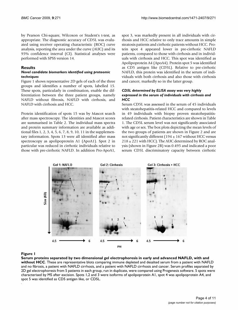
ResultsNovel candidate biomarkers identified using proteomic techniquesFigure 1 shows representative 2D gels of each of the threegroups and identifies a number of spots, labelled 15.These spots, particularly in combination, enable the dif-ferentiation between the three patient groups, namelyNAFLD without fibrosis, NAFLD with cirrhosis, andNAFLD with cirrhosis and HCC.
Protein identification of spots 15 was by Mascot searchafter mass spectroscopy. The identities and Mascot scoresare summarised in Table 2. The individual mass spectraand protein summary information are available as addi-tional files 1, 2, 3, 4, 5, 6, 7, 8, 9, 10, 11 in the supplemen-tary information. Spots 13 were all identified after massspectroscopy as apolipoprotein A1 (ApoA1). Spot 2 inparticular was reduced in cirrhotic individuals relative tothose with pre-cirrhotic NAFLD. In addition Pro-ApoA1,
spot 3, was markedly present in all individuals with cir-rhosis and HCC relative to only trace amounts in simplesteatosis patients and cirrhotic patients without HCC. Pro-tein spot 4 appeared lower in pre-cirrhotic NAFLDpatients, compared to those with cirrhosis and in individ-uals with cirrhosis and HCC. This spot was identified asApolipoprotein A4 (ApoA4). Protein spot 5 was identifiedas CD5 antigen like (CD5L). Relative to pre-cirrhoticNAFLD, this protein was identified in the serum of indi-viduals with both cirrhosis and also those with cirrhosisand cancer, markedly so in the latter group.
CD5L determined by ELISA assay was very highly expressed in the serum of individuals with cirrhosis and HCCSerum CD5L was assessed in the serum of 45 individualswith steatohepatitis-related HCC and compared to levelsin 49 individuals with biopsy proven steatohepatitis-related cirrhosis. Patient characteristics are shown in Table1. The CD5L serum level was not significantly associatedwith age or sex. The box plots depicting the mean levels ofthe two groups of patients are shown in Figure 2 and arenot significantly different (194 ± 167 without HCC versus218 ± 221 with HCC). The AUC determined by ROC anal-ysis (shown in Figure 2B) was 0.495 and indicated a poorserum CD5L discriminatory capacity between cirrhotic
Serum proteins separated by two dimensional gel electrophoresis in early and advanced NAFLD, with and without HCCFigure 1Serum proteins separated by two dimensional gel electrophoresis in early and advanced NAFLD, with and without HCC. These are representative blots comparing immune depleted and desalted serum from a patient with NAFLD and no fibrosis, a patient with NAFLD cirrhosis, and a patient with NAFLD cirrhosis and cancer. Serum profiles separated by 2D gel electrophoresis from 5 patients in each group, run in duplicate, were compared using Progenesis software. 5 spots were characterised by MS after excision. Spots 1,2 and 3 were isoforms of apolipoprotein A1, spot 4 was apolipoprotein A4, and spot 5 was identified as CD5 antigen like, or CD5L.
Page 4 of 11(page number not for citation purposes)

BMC Cancer 2009, 9:271 http://www.biomedcentral.com/1471-2407/9/271
individuals with and without cancer. Some individualswith HCC did have, however, particularly high CD5L lev-els. A level greater than 400 ng/ml has a specificity forHCC of 88%. The sensitivity at this level was unacceptablypoor (20%).
CD5L ELISA in individuals with different stages of NAFLD was a good predictor of those with cirrhosisSerum from 77 patients, all of whom had biopsy provenNAFLD and had an alcohol intake of < 21 units per weekfor men and < 14 units for women, was studied. In thisgroup of individuals the serum level of CD5L ng/ml in cir-rhotic individuals (236 ± 223) was similar to those cir-rhotic individuals with and without HCC describedabove. This level for stage 4 fibrosis/cirrhosis was signifi-cantly elevated compared to individuals with all otherpre-cirrhotic stages (ANOVA p = 0.001, Bonferroni correc-tion applied). Mean levels of CD5L were not significantlydifferent between any other histologically defined group(steatosis or the presence or absence of necroinflamma-tion). The level of CD5L was significantly associated withfibrosis independently of age, sex, steatosis or necroin-flammation, as assessed by General Linear Model analysis(GLM, SPSS). The ROC analyses of CD5L as a predictor ofstage 4 fibrosis in this independent group of patients withNAFLD is shown in Figure 3B. The area under the curvewas 0.712 (95% CI 0.5340.889).
CD5L mRNA expression was elevated in individuals with NAFLD versus those with no underlying liver diseaseCD5L mRNA expression was quantified in one set of liverbiopsy tissues from patients with histologically stagedNAFLD, and a set of normal liver tissues collected at thetime of resection for benign or secondary cancers. (Seemethods) Data are expressed relative to the GAPDH con-trol gene and a single normal liver sample from an indi-vidual undergoing resection for focal nodular hyperplasia.In the pre-cirrhotic NAFLD biopsy tissues, there was no
significant difference in association with either fat,inflammatory or fibrosis score, as shown in Figure 4AC.What was most notable, was that the liver mRNA expres-sion of CD5L was significantly increased in the diseasedNAFLD group as a whole (n = 21), relative to the normalliver group (n = 13) (6.945 ± 0.722 versus1.68 ± 0.269; p= 0.000). Although some of the 'normal' liver samplesfrom patients undergoing resection for secondary malig-nancies had a 'mild steatosis' or a 'minimal focal mono-nuclear cell infiltrate' noted at the time of histologicalexamination, there was no significant difference in CD5Lexpression in association with either of these minorchanges (data not shown). The marked CD5L mRNAexpression shown in the single cirrhotic tissue and HCCpair (Figure 4C) were in keeping with the serum ELISAassays from the much larger patient group.
DiscussionThe prevalence of chronic liver disease is increasing stead-ily in the UK, as is the population at risk of developingand dying from HCC. An increasing incidence of HCC hasalready been documented [5] and this is unfortunatelycompounded by the majority of tumours being diagnosedat a late stage when curative treatments are not possible.[14] Regular surveillance of high risk individuals is recom-mended but is presently hindered by the poor perform-ance of the commonly used serum marker, AFP, [11] evenin combination with abdominal USS. A significant efforthas been and continues to be applied to the search forimproved HCC biomarkers. As yet, none has proved supe-rior to AFP in performance, but in combination some mayhave complimentary roles. [15,16] Our particular concernrelates to the marked increase in the prevalence of ALDand NAFLD related HCC on our own unit. These individ-uals are often over 65 years of age and have cardiovascularco-morbidities precluding curative resection or transplan-tation. Much attention has been focused on validatingquantitative fibrosis or cirrhosis markers in these individ-
Table 2: The identification of the differentiating protein spots on 2D gels.
Protein Spot Protein Identified by Mascot Search
Accession Code (Entrez Protein)
Mascot Score (Expect value in brackets)*
Sequence Coverage %**
1 ApoA1 (human) CAA00975 247 (6.5e-19) 802 ApoA1 (human) CAA00975 236 (8.1e-18) 753 Pro-ApoA1 (human)*** CAA00975 195 (1.0e-13) 624 ApoA4 (human) AAA51748 338 (5.1e-28) 645 CD5L/AIM (human) AAD01446 218 (5.1e-16) 68
The protein spots are as numbered in Figure 1. The * Mascot score is shown as -10*Log(P), where P indicates the probability of a random match. Scores > 78 indicate that the protein identification is statistically significant (P < 0.05). ** Sequence coverage from matched peptides in spectra is provided in additional figures 110. *** Comparisons between zoomed mass spectra for spots 1 and 2 versus spot 3 are provided in additional figure 11. The pro-peptide sequence (RHFWQQDEPPQSPWDR...) generates an expected tryptic peptide at m/z = 2109.9 ([M+H]+). Mature ApoA1 sequence generates a predicted N-terminal tryptic peptide (DEPPQSPWDR...) of m/z = 1226.5 ([M+H]+). AIM = Apoptosis inhibitor expressed by Macrophages (alternate name for CD5L).
Page 5 of 11(page number not for citation purposes)

BMC Cancer 2009, 9:271 http://www.biomedcentral.com/1471-2407/9/271
uals, with combinations of serum markers [8] showingencouraging if still suboptimal improvements in perform-ance. Imaging methods such as the fibroscan mayimprove the efficiency of cirrhosis detection [17-19], butpresently this technique needs to be interpreted with cau-tion in obese individuals. [19-23] The need for furtherimprovements in serum biomarkers for early detection ofboth advanced fibrosis and HCC grows ever more press-ing.
We have applied a proteomic strategy using an optimisedmethod of patient serum preparation in order to identifycandidate biomarkers of either cirrhosis or HCC. Thisincludes immune depletion prior to separation of serum
CD5L discriminates poorly between cirrhotic patients with and without HCCFigure 2CD5L discriminates poorly between cirrhotic patients with and without HCC. Serum levels of CD5L were measured by ELISA assay in cirrhotic patients with (218 ± 221; n = 45) and without (194 ± 166; n = 49) HCC, as shown in 2A. Characteristics of the individuals are shown in Table 1. There was no significant difference between the two groups. ROC analyses is shown in 2B. The area under the curve is 0.495 (95% confidence intervals 0.376 and 0.614). A level > 400 ng/ml has a sensitivity of only 20%, but a specifi-city of 88%. Levels > 500 ng/ml have a specificity of 96%, > 600 ng/ml of 98% and > 700 ng/ml of 100%. For comparison, the mean CD5L serum level from patients without cirrhosis (detailed in figure 3) is also shown.
Figure 3
Page 6 of 11(page number not for citation purposes)

BMC Cancer 2009, 9:271 http://www.biomedcentral.com/1471-2407/9/271
by 2D gel electrophoresis. While this might remove somekey biomarkers, we believe that this strategy improves thesensitivity of the technique applied to the remainingserum proteins. Having identified a number of spots ableto discriminate between patients in at least one of our pre-defined disease groups, namely pre-cirrhotic NAFLD, cir-rhotic NAFLD, and cirrhotic NAFLD with cancer, weselected candidates for identification by peptide massspectroscopy.
The novel serum protein CD5L was identified in theserum of cirrhotic individuals with and without HCC byour proteomic strategy. CD5L, also known as Sp alpha, isa soluble protein belonging to group B of the scavengerreceptor cysteine-rich (SRCR) superfamily for which littlefunctional information is available.[24] It is expressed bymacrophages present in lymphoid tissues (spleen, lymphnode, thymus, and bone marrow), and it binds to myelo-monocytic and lymphoid cells, suggesting that it may playan important role in the regulation of the innate andadaptive immune systems. It was recently identified in thesera of individuals with liver fibrosis related to HCV infec-tion and, based on its proposed role in immune systemregulation, was thought most likely associated with viralinfection rather than cirrhosis.[25] While this may yet betrue, as a mRNA transcript, it has been previously reportedupregulated in HCC relative to dysplastic nodules.[26] Wehave investigated the potential of CD5L as a candidatebiomarker for either advanced liver disease, or advancedliver disease and cancer. Our ELISA assay indicated a poorperformance for CD5L as a surveillance tool for HCC, butagain suggested value for cirrhosis detection. Our dataclearly indicate, however, an association with cirrhosis inindividuals without viral infection, which is independentof the presence or absence of histologically assessed
inflammation. Our CD5L mRNA expression data frompre-cirrhotic NAFLD liver biopsies indicate an increase inassociation with fatty liver disease which, again, is notaltered by the grade of either fat or inflammation. AsCD5L does not increase incrementally with the level orstage of fibrosis, we propose that its dramatic increase inthe serum of individuals with a background cirrhosisreflects hepatocyte regeneration, rather than the advancedfibrosis per se. The recent identification of CD5L by micro-array as one of 30 mRNA transcripts expressed in patientswith cirrhosis with a 'high risk' of HCC development [27]was one reason for our focus on this serum protein. Cer-tainly some of our HCC patients had markedly high CD5Llevels, while those with cirrhosis who had particularlyhigh levels may be at high risk and may yet develop HCC.As a cirrhosis biomarker, the ROC analysis for CD5L indi-cates an accuracy of 72%. While this falls short of the accu-racy of the European Liver Fibrosis (ELF) panel, recentlyvalidated in NAFLD patients and having an AUC of 0.9 foradvanced fibrosis, CD5L is a single biomarker whose per-formance in conjunction with others may yet improve. Inaddition, it may have a value complementing that of AFP,which is predominantly elevated in patients withadvanced HCC, in highlighting individuals with cirrhosiswho are at greater risk of HCC development. At a level >200 ng/ml, CD5L had a specificity for cirrhosis of 96%and a specificity for cancer of 60%.
Particularly prominent on our gel images were three spotsidentified as apolipoprotein A1 (ApoA1). ApoA1 is themajor protein component of high density lipoprotein inplasma. It promotes cholesterol efflux from tissues to theliver for excretion and it is a cofactor for lecithin cholester-olacyltransferase which is responsible for the formation ofmost plasma cholesterol esters. It is not that surprisingthat this liver function associated protein alters in theserum of individuals with chronic liver disease and thishas been previously reported in serum proteomic stud-ies.[28] The mass of spots 1 and 2 (Figure 1) varied byapproximately 30 daltons using MALDI analysis (Addi-tional files 1 and 3) and are most likely attributable topost translational modification by oxidation. Notably,these two spots were reduced in cirrhotic individuals rela-tive to those with pre-cirrhotic NAFLD, which is in keep-ing with previous reports and validates the clinicalrelevance of our methodology. In fact, a reduction ofApoA1 in the serum of patients with cirrhosis hasremained a constitutive part of combined peptide panelsused to predict fibrosis for a number of years. [6,29] Inaddition, however, we identified a novel isoform spot 3.The difference in mass between spots 1 and 2 and spot 3was much greater and in the order of 900 daltons (Addi-tional files 1, 3 and 5). MALDI MS analysis of this spot hasidentified it as pro-Apolipoprotein A1 (Additional files 6and 11), similarly detected and reported by an independ-
CD5L discriminates between steatohepatitis patients with and without cirrhosisFigure 3CD5L discriminates between steatohepatitis patients with and without cirrhosis. Serum levels of CD5L were measured in a total of 113 patients with either ALD or NAFLD. Fibrosis was scored histologically on liver biopsy as per the Brunt scoring system. The numbers of individuals in each group are shown within the boxes of chart 3A. The dif-ference in CD5L levels between the different stages of fibro-sis is statistically significant by univariate analysis (p = 0.004) controlled for both age and sex. This difference The ROC analyses for the identification of those individuals with stage 4 fibrosis (cirrhosis) is depicted in 3B. The area under the curve is 0.719 (95% confidence intervals 0.623 and 0.816), p < 0.0001. A level of CD5L 50 ng/ml has a sensitivity of 78% and a specificity of 46%, while a level of 100 ng/ml has a sensi-tivity of 63% and a specificity of 72%. A level greater than 200 ng/ml has a specificity of 95% (sensitivity 41%), and greater than 300 ng/ml of 97% (sensitivity 27%).
Page 7 of 11(page number not for citation purposes)

BMC Cancer 2009, 9:271 http://www.biomedcentral.com/1471-2407/9/271
ent group of researchers studying patients with lung can-cer. [30] Pro-Apolipoprotein A1 is proposed as a novelserum marker of brain metastases in lung cancer patients[30] and there may well be a rationale for its upregulationalso in HCC patients. ApoA1 is secreted as the proprotein(pro-Apolipoprotein A1/spot 3) and is then cleaved, regu-lating its activation for lipid binding, by a metalloprotei-nase. One candidate metalloproteinase responsible is thec-terminal procollagen endoproteinase, Bone morpho-genic protein 1 (BMP-1). [31] BMP-1 is secreted by theliver, but protease inhibitors, such as alpha-2-macroglob-ulin (A2M), are also secreted by the liver, often at elevatedlevels in inflammation or chronic disease.[32] Either areduction in BMP-1, or an increase in inhibitors such asA2M as reportedly occurs in HCC [33], could block thematuration of pro-Apolipoprotein A1, hence contributing
to relative increase in this isoform (spot 3). In fact, the rel-ative decrease in mature apoA1 in cirrhotic patients (spot2) may also reflect increases in protease inhibition andA2M has similarly contributed significantly to serum diag-nostic tests for fibrosis. [32,34] Further investigation hasyet to determine the sensitivity and specificity of pro-Apolipoprotein A1 as a novel candidate biomarker inpatients with chronic liver disease and HCC, but our pilotstudy suggests that its up-regulation is specifically a fea-ture in the serum of patients with HCC.
ConclusionWhile non-hypothesis driven methodologies may be crit-icised by some as 'fishing expeditions', encouraging agree-ment is beginning to emerge when comparing datagenerated by proteomic techniques in the field of chronic
CD5L mRNA expression is not altered in association with either fat, inflammation or fibrosis scores in pre-cirrhotic NAFLD liver tissuesFigure 4CD5L mRNA expression is not altered in association with either fat, inflammation or fibrosis scores in pre-cir-rhotic NAFLD liver tissues. mRNA CD5L expression was quantified by real-time PCR, relative to GAPDH and a normal liver sample, in 21 pre-cirrhotic NAFLD biopsy tissues, 13 normal liver samples taken at the time of liver resection, one cir-rhotic liver and one HCC. As shown in 4A-C, there was no difference in any pre-cirrhotic NAFLD biopsy tissues in association with the degree of fat, inflammation or fibrosis. There was a significant increase in the NAFLD tissues as a group (n = 21), com-pared with normal liver tissues (n = 13) as represented in 4D (6.945 ± 0.722 versus1.68 ± 0.269; p = 0.000, ***). The elevated CD5L mRNA expression in one cirrhotic and HCC tissue pair, obtained at the time of laparoscopic radiofrequency ablation, is also presented in 4C and is in keeping with the elevated serum CD5L levels identified in the larger cohort of patients studied.
Page 8 of 11(page number not for citation purposes)

BMC Cancer 2009, 9:271 http://www.biomedcentral.com/1471-2407/9/271
liver disease, particularly when it is interpreted in the con-text of the ever growing academic literature generated bygene expression profiling. The data presented in this pilotstudy have identified a pattern of serum apolipoproteinswhich, in combination with CD5L, can discriminate pre-cirrhotic NAFLD, cirrhotic NAFLD, and cirrhotic NAFLDwith HCC. These methodologies, even in small pilot stud-ies requiring additional validation, contribute signifi-cantly to this field and provide the hope that improvedserum biomarkers may yet become available as surveil-lance, diagnostic and prognostic tools in patients withchronic liver disease.
AbbreviationsNAFLD: Non-alcoholic fatty liver disease; ALD: alcoholicliver disease; AFP: alpha alphafetoprotein; CD5L: CD5molecule-like; HCC: hepatocellular cancer; HBV: hepatitisB; HCV: hepatitis C; USS: ultrasound scan; APO: apolipo-protein; MALDI-TOF MS: matrix-assisted laser desorptionionization time of flight mass spectroscopy.
Competing interestsThe authors declare that they have no competing interests.
Authors' contributionsJG supervised the serum preparation and all proteomicstudies and performed all the peptide mass analyses, DChas collected samples and with supervision performed theproteomic analyses, western blotting, ELISA assays, GSBhas directly supervised the ELISA assay optimisation anddata collection, GLP isolated RNA from tissue samplesand performed real-time PCR analyses, LM helped in datainterpretation, collected liver biopsy samples and collatedhistological staging enabling tissue studies, BPK recon-firmed the proteomic 2D Gel data, SS, MH, CPD havecontributed to the study design and recruited patients tothe study, DMM and HLR conceived the study, contrib-uted to its design and co-ordination, HLR has completedfinal data analyses and written the manuscript. All authorsread and approved the final manuscript.
Additional material
Additional File 1Spot 1 mass spectra. Maldi-TOF MS spectra for spot 1.Click here for file[http://www.biomedcentral.com/content/supplementary/1471-2407-9-271-S1.pdf]
Additional File 2Spot 1 is ApoA1. The protein summary report for spot 1, generated using Mascot Peptide Mass Fingerprint search program (Matrix Science Ltd), identifies it as ApoA1.Click here for file[http://www.biomedcentral.com/content/supplementary/1471-2407-9-271-S2.pdf]
Additional File 3Spot 2 mass spectra. Maldi-TOF MS spectra for spot 2.Click here for file[http://www.biomedcentral.com/content/supplementary/1471-2407-9-271-S3.pdf]
Additional File 4Spot 2 is also ApoA1. The protein summary report for spot 2, generated using Mascot Peptide Mass Fingerprint search program (Matrix Science Ltd), identifies it as ApoA1.Click here for file[http://www.biomedcentral.com/content/supplementary/1471-2407-9-271-S4.pdf]
Additional File 5Spot 3 mass spectra. Maldi-TOF MS spectra for spot 3.Click here for file[http://www.biomedcentral.com/content/supplementary/1471-2407-9-271-S5.pdf]
Additional File 6Spot 3 is Pro- ApoA1. The protein summary report for spot 3, generated using Mascot Peptide Mass Fingerprint search program (Matrix Science Ltd), is compatible with it being Pro- ApoA1.Click here for file[http://www.biomedcentral.com/content/supplementary/1471-2407-9-271-S6.pdf]
Additional File 7Spot 4 mass spectra. Maldi-TOF MS spectra for spot 4.Click here for file[http://www.biomedcentral.com/content/supplementary/1471-2407-9-271-S7.pdf]
Additional File 8Spot 4 is ApoA4. The protein summary report for spot 4, generated using Mascot Peptide Mass Fingerprint search program (Matrix Science Ltd), identifies it as ApoA4.Click here for file[http://www.biomedcentral.com/content/supplementary/1471-2407-9-271-S8.pdf]
Additional File 9Spot 5 mass spectra. Maldi-TOF MS spectra for spot 5.Click here for file[http://www.biomedcentral.com/content/supplementary/1471-2407-9-271-S9.pdf]
Additional File 10Spot 5 is CD5L. The protein summary report for spot 5, generated using Mascot Peptide Mass Fingerprint search program (Matrix Science Ltd), identifies it as CD5L.Click here for file[http://www.biomedcentral.com/content/supplementary/1471-2407-9-271-S10.pdf]
Page 9 of 11(page number not for citation purposes)

BMC Cancer 2009, 9:271 http://www.biomedcentral.com/1471-2407/9/271
AcknowledgementsWe thank the individuals agreeing to study participation, the Newcastle NHS Combined Trust for acting as study sponsor, The LiverNorth patient support group for financial support and encouragement, and the Hepatolo-gists and Hepatobiliary Surgeons who have contributed to patient recruit-ment to the study.
Funding: LiverNorth patient support group award to HR. LiverNorth have provided the funds for sample collection and sample analysis.
Ethics: The Newcastle and North Tyneside Local Research Ethics Com-mittee approved this study.
References1. Neuschwander-Tetri BA, Caldwell SH: Nonalcoholic steatohepa-
titis: summary of an AASLD Single Topic Conference. Hepa-tology 2003, 37(5):1202-1219.
2. Parkin DM, Bray F, Ferlay J, Pisani P: Estimating the world cancerburden: Globocan 2000. Int J Cancer 2001, 94(2):153-156.
3. Bruix J, Llovet JM: Prognostic prediction and treatment strat-egy in hepatocellular carcinoma. Hepatology 2002,35(3):519-524.
4. Deuffic S, Poynard T, Buffat L, Valleron AJ: Trends in primary livercancer. Lancet 1998, 351(9097):214-215.
5. Bosch FX, Ribes J, Diaz M, Cleries R: Primary liver cancer: world-wide incidence and trends. Gastroenterology 2004, 127(5 Suppl1):S5-S16.
6. Halfon P, Imbert-Bismut F, Messous D, Antoniotti G, Benchetrit D,Cart-Lamy P, Delaporte G, Doutheau D, Klump T, Sala M, et al.: Aprospective assessment of the inter-laboratory variability ofbiochemical markers of fibrosis (FibroTest) and activity(ActiTest) in patients with chronic liver disease. Comp Hepatol2002, 1(1):3.
7. Angulo P, Hui JM, Marchesini G, Bugianesi E, George J, Farrell GC,Enders F, Saksena S, Burt AD, Bida JP, et al.: The NAFLD fibrosisscore: a noninvasive system that identifies liver fibrosis inpatients with NAFLD. Hepatology 2007, 45(4):846-854.
8. Guha IN, Parkes J, Roderick P, Chattopadhyay D, Cross R, Harris S,Kaye P, Burt AD, Ryder SD, Aithal GP, et al.: Noninvasive markersof fibrosis in nonalcoholic fatty liver disease: Validating theEuropean Liver Fibrosis Panel and exploring simple mark-ers. Hepatology 2008, 47(2):455-460.
9. Bedogni G, Miglioli L, Masutti F, Tiribelli C, Marchesini G, BellentaniS: Prevalence of and risk factors for nonalcoholic fatty liverdisease: the Dionysos nutrition and liver study. Hepatology2005, 42(1):44-52.
10. Sherman M: Alphafetoprotein: an obituary. J Hepatol 2001,34(4):603-605.
11. Bruix J, Sherman M, Llovet JM, Beaugrand M, Lencioni R, BurroughsAK, Christensen E, Pagliaro L, Colombo M, Rodes J: Clinical man-agement of hepatocellular carcinoma. Conclusions of theBarcelona-2000 EASL conference. European Association forthe Study of the Liver. J Hepatol 2001, 35(3):421-430.
12. Brunt EMJC, Di Bisceglie AM, Neuschwander-Tetri BA, Bacon BR:Nonalcoholic steatohepatitis: a proposal for grading andstaging the histological lesions. Am J Gastroenterol 1999,94(9):2467-2474.
13. Miele L, Beale G, Patman G, Nobili V, Leathart J, Grieco A, Abate M,Friedman SL, Narla G, Bugianesi E, et al.: The Kruppel-like factor
6 genotype is associated with fibrosis in nonalcoholic fattyliver disease. Gastroenterology 2008, 135(1):282-291. e281.
14. Wilson JF: Liver cancer on the rise. Ann Intern Med 2005, 142(12Pt 1):1029-1032.
15. Giannelli G, Fransvea E, Trerotoli P, Beaugrand M, Marinosci F, LupoL, Nkontchou G, Dentico P, Antonaci S: Clinical validation ofcombined serological biomarkers for improved hepatocellu-lar carcinoma diagnosis in 961 patients. Clin Chim Acta 2007,383(12):147-152.
16. Beale G, Chattopadhyay D, Gray J, Stewart S, Hudson M, Day C, Tre-rotoli P, Giannelli G, Manas D, Reeves H: AFP, PIVKAII, GP3,SCCA-1 and follisatin as surveillance biomarkers for hepato-cellular cancer in non-alcoholic and alcoholic fatty liver dis-ease. BMC cancer 2008, 8:200.
17. Rigamonti C, Fraquelli M: Do not trivialize the Fibroscan exam-ination, value its accuracy. J Hepatol 2007, 46(6):1149.
18. Yoneda M, Yoneda M, Mawatari H, Fujita K, Endo H, Iida H, NozakiY, Yonemitsu K, Higurashi T, Takahashi H, et al.: Noninvasiveassessment of liver fibrosis by measurement of stiffness inpatients with nonalcoholic fatty liver disease (NAFLD). DigLiver Dis 2008, 40(5):371-378.
19. Kettaneh A, Marcellin P, Douvin C, Poupon R, Ziol M, Beaugrand M,de Ledinghen V: Features associated with success rate and per-formance of FibroScan measurements for the diagnosis ofcirrhosis in HCV patients: a prospective study of 935patients. J Hepatol 2007, 46(4):628-634.
20. Foucher J, Castera L, Bernard PH, Adhoute X, Laharie D, Bertet J,Couzigou P, de Ledinghen V: Prevalence and factors associatedwith failure of liver stiffness measurement using FibroScan ina prospective study of 2114 examinations. Eur J GastroenterolHepatol 2006, 18(4):411-412.
21. Li LF, Dai L, Zhang Q, Chen YP, Feng XR, Guo YB, Hou JL: [Factorsinfluencing the success rate and stability of transient elastog-raphy for liver fibrosis evaluation.]. Nan fang yi ke da xue xue bao= Journal of Southern Medical University 2008, 28(4):595-597.
22. Sandrin L, Yon S, Fournier C, Miette V: Transient elastography:changing clinical practice in hepatology. The Journal of the Acous-tical Society of America 2008, 123(5):3000.
23. Sasso M, Miette V, Sandrin L: Improvements in liver diseasesevaluation using transient elastography for obese patients.The Journal of the Acoustical Society of America 2008, 123(5):3000.
24. Sarrias MR, Rosello S, Sanchez-Barbero F, Sierra JM, Vila J, Yelamos J,Vives J, Casals C, Lozano F: A role for human Sp alpha as a pat-tern recognition receptor. J Biol Chem 2005,280(42):35391-35398.
25. Gangadharan B, Antrobus R, Dwek RA, Zitzmann N: Novel serumbiomarker candidates for liver fibrosis in hepatitis Cpatients. Clin Chem 2007, 53(10):1792-1799.
26. Yoon SH, Kim JS, Song HH: Statistical inference methods fordetecting altered gene associations. Genome informatics 2003,14:54-63.
27. Kim JW, Ye Q, Forgues M, Chen Y, Budhu A, Sime J, Hofseth LJ, KaulR, Wang XW: Cancer-associated molecular signature in thetissue samples of patients with cirrhosis. Hepatology 2004,39(2):518-527.
28. Steel LF, Shumpert D, Trotter M, Seeholzer SH, Evans AA, LondonWT, Dwek R, Block TM: A strategy for the comparative analy-sis of serum proteomes for the discovery of biomarkers forhepatocellular carcinoma. Proteomics 2003, 3(5):601-609.
29. Teare JP, Sherman D, Greenfield SM, Simpson J, Bray G, Catterall AP,Murray-Lyon IM, Peters TJ, Williams R, Thompson RP: Comparisonof serum procollagen III peptide concentrations and PGAindex for assessment of hepatic fibrosis. Lancet 1993,342(8876):895-898.
30. Marchi N, Mazzone P, Fazio V, Mekhail T, Masaryk T, Janigro D:ProApolipoprotein A1: a serum marker of brain metastasesin lung cancer patients. Cancer 2008, 112(6):1313-1324.
31. Chau P, Fielding PE, Fielding CJ: Bone morphogenetic protein-1(BMP-1) cleaves human proapolipoprotein A1 and regulatesits activation for lipid binding. Biochemistry 2007,46(28):8445-8450.
32. Naveau S, Poynard T, Benattar C, Bedossa P, Chaput JC: Alpha-2-macroglobulin and hepatic fibrosis. Diagnostic interest. DigDis Sci 1994, 39(11):2426-2432.
33. Kurokawa Y, Matoba R, Takemasa I, Nakamori S, Tsujie M, NaganoH, Dono K, Umeshita K, Sakon M, Ueno N, et al.: Molecular fea-
Additional File 11Spot 3 is confirmed as Pro-ApoA1. The Zoomed MALDI spectra con-firming the peptide differences between spots 1 and 2 versus spot 3.Click here for file[http://www.biomedcentral.com/content/supplementary/1471-2407-9-271-S11.jpeg]
Page 10 of 11(page number not for citation purposes)
http://www.ncbi.nlm.nih.gov/entrez/query.fcgi?cmd=Retrieve&db=PubMed&dopt=Abstract&list_uids=9449893
http://www.ncbi.nlm.nih.gov/entrez/query.fcgi?cmd=Retrieve&db=PubMed&dopt=Abstract&list_uids=9449893
http://www.ncbi.nlm.nih.gov/entrez/query.fcgi?cmd=Retrieve&db=PubMed&dopt=Abstract&list_uids=8105167
http://www.ncbi.nlm.nih.gov/entrez/query.fcgi?cmd=Retrieve&db=PubMed&dopt=Abstract&list_uids=8105167
http://www.ncbi.nlm.nih.gov/entrez/query.fcgi?cmd=Retrieve&db=PubMed&dopt=Abstract&list_uids=8105167
http://www.ncbi.nlm.nih.gov/entrez/query.fcgi?cmd=Retrieve&db=PubMed&dopt=Abstract&list_uids=7525168

BMC Cancer 2009, 9:271 http://www.biomedcentral.com/1471-2407/9/271
Publish with BioMed Central and every scientist can read your work free of charge
"BioMed Central will be the most significant development for disseminating the results of biomedical research in our lifetime."
Sir Paul Nurse, Cancer Research UK
Your research papers will be:
available free of charge to the entire biomedical community
peer reviewed and published immediately upon acceptance
cited in PubMed and archived on PubMed Central
yours — you keep the copyright
Submit your manuscript here:http://www.biomedcentral.com/info/publishing_adv.asp
BioMedcentral
tures of non-B, non-C hepatocellular carcinoma: a PCR-array gene expression profiling study. J Hepatol 2003,39(6):1004-1012.
34. Cales P, Oberti F, Michalak S, Hubert-Fouchard I, Rousselet MC,Konate A, Gallois Y, Ternisien C, Chevailler A, Lunel F: A novelpanel of blood markers to assess the degree of liver fibrosis.Hepatology 2005, 42(6):1373-1381.
Pre-publication historyThe pre-publication history for this paper can be accessedhere:
http://www.biomedcentral.com/1471-2407/9/271/prepub
Page 11 of 11(page number not for citation purposes)