PNPLA3 (rs738409 C\u003eG) is a common risk variant associated with hepatocellular carcinoma in...
6
CORRESPONDENCE Sorafenib in Clinical Practice: Evidence-Based Use or Abuse? To the Editor: In their study, Iavarone et al. 1 found a prolonged time to progres- sion (TTP) in comparison with the Sorafenib HCC Assessment Randomized Protocol study, and they speculated that it was probably related to the utilization of the modified Response Evaluation Crite- ria in Solid Tumors. 2 Moreover, they hypothesized that down-dosing sorafenib allowed a high percentage of the patients to retain the ben- efits of the drug and achieve improved survival. If TTP was so remarkably improved only because of the accuracy of the modified Response Evaluation Criteria in Solid Tumors (published in the year of the study’s start), it seems strange that the median overall survival was comparable to that of phase 3 trials. 2 The most unusual observa- tion was the very short postprogression survival (1 month versus 5 months in the Sorafenib HCC Assessment Randomized Protocol study). As a rule, a median TTP approaching the median overall sur- vival occurs because of undiagnosed progression. In our opinion, this means that the interval between computed tomography scans was longer than 2 months in actual daily practice. Another issue was the high rate of liver function deterioration, which the authors attributed to drug-related adverse events. 1 It is not clear how they distinguished this from clinical tumor progres- sion. The analysis suggesting longer survival for patients receiving half-dose sorafenib was not preplanned, and this means that the patients were not stratified according to prognostic factors and that the sample size was not calculated in advance for the hypothesis that the authors intended to demonstrate. For this reason, the aforementioned analysis was methodologically inappropriate for an observational study; it was also inconclusive because the patients’ survival might simply have depended on the selection of a good- prognosis cohort. Finally, two other aspects deserve careful consideration. First, the study lacked formal approval by an ethics committee, which implies the generation of a European Union Drug Regulating Authorities Clinical Trials number according to Italian rules. Sec- ond, a cohort of Child B patients was treated, but sorafenib ap- proval in Italy is restricted to Child A patients. Taken together, our observations suggest the following: (1) the design of this prospective study had potentially serious limitations, (2) sorafenib was used in field practice outside the approved indi- cation, (3) computed tomography scans were not scheduled on a bimonthly basis, and (4) TTP was largely overestimated. In addi- tion, this article subliminally conveys a somewhat hazardous and not adequately supported message about both the arbitrary accep- tance of the physician’s judgment in deciding the likelihood of benefits without radiological confirmation and the continuation of sorafenib beyond progression almost until death. MICHELE BASSO, M.D. 1 MARIA BASSO, M.D. 2 CARLO BARONE, M.D. 1 1 Medical Oncology and 2 Internal Medicine Catholic University of Sacred Heart Rome, Italy References 1. Iavarone M, Cabibbo G, Piscaglia F, Zavaglia C, Grieco A, Villa E, et al.; for SOFIA (Sorafenib Italian Assessment) Study Group. Field- practice study of sorafenib therapy for hepatocellular carcinoma: a pro- spective multicenter study in Italy. HEPATOLOGY 2011;54:2055-2063. 2. Llovet JM, Ricci S, Mazzaferro V, Hilgard P, Gane E, Blanc JF, et al.; for SHARP Investigators Study Group. Sorafenib in advanced hepato- cellular carcinoma. N Engl J Med 2008;359:378-390. Copyright V C 2011 by the American Association for the Study of Liver Diseases. View this article online at wileyonlinelibrary.com. DOI 10.1002/hep.25541 Potential conflict of interest: Dr. Barone received grants from Pfizer. Reply: We thank Basso et al. for their comments about our article on field practice with sorafenib in Italy, 1 although we have to say that overall their letter reflects a substantial misinterpretation of our report. As we clearly stated, the Sorafenib Italian Assessment (SOFIA) study is in fact just an observational study not framed by a control setting or other limitations, and this has allowed us to evaluate meas- ures of real-world effectiveness without the need for registration with a European Union Drug Regulating Authorities Clinical Trials num- ber or for approval by an ethics committee. Although by the same to- ken the SOFIA study did not require a predefined sample size, it was indeed adequately powered because it ended with the enrollment of approximately the same number of patients found in the sorafenib arm of the Sorafenib HCC Assessment Randomized Protocol registra- tion trial (296 versus 299). 2 Let us also clarify that the Child-Pugh A label for sorafenib has implications for the reimbursement policy only; this means that the drug can be prescribed off label to Child- Pugh B patients with advanced hepatocellular carcinoma with the understanding that the treatment costs will be reimbursed by partners other than the National Health System (including the hospital itself) according to proper administrative procedures. As we clearly indi- cated, the time to progression was not overestimated by the modified Response Evaluation Criteria in Solid Tumors, which in fact are supe- rior to the Response Evaluation Criteria in Solid Tumors for the cap- ture of hepatocellular carcinoma responses to both loco-ablative and drug-targeted therapy. 3 None of the patients in the SOFIA study received sorafenib ‘‘beyond progression almost until death,’’ as inap- propriately stated by Basso et al., because the median survival of the patients was greater than the duration of the sorafenib therapy (10.5 versus 3.8 months).We also disagree with their statement that the sig- nificantly increased survival of the 77 patients receiving half-dose sor- afenib (21.6 months for those patients versus 9.6 months for the 219 full-dose patients) was ‘‘inconclusive’’ and biased by a ‘‘methodologi- cally inappropriate’’ analysis because a multivariate analysis demon- strated a strong and independent association between full-dose sorafe- nib and an increased risk of liver-related mortality after corrections were made for multiple confounders. Although the prolonged survival of the down-dosed patients unlikely reflected the selection of good- prognosis patients, our observation that cancer-related morbidity is frequently overshadowed by the complications of cirrhosis highlights the driving role of hepatologists in the multidisciplinary management of patients with hepatocellular carcinoma. MASSIMO IAVARONE, M.D. 1 GIUSEPPE CABIBBO, M.D. 2,3 MASSIMO COLOMBO, M.D. 1 1 Ca’Granda Maggiore Hospital, Foundation of the Scientific Institute for Research, Hospitalization, and Health Care University of Milan Milan, Italy 2 Biomedical Department of Internal and Specialty Medicine, and 3 Department of Biopathology and Biomedical Methodology University of Palermo Palermo, Italy 1305
-
Upload
independent -
Category
Documents
-
view
4 -
download
0
Transcript of PNPLA3 (rs738409 C\u003eG) is a common risk variant associated with hepatocellular carcinoma in...

CORRESPONDENCE
Sorafenib in Clinical Practice: Evidence-Based Use or Abuse?
To the Editor:
In their study, Iavarone et al.1 found a prolonged time to progres-sion (TTP) in comparison with the Sorafenib HCC AssessmentRandomized Protocol study, and they speculated that it was probablyrelated to the utilization of the modified Response Evaluation Crite-ria in Solid Tumors.2 Moreover, they hypothesized that down-dosingsorafenib allowed a high percentage of the patients to retain the ben-efits of the drug and achieve improved survival. If TTP was soremarkably improved only because of the accuracy of the modifiedResponse Evaluation Criteria in Solid Tumors (published in the yearof the study’s start), it seems strange that the median overall survivalwas comparable to that of phase 3 trials.2 The most unusual observa-tion was the very short postprogression survival (1 month versus 5months in the Sorafenib HCC Assessment Randomized Protocolstudy). As a rule, a median TTP approaching the median overall sur-vival occurs because of undiagnosed progression. In our opinion, thismeans that the interval between computed tomography scans waslonger than 2 months in actual daily practice.
Another issue was the high rate of liver function deterioration,which the authors attributed to drug-related adverse events.1 It isnot clear how they distinguished this from clinical tumor progres-sion. The analysis suggesting longer survival for patients receivinghalf-dose sorafenib was not preplanned, and this means that thepatients were not stratified according to prognostic factors and thatthe sample size was not calculated in advance for the hypothesisthat the authors intended to demonstrate. For this reason, theaforementioned analysis was methodologically inappropriate for anobservational study; it was also inconclusive because the patients’survival might simply have depended on the selection of a good-prognosis cohort.
Finally, two other aspects deserve careful consideration. First,the study lacked formal approval by an ethics committee, whichimplies the generation of a European Union Drug RegulatingAuthorities Clinical Trials number according to Italian rules. Sec-ond, a cohort of Child B patients was treated, but sorafenib ap-proval in Italy is restricted to Child A patients.
Taken together, our observations suggest the following: (1) thedesign of this prospective study had potentially serious limitations,(2) sorafenib was used in field practice outside the approved indi-cation, (3) computed tomography scans were not scheduled on abimonthly basis, and (4) TTP was largely overestimated. In addi-tion, this article subliminally conveys a somewhat hazardous andnot adequately supported message about both the arbitrary accep-tance of the physician’s judgment in deciding the likelihood ofbenefits without radiological confirmation and the continuation ofsorafenib beyond progression almost until death.
MICHELE BASSO, M.D.1
MARIA BASSO, M.D.2
CARLO BARONE, M.D.11Medical Oncology and 2Internal Medicine
Catholic University of Sacred HeartRome, Italy
References1. Iavarone M, Cabibbo G, Piscaglia F, Zavaglia C, Grieco A, Villa E,
et al.; for SOFIA (Sorafenib Italian Assessment) Study Group. Field-practice study of sorafenib therapy for hepatocellular carcinoma: a pro-spective multicenter study in Italy. HEPATOLOGY 2011;54:2055-2063.
2. Llovet JM, Ricci S, Mazzaferro V, Hilgard P, Gane E, Blanc JF, et al.;for SHARP Investigators Study Group. Sorafenib in advanced hepato-cellular carcinoma. N Engl J Med 2008;359:378-390.
CopyrightVC 2011 by the American Association for the Study of Liver Diseases.View this article online at wileyonlinelibrary.com.DOI 10.1002/hep.25541Potential conflict of interest: Dr. Barone received grants from Pfizer.
Reply:
We thank Basso et al. for their comments about our article onfield practice with sorafenib in Italy,1 although we have to say thatoverall their letter reflects a substantial misinterpretation of ourreport. As we clearly stated, the Sorafenib Italian Assessment (SOFIA)study is in fact just an observational study not framed by a controlsetting or other limitations, and this has allowed us to evaluate meas-ures of real-world effectiveness without the need for registration witha European Union Drug Regulating Authorities Clinical Trials num-ber or for approval by an ethics committee. Although by the same to-ken the SOFIA study did not require a predefined sample size, it wasindeed adequately powered because it ended with the enrollment ofapproximately the same number of patients found in the sorafenibarm of the Sorafenib HCC Assessment Randomized Protocol registra-tion trial (296 versus 299).2 Let us also clarify that the Child-Pugh Alabel for sorafenib has implications for the reimbursement policyonly; this means that the drug can be prescribed off label to Child-Pugh B patients with advanced hepatocellular carcinoma with theunderstanding that the treatment costs will be reimbursed by partnersother than the National Health System (including the hospital itself)according to proper administrative procedures. As we clearly indi-cated, the time to progression was not overestimated by the modifiedResponse Evaluation Criteria in Solid Tumors, which in fact are supe-rior to the Response Evaluation Criteria in Solid Tumors for the cap-ture of hepatocellular carcinoma responses to both loco-ablative anddrug-targeted therapy.3 None of the patients in the SOFIA studyreceived sorafenib ‘‘beyond progression almost until death,’’ as inap-propriately stated by Basso et al., because the median survival of thepatients was greater than the duration of the sorafenib therapy (10.5versus 3.8 months).We also disagree with their statement that the sig-nificantly increased survival of the 77 patients receiving half-dose sor-afenib (21.6 months for those patients versus 9.6 months for the 219full-dose patients) was ‘‘inconclusive’’ and biased by a ‘‘methodologi-cally inappropriate’’ analysis because a multivariate analysis demon-strated a strong and independent association between full-dose sorafe-nib and an increased risk of liver-related mortality after correctionswere made for multiple confounders. Although the prolonged survivalof the down-dosed patients unlikely reflected the selection of good-prognosis patients, our observation that cancer-related morbidity isfrequently overshadowed by the complications of cirrhosis highlightsthe driving role of hepatologists in the multidisciplinary managementof patients with hepatocellular carcinoma.
MASSIMO IAVARONE, M.D.1
GIUSEPPE CABIBBO, M.D.2,3
MASSIMO COLOMBO, M.D.11Ca’Granda Maggiore Hospital, Foundation of the Scientific Institute for
Research, Hospitalization, and Health CareUniversity of MilanMilan, Italy
2Biomedical Department of Internal and Specialty Medicine, and3Department of Biopathology and Biomedical Methodology
University of PalermoPalermo, Italy
1305

References1. Iavarone M, Cabibbo G, Piscaglia F, Zavaglia C, Grieco A, Villa E,
et al.; for SOFIA (Sorafenib Italian Assessment) Study Group. Field-practice study of sorafenib therapy for hepatocellular carcinoma: a pro-spective multicenter study in Italy. HEPATOLOGY 2011;54:2055-2063.
2. Llovet JM, Ricci S, Mazzaferro V, Hilgard P, Gane E, Blanc JF, et al. Sorafe-nib in advanced hepatocellular carcinoma. N Engl J Med 2008;359:378-390.
3. Bruix J, Reig M, Rimola J, Forner A, Burrel M, Vilana R, et al. Clini-cal decision making and research in hepatocellular carcinoma: pivotalrole of imaging techniques. HEPATOLOGY 2011;54:2238-2244.
CopyrightVC 2011 by the American Association for the Study of Liver Diseases.View this article online at wileyonlinelibrary.com.DOI 10.1002/hep.25547Potential conflict of interest: Massimo Iavarone received travel support fromGilead, and Giuseppe Cabibbo received travel support from Bayer. MassimoColombo received grant and research support from Schering-Plough, Roche,Bristol-Myers Squibb, Gilead, and Bayer; he also serves on advisory boards forSchering-Plough, Roche, Novartis, Vertex, Bristol-Myers Squibb, Gilead,Bayer, and Tibotec and speaks and teaches for Schering-Plough, Roche, Novar-tis, Vertex, Bristol-Myers Squibb, Gilead, and Bayer.
Acute Liver Failure: What Is It?
To the Editor:
Quantitative synthesis of prognostic research findings in acuteliver failure (ALF) requires uniformity in the definition of this dis-ease. We wish to assess the variability and explicitness of the usedALF definitions in studies focusing on evaluating indicators of ALFoutcome. We included studies published in HEPATOLOGY in the lastdecade that report original data from a clinical trial or observatio-nal study on adult patients with ALF and of which one of themain objectives was evaluating prognostic indicators for predictionof outcome, including adverse outcome.
Out of eight included studies, seven used five different ALFdefinitions (Table 1), and one did not report an explicit definition.
The differences in the ALF definitions can be characterized byfour main components: (1) presence of hepatic encephalopathywith or without its grade of severity (reported in all studies, two ofwhich reported its grade); (2) the interval between onset of diseaseand occurrence of hepatic encephalopathy (reported in six studies,three of which reported 26 weeks, two of which reported 8 weeks,and one of which reported 4 weeks); (3) presence of coagulopathywith or without its threshold value (reported in five studies, one ofwhich operationalized it by international normalized ratio [INR],four of which operationalized it by prothrombin time or INR;four studies presented thresholds); and (4) presence of preexistingliver disease (reported in five studies). Some studies mentionedother elements in the definition, such as jaundice (one study) andasterixis, hyperbilirubinemia, or hypoglycemia (one study).
We found a large variation in the definition of ALF in eightstudies in one decade in one journal. Although the exact implica-tions of the differences in ALF definitions on performance of prog-
nostic markers remain unknown, these differences hinder suchquantitative analysis. There is room for improvement in the report-ing of ALF definitions, and there is a need for a uniform accepteddefinition of ALF in future studies to evaluate prognosticindicators.
KAMA A. WLODZIMIROW, M.ENG1
SAEID ESLAMI, PHARMD, PH.D1
AMEEN ABU-HANNA, PH.D1
MARTIN NIEUWOUDT, PH.D2
ROBERT A.F.M. CHAMULEAU, M.D., PH.D.31Department of Medical Informatics
Academic Medical Center3Tytgat Institute for Liver and Intestinal Research
Academic Medical CenterUniversity of AmsterdamAmsterdam, The Netherlands
2South African DST/NRF Centre of Excellence in EpidemiologicalModelling and AnalysisStellenbosch, South Africa
References1. Khandelwal N, James LP, Sanders C, Larson AM, Lee WM; and the Acute
Liver Failure Study Group. Unrecognized acetaminophen toxicity as acause of indeterminate acute liver failure. HEPATOLOGY 2011;53:567-576.
2. Kumar R,ShalimarBhatia V, Khanal S, Sreenivas V, Gupta SD, et al.Antituberculosis therapy-induced acute liver failure: Magnitude, profile,prognosis, and predictors of outcome. HEPATOLOGY 2010;51:1665-1674.
Table 1. Summary of Definitions
Study
No. of Patients Evaluating
Outcome Predictors
Definition
Hepatic
Encephalopathy
Weeks From
Onset Coagulopathy
No Preexisting
Liver Disease Other
Khandelwal et al.1 309 þ 26 PT >15s or INR �1.5 þKumar et al.2 70 þ 4 þVolkmann et al.3 70 þ 8 INR >1.5 þ Jaundice
Parekh et al.4 187 þ 26 PT >15s or INR �1.5 þSchmidt and Larsen5 124 2-4
Taylor et al.6 29 þ 26 PT >15s or INR �1.5 þKremers et al.7 388 2 8 PT (INR) Asterixis, hyperbilirubinemia,
or hypoglycemia
Antoniades et al.8 50 Not reported
Abbreviations: INR, international normalized ratio; PT, prothrombin time.
þ, included in the definition.
1306 CORRESPONDENCE HEPATOLOGY, April 2011

3. Volkmann X, Anstaett M, Hadem J, Stiefel P, Bahr MJ, Lehner F, et al.Caspase activation is associated with spontaneous recovery from acuteliver failure. HEPATOLOGY 2008;47:1624-1633.
4. Parekh NK, Hynan LS, De Lemos J, Lee WM; Acute Liver Failure
Study Group. Elevated troponin I levels in acute liver failure: Is myo-
cardial injury an integral part of acute liver failure? HEPATOLOGY 2007;
45:1489-1495.5. Schmidt LE, Larsen FS. MELD score as a predictor of liver failure and
death in patients with acetaminophen-induced liver injury. HEPATOLOGY
2007;45:789-796.6. Taylor RM, Davern T, Munoz S, Han SH, McGuire B, Larson AM,
et al. Fulminant hepatitis A virus infection in the United States: inci-dence, prognosis, and outcomes. HEPATOLOGY 2006;44:1589-1597.
7. Kremers WK, van IJperen M, Kim WR, Freeman RB, Harper AM,Kamath PS, et al. MELD score as a predictor of pretransplant andposttransplant survival in OPTN/UNOS status 1 patients. HEPATOLOGY
2004;39:764-769.8. Antoniades CG, Berry PA, Davies ET, Hussain M, Bernal W, Vergani
D, et al. Reduced monocyte HLA-DR expression: a novel biomarker ofdisease severity and outcome in acetaminophen-induced acute liver fail-ure. HEPATOLOGY 2006;44:34-43.
CopyrightVC 2011 by the American Association for the Study of Liver Diseases.View this article online at wileyonlinelibrary.com.DOI 10.1002/hep.25519Potential conflict of interest: Nothing to report.
PNPLA3 (rs738409 C>G) Is a Common Risk Variant Associated With HepatocellularCarcinoma in Alcoholic Cirrhosis
To the Editor:
A common variant (rs738409 C>G) in the PNPLA3 gene hasbeen consistently associated with liver fat but also fibrosis in nonal-coholic fatty liver disease, alcoholic liver disease (ALD), andchronic hepatitis C (CHC).1-4 The study by Valenti et al.4 in arecent issue of HEPATOLOGY shows that in Caucasian CHC patients,this variant was also linked to hepatocellular carcinoma (HCC).This latter finding has been replicated in another independent Eu-ropean cohort.5
We tested the association between rs738409 and HCC in ALD.To this end, we genotyped 325 Caucasian patients from Belgiumwith alcoholic cirrhosis (67% men; mean age, 54.9 6 9.1 years;mean body mass index [BMI], 26.765.5 kg/m2; 17% had diabe-tes) and 246 French Caucasian patients with alcoholic cirrhosis(86% men; mean age, 64.369.1 years; mean BMI, 27.464.9 kg/m2; 37% had diabetes). HCC was confirmed by histology or typi-cal imaging findings in 12% of the Belgian cohort and 43% of theFrench cohort. The French unit included a higher proportion ofHCC patients, reflecting the specificity of this tertiary center spe-cializing in liver cancer management. HCC was present in 9% ofCC, 10% of CG, and 25% of GG genotype in the Belgian cohortand in 28% of CC, 41% of CG, and 78% of GG genotype in theFrench group. The minor allele frequency was not statistically dif-ferent between the Belgian and French centers (36.8% versus39.8% [P ¼ 0.296]). Under a recessive model of inheritance,rs738409 was significantly associated with HCC after adjustmentfor, age, sex, BMI, and diabetes in both cohorts (Table 1).
The rs738409 variant is associated with liver fat accumulation;however, the exact function of PNPLA3 and the consequence ofthe related nonsynonymous variation remains unknown.6 Although
the remarkable observation that rs738409 confers higher risk ofHCC in CHC and ALD warrants additional replication, it maywell illustrate gene-host interactions and indicate that the influenceof PNPLA3 on chronic liver disease heritability could go farbeyond a mere impact on steatosis.
ERIC TREPO, M.D.1,2
ERWAN GUYOT, PH.D.3,4
NATHALIE GANNE-CARRIE, M.D., PH.D.5
DELPHINE DEGRE, M.D.1,2
THIERRY GUSTOT, M.D., PH.D.1,2
DENIS FRANCHIMONT, M.D., PH.D.1,2
ANGELA SUTTON, PH.D.3,4
PIERRE NAHON, M.D., PH.D.5,6
CHRISTOPHE MORENO, M.D., PH.D.1,21Department of Gastroenterology, Hepatopancreatology
and Digestive OncologyErasme Hospital
2Laboratory of Experimental GastroenterologyUniversit�e Libre de BruxellesBrussels, Belgium
3INSERM U6988Universit�e Paris 13Bobigny, France
4Service de Biochimie5Service d’H�epatologie
Hopital Jean Verdier, AP-HP, Bondy, France6INSERM U773
Centre de Recherche Bichat Beaujon CRB3Universit�e Paris 7Paris, France
Table 1. Multivariable Logistic Regression Analysis for the Association of PNPLA3 (rs738409 C>G) Variant and HCC
Belgian Cohort (n ¼ 325) French Cohort (n ¼ 246) Overall (n ¼ 571)
OR (95% CI) P OR (95% CI) P OR (95% CI) P
rs738409 GG* 3.17 (1.39-7.21) 0.006 9.23 (3.37-25.27) 1.50 � 10�5 4.70 (2.63-8.42) 1.83 � 10�7
Age, years 1.04 (0.99-1.08) 0.107 1.09 (1.05-1.13) 2.00 � 10�5 1.08 (1.06-1.11) 5.36 � 10�10
Sex, men 1.15 (0.53-2.51) 0.728 14.03 (2.94-66.90) 0.001 3.17 (1.64-6.13) 0.001
BMI (kg/m2) 1.05 (0.98-1.12) 0.150 1.03 (0.97-1.11) 0.349 1.04 (0.99-1.08) 0.146
Diabetes 2.30 (1.05-5.04) 0.038 1.39 (0.73-2.65) 0.322 1.89 (1.17-3.04) 0.009
Abbreviations: HCC, hepatocellular carcinoma; BMI, body mass index; CI, confidence interval; OR, odds ratio.
*Based on a recessive model for rs738409 genotype.
HEPATOLOGY, Vol. 55, No. 4, 2012 CORRESPONDENCE 1307
References1. Sookoian S, Pirola CJ. Meta-analysis of the influence of I148M variant
of patatin-like phospholipase domain containing 3 gene (PNPLA3) onthe susceptibility and histological severity of nonalcoholic fatty liver dis-ease. HEPATOLOGY 2011;53:1883-1894.
2. Stickel F, Buch S, Lau K, zu Schwabedissen HM, Berg T, Ridinger M,et al. Genetic variation in the PNPLA3 gene is associated with alco-holic liver injury in caucasians. HEPATOLOGY 2011;53:86-95.
3. Tr�epo E, Pradat P, Potthoff A, Momozawa Y, Quertinmont E, GustotT, et al. Impact of patatin-like phospholipase-3 (rs738409 C>G) poly-morphism on fibrosis progression and steatosis in chronic hepatitis C.HEPATOLOGY 2011;54:60-69.
4. Valenti L, Rumi M, Galmozzi E, Aghemo A, Del Menico B, De NicolaS, et al. Patatin-Like phospholipase domain-containing 3 I148M poly-
morphism, steatosis, and liver damage in chronic hepatitis C. HEPATO-
LOGY 2011;53:791-799.5. Corradini SG, Burza MA, Molinaro A, Romeo S. Patatin-like phospho-
lipase domain containing 3 sequence variant and hepatocellular carci-
noma. HEPATOLOGY 2011;53:1776.6. Browning JD, Cohen JC, Hobbs HH. Patatin-like phospholipase
domain-containing 3 and the pathogenesis and progression of
pediatric nonalcoholic fatty liver disease. HEPATOLOGY 2010;52:
1189-1192.
CopyrightVC 2011 by the American Association for the Study of Liver Diseases.View this article online at wileyonlinelibrary.com.DOI 10.1002/hep.25518Potential conflict of interest: Nothing to report.
Feasibility and Performance of the FibroScan XL Probe
To the Editor:
We read with interest the article by Myers et al.,1 who exam-ined the feasibility and performance of the M and XL FibroScanprobes in a cohort of patients with chronic liver disease. Althoughthere are substantial data validating the use of the M probe as anoninvasive assessment of hepatic fibrosis,2 there are few dataregarding the use of the XL probe.3,4 This represents a particularlyimportant issue considering the high prevalence of obesity in west-ern communities and the relationship between body mass index(BMI) and frequency of liver stiffness measurement (LSM) failureand/or unreliability.5 Our analysis of over 2000 FibroScan referralsindicates that 23% of subjects have a BMI >30 kg/m2.
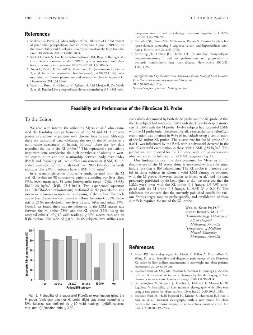
In a recent single-center prospective study, we used both the Mand XL probes on 98 consecutive patients attending our liver clinic(53% men; mean age, 54 years [interquartile range (IQR), 48-62];BMI, 26 kg/m2 [IQR, 23.5-30.1]). Two experienced operators(>1,000 FibroScan examinations) performed all the procedures usingsonographic images to optimize positioning of the probes. The etiol-ogy of liver disease was distributed as follows: hepatitis C, 38%; hepa-titis B, 21%; nonalcoholic fatty liver disease, 14%; and other, 27%.Overall, we found there was no difference in the LSM success ratebetween the M probe (78%) and the XL probe (82%) using theaccepted criteria6 of �10 valid readings, �60% success rate, and anIQR/median LSM ratio of �0.30. In 65 subjects, liver stiffness was
successfully determined by both the M probe and the XL probe. A fur-ther 16 subjects had successful LSMs with the XL probe despite unsuc-cessful LSMs with the M probe. Twelve subjects had successful LSMswith the M probe only. Therefore, overall, a successful valid FibroScanexamination was obtained in 94% of individuals using a combinationof the M and/or XL probes. The success rate for the M probe (P >0.001) was influenced by the BMI, with a substantial decrease in therate of successful examination in those with a BMI �35 kg/m2. Thispattern was not observed for the XL probe, with similar success ratesobserved across the full spectrum of BMI categories (Fig. 1).
Our findings support the data presented by Myers et al.1 inthat the use of the M probe alone is associated with a substantialfailure rate that is BMI-dependent. The XL probe is therefore use-ful in those subjects in whom a valid LSM cannot be obtainedwith the M probe. However, similar to Myers et al.1 and the datapreviously published by de Ledinghen et al.,3 we observed that theLSMs were lower with the XL probe (6.1 [range, 4.9-7.9]) com-pared with the M probe (6.5 [range, 5.1-9.5]) (P > 0.005). Thisreinforces the concept that the currently published cutoffs for vari-ous fibrosis stages may be probe-specific, and revalidation of thesecutoffs is required for use of the XL probe.
WILLIAM KEMP, PH.D.1,2
STUART ROBERTS, M.D.1,21Gastroenterology Department
Alfred HospitalMelbourne, Australia
2Department of MedicineMonash UniversityMelbourne, Australia
References1. Myers RP, Pomier-Layrargues G, Kirsch R, Pollett A, Duarte-Rojo A,
Wong D, et al. Feasibility and diagnostic performance of the FibroScanXL probe for liver stiffness measurement in overweight and obese patients.HEPATOLOGY 2012;55:199-208.
2. Friedrich-Rust M, Ong MF, Martens S, Sarrazin C, Bojunga J, ZeuzemS, et al. Performance of transient elastography for the staging of liverfibrosis: a meta-analysis. Gastroenterology 2008;134:960-974.
3. de Ledinghen V, Vergniol J, Foucher J, El-Hajbi F, Merrouche W,Rigalleau V. Feasibility of liver transient elastography with FibroScanusing a new probe for obese patients. Liver Int 2010;30:1043-1048.
4. Friedrich-Rust M, Hadji-Hosseini H, Kriener S, Herrmann E, Sircar I,Kau A, et al. Transient elastography with a new probe for obesepatients for non-invasive staging of non-alcoholic steatohepatitis. EurRadiol 2010;20:2390-2396.
Fig. 1. Probability of a successful FibroScan examination using theM probe (dark gray bars) or XL probe (light gray bars) according toBMI. Success was defined as �10 valid readings, �60% successrate, and IQR/median ratio �0.30.
1308 CORRESPONDENCE HEPATOLOGY, April 2011

5. Castera L, Foucher J, Bernard PH, Carvalho F, Allaix D, MerroucheW, et al. Pitfalls of liver stiffness measurement: a 5-year prospectivestudy of 13,369 examinations. HEPATOLOGY 2010;51:828-835.
6. Castera L, Forns X, Alberti A. Non-invasive evaluation of liver fibrosisusing transient elastography. J Hepatol 2008;48:835-847.
CopyrightVC 2011 by the American Association for the Study of Liver Diseases.View this article online at wileyonlinelibrary.com.DOI 10.1002/hep.25521Potential conflict of interest: Nothing to report.
Assessment by Fibroscan of Fibrosis in Nonalcoholic Fatty Liver Disease: XL Versus M Probe?
To the Editor:
The study by Myers et al.,1 which tests the performance of the newFibroscan XL probe, in comparison to the standard M probe, in theevaluation of liver stiffness in overweight/obese patients with metabolic(nonalcolholic fatty liver disease [NAFLD]) or viral (hepatitis B virus[HBV] or HCV) chronic liver disease, marks an important point, i.e.,that the failure of measurement occurs only in 1% of patients with theXL probe as compared with 16% with the M probe.
While this clearly increases the reliability in the setting ofpatients with a high body mass index (BMI), some issues shouldbe considered. First of all, the XL probe does not improve the ac-curacy in predicting the stage of fibrosis, as assessed by using liverbiopsy as the ‘‘gold standard.’’ In fact, both the M and XL probesshowed a comparable area under the curve (AUC) for significantand severe fibrosis and for cirrhosis. Second, 27% of cases usingthe XL probe were still inadequate. In addition, it would be inter-esting to know if the BMI, other than affecting reliability of stiff-ness measurement using both M and XL probes, is also able tointerfere with its performance in predicting fibrosis, as we haverecently shown in a cohort of biopsy-proven NAFLD patients.2
These considerations somewhat mitigate our enthusiasm for theXL probe, since even in overweight/obese patients the M probe isclearly not inferior, when the test was feasible, to the XL.
Another relevant issue is that with the inclusion of patientswith viral liver diseases (HBV and HCV), the diagnostic perform-ance of Fibroscan3 may be vastly different and could lead to poten-tially misleading results in the setting of obese liver patients, wheremost if not all have NAFLD.
Finally, interobserver reproducibility was not explicitly assessedin this study, which reports data on a new diagnostic tool, recordedat five different centers by five different operators. Thus, a poten-tial observation bias cannot be excluded.
In our opinion, given the already high cost of the Fibroscan,there is no sufficient evidence yet to suggest that the XL probe
should complement the M probe to assess fibrosis in overweight/obese patients. In fact, other techniques such as acoustic radiationforce impulse (ARFI), easily implementable on standard ultrasoundmachines, can give a precise, noninvasive assessment of fibrosis inchronic liver disease while bringing to zero the number of unreli-able examinations even in patients with a high BMI.4
SALVATORE PETTAANTONIO CRAXI
Sezione di Gastroenterologia, Di.Bi.M.I.S., University of Palermo,Palermo, Italy
References1. Myers RP, Pomier-Layrargues G, Kirsch R, Pollett A, Duarte-Rojo A,
Wong D, et al. Feasibility and diagnostic performance of the FibroScanXL probe for liver stiffness measurement in overweight and obesepatients. HEPATOLOGY 2012;55:199-208.
2. Petta S, Di Marco V, Camma C, Butera G, Cabibi D, Craxı A. Reli-ability of liver stiffness measurement in non-alcoholic fatty liver disease:the effects of body mass index. Aliment Pharmacol Ther 2011;33:1350-1360.
3. Friedrich-Rust M, Ong MF, Martens S, Sarrazin C, Bojunga J, ZeuzemS, et al. Performance of transient elastography for the staging of liverfibrosis: a meta-analysis. Gastroenterology 2008;134:960-974.
4. Rizzo L, Calvaruso V, Cacopardo B, Alessi N, Attanasio M, Petta S,et al. Comparison of transient elastography and acoustic radiation forceimpulse for non-invasive staging of liver fibrosis in patients withchronic hepatitis C. Am J Gastroenterol 2011;106:2112-2120.
CopyrightVC 2011 by the American Association for the Study of Liver Diseases.View this article online at wileyonlinelibrary.com.DOI 10.1002/hep.25638Potential conflict of interest: Nothing to report.
Reply:
We thank Drs. Kemp and Roberts for their interest in ourstudy describing the utility of the FibroScan XL probe for liverstiffness measurement (LSM) in overweight and obese patientswith various liver disorders.1 The findings of their single-centerAustralian study, which included a lighter cohort than our own(mean body mass index [BMI] 26 kg/m2 versus 34 kg/m2), arelargely confirmatory of our findings and support the generalizabil-ity of our results. First, both studies demonstrated that the XLprobe increases the number of reliable LSMs in obese patients(BMI �30 kg/m2), while the M probe is sufficiently reliable inlighter individuals. In 16% (16/98) of their cohort, the XL probe
produced reliable results despite unreliable findings using the Mprobe. In our study, 50% of patients had unreliable M proberesults, due to the higher prevalence of obesity in our cohort, and61% of these patients were successfully measured using the XLprobe. Second, as in our report, the reliability rate of the M probewas influenced by BMI, particularly in patients with BMI over 35kg/m2. The XL probe was largely immune to this effect, althoughwe observed reduced reliability in patients with severe obesity(BMI �40 kg/m2), which were few in number in Kemp and Rob-erts’ population. Finally, the authors observed lower mean LSMswith the XL probe compared with the M probe (6.1 versus 6.5kPa). Interestingly, we observed a larger mean LSM differencebetween probes (2.3 kPa). We hypothesize that this discrepancy
HEPATOLOGY, Vol. 55, No. 4, 2012 CORRESPONDENCE 1309

relates to greater overestimation of liver stiffness using the M probecaused by subcutaneous adipose tissue in our cohort of heavierpatients. This discrepancy was eliminated when our M and XLprobe data were recalibrated to measure the same region of interest(i.e., 36-65 mm and 35-75 mm below the skin).1
Drs. Petta and Craxi raise several points regarding our study thatrequire clarification. As they have stated, the XL and M probes hadsimilar diagnostic performance in predicting the severity of fibrosis inpatients with successful LSMs using both probes.1 Importantly, becausefailure occurred in only 1% of patients using the XL probe comparedwith 16% using the M probe, the XL probe clearly improves the overallaccuracy of LSM when one takes an intention-to-diagnose perspective.This finding is obviously crucial considering the rising prevalence ofobesity worldwide. Second, Petta and Craxi are concerned that 27% ofLSMs using the XL probe were deemed unreliable (compared with50% using the M probe). It is important to note that this definition ofreliability (valid shots �10, success rate �60%, and IQR/M �30%) isbased on criteria advocated for the M probe; their applicability to theXL probe is debatable. In fact, we have shown that after adjustment forBMI and liver stiffness, these criteria are not associated with discord-ance between LSM measured using the XL probe and biopsy, nor per-formance of transient elastography (TE) as judged by areas under re-ceiver operating characteristics curves (AUROCs).2,3 In this study, BMIwas the only independent predictor of discordance, with a 9% increase(95% confidence interval [CI], 1%-18%) in the odds of discordanceper 1-kg/m2 increase in BMI.2 In light of these findings, additionalempiric investigation is necessary to determine the optimal TE reliabil-ity criteria when using the XL probe.
Third, the authors are concerned that our inclusion of patientswith chronic viral hepatitis may have led to potentially misleadingresults regarding the performance of the XL probe in the generalpopulation of obese patients, who are largely affected by nonalco-holic fatty liver disease (NAFLD). As table 4 in our study con-firms, disease-specific subgroup analyses revealed similar perform-ance in patients with viral hepatitis and NAFLD.1 Importantly, theXL probe was designed for use in any obese patient, not just thosewith NAFLD. Furthermore, Petta and Craxi are concerned thatour failure to assess interobserver reproducibility of TE using theXL probe between the five study centers may have biased our find-ings. Reassuringly, the study center was not a significant predictorof XL probe reliability in our multivariate analysis, nor did it influ-ence the AUROCs for predicting fibrosis (data not shown).1
Finally, the authors have concluded that there is insufficient evi-dence to support use of the XL probe in overweight and obesepatients, particularly in light of the availability of alternative tools,such as acoustic radiation force impulse imaging (ARFI). Althoughwe agree that ARFI and other noninvasive methods (e.g., magneticresonance elastography and serum markers) are promising, additionalvalidation is necessary. In a recent meta-analysis of ARFI,4 only ninestudies including fewer than 600 patients were identified. Contraryto Petta and Craxi’s assertions, none of these studies were designedto specifically address the reliability of ARFI in obese patients (themean BMI in the individual studies ranged from 24 to 27 kg/m2).
Moreover, measurement failure with ARFI actually occurred in 3%of cases. Reported reasons for ARFI failure include high attenuationof the signal in obese patients (making it difficult for the system toidentify the shear wave peak consistently as it propagates), excessivetissue motion (e.g., due to cardiac pulsations), and very high stiffnessof the tissue impairing the shear wave velocity estimate.5 Moreover,ARFI is available on only a single device (ACUSON S2000; Sie-mens Healthcare), which limits the universal adoption of this tech-nology. Until comparative studies of TE using the XL probe withtools such ARFI are conducted in obese patients with formal cost-effectiveness analyses, it is premature to define the optimal approachto noninvasive assessment of fibrosis in these patients.
Acknowledgment: Dr. Myers is supported by grants from the Cana-dian Institutes of Health Research and Alberta Heritage Foundation forMedical Research (now Alberta Innovates – Health Solutions).
ROBERT P. MYERS, MD, MSC, FRCPCLiver UnitDivision of Gastroenterology and HepatologyDepartment of MedicineUniversity of CalgaryCalgary, Alberta, Canada
References1. Myers RP, Pomier-Layrargues G, Kirsch R, Pollett A, Duarte-Rojo A,
Wong D, et al. Feasibility and diagnostic performance of the FibroScanXL probe for liver stiffness measurement in overweight and obesepatients. HEPATOLOGY 2012;55:199-208.
2. Myers RP, Pomier-Layrargues G, Kirsch R, Pollett A, Beaton M, Lev-stik M, et al. Discordance in fibrosis staging between liver biopsy andtransient elastography using the FibroScan XL probe. J Hepatol 2012;56:564-570.
3. Myers RP, Crotty P, Pomier-Layrargues G, Ma M, Urbanski SJ, ElkashabM. Prevalence, risk factors and causes of discordance in fibrosis stagingby transient elastography and liver biopsy. Liver Int 2010;30:1471-1480.
4. Friedrich-Rust M, Nierhoff J, Lupsor M, Sporea I, Fierbinteanu-Brati-cevici C, Strobel D, et al. Performance of acoustic radiation forceimpulse imaging for the staging of liver fibrosis: a pooled meta-analysis.J Viral Hepat 2012;19:e212-e219.
5. Lupsor M, Badea R, Stefanescu H, Sparchez Z, Branda H, Serban A, et al.Performance of a new elastographic method (ARFI technology) compared tounidimensional transient elastography in the noninvasive assessment of chronichepatitis C. Preliminary results. J Gastrointestin Liver Dis 2009;18:303-310.
CopyrightVC 2011 by the American Association for the Study of Liver Diseases.View this article online at wileyonlinelibrary.com.DOI 10.1002/hep.25678Potential conflict of interest: Dr. Myers has received lecture fees from KNSCanada (distributors of the FibroScan in Canada), research support fromEchosens, and consultancy fees from GE Healthcare.
1310 CORRESPONDENCE HEPATOLOGY, April 2011