
Liver transplantation for unresectable hepatocellular carcinoma in normal livers

Dth
SBFB
a
ARRAA
KCNLCCA
1
cdcrw
2(
Toxicology Reports 2 (2015) 1–11
Contents lists available at ScienceDirect
Toxicology Reports
journa l h om epa ge: www.elsev ier .com/ locate / toxrep
ietary administration of Nexrutine inhibits rat liverumorigenesis and induces apoptotic cell death in humanepatocellular carcinoma cells
hamshad Alam, Ravi S. Yadav, Anu Pal, Shakendra K. Purshottam,hushan P. Chaudhari, Mukul Das, Kausar M. Ansari ∗
ood, Drug and Chemical Toxicology Group, CSIR-Indian Institute of Toxicology Research (CSIR-IITR), Mahatma Gandhi Marg, P.O.ox#80, Lucknow 226001, India
r t i c l e i n f o
rticle history:eceived 13 August 2014eceived in revised form 4 November 2014ccepted 4 November 2014vailable online 13 November 2014
eywords:hemopreventionexrutineiver cancerell proliferationell deathnimal model
a b s t r a c t
Epidemiological studies suggested that plant-based dietary supplements can reduce therisk of liver cancer. Nexrutine (NX), an herbal extract from Phellodendronamurense, hasbeen shown to have anti-inflammatory, anti-microbial and anti-tumor activities. In thepresent study, we have shown the anti-tumor potential of NX against Solt-Farber modelwith elimination of PH, rat liver tumor induced by diethylnitrosoamine (DEN) as carcinogenand 2-acetylaminofluorene (2-AAF) as co-carcinogen. The elucidation of mechanistic path-ways was explored in human liver cancer cells. Dietary intake of NX significantly decreasedthe cell proliferation and inflammation, as well as increased apoptosis in the liver sectionsof DEN/2-AAF-treated rats. Moreover, NX (2.5–10 �g/ml) exposure significantly decreasedthe viability of liver cancer cells and modulated the levels of Bax and Bcl-2 proteins levels.NX treatment resulted in increased cytochrome-c release and cleavage of caspases 3 and 9.In addition, NX decreased the expression of CDK2, CDK4 and associated cyclins E1 and D1,while up-regulated the expression of p21, p27 and p53 expression. NX also enhanced phos-
phorylation of the mitogen-activated protein kinases (MAPKs) ERK1/2, p38 and JNK1/2.Collectively, these findings suggested that NX-mediated protection against DEN/2-AAF-induced liver tumorigenesis involves decrease in cell proliferation and enhancement inapoptotic cell death of liver cancer cells.© 2014 Published by Elsevier Ireland Ltd. This is an open access article under the CCY-NC-N
B. Introduction
Hepatocellular carcinoma (HCC) is the sixth mostommon cancer in the world. The incidence of HCC isramatically increasing and is the third-leading cause of
ancer death in worldwide (Ferlay et al. [50]). The majorisk factors for hepatic cancer include chronic infectionith hepatitis B and C (accounting for 54% and 31% of cases∗ Corresponding author. Tel.: +91 522 2628227; fax: +91 522 2628227.E-mail address: [email protected] (K.M. Ansari).
http://dx.doi.org/10.1016/j.toxrep.2014.11.006214-7500/© 2014 Published by Elsevier Ireland Ltd. This is an open access articlhttp://creativecommons.org/licenses/by-nc-nd/3.0/).
D license (http://creativecommons.org/licenses/by-nc-nd/3.0/).
worldwide respectively), the consumption of food grainscontaminated with mycotoxins (produced by fungi duringstorage in tropical or sub-tropical climatic countries) andlast, but not the least, heavy alcohol consumption [1–3].Hepatocarcinogenesis involves initial genotoxic insult (ini-tiation), clonal expansion from premalignant to malignantlesions (promotion) and finally tumor progression bymeans of further clonal expansion [4]. To date, surgery
remains the best choice of treatment that could prolongHCC patients’ survival. However, poor prognosis at timesafter surgery along with side effects of various chemother-apeutic drugs are also being seen as causes of relapse [5].e under the CC BY-NC-ND license

ology Re
2 S. Alam et al. / ToxicIn addition to surgery, chemoprevention is another keyapproach to control HCC, where one or more nontoxic, nat-urally occurring or synthetic agents are administrated toprevent, improve or reverse the occurrence of disease sub-stantially. Thus, chemopreventive intervention may serveas a feasible alternative strategy for prevention of livertumorigenesis.
In recent years, considerable efforts have been made tosearch naturally occurring substances for the interventionof carcinogenesis [6,7]. Nexrutine® (NX), a commer-cially available herbal extract from Phellodendronamurense,widely used for the treatment of inflammation, gastroen-teritis, abdominal pain and diarrhea, has shown to exhibitminimal toxicity to normal tissues [8]. Active componentsof NX are isoquinoline alkaloids, phenolic compoundsand flavone glycosides. A recent study revealed that NXinhibited the proliferation of prostate and lung cancercells through the modulation of Akt and CREB-mediatedsignaling pathways, and that its anti-proliferative effectsare comparable to that of berberine, a well-known chemo-preventive agent [9–11]. Other findings also established NXto be effective against early-stage prostate tumor devel-opment as well as tumor progression in the transgenicadenocarcinoma of mouse prostate (TRAMP) model [8,12].In addition, recently our group showed that NX inhibitedthe promotion of skin tumorigenesis in the two-stagemouse skin tumorigenesis model [13]. Although NX hasproven to be a potent anti-cancer agent for prostate, skinand lung cancer, no study so far has reported the anti-tumoreffects of NX on liver cancer.
Therefore, in this study, anti-inflammatory and anti-tumor promoting potential of NX was demonstratedin partially modified Solt-Farber rat liver tumorigenesismodel. We found NX’s anti-tumor mechanisms involvedinhibition of cell proliferation and induction of apopto-sis, mediated by modulation of p53 and cyclin-dependentkinase (CDK) inhibitor levels, alternations in mitogen-activated protein kinases (MAPKs) signaling and activationof caspases 3 and 9.
2. Materials and methods
2.1. Preparation of NX
Nexrutine® (NX) was obtained from Next Pharmaceut-icals (Irvine, CA). Stock solution of NX was prepared bydissolving NX in dimethylsulfoxide (DMSO) at a concen-tration of 1.0 mg/ml. The stock solution was further dilutedeither in milli Q water or culture medium to obtain variousworking concentrations.
2.2. Antibodies and chemicals
Antibodies specific for cyclooxygenase-2 (COX-2) andinducible nitric oxide synthase (iNOS) were procuredfrom Cayman Chemical Company (Ann Arbor, MI). Anti-bodies specific for ERK1/2, p38, JNK, CDK2, CDK4, p27,
p53, p21, cytochrome c, cyclin E1, cyclin D1 and �-Actin-HRP were purchased from Santa Cruz Biotechnology(Santa Cruz, CA), while p-ERK1/2, p-p38, p-JNK, Bax, Bcl-2, cleaved-caspase 3, cleaved-caspase 9, and proliferatingports 2 (2015) 1–11
cell nuclear antigen (PCNA) were purchased from cellsignaling (Beverly, MA). 2-Acetylaminofluorene (2-AAF), 2-� mercaptoethanol (BME), bovine serum albumin (BSA),dimethyl sulfoxide (DMSO), diethylnitrosamine (DEN),dithiothreitol (DTT), Dulbecco’s Modified Eagle’s Medium(DMEM), Fetal bovine serum (FBS), streptomycin, peni-cillin, ethylenediaminetetraacetic acid (EDTA) disodiumsalt, trypsin/EDTA solution, 3-(4,5-Dimethylthiazol-2-yl)-2,5-diphenyltetrazolium bromide (MTT), phenylmethyl-sulphonyl fluoride (PMSF), propidium iodide (PI), RNase A,protease inhibitor cocktail set-I, Tris buffer, Triton X-100and Tween-20 were from Sigma Chemicals Co. (St. Louis,MO). All other chemicals and reagents used were of highestpurity commercially available.
2.3. Animals
Four to six week old male Wistar rats (160–180 g), wereobtained from the animal breeding colony of CSIR-IndianInstitute of Toxicology Research (CSIR-IITR), Lucknow,acclimatized under standard laboratory conditions andgiven a commercial pellet diet (Provimi Animal NutritionIndia Pvt Limited, India) and water ad libitum. Animalswere housed in plastic cages on rice husk as bedding andmaintained in controlled atmosphere of 12 h dark/lightcycle, 22 ± 2 ◦C temperature and 50–60% humidity as perrules laid down by Animal Welfare Committee of CSIR-IITR, Lucknow. All the experiments involving animals wereapproved by the Institutional Animal Ethics Committee(IAEC), CSIR-IITR, Lucknow. Animals were sacrificed by cer-vical dislocation with minimal suffering as per CSIR-IITRguidelines.
2.4. Animal experimental protocol
To study the protective effect of NX slightly mod-ified experimental schedule of Solt and Farber livertumorigenesis protocol was followed [14,15]. This modi-fied experimental protocol eliminates partial hepatectomy(PH). Because, PH is desirable to increase the sensitivitywith weak agents and PH requires extensive surgical pro-cedure that causes a lot of pain and mortality of animals.In the classical Solt-Farber model, along with 2-AAF, PHwas done for vigorous liver cell proliferation and in thisprotocol growth can be grossly visible within a period of1 week. While literature suggest that alone 2-AAF is suffi-cient to induce tumorigenesis in rats by stimulation of cellproliferation [16]. Therefore, in the present study, we haveused a combination of DEN + 2-AAF to develop hepatotu-morigenesis in Wistar rats. Here, thirty male Wistar ratswere randomly allocated into five groups of six rat each.Animals of Group I received only saline intraperitoneallyand kept on normal basal diet. Group II animals were initi-ated by single intraperitoneal injection of 200 mg/kg bodyweight of DEN in saline followed by 2-AAF (0.02% (w/w) indiet from day 14 until 8 weeks after initiation). Groups IIIand IV were served as prevention groups, where in addition
to carcinogen treatment as in Group II, animals receiveddietary administration of NX at doses of 300 and 600 ppmrespectively, along with 2-AAF. Group V served as a neg-ative control and received only NX treatments in the diet
logy Re
fitrch
2
csSL
2
PsR
2
ecU
2
fwa(
2
4tw1babwa
2d
wa1wba(
S. Alam et al. / Toxico
or 8 weeks. Eight weeks after initiation period, animalsn all the groups were observed for any apparent signs ofoxicity as well as mortality, were fasted overnight and sac-ified. Livers were excised, part of which was used for wholeell lysate preparation and part fixed in 10% formalin foristopathologycal and immunohistopathological analysis.
.5. Histopathological evaluation
The formalin-fixed tissue samples were processedonventionally to prepare paraffin blocks followed by tis-ue sectioning at 5 �m and hematoxylin-eosin staining.tained slides were observed under light microscope ofeica (Heerbrugg, Switzerland) and photographed.
.6. Immunohistochemical analysis
Immunohistochemical analysis of COX-2, iNOS andCNA were performed in liver sections using Super Sen-itive Polymer-HRP Detection System from BioGenex (Sanamon, CA) as per the manufacturer’s instructions.
.7. In situ cell death detection using TUNEL assay
In situ apoptosis analysis was performed in the paraffin-mbedded liver sections by the TUNEL method using in situell death detection kit (Roche Diagnostics, Indianapolis, IN,SA) according to manufacturer’s protocol.
.8. Cell culture
The liver cancer cell line (HepG2) cells, were obtainedrom National Centre for Cell Science, Pune, India. Cellsere cultured in DMEM supplemented with heat inactiv-
ted FBS (10%), penicillin (100 U/ml) and streptomycin100 U/ml) at 37 ◦C in humid air containing 5% CO2.
.9. Cell growth and viability assay
The liver cancer cells were plated at 5000 cells/cm2 in8-well plate as described above. At 60–70% confluency,he cells were fed with fresh medium and treated eitherith DMSO alone or different concentrations (1.0, 2.5, 5.0,
0.0 and 25 �g/ml) of NX in DMSO for 24 and 48 h. Via-ility of the HepG2 cells were determined by MTT assays described previously [17]. The effect of NX on cell via-ility is presented as the relative cell viability comparedith vehicle-treated control cells, which were arbitrarily
ssigned 100% viability.
.10. Analysis of apoptotic cell death and cell cycleistribution by flow cytometer
Liver cancer cells (60–70% confluent) were seeded in 6-ell cell culture plate at a concentration of 5 × 10⁵ cells/ml
nd treated with NX at concentrations of 2.5, 5.0 and0.0 �g/ml for 48 h, and both adherent and floating cells
ere collected, washed twice with ice-cold phosphate-uffered saline and 5.0 × 10⁵ cells were used for apoptosisnalysis using Annexin V: FITC Apoptosis Detection KitBD Pharmingen, San Jose, CA) as per the manufacturer’s
ports 2 (2015) 1–11 3
instructions using FACS CantoTM II (Becton Dickinson,Franklin Lakes, NJ) flow cytometer. For cell cycle analysis,5.0 × 105 cells were fixed in 70% ethanol for 1 h at −20 ◦Cand subsequently incubated with PI (20 �g/ml) and RNaseA (200 �g/ml) for another 30 min at 37 ◦C and a minimum of10,000 events per sample were acquired in flow cytometerand DNA histograms were analyzed by FACS Diva software(Becton Dickinson, Franklin Lakes, NJ).
2.11. Protein extraction and western blot analysis
In another set of experiment, liver cancer cells (60–70%confluent) were treated with either DMSO or NX (0,2.5, 5.0 and 10.0 �g/ml) and after 48 h, cells were har-vested, washed with cold phosphate-buffered saline,and lysed with ice-cold RIPA (Radio-immunoprecipitationAssay) buffer supplemented with protease inhibitors. Pro-teins (50 �g) were subjected to 10% sodium dodecylsulfate-polyacrylamide gel electrophoresis, transferred toa polyvinylidene difluoride membrane (Millipore, Billerica,MA) and incubated with specific primary antibodies at 4 ◦Covernight, followed by incubation with HRP-conjugatedsecondary antibody (Sigma, St. Louis, MO). Bound anti-body was detected by enhanced chemiluminescence usingLuminata Forte Western HRP substrate following the man-ufacturer’s instructions (Millipore, Billerica, MA). All theblots were stripped and reprobed for either total of respec-tive protein or �-actin to ensure equal loading of protein.
2.12. Statistical analysis
The results were expressed as the mean ± S.E. The sta-tistical significance of difference between the values ofcontrol and treatment groups was determined using two-tailed Student’s t test. A p value of <0.05 was consideredstatistically significant.
3. Results
3.1. General observation
During the entire period of our study no difference infood or water consumption was observed among the var-ious groups of animals. All the animals had a steady bodyweight during the treatment. The administration of DEN/2-AAF alone or along with NX (300 or 600 ppm) did not affectthe growth of the rats measured at weekly interval.
3.2. Effect of Nexrutine in DEN/2-AAF-inducedhistopathological changes
Rats treated with DEN/2-AAF showed abnormal hep-atocyte shape (Fig. 1B). These cells were small with largehyperchromatic nuclei compared to liver cells from controlrats (Fig. 1A) and showed cytoplasmic granulation andintracytoplasmic violet-colored material. Treatment of
animals with 300 pm NX along with DEN/2-AAF showedslightly enhanced hepatocellular architecture (Fig. 1C),while the liver architecture of rats those that received600 ppm NX (Fig. 1D) were comparable to that of the
4 S. Alam et al. / Toxicology Reports 2 (2015) 1–11
Fig. 1. Effect of Nexrutine on histopathology of DEN/2-AAF-treated rat liver. Control group rat liver (A) showed normal cellular architecture while DEN/2-AAF-treated rat liver (B) showed areas with cytoplasmic granulation, enlarged hyperchromatic nuclei and intracytoplasmic violaceous material. The group
, respec (E) sho
treated with DEN/2-AAF along with NX (300 ppm and 600 ppm; C and Dover the DEN/2-AAF-treated livers. NX (600 ppm)-treated rat liver section
normal rat (Fig. 1A). The size of the nuclei of mononuclearcells in the liver of NX-treated group was essentiallyuniform and fewer binucleated cells were seen in theserats compared to the DEN/2-AAF treated group (Fig. 1B).
3.3. Inhibitory effect of Nexrutine on DEN/2-AAF-inducedCOX-2 and iNOS protein expression
COX-2 and iNOS are well-established molecularbiomarkers of inflammation and tumor promotion and thuscould be promising molecular targets for designing of drugstargeting cancer prevention as well as therapy [18,19]. Inthe present study, we observed that both COX-2 and iNOSprotein expression were elevated in DEN/2-AAF-treated ratliver (Figs 2B and 3B) respectively. Interestingly, dietaryexposure of NX (300 and 600 ppm) resulted in substantialdecrease in COX-2 and iNOS expression in DEN/2-AAF-treated rat liver (Figs 2C–D and 3C–D) respectively. Theseresults suggest that NX suppresses DEN/2-AAF-inducedinflammation by down regulating COX-2 and iNOS expres-sion in the rat liver.
3.4. Inhibitory effect of dietary Nexrutine on PCNAlabeling index
PCNA is an auxiliary protein of DNA polymerase-deltaand higher level of its expression is correlated with cellproliferation, suggesting PCNA is an excellent marker of
tively) showed marginal to moderate improvement of hepatic histologyws near-normal architecture.
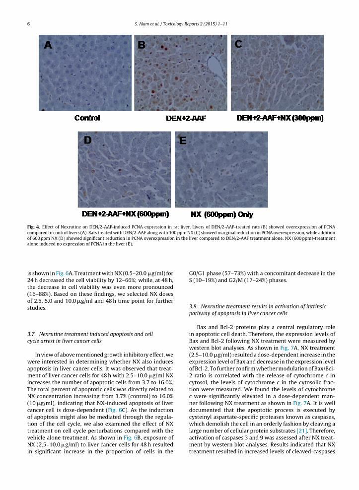
cellular proliferation [20]. In our study, the PCNA antigenwas not expressed in liver sections of control rats (Fig. 4A).However, liver sections from DEN/2-AAF-treated rats werepositive for the PCNA staining, indicative of active cell pro-liferation in liver tissue (Fig. 4B). We observed lower PCNAexpression (Fig. 4C–D) in the treatment groups of NX withDEN/2-AAF suggesting NX has an anti-proliferative effecton DEN/2-AAF-induced liver tumorigenesis in rats.
3.5. Nexrutine induced apoptosis in liver tissue treatedwith DEN/2-AAF animals
An apoptotic response of NX in the liver tissue ofDEN/2-AAF-induced rats was investigated using TUNELstaining. Representative photographs for TUNEL-positivecells in DEN/2-AAF-treated alone or NX with DEN/2-AAF-treated animals are shown in Fig. 5. There was an increasein the number of TUNEL positive cells in the livers ofNX +DEN/2-AAF treated rats (Fig. 5C–D) compared toDEN/2-AAF-treated rats (Fig. 5B). However, the apoptoticinduction by NX was more pronounced in the group where600 ppm of NX was given along with DEN/2-AAF (Fig. 5D).
3.6. Nexrutine treatment resulted inhibition in cell
growth of liver cancer cellsThe inhibitory effect of NX (0.5–20.0 �g/ml) on thegrowth of liver cancer cells was assessed by MTT assay and

S. Alam et al. / Toxicology Reports 2 (2015) 1–11 5
Fig. 2. Effect of Nexrutine on DEN/2-AAF-induced COX-2 expression in rat liver. Livers of DEN/2-AAF-treated rats (B) showed overexpression of COX-2compared to control livers (A), while that of rats treated with DEN/2-AAF along with 300 ppm (C) and 600 ppm (D) NX showed marginal and significantreduction in COX-2 overexpression, respectively, compared to DEN/2-AAF treatment alone. NX (600 ppm)-treated rat liver section (E) shows no expressionof COX-2.
Fig. 3. Effect of Nexrutine on DEN/2-AAF-induced iNOS expression in rat liver. Livers of DEN/2-AAF-treated rats (B) showed overexpression of iNOScompared to control livers (A). Treatment groups which were administered NX (300 ppm and 600 ppm) along with DEN/2-AAF (C and D, respectively)showed reduction in iNOS overexpression compared to DEN/2-AAF alone.
6 S. Alam et al. / Toxicology Reports 2 (2015) 1–11
Fig. 4. Effect of Nexrutine on DEN/2-AAF-induced PCNA expression in rat liver. Livers of DEN/2-AAF-treated rats (B) showed overexpression of PCNAcompared to control livers (A). Rats treated with DEN/2-AAF along with 300 ppm NX (C) showed marginal reduction in PCNA overexpression, while additionof 600 ppm NX (D) showed significant reduction in PCNA overexpression in the liver compared to DEN/2-AAF treatment alone. NX (600 ppm)-treatmentalone induced no expression of PCNA in the liver (E).
is shown in Fig. 6A. Treatment with NX (0.5–20.0 �g/ml) for24 h decreased the cell viability by 12–66%; while, at 48 h,the decrease in cell viability was even more pronounced(16–88%). Based on these findings, we selected NX dosesof 2.5, 5.0 and 10.0 �g/ml and 48 h time point for furtherstudies.
3.7. Nexrutine treatment induced apoptosis and cellcycle arrest in liver cancer cells
In view of above mentioned growth inhibitory effect, wewere interested in determining whether NX also inducesapoptosis in liver cancer cells. It was observed that treat-ment of liver cancer cells for 48 h with 2.5–10.0 �g/ml NXincreases the number of apoptotic cells from 3.7 to 16.0%.The total percent of apoptotic cells was directly related toNX concentration increasing from 3.7% (control) to 16.0%(10 �g/ml), indicating that NX-induced apoptosis of livercancer cell is dose-dependent (Fig. 6C). As the inductionof apoptosis might also be mediated through the regula-tion of the cell cycle, we also examined the effect of NX
treatment on cell cycle perturbations compared with thevehicle alone treatment. As shown in Fig. 6B, exposure ofNX (2.5–10.0 �g/ml) to liver cancer cells for 48 h resultedin significant increase in the proportion of cells in theG0/G1 phase (57–73%) with a concomitant decrease in theS (10–19%) and G2/M (17–24%) phases.
3.8. Nexrutine treatment results in activation of intrinsicpathway of apoptosis in liver cancer cells
Bax and Bcl-2 proteins play a central regulatory rolein apoptotic cell death. Therefore, the expression levels ofBax and Bcl-2 following NX treatment were measured bywestern blot analyses. As shown in Fig. 7A, NX treatment(2.5–10.0 �g/ml) resulted a dose-dependent increase in theexpression level of Bax and decrease in the expression levelof Bcl-2. To further confirm whether modulation of Bax/Bcl-2 ratio is correlated with the release of cytochrome c incytosol, the levels of cytochrome c in the cytosolic frac-tion were measured. We found the levels of cytochromec were significantly elevated in a dose-dependent man-ner following NX treatment as shown in Fig. 7A. It is welldocumented that the apoptotic process is executed bycysteinyl aspartate-specific proteases known as caspases,which demolish the cell in an orderly fashion by cleaving a
large number of cellular protein substrates [21]. Therefore,activation of caspases 3 and 9 was assessed after NX treat-ment by western blot analyses. Results indicated that NXtreatment resulted in increased levels of cleaved-caspases
S. Alam et al. / Toxicology Reports 2 (2015) 1–11 7
Fig. 5. Effect of Nexrutine on apoptosis in liver tissue of DEN/2-AAF animals. An apoptotic response to NX in the liver tissue of DEN/2-AAF-induced ratsw EL-posia inducti6
3c
3p
aspesccpaoaiiawaFlHpc
as investigated using TUNEL staining. An increase in the number of TUNnd D) compared to DEN/2-AAF-treated rats (B). However, the apoptotic00 ppm of NX along with DEN/2-AAF (D).
and 9 in a dose-dependent manner, while there was nohange in expression level of caspase 8 (Fig. 7A).
.9. Nexrutine modulate cell cycle regulators andhosphorylation of MAPKs
Altered expression of cell cycle regulatory protein suchs CDKs and cyclins has been implicated in tumorigene-is [22,23]. As our results demonstrated inhibition of cellroliferation upon NX treatment, we further examined it’sffect on the expression of cell regulatory proteins. Ashown in Fig. 7B, NX exposure caused a decrease in cyclinE,yclinD1, CDK2 and CKD4 levels in liver cancer cells. Duringell cycle analysis we found that NX treatment caused G1hase cell cycle arrest. We also found from immunoblotnalysis that NX treatment caused significant inductionf p21WAF, a key regulator of G1-S phase transition, in
dose-dependent manner (Fig. 7B). Kip1/p27 is anothermportant CDK inhibitor that regulates Cdk-cyclin activ-ty at G1-S transition [24]. Protein levels of Kip1/p27 werelso strongly upregulated after NX exposure. In addition,e found that NX treatment to liver cancer cells caused
dose-dependent increase expression of p53 (Fig. 7B).urther, we investigated the level of activated (phosphory-
ated) and total ERK1/2, JNK and p38 kinases in NX-treatedepG2 cells and found phosphorylation of ERK1/2, JNK, and38 kinase levels were downregulated by NX without anyhange in their total protein levels (Fig. 7C)tive cells was seen in the group that was treated with NX + DEN/2-AAF (Con by NX was more pronounced in group where the rats are dosed with
4. Discussion
The present study we have shown that NX inhibited2-AAF-mediated liver tumor promotion in DEN-initiatedrats, which was correlated with a decrease in proliferationindex together with inhibition of COX-2, iNOS and PCNAexpression. Besides its anti-tumor promoting activity, wealso observed that NX causes apoptotic cell death to humanliver cancer cells.
Cancer development is a sequential event which ofteninvolves chronic inflammation and hyperplasia. COX-2 iswell known biomarker of cell proliferation and tumorpromotion as it catalyzes the formation of prostaglandinE2, which is reported to be involved in cell proliferation,inflammation and angiogenesis [25]. Similarly, anothermajor mediator in chronic inflammatory processes is nitricoxide (NO•), which is produced by liver parenchymal andnon-parenchymal cells from l-arginine via nitric oxide syn-thase (NOS). NO• is considered to exert a hepatoprotectiveaction against tissue injury and cytotoxic effects due toinvading microorganisms, parasites and tumor cells. How-ever, many situations that cause uncontrolled, prolongedand/or massive production of NO• by inducible NOS (iNOS)may result in liver damage, leading to inflammation andeven tumor development [26]. iNOS produces much larger
amounts of NO• and has been detected in many humantumors, such as breast cancer, melanoma, bladder cancer,and colorectal cancer [27–30]. A considerable amount ofcompelling evidence suggests that the inhibition of iNOS
8 S. Alam et al. / Toxicology Reports 2 (2015) 1–11
Fig. 6. Effect of Nexrutine on cell growth, cell cycle distribution and apoptosis. (A) Effect of NX on liver cancer cells (HepG2) growth. Liver cancer cells weretreated with NX (0.5–20.0 �g/ml) for 24 and 48 h and the viability of cells was determined by the MTT assay. The data are shown as the relative cell viabilitycompared to vehicle-treated control cells and represent the means ± S.E. of three experiments, in which each treatment was performed in multiple wells.(*p < 0.05 versus control). (B) Effect of NX on cell cycle phase distribution in liver cancer cells. The cells were treated with NX (2.5–10.0 �g/ml) for 48 h andharvested, stained with propidium iodide solution and data were acquired by flow cytometry as described in text. The data are shown as percentage ofcells in each phase and represent the mean ± S.E. of three experiments in which each treatment was performed in multiple flasks. (C) Effect of Nexrutine oninduction of apoptosis in liver cancer cells as assessed by Annexin-V FITC staining. The cells were treated with NX (2.5–10.0 �g/ml) for 48 h and collected,stained with Annexin-V FITC (FITC-A; x-axis) and propidium iodide (PE-Texas Red; y-axis). The data were acquired by flow cytometry as described in textand are shown as percentage of cells in Q2 quadrant (positive for both annexin V and PI) and tabulated as the mean ± S.E. of three experiments, in whicheach treatment was performed in multiple flasks (*p < 0.05 versus control).

S. Alam et al. / Toxicology Reports 2 (2015) 1–11 9
Fig. 7. Effect of Nexrutine on proteins involved in apoptosis, cell cycle regulators and MAPK activation cascades in liver cancer (HepG2) cells. As detailedin text, the cells were treated with NX (2.5–10.0 �g/ml) for 48 h and then harvested. Total cell lysates were prepared and 50 �g protein was subjected tosodium dodecyl sulfate-polyacrylamide gel electrophoresis followed by western blot analyses. The membranes were probed for Bax, Bcl-2, caspases 3, 8,9 and cytochrome c, cell cycle regulators and MAPKs, employing specific antibodies detailed in Section 2 followed by peroxidase-conjugated appropriatesecondary antibody. Proteins were visualized with enhanced chemiluminescence detection system. Equal loading of protein was confirmed by strippingt alues abc
attantea2prNl[
he blot and reprobing it for �-actin or for total MAPK expression. The vompared to control after normalization with total protein or �-actin.
nd COX-2 expression or activity is important not only forreatment of chronic inflammation, but also for the preven-ion of cancer [13,31,32]. Therefore, suppression of iNOSnd COX-2 induction during cancer progression is recog-ized as an important and commonly accepted approacho effectively inhibit tumor promotion. These biomark-rs were highly expressed in liver of DEN/2-AAF-treatednimals. Treatment with NX remarkably suppressed COX-
and iNOS in DEN/2-AAF-induced animals, suggesting alausible anti-tumor promotion role of NX in vivo. These
esults agree with earlier studies that have been shownX to inhibit prostate, lung and skin cancer cell pro-iferation by modulation of COX-2 and iNOS inhibition8,12,13].
ove the figures represent relative density in the term of fold change as
PCNA, is a 36 kDa nuclear protein and its expression inthe nucleus is associated with the DNA synthesis phase ofcell cycle, and serves as a biomarker of proliferation [20].Earlier studies have reported that PCNA is highly associatedwith DEN/2-AAF-induced liver carcinogenesis, which couldbe detected immunohistochemically [33]. In our study, wefound that NX reduced the hepatic PCNA expression inDEN/2-AAF treated rats.
Cell cycle regulation is one important mechanism ofanti-proliferation in cancers [34]. In the present study, we
investigated the cell cycle distribution after treatment withNX and found accumulation of liver cancer cells at G1phase of cell cycle. Similarly, earlier reports with skin andprostate cancer cells showed NX treatment arrested cell
ology Re
[
[
10 S. Alam et al. / Toxic
cycle progression at the G0/G1 phase [13]. Studies have alsosuggested that regulation of cyclin activity plays a key rolein cell cycle progression at different phases, in which CDKsare negatively regulated by a group of functionally relatedproteins known as CDK inhibitors [24]. Cip/p21 binds andinhibits the cyclins E1, D1 and Adependent kinases, regu-lating the G1 to S phase transition of the cell cycle. Cip1/p21is also known to influence the outcome of the p53 responseto DNA damage and plays a protective role in survival signalagainst apoptosis. Kip1/p27 is up-regulated in response toanti-proliferative signals [35,36]. In accordance with theseobservations, our study also revealed an up-regulation ofKip1/p27 and Cip1/p21, and a decrease in the levels ofCDK2, CDK4, cyclins E1 and D1 proteins. These resultsprovide a mechanism by which NX induces cell cycle arrestthat results in a decrease in cell proliferation of liver cancercells.
MAPKs are important upstream regulators of tran-scription factor activation and their signaling is criticalto transduction of a wide variety of extracellular stimuliinto intracellular cascades, thereby controlling the cellularevents such as proliferation, differentiation and apoptosis[37]. Our results demonstrated that NX treatment blockedthe phosphorylation, and hence, activation of MAPKs,including ERK1/2 p38, and JNK in liver cancer cells. Thesefindings are similar to previous studies where inhibitionof ERK1/2, p38 and JNK by chemopreventive agents arecapable of preventing skin carcinogenesis [38,39].
Apoptotic cell death represents a universal andexquisitely efficient suicidal pathway and an ideal way forelimination of unwanted cells; however, cancerous cellsshow dysregulation of this mechanism, which makes thecells virtually immortal and resistant to stress stimuli aswell as therapeutic agents [40]. Therefore, the apoptoticpathway is widely studied as a potential target for can-cer chemotherapy [41,42]. In our study, NX treatment toliver cancer cells resulted in a dose-dependent apoptoticcell death, which would contribute to NX-mediated cellgrowth inhibition. In support these findings, prior studieshave shown that various chemotherapeutic phytochemi-cals possess the ability to induce apoptosis in cancer cellsby arresting the cell cycle progression in various phases ofcell division [43–45]. Furthermore, NX treatment to livercancer cells results in significant decrease in the levels ofBcl-2 protein along with an increase in the levels of Baxprotein, thus enhancing the Bax/Bcl-2 ratio, which favorsapoptosis. Increase in Bax/Bcl-2 ratio acts as a proapopto-tic signal resulting in the release of cytochrome c proteinfrom mitochondria to cytoplasm, activating the apopto-some, which further leads to auto-activation of caspase 9and cleavage of pro-caspase 3 to its activated form cas-pase 3, the executioner caspase [46–48]. Caspases are themediators of execution mechanism of apoptosis, and theiractivation results in the cleavage of PARP protein, a DNArepair enzyme in the cell, and subsequent DNA degradationand apoptotic death [21]. Since, caspase 8 was not foundto be activated after NX treatment in liver cancer cells, it
can be deduced that NX-induced apoptosis is mediated viaactivation of the intrinsic pathway.In summary, this study demonstrated that NX inhibitedDEN/2-AAF-induced enhanced cell proliferation in liver. In
[
ports 2 (2015) 1–11
addition, it also caused dose-as well as time-dependentcytotoxicity in liver cancer (HepG2) cells. NX induced accu-mulation of liver cancer cells at the G1 phase of cell cycle aswell as apoptosis. Taken together, these in vivo and in vitrostudies provide strong evidence that NX could be useful inthe management (chemoprevention as well as chemother-apy) of liver cancer.
Conflict of interest
None.
Transparency document
The Transparency document associated with this articlecan be found in the online version.
Acknowledgements
We are grateful to the Director of our institute, forhis keen interest in this present study. This work wassupported by funds from Department of Science and Tech-nology (Govt of India) and CSIR Supra-institutional Project08 (SIP-08) New Delhi. S.A. is thankful to Council of Scien-tific and Industrial Research, New Delhi for the award ofSenior Research Fellowship. We are grateful to Prof JoyceE. Rundhaug, MD Anderson Cancer Centre, Texas for criti-cally reading the manuscript and editorial assistance. Themanuscript is IITR communication # 3213
References
[1] T.R. Morgan, S. Mandayam, M.M. Jamal, Alcohol and hepatocellularcarcinoma, Gastroenterology 127 (2004) S87–S96.
[2] S.C. Chuang, C. La Vecchia, P. Boffetta, Liver cancer: descriptive epi-demiology and risk factors other than HBV and HCV infection, CancerLett. 286 (1) (2009) 9–14.
[3] IARC World Cancer Report, 2008, IARC, 2008.[4] S.S. Thorgeirsson, J.W. Grisham, Molecular pathogenesis of human
hepatocellular carcinoma, Nat. Genet. 31 (4) (2002) 339–346.[5] N. Nagasue, T. Ono, A. Yamanoi, H. Kohno, O.N. El-Assal, H. Taniura,
M. Uchida, Prognostic factors and survival after hepatic resectionfor hepatocellular carcinoma without cirrhosis, Br. J. Surg. 88 (2001)515–522.
[6] Y.-J. Surh, Cancer chemoprevention with dietary phytochemicals,Nat. Rev. Cancer 3 (2003) 768–780.
[7] K.H. Kwon, A. Barve, S. Yu, M. Huang, A.T. Kong, Cancerchemoprevention by phytochemicals: potential molecular targets,biomarkers and animal models, Acta Pharmacol Sin 28 (9) (2007)1409–1421.
[8] A.P. Kumar, S. Bhaskaran, M. Ganapathy, K. Crosby, M.D. Davis,P. Kochunov, J. Schoolfield, I. Yeh, D.A. Troyer, R. Ghosh,Akt/cAMP-responsive element binding protein/Cyclin D1 network:a novel target for prostate cancer inhibition in transgenic adenocar-cinoma of mouse prostate model mediated by NX, a Phellodendronamurense bark extract, Clin. Cancer Res. 13 (2007) 2784.
[9] G.E. Garcia, N. Arevalo, S. Bhaskaran, A. Gupta, N. Kyprianou, A.P.Kumar, Akt and CREB mediated prostate cancer cell proliferationinhibition by NX a phellodendronamurense extract, Neoplasia 8(2006) 523–533.
10] S.B. Muralimanoharan, A.B. Kunnumakkara, B. Shylesh, K.H. Kulkarni,X. Haiyan, H. Ming, B.B. Aggarwal, G. Rita, A.P. Kumar, Butanol fractioncontaining berberine or related compound from nexrutine inhibitsNFkappaB signaling and induces apoptosis in prostate cancer cells,Prostate 69 (5) (2009) 494–504.
11] M.A. James, H. Fu, Y. Liu, D.R. Chen, M. You, Dietary administration
of berberine or phellodendronamurense extract inhibits cell cycleprogression and lung tumorigenesis, Mol. Carcinog. 50 (1) (2011)1–7.12] R. Ghosh, H. Graham, P. Rivas, X.J. Tan, K. Crosby, S. Bhaskaran,J. Schoolfield, J. Banu, G. Fernandes, I.T. Yeh, A.P. Kumar,

logy Re
[
[
[
[
[
[
[
[
[
[
[
[
[
[
[
[
[
[
[
[
[
[
[
[
[
[
[
[
[
[
[
[
[
[
[[
S. Alam et al. / Toxico
Phellodendronamurense bark extract prevents progression ofprostate tumors in transgenic adenocarcinoma of mouse prostate:potential for prostate cancer management, Anticancer Res. 30 (3)(2010) 857–865.
13] R. Kumar, M. Das, K.M. Ansari, Nexrutine® inhibits tumorigenesis inmouse skin and induces apoptotic cell death in human squamouscarcinoma A431 and human melanoma A375 cells, Carcinogenesis33 (10) (2012) 1909–1918.
14] H. Enzmann, E. Bomhard, M. Iatropoulos, H.J. Ahr, G. Schlueter, G.M.Williams, Short- and intermediate-term carcinogenicity testing-areview. Part 1: The prototypes mouse skin tumour assay and rat liverfocus assay, Food Chem. Toxicol. 36 (1998) 979–995.
15] S. Malik, S. Bhatnagar, N. Chaudhary, D.P. Katare, S.K. Jain,DEN+2-AAF-induced multistep hepatotumorigenesis in Wistar rats:supportive evidence and insights, Protoplasma 250 (1) (2013)175–183.
16] D. Tiwawech, R. Hasegawa, Y. Kurata, M. Tatematsu, M.A. Shibata, W.Thamavit, N. Ito, Dose-dependent effects of 2-acetylamino fluoreneon hepatic foci development and cell proliferation in rats, Carcino-genesis 12 (1991) 985–990.
17] V. Mishra, D.K. Saxena, M. Das, Effect of argemone oil and argemonealkaloid, sanguinarine on Sertoli-germ cell coculture, Toxicol. Lett.186 (2) (2009) 104–110.
18] L. Franco, D. Doria, E. Bertazzoni, A. Benini, C. Bassi, Increasedexpression of inducible nitric oxide synthase and cyclooxygenase-2 in pancreatic cancer, Prostaglandins Other Lipid Mediat. 73 (1–2)(2004) 51–58.
19] W. Li, R.J. Xu, L.H. Jiang, J. Shi, X. Long, B. Fan, Expression ofcyclooxygenase-2 and inducible nitric oxide synthase correlates withtumor angiogenesis in endometrial carcinoma, Med. Oncol. 22 (1)(2005) 63–70.
20] G. Maga, U. Hubscher, Proliferating cell nuclear antigen (PCNA): adancer with many partners, J. Cell Sci. 116 (Pt 15) (2003) 3051–3060.
21] B. Zhivotovsky, A. Samali, A. Gahm, S. Orrenius, Caspases: their intra-cellular localization and translocation during apoptosis, Cell DeathDiffer. 6 (1999) 644–651.
22] C.J. Sherr, J.M. Roberts, CDK inhibitors: positive and negative regula-tors of G1-phase progression, Genes Dev. 12 (1999) 1501–1512.
23] D.O. Morgan, Principles of CDK regulation, Nature 374 (1995)131–134.
24] X. Grana, E.P. Reddy, Cell cycle control in mammalian cells: role ofcyclins, cyclin dependent kinases (CDKs), growth suppressor genesand cyclin-dependent kinase inhibitors (CKIs), Oncogene 11 (2)(1995) 211–219.
25] S.M. Prescott, F.A. Fitzpatrick, Cyclooxygenase-2 and carcinogenesis,Biochim. Biophys. Acta 1470 (2000) 69–78.
26] H. Suzuki, M. Menegazzi, A. Carcereri de Prati, S. Mariotto, U. Armato,Nitric oxide in the liver: physiopathological roles, Adv. Neuroim-munol. 5 (1995) 379–410.
27] M. Vakkala, K. Kahlos, E. Lakari, P. Pääkkö, V. Kinnula, Y. Soini,Inducible nitric oxide synthase expression, apoptosis, and angiogen-esis in in-situ and invasive breast carcinomas, Clin. Cancer Res. 6(2000) 2408–2416.
28] S. Ekmekcioglu, J. Ellerhorst, C.M. Smid, V.G. Prieto, M. Munsell, A.C.Buzaid, E.A. Grimm, Inducible nitric oxide synthase and nitrotyrosinein human metastatic melanoma tumors correlate with poor survival,Clin. Cancer Res. 6 (2000) 4768–4775.
29] H. Wolf, C. Haeckel, A. Roessner, Inducible nitric oxide synthaseexpression in human urinary bladder cancer, Virchows Arch. 437(2000) 662–666.
30] N. Yagihashi, H. Kasajima, S. Sugai, K. Matsumoto, Y. Ebina, T. Morita,T. Murakami, S. Yagihashi, Increased in situ expression of nitric oxide
[
ports 2 (2015) 1–11 11
synthase in human colorectal cancer, Virchows Arch. 437 (2000)109–114.
31] M.A. Rahman, D.K. Dhar, E. Yamaguchi, S. Maruyama, T. Sato, H.Hayashi, T. Ono, A. Yamanoi, H. Kohno, N. Nagasue, Coexpressionof inducible nitric oxide synthase and COX-2 in hepatocellular carci-noma and surrounding liver: possible involvement of COX-2 in theangiogenesis of hepatitis C virus-positive cases, Clin. Cancer Res. 7(2001) 1325–1332.
32] S. Sarfaraz, I.A. Siddiqui, D.N. Syed, F. Afaq, H. Mukhtar, Guggulsteronemodulates MAPK and NF-kB pathways and inhibits skin tumorigen-esis in SENCAR mice, Carcinogenesis 29 (2008) 2011–2018.
33] T. Takizawa, T. Imai, J. Onose, M. Ueda, T. Tamura, K. Mitsumori,K. Izumi, M. Hirose, Enhancement of hepatocarcinogenesis bykojic acid in rat two-stage models after initiation with N-bis(2-hydroxypropyl)nitrosamine or N-diethylnitrosamine, Toxicol. Sci. 81(1) (2004) 43–49.
34] I.E. Gerard, H.V. Karen, Proliferation, cell cycle and apoptosis in can-cer, Nature 411 (2001) 42–48.
35] R. Fotedar, M. Bendjennat, A. Fotedar, Role of p21WAF1 in the cellularresponse to UV, Cell Cycle 3 (2) (2004) 134–137.
36] K. Polyak, J.Y. Kato, M.J. Solomon, C.J. Sherr, J. Massague, J.M. Roberts,A. Koff, p27Kip1, a cyclin-CDK inhibitor, links transforming growthfactor-� and contact inhibition to cell cycle arrest, Genes Dev. 8(1994) 9–22.
37] R. Seger, E.G. Krebs, The MAPK signaling cascade, FASEB J. 9 (1995)726–735.
38] Y.J. Surh, K.S. Chun, H.H. Cha, S.S. Han, Y.S. Keum, K.K. Park, S.S.Lee, Molecular mechanisms underlying chemopreventive activitiesof anti-inflammatory phytochemicals: down-regulation of COX-2and iNOS through suppression of NF-kappa B activation, Mutat. Res.68 (243) (2001) 480–481.
39] Y. Kim, R.C. Sills, D. Houle, Overview of the molecular biology ofhepatocellular neoplasms and hepatoblastomas of the mouse liver,Toxicol. Pathol. 33 (2005) 175–180.
40] F.H. Igney, P.H. Krammer, Death and anti-death: tumour resistanceto apoptosis, Nat. Rev. Cancer. 2 (4) (2002) 277–288.
41] G. Galati, S. Teng, M.Y. Moridani, T.S. Chan, P.J. O’Brien, CancerChemoprevention and apoptosis mechanisms induced by dietarypolyphenolics, Drug Metabol. Drug Interact. 17 (1–4) (2000)311–350.
42] N. Khan, F. Afaq, H. Mukhtar, Apoptosis by dietary factors: the suicidesolution for delaying cancer growth, Carcinogenesis 28 (2) (2007)233–239.
43] S.J. Elledge, Cell cycle checkpoints: preventing an identity crisis, Sci-ence 274 (5293) (1996) 1664–1672.
44] G.I. Evan, K.H. Vousden, Proliferation, cell cycle and apoptosis in can-cer, Nature 411 (6835) (2001) 342–348.
45] N. Khan, F. Afaq, D.N. Syed, H. Mukhtar, Fisetin, a novel dietaryflavonoid, causes apoptosis and cell cycle arrest in human prostatecancer LNCaP cells, Carcinogenesis 29 (5) (2008) 1049–1056.
46] F. Cecconi, Apaf1 and the apoptotic machinery, Cell Death Differ. 6(1999) 1087–1098.
47] J.C. Reed, Bcl-2 family proteins, Oncogene 17 (1998) 3225–3236.48] J.M. Jurgensmeier, Z. Xie, Q. Deveraux, L. Ellerby, D. Bredesen,
J.C. Reed, Bax directly induces release of cytochome c from iso-lated mitochondria, Proc. Natl. Acad. Sci. U. S. A. 95 (1998)4997–5002.
50] J. Ferlay, H.R. Shin, F. Bray, D. Forman, C. Mathers, D.M. Parkin, A.N.Globoc, v1.2, Cancer Incidence and Mortality Worldwide: IARC Can-cer Base No. 10, International Agency for Research on Cancer, Lyon,France, 2010, Available from: http://globocan.iarc.fr (accessed May2011).
Copyright © 2022 FDOKUMEN


