Magnitude of peripheral neuropathy in cirrhosis of liver patients from central rural India
8
-
Upload
independent -
Category
Documents
-
view
0 -
download
0
Transcript of Magnitude of peripheral neuropathy in cirrhosis of liver patients from central rural India
Annals of Indian Academy of Neurology, October-December 2014, Vol 17, Issue 4
and gastrointestinal bleed.[1] Most studies focused on portal hypertension, variceal bleeding, overt hepatic encephalopathy, while complication like peripheral neuropathy (PN) were seldom studied.
PN need to be focused as it is important complication of cirrhosis of liver that may seriously impair patient’s routine daily activities and quality of life. Various studies revealed prevalence of PN varying from 19-80% in the cirrhotic patients on nerve conduction studies.[2-9] Peripheral nerve dysfunction is significantly more frequent in advanced liver disease compared with early liver damage. However, only few hepatologists really screen patients for PN due to time-consuming neurophysiological tests. Controversy also exists in literature regarding the correlation between severity of liver disease and prevalence of PN. Cirrhosis of liver also poses a huge financial, psychological burden for the patients and their families. Since treatment regimens are limited early diagnosis of is the most effective approach especially in resource restrained settings.
Introduction
Cirrhosis of liver is a major global health problem and it is an important cause of morbidity and mortality. Approximately 40% of patients with cirrhosis are asymptomatic, and the condition often is discovered during a routine clinical or laboratory or radiographic examination. In our hospital, the patients of cirrhosis presented usually with serious and life-threatening complications like encephalopathy, ascites,
Magnitude of peripheral neuropathy in cirrhosis of liver patients from central rural India
Jyoti Jain, Ramji Singh1, Shashank Banait2, Nitin Verma3, Satish Waghmare3
Departments of Medicine, and 3Physiology, Mahatma Gandhi Institute of Medical Sciences, Sevagram, Wardha, Maharashtra, 1Department of Physiology, All India Institute of Medical Sciences, Patna, Bihar, 2Department of
Ophthalmology, Jawaharlal Nehru Medical College, Sawangi Meghe, Wardha, Maharashtra, India
Abstract
Context: Cirrhosis of liver is an important cause of morbidity and mortality and if associated with peripheral neuropathy (PN) it also poses a huge financial, psychological burden for the patients and their families. Aim: The aim of the present study was to study the magnitude of PN among subjects with cirrhosis of liver presenting to tertiary care teaching hospital in central rural India. Settings and Design: A cross-sectional study was performed in a tertiary care teaching hospital. Materials and Methods: In all patients of cirrhosis of liver irrespective of etiology, aged 15 and above, undergone clinical assessment for peripheral nervous systems damage and confirmed by nerve conduction studies. Statistical Analysis Used: We used chi square test to study associations. P value ≤0.05 was considered as significant. Crude odds ratios were computed to assess the strength of association between independent variables and dependent variables along with their 95% confidence intervals. Results: We included 207 of cirrhosis of liver patients admitted in medicine department from November 2010 through November 2013. Nearly 83% patients were male and 63.2% patients were under the age of 45 years. Common features in these patients were ascites (71%) splenomegaly (63.3%) pedal edema (61.4%) icterus (46.4%) tingling (44.9%) gastrointestinal bleeding(39.1%), ataxia (26.6%),numbness(26.6%),distal motor weakness (21.7%) and paresthesia(20.8%).Among the manifestation of peripheral nerve involvement, loss of ankle reflex was the most common feature in 51.7%, followed by loss of temperature sense 29.5%, loss of vibration sense 20.8%, loss of touch 16.4%, loss of position sense 14.5% and loss of pain in 6.3% of the patients. Peripheral neuropathy was found in 53.6% [95% CI: 46.58- 60.56] study subjects on electrophysiological study. Conclusions: Analysis of electrophysiological study shows that the PN is very common in study subjects with cirrhosis of liver, especially in male subjects, during the middle age group.
Key Words
Chronic liver disease, cirrhosis of liver, nerve conduction studies, peripheral neuropathy
For correspondence: Dr. Jyoti Jain, Department of Medicine, Mahatma Gandhi Institute of Medical Sciences
Sewagram, Wardha - 442 102, Maharashtra, India. E-mail: [email protected]
Ann Indian Acad Neurol 2014;17:409-15
Original Article
Access this article onlineQuick Response Code: Website:
www.annalsofian.org
DOI: 10.4103/0972-2327.144012
410 Jain, et al: Peripheral neuropathy and cirrhosis of liver
Annals of Indian Academy of Neurology, October-December 2014, Vol 17, Issue 4
Limited literature is available from central India, hence the present study was carried out to study the magnitude and correlates of PN among patients with cirrhosis of liver presenting to tertiary care teaching hospital in central rural India.
Materials and Methods
EthicsThe study was approved by the ethics committee of Mahatma Gandhi Institute of Medical Sciences (IRB00003623). We obtained a written informed consent from all study subjects before enrolling them in the study.
Study settingThe study was conducted in the Neurophysiology unit of the Department of Physiology and Department of Medicine, Mahatma Gandhi Institute of Medical Sciences, Sevagram which is a 650-bedded teaching tertiary care hospital located in a town in central India. Most patients visiting the hospital come from rural areas. All patients aged 15 years or above are screened and examined first in the Medicine inpatient department where after a brief history and examination, study subjects were referred to department of Physiology for electrophysiological studies.
Study designBetween November 1, 2010 and November 30, 2013 a cross-sectional study was conducted to study magnitude and correlates of PN of all consecutive cirrhosis of liver patients, aged 15 years and above, irrespective of etiology, admitted to the medicine wards.
Study subjectsCirrhosis of liver was diagnosed on a clinical basis (symptoms and signs) along with biochemical tests: Two sets of abnormal liver function tests (LFTs) that is Hypoalbuminemia, elevated alanine aminotransferase, aspartate aminotransferase, or concurrently elevated alkaline phosphatase, and gamma glutamyltransferase, more than 6 months apart, along with a liver-related clinical event hepatic encephalopathy (HE), variceal bleed, ascites, or spontaneous bacterial peritonitis or ultrasonographic (USG) findings indicating showed positive signs such as nodular or shrunken liver, portal hypertension (splenomegaly) and ascites, endoscopic evidence of esophageal varices, and liver histology if consistent with cirrhosis.
Patients were excluded if they had history of diabetes mellitus (DM), other causes of neuropathy such as toxin or drugs exposure, infection with human immunodeficiency virus (HIV), and limb injuries, stroke, dementia, Parkinson’s disease, overt hepatic encephalopathy, significant co-morbid illness such as heart, respiratory failure, renal failure, and malignancy or not provided written informed consent.
Data collection toolsAll study subjects included in the study underwent a relevant clinical history and neurological examination. Demographic data including age and sex; history of associated complaints like tingling, numbness, paresthesia, and ataxia were obtained during baseline interview. We elicited information about
personal habits such as alcohol consumption, smoking, tobacco chewing, betel-nut chewing, and serological tests for hepatitis B as well as hepatitis C infection to all study subjects. Current smokers were defined as persons who reported smoking for at least 3 months in their life and who currently smoke at least on some days of a week. Current alcoholic are classified as persons who reported alcohol consumption in the last 3 months while past drinkers were those who have not consumed alcohol in last 3 months. We recorded routine LFTs (serum bilirubin, serum albumin, ALT, alkaline phosphatase) and hematologic parameters by conventional methods. Modified Child-Turcotte-Pugh (CTP) score for severity of cirrhosis was computed and patients were classified as belonging to Child class A, B, or C.[10] Interview of all study subjects was undertaken by the same research associate and was pilot tested. All study subjects underwent clinical assessment of peripheral nervous systems (PNS) damage like ankle jerks, knee jerks and pinprick sense, temperature sense, touch sense, position sense, and vibration sense (128 Hz). Absent or diminished response was considered as abnormal.[11]
Electrophysiological testing (Nerve conduction studies)Electrophysiological testing was performed using complete computerized physiology lab system, developed by Recorder and Medicare system, electromyography evoked potential (EMG EP) Mark-II machine. Nerve conductivity testing was performed in upper and lower limbs using surface electrodes in motor and sensory fibers of ulnar, median, tibial, and peroneal nerve as well as in sensory fibers of sural nerve in all study subjects. Recordings were performed by the same investigator with training in and 5-year-expereince of performing these studies, under constant room temperature (28-30°C) to minimize errors, careful distance measurements, and recording of well-defined and artefact-free responses. Latency and amplitude values were read from the equipment after accurate cursor placement was ensured. Distance values were entered into the counterpoint device and conduction velocities were calculated automatically.[12]
We measured parameters like distal motor latency (DML), amplitude of the compound muscle action potential (CMAP), nerve conduction velocity (NCV), F wave minimum latency for motor nerve, and sensory nerve action potential (SNAP) amplitude, as well as conduction velocity for sensory nerves.
PN was diagnosed if more than one peripheral nerve was affected on nerve conduction study. Motor nerve affection was defined if all the three parameters, that is DML, amplitude of CMAP, and NCV were abnormal, whereas involvement of sensory nerve was present if both the parameters — amplitude of SNAP and NCV were abnormal. Degree of severity of PN was divided into the three groups: Normal, early damage, and definite damage, according to the number of peripheral nerves involved. Normal or no peripheral damage was defined if only one peripheral nerve was involved. Early damage, if two or three peripheral nerves were involved and definite damage, if more than three peripheral nerves were involved.[13] We also categorized pattern of PN as Axonal, Demyelinating, and Mixed. Axonal neuropathy was defined as normal DML, decreased amplitude of CMAP and normal conduction velocity for nerves. Demyelinating neuropathy was defined as normal CMAP amplitude; prolonged DML and slow conduction
Jain, et al: Peripheral neuropathy and cirrhosis of liver 411
Annals of Indian Academy of Neurology, October-December 2014, Vol 17, Issue 4
velocity were found on NCS. Mixed pattern of PN was diagnosed if all the three parameters were abnormal.
Statistical analysisWe used statistical software STATA (version 16, Stata Corporation, Texas, USA). Magnitude of PN was expressed in percentage and 95% confidence interval. We used chi square test to study associations. Unadjusted odds ratio (ORs) were computed to assess the strength of association between independent variables (age, sex,occupation, area of residence, probable reason of cirrhosis, hematological profile, and severity of cirrhosis by Child-Pugh (CP) score and dependent variables (PN) along with their 95% confidence intervals. P value ≤ 0.05 was considered as significant.
Results
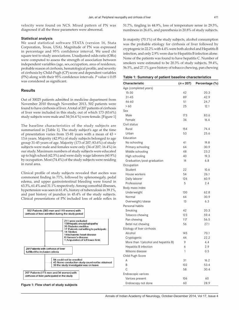
Out of 30025 patients admitted in medicine department from November 2010 through November 2013, 502 patients were found to have cirrhosis of liver. A total of 207 patients of cirrhosis of liver were included in this study, out of which 173 (83.6%) study subjects were male and 34 (16.4 %) were female. [Figure 1]
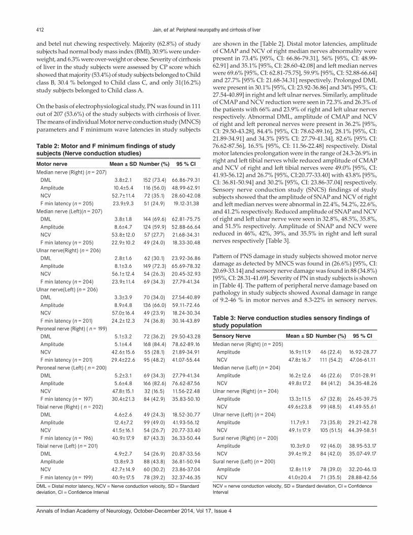
The baseline characteristics of the study subjects are summarized in [Table 1]. The study subject’s age at the time of presentation varies from 15-81 years with a mean of 43 + 13.6 years. Majority (42.9%) of study subjects belonged to age group 31-45 years of age. Majority (173 of 207; 83.6%) of study subjects were male and females were only (34 of 207; 16.4%) in our study. Maximum numbers of study subjects were educated up to high school (42.5%) and were daily wage laborers (60.9%) by occupation. Most (74.4%) of the study subjects were residing in rural area.
Clinical profile of study subjects revealed that ascites was commonest finding in 71%, followed by splenomegaly, pedal edema, and upper gastrointestinal bleeding were found in 63.3%, 61.4% and 31.1% respectively. Among comorbid illnesses, hypertension was seen in 61.4%, history of tuberculosis in 39.1%, and past history of jaundice in 45.4% of the study subjects. Clinical presentations of PN included loss of ankle reflex in
51.7%, tingling in 44.9%, loss of temperature sense in 29.5%, numbness in 26.6%, and paresthesia in 20.8% of study subjects.
In majority (70.1%) of the study subjects, alcohol consumption was the probable etiology for cirrhosis of liver followed by cryptogenic in 22.2% with 4.4% were both alcohol and Hepatitis B infection, and only 2.9% were due to Hepatitis B infection alone. None of the patients was found to have hepatitis C. Number of smokers were estimated to be 20.3% of study subjects, 59.4%, 56.5%, and 27.1% gave history of tobacco chewing, pan chewing
Figure 1: Flow chart of study subjects
Table 1: Summary of patient baseline characteristics
Characteristic (n = 207) Percentage (%)Age (completed years)
15-30 42 20.331-45 89 42.946-60 51 24.7> 60 25 12.1
SexMale 173 83.6Female 34 16.4
Civil statusRural 154 74.4Urban 53 25.6
EducationNo schooling 41 19.8Primary schooling 64 30.9Middle schooling 48 23.2High schooling 40 19.3Graduation/post-graduation 14 6.8
OccupationStudent 22 10.6House workers 54 26.1Daily laborer 126 60.9Professional 5 2.4
Body mass indexUnderweight 130 62.8Normal 64 30.9Overweight/obese 13 6.3
Personal habitsSmoking 42 20.3Tobacco chewing 123 59.4Pan chewing 117 56.5Betel nut chewing 56 27.1
Etiology of liver cirrhosisAlcohol 145 70.1Cryptogenic 44 22.2More than 1(alcohol and hepatitis B) 9 4.4Hepatitis B infection 6 2.9Wilsons disease 1 0.5
Child Pugh ScoreA 31 16.2B 102 53.4C 58 30.4
Endoscopic varicesVarices present 106 60Endoscopy not done 60 28.9
412 Jain, et al: Peripheral neuropathy and cirrhosis of liver
Annals of Indian Academy of Neurology, October-December 2014, Vol 17, Issue 4
and betel nut chewing respectively. Majority (62.8%) of study subjects had normal body mass index (BMI), 30.9% were under-weight, and 6.3% were over-weight or obese. Severity of cirrhosis of liver in the study subjects were assessed by CP score which showed that majority (53.4%) of study subjects belonged to Child class B, 30.4 % belonged to Child class C, and only 31(16.2%) study subjects belonged to Child class A.
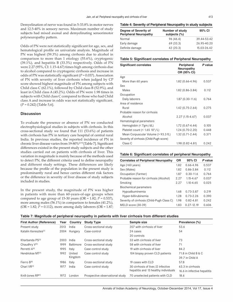
On the basis of electrophysiological study, PN was found in 111 out of 207 (53.6%) of the study subjects with cirrhosis of liver. The means of individual Motor nerve conduction study (MNCS) parameters and F minimum wave latencies in study subjects
are shown in the [Table 2]. Distal motor latencies, amplitude of CMAP and NCV of right median nerves abnormality were present in 73.4% [95%, CI: 66.86-79.31], 56% [95%, CI: 48.99-62.91] and 35.1% [95%, CI: 28.60-42.08] and left median nerves were 69.6% [95%, CI: 62.81-75.75], 59.9% [95%, CI: 52.88-66.64] and 27.7% [95% CI: 21.68-34.31] respectively. Prolonged DML were present in 30.1% [95%, CI: 23.92-36.86] and 34% [95%, CI: 27.54-40.89] in right and left ulnar nerves. Similarly, amplitude of CMAP and NCV reduction were seen in 72.3% and 26.3% of the patients with 66% and 23.9% of right and left ulnar nerves respectively. Abnormal DML, amplitude of CMAP and NCV of right and left peroneal nerves were present in 36.2% [95%, CI: 29.50-43.28], 84.4% [95%, CI: 78.62-89.16], 28.1% [95%, CI: 21.89-34.91] and 34.3% [95% CI: 27.79-41.34], 82.6% [95% CI: 76.62-87.56], 16.5% [95%, CI: 11.56-22.48] respectively. Distal motor latencies prolongation were in the range of 24.3-26.9% in right and left tibial nerves while reduced amplitude of CMAP and NCV of right and left tibial nerves were 49.0% [95%, CI: 41.93-56.12] and 26.7% [95%, CI:20.77-33.40] with 43.8% [95%, CI: 36.81-50.94] and 30.2% [95%, CI: 23.86-37.04] respectively. Sensory nerve conduction study (SNCS) findings of study subjects showed that the amplitude of SNAP and NCV of right and left median nerves were abnormal in 22.4%, 54.2%, 22.6%, and 41.2% respectively. Reduced amplitude of SNAP and NCV of right and left ulnar nerve were seen in 32.8%, 48.5%, 35.8%, and 51.5% respectively. Amplitude of SNAP and NCV were reduced in 46%, 42%, 39%, and 35.5% in right and left sural nerves respectively [Table 3].
Pattern of PNS damage in study subjects showed motor nerve damage as detected by MNCS was found in (26.6%) [95%, CI: 20.69-33.14] and sensory nerve damage was found in 88 (34.8%) [95%, CI: 28.31-41.69]. Severity of PN in study subjects is shown in [Table 4]. The pattern of peripheral nerve damage based on pathology in study subjects showed Axonal damage in range of 9.2-46 % in motor nerves and 8.3-22% in sensory nerves.
Table 2: Motor and F minimum findings of study subjects (Nerve conduction studies)
Motor nerve Mean ± SD Number (%) 95 % CIMedian nerve (Right) (n = 207)
DML 3.8±2.1 152 (73.4) 66.86-79.31Amplitude 10.4±5.4 116 (56.0) 48.99-62.91NCV 52.7±11.4 72 (35.1) 28.60-42.08F min latency (n = 205) 23.9±9.3 51 (24.9) 19.12-31.38
Median nerve (Left)(n = 207)DML 3.8±1.8 144 (69.6) 62.81-75.75Amplitude 8.6±4.7 124 (59.9) 52.88-66.64NCV 53.8±12.0 57 (27.7) 21.68-34.31F min latency (n = 205) 22.9±10.2 49 (24.0) 18.33-30.48
Ulnar nerve(Right) (n = 206)DML 2.8±1.6 62 (30.1) 23.92-36.86Amplitude 8.1±3.6 149 (72.3) 65.69-78.32NCV 56.1±12.4 54 (26.3) 20.45-32.93F min latency (n = 204) 23.9±11.4 69 (34.3) 27.79-41.34
Ulnar nerve(Left) (n = 206)DML 3.3±3.9 70 (34.0) 27.54-40.89Amplitude 8.9±4.8 136 (66.0) 59.11-72.46NCV 57.0±16.4 49 (23.9) 18.24-30.34F min latency (n = 201) 24.2±12.3 74 (36.8) 30.14-43.89
Peroneal nerve (Right) ( n = 199)DML 5.1±3.2 72 (36.2) 29.50-43.28Amplitude 5.1±4.4 168 (84.4) 78.62-89.16NCV 42.6±15.6 55 (28.1) 21.89-34.91F min latency (n = 201) 29.4±22.6 95 (48.2) 41.07-55.44
Peroneal nerve (Left) ( n = 200)DML 5.2±3.1 69 (34.3) 27.79-41.34Amplitude 5.6±4.8 166 (82.6) 76.62-87.56NCV 47.8±15.1 32 (16.5) 11.56-22.48F min latency (n = 197) 30.4±21.3 84 (42.9) 35.83-50.10
Tibial nerve (Right) ( n = 202)DML 4.6±2.6 49 (24.3) 18.52-30.77Amplitude 12.4±7.2 99 (49.0) 41.93-56.12NCV 41.5±16.1 54 (26.7) 20.77-33.40F min latency (n = 196) 40.9±17.9 87 (43.3) 36.33-50.44
Tibial nerve (Left) (n = 201)DML 4.9±2.7 54 (26.9) 20.87-33.56Amplitude 13.8±9.3 88 (43.8) 36.81-50.94NCV 42.7±14.9 60 (30.2) 23.86-37.04F min latency (n = 199) 40.9±17.5 78 (39.2) 32.37-46.35
DML = Distal motor latency, NCV = Nerve conduction velocity, SD = Standard deviation, CI = Confidence Interval
Table 3: Nerve conduction studies sensory findings of study population
Sensory Nerve Mean ± SD Number (%) 95 % CIMedian nerve (Right) (n = 205)
Amplitude 16.9±11.9 46 (22.4) 16.92-28.77NCV 47.8±16.7 111 (54.2) 47.06-61.11
Median nerve (Left) (n = 204)Amplitude 16.2±12.6 46 (22.6) 17.01-28.91NCV 49.8±17.2 84 (41.2) 34.35-48.26
Ulnar nerve (Right) (n = 204)Amplitude 13.3±11.5 67 (32.8) 26.45-39.75NCV 49.6±23.8 99 (48.5) 41.49-55.61
Ulnar nerve (Left) (n = 204)Amplitude 11.7±9.1 73 (35.8) 29.21-42.78NCV 49.1±17.9 105 (51.5) 44.39-58.51
Sural nerve (Right) (n = 200)Amplitude 10.3±9.0 92 (46.0) 38.95-53.17NCV 39.4±19.2 84 (42.0) 35.07-49.17
Sural nerve (Left) (n = 200)Amplitude 12.8±11.9 78 (39.0) 32.20-46.13NCV 41.0±20.4 71 (35.5) 28.88-42.56
NCV = nerve conduction velocity, SD = Standard deviation, CI = Confidence Interval
Jain, et al: Peripheral neuropathy and cirrhosis of liver 413
Annals of Indian Academy of Neurology, October-December 2014, Vol 17, Issue 4
Demyelination of nerve was found in 5-33.8% in motor nerves and 12.5-40% in sensory nerves. Maximum number of study subjects had mixed axonal and demyelinating sensorimotor polyneuropathy pattern.
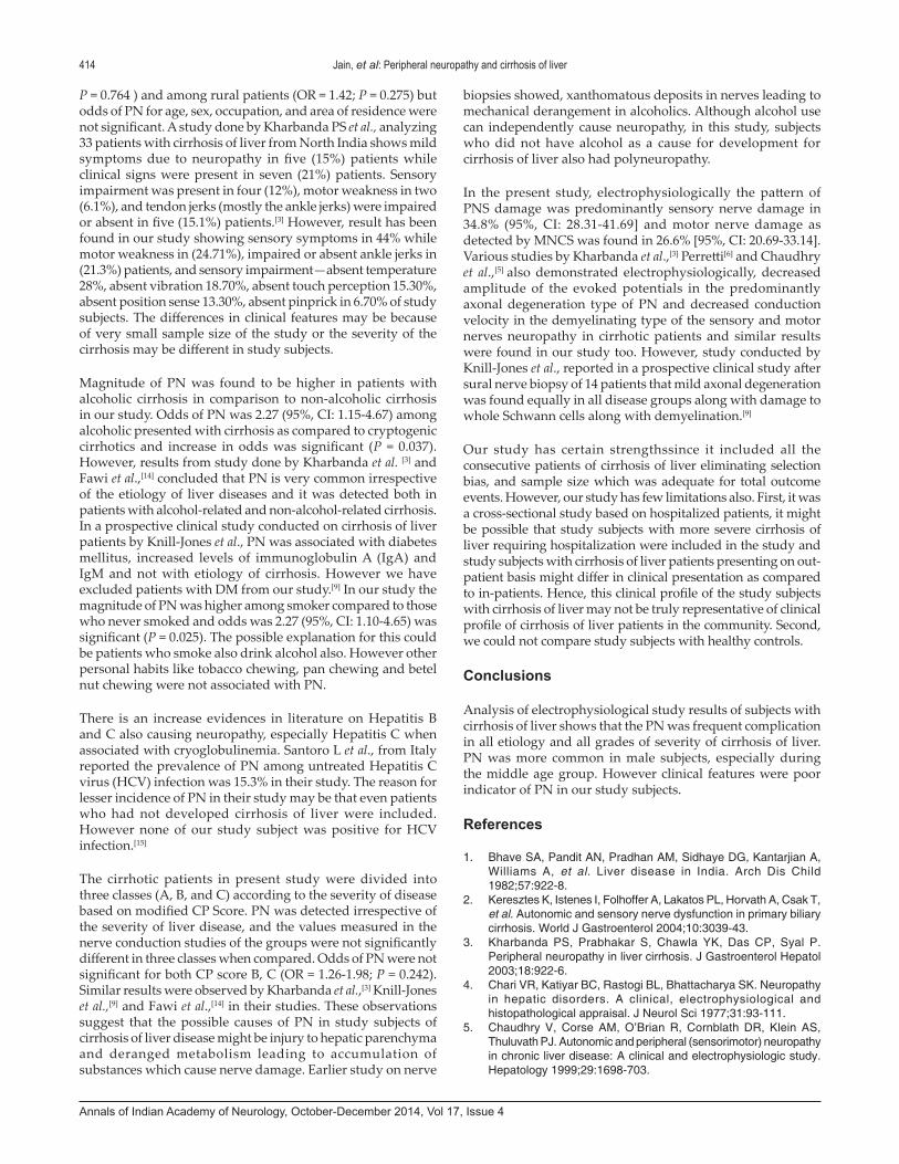
Odds of PN were not statistically significant for age, sex, and hematological profile on univariate analysis. Magnitude of PN was highest (59.3%) among cirrhosis due to alcohol in comparison to more than 1 etiology (55.6%), cryptogenic (39.1%), and hepatitis B (33.3%) respectively. Odds of PN were 2.27 (95%, CI: 1.15-4.67) times high among cirrhosis due to alcohol compared to cryptogenic cirrhosis and increase in odds of PN was statistically significant (P = 0.037). Association of PN with severity of liver cirrhosis when judged by CP score showed highest magnitude of PN among subjects with Child class C (62.1%), followed by Child class B (52.9%), and least in Child class A (45.2%). Odds of PN were 1.98 times in subjects with Child class C compared to those who had Child class A and increase in odds was not statistically significant. (P = 0.242) [Table 5,6].
Discussion
To evaluate the presence or absence of PN we conducted electrophysiological studies in subjects with cirrhosis. In this cross-sectional study we found that 111 (53.6%) of patients with cirrhosis has PN in tertiary care hospital of central rural India. In previous studies, the reported incidence of PN in chronic liver disease varies from 19-80%[2-9] [Table 7]. Significant differences existed in the present study subjects and the other studies carried out on patients with cirrhosis of liver. This variation in magnitude is mainly because of the methods used to detect PN, the different criteria used to define neuropathy and different study settings. These differences are likely because the profile of the population in the present study is predominantly rural and hence carries different risk factors or the difference in severity of liver disease of study subjects included in studies.
In the present study, the magnitude of PN was higher in patients with more than 60-years-of-age groups when compared to age group of 15-30 years (OR = 1.82; P = 0.537), more among males (56.1%) in comparison to females (41.2%), (OR = 1.82; P = 0.112), more among daily laborers (OR = 1.87;
Table 5: Significant correlates of Peripheral Neuropathy
Significant correlates Peripheral Neuropathy OR (95% CI)
P value
AgeMore than 60 years 1.82 (0.66-4.96) 0.537
SexMales 1.82 (0.86-3.84) 0.112
OccupationDaily laborers 1.87 (0.30-11.6) 0.764
Area of residenceRural 1.42 (0.75-2.64) 0.275
Probable reason for cirrhosisAlcohol 2.27 (1.15-4.67) 0.037
Hematological parametersHemoglobin (< 7gm/dL) 1.72 (0.67-4.44) 0.105Platelet count (< 1.65 109/L) 1.24 (0.70-2.20) 0.448Mean Corpuscular Volume (> 93.3 fL) 1.32 (0.71-2.44) 0.371
Severity of cirrhosis (Child-Pugh score)Class C 1.98 (0.82-4.81) 0.242
Table 4: Severity of Peripheral Neuropathy in study subjects
Degree of Severity of Peripheral Neuropathy
Number of study subjects (%)
95% CI
Normal 96 (46.4) 39.44-53.42Early damage 69 (33.3) 26.95-40.20Definite damage 42 (20.3) 15.03-26.42
Table 6: Significant correlates of peripheral Neuropathy
Correlates of Peripheral Neuropathy OR 95% CI P valueAge (>60 years) 1.82 0.66-4.96 0.537Sex (Males) 1.82 0.86-3.84 0.112Occupation (Farmer) 1.87 0.30-11.6 0.764Probable reason for cirrhosis (Alcohol) 2.27 1.15-4.67 0.037Smoking 2.27 1.10-4.65 0.025Biochemical parameters
Hypoalbuminemia 1.68 0.73-3.87 0.219Hyper-billirubinemia 1.28 0.73-2.26 0.390
Severity of cirrhosis (Child-Pugh Class C) 1.98 0.82-4.81 0.242MELD score (30-39) 1.83 0.27-12.19 0.606
Table 7: Magnitude of peripheral neuropathy in patients with liver cirrhosis from different studies
First Author (Reference) Year Country Study Type Sample size Prevalence (%)Present study 2013 India Cross-sectional study 207 with cirrhosis of liver 53.6Katalin Keresztes[2] 2004 Hungary Case-control 24 cases
20 controls54
Kharbanda PS[3] 2003 India Cross-sectional study 33 with cirrhosis of liver 73Chaudhry V[5] 1999 Baltimore Cross-sectional study 58 with cirrhosis of liver 71Perretti A[6] 1995 Italy Case control study 19 with cirrhosis of liver 84.2Hendrickse MT[7] 1992 United
KingdomCase control study 104 biopsy proven CLD patients 71.8 in Child B & C
39.7 in Child AFierro B[8] 1986 Italy Cross-sectional study 19 cases with CLD 57.8Chari VR[4] 1977 India Case control study 30 cirrhosis of liver,12 infective
hepatitis and 13 healthy individuals63.3 in cirrhosis16.6 in infective hepatitis
Knill-Jones RP[9] 1972 London Prospective observational study 70 unselected patients with CLD 18.6
414 Jain, et al: Peripheral neuropathy and cirrhosis of liver
Annals of Indian Academy of Neurology, October-December 2014, Vol 17, Issue 4
P = 0.764 ) and among rural patients (OR = 1.42; P = 0.275) but odds of PN for age, sex, occupation, and area of residence were not significant. A study done by Kharbanda PS et al., analyzing 33 patients with cirrhosis of liver from North India shows mild symptoms due to neuropathy in five (15%) patients while clinical signs were present in seven (21%) patients. Sensory impairment was present in four (12%), motor weakness in two (6.1%), and tendon jerks (mostly the ankle jerks) were impaired or absent in five (15.1%) patients.[3] However, result has been found in our study showing sensory symptoms in 44% while motor weakness in (24.71%), impaired or absent ankle jerks in (21.3%) patients, and sensory impairment—absent temperature 28%, absent vibration 18.70%, absent touch perception 15.30%, absent position sense 13.30%, absent pinprick in 6.70% of study subjects. The differences in clinical features may be because of very small sample size of the study or the severity of the cirrhosis may be different in study subjects.
Magnitude of PN was found to be higher in patients with alcoholic cirrhosis in comparison to non-alcoholic cirrhosis in our study. Odds of PN was 2.27 (95%, CI: 1.15-4.67) among alcoholic presented with cirrhosis as compared to cryptogenic cirrhotics and increase in odds was significant (P = 0.037). However, results from study done by Kharbanda et al. [3] and Fawi et al.,[14] concluded that PN is very common irrespective of the etiology of liver diseases and it was detected both in patients with alcohol-related and non-alcohol-related cirrhosis. In a prospective clinical study conducted on cirrhosis of liver patients by Knill-Jones et al., PN was associated with diabetes mellitus, increased levels of immunoglobulin A (IgA) and IgM and not with etiology of cirrhosis. However we have excluded patients with DM from our study.[9] In our study the magnitude of PN was higher among smoker compared to those who never smoked and odds was 2.27 (95%, CI: 1.10-4.65) was significant (P = 0.025). The possible explanation for this could be patients who smoke also drink alcohol also. However other personal habits like tobacco chewing, pan chewing and betel nut chewing were not associated with PN.
There is an increase evidences in literature on Hepatitis B and C also causing neuropathy, especially Hepatitis C when associated with cryoglobulinemia. Santoro L et al., from Italy reported the prevalence of PN among untreated Hepatitis C virus (HCV) infection was 15.3% in their study. The reason for lesser incidence of PN in their study may be that even patients who had not developed cirrhosis of liver were included. However none of our study subject was positive for HCV infection.[15]
The cirrhotic patients in present study were divided into three classes (A, B, and C) according to the severity of disease based on modified CP Score. PN was detected irrespective of the severity of liver disease, and the values measured in the nerve conduction studies of the groups were not significantly different in three classes when compared. Odds of PN were not significant for both CP score B, C (OR = 1.26-1.98; P = 0.242). Similar results were observed by Kharbanda et al.,[3] Knill-Jones et al.,[9] and Fawi et al.,[14] in their studies. These observations suggest that the possible causes of PN in study subjects of cirrhosis of liver disease might be injury to hepatic parenchyma and deranged metabolism leading to accumulation of substances which cause nerve damage. Earlier study on nerve
biopsies showed, xanthomatous deposits in nerves leading to mechanical derangement in alcoholics. Although alcohol use can independently cause neuropathy, in this study, subjects who did not have alcohol as a cause for development for cirrhosis of liver also had polyneuropathy.
In the present study, electrophysiologically the pattern of PNS damage was predominantly sensory nerve damage in 34.8% (95%, CI: 28.31-41.69] and motor nerve damage as detected by MNCS was found in 26.6% [95%, CI: 20.69-33.14]. Various studies by Kharbanda et al.,[3] Perretti[6] and Chaudhry et al.,[5] also demonstrated electrophysiologically, decreased amplitude of the evoked potentials in the predominantly axonal degeneration type of PN and decreased conduction velocity in the demyelinating type of the sensory and motor nerves neuropathy in cirrhotic patients and similar results were found in our study too. However, study conducted by Knill-Jones et al., reported in a prospective clinical study after sural nerve biopsy of 14 patients that mild axonal degeneration was found equally in all disease groups along with damage to whole Schwann cells along with demyelination.[9]
Our study has certain strengthssince it included all the consecutive patients of cirrhosis of liver eliminating selection bias, and sample size which was adequate for total outcome events. However, our study has few limitations also. First, it was a cross-sectional study based on hospitalized patients, it might be possible that study subjects with more severe cirrhosis of liver requiring hospitalization were included in the study and study subjects with cirrhosis of liver patients presenting on out-patient basis might differ in clinical presentation as compared to in-patients. Hence, this clinical profile of the study subjects with cirrhosis of liver may not be truly representative of clinical profile of cirrhosis of liver patients in the community. Second, we could not compare study subjects with healthy controls.
Conclusions
Analysis of electrophysiological study results of subjects with cirrhosis of liver shows that the PN was frequent complication in all etiology and all grades of severity of cirrhosis of liver. PN was more common in male subjects, especially during the middle age group. However clinical features were poor indicator of PN in our study subjects.
References
1. Bhave SA, Pandit AN, Pradhan AM, Sidhaye DG, Kantarjian A, Williams A, et al. Liver disease in India. Arch Dis Child 1982;57:922-8.
2. Keresztes K, Istenes I, Folhoffer A, Lakatos PL, Horvath A, Csak T, et al. Autonomic and sensory nerve dysfunction in primary biliary cirrhosis. World J Gastroenterol 2004;10:3039-43.
3. Kharbanda PS, Prabhakar S, Chawla YK, Das CP, Syal P. Peripheral neuropathy in liver cirrhosis. J Gastroenterol Hepatol 2003;18:922-6.
4. Chari VR, Katiyar BC, Rastogi BL, Bhattacharya SK. Neuropathy in hepatic disorders. A clinical, electrophysiological and histopathological appraisal. J Neurol Sci 1977;31:93-111.
5. Chaudhry V, Corse AM, O’Brian R, Cornblath DR, Klein AS, Thuluvath PJ. Autonomic and peripheral (sensorimotor) neuropathy in chronic liver disease: A clinical and electrophysiologic study. Hepatology 1999;29:1698-703.
Jain, et al: Peripheral neuropathy and cirrhosis of liver 415
Annals of Indian Academy of Neurology, October-December 2014, Vol 17, Issue 4
How to cite this article: Jain J, Singh R, Banait S, Verma N, Waghmare S. Magnitude of peripheral neuropathy in cirrhosis of
liver patients from central rural India. Ann Indian Acad Neurol 2014;17:409-15.
Received: 15-01-14, Revised: 05-03-14, Accepted: 08-05-14
Source of Support: The study was supported by a grant from the International Epilepsy Trust, Conflict of Interest: None declared.
6. Perretti A, Gentile S, Balbi P, Persico M, Caruso G. Peripheral neuropathy in liver cirrhosis. A clinical and electrophysiological study. Ital J Gastroenterol 1995;27:349-54.
7. Hendrickse MT, Thuluvath PJ, Triger DR. Natural history of autonomic neuropathy in chronic liver disease. Lancet 1992;339:1462-4.
8. Fierro B, Raimondo D, Castiglione MG, Migneco G, Scoppa F, Savettieri G. Peripheral nerve involvement in chronic liver disease. Clinical and electrophysiological study. Ital J Neurol Sci 1986;7:589-90.
9. Knill-Jones RP, Goodwill CJ, Dayan AD, Williams R. Peripheral neuropathy in chronic liver disease: Clinical, electrodiagnostic, and nerve biopsy findings. J Neurol Neurosurg Psychiatry 1972;35:22-30.
10. Attia KA, Ackoundou-N’guessan KC, N’Dri-Yoman AT, Mahassadi AK, Messou E, Bathaix YF, et al. Child-Pugh-Turcott versus Meld score for predicting survival in a retrospective cohort of black African cirrhotic patients. World J Gastroenterol 2008;14:286-91.
11. Jayaprakash P, Bhansali A, Bhansali S, Dutta P, Anantharaman R, Shanmugasundar G, et al. Validation of bedside methods in evaluation of diabetic peripheral neuropathy. Indian J Med Res 2011;133:645-9.
12. Mishra UK, Kalita J. Clinical neurophysiology: Nerve conduction study. 2nd ed. New Delhi: Elseveirs; 2006. p. 21-128.
13. Camdessanche JP, Jousserand G, Ferraud K, Vial C, Petiot P, Honnorat J, et al. The pattern and diagnostic criteria of sensory neuronopathy: A case-control study. Brain 2009;132:1723-33.
14. Fawi GH, Khalifa GA, LA D. Autonomic and peripheral neuropathies in chronic l iver diseases: Cl inical and neurophysiological study. Egypt J Neurol Psychiat Neurosurg 2005;42:87-200.
15. Santoro L, Manganelli F, Briani C, Giannini F, Benedetti L, Vitelli E, et al. Prevalence and characteristics of peripheral neuropathy in hepatitis C virus population. J Neurol Neurosurg Psychiatry 2006;77:626-9.