Vitamin E inhibits CD36 scavenger receptor expression in hypercholesterolemic rabbits
6
Atherosclerosis 184 (2006) 15–20 Vitamin E inhibits CD36 scavenger receptor expression in hypercholesterolemic rabbits Nesrin Kartal ¨ Ozer a , Yesim Negis a , Nurg¨ ul Aytan a , Luis Villacorta b , Roberta Ricciarelli c , Jean-Marc Zingg b , Angelo Azzi b,∗ a Department of Biochemistry, Faculty of Medicine, Marmara University, 34668 Haydarpasa, Istanbul, Turkey b Institute of Biochemistry and Molecular Biology, University of Bern, 3012 Bern, Switzerland c Department of Experimental Medicine, University of Genoa, 16132 Genoa, Italy Received 22 August 2004; received in revised form 28 February 2005; accepted 29 March 2005 Available online 23 June 2005 Abstract A numerous studies suggest that Vitamin E has a preventive role in atherosclerosis, although the mechanism of action still remains unclear. CD36, a member of the scavenger receptor family is centrally involved in the uptake of oxidized low density proteins (oxLDLs) from bloodstream. During the atherosclerotic process, the lipid cargo of oxLDL accumulates in macrophages and smooth muscle cells, inducing their pathological conversion to foam cells. In the present study, we investigate the role of Vitamin E on CD36 expression in an in vivo model. Atherosclerosis was induced by a 2% cholesterol containing Vitamin E poor diet. Three groups of six rabbits each were studied. The first group (control) was fed on Vitamin E poor diet. The second group was fed with Vitamin E poor diet containing 2% cholesterol and the rabbits in the third group were fed with Vitamin E poor diet containing 2% cholesterol and received injections of 50 mg/kg of Vitamin E i.m. After 4 weeks, aortas were removed and analysed by light microscopy for atherosclerotic lesions. Aortic samples were analysed for CD36 mRNA expression. The aortas of cholesterol-fed rabbits showed typical atherosclerotic lesions, detected by macroscopic and microscopic examination, and exhibited an increase in CD36 mRNA expression. Vitamin E fully prevented cholesterol induced atherosclerotic lesions and the induction of CD36 mRNA expression. The effects observed at the level of CD36 scavenger receptor expression in vivo suggest an involvement of reduced foam cell formation in the protective effect of Vitamin E against atherosclerosis. © 2005 Elsevier Ireland Ltd. All rights reserved. Keywords: Atherosclerosis; CD36; Scavenger receptor; Vitamin E; Alpha-tocopherol; Rabbit aortic smooth muscle cells 1. Introduction Atherosclerosis is a multi factorial process in which both elevated plasma cholesterol levels and proliferation of smooth muscle cells play a central role. Monocytes, macrophages and T cells attracted to the site of injury produce inflam- matory cytokines, chemokines and reactive oxygen species [1–4]. Many epidemiological, clinical, genetic and animal studies have indicated that hypercholesterolemia, with ele- vated levels of cholesterol-rich low density lipoprotein, is one of the most important risk factors for the disease; choles- ∗ Corresponding author. Tel.: +41 31 6314131; fax: +41 31 6313737. E-mail address: [email protected] (A. Azzi). terol, accumulating in the atherosclerotic lesions, originates in fact primarily from plasma lipoproteins, including low density lipoproteins [5,6]. A family of genes, the scavenger receptors recognizes and internalizes modified lipoproteins, making them susceptible to degradation [7]. Foam cell formation is characteristic feature of atheroscle- rotic lesions, which are formed mainly by uncontrolled uptake of oxidized LDL containing cholesterol and lipids by smooth muscle cells and macrophages. Among the fac- tors, which have been found to retard the development of atherosclerosis, is the intake with food of a sufficient amount of Vitamin E. An inverse association between serum Vitamin E and coronary heart disease mortality has been demonstrated in epidemiological studies [8–11]. Some of the effects of 0021-9150/$ – see front matter © 2005 Elsevier Ireland Ltd. All rights reserved. doi:10.1016/j.atherosclerosis.2005.03.050
Transcript of Vitamin E inhibits CD36 scavenger receptor expression in hypercholesterolemic rabbits

Atherosclerosis 184 (2006) 15–20
Vitamin E inhibits CD36 scavenger receptor expression inhypercholesterolemic rabbits
Nesrin KartalOzera, Yesim Negisa, Nurgul Aytana, Luis Villacortab,Roberta Ricciarellic, Jean-Marc Zinggb, Angelo Azzib,∗
a Department of Biochemistry, Faculty of Medicine, Marmara University, 34668 Haydarpasa, Istanbul, Turkeyb Institute of Biochemistry and Molecular Biology, University of Bern, 3012 Bern, Switzerland
c Department of Experimental Medicine, University of Genoa, 16132 Genoa, Italy
Received 22 August 2004; received in revised form 28 February 2005; accepted 29 March 2005Available online 23 June 2005
Abstract
A numerous studies suggest that Vitamin E has a preventive role in atherosclerosis, although the mechanism of action still remains unclear.C s) fromb ls, inducingt n in vivom udied. Thefi l and ther in E i.m.A d for CD36m icroscopice tic lesionsa suggest ani©
K
1
emam[svo
ateswereins,
cle-ledidsfac-
nt ofountmintratedf
0d
D36, a member of the scavenger receptor family is centrally involved in the uptake of oxidized low density proteins (oxLDLloodstream. During the atherosclerotic process, the lipid cargo of oxLDL accumulates in macrophages and smooth muscle cel
heir pathological conversion to foam cells. In the present study, we investigate the role of Vitamin E on CD36 expression in aodel. Atherosclerosis was induced by a 2% cholesterol containing Vitamin E poor diet. Three groups of six rabbits each were st
rst group (control) was fed on Vitamin E poor diet. The second group was fed with Vitamin E poor diet containing 2% cholesteroabbits in the third group were fed with Vitamin E poor diet containing 2% cholesterol and received injections of 50 mg/kg of Vitamfter 4 weeks, aortas were removed and analysed by light microscopy for atherosclerotic lesions. Aortic samples were analyseRNA expression. The aortas of cholesterol-fed rabbits showed typical atherosclerotic lesions, detected by macroscopic and m
xamination, and exhibited an increase in CD36 mRNA expression. Vitamin E fully prevented cholesterol induced atherosclerond the induction of CD36 mRNA expression. The effects observed at the level of CD36 scavenger receptor expression in vivo
nvolvement of reduced foam cell formation in the protective effect of Vitamin E against atherosclerosis.2005 Elsevier Ireland Ltd. All rights reserved.
eywords: Atherosclerosis; CD36; Scavenger receptor; Vitamin E; Alpha-tocopherol; Rabbit aortic smooth muscle cells
. Introduction
Atherosclerosis is a multi factorial process in which bothlevated plasma cholesterol levels and proliferation of smoothuscle cells play a central role. Monocytes, macrophagesnd T cells attracted to the site of injury produce inflam-atory cytokines, chemokines and reactive oxygen species
1–4]. Many epidemiological, clinical, genetic and animaltudies have indicated that hypercholesterolemia, with ele-ated levels of cholesterol-rich low density lipoprotein, isne of the most important risk factors for the disease; choles-
∗ Corresponding author. Tel.: +41 31 6314131; fax: +41 31 6313737.E-mail address: [email protected] (A. Azzi).
terol, accumulating in the atherosclerotic lesions, originin fact primarily from plasma lipoproteins, including lodensity lipoproteins[5,6]. A family of genes, the scavengreceptors recognizes and internalizes modified lipoprotmaking them susceptible to degradation[7].
Foam cell formation is characteristic feature of atherosrotic lesions, which are formed mainly by uncontroluptake of oxidized LDL containing cholesterol and lipby smooth muscle cells and macrophages. Among thetors, which have been found to retard the developmeatherosclerosis, is the intake with food of a sufficient amof Vitamin E. An inverse association between serum VitaE and coronary heart disease mortality has been demonsin epidemiological studies[8–11]. Some of the effects o
021-9150/$ – see front matter © 2005 Elsevier Ireland Ltd. All rights reserved.oi:10.1016/j.atherosclerosis.2005.03.050

16 N.K. Ozer et al. / Atherosclerosis 184 (2006) 15–20
alpha-tocopherol, which is the most active form of VitaminE, can be attributed to special properties of this compoundsuch as the inhibition of smooth muscle cells proliferation,an important event during the progression of atherosclero-sis, via inhibition of protein kinase C activity[12–15]. In ourprevious studies, we have also shown that Vitamin E protectsdevelopment of atherosclerosis in cholesterol-fed rabbits byinhibiting vascular smooth muscle protein kinase C activity[16,17]. Similarly, it has been shown that alpha-tocopherolinhibits oxLDL uptake by a mechanism involving down-regulation of CD36 mRNA and protein expression in vitro[18]. Also, in rat liver, it has been shown that Vitamin E reg-ulates the scavenger receptor CD36, the coagulation factorIX, the hepatic gamma glutamyl–cysteinyl synthetase, andthe 5-alpha-steroid reductase type 1 gene at the transcrip-tional level[19].
CD36 is a multifunctional membrane receptor and a cell-adhesion molecule expressed by platelets, monocytes/macrophages, and capillary endothelial cells, adipocytes,human cardiac and skeletal muscles and at very low levelsin the liver [20–23]. It has been implicated in hemostasis,malaria, inflammation, lipid metabolism and atherogenesis[24]. It has also been shown that mice knockouts for theapolipoprotein E exhibited a marked decrease in atheroscle-rotic lesions if also CD36 gene was made inactive[25].
The following study was carried out to investigate the roleo Wes ssedi reg-u ts ont ntiono
2
maI restc
2
erea with1 ed tot atedw riedo terola
fedw nts.T ain-i eref ndr rlyo
After 4 weeks, following withdrawal of food overnight,rabbits were anaesthetised, by using 50 mg/kg ketaminehydrochloride. Blood was taken for Vitamin E and choles-terol determinations. The aortas of two animals in each groupwere removed, fixed in 10% formalin, stained with SudanIV (2% in ethanol) and photographed. The digitalized pic-tures were quantified by using Photoshop. The thoracic aortasof the remaining animals were removed, immediately takeninto RNA stabilization reagent (Qiagen) and were frozen at−20◦C until they were homogenized. Samples from thoracicaorta were taken for light microscopic evaluation. Aortic sam-ples were embedded in paraffin.
2.2. Microscopic examination
The samples, fixed in 10% buffered formaldehyde for 4 hthen dehydrated and incubated in xylol for 1 h twice, embed-ded in paraffin and sectioned it 5�m. Sections were stainedwith hemotoxylene eosin before microscopic examination.
2.3. Chemical methods
Serum cholesterol levels were determined using an auto-mated enzymatic technique. Vitamin E levels were measuredby reverse phase high pressure liquid chromatography[27].
2
edia)f pres-s rtaw ion-p totalR mem NAe tion-pK( RF:5 :5 t9 sw H;Ga -3 .T bys d ona ager(
3
f thet -
f Vitamin E on CD36 expression in an in vivo model.how here that CD36 scavenger receptor is highly expre
n hypercholesterolemic rabbits and that Vitamin E downlates its expression. The significance of this finding res
he fact that Vitamin E appears to have a role in the prevef atherosclerosis.
. Material and methods
Vitamin E (Evigen, i.m.) was obtained from Aksu Pharndustry, Turkey. All other chemicals used were of the puommercially available grade.
.1. Experimental procedures
Twenty-four male albino rabbits (2–3 months old) wssigned randomly to three groups. All rabbits were fed00 g per day of Vitamin E poor diet. Cholesterol was add
he diet as diethyl ether solution. The control diet was treith the same amount of pure solvent. All diets were df the solvent before use. The concentrations of cholesnd Vitamin E were based on previous reports[16,26].
The first group of rabbits, the control rabbits, were onlyith Vitamin E poor diet, without additions and treatmehe second group was fed with Vitamin E poor diet cont
ng 2% cholesterol and the rabbits in the third group wed with Vitamin E poor diet containing 2% cholesterol aeceived injections of 50 mg/kg of Vitamin E intramusculan alternate rear legs, once daily.
.4. CD36 mRNA expression
Homogenates were prepared from the aorta strips (mor measurement of CD36 scavenger receptor mRNA exion. Total RNA was isolated from rabbit thoracic aoith an RNA extraction kit (Qiagen). Reverse transcriptolymerase chain reaction (RT-PCR) was applied toNA isolated from rabbit smooth muscle cells with soodifications. Semi-quantitative assays for CD36 mR
xpression were performed with a reverse transcripolymerase chain reaction kit (GeneAmp® RNA PCRit, Perkin-Elmer) with HotStarTaqTM DNA Polymerase
Qiagen) and primers specific for rabbit CD36; CD36PC′-TTGGTGTGTTTTATCCTTAC-3′ and CD36PCRR′-GGTTCCAGTCTCATTAAGCC-3′ for 40 cycles a5◦C, 30 s; 56.1◦C, 30 s; 72◦C, 30 s. Control reactionere performed with primers specific for rabbit GAPDAPDHR: 5′-TGCCGAAGTGGTCGTGGATGACCT-3′nd GAPDHF: 5′-GCGCCTGGTCACCAGGGCTGCTTT′ for 30 cycles at 95◦C, 30 s; 64◦C, 30 s; 72◦C, 30 she identity of the PCR fragments was confirmedequencing (Microsynth). The PCR products were loade2.5% agarose gel and were quantified with a Lumi-Im
Roche).
. Results and discussion
Cholesterol and Vitamin E plasma concentrations ohree animal groups are shown inTable 1. Two percent choles

N.K. Ozer et al. / Atherosclerosis 184 (2006) 15–20 17
Table 1Effect of cholesterol and Vitamin E treatment on their plasma levels in rabbits
Group Cholesterol (mg/dl) Vitamin E (�g/mL)
Control 51.2± 15.9 1.4± 0.5Cholesterol 1110.8± 543.5* 11.9± 5.9*
Cholesterol + Vitamin E 839.3± 380.6* 31.0± 11.2*
The plasma level of cholesterol and Vitamin E have been measured in allthe six rabbits of the three diet groups. The numbers (mean± S.D.,n = 6)represent the plasma values measured after 1 month diet. Thep-value wascalculated using the Student’st-test.
* p < 0.01.
terol diet supplementation for 4 weeks resulted in an approx-imately 20-fold increase of plasma cholesterol. Additionalsupplementation with Vitamin E induced a plasma choles-terol increase of only 16-fold relative to the control. PlasmaVitamin E concentrations were higher in the cholesterol-fedrabbits what is in agreement with literature data[16,28–32].This event is caused by a higher Vitamin E uptake (the dietcontains low, but not zero Vitamin E) resulting from increasedlipid uptake produced by the high cholesterol diet. A corol-lary to this finding is that if Vitamin E were really absentfrom the diet the effect of cholesterol would be more disrup-tive and the effect of tocopherol supplementation would bealso exaggerated.
The aortas of two rabbits from each group were fixed,stained with Sudan IV and photographed. A set of these aortasis shown inFig. 1. The digitalized pictures were analysed forthe Sudan IV staining regions. A difference in atheroscleroticplaque in the control animals, cholesterol-fed animals andcholesterol plus tocopherol-treated animals was evident atthe simple inspection. A quantitative evaluation has shown
that the amount of plaque in the aorta of control animals was4%, in cholesterol-fed animals it increased to 59%, wherebythe addition of a Vitamin E to the diet of the cholesterol-fedanimals reduced the plaque to 18%. The effect of Vitamin Eis thus a reduction of the plaque of 69.4%.
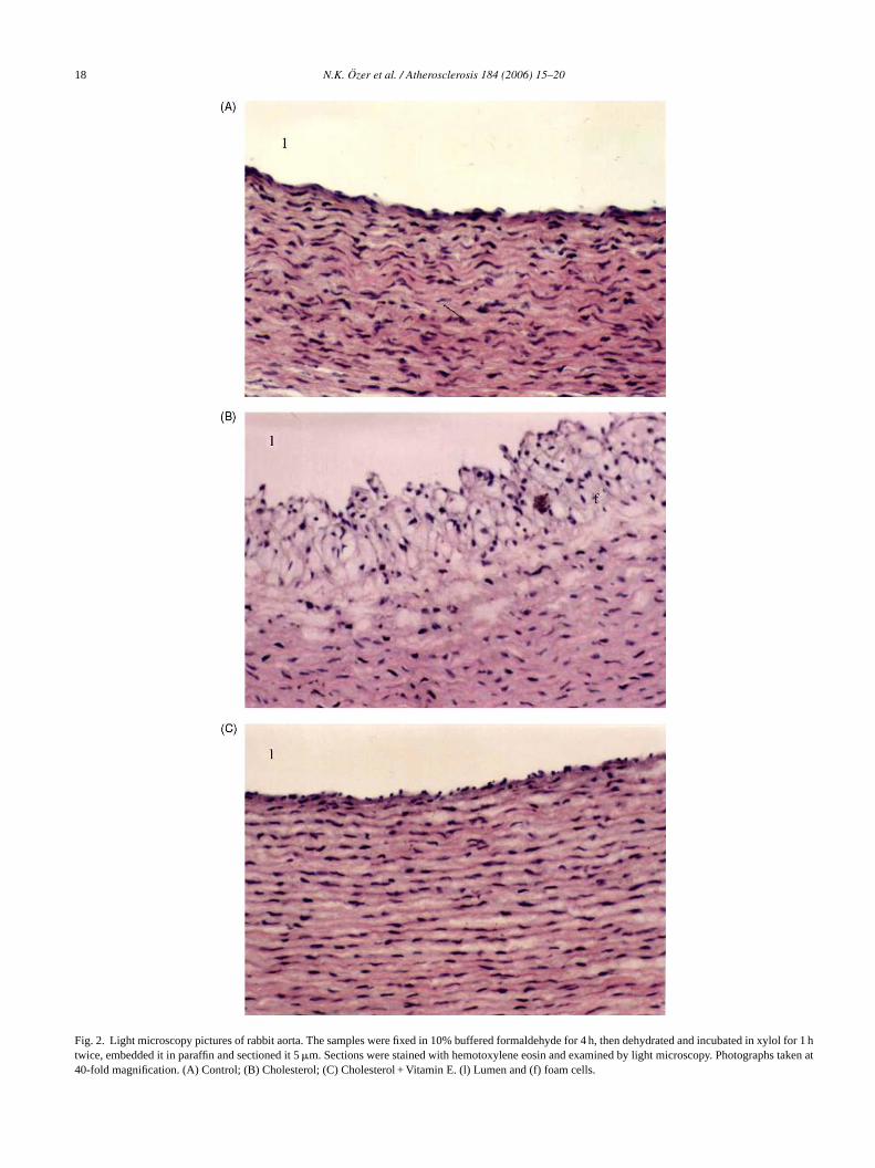
All remaining rabbit aortas from the three groups werestudied by light microscopy. The results are shown inFig. 2.From each situation 10 pictures were taken, the pictures arerepresentative of the overall situation in each animal group.After 4 weeks, the cholesterol-fed rabbits developed lesionswithin the thoracic aortas and endothelial cell integrity wasimpaired relative to the control.Fig. 2B shows that in themedia and intima of cholesterol-fed animals, vacuole richcells and typical foam cells were visible, respectively. How-ever, in cholesterol plus Vitamin E-treated animals (Fig. 2C)no lipid accumulation and foam cell formation was visible(compareFig. 2B and C).
In order to understand if CD36, a scavenger receptorresponsible for the uptake of oxidized LDL and foam cell for-mation, is regulated by hypercholesterolemia and if VitaminE can inhibit its expression in vivo, CD36 mRNA expres-sion was measured in aortic samples (Fig. 3). We found anincreased CD36 mRNA expression in the cholesterol-treatedgroup (174%) referred to a control value set equal to 100%.With cholesterol supplementation CD36 mRNA expressionincreased 74% more. Similar findings were reported by Hane ion ise terole ,t ibi-t ol).V the
fixed in
Fig. 1. Pictures of rabbit aortast al., showing that CD36 scavenger receptor expressnhanced by cholesterol and down regulated by cholesfflux in macrophages in an in vitro system[33]. However
he cholesterol plus Vitamin E-treated group showed inhion of CD36 mRNA expression (75% relative to contritamin E treatment was able, in this case, to reduce
formalin and stained with Sudan IV.
18 N.K. Ozer et al. / Atherosclerosis 184 (2006) 15–20
Fig. 2. Light microscopy pictures of rabbit aorta. The samples were fixed in 10% buffered formaldehyde for 4 h, then dehydrated and incubated in xylol for 1 htwice, embedded it in paraffin and sectioned it 5�m. Sections were stained with hemotoxylene eosin and examined by light microscopy. Photographs taken at40-fold magnification. (A) Control; (B) Cholesterol; (C) Cholesterol + Vitamin E. (l) Lumen and (f) foam cells.

N.K. Ozer et al. / Atherosclerosis 184 (2006) 15–20 19
Fig. 3. (a and b) Expression of CD36 mRNA in rabbit aorta. (A) Control;(B) Cholesterol; (C) Cholesterol + Vitamin E. RT-PCR was performed asdescribed in Section2. Graphical displays of CD36 signals are normalizedto GAPDH. The data were expressed as mean± S.D. of six rabbits in eachgroup. The representative picture is shown for PCR. Ordinate: % expression.
CD36 mRNA expression, compared to both the control andthe cholesterol group, by 25% and 57%, respectively. CD36scavenger receptor has been shown to be expressed in cultured human aortic smooth muscle cells and macrophages;treatment with alpha-tocopherol (at physiological concen-tration) down regulates CD36 expression by reducing itspromoter activity [34]. A similar phenomenon has beenalso shown in human monocyte-derived macrophages wherealpha-tocopherol decreases CD36 expression by inhibition oftyrosine kinase[35,36]. Also liver CD36 appears to be regu-lated by Vitamin E; Barella et al. have shown that Vitamin Edepletion in rats is associated with over expression of CD36[19]. Our observation is however the first, linking CD36expression in vascular tissues with hypercholesterolemia andVitamin E concentration in plasma. Increased cholesterol inplasma acts as a stimulant to activate CD36 expression, whileVitamin E produces its down regulation. The consequenceof these events is that increased expression of CD36 mayfavor the uptake of oxidized lipoproteins and their depositionto form foam cells. On the contrary, the Vitamin E induceddiminution of CD36 expression will lead to a lesser fat accu-mulation by macrophage and smooth muscle cells. Due tothe sensitivity of the scavenger receptors class A (SR-A) toVitamin E (similar to CD36) the reduction in foam cell for-mation observed in the Vitamin E-treated group may not besolely attributed to the suppression of CD36 expression buta
Common models of atherosclerosis[37] indicate in theformation of foam cells an early event in the onset ofatherosclerosis. Thus, the inhibition of foam cell formation byVitamin E may be important in the prevention of the disease.A central role of CD36 in atherogenesis in a mouse modelhas been described by Febbraio et al.[38]. This group hasshown that targeted disruption of the class B scavenger recep-tor CD36 protects against atherosclerotic lesion developmentin mice. In conclusion, this study indicates that non-geneticmanipulation of the CD36 expression product by Vitamin Eadministration to rabbits on a high cholesterol diet, results ina diminution of CD36 expression and of foam cell formation.The value and novelty of this study is to indicate, for the firsttime in an in vivo model, that the molecular mechanism ofatherosclerosis prevention by Vitamin E is consistent with adiminution of the class B scavenger receptor CD36 expres-sion caused by this vitamin in vascular tissues. The questionremains open as to why this rabbit model of atherosclerosisshows a very clear, positive effect of Vitamin E, while clinicalintervention studies have often given disappointing results.Needless to say, animal model studies cannot be transferred tohuman situations. However, the following considerations canalso be made. As indicated by the propensity to atheroscle-rosis created in the mouse by the knock out of the ApoEgene (and the protection by additional CD36 knockout)[38]also the rabbit may have a genetic background that make ith iono . Then oreh ct ofV nso und,e mayl mongh
A
sityR unda-t
R
L. N
ed
ci
Med
contribution SR-A may also be present.-
ighly sensitive to the effect of Vitamin E on the expressf CD36, and possibly of the SR-A, scavenger receptorsotion that small intervention studies, conducted with a momogeneous population, have shown a significant effeitamin E (for a review see[39]) suggests that investigation individuals having a homogeneous genetic backgrospecially referred to ApoE and other polymorphisms,
ead to a better understanding of the discrepancies auman studies and with the animal ones.
cknowledgements
This study was supported by the Marmara Univeresearch Foundation and Swiss National Science Fo
ion.
eferences
[1] Steinberg D, Parthasarathy S, Carew TE, Khoo JC, Witztum JEngl J Med 1989;320:915–24.
[2] Steinberg D. Adv Exp Med Biol 1995;369:39–48.[3] Witztum JL, Steinberg D. J Clin Invest 1991;88:1785–92.[4] Ross R. Annu Rev Physiol 1995;57:791–804.[5] Parthasarathy S, Steinberg D, Witztum JL. Annu Rev M
1992;43:219–25.[6] Meydani M. J Nutr 2001;131:366S–8S.[7] Yamada Y, Doi T, Hamakubo T, Kodama T. Cell Mol Life S
1998;54:628–40.[8] Stampfer MJ, Hennekens CH, Manson JE, et al. N Engl J
1993;328:1444–9.

20 N.K. Ozer et al. / Atherosclerosis 184 (2006) 15–20
[9] Rimm EB, Stampfer MJ, Ascherio A, et al. N Engl J Med1993;328:1450–6.
[10] Stephens NG, Parsons A, Schofield PM, et al. Lancet1996;347:781–6.
[11] Gey KF, Puska P, Jordan P, Moser UK. Am J Clin Nutr1991;53:326S–34S.
[12] Zingg JM, Azzi A. Curr Med Chem 2004;11:1113–33.[13] Ricciarelli R, Zingg JM, Azzi A. IUBMB Life 2001;52:71–6.[14] Azzi A, Stocker A. Prog Lipid Res 2000;39:231–55.[15] Boscoboinik D, Szewczyk A, Hensey C, Azzi A. J Biol Chem
1991;266:6188–94.[16] Ozer NK, Sirikci O, Taha S, et al. Free Radic Biol Med
1998;24:226–33.[17] Ozer N, Azzi A. Toxicology 2000;148:179–85.[18] Ricciarelli R, Zingg JM, Azzi A. Circulation 2000;102:82–7.[19] Barella L, et al. Biochim Biophys Acta 2004;1689:66–74.[20] Aitman TJ. Lancet 2001;357:651–2.[21] Hirano K, Kuwasako T, Nakagawa-Toyama Y, et al. Trends Cardio-
vasc Med 2003;13:136–41.[22] Nicholson AC, Febbraio M, Han J, Silverstein RL, Hajjar DP. Ann
NY Acad Sci 2000;902:128–31.[23] Simantov R, Silverstein RL. Front Biosci 2003;8:s874–82.[24] de Winther MP, van Dijk KW, Havekes LM, Hofker MH. Arterioscler
Thromb Vasc Biol 2000;20:290–7.
[25] Suzuki H, et al. J Atheroscler Thromb 1997;4:1–11.[26] Sirikci O, Ozer NK, Azzi A. Atherosclerosis 1996;126:253–63.[27] Nierenberg DW, Nann SL. Am J Clin Nutr 1992;56:417–26.[28] Godfried SL, Combs Jr GF, Saroka JM, Dillingham LA. Br J Nutr
1989;61:607–17.[29] Wilson RB, Middleton CC, Sun GY. J Nutr 1978;108:1858–67.[30] Morel DW, De la Llera-Moya M, Friday KE. J Nutr
1994;124:2123–30.[31] Bitman J, Weyant J, Wood DL, Wrenn TR. Lipids 1976;11:449–
61.[32] Bjorkhem I, Henriksson Freyschuss A, Breuer O, et al. Arterioscler
Thromb 1991;11:15–22.[33] Han J, Hajjar DP, Febbraio M, Nicholson AC. J Biol Chem
1997;272:21654–9.[34] Ricciarelli R, Zingg JM, Azzi A. IUBMB Life 2001;52:71–6.[35] Devaraj S, Hugou I, Jialal I. J Lipid Res 2001;42:521–7.[36] Venugopal SK, Devaraj S, Jialal I. Atherosclerosis 2004;175:213–
20.[37] Keaney Jr JF, Simon DI, Freedman JE. FASEB J 1999;13:965–75.[38] Febbraio M, Podrez E, Smith J, et al. J Clin Invest
2000;105:1049–56.[39] Jialal I, Devaraj S. J Nutr 2005;135:348–53.