Obsessive-compulsive disorder phenotypes: implications for genetic studies
Upload
independentCategory
view
0download
0
Positive selection of a CD36 nonsense variantin sub-Saharan Africa, but no associationwith severe malaria phenotypes
Andrew E. Fry1,�, Anita Ghansa1,2, Kerrin S. Small1, Alejandro Palma3, Sarah Auburn1,4,
Mahamadou Diakite1, Angela Green1, Susana Campino1,4, Yik Y. Teo1, Taane G. Clark1,4,
Anna E. Jeffreys1, Jonathan Wilson1, Muminatou Jallow5, Fatou Sisay-Joof5, Margaret Pinder5,
Michael J. Griffiths1,6, Norbert Peshu6, Thomas N. Williams6,7, Charles R. Newton6,8,
Kevin Marsh6,7, Malcolm E. Molyneux9,10, Terrie E. Taylor11,12, Kwadwo A. Koram2,
Abraham R. Oduro13, William O. Rogers14, Kirk A. Rockett1, Pardis C. Sabeti3,15
and Dominic P. Kwiatkowski1,4
1The Wellcome Trust Centre for Human Genetics, Roosevelt Drive, Oxford OX3 7BN, UK, 2Noguchi Memorial Institute for
Medical Research, PO Box LG581, Legon-Accra, Ghana, 3Broad Institute, Cambridge, MA 02142, USA, 4Wellcome Trust
Genome Campus, Hinxton, Cambridge CB10 1SA, UK, 5Medical Research Council, PO Box 273, Banjul, The Gambia,6Kenya Medical Research Institute Centre for Geographical Medicine Research (Coast), PO Box 230, Kilifi, Kenya,7Nuffield Department of Medicine, John Radcliffe Hospital, Oxford OX3 9DS, UK, 8Institute of Child Health, University
College of London, London WC1N 1EH, UK, 9Malawi–Liverpool Wellcome Trust Programme of Clinical Tropical
Research, PO Box 30096, Blantyre, Malawi, 10Liverpool School of Tropical Medicine, Pembroke Place, Liverpool L3 5QA,
UK, 11Blantyre Malaria Project, College of Medicine, PO Box 30096, Blantyre, Malawi, 12Department of Internal Medicine,
College of Osteopathic Medicine, Michigan State University, East Lansing, MI 48824, USA, 13Navrongo Health Research
Centre, PO Box 114, Navrongo, Ghana, 14Naval Medical Research Unit 2, Jakarta, Indonesia and and 15Department of
Organismic and Evolutionary Biology, Center for Systems Biology, Harvard University, Cambridge, MA 02138, USA
Received December 15, 2008; Revised April 18, 2009; Accepted April 27, 2009
The prevalence of CD36 deficiency in East Asian and African populations suggests that the causal variantsare under selection by severe malaria. Previous analysis of data from the International HapMap Project indi-cated that a CD36 haplotype bearing a nonsense mutation (T1264G; rs3211938) had undergone recent posi-tive selection in the Yoruba of Nigeria. To investigate the global distribution of this putative selection event,we genotyped T1264G in 3420 individuals from 66 populations. We confirmed the high frequency of 1264G inthe Yoruba (26%). However, the 1264G allele is less common in other African populations and absent from allnon-African populations without recent African admixture. Using long-range linkage disequilibrium, westudied two West African groups in depth. Evidence for recent positive selection at the locus was demon-strable in the Yoruba, although not in Gambians. We screened 70 variants from across CD36 for an associ-ation with severe malaria phenotypes, employing a case–control study of 1350 subjects and a family study of1288 parent–offspring trios. No marker was significantly associated with severe malaria. We focused onT1264G, genotyping 10 922 samples from four African populations. The nonsense allele was not associatedwith severe malaria (pooled allelic odds ratio 1.0; 95% confidence interval 0.89–1.12; P 5 0.98). These resultssuggest a range of possible explanations including the existence of alternative selection pressures on CD36,co-evolution between host and parasite or confounding caused by allelic heterogeneity of CD36 deficiency.
�To whom correspondence should be addressed. Tel: þ44 1865287536; Fax: þ44 1865287533; Email: [email protected]
# 2009 The Author(s).This is an Open Access article distributed under the terms of the Creative Commons Attribution Non-Commercial License (http://creativecommons.org/licenses/by-nc/2.0/uk/) which permits unrestricted non-commercial use, distribution, and reproduction in any medium, provided the original work isproperly cited.
Human Molecular Genetics, 2009, Vol. 18, No. 14 2683–2692doi:10.1093/hmg/ddp192Advance Access published on April 29, 2009

INTRODUCTION
Erythrocytes infected with mature forms of the Plasmodiumfalciparum parasite adhere to endothelium, platelets, leuco-cytes and uninfected erythrocytes, a behaviour consideredkey to the pathogenesis of severe falciparum malaria (1).The majority of P. falciparum clinical isolates bind CD36(1–4). A family of variant molecules called P. falciparumerythrocyte membrane protein 1 (PfEMP1) is responsible forbinding CD36 and other host antigens (5). PfEMP1 isexpressed by the parasite onto the surface of infected redblood cells (iRBCs) and is subject to switching during thecourse of an infection. CD36 is found on a range of celltypes including platelets, dendritic cells and endothelium (6).Adhesion of iRBCs to endothelial CD36 helps the parasiteavoid splenic passage, contributes to microcirculatory occlu-sion and promotes local inflammatory responses (1).CD36-mediated binding of iRBCs to dendritic cells inhibitstheir maturation and function (7), and CD36 on platelets isrequired for the formation of platelet-mediated clumps,which are associated with severe disease (8).
CD36 deficiency (or Naka-negative blood group) has beenreported in Japanese (9), Thais (10) and African-Americans(11). The prevalence of CD36 deficiency in East Asian andAfrican populations raises the possibility that the responsiblevariants have been selected for by malaria. The molecularbasis of CD36 deficiency in African-Americans is distinctfrom that found in East Asia (12). Common alleles reportedin East Asia include a proline to serine missense mutation atcodon 90 of the CD36 gene (C478T) and a frameshift mutationat codon 317 (1159insA) (13). The commonest reported AfricanCD36 deficiency allele is a nonsense mutation in exon 10 ofCD36 (T1264G; rs3211938), which terminates the polypeptidebefore the second transmembrane domain (12).
Further evidence that evolutionary selection has shapedgenetic variation at the CD36 locus emerged from phase 1 ofthe International Haplotype Map (HapMap) project (14). TheHapMap project performed whole-genome high-resolution gen-otyping in samples from four populations including 30 parent–offspring trios from the Yoruba ethnic group from Ibadan insouthwestern Nigeria, 30 parent–offspring trios from Utah (ofnorthern and western European ancestry), 45 unrelated HanChinese from Beijing and 45 unrelated Japanese from Tokyo.The CD36 nonsense allele, 1264G, was common in theYoruba (�25%) but not detected in other populations. The1264G allele was present on haplotypes found to be unusuallysimilar over hundreds of kilobases (see SupplementaryMaterial, Fig. S1). This signal, termed extended haplotypehomozygosity (EHH), suggested that 1264G had been underrecent positive evolutionary selection (15,16).
Genetic epidemiological studies of severe falciparummalaria have examined whether CD36 deficiency allelesaffect susceptibility, but have not produced consistentanswers (Table 1). An initial case–control study of 1359Gambian and Kenyan children found CD36 deficiencyappeared to be associated with susceptibility to severedisease, leading the authors to propose that another infectiouspathogen was responsible for the frequency of CD36deficiency in East Asians and Africans (12). In contrast, amatched case–control study of 693 pairs of Kenyan children
found that 1264G heterozygotes were protected frommalaria, particularly from having multiple syndromes ofmalaria (17). A third study of 223 children from Ibadan inNigeria was unable to detect a significant difference inT1264G frequency between children with severe disease andthose with asymptomatic parasitaemia (18). However, arecent study of 913 Kenyan children has, once again,suggested that 1264G is associated with susceptibility tomalaria (19).
We set out to analyse in detail the relationship betweenCD36 variation and severe malaria. Our aims were as follows.
(i) Determine the distribution of the putative T1264G selec-tion event in a range of African and non-African popu-lations.
(ii) Replicate the CD36 EHH signal in the Yoruba and, ifpossible, in additional populations.
(iii) Test single-nucleotide polymorphisms (SNPs) acrossCD36 for associations with severe malaria. Previousgenetic association studies have focused on T1264G,and only a fraction of variation across the gene hasbeen analysed. Other SNPs in or near CD36 may demon-strate novel disease associations and could assist the fine-mapping of functional changes.
(iv) Employ a series of population- and family-based studiesof severe malaria phenotypes to refine the risk estimateassociated with T1264G.
RESULTS
A global survey of CD36 haplotypes and T1264G
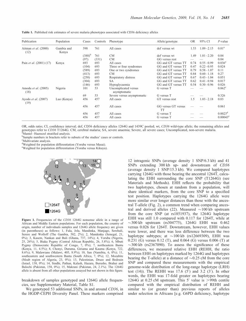
We genotyped the T1264G polymorphism in 3420 individualsfrom 66 ethnic groups. These included 974 individuals fromthe Human Genome Diversity Project (HGDP)-Centred’Etude du Polymorphisme Humain (CEPH) Diversity Panel(51 ethnic groups) and 2446 additional Gambian, Malawian,Kenyan and Ghanaian cord blood samples and population con-trols (15 ethnic groups). The 1264G allele was at a frequencyof 26% in the Yoruba of Nigeria, a finding consistent with theHapMap Yoruba (24.2%) and another recent study of Nigerianchildren (18). The 1264G allele was present in populationsacross sub-Saharan Africa, but was generally less frequentthan in the Yoruba (Fig. 1). The 1264G allele was not seenin two African populations living in the regions of lowermalaria risk, the South African Bantu and the Namibian San.However, their limited sample sizes may mean the allele ispresent at low frequencies but undetected. The pygmies ofcentral Africa, such as the Biaka (Central African Republic)and the Mbuti (Democratic Republic of Congo), are relativelygenetically isolated from other sub-Saharan populations. Thereis, however, evidence that the Biaka have experienced admix-ture with neighbouring groups (20). This is consistent with thepresence of 1264G alleles in the Biaka (5.8%) but not in theMbuti (0%). The 1264G allele was absent from almost all non-African ethnic groups. The Makrani of Pakistan were the onlygroup outside of Africa (and the only Pakistani ethnic group)to have 1264G (2.1%). The presence of 1264G is consistentwith past African admixture in the Makrani (21). For a full
2684 Human Molecular Genetics, 2009, Vol. 18, No. 14
breakdown of samples genotyped and 1264G allele frequen-cies, see Supplementary Material, Table S1.
We genotyped 53 additional SNPs, in and around CD36, inthe HGDP-CEPH Diversity Panel. These markers comprised
12 intragenic SNPs (average density 1 SNP/6.3 kb) and 41SNPs extending 300 kb up- and downstream of CD36(average density 1 SNP/15.3 kb). We compared haplotypescarrying 1264G with those bearing the ancestral 1264T, calcu-lating the EHH surrounding the core SNP (T1264G) (seeMaterials and Methods). EHH reflects the probability thattwo haplotypes, chosen at random from a population, willshare identical markers, from the core SNP to a specifiedtest position. Haplotypes carrying the 1264G allele weremore similar over longer distances than those with the ances-tral T-allele (Fig. 2), a common trend when comparing ances-tral and derived alleles (22). Measured �100 kb upstreamfrom the core SNP (at rs1851937), the 1264G haplotypeEHH was still 1.0 compared with 0.117 for 1264T, while at�300 kb upstream (rs304775), 1264G EHH was 0.462versus 0.026 for 1264T. Downstream, however, EHH valueswere lower, and there was less difference between the twohaplotype subtypes; at �100 kb (rs12669309), EHH was0.231 (G) versus 0.12 (T), and 0.064 (G) versus 0.006 (T) at�300 kb (rs2367090). To assess the significance of thesedifferences, we measured relative EHH (REHH, the ratiobetween EHH on haplotypes marked by 1264G and haplotypesbearing the T-allele) at a distance of �0.25 cM from the coreSNP and compared these measurements with the empiricalgenome-wide distribution of the long-range haplotype (LRH)test (14)). The REHH was 17.6 (50) and 3.2 (30). In otherwords, the EHH was 17-fold greater on haplotypes bearing1264G at 0.25 cM upstream. This 50 value is .99th centilecompared with the empirical distribution of REHH andsimilar to (or greater than) previous reports of allelesunder selection in Africans [e.g. G6PD deficiency, haplotype
Table 1. Published risk estimates of severe malaria phenotypes associated with CD36 deficiency alleles
Publication Population Cases Controls Phenotype Allele/genotype OR 95% CI P-value
Aitman et al. (2000)(12)
Gambia andKenya
598 761 All cases def versus wt 1.53 1.09–2.15 0.01a
(388)b 761 CM def versus wt 1.49 1.01–2.20 0.04(97) (331) CM GG versus rest — — 0.04
Pain et al. (2001) (17) Kenya 693 693 All cases GG and GT versus TT 0.74 0.55–0.99 0.036a
(104) 693 Three or four syndromes GG and GT versus TT 0.47 0.22–0.95 0.024(589) 693 One or two syndromes GG and GT versus TT 0.79 0.58–1.07 0.11(413) 693 CM GG and GT versus TT 0.84 0.60–1.18 0.27(250) 693 Respiratory distress GG and GT versus TT 0.67 0.43–1.04 0.051(304) 693 SA GG and GT versus TT 0.62 0.41–0.94 0.017(146) 693 Hypoglycaemia GG and GT versus TT 0.54 0.30–0.96 0.026
Amodu et al. (2005)(18)
Nigeria 101 53 Uncomplicated versusasymptomatic
G versus T — — 0.062c
69 53 Severe versus asymptomatic G versus T — — 0.326Ayodo et al. (2007)
(19)Luo (Kenya) 456 457 All cases GT versus rest 1.5 1.03–2.18 0.03
456 457 All cases GG versus GT versusTT
— — 0.061
456 457 All cases G versus T — — 0.0023d
456 457 All cases G versus T — — 0.00043e
OR, odds ratio; CI, confidence interval; def, CD36 deficiency alleles 1264G and 1439C pooled; wt, CD36 wild-type allele; the remaining alleles andgenotypes refer to CD36 T1264G. CM, cerebral malaria; SA, severe anaemia; Severe, all severe cases; Uncomplicated, non-severe malaria.aMantel–Haenszel stratified analysis.bSample numbers in brackets refer to subsets of the studies’ cases or controls.cMultivariate analysis.dWeighted for population differentiation (Yoruba versus Masai).eWeighted for population differentiation (Yoruba versus Kikuyu).
Figure 1. Frequencies of the CD36 1264G nonsense allele in a range ofAfrican and Middle Eastern populations. For each population, the country oforigin, number of individuals samples and 1264G allele frequency are given(in parentheses) as follows: 1, Fula, Jola, Mandinka, Manjago, Serehuli,Serere and Wolloff (The Gambia, 582, 2%); 2, Mandenka (Senegal, 21,0%); 3, Kasem, Nankan and Buli (Ghana, 737, 14%); 4, Yoruba (Nigeria,25, 26%); 5, Biaka Pygmy (Central African Republic, 26, 5.8%); 6, MbutiPygmy (Democratic Republic of Congo, 7, 0%); 7, northeastern Bantu(Kenya, 11, 4.5%); 8, Chonyi, Duruma, Giriama and Kauma (Kenya, 722,8.6%); 9, Malawians (Malawi, 405, 8.9%); 10, San (Namibia, 6, 0%); 11,southeastern and southwestern Bantu (South Africa, 7, 0%); 12, Mozabite(Mzab region of Algeria, 23, 0%); 13, Palestinian, Druze and Bedouin(Israel, 138, 0%); 14, Sindhi, Pathan, Kalash, Hazara, Burusho, Brahui andBalochi (Pakistan, 158, 0%); 15, Makrani (Pakistan, 24, 2.1%). The 1264Gallele is absent from all other populations assayed but not shown in this figure.
Human Molecular Genetics, 2009, Vol. 18, No. 14 2685

frequency 18%, REHH 7 (15)]. However, the 30 REHH of 3.2was not significant.
For comparison, we downloaded phased haplotypes from theHapMap Yoruba, paring down the data set to the same 54 SNPs(including T1264G) (Fig. 2). As expected, EHH measurementswere greater on 1264G haplotypes. Upstream EHH was, at�100 kb, 0.741 (G) versus 0.089 (T); at �300 kb, 0.571 (G)versus 0.012 (T); downstream, at �100 kb, 0.483 (G) versus0.129 (T); and at �300 kb, 0.227 (G) versus 0.005 (T). TheHapMap Yoruba had REHH values of 29.7 (50) and 14.6 (30)[both .99th centile and consistent with the original observation(14)]. REHH values for our HGDP Yoruba are lower than thosefor the HapMap Yoruba, particularly downstream of T1264G.There are a range of explanations for these differences: (i) theHapMap data had a slightly lower missing genotype rate (1versus 3% for HGDP); (ii) the HapMap haplotypes wereinferred from higher density marker data; (iii) our HGDPsample size was smaller (50 haplotypes versus 120 HapMaphaplotypes); and (iv) the HapMap haplotypes were inferredfrom parent–offspring trios. Offspring genotypes can be usedto unambiguously resolve phase at some parental markers. To
test the impact of phasing accuracy on EHH, we obtainedYoruba genotypes from the HapMap project and phased themwithout utilizing offspring data (treating the parents as 60 unre-lated individuals). The resulting haplotypes had an REHH of29.6 upstream, but only 5.1 downstream (SupplementaryMaterial, Fig. S2). This suggests that the asymmetry in HGDPYoruba REHH, between 50 and 30 regions, may reflect technicalartefacts affecting haplotype estimation downstream (e.g. assayperformance, phasing accuracy) rather than a biological signal(e.g. selection acting on sequences upstream of T1264G).
Too few 1264G haplotypes were sampled from the Makrani,the Biaka of the Central African Republic or the northeasternBantu for a valid assessment of EHH to be made. To study1264G haplotypes in detail, we selected 101 parent–offspringtrios from our Gambian family study (see next section). Twenty-six trios were selected because one or both parents had the1264G allele. The remaining 75 were randomly chosen fromthe family study. Samples were genotyped and analysed in amanner similar to the HGDP-CEPH Diversity Panel. Offspringgenotypes were used to assist phasing. In total, 30 1264Ghaplotypes were compared with 374 1264T haplotypes(Fig. 2). EHH values on Gambian CD36 1264G haplotypeswere raised. Upstream EHH was, at �100 kb, 0.405 (G)versus 0.082 (T); at�300 kb, 0.184 (G) versus 0.006 (T); down-stream, at�100 kb, 0.607 (G) versus 0.114 (T); and at�300 kb,0.122 (G) versus 0.003 (T). REHH was 19.1 upstream and 12.8downstream. However, EHH is often raised around low fre-quency, derived alleles. The significance of these results wasdetermined using phased genome-wide SNP data from 658Gambian parent–offspring trios genotyped on an Illumina650Y BeadArray platform (manuscript in preparation). Theoffspring in these trios were malaria cases; so to minimizebias, we analysed only untransmitted parental chromosomes.We calculated the REHH for 15 807 derived alleles of frequency1–3%, where REHH could be assessed at 0.25+ 0.01 cM fromthe core SNP. Comparison with this empirical, genome-widedistribution of the LRH test suggests our Gambian REHHvalues are not significantly unusual, �59th centile (12.8) and�77th centile (19.1). This finding highlights that low-frequencyalleles are often surrounded by long haplotypes, limiting thepower of EHH testing.
Genetic association studies with severe malaria phenotypes
Seventy CD36 SNPs, including T1264G and G1439C [a mis-sense mutation in strong linkage disequilibrium in Gambianswith the 1444delA frameshift mutation, another CD36deficiency allele (12)], were tested for disease associationsin Gambian case–control and family studies (see Materialand Methods for further details of SNP selection). Thefamily study comprised 1288 children affected by severemalaria and both parents [these trios included 508 cases of cer-ebral malaria (CM) and 333 cases of severe malarial anaemia(SA)]. The case–control study was separate from the familystudy and comprised 727 cases of severe malaria (339 CM,226 SA) and 623 population controls. Both studies werepowered to detect realistic effect sizes at a significancethreshold of P , 0.05. Only one marker, rs4728191, an intro-nic SNP, exceeded this threshold in both studies. The minorallele was associated with protection on both occasions
Figure 2. EHH decay surrounding T1264G in HGDP Yoruba, HapMapYoruba and Gambian trios. Breakdown of extended haplotype homozygosity(EHH) with distance, on haplotypes partitioned by the alleles of T1264G(rs3211938; the ‘core’ SNP). We compared (A) 25 HGDP-CEPH Yoruba indi-viduals, (B) HapMap genotypes from 60 Yoruba parents and (C) 202 Gambianparents.
2686 Human Molecular Genetics, 2009, Vol. 18, No. 14

[case–control, severe malaria, allelic odds ratio (OR) 0.76;95% confidence interval (CI) 0.61–0.96, P ¼ 0.019; familystudy, SA, allelic OR 0.70; 95% CI 0.5–0.99, P ¼ 0.041].This finding needs cautious interpretation, as the associationswere with different phenotypes and the Hardy–WeinbergP-value for rs4728191 in cord blood controls was marginal(P ¼ 0.006). All results from the case–control and familystudies with P , 0.05 are documented in SupplementaryMaterial, Table S2.
Using the UNPHASED application, we combined the case–control and family data into a single analysis (Fig. 3). Giventhat 70 SNPs have been tested, the formal significancethreshold, with conservative Bonferroni correction, would beroughly 0.0007 (0.05/70). No marker in CD36 had P ,0.002 for any genetic model or phenotype. Rs4728191 wasnot significantly associated with severe malaria phenotypesin the combined analysis (severe malaria, allelic OR 0.89;95% CI 0.79–1.01, P ¼ 0.06; SA allelic OR 0.84; 95% CI0.67–1.06, P ¼ 0.13). Neither T1264G (severe malaria,allelic OR 0.93; 95% CI 0.67–1.29, P ¼ 0.65), nor G1439C(severe malaria, allelic OR 0.90; 95% CI 0.68–1.19, P ¼0.46), nor both deficiency alleles considered together (severemalaria, allelic OR 0.91; 95% CI 0.73–1.13, P ¼ 0.39)were significantly associated with severe malaria phenotypes.Complete results from the pooled analysis are documented inSupplementary Material, Table S3.
Given the previous report of protection associated with1264G heterozygotes (17), we specifically checked for suchan effect. Deficiency allele heterozygotes did tend to be under-represented among cases (versus wild-type homozygotes, OR0.83; 95% CI 0.66–1.05). In addition, no CD36-deficient indi-vidual (e.g. 1264G homozygote, 1439C homozygote or com-pound heterozygote) was found among the 727 cord bloodcontrols, although several were seen among the cases (threein the case–control study and eight in the family study)(deficiency allele homozygotes versus wild-type homozygotesOR 1.59; 95% CI 0.74–3.39). Together these results suggesteda non-significant trend towards heterozygote advantage
(overdominant model P ¼ 0.09). However, the low frequencyof 1264G (1.9%) and 1439C (1.8%) in Gambians limited thestatistical power. To increase the power, we genotypedT1264G and G1439C in five further sample sets derivedfrom populations where 1264G is more common (8.2% inKenyan controls, 9.0% in Malawian controls and 14.2% inGhanaian controls). These additional studies comprised:
(i) 718 Malawian cases and 405 controls (640 CM, 101 SA);(ii) 708 Kenyan cases and 902 controls (216 CM, 270 SA);
(iii) 225 Malawi trios (216 CM, 39 SA);(iv) 234 Kenyan trios (114 CM, 85 SA);(v) 792 Ghanaian cases and 806 controls (44 CM, 296 SA).
The 1439C allele was at a frequency of 0.1% in Ghanaiancontrols, but absent in Kenya and Malawi.
T1264G was not associated with severe malarial phenotypesin the additional five studies individually. Pooling data acrossall seven studies (5681 cases and controls, and 1747 nuclearfamily trios) using the UNPHASED application, we foundno association between T1264G (severe malaria, allelic OR1.0; 95% CI 0.89–1.12, P ¼ 0.98) or deficiency alleles(1264G and 1439C pooled, severe malaria, allelic OR 0.98;95% CI 0.89–1.09, P ¼ 0.76) and severe malaria (Table 2).The putative trend towards a heterozygous advantage,suggested by the Gambian data, was not supported by thislarger data set (deficiency allele heterozygotes versus wild-type homozygotes, OR 0.97; 95% CI 0.86–1.09; deficiencyallele homozygotes versus wild-type homozygotes, OR 1.09;95% CI 0.74–1.6; overdominant model P ¼ 0.53) (Table 3).
DISCUSSION
We set out to analyse the distribution of 1264G haplotypes in arange of global populations, to screen variation across the genefor association with severe malaria and to target 1264G withwell-powered tests of disease association. It is important tostress that the initial observation of raised EHH on 1264G
Figure 3. P-values for the pooled Gambian case–control and family studies. Allelic model P-values derived from UNPHASED analysis of 70 SNPs acrossCD36, for severe falciparum malaria (Severe), and the subphenotypes of cerebral malaria (CM) and severe malarial anaemia (SA). The arrow marks the positionof T1264G (rs3211938). Chromosome 7 coordinates from NCBI build 36/dbSNP 126.
Human Molecular Genetics, 2009, Vol. 18, No. 14 2687

haplotypes was only briefly noted in previous work, with littlediscussion of its biological significance (14,16). This studyrepresents the first detailed description of the CD36 EHHsignal found in the HapMap Yoruba. In addition, we presentednovel data from the HGDP collection exploring the frequencyof the nonsense allele across Africa and replicating the EHHsignal in the Yoruba. Our association study of severemalaria and T1264G employed a sample size more thantwice that of all previous studies combined (Table 1). Givenour relative statistical power, the marginal significance ofpast results and their inconsistent outcomes, our data suggestthat CD36 T1264G is not associated with severe malaria. Fur-thermore, no CD36 marker tested appeared to be significantlyassociated with disease susceptibility.
The reason for the expansion of the 1264G allele in West-Central Africa is unclear. Nigeria has a high prevalence ofmalaria infection, however, so does the Gambia (1264G fre-quency 2%) and costal regions of Kenya (9%). The high fre-quency in Nigeria might indicate a recent origin for theallele in that region with subsequent migration. The lowerallele frequency outside Nigeria could simply reflect selectivesweeps in progress, but at earlier stages. There are a range ofplausible explanations for the presence of selection in theabsence of a malaria association. It is possible that thesignal of selection detected around 1264G could be spurious;or that host–parasite co-evolution has eliminated the advan-tage of this allele, subsequent to an initial selective sweep.Alternatively, the failure to detect an association might rep-resent type II error. The 1264G allele could provide amodest degree of protection from severe malaria (e.g. an ORbetween 0.95 and 1); protect from an infrequent but life-threatening malaria phenotype; or only offer protection tothe rare deficiency allele homozygotes. This associationstudy would have only limited power to detect an effect inthese situations. A small effect size might be sufficient tomaintain the allele at low frequency in populations. Hypothe-tically, the expansion of 1264G alleles in the Yoruba couldrepresent a local selection event on top of a low-level back-ground of selection by malaria. The additional Yoruba eventcould also be malaria-related, for example the effects of a P.
falciparum strain specific to southwestern Nigeria. However,CD36 operates in a range of key biological processes includ-ing thrombostasis (23), glucose metabolism (24), lipid hand-ling (25), immune function (26), angiogenesis (27) andpossibly taste (28). Therefore, other environmental factorsneed to be considered as selective pressures.
Combining evidence for natural selection with diseaseassociation statistics has been proposed as a technique toincrease power to detect disease associations. Our data high-lights a potential pitfall for this approach. In recently pub-lished work, an association between T1264G and severemalaria (P ¼ 0.03) was combined with the substantial allelefrequency difference between the Yoruba (a populationexposed to endemic malaria) and the Kikuyu and Masai (popu-lations at low malaria risk). The combined P-values reportedwere highly significant (up to P ¼ 0.00043) (19). However,as we have seen, there is a substantial T1264G allele fre-quency difference between the Yoruba and most Africanpopulations, including others exposed to endemic malaria.This highlights the possibility that marginal evidence of agenetic association with disease A (e.g. severe malaria)could be conflated with strong evidence of natural selectioncaused by an unrelated process B (e.g. a novel dietarysource). Combined analysis remains an exciting approach,but ensuring a signal of selection relates to the same diseaseas association data (or gauging the degree of confidence)will be a challenge.
It is possible that CD36 deficiency alleles cause deleteriousconsequences, particularly in the homozygous state, thatbalance any hypothetical advantages. These include neonatalimmune thrombocytopenia (29), altered aerobic exercisecapacity (30) and dysregulation of lipid metabolism (25).There is also, ironically, evidence that absence of CD36 is dis-advantageous to the host during malaria infection. IRBCs arerecognized and phagocytosed by monocytes and macrophagesfollowing CD36 binding (31,32). One disease associationstudy based in Thailand (475 adult patients with severe ormild malaria) reported an intronic dinucleotide repeat allelein CD36 [in3(TG)12] associated with protection from severemalaria (33). The authors showed that other alleles of thesame microsatellite were associated with the production of ashort CD36 isoform altering the P. falciparum-bindingepitope. In contrast, the protective in3(TG)12 allele was associ-ated with a full-length transcript, leading the authors tosuggest that iRBC binding to intact CD36 could facilitate effi-cient clearance. The majority of P. falciparum isolates bindCD36 (1–4) yet only a fraction of infections lead to life-threatening consequences. In vitro binding studies suggestthat parasite strains from asymptomatic controls with parasi-taemia have high levels of CD36 binding, whereas strainsfrom individuals with non-severe, uncomplicated diseasehave moderate levels of CD36 binding. The lowest levels ofCD36 binding are found in isolates from individuals withsevere phenotypes such as CM, and particularly SA(2,3,34,35). CD36 (also known as fatty acid translocase) ishighly expressed in adipose tissue and skeletal muscle (6).Absence of CD36 as a target for sequestration on these largecapillary beds may promote splenic passage and clearance ofCD36-binding parasites. This could select for parasite cloneswhich have switched to alternative host ligands. Post-mortem
Table 2. Estimated risk for CD36 T1264G in severe malaria
Population Cases Controls OR 95% CI P-value
Population-based studiesGambia 727 623 0.74 0.4–1.37 0.34Malawi 718 405 0.91 0.67–1.25 0.57Kenya 708 902 1.20 0.93–1.55 0.15Ghana 792 806 1.05 0.86–1.29 0.63
Family-based studiesGambia 1288 2576 0.92 0.59–1.45 0.73Malawi 225 450 1.00 0.62–1.62 1.00Kenya 234 468 0.86 0.55–1.34 0.50
Pooled 4692 6230 1.00 0.89–1.12 0.98
Allelic odds ratios (OR) and 95% confidence intervals (CI) for CD361264G compared with the T-allele, in severe malaria. P-values werederived from logistic regression analysis with covariates of ethnic group,gender and HbS genotype (population-based studies), case–pseudo-control analysis (family studies) or UNPHASED analysis (pooleddata across all studies).
2688 Human Molecular Genetics, 2009, Vol. 18, No. 14

immunohistochemistry studies show relatively low expressionof CD36 on brain endothelium without increased expressionduring malaria (1,36). In contrast, other host ligands ofPfEMP1, such as intercellular adhesion molecule-1 andE-selectin, are commonly expressed on cerebral endotheliumand induced by inflammation (36,37).
CD36 deficiency alleles have been reported to modulateexpression in a tissue-specific way. Some variants preventexpression on platelets (CD36 deficiency type II), whereasother alleles abolish CD36 expression on a wider range ofcells including platelets, macrophages, monocytes and probablyother cell types (type I) (38,39). The 1264G allele is consideredto be a type I allele, but little is known about expression patterns(if any) on non-haematopoietic cells. In addition to T1264G and1444delA, a range of CD36 deficiency alleles have beenreported in populations of African origin. These include990delG and 1530ins14bp (12). Sequencing in Gambians hassuggested that 1264G and 1444delA are the commonestdeficiency alleles in this population, with others being ,1% fre-quency (12). However, caution is required when extrapolatingthese findings across Africa. Allelic heterogeneity, particularlywhen unrecognized, has the potential to impair power in associ-ation analysis (40,41) and could also be complicating our popu-lation genetic study. The frequency distribution of CD36deficiency across Africa could be quite different than suggestedby typing just 1264G and 1439Cþ1444delA. Resequencing ofCD36 in a range of African and Asian populations is needed todescribe the full repertoire and geographic distribution of CD36deficiency alleles.
In conclusion, the relationship between CD36 and malaria iscomplex. The putative selection event associated with theCD36 1264G nonsense allele appears to be focused in West-Central Africa, although this and other CD36 deficiencyalleles are present across sub-Saharan and Asian populations.
A number of factors may explain the lack of an associationbetween CD36 variation and severe malaria phenotypes.However, the existence of alternative (or additional) selectionpressures on CD36 deficiency alleles is an interesting possi-bility that needs further investigation. Additional field- andin vitro work is required to define the phenotypic conse-quences of CD36 deficiency alleles in health and disease.
MATERIALS AND METHODS
Human Genome Diversity Panel
Sample preparation and genotyping. DNA was derived fromlymphoblastoid cell lines of 1064 individuals from theHGDP-CEPH Diversity Panel (http://www.cephb.fr/HGDP-CEPH-Panel/) (42). Samples from 15 known duplicateand atypical individuals were excluded (43). Genotyping wasperformed using Sequenom iPLEX assays. SNP identifiers,coordinates and genotyping success rates are documented inSupplementary Material, Table S4. Following quality controlfor missing genotypes, a set of 974 individuals were selectedfor further analysis (average genotyping success rate 96.7%;rs3211938 success rate 99.8%).
Statistical analysis. Phased haplotypes and missing data wereinferred using PHASE (version 2.1) (44,45). Genotype datafrom each population was phased on its own. EHH calcu-lations were performed using a web-based calculator (http://ihg2.helmholtz-muenchen.de/cgi-bin/mueller/webehh.pl).REHH was assessed as described previously (15,46). REHHwas assessed both sides of the core SNP (50 and 30). Theaverage REHH values for the two markers closest toand either side of 0.25 cM were used for the LRH test
Table 3. Pooled analysis of CD36 deficiency alleles and severe malaria phenotypes
Phenotype Marker Genotype OR 95% CI rec over dom allelic
Severe T1264G GT versus TT 1.00 0.88–1.14 — — — —— GG versus TT 0.98 0.64–1.52 — — — —
0.94 0.99 0.99 0.98def het versus wt 0.97 0.86–1.09 — — — —— hom versus wt 1.09 0.74–1.61 — — — —
0.62 0.53 0.64 0.76SA T1264G GT versus TT 1.05 0.86–1.28 — — — —
— GG versus TT 0.79 0.35–1.740.53 0.62 0.75 0.89
def het versus wt 1.05 0.87–1.27 — — — —— hom versus wt 0.89 0.43–1.84 — — — —
0.76 0.60 0.67 0.75CM T1264G GT versus TT 1.09 0.91–1.31 — — — —
— GG versus TT 0.78 0.37–1.64 — — — —0.47 0.34 0.45 0.60
def het versus wt 1.03 0.87–1.22 — — — —— hom versus wt 1.03 0.55–1.93 — — — —
0.95 0.74 0.73 0.74
Pooled analysis performed from our seven studies using the UNPHASED application. Phenotypes: Severe, severe falciparum malaria; SA, severemalarial anaemia; CM, cerebral malaria. Markers: T1264G or both CD36 deficiency (def) alleles pooled (G1439C þ 1444delA and T1264G). T1264Ggenotypes: GG, 1264G homozygotes; GT, heterozygotes; TT, 1264T homozygotes; het, deficiency allele heterozygotes; hom, deficiency allelehomozygotes; wt, wild-type homozygotes; OR, odds ratio; CI, confidence interval; P-values are reported for four genetic models rec(essive),over(dominant), dom(inant) and allelic.
Human Molecular Genetics, 2009, Vol. 18, No. 14 2689

[50, rs704871 (0.267 cM) and rs2944398 (0.225 cM); 30,rs10487878 (0.236 cM) and rs4731861 (0.325 cM)].
Genetic association studies
Phenotype definition. All cases were children admitted to hos-pital with evidence of P. falciparum on blood film and clinicalfeatures of severe malaria (47,48). We used a Blantyre comascore of �2 as a criterion of CM (because of the limitationsof the available data, we used �3 in Ghanaian cases) and,5 g/dl or packed cell volume ,15% as a criterion of SA.Some individuals had both CM and SA. Of the severemalaria cases that were not CM or SA by these criteria,most had lesser degrees of coma (Blantyre coma score 3) oranaemia (haemoglobin 5–6 g/dl), or other complicationssuch as respiratory distress. In Gambia, Malawi and Kenya,controls were cord blood samples obtained from birth clinicsin the same locations as the cases. Ghanaian controls werecommunity controls matched for age, ethnic group andlocation of origin.
Subjects. Patient samples were collected during ongoing epi-demiological studies of severe malaria at the Royal VictoriaHospital, Banjul, The Gambia; the Queen Elizabeth CentralHospital, Blantyre, Malawi; Kilifi District Hospital, Kilifi,Kenya; and Navrongo War Memorial Hospital, Ghana. AllDNA samples were collected and genotyped followingapproval from the relevant research ethics committees andinformed consent from participants. See SupplementaryMaterial, Table S5, for demographic details of patients andcontrols.
Power calculations. Power calculations were performed usingthe Genetic Power Calculator (http://pngu.mgh.harvard.edu/~purcell/gpc/) (49). For a type I error rate of 0.05, theGambian case–control and family studies had 85.7 and98.7% power, respectively. This is based on an allelic OR of1.3 and a high-risk allele frequency of 0.25 (the averageminor allele frequency of the SNPs screened in the Gambians).With lower allele frequencies (0.1, 0.05 and 0.01), the case–control studies (each roughly 700 cases and 700 controls)had lower power, 59.1, 36 and 11.4%, respectively. Theadditional family studies (approximately 250 trios) each had�25.8, 16.0 and 7.2% power. The power of the combinedcase–control study (2945 cases and 2736 controls) was 99.3,89.6 and 31.5%. The combined family study (1747 trios)had 93.3, 71.6 and 21.3% power.
SNP selection. SNP data from HapMap release 21a (January2007, http://www.hapmap.org) within 10 kb of the CD36gene was downloaded and assessed using HAPLOVIEW(version 3.32) (50). Polymorphic SNPs (minor allele fre-quency .5%) were identified in the HapMap Yoruba.Tagging SNPs were defined using the TAGGER algorithm(51). Sequenom iPLEX genotyping assays were designed for69 tagging SNPs and three additional variants, T1264G,G1439C and rs3173798 (located at a conserved splice site).Following quality control, 70 SNPs were accepted for associ-ation analysis. This final set of SNPs tagged 116 of 133(87%) polymorphic markers (.5% frequency) across CD36
(including 10 kb up- and downstream), using pairwise taggingand an r2 .0.8 (mean r2 ¼ 0.97), in the HapMap Yoruba.
Sample preparation and genotyping. Genomic DNA samplesused in the association analysis underwent whole-genomeamplification through either primer extension pre-amplification (52) or multiple displacement amplification(53), before Sequenom iPLEX genotyping. Genotype dataunderwent quality control for missing data (,10%), Hardy–Weinberg equilibrium (P . 0.001) and Mendelian error rate.Genotype counts and quality control measures are documentedin Supplementary Material, Table S6.
Statistical analysis. Case–control association analysis wasperformed by logistic regression using covariates of ethnicgroup, gender and haemoglobin S (HbS) status. Sequenomgenotyping for the HbS variant was performed for allsamples as described previously (54). Family-based associ-ation analysis was performed using a case–pseudo-controlapproach and conditional logistic regression based on parentalgenotypes. Trios were drawn from a larger pool of samplesassessed for relationship misspecification by Mendelian errorrates. We utilized the R statistical application (version 2.6.0)along with the dgc.genetics library (http://www-gene.cimr.cam.ac.uk/clayton/software/) and the SNPas-soc package (55). In both family- and population-basedstudies, we tested each marker in allelic (or ‘multiplicative’),dominant, recessive and overdominant (or ‘heterozygoteadvantage’) genetic models and with three phenotypes (CM,SA and all severe cases). Pooling across case–control andfamily studies was performed using the UNPHASED appli-cation (version 3.0.12) (http://www.mrc-bsu.cam.ac.uk/personal/frank/software/unphased/) (56). Ethnic origin was asignificant confounder and was retained as a covariate in theUNPHASED analysis.
SUPPLEMENTARY MATERIAL
Supplementary Material is available at HMG online.
ACKNOWLEDGEMENTS
We would like to thank the MalariaGEN consortium for pro-viding access to the Illumina 650Y Gambian parent–offspringtrio data. This manuscript is published with the permission ofthe director of the Kenya Medical Research Institute. Theviews expressed here are the personal ones of the authorsand do not purport to represent those of the United StatesDepartment of the Navy.
Conflict of Interest statement. None declared.
FUNDING
This work was supported by the Wellcome Trust(GR074586AIA) through a Clinical Research Training Fel-lowship to A.E.F. and UK Medical Research Councilfunding (to D.P.K.). T.N.W. was funded by the WellcomeTrust, MalariaGEN and the European Union Network 6
2690 Human Molecular Genetics, 2009, Vol. 18, No. 14

BioMalPar Consortium. The work of the Ghanaian study wassupported by funding from the National Institute of Allergyand Infectious Diseases/National Institutes of Health (contractnumber AI95363) to the Noguchi Memorial Institute forMedical Research. Funding to pay the Open Access chargewas provided by the Wellcome Trust.
REFERENCES
1. Newbold, C., Craig, A., Kyes, S., Rowe, A., Fernandez-Reyes, D. andFagan, T. (1999) Cytoadherence, pathogenesis and the infected red cellsurface in Plasmodium falciparum. Int. J. Parasitol., 29, 927–937.
2. Heddini, A., Pettersson, F., Kai, O., Shafi, J., Obiero, J., Chen, Q.,Barragan, A., Wahlgren, M. and Marsh, K. (2001) Fresh isolates fromchildren with severe Plasmodium falciparum malaria bind to multiplereceptors. Infect. Immun., 69, 5849–5856.
3. Rogerson, S.J., Tembenu, R., Dobano, C., Plitt, S., Taylor, T.E. andMolyneux, M.E. (1999) Cytoadherence characteristics of Plasmodiumfalciparum-infected erythrocytes from Malawian children with severe anduncomplicated malaria. Am. J. Trop. Med. Hyg., 61, 467–472.
4. Udomsangpetch, R., Reinhardt, P.H., Schollaardt, T., Elliott, J.F., Kubes,P. and Ho, M. (1997) Promiscuity of clinical Plasmodium falciparumisolates for multiple adhesion molecules under flow conditions.J. Immunol., 158, 4358–4364.
5. Baruch, D.I., Gormely, J.A., Ma, C., Howard, R.J. and Pasloske, B.L.(1996) Plasmodium falciparum erythrocyte membrane protein 1 is aparasitized erythrocyte receptor for adherence to CD36, thrombospondin,and intercellular adhesion molecule 1. Proc. Natl Acad. Sci. USA, 93,3497–3502.
6. Febbraio, M., Hajjar, D.P. and Silverstein, R.L. (2001) CD36: a class Bscavenger receptor involved in angiogenesis, atherosclerosis,inflammation, and lipid metabolism. J. Clin. Invest., 108, 785–791.
7. Urban, B.C., Willcox, N. and Roberts, D.J. (2001) A role for CD36 in theregulation of dendritic cell function. Proc. Natl Acad. Sci., USA, 98,8750–8755.
8. Pain, A., Ferguson, D.J., Kai, O., Urban, B.C., Lowe, B., Marsh, K. andRoberts, D.J. (2001) Platelet-mediated clumping of Plasmodiumfalciparum-infected erythrocytes is a common adhesive phenotype and isassociated with severe malaria. Proc. Natl Acad. Sci. USA, 98, 1805–1810.
9. Ikeda, H., Mitani, T., Ohnuma, M., Haga, H., Ohtzuka, S., Kato, T.,Nakase, T. and Sekiguchi, S. (1989) A new platelet-specific antigen,Naka, involved in the refractoriness of HLA-matched platelet transfusion.Vox Sang., 57, 213–217.
10. Urwijitaroon, Y., Barusrux, S., Romphruk, A. and Puapairoj, C. (1995)Frequency of human platelet antigens among blood donors in northeasternThailand. Transfusion, 35, 868–870.
11. Curtis, B.R. and Aster, R.H. (1996) Incidence of the Nak(a)-negativeplatelet phenotype in African Americans is similar to that of Asians.Transfusion, 36, 331–334.
12. Aitman, T.J., Cooper, L.D., Norsworthy, P.J., Wahid, F.N., Gray, J.K.,Curtis, B.R., McKeigue, P.M., Kwiatkowski, D., Greenwood, B.M.,Snow, R.W. et al. (2000) Malaria susceptibility and CD36 mutation.Nature, 405, 1015–1016.
13. Yanai, H., Chiba, H., Fujiwara, H., Morimoto, M., Abe, K., Yoshida, S.,Takahashi, Y., Fuda, H., Hui, S.P., Akita, H. et al. (2000) Phenotype–genotype correlation in CD36 deficiency types I and II. Thromb.Haemost., 84, 436–441.
14. International HapMap Consortium (2005) A haplotype map of the humangenome. Nature, 437, 1299–1320.
15. Sabeti, P.C., Reich, D.E., Higgins, J.M., Levine, H.Z., Richter, D.J.,Schaffner, S.F., Gabriel, S.B., Platko, J.V., Patterson, N.J., McDonald,G.J. et al. (2002) Detecting recent positive selection in the human genomefrom haplotype structure. Nature, 419, 832–837.
16. Sabeti, P.C., Schaffner, S.F., Fry, B., Lohmueller, J., Varilly, P.,Shamovsky, O., Palma, A., Mikkelsen, T.S., Altshuler, D. and Lander,E.S. (2006) Positive natural selection in the human lineage. Science, 312,1614–1620.
17. Pain, A., Urban, B.C., Kai, O., Casals-Pascual, C., Shafi, J., Marsh, K. andRoberts, D.J. (2001) A non-sense mutation in Cd36 gene is associatedwith protection from severe malaria. Lancet, 357, 1502–1503.
18. Amodu, O.K., Gbadegesin, R.A., Ralph, S.A., Adeyemo, A.A., Brenchley,P.E., Ayoola, O.O., Orimadegun, A.E., Akinsola, A.K., Olumese, P.E. andOmotade, O.O. (2005) Plasmodium falciparum malaria in south-westNigerian children: is the polymorphism of ICAM-1 and E-selectin genescontributing to the clinical severity of malaria? Acta Trop., 95, 248–255.
19. Ayodo, G., Price, A.L., Keinan, A., Ajwang, A., Otieno, M.F., Orago,A.S., Patterson, N. and Reich, D. (2007) Combining evidence of naturalselection with association analysis increases power to detectmalaria-resistance variants. Am. J. Hum. Genet., 81, 234–242.
20. Bamshad, M.J., Wooding, S., Watkins, W.S., Ostler, C.T., Batzer, M.A.and Jorde, L.B. (2003) Human population genetic structure and inferenceof group membership. Am. J. Hum. Genet., 72, 578–589.
21. Qamar, R., Ayub, Q., Mohyuddin, A., Helgason, A., Mazhar, K.,Mansoor, A., Zerjal, T., Tyler-Smith, C. and Mehdi, S.Q. (2002)Y-chromosomal DNA variation in Pakistan. Am. J. Hum. Genet., 70,1107–1124.
22. Fry, A.E., Trafford, C.J., Kimber, M.A., Chan, M.S., Rockett, K.A. andKwiatkowski, D.P. (2006) Haplotype homozygosity and derived alleles inthe human genome. Am. J. Hum. Genet., 78, 1053–1059.
23. Asch, A.S., Barnwell, J., Silverstein, R.L. and Nachman, R.L. (1987)Isolation of the thrombospondin membrane receptor. J. Clin. Invest., 79,1054–1061.
24. Lepretre, F., Linton, K.J., Lacquemant, C., Vatin, V., Samson, C., Dina,C., Chikri, M., Ali, S., Scherer, P., Seron, K. et al. (2004) Genetic study ofthe CD36 gene in a French diabetic population. Diabetes Metab., 30,459–463.
25. Love-Gregory, L., Sherva, R., Sun, L., Wasson, J., Schappe, T., Doria, A.,Rao, D.C., Hunt, S.C., Klein, S., Neuman, R.J. et al. (2008) Variants in theCD36 gene associate with the metabolic syndrome and high-densitylipoprotein cholesterol. Hum. Mol. Genet., 17, 1695–1704.
26. Savill, J., Hogg, N. and Haslett, C. (1991) Macrophage vitronectinreceptor, CD36, and thrombospondin cooperate in recognition ofneutrophils undergoing programmed cell death. Chest, 99, 6S–7S.
27. Dawson, D.W., Pearce, S.F., Zhong, R., Silverstein, R.L., Frazier, W.A.and Bouck, N.P. (1997) CD36 mediates the in vitro inhibitory effects ofthrombospondin-1 on endothelial cells. J. Cell Biol., 138, 707–717.
28. Laugerette, F., Passilly-Degrace, P., Patris, B., Niot, I., Febbraio, M.,Montmayeur, J.P. and Besnard, P. (2005) CD36 involvement inorosensory detection of dietary lipids, spontaneous fat preference, anddigestive secretions. J. Clin. Invest., 115, 3177–3184.
29. Curtis, B.R., Ali, S., Glazier, A.M., Ebert, D.D., Aitman, T.J. and Aster,R.H. (2002) Isoimmunization against CD36 (glycoprotein IV): descriptionof four cases of neonatal isoimmune thrombocytopenia and brief reviewof the literature. Transfusion, 42, 1173–1179.
30. Yanai, H., Watanabe, I., Ishii, K., Morimoto, M., Fujiwara, H., Yoshida,S., Hui, S.P., Matsuno, K. and Chiba, H. (2007) Attenuated aerobicexercise capacity in CD36 deficiency. J. Med. Genet., 44, 445–447.
31. McGilvray, I.D., Serghides, L., Kapus, A., Rotstein, O.D. and Kain, K.C.(2000) Nonopsonic monocyte/macrophage phagocytosis of Plasmodium
falciparum-parasitized erythrocytes: a role for CD36 in malarialclearance. Blood, 96, 3231–3240.
32. Smith, T.G., Serghides, L., Patel, S.N., Febbraio, M., Silverstein, R.L. andKain, K.C. (2003) CD36-mediated nonopsonic phagocytosis oferythrocytes infected with stage I and IIA gametocytes of Plasmodium
falciparum. Infect. Immun., 71, 393–400.
33. Omi, K., Ohashi, J., Patarapotikul, J., Hananantachai, H., Naka, I.,Looareesuwan, S. and Tokunaga, K. (2003) CD36 polymorphism isassociated with protection from cerebral malaria. Am. J. Hum. Genet., 72,364–374.
34. Newbold, C., Warn, P., Black, G., Berendt, A., Craig, A., Snow, B.,Msobo, M., Peshu, N. and Marsh, K. (1997) Receptor-specific adhesionand clinical disease in Plasmodium falciparum. Am. J. Trop. Med. Hyg.,57, 389–398.
35. Traore, B., Muanza, K., Looareesuwan, S., Supavej, S., Khusmith, S.,Danis, M., Viriyavejakul, P. and Gay, F. (2000) Cytoadherencecharacteristics of Plasmodium falciparum isolates in Thailand using anin vitro human lung endothelial cells model. Am. J. Trop. Med. Hyg., 62,38–44.
36. Turner, G.D., Morrison, H., Jones, M., Davis, T.M., Looareesuwan, S.,Buley, I.D., Gatter, K.C., Newbold, C.I., Pukritayakamee, S., Nagachinta,B. et al. (1994) An immunohistochemical study of the pathology of fatalmalaria. Evidence for widespread endothelial activation and a potential
Human Molecular Genetics, 2009, Vol. 18, No. 14 2691

role for intercellular adhesion molecule-1 in cerebral sequestration.Am. J. Pathol., 145, 1057–1069.
37. Silamut, K., Phu, N.H., Whitty, C., Turner, G.D., Louwrier, K., Mai, N.T.,Simpson, J.A., Hien, T.T. and White, N.J. (1999) A quantitative analysisof the microvascular sequestration of malaria parasites in the human brain.Am. J. Pathol., 155, 395–410.
38. Kashiwagi, H., Tomiyama, Y., Honda, S., Kosugi, S., Shiraga, M., Nagao,N., Sekiguchi, S., Kanayama, Y., Kurata, Y. and Matsuzawa, Y. (1995)Molecular basis of CD36 deficiency. Evidence that a 478C– .Tsubstitution (proline90– .serine) in CD36 cDNA accounts for CD36deficiency. J. Clin. Invest., 95, 1040–1046.
39. Kashiwagi, H., Tomiyama, Y., Nozaki, S., Kiyoi, T., Tadokoro, S.,Matsumoto, K., Honda, S., Kosugi, S., Kurata, Y. and Matsuzawa, Y.(2001) Analyses of genetic abnormalities in type I CD36 deficiency inJapan: identification and cell biological characterization of two novelmutations that cause CD36 deficiency in man. Hum. Genet., 108,459–466.
40. Longmate, J.A. (2001) Complexity and power in case–control associationstudies. Am. J. Hum. Genet., 68, 1229–1237.
41. Slager, S.L., Huang, J. and Vieland, V.J. (2000) Effect of allelicheterogeneity on the power of the transmission disequilibrium test. Genet.
Epidemiol., 18, 143–156.
42. Cann, H.M., de Toma, C., Cazes, L., Legrand, M.F., Morel, V., Piouffre,L., Bodmer, J., Bodmer, W.F., Bonne-Tamir, B., Cambon-Thomsen, A.et al. (2002) A human genome diversity cell line panel. Science, 296,261–262.
43. Rosenberg, N.A. (2006) Standardized subsets of the HGDP-CEPHHuman Genome Diversity Cell Line Panel, accounting for atypical andduplicated samples and pairs of close relatives. Ann. Hum. Genet., 70,841–847.
44. Stephens, M. and Donnelly, P. (2003) A comparison of Bayesian methodsfor haplotype reconstruction from population genotype data. Am. J. Hum.
Genet., 73, 1162–1169.
45. Stephens, M., Smith, N.J. and Donnelly, P. (2001) A new statisticalmethod for haplotype reconstruction from population data. Am. J. Hum.
Genet., 68, 978–989.
46. Sabeti, P.C., Walsh, E., Schaffner, S.F., Varilly, P., Fry, B., Hutcheson,H.B., Cullen, M., Mikkelsen, T.S., Roy, J., Patterson, N. et al. (2005) Thecase for selection at CCR5-Delta32. PLoS Biol., 3, e378.
47. Division of Control of Tropical Diseases, World Health Organization(1990) Severe and complicated malaria. Trans. R. Soc. Trop. Med. Hyg.,84 (Suppl. 2), 1–65.
48. Marsh, K., Forster, D., Waruiru, C., Mwangi, I., Winstanley, M., Marsh,V., Newton, C., Winstanley, P., Warn, P., Peshu, N. et al. (1995)Indicators of life-threatening malaria in African children. N. Engl. J. Med.,332, 1399–1404.
49. Purcell, S., Cherny, S.S. and Sham, P.C. (2003) Genetic Power Calculator:design of linkage and association genetic mapping studies of complextraits. Bioinformatics, 19, 149–150.
50. Barrett, J.C., Fry, B., Maller, J. and Daly, M.J. (2005) Haploview: analysisand visualization of LD and haplotype maps. Bioinformatics, 21, 263–265.
51. de Bakker, P.I., Yelensky, R., Pe’er, I., Gabriel, S.B., Daly, M.J. andAltshuler, D. (2005) Efficiency and power in genetic association studies.Nat. Genet., 37, 1217–1223.
52. Zhang, L., Cui, X., Schmitt, K., Hubert, R., Navidi, W. and Arnheim, N.(1992) Whole genome amplification from a single cell: implications forgenetic analysis. Proc. Natl Acad. Sci. USA, 89, 5847–5851.
53. Gonzalez, J.M., Portillo, M.C. and Saiz-Jimenez, C. (2005) Multipledisplacement amplification as a pre-polymerase chain reaction (pre-PCR)to process difficult to amplify samples and low copy number sequencesfrom natural environments. Environ. Microbiol., 7, 1024–1028.
54. Fry, A.E., Griffiths, M.J., Auburn, S., Diakite, M., Forton, J.T., Green, A.,Richardson, A., Wilson, J., Jallow, M., Sisay-Joof, F. et al. (2008)Common variation in the ABO glycosyltransferase is associated withsusceptibility to severe Plasmodium falciparum malaria. Hum. Mol.Genet., 17, 567–576.
55. Gonzalez, J.R., Armengol, L., Sole, X., Guino, E., Mercader, J.M.,Estivill, X. and Moreno, V. (2007) SNPassoc: an R package to performwhole genome association studies. Bioinformatics, 23, 644–645.
56. Dudbridge, F. (2008) Likelihood-based association analysis for nuclearfamilies and unrelated subjects with missing genotype data. Hum. Hered.,66, 87–98.
2692 Human Molecular Genetics, 2009, Vol. 18, No. 14
Copyright © 2022 FDOKUMEN