The use of human CD68 transcriptional regulatory sequences to direct high-level expression of class...
11
The use of human CD68 transcriptional regulatory sequences to direct high-level expression of class A scavenger receptor in macrophages in vitro and in vivo PETER J. GOUGH,* SIAMON GORDON & DAVID R. GREAVES Sir William Dunn School of Pathology, University of Oxford, Oxford, UK SUMMARY Macrophages (Mw) play a key role in innate and acquired immunity. The study of Mw biology has been hampered by the absence of suitable gene regulatory sequences for the overexpression of heterologous genes in Mw. The human CD68 gene encodes a glycoprotein that is expressed in monocytes and Mw, and therefore represents an attractive candidate gene for the generation of aMw-specific gene-targeting vector. A transgene expression cassette that combines 2 . 9 kb of CD68 5k flanking sequence with the 83-bp first intron (IVS-1) of the CD68 gene, directed high-level, long-lasting expression of class A human scavenger receptor (hSR-A) isoforms in the murine Mw cell line, RAW-264. By using this CD68 expression cassette to generate Mw cell lines that overexpress a soluble secreted form of the extracellular portion of type I human SR-A, we were able to purify significant quantities of this protein and show its ability to inhibit SR-A-mediated endocytosis. Analysis of two independent lines of transgenic mice that expressed type III human SR-A under the control of the CD68 gene sequences revealed transgene mRNA expression in elicited Mw populations and in mouse tissues in a pattern that was consistent with Mw-specific gene targeting. These data show that CD68 transcriptional regulatory sequences can be used to direct high-level transgene expression in Mw in vitro and in vivo. INTRODUCTION Tissue macrophages (Mws) play an important role in tissue homeostasis and are important in innate and acquired immune responses. 1 Moreover, monocyte recruitment and Mw differ- entiation are important in the pathogenesis of human diseases such as atherosclerosis. 2 The class A scavenger receptors (SR-A) are trimeric integral membrane glycoproteins that have been implicated in various Mw functions, including endocy- tosis, 3,4 adhesion, 5 phagocytosis 6 and intracellular signalling. 7 There are three forms of the receptor, which are derived by alternative splicing of a single gene. 8–10 The three isoforms each contain six predicted structural domains and differ only at the C-terminus. 11–13 Type I SR-A has the 110-amino acid scavenger receptor cysteine-rich domain (SRCR), type II SR- A has a short C-terminal domain and type III SR-A has a truncated form of the SRCR domain and has been shown to act as a dominant negative receptor. 10 Both type I and type II SR-As bind a diverse array of macromolecules, including modified lipoproteins, bacterial surface lipids (endotoxin and lipoteichoic acid), proteins modified by advanced glycation and b-amyloid fibrils. 3,14–18 A number of in vivo roles for SR-A have been proposed, based on the diverse binding properties and cellular functions of this Mw scavenger receptor. These include lipid accumulation by Mws in developing athero- sclerotic lesions, clearance of apoptotic cells and host defence. 4,6,19 The development of SR-A-deficient mice has allowed many questions regarding the role of SR-A to be addressed. 6,18,20 However, numerous questions regarding the in vivo function of SR-A isoforms remain unanswered. The development of a system to overexpress SR-A isoforms in Mws in vitro and in vivo would be of great benefit for addressing these unanswered questions. Mw cell lines repress the expression of genes under the control of the human cytomegalovirus (CMV) major imme- diate-early promoter, the most widely used promoter in Correspondence: David R. Greaves, Sir William Dunn School of Pathology, University of Oxford, South Parks Road, Oxford OX1 3RE, UK. E-mail: [email protected] Received 12 December 2000; revised 6 March 2001; accepted 29 March 2001. Abbreviations: bGHpA, bovine growth hormone polyadenylation signal; BMDM, bone marrow-derived macrophage; CMV, human cytomegalovirus; hSR-A, human class A scavenger receptor; IVS-1, first intron; Mw, macrophage; poly C, polycytidylic acid; poly I, polyinosinic acid; shSR-AI, soluble secreted form of type I human class A scavenger receptor; SR-A, class A scavenger receptor; Tg + , transgenic; Tg – , non-transgenic. *Present address: Harborview Medical Center, Pathology, Box 359675, 325 9th Ave., Seattle, WA 98104-2499, USA. Immunology 2001 103 351–361 # 2001 Blackwell Science Ltd 351
Transcript of The use of human CD68 transcriptional regulatory sequences to direct high-level expression of class...
The use of human CD68 transcriptional regulatory sequences to direct high-level
expression of class A scavenger receptor in macrophages in vitro and in vivo
PETER J. GOUGH,* SIAMON GORDON & DAVID R. GREAVES Sir William Dunn School of Pathology, University of
Oxford, Oxford, UK
SUMMARY
Macrophages (Mw) play a key role in innate and acquired immunity. The study of Mw biology has
been hampered by the absence of suitable gene regulatory sequences for the overexpression of
heterologous genes in Mw. The human CD68 gene encodes a glycoprotein that is expressed in
monocytes and Mw, and therefore represents an attractive candidate gene for the generation of
a Mw-speci®c gene-targeting vector. A transgene expression cassette that combines 2.9 kb of
CD68 5k ¯anking sequence with the 83-bp ®rst intron (IVS-1) of the CD68 gene, directed high-level,
long-lasting expression of class A human scavenger receptor (hSR-A) isoforms in the murine Mwcell line, RAW-264. By using this CD68 expression cassette to generate Mw cell lines that
overexpress a soluble secreted form of the extracellular portion of type I human SR-A, we were able
to purify signi®cant quantities of this protein and show its ability to inhibit SR-A-mediated
endocytosis. Analysis of two independent lines of transgenic mice that expressed type III human
SR-A under the control of the CD68 gene sequences revealed transgene mRNA expression in
elicited Mw populations and in mouse tissues in a pattern that was consistent with Mw-speci®c gene
targeting. These data show that CD68 transcriptional regulatory sequences can be used to direct
high-level transgene expression in Mw in vitro and in vivo.
INTRODUCTION
Tissue macrophages (Mws) play an important role in tissue
homeostasis and are important in innate and acquired immune
responses.1 Moreover, monocyte recruitment and Mw differ-
entiation are important in the pathogenesis of human diseases
such as atherosclerosis.2 The class A scavenger receptors
(SR-A) are trimeric integral membrane glycoproteins that have
been implicated in various Mw functions, including endocy-
tosis,3,4 adhesion,5 phagocytosis6 and intracellular signalling.7
There are three forms of the receptor, which are derived by
alternative splicing of a single gene.8±10 The three isoforms each
contain six predicted structural domains and differ only at the
C-terminus.11±13 Type I SR-A has the 110-amino acid
scavenger receptor cysteine-rich domain (SRCR), type II SR-
A has a short C-terminal domain and type III SR-A has a
truncated form of the SRCR domain and has been shown to
act as a dominant negative receptor.10 Both type I and type II
SR-As bind a diverse array of macromolecules, including
modi®ed lipoproteins, bacterial surface lipids (endotoxin and
lipoteichoic acid), proteins modi®ed by advanced glycation and
b-amyloid ®brils.3,14±18 A number of in vivo roles for SR-A
have been proposed, based on the diverse binding properties
and cellular functions of this Mw scavenger receptor. These
include lipid accumulation by Mws in developing athero-
sclerotic lesions, clearance of apoptotic cells and host
defence.4,6,19 The development of SR-A-de®cient mice has
allowed many questions regarding the role of SR-A to be
addressed.6,18,20 However, numerous questions regarding the
in vivo function of SR-A isoforms remain unanswered. The
development of a system to overexpress SR-A isoforms in Mws
in vitro and in vivo would be of great bene®t for addressing
these unanswered questions.
Mw cell lines repress the expression of genes under the
control of the human cytomegalovirus (CMV) major imme-
diate-early promoter, the most widely used promoter in
Correspondence: David R. Greaves, Sir William Dunn School of
Pathology, University of Oxford, South Parks Road, Oxford OX1
3RE, UK. E-mail: [email protected]
Received 12 December 2000; revised 6 March 2001; accepted
29 March 2001.
Abbreviations: bGHpA, bovine growth hormone polyadenylation
signal; BMDM, bone marrow-derived macrophage; CMV, human
cytomegalovirus; hSR-A, human class A scavenger receptor; IVS-1,
®rst intron; Mw, macrophage; poly C, polycytidylic acid; poly I,
polyinosinic acid; shSR-AI, soluble secreted form of type I human class
A scavenger receptor; SR-A, class A scavenger receptor; Tg+,
transgenic; Tg±, non-transgenic.
*Present address: Harborview Medical Center, Pathology, Box
359675, 325 9th Ave., Seattle, WA 98104-2499, USA.
Immunology 2001 103 351±361
# 2001 Blackwell Science Ltd 351

mammalian expression vectors.21 To study SR-A function in
vivo, through its overexpression in Mws of transgenic mice,
requires a gene-targeting vector that directs high-level SR-A
expression in a Mw-speci®c manner. Several cis-acting DNA
elements have been tested for their ability to direct Mw-speci®c
gene expression (for a recent review see ref. 22). A number of
transgenic mouse lines have been generated using promoter
fragments from the human CD11b, c-fms, lysozyme and SR-A
genes to drive expression of exogenous genes in Mws.23±28
These promoters are not ideal for overexpression of SR-A in
Mws in vivo as they can either give rise to expression in non-Mwcell types or only direct expression in a subset of Mws, or have
subsequently been shown to give inconsistent results in
transgenic mice. Hence, other candidates for potential Mw-
speci®c promoters were considered.
Human CD68 and macrosialin, its murine homologue, are
both heavily glycosylated type I transmembrane proteins that
belong to the lysosomal/endosomal-associated membrane
glycoprotein (LAMP) family.29±33 Both CD68 and macrosialin
are expressed in the endosomal compartment of all cells of the
mononuclear phagocyte lineage, including monocytes, Mws,
microglia, osteoclasts and, to a lesser extent, immature
dendritic cells.34±38 CD68 expression has also been reported
in other haematopoietic cell types, although this may merely
re¯ect antibody recognition of shared, non-protein epitopes on
other antigens.34,36,39,40 The human CD68 gene lies 667 bp
downstream of the EIF4A1 gene, which encodes eukaryotic
initiation factor 4A1 (eIF-4A1).41 A 666-bp fragment of the
human CD68 promoter, corresponding to the eIF4A1/CD68
intergenic region, has been shown to direct CAT reporter gene
expression in Mw cell lines, at levels equal to or higher than
the human CD11b and lysozyme promoters.42 The 83-bp ®rst
intron (IVS-1) of the human CD68 gene can act as a Mw-
speci®c enhancer when added to the 666-bp CD68 promoter
fragment, and this combination generated higher levels of CAT
enzyme activity than the SV40 promoter/enhancer sequences.42
We show that an expression cassette combining 2.9 kb of
the CD68 5k ¯anking sequence with the 83-bp ®rst intron of the
CD68 gene is able to give high-level, long-lasting expression of
human SR-A (hSR-A) in the murine Mw cell line, RAW-264.
We have used this CD68 expression cassette to generate stable
cell lines that secrete a soluble form of the extracellular portion
of type I hSR-A (shSR-AI). The potential utility of CD68
gene sequences to direct Mw-speci®c expression in vivo was
demonstrated in two lines of transgenic mice that express type
III hSR-A in elicited Mw populations and in mouse tissues, in
a pattern that is consistent with Mw-speci®c targeting. These
data show that CD68 gene regulatory elements offer a new tool
for the study of Mw gene function in vitro and in vivo.
MATERIALS AND METHODS
Cell culture and transfection
RAW-264 cells were maintained in RPMI-1640 supplemented
with 50 IU/ml of penicillin G, 50 mg/ml of streptomycin, 2 mM
glutamine (PSG) (all from Invitrogen Life Technologies,
Paisley, UK) and 10% fetal calf serum (FCS) (Sigma-Aldrich,
Poole, UK). Transfection of the RAW-264 cell line was achieved
by electroporation, as described previously.42,43 Brie¯y, cells
were harvested, washed twice in Opti-MEM (Invitrogen
Life Technologies) and resuspended at 4r107 cells/ml in
Opti-MEM. A 0.5-ml aliquot of cells was mixed with 50 mg
of plasmid DNA, added to a 0.4-cm electrode gap
electroporation cuvette (Bio-Rad, Hemel Hempstead, UK)
and shocked in a BioRad GenePulser (300 V, 960 mFD) at
room temperature. Immediately postshock, cells were resus-
pended in 1 ml of prewarmed culture medium prior to
transfer to a 9-cm tissue culture plastic Petri dish containing
10 ml of growth medium. For the generation of stable
cell lines, cells were grown in culture medium containing
0.5 mg/ml of G418.
Generation of constructs
A BstXI fragment corresponding to the 2940 bp 5k of the ATG
initiation codon of the CD68 gene was excised from cosmid
cosCD68C1,43 rendered blunt ended and cloned into EcoRV-
restricted pBluescript SK- (Stratagene, La Jolla, CA). The
®rst intron of the CD68 gene was polymerase chain reaction
(PCR) ampli®ed using primers 5k-CCGGAATTCTGCTGG-
GGCTACTGGCAG-3k and 5k-TGATCTAGAGTCCCCTG-
GGCTTTTGGCAG-3k, which resulted in the addition of
EcoRI and XbaI sites (the underlined bases in the sequences).
Following digestion with EcoRI and XbaI, the ®rst intron
fragment was cloned into a similarly digested pBluescript
vector containing the BstXI CD68 promoter fragment to
generate plasmid pBSCD68. Restriction fragments containing
hSR-A cDNA and bovine growth hormone polyadenylation
sequences (bGHpA) were excised from pcDNA3 (Invitrogen
Life Technologies) plasmids using HindIII and AvrII
and cloned into plasmid pBSCD68, which had been digested
with NotI and blunt ended using the Klenow fragment
of Escherichia coli DNA polymerase I (Fig. 1a). Three such
constructs were created using cDNA fragments encoding
full-length, FLAG-epitope tagged type I (pBSCD68hSR-AI)
and type III (pBSCD68hSR-AIII) hSR-A isoforms, and a
soluble secreted form of type I hSR-A (pBSCD68shSR-AI),
using inserts excised from pcDNA3-based plasmids, as
described previously.10,44 To create a plasmid with a selectable
marker for the generation of stable cell lines expressing hSR-A
under the control of the CD68 promoter, fragments containing
the CD68 promoter, hSR-A cDNA and bGHpA were excised
from pBluescript vectors by digestion with KpnI and BsaAI,
blunt ended and cloned into a pcDNA3 expression vector in
which the CMV promoter, multiple cloning site and bGHpA
sequence had been removed by digestion with NruI and BbsI
and blunt ended using Klenow fragment. These plasmids
were designated pC3CD68hSR-AI, pC3CD68hSR-AIII and
pC3CD68shSR-AI.
Immuno¯uorescence microscopy of transfected RAW-264 cells
RAW-264 cells were grown on 11-mm glass coverslips for 24 hr
after electroporation prior to ®xing in a 4% paraformaldehyde
solution in phosphate-buffered saline (PBS). Visualization of
hSR-A expression was achieved by staining of permeabilized
cells with anti-FLAG antibody M2 and ¯uorescein isothio-
cyanate (FITC)-conjugated goat anti-mouse immunoglobulin
G (IgG) F(abk)2, as previously described.10
Immunoblotting
Cell lysates were prepared by washing adherent monolayers
three times in PBS prior to lysis on ice in lysis buffer (150 mM
NaCl, 10 mM EDTA, 10 mM NaN3, 10 mM Tris, pH 8.0, 1 mM
352 P. J. Gough et al.
# 2001 Blackwell Science Ltd, Immunology, 103, 351±361
phenylmethylsulphonyl ¯uoride [PMSF], 5 mM iodoacetamide
and 1% Nonidet P-40 [NP-40]) and centrifugation at 15 000 gfor 10 min to remove debris. Lysates were boiled for 5 min in
non-reducing Laemmli sample buffer and resolved by 6%
sodium dodecyl sulphate±polyacrylamide gel electrophoresis
(SDS-PAGE)45 with protein lysate from an equal number of
cells loaded per lane. Separated proteins were transferred to
nitrocellulose membranes (Hybond-C; Amersham Pharmacia
Biotech, Little Chalfont, UK). For dot-blot analysis, 100 ml
of tissue culture supernatant was spotted onto nitrocellulose
membrane using a 96-well vacuum blot apparatus. Membranes
were blocked for 1 hr at room temperature in PBS contain-
ing 3% (w/v) powdered milk and 0.1% Tween-20 prior to the
addition of primary antibody, at the indicated dilution, in
blocking buffer. Binding was detected by incubation with
an appro priate peroxidase-conjugated anti-primary species
of IgG (Sigma) diluted 1 : 1000 in blocking buffer and
visualised by enhanced chemiluminescence (ECL; Amersham
Pharmacia Biotech).
Generation, puri®cation and characterization of soluble
secreted type I SR-A
RAW-264 cells, stably expressing shSR-AI, were generated by
transfection with plasmid pC3CD68shSR-AI and selection in
medium containing G418, as described above. Single-cell clones
were generated by plating cells at a density of one cell per
three wells in 96-well plates in medium containing 1 mg/ml
of G418, and were screened by a combination of dot-blot
and ¯uorescence-activated cell sorter (FACS) analysis to
isolate clone RAW-1F7, which secreted the highest levels of
shSR-AI protein.
Puri®cation of shSR-AI was performed using a modi®ed
version of the protocol used by Krieger and colleagues and is
summarized in Fig. 2(a).46 RAW-1F7 cells were seeded into
1400-cm2 roller bottles in normal growth medium at a density
of 5r107 cells/bottle. Cells were grown to con¯uence, washed
once in PBS and re-fed with 250 ml of RPMI-1640 supple-
mented with PSG and allowed to secrete for 5 days, after which
cells were similarly re-fed and allowed to secrete for a further
9 days. Conditioned media were pooled, clari®ed by centrifu-
gation and sodium azide was added to a ®nal concentration of
10 mM.
All subsequent steps were carried out at 4u, or at room
temperature using ice-cold reagents. Supernatants were con-
centrated using an Amicon stirred ultra®ltration cell, pressur-
ized liquid reservoir and a 100-kDa MWCO cellulose-ester
membrane (Millipore, Watford, UK). After concentration of
5 l of conditioned media to < 75 ml, the supernatant was
dialysed against 50 mM Tris (pH 7.6) and 50 mM NaCl (wash
buffer) before passing through a 0.45-mm ®lter. The super-
natant was then applied overnight to a 6r1.5-cm column
containing 10 ml of poly G resin (Sigma) at a rate of 1 ml/min
using a peristaltic pump and allowing the supernatant to
recycle. After washing the column with 100 ml of wash buffer,
bound proteins were eluted with 50 mM Tris (pH 7.6) and 1 M
NaCl, and 1-ml fractions were collected. Fractions containing
shSR-AI protein were determined by measurement of absor-
bance at 280 nm in a spectrophotometer and by SDS±PAGE
with Coomassie Brilliant Blue staining, and were subsequently
pooled and dialysed against PBS. Soluble hSR-A protein was
(a)
(b)
(c)
Kpn I
Xba ITY 1 TY 2 Xba I
Bsa AI
–2.9kb CD68 IVS-1 hSR-A bGHpA
CMV –2.9kb + IVS-1 CD68
hSR-AI
hSR-AIII
d1 d2 d3 d4 d5 d6 d7 d8 d9 +ve
–2.9kb + IVS-1 CD68shSR-AI
CMVshSR-AI
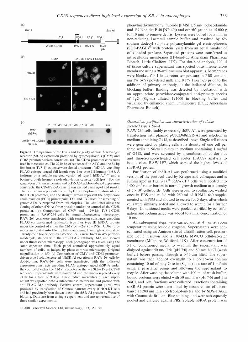
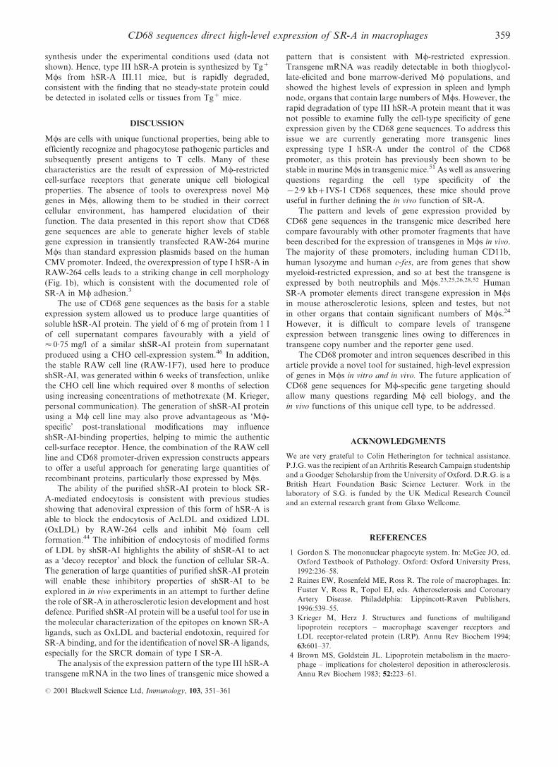
Figure 1. Comparison of the levels and longevity of class A scavengerreceptor (SR-A) expression provided by cytomegalovirus (CMV) andCD68 promoter-driven constructs. (a) The CD68 promoter constructsused in these studies. The 2940 bp of sequence 5k to ATG and the 83 bp®rst intron (IVS-1) sequence were cloned upstream of cDNAs encodingFLAG epitope-tagged full-length type I or type III human (h)SR-Aisoforms or a soluble secreted version of type I hSR-A,10,44 and abovine growth hormone polyadenylation cassette (bGHpA). For thegeneration of transgenic mice and pcDNA3 backbone-based expressionconstructs, the CD68/SR-A cassette was excised using KpnI and BsaAI.The bent arrow represents the multiple transcription initiation sites ofthe CD68 promoter, and the straight arrows represent the polymerasechain reaction (PCR) primer pairs TY1 and TY2 used for screening ofgenomic DNA prepared from tail biopsies. The XbaI sites allow thecloning of other cDNAs for expression under the control of the CD68promoter. (b) Comparison of CMV and x2.9 kb+IVS-1 CD68promoters in RAW-264 cells by immuno¯uorescence microscopy.RAW-264 cells were transfected with expression constructs encodingFLAG epitope-tagged full-length type I or type III hSR-A proteinunder the control of either the CMV or x2.9 kb+IVS-1 CD68 pro-moter and plated into 10-cm plates containing 11-mm glass coverslips.Twenty-four hours post-transfection, cells were ®xed in 4% parafor-maldehyde, stained with the anti-FLAG antibody, M2, and viewedunder ¯uorescence microscopy. Each photograph was taken using thesame exposure time. Each panel contained approximately equalnumbers of cells, as judged by phase-contrast microscopy. Originalmagni®cation r 150. (c) Comparison of CMV and CD68 promoter-driven type I soluble secreted (s)hSR-AI secretion in RAW-264 cells bydot-blotting. RAW-264 cells were transfected with the indicatedexpression constructs encoding FLAG epitope-tagged shSR-A underthe control of either the CMV promoter or the x2.9kb+IVS-1 CD68sequence. Supernatants were harvested and the media replaced every24 hr for a total of 9 days. One-hundred microlitres of each super-natant was spotted onto a nitrocellulose membrane and probed withanti-FLAG M2 antibody. Positive control supernatant (+ve) wasproduced by transfection of Chinese hamster ovary (CHO)-K1 cellsand had previously been shown to contain shSR-AI protein by Westernblotting. Data are from a single experiment and are representative ofthree similar experiments.
353CD68 sequences direct high-level expression of SR-A in macrophages
# 2001 Blackwell Science Ltd, Immunology, 103, 351±361

further puri®ed by twice passing the supernatant over a 5-ml
anti-FLAG antibody M2 sepharose-af®nity column (Sigma),
washing with 50 ml PBS and eluting with 15 ml of 0.1 M glycine
(pH 2.9), collecting 1-ml fractions. Fractions containing
shSR-AI were determined as described above, prior to being
pooled, dialysed against PBS and the ®nal protein concen-
tration measured using the BCA assay (Pierce & Warriner,
Chester, UK). The yield of shSR-AI protein was typically
30 mg from 5 l of conditioned supernatant, and showed no
contaminating protein bands when stained with Coomassie
Brilliant Blue.
The binding properties of puri®ed shSR-AI were analysed
using a poly G bead binding assay, as described previously.47
Brie¯y, 0.1 mg of puri®ed shSR-AI protein was added to 1 ml
of 150 mM NaCl, 50 mM Tris (pH 7.6) buffer and incubated
with poly G agarose beads for 2 hr at 4u, in the presence or
absence of a panel of SR-A ligands and their cognate non-
ligand controls. The beads were washed three times in 150 mM
NaCl, 50 mM Tris (pH 7.6), and bound protein was eluted
by boiling the beads in protein gel loading buffer for 5 min.
The samples were separated by SDS±PAGE and the binding
of shSR-AI was determined by Western blotting using anti-
FLAG antibody M2.
Quanti®cation of acetylated low-density lipoprotein uptake
To assay the uptake of acetylated low-density lipoprotein
(AcLDL), cells in 24-well plates were washed twice in PBS and
then preincubated with inhibitors at the indicated concentra-
tions for 30 min prior to labelling for 90 min with 2 mg/ml of
DiI (1,1k-dioctadecyl-1±3,3,3k,3k-tetramethylindocarbocyanine
perchlorate)-labelled-AcLDL (DiI-AcLDL) (Biogenesis,
Poole, UK) for 90 min at 37u in the presence of inhibitors.
Cells were washed ®ve times in PBS, resuspended in PBS
containing 5 mM EDTA and 10 mg/ml Lidocaine-HCl (Sigma),
®xed in a 4% solution of paraformaldehyde in PBS and
analysed on a ¯uorescence-activated cell sorter (FACScan; BD
Pharmingen, San Diego, CA) using the FL2 photomultiplier.
Generation of transgenic lines
A linear DNA fragment for the generation of transgenic
mice, shown in Fig. 1(a), was generated by digesting
plasmid pBSCD68hSR-AIII with KpnI, BsaAI and DrdI,
and purifying the separated fragment from an 0.8%
agarose gel using a QIAEX II gel extraction kit (Qiagen,
Crawley, UK) and an Elutip-D column (Schleicher & Schuell,
London, UK). Microinjection of CBA/B6 eggs with puri®ed
DNA and their subsequent transfer into pseudopregnant foster
(a) (b)
(c)
200>
97.4>
Con
trol
Bea
ds o
nly
+ P
oly
C
+ P
oly
I
+ B
SA
+ m
BS
A+
ChS
O4
+ Fu
coid
an
+ D
xSO
4
200>
97.4>
69>
46>
30>
200>
97.4>
69>
46>
30>
Non-reducing Reducing1l of supernatant from transfected raw cells
Concentrated to 40ml with MWCO 100Kd
Dialysed against 50mM Tris pH 7.6, 50mM NaCl
Partially purified with poly G agarose column
Further purified with a-FLAG sepharose column
Dialysed against PBS
Yield 6mg of shSR-AI
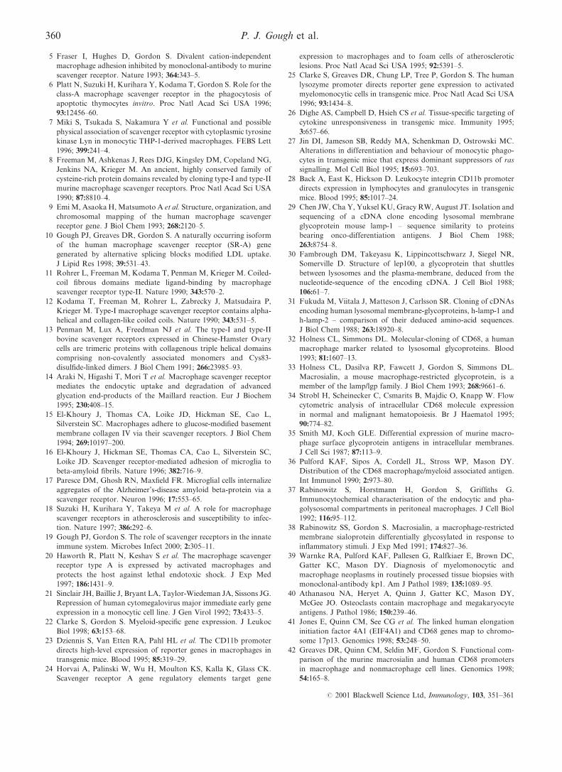
Figure 2. Generation and characterization of the soluble secreted form of type I human class A scavenger receptor (shSR-AI).
(a) Scheme for puri®cation of the shSR-AI protein. The ¯ow diagram shows the important steps involved in the puri®cation of
shSR-AI and highlights the 6-mg yield obtained per litre of conditioned tissue culture supernatant. (b) Analysis of puri®ed shSR-AI
protein by sodium dodecyl sulphate±polyacrylamide gel electrophoresis (SDS±PAGE) and Coomassie Brilliant Blue staining. Puri®ed
type I shSR-AI protein (2.5 mg) was separated by 10% SDS±PAGE under non-reducing and reducing conditions. Protein visualization
was achieved by staining with Coomassie R-250. (c) Analysis of shSR-AI binding properties by poly G±agarose. The binding of
puri®ed type I shSR-AI protein was performed in the presence of the following inhibitors: polycytidylic acid (poly C), polyinosinic
acid (poly I) (25 mg/ml), bovine serum albumin (BSA), maleylated BSA (mBSA) (100 mg/ml), chondroitin sulphate (ChSO4), dextran
sulphate (DxSO4) and fucoidan (150 mg/ml). The control sample lane was loaded with 25 ml of unpuri®ed conditioned medium.
354 P. J. Gough et al.
# 2001 Blackwell Science Ltd, Immunology, 103, 351±361
mothers was performed using standard techniques.48 Trans-
genic founders were identi®ed by the PCR using genomic
DNA extracted from 1-cm tail clips and two primer pair
combinations, as shown in Fig. 1(a): TY1: 5k-TTCTCGGC-
TCTGTGAATGACA-3k and 5k-CAGCCCTCTCTTGGAAA-
GGAGG-3k; TY2: 5k-AAGTGGGAAACGAAGAATTGC-3kand 5k-CTCTTGTTGTTTGAAGGTATTCTC-3k. Founder
mice positive for the transgene were mated with C57BL/6
mice, and all subsequent analyses were performed using the
resulting F1 progeny.
Isolation of primary Mw populations
Peritoneal cells were isolated by peritoneal lavage with PBS
at the indicated time-points after intraperitoneal injection of
1 ml of 4% thioglycollate broth. Bone marrow-derived Mws
(BMDM) were generated by the culture of bone marrow cells
in RPMI-1640 containing PSG, 10% FCS and 15% L-cell
conditioned medium as a source of macrophage-colony
stimulating factor (M-CSF).49 All animal procedures were
carried out in accordance with the Animals (Scienti®c
Procedures) Act 1986, and in accordance with Departmental
Ethical Review.
Analysis of transgene RNA expression
Total RNA was isolated from Mw populations and murine
organs using RNAzol (Biogenesis), according to the manu-
facturer's recommended protocol, and a Polytron tissue homo-
genizer (Brinkmann Instruments, Westbury, NY) to ensure
complete disruption of tissues. For Northern blot analyses,
total RNA (5±10 mg) was fractionated on a 1% formaldehyde/
agarose gel and transferred, by capillary blotting, to nylon
membrane (Hybond N+ Amersham Pharmacia Biotech).
Membranes were baked at 85u for 3 hr prior to prehybridiza-
tion in ExpressHyb hybridization solution (Clontech, Palo
Alto, CA) at 68u for 1 hr. Probes for b-actin (a 2-kb fragment
of human b-actin; Clontech) and hSR-AIII (a 1.2-kb fragment
generated by digestion of the expression plasmid, pcDNA3,10
with HindIII and XbaI) were labelled by random priming with
[a-32P]dATP and then incubated with blots in ExpressHyb for
2 hr at 68u. Blots were washed according to the manufacturer's
recommendations and then subjected to autoradiography at
x70u with intensifying screens.
For reverse transcription±PCR (RT±PCR) analyses, total
RNA was reverse transcribed using Moloney murine leukaemia
virus reverse transcriptase and an Oligo-dT primer (Life
Technologies), with reactions set up in the presence and
absence of reverse transcriptase to control for genomic DNA
contamination. The cDNA obtained served as a template
for PCR using the following oligonucleotide pairs: HPRT:
5k-GCTACCTGCTGGATTACAT-3k and 5k-CCAGTTTCAC-
TAATGACACAA-3k; macrosialin: 5k-TCCTTCACGATGAC-
ACCTACAG-3k and 5k-GGACCAGGCCAATGATGAGAG-
3k; hSR-A III transgene: 5k-GGGTGAGGCGGTTCAGCC-3kand5k-TTTCCTCTTCGCTGTCATTTC-3k.ThePCRproducts
were separated by electrophoresis on a 1.2% agarose gel and
visualized by ethidium bromide staining.
Metabolic labelling and pulse-chase analysis
Bone marrow cells were plated in six-well plates at a density of
5r105 cells/well and cultured for 6 days in medium containing
M-CSF, as described above. Adherent cells were washed three
times with PBS and pulse-labelled for 30 min with 0.5 ml of
methionine- and cysteine-de®cient RPMI-1640 supplemented
with 5% dialysed FCS and 500 mCi/ml of 35S-Trans label
(ICN Biomedicals, Aurora, OH). Cells were washed three
times with PBS and incubated in `chase' medium consisting of
RPMI-1640 supplemented with 10% FCS, PSG, 1 mM
unlabelled methionine and 4 mM unlabelled cysteine. After
incubation for the indicated times, cells were washed three
times in PBS and lysates were prepared as described above.
After preclearing samples by incubation with protein G±
sepharose (Amersham Pharmacia Biotech) at 4u, antigen was
precipitated by overnight incubation with 10 mg/ml of the anti-
FLAG antibody, M2, and protein G±sepharose. Samples were
washed six times, as described in ref. 10, and immunopreci-
pitated protein was collected by boiling the beads in reducing
Laemmli sample buffer prior to separation by 10% SDS±
PAGE.45 Gels were ®xed and impregnated with EN3HANCE
(NEN Life Science Products, Zaventen, Belgium) before drying
and exposure to ®lm at x70u.
RESULTS
CD68 gene sequences direct high-level, long-lasting expression
of hSR-A in RAW-264 cells
To characterize the usefulness of CD68 transcriptional
regulatory sequences as a tool to overexpress hSR-A in Mws,
we generated constructs in which 2.9 kb of 5k sequence and the
IVS-1 from the CD68 gene (x2.9 kb+IVS-1 CD68) were
placed upstream of an hSR-A cDNA and bGHpA (Fig. 1a).
Three plasmids with a pBluescript backbone were generated,
containing cDNAs for membrane-bound, FLAG-epitope
tagged type I or type III hSR-A (see ref. 10 for details) and
a soluble secreted type I hSR-A (shSR-AI) (see ref. 44 for
details).
The ability of these constructs to direct expression of hSR-
A was tested by transient transfection of the murine Mw cell
line, RAW-264. Immuno¯uorescence microscopy of cells 24-hr
post-transfection showed high-level expression of both type I
and type III hSR-A, as assessed by staining with the anti-
FLAG antibody, M2 (Fig. 1b). The levels of expression of both
hSR-A I and III, as judged by the brightness of cells, was much
greater in cells transfected with x2.9kb+IVS-1 CD68 pro-
moter-driven constructs than in CMV-driven controls. The
high levels of hSR-A expression produced by the x2.9kb+IVS-1 CD68 promoter-driven plasmids allowed for differences
in functions of the type I and type III hSR-A isoforms to be
appreciated. Cells overexpressing type I hSR-A spread out
more on glass coverslips, displaying a large veil of membrane
containing type I hSR-A protein at the point of adherence to
the glass surface, while type III hSR-A showed a predominately
perinuclear staining pattern in transiently transfected RAW
cells. These data are consistent with a role for type I SR-A in
Mw adhesion to glass surfaces. In previous experiments we
have shown that type III SR-A becomes trapped in the endo-
plasmic reticulum where it acts as a dominant negative
regulator of SR-A function.10 The data of Fig. 1 highlight
the usefulness of CD68 promoter-mediated overexpression for
studying SR-A biology in transfected Mw cell lines.
To test whether CD68 gene sequences could give sustained
expression of hSR-A, we examined the secretion of shSR-AI
355CD68 sequences direct high-level expression of SR-A in macrophages
# 2001 Blackwell Science Ltd, Immunology, 103, 351±361
(over a 9-day period) in RAW-264 cells transiently transfected
with either CMV- or x2.9kb+IVS-1 CD68-driven expression
constructs. Cell supernatants were harvested and replaced with
fresh medium every 24 hr, and the presence of the shSR-AI
protein in the supernatant was detected by dot-blot analysis
(Fig. 1c). In the supernatant obtained 24 hr post-transfection,
cells transfected with CD68 and CMV expression vectors
produced approximately equal levels of shSR-AI. Secretion of
shSR-AI by cells transfected with constructs containing the
CMV promoter declined rapidly, while the CD68-based
plasmid directed high levels of expression for the duration of
the experiment. Hence, the x2.9kb+IVS-1 CD68 promoter
fragment is able to generate high-level, long-lasting gene
expression in transiently transfected RAW-264 cells.
Puri®cation and characterization of soluble human SR-AI
protein
In previous experiments we had been unable to produce
milligram quantities of shSR-AI, using a variety of different
eukaryotic expression systems. The ability of the
x2.9kb+IVS-1 CD68 sequences to direct high-level secretion
of shSR-AI in RAW-264 cells offered a new approach for the
generation of large quantities of shSR-AI protein. We
generated stable shSR-AI-expressing RAW-264 polyclonal
populations using selection in medium containing 0.5 mg/ml
of G418. Single-cell clones were generated in medium contain-
ing 1 mg/ml of G418 and were screened for high-level
expression of shSR-AI through a combination of dot-blot
and FACS analysis (data not shown). Clone RAW-1F7 was
selected for further use and secreted shSR-AI at < 5 mg/ml
(data not shown). Puri®cation of shSR-AI protein was
performed using supernatants from bulk cultures of RAW-
1F7 as the starting material, as outlined in Fig. 2(a) and
described in detail in the Materials and methods. The purity of
shSR-AI preparations was assessed by SDS±PAGE and
Coomassie Brilliant Blue staining (Fig. 2b). Only the predicted
bands corresponding to dimeric and monomeric shSR-AI
protein (Mr 160 and 85 kDa, respectively) were detected under
non-reducing conditions, and only a single band of 85 kDa,
representing monomeric shSR-AI, was detected under reducing
conditions. In two independent preparations of shSR-AI,
yields of puri®ed protein averaged 6 mg/l of conditioned
culture supernatant.
To ensure that the shSR-AI protein produced by the RAW
cells had binding properties that were similar to the cellular
form of the receptor, the binding of shSR-AI to poly G±
agarose was examined. This approach has been used previously
to show that a radiolabelled, secreted version of the bovine
receptor had the requisite binding properties.47 Binding of
puri®ed shSR-AI to poly G±agarose was performed in the
presence or absence of a number of SR-A ligands and their
cognate non-ligands, and was assessed by SDS±PAGE and
Western blotting using the anti-FLAG antibody (Fig. 2c).
Under non-reducing conditions, the shSR-AI protein migrated
(as predicted) with bands corresponding to dimeric and
monomeric shSR-AI (160 and 85 kDa, respectively), detected
when binding occured in the absence of inhibitors. The binding
of shSR-AI to the poly G beads was speci®cally inhibited by the
SR-A ligands poly I, maleylated bovine serum albumin (BSA),
dextran sulphate and fucoidan, but not by the appropriate
non-ligands, indicating that the binding properties of shSR-AI
closely resemble those of its cellular counterpart.
Soluble human SR-I protein inhibits SR-A-mediated
endocytosis
We examined the effect of puri®ed shSR-AI protein on SR-
A-mediated endocytosis. Chinese hamster ovary (CHO) -K1
cells expressing high levels of type II hSR-A10 and RAW-264
cells were labelled with the ¯uorescently labelled scavenger
receptor ligand, DiI-AcLDL, in the presence or absence of poly
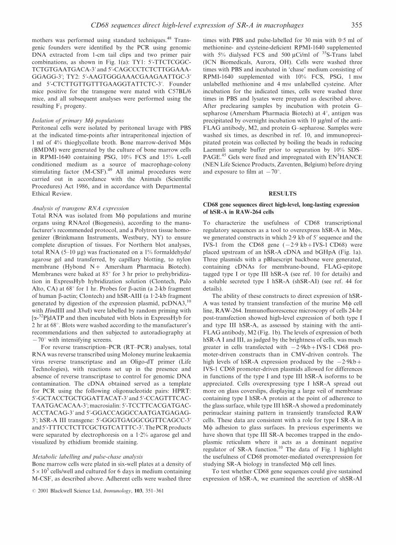
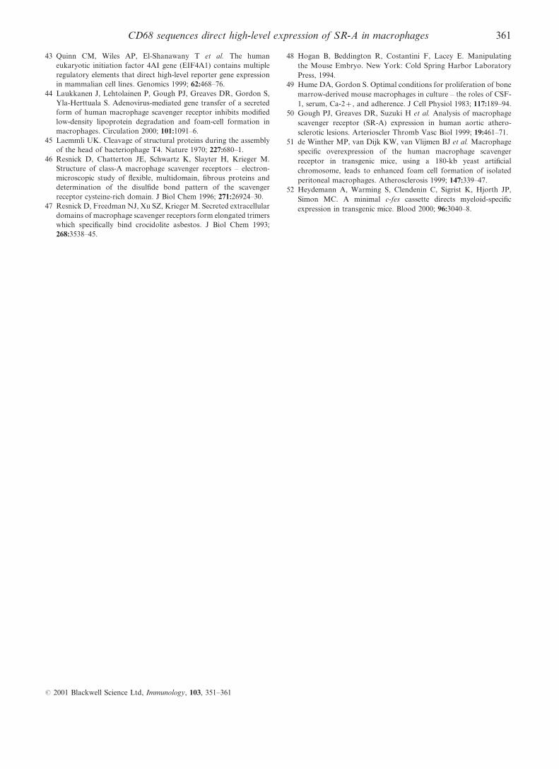
I, poly C and shSR-AI (Fig. 3). The uptake of DiI-AcLDL was
inhibited by poly I in both cell types, although this was
incomplete in RAW-264 cells owing to the expression of a non-
poly I inhibitable scavenger receptor. Increasing concentra-
tions of shSR-AI protein produced a dose-dependent inhibition
of DiI-AcLDL uptake in both cell types, with a 70% reduction
at the maximal concentration of shSR-AI used in this
experiment. This inhibition was speci®c for SR-A-mediated
endocytosis as the uptake of ¯uorescently labelled LDL, a non-
SR-A ligand, was unaffected and therefore shSR-AI protein
did not induce a general suppression of endocytic activity in
these cells (data not shown). Hence, puri®ed shSR-AI protein
can be used to inhibit SR-A-mediated endocytosis.
CD68 gene sequences direct expression of type III hSR-A
mRNA in Mw populations of transgenic mice
To test the ability of the x2.9 kb+IVS-1 CD68 gene
sequences to direct Mw-speci®c transgene expression in vivo,
fragments containing the CD68 promoter, IVS-1 and hSR-A
cDNA, were used to generate transgenic mice by pronuclear
injection. Linear DNA fragments containing the x2.9 kb
CD68 promoter + IVS-1, the coding sequence for type III
Dil-AcLDL uptake(percentage control)
0
+ shSR-AI 50µg/ml+ shSR-AI 20µg/ml+ shSR-AI 10µg/ml+ shSR-AI 5µg/ml+ shSR-AI 1µg/ml+ Poly C 50µg/ml+ Poly I 50µg/mlDil-AcLDL alone
20 40 60 80 100120
Dil-AcLDL uptake(percentage control)
0 20 40 60 80 100 120
RAW-264CHO SR-AII
Figure 3. Analysis of the ability of the soluble secreted form of type I
human class A scavenger receptor (shSR-AI) to inhibit SR-A-mediated
endocytosis. Comparison of the effects of shSR-AI on the uptake of
1,1k-dioctadecyl-1±3,3,3k,3k-tetramethylindocarbocyanine perchlorate-
labelled acetylated low-density lipoprotein (DiI-AcLDL) by Chinese
hamster ovary (CHO)-K1 cells expressing high levels of type II hSR-A
and by RAW-264 cells. Cells were labelled with DiI-AcLDL (2.5 mg/
ml) in the presence of inhibitors at the indicated concentrations for
90 min. Speci®c ¯uorescence intensity was calculated by subtracting
auto¯uorescence from the ¯uorescence intensity of DiI-AcLDL-
labelled cells using means derived by data analysis with CELLQUEST
software (Becton-Dickinson). Data are expressed as the percentage of
uptake by control cells labelled in the absence of inhibitors in the same
experiment. Results are from a single experiment and are representative
of three independent experiments. Poly C, polycytidylic acid; poly I,
polyinosinic acid.
356 P. J. Gough et al.
# 2001 Blackwell Science Ltd, Immunology, 103, 351±361
membrane bound hSR-A and bovine growth hormone poly
A+ sequences (Fig. 1a) were microinjected into fertilized
mouse eggs. Potential transgenic founders were screened by
using a combination of two PCR primer pairs, TY1 and TY2
(Fig. 1a), and con®rmed by Southern blot analysis of
restriction enzyme-digested genomic DNA (data not shown).
Two founder mice were obtained and bred with C57BL/6 mice
to establish the F1 animals used for all subsequent analyses.
Both lines of mice generated using the CD68 type III hSR-A
fragment, lines III.4 and III.11, showed germline transgene
transmission to F1 offspring in an approximately Mendelian
pattern (data not shown).
Expression of transgene mRNA in isolated Mw populations
from both transgenic lines was assessed by Northern blotting.
Peritoneal cells elicited either 1 or 4 days after injection of
III.11 mice with thioglycollate broth expressed type III hSR-A
mRNA, while no signal was detectable from transgene negative
(Tg±) littermates, despite the presence of equal amounts of
RNA, as judged by probing with b-actin (Fig. 4a). The
intensity of the hSR-A speci®c band was greater in the RNA
sample from the cells elicited 4 days after injection with
thioglycollate broth. The peritoneal cell population obtained
1 day after injection of thioglycollate broth was a mixture of
newly recruited and resident peritoneal Mws and recruited
neutrophils. In contrast, by 4 days after thioglycollate injec-
tion, all the recruited neutrophils had died and were replaced
with recruited monocytes and Mws. Hence, the relative levels of
type III hSR-A mRNA 1 or 4 days after thioglycollate
injection was consistent with Mw-restricted transgene expres-
sion. Expression of type III hSR-A mRNA in Mws from III.11
transgenic mice was further tested by examining expression in
BMDMs. Two independent BMDM preparations from
transgenic (Tg+) mice showed type III hSR-A transgene
mRNA expression, with no detectable expression from Tg±
littermates. Similar results were obtained in an analysis of
transgene mRNA expression by Mws isolated from Tg+ III.4
mice (data not shown). These data show that the CD68
sequences can drive transgene expression in Mw populations of
transgenic mice.
The pattern of type III hSR-A mRNA expression was
examined in a variety of murine tissues from Tg+ mice.
Northern blot analysis showed that transgene mRNA can be
detected in all of the organ samples from Tg+ III.4 mice (except
for liver where the b-actin probed control showed low levels of
intact mRNA), with lung, spleen and lymph nodes showing the
highest levels of expression (Fig. 4b). No bands of the
appropriate size could be detected in RNA samples from a
Tg± mouse. This pattern of transgene expression is consistent
(a)
Macrosialin
hSR-A
HPRT
hSR-A(gDNA)
+ – + – + – + – + – + –
Live
r
Lung
Hea
rt
Kidn
ey
Sple
en
Lym
ph n
ode
(c)
hSR-A
b-actin
Hum
an Mw
Live
rLu
ngH
eart
Kidn
eySp
leen
L. b
owel
S. b
owel
Sk. m
uscl
eLy
mph
nod
eBr
ain
Live
rLu
ngSp
leen
TG+ TG–(b)
hSR-A
b-actin
d1 T
G+
d4 T
G+
d4 T
G–
TG+
TG+
TG–
Thio-elicitedPeritoneal cells
Bone marrowderived Mw
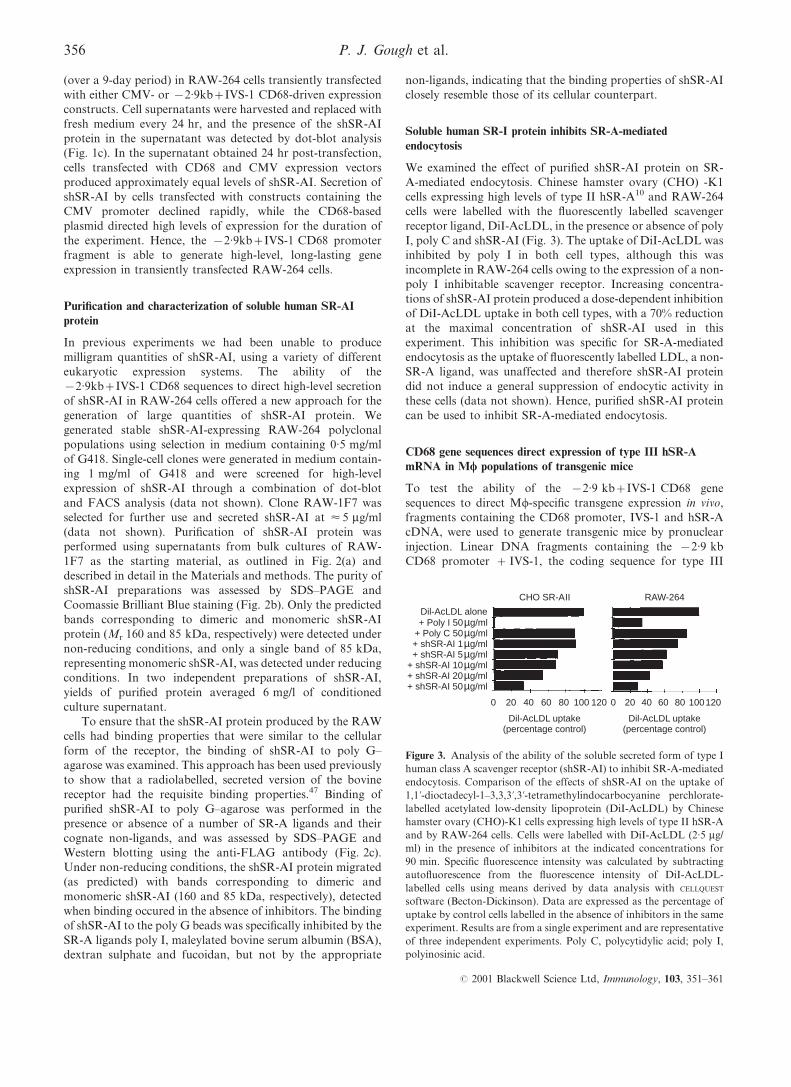
Figure 4. Analysis of human class A scavenger receptor (hSR-A) III
transgene RNA expression. (a) Northern blot of isolated macrophage
(Mw) populations. Total RNA was prepared from peritoneal cells
isolated from transgenic line III.11 (Tg+) mice and non-transgenic
(Tg±) littermates, either 1 or 4 days after injection of 1 ml of
thioglycollate broth. Bone marrow-derived Mws (BMDMs) were
generated by the culture of bone marrow precursors in medium
containing macrophage colony-stimulating factor (M-CSF) and total
RNA isolated from adherent cells cultured for 6 days. Total RNA
(5 mg) was denatured and separated on a 1% formaldehyde±agarose gel
prior to capillary transfer to a nylon membrane. Blots were hybridized
with [a-32P]dATP-labelled DNA probes for b-actin (a 2-kb fragment of
human b-actin; Clontech) or hSR-A III transgene (a 1.2-kb fragment of
type III hSR-A coding sequence generated by digestion of expression
plasmid pcDNA 3 with HindIII and XbaI) and exposed to ®lm. (b)
Northern blot of adult tissues. Total tissue RNA (10 mg) prepared
using organs collected from transgenic line III.4 (Tg+) mice and non-
transgenic (Tg±) littermates, and from human monocyte-derived Mw
total RNA (10 mg), was denatured and separated on a 1% formalde-
hyde±agarose gel prior to transfer and hybridization as described in (a).
The different sizes of SR-A hybridizing bands in the human and
transgenic RNA samples are caused by the absence of 3k untranslated
sequences in the CD68 SR-A type III transgene. (c) Reverse
transcription±polymerase chain reaction (RT±PCR) of adult tissues.
Total tissue RNA from organs collected from transgenic line III.4
(Tg+) mice was reverse transcribed in the presence (+) or absence (±)
of Moloney murine leukemia virus reverse transcriptase. cDNA
synthesized from 100 ng of total RNA served as a template for
subsequent PCR using primers speci®c for hypoxanthine phosphor-
ibosyl transferase (HPRT), macrosialin or the hSR-A III transgene.
To enable a comparison of the relative levels of RNA for macrosialin
and the hSR-A III transgene, PCR was performed with both
oligonucleotide pairs in the same reaction tube. Primers for HPRT
and macrosialin do not generate a detectable reaction product from
contaminating genomic DNA (gDNA) under the conditions used,
while the product corresponding to the ampli®cation of the hSR-A
III transgene genomic DNA is 83 bp larger owing to the presence
of the ®rst intron (IVS-1) sequence, which is spliced out during
the processing of the primary RNA transcript. Reaction products
were separated on a 1.2% agarose gel and visualized by ethidium
bromide staining.
357CD68 sequences direct high-level expression of SR-A in macrophages
# 2001 Blackwell Science Ltd, Immunology, 103, 351±361
with Mw-restricted targeting as the spleen and lymph nodes are
organs that contain the largest proportion of Mws as a
percentage of the total cell population. Comparison of the
intensity of SR-A-speci®c bands between RNA samples from
human monocyte-derived Mws, which express high levels of
SR-A mRNA,50 and transgenic spleen, where Mws constitute
< 10±20% of the total cell number, shows that CD68 sequences
direct signi®cant levels of transgene RNA expression.
To further examine the speci®city of expression generated
by CD68 gene sequences, the expression of transgene RNA was
compared to that of macrosialin, the murine homologue of
human CD68, using RT±PCR. Each of the samples generated
from organs from III.4 Tg+ mice had approximately equal
amounts of cDNA, as judged by the intensity of the bands
produced using an oligonucleotide pair speci®c for the
housekeeping gene, hypoxanthine phosphoribosyl transferase
(HPRT) (Fig. 4c). The highest levels of macrosialin expression
were seen in the lung, spleen and lymph nodes, although all
tissues expressed some mRNA. The pattern of mRNA
expression seen with hSR-A transgene speci®c primers was
largely similar to that for macrosialin, although there were
variations in the relative intensities of the bands for the two
mRNA species. There was no detectable hSR-A transgene
expression in the liver cDNA sample. In the spleen cDNA
sample, the type III hSR-A-speci®c band was of higher
intensity than that for macrosialin, suggesting that the
transgene mRNA was present at higher levels than the
endogenous gene. Analysis of type III hSR-A mRNA
expression in a similar panel of tissues from III.11 Tg+ mice
revealed a similar pattern of transgene expression (data not
shown). Hence, CD68 gene sequences can direct transgene
expression to Mw populations in vivo, in a pattern that is
suggestive of Mw-speci®c targeting.
Type III hSR-A protein is not readily detected in Mws from
transgenic mice as a result of its rapid degradation
To examine further the cell type speci®city of transgene
expression, immunohistochemistry was performed on a panel
of tissues from III.4 and III.11 Tg+ mice. Tissue sections were
probed with the anti-FLAG monoclonal antibody (mAb) and
anti-hSR-A polyclonal antibodies, but no staining pattern
unique to Tg+ mice was observed for either transgenic line
(data not shown). Appropriate staining patterns were observed
with anti-macrosialin and anti-F4/80 antibodies, and were
identical in tissue sections from Tg+ and Tg± mice (data not
shown), indicating normal Mw distributions in these mice. In
an effort to optimize staining protocols for the detection of type
III hSR-A protein using anti-FLAG and anti-hSR-A anti-
bodies, we performed immuno¯uorescence staining of
BMDMs from both transgenic lines. Despite numerous
attempts using many different experimental conditions, no
type III hSR-A protein could be detected (data not shown). As
an alternative approach to detect type III hSR-A protein
expression, Western blotting of protein lysates from BMDMs
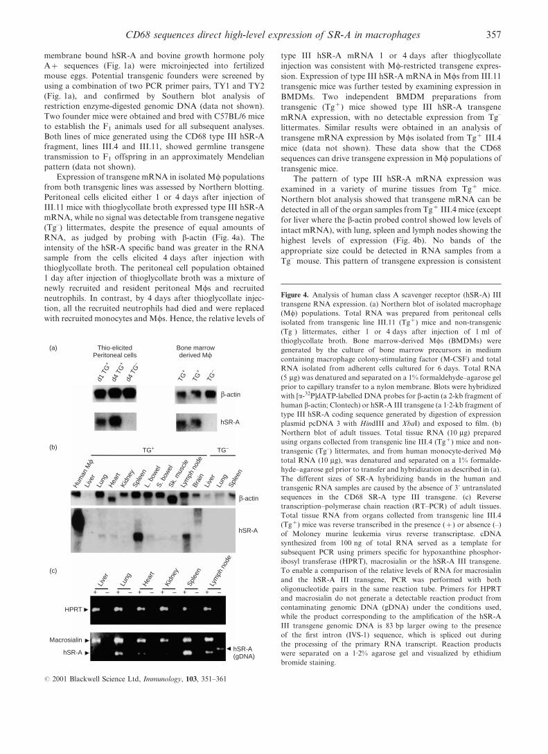
derived from III.11 mice was performed as shown in Fig. 5(a).
Under conditions in which we could readily detect type III
hSR-A protein expressed by transfected RAW-264 cells, no
expression could be detected in Tg+ BMDMs. Identical blots
probed for macrosialin (FA-11) or mannose receptor (a-MR)
showed that other Mw antigens were readily detectable in these
cells (Fig. 5a). Hence, despite the expression of type III hSR-A
mRNA in isolated Mws and tissues from both lines of
transgenic mice, no type III hSR-A protein could be detected.
Type III hSR-A protein is retained in the endoplasmic
reticulum of cells where it is able to exert dominant negative
properties, presumably by inducing the degradation of type I
and type II SR-A.10 The protein can be readily detected at
steady-state levels when expressed in RAW-264 cells (Fig. 1b).
However, it is possible that a combination of lower rates of
synthesis and higher rates of degradation in the endoplasmic
reticulum of Mws of CD68-type III hSR-A transgenic mice
mean that steady-state levels of type III hSR-A protein do not
accumulate. To test this hypothesis, BMDMs from Tg+ hSR-A
III.11 mice were pulse labelled for 30 min with 35S-methionine
and cysteine, and chased for the times indicated in Fig. 5(b).
Type III hSR-A protein was immunoprecipitated with an anti-
FLAG antibody, and samples were separated under reducing
conditions by SDS±PAGE prior to visualization by autoradio-
graphy. A band of the correct size for monomeric type III hSR-
A protein (Mr < 80 kDa) was clearly evident after the initial
pulse, but this disappeared after 1 or 3 hr of chase. The other
bands present at constant intensity through the chase period
probably represent Fc receptors synthesized by Mws and
immunoprecipitated because of their interaction with the
murine anti-FLAG antibody. Attempts to alter the kinetics
of type III hSR-A protein degradation through use of the
protease inhibitors lactacystin or NH4Cl were unsuccessful
owing to a signi®cant inhibition in the basal levels of protein
(b)
<200>
<97.4>
<69>
<46>
<30>
<200>
<97.4>
<69>
<46>
<30>
200>
97.4>
69>
46>
30>
200>
97.4>
69>
46>
– + – + – + 0 1 3
Chase (hr)(a)
a-FLAGM2
FA-11 a-MR
Figure 5. Analysis of human class A scavenger receptor (hSR-A) III
protein expression in bone marrow-derived macrophages (BMDMs)
isolated from line III.11 transgenic mice. (a) Western blotting. Protein
lysates were generated from adherent BMDMs cultured for 6 days in
medium containing macrophage colony-stimulating factor (M-CSF).
Total protein lysate (30 mg) from non-transgenic (±) and transgenic (+)
BMDMs was separated by 6% non-reducing sodium dodecyl sulphate±
polyacrylamide gel electrophoresis (SDS±PAGE). After transfer to
nitrocellulose, blots were probed with anti-FLAG antibody, M2
(10 mg/ml), anti-mouse macrosialin antibody, FA-11 (10 mg/ml), or
anti-mouse mannose receptor polyclonal antibody (1 : 5000) and the
appropriate horseradish peroxidase (HRP)-conjugated secondary
antibody prior to development with enhanced chemiluminescence
(ECL) reagent. (b) Pulse-chase. BMDMs cultured for 6 days in
medium containing M-CSF were pulse-labelled for 30 min with 35S-
labelled cysteine and -methionine and chased for the indicated times
prior to lysis and immunoprecipitation using anti-FLAG antibody,
M2, and protein G±sepharose beads. Samples were separated by SDS±
PAGE, on an 8% gel, under reducing conditions prior to visualization
by autoradiography.
358 P. J. Gough et al.
# 2001 Blackwell Science Ltd, Immunology, 103, 351±361
synthesis under the experimental conditions used (data not
shown). Hence, type III hSR-A protein is synthesized by Tg+
Mws from hSR-A III.11 mice, but is rapidly degraded,
consistent with the ®nding that no steady-state protein could
be detected in isolated cells or tissues from Tg+ mice.
DISCUSSION
Mws are cells with unique functional properties, being able to
ef®ciently recognize and phagocytose pathogenic particles and
subsequently present antigens to T cells. Many of these
characteristics are the result of expression of Mw-restricted
cell-surface receptors that generate unique cell biological
properties. The absence of tools to overexpress novel Mwgenes in Mws, allowing them to be studied in their correct
cellular environment, has hampered elucidation of their
function. The data presented in this report show that CD68
gene sequences are able to generate higher levels of stable
gene expression in transiently transfected RAW-264 murine
Mws than standard expression plasmids based on the human
CMV promoter. Indeed, the overexpression of type I hSR-A in
RAW-264 cells leads to a striking change in cell morphology
(Fig. 1b), which is consistent with the documented role of
SR-A in Mw adhesion.3
The use of CD68 gene sequences as the basis for a stable
expression system allowed us to produce large quantities of
soluble hSR-AI protein. The yield of 6 mg of protein from 1 l
of cell supernatant compares favourably with a yield of
< 0.75 mg/l of a similar shSR-AI protein from supernatant
produced using a CHO cell-expression system.46 In addition,
the stable RAW cell line (RAW-1F7), used here to produce
shSR-AI, was generated within 6 weeks of transfection, unlike
the CHO cell line which required over 8 months of selection
using increasing concentrations of methotrexate (M. Krieger,
personal communication). The generation of shSR-AI protein
using a Mw cell line may also prove advantageous as `Mw-
speci®c' post-translational modi®cations may in¯uence
shSR-AI-binding properties, helping to mimic the authentic
cell-surface receptor. Hence, the combination of the RAW cell
line and CD68 promoter-driven expression constructs appears
to offer a useful approach for generating large quantities of
recombinant proteins, particularly those expressed by Mws.
The ability of the puri®ed shSR-AI protein to block SR-
A-mediated endocytosis is consistent with previous studies
showing that adenoviral expression of this form of hSR-A is
able to block the endocytosis of AcLDL and oxidized LDL
(OxLDL) by RAW-264 cells and inhibit Mw foam cell
formation.44 The inhibition of endocytosis of modi®ed forms
of LDL by shSR-AI highlights the ability of shSR-AI to act
as a `decoy receptor' and block the function of cellular SR-A.
The generation of large quantities of puri®ed shSR-AI protein
will enable these inhibitory properties of shSR-AI to be
explored in in vivo experiments in an attempt to further de®ne
the role of SR-A in atherosclerotic lesion development and host
defence. Puri®ed shSR-AI protein will be a useful tool for use in
the molecular characterization of the epitopes on known SR-A
ligands, such as OxLDL and bacterial endotoxin, required for
SR-A binding, and for the identi®cation of novel SR-A ligands,
especially for the SRCR domain of type I SR-A.
The analysis of the expression pattern of the type III hSR-A
transgene mRNA in the two lines of transgenic mice showed a
pattern that is consistent with Mw-restricted expression.
Transgene mRNA was readily detectable in both thioglycol-
late-elicited and bone marrow-derived Mw populations, and
showed the highest levels of expression in spleen and lymph
node, organs that contain large numbers of Mws. However, the
rapid degradation of type III hSR-A protein meant that it was
not possible to examine fully the cell-type speci®city of gene
expression given by the CD68 gene sequences. To address this
issue we are currently generating more transgenic lines
expressing type I hSR-A under the control of the CD68
promoter, as this protein has previously been shown to be
stable in murine Mws in transgenic mice.51 As well as answering
questions regarding the cell type speci®city of the
x2.9 kb+IVS-1 CD68 sequences, these mice should prove
useful in further de®ning the in vivo function of SR-A.
The pattern and levels of gene expression provided by
CD68 gene sequences in the transgenic mice described here
compare favourably with other promoter fragments that have
been described for the expression of transgenes in Mws in vivo.
The majority of these promoters, including human CD11b,
human lysozyme and human c-fes, are from genes that show
myeloid-restricted expression, and so at best the transgene is
expressed by both neutrophils and Mws.23,25,26,28,52 Human
SR-A promoter elements direct transgene expression in Mws
in mouse atherosclerotic lesions, spleen and testes, but not
in other organs that contain signi®cant numbers of Mws.24
However, it is dif®cult to compare levels of transgene
expression between transgenic lines owing to differences in
transgene copy number and the reporter gene used.
The CD68 promoter and intron sequences described in this
article provide a novel tool for sustained, high-level expression
of genes in Mws in vitro and in vivo. The future application of
CD68 gene sequences for Mw-speci®c gene targeting should
allow many questions regarding Mw cell biology, and the
in vivo functions of this unique cell type, to be addressed.
ACKNOWLEDGMENTS
We are very grateful to Colin Hetherington for technical assistance.
P.J.G. was the recipient of an Arthritis Research Campaign studentship
and a Goodger Scholarship from the University of Oxford. D.R.G. is a
British Heart Foundation Basic Science Lecturer. Work in the
laboratory of S.G. is funded by the UK Medical Research Council
and an external research grant from Glaxo Wellcome.
REFERENCES
1 Gordon S. The mononuclear phagocyte system. In: McGee JO, ed.
Oxford Textbook of Pathology. Oxford: Oxford University Press,
1992:236±58.
2 Raines EW, Rosenfeld ME, Ross R. The role of macrophages. In:
Fuster V, Ross R, Topol EJ, eds. Atherosclerosis and Coronary
Artery Disease. Philadelphia: Lippincott-Raven Publishers,
1996:539±55.
3 Krieger M, Herz J. Structures and functions of multiligand
lipoprotein receptors ± macrophage scavenger receptors and
LDL receptor-related protein (LRP). Annu Rev Biochem 1994;
63:601±37.
4 Brown MS, Goldstein JL. Lipoprotein metabolism in the macro-
phage ± implications for cholesterol deposition in atherosclerosis.
Annu Rev Biochem 1983; 52:223±61.
359CD68 sequences direct high-level expression of SR-A in macrophages
# 2001 Blackwell Science Ltd, Immunology, 103, 351±361
5 Fraser I, Hughes D, Gordon S. Divalent cation-independent
macrophage adhesion inhibited by monoclonal-antibody to murine
scavenger receptor. Nature 1993; 364:343±5.
6 Platt N, Suzuki H, Kurihara Y, Kodama T, Gordon S. Role for the
class-A macrophage scavenger receptor in the phagocytosis of
apoptotic thymocytes invitro. Proc Natl Acad Sci USA 1996;
93:12456±60.
7 Miki S, Tsukada S, Nakamura Y et al. Functional and possible
physical association of scavenger receptor with cytoplasmic tyrosine
kinase Lyn in monocytic THP-1-derived macrophages. FEBS Lett
1996; 399:241±4.
8 Freeman M, Ashkenas J, Rees DJG, Kingsley DM, Copeland NG,
Jenkins NA, Krieger M. An ancient, highly conserved family of
cysteine-rich protein domains revealed by cloning type-I and type-II
murine macrophage scavenger receptors. Proc Natl Acad Sci USA
1990; 87:8810±4.
9 Emi M, Asaoka H, Matsumoto A et al. Structure, organization, and
chromosomal mapping of the human macrophage scavenger
receptor gene. J Biol Chem 1993; 268:2120±5.
10 Gough PJ, Greaves DR, Gordon S. A naturally occurring isoform
of the human macrophage scavenger receptor (SR-A) gene
generated by alternative splicing blocks modi®ed LDL uptake.
J Lipid Res 1998; 39:531±43.
11 Rohrer L, Freeman M, Kodama T, Penman M, Krieger M. Coiled-
coil ®brous domains mediate ligand-binding by macrophage
scavenger receptor type-II. Nature 1990; 343:570±2.
12 Kodama T, Freeman M, Rohrer L, Zabrecky J, Matsudaira P,
Krieger M. Type-I macrophage scavenger receptor contains alpha-
helical and collagen-like coiled coils. Nature 1990; 343:531±5.
13 Penman M, Lux A, Freedman NJ et al. The type-I and type-II
bovine scavenger receptors expressed in Chinese-Hamster Ovary
cells are trimeric proteins with collagenous triple helical domains
comprising non-covalently associated monomers and Cys83-
disul®de-linked dimers. J Biol Chem 1991; 266:23985±93.
14 Araki N, Higashi T, Mori T et al. Macrophage scavenger receptor
mediates the endocytic uptake and degradation of advanced
glycation end-products of the Maillard reaction. Eur J Biochem
1995; 230:408±15.
15 El-Khoury J, Thomas CA, Loike JD, Hickman SE, Cao L,
Silverstein SC. Macrophages adhere to glucose-modi®ed basement
membrane collagen IV via their scavenger receptors. J Biol Chem
1994; 269:10197±200.
16 El-Khoury J, Hickman SE, Thomas CA, Cao L, Silverstein SC,
Loike JD. Scavenger receptor-mediated adhesion of microglia to
beta-amyloid ®brils. Nature 1996; 382:716±9.
17 Paresce DM, Ghosh RN, Max®eld FR. Microglial cells internalize
aggregates of the Alzheimer's-disease amyloid beta-protein via a
scavenger receptor. Neuron 1996; 17:553±65.
18 Suzuki H, Kurihara Y, Takeya M et al. A role for macrophage
scavenger receptors in atherosclerosis and susceptibility to infec-
tion. Nature 1997; 386:292±6.
19 Gough PJ, Gordon S. The role of scavenger receptors in the innate
immune system. Microbes Infect 2000; 2:305±11.
20 Haworth R, Platt N, Keshav S et al. The macrophage scavenger
receptor type A is expressed by activated macrophages and
protects the host against lethal endotoxic shock. J Exp Med
1997; 186:1431±9.
21 Sinclair JH, Baillie J, Bryant LA, Taylor-Wiedeman JA, Sissons JG.
Repression of human cytomegalovirus major immediate early gene
expression in a monocytic cell line. J Gen Virol 1992; 73:433±5.
22 Clarke S, Gordon S. Myeloid-speci®c gene expression. J Leukoc
Biol 1998; 63:153±68.
23 Dziennis S, Van Etten RA, Pahl HL et al. The CD11b promoter
directs high-level expression of reporter genes in macrophages in
transgenic mice. Blood 1995; 85:319±29.
24 Horvai A, Palinski W, Wu H, Moulton KS, Kalla K, Glass CK.
Scavenger receptor A gene regulatory elements target gene
expression to macrophages and to foam cells of atherosclerotic
lesions. Proc Natl Acad Sci USA 1995; 92:5391±5.
25 Clarke S, Greaves DR, Chung LP, Tree P, Gordon S. The human
lysozyme promoter directs reporter gene expression to activated
myelomonocytic cells in transgenic mice. Proc Natl Acad Sci USA
1996; 93:1434±8.
26 Dighe AS, Campbell D, Hsieh CS et al. Tissue-speci®c targeting of
cytokine unresponsiveness in transgenic mice. Immunity 1995;
3:657±66.
27 Jin DI, Jameson SB, Reddy MA, Schenkman D, Ostrowski MC.
Alterations in differentiation and behaviour of monocytic phago-
cytes in transgenic mice that express dominant suppressors of ras
signalling. Mol Cell Biol 1995; 15:693±703.
28 Back A, East K, Hickson D. Leukocyte integrin CD11b promoter
directs expression in lymphocytes and granulocytes in transgenic
mice. Blood 1995; 85:1017±24.
29 Chen JW, Cha Y, Yuksel KU, Gracy RW, August JT. Isolation and
sequencing of a cDNA clone encoding lysosomal membrane
glycoprotein mouse lamp-1 ± sequence similarity to proteins
bearing onco-differentiation antigens. J Biol Chem 1988;
263:8754±8.
30 Fambrough DM, Takeyasu K, Lippincottschwarz J, Siegel NR,
Somerville D. Structure of lep100, a glycoprotein that shuttles
between lysosomes and the plasma-membrane, deduced from the
nucleotide-sequence of the encoding cDNA. J Cell Biol 1988;
106:61±7.
31 Fukuda M, Viitala J, Matteson J, Carlsson SR. Cloning of cDNAs
encoding human lysosomal membrane-glycoproteins, h-lamp-1 and
h-lamp-2 ± comparison of their deduced amino-acid sequences.
J Biol Chem 1988; 263:18920±8.
32 Holness CL, Simmons DL. Molecular-cloning of CD68, a human
macrophage marker related to lysosomal glycoproteins. Blood
1993; 81:1607±13.
33 Holness CL, Dasilva RP, Fawcett J, Gordon S, Simmons DL.
Macrosialin, a mouse macrophage-restricted glycoprotein, is a
member of the lamp/lgp family. J Biol Chem 1993; 268:9661±6.
34 Strobl H, Scheinecker C, Csmarits B, Majdic O, Knapp W. Flow
cytometric analysis of intracellular CD68 molecule expression
in normal and malignant hematopoiesis. Br J Haematol 1995;
90:774±82.
35 Smith MJ, Koch GLE. Differential expression of murine macro-
phage surface glycoprotein antigens in intracellular membranes.
J Cell Sci 1987; 87:113±9.
36 Pulford KAF, Sipos A, Cordell JL, Stross WP, Mason DY.
Distribution of the CD68 macrophage/myeloid associated antigen.
Int Immunol 1990; 2:973±80.
37 Rabinowitz S, Horstmann H, Gordon S, Grif®ths G.
Immunocytochemical characterisation of the endocytic and pha-
golysosomal compartments in peritoneal macrophages. J Cell Biol
1992; 116:95±112.
38 Rabinowitz SS, Gordon S. Macrosialin, a macrophage-restricted
membrane sialoprotein differentially glycosylated in response to
in¯ammatory stimuli. J Exp Med 1991; 174:827±36.
39 Warnke RA, Pulford KAF, Pallesen G, Ralfkiaer E, Brown DC,
Gatter KC, Mason DY. Diagnosis of myelomonocytic and
macrophage neoplasms in routinely processed tissue biopsies with
monoclonal-antibody kp1. Am J Pathol 1989; 135:1089±95.
40 Athanasou NA, Heryet A, Quinn J, Gatter KC, Mason DY,
McGee JO. Osteoclasts contain macrophage and megakaryocyte
antigens. J Pathol 1986; 150:239±46.
41 Jones E, Quinn CM, See CG et al. The linked human elongation
initiation factor 4A1 (EIF4A1) and CD68 genes map to chromo-
some 17p13. Genomics 1998; 53:248±50.
42 Greaves DR, Quinn CM, Seldin MF, Gordon S. Functional com-
parison of the murine macrosialin and human CD68 promoters
in macrophage and nonmacrophage cell lines. Genomics 1998;
54:165±8.
360 P. J. Gough et al.
# 2001 Blackwell Science Ltd, Immunology, 103, 351±361
43 Quinn CM, Wiles AP, El-Shanawany T et al. The human
eukaryotic initiation factor 4AI gene (EIF4A1) contains multiple
regulatory elements that direct high-level reporter gene expression
in mammalian cell lines. Genomics 1999; 62:468±76.
44 Laukkanen J, Lehtolainen P, Gough PJ, Greaves DR, Gordon S,
Yla-Herttuala S. Adenovirus-mediated gene transfer of a secreted
form of human macrophage scavenger receptor inhibits modi®ed
low-density lipoprotein degradation and foam-cell formation in
macrophages. Circulation 2000; 101:1091±6.
45 Laemmli UK. Cleavage of structural proteins during the assembly
of the head of bacteriophage T4. Nature 1970; 227:680±1.
46 Resnick D, Chatterton JE, Schwartz K, Slayter H, Krieger M.
Structure of class-A macrophage scavenger receptors ± electron-
microscopic study of ¯exible, multidomain, ®brous proteins and
determination of the disul®de bond pattern of the scavenger
receptor cysteine-rich domain. J Biol Chem 1996; 271:26924±30.
47 Resnick D, Freedman NJ, Xu SZ, Krieger M. Secreted extracellular
domains of macrophage scavenger receptors form elongated trimers
which speci®cally bind crocidolite asbestos. J Biol Chem 1993;
268:3538±45.
48 Hogan B, Beddington R, Costantini F, Lacey E. Manipulating
the Mouse Embryo. New York: Cold Spring Harbor Laboratory
Press, 1994.
49 Hume DA, Gordon S. Optimal conditions for proliferation of bone
marrow-derived mouse macrophages in culture ± the roles of CSF-
1, serum, Ca-2+, and adherence. J Cell Physiol 1983; 117:189±94.
50 Gough PJ, Greaves DR, Suzuki H et al. Analysis of macrophage
scavenger receptor (SR-A) expression in human aortic athero-
sclerotic lesions. Arterioscler Thromb Vasc Biol 1999; 19:461±71.
51 de Winther MP, van Dijk KW, van Vlijmen BJ et al. Macrophage
speci®c overexpression of the human macrophage scavenger
receptor in transgenic mice, using a 180-kb yeast arti®cial
chromosome, leads to enhanced foam cell formation of isolated
peritoneal macrophages. Atherosclerosis 1999; 147:339±47.
52 Heydemann A, Warming S, Clendenin C, Sigrist K, Hjorth JP,
Simon MC. A minimal c-fes cassette directs myeloid-speci®c
expression in transgenic mice. Blood 2000; 96:3040±8.
361CD68 sequences direct high-level expression of SR-A in macrophages
# 2001 Blackwell Science Ltd, Immunology, 103, 351±361