textbook and color atlas of salivary gland pathology
359
-
Upload
khangminh22 -
Category
Documents
-
view
4 -
download
0
Transcript of textbook and color atlas of salivary gland pathology

TEXTBOOK AND COLOR ATLAS OF SALIVARY GLAND PATHOLOGY DIAGNOSIS AND MANAGEMENT


TEXTBOOK AND COLOR ATLAS OF SALIVARY GLAND PATHOLOGY DIAGNOSIS AND MANAGEMENT
By
Eric R. Carlson
Robert A. Ord
A John Wiley & Sons, Inc., Publication

Edition fi rst published 2008
© 2008 Wiley-Blackwell
Blackwell Munksgaard, formerly an imprint of Blackwell Publishing was acquired by John Wiley & Sons in February 2007. Blackwell’s publishing programme has been merged with Wiley’s global Scientifi c, Technical, and Medical business to form Wiley-Blackwell.
Editorial Offi ce2121 State Avenue, Ames, Iowa 50014-8300, USA
For details of our global editorial offi ces, for customer services and for information about how to apply for permission to reuse the copyright material in this book please see our website at www.wiley.com/wiley-blackwell.
Authorization to photocopy items for internal or personal use, or the internal or personal use of specifi c clients, is granted by the publisher, provided that the base fee is paid directly to the Copyright Clearance Center, 222 Rosewood Drive, Danvers, MA 01923. For those organizations that have been granted a photocopy license by CCC, a separate system of payments has been arranged. The fee codes for users of the Transactional Reporting Service are ISBN-13: 978-0-8138-0262-6/2008.
Designations used by companies to distinguish their products are often claimed as trademarks. All brand names and product names used in this book are trade names, service marks, trademarks or registered trademarks of their respective owners. The publisher is not associated with any product or vendor mentioned in this book. This publication is designed to provide accurate and authoritative information in regard to the subject matter covered. It is sold on the understanding that the publisher is not engaged in rendering professional services. If professional advice or other expert assistance is required, the services of a competent professional should be sought.
Library of Congress Cataloguing-in-Publication Data
Textbook and color atlas of salivary gland pathology : diagnosis and management / edited by Eric Carlson, Robert Ord. p. ; cm. Includes bibliographical references and index. ISBN-13: 978-0-8138-0262-6 (alk. paper) ISBN-10: 0-8138-0262-8 (alk. paper) 1. Salivary glands–Diseases–Textbooks. 2. Salivary glands–Diseases–Atlases. I. Carlson, Eric R. II. Ord, Robert A. [DNLM: 1. Salivary Gland Diseases–diagnosis–Atlases. 2. Salivary Gland Diseases–surgery–Atlases. 3. Salivary Glands–anatomy & histology–Atlases. WI 17 T355 2008] RC815.5.T49 2008 616.3′16–dc22 2008006941A catalogue record for this book is available from the U.S. Library of Congress.
Set in 10.5/12pt ITC Slimbach by SNP Best-set Typesetter Ltd., Hong KongPrinted in Singapore by Markono Print Media Pte Ltd
1 2008
DisclaimerThe contents of this work are intended to further general scientifi c research, understanding, and discussion only and are not intended and should not be relied upon as recommending or promoting a specifi c method, diagnosis, or treatment by practitioners for any particular patient. The publisher and the author make no representations or warranties with respect to the accuracy or completeness of the contents of this work and specifi cally disclaim all warranties, including without limitation any implied warranties of fi tness for a particular purpose. In view of ongoing research, equipment modifi cations, changes in governmental regulations, and the constant fl ow of information relating to the use of medicines, equipment, and devices, the reader is urged to review and evaluate the information provided in the package insert or instructions for each medicine, equipment, or device for, among other things, any changes in the instructions or indication of usage and for added warnings and precautions. Readers should consult with a specialist where appropriate. The fact that an organization or Website is referred to in this work as a citation and/or a potential source of further information does not mean that the author or the publisher endorses the information the organization or Website may provide or recommendations it may make. Further, readers should be aware that Internet Websites listed in this work may have changed or disappeared between when this work was written and when it is read. No warranty may be created or extended by any promotional statements for this work. Neither the publisher nor the author shall be liable for any damages arising herefrom.

Contents
Foreword viiBy R. Bruce MacIntosh, DDSPreface ixAcknowledgments xiContributors xiii
Chapter 1 Surgical Anatomy, Embryology, and Physiology of the Salivary Glands 3 By John D. Langdon, FKC, MB BS, BDS, MDS, FDSRCS, FRCS, FMedSCi
Chapter 2 Diagnostic Imaging of Salivary Gland Pathology 19 By Pradeep K. Jacob, MD, MBA
Chapter 3 Infections of the Salivary Glands 67
Chapter 4 Cysts of the Salivary Glands 91
Chapter 5 Sialolithiasis 109
Chapter 6 Systemic Diseases Affecting the Salivary Glands 131
Chapter 7 Classifi cation, Grading, and Staging of Salivary Gland Tumors 149 By John J. Sauk, DDS, MS, FAAAS, FAHNS
Chapter 8 Tumors of the Parotid Gland 171
Chapter 9 Tumors of the Submandibular and Sublingual Glands 199
Chapter 10 Tumors of the Minor Salivary Glands 217
Chapter 11 Non-salivary Tumors of the Salivary Glands 263
Chapter 12 Trauma and Injuries to the Salivary Glands 287
Chapter 13 Miscellaneous Pathologic Processes of the Salivary Glands 315
Index 327
v


Foreword
The mention of “head and neck cancer” immedi-ately connotes the sobering realities and potentials of oral squamous cell carcinoma. Left to secondary recollection and awareness is the signifi cance of salivary gland malignancy. The same can be said for the general perception of benign salivary neoplasia. In this brilliant new textbook, authors Carlson and Ord correct these notions, focusing proper empha-sis on the group of diseases which, in their malig-nant form, represent some three percent of all North American head and neck tumors, affecting a minimum of twenty-fi ve hundred victims per year.
One marvels at the dedication, energies, and resources—to say nothing of the expertise—mustered to produce a volume of this depth and expanse. While almost forty percent of the effort is directed toward the vitally signifi cant elements of classifi cation, diagnosis, and clinical care of neoplasia, there is more—much more—here, for both the training and practicing readerships. The whole array of salivary gland dysfunctions is mar-velously displayed in meaningful clinical color, in easily grasped sketches and graphs, and in well-chosen descriptive imaging. From the mandatory fundaments for such an undertaking—John Lang-don’s discourse on macro- and microanatomy, Pradeep Jacob’s presentation on imaging diagnos-tics (forty-fi ve pages!), John Sauk’s explanations of current classifi cation and staging of tumors—to the surgical demonstrations of pathology, anatomy, and technique, the visual material is extraordinary.
What are the vagaries in defi ning the SMAS layer, can cell type be distinguished on the basis of imaging alone, what infl uence do genomics and biomarkers have in clinical classifi cation, does contemporary understanding explain the etiology of mucous escape phenomena? Up-to-date proposi-tions on such topics occupy these chapters. Clini-cal challenges, traditional and new, e.g., transection of ducts and nerves, intraductal micromanipula-tions, salivary diagnostics—they’re all here, pre-sented in clear, expansive, prose (twenty-eight pages of information on sialolithiasis alone!). The
vii
detriments of age and metabolic disorder on gland function, the genesis of non-salivary tumors inside the glands, and the lodging of metastatic disease within their confi nes receive emphasis in these pages. So do the presence of aberrant glands and the esoteric transplantation of salivary tissue in the management of xerophthalmia.
The Textbook and Color Atlas of Salivary Gland Pathology is authoritative. Its authors do not write anecdotally, but from the combined experi-ence of decades which has elevated them both to international recognition in the fi eld of head and neck neoplasia. Their clinical material here pre-sented represents volumes in the operating room, and the comprehensive bibliographies in each of the text’s chapters testify to the authors’ awareness of their topic and their world-views. Eric Carlson displays the fruits of his earlier endeavors in Pitts-burgh, Detroit, and Miami, and speaks now from his position as Professor and Chairman in the Department of Oral and Maxillofacial Surgery at the University of Tennessee Graduate School of Medicine in Knoxville. Robert Ord established his worthy reputation in Britain before resettling himself in Baltimore on the western shores of the Atlantic some twenty years ago, where he now serves as Professor and Chairman of the Depart-ment of Oral and Maxillofacial Surgery at the Uni-versity of Maryland. Theirs is the fi rst tome in this domain engineered authoritatively by oral and maxillofacial surgeons, and does honor to their colleagues and forebears in the specialty who have toiled in the vineyards of salivary gland pathology. Neither in design nor execution, however, is their marvelous achievement directed to a parochial audience. Rather, surgeons or clinicians of what-ever ilk will offer the authors a nod of appreciation in benefi tting from this text.
Probably, one day, an expansion of this work will be written; and, undoubtedly, Carlson and Ord will write it.
R. Bruce MacIntosh, DDSDetroit


Preface
The concept of this book devoted to the diagnosis and management of salivary gland pathology arose from our long-standing friendship and professional relationship, when we fi rst collaborated in the early 1990s. This led to a trip to India with the Health Volunteers Overseas in 1996, where we operated on numerous complex cancer cases, including salivary gland malignancies. Dr. Carlson’s interest in benign and malignant tumor surgery was fostered by the expert surgical tute-lage of Dr. Robert E. Marx at the University of Miami Miller School of Medicine/Jackson Memo-rial Hospital in Miami, Florida. It was the training by Professor John Langdon who nurtured Dr. Ord’s love of the parotidectomy. Over the years, follow-ing the publication of several papers and book chapters devoted to salivary gland surgery, we realized that a textbook and atlas related to this discipline should be produced. It was believed that a work written by two surgeons who shared similar surgical philosophies would be a unique addition to the current literature. This has been a project that we have approached with energy and enthu-siasm, which hopefully is evident to the reader.
The diagnosis and management of salivary gland pathology is an exciting and thought-provok-ing discipline in medicine, dentistry, and surgery. It is incumbent on the clinician examining a patient
ix
with a suspected developmental, neoplastic, or non-neoplastic lesion of the major or minor sali-vary glands to obtain a comprehensive history and physical examination, after which time a differen-tial diagnosis is established. A defi nitive diagnosis is provided with either an excisional or incisional biopsy, depending on the gland involved and the differential diagnosis established preoperatively. A complete understanding of the anatomic barriers surrounding a salivary gland lesion is paramount when performing surgery for a salivary gland neoplasm.
It is the purpose of this Textbook and Color Atlas of Salivary Gland Pathology to provide both text and clinical images, thereby making this a singular work. The reader interested in the science and evidence-based medicine associated with the management of salivary gland pathology will be attracted to our text. The reader interested in how to perform salivary gland surgery as a function of diagnosis and anatomic site will fi nd the real-time images useful. To that end, artist sketches are limited in this book. Where appropriate, algorithms have been included as a guide for diagnosis and management. It is our hope that this text and atlas will fi nd a home on the bookshelves of those sur-geons who share our fascination with the diagno-sis and management of salivary gland disease.


Acknowledgments
I would like to thank the loves of my life, Susan, Katie, and Kristen Carlson, for excusing me during the time required to write this book. I also thank my father, Reinhold Carlson, who has always reminded me that the goal of education is not to give you all the answers, but to provide resources where you can fi nd them. I hope this book repre-sents a resource for answers to your questions regarding the diagnosis and management of sali-vary gland pathology.
ERC
To my wife, Sue.RAO
xi
We thank Sophia Joyce, Senior Commissioning Editor; Shelby Hayes, Editorial Assistant; Erin Magnani, Production Editor; and Sarah Brown, Copy Editor for their valuable and insightful edito-rial support, without whom this book would not have been possible.
ERC, RAO


Contributors
Eric R. Carlson, DMD, MD, FACSProfessor and Chairman Department of Oral and Maxillofacial SurgeryDirector, Oral and Maxillofacial Surgery Residency
ProgramUniversity of Tennessee Medical Center and the
University of Tennessee Cancer InstituteKnoxville, Tennessee
Robert A. Ord, DDS, MD, FRCS, FACS, MSProfessor and ChairmanDepartment of Oral and Maxillofacial SurgeryUniversity of Maryland Medical Center and the
Greenbaum Cancer InstituteBaltimore, Maryland
Pradeep Jacob, MD, MBAClinical Assistant Professor Department of Radiology The University of Tennessee at ChattanoogaCollege of MedicineChattanooga, Tennessee
John D. Langdon, FKC, MB BS, BDS, MDS, FDSRCS, FRCS, FMedSCi
Emeritus Professor of Maxillofacial SurgeryKing’s CollegeLondon, England
John J. Sauk, DDS, MS, FAAAS, FAHNSDean and Professor School of DentistryUniversity of LouisvilleLouisville, Kentucky
xiii


TEXTBOOK AND COLOR ATLAS OF SALIVARY GLAND PATHOLOGY DIAGNOSIS AND MANAGEMENT

Chapter 1
Surgical Anatomy, Embryology, and Physiology of the Salivary GlandsJohn D. Langdon, FKC, MB BS, BDS, MDS, FDSRCS, FRCS, FMedSCi
gual glands. In addition there are numerous minor glands distributed throughout the oral cavity within the mucosa and submucosa.
On average about 0.5 liters of saliva are pro-duced each day but the rate varies throughout the day. At rest, about 0.3 ml/min are produced, but this rises to 2.0 ml/min with stimulation. The con-tribution from each gland also varies. At rest, the parotid produces 20%, the submandibular gland 65%, and the sublingual and minor glands 15%. On stimulation, the parotid secretion rises to 50%. The nature of the secretion also varies from gland to gland. Parotid secretions are almost exclusively serous, the submandibular secretions are mixed, and the sublingual and minor gland secretions are predominantly mucinous.
Saliva is essential for mucosal lubrication, speech, and swallowing. It also performs an essen-tial buffering role that infl uences demineralization of teeth as part of the carious process. When there is a marked defi ciency in saliva production, xero-stomia, rampant caries, and destructive peri-odontal disease ensue. Various digestive enzymes—salivary amylase—and antimicrobial agents—IgA, lysozyme, and lactoferrin—are also secreted with the saliva.
The Parotid Gland
EMBRYOLOGY
The parotid gland develops as a thickening of the epithelium in the cheek of the oral cavity in the 15 mm Crown Rump length embryo. This thicken-ing extends backward toward the ear in a plane superfi cial to the developing facial nerve. The deep aspect of the developing parotid gland produces bud-like projections between the branches of the facial nerve in the third month of intra-uterine life. These projections then merge to form the deep
OutlineIntroductionThe Parotid Gland
EmbryologyAnatomyContents of the Parotid Gland
The Facial NerveAuriculotemporal NerveRetromandibular VeinExternal Carotid ArteryParotid Lymph NodesParotid DuctNerve Supply to the Parotid
The Submandibular GlandEmbryologyAnatomy
The Superfi cial LobeThe Deep LobeThe Submandibular DuctBlood Supply and Lymphatic DrainageNerve Supply to the Submandibular Gland
Parasympathetic InnervationSympathetic InnervationSensory Innervation
The Sublingual GlandEmbryologyAnatomy
Sublingual DuctsBlood Supply, Innervation, and Lymphatic Drainage
Minor Salivary GlandsHistology of the Salivary GlandsControl of SalivationSummaryReferences
Introduction
There are three pairs of major salivary glands con-sisting of the parotid, submandibular, and sublin-
3
4 Surgical Anatomy, Embryology, and Physiology of the Salivary Glands
lobe of the parotid gland. By the sixth month of intra-uterine life the gland is completely canal-ized. Although not embryologically a bilobed structure, the parotid comes to form a larger (80%) superfi cial lobe and a smaller (20%) deep lobe joined by an isthmus between the two major divi-sions of the facial nerve. The branches of the nerve lie between these lobes invested in loose connec-tive tissue. This observation is vital in the under-standing of the anatomy of the facial nerve and surgery in this region (Berkovitz, Langdon, and Moxham 2003).
ANATOMY
The parotid is the largest of the major salivary glands. It is a compound, tubuloacinar, merocrine, exocrine gland. In the adult, the gland is composed entirely of serous acini.
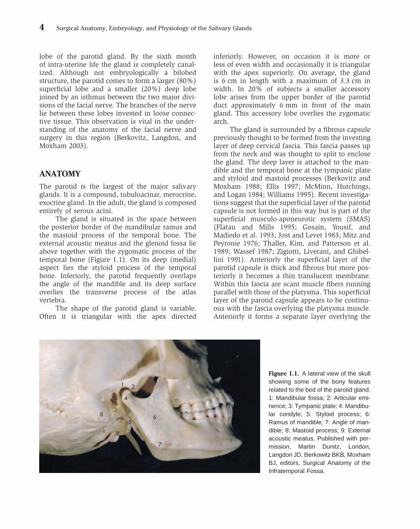
The gland is situated in the space between the posterior border of the mandibular ramus and the mastoid process of the temporal bone. The external acoustic meatus and the glenoid fossa lie above together with the zygomatic process of the temporal bone (Figure 1.1). On its deep (medial) aspect lies the styloid process of the temporal bone. Inferiorly, the parotid frequently overlaps the angle of the mandible and its deep surface overlies the transverse process of the atlas vertebra.
The shape of the parotid gland is variable. Often it is triangular with the apex directed
inferiorly. However, on occasion it is more or less of even width and occasionally it is triangular with the apex superiorly. On average, the gland is 6 cm in length with a maximum of 3.3 cm in width. In 20% of subjects a smaller accessory lobe arises from the upper border of the parotid duct approximately 6 mm in front of the main gland. This accessory lobe overlies the zygomatic arch.
The gland is surrounded by a fi brous capsule previously thought to be formed from the investing layer of deep cervical fascia. This fascia passes up from the neck and was thought to split to enclose the gland. The deep layer is attached to the man-dible and the temporal bone at the tympanic plate and styloid and mastoid processes (Berkovitz and Moxham 1988; Ellis 1997; McMinn, Hutchings, and Logan 1984; Williams 1995). Recent investiga-tions suggest that the superfi cial layer of the parotid capsule is not formed in this way but is part of the superfi cial musculo-aponeurotic system (SMAS) (Flatau and Mills 1995; Gosain, Yousif, and Madiedo et al. 1993; Jost and Levet 1983; Mitz and Peyronie 1976; Thaller, Kim, and Patterson et al. 1989; Wassef 1987; Zigiotti, Liverani, and Ghibel-lini 1991). Anteriorly the superfi cial layer of the parotid capsule is thick and fi brous but more pos-teriorly it becomes a thin translucent membrane. Within this fascia are scant muscle fi bers running parallel with those of the platysma. This superfi cial layer of the parotid capsule appears to be continu-ous with the fascia overlying the platysma muscle. Anteriorly it forms a separate layer overlying the
1 2
5
4
63
7
8
9
Figure 1.1. A lateral view of the skull showing some of the bony features related to the bed of the parotid gland. 1: Mandibular fossa; 2: Articular emi-nence; 3: Tympanic plate; 4: Mandibu-lar condyle; 5: Styloid process; 6: Ramus of mandible; 7: Angle of man-dible; 8: Mastoid process; 9: External acoustic meatus. Published with per-mission, Martin Dunitz, London, Langdon JD, Berkowitz BKB, Moxham BJ, editors, Surgical Anatomy of the Infratemporal Fossa.

Surgical Anatomy, Embryology, and Physiology of the Salivary Glands 5
masseteric fascia, which is itself an extension of the deep cervical fascia. The peripheral branches of the facial nerve and the parotid duct lie within a loose cellular layer between these two sheets of fascia. This observation is important in parotid surgery. When operating on the parotid gland, the skin fl ap can either be raised in the subcutaneous fat layer or deep to the SMAS layer. The SMAS layer itself can be mobilized as a separate fl ap and can be used to mask the cosmetic defect following parotidectomy by reattaching it fi rmly to the ante-rior border of the sternocleidomastoid muscle as
an advancement fl ap (Meningaud, Bertolus, and Bertrand 2006).
The superior border of the parotid gland (usually the base of the triangle) is closely molded around the external acoustic meatus and the tem-poromandibular joint. An avascular plane exists between the gland capsule and the cartilaginous and bony acoustic meatus (Figure 1.2). The infe-rior border (usually the apex) is at the angle of the mandible and often extends beyond this to overlap the digastric triangle, where it may lie very close to the posterior pole of the submandibular salivary
1
22
5
4
6
3
7
8
9
10
11
11
1213
14
15
Figure 1.2. The parotid gland and associated structures. 1: Auriculotem-poral nerve; 2: Superfi cial temporal vessels; 3: Temporal branch of facial nerve; 4: Zygomatic branch of facial nerve; 5: Buccal branch of facial nerve; 6: Mandibular branch of facial nerve; 7: Cervical branch of facial nerve; 8: Parotid duct; 9: Parotid gland; 10: Masseter muscle; 11: Facial vessels; 12: Platysma muscle; 13: External jugular vein; 14: Sternocleidomastoid muscle; 15: Great auricular nerve. Published with permission, Martin Dunitz, London, Langdon JD, Berkow-itz BKB, Moxham BJ, editors, Surgical Anatomy of the Infratemporal Fossa.

6 Surgical Anatomy, Embryology, and Physiology of the Salivary Glands
CONTENTS OF THE PAROTID GLAND
The Facial NerveFrom superfi cial to deep, the facial nerve, the auriculotemporal nerve, the retromandibular vein, and the external carotid artery pass through the substance of the parotid gland.
The facial nerve exits the skull base at the stylomastoid foramen. The surgical landmarks are important (Figure 1.4). To expose the trunk of the facial nerve at the stylomastoid foramen the dissec-tion passes down the avascular plane between the parotid gland and the external acoustic canal until the junction of the cartilaginous and bony canals can be palpated. A small triangular extension of the cartilage points toward the facial nerve as it exits
1
2
5
4
6
3
7 8 9
12
1011
13
Figure 1.3. The mandibulostylohyoid ligament. 1: Styloid process; 2: Stylomandibular ligament; 3: Mandibulostylo-hyoid ligament; 4: Masseter muscle; 5: Posterior border of ramus; 6: Lateral pterygoid muscle; 7: Medial pterygoid muscle; 8: Superior pharyngeal constrictor muscle; 9: Stylopharyngeus muscle; 10: Middle pharyngeal constric-tor muscle; 11: Inferior pharyngeal constrictor muscle; 12: Submandibular gland; 13: Facial vein and artery. Pub-lished with permission, Martin Dunitz, London, Langdon JD, Berkowitz BKB, Moxham BJ, editors, Surgical Anatomy of the Infratemporal Fossa.
gland. The anterior border just overlaps the poste-rior border of the masseter muscle and the poste-rior border overlaps the anterior border of the sternocleidomastoid muscle.
The superfi cial surface of the gland is covered by skin and platysma muscle. Some terminal branches of the great auricular nerve also lie super-fi cial to the gland. At the superior border of the parotid lie the superfi cial temporal vessels with the artery in front of the vein. The auriculotemporal branch of the mandibular nerve runs at a deeper level just behind the superfi cial temporal vessels.
The branches of the facial nerve emerge from the anterior border of the gland. The parotid duct also emerges to run horizontally across the mas-seter muscle before piercing the buccinator muscle anteriorly to end at the parotid papilla. The trans-verse facial artery (a branch of the superfi cial tem-poral artery) runs across the area parallel to and approximately 1 cm above the parotid duct. The anterior and posterior branches of the facial vein emerge from the inferior border.
The deep (medial) surface of the parotid gland lies on those structures forming the parotid bed. Anteriorly the gland lies over the mas-seter muscle and the posterior border of the mandibular ramus from the angle up to the condyle. As the gland wraps itself around the ramus it is related to the medial pterygoid muscle at its insertion on to the deep aspect of the angle. More posteriorly, the parotid is molded around the styloid process and the styloglossus, stylohyoid, and stylopharyngeus muscles from below upward. Behind this, the parotid lies on the posterior belly of the digastric muscle and the sternocleidomas-toid muscle. The digastric and the styloid muscles separate the gland from the underlying internal jugular vein, the external and internal carotid arteries and the glossopharyngeal, vagus, acces-sory, and hypoglossal nerves and the sympathetic trunk.
The fascia that covers the muscles in the parotid bed thickens to form two named ligaments (Figure 1.3). The stylomandibular ligament passes from the styloid process to the angle of the man-dible. The mandibulostylohyoid ligament (the angular tract) passes between the angle of the mandible and the stylohyoid ligament. Inferiorly it usually extends down to the hyoid bone. These ligaments are all that separates the parotid gland anteriorly from the posterior pole of the superfi cial lobe of the submandibular gland.

Surgical Anatomy, Embryology, and Physiology of the Salivary Glands 7
the foramen (Langdon 1998b). The nerve lies about 9 mm from the posterior belly of the digastric muscle and 11 mm from the bony external meatus (Holt 1996). The facial nerve then passes down-ward and forward over the styloid process and associated muscles for about 1.3 cm before enter-ing the substance of the parotid gland (Hawthorn and Flatau 1990). The fi rst part of the facial nerve gives off the posterior auricular nerve supplying the auricular muscles and also branches to the poste-rior belly of the digastric and stylohyoid muscles.
On entering the parotid gland the facial nerve separates into two divisions, temporofacial and cervicofacial, the former being the larger. The divi-sion of the facial nerve is sometimes called the “pes anserinus” due to its resemblance to the foot of a goose. From the temporofacial and cervicofa-cial divisions, the facial nerve gives rise to fi ve named branches—temporal, zygomatic, buccal, mandibular, and cervical (Figure 1.5). The periph-eral branches of the facial nerve form anastomotic arcades between adjacent branches to form the parotid plexus. These anastomoses are important during facial nerve dissection, as accidental damage to a small branch often fails to result in any facial
weakness due to dual innervation from adjacent branches. Davis et al. (1956) studied these patterns following the dissection of 350 facial nerves in cadavers. The anastomotic relationships between adjacent branches fell into six patterns (Figure 1.6). They showed that in only 6% of cases (type VI) is there any anastomosis between the man-dibular branch and adjacent branches. This explains why, when transient facial weakness follows facial nerve dissection, it is usually the mandibular branch that is affected.
Auriculotemporal NerveThe auriculotemporal nerve arises from the poste-rior division of the mandibular division of the trigeminal nerve in the infratemporal fossa. It runs backward beneath the lateral pterygoid muscle between the medial aspect of the condylar neck and the sphenomandibular ligament. It enters the
Figure 1.5. Clinical photograph of dissected facial nerve following superfi cial parotidectomy.
1
2
5
4
6
3
Figure 1.4. Anatomical landmarks of the extratemporal facial nerve. 1: Cartilaginous external acoustic meatus; 2: Parotid gland; 3: Sternocleidomastoid muscle; 4: Tip of the mastoid process; 5: Styloid process; 6: Posterior belly of digastric muscle. Published with permission, Martin Dunitz, London, Langdon JD, Berkowitz BKB, Moxham BJ, editors, Surgical Anatomy of the Infratemporal Fossa.

8 Surgical Anatomy, Embryology, and Physiology of the Salivary Glands
anteromedial surface of the parotid gland passing upward and outward to emerge at the superior border of the gland between the temporomandibu-lar joint and the external acoustic meatus. This nerve communicates widely with the temporofa-cial division of the facial nerve and limits the mobility of the facial nerve during surgery (Flatau and Mills 1995). Further communications with the temporal and zygomatic branches loop around the transverse facial and superfi cial temporal vessels (Bernstein and Nelson 1984).
Retromandibular VeinThe vein is formed within the parotid gland by the union of the superfi cial temporal vein and the maxillary vein. The retromandibular vein passes downward and close to the lower pole of the parotid, where it often divides into two branches passing out of the gland. The posterior branch passes backward to unite with the posterior auric-ular vein on the surface of the sternocleidomastoid muscle to form the external jugular vein. The ante-rior branch passes forward to join the facial vein.
The retromandibular vein is an important landmark during parotid gland surgery. The divi-sion of the facial nerve into its temporofacial and
cervicofacial divisions occurs just behind the ret-romandibular vein (Figure 1.7). The two divisions lie just superfi cial to the vein in contact with it. It is all too easy to tear the vein while exposing the division of the facial nerve!
External Carotid ArteryThe external carotid artery runs deeply within the parotid gland. It appears from behind the posterior belly of the digastric muscle and grooves the parotid before entering it. It gives off the posterior auricular artery before ascending and dividing into its terminal branches, the superfi cial temporal and maxillary arteries at the level of the condyle. The superfi cial temporal artery continues vertically to emerge at the superior border of the gland and crosses the zygomatic arch. Within the substance of the parotid it gives off the transverse facial artery, which emerges at the anterior border of the gland to run across the face above the parotid duct. The maxillary artery emerges from the deep aspect of the gland anteriorly to enter the infratemporal fossa. The maxillary artery gives off the deep auric-ular artery and the anterior tympanic artery within the substance of the parotid. All these branches from the external carotid also give off numerous
1
1
2
2 2
5
5
55
5
4
4
4
4 3
3
3
3
3
3
5
III
1
1
1
2
2
4
4
V
VI
IVII
I 3
2
1
Figure 1.6. The branching patterns of the facial nerve. I: Type I, 13%; II: Type II, 20%; III: Type III, 28%; IV: Type IV, 24%; V: Type V, 9%; VI: Type VI, 6%; 1: Temporal branch; 2: Zygomatic branch; 3: Buccal branch; 4: Mandibular branch; 5: Cervical branch. Published with permission, Martin Dunitz, London, Langdon JD, Berkowitz BKB, Moxham BJ, editors, Surgical Anatomy of the Infratemporal Fossa.

Surgical Anatomy, Embryology, and Physiology of the Salivary Glands 9
small branches within the parotid to supply the gland itself.
Parotid Lymph NodesLymph nodes are found within the subcutaneous tissues overlying the parotid to form the preauricu-lar nodes and also within the substance of the
1
25
4
6109
3
78
Figure 1.7. The facial nerve and its relationship to the retromandibular vein within the parotid gland. 1: Facial nerve at stylomastoid foramen; 2: Temporofacial branch of facial nerve; 3: Cervicofacial branch of facial nerve; 4: Temporal branch of facial nerve; 5: Zygomatic branch of facial nerve; 6: Buccal branch of facial nerve; 7: Man-dibular branch of facial nerve; 8: Cer-vical branch of facial nerve; 9: Posterior belly of digastric muscle; 10: Retro-mandibular vein and external carotid artery. Published with permission, Martin Dunitz, London, Langdon JD, Berkowitz BKB, Moxham BJ, editors, Surgical Anatomy of the Infratemporal Fossa.
gland. There are typically ten nodes within the substance of the gland, the majority being within the superfi cial lobe and therefore superfi cial to the plane of the facial nerve. Only one or two nodes lie within the deep lobe (Garetea-Crelgo et al. 1993; Marks 1984; McKean, Lee, and McGregor 1985). All the parotid nodes drain into the upper deep cervical chain.

10 Surgical Anatomy, Embryology, and Physiology of the Salivary Glands
Parotid DuctThe parotid duct emerges from the anterior border of the parotid gland and passes horizontally across the masseter muscle. The surface markings of the duct are obtained by drawing a line from the lowest point of the alar cartilage to the angle of the mouth (Figure 1.8). This line is bisected and its midpoint is joined with a straight line to the most anterior point of the tragus. This line is divided into three equal parts and the middle section cor-responds to the position of the parotid duct. The duct lies approximately 1 cm below the transverse facial vessels. The accessory lobe of the parotid gland, when present, drains into its upper border via one or two tributaries. Anastomosing branches
Figure 1.8. The surface markings for the parotid duct.
between the buccal and zygomatic branches of the facial nerve cross the duct. At the anterior border of the masseter, the duct bends sharply to perfo-rate the buccal pad of fat and the buccinator muscle at the level of the upper molar teeth. The duct then bends again to pass forward for a short distance before entering the oral cavity at the parotid papilla.
Nerve Supply to the ParotidThe parasympathetic secretomotor nerve supply comes from the inferior salivatory nucleus in the brain stem (Figure 1.9). From there the fi bers run in the tympanic branch of the glossopharyngeal nerve contributing to the tympanic plexus in the middle ear. The lesser petrosal nerve arises from the tympanic plexus leaving the middle ear and running in a groove on the petrous temporal bone in the middle cranial fossa. From here it exits through the foramen ovale to the otic ganglion, which lies on the medial aspect of the mandibular branch of the trigeminal nerve. Postsynaptic post-ganglionic fi bers leave the ganglion to join the auriculotemporal nerve, which distributes the par-asympathetic secretomotor fi bers throughout the parotid gland. Some authorities suggest that there are also some parasympathetic innervations to the parotid from the chorda tympani branch of the facial nerve.
The sympathetic nerve supply to the parotid arises from the superior cervical sympathetic gan-glion. The sympathetic fi bers reach the gland via the plexus around the middle meningeal artery. They then pass through the otic ganglion without synapsing and innervate the gland through the auriculotemporal nerve. There is also sympathetic innervation to the gland arising from the plexuses that accompany the blood vessels supplying the gland.
Sensory fi bers arising from the connective tissue within the parotid gland merge into the auriculotemporal nerve and pass proximally through the otic ganglion without synapsing. From there the fi bers join the mandibular division of the trigeminal nerve. The sensory innervation of the parotid capsule is via the great auricular nerve.

Surgical Anatomy, Embryology, and Physiology of the Salivary Glands 11
Figure 1.9. The parasympathetic innervations of the salivary glands. The parasympathetic fi bers are shown as blue lines. Published with permis-sion, Elsevier Churchill Livingstone, Oxford, Standring S, Editor in Chief, Gray’s Anatomy. 39th edition.
The Submandibular GlandEMBRYOLOGY
The submandibular gland begins to form at the 13 mm stage as an epithelial outgrowth into the mesenchyme forming the fl oor of the mouth in the linguogingival groove. This proliferates rapidly, giving off numerous branching processes that eventually develop lumina. Initially the developing gland opens into the fl oor of the mouth posteriorly, lateral to the tongue. The walls of the groove into which it drains come together to form the sub-mandibular duct. This process commences poste-riorly and moves forward so that ultimately the orifi ce of the duct comes to lie anteriorly below the tip of the tongue close to the midline.
ANATOMY
The submandibular gland consists of a larger superfi cial lobe lying within the digastric triangle in the neck and a smaller deep lobe lying within the fl oor of the mouth posteriorly (Figure 1.10). The two lobes are continuous with each other around the posterior border of the mylohyoid muscle. As in the parotid gland, the two “lobes” are not true lobes embryologically, as the gland arises as a single epithelial outgrowth. However, surgically it consists of the two lobes as described above. It is a mixed seromucinous gland.
The Superfi cial LobeThe superfi cial lobe lies within the digastric trian-gle. Its anterior pole reaches the anterior belly of

12 Surgical Anatomy, Embryology, and Physiology of the Salivary Glands
the digastric muscle and the posterior pole reaches the stylomandibular ligament. This structure is all that separates the superfi cial lobe of the sub-mandibular gland from the parotid gland. It is important to realize just how close the lower pole of the parotid is to the posterior pole of the sub-mandibular gland, as confusion can arise if a mass in the region is incorrectly ascribed to the wrong anatomical structure (Figure 1.2). Superiorly, the superfi cial lobe lies medial to the body of the man-dible. Inferiorly it often overlaps the intermediate tendon of the digastric muscles and the insertion of the stylohyoid. The lobe is partially enclosed between the two layers of the deep cervical fascia that arise from the greater cornu of the hyoid bone and is in intimate proximity of the facial vein and artery (Figure 1.11). The superfi cial layer of the fascia is attached to the lower border of the man-dible and covers the inferior surface of the super-fi cial lobe. The deep layer of fascia is attached to the mylohyoid line on the inner aspect of the man-dible and therefore covers the medial surface of the lobe.
The inferior surface, which is covered by skin, subcutaneous fat, platysma, and the deep fascia, is crossed by the facial vein and the cervical
Figure 1.11. Superfi cial dissection of the left submandibu-lar gland. The investing layer of the deep cervical fascia is elevated off of the submandibular gland and the facial vein is identifi ed.
a
b
cFigure 1.10. The relationship of the superfi cial and deep lobes of the submandibular gland. Cross-sectional anatomy (a). The superfi cial lobe from outside (b). The relationship of the deep and superfi cial lobes to the mylo-hyoid muscle (c).

Surgical Anatomy, Embryology, and Physiology of the Salivary Glands 13
branch of the facial nerve, which loops down from the angle of the mandible and subsequently innervates the lower lip. The submandibular lymph nodes lie between the salivary gland and the mandible. Sometimes one or more lymph nodes may be embedded within the salivary gland.
The lateral surface of the superfi cial lobe is related to the submandibular fossa, a concavity on the medial surface of the mandible, and the attach-ment of the medial pterygoid muscle. The facial artery grooves its posterior part lying at fi rst deep to the lobe and then emerging between its lateral surface and the mandibular attachment of the medial pterygoid muscle from which it reaches the lower border of the mandible.
The medial surface is related anteriorly to the mylohyoid from which it is separated by the mylo-hyoid nerve and submental vessels. Posteriorly, it is related to the styloglossus, the stylohyoid liga-ment, and the glossopharyngeal nerve separating it from the pharynx. Between these, the medial aspect of the lobe is related to hyoglossus muscle from which it is separated by styloglossus muscle, the lingual nerve, submandibular ganglion, hypoglos-sal nerve, and deep lingual vein. More inferiorly, the medial surface is related to the stylohyoid muscle and the posterior belly of the digastric.
The Deep LobeThe deep lobe of the gland arises from the super-fi cial lobe at the posterior free edge of the mylo-hyoid muscle and extends forward to the back of the sublingual gland (Figure 1.12). It lies between the mylohyoid muscle inferolaterally, the hyoglos-sus and styloglossus muscles medially, the lingual nerve superiorly and the hypoglossal nerve and deep lingual vein inferiorly.
The Submandibular DuctThe submandibular duct is about 5 cm long in the adult. The wall of the submandibular duct is thinner than that of the parotid duct. It arises from numer-ous tributaries in the superfi cial lobe and emerges from the medial surface of this lobe just behind the posterior border of the mylohyoid. It crosses the deep lobe, passing upward and slightly backward for 5 mm before running forward between the mylohyoid and hyoglossus muscles. As it passes forward, it runs between the sublingual gland and genioglossus to open into the fl oor of the mouth on
the summit of the sublingual papilla at the side of the lingual frenum just below the tip of the tongue. It lies between the lingual and hypoglossal nerves on the hyoglossus. At the anterior border of the hyoglossus muscle it is crossed by the lingual nerve. As the duct traverses the deep lobe of the gland it receives tributaries draining that lobe.
Blood Supply and Lymphatic DrainageThe arterial blood supply arises from multiple branches of the facial and lingual arteries. Venous blood drains predominantly into the deep lingual vein. The lymphatics drain into the deep cervical group of nodes, mostly into the jugulo-omohyoid node, via the submandibular nodes.
Nerve Supply to the Submandibular GlandParasympathetic InnervationThe secretomotor supply to the submandibular gland arises from the submandibular (sublingual) ganglion. This is a small ganglion lying on the upper part of the hyoglossus muscle. There are additional ganglion cells at the hilum of the gland. The submandibular ganglion is suspended from the lingual nerve by anterior and posterior fi la-ments (Figure 1.13).
Figure 1.12. Deep dissection of the left submandibular gland. With the submandibular gland retracted, the facial artery is identifi ed in proximity to the facial vein.

14 Surgical Anatomy, Embryology, and Physiology of the Salivary Glands
The parasympathetic secretomotor fi bers originate in the superior salivatory nucleus and the preganglionic fi bers, then travel via the facial nerve, chorda tympani, and lingual nerve to the ganglion via the posterior fi laments connecting the ganglion to the lingual nerve. They synapse within the ganglion, and the postganglionic fi bers inner-vate the submandibular and sublingual glands (Figure 1.9). Some fi bers are thought to reach the lower pole of the parotid gland.
Sympathetic InnervationThe sympathetic root is derived from the plexus on the facial artery. The postganglionic fi bers arise from the superior cervical ganglion and pass through the submandibular ganglion without syn-apsing. They are vasomotor to the vessels supply-ing the submandibular and sublingual glands. Five or six branches from the ganglion supply the sub-mandibular gland and its duct. Others pass back into the lingual nerve via the anterior fi lament to innervate the sublingual and other minor salivary glands in the region.
Sensory InnervationSensory fi bers arising from the submandibular and sublingual glands pass through the ganglion without synapsing and join the lingual nerve, itself a branch of the trigeminal nerve.
The Sublingual GlandEMBRYOLOGY
The sublingual gland arises in 20 mm embryos as a number of small epithelial thickenings in the linguogingival groove and on the outer side of the groove. Each thickening forms its own canal and so many of the sublingual ducts open directly onto the summit of the sublingual fold. Those that arise within the linguogingival groove end up draining into the submandibular duct.
ANATOMY
The sublingual gland is the smallest of the major salivary glands. It is almond shaped and weighs approximately 4 g. It is predominantly a mucous gland. The gland lies on the mylohyoid and is covered by the mucosa of the fl oor of the mouth, which is raised as it overlies the gland to form the sublingual fold. Posteriorly, the sublingual gland is in contact with the deep lobe of the submandib-ular gland. The sublingual fossa of the mandible is located laterally and the genioglossus muscle is located medially. The lingual nerve and the sub-mandibular duct lie medial to the sublingual gland between it and the genioglossus.
Sublingual DuctsThe gland has a variable number of excretory ducts ranging from 8 to 20. The majority drain into the fl oor of the mouth at the crest of the sublingual fold. A few drain into the submandibu-lar duct. Sometimes, a collection of draining ducts coalesce anteriorly to form a major duct (Bartholin’s duct), which opens with the orifi ce of the submandibular duct at the sublingual papilla.
Blood Supply, Innervation, and Lymphatic DrainageThe arterial supply is from the sublingual branch of the lingual artery and also the submental branch of the facial artery. Innervation is via the sublin-gual ganglion as described above. The lymphatics drain to the submental nodes.
Figure 1.13. Clinical photograph showing the relationship of the lingual nerve to the submandibular gland.

Surgical Anatomy, Embryology, and Physiology of the Salivary Glands 15
Minor Salivary Glands
Minor salivary glands are distributed widely in the oral cavity and oropharynx. They are grouped as labial, buccal, palatoglossal, palatal, and lingual glands. The labial and buccal glands contain both mucous and serous acini, whereas the palatoglos-sal glands are mucous secreting. The palatal glands, which are also mucous secreting, occur in both the hard and soft palates. The anterior and posterior lingual glands are mainly mucous. The anterior glands are embedded within the muscle ventrally and they drain via four or fi ve ducts near the lingual frenum. The posterior lingual glands are located at the root of the tongue. The deep poste-rior lingual glands are predominantly serous. Addi-tional serous glands (of von Ebner) occur around the circumvallate papillae on the dorsum of the tongue. Their watery secretion is thought to be important in spreading taste stimuli over the taste buds.
Histology of the Salivary Glands
The salivary glands are composed of large numbers of secretory acini, which may be tubular or globu-lar in shape. Each acinus drains into a duct. These microscopic ducts coalesce to form lobular ducts. Each lobule has its own duct and these then merge to form the main ducts. The individual lobes and lobules are separated by dense connective tissue, which is continuous with the gland capsule. The ducts, blood vessels, lymphatics, and nerves run through and are supported by this connective tissue.
The acini are the primary secretory organs but the saliva is modifi ed as it passes through the inter-calated, striated, and excretory ducts before being discharged into the mouth and oropharynx (Figure 1.14). The lobules also contain signifi cant amounts of adipose tissue particularly in the parotid gland. The proportion of adipose tissue relative to excre-tory acinar cells increases with age.
In the human parotid, the excretory acini are almost entirely serous. In the submandibular gland,
again, the secretory units are mostly serous but there are additional mucous tubules and acini. In some areas the mucinous acini have crescentic “caps” of serous cells called serous demilunes. In the sublingual gland the acini are almost entirely mucinous, although there are occasional serous acini or demilunes.
The serous cells contain numerous pro-teinaceous secretory (zymogen) granules. These granules contain high levels of amylase. In addition, the secretory cells produce kallikrein, lactoferrin, and lysozyme. In mucous cells, the cytoplasm is packed with large pale secretory droplets.
Initially the secretory acini drain into interca-lated ducts. These function mainly to conduct the saliva but they may also modify the electrolyte content and secrete immunoglobulin A. The intercalated ducts drain into striated ducts, which coalesce into intralobular and extralobular collect-ing ducts. The intercalated duct cells are very active metabolically and they transport potassium and bicarbonate into saliva. They reabsorb sodium and chloride ions so that the resulting saliva is hypotonic. They also secrete immunoglobulin A, lysozyme, and kallikrein. The immunoglobulin is produced by plasma cells adjacent to the striated duct cells and it is then transported through the epithelial lining into the saliva. The main collecting ducts are simple conduits for saliva and do not modify the composition of the saliva.
Myoepithelial cells are contractile cells closely related to the secretory acini and also much of the duct system. The myoepithelial cells lie between the basal lamina and the epithelial cells. Numerous cytoplasmic processes arise from them and sur-round the serous acini as basket cells. Those asso-ciated with the duct cells are more fusiform and are aligned along the length of the ducts. The cytoplasm of the myoepithelial cells contains actin myofi laments, which contract as a result of both parasympathetic and sympathetic activity. Thus the myoepithelial cells “squeeze” the saliva out of the secretory acini and ducts and add to the sali-vary secretory pressure.

Junction betweenstriated duct cell and
cholinergic axon
Striated(intralobular)
ductaccompanied by
nonmyelinatedpostganglionic
axon
Complex arborizationof adrenergic andcholinergic axonsaround secretory unitor ‘endpiece’
Adrenergicnerveterminals
Arterioleaccompanied byadrenergic axons
Seroussecretoryendpiece
Serous demilune(as seen in routinehistological preparations)
Myoepithelialcell
Mucous secretoryendpiece
Immunoglobulin
LysozymeKallikreinK+
Na+
ClWaterSalts€-amylasePeroxidaseProline-richproteins
Intercalatedducts
To interlobularexcretory ducts
Junctions between cholinergic axonsand intercalated duct cells
Serous cell
Mitochondria
Infoldings of basalplasma membraneresulting in striatedappearance
Striated duct cell
Intercalated duct cell
Prominent apical webof microfilaments
Process ofmyoepithelial cell
Mucous cell
Apical microvillus
Pinocytotic vesicle
Intercellular secretorycanaliculus
Tight junction
Heterogeneouselectron-densesecretory granules
Sphericalnucleus
Centrally-locatednucleus
WaterSaltsNeutralglycoproteinsSialomucinsSulphomucins
Tight junction
Homogeneouselectron-translucentsecretory vesicles
Flattened basalnucleus
Figure 1.14. Diagram showing the histology of the major components of the salivary glands. Published with permission, Elsevier Churchill Livingstone, Oxford, Standring S, Editor in Chief, Gray’s Anatomy, 39th edition.

Surgical Anatomy, Embryology, and Physiology of the Salivary Glands 17
Control of Salivation
There is a continuous low background saliva pro-duction that is stimulated by drying of the oral and pharyngeal mucosa. A rapid increase in the resting levels occurs as a refl ex in response to masticatory stimuli including the mechanoreceptors and taste fi bers. Other sensory modalities such as smell are also involved. The afferent input is via the saliva-tory centers, which are themselves infl uenced by the higher centers. The higher centers may be facilitory or inhibitory depending on the circum-stances. The efferent secretory drive to the salivary glands passes via the parasympathetic and sympa-thetic pathways. There are no peripheral inhibitory mechanisms.
Cholinergic nerves (parasympathetic) often accompany ducts and branch freely around the secretory endpieces (acini). Adrenergic nerves (sympathetic) usually enter the glands along the arteries and arterioles and ramify with them. Within the glands, the nerve fi bers intermingle such that cholinergic and adrenergic axons fre-quently lie in adjacent invaginations of a single Schwann cell. Secretion and vasoconstriction are mediated by separate sympathetic axons, whereas a single parasympathetic axon may, through serial terminals, result in vasodilatation, secretion, and constriction of myoepithelial cells.
Secretory endpieces are the most densely innervated structures in the salivary glands. Indi-vidual acinar cells may have both cholinergic and adrenergic nerve endings. The secretion of water and electrolytes, which accounts for the volume of saliva produced, results from a complex set of stimuli that are largely parasympathetic. The active secretion of proteins into the saliva depends upon the relative levels of both sympathetic and para-sympathetic stimulation.
Although the ducts are less densely inner-vated than secretory acini, they do infl uence the composition of the saliva. Adrenal aldosterone promotes resorption of sodium and secretion of potassium into the saliva by striated ductal cells. Myoepithelial cell contraction is stimulated pre-dominantly by adrenergic fi bers, although there may be an additional role for cholinergic axons.
Summary
• Although embryologically the parotid consists of a single lobe, anatomically the facial nerve
lies in a distinct plane between the anatomical superfi cial and deep lobes.
• There are fi xed anatomical landmarks indicating the origin of the extracranial facial nerve as it leaves the stylomastoid foramen.
• The lower pole of the parotid gland is sepa-rated from the posterior pole of the sub-mandibular gland by only thin fascia. This can lead to diagnostic confusion in determin-ing the origin of a swelling in this area.
• The relationship of the submandibular sali-vary duct to the lingual nerve is critical to the safe removal of stones within the duct.
• Great care must be taken to identify the lingual nerve when excising the submandibular gland. The lingual nerve is attached to the gland by the parasympathetic fi bers synapsing in the submandibular (sublingual) ganglion.
• The sublingual gland may drain into the sub-mandibular duct or it may drain directly into the fl oor of the mouth via multiple secretory ducts.
References
Berkovitz BKB, Langdon JD, Moxham BJ. 2003. The facial nerve and the parotid gland. In: Langdon JD, Berkovitz BKB, Moxham BJ (eds), Surgical Anatomy of the Infratem-poral Fossa. London: Martin Dunitz, pp. 181–206.
Berkovitz BKB, Moxham BJ. 1988. A Textbook of Head and Neck Anatomy. London: Wolfe.
Bernstein L, Nelson RH. 1984. Surgical anatomy of the extraparotid distribution of the facial nerve. Arch Otolaryngol 110:177–183.
Davis RA, Anson BJ, Budinger JM, Kurth LE. 1956. Surgical anatomy of the facial nerve and parotid gland based on 350 cervicofacial halves. Surg Gynecol Obstet 102:385–412.
Ellis H. 1997. Clinical Anatomy (9th ed.). Oxford: Blackwell.Flatau AT, Mills PR. 1995. Regional anatomy. In: Norman
JE deB, McGurk M (eds.), Color Atlas and Text of the Salivary Glands. London: Mosby Wolfe, pp. 13–39.
Garetea-Crelgo J, Gay-Escoda C, Bermejo B, Buenechea-Imaz R. 1993. Morphological studies of the parotid lymph nodes. J Cranio-Maxillo-Facial Surg 21:207–209.
Gosain AK, Yousif NJ, Madiedo G et al. 1993. Surgical anatomy of the SMAS: A reinvestigation. Plast Reconstr Surg 92:1254–1263.
Hawthorn R, Flatau A. 1990. Temporomandibular joint anatomy. In: Norman JE deB, Bramley P (eds.), A Text-book and Colour Atlas of the Temporomandibular Joint. London: Mosby Wolfe, pp. 1–51.
Holt JJ. 1996. The stylomastoid area: Anatomic-histologic study and surgical approach. Laryngoscope 106:396–399.

18 Surgical Anatomy, Embryology, and Physiology of the Salivary Glands
Jost G, Levet Y. 1983. Parotid fascia and face lifting: A critical evaluation of the SMAS concept. Plast Reconstr Surg 74:42–51.
Langdon JD. 1998. Sublingual and submandibular gland excision. In: Langdon JD, Patel MF (eds.), Operative Maxillofacial Surgery. London: Chapman & Hall, pp. 376–380.
Langdon JD. 1998. Parotid surgery. In: Langdon JD, Patel MF (eds.), Operative Maxillofacial Surgery. London: Chapman & Hall, pp. 386–388.
Marks NJ. 1984. The anatomy of the lymph nodes of the parotid gland. Clin Otolaryngol 9:271–275.
McKean ME, Lee K, McGregor IA. 1985. The distribution of lymph nodes in and around the parotid gland: An anatomical study. Br J Plast Surg 38:1–5.
McMinn RMH, Hutchings RT, Logan BM. 1984. A Colour Atlas of Applied Anatomy. London: Wolfe.
Meningaud J-P, Bertolus C, Bertrand J-C. 2006. Parotidec-
tomy: Assessment of a surgical technique including facelift incision and SMAS advancement. J Cranio-Maxil-lofacial Surg 34:34–37.
Mitz V, Peyronie M. 1976. The superfi cial musculo-aponeu-rotic system (SMAS) in the parotid and cheek area. Plast Reconstr Surg 58:80–88.
Thaller SR, Kim S, Patterson H et al. 1989. The submuscu-lar aponeurotic system (SMAS): A histologic and com-parative anatomy evaluation. Plast Reconstr Surg 86:691–696.
Wassef M. 1987. Superfi cial fascia and muscular layers in the face and neck: A histological study. Aesthetic Plast Surg 11:171–176.
Williams PL (ed.). 1995. Gray’s Anatomy (38th ed.). Oxford: Blackwell.
Zigiotti GL, Liverani MB, Ghibellini D. 1991. The relation-ship between parotid and superfi cial fasciae. Surg Radiol Anat 13:293–300.

Chapter 2
Diagnostic Imaging of Salivary Gland PathologyPradeep K. Jacob, MD, MBA
Sialadenosis (Sialosis)SialolithiasisSjogren’s SyndromeSarcoidosisCongenital Anomalies of the Salivary Glands
First Branchial Cleft CystNeoplasms—Salivary, Epithelial
BenignPleomorphic AdenomaWarthin’s TumorOncocytoma
Malignant TumorsMucoepidermoid CarcinomaAdenoid Cystic Carcinoma
Neoplasms—Non-salivaryBenign
LipomaNeurogenic Tumors
MalignantLymphomaMetastases
SummaryReferences
Introduction
Anatomic and functional diagnostic imaging plays a central role in modern medicine. Virtually all specialties of medicine to varying degrees depend on diagnostic imaging for diagnosis, therapy, and follow-up of treatment. Because of the complexity of the anatomy, treatment of diseases of the head and neck, including those of the salivary glands, are particularly dependent on quality medical imaging and interpretation. Medical diagnostic imaging is divided primarily into two major catego-ries, anatomic and functional. The anatomic imaging modalities include computed tomography (CT), magnetic resonance imaging (MRI), and
OutlineIntroductionImaging Modalities
Computed Tomography (CT)CT Technique
Advanced Computed TomographyMagnetic Resonance Imaging (MRI)
MRI TechniqueSpin-Echo T1Spin-Echo T2Proton Density Images (PD)Gradient Recalled Echo Imaging (GRE)Short Tau Inversion Recovery (STIR)Gadolinium (Gd) ContrastFluid Attenuation Inversion Recovery (FLAIR)Diffusion Weighted Images (DWI)MR Spectroscopy (MRS)Dynamic Contrast Enhanced Magnetic Resonance
ImagingOther Magnetic Resonance Imaging Techniques
Ultrasonography (US)US Technique
Radionuclide Imaging (RNI)Positron Emission Tomography (PET)Positron Emission Tomography/Computed
Tomography (PET/CT)Diagnostic Imaging Anatomy
Parotid GlandSubmandibular Gland (SMG)Sublingual Gland (SLG)Minor Salivary Glands
Pathology of the Salivary GlandsVascular Lesions
Lymphangioma (Cystic Hygroma)Hemangioma
Acute SialadenitisChronic SialadenitisHIV-Lymphoepithelial LesionsMucous Escape Phenomena
19

20 Diagnostic Imaging of Salivary Gland Pathology
ultrasonography (US). Although occasionally obtained, plain fi lm radiography for the head and neck, including salivary gland disease, is mostly of historical interest. In a similar manner, the use of sialography has been signifi cantly reduced, although both plain fi lms and sialography are of some use in imaging sialoliths. Functional diagnos-tic imaging techniques include planar scintigraphy, single photon emission computed tomography (SPECT), positron emission tomography (PET), and magnetic resonance spectroscopy (MRS), all of which are promising technologies. Recently, the use of a combined anatomic and functional modal-ity in the form of PET/CT has proved invaluable in head and neck imaging. Previously widely employed procedures including gallium radionu-clide imaging are less important today than in the past.
Imaging ModalitiesCOMPUTED TOMOGRAPHY (CT)
Computed tomography has become indispensable in the diagnosis, treatment, and follow-up of dis-eases of the head and neck. The latest generation of multiple-row detector CT (MDCT) provides excellent soft tissue and osseous delineation. The rapid speed with which images can be obtained along with the high spatial resolution and tissue contrast make CT the imaging modality of choice in head and neck imaging. True volumetric data sets obtained from multidetector row scanners allow for excellent coronal, sagittal, or oblique reformation of images as well as a variety of 3-D renderings. This allows the radiologist and surgeon to characterize a lesion and assess involvement of adjacent structures or local spread from the orthog-onal projections or three-dimensional rendering. The ability to manipulate images is critical when assessing pathology in complex anatomy, such as evaluation of parotid gland masses to determine deep lobe involvement, facial nerve involvement, or extension into the skull base. Images in the coronal plane are important in evaluating the sub-mandibular gland in relation to the fl oor of the mouth. Lymphadenopathy and its relationship to the carotid sheath and its contents and other struc-tures are also well delineated. CT is also superior to MRI in demonstrating bone detail and calcifi ca-tions. CT is also the fastest method of imaging head and neck anatomy. Other advantages include
widespread availability of scanners, high-resolu-tion images, and speed of image acquisition, which also reduces motion artifacts. Exposure to ionizing radiation and the administration of IV contrast are the only signifi cant disadvantages to CT scanning.
CT TechniqueThe CT scanner contains a gantry, which holds an X-ray tube and a set of detectors. The X-ray tube is positioned opposite the detectors and is physi-cally coupled. A “fan beam” of X-rays is produced and passes through the patient to the detectors as the tube and detector rotate around the patient. In the newer generation of scanners, the multiple rows of detectors are fi xed around the gantry and only the tube rotates. A table carries the patient through the gantry. The detectors send signals, dependent on the degree of X-ray attenuation, to a computer, which uses this data to construct an image using complex algorithms.
For most CT studies (especially in the head and neck), intravenous contrast is administered. IV contrast is a solution consisting of organic com-pounds bonded with iodine molecules. Iodine is a dense atom with an atomic weight of 127, which is good at absorbing X-rays and is biocompatible. IV contrast readily attenuates the X-ray beam at concentrations optimal for vascular and soft tissue “enhancement,” but short of causing attenuation-related artifacts. Streak artifacts, however, can occur if the concentration is too high, as seen occasionally at the thoracic inlet and supraclavicu-lar region from dense opacifi cation of the subcla-vian vein during rapid bolus injection of IV contrast.
CT of the neck should be performed with intravenous contrast whenever possible to opti-mize delineation of masses and infl ammatory or infectious changes in the tissues and to enhance vascular structures. Imaging is obtained from the level of the orbits through the aortic arch in the axial plane with breath hold. The images are recon-structed using a computer algorithm to optimize soft tissue delineation, and displayed in soft tissue window and level settings (Figures 2.1 and 2.2). In a similar manner images are reconstructed using a computer algorithm to optimize bone details as more sharp and defi ned (Figure 2.3). The lung apex is often imaged in a complete neck evalua-tion and displayed using lung window settings

Diagnostic Imaging of Salivary Gland Pathology 21
(Figure 2.4a). Dedicated CT scans of the chest are benefi cial in the postoperative evaluation of patients with salivary gland malignancies, as lung nodules can be observed, possibly indicative of metastatic disease (Figure 2.4b). Multiplanar refor-matted images of the neck are obtained typically in the coronal and sagittal planes (Figures 2.5 and 2.6), although they may be obtained in virtually any plane desired or in a 3-D rendering.
The Hounsfi eld unit (H) (named for Godfrey Hounsfi eld, inventor of the CT scanner) is the unit of density measurement for CT. These units are assigned based on the degree of attenuation of the X-ray beam by tissue in a given voxel (volume element) and are assigned relative to water (0 H) (Table 2.1). The scale ranges from −1024 H for air, to +4000 H for very dense bone. The images are created based on a grayscale from black (−1024 H) to white (+4000 H) and shades of gray. Despite the wide range of units, the majority of tissues in the human body are between −100 and +100 H. Soft tissues and parenchymal organs are in a range between 20 and 80 H, whereas fat is approximately −100 H. Simple fl uid is 0 H, but proteinaceous fl uid can be upward of 25 H. Unclotted and clotted
Figure 2.1. Axial CT of the neck in soft tissue window without contrast demonstrating poor defi nition between soft tissue structures. The blood vessels are unopacifi ed and cannot be easily distinguished from lymph nodes. Note the sialolith (arrow) in the hilum of the left submandibular gland.
Figure 2.2. Axial CT of the neck in soft tissue window with IV contrast demonstrates improved visualization of struc-tures with enhancement of tissues and vasculature. Note the small lipoma (arrow) anterior to the left submandibular gland, which distorts the anterior aspect of the gland with slight posterior displacement.
Figure 2.3. Axial CT of the skull base reconstructed in a sharp algorithm and in bone window and level display demonstrating sharp bone detail. Note the sharply defi ned normal right stylomastoid foramen (arrow).

22 Diagnostic Imaging of Salivary Gland Pathology
blood varies depending on the hemoglobin con-centration and hematocrit, but average measure-ments are 50 H and 80 H, respectively. CT images are displayed using a combination of “window widths” (WW, range of CT numbers from black to white), and “window levels” (WL, position of the
a
bFigure 2.4. Axial CT of the neck at the thoracic inlet in lung windows demonstrating lung parenchyma (a). Axial image of dedicated CT of chest demonstrating cannon ball lesions in a patient previously treated for adenoid cystic carcinoma of the palate (b). These lesions are representa-tive of diffuse metastatic disease of the lungs, but not pathognomonic of adenoid cystic carcinoma.
Figure 2.5. Coronal CT reformation of the neck in soft tissue window at the level of the submandibular glands. Orthogonal images with MDCT offer very good soft tissue detail in virtually any plane of interest in order to assess anatomic and pathologic relationships.
Figure 2.6. Sagittal CT reformation of the neck in soft tissue window at the level of the parotid gland. Note the accessory parotid gland (black arrow) sitting atop the parotid (Stenson’s) duct (thin white arrow). Also note the retromandibular vein (large white arrow) and external audi-tory canal.

Diagnostic Imaging of Salivary Gland Pathology 23
window on the scale), which are based on the attenuation characteristics of tissues. Typically, head and neck images are interpreted using “soft tissue windows” (WW 500 H, WL 30 H), “bone windows” (WW 2000, WL 500), or “lung windows” (WW 1500, WL −500). “Soft tissue windows” dem-onstrate the slight density differences of soft tissues, whereas “bone windows” demonstrate cortical and medullary features of bones with sharp detail. “Lung windows” demonstrate the sharp interface of air and the fi ne soft tissue components of lung parenchyma.
Although the density of the salivary glands is variable, the parotid glands tend to be slightly lower in density relative to muscle, secondary to a higher fat content, and become progressively more fat replaced over time. The CT density of parotid glands varies from −10 to +30 H. The sub-mandibular glands are denser than parotid glands and are equivalent in density to muscle. The sub-mandibular glands vary in density from +30 to +60 H.
CT angiography (CTA) is a powerful method that allows visualization of arterial vasculature, demonstrating the vascular anatomy of arteries and veins. CTA can be critical in preoperative evaluation to determine the degree of vascularity
of lesions and to plan an appropriate surgical approach to minimize blood loss or perform pre-operative embolization. CTA is obtained with fast image acquisition over a defi ned region of interest while administering a rapid IV contrast bolus timed to arrive in the region of interest during image acquisition. CTA images may be rendered in 3-D data sets and rotated in any plane (Figure 2.7). CTA is not only useful for preoperative planning; it can also be quite useful in diagnosis of salivary gland vascular pathology such as aneurysms or arteriovenous fi stulae (AVFs) (Wong, Ahuja, and King et al. 2004).
CT scanning, as with all imaging modalities, is prone to artifacts. Artifacts can be caused by motion, very dense or metallic implants (dental amalgam), and volume averaging. Motion artifact is common and may result from breathing, swal-lowing, coughing, or sneezing during the image acquisition or from an unaware or uncooperative patient. Metallic implants cause complete attenua-tion of X-rays in the beam and result in focal loss of data and bright and dark streaks in the image. Because the image is created from a three-dimen-sional section of tissue averaged to form a two-dimensional image, the partial volume or volume averaging artifact results from partial inclusion of structures in adjacent images. Finally, the beam
Table 2.1. CT density in Hounsfi eld units (H).
Tissue or Structure Hounsfi eld Unit (H)
Water or CSF 0Fat −100Soft tissue, muscle (a) 50–60Unclotted blood (b) 35–50Clotted blood (b) 50–75Parotid gland (c) −10 to +30Submandibular gland (c) +30–+60Sublingual gland (d) 60–90Bone 1000Lung −850Air −1024Calcifi cation 150–200Grey matter 35–40White matter 25–35
(a) Depends on degree of fat deposition.(b) Depends on the hemoglobin concentration and hematocrit.(c) Depends on age and fat deposition.(d) Very limited evaluation secondary to partial volume effect.CSF = cerebrospinal fl uid.
Figure 2.7. CT angiogram of the neck at the level of the parotid gland demonstrating the retromandibular vein and adjacent external carotid artery (large white arrow). Note the right cervical lymphangioma (thin white arrow) associ-ated with the tail of the right parotid gland.

24 Diagnostic Imaging of Salivary Gland Pathology
hardening artifact is produced by attenuation of low energy X-rays, by dense objects, from the energy spectrum of the X-ray beam, resulting in a residual average high energy beam (or hard X-rays), which results in loss of data and dark lines on the image. This phenomenon is often seen in the posterior fossa of head CT scans caused by the very dense petrous bones. A multidetector row CT scanner can help reduce metallic artifacts using advanced algorithms, and can reduce motion arti-facts secondary to faster scanning speeds.
Advanced Computed TomographyNewer CT techniques including CT perfusion and dynamic contrast enhanced multi-slice CT have been studied. Dynamic multi-slice contrast enhanced CT is obtained while scanning over a region of interest and simultaneously administer-ing IV contrast. The characteristics of tissues can then be studied as the contrast bolus arrives at the lesion and “washes in” to the tumor, reaches a peak presence within the mass, and then decreases over time, that is, “washes out.” This technique has demonstrated differences in various histologic types of tumors, for example, with early enhance-ment in Warthin’s tumor with a time to peak at 30 seconds and subsequent fast washout. The malig-nant tumors show a time to peak at 90 seconds. The pleomorphic adenomas demonstrate a contin-ued rise in enhancement in all four phases (Yerli, Aydin, and Coskum et al. 2007).
CT perfusion attempts to study physiologic parameters of blood volume, blood fl ow, mean transit time, and capillary permeability surface product. Statistically signifi cant differences between malignant and benign tumors have been demon-strated with the mean transit time measurement. A rapid mean transit time of less then 3.5 seconds is seen with most malignant tumors, but with benign tumors or normal tissue the mean transit time is signifi cantly longer (Rumboldt, Al-Okkaili, and Deveikis 2005).
MAGNETIC RESONANCE IMAGING (MRI)
Magnetic resonance imaging represents imaging technology with great promise in characterizing salivary gland pathology. The higher tissue con-trast of MRI, when compared to CT, enables subtle differences in soft tissues to be demonstrated. Gad-olinium contrast enhanced MRI further and accen-
tuates the soft tissue contrast. Subtle pathologic states such as perineural spread of disease are better delineated when compared with CT. This along with excellent resolution and exquisite details make MRI a very powerful technique in head and neck imaging, particularly at the skull base. However, its susceptibility to motion artifacts and long imaging time as well as contraindication due to claustrophobia, pacemakers, aneurysm clips, and deep brain and vagal nerve stimulators limit its usefulness in the general population as a routine initial diagnostic and follow-up imaging modality. Many of the safety considerations are well defi ned and detailed on the popular Web site www.mrisafety.com.
MRI TechniqueAlthough the physics and instrumentation of MRI are beyond the scope of this text, a fundamental understanding of the variety of different imaging sequences and techniques should be understood by clinicians in order to facilitate reciprocal com-munication of the clinical problem and under-standing of imaging reports.
In contrast to CT, which is based on the use of ionizing radiation, MRI utilizes a high magnetic fi eld and pulsed radiofrequency waves in order to create an image or obtain spectroscopic data. MRI is based on the proton (hydrogen ion) distribution throughout the body. The basic concept is that protons are normally oriented in a random state. However, once placed in the imaging magnet, a high magnetic fi eld, a large proportion of protons align with the magnetic fi eld. The protons remain aligned and precess (spin) in the magnetic fi eld until an external force acts upon them and forces them out of alignment. This force is an applied radiofrequency pulse, used for a specifi ed time and specifi ed frequency by an antenna called a transmit coil. As the protons return to the aligned state, they give off energy in the form of their own radiofrequency pulse, determined by their local chemical state and tissue structure. The radiofrequency pulse given off is captured by an antenna, called a receive coil. The energy of the pulse and location is recorded and the process repeated multiple times and averaged, as the signal is weak. The recorded signal is used to form the image. Several different types of applied pulse sequences of radio waves result in different types of images.

Diagnostic Imaging of Salivary Gland Pathology 25
The impact of MRI is in the soft tissue con-trast that can be obtained, non-invasively. The relaxation times of tissues can be manipulated to bring out soft tissue detail. The routine sequences used in clinical scanning are spin-echo (SE), gradi-ent echo (GRE), and echo-planar (EPI). Typical pulse sequences for head and neck and brain imaging include spin-echo T1, spin-echo T2, proton density (PD), FLAIR, dwi, post-contrast T1, and STIR. A variant of the spin-echo, the fast spin-echo sequence (FSE), allows for a more rapid acquisi-tion of spin-echo images. Any one of these can be obtained in the three standard orientations of axial, coronal, and sagittal planes. Oblique planes may be obtained in special circumstances.
Spin-Echo T1On T1 weighted images a short repetition time (tr) and short echo time (te) are applied, resulting in an image commonly used for anatomic depiction. Water signal is very low and is displayed as dark gray to black pixels on the grayscale. Fat is very bright, allowing tissue planes to be delineated. Fast fl owing blood is devoid of signal and is therefore very black. Muscle tissue is an intermedi-ate gray. Bone that has few free protons is also largely devoid of signal. Bone marrow, however, will vary depending on the relative percentage of red versus yellow marrow. Red marrow will have a signal similar to but slightly lower than muscle, whereas yellow marrow (fat replaced) will be bright. In the brain, cerebrospinal fl uid (CSF) is dark, and fl owing blood is black. Grey matter is dark relative to white matter (contains fatty myelin), but both are higher than CSF but less than fat. Cysts (simple) are dark in signal unless they are complicated by hemorrhage or infection or have elevated protein concentration, which results in an increased signal and slightly brighter display (Figure 2.8, Table 2.2).
Spin-Echo T2The T2 images are obtained with a long tr and te. The T2 image is sensitive to the presence of water in tissues and depicts edema as a very bright signal. Therefore, CSF or fl uid-containing struc-tures such as cysts are very bright. Complicated cysts can vary in T2 images. If hemorrhagic, they can have a heterogenous or even uniformly dark signal caused by a susceptibility artifact. These
Figure 2.8. Axial MRI T1 weighted image at level of the skull base and brainstem without contrast demonstrating high signal in the subcutaneous fat, intermediate signal of the brain, and low signal of the CSF and mucosa. Note dilated right parotid duct (arrow).
Figure 2.9. Axial MRI FSE T2 weighted image demon-strating the high signal of CSF and subcutaneous fat, inter-mediate signal of the brain and mucosa, and the low signal in the arteries.

26 Diagnostic Imaging of Salivary Gland Pathology
artifacts can be caused by metals, melanin, forms of calcium, and the iron in hemoglobin. Increased tissue water from edema stands out as bright rela-tive to the isointense soft tissue. The fast spin-echo T2 is a common sequence that is many times faster than the conventional spin-echo T2 but does alter the image. Fat stays brighter on the fast spin-echo (FSE) sequence relative to the conventional (Figure 2.9, Table 2.2).
Proton Density Images (PD)Proton density images are obtained with a long tr but short te, resulting in an image with less tissue contrast but high signal to noise ratio. These are uncommonly used in the head and neck.
Gradient Recalled Echo Imaging (GRE)Gradient recalled echo imaging is the second most common type of imaging sequence after the spin-
Table 2.2. Tissue characteristics on T1 and T2 MRI.*
T1 T2
Increased signal – Fat– Calcium (a)– Proteinaceous fl uid (high) (b)– Slow-fl owing blood– Melanin – Hyperacute hemorrhage (#) (oxyhemoglobin)– Subacute hemorrhage (intracellular and
extracellular methemoglobin)– Gadolinium contrast– Manganese– Cholesterol
– Water (CSF) or edema – Proteinaceous fl uid– Hyperacute hemorrhage
(oxyhemoglobin – Subacute hemorrhage (extracellular
methemoglobin)– Slow-fl owing blood– Fat (FSE T2 scans)
Intermediate signal – Hyperacute hemorrhage (oxyhemoglobin)– Acute hemorrhage (deoxyhemoglobin)– Calcium (a)– Grey matter– White matter (brighter than grey matter)– Soft tissue (muscle)– Proteinaceous fl uid (b)
– Grey matter (brighter than white matter)– White matter– Proteinaceous fl uid (b)– Calcium (a)
Decreased signal – Water (CSF) or edema– Fast-fl owing blood– Calcium (a)– Soft tissue– Acute hemorrhage (deoxyhemoglobin)– Chronic hemorrhage (hemosiderin)– Calcifi cation – Air– Simple cyst (low protein)
– Calcium (a)– Melanin– Hemosiderin– Flowing blood– Hemorrhagic cyst– Iron deposition– Acute hemorrhage (deoxyhemoglobin)– Early subacute hemorrhage (intracellular
methemoglobin) – Chronic hemorrhage (hemosiderin)– Air– Fast fl ow– Fat (conventional or non-FSE T2 scan)
(*) MRI signal on T1 and T2 predominantly from intracranial exam at 1.5T (Tesla).(#) MRI signal of intracranial hemorrhage is quite complex and dependent on multiple factors with degrees of variability.(a) Signal from calcium deposition is complex. Calcium concentration of under 30% by weight has high T1 signal and intermediate T2
signal, but over 40% has decreasing signal on T1 and T2. The surface area of the calcium particle also has an effect, with large surface area resulting in increased T1 signal (Henkelman, Watts, and Kucharczyk 1991).
(b) Depends on the protein concentration (complex cysts, abscess).CSF = cerebrospinal fl uid.

Diagnostic Imaging of Salivary Gland Pathology 27
Figure 2.10. Axial MRI GRE image.
Figure 2.11. Axial MRI STIR image at the skull base dem-onstrating the high signal of CSF but suppression of sub-cutaneous fat signal.
Figure 2.12. Sagittal MRI STIR image at the level of the parotid gland demonstrating the deep lobe seen through the stylomandibular tunnel (arrows). Note the parotid gland extending superiorly to the skull base.
echo. This sequence is very susceptible (more than spin-echo T2) to magnetic fi eld inhomogeneity and is commonly used in the brain to identify blood products and metal deposition such as iron, manga-nese, and non-metals such as calcium. This sequence is very sensitive but not specifi c. The “fl ip angle” used in obtaining GRE can be altered resulting in either T1 weighted (long fl ip angle) or T2 weighted (short fl ip angle) images (Figure 2.10).
Short Tau Inversion Recovery (STIR)Short tau inversion recovery is commonly acquired because of its very high sensitivity to fl uid and ready detection of subtle edema in tissues. When acquired in the conventional method, STIR also results in nulling the fat signal, thereby further increasing the signal of tissue fl uid relative to back-ground. This is the best sequence for edema, par-ticularly when trying to determine bone invasion by tumors. It can also be useful in assessing skull base foramina (Figures 2.11 and 2.12).
Gadolinium (Gd) ContrastIntravenous contrast with gadolinium, a paramag-netic element, alters (shortens) T1 and T2 relax-ation times, which results in a brighter signal. Its

28 Diagnostic Imaging of Salivary Gland Pathology
effect is greater on T1 than on T2 weighted images. Areas of tissue that accumulate Gd will have a higher or brighter signal and “enhance.” In the head and neck, post-contrast T1 images should be performed with fat saturation to null the fat signal and therefore increase the signal of Gd accumula-tion (Figure 2.13).
Fluid Attenuation Inversion Recovery (FLAIR)Fluid attenuation inversion recovery is not as com-monly used in the neck but is a necessity in brain imaging. By nulling the CSF signal, brain tissue edema from a variety of causes stands out and is easily identifi ed. It is, however, not specifi c. FLAIR can be useful for assessing skull base or foraminal invasion by tumors. However, artifacts can result from CSF pulsation or high FiO2 administration and can mimic pathologic processes such as sub-arachnoid hemorrhage or meningitis (bacterial, carcinomatous, viral, or aseptic) (Figure 2.14).
Diffusion Weighted Images (DWI)Diffusion weighted images are not routinely clini-cally used in the neck or head but are indispens-
able in the brain. Typical intracranial application is for assessing acute stroke, but can be applied for the assessment of active multiple sclerosis (MS) plaques, and abscesses (Figure 2.15). The concept of DWI is based on the molecular motion of water and the sensitivity of certain MRI sequences to detect the diffusion or movement of water in tissues at the cellular level.
The use of DWI and specifi cally apparent dif-fusion coeffi cient (ADC) values and maps for sali-vary gland imaging are under investigation and show promise in differentiating benign from malig-nant tissues (Abdel Razek, Kandeel, and Soliman et al. 2007; Eida, Sumi, and Sakihama et al. 2007; Habermann, Gossrau, and Kooijman et al. 2007; Shah et al. 2003). The ADC values are affected by technical factors (b-value setting, image resolu-tion, choice of region-of-interest, susceptibility artifacts, and adequate shimming) as well as phys-iologic factors (biochemical composition of tumors, hemorrhage, perfusion, and salivary fl ow) (Eida, Sumi, and Sakihama et al. 2007). The ADC values of salivary glands change with gustatory stimula-tion. Although there are mixed results reported, there is generally an increase in the ADC value from pre-stimulation to post-stimulation measure-ments (Habermann, Gossrau, and Kooijman et al.
Figure 2.13. Coronal MRI T1 post-contrast fat saturated image of the skull base demonstrating a mass in the left parotid gland extending to the stylomastoid foramen (arrow). Note the mild vascular enhancement and suppres-sion of fat high signal on T1 weighted image.
Figure 2.14. Axial MRI FLAIR image at the skull base demonstrating CSF fl ow-related artifactual increased signal in the right prepontine cistern.

Diagnostic Imaging of Salivary Gland Pathology 29
2007). The normal parotid, submandibular, and sublingual glands have measured ADC values of 0.63 ± 0.11 × 10⁻³ mm2/s, 0.97 ± 0.09 × 10⁻³ mm2/s, and 0.87 ± 0.05 × 10⁻³ mm2/s (Eida, Sumi, and Sakihama et al. 2007). In pleomorphic adenomas the ADC maps demonstrate areas of cellular pro-liferation to have intermediate ADC levels and areas of myxomatous changes to have high ADC values (Eida, Sumi, and Sakihama et al. 2007). Warthin’s tumor showed lymphoid tissue to have a very low ADC, necrosis with intermediate ADC, and low ADC in cysts among the lymphoid tissue (Eida, Sumi, and Sakihama et al. 2007). Among the malignant lesions, mucoepidermoid carcinoma shows low ADC in a more homogenous pattern, whereas the adenoid cystic carcinomas demon-strated a more speckled pattern with areas of low and high ADC likely from multiple areas of cystic or necrotic change (Eida, Sumi, and Sakihama et al. 2007). Lymphoma in salivary glands has been demonstrated to have a diffuse extremely low ADC likely from the diffuse uniform cellularity of lym-phoma (Eida, Sumi, and Sakihama et al. 2007). In general, cystic, necrotic, or myxomatous changes tend to have higher ADC, and regions of cellular-ity, low ADC. Malignant tumors tend to show very low to intermediate ADC, whereas benign lesions
have higher ADC, but with a heterogenous pattern. Overlaps do occur, for example, with Warthin’s tumor demonstrating very low ADC regions and adenoid cystic carcinoma with areas of high ADC (Eida, Sumi, and Sakihama et al. 2007).
Evaluating postoperative changes for residual or recurrent tumors is also an area where DWI and ADC may have a signifi cant impact. In general (with overlap of data), residual or recurrent lesions have been shown to have ADC values lower than post-treatment changes (Abdel Razek, Kandeel, and Soliman et al. 2007). The lower ADC may be a result of smaller diffusion spaces for water in intracellular and extracellular tissues in hypercel-lular tumors. The benign post-treatment tissue with edema and infl ammatory changes has fewer barriers to diffusion and increased extracellular space resulting in a higher ADC (Abdel Razek, Kandeel, and Soliman et al. 2007).
Evaluation of connective tissue disorders with DWI has demonstrated early changes with an increase in ADC prior to changes on other MRI sequences. This may be a result of early edema and/or early lymphocellular infi ltration (Patel et al. 2004). Therefore, DWI and ADC may play an important role in early assessment of connective tissue disorders and preoperative evaluation of salivary tumors, as well as surveillance for recur-rent disease.
MR Spectroscopy (MRS)Magnetic resonance spectroscopy falls under the category of functional MRI (fMRI), which contains a variety of different exams created to elucidate physiologic functions of the body. DWI, spectros-copy, perfusion weighted imaging (PWI), and acti-vation studies are examples of fMRI. Of these, MRS of brain lesions is the most commonly performed functional study in clinical imaging. Spectroscopy is, after all, the basis for MRI. MRS attempts to elicit the chemical processes in tissues. Although a variety of nuclei may be interrogated, protons, demonstrating the highest concentration in tissues, are the most practical to evaluate. The majority of MRS studies are performed for the brain, but several recent studies have evaluated head and neck tumors. The need for a very homogenous magnetic fi eld and patient cooperation (prevention of motion) are the keys to successful MRS. Suscep-tibility artifact and vascular pulsation artifact add to the challenge of MRS. With higher fi eld strength
Figure 2.15. Axial MRI DWI image at the skull base dem-onstrating susceptibility artifact adjacent to the left tempo-ral bone (arrow).

30 Diagnostic Imaging of Salivary Gland Pathology
magnets MRS shows promise in determining the biochemical nature of tissues (King, Yeung, and Ahuja et al. 2005).
As in brain tumors, the most reliable markers for tumors are choline and creatine. Choline is considered to be an important constituent of cell membranes. Increased levels of choline are thought to be related to increased biosynthesis of cell mem-branes, which is seen in tumors, particularly those demonstrating rapid proliferation. The choline signal is comprised of signals from choline, phos-phocholine, phosphatidylcholine, and glycero-phosphocholine. Elevation of the choline peak in the MR spectra is associated with tumors relative to normal tissue. This unfortunately can be seen in malignant lesions, infl ammatory processes, and hypercellular benign lesions (King, Yeung, and Ahuja et al. 2005). Another important con-stituent is creatine, a marker for energy metabo-lism. Its peak is comprised of creatine and phosphocreatine. The reduction of the creatine peak in neoplasms may represent the higher energy demands of neoplasms. The elevation of choline and more importantly the elevation of the ratio of choline to creatine has been associated with neo-plasms relative to normal tissue. The elevation of choline is not tumor specifi c and may be seen with squamous cell carcinomas as well as a variety of salivary gland tumors, including benign tumors. It has been described in Warthin’s tumor, pleomor-phic adenomas, glomus tumors, schwannomas, infl ammatory polyps, and inverting papillomas (Shah et al. 2003). In fact, Warthin’s tumor and pleomorphic adenoma demonstrate higher choline to creatine ratios than other tumors (King, Yeung, and Ahuja et al. 2005). King, Yeung, and Ahuja et al. also evaluated choline to water ratios and suggest that this may be an alternative method. Although the role of MRS in distinguishing between benign and malignant tumors may be limited, it nevertheless remains an important biomarker for neoplasms and plays a complementary role to other functional parameters and imaging charac-teristics (Shah et al. 2003). An area where MRS may play a more signifi cant role is in a tumor’s response to therapy and assessment of recurrence. Elevation of the choline to creatine ratio is seen in recurrent tumors, whereas the ratio remains low in post-treatment changes. Progressive reduction of choline is seen with a positive response to therapy and persistent elevation is seen in failure of therapy (Shah et al. 2003). Use of artifi cial intel-
ligence and neural network analysis of MR spec-troscopy has demonstrated improved diagnostic accuracy of MRS using neural network analysis over linear discriminate analysis (Gerstle et al. 2000). Currently MRS of salivary gland tumors is under study and not employed clinically.
Dynamic Contrast Enhanced Magnetic Resonance ImagingDynamic contrast enhanced MRI has demonstrated improved diagnostic capability of tumor masses in the salivary glands and elsewhere in the body. Distinct enhancement curves can be generated based on the time points of acquisition resulting in improved differentiation of tumors (Alibek et al. 2007; Shah et al. 2003; Yabuuchi, Fukuya, and Tajima et al. 2002). However, data demonstrates similar characteristics in Warthin’s tumor and malignant tumors, with a rapid increase in the signal intensity post-contrast. Pleomorphic ade-noma demonstrates a more gradual increase in intensity (Alibek et al. 2007; Yabuuchi, Fukuya, and Tajima et al. 2002). Primary salivary duct carcinomas have also demonstrated the rapid enhancement as well as low ADC values as are seen with the more common primary malignancies of the salivary glands (Motoori, Iida, and Nagai et al. 2005).
Other Magnetic Resonance Imaging TechniquesIn order to replace the invasive technique of digital subtraction sialography, attempts have been made to develop MR sialography. The techniques are based on acquiring heavily T2 weighted images in order to depict the ducts and branches. The lower spatial resolution and other technical factors have not allowed MR sialography to become a standard of care. This may change with newer single shot MR sequences and higher fi eld strength magnets (Kalinowski et al. 2002; Shah et al. 2003; Takagi et al. 2005b). Dynamic MR sialography has also been used to assess function of parotid and sub-mandibular glands at rest and under stimulation (Tanaka, Ono, and Habu et al. 2007).
An extension of this concept is MR virtual endoscopy. MR virtual endoscopy can provide high-resolution images of the lumen of salivary ducts comparable to sialoendoscopy (Su et al. 2006). Although this initial experience was a

Diagnostic Imaging of Salivary Gland Pathology 31
preoperative assessment of the technology, it appears to be a promising method of non-invasive assessment of the ducts. In a similar manner, MR microscopy is a high-resolution imaging technique employing tiny coils enabling highly detailed images of the glands (Takagi, Sumi, and Sumi et al. 2005a). This technique was used to demonstrate morpho-logic changes in Sjogren’s syndrome.
Use of supraparamagnetic iron oxide particle MR contrast agents has been under investigation for several years. The particles used for evaluation of lymph nodes are 20 nm or less. These are intra-venously injected and are taken up by the cells in the reticuloendothelial system (RES). Since normal lymph nodes have a RES that is intact they readily take up the iron oxide agents. MR imaging using T2 and T2* weighted images demonstrate suscep-tibility to the iron oxide and result in signal loss at sites of iron accumulation. Therefore, normal lymph nodes lose signal, whereas metastatic lymph nodes whose RES has been replaced by metastases do not take up the particles and do not lose signal (Shah et al. 2003). Although not a direct imaging technique for the salivary glands, it may prove to be useful in the evaluation of nodal metastases.
ULTRASONOGRAPHY (US)
Ultrasound is performed infrequently for head and neck imaging relative to CT and MRI. Although US is able to depict normal anatomy and pathology in the major salivary glands, it is limited in evalua-tion of the deep lobe of the parotid and subman-dibular glands (Figures 2.16 and 2.17). US is operator dependent and takes signifi cantly longer to perform on bilateral individual salivary glands
when compared to contrast enhanced CT of the entire neck. US is quite effective at delineating cystic from solid masses and determining degree of vascularity. US can be used to image calculi and observe the resulting ductal dilatation. Normal lymph nodes and lymphadenopathy can also be reliably distinguished. US can be used to initially stage disease. It is not, however, optimal for post-therapy follow-up, be it radiation or surgery. When compared with CT or MRI, US signifi cantly lacks in soft tissue resolution and contrast. Because of its real-time imaging capability and ease of hand-held imaging, US is quite good at image guided fi ne needle aspiration and biopsy. The application of color Doppler or power Doppler US can distin-guish arteries from veins, which is critical for image guided biopsy (Figures 2.18 and 2.19). Eigh-
Figure 2.16. Ultrasound of the submandibular gland (black arrow) adjacent to the mylohyoid muscle (white arrow).
Figure 2.17. Ultrasound of the parotid gland demonstrat-ing a normal intraparotid lymph node on a hyperechoic background. The lymph node is round and has a hypoechoic rim but demonstrates a fatty hyperechoic hilum (arrow).
Figure 2.18. Ultrasound of the parotid gland in longitudinal orientation demonstrating the Doppler signal of the exter-nal carotid artery.

32 Diagnostic Imaging of Salivary Gland Pathology
teen gauge core biopsies of the parotid may be safely performed under US guidance (Wan, Chan, and Chen 2004).
US TechniqueHigh-frequency transducers such as 5, 7.5, or 10 MHz are typically applied to image superfi cial small parts. Real-time imaging and image acquisi-tion is performed by a technologist or physician. Doppler US may be applied to observe the vascu-larity of the glands (increased in infl ammatory conditions) or tumors within the glands. Doppler US can easily determine arterial from venous channels.
RADIONUCLIDE IMAGING (RNI)
Radionuclide imaging has, throughout its history, been a functional imaging modality without the quality of anatomic depiction when compared with CT, MRI, or even US. The majority of radionuclide imaging has been performed with planar imaging systems that produce single view images of func-tional processes. All RNI exams employ a radio-active tracer either bound to a ligand (radiopharmaceutical) or injected directly (radio-nuclide). As the radionuclide undergoes radioac-tive decay it emits either a gamma ray (photon) and/or a particle such as an alpha particle (helium nucleus), beta particle (electron), or a positron (a positively charged electron). Gamma rays differ from X-rays in that gamma rays (for medical imaging) are an inherent nuclear event and are
emitted from the nucleus of an unstable atom in order to achieve stability. X-rays (in the conven-tional sense) are produced in the electron cloud surrounding the nucleus. In medical imaging X-rays are artifi cially or intentionally produced on demand, whereas gamma rays (and other parti-cles) are part of an on-going nuclear decay enabling unstable radioactive atoms to reach a stable state. The length of time it takes for one-half of the unstable atoms to reach their stable state is called their half-life. Radionuclide imaging involves the emission of a photon, which is imaged using a crystal or solid state detector. The detector may be static and produces images of the event in a single plane or the detector may be rotated about the patient in order to gather three-dimensional data and reconstruct a tomographic image in the same manner as a CT scanner. This is the basis for SPECT. Examples of planar images used in salivary gland diseases include Gallium (67Ga) for evalua-tion of infl ammation, infection, and neoplasms (lymphoma). SPECT, which produces tomographic cross-sectional images, is less commonly used in oncologic imaging, although novel radionuclides and ligands are under investigation. The recent introduction of SPECT/CT, a combined functional and anatomic imaging machine, may breathe new life into SPECT imaging.
POSITRON EMISSION TOMOGRAPHY (PET)
Positron emission tomography is a unique imaging modality that records a series of radioactive decay events. Positron emission is a form of radioactive decay in which a positron (positively charged elec-tron) is emitted from the unstable atom in order to achieve a more stable state. The positron almost immediately collides with an electron (negatively charged) and undergoes an annihilation event in which both particles are destroyed and converted into pure energy. The annihilation event produces two gamma rays, each with 511 Kev (kilo electron volt) of energy, and traveling in 180-degree opposi-tion. By using sophisticated solid state detectors and coincidence circuitry, the PET system is able to record the source of the event, thereby localiz-ing the event in three-dimensional space. Using a complex algorithm similar to SPECT and CT, a three-dimensional block of data is produced and can be “sliced” in any plane, but most commonly
Figure 2.19. Ultrasound of the parotid gland in longitudinal orientation demonstrating the Doppler signal of the retro-mandibular vein.

Diagnostic Imaging of Salivary Gland Pathology 33
in axial, coronal, and sagittal planes, as well as a maximum intensity projection (MIP) rendering.
PET radionuclides are produced in a cyclo-tron and are relatively short lived. Typical radio-nuclides include 18F, 11C, 15O, 82Rb, and 13N. A variety of ligands have been labeled and studied for the evaluation of perfusion, metabolism, and cell surface receptors. The most commonly avail-able is 18F-deoxyglucose (FDG), which is used to study glucose metabolism of cells. Most common uses of FDG include oncology, cardiac viability, and brain metabolism. PET has a higher spatial resolution than SPECT. Both systems are prone to multiple artifacts, especially motion. Acquisition times for both are quite long, limiting the exam to patients who can lie still for prolonged periods of time. Both systems, but PET in particular, are very costly to install and maintain. Radiopharmaceuti-cals are now widely available to most institutions through a network of nuclear pharmacies.
The oncologic principle behind FDG PET is that neoplastic tissues can have a much higher metabolism than normal tissues and utilize glucose at a higher rate (Warburg 1925). Glucose metabo-lism in the brain was extensively studied using autoradiography by Sokoloff and colleagues at the National Institutes of Health (NIH) (Sokoloff 1961). The deoxyglucose metabolism is unique in that it mimics glucose and is taken up by cells using the same transporter proteins. Both glucose and deoxy-glucose undergo phosphorylation by hexokinase to form glucose-6-phosphate. This is where the similarities end. Glucose-6-phosphate continues to be metabolized, eventually to form CO2 and H2O. Deoxyglucose-6-phosphate cannot be further metabolized and becomes trapped in the cell, as it cannot diffuse out through the cell membrane. Therefore, the accumulation of FDG refl ects the relative metabolism of tissues (Sokoloff 1986). The characteristic increased rate of glucose metabolism by malignant tumors was initially described by Warburg and is the basis of FDG PET imaging of neoplasms (Warburg 1925).
FDG PET takes advantage of the higher utili-zation of glucose by neoplastic tissues to produce a map of glucose metabolism. Although the FDG PET system is sensitive, it is not specifi c. Several processes can elevate glucose metabolism, includ-ing neoplastic tissue, infl ammatory or infected tissue, and normal tissue in a high metabolic state. An example of the latter includes uptake of FDG in skeletal muscle that was actively contracting
during the uptake phase of the study. (Figure 2.20a–d). Another peculiar hypermetabolic phe-nomenon is brown adipose tissue (BAT) FDG uptake (Figure 2.21a–d). BAT is distributed in mul-tiple sites in the body including interscapular, paravertebral, around large blood vessels, deep cervical, axillary, mediastinal, and intercostal fat, but is concentrated in the supraclavicular regions (Cohade et al. 2003; Tatsumi, Engles, and Ishimori et al. 2004). BAT functions as a thermogenic organ producing heat in mammals, and most commonly demonstrates uptake in the winter (Tatsumi, Engles, and Ishimori et al. 2004). BAT is inner-vated by the sympathetic nervous system, has higher concentration of mitochondria, and is stimulated by cold temperatures (Cohade, Mourtziks, and Wahl 2003; Tatsumi, Engles, and Ishimori et al. 2004). Administration of ketamine anesthesia in rats markedly increased FDG uptake presumably from sympathetic stimulation (Tatsumi, Engles, and Ishimori et al. 2004). Although typically described on FDG PET/CT exams, it can be demonstrated with 18F-Fluorodo-pamine PET/CT, 99mTc-Tetrofosmin, and 123I-MIBG SPECT as well as 201TlCl and 3H-l-methionine (Baba, Engles, and Huso et al. 2007; Hadi, Chen, and Millie et al. 2007). Propranalol and Reserpine administration appears to decrease the degree of FDG uptake, whereas diazepam does not appear to have as signifi cant an effect (Tatsumi, Engles, and Ishimori et al. 2004). Exposure to nicotine and ephedrine also resulted in increased BAT uptake; therefore, avoiding these substances prior to PET scanning can prevent or reduce BAT uptake (Baba, Engles, and Huso et al. 2007). Preventing BAT uptake of FDG can be accomplished by having the patient stay in a warm ambient temperature for 48 hours before the study and by keeping the patient warm during the uptake phase of FDG PET (Cohade, Mourtzikos, and Wahl 2003; Delbeke, Coleman, and Guiberteau et al. 2006). Although somewhat controversial, diazepam or lorazepam and propranalol can reduce BAT uptake by block-ing sympathetic activity as well as reducing skele-tal muscle uptake from reduced anxiety and improved relaxation (Delbeke, Coleman, and Guib-erteau et al. 2006). Understanding the distribution of BAT and the physiology that activates BAT, as well as recognizing the uptake of FDG in BAT in clinical studies, is critical in preventing a false positive diagnosis of supraclavicular, paraverte-bral, and cervical masses or lymphadenopathy.

34 Diagnostic Imaging of Salivary Gland Pathology
FDG uptake in all salivary glands in the normal state is usually mild and homogenous (Burrell and Van den Abbeele 2005; Wang et al. 2007) (Figures 2.22a and b and 2.23a and b). After therapy, radiation, or chemotherapy, the uptake can be very high (Burrell and Van den Abbeele 2005). Standardized uptake value (SUV), a semi-quantitative measurement of the degree of uptake of a radiotracer (FDG), may be calculated on PET scans. There are many factors that impact the
measurement of SUVs, including the method of attenuation correction and reconstruction, size of lesion, size of region of interest, motion of lesion, recovery coeffi cient, plasma glucose concentra-tion, body habitus, and time from injection to imaging (Wang et al. 2007, Schoder, Erdi, and Chao et al. 2004 and Beaulieu, Kinaha, and Tseng et al. 2003).
A range of SUVs can be calculated in normal volunteers for each salivary gland. Wang et al.
d
a
b
cFigure 2.20. CT (a), PET (b), and fused PET/CT (c) images in axial plane, and an anterior MIP image (d) demonstrating skeletal muscle uptake in the sternocleidomastoid muscle and biceps muscle (arrows). Also note the intense uptake in the abdominal, psoas, and intercostal muscles on the MIP image. The very high focal uptake in the middle of the image is myocardial activity.

Diagnostic Imaging of Salivary Gland Pathology 35
measured SUVs in normal tissues to determine the maximum SUV and mean SUV as well as assign-ment of an uptake grade ranging from none (mean SUV less than aortic blood pool), mild (mean SUV greater than mean SUV of aortic blood pool but less then 2.5), moderate (mean SUV between 2.5 and 5.0), and intense (mean SUV greater than 5.0). SUV greater than 2.5 was considered signifi cant (Wang et al. 2007). Parotid glands (n = 97) had a range of SUVmax of 0.78–20.45 and an SUVmean range of 1.75 ± 0.79. Fifty-three percent of the SUV measure-ments fell into the “none” category, 33% into the “mild” category, and 14% into the “moderate” cat-egory. No SUV measurement fell into the “intense” category. Submandibular glands (n = 99) had an SUVmax range of 0.56–5.14 and an SUVmean of
2.22 ± 0.77. The uptake grades consisted of the fol-lowing: 25% were in the “none” category, 44% in the “mild,” and 31% were “moderate.” The sublin-gual gland (n = 102) had an SUVmax range of 0.93–5.91 and an SUVmean of 4.06 ± 1.76. Four percent of these fell into the “none” category, 19% in the “mild,” 54% in the “moderate,” and 23% in the “intense” group (Wang et al. 2007). Similar work by Nakamoto et al. demonstrated a SUVmean of 1.9 ± 0.68 for the parotid gland, 2.11 ± 0.57 for the submandibular gland, and 2.93 ± 1.39 for the sublingual gland (Nakamoto, Tatsumi, and Hammoud et al. 2005). This demonstrates the wide range of normal uptake values (Table 2.3).
Although FDG does accumulate in the saliva, the concentration varies from 0.2 to 0.4 SUV but
a
b
c dFigure 2.21. PET image (a), corresponding CT image (b), and a fused PET/CT image (c) in the axial plane demonstrating BAT uptake in the supraclavicular regions bilaterally, which could mimic lymphadenopathy (see arrows on Figures 2.21a and 2.21b). Direct correlation enabled by the PET/CT prevents a false positive fi nding. Note the similar uptake on the MIP image (arrow) (d) including paraspinal BAT uptake.

36 Diagnostic Imaging of Salivary Gland Pathology
does not infl uence FDG imaging (Stahl et al. 2002). SUV of greater then 2.5 has become a threshold for abnormal or neoplastic uptake (originally described by Patz et al.) (Patz, Lowe, and Hoffman et al. 1993; Wang et al. 2007). However, careful analysis must be undertaken when evaluating lesions based on SUVs, as there is a signifi cant
overlap of SUVs for malignant and benign tumors and infl ammatory conditions. One cannot depend on SUV measurements alone and must take into consideration clinical data as well as radiologic imaging fi ndings.
a
bFigure 2.22. CT (a) and PET (b) images in axial plane demonstrating normal parotid gland activity (arrow).
a
bFigure 2.23. CT (a) and PET (b) images in axial plane demonstrating normal submandibular (long thin arrow) and sublingual gland (medium arrow) activity. Note the abnor-mal uptake higher than and anterior to the submandibular glands (short fat arrow). Metastatic lymphadenopathy was diagnosed at the time of surgery.

Diagnostic Imaging of Salivary Gland Pathology 37
POSITRON EMISSION TOMOGRAPHY/COMPUTED TOMOGRAPHY (PET/CT)
Head and neck imaging has greatly benefi ted from the use of FDG PET imaging for the staging, restag-ing, and follow-up of neoplasms. The recent intro-duction of PET/CT has dramatically changed the imaging of diseases of the head and neck by directly combining anatomic and functional imaging.
The evaluation of the head and neck with FDG PET/CT has been signifi cantly and positively affected with detection and demonstration of the extent of primary disease, lymphadenopathy, and scar versus recurrent or residual disease, pre-surgi-cal staging, pre-radiosurgery planning, and follow-up post-therapy.
The role of FDG PET or PET/CT and that of conventional CT and MRI on the diagnosis, staging, restaging, and follow-up post-therapy of salivary gland tumors have been studied (Bui, Ching, and Carlos et al. 2003; de Ru, Van Leeuwen, and Van Benthem et al. 2007; Keyes, Harkness, and Greven et al. 1994; Otsuka et al. 2005; Roh, Ryu, and Choi et al. 2007). Although both CT and MRI are rela-tively equal in anatomic localization of disease and the effect of the tumors on local invasion and cer-vical nodal metastases, FDG PET/CT signifi cantly improved sensitivity and specifi city for salivary malignancies including nodal metastases (de Ru, Van Leeuwen, and Van Benthem et al. 2007; Jeong, Chung, and Son et al. 2007; Otsuka et al. 2005; Roh, Ryu, and Choi et al. 2007; Uchida, Minoshima, and Kawata et al. 2005).
Early studies have demonstrated FDG PET’s relative inability to distinguish benign from malig-nant salivary neoplasms (Keyes, Harkness, and Greven et al. 1994). The variable uptake of FDG
by pleomorphic adenomas and the increased uptake and SUVs by Warthin’s tumor result in signifi cant false positives (Jeong, Chung, and Son et al. 2007; Roh, Ryu, and Choi et al. 2007). In a similar manner, adenoid cystic carcinomas, which are relatively slower growing, may not accumulate signifi cant concentrations of FDG and demonstrate low SUVs and therefore contribute to the false negatives (Jeong, Chung, and Son et al. 2007; Keyes, Harkness, and Greven et al. 1994). False negatives may also be caused by the relatively lower mean SUV of salivary tumors (SUV 3.8 ± 2.1) relative to squamous cell carcinoma (SUV 7.5 ± 3.4)(Roh, Ryu, and Choi et al. 2007). The low SUV of salivary neoplasms may also be obscured by the normal uptake of FDG by salivary glands (Roh, Ryu, and Choi et al. 2007). In general, FDG PET has demonstrated that lower grade malignan-cies tend to have lower SUV and vice versa for higher grade malignancies (Jeong, Chung, and Son et al. 2007; Roh, Ryu, and Choi et al. 2007). FDG PET has been shown to be more sensitive and specifi c compared to conventional CT or MRI (Cermik, Mavi, and Acikgoz et al. 2007; Otsuka et al. 2005; Roh, Ryu, and Choi et al. 2007). Small tumor size can contribute to false negative results and infl ammatory changes contribute to false posi-tive results (Roh, Ryu, and Choi et al. 2007). The use of concurrent salivary scintigraphy with 99mTc-pertechnetate imaging can improve the false posi-tive rate by identifying Warthin’s tumors and oncocytomas, which tend to accumulate pertech-netate (and retain it after induced salivary gland washout) and have increased uptake of FDG (Uchida, Minoshima, and Kawata et al. 2005).
Diagnostic Imaging AnatomyPAROTID GLAND
The average adult parotid gland measures 3.4 cm in AP, 3.7 cm in LR, and 5.8 cm in SI dimensions and is the largest salivary gland. The parotid gland is positioned high in the suprahyoid neck directly inferior to the external auditory canal (EAC) and wedged between the posterior border of the man-dible and anterior border of the styloid process, sternocleidomastoid muscle, and posterior belly of the digastric muscle (Figures 2.24 through 2.30; also see fi gures 2.17 through 2.19). This position, as well as the seventh cranial nerve, which tra-verses the gland, divides the gland functionally
Table 2.3. SUV of salivary glands.
Gland SUV Max (Range) (a)
SUV Mean ± SD (a)
SUV Mean ± SD (b)
Parotid gland 0.78–20.45 1.75 ± 0.79 1.90 ± 0.68Submandibular gland
0.56–5.14 2.22 ± 0.77 2.11 ± 0.57
Sublingual gland
0.93–5.91 4.06 ± 1.76 2.93 ± 1.39
(a) Wang et al. 2007.(b) Nakamoto, Tatsumi, and Hammoud et al. 2005.SD = standard deviation.

38 Diagnostic Imaging of Salivary Gland Pathology
(not anatomically) into superfi cial and deep “lobes.” Its inferior extent is to the level of the angle of the mandible, where its “tail” is inter-posed between the platysma superfi cially and the sternocleidomastoid muscle (SCM) deep to the tail of the parotid. The parotid gland is surrounded by the superfi cial layer of the deep cervical fascia. The parotid space is bordered medially by the parapha-ryngeal space (PPS), the carotid space (CS), and the posterior belly of the digastric muscle. The anterior border is made up of the angle and ramus of the mandible along with the masticator space (MS). The posterior border is made up of the styloid and mastoid processes and the SCM. The gland traverses the stylomandibular tunnel, which
Figure 2.24. Axial CT of the neck demonstrates the inter-mediate to low density of the parotid gland.
Figure 2.25. Reformatted coronal CT of the neck at the level of the parotid gland demonstrating its relationship to adjacent structures. Note the distinct soft tissue anatomy below the skull base.
Figure 2.26. Reformatted sagittal CT of the neck at the level of the parotid gland demonstrating its relationship to adjacent structures including the external auditory canal. Note the slightly denser soft tissue density in the parotid tail, the so-called “earring lesion” of the parotid gland. Cervical lymphadenopathy (arrow) was diagnosed at surgery.

Diagnostic Imaging of Salivary Gland Pathology 39
is formed by the posterior border of the mandibu-lar ramus, the anterior border of the sternocleido-mastoid muscle, the anterior border of the stylomandibular ligament, and the anterior border of the posterior belly of the digastric muscle and the skull base on its superior aspect (Beale and Madani 2006; Som and Curtin 1996). The external carotid artery (ECA) and retromandibular vein (RMV) traverse the gland in a craniocaudal direc-tion, posterior to the posterior border of the man-dibular ramus. The seventh cranial nerve (CN 7) traverses the gland in the slightly oblique antero-posterior direction from the stylomastoid foramen to the anterior border of the gland passing just lateral to the RMV. The seventh cranial nerve divides into fi ve branches (temporal, zygomatic, buccal, mandibular, and cervical) within the sub-stance of the gland. Prior to entering the substance of the parotid gland, the facial nerve gives off small branches, the posterior auricular, posterior digas-tric, and the stylohyoid nerves. The intraparotid facial nerve and duct can be demonstrated by MRI using surface coils and high-resolution acquisition (Takahashi et al. 2005). Because the parotid gland encapsulates later in development than other sali-vary glands, lymph nodes become incorporated
Figure 2.27. Axial T1 MRI image at the level of the parotid gland demonstrating the slightly higher signal as compared to skeletal muscle but less than subcutaneous fat.
Figure 2.28. Coronal STIR MRI image at the level of the parotid gland demonstrating the nulling of the subcutane-ous fat signal on STIR images and low signal from the partially fatty parotid gland.
Figure 2.29. Sagittal fat suppressed T1 MRI image of the parotid gland demonstrating mild enhancement and lack of subcutaneous fat signal in the upper neck but incom-plete fat suppression at the base of the neck.

40 Diagnostic Imaging of Salivary Gland Pathology
terminate in the oral mucosa lateral to the maxil-lary second molar. Fifteen to 20% of the general population also has an accessory parotid gland that lies along the surface of the masseter muscle in the path of the parotid duct.
In the pediatric population, the parotid gland is isodense to skeletal muscle by CT and becomes progressively but variably fatty replaced with aging. Therefore the CT density will progressively decrease over time (Drumond 1995). By MRI the parotid gland is isointense to skeletal muscle on T1 and T2 weighted images, but with progressive fatty replacement demonstrates progressive increase in signal (brighter) similar to but remain-ing less than subcutaneous fat. Administration of iodinated contrast for CT results in slight enhance-ment (increase in density and therefore bright-ness). Administration of intravenous gadolinium (Gd) contrast results in an increase in signal (T1 shortening) and therefore brightness on MRI scans. By US the acoustic signature is isoechoic to muscle, but with fatty replacement becomes hyperechoic (more heterogenous grey). Therefore, masses tend to stand out as less echogenic foci. Normal uptake on FDG PET varies but is mild to moderate relative to muscle and decreases over age.
SUBMANDIBULAR GLAND (SMG)
The submandibular gland is located in the upper neck in the submandibular space (SMS) and the posterior oral cavity in the sublingual space (SLS). The SMG is more diffi cult to measure, but the average adult superfi cial SMG measures 3.5 cm in oblique AP, 1.4 cm in oblique LR, and 3.3 cm in SI dimensions. The gland “wraps” around the poste-rior border of the mylohyoid muscle and traverses the two spaces. The superfi cial portion is in the SMS adjacent to level one lymph nodes (level 1b). The deep portion of the submandibular gland is located in the SLS. The SMS is bordered inferiorly by the hyoid bone and platysma and superiorly by the mylohyoid muscle. It is bordered laterally by the mandible and it is surrounded by the superfi cial layer of deep cervical fascia. Its medial border is a combination of the mylohyoid sling and anterior belly of the digastric muscle (Beale and Madani 2006) (Figures 2.31 through 2.37).
The submandibular duct emanates from the anterior-superior aspect of the gland and turns anteriorly and lies along the superior surface of the mylohyoid muscle between the genioglossus muscle medially and the sublingual gland laterally. The
a
bFigure 2.30. Axial CT scan (a) and corresponding PET scan (b) at the level of the parotid gland. Note the asymmetric slightly higher uptake on the right correspond-ing to partially resected parotid gland on the left, confi rmed by CT.
into the substance of the gland. The parotid duct emanates from the superfi cial anterior part of the gland and is positioned along the superfi cial surface of the masseter muscle. Along the anterior aspect of the masseter muscle the duct turns medially, posterior to the zygomaticus major and minor muscle, to penetrate the buccinator muscle and

41
Figure 2.31. Axial CT at the level of the submandibular gland demonstrating density higher than skeletal muscle.
Figure 2.32. Reformatted coronal CT at the level of the submandibular gland demonstrating its relationship to the mylohyoid muscle and fl oor of the mouth.
Figure 2.33. Reformatted sagittal CT at the level of the submandibular gland demonstrating its relationship to the fl oor of the mouth. Note the slight notch at the hilum of the gland. Majority of the gland “hangs” below the mylohyoid muscle.
Figure 2.34. Axial T1 MRI of the submandibular gland demonstrating slight hyperintensity to muscle. Note the bright subcutaneous fat.

42
Figure 2.35. Coronal fat saturated T2 MRI of the subman-dibular gland. Note the slightly incomplete fat suppression and the engorged and edematous mucosa of the nasal cavity and turbinates.
Figure 2.36. Sagittal T1 fat saturated MRI of the subman-dibular gland demonstrating the well-defi ned appearance on a fat suppressed background. Note the slight notch at the hilum. Also note the entire internal jugular vein is visualized.
a bFigure 2.37. Axial CT (a) and corresponding PET (b) of the submandibular gland demonstrating slight normal uptake. Note the strong asymmetry of uptake on the PET corresponds to the absent submandibular gland on the right confi rmed by the CT.

Diagnostic Imaging of Salivary Gland Pathology 43
ducts open into the anterior medial (paramidline) fl oor of the mouth at the sublingual papillae.
On CT scans the submandibular gland has a density that is isodense to slightly hyperdense rela-tive to skeletal muscle. The gland does not become as fatty replaced as the parotid gland. The SMG demonstrates a signal characteristic similar to that of skeletal muscle on T1 and T2 weighted images and is less intense when compared to the parotid gland secondary to less fatty replacement. The FDG uptake is moderate but higher than that of the parotid gland. The SMG undergoes contrast enhancement by CT and MRI (Kaneda 1996).
SUBLINGUAL GLAND (SLG)
The sublingual gland is the smallest of the major salivary glands and is the least likely to be involved with pathology. The SLG measures approximately 3.5 cm in oblique AP, 1.0 cm in oblique LR, and 1.5 cm in SI dimensions. Anatomically, the SLGs are located in the fl oor of the mouth and lie on the superior surface of the mylohyoid muscle, bor-dered anteriorly and laterally by the mandible, and medially by the submandibular duct, genioglossus muscle, and geniohyoid muscle. The submandibu-lar gland serves as its posterior border (Figures 2.38 through 2.40). The sublingual gland commu-
Figure 2.38. Axial CT of the neck at the level of the sub-lingual gland demonstrating mild normal enhancement along the lateral fl oor of the mouth.
a
bFigure 2.39. Axial contrast enhanced T1 MRI of the sub-lingual gland demonstrating enhancement (a). Note the deep lobe of the submandibular glands seen at the poste-rior margin of the sublingual glands. Coronal T2 weighted image demonstrating the sublingual gland “cradled” between the mandible laterally, the genioglossus muscle medially, the geniohyoid muscle inferomedially, and the combined mylohyoid and digastric muscles inferiorly (b).

44 Diagnostic Imaging of Salivary Gland Pathology
nicates with the oral cavity via multiple small ducts (ducts of Rivinus) that open into the fl oor of the mouth adjacent to the sublingual papilla. These small ducts may be fused and form a larger single duct (duct of Bartholin) and empty into the sub-mandibular duct (Beale and Madani 2006).
The SLG can be seen by CT and MRI and is similar in appearance to the SMG, although smaller (Sumi et al. 1999a). FDG uptake is less well defi ned since it is small and closely approxi-mated to adjacent skeletal muscle, but the uptake is moderate.
Occasionally accessory salivary tissue is found in the SMS along the anterior aspect (ante-rior to the normal submandibular gland). This is caused by herniation of SLG through defects in the mylohyoid muscle, called a mylohyoid bouton-niere, which typically occurs between the anterior and posterior parts of the mylohyoid muscle. The accessory gland may be accompanied by sublin-gual branches of the facial artery and vein. Although the accessory tissue may mimic a tumor, this should be readily identifi ed as normal since the accessory tissue has the same characteristics on CT and MRI as normal sublingual or subman-dibular gland (Hopp, Mortensen, and Kolbenstvedt
Figure 2.40. Axial PET of the sublingual gland demon-strating the intense uptake seen in the sublingual glands bilaterally medial to the mandible (photopenic linear regions).
2004; White, Davidson, and Harnsberger et al. 2001).
MINOR SALIVARY GLANDS
The minor salivary glands are unevenly distributed throughout the upper aerodigestive tract and are submucosal in location. They are more concen-trated in the oral mucosa, where they inhabit the mucosa of the hard and soft palate, buccal mucosa, and fl oor of the mouth, as well as the mucosa of the lips, gingiva, and tongue. They are also found in the pharynx (nasal and oral), sinonasal spaces, larynx, trachea, and bronchi. Functionally they are either mucinous (predominantly in the palatal mucosa) or mixed seromucinous glands. The serous minor salivary glands are found only on the tongue at the circumvallate papilla. The minor salivary glands do not have large defi ned ducts but do contain multiple small excretory ducts. MRI of minor salivary glands has been achieved with high-resolution surface coils of the upper and lower lips. Patients with Sjogren’s disease had a smaller gland area relative to normal, best demon-strated in the upper lip (Sumi et al. 2007).
Pathology of the Salivary Glands
Pathologic states of the salivary glands include tumors (epithelial and non-epithelial), infections and infl ammation, autoimmune diseases, vascular lesion, and non-salivary tumors.
Of all salivary gland tumors, the vast majority (80%) are found in the parotid gland. The sub-mandibular gland contains approximately 10%, with the remainder in the sublingual and minor salivary glands. Of all parotid gland tumors, 80% are benign and 20% malignant. About 50% of submandibular gland tumors are benign and the vast majority of sublingual gland tumors are malig-nant. About 50% of minor salivary gland tumors are benign. The smaller the gland, the more likely that a mass within it is malignant. The pleomor-phic adenoma and papillary cystadenoma lympho-matosum (Warthin’s) account for the vast majority of benign salivary tumors, with the former being the more common at about 80% of benign and the latter less common at about 15% of benign masses. Most of the malignant salivary gland tumors are represented by mucoepidermoid and adenoid cystic carcinomas.

Diagnostic Imaging of Salivary Gland Pathology 45
Malignancies of the parotid gland may result in metastatic involvement of intraparotid and adja-cent level II and III jugular chain lymph nodes. The SMG drains primarily into adjacent level Ib lymph nodes and then into the jugular chain and deep cervical nodes. The SLG drains into both level IA and IB nodes and then subsequently into the jugular chain and deep cervical nodes.
VASCULAR LESIONS
Lymphangioma (Cystic Hygroma)The cystic hygroma is included in this discussion because of its transpatial location and the fact that it may mimic other cystic masses. It is typically multilocular and has an epicenter in the posterior triangle, but it may be found in the submandibular space and less commonly in the sublingual space. The imaging characteristics are those of cysts and follow fl uid density on CT and signal intensity on MRI, although they do typically demonstrate inter-nal architecture from septation with varying thick-ness. CT typically demonstrates isodensity to simple fl uid or slight hyperdensity if infected or if it contains products of hemorrhage (Koeller et al. 1999; Makariou, Pikis, and Harley 2003) (Figure 2.41). US demonstrates anechoic spaces consistent with simple fl uid with septa of variable thickness. Like cystic lesions (and a few solid lesions), there is increased through-transmission. Infection and hemorrhage cause variable degrees of echogenicity and thicker septations (Koeller et al. 1999; Makariou, Pikis, and Harley 2003). MRI, however, can be variable on both T1 and T2 sequences based on the fl uid characteristics. With simple fl uid, T1 and T2 are isointense to simple fl uid (CSF), but with infection or hemorrhage products, the increased protein concentration as well as cel-lular debris and iron from hemoglobin can result in varying degrees of T1 hyperintensity and vari-able hypo- or hyperintensity on T2 (Figure 2.42). Any of these modalities may demonstrate fl uid-fl uid or fl uid-debris layers. Both CT and MRI will demonstrate enhancement in the setting of infec-tion (Macdonald, Salzman, and Hansberger 2003). These lesions are more common in the pediatric age group, although small lesions may persist into adulthood. When found in the submandibular or sublingual space, they may be mistaken for a ranula (especially giant or plunging ranulae) and less likely hemangioma or thyroglossal duct cyst if midline (Kurabayashi, Ida, and Yasumoto et al.
Figure 2.41. Axial contrast enhanced CT of the neck at the level of the submandibular glands demonstrating a low-density structure on the right of approximately fl uid density (compare to the CSF in the spinal canal), which is intermediate in density relative to the muscles and subcu-taneous fat. A large lymphangioma associated with the right submandibular gland was diagnosed.
Figure 2.42. Coronal STIR MRI of the face of a different patient with a very large lymphangioma with large septa-tions. Note the lymphangioma fl uid is brighter than the CSF and there is fat suppression of the subcutaneous fat.

46 Diagnostic Imaging of Salivary Gland Pathology
2000; Macdonald, Salzman, and Hansberger 2003). Although dermoids are in the differential diagno-sis, they are usually identifi ed by their imaging characteristics secondary to their contents of fat and dermal elements. Epidermoid cysts may be more diffi cult to differentiate from cystic hygromas and ranulae because of similar imaging character-istics (Koeller et al. 1999). Because the lymphan-giomas have a vasculolymphatic origin, they may be associated with venous anomalies and rarely saccular venous aneurysms (Makariou, Pikis, and Harley 2003). Vascular fl ow signals may be seen with Doppler US. The venous anomalies or aneu-rysms may be diffi cult to differentiate from other vascular malformations; however, their associa-tion with typical fi ndings of lymphangiomas may assist in diagnosis.
HemangiomaHemangiomas are typically found in the pediatric age group. The majority are of the cavernous type and less likely the capillary type. They are best demonstrated by MRI and show marked enhance-ment. They are also very bright on T2 MRI. Foci of signal void may be vascular channels or phlebo-liths (Figures 2.43 through 2.45). They are typically slow fl ow lesions and may not be angiographically evident. US can vary from hypoechoic to heteroge-nous (Wong, Ahuja, and King et al. 2004).
Other rare vascular lesions within salivary glands, most commonly the parotid gland, include aneurysms, pseudoaneurysms, and arteriovenous fi stulae (AVFs). The aneurysms or pseudoaneu-rysms are most commonly associated with trauma or infection (mycotic). MRI in high fl ow lesions demonstrates “fl ow voids” or an absence of signal, but slow fl ow lesions or turbulent fl ow can dem-onstrate a heterogenous signal mimicking a mass. Contrast enhancement and magnetic resonance angiography (MRA) can help delineate vascular lesions from masses. CT without contrast, however, demonstrates a mass or masses isodense to skele-tal muscle or normal blood vessels. With contrast the often large vascular channels become more obvious, although smaller lesions may still mimic a mass. US (especially Doppler US) can reveal characteristic fl ow patterns of arterial waveforms in the venous channels for AVFs. US can also delineate aneurysms with their turbulent fl ow pat-terns. Angiography is typically reserved for endo-vascular treatment. CTA or MRA is useful for non-invasive assessment of arterial feeders and
Figure 2.43. Direct coronal CT displayed in bone window demonstrating smooth erosion of the hard palate on the right lateral aspect, along with a dense calcifi cation con-sistent with a phlebolith (arrow). A hemangioma is pre-sumed based on this CT scan.
Figure 2.44. Coronal fat suppressed contrast enhanced T1 MRI image corresponding to the same level as Figure 2.43, demonstrating a sharply marginated homogenously enhancing mass (arrow).

Diagnostic Imaging of Salivary Gland Pathology 47
venous anatomy in AVFs and in defi ning aneu-rysms (Wong, Ahuja, and King et al. 2004).
ACUTE SIALADENITIS
Acute sialadenitis may be bacterial or viral in nature and may be a result of obstruction from a calculus, stricture, or mass (see chapters 3 and 5). Viral parotitis or mumps may be caused by a variety of viruses but most commonly the paro-myxovirus is the culprit. The patient presents with an enlarged, tender, and painful gland. Acute sup-purative parotitis (sialadenitis) presents in a similar manner as viral parotitis with the additional sign of purulent exudate. Oral bacterial pathogens are the causative agents, with staphylococcal and streptococcal species being the most common. CT scan demonstrates an enlarged gland with ill-defi ned margins and infi ltration of the surrounding fat by edema fl uid. The gland, especially the parotid, is increased in density because of the edema fl uid, which is of higher density than fat. CT contrast demonstrates heterogenous enhance-ment and may show an abscess. On T1 MRI scan the overall gland signal may be decreased slightly from the edema but does enhance heterogenously with contrast. T2 MRI scan shows increased signal
secondary to edema. Both CT and MRI may dem-onstrate enhancement and enlargement of the parotid (or sublingual) duct. US shows slight decrease in echogenicity relative to normal. These patterns are not unique to bacterial or viral infec-tion or infl ammation and may be seen with auto-immune diseases such as Sjogren’s syndrome or a diffusely infi ltrating mass. The surrounding subcu-taneous fat also demonstrates heterogenous increased density from edema resulting in a “dirty fat” appearance. There is also thickening of fascia and the platysma muscle (Bialek, Jakubowski, and Zajkowski et al. 2006; Madani and Beale 2006a; Shah 2002).
With acute submandibular sialadenitis the gland becomes enlarged and may be associated with a dilated duct if a sialolith is present. By CT the calculus may be readily identifi ed but not as easily seen by MRI. There may be varying degrees of cellulitis or frank abscess formation. The infl amed gland undergoes greater contrast enhancement. MRI demonstrates an enlarged heterogenous gland with a dilated fl uid-fi lled duct and gland, which on T2 images is of high signal. On ultrasound the acutely infl amed gland demonstrates enlargement with focal hypoechoic foci (Bialek, Jakubowski, and Zajkowski et al. 2006; Madani and Beale 2006a; Shah 2002) (Figures 2.46 through 2.48).
Figure 2.45. Coronal fat saturated T2 MRI image demon-strating a well-demarcated hyperintense mass with a focal signal void centrally. A hemangioma containing a phlebolith (arrow) was presumed based on this MRI.
Figure 2.46. Axial CT with contrast at the level of the mas-seter muscles demonstrating a left accessory parotid gland abscess.

48 Diagnostic Imaging of Salivary Gland Pathology
CHRONIC SIALADENITIS
The etiology of chronic infl ammatory states of the salivary glands varies by the particular gland in question. Chronic infl ammatory changes in the parotid gland tend to be related to autoimmune disease (Sjogren’s syndrome), recurrent suppura-tive parotitis, or radiation injury. Other etiologies include granulomatous infections such as tubercu-losis or sarcoidosis. Chronic infl ammation of the submandibular gland and to a lesser degree the sublingual gland is more commonly due to obstruc-tive disease, particularly sialolithiasis. In the chronically infl amed state the glands are enlarged but over longer periods of time progressively reduced in size, and heterogenous density may be seen on CT with extensive fi brosis and small focal (punctate) calcifi cation. The density on CT is often increased due to cellular infi ltration and edema during acute phases of exacerbation. The sur-rounding subcutaneous fat may not show signs of edema as is seen with acute sialadenitis. MRI dem-onstrates similar changes with heterogenous signal on both T1 and T2. The duct or ducts may be dilated, strictured, or both. Both may be visible by contrast CT and MRI (Bialek, Jakubowski, and Zajkowski et al. 2006; Madani and Beale 2006a; Shah 2002; Sumi et al. 1999b). Chronic sclerosing sialadenitis (aka Kuttner tumor) can mimic a mass of the salivary (most commonly submandibular) glands (Huang et al. 2002). It presents with a fi rm, enlarged gland mimicking a tumor. The most common etiology is sialolithiasis (50–83%), but other etiologies include chronic infl ammation from autoimmune disease (Sjogren’s syndrome), con-genital ductal dilatation and stasis, and disorders of secretion (Huang et al. 2002). It is best diag-nosed by gland removal and pathologic examina-tion as fi ne needle aspiration biopsy may be misleading (Huang et al. 2002). Chronic sialadeni-tis can also be caused by chronic radiation injury. US studies have demonstrated a difference in imaging characteristics between submandibular sialadenitis caused by acalculus versus calculus disease. The acalculus sialadenitis submandibular gland US demonstrates multiple hypoechoic lesions, mimicking cysts, with diffuse distribution throughout a heterogenous hypoechoic gland. They do not, however, demonstrate increased through-transmission, which is typically seen with cysts and some soft tissue tumors. Sialadenitis caused by calculus disease demonstrates hypere-
Figure 2.47. Axial contrast enhanced fat saturated T1 MRI demonstrating heterogenous enhancement consistent with abscess of the left accessory parotid gland.
Figure 2.48. Reformatted coronal CT demonstrating enlargement and enhancement of the submandibular glands consistent with viral sialadenitis.

Diagnostic Imaging of Salivary Gland Pathology 49
choic glands relative to the adjacent digastric muscle, but some are iso- or hypoechoic relative to the contralateral gland (Ching, Ahuja, and King et al. 2001).
HIV-LYMPHOEPITHELIAL LESIONS
These lesions are comprised of mixed cystic and solid masses within the parotid (much less in the SMG and SLG). CT shows multiple cystic and solid masses with associated parotid enlargement. IV contrast shows mild peripheral enhancement in the cysts and more heterogenous enhancement in the solid lesions (Figure 2.49). MRI of the cysts is typical with low signal on T1 and high on T2. The more solid lesions are of heterogenous soft tissue signal on T1 and increase on T2. Contrast MRI images follow the same pattern as CT (Holliday 1998). The US images show heterogenous cystic lesions with internal architecture of septation and vascularity and a slightly hypoechoic signal of the solid masses. Mural nodules may be seen in a predominantly cystic lesion. Associated cervical lymphadenopathy is commonly seen, as well as hypertrophy of tonsillar tissues. Differential diagnosis of these fi ndings includes Sjogren’s
syndrome, lymphoma, sarcoidosis, other granulo-matous diseases, metastases, and Warthin’s tumor (Kirshenbaum and Nadimpalli et al. 1991; Madani and Beale 2006a; Martinoli, Pretolesi, and Del Bono et al. 1995; Shah 2002; Som, Brandwein, and Silver 1995).
MUCOUS ESCAPE PHENOMENA
The mucous escape phenomenon most commonly results from obstruction in the sublingual gland resulting in a back-up of salivary secretions (see chapter 4). Ductal obstruction may be caused by calculi, stricture from prior infection, or trauma. The chronic dilatation of the duct and accumula-tion of fl uid produces a cystic mass by CT, MRI, and US. The simple ranula remains in the sublin-gual space and typically presents with a unilocular, well-demarcated, and homogenous structure unless complicated by hemorrhage or infection. The walls may enhance slightly if a ranula remains contained above the mylohyoid muscle. However, it may rupture into the surrounding tissues and extrava-sate along a path of least resistance and extend inferiorly into the submandibular space or posteri-orly into the parapharyngeal space, under which circumstances it is termed a “plunging ranula” (see chapter 4). The non-plunging ranula has a dilated ovoid confi guration on axial images, but when it herniates into the submandibular space the dilated space shrinks into a tail-like confi guration in the sublingual space. The tail sign is pathognomonic for ranulae and may be seen in both simple and plunging types. The ranula can usually be differ-entiated from a hemangioma and lymphangioma by its lack of internal architecture (unless compli-cated). The ranulae are typically homogenous internally with well-defi ned margins, unless infected or hemorrhagic, and follow fl uid density on CT (isodense to simple fl uid) and intensity on MRI (low on T1 and high on T2). Both simple and plunging ranulae have these characteristics. The plunging component may be in the parapha-ryngeal space if the lesion plunges posterior to the mylohyoid muscle or in the anterior submandibu-lar space if it plunges through the anterior and posterior portions of the mylohyoid muscle or through a defect in the muscle. Involvement of the parapharyngeal space and the submandibular space results in a large cystic mass termed “giant ranula” and may mimic a cystic hygroma
Figure 2.49. Axial CT demonstrating a large cystic lesion in the right parotid gland and multiple small lesions in the left parotid diagnosed as lymphoepithelial cysts.

50 Diagnostic Imaging of Salivary Gland Pathology
(Kurabayashi, Ida, and Yasumoto et al. 2000; Macdonald, Salzman, and Hansberger 2003; Makariou, Pikis, and Harley 2003; Cholankeril and Scioscia 1993).
SIALADENOSIS (SIALOSIS)
Sialadenosis, also known as sialosis, is a painless bilateral enlargement of the parotid glands and less commonly the submandibular and sublingual glands (see chapter 6). It is typically bilateral and without infl ammatory changes. No underlying mass is present. It has been associated with mal-nutrition, alcoholism, medications, and a variety of endocrine abnormalities, the most common of which is diabetes mellitus. In the early stages there is gland enlargement, but it may progress to fatty replacement and reduction in size by late stages. By CT there is a slight increase in density of the entire gland in the early setting, but the density decreases in the late stage when the gland is pre-dominantly fatty. On T1 weighted MRI images in the early stage, the gland demonstrates a slight decrease in signal corresponding to the lower fat content and increased cellular component. T2 images show a slight increase in signal (Bialek, Jakubowski, and Zajkowski et al. 2006; Madani and Beale 2006a; Som and Curtin 1996).
SIALOLITHIASIS
Approximately 80–90% of salivary calculi form in the submandibular gland due to the chemistry of the secretions as well as the orientation and size of the duct in the fl oor of the mouth. Eighty percent of submandibular calculi are radio-opaque, while approximately 40% of parotid sialoliths are radio-opaque (see chapter 5). CT without contrast is the imaging modality of choice as it easily depicts the dense calculi (Figures 2.50 through 2.52). MRI is less sensitive and may miss calculi. Vascular fl ow voids can be false positives on MRI. MR sialogra-phy as previously discussed may become more important in the assessment of calculi not readily visible by CT or for evaluation of strictures, and it is more important as part of therapeutic maneu-vers. US can demonstrate stones over 2 mm with distal shadowing (Bialek, Jakubowski, and Zajkowski et al. 2006; Madani and Beale 2006a; Shah 2002).
Figure 2.50. Reformatted coronal contrast enhanced CT of the submandibular gland demonstrating a sialolith in the hilum of the right submandibular gland.
Figure 2.51. Axial contrast enhanced CT of the parotid gland demonstrating a small left parotid sialolith (arrow).

Diagnostic Imaging of Salivary Gland Pathology 51
SJOGREN’S SYNDROME
This autoimmune disease affects the salivary glands and lacrimal glands and is called “primary Sjogren’s” if no systemic connective tissue disease is present, but it is considered secondary Sjogren’s if the salivary disease is associated with systemic connective tissue disease (Madani and Beale 2006a). The presentations vary radiographically according to stage. Typically early in the disease the gland may appear normal on CT and MRI. Early in the course of the disease there may be premature fat deposition, which may be demon-strated radiographically and may be correlated with abnormal salivary fl ow (Izumi, Eguchi, and Hideki et al. 1997). Also in the early course of the disease tiny cysts may form consistent with dilated acinar ducts and either enlarge or coalesce as the disease progresses. These can give a mixed density appearance of the gland with focal areas of increased and decreased density by CT and areas of increased and decreased signal on T1 and T2 MRI giving a “salt and pepper” appearance (Takashima, Takeuchi, and Morimoto et al. 1991; Takashima, Tomofumi, and Noguchi et al. 1992). There may be diffuse glandular swelling from the infl ammatory reaction, or this may present as a
focal area of swelling. The diffuse swelling may mimic viral or bacterial sialadenitis. The focal swelling may mimic a tumor, benign or malignant, including lymphoma. Pseudotumors may be cystic lesions from coalescence or formation of cysts or dilatation of ducts, or they may be solid from lym-phocytic infi ltrates (Takashima, Takeuchi, and Morimoto et al. 1991; Takashima, Tomofumi, and Noguchi et al. 1992). As glandular enlargement and cellular infi ltration replace the fatty elements, the gland appears denser on CT and lower in signal on T1 and T2 MRI. But when chronic infl ammatory changes have progressed, tiny or coarse calcifi ca-tions may develop. The cysts are variable in size, and the larger cysts may represent confl uent small cysts or abscesses. The CT and MRI appearance can be similar to that of lymphoepithial lesions seen with HIV but does include calcifi cations. Typi-cally there is no diffuse cervical lymphadenopathy. The development of cervical adenopathy may indi-cate development of lymphoma (Takashima, Tomofumi, and Noguchi et al. 1992). Solid nodules or masses can also represent underlying lymphoma (non-Hodgkin’s type) to which these patients are prone. The latter stages of the disease produce a smaller and more fi brotic gland (Bialek, Jakubowski, and Zajkowski et al. 2006; Madani and Beale 2006a; Shah 2002).
SARCOIDOSIS
Sarcoidosis is a granulomatous disease of unknown etiology (see chapter 6). It typically presents with bilateral parotid enlargement. It may be an asymp-tomatic enlargement or may mimic a neoplasm with facial nerve palsy. The parotid gland usually demonstrates multiple masses bilaterally, which is a nonspecifi c fi nding and can also be seen with lymphoma, tuberculosis (TB), or other granuloma-tous infections, including cat-scratch disease. There is usually associated cervical lymphadenop-athy. The CT characteristics of the masses are slightly hypodense to muscle but hyperdense to the more fatty parotid gland. MRI also demon-strates nonspecifi c fi ndings. Doppler US demon-strates hypervascularity, which may be seen with any infl ammatory process. The classically described “panda sign” seen with uptake of 67Ga-citrate in sarcoidosis is also not pathognomonic for this disease and may be seen with Sjogren’s syndrome, mycobacterial diseases, and lymphoma.
Figure 2.52. Axial contrast enhanced CT at the level of the submandibular glands with a very large left hilum sialo-lith (arrow).

52 Diagnostic Imaging of Salivary Gland Pathology
CONGENITAL ANOMALIES OF THE SALIVARY GLANDS
First Branchial Cleft CystThis congenital lesion is in the differential diagno-sis of cystic masses in and around the parotid gland along with lymphoepithelial lesions, abscesses, infected or necrotic lymph nodes, cystic hygromas, and Sjogren’s syndrome. Pathologically the fi rst branchial cleft cyst is a remnant of the fi rst branchial apparatus. Radiographically it has typical characteristics of a benign cyst if uncomplicated by infection or hemorrhage, with water density by CT and signal intensity by MRI. It may demon-strate slightly increased signal on T1 and T2 images if the protein concentration is elevated and may be heterogenous if infected or hemorrhagic. Contrast enhancement by either modality is seen if infection is present. Ultrasound demonstrates hypoechoic or anechoic signal if uncomplicated and hyperechoic if infected or hemorrhagic. There is no increase in FDG uptake unless complicated. Anatomically it may be intimately associated with the facial nerve or branches. They are classifi ed as type I (Figure 2.53) if found in the external auditory canal (the less common of the two types) and type II if found in the parotid gland or adjacent to the angle of the mandible (Figure 2.54) and may extend into the
a bFigure 2.53. Axial contrast enhanced CT (a) of the head with a cystic mass at the level of the left external auditory canal and sagittal T2 MRI of a different patient (b) consistent with a type I branchial cleft cyst.
Figure 2.54. Axial contrast enhanced CT of the maxillofa-cial soft tissues with a cystic mass interposed between the left submandibular gland and sternocleidomastoid muscle, consistent with a type II branchial cleft cyst.

Diagnostic Imaging of Salivary Gland Pathology 53
parapharyngeal space. It may have a fi stulous con-nection to the external auditory canal or the skin surface. Infected or previously infected cysts may mimic a malignant tumor. Although not typically associated with either the parotid or submandibu-lar glands, the second branchial cleft cyst, which is found associated with the sternocleidomastoid muscle and carotid sheath, may extend superiorly to the tail of the parotid or antero-inferiorly to the posterior border of the submandibular gland. It has imaging characteristics similar to the fi rst bran-chial cleft cyst. Therefore this must be differenti-ated from cervical chain lymphadenopathy or exophytic salivary masses. The third and fourth branchial cleft cysts are rare, are not associated with the salivary glands, and are found in the posterior triangle and adjacent to the thyroid gland, respectively (Koeller et al. 1999).
NEOPLASMS—SALIVARY, EPITHELIAL
BenignPleomorphic AdenomaPleomorphic adenoma (PA) is the most common tumor of the salivary glands and is comprised of epithelial, myoepithelial, and stromal components. It is also the most common benign tumor of the minor salivary glands (Jansisyanont, Blanchaert, and Ord 2002). Typically unilateral, lobulated, and most commonly sharply marginated, the PA can vary in size, be up to 8 cm in long dimension, and involve superfi cial and deep parotid lobes. The lobulated regions are sometimes referred to as a “cluster of grapes” (Shah 2002). The majority (80%) are located in the superfi cial lobe of the parotid gland. Small lesions are better circum-scribed, have homogenous enhancement, and are of uniform soft tissue density (skeletal muscle). There can be mild to moderate enhancement and the lesion is relatively homogenous. The larger lesions have a heterogenous density, enhancement pattern, and low attenuation foci from necrosis and cyst formation as well as calcifi cation. The T1 signal can be as variable as the density on CT but tends to follow muscle or soft tissue signal against a background of fat of the normal parotid gland (Figure 2.55). The masses may be hypointense when small, and then become heterogenous with the cystic and calcifi c changes, and can be hyper-intense secondary to areas of hemorrhage and cal-cifi cations. The T2 imaging characteristic is that of
high signal intensity, with a thin low signal rim, except when hemorrhage may cause the signal to be heterogenous. The cystic or necrotic regions will tend to be low to intermediate signal on T1 and high on T2. There is mild homogenous enhancement when small and heterogenous when large. US usually demonstrates a homogenous hypoechoic mass but may also have heterogenous hypoechoic features with slight increase in through-transmission (Madani and Beale 2006b). These features may be shared with other benign and malignant lesions, but only tumors that have both lobulation of the contour and a well-defi ned pseu-docapsule are benign (Ikeda, Tsutomu, and Ha-Kawa et al. 1996). The tumor’s components, cellular or myxoid, determine the MRI signal. The hypercellular regions have lower signal on T2 and STIR sequences as well as reduced ADC values on DWI and earlier time vs. signal intensity curves (TIC) peak on dynamic MRI (Motoori, Yamamoto, and Ueda et al. 2004). The high cellular compo-nents may be seen with other tumor types includ-ing malignant types. The myxoid components, which are more diagnostic of PAs, result in high T2 and STIR signal, high ADC values on DWI, and
Figure 2.55. Axial contrast enhanced CT of the parotid gland with a heterogenous mass with cystic changes. A pleomorphic adenoma (arrow) was diagnosed at surgery.

54 Diagnostic Imaging of Salivary Gland Pathology
progressive enhancement on dynamic MRI (Motoori, Yamamoto, and Ueda et al. 2004). In fact, of the three types of PAs, myxoid, cellular, and classic, the myxoid is the most common and the most common to recur (Moonis, Patel, and Koshkareva et al. 2007). MRI with T2 and STIR sequences has been shown to be quite sensitive in detecting recurrent PA of the myxoid type by dem-onstrating the focal, diffuse, or multifocal high signal of the myxoid material (Kinoshita and Okitsu 2004; Moonis, Patel, and Koshkareva et al. 2007). While most PAs demonstrate the benign and non-aggressive features of smooth margins and homog-enous enhancement, the more aggressive and invasive features may be seen with carcinoma-ex pleomorphic adenoma, which is seen in areas of previously or concurrently benign PAs. Carcinoma-ex pleomorphic adenomas can result in distance metastatic foci, including the brain (Sheedy et al. 2006). Heterogenous signal within PAs can indi-cate a concurrent high-grade malignancy, which can be low on T1 and T2 (Kinoshita and Okitsu 2004). FDG PET can be variable but tends to have increased uptake (Figure 2.56). Benignancy cannot be determined by imaging and therefore the dif-ferential includes primary parotid malignancy,
metastases, and lymphoma as well as benign Warthin’s tumors (Madani and Beale 2006b; Shah 2002; Thoeny 2007).
Warthin’s TumorPapillary cystadenoma lymphomatosum, or War-thin’s tumor, is the second most common benign lesion of the salivary glands. These tumors are typically well marginated but inhomogenous and found in the parotid tail of the superfi cial lobe. Fifteen percent present as bilateral or multicentric disease (Madani and Beale 2006b). There is by CT a heterogenous density and very mild enhance-ment (Figure 2.57). They typically have small cysts but do not demonstrate calcifi cations; therefore differential includes lymphoepithelial cysts, primary neoplasms, metastatic disease, and lym-phoma. MRI signal on T1 is generally low but may be heterogenous, and T2 is either high signal based on its more cystic features or heterogenous. If the tumor is primarily solid, the imaging characteris-tics may mimic a PA with relatively homogenous hypoechoic architecture. Contrast with Gd follows CT characteristics. FDG uptake can be high on PET imaging. Warthin’s tumors contain oncocytes and
Figure 2.56. Axial PET/CT fused image demonstrating intense FDG uptake in a parotid mass. Pleomorphic adenoma was diagnosed at surgery.
Figure 2.57. Axial contrast enhanced CT of a heteroge-nous parotid mass at the tail of the gland, with multiplicity and cystic or necrotic changes, diagnosed as a Warthin’s tumor (arrow).

Diagnostic Imaging of Salivary Gland Pathology 55
are thought to be the mechanism by which they tend to accumulate 99mTc-pertechnetate. The 99mTc-pertechnetate uptake and retention after lemon juice stimulated washout by the normal tissue is a good indicator of the diagnosis of War-thin’s tumor (Miyake, Matsumoto, and Hori et al. 2001). This pattern is much less commonly seen by other lesions such as lymphoepithelial cysts, PAs, and oncocytomas. This technique allows visualization of Warthin’s tumors as small as 9 mm (Miyake, Matsumoto, and Hori et al. 2001). By its peripheral location and cystic components, it can be mistaken for a necrotic lymph node or second branchial cleft cyst. The tail of the parotid region can be diffi cult to differentiate from adjacent cervi-cal lymphadenopathy. However, coronal images can aid in determining the site of origin. If the lesion is medial to the parotid tail it is more likely cervical jugular chain lymphadenopathy and if it is more laterally located, it is more likely an exo-phytic tumor from the parotid gland (Hamilton et al. 2003).
OncocytomaThese relatively rare tumors exist primarily in the parotid gland. Their imaging characteristics are that of PAs except that they do accumulate 99mTc-pertechnetate. They are also reported to have high 18F-FDG uptake. They are considered benign but may have some invasive features.
Malignant TumorsMucoepidermoid CarcinomaMucoepidermoid carcinoma is the most common malignant lesion of the salivary glands. It is also the most common salivary malignancy in the pedi-atric population. One-half occur in the parotid gland and the other half in minor salivary glands (Jansisyanont, Blanchaert, and Ord 2002; Shah 2002). The imaging characteristics of mucoepider-moid carcinoma are based on histologic grade. The low-grade lesions are sharply marginated and inhomogenous, mimicking PA and Warthin’s tumor. These well-differentiated lesions can have increased signal on T2 weighted sequences. The low-grade lesions are more commonly cystic (Madani and Beale 2006b). The high-grade, inva-sive lesions mimic adenoid cystic carcinoma and lymphoma or large heterogenous PAs or carci-noma ex-pleomorphic adenoma. They tend to have a lower signal of T2. Contrast enhanced studies
demonstrate enhancement in the more solid com-ponents (Bialek, Jakubowski, and Zajkowski et al. 2006; Lowe, Stokes, and Johnson et al. 2001; Madani and Beale 2006b; Sigal, Monnet, and De Baere et al. 1992) (Figures 2.58 through 2.60). Therefore standard imaging cannot exclude a malignant neoplasm. Defi ning the tumor’s extent is critical. Contrast MRI, especially in the coronal
Figure 2.58. Axial contrast enhanced CT demonstrating an ill-defi ned mass diagnosed as a mucoepidermoid car-cinoma (arrow).
Figure 2.59. Axial contrast enhanced CT demonstrating large bulky cervical lymphadenopathy with ill-defi ned borders diagnosed as a mucoepidermoid carcinoma.

56 Diagnostic Imaging of Salivary Gland Pathology
or sagittal plane, is essential to identify perineural invasion into the skull base.
Adenoid Cystic CarcinomaAdenoid cystic carcinoma has similar characteris-tics to mucoepidermoid carcinoma in that their imaging fi ndings are based on histologic grade. Adenoid cystic carcinoma is the most common malignant neoplasm of the submandibular and sublingual glands, as well as the minor salivary glands in the palate. These tumors have a high rate of local recurrence, higher rate of distance metas-tases as opposed to nodal disease, and may recur after a long latency period (Madani and Beale 2006b). MRI is the imaging method of choice dem-onstrating high signal due to increased water content. Contrast enhanced fat saturated images are critical to evaluate for perineural spread, which is demonstrated by nerve thickening and enhance-ment (Madani and Beale 2006b; Shah 2002). CT can be helpful to evaluate bone destruction or foraminal widening. It is important to defi ne the tumor’s extent and identify perineural invasion into the skull base (Figures 2.61 through 2.65).
Figure 2.60. Reformatted coronal contrast enhanced CT demonstrating an ill-defi ned heterogenous density mass diagnosed as a mucoepidermoid carcinoma (arrow).
Figure 2.61. Coronal contrast enhanced MRI of the skull base demonstrating a mass extending through the skull base via the left foramen ovale (arrow) diagnosed as an adenoid cystic carcinoma originating from a minor salivary gland of the pharyngeal mucosa.
Figure 2.62. Axial CT in bone window demonstrating a mass eroding through the left side of the hard palate and extending into the maxillary sinus (arrow) diagnosed as adenoid cystic carcinoma.

Diagnostic Imaging of Salivary Gland Pathology 57
NEOPLASMS—NON-SALIVARY
BenignLipomaIn the cervical soft tissues, lipomas are slightly more commonly seen within the parotid gland rather than peri-parotid. Lipomas of the salivary glands are uncommon (Shah 2002). The CT and MRI characteristics are those of subcutaneous fat with CT density very low (−100 H) and hyperin-tense on both T1 and T2. Lipomas tend to be echogenic on US. They may be uniform on imaging but may have areas of fi brosis. The heterogenous density from fi brosis, or hemorrhage, carries the additional differential diagnosis of liposarcoma or other neoplasms (Som, Brandwein, and Silver 1995) (Figures 2.66 and 2.67).
Figure 2.63. Coronal CT corresponding to the case illus-trated in Figure 2.62 with a mass eroding the hard palate and extending into the left maxillary sinus (arrow).
Figure 2.64. Reformatted contrast enhanced coronal CT with a mass in the right submandibular gland (arrow) diag-nosed as an adenoid cystic carcinoma.
Figure 2.65. Reformatted sagittal contrast enhanced CT corresponding to the case illustrated in Figure 2.64.

58 Diagnostic Imaging of Salivary Gland Pathology
Figure 2.67. Axial contrast enhanced CT through the sub-mandibular gland with fat density mass partially surround-ing the gland. A lipoma was diagnosed.
Figure 2.68. Coronal T1 contrast enhanced MRI demon-strating a mass in the left parotid gland with smooth margins. The mass extends superiorly into the skull base at the stylomastoid foramen (arrow). A benign schwannoma was diagnosed.
Figure 2.66. Axial contrasted enhanced CT of the head with a fat density mass at the level of the parotid gland and extend-ing to the submandibular gland, diagnosed as a lipoma.
facial nerve schwannomas are on the intratempo-ral facial nerve with only 9% extra-temporal and in the parotid gland (Shimizu, Iwai, and Ikeda et al. 2005) (Figures 2.68 through 2.70). These are diffi cult to preoperatively diagnose as they do not typically present with facial nerve dysfunction. As seen in other parts of the body, they tend to be sharply marginated and have an ovoid shape along the axis of the involved nerve, such as the facial nerve. The CT density is that of soft tissue but post-contrast both enhance (schwannoma slightly greater then neurofi broma). Both are seen as low signal on T1 and high on T2. MRI enhancement pattern follows that of CT. They may demonstrate a target sign appearance with peripheral hyperin-tensity relative to a central hypointensity (Martin et al. 1992; Shimizu, Iwai, and Ikeda et al. 2005; Suh, Abenoza, and Galloway et al. 1992). However, this sign is not pathognomonic and may be seen in schwannomas or neurofi bromas. Increased uptake is seen on FDG PET in both diseases. The neurofi broma may be associated with Von Reck-linghausen’s disease. Although the vast majority of schwannomas and neurofi bromas are benign, they are reported as demonstrating increased uptake (hypermetabolism of glucose) of 18F-FDG
Neurogenic TumorsNeurogenic tumors are uncommon in the salivary glands but when encountered are most commonly found in the parotid gland. The majority of the

Diagnostic Imaging of Salivary Gland Pathology 59
Figure 2.69. Coronal T2 MRI corresponding to the case illustrated in Figure 2.68.
Figure 2.70. Axial CT at the skull base displayed in bone window showing dilatation of the stylomastoid foramen with soft tissue mass (arrow). A benign schwannoma was diagnosed.
on PET imaging (Hsu et al. 2003). Although the calculated SUV can be helpful in differentiating benign from malignant lesions, there is a signifi -cant overlap (Ioannidis and Lau 2003). There is diffi culty in separating low-grade malignant lesions from benign lesions (Ioannidis and Lau 2003) (Figures 2.71 and 2.72). Plexiform neurofi bromas are also slow-growing and rare. They present with
Figure 2.71. Coronal T1 contrast image showing a very ill-defi ned mass with heterogenous enhancement in the parotid gland with skull base extension via the stylomastoid foramen. A malignant schwannoma was diagnosed.
Figure 2.72. Axial T2 MRI image corresponding to the case illustrated in Figure 2.71.
multiple cord-like masses and are also far more common in the parotid gland relative to other sali-vary glands. By CT and MRI they are sometimes described as a “branching” pattern or “bag of

60 Diagnostic Imaging of Salivary Gland Pathology
worms” secondary to the multiple lesions growing along nerve branches. They have CT and MRI signal characteristics similar to the neurofi bromas and schwannomas including the “target sign” (Aribandi et al. 2006; Lin and Martel 2001). The target sign may also be seen by US as a hypoechoic periphery surrounding a subtle slightly hypere-choic center. There may also be slight increased through-transmission (Lin and Martel 2001).
MalignantLymphomaBoth primary and secondary lymphomas of the salivary glands are rare. Primary lymphoma of the salivary glands is the mucosa-associated lymphoid tissue subtype (MALT). MALT lymphomas consti-tute about 5% of non-Hodgkin’s lymphomas (Jhanvar and Straus 2006). These lymphomas are seen in the gastrointestinal tract and are associated with chronic infl ammatory or autoimmune dis-eases. The salivary glands do not typically contain MALT but may in the setting of chronic infl amma-tion (Ando, Matsuzaki, and Murofushi 2005). The MALT lymphomas found in the gastrointestinal tract are not typically associated with Sjogren’s syndrome. The MALT lymphoma, a low-grade B-cell type, tends to be a slow-growing neoplasm. Metastases tend to be to other mucosal sites, a demonstration of tissue tropism. The MALT lym-phomas are amenable to radiotherapy but can relapse in the contralateral gland, demonstrating tropism for the glandular tissue (MacManus et al. 2007). In Sjogren’s syndrome, there is an approxi-mately forty-fold increased incidence of develop-ing lymphoma compared to age-controlled populations. Of the various subtypes of lymphoma that are seen associated with Sjogren’s syndrome (follicular, diffuse large B-cell, large cell, and immunoblastic), the MALT subtype is the most common at around 50% (Tonami, Munetaka, and Yokota et al. 2002). The parotid gland is the most commonly affected (80%). Less commonly the submandibular and rarely the sublingual gland may be involved. Clinically, it may present with a focal mass or diffuse unilateral or bilateral glandu-lar swelling.
67Ga-citrate scintigraphy had been the stan-dard imaging modality used to assess staging and post-therapy follow-up for lymphomas (Hodgkin’s and non-Hodgkin’s) for many decades. PET/CT with FDG is quickly becoming the standard for
staging and follow-up for many lymphoma sub-types (Jhanvar and Straus 2006).
The imaging fi ndings in salivary lymphomas, however, are not specifi c. CT may demonstrate focal or diffuse low to intermediate density mass with cystic areas and calcifi cations. MRI shows the soft tissue areas to be isointense to skeletal muscle on T1 images and hypointense relative to fat on T2 images along with diffuse enhancement post-contrast (Tonami, Munetaka, and Yokota et al. 2002). Although there may be cystic changes dem-onstrated by CT, MRI, or US, they are thought to be dilated ducts as a result of compression of ter-minal ducts (Ando, Matsuzaki, and Murofushi 2005). The US characteristics of MALT lymphoma may demonstrate multifocal hypoechoic intrapa-rotid nodules and cysts (which may be dilated ducts), and calcifi cation as well. Large B-cell intra-parotid lymphoma has been described as a hypoechoic, homogenous, well-marginated mass exhibiting increased through-transmission (a char-acteristic of cysts) and hypervascularity (Eichhorn, Iakovos, and Ridder 2002). Although there are reports of hypermetabolism in MALT lymphomas, PET imaging fi ndings are also not conclusive (Mac-Manus et al. 2007). Uptake in the tumor and a
Figure 2.73. Axial CT scan with contrast at the level of the parotid tail demonstrating an ill-defi ned heterogeneously enhancing mass adjacent to or exophytic from the parotid tail medially (arrow). Lymphoma in cervical lymphadenopa-thy was diagnosed at surgery.

Diagnostic Imaging of Salivary Gland Pathology 61
background of chronic infl ammatory changes of chronic sialadenitis may result in variably elevated uptake of FDG.
Secondary lymphomas (Hodgkin’s and non-Hodgkin’s) are also quite rare, with the histology most commonly encountered being of the large cell type. There is typically extraglandular lymphade-nopathy associated. The imaging features are also nonspecifi c, although there is usually no associ-ated chronic sialadenitis (Figures 2.73 through 2.75).
MetastasesIntraglandular lymph nodes are found in the parotid gland due to its early encapsulation during devel-opment. The sublingual gland and submandibular gland do not contain lymph nodes. The parotid and periparotid lymph nodes are the fi rst order nodal site for lesions that affect the scalp, skin of the upper face, and external ear (Ollila and Leland et al. 1999). The most common malignancy to metas-tasize to the parotid nodes is squamous cell carci-noma, followed by melanoma and less commonly Merkel cell carcinoma (Bron, Traynor, and McNeil et al. 2003) (Figures 2.76 through 2.78).
The imaging fi ndings are not specifi c. CT in early stages demonstrates the nodes to have sharp
Figure 2.74. Axial PET scan image corresponding to the case in Figure 2.73. A large mass of the left parotid gland (arrow) is noted.
Figure 2.75. Fused axial PET/CT image corresponding to the case illustrated in Figure 2.73.
Figure 2.76. Axial CT of a mass in the right parotid gland with homogenous enhancement. The patient had a history of right facial melanoma. Metastatic melanoma was diag-nosed at surgery.

62 Diagnostic Imaging of Salivary Gland Pathology
Figure 2.77. Axial PET scan corresponding to the case illustrated in Figure 2.76. The mass in the right parotid gland (arrow) is hypermetabolic. Also note two foci of intense uptake corresponding to infl ammatory changes in the tonsils.
Figure 2.78. Axial contrast enhanced CT scan through the parotid glands demonstrating a large mass of heteroge-nous density and enhancement partially exophytic from the gland. Metastatic squamous cell carcinoma from the scalp was diagnosed.
margins and round or ovoid architecture but without a fatty hilum. Late in the disease, mass can mimic infected or infl ammatory nodes with heterogenous borders, enhancement, and necrosis. Late in the disease with extranodal spread the margins blur and are ill-defi ned. Contrast enhance-ment is heterogenous. Similar fi ndings are seen on MRI with T1 showing low to intermediate signal pre-contrast and homogenous to heterogenous signal post-contrast depending on intranodal versus extranodal disease. PET with FDG is abnormal in infectious, infl ammatory, and neoplastic etiology and is not typically helpful within the parotid, but can aid in localizing the site of the primary lesion as well as other sites of metastases. This can be signifi cant since the incidence of clinically occult neck disease is high in skin cancer metastatic to the parotid gland (Bron, Traynor, and McNeil et al. 2003). Local failure was highest with metastatic squamous cell carcinoma and distant metastases were higher in melanoma (Bron, Traynor, and McNeil et al. 2003).
With either squamous cell carcinoma or mel-anoma there is also a concern for perineural invasion and spread. Tumors commonly known to have perineural spread in addition to the above include adenoid cystic carcinoma, lymphoma, and schwannoma. The desmoplastic subtype of mela-noma has a predilection for neurotropism (Chang, Fischbein, and McCalmont et al. 2004). The peri-neural spread along the facial nerve in the parotid gland and into the skull base at the stylomastoid foramen must be carefully assessed. MRI with con-trast is the best means of evaluating the skull base foramina for perineural invasion. Gadolinium enhanced T1 MRI in the coronal plane provides optimal view of the skull base (Chang, Fischbein, and McCalmont et al. 2004). There may also be symptomatic facial nerve involvement with lymph-adenopathy from severe infectious adenopathy or infl ammatory diseases such as sarcoidosis.
Summary
• Among the choices for imaging of the salivary glands, CT with IV contrast is the most com-monly performed procedure. Coronal and sag-ittal reformatted images provide excellent evaluation of soft tissues in orthogonal planes. The latest generation MDCT scanners provide rapid image acquisition reducing motion

Diagnostic Imaging of Salivary Gland Pathology 63
artifact and produce exquisite multi-planar reformatted images.
• US has the inherent limitation of being opera-tor dependent and poor at assessing deep lobe of the parotid gland and surveying the neck for lymphadenopathy, as well as time con-suming relative to the latest generation MDCT scanners.
• MRI should not be used as a primary imaging modality but reserved for special situations, such as assessment of the skull base for peri-neural spread of tumors. Although MRI provides similar information to CT, it is more susceptible to motion and has longer image acquisition time but has better soft tissue delineation.
• PET/CT can also be utilized for initial diagno-sis and staging but excels in localizing recur-rent disease in post-surgical or radiation fi elds. Its limitation is specifi city, as infl ammatory diseases and some benign lesions can mimic malignant neoplasms, and malignant lesions such as adenoid cystic carcinoma may not demonstrate signifi cantly increased uptake of FDG. A major benefi t is its ability to perform combined anatomic and functional evaluation of the head and neck as well as upper and lower torso in the same setting. The serial acquisitions are fused in order to provide a direct anatomic correlate to a focus of radio-tracer uptake.
• Newer MRI techniques such as dynamic con-trast enhancement, MR sialography, diffusion weighted imaging, MR spectroscopy, and MR microscopy are challenging PET/CT in func-tional evaluation of salivary gland disease and delineation of benign versus malignant tumors. However, PET/CT with novel tracers may repel this challenge.
• Conventional radionuclide scintigraphic imaging has largely been displaced. However, conventional scintigraphy with 99mTc-pertech-netate can be useful for the evaluation of masses suspected to be Warthin’s tumor or oncocytoma, which accumulate the tracer and retain it after washout of the normal gland with acid stimulants. The advent of SPECT/CT in a similar manner to PET/CT may breathe new life into older scintigraphic exams.
• Radiology continues to provide a very signifi -cant contribution to clinicians and surgeons in the diagnosis, staging, and post-therapy
follow-up of disease. Because of the complex anatomy of the head and neck, imaging is even more important in evaluation of diseases affecting this region. The anatomic and func-tional imaging, as well as the direct fusion of data from these methods, has had a benefi cial effect on disease treatment and outcome. A close working relationship is important between radiologists and clinicians and sur-geons in order to achieve these goals.
References
Abdel Razek A, Kandeel A, Soliman N et al. 2007. Role of diffusion-weighted echo-planar MR imaging in differen-tiation of residual or recurrent head and neck tumors and post-treatment changes. Am J Neuroradiol 28:1146–1152.
Alibek S, Zenk J, Bozzato A, Lell M et al. 2007. The value of dynamic MRI studies in parotid tumors. Academic Radiology 14:701–710.
Ando M, Matsuzaki M, Murofushi, T. 2005. Mucosa-associ-ated lymphoid tissue lymphoma presents as diffuse swelling of the parotid gland. Am J Otolaryngol 26:285–288.
Aribandi M, Wood W, Elston D, Weiss, D. 2006. CT fea-tures of plexiform neurofi broma of the submandibular gland. Am J Neuroradiol 27:126–128.
Baba S, Engles J, Huso D et al. 2007. Comparison of uptake of multiple clinical radiotracers into brown adipose tissue under cold-stimulated and nonstimulated conditions. J Nucl Med 48:1715–1723.
Beale T, Madani G. 2006. Anatomy of the salivary glands. Semin Ultrasound CT MRI 27:436–439.
Beaulieu S, Kinaha P, Tseng J et al. 2003. SUV varies with time after injection in 18F-FDG PET of breast cancer: Characterization and method to adjust for time differ-ences. J Nucl Med 44:1044–1050.
Bialek E, Jakubowski W, Zajkowski P et al. 2006. US of the major salivary glands: Anatomy and spatial relation-ships, pathologic conditions and pitfalls. Radiographics 26:745–763.
Bron L, Traynor S, McNeil E et al. 2003. Primary and meta-static cancer of the parotid: Comparison of clinical behavior in 232 cases. Laryngoscope 113(6):1070–1075.
Bui C, Ching A, Carlos R et al. 2003. Diagnostic accuracy of 2-[fl uorine-18]-fl uro-2-deoxy-D-glucose positron emis-sion tomography imaging in non-squamous tumors of the head and neck. Invest Radiol 38:593–601.
Burrell S, Van den Abbeele A. 2005. 2-deoxy-2-(F-18)-fl uoro-D-glucose positron emission tomography of the head and neck: An atlas of normal uptake and variants. Mol Imaging Biol 7:244–256.

64 Diagnostic Imaging of Salivary Gland Pathology
Cermik T, Mavi A, Acikgoz G et al. 2007. FDG PET in detecting primary and recurrent malignant salivary gland tumors. Clin Nucl Med 32(4):286–291.
Chang P, Fischbein N, McCalmont T et al. 2004. Perineural spread of malignant melanoma of the head and neck: Clinical and imaging features. Am J Neuroradiol 25:5–11.
Ching A, Ahuja A, King A et al. 2001. Comparison of the sonographic features of acalculous and calculous sub-mandibular sialadenitis. J Clin Ultrasound 29(6):332–338.
Cholankeril J, Scioscia P. 1993. Post-traumatic sialoceles and mucoceles of the salivary glands. Clinical Imaging 17(1):41–45.
Cohade C, Mourtzikos K, Wahl R. 2003. USA-Fat: Preva-lence is related to ambient outdoor temperature—evalu-ation with 18F-FDG PET/CT. J Nucl Med 44:1267–1270.
Cohade C, Osman M, Pannu H, Wahl R. 2003. Uptake in supraclavicular area fat (USA-Fat): Description on 18F-FDG PET/CT. J Nucl Med 44:170–176.
de Ru JA, Van Leeuwen M, Van Benthem P et al. 2007. Do magnetic resonance imaging and ultrasound add any-thing to the workup of parotid gland tumors? J Oral and Maxillofac Surg 65:945–952.
Delbeke D, Coleman R, Guiberteau M et al. 2006. Procedure guidelines for tumor imaging with 18F-FDG PET/CT. J Nucl Med 47:887–895.
Drumond J. 1995. Tomographic measurements of age changes in the human parotid gland. Gerodontology 12(1):26–30.
Eichhorn K, Iakovos A, Ridder G. 2002. Malignant non-Hodgkin’s lymphoma mimicking a benign parotid tumor: Sonographic fi ndings. J Clin Ultrasound 30(1):42–44.
Eida S, Sumi M, Sakihama N et al. 2007. Apparent diffusion coeffi cient mapping of salivary gland tumors: Prediction of the benignancy and malignancy. Am J Neuroradiol 28:116–121.
Gerstle R, Aylward S, Kromhout-Schiro S, Mukherji S et al. 2000. The role of neural networks in improving the accuracy of MR spectroscopy for the diagnosis of head and neck squamous cell carcinoma. Am J Neuroradiol 21:1133–1138.
Habermann C, Gossrau P, Kooijman H et al. 2007. Monitor-ing of gustatory stimulation of salivary glands by diffu-sion weighted MR imaging: Comparison of 1.5 T and 3 T. Am J Neuroradiol 28:1547–1551.
Hadi M, Chen C, Millie W et al. 2007. PET/CT, and 123I-MIBG SPECT: A study of patients being evaluated for pheochromocytoma. J Nucl Med 48:1077–1083.
Hamilton B, Salzman K, Wiggins R, Harnsberger H. 2003. Earring Lesions of the Parotid Tail. Am J Neuroradiol 24:1757–1764.
Henkelman R, Watts J, Kucharczyk W. 1991. High signal intensity in MR images in calcifi ed brain tissue. Radio-logy 179:199–206.
Holliday R. 1998. Benign lymphoepithelial parotid cysts and hyperplastic cervical adenopathy in AIDS-risk patients: A new CT appearance. Radiology 168:439–441.
Hopp E, Mortensen B, Kolbenstvedt A. 2004. Mylohyoid herniation of the sublingual gland diagnosed by magnetic resonance imaging. Dentomaxillofacial Radiology 33:351–353.
Hsu C, Lee C, Wang F, Fang C. 2003. Neurofi broma with increased uptake of F-18-fl uoro-2-deoxy-D-glucose inter-preted as a metastatic lesion. Annals of Nuclear Medicine 17:609–611.
Huang C, Damrose E, Bhuta S, Abemayor E. 2002. Kuttner tumor (chronic sclerosing sialadenitis). Am J Otolaryngol 23(6):394–397.
Ikeda M, Motoori K, Hanazawa T et al. 2004. Warthin tumor of the parotid gland: Diagnostic value of MR imaging with histopathologic correlation. Am J Neurora-diol 25:1256–1262.
Ikeda K, Tsutomu K, Ha-Kawa S et al. 1996. The usefulness of MR in establishing the diagnosis of parotid pleomor-phic adenoma. Am J Neuroradiol 17:555–559.
Ioannidis J, Lau J. 2003. 18F-FDG PET for the diagnosis and grading of soft-tissue sarcoma: A meta-analysis. J Nucl Med 44:717–724.
Izumi M, Eguchi K, Hideki N et al. 1997. Premature fat deposition in the salivary glands associated with Sjo-gren’s syndrome: MR and CT evidence. Am J Neuroradiol 18(5):951–958.
Jansisyanont P, Blanchaert R, Ord R. 2002. Intraoral minor salivary gland neoplasm: A single institution experience of 80 cases. Int J Oral Maxillofac Surg 31(3):257–261.
Jeong H, Chung M, Son Y et al. 2007. Role of 18-F-FDG PET/CT in management of high-grade salivary gland malignancies. J Nucl Med 48:1237–1244.
Jhanvar Y, Straus D. 2006. The role of PET in lymphoma. J Nucl Med 47:1326–1334.
Kalinowski M, Heverhagen J, Rehberg E, Klose K et al. 2002. Comparative study of MR sialography and digital subtraction sialography for benign salivary gland disor-ders. Am J Neuroradiol 23:1485–1492.
Kaneda T. 1996. MR of the submandibular gland: Normal and pathologic states. AJR 17:1575–1581.
Keyes J, Harkness B, Greven K et al. 1994. Salivary gland tumors: Pretherapy evaluation with PET. Radiology 192:99–102.
King A, Yeung D, Ahuja A et al. 2005. Salivary gland tumors at in-vivo proton MR spectroscopy. Radiology 237:563–569.
Kinoshita T, Okitsu T. 2004. MR imaging fi ndings of parotid tumors with pathologic diagnostic clues: A pictorial essay. Clinical Imaging 28:93–101.
Kirshenbaum K, Nadimpalli S et al. 1991. Benign lym-phoepithelial parotid tumors in AIDS patients: CT and MR fi ndings in nine cases. Am J Neuroradiol 12:271–274.

Diagnostic Imaging of Salivary Gland Pathology 65
Koeller K, Alamo L, Adair C, Smirniotopoulos J. 1999. Congenital cystic masses of the neck: Radiologic-patho-logic characteristics. Radiographics 19:121–146.
Kurabayashi T, Ida M, Yasumoto M et al. 2000. MRI of ranulas. Neuroradiology 42(12):917–922.
Lin J, Martel W. 2001. Cross-sectional imaging of periph-eral nerve sheath tumors: Characteristic signs on CT, MR imaging and sonography. AJR 176:75–82.
Lowe L, Stokes L, Johnson J et al. 2001. Swelling at the angle of the mandible: Imaging of the pediatric parotid gland and periparotid region. Radiographics 21:1211–1227.
Macdonald A, Salzman K, Hansberger H. 2003. Giant ranula of the neck: Differentiation from cystic hygroma. Am J Neuroradiol 24:757–761.
MacManus M, Ryan G, Lau E, Wirth A, Hicks R. 2007. Positron emission tomography of stage IV mucosa-asso-ciated lymphoid tissue lymphoma confi ned to the four major salivary glands. Australian Radiology 51:68–70.
Madani G, Beale T. 2006a. Infl ammatory conditions of the salivary glands. Semin Ultrasound CT MRI 27:440–451.
Madani G, Beale T. 2006b. Tumors of the salivary glands. Semin Ultrasound CT MRI 27:452–464.
Makariou E, Pikis A, Harley E. 2003. Cystic hygroma of the neck: Associated with a growing venous aneurysm. Am J Neuroradiol 24:2102–2104.
Martin N, Serkers O, Mompoint D, Nahum H. 1992. Facial nerve neuromas: MR imaging-report of four cases. Neu-roradiology 34:62–67.
Martinoli C, Pretolesi F, Del Bono V et al. 1995. Benign lymphoepithelial parotid lesion in HIV-positive patients: Spectrum of fi ndings at gray-scale and Doppler sonogra-phy. AJR 165:975–979.
Miyake H, Matsumoto A, Hori Y et al. 2001. Warthin’s tumor of parotid gland on Tc-99m pertechnetate scintig-raphy with lemon juice stimulation: Tc99m uptake, size, and pathologic correlation. Eur Radiol 11(12):2472–2478.
Moonis G, Patel P, Koshkareva Y et al. 2007. Imaging characteristics of recurrent pleomorphic adenoma of the parotid gland. Am J Neuroradiol 28:1532–1536.
Motoori K, Iida Y, Nagai Y et al. 2005. MR imaging of sali-vary duct carcinoma. Am J Neuroradiol 26:1201–1206.
Motoori K, Yamamoto S, Ueda T et al. 2004. Inter- and intratumoral variability in magnetic resonance imaging of pleomorphic adenoma. J Comput Assist Tomogr 28:233–246.
Nakamoto Y, Tatsumi M, Hammoud D et al. 2005. Normal FDG distribution patterns in the head and neck: PET/CT evaluation. Radiology 234:879–885.
Ollila DF, Leland ER et al. 1999. Parotid region lymphatic mapping and sentinel lymphadenopathy for cutaneous melanoma. Ann of Surg Oncol 6(2):150–154.
Otsuka H, Graham M, Kogame M, Nishitani H. 2005. The impact of FDG-PET in the management of patients with
salivary gland malignancy. Annals of Nuclear Medicine 19(8):691–694.
Patel R, Carlos R, Midia M, Mukherji S. 2004. Apparent diffusion coeffi cient mapping of the normal parotid gland and parotid involvement in patients with systemic con-nective tissue disorders. Am J Neuroradiol 25:16–20.
Patz E, Lowe V, Hoffman J et al. 1993. Focal pulmonary abnormalities: Evaluation with F-18 fl uorodeoxyglucose PET scanning. Radiology 188:487–490.
Roh J, Ryu C, Choi S et al. 2007. Clinical utility of 18F-FDG PET for patients with salivary gland malignancies. J Nucl Med 48:240–246.
Rumboldt Z, Al-Okkaili R, Deveikis J. 2005. Perfusion CT for head and neck tumors: A pilot study. Am J Neurora-diol 26:1178–1185.
Schoder H, Erdi Y, Chao K et al. 2004. Clinical implications of different image reconstruction parameters for interpre-tation of whole-body PET studies in cancer patients. J Nucl Med 45:559–566.
Shah G. 2002. MR imaging of salivary glands. Mag Reson Clin of N Am 10:631–662.
Shah G, Fischbein N, Patel R, Mukherji S. 2003. Newer MR imaging techniques for head and neck. Magn Reson Clin of N Am 11:449–469.
Sheedy S, Welker K, Delone D, Gilbertson J. 2006. CNS metastases of carcinoma ex pleomorphic adenoma of the parotid gland. Am J Neuroradiol 27:1483–1485.
Shimizu K, Iwai H, Ikeda K et al. 2005. Intraparotid facial nerve schwannoma: A report of fi ve cases and an analy-sis of MR imaging results. Am J Neuroradiol 26:1328–1330.
Sigal R, Monnet O, De Baere T et al. 1992. Adenoid cystic carcinoma of the head and neck: Evaluation with MR imaging and clinical-pathologic correlation in 27 patients. Radiology 184:95–101.
Sokoloff L. 1961. Local cerebral circulation at rest and during altered cerebral activity induced by anesthesia or visual stimulation. In Kety SS, Elkes J (eds.), The Regional Chemistry, Physiology and Pharmacology of the Nervous System. Oxford: Pergamon Press, pp 107–117.
Sokoloff L. 1986. Cerebral circulation, energy metabolism, and protein synthesis: General characteristics and prin-ciples of measurement. In Phelps M, Mazziotta J, Schelbert H (eds.), Positron Emission Tomography and Autoradiography: Principles and Applications for the Brain and Heart. New York: Raven Press, pp. 1–71.
Som P, Brandwein M, Silver A. 1995. Nodal inclusion cysts of the parotid gland and parapharyngeal space: A discus-sion of lymphoepithelial, AIDS-related parotid and bran-chial cysts, cystic Warthin’s tumors, and cysts in Sjogren’s syndrome. Laryngoscope 105(10):1122–1128.
Som P, Curtin H (eds.). 1996. Head and Neck Imaging (3rd ed.). St. Louis: Mosby, pp. 823–914.
Stahl A, Dzewas B, Schwaige W, Weber W. 2002. Excretion of FDG into saliva and its signifi cance for PET imaging. Nuklearmedizin 41:214–216.

66 Diagnostic Imaging of Salivary Gland Pathology
Su Y, Liao G, Kang Z, Zou Y. 2006. Application of magnetic resonance virtual endoscopy as a presurgical procedure before sialoendoscopy. Laryngoscope 116:1899–1906.
Sugai S. 2002. Mucosa-associated lymphoid tissue lym-phoma in Sjogren’s syndrome. AJR 179:485–489.
Suh J, Abenoza P, Galloway H et al. 1992. Peripheral (extracranial) nerve tumors: Correlation of MR imaging and histologic fi ndings. Radiology 183:341–346.
Sumi M, Izumi M, Yonetsu K, Nakamura T. 1999a. Sublin-gual gland: MR features of normal and diseased states. AJR 172(3):717–722.
Sumi M, Izumi M, Yonetsu K, Nakamura T. 1999b. The MR imaging assessment of submandibular gland sialoadeni-tis secondary to sialolithiasis: Correlation with CT and histopathologic fi ndings. Am J Neuroradiol 20:1737–1743.
Sumi M, Yamada T, Takagi Y, Nakamura T. 2007. MR imaging of labial glands. Am J Neuroradiol 28:1552–1556.
Takagi Y, Sumi M, Sumi T et al. 2005a. MR microscopy of the parotid glands in patients with Sjogren’s syndrome: Quantitative MR diagnostic criteria. Am J Neuroradiol 26:1207–1214.
Takagi Y, Sumi M, Van Cauteren M, Nakamura T. 2005b. Fast and high resolution MR sialography using a small surface coil. J Magn Reson Imaging 22:29–37.
Takahashi N, Okamoto K, Ohkubo M, Kawana M. 2005. High-resolution magnetic resonance of the extracranial facial nerve and parotid duct: Demonstration of the branches of the intraparotid facial nerve and its relation to parotid tumours by MRI with a surface coil. Clinical Radiology 60:349–354.
Takashima S, Takeuchi N, Morimoto S et al. 1991. MR imaging of Sjogren’s syndrome: Correlation with sialog-raphy and pathology. J Comput Assist Tomogr 15(3):393–400.
Takashima S, Tomofumi N, Noguchi Y et al. 1992. CT and MR appearances of parotid pseudotumors in Sjo-gren’s syndrome. J Comput Assist Tomogr 16(3):376–383.
Tanaka T, Ono K, Habu M et al. 2007. Functional evalua-tion of the parotid and submandibular glands using
dynamic magnetic resonance sialography. Dentomaxillo-facial radiology 36:218–223.
Tatsumi M, Engles J, Ishimori T et al. 2004. Intense 18F-FDG uptake in brown fat can be reduced pharmacologically. J Nucl Med 45:1189–1193.
Thoeny H. 2007. Imaging of salivary gland tumors. Cancer Imaging 7:52–62.
Tonami H, Matoba M, Yokota H et al. 2005. Diagnostic value of FDG PET and salivary gland scintigraphy for parotid tumors. Clin Nucl Med 30(3):170–176.
Tonami H, Munetaka M, Yokota H et al. 2002. Mucosa-associated lymphoid tissue lymphoma in Sjogren’s syn-drome: Initial and follow-up imaging features. AJR 179:485–489.
Uchida Y, Minoshima S, Kawata T et al. 2005. Diagnostic value of FDG PET and salivary gland scintigraphy for parotid tumors. Clin Nucl Med 30:170–176.
Wan Y, Chan S, Chen Y. 2004. Ultrasonography-guided core-needle biopsy of parotid gland masses. Am J Neu-roradiol 25:1608–1612.
Wang Y, Chiu E, Rosenberg J, Gambhir S. 2007. Standard-ized uptake value atlas: Characterization of physiological 2-deoxy-2-[18F]fl uoro-D-glucose uptake in normal tissues. Mol Imaging Biol 9(2):83–90.
Warburg O. 1925. Uber den Stoffwechsel der Carcinom-Zelle. Klinsche Wochenschrift 4:534–536.
White D, Davidson H, Harnsberger H et al. 2001. Accessory salivary tissue in the mylohyoid boutonniere: A clinical and radiologic pseudolesion of the oral cavity. Am J Neuroradiol 22:406–412.
Wong K, Ahuja A, King A et al. 2004. Vascular lesion in the parotid gland in adult patients: Diagnosis with high-resolution ultrasound and MRI. British J of Radiol 77:600–606.
Yabuuchi H, Fukuya T, Tajima T et al. 2002. Salivary gland tumors: Diagnostic value of gadolinium enhanced dynamic MR imaging with histopathologic correlation. Radiology 226:345–354.
Yerli H, Aydin E, Coskum M et al. 2007. Dynamic multislice CT of parotid gland. J Comput Assist Tomogr 31(2):309–316.

Chapter 3
Infections of the Salivary Glands
underlying bacterial, viral, fungal, mycobacterial, parasitic, or immunologically mediated infections (Miloro and Goldberg 2002). The most common of these diagnoses include acute bacterial parotitis and acute submandibular sialadenitis (see Table 3.1). A number of risk factors may predispose patients to sialadenitis. The classic risk factor is the hospitalized patient who recently underwent surgery with general anesthesia. Dehydration may exacerbate this condition. In general terms, stasis and decreased salivary fl ow predispose patients to sialadenitis, although medications and comorbid diagnoses may also contribute to this problem (see Table 3.2).
General Considerations
Evaluation and treatment of the patient with sial-adenitis begins with a thorough history and physi-cal examination. The setting in which the evaluation occurs, for example, a hospital ward vs. an offi ce, may provide information as to the underlying cause of the infection. Many cases of acute bacte-rial parotitis (ABP) occur in elderly debilitated patients, some of whom are admitted to the hos-pital, who demonstrate inadequate fl uid intake with resultant dehydration. This notwithstanding, many cases of acute bacterial parotitis and sub-mandibular sialadenitis are evaluated initially in an outpatient setting. The formal history taking begins by obtaining the chief complaint. Sialadeni-tis commonly begins as swelling of the salivary gland with pain due to stretching of that gland’s innervated capsule. Patients may or may not describe the perception of pus associated with sali-vary secretions, and the absence of pus may be confi rmed on physical examination.
History taking is important so as to disclose the acute or chronic nature of the problem that will
OutlineIntroductionGeneral ConsiderationsBacterial Salivary Gland Infections
Acute Bacterial Parotitis (ABP)Variants of ABP and Their EtiologyDiagnosis of ABPTreatment of ABP
Chronic (Recurrent or Refractory) Bacterial ParotitisTreatment of Chronic Bacterial Parotitis
Chronic Recurrent Juvenile ParotitisBartonella henselae (Cat Scratch Disease)Acute Bacterial Submandibular Sialadenitis (ABSS)
Treatment of ABSSChronic Recurrent Submandibular SialadenitisTuberculous Mycobacterial DiseaseNontuberculous Mycobacterial Disease
Viral Salivary Gland InfectionsMumpsHuman Immunodefi ciency Virus
Collagen SialadenitisSummaryReferences
Introduction
Most non-neoplastic swellings of the major sali-vary glands represent acute or chronic infections of these glands. Sialadenitis, a generic term to describe infection of the salivary glands, has a diverse range of signs and symptoms and predis-posing factors. Although any of the major and minor salivary glands can become infected, these conditions most commonly occur in the parotid (Figure 3.1) and submandibular (Figure 3.2) glands, with minor salivary gland and sublingual gland infections being very rare. From an etiologic standpoint, these infections may be related to
67

68
a
b
c
dFigures 3.1a and 3.1b. A 55-year-old woman with a 1-week history of pain and swelling in the left parotid gland. No pus was present at Stenson’s duct. The diagnosis was community acquired acute bacterial parotitis. Conservative measures were instituted, including the use of oral antibiot-ics, warm compresses to the left face, sialogogues, and digital massage.
Figures 3.1c and 3.1d. Two weeks later, she was asymp-tomatic, and physical examination revealed resolution of her swelling.

69
Table 3.1. Classifi cation of salivary gland infections.
Bacterial infections Acute bacterial parotitis Chronic bacterial parotitis Chronic recurrent juvenile parotitis Acute suppurative submandibular sialadenitis Chronic recurrent submandibular sialadenitis Acute allergic sialadenitisViral infections Mumps HIV/AIDS CytomegalovirusFungal infectionsMycobacterial infections Tuberculosis Atypical mycobacteriaParasitic infectionsAutoimmune-related infections Systemic lupus erythematosus Sarcoidosis Sjogren’s syndrome
Table 3.2. Risk factors associated with salivary gland infections.
Modifi able risk factors Dehydration Recent surgery and anesthesia Malnutrition Medications Antihistamines Diuretics Tricyclic antidepressants Phenothiazines Antihypertensives Barbiturates Antisialogogues Anticholinergics Chemotherapeutic agents Sialolithiasis Oral infectionNon-modifi able risk factors Advanced ageRelatively non-modifi able risk factors Radiation therapy where cytoprotective agents were
not administered Renal failure Hepatic failure Congestive heart failure HIV/AIDS Diabetes mellitus Anorexia nervosa/bulimia Cystic fi brosis Cushing’s disease
Figure 3.2a. A 45-year-old man with a 6-month history of left submandibular pain and swelling. A clinical diagnosis of chronic submandibular sialadenitis was made.
Figure 3.2b. A screening panoramic radiograph was obtained that revealed the presence of a large sialolith in the gland. As such, the obstruction of salivary outfl ow by the sialolith was responsible for the chronic sialadenitis. This case underscores the importance of obtaining a screening panoramic radiograph in a patient with a clinical diagnosis of sialadenitis, as it permitted expedient diagno-sis of sialolithiasis.

70 Infections of the Salivary Glands
signifi cantly impact on how the sialadenitis is ulti-mately managed. For the purpose of prognosis and the anticipation as to the possible need for future surgical intervention, an acute sialadenitis is some-what arbitrarily classifi ed as one where symptoms are less than 1 month in duration, while a chronic sialadenitis is defi ned as having been present for longer than 1 month. In addition, the history will permit the clinician to assess the risk factors associ-ated with the condition. In so doing, the realization of modifi able vs. relatively non-modifi able vs. non-modifi able risk factors can be determined. For example, dehydration, recent surgery, oral infec-tion, and some medications represent modifi able risk factors predisposing patients to sialadenitis. On the other hand, advanced age is a non-modifi able risk factor, and chronic medical illnesses and radia-tion therapy constitute relatively non-modifi able risk factors associated with these infections. The distinction between modifi able and relatively non-modifi able risk factors is not intuitive. For example, dehydration is obviously modifi able. The sialadeni-tis associated with diabetes mellitus may abate clinically as evidenced by decreased swelling and pain; however, the underlying medical condition is not reversible. The same is true for HIV/AIDS. While much medical comorbidity can be controlled and palliated, these conditions often are not curable such that patients may be fraught with recurrent sialadenitis at unpredictable time frames following the initial event. As such these and many other risk factors are considered relatively non-modifi able.
Other features of the history, such as the presence or absence of prandial pain, may direct the physical and radiographic examinations to the existence of an obstructive phenomenon. The pres-ence of medical conditions and the use of medica-tions to manage these conditions are very important elements of the history taking of a patient with a chief complaint suggestive of sialadenitis. They may be determined to be of etiologic signifi cance when the physical examination confi rms the diag-nosis of sialadenitis. Musicians playing wind instruments who present for evaluation of bilateral parotid swelling and pain after a concert may have acute air insuffl ation of the parotid glands as part of the “trumpet blower’s syndrome” (Miloro and Goldberg 2002). Recent dental work, specifi cally the application of orthodontic brackets, may result in traumatic introduction of bacteria into the ductal system with resultant retrograde sialadenitis. Deep facial lacerations proximal to an imaginary line connecting the lateral canthus of the eye to the oral
commissure, and along an imaginary line connect-ing the tragus to the mid-philtrum of the lip, may violate the integrity of Stenson’s duct. While a thorough exploration of these wounds with can-nulation and repair of Stenson’s duct is meticu-lously performed, it is possible for foreign bodies to result in obstruction of salivary fl ow with resul-tant parotid swelling. A number of autoimmune diseases with immune complex formation can also be responsible for sialadenitis, and confi rmation of their diagnosis should be sought during the history and physical examination.
After the history has been completed, the physical examination should be performed. In the patient with suspected sialadenitis, the examina-tion is focused on the head and neck and begins with the extraoral examination followed by the intraoral examination. In particular, the salivary glands should be assessed in a bimanual fashion for asymmetries, erythema, tenderness to palpa-tion, swellings, and warmth. In so doing, one of the most important aspects of this examination is to rule out the presence of a tumor. A neoplastic process of the parotid gland presents as a discrete mass within the gland, with or without symptoms of pain. An infectious process presents as a diffuse enlargement of the parotid gland that is commonly symptomatic. It is possible for an indurated infl am-matory lymph node within the parotid gland to simulate neoplastic disease. The distinction in the character of the parotid gland is important so as to not waste time treating a patient for an infec-tious process when they have a tumor in the parotid gland, particularly in the event of a malig-nancy. Evidence of facial trauma, including healing facial lacerations or ecchymoses, should be ascer-tained. The intraoral examination focuses on the observation of the quality and quantity of sponta-neous and stimulated salivary fl ow. It is important to understand, however, that the anxiety and sym-pathomimetic response associated with the exami-nation is likely to decrease salivary fl ow. Nonetheless, an advanced case of sialadenitis will often allow the clinician to appreciate the fl ow of pus from the salivary ducts (Figure 3.3). If pus is not observed, mucous plugs, small stones, or “sali-vary sludge” may be noted. As part of the exami-nation, it may be appropriate to perform cannulation of the salivary duct with a series of lacrimal probes (Figure 3.4). This maneuver may dislodge obstruc-tive material or diagnose an obstruction. The deci-sion to perform this instrumentation, however, must not be made indiscriminately. This procedure

Infections of the Salivary Glands 71
may introduce bacteria into the salivary duct that normally colonize around the ductal orifi ce, thereby permitting retrograde contamination of the gland. This procedure is probably contraindicated in acute bacterial parotitis. The head and neck examination concludes by palpating the regional lymph nodes, including those in the preauricular and cervical regions.
Radiographs of the salivary glands may be obtained after performing the history and physical examination. Since radiographic analysis of the salivary glands is the subject of chapter 2, they will not be discussed in detail in this chapter. Nonethe-
less, plain fi lms and specialized imaging studies may be of value in evaluating patients with a clini-cal diagnosis of sialadenitis. Screening plain radio-graphs such as a panoramic radiograph and/or an occlusal radiograph is important data to obtain when a history exists that suggests an obstructive phenomenon. The presence of a sialolith on plain fi lms, for example, represents very important infor-mation with which to direct therapy. It permits the clinician to identify the etiology of the sialadenitis and to remove the stone at an early time frame. Such expedience may permit the avoidance of chro-nicity such that gland function can be maintained.
Bacterial Salivary Gland InfectionsACUTE BACTERIAL PAROTITIS (ABP)
World history indicates that acute bacterial paroti-tis played a signifi cant role in its chronicles, partic-ularly in the United States. We are told that the fi rst case of ABP occurred in Paris in 1829 in a 71-year-old man where the parotitis progressed to gangrene (McQuone 1999; Miloro and Goldberg 2002). As mumps plays a role in the differential diagnosis of infectious parotitis, Brodie’s distinction between acute bacterial parotitis and viral mumps in 1834 represents a major inroad into the understanding of this pathologic process (Brodie 1834; Goldberg and Bevilacqua 1995). Prior to the modern surgical era, ABP was not uncommonly observed, and indeed represented a dreaded complication of major surgery, with a mortality rate as high as 50% (Gold-berg and Bevilacqua 1995). Ineffective postopera-tive intravascular volume repletion with resultant diminished salivary fl ow and dry mouth were the norm rather than the exception. President James Garfi eld sustained a gunshot wound to the abdomen in July 1881 and developed chronic peritonitis and ultimately died several months later. The terminal event was described as suppurative parotitis that led to sepsis (Goldberg and Bevilacqua 1995). It has been pointed out that upper and lower aerodiges-tive tract surgeries require patients to be without oral nutritional intake or with limited oral intake postoperatively (McQuone 1999). The reduction of salivary stimulation predisposes these patients to acute bacterial parotitis, with an estimated inci-dence of 1 in 1,000 postoperative patients (Andrews, Abemayor, and Alessi et al. 1989). Other fi gures showed 3.68 cases per 10,000 operations in the preantibiotic era compared with 0.173 cases per 10,000 operations in the antibiotic era (Robinson
Figure 3.3. Pus expressed from Stenson’s duct that refl ects an acute bacterial parotitis.
Figure 3.4. Lacrimal probes are utilized to probe the sali-vary ducts. The four shown in this fi gure incrementally increase in size. Cannulation of salivary ducts begins with the smallest probe and proceeds sequentially to the largest so as to properly dilate the duct. It is recommended that patients initiate a course of antibiotics prior to probing sali-vary ducts so as to not exacerbate the sialadenitis by introducing oral bacteria into the gland.

72 Infections of the Salivary Glands
1955). The prophylactic use of antibiotics has prob-ably contributed to the reduction of cases of acute bacterial parotitis. In addition, intraoperative and postoperative intravenous hydration became well accepted in the 1930s, particularly during World War II, therefore also contributing to the reduction in the incidence of ABP. In 1958 Petersdorf reported 7 cases of staphylococcal parotitis, and the 1960s ushered in several reports of ABP as a disease making a comeback (Goldberg and Bevilacqua 1995; Petersdorf, Forsyth, and Bernanke 1958). Of Petersdorf’s 7 cases, 5 of the patients had under-gone surgery, and 2 of the patients died in the hos-pital. Oral and maxillofacial surgeons began to report cases of ABP in the literature in the 1960s (Goldberg and Harrigan 1965; Guralnick, Donoff, and Galdabini 1968).
The parotid gland’s relative propensity for infection results from physiologic and anatomic factors. Parotid saliva differs from that of the sub-mandibular and sublingual glands. Parotid saliva is predominantly serous compared to the mucinous saliva from the submandibular and sublingual glands. Mucoid saliva contains lysosomes and IgA antibodies that protect against bacterial infection. Mucins also contain sialic acid, which agglutinates bacteria, thereby preventing its adherence to host tissues. Glycoproteins found in mucins bind epithe-lial cells, thereby inhibiting bacterial attachment to the epithelial cells of the salivary duct.
Variants of ABP and Their EtiologyOver the past several decades changes have occurred in the bacterial fl ora of the oral cavity that directly refl ect the identifi cation of organisms in ABP. In part, this change is evident due to the increased incidence of nosocomial and opportunis-tic infections in patients who are immunocompro-mised as well as those critically ill patients in hospital intensive care units whose mouths became colonized with micro-organisms that were previ-ously only rarely found in the oral cavity. More-over, improved culturing techniques have permitted the identifi cation of anaerobes that were previ-ously diffi cult to recover in the microbiology labo-ratory. Finally, the occasionally indiscriminate use of antibiotics has allowed for the occupation of other organisms in the oral cavity such as Gram-negative enteric organisms. Bacterial Darwinism has also occurred such that iatrogenically and genetically altered staphylococcal organisms have developed penicillin resistance.
Acute bacterial parotitis has two well-defi ned presentations, community acquired and hospital acquired variants. Numerous factors predispose the parotid gland to sialadenitis. Retrograde infection is recognized as the major cause of ABP. As a result of acute illness, sepsis, trauma, or surgery, depleted intravascular volume may result in diminished sali-vary fl ow that in turn diminishes the normal fl ush-ing action of saliva as it passes through Stenson’s duct. Patients with salivary secretions of modest fl ow rates show bacteria at the duct papillae and in cannulated ducts, while patients with salivary secretions of high rates show bacteria at the duct papillae but not within the duct (Katz, Fisher, and Levine 1990). In a healthy state, fi bronectin exists in high concentrations within parotid saliva, which promotes the adherence of Streptococcus species and S. aureus around the ductal orifi ce of Stenson’s duct (Katz, Fisher, and Levine 1990). Low levels of fi bronectin as occur in the unhealthy host are known to promote the adherence of Pseudomonas and E. coli. This observation explains the clinical situation whereby colonization as a result of dehy-dration leads to a Gram-positive sialadenitis in ABP compared to the development of Gram-negative sialadenitis of the parotid gland in immunocompro-mised patients (Miloro and Goldberg 2002). Depending on the health of the host, therefore, specifi c colonized bacteria are able to infect the parotid gland in a retrograde fashion. Hospital acquired ABP still shows cultures of Staphylococcus aureus in over 50% of cases (Goldberg and Bevilac-qua 1995). Methicillin resistant Staphylococcus aureus should be ruled out in this population of in-patients. Critically ill and immunocompromised in-patients may also show Pseudomonas, Klebsiella, Escherichia coli, Proteus, Eikenella corrodens, Haemophilus infl uenzae, Prevotella, and Fusobacte-rium species. Postoperative parotitis has been reported from 1 to 15 weeks following surgery, but most commonly occurs within 2 weeks after surgery (McQuone 1999). The peak incidence of this disease seems to be between postoperative days 5 and 7.
Community acquired ABP is diagnosed fi ve times more commonly than hospital acquired ABP and is diagnosed in emergency departments, offi ces, and outpatient clinics. This variant of ABP is most commonly associated with staphylococcal and streptococcal species. As community acquired methicillin resistant Staphylococcus aureus becomes more common in society, this organism will become more prevalent in community acquired

Infections of the Salivary Glands 73
ABP. Etiologic factors in community acquired ABP include medications that decrease salivary fl ow, trauma to Stenson’s duct, cheek biting, toothbrush trauma, trumpet blower’s syndrome, and medical conditions such as diabetes, malnutrition, and dehydration from acute or chronic gastrointestinal disorders with loss of intravascular volume. Sialo-liths present in Stenson’s duct with retrograde infection are less common than in Wharton’s duct, but this possibility should also be considered in the patient with community acquired ABP.
Diagnosis of ABPDiagnosis of ABP requires a thorough history and physical examination followed by laboratory and radiographic corroboration of the clinical diagno-sis. Whether occurring in out-patient or in-patient arenas, a history of use of antisialogogue medica-tions, dehydration, malnutrition, diabetes mellitus, immunosuppression, surgery, or systemic disease supports this diagnosis. A predilection for males exists for ABP, and the average age at presentation is 60 years (Miloro and Goldberg 2002). A systemic disorder will result in both glands being affected, but when one gland is affected, the right gland seems to be involved more commonly than the left gland (Miloro and Goldberg 2002). The declaration of acute requires that the parotitis has been present for one month or shorter.
The classic symptoms include an abrupt history of painful swelling of the parotid region, typically when eating. The physical fi ndings are commonly dramatic, with parotid enlargement, often displacing the ear lobe, and tenderness to palpation. If Stenson’s duct is patent, milking the gland may produce pus (Figure 3.3). A comparison of salivary fl ow should be performed by also exam-ining the contralateral parotid gland as well as the bilateral submandibular glands. The identifi cation of pus should alert the clinician to the need to obtain a sterile culture and sensitivity. Constitu-tional symptoms may be present, including fever and chills, and temperature elevation may exist as long as the gland is infected. If glandular obstruc-tion is present without infection, temperature ele-vation may not be present. Laboratory values will show a leukocytosis with a bandemia in the pres-ence of true bacterial infection, with elevated hematocrit, blood urea nitrogen, and urine specifi c gravity if the patient is dehydrated. Electrolyte determinations should be performed in this patient population, particularly in in-patients and out-
patients who are malnourished. Probing of Sten-son’s duct is considered contraindicated in ABP. The concern is for pushing purulent material proxi-mally in the gland, although an argument exists that probing may relieve duct strictures and mucous plugging.
The radiographic assessment of ABP is dis-cussed in detail in chapter 2. Briefl y, plain fi lms are of importance so as to rule out sialoliths, and special imaging studies are indicated to further image the parotid gland. The presence of an intra-parotid abscess on special imaging studies, for example, may direct the clinician to the need for expedient incision and drainage.
Treatment of ABPThe treatment of ABP is a function of the setting in which ABP is diagnosed, as well as the severity of the disease within the parotid gland and the pres-ence of medical comorbidities (Figure 3.5). In the outpatient setting, the presence or absence of pus will assist in directing specifi c therapy. The pres-ence of pus should result in culture and sensitivity. Early species-specifi c antibiotic therapy is the sine qua non of treatment of ABP. Empiric antibiotic therapy should be based on a Gram stain of ductal exudates. In general terms, an anti-staphylococcal penicillin or a fi rst-generation cephalosporin is a proper choice. Antibiotics should be changed if cultures and sensitivities show methicillin resistant staphylococcal species, in which case clindamycin is indicated in community acquired ABP. In the absence of pus, empiric antibiotic therapy should be instituted as described above. Antibiotic compli-ance is often diffi cult for patients such that once- or twice-daily antibiotics are always preferable. In all patients with community acquired ABP, other general measures should be followed including the stimulation of salivary fl ow with digital massage, the use of dry heat, and the use of sour ball candies. Sugarless sour ball candies should be recommended for diabetics or those with impaired glucose toler-ance. Some elderly and debilitated out-patients may require admission to the hospital, in which case intravenous antibiotic therapy will be insti-tuted and incision and drainage may be required. Alteration of anti-sialogogue medications should be accomplished as soon as possible. In the out-patient setting, these commonly include urinary inconti-nence medications, loop diuretics, beta blockers, and antihistamines. Glycemic control in diabetics is benefi cial in the control of ABP. Finally, effective

74 Infections of the Salivary Glands
control of viral load in HIV infected patients is of utmost importance.
Imaging of out-patients with community acquired ABP is based on the severity of the clini-cal disease, its chronicity, and the clinician’s sus-picion for intra-parotid abscess. Obtaining routine plain fi lms, such as panoramic and occlusal radio-graphs, is certainly indicated. The main purpose of obtaining these fi lms is to investigate for the presence of a sialolith. It may be acceptable, however, to defer special imaging studies in these patients until refractory infection develops. Patients with severe symptoms, fever, and concern for abscess formation within the parotid gland should be imaged with CT scans in an expedient fashion. Except in the presence of severe immunosuppres-sion or other medical comorbidity, refractory infec-tions are uncommonly seen in ABP.
The general principles of the management of hospital acquired ABP are identical to those of the community acquired ABP. As previously described,
however, the risk factors differ. In these in-patients, rehydration should be performed with caution to avoid cardiac overload. Empiric intravenous anti-biotics should be instituted in these patients, and confi rmed as to their effi cacy with culture and sensitivity of purulent parotid exudates whenever possible. The use of heat to the affected gland is appropriate in this setting, as well. The in-patient should be monitored closely for clinical improve-ment. Despite the institution of conservative mea-sures, if the patient’s course deteriorates within 48–72 hours as evident by increased swelling and pain, or an increase in white blood cell count, an incision and drainage procedure is indicated (Figure 3.6). Such a procedure must be guided by CT scans so as to explore all loculations of pus. A needle aspiration of a parotid abscess is unlikely to represent a defi nitive drainage procedure, although it will permit the procurement of a sample of pus prior to instituting antibiotic therapy in preparation for incision and drainage.
Parotid swelling
Diffuse enlargement Discrete mass
Parotid swelling?Purulent drainage at Stenson’s duct?Leukocystosis?Elevated temperature?
Parotid neoplasm See chapter 8
Yes
Hospital acquired ABP Community acquired ABP
Culture and sensitivityEmpiric intravenous antibioticsIntravenous hydrationHeat to faceDiscontinue antisialogogues when
appropriateControl medical comorbidity
Culture and sensitivityEmpiric oral antibioticsOral hydrationSour ball candiesDiscontinue antisialogogues when
appropriateControl medical comorbidity
Remove ductal obstruction
Resolution No resolution Incision and drainageCulture and sensitivity
No resolution Parotidectomy
Resolution Consider sialoendoscopy
Figure 3.5. Algorithm for treatment of acute bacterial parotitis.

75
a bFigures 3.6a and 3.6b. A 65-year-old man with a 2-week history of left parotid/neck swelling and pain.
Figure 3.6c. Computerized tomograms revealed an abscess within the left parotid gland.
Figure 3.6d. The patient underwent incision and drainage in the operating room for a diagnosis of community acquired acute bacterial parotitis with abscess formation. Methicillin resistant Staph aureus species were cultured.

76 Infections of the Salivary Glands
e fFigures 3.6e and 3.6f. At 2 months postoperatively he showed resolution of the disease.
CHRONIC (RECURRENT OR REFRACTORY) BACTERIAL PAROTITIS
Chronic bacterial parotitis occurs in at least three clinical settings. The fi rst is one in which the patient defers evaluation such that the condition has persisted for at least 1 month. The second includes the setting in which acute bacterial par-otitis was managed conservatively but without resolution (refractory sialadenitis). Finally, it is possible for an untreated parotitis to become recur-rent such that periods of remission separate recur-rent episodes of ABP. The parotid gland may demonstrate evidence of latent infection despite clinical resolution of the disease. The result is scar-ring in the gland such that function is impaired. Histology will show dilation of glandular ducts, abscess formation, and atrophy (Patey 1965). Pus is rarely observed in chronic bacterial parotitis (Baurmash 2004). Rather, there is a marked reduc-tion of salivary fl ow, and the parotid secretions are viscous and milky in appearance. The microbio-logic etiology of chronic bacterial parotitis is most commonly streptococci and staphylococci, but other organisms may be found as a function of the patient’s immune status, the setting in which the parotitis originally occurred, and medical comor-
Figure 3.7a. The miniature endoscope for diagnostic and interventional sialoendoscopic procedures (Karl Storz Endoscopy-America, Inc., Culver City California). The instru-mentation seen here is utilized for diagnostic procedures only. The endoscope may be connected to an operating sheath for interventional procedures (see chapter 5).
bidity. It has been suggested that the accumulation of a semisolid material that obstructs the parotid duct is the culprit in chronic bacterial parotitis (Baurmash 2004). The clinical course of the disease shows pain and swelling waxing and waning.

80 Infections of the Salivary Glands
and infl ammatory cells (Qi, Liu, and Wang 2005). A sialoendoscopic procedure may address any or all of these problems, thereby sparing the gland (Figure 3.7). If pain and swelling become intoler-able for the patient, or if special imaging studies reveal abscess formation in the parotid gland, then nerve-sparing parotidectomy is the treatment of choice (Figure 3.8).
CHRONIC RECURRENT JUVENILE PAROTITIS
Recurrent juvenile parotitis is commonly noted prior to puberty and is manifested by numerous episodes of painful enlargements of the parotid gland. Chronic recurrent parotitis in children is ten times more common than chronic recurrent paroti-tis in adults (Baurmash 2004). Several etiologies have been offered including congenital abnormali-ties or strictures of Stenson’s duct, trauma, foreign bodies within the duct, or a history of viral mumps. Many cases will resolve prior to the onset of puberty such that conservative measures are rec-ommended. These include long-term antibiotics and analgesia. Spontaneous regeneration of sali-vary function has been reported (Galili and Marmary 1985).
BARTONELLA HENSELAE (CAT SCRATCH DISEASE)
Cat scratch disease (CSD) is a granulomatous lymphadenitis that most commonly results from cutaneous inoculation caused by a scratch from a domestic cat. The causative microorganism is Bar-tonella henselae, a Gram-negative bacillus. Approx-imately 90% of patients who have cat scratch disease have a history of exposure to cats, and 75% of these patients report a cat scratch or bite (Arrieta and McCaffrey 2005). Dogs have been implicated in 5% of these cases. This disease process begins in the preauricular and cervical lymph nodes as a chronic lymphadenitis and may ultimately involve the salivary glands, most com-monly the parotid gland by contiguous spread (English, Wear, and Margileth et al. 1988).
The diagnosis of CSD has changed with advances in serologic and molecular biologic tech-niques. These methods have replaced the need for the Rose Hanger skin test previously used. Testing for the presence of antibodies to Bartonella henselae
is now the most commonly used test to confi rm the diagnosis. The two methods used for antibody detec-tion are the indirect fl uorescent antibody (IFA) and the enzyme immunoassay (EIA). When tissue is removed for diagnosis, histologic examination might demonstrate bacilli with the use of Warthin-Starry staining or a Steiner stain. Lymph node involvement shows reticular cell hyperplasia, granuloma forma-tion, and occasionally a stellate abscess.
In most cases, no active therapy is required. The patient should be reassured that the lymph-adenopathy is self-limited and will spontaneously resolve in 2–4 months. Antibiotic therapy is indi-cated when patients are symptomatic. Antibiotics reported to be most effective include rifampin, erythromycin, gentamycin, azithromycin, and cip-rofl oxacin. Surgery becomes necessary when the diagnosis is equivocal, or when incision and drain-age is indicated (Figure 3.9).
Figure 3.9a. A 21-year-old woman with a 2-week history of left submandibular pain and swelling. A history of animal scratch was provided.

81
c
Figure 3.9b. Computerized tomograms revealed a mass of the left submandibular gland. The patient was taken to the operating room, where excision of the submandibular gland and mass was performed.
dFigures 3.9c and 3.9d. Wide access was afforded (c) and the mass was exposed (d).
Figure 3.9e. The specimen.
Figure 3.9f. Histopathology showed a stellate abscess.
Figure 3.9g. A Steiner stain showed Bartonella (gram negative bacillus).

82 Infections of the Salivary Glands
ACUTE BACTERIAL SUBMANDIBULAR SIALADENITIS (ABSS)
Acute bacterial submandibular sialadenitis is usually associated with physical obstruction of Wharton’s duct. Since sialolithiasis, the likely cause of obstruction of the duct, is discussed in chapter 5, it is only briefl y mentioned here. Suffi ce it to say that the submandibular ductal system is prone to stone formation. The common features of ABSS are swelling in the submandibular region associated with prandial pain. ABSS is a commu-nity acquired disease that less frequently is associ-ated with dehydration and hospitalization as compared to ABP. Purulence may be expressed from the opening of Wharton’s duct, but in many cases complete obstruction to pus and saliva occurs.
Treatment of ABSSTreatment of ABSS consists of antibiotic therapy, hydration, avoidance of anti-sialogogues, and removal of a sialolith, if one is identifi ed. Empiric antibiotics used to treat ABSS are similar to ABP, including an extended-spectrum penicillin, a fi rst-generation cephalosporin, clindamycin, or a mac-rolide. Patients are also encouraged to use sialogogues, such as sour ball candies.
CHRONIC RECURRENT SUBMANDIBULAR SIALADENITIS
Chronic recurrent submandibular sialadenitis usually follows ABSS and is associated with recur-rent sialolithiasis. Chronic recurrent submandibu-lar sialadenitis occurs more commonly than chronic
h i
Figures 3.9h and 3.9i. Her disease resolved without long-term antibiotics as seen in 5-year postoperative images.

Infections of the Salivary Glands 83
recurrent bacterial parotitis. Initial treatment for chronic recurrent submandibular sialadenitis begins with antibiotic therapy, sialogogues, and hydration. Sialoendoscopic intervention may also be of benefi t
Figure 3.10a. A 52-year-old man with a 1-year history of vague discomfort in the left upper neck.
Figure 3.10b. Screening panoramic radiograph showed no evidence of a sialolith.
Figure 3.10c. His diagnosis was chronic submandibular sialadenitis and he was prepared for left submandibular gland excision.
Figure 3.10d. The surgery was carried through anatomic planes, including the investing layer of the deep cervical fascia.
in the treatment of chronic recurrent submandibu-lar sialadenitis prior to subjecting the patient to sialadenectomy. Ultimately, removal of the sub-mandibular gland is often necessary (Figure 3.10).

84
Figure 3.10e. The dissection is carried deep to this layer since a cancer surgery is not being performed that would require a dissection superfi cial to the investing fascia. Exposure of the gland demonstrates a small submandibu-lar gland due to scar contracture.
Figure 3.10f. Inferior retraction of the gland allows for identifi cation and preservation of the lingual nerve.
g
Figures 3.10g and 3.10h. The specimen (g) is bivalved (h), which allows for the appreciation of scar within the gland.
h
Figure 3.10i. The resultant tissue bed shows the hypo-glossal nerve, which is routinely preserved in excision of the submandibular gland.

Infections of the Salivary Glands 85
Figure 3.10j. Histopathology shows a sclerosing siala-denitis.
Figure 3.10k. The patient’s symptoms were eliminated postoperatively, and he healed uneventfully, as noted at 1 year following the surgery.
TUBERCULOUS MYCOBACTERIAL DISEASE
The most common head and neck manifestation of mycobacterium tuberculosis is infection of the cervical lymph nodes. Tuberculous infection of the salivary glands is generally seen in older children and adults. The infection is believed to originate in the tonsils or gingiva and most commonly ascends to the parotid gland via its duct (Arrieta and McCaffrey 2005). Secondary infection of the salivary glands occurs by way of the lymphatic or hematogenous spread from the lungs. Clinically, tuberculous salivary gland infection presents in two different forms. The fi rst is an acute infl am-matory lesion with diffuse glandular edema that may be confused with an acute sialadenitis or abscess. The chronic lesion occurs as a slow-growing mass that mimics a tumor.
NONTUBERCULOUS MYCOBACTERIAL DISEASE
Nontuberculous mycobacterial disease has become an important entity in the pediatric population. It has been estimated that greater than 92% of myco-bacterial cervicofacial infections in children are a result of nontuberculous mycobacteria (Arrieta and McCaffrey 2005). The disease primarily affects children younger than 5 years of age. The specifi c organisms are M. Kansasii, M. avium-intracellulare, and M. scrofulaceum. The typical clinical presentation is that of a rapidly enlarging and persistent parotid and/or neck mass that has failed to resolve with antibiotic therapy (Figure 3.11). A characteristic violaceous discoloration to the skin develops. The treatment of choice is surgical removal of the involved salivary gland and associated lymph nodes.

86
Figure 3.11a. 9-year-old girl with a left parotid swelling with overlying erythema of skin but no signs of acute infec-tion. Reprinted from: J. Cranio-Max.-Fac. Surg volume 16, Mitchell DA, Ord RA, Atypical mycobacterial infection pre-senting as a parotid mass in a child, 221–223, 1988, Georg Thieme Verlag Stuttgart, New York.
Figure 3.11b. The patient underwent left superfi cial paroti-dectomy and excision of a submandibular lymph node. Histo-pathology showed non-caseating granulomas, and cultures showed mycobacterium avium intracellulare. Reprinted from: J. Cranio-Max.-Fac. Surg volume 16, Mitchell DA, Ord RA, Atypical mycobactevial infection presenting as parvotid mass in a child, 221–223, 1988, Georg Thieme Verlag Stuttgart, New York.
Figure 3.11c. Two months following the parotidectomy, a left submandibular lymph node became enlarged and was treated with medical therapy. Reprinted from: J. Cranio-Max.-Fac. Surg volume 16, Mitchell DA, Ord RA, Atypical mycobacterial infection presenting as a parotid mass in a child, 221–223, 1988, Georg Thieme Verlag Stuttgart, New York.

Infections of the Salivary Glands 87
Viral Salivary Gland InfectionsMUMPS
Viral mumps is an acute, nonsuppurative commu-nicable disease that often occurs in epidemics during the spring and winter months. The term “mumps” is derived from the Danish “mompen,” which refers to mumbling, thereby describing the diffi culty with speech because of infl ammation and trismus (Arrieta and McCaffrey 2005; McQuone 1999). The nearly routine administration of the measles-mumps-rubella (MMR) vaccination has decreased the incidence of mumps in industrial-ized nations. Since the introduction of the live attenuated vaccine in the United States in 1967 and its administration as part of the MMR vaccine, the yearly incidence of mumps cases has declined from 76 to 2 cases per 100,000 (Murray, Kobayashi, and Pfaller 1994). Mumps characteristically occurs in the parotid glands. Although the disease is typi-cally seen in children between 6 and 8 years of age, it may occur in adults who have avoided childhood infection as well, and displays an equal sex predilection. The disease is caused most com-monly by a paramyxovirus, a ribonucleic acid virus related to the infl uenza and parainfl uenza virus groups. Several other nonparamyxoviruses may cause mumps, including coxsackie A and B viruses, Epstein-Barr virus, infl uenza and parain-fl uenza viruses, enteric cytopathic human orphan (ECHO) virus, and human immunodefi ciency virus (HIV). Mumps is transmitted by infected saliva and urine. The incubation period between exposure and the development of signs and symptoms is 15–18 days. A prodromal period occurs that lasts 24–48 hours and involves fever, chills, headache, and preauricular tenderness. Following the prodro-mal period, rapid and painful unilateral or bilateral swelling of the parotid glands occurs. Features that distinguish sialadenitis due to mumps vs. bacteria include a lack of purulent discharge, positive serum titers for mumps, and a relative lymphocytosis in mumps. The diagnosis is made by demonstrating complement-fi xing soluble (S) antibodies to the nucleoprotein core of the virus, which are the earliest antibodies to appear. These antibodies peak at 10 days to 2 weeks and disappear within 8–9 months. The S antibodies are therefore associ-ated with active infection. The complement-fi xing viral (V) antibodies are against outer surface hem-agglutinin and appear later than S antibodies but
persist at low levels for years. The diagnosis may also be made by isolating the virus from urine, which is possible up to 6 days prior and 13 days after the salivary gland symptoms occur (Rice 1998). Serum amylase levels may be elevated regardless of an associated pancreatitis. Abdomi-nal pain is often indicative of mumps pancreatitis. Mumps orchitis occurs in 20% of adult males who have mumps parotitis (Goldberg and Bevilacqua 1995). Approximately half of these males will experience secondary testicular atrophy that may result in sterility if the testicular atrophy occurs bilaterally. Other rare complications of mumps include mumps hepatitis, mumps myocarditis, and mumps thyroiditis.
Treatment of mumps is supportive as spon-taneous resolution of the disease occurs within 5–10 days. Such supportive care includes bedrest, proper hydration, and dietary modifi cations to minimize glandular activity. Persistent or recurrent parotid swelling may indicate the presence of sialadenitis.
HUMAN IMMUNODEFICIENCY VIRUS
HIV infection is associated with numerous patho-logic processes involving the salivary glands, with the parotid gland being the most common. “HIV-associated salivary gland disease” (HIV-SGD) is a term used to describe the diffuse enlargement of the salivary glands. HIV-SGD may affect patients throughout all stages of the infection, and may be the initial manifestation of HIV infection (Schiodt, Dodd, and Greenspan et al. 1992).
Patients with HIV-SGD present with a history of nontender swelling of one or more of the sali-vary glands (Figure 3.12). These swellings may fl uctuate but are generally persistent. Imaging studies are generally benefi cial so as to diagnose lymphoepithelial cysts in this patient population that may clinically resemble the nontender swell-ings of the parotid glands in this population. Decreased salivary gland function results in xero-stomia and sicca symptoms. This sicca symptom complex mimics Sjogren’s syndrome and has resulted in the classifi cation of another HIV-related salivary gland process known as the diffuse infi l-trative lymphocytosis syndrome (DILS). This pathologic process is characterized by the presence of persistent circulating CD8 lymphocytes and infi ltration of organs by CD8 lymphocytes that occur predominantly in the salivary glands and

88 Infections of the Salivary Glands
lungs. While DILS appears similar to Sjogren’s syndrome, it can be differentiated by the presence of extraglandular involvement of the lungs, kidneys, and gastrointestinal tract. In addition, Sjogren’s autoantibodies will be absent in patients with DILS.
Medical management of HIV-SGD involves the use of antiretrovirals, observing meticulous oral hygiene, and the use of sialogogues. Cortico-steroids may also be of use.
Collagen Sialadenitis
All of the collagen vascular diseases may affect the salivary glands, including polymyositis, dermato-myositis, and scleroderma; however, systemic lupus erythematosus is most commonly responsi-ble. This disease is most frequently seen in fourth- and fi fth-decade women. Any of the salivary glands may become involved, and a slowly enlarging gland is the presentation. The diagnosis is made by iden-tifi cation of the underlying systemic disorder, and salivary chemistry levels will reveal sodium and chloride ion levels that are elevated two to three times normal levels (Miloro and Goldberg 2002). The treatment of collagen sialadenitis involves treatment of the responsible systemic disease.
Summary
• Sialadenitis is an infection of salivary glands that has numerous etiologies including micro-organisms and autoimmune diseases.
• Staphylococcal and streptococcal species are involved in community acquired acute bacte-rial parotitis, and Pseudomonas, Klebsiella, Prevotella, Fusobacterium, Hemophilus, and Proteus species are cultured from hospital acquired cases of acute bacterial parotitis. Methicillin resistant Staphylococcal aureus may be cultured from cases of community acquired and hospital acquired acute bacterial parotitis.
• The clinician must rule out a neoplastic process in a prompt fashion during the course of treating the sialadenitis.
• The presence of a sialolith must be considered in the initial workup of patients with a clinical diagnosis of sialadenitis. A screening pan-oramic radiograph or occlusal radiograph should be obtained.
• The parotid and submandibular glands are the most commonly affected salivary glands by sialadenitis.
• The purpose of initial treatment for sialadeni-tis is to provide medical therapy for the dis-order, with surgical therapy being introduced if the disorder becomes refractory to medical treatment.
• Minimally invasive strategies have a role to play in the surgical treatment of sialadenitis, as well as surgical removal of the salivary gland.
Figure 3.12. A 6-year-old African female with AIDS showing involvement of the right parotid gland by diffuse infi ltrative lymphocytosis syndrome (DILS).

Infections of the Salivary Glands 89
ReferencesAndrews JC, Abemayor E, Alessi DM et al. 1989. Parotitis
and facial nerve dysfunction. Arch Otolaryngol Head Neck Surg 115:240–242.
Arrieta AJ, McCaffrey TV. 2005. Infl ammatory disorders of the salivary glands. In: Cummings CW (ed.), Cummings Otolaryngology: Head and Neck Surgery (4th ed.). Phila-delphia: Elsevier Mosby, pp. 1323–1338.
Baurmash HD. 2004. Chronic recurrent parotitis: A closer look at its origin, diagnosis, and management. J Oral Maxillofac Surg 62:1010–1018.
Brodie, BC. 1834. Infl ammation of the parotid gland and salivary fi stulae. Lancet 1:450–452.
English CK, Wear DJ, Margileth AM et al. 1988. Cat-scratch disease. JAMA 259:1347–1352.
Galili D, Marmary Y. 1985. Spontaneous regeneration of the parotid salivary gland following juvenile recurrent parotitis. Oral Surg 60:605–606.
Goldberg M, Harrigan W. 1965. Acute suppurative paroti-tis. Oral Surg 20:281–286.
Goldberg MH, Bevilacqua RG. 1995. Infections of the sali-vary glands. In: Carlson, ER (ed.), The Comprehensive Management of Salivary Gland Pathology. Philadelphia: W.B. Saunders, pp. 423–430.
Guralnick W, Donoff R, Galdabini J. 1968. Parotid swelling in a dehydrated patient. J Oral Surg 26:669–675.
Hasson O. 2007. Sialoendoscopy and sialography: Strate-gies for assessment and treatment of salivary gland obstructions. J Oral Maxillofac Surg 65:300–304.
Katz J, Fisher D, Levine S. 1990. Bacterial colonization of the parotid duct in xerostomia. Int J Oral Maxillofac Surg 19:7–9.
McQuone SJ. 1999. Acute viral and bacterial infections of the salivary glands. Otolaryngol Clin North Am 32:793–811.
Miloro M, Goldberg MH. 2002. Salivary gland infections. In: Topazian RG, Goldberg MH, Hupp JR (eds.), Oral and Maxillofacial Infections (4th ed.). Philadelphia: W.B. Saunders, pp. 279–293.
Mitchell DA, Ord RA. 1988. Atypical mycobacterial infec-tion presenting as a parotid mass in a child. J. Cranio-Max.-Fac. Surg. 16:221–223.
Murray PR, Kobayashi GS, Pfaller KS. 1994. Paramyxovi-ruses. In: Medical Microbiology (2nd ed.). St. Louis: Mosby, pp. 629–640.
Nahlieli O, Nakar LH, Nazarian Y, Turner MD. 2006. Sialo-endoscopy: A new approach to salivary gland obstructive pathology. JADA 137:1394–1400.
Patey DH. 1965. Infl ammation of the salivary glands with particular reference to chronic and recurrent parotitis. Ann R Col Surg Engl 36:26–44.
Petersdorf R, Forsyth B, Bernanke D. 1958. Staphylococcal parotitis. N Engl J Med 259:1250–1254.
Qi S, Liu X, Wang S. 2005. Sialoendoscopic and irrigation fi ndings in chronic obstructive parotitis. Laryngoscope 115:541–545.
Rice DH. 1998. Diseases of the salivary glands—nonneo-plastic. In: Bailey, BJ (ed.), Head and Neck Surgery—Otolaryngology (2nd ed.). Philadelphia: Lippincott Raven Publishers, pp. 561–570.
Robinson JR. 1955. Surgical parotitis, vanishing disease. Surgery 38:703–707.
Schiodt M, Dodd C, Greenspan D, et al. 1992. Natural history of HIV-associated salivary gland disease. Oral Surg Oral Med Oral Pathol 74:326–331.


Chapter 4
Cysts of the Salivary Glands
entities that are associated with the salivary glands. In addition, there are those salivary cysts that exhibit a true cystic epithelium and those that are lined with a non-epithelial lining, that is, pseudo-cysts. Finally, it is possible to categorize these lesions as acquired (obstructive due to stricture, neoplasms, sialoliths, or trauma) and developmen-tal (dermoid, branchial cleft, branchial pouch, and ductal). It is the purpose of this chapter to discuss those salivary gland cysts that are developmental and acquired in a non-neoplastic nature (see Table 4.1).
Mucous Escape Reaction
The mucous escape reaction can be defi ned as a pooling of salivary mucus within a connective tissue lining. This concept is defi ned by a number of names including mucocele, ranula, mucous retention phenomenon, and mucous retention cyst. Of these, mucocele and ranula are the two best-known entities to clinicians diagnosing and man-aging pathology in the head and neck region. It was once believed that the lesion developed as a result of obstruction of a salivary gland’s excretory duct with the subsequent formation of an epitheli-ally lined cyst (Thoma 1950). Early studies inves-tigated the result of ligation of the excretory ducts of the submandibular and sublingual glands (Bhaskar, Bolden, and Weinmann 1956a). A mucous escape reaction did not result, thereby leading to further investigation. The complete obstruction of a salivary duct by the presence of a sialolith without the development of a mucous escape reaction substantiates the lack of a cause and effect relationship. Subsequent studies deter-mined that severance of the excretory duct was required to produce extravasation of salivary mucin into the surrounding tissues with the development
OutlineIntroductionMucous Escape Reaction
Clinical Features and Treatment of the Mucous Escape Reaction
MucoceleRanulaCyst of Blandin and Nuhn’s Gland
Mucous Retention CystParotid Cysts Associated with Human Immunodefi ciency
Virus InfectionBranchial Cleft CystsSummaryReferences
Introduction
Cysts of the salivary glands may originate as benign non-neoplastic entities or in association with benign and malignant tumors of the salivary glands. Cystic development as part of specifi c neo-plasms of the salivary glands is well recognized, including those that occur in the pleomorphic adenoma, Warthin’s tumor, mucoepidermoid car-cinoma, acinic cell carcinoma, and the adenoid cystic carcinoma. The histologic features of these neoplasms are suffi ciently distinctive; however, non-neoplastic salivary gland cysts do require dif-ferentiation from cystadenoma, mucoepidermoid carcinoma, and acinic carcinoma (Dardick 1996). Many cysts of the salivary glands may be generi-cally attributed to an obstructive process. They can occur as a result of traumatic severance of salivary gland ducts, partial or complete blockage of the excretory ducts, or stasis of salivary fl ow in ducts. For the purpose of this chapter, salivary cysts are categorized in many ways, including those that originate directly from the salivary gland and those
91

92 Cysts of the Salivary Glands
of a lesion histologically identical to the mucous escape reaction observed in humans (Bhaskar, Bolden, and Weinmann 1956b). It is now accepted that severance of a salivary duct with resultant pooling of mucus into surrounding tissues is the pathophysiology of the mucous escape reaction. The fi brous connective tissue encasing the pooled saliva is presumably due to the foreign body nature of the saliva. The occasional report of an epithelial-like lining can be explained as a misinterpretation of compressed macrophages resembling a layer of cuboidal-shaped cells (van den Akker, Bays, and Becker 1978). When these lesions occur in the fl oor of the mouth, a designation of ranula is given, while a similar lesion in the lower lip carries a designation of mucocele.
CLINICAL FEATURES AND TREATMENT OF THE MUCOUS ESCAPE REACTION
The mucous escape reaction may develop from a major or minor salivary gland but seems to be
more commonly observed in the minor glands. Armed Forces Institute of Pathology data of 2,339 cases indicates that the minor glands are the site of predilection of this lesion, with 2,273 (97.2%) of the 2,339 lesions occurring in these glands. The lip accounted for 1,502 of these lesions (64.2%), with the lower lip being the most common site (98.8% when the site was specifi ed). This fi gure is consistent with other series that indicate a pre-dilection of lower lip lesions (Cataldo and Mosa-domi 1970). The major glands showed a nearly equal distribution of occurrence in the parotid, submandibular, and sublingual glands, and col-lectively accounted for only 2.9% of the 2,339 cases.
Most investigators consider these lesions to be most common in children and young adults, with a mean age of 25 years. No signifi cant sex predilection has been offered. The clinical appear-ance of these lesions differs depending on their depth within surrounding soft tissues. Superfi cial lesions present as blue, raised soft tissue swellings with a fl uctuant character on palpation (Figure 4.1). The blue hue is generally refl ective of the color of pooled saliva at the mucosal surface. Lesions that are located more deeply in the soft tissues take on the color of the surrounding soft tissues; however, they may retain their fl uctuant character. The most common clinical course of mucous escape reactions is that of a painless mucosal swelling that develops during a period of between a few days to 1 week and ruptures with
Table 4.1. Cystic lesions of the salivary glands—nomen-clature and classifi cation.
Nomenclature Mucous escape reaction Mucous retention cysts Lymphoepithelial cysts HIV-associated lymphoepithelial cysts Developmental cysts Branchial cleft cysts Dermoid cysts Polycystic (dysgenetic) diseaseClassifi cation I. Etiology a. Origination from salivary gland tissue i. Mucous escape reaction ii. Mucous retention cyst b. Association with salivary gland tissue II. Lining a. True cystic lining i. Mucous retention cyst b. Non-epithelial lining (pseudocyst) i. Mucous escape reactionIII. Occurrence a. Acquired i. Mucous escape reaction ii. Mucous retention cyst b. Developmental
Figure 4.1. The typical appearance of a ranula of the fl oor of the mouth. The characteristic raised nature of the lesion, as well as its blue hue, are appreciated.

Cysts of the Salivary Glands 93
apparent resolution with subsequent recurrence occurring within 1 month later. Mild symptoms of pain may accompany mucous escape reactions if secondary trauma or infl ammation occurs. Pain may also occur in the rare event that the mucous escape reaction impedes the fl ow of saliva due to obstruction (Figure 4.2).
MucoceleMucoceles are common lesions of the oral mucosa, and perhaps the most common benign salivary gland lesion in the oral cavity. The incidence of mucoceles is understandable due to the prevalence of minor salivary gland tissue in the oral cavity and the frequent occurrence of trauma to these tissues, which results in their formation. These lesions are painless, freely movable, smooth, and fl uctuant. Their appearance is so characteristic that the clini-cal diagnosis is most frequently confi rmed by sub-sequent histopathologic diagnosis following removal (Figure 4.3). As such, an incisional biopsy is not required for proper surgical treatment of
the mucocele. Clearly, the most common location for these lesions is the lip, and specifi cally the lower lip. This notwithstanding, mucoceles occur on the buccal mucosa, tongue, and palate. Patients often give a history of the lesions spontaneously bursting with predictable recurrence. Mucoceles occur most commonly in children and young adults, probably due to the relatively high incidence of oral trauma in younger patients. Treatment with surgi-cal excision of the mucocele and its associated minor salivary gland tissue is highly curable.
RanulaThe ranula represents the prototypical mucous escape reaction occurring in the fl oor of the mouth. Its nomenclature stems from its derivation from the Latin diminutive “rana,” or frog, which refers to its resemblance to the belly of a frog (Figure 4.4). The lesion has a characteristic appearance and history, commonly exhibiting a blue color and displaying periods of bursting of the lesion with liberation of saliva, only to relapse some time thereafter (Figure 4.5). The development of a cer-vical component of the ranula has been a subject of fascination for centuries (Catone 1995). The oral and cervical mucous escape reactions may exist simultaneously (Figure 4.6), or they may occur independently of one another. As such, it was once considered possible that they had different etiolo-gies, with the oral lesion being derived from the sublingual gland and the cervical lesion being derived from the submandibular gland. Some
Figure 4.2. This ranula has resulted in signifi cant pain experienced by the patient. The size of the ranula has resulted in obstruction of the sublingual gland.
Figure 4.3. The classic appearance of a mucocele of the lower lip. Similar to a ranula of the fl oor of the mouth, it shows an elevated blue lesion.

94 Cysts of the Salivary Glands
observed that the neck mass was often preceded by repeated spontaneous evacuations or surgical drainages of the oral lesion. This was perhaps the fi rst explanation that scar tissue formation in the mucosa of the fl oor of the mouth was responsible for the development of the cervical mass, as it descended through the cleft of the posterior extent of the mylohyoid muscle as a path of least resis-tance (Figure 4.7) (Braun and Sotereanos 1982). It has also been demonstrated that approximately one-third of the population has discontinuities of the mylohyoid muscle such that direct invasion of
Figure 4.4. A ranula of the left fl oor of the mouth. While the lesion is clearly elevated, only subtle signs of its blue color exist.
Figure 4.5. A ranula of the right fl oor of the mouth. Classic signs of elevation and the blue discoloration are present.
a
bFigures 4.6a and 4.6b. An 8-year-old girl with obvious right submandibular swelling (a), as well as simultaneous clinical evidence of a ranula in the right fl oor of the mouth (b).

Cysts of the Salivary Glands 95
the pseudocyst through these defects of the muscle permits extension into the neck (McClatchey et al. 1984). Perhaps equally controversial is the concept of how it is most appropriate to treat the ranula.
If anything has been learned by reading the scientifi c literature on this topic, it is the common pathogenesis of three clinical entities: the muco-cele, the oral ranula, and the plunging ranula. Spe-cifi cally, it is their lack of an epithelial lining and their association with a salivary gland, whether major or minor. If the offending salivary gland is not removed, the lesion has a statistical likelihood of recurrence (Catone, Merrill, and Henny 1969). This notwithstanding, there are several published papers adamantly recommending that more con-servative procedures be performed as fi rst-line therapy (Baurmash 1992, 2007). One such proce-
Figure 4.7a. This elderly woman shows left submandibu-lar swelling. Her history includes numerous aspirations of fl uid within a ranula of the left fl oor of the mouth.
Figure 4.7b. Computerized tomograms of the neck show a fl uid-fi lled lesion of the submandibular region.
Figure 4.7c. A diagnosis of plunging ranula was made and the patient underwent left sublingual gland excision. Exam-ination of the left fl oor of the mouth did not show signs of ranula in this region. Scar tissue formation from her previ-ous aspirations resulted in the development of a plunging ranula.

96 Cysts of the Salivary Glands
dure is marsupialization with packing (Baurmash 1992). The author contends that routine sublingual removal is inappropriate therapy for several reasons. The fi rst is that the term “ranula” is loosely applied to any cyst-like structure of the fl oor of the mouth. He believes that some of these lesions are unrelated to the sublingual gland, such that its removal is not indicated. Specifi cally, he cites the existence of mucoceles arising from the mucous-secreting incisal gland in the anterior fl oor of the mouth, single or multiple retention cysts involving the openings of the ducts of Rivinus, and retention cysts at Wharton’s duct orifi ce that can resemble the sublingual gland associated ranula but that would possibly not be cured with sublingual gland removal. Moreover, the author states that sublin-gual gland excision is potentially associated with signifi cant morbidity such as injury to Wharton’s duct with resultant salivary obstruction or salivary leakage, and lingual nerve injury (Baurmash 1992). Zhao and his group presented an objective assess-ment of complications associated with surgical management of ranulas treated with a variety of procedures (Zhao, Jia, and Jia 2005). These included 9 marsupializations, 28 excisions of the ranula only, 356 sublingual gland excisions, and 213 exci-sions of both the sublingual gland and ranula. A total of 569 sublingual gland excisions were per-formed in 571 patients undergoing 606 operations. Injury to Wharton’s duct occurred in 11 of 569 patients who underwent excision of the sublingual gland with or without excision of the ranula, com-pared to 0 of 37 patients who did not undergo sub-lingual gland excision. Injury to the lingual nerve occurred in 21 of the patients who underwent sub-lingual gland excision, compared to 0 patients who did not undergo sublingual gland excision. Of par-ticular note is that recurrence of the ranula occurred in 1.2% of patients who underwent excision of their sublingual glands, compared to 60% of patients who underwent marsupialization or exci-sion of the ranula only. Baurmash laments that simple marsupialization has fallen into disfavor because of the excessive number of failures associ-ated with this procedure (Baurmash 1992). The recurrence patterns have been confi rmed by other authors, as well (Yoshimura et al. 1995). As such, he recommends packing the cystic cavity with gauze for 7–10 days. In so doing, he reports that the recurrence rate is reduced to 10–12% (Baurmash 2007). McGurk points out that the downside of this procedure is that the results are unpredictable and that the packing is uncomfortable for the patient
(McGurk 2007). He concludes by stating that reli-able eradication of the ranula comes from removal of the sublingual gland. It is true that the surgery is somewhat anatomically demanding, such that some surgeons may wish to defer the sublingual gland excision for recurrences. Unfortunately, the development of scar tissue in the fl oor of the mouth is such that the anatomy may be more obscured related to a recurrence after a marsupialization and packing procedure. With this issue in mind, a sub-lingual gland excision should probably be per-formed from the outset (Figure 4.8). While the
Figure 4.8a. The excision of the sublingual gland and associated ranula from Figure 4.1. An incision is designed over the prominence of the sublingual gland and ranula, and lateral to Wharton’s duct.
Figure 4.8b. Careful dissection allows for separation of the mucosa from the underlying pseudocystic membrane.

Figure 4.8c. The dissection continues to separate the sub-lingual gland from surrounding tissues, including the under-lying Wharton’s duct and the lingual nerve beneath Wharton’s duct.
e f
Figure 4.8d. The specimen and ranula are able to be delivered en bloc.
Figures 4.8e and 4.8f. If the pseudocyst bursts intraoperatively, no compromise in cure exists as long as the sublingual gland is completely excised. The histopathology shows the non-epithelial lining (e) and the intimate association of the sub-lingual gland and mucous escape reaction (f).
Figure 4.8g. The remaining tissue bed shows the ana-tomic relationship of the preserved superfi cial Wharton’s duct and underlying lingual nerve. Wharton’s duct origi-nates posteriorly in a medial position to the lingual nerve and terminates in a position lateral to the nerve. The sub-lingual vein can be visualized in the tissue bed lateral to the anterior aspect of Wharton’s duct.
Figure 4.8h. Healing is uneventful as noted in the 1-month postoperative image.

98
Figure 4.9. The specimen from the excision of the muco-cele seen in Figure 4.3. The minor salivary gland tissue remains attached to the mucous escape reaction.
b
Figure 4.10a. The patient seen in Figure 4.7 underwent excision of her left sublingual gland for her plunging ranula. The specimen includes the sublingual gland and associ-ated mucous escape reaction.
cFigures 4.10b and 4.10c. Her 2-year postoperative exam-ination shows no mass in the submandibular region (b) and a normal oral examination without recurrence of the ranula (c).

Cysts of the Salivary Glands 99
development of a mucous escape reaction. This process is referred to as a cyst of Blandin and Nuhn’s gland (Figure 4.11). This designation is a misnomer, as this process represents a mucous escape reaction, rather than a true cyst. In this sense, then, it represents a ranula of the tongue. Simple excision of the “cyst” and the associated gland of Blandin and Nuhn is the treatment of choice with recurrence being uncommon.
Mucous Retention Cyst
The mucous retention cyst is less common than the mucous escape reaction. This entity is a true cyst that is lined by epithelium. The exact classifi -cation of this lesion seems to be in question. Some prefer to simply include it with the more common mucous escape reaction, whereas others describe it as a separate entity (Koudelka 1991). The patho-genesis seems to be related to partial obstruction of a duct, as opposed to complete severance of the salivary duct that is seen in the mucous escape reaction. The increased pressure in the salivary duct causes dilatation without rupture such that proliferation of the ductal epithelium occurs. The Armed Forces Institute of Pathology reviewed 178 cases of mucous retention cysts, accounting for 0.9% of all salivary gland pathology cases in their fi les (Koudelka 1991). One hundred seventy-one cases (96%) occurred in the major salivary glands, with 156 (87.6%) occurring in the parotid gland (Figure 4.12), 14 cases (7.8%) occurring in the submandibular gland, and only 1 case occurring in the sublingual gland. Only 1 case was specifi cally documented as occurring in the minor salivary glands. The mean age of patients was late 40s, with a nearly equal gender predilection. The clini-cal presentation of the mucous retention cyst is that of a slowly enlarging, painless, fl uctuant soft tissue swelling that may persist from months to years. These cysts vary in their size, and the color of the overlying tissues depends on their depth within the soft tissue. Superfi cial lesions are blue in color, whereas deep lesions take on the same color of the overlying tissue. Some pathologists have split the mucous retention cysts into separate categories. Eversole has categorized these lesions as mucous retention cysts, reactive oncocytoid cysts, and mucopapillary cysts (Eversole 1987.) In his series of 121 mucous retention cysts, he found 70 mucous retention cysts, 41 reactive oncocytoid
anatomy of the fl oor of the mouth might be consid-ered foreign and intimidating to some surgeons, preservation of the lingual nerve and Wharton’s duct is not a diffi cult task, and treatment of this pathologic process with sublingual excision should be a curative procedure. One pathologic and clini-cal similarity of the ranula and mucocele is their derivation from salivary gland tissue. There does not seem to be a dispute among clinicians as to the best surgical therapy for the mucocele, with com-plete surgical excision of the etiologic minor sali-vary tissue along with the mucous escape reaction being highly accepted (Figure 4.9). As such, it is advisable to apply the same approach to the ranula that only differs from the mucocele in the anatomic region in which it occurs. With regard to the ranula and plunging ranula, even the most extensive lesions are predictably treated for cure with exci-sion of the offending sublingual gland. While it is not essential to remove the non-epithelial lined pseudocyst with the sublingual gland, it is common for the tightly adherent pseudocyst to be delivered with the sublingual gland specimen (Figure 4.10). As such, documentation of a plunging component to the ranula serves a matter of medical complete-ness rather than implications for treatment.
Cyst of Blandin and Nuhn’s GlandOn rare occasions, the mucosa of the ventral surface of the tongue may become a source for the
Figure 4.11. A cyst of Blandin and Nuhn’s gland of the ventral surface of the tongue. Simple excision of the cyst and associated minor salivary gland tissue is curative for this mucous escape phenomenon.

100 Cysts of the Salivary Glands
a b
c d
Figures 4.12a and b. A mucous retention cyst of the parotid gland as noted on MRI (T1 images, a; T2 images, b). The patient underwent a superfi cial parotidectomy due to the concern for a cystic neoplasm.
Figures 4.12c and 4.12d. Histopathology showed a parotid cyst lined by columnar epithelium in one section of the cyst (c) and squamoid epithelium in another section (d).
cysts, and 10 mucopapillary cysts. From a patho-logic and surgical standpoint, perhaps the most striking piece of information in this report was the need to distinguish the mucopapillary cyst from the low-grade mucoepidermoid carcinoma.
Treatment of mucous retention cysts is most commonly conservative surgical excision (Figure 4.12). Cysts within or closely associated with a salivary gland should include that salivary gland with the excision. Some mucous retention cysts, however, may be removed without the inclusion of the salivary gland, a distinct departure from the recommendations associated with mucous escape reactions.
Parotid Cysts Associated with Human Immunodefi ciency Virus Infection
Infection with the human immunodefi ciency virus has been shown to manifest in a variety of ways. Symptoms related to the head and neck have his-torically been encountered in this disease. It has been reported that 41% of patients with acquired immunodefi ciency syndrome (AIDS) initially pre-sented with signs or symptoms of head and neck disease (Marcussen and Sooy 1985). Salivary gland diseases include the enlargement of major salivary

Cysts of the Salivary Glands 101
glands with or without hypofunction and xerosto-mia (Owotade, Fatusi, and Adebiyi et al. 2005). In early lesions the submandibular and sublingual glands are often initially affected and enlarged. As the disease progresses, however, parotid gland swelling is more commonly noted. As many as 5–10% of patients with HIV-1 infection have been reported to have parotid swelling with the inci-dence increasing to approximately 20% in AIDS patients (Owotade, Fatusi, and Adebiyi et al. 2005). Ryan and his group were the fi rst to describe sali-vary gland involvement in HIV disease as intrasali-vary gland lymphadenopathy (Ryan, Ioachim, and Marmer et al. 1985). Shortly thereafter, parotid gland cysts were reported and were noted to resem-ble the benign lymphoepithelial lesion (BLL) histo-logically (Colebunders, Francis, and Mann et al. 1988). The BLL is a benign sialadenopathy associ-ated with Sjogren’s syndrome with pathognomonic
epimyoepithelial islands. It is felt to represent an autoimmune reaction in Sjogren’s syndrome, but the BLL is felt to be of unknown pathogenesis in HIV (Sperling, Lin, and Lucente 1990). It remains unclear whether lymphoepithelial cysts within parotid glands in HIV/AIDS patients develop from pre-existing salivary gland inclusions in intra-parotid lymph nodes or from a lymphoepithelial lesion of the parenchyma of the salivary gland.
Treatment of lymphoepithelial cysts of the parotid gland in HIV/AIDS patients is a function of the size of the cysts, the patient’s concern for cos-metics, and compliance with medical therapy. Fol-lowing their original description, these cysts were managed in a variety of ways including periodic aspirations, simple excision of the cysts, and nerve-sparing superfi cial parotidectomy (Figure 4.13). Shaha and his group reported an early experience with 50 patients with lymphoepithelial cysts of the
a
Figures 4.13a and 4.13b. A 50-year-old HIV positive male presented in 1994 with obvious right parotid swelling. This time period pre-dated the development of HAART. Examination of the bilateral parotid gland regions revealed a large mass of the right parotid gland, and a smaller mass of the left parotid gland.
b

Figure 4.13c. Computerized tomograms confi rmed the fi ndings of the physical examination. A clinical diagnosis of bilateral lymphoepithelial cysts was made. The patient requested removal of these cysts.
Figure 4.13d. A standard incision was made.
e f
Figures 4.13e and 4.13f. This permitted unroofi ng of the large cyst in the right parotid gland (e) and the smaller cysts in the left parotid gland (f).

103
g
Figures 4.13g and 4.13h. The specimen from the right parotid gland (g) and the left parotid gland (h) showed typical gross signs of lymphoepithelial cysts.
h
Figure 4.13i. The resultant right parotid tissue bed is noted.

k
l
m
j

Cysts of the Salivary Glands 105
bilateral parotid glands (Shaha, DiMaio, and Webber et al. 1993). Their initial approach involved superfi cial parotidectomy with identifi cation and preservation of the facial nerve. They subsequently performed excision of the cyst only. Ferraro and his group recommended against superfi cial parotidec-tomy due to possible recurrence in the deep lobe at a later date (Ferraro et al. 1993). They indicated that aspiration is usually ineffective as a long-term solution because of the high rates of recurrence, in addition to the inability to obtain a tissue diagnosis of the cyst wall. Their solution for recurrence of the cyst was a second enucleation procedure.
Improved and evolving pharmacologic therapy of HIV/AIDS has changed the manage-
ment of these cysts. Highly active antiretroviral therapy (HAART) uses combinations of drugs to maximize viral suppression and minimize selec-tion of drug-resistant strains. Most commonly, HAART consists of a backbone of two nucleoside analog reverse transcriptase inhibitors in combina-tion with either a protease inhibitor or a non-nucleoside reverse transcriptase inhibitor. Gland enlargement has been shown to be signifi cantly and positively associated with viral load in a linear fashion (Mulligan, Navazesh, and Komaroff et al. 2000). Compliance with HAART, therefore, has led to the observation that this therapy will result in these cysts subsiding without the need for surgery (Figure 4.14).
Figures 4.13j, 4.13k, 4.13l, and 4.13m. Six months postoperatively, the patient showed well-healed surgical sites without signs of recurrent lymphoepithelial cysts.
a bFigures 4.14a and 4.14b. A 35-year-old HIV positive man presented in 2005 with a complaint of bilateral parotid swellings. He admitted to non-compliance with his HAART. His CD4/CD8 was 0.69 at the time of initial consultation. Physical exami-nation revealed an obvious right parotid swelling and a subtle mass of the left parotid gland.

106
c
Figures 4.14c and 4.14d. Computerized tomograms confi rmed these fi ndings. A fi ne needle aspiration biopsy was per-formed that yielded thick white fl uid. A diagnosis of lymphoepithelial cysts was made.
d
e fFigures 4.14e and 4.14f. The patient resumed his HAART and the cysts regressed, as noted on an examination 4 months later. His CD4/CD8 was 1.12 at this time.

Cysts of the Salivary Glands 107
Branchial Cleft Cysts
Patients with fi rst branchial anomalies usually present with a unilateral painless swelling of the parotid gland. Bilateral swelling is rare. Work has classifi ed these cysts as types I and II (Work 1977). Type I branchial defects are duplication anomalies of the membranous external auditory canal. These defects are composed of ectoderm only. They are located within the preauricular soft tissues and parotid gland and present as sinus tracts or areas of localized swelling near the anterior tragus. Com-plete surgical removal is curative. Type II branchial anomalies are less common than type I anomalies. This defect is a duplication anomaly consisting of an anomalous membranous and cartilaginous external auditory canal. Unlike type I cysts, type II cysts are composed of ectoderm and mesoderm. They commonly present in the upper neck and are located posterior or inferior to the angle of the man-dible and can extend into the external auditory canal or middle ear cavity. Sinus tracts are common and abscess formation may also occur. Complete surgical excision during an asymptomatic period is the treatment of choice. The reader is directed to chapter 13 for a more detailed discussion and illus-tration of these pathologic processes.
Summary
• Cysts of the salivary glands may be associated with neoplasms or occur independently.
• While these lesions are collectively referred to as cysts, many are not actually lined by epithelium.
• The ranula and mucocele are examples of mucous escape reactions that are not lined by epithelium.
• Severance of a salivary duct due to trauma with resultant pooling of mucus into sur-rounding tissues is the pathophysiology of the mucous escape reaction.
• Excision of the salivary gland with or without the associated mucous escape reaction repre-sents curative therapy for this process.
• The mucous escape reaction is most com-monly seen in the minor salivary glands.
• Mucous retention cysts are lined by epithe-lium, but are very rare.
• When mucous retention cysts do occur, they seem to be most common in the major sali-
vary glands, particularly the parotid gland. Simple excision is the treatment of choice.
• As many as 5–10% of patients with HIV-1 infection have been reported to have parotid swelling with the incidence increasing to approximately 20% in AIDS patients. Lym-phoepithelial cysts account for the majority of these swellings.
References
Baurmash HD. 1992. Marsupialization for treatment of oral ranula: A second look at the procedure. J Oral Maxillofac Surg 50:1274–1279.
Baurmash HD. 2007. A case against sublingual gland removal as primary treatment of ranulas. J Oral Maxil-lofac Surg 65:117–121.
Bhaskar SN, Bolden TE, Weinmann JP. 1956a. Experimen-tal obstructive adenitis in the mouse. J Dent Res 35:852–862.
Bhaskar SN, Bolden TE, Weinmann JP. 1956b. Pathogene-sis of mucoceles. J Dent Res 35:863–874.
Braun TW, Sotereanos GC. 1982. Cervical ranula due to an ectopic sublingual gland. J Max Fac Surg 10:56–58.
Cataldo E, Mosadomi A. 1970. Mucoceles of the oral mucous membrane. Arch Otolaryngol 91:360–365.
Catone GA. 1995. Sublingual gland mucous escape: Pseudocysts of the oral-cervical region. In: Carlson, ER (ed.). The Comprehensive Management of Salivary Gland Pathology. Philadelphia: W.B. Saunders, pp. 431–477.
Catone GA, Merrill RG, Henny FA. 1969. Sublingual gland mucus-escape phenomenon—treatment by excision of sublingual gland. J Oral Surg 27:774–786.
Colebunders R, Francis H, Mann JM et al. 1988. Parotid swelling during human immunodefi ciency virus infection. Arch Otolaryngol Head Neck Surg 114:330–332.
Dardick I. 1996. Mucocele and sialocysts. In: Color Atlas/Text of Salivary Gland Tumor Pathology. New York: Igaku-Shoin Medical Publishers, pp. 131–141.
Eversole LR. 1987. Oral sialocysts. Arch Otolaryngol Head Neck Surg 113:51–56.
Ferraro FJ, Rush BF, Ruark D, Oleske J. 1993. Enucleation of parotid lymphoepithelial cyst in patients who are human immunodefi ciency virus positive. Surg Gyn Obstet 177:525–527.
Koudelka BM. 1991. Obstructive disorders. In: Ellis GL, Auclair PL, Gnepp DR (eds.). Surgical Pathology of the Salivary Glands. Philadelphia: W.B. Saunders, pp. 26–38.
Marcussen DC, Sooy CD. 1985. Otolaryngologic and head and neck manifestations of acquired immunodefi ciency syndrome (AIDS). Laryngoscope 95:401–405.

108 Cysts of the Salivary Glands
McClatchey KD, Appelblatt NH, Zarbo RJ, Merrel DM. 1984. Plunging ranula. Oral Surg 57:408–412.
McGurk M. 2007. Management of the ranula. J Oral Maxil-lofac Surg 65:115–116.
Mulligan R, Navazesh M, Komaroff et al. 2000. Salivary gland disease in human immunodefi ciency virus-positive women from the WIHS study. Oral Surg Oral Med Oral Pathol Oral Radiol Endod 89:702–709.
Owotade FJ, Fatusi OA, Adebiyi KE et al. 2005. Clinical experience with parotid gland enlargement in HIV infec-tion: A report of fi ve cases in Nigeria. J Contemp Dent Pract 15:136–145.
Ryan JR, Ioachim HL, Marmer J et al. 1985. Acquired immune defi ciency syndrome—related lymphadenopath-ics presenting in the salivary gland lymph nodes. Arch Otolaryngol Head Neck Surg 111:554–556.
Shaha AR, DiMaio T, Webber C et al. 1993. Benign lym-phoepithelial lesions of the parotid. Am J Surg 166:403–406.
Sperling NM, Lin P, Lucente FE. 1990. Cystic parotid masses in HIV infection. Head & Neck 12:337–341.
Thoma KH. 1950. Cysts and tumors of the salivary and mucous glands. In: Oral Pathology: A Histological, Roent-genological, and Clinical Study of the Diseases of the Teeth, Jaws, and Mouth (3rd ed.). St. Louis: C.V. Mosby Co., pp. 1260–1265.
van den Akker HP, Bays RA, Becker AE. 1978. Plunging or cervical ranula: Review of the literature and report of 4 cases. J Max Fac Surg 6:286–293.
Work WP. 1977. Cysts and congenital lesions of the parotid gland. Otlaryngol Clin North Am 10:339–343.
Yoshimura Y, Obara S, Kondoh T, Naitoh S. 1995. A com-parison of three methods used for treatment of ranula. J Oral Maxillofac Surg 53:280–282.
Zhao YF, Jia J, Jia Y. 2005. Complications associated with surgical management of ranulas. J Oral Maxillofac Surg 63:51–54.

Chapter 5
Sialolithiasis
causative factor, however, is unknown (Williams 1999). This chapter will therefore discuss sialoli-thiasis and review some important concepts of sialadenitis previously discussed in chapter 3.
Pathophysiology of Sialolithiasis
Sialolithiasis results from the deposition of calcium salts within the ductal system of salivary glands. The salivary stones are comprised primarily of calcium phosphate with traces of magnesium and ammonia with an organic matrix consisting of car-bohydrates and amino acids. Historically, it has been taught that salivary stones develop around a central nidus of any number of elements, including desquamated epithelial cells, foreign bodies, micro-organisms, and mucous plugs (Bodner 1993). Pro-gression occurs once the nidus becomes lodged within the salivary ductal system. Stagnation of saliva enhances the development of the sialolith and occurs secondary to either the nidus itself, or due to the tortuosity of the ductal system. The nidus subsequently becomes bathed in a solution supersaturated with respect to calcium and phos-phate and slowly calcifi es. This pathophysiologic mechanism has been empirically accepted for decades, while a paucity of evidence exists on the specifi c cause of sialolith formation. Kasaboglu et al. (2004) analyzed the chemical composition and micromorphology of sialoliths using X-ray diffrac-tion analysis (EDX) and scanning electron micros-copy (SEM). In their six cases reported, X-ray diffraction analysis determined that the sialoliths were comprised completely of multiple and poly-morphous hydroxyapatite crystals. In their SEM evaluation, no foreign body or organic material and no signs of micro-organism-dependent core formation were detected.
OutlineIntroductionPathophysiology of SialolithiasisClinical Features of SialolithiasisDifferential Diagnosis and Diagnosis of SialolithiasisTreatment of Sialolithiasis
Submandibular SialolithiasisParotid Sialolithiasis
Miscellaneous SialolithiasisSummaryReferences
Introduction
Sialolithiasis is a relatively common disorder of the salivary glands characterized by the development of calculi. Sialolithiasis is thought to affect approxi-mately 1% of the population based on autopsy studies (Williams 1999). It has been estimated to represent more than 50% of major salivary gland diseases and is the most common cause of acute and chronic salivary gland infections (Escudier 1998). Sialadenitis (see chapter 3) and sialolithia-sis are disorders of the salivary glands that go hand in hand. Some consider sialolithiasis to be both a consequence and cause of sialadenitis (Berry 1995). For example, in some cases, the presence of a sialolith may cause obstruction such that the sali-vary gland is predisposed to retrograde infection. In other cases, the presence of sialadenitis may result in a change in the characteristics of the saliva, thereby favoring the deposition of calcium and subsequent formation of a sialolith. In addi-tion, the development of edema within a salivary duct can exacerbate existing obstruction when a small sialolith is present. As such, sialadenitis and sialolithiasis should be considered together. Which of these pathologic processes is the instigating
109

110 Sialolithiasis
There are several reasons for sialolithiasis being observed most commonly in the submandib-ular system. First, the submandibular gland lies inferior to Wharton’s duct such that the fl ow of saliva must travel against the forces of gravity. The physical characteristics of Wharton’s duct, specifi cally its length and two acute bends, also theoretically predispose the ductal system to the development of sialolithiasis. The relatively long duct increases the transit time of saliva in the ductal system. The fi rst bend occurs as the gland courses posterior to the mylohyoid muscle, and the second occurs just proximal to the exit of the duct superi-orly into the anterior fl oor of the mouth. While the anatomic nature of Wharton’s duct has been con-sidered to be etiologic in the genesis of sialoliths in this system, the angle of the genu of the duct has been investigated as to whether it represents a sig-nifi cant contributory factor (Drage, Wilson, and McGurk 2002). Specifi cally, these researchers retro-spectively studied this issue using sialograms in 23 patients with sialadenitis, 61 patients with sialoli-thiasis, and a control group of 18 patients. There were no statistical differences in the angle of the genu in the three groups, suggesting that the differ-ence in the angle of the genu of the submandibular duct in the sagittal plane is not of etiologic signifi -cance in the formation of sialoliths. The authors indicated that the length of the duct might be of sig-nifi cance in the formation of stones; however, that parameter was not investigated in their study. One fi nal issue related to submandibular sialolithiasis is the alkaline nature of the saliva, its viscosity, and its relatively high content of calcium salts, specifi -cally phosphates, carbonates, and oxalates that make the submandibular saliva more prone to sialo-lithiasis than the other major glands (see Table 5.1). All of these features contribute to salivary stasis, crystallization of precipitated calcium salts with calculus formation, obstruction to salivary fl ow, and infection. Interestingly, partial obstruction appears to be of great importance in the develop-ment of sialoliths. A completely obstructed gland, although possessing salivary stagnation, does not result in an increase in stone formation (Williams 1999). In completely obstructed glands, the calcium secretory granules in the acini become depleted and the saliva is less likely to produce stones. Baurmash has stated that salivary stasis and salivary viscosity, rather than the calcium content of the salivary secretion, determine the development of sialoliths (Baurmash 2004).
Clinical Features of Sialolithiasis
Approximately 85% of sialoliths occur in the sub-mandibular gland, 10% in the parotid gland, 5% in the sublingual gland, and the incidence of this pathology is extremely rare in the minor salivary glands (Miloro 1998). When involved, minor sali-vary gland sialoliths occur in the buccal mucosa or upper lip, forming an indurated nodule that may mimic a neoplastic process. Sialolithiasis occurs more often in males, with a peak occurrence between 20 and 50 years of age (Lustmann, Regev, and Melamed 1990). The left submandibular gland is more often affected than the right gland, and bilateral involvement in the absence of another systemic disorder is rare. In fact, stone formation is not highly associated with systemic abnormali-ties of calcium metabolism (King, Ridgley, and Kabasela 1990). Gout is the only systemic disease known to predispose to salivary stone formation. These stones are primarily made up of uric acid. Multiple occurrences of sialolith formation inde-pendent of systemic illness in the same gland, however, are common. While salivary stones are single in 70–80% of cases (Figure 5.1), two calculi occur in 20% of cases, and more than two calculi occur in 5% of cases (Miloro and Goldberg 2002; Williams 1999) (Figure 5.2). Sialolithiasis of the parotid gland is rare. When stones occur in the parotid gland, they are smaller than submandibu-lar gland stones, and more often multiple (Figure 5.3). With regard to location, submandibular stones are located in the duct 75–85% of the time, while
Table 5.1. Composition of normal adult saliva.
Submandibular Gland Parotid Gland
Calcium 3.6 mEq/L 2.0 mEq/LPhosphate 4.5 mEq/L 6.0 mEq/LBicarbonate 18 mEq/L 20 mEq/LSodium 21 mEq/L 23 mEq/LPotassium 17 mEq/L 20 mEq/LChloride 20 mEq/L 23 mEq/LMagnesium 0.3 mEq/L 0.2 mEq/LUrea 7.0 mEq/L 15 mEq/LProteins 150 mg/dL 250 mg/dLAmino acids <1 mg/dL 1.5 mg/dLFatty acids <1 mg/dL 1 mg/dLGlucose <1 mg/dL <1 mg/dL

Sialolithiasis 111
parotid stones are located in the hilum or gland parenchyma in at least half of the cases (Williams 1999). Submandibular stones located within the gland are oval in shape (Figure 5.4) and elongated in shape when they occur in the duct. When present for long periods of time, these stones may become quite large (Figure 5.5). Bilateral salivary stones are quite rare; however, they have been observed (Lutcavage and Schaberg 1991) (Figure 5.6).
Sialolithiasis most commonly presents with painful swelling, although painless swelling or pain only are occasionally reported as symptoms. Lustmann’s study showed swelling to be present in 94% of their 245 cases of sialolithiasis, while pain occurred in 65.2%, pus secretion in 15.5%, and an absence of symptoms in 2.4% of their patients (Lustmann, Regev, and Melamed 1990).
When symptoms do occur, their magnitude seems to vary according to the gland involved and the location and size of the sialolith. A small sialolith may be asymptomatic and serendipitously discov-ered during routine dental radiographic examina-tion. Once the stone increases in size, salivary fl ow will be impaired, and spasmodic pain occurs during eating. Purulent infection may accompany sialolithiasis.
Figure 5.1. A single sialolith noted within the right subman-dibular gland. Isolated stones are most common in the submandibular system.
Figure 5.2. This panoramic radiograph close-up shows two sialoliths within the left submandibular gland.
Figure 5.3. This lateral cephalometric radiograph shows a single stone located within Stenson’s duct.
Figure 5.4. This panoramic radiograph shows an oval sialolith of the left submandibular gland.

112 Sialolithiasis
Differential Diagnosis and Diagnosis of Sialolithiasis
Patients with sialolithiasis most commonly present with clinical and historical evidence of salivary calculi. A history of submandibular swelling, pran-dial pain, and bouts of sialadenitis are highly
suggestive of a diagnosis of sialolithiasis. This not-withstanding, many patients are asymptomatic such that only a panoramic radiograph may allow for the diagnosis of submandibular sialolithiasis, as it may reveal calcifi cations within the subman-dibular triangle. It has been observed that subman-dibular stones located anteriorly are more often symptomatic than those lodged in the intraglandu-lar portion of the duct (Karas 1998). While such calcifi cations may lead to a diagnosis of subman-dibular sialolithiasis, it is important for the clini-cian to consider other diagnoses that present with submandibular calcifi cations, particularly when pain is absent. Among these are calcifi ed lymph nodes associated with mycobacterial adenitis (scrofula) (Figure 5.7), phleboliths associated with oral/facial hemangiomas (Figure 5.8), and a man-dibular osteoma as might occur in Gardner’s syn-drome (Figure 5.9). All of these calcifi cations may, at fi rst glance, appear similar to submandibular sialolithiasis. Close examination of panoramic radiographs may, however, allow for the clinician to establish a radiographic diagnosis other than submandibular sialolithiasis (Mandel 2006). Most submandibular calculi contain smooth borders when they exist within the gland. Calcifi ed lymph nodes generally show irregular borders, and osteo-mas of the mandible are larger than most salivary
Figure 5.5a. This very large sialolith is associated with the right submandibular gland as seen on panoramic radiograph.
Figure 5.5b. Due to its size, it might be confused with an osteoma of the mandible such that computerized tomo-grams help to identify its presence within the submandibu-lar gland.
Figure 5.6. This axial section of computerized tomograms shows the presence of bilateral sialoliths of the subman-dibular glands.

113
Figure 5.7a. A close-up of a panoramic radiograph obtained in a patient with a chief complaint of right sub-mandibular pain. The calcifi cations noted on this radio-graph are located in the retromandibular region as well as the submandibular gland area.
Figure 5.7b. Exploration of the neck showed indurated lymph nodes present in association with the right subman-dibular gland, but clearly not sialoliths.
c
Figures 5.7c, 5.7d, and 5.7e. The lymph nodes were removed (c) and bisected, showing macroscopic (d) and microscopic evidence of caseous necrosis (e). A diagnosis of tuberculous adenitis was therefore established. The patient was subjected to a purifi ed protein derivative (PPD) skin test that was positive.
d
e

114
Figure 5.8a. A panoramic radiograph demonstrating calcifi cations within the left submandibular region.
Figure 5.8b. At fi rst glance of the radiograph, submandibular sialolithiasis is a reasonable consideration. Close examination of the radiograph reveals multicentric lamellated calcifi cations in the submandibular and preauricular regions, as well as a cal-cifi cation superimposed on the left mandibular second molar roots. A complete physical examination revealed signs consistent with a hemangioma associated with the left mandibular gingiva. As such, the calcifi cations are presumed to represent phlebo-liths, and are not removed. It is important, therefore, to diagnose sialolithiasis based on a review of a radiograph as well as a physical examination.

115
b c
d
Figure 5.9a. This panoramic radiographic close-up shows an irregular mass associated with the left submandibular region.
Figures 5.9b, 5.9c, and 5.9d. Computerized tomograms were not obtained preoperatively, and a differential diagnosis of submandibular sialolithiasis was established. The calcifi ca-tion, however, does not show typical radiographic signs of a sialolith, including its irregular borders. The patient underwent exploration of the left submandibular region, whereupon the calcifi ed mass was identifi ed as a distinct entity from the left submandibular gland (b). The mass was removed (c) and the left submandibular gland remained in the tissue bed (d). A histopathologic diagnosis of osteoma was made. A subse-quent diagnosis of Gardner’s syndrome was made, and the patient underwent colectomy when a diagnosis of adenocar-cinoma of the colon was established.

116 Sialolithiasis
gland stones and are intimately associated with the mandible. Phleboliths are commonly multiple in number and also exist within the neck outside of the submandibular triangle. They are scattered and have a classic lamellated appearance with a lucent core. Finally, phleboliths are smaller than sialoliths and demonstrate an oval shape, compared to the sialolith, whose elliptical shape has been created by a salivary duct (Mandel and Surattanont 2004). One further entity worthy of mention is calcifi ed atheromas of the carotid artery, which is suffi -ciently distant from the submandibular triangle so as to not be confused with a submandibular sialo-lith. These are most commonly located inferior and posterior to the mandibular angle adjacent to the intervertebral space between cervical vertebrae 3 and 4 (Friedlander and Freymiller 2003).
While the diagnosis of sialolithiasis is fre-quently confi rmed radiographically, it is important for the clinician to not obtain radiographs prior to performing a physical examination. Bimanual pal-pation of the fl oor of the mouth may reveal evi-dence of a stone in a large number of patients. Similar palpation of the gland may also permit detection of a stone as well as the degree of fi brosis present within the gland. Examining the opening of Wharton’s duct for the fl ow of saliva or pus is an important aspect of the evaluation. It has been estimated that approximately one-quarter of symp-tomatic submandibular glands that harbor stones are non-functional or hypofunctional. Radiographs should be obtained and may reveal the presence of a stone. It has been reported that 80% of submandibular stones are radio-opaque, 40% of parotid stones are radio-opaque, and 20% of sub-lingual gland stones are radio-opaque (Miloro 1998).
Treatment of Sialolithiasis
General principles of management of patients with sialolithiasis include conservative measures such as effective hydration, the use of heat, gland massage, and sialogogues that might result in fl ushing a small stone out of the duct. A course of oral antibiotics may also be benefi cial. These mea-sures may be particularly appropriate since some patients may carry a clinical diagnosis of sialadeni-tis in case of a radiolucent sialolith. As such, the treatment is the same in the initial management of both diagnoses.
SUBMANDIBULAR SIALOLITHIASIS
The treatment of salivary calculi of the subman-dibular gland is a function of the location and size of the sialolith (Figure 5.10). For example, sialo-liths present within the duct may often be retrieved with a transoral sialolithotomy procedure and sialodochoplasty. In general terms, if the stone can be palpated transorally, it can probably be removed transorally. A review of 172 patients who under-went intraoral sialolithotomy of a submandibular stone assessed results as to complete removal, partial removal, and failure (Park, Sohn, and Kim 2006). The effect of location, size, presence of infection, and palpability of the calculi on the results was assessed. Univariate analysis showed that palpability and the presence of infection were statistically signifi cant factors affecting transoral sialolithotomy. Palpability was the only signifi cant factor after multivariate analysis. This study pro-vides scientifi c evidence supporting intraoral removal of extraglandular submandibular gland stones regardless of location, size, presence of infection, or recurrence of calculi as long as the calculi are palpable. This procedure involves excis-ing Wharton’s duct overlying the stone, thereby permitting its retrieval (Figure 5.11). Reconstruc-tion of the duct in the form of a sialodochoplasty permits shortening of the duct and enlargement of salivary outfl ow, thereby preventing recurrence and allowing for healing of the gland (Rontal and Rontal 1987). A properly performed sialodocho-plasty ensures effective fl ow of saliva from the gland in hopes of maintaining the health of the salivary gland. This procedure involves suturing the edges of the duct’s mucosa to the surrounding oral mucosa (Figure 5.11). The number of sutures placed is arbitrary; however, a suffi cient number of sutures is required so as to stabilize the recon-structed duct to the fl oor of the mouth. Proper postoperative hydration of the patient with free fl owing saliva maintains patency of the sialodocho-plasty, thereby enhancing the potential for reversal or stabilization of the underlying sialadenitis. Chronic submandibular obstructive sialolithiasis clearly leads to chronic sialadenitis with presumed parenchymal destruction. After removal of the sialolith, however, the apparent resiliency of the submandibular gland usually results in no adverse symptoms (Baurmash 2004). As such, the ability to effectively retrieve a sialolith usually refutes the need to also remove the affected salivary gland.

Sialolithiasis 117
Sialoliths located within the submandibular gland or its hilum are most commonly managed with submandibular gland excision (Figure 5.12). This controversial statement is made based on the rela-tive diffi culty to retrieve stones from this anatomic region of the gland, rather than based on the assumption that proximal stones cause permanent structural damage to the gland that results in the need for removal of the gland. To this end, a study examined a series of 55 consecutive patients who underwent transoral removal of stones from the hilum of the submandibular gland (McGurk, Makdissi, and Brown 2004). Stones were able to be retrieved in 54 patients (98%), but four glands
(8%) required subsequent removal due to recur-rent obstruction. The authors emphasized that it was necessary for the stone to be palpable and no limitation of oral opening should exist in order for patients to undergo their technique. They reported an acceptable incidence of complications associ-ated with their technique, although they lamented that it remained to be seen if the asymptomatic nature of their patients would be maintained over time.
Shock wave lithotripsy has been reported as a primary form of treatment for submandibular sali-vary gland stones. Salivary stone lithotripsy requires a gland to be functional by virtue of production of
Submandibular sialolithiasis
Location
Intraglandular ductHilum
Extraglandular duct
Transoral sialolithotomy with sialodochoplastyConsider sialoendoscopy with sialolithotomy,
possible papillotomy
Resolved?
Yes No
Monitor Submandibular glandexcision
Size
> 7 mm < 7 mm
Considerlithotripsy
Resolved?
YesNo
Monitor Submandibulargland excision
Lithotripsy
Resolved?
Yes No
Monitor Considersialoendoscopywith sialolithotomy
possible papillotomy
Resolved
Monitor
Yes No
Submandibular gland excision
Figure 5.10. Algorithm for submandibular sialolithiasis.

118
b
c
d
Figure 5.11a. A sialolith is noted at the opening of the right Wharton’s duct. Since this stone was able to be palpated on oral examination, it was removed transorally without necessi-tating the removal of the right submandibular gland. Reprinted from: Berry, RL. Sialadenitis and sialolithiasis. Diagnosis and management. In: The Comprehensive Management of Sali-vary Gland Pathology, Carlson ER (ed), Oral and Maxillofacial Surgery Clinics of North America, WB Saunders, Philadelphia, 407–503.
Figures 5.11b, 5.11c, and 5.11d. The main stone was removed (b), after which time exploration of the proximal duct revealed two additional stones that were also removed (c). A sialodochoplasty was performed to widen and shorten the right Wharton’s duct (d). A sialodochoplasty performed near the papilla of Wharton’s duct is termed a “papillotomy.” Reprinted from: Berry, RL. Sialadenitis and sialolithiasis. Diagnosis and management. In: The Comprehensive Man-agement of Salivary Gland Pathology, Carlson ER (ed), Oral and Maxillofacial Surgery Clinics of North America, WB Saunders, Philadelphia, 407–503.

119
Figure 5.12a. The clinical appearance of a man with pain and left submandibular swelling.
Figure 5.12b. His panoramic radiographic shows a sialo-lith in the left submandibular gland.
Figure 5.12c. A standard transcutaneous approach was followed to submandibular gland excision.
Figure 5.12d. A subfascial dissection of the gland was performed. Inferior retraction on the gland allowed for pres-ervation of the marginal mandibular branch of the facial nerve.

120 Sialolithiasis
saliva in order for the stone fragments to be elimi-nated from the duct. Some authors have imple-mented a sour gum test prior to performing extracorporeal lithotripsy (Williams 1999). This test involves the patient chewing sour gum while the clinician looks for swelling of the gland. The development of swelling indicates that the gland is functional such that extracorporeal lithotripsy may be attempted. In the absence of swelling, extracor-poreal lithotripsy is contraindicated, and the gland is planned for removal. Two techniques of salivary lithotripsy have been developed, including extra-corporeal sonographically controlled lithotripsy and intracorporeal endoscopically guided litho-tripsy (Escudier 1998). Extracorporeal shockwave lithotripsy was fi rst used to treat renal stones in the early 1980s. The shockwaves can be generated by electromagnetic, piezoelectric, and electrohydrau-lic mechanisms and the resultant waves are brought to a focus through acoustic lenses. They then pass through a water-fi lled cushion to the stone, where stress and cavitation act to fracture the stone. At the sialolith-water interface a compressive wave is propagated through the stone, thereby subjecting it to stress. Cavitation occurs when refl ected energy at the sialolith-water interface results in a rebound-ing tensile or expansion wave that induces bubbles. When these bubbles collapse a jet of water is pro-jected through the bubble onto the stone’s surface. This force is suffi cient to pit the stone and break it. Extracorporeal lithotripsy for submandibular gland stones is somewhat less successful than that of parotid stones (Williams 1999). Ottaviani and his group evaluated the results of 52 patients treated with electromagnetic extracorporeal lithotripsy for calculi of the submandibular gland (n = 36 patients) and parotid gland (n = 16 patients). Complete dis-integration was achieved in 46.1% of patients, including 15 with submandibular sialolithiasis and 9 with parotid sialolithiasis. Elimination of the stones was confi rmed by sonogram. Residual con-crements were detected by ultrasound in 30.8% of patients, including 9 with submandibular stones and 7 with parotid stones. Four patients with re -sidual submandibular stones required surgical retrieval. The authors concluded by indicating that if hilar and intraglandular duct stones are smaller than 7 mm in size, they may be successfully treated with lithotripsy (Williams 1999). The surgeon should proceed with submandibular gland excision if this trial of lithotripsy is not successful, or if stones larger than 7 mm are identifi ed.
Figure 5.12e. Superior and anterior retraction of the gland allowed for identifi cation of the sialolith that was located at the hilum of the gland.
f
g
Figures 5.12f and 5.12g. The excised gland (f) was bisected (g) and demonstrated signifi cant scar tissue formation.

Sialolithiasis 121
Intracorporeal lithotripsy techniques are now used in which a miniature endoscope is utilized to manipulate the stone under direct vision. In this technique, shockwaves are applied directly to the surface of the stone under endoscopic guidance. The shockwave may be derived from an electro-hydraulic source, a pneumoballistic source, or from a laser. Pneumoballistic energy has been shown to produce calculus fragmentation with greater effi ciency than lasertripsy (Arzoz et al. 1996). The disadvantage of these techniques is that the size of the endoscope and probe requires that the duct be incised so as to facilitate entry.
Finally, interventional sialoendoscopy has been developed that may permit the use of a fi ne
a
Figures 5.13a and 5.13b. Interventional sialoendoscopic instrumentation for retrieval of salivary calculus, including the operating sheaths (a) that accept the miniature endo-scope (Karl Storz Endoscopy-America, Inc., Culver City, California) in the telescope channel (b).
b
c
d
e
sialoendoscope to retrieve salivary stones (Nakayama, Yuasa, and Beppu et al. 2003) (Figure 5.13). The size of some sialoliths, however, is such that an incision of the papilla may be necessary for their delivery. Interventional sialoendoscopy may be used with lithotripsy to fragment large
Figures 5.13c, 5.13d, and 5.13e. The grasping forceps (c), are placed within the working channel of the operating sheaths (d), and are able to retrieve stones that may be identifi ed on diagnostic sialoendoscopy (e). Figure 5.13e courtesy of Dr. Maria Troulis, Boston, Massachusetts.

122 Sialolithiasis
stones so as to achieve a completely non-invasive therapeutic sialoendoscopy.
McGurk, Escudier, and Brown (2004) assessed the effi cacy of extracorporeal shock wave litho-tripsy, basket retrieval as part of interventional sialoendoscopy, and intraoral surgical removal of salivary calculi. Three hundred twenty three patients with submandibular calculi were managed. Extracorporeal shockwave lithotripsy was success-ful in 43 of 131 (32.8%) patients, basket retrieval was successful in 80 of 109 (73.4%) patients, and surgical removal was successful in 137 of 143 (95.8%) patients with submandibular stones.
PAROTID SIALOLITHIASIS
Sialoliths of the parotid gland are divided anatomi-cally into those that are located within the intrag-
landular duct and the extraglandular duct (Figure 5.14). Extraglandular duct sialoliths may be removed surgically through an intraoral approach (Figure 5.15). In this procedure, a C-shaped inci-sion is made anterior to Stenson’s papilla. Dissec-tion is performed deep (lateral) to the duct such that it is included in the mucosal fl ap so that the duct is separated from the more lateral soft tissues. A retraction suture may be placed at the anterior aspect of the mucosal aspect of the fl ap. The duct is dissected from anterior to posterior so as to identify the stone within the duct. Once the stone is located, the duct is incised longitudinally, thereby allowing for retrieval of the sialolith. The mucosal fl ap is reapproximated; however, the incision in the duct is not sutured. These longitudinal inci-sions placed in the duct do not appear to result in the formation of strictures, although transverse
Parotid sialolithiasis
Location
Intraglandular duct Extraglandular duct
Consider lithotripsy
Resolved?
Yes No
Monitor Consider extraoralsialolithotomyConsider sialoendoscopy
Resolved?
YesNo
Monitor Superficialparotidectomy
ConsiderSialoendoscopyWith sialolithotomyPossible papillotomy
Transoralsialolithotomy
Resolved?
Yes No
Monitor Superficialparotidectomy
Resolved?
Yes
Monitor
No
Superficialparotidectomy
Figure 5.14. Algorithm for parotid sialolithiasis.

123
Figure 5.15a. Management of an extraglandular parotid duct sialolith. The panoramic radiograph demonstrates a small sialolith in the right Stenson’s duct.
Figure 5.15b. The approach for this transoral sialolithot-omy involved a mucosal incision anterior to Stenson’s papilla.
Figure 5.15c. A mucosal fl ap was developed that included Stenson’s duct such that the dissection occurred lateral to the duct.
d
eFigures 5.15d and 5.15e. Continued dissection allowed for palpation of the sialolith within the duct. The duct was longitudinally incised over the sialolith (d), such that the stone was able to be removed (e).

124 Sialolithiasis
incisions in the duct may result in stricture forma-tion (Berry 1995; Seward 1968). Strictures in the parotid duct will respond favorably to intermittent dilation; however, submandibular duct strictures usually require surgical intervention.
Parotid sialoliths located within the intraglan-dular portion of the ductal system may be addressed through an extraoral approach. Two options exist, one involving a traditional parotidectomy approach (without performing a parotidectomy) with a cur-vilinear skin incision in the preauricular and upper neck regions (Berry 1995), and the other involving a horizontal incision over the duct in the cheek region (Baurmash and Dechiara 1991). In the former approach, the skin fl ap is elevated superfi -cial to the parotid fascia, and the duct is identifi ed at the point where it exits the anterior border of the gland. The placement of a lacrimal probe within Stenson’s duct may permit accurate identi-fi cation of the duct. Once the duct is located, it is dissected posteriorly into the gland and the stone is identifi ed. A longitudinal incision is made over the duct and the stone is retrieved (Figure 5.16). As in the case of a transoral sialolithotomy, the incision in Stenson’s duct is not closed at the con-clusion of the surgery. Sialolithotomy performed with a transcutaneous approach in the cheek may also be accomplished for a diagnosis of parotid sialolithiasis (Figure 5.17).
Extracorporeal lithotripsy seems to be quite effective for the treatment of intraparotid stones. With three outpatient treatments, 50% of patients have been reported to be rendered free of calculus (Williams 1999). Half of the remaining patients may be rendered free of symptoms but having small fragments left in the ductal system. In Ottaviani’s cohort of 16 patients with parotid stones, all were relieved of their symptoms with extracorporeal lithotripsy (Ottaviani, Capaccio, and Campi et al. 1996). Nine of their 16 patients experienced complete disintegration and elimina-tion of stones, and 7 patients showed residual stone fragments that were able to be fl ushed out spontaneously or with salivation induced by citric acid.
McGurk, Escudier, and Brown (2004) found extracorporeal shock wave lithotripsy to be suc-cessful in 44 of 90 (48.9%) patients with parotid sialoliths, and basket retrieval was successful in 44 of 57 (77.2%) patients with parotid sialoliths. Interestingly, no patients with parotid stones underwent transoral surgical removal.
Figure 5.15f. The mucosal fl ap was sutured without reap-proximating the incision in Stenson’s duct.
Figure 5.15g. Patent salivary fl ow was re-established as noted 2 months postoperatively. No further treatment of the gland was required.

125
Figure 5.16a. Axial CT scan demonstrating a parotid sialo-lith at the hilum with proximal dilatation of the duct due to obstruction of salivary fl ow.
Figure 5.16b. A parotidectomy approach to stone retrieval was performed without parotidectomy.
Figure 5.17a. A lateral oblique plain fi lm demonstrating two sialoliths of Stenson’s duct.
Figure 5.17b. An incision through skin was placed in a resting tension line of the cheek.

126 Sialolithiasis
Miscellaneous SialolithiasisThe incidence of sialolithiasis of the sublingual gland and the minor salivary glands is very rare. In McGurk, Escudier, and Brown’s (2004) study of 455 cases of salivary calculi, no cases were present in the sublingual gland or minor salivary glands. As such, swellings of these glands are most likely to engender a clinical diagnosis of neoplastic disease, with the diagnosis of sialolithiasis made only after fi nal histopathologic analysis of the gland occurs (Figure 5.18). One report examining sialolithiasis of the minor salivary glands found that only 20% of cases were correctly clinically diagnosed as sialolithiasis (Anneroth and Hansen 1983). The paucity of accurate diagnosis may also stem from the frequent spontaneous resolution of
the problem due to ejection of the calculus (Lagha, Alantar, and Samson et al. 2005). Two stages of minor salivary gland sialolithiasis have been described, including an acute stage characterized by infl amed overlying soft tissue whereby the most common clinical diagnosis is cellulitis of the soft tissue. The chronic stage follows and calls to mind a differential diagnosis of neoplasm, irritation fi broma, or foreign body. An anatomic distribution of 126 cases of sialolithiasis of the minor salivary glands identifi ed a signifi cant majority occurring in either the upper lip or the buccal mucosa. As such, sialolithiasis should be included on the differential diagnosis of an indu-rated submucosal nodule of the upper lip or buc-cal mucosa, and surgical excision should be performed.
Figure 5.17c. A fi nger was inserted in the oral cavity to create better access to the duct, thereby permitting stone retrieval.
Figure 5.17d. A primary closure was obtained. Reprinted with permission from Ord RA. 2000. Salivary gland disease. In: Fonseca R (ed.), Oral and Maxillofacial Surgery, Volume 5, Surgical Pathology. Philadelphia: W.B. Saunders Co., pp. 273–293.

127
Figure 5.18a. Floor of the mouth swelling present in a 55-year-old woman. Reprinted from: Berry BL. Sialadenitis and sialolithiasis. Diagnosis and management. In: The Comprehensive Management of Salivary Gland Pathology, Carlson ER (ed), Oral and Maxillofacial Surgery Clinics of North America, WB Saunders, Philadelphia, 479–503.
Figure 5.18b. A diffuse mass is noted beneath the surface mucosa that is smooth and of normal color. A presumptive diagnosis of ranula vs. neoplasm was established. A left sublingual gland excision was performed in the standard fashion. Reprinted from: Berry BL. Sialadenitis and sialoli-thiasis. Diagnosis and management. In: The Comprehen-sive Management of Salivary Gland Pathology, Carlson ER (ed), Oral and Maxillofacial Surgery Clinics of North America, WB Saunders, Philadelphia, 479–503.
d
Figure 5.18c. The specimen exhibited mild induration without signs of ranula, such that a neoplastic process was favored while the possibility of a mucous escape reaction was discarded. Reprinted from: Berry BL. Sialadenitis and sialolithiasis. Diagnosis and management. In: The Com-prehensive Management of Salivary Gland Pathology, Carlson ER (ed), Oral and Maxillofacial Surgery Clinics of North America, WB Saunders, Philadelphia, 479–503.
Figures 5.18d and 5.18e. Final histopathology showed a sialolith (d) in the background of sialadenitis (e). Reprinted from: Berry BL. Sialadenitis and sialolithiasis. Diagnosis and management. In: The Comprehensive Management of Salivary Gland Pathology, Carlson ER (ed), Oral and Max-illofacial Surgery Clinics of North America, WB Saunders, Philadelphia, 479–503.
e

128 Sialolithiasis
Figure 5.18g. A 6-month postoperative evaluation showed acceptable healing. Reprinted from: Berry BL. Sialadenitis and sialolithiasis. Diagnosis and management. In: The Comprehensive Management of Salivary Gland Pathology, Carlson ER (ed), Oral and Maxillofacial Surgery Clinics of North America, WB Saunders, Philadelphia, 479–503.
Summary
• Sialoliths are calcium phosphate stones that develop within the ductal system of salivary glands.
• Sialolithiasis is thought to affect approxi-mately 1% of the population based on autopsy studies.
• It has been estimated to represent more than 50% of major salivary gland disease and is the most common cause of acute and chronic salivary gland infections.
• Approximately 85% of sialolithiasis occurs in the submandibular gland, 10% in the parotid gland, 5% in the sublingual gland, and the incidence of this pathology is rare in the sub-lingual gland and minor salivary glands.
• 80% of submandibular sialoliths are radio-opaque, while 40% of sialoliths of the parotid gland are radio-opaque.
• Systemic disorders of calcium metabolism do not seem to represent predisposing factors to sialolithiasis. The one exception to this rule is gout, where a higher incidence of uric acid stones has been observed.
• 75–85% of submandibular stones are located in the duct, while parotid stones are located
Figure 5.18f. The tissue bed is noted, particularly the lingual nerve (retracted with the vessel loop) and Whar-ton’s duct. Reprinted from: Berry BL. Sialadenitis and sialolithiasis. Diagnosis and management. In: The Com-prehensive Management of Salivary Gland Pathology, Carlson ER (ed), Oral and Maxillofacial Surgery Clinics of North America, WB Saunders, Philadelphia, 479–503.
in the hilum or gland parenchyma in at least half of cases.
• Several “great imitators” of submandibular sialolithiasis exist, including scrofula, phle-boliths, osteomas, and occasionally carotid plaques.
• Numerous techniques are available to treat sialolithiasis including surgical sialolithotomy with or without sialodochoplasty, sialoendos-copy with sialolithotomy, intracorporeal or extracorporeal lithotripsy, or gland removal.
References
Anneroth G, Hansen LS. 1983. Minor salivary gland calculi. A clinical and histopathological study of 49 cases. Int J Oral Maxillofac Surg 12:80–89.
Arzoz E, Santiago A, Esnal F, Palomero R. 1996. Endo-scopic intracorporeal lithotripsy for sialolithiasis. J Oral Maxillofac Surg 54:847–850.
Baurmash HD. 2004. Submandibular salivary stones: Current management modalities. J Oral Maxillofac Surg 62:369–378.
Baurmash H, Dechiara SC. 1991. Extraoral parotid sialoli-thotomy. J Oral Maxillofac Surg 49:127–132.
Berry RL. 1995. Sialadenitis and sialolithiasis: Diagnosis and management. Oral Maxillofac Surg Clin North Am 7:479–503.

Sialolithiasis 129
Bodner L. 1993. Salivary gland calculi: Diagnostic imaging and surgical management. Compend Contin Educ Dent 14:572–584.
Drage NA, Wilson RF, McGurk M. 2002. The genu of the submandibular duct—is the angle signifi cant in sali-vary gland disease? Dentomaxillofacial Radiology 31:15–18.
Escudier MP. 1998. The current status and possible future for lithotripsy of salivary calculi. Atlas Oral Maxillofac Surg Clin North Am 6:117–132.
Friedlander AH, Freymiller EG. 2003. Detection of radia-tion-accelerated atherosclerosis of the carotid artery by panoramic radiography. JADA 134:1361–1365.
Karas ND. 1998. Surgery of the salivary ducts. Atlas of the Oral Maxillofac Surg Clin North Am 6:99–116.
Kasaboglu O, Er N, Tumer C, Akkocaoglu M. 2004. Micromorphology of sialoliths in submandibular salivary gland: A scanning electron microscope and x-ray diffraction analysis. J Oral Maxillofac Surg 62:1253–1258.
King CA, Ridgley GV, Kabasela K. 1990. Sialolithiasis of the submandibular gland: A case report. Compend Contin Educ Dent 11:262–264.
Lagha NB, Alantar A, Samson J et al. 2005. Lithiasis of minor salivary glands: Current data. Oral Surg Oral Med Oral Pathol 100:345–348.
Lustmann J, Regev E, Melamed Y. 1990. Sialolithiasis: A survey on 245 patients and a review of the literature. Int J Oral Maxillofac Surg 19:135–138.
Lutcavage GJ, Schaberg SJ. 1991. Bilateral submandibular sialolithiasis and concurrent sialadenitis: A case report. J Oral Maxillofac Surg 49:1220–1222.
Mandel L. 2006. Tuberculous cervical node calcifi cations mimicking sialolithiasis: A case report. J Oral Maxillofac Surg 64:1439–1442.
Mandel L, Surattanont F. 2004. Clinical and imaging diag-noses of intramuscular hemangiomas: The wattle sign and case reports. J Oral Maxillofac Surg 62:754–758.
McGurk M, Escudier MP, Brown JE. 2004. Modern manage-ment of salivary calculi. Br J Surg 92:107–112.
McGurk M, Makdissi J, Brown JE. 2004. Intra-oral removal of stones from the hilum of the submandibular gland: Report of technique and morbidity. Int J Oral Maxillofac Surg 33:683–686.
Miloro M. 1998. The surgical management of submandibu-lar gland disease. Atlas Oral Maxillofac Surg Clin North Am 6:29–50.
Miloro M, Goldberg MH. 2002. Salivary gland infections. In: Topazian RG, Goldberg MH, Hupp JR (eds.), Oral and Maxillofacial Infections (4th ed.). Philadelphia: W.B. Saunders, pp. 279–293.
Nakayama E, Yuasa K, Beppu M et al. 2003. Interventional sialendoscopy: A new procedure for noninvasive inser-tion and a minimally invasive sialolithectomy. J Oral Maxillofac Surg 61:1233–1236.
Ottaviani F, Capaccio P, Campi M et al. 1996. Extracorpo-real electromagnetic shock-wave lithotripsy for salivary gland stones. Laryngoscope 106:761–764.
Park JS, Sohn JH, Kim JK. 2006. Factors infl uencing intra-oral removal of submandibular calculi. Otolaryngol Head Neck Surg 135:704–709.
Rontal M, Rontal E. 1987. The use of sialodochoplasty in the treatment of benign infl ammatory obstructive submandibular gland disease. Laryngoscope 97:1417–1421.
Seward GR. 1968. Anatomic surgery for salivary calculi. I: Symptoms, signs, and differential diagnosis. Oral Surg Oral Med Oral Pathol 25:150–157.
Williams MF. 1999. Sialolithiasis. Otolaryngol Clin North Am 32:819–834.


Chapter 6
Systemic Diseases Affecting the Salivary Glands
progressive disease characterized by keratocon-junctivitis sicca and xerostomia. Since 1965, it has been defi ned as a triad of dry eyes, dry mouth, and rheumatoid arthritis or other connective tissue dis-eases (Daniels 1991). This process may evolve from an exocrine organ–specifi c disorder to an extraglandular multisystem disease affecting the lungs, kidneys, blood vessels, and muscles (Table 6.1). These features are believed to be the result of immune system activation with the production of various autoantibodies with lymphocyte inva-sion of the salivary and lacrimal glands and other affected organs. The autoantibodies include those produced to the ribonucleoprotein particles SS-A/Ro and SS-B/La, and these are thought to inter-fere with muscarinic receptors (Garcia-Carrasco, Fuentes-Alexandro, and Escarcega et al. 2006). One study identifi ed IgG from patients with primary Sjogren’s syndrome containing autoantibodies capable of damaging saliva production and con-tributing to xerostomia (Dawson, Stanbury, and Venn et al. 2006). Other mechanisms of glandular dysfunction include destruction of glandular ele-ments by cell-mediated mechanisms; secretion of cytokines that activate pathways bearing the sig-nature of type 1 and 2 interferons; and secretion of metalloproteinases (MMP) that interfere with the interaction of the glandular cell with its extracellular matrix (Garcia-Carrasco, Fuentes-Alexandro, and Escarcega et al. 2006). In addition, increased MMP-3 and MMP-9 expression has been found to be responsible for acinar destruction in Sjogren’s syndrome (Perez, Kwon, and Alliende et al. 2005). These substantial increases in MMP expression in diseased labial salivary glands may be potentiated by moderate decreases in tissue inhibitors of matrix metalloproteinases (TIMP).
Primary Sjogren’s syndrome is designated when it is not associated with other connective tissue diseases. This notwithstanding, evidence exists that shows genetic aggregation of autoim-
OutlineIntroductionSjogren’s Syndrome
Clinical Manifestations of Sjogren’s SyndromeMikulicz’s Disease and the Benign Lymphoepithelial
LesionDiagnosis of Sjogren’s Syndrome with Salivary Gland
BiopsyHistopathology of Sjogren’s Syndrome
SarcoidosisClinical Manifestations of SarcoidosisDiagnosis of Sarcoidosis with Salivary Gland Biopsy
Histopathology of SarcoidosisSialosis
Clinical Manifestations of SialosisDiagnosis of Sialosis with Salivary Gland Biopsy
Histopathology of SialosisSummaryReferences
Introduction
A number of systemic diseases result in infi ltration of the salivary glands. These include immune-modulated or idiopathic diseases such as sarcoidosis, Sjogren’s disease, sialosis, and lym-phoepithelial lesions, as well as lymphoma, a malignant proliferation of B or T lymphocytes. Each of these processes involves multiple physio-logic systems and may be diagnosed at an early stage with salivary gland biopsy. It is the purpose of this chapter to describe the clinical features of salivary gland involvement of systemic diseases.
Sjogren’s Syndrome
Sjogren’s syndrome is an infl ammatory autoim-mune disease that manifests as a chronic, slowly
131

132 Systemic Diseases Affecting the Salivary Glands
mune diseases in families of patients with primary Sjogren’s syndrome (Anaya et al. 2006). The sug-gestion is that autoimmune diseases in general may aggregate as a trait favoring a common immu-nogenetic origin for diverse autoimmune phe-notypes, such that a risk factor exists for the development of primary Sjogren’s syndrome and other autoimmune diseases. Secondary Sjogren’s syndrome is defi ned when the disease is associated with other clinically expressed autoimmune pro-cesses, specifi cally rheumatoid arthritis, systemic lupus erythematosus, myositis, biliary cirrhosis, systemic sclerosis, chronic hepatitis, cryoglobuli-nemia, thyroiditis, and vasculitis. Following rheu-matoid arthritis, Sjogren’s syndrome is the second most common autoimmune rheumatic disorder (Moutsopoulos 1993). Eight to 10 years are gener-ally required for the disorder to progress from initial symptoms to the development of the syn-drome. Although typically seen in middle-aged women, Sjogren’s syndrome can occur in all ages and in males. It has been estimated that 80–90% of patients are women, and that the mean age at diagnosis is 50 years (Daniels 1991).
CLINICAL MANIFESTATIONS OF SJOGREN’S SYNDROME
Most patients with Sjogren’s syndrome develop symptoms related to decreased salivary gland and lacrimal gland function. Primary Sjogren’s syn-drome patients generally complain of dry eyes, often described as a sandy or gritty feeling under the eyelids. Other symptoms such as itching of the
eyes, eye fatigue, and increased sensitivity to light can accompany the primary symptoms. Many of these symptoms are due to the destruction of corneal and bulbar conjunctival epithelium and come under the diagnosis of keratoconjunctivitis sicca. This disorder is assessed by tear fl ow and composition. Tear fl ow is measured using the Schirmer test, while tear composition can be deter-mined by tear break-up time or tear lysozyme content. The Schirmer test is considered positive when fi lter paper wetting of less than 5 mm occurs in 5 minutes, and suggests clinically signifi cant keratoconjunctivitis sicca (Moutsopoulos 1993). There are, nonetheless, numerous false positive and negative results, such that the predictive value is limited. The integrity of the corneal and bulbar conjunctiva may be assessed using the Rose Bengal staining procedure and slip lamp examination. Punctate corneal ulcerations and attached fi la-ments of corneal epithelium indicative of corneal and bulbar conjunctival epithelial destruction are noted on slip lamp examination in Sjogren’s patients.
Xerostomia is the second principal symp-tom of Sjogren’s syndrome. Xerostomia can be documented by salivary fl ow measurements, parotid sialography, and salivary scintigraphy. Salivary fl ow measurements must be adjusted for age, time of day, gender, and concomitant medica-tions. Patients with dry mouths complain of a burning oral discomfort and diffi culty in chewing and swallowing dry foods. Xerostomia is com-monly associated with changes in taste and the inability to speak continuously for longer than several minutes.
Salivary gland enlargement occurs in as many as 30% of patients with Sjogren’s syndrome during the course of their illness, with the parotid gland being most often enlarged (Figure 6.1) (Kulkarni 2005). Bilateral painful submandibular glands have been described as a presenting symptom of this syndrome (Kulkarni 2005). While the parotid glands are most commonly enlarged, they may be the last glands to be affected in patients with Sjogren’s syndrome from the standpoint of decreased saliva production (Pijpe, Kalk, and Bootsma et al. 2007). The parotid glands have a longer-lasting secretory capacity in patients with Sjogren’s syndrome, and therefore are the last glands to manifest hyposaliva-tion during the disease. In the more advanced stages of the disease, both unstimulated and stimu-lated submandibular, sublingual, and parotid func-
Table 6.1. Frequency of extraglandular fi ndings in primary Sjogren’s syndrome.
Clinical Involvement Percent
Arthritis 60Kidney 9Liver 6Lung 14Lymphadenopathy 14Lymphoma 6Myositis 1Peripheral neuropathy 5Raynaud’s phenomenon 35Splenomegaly 3

133
Figure 6.1. A 36-year-old woman with a known history of Sjogren’s syndrome associated with rheumatoid arthritis. She described a recent history of painful swelling of the right parotid gland such that an incisional parotid biopsy was recommended.
Benign lymphoepithelial lesion
Sjogren’ssyndrome
Lymphoma Lymphoma in Sjogren’s syndrome
Lymphoma in benign lymphoepithelial lesion
Benign lymphoepithelial lesion in Sjogren’s syndrome
Figure 6.2. The association between the lymphoepithelial lesion, Sjogren’s syndrome, and lymphoma.

134 Systemic Diseases Affecting the Salivary Glands
tions fall to a low level. The accelerated development of dental caries is also noted. Enlargement of the lacrimal glands is uncommon. Even when the sali-vary glands are not enlarged, they always exhibit lymphohistiocyte-mediated acinar destruction (Marx 1995). When enlarged, however, they show features of the benign lymphoepithelial lesion (BLL) in almost all cases. These lesions may occur in patients who do not have Sjogren’s syndrome. Furthermore, they may undergo malignant trans-formation to lymphomas in patients with or without Sjogren’s syndrome (Figure 6.2). This concept, as well as the entity Mikulicz’s disease, is clearly worthy of additional discussion.
MIKULICZ’S DISEASE AND THE BENIGN LYMPHOEPITHELIAL LESION
The pathologic entity known as the benign lym-phoepithelial lesion was once referred to as Miku-licz’s disease. The German surgeon Johann Mikulicz fi rst described the benign lymphoepithe-lial lesion in 1888 in a report of a single case of lacrimal gland involvement (Daniels 1991). The lacrimal gland enlargement was followed by enlargement of the submandibular and parotid glands, as well as minor salivary gland tissue. The term “Mikulicz’s disease” was subsequently applied to a variety of cases of bilateral salivary or lacrimal gland enlargement, including those caused by sarcoidosis, lymphoma, tuberculosis, or syphi-lis. The term “lymphoepithelial lesion” was pro-posed by Godwin in 1952 to describe parotid gland lesions previously called Mikulicz’s disease, ade-nolymphoma, chronic infl ammation, lymphoepi-thelioma, or lymphocytic tumor (Godwin 1952). One year later, Morgan and Castleman observed numerous similarities of the benign lymphoepithe-lial lesion to the histopathology of Sjogren’s syn-drome and proposed that Mikulicz’s disease is not
a distinct clinical and pathologic entity but rather one manifestation of the symptom complex of the syndrome (Morgan and Castleman 1953). The benign lymphoepithelial lesion may become large enough to present as a mass resembling a parotid tumor (Figure 6.3).
Acinar degeneration and hyperplasia and metaplasia of the ducts led to the formation of the pathognomonic epimyoepithelial islands, which defi ne the condition. Whether myoepithelial cells or ductal basal cells are responsible for these islands has been questioned. An immunohisto-chemical investigation has shown that myoepithe-lial cells do not play a role in the formation of these islands and they should be designated lymphoepi-thelial metaplasia (Ihrler, Zietz, and Sendelhofert et al. 1999). The condition is often a manifestation of Sjogren’s syndrome or other immunological abnormality but may occur outside the Sjogren’s disease process. Usually the lesion starts unilater-ally but becomes bilateral in the parotids (Figure 6.4). It is less common in the submandibular and minor salivary glands (Figure 6.5).
The lesion may reach a large size, although it is usually asymptomatic. It may be diagnosed by fi ne needle aspiration if the etiology is uncertain and may require removal by parotidectomy for aesthetic reasons. Sudden growth or pain may be an ominous feature, as a benign lymphoepithelial lesion can undergo malignant change and is perhaps not as benign as its name suggests. The lymphocytic component can undergo change to MALT lymphoma (see chapter 11), particularly in Sjogren’s syndrome (Abbondanzo 2001) but also in HIV infections (Del Bono, Pretolesi, and Pontali et al. 2000). Recurrent benign lymphoepithelial lesion may also undergo malignant change of its epithelial component to become an undifferenti-ated carcinoma with lymphoid stroma (see chapter 8) (Cai, Wang, and Lu 2002).

135
a b
Figures 6.3a and 6.3b. A 75-year-old woman with a left parotid mass. Fine needle aspiration biopsy suggested lymphoma, leading to superfi cial parotidectomy. Histopathology identifi ed benign lymphoepithelial lesion.
a bFigures 6.4a and 6.4b. A 45-year-old woman diagnosed with Sjogren’s syndrome with bilateral parotid lesions shown on axial (a) and coronal images (b).

136
Figure 6.5a. A 55-year-old woman with a 10-year history of a progressively enlarging mass of the left cheek that is able to be visualized inferior to her left zygomatic buttress when she opens her mouth. She reported a history of Sjogren’s syndrome.
Figure 6.5b. Computerized tomograms identifi ed a heter-ogenous mass of the left buccal region, associated with minor salivary gland tissue vs. an accessory parotid gland. A salivary gland neoplasm was favored based on clinical and radiographic information.
Figure 6.5c. Excision was accomplished with a Weber-Ferguson incision so as to provide access and minimize trauma to Stenson’s duct and the buccal branch of the facial nerve.

Systemic Diseases Affecting the Salivary Glands 137
dFigures 6.5d and 6.5e. An incision in the buccal mucosa (d) was also utilized, which permitted effective dissection of the tissue bed (e).
e
f g
Figures 6.5f and 6.5g. The specimen was able to be removed without diffi culty (f) and was diagnosed as a lymphoepithelial lesion.
DIAGNOSIS OF SJOGREN’S SYNDROME WITH SALIVARY GLAND BIOPSY
Incisional biopsy of minor salivary glands was introduced as a clinical diagnostic procedure for Sjogren’s syndrome in 1966. Many studies since
that time have examined the value of this biopsy procedure (Marx, Hartman, and Rethman 1988). One study graded infl ammation in labial salivary gland biopsy specimens from patients with various rheumatologic diseases and in postmortem speci-mens (Chisolm and Mason 1968). A grading

138 Systemic Diseases Affecting the Salivary Glands
scheme of lymphocytes and plasma cells per 4 mm2 was established and has been described by numer-ous authors since its original description (Greens-pan et al. 1974). Grade 0 referred to the absence of these cells, grade 1 showed a slight infi ltrate, grade 2 showed a moderate infi ltrate or less than one focus per 4 mm2, grade 3 showed one focus per 4 mm2, and grade 4 showed more than one focus per 4 mm2. It has been noted that grade 4 (more than one focus of 50 or more lymphocytes per 4 mm2 area of gland) is seen only in patients with Sjogren’s syndrome and was not seen in post-mortem specimens. Due to the strong association with the presence of Sjogren’s syndrome, focal sialadenitis in a labial minor salivary gland inci-sional biopsy specimen with a focus score of more than one focus/4 mm2 has been proposed as the diagnostic criterion for the salivary component of this disease (Daniels, Silverman, and Michalski et al. 1975). It has been pointed out that the focus score cannot separate early from late disease as chronicity of symptoms and focus score did not show a relationship (Greenspan et al. 1974). The highest focus score, however, was seen in patients with the sicca components of Sjogren’s syndrome without associated connective tissue disease. Finally, since variation of disease apparently exists from minor salivary gland lobe to lobe, at least 4–7 lobes of minor salivary gland tissue should be removed and examined microscopically (Greens-pan et al. 1974).
Incisional biopsy of the parotid gland has at least theoretical benefi t and justifi cation in the diagnosis of Sjogren’s syndrome. Previous recom-mendations for major salivary gland biopsy reported potential complications of facial nerve damage, cutaneous fi stula, and scarring of the facial skin when utilizing a parotid biopsy to estab-lish or confi rm a diagnosis of Sjogren’s syndrome. Incisional parotid biopsy may be performed without assuming any of these complications, except in very rare circumstances (Marx, Hartman, and Rethman 1988). Recent studies, in fact, point to a higher yield of diagnosis when using the parotid biopsy (Marx 1995) (Figure 6.6). In Marx’s series of 54 patients with Sjogren’s syndrome, 31 (58%) had a positive labial biopsy, while 54 (100%) had a positive parotid biopsy (Marx 1995). He con-cluded his study by stating that incisional parotid biopsy will confi rm and defi nitively document the diagnosis of Sjogren’s syndrome (Figure 6.7). The incisional parotid biopsy will also serve to rule out
the presence of lymphoma, which is observed to develop in approximately 5–10% of patients with Sjogren’s syndrome (Daniels 1991; Talal and Bunim 1964). Patients with Sjogren’s syndrome are felt to have 47 times greater incidence of lym-phoma than that of an age-controlled population (Marx, Hartman, and Rethman 1988). Ten such lymphomas were reported in Marx’s study. They developed 4–12 years after the diagnosis of Sjo-gren’s syndrome was made, with a mean of 7.2 years. In 8 of the 10 cases, a rapid change in the size of the parotid enlargement was noted, and all of the patients exhibited a darkening of the skin overlying the enlarged parotid gland. These changes dictated biopsy of the parotid gland in the background of the systemic disease, with the knowledge that lymphoma does not develop in the lower lip in patients with Sjogren’s syndrome.
Figure 6.6a. A 32-year-old woman with the recent devel-opment of dry eyes and mouth, possibly suggestive of Sjogren’s syndrome.

139
b cFigures 6.6b and 6.6c. No swelling of the parotid glands was appreciated on physical examination. An incisional biopsy of the lower lip and right parotid gland were performed. The histopathology showed a normal lower lip biopsy (b) and signs consistent with Sjogren’s syndrome on parotid biopsy (c).
Figure 6.7a. An incisional parotid biopsy was performed on the patient seen in Figure 6.1. The incision is placed behind the right ear, which enables the surgeon to procure suffi cient parotid tissue to establish a diagnosis, while also providing a cosmetic scar.
Figure 6.7b. The dissection proceeds through skin and subcutaneous tissue, after which time the parotid capsule is noted. This is incised and a 1 cm2 specimen of parotid gland is removed. The closure requires a reapproximation of the parotid capsule so as to avoid a salivary fi stula postoperatively.

140 Systemic Diseases Affecting the Salivary Glands
Histopathology of Sjogren’s SyndromeAbnormal salivary gland function is associated with well-defi ned histologic alterations including clustering of lymphocytic infi ltrates as a common feature of all salivary glands and other organs affected by Sjogren’s syndrome (Figure 6.8). His-tologic evaluation of enlarged parotid or sub-mandibular glands usually reveals the benign lymphoepithelial lesion, with a lymphocytic infi l-trate and epimyoepithelial islands. These features are not invariably noted in the major salivary glands, however (Daniels 1991). The characteristic microscopic feature of Sjogren’s syndrome in the minor glands is a focal lymphocytic infi ltrate, and includes focal aggregates of 50 or more lympho-cytes, defi ned as a focus, that are adjacent to normal appearing acini and the consistent pres-ence of these foci in all or most of the glands in the specimen (Daniels 1991). Epimyoepithelial islands occur uncommonly in minor glands of patients affected by Sjogren’s syndrome.
Sarcoidosis
Sarcoidosis is a chronic systemic disease character-ized by the production of non-caseating granulo-mas whose etiology is unknown. It can affect any organ system, thereby mimicking rheumatic dis-eases causing fever, arthritis, uveitis, myositis, and rash (Table 6.2). The peripheral blood shows a dichotomy of depressed cellular immunity and enhanced humoral immunity. Depressed cellular immunity is manifested by lymphopenia and cuta-neous energy. The enhanced humoral immunity is noted by polyclonal gammopathy and autoanti-body production.
CLINICAL MANIFESTATIONS OF SARCOIDOSIS
Sarcoidosis occurs most commonly in American blacks and northern European Caucasians. It is eight times more common in American blacks than American Caucasians (Hellmann 1993). Women are affected slightly more frequently than men. Patients with sarcoidosis generally present with one of the following four problems: respiratory symptoms such as dry cough, shortness of breath, and chest pain (40–50%); constitutional symptoms such as fever, weight loss, and malaise (25%); extrathoracic infl ammation such as peripheral lymphadenopathy (25%); and rheumatic symp-toms such as arthritis (5–10%) (Hellmann 1993).
Respiratory symptoms are the most common presenting chief complaints including those previ-ously mentioned. Regardless of symptoms, greater than 90% of patients with sarcoidosis have an a
bFigures 6.8a and 6.8b. The histopathology of the inci-sional parotid biopsy of the patient in Figure 6.1. Signs consistent with Sjogren’s syndrome were noted.
Table 6.2. Clinical involvement by sarcoidosis.
Clinical Finding Frequency in Sarcoidosis (%)
Differential Diagnosis
Arthritis 5–10 Rheumatoid arthritisParotid gland enlargement
5 Sjogren’s syndrome
Upper airway disease
3 Wegener’s granulomatosis
Uveitis 22 SpondyloarthropathiesFacial nerve palsy 2 Lyme diseaseKeratoconjunctivitis 5 Sjogren’s syndrome

Systemic Diseases Affecting the Salivary Glands 141
abnormal chest radiograph. Four types of radio-graphic appearance have been described: type 0 is normal; type I shows enlargement of hilar, medi-astinal, and occasionally paratracheal lymph nodes; and type II shows the adenopathy seen in type I as well as pulmonary infi ltrates (Figure 6.9). Type III demonstrates the infi ltrates without the adenop-athy. Type II involvement is the most common among patients with sarcoidosis who have respira-tory distress.
Two patterns of arthritis are observed in sar-coidosis, and are classifi ed as to whether the arthritis occurs within the fi rst 6 months after the onset of the disease, or late in the course of the disease. The early form of arthritis often begins in the ankles and may spread to involve the knees and other joints. The axial skeleton is typically spared. Monarthritis in the early phase is unusual.
Erythema nodosum, a syndrome of infl ammatory cutaneous nodules frequently found on the exten-sor surfaces of the lower extremities, occurs in about two-thirds of patients and is strikingly asso-ciated with early arthritis. Lofgren’s syndrome involves a triad of hilar lymphadenopathy, ery-thema nodosum, and arthritis. The late form of arthritis occurs at least 6 months after the onset of sarcoidosis, and is generally less dramatic than the early form. The knees are the most common joints to be involved, followed by the ankles. Monarthri-tis can occur in the late form of arthritis, and ery-thema nodosum is not commonly noted.
Other rheumatic manifestations associated with sarcoidosis include involvement of the larynx, nasal turbinates, and nasal cartilage, thereby resembling the clinical presentation of Wegener’s granulomatosis (Figure 6.10). Eye involvement
a
Figures 6.9a and 6.9b. Posterior-anterior (a) and lateral (b) chest radiographs of a patient with type II sarcoidosis. This patient presented with severe shortness of breath.

142
b Figures 6.9a and 6.9b. Continued
Figure 6.10. Severe nasal cartilage involvement by sar-coidosis in this elderly woman. (Image courtesy of Dr. James Sciubba.)
Figure 6.11. Bilateral parotid enlargement in a 50-year-old black woman (left > right) with a known history of sarcoid-osis. (Image courtesy of Dr. James Sciubba.)

Systemic Diseases Affecting the Salivary Glands 143
a b
c
Figures 6.12a, 6.12b, and 6.12c. A 55-year-old man with bilateral submandibular gland swellings (a and b), as well as lower lip lesions (c). Excision of the left submandibular gland and biopsy of the lower lip swelling identifi ed non-caseating granulomas. Additional workup identifi ed signs consistent with sarcoidosis.
occurs in 22% of patients, with uveitis being most common (Hellmann 1993). The triad of anterior uveitis in conjunction with parotitis and facial nerve palsy has been referred to as Heerfordt’s syndrome, also known as uveoparotid fever.
While salivary gland involvement seems to primarily involve the parotid gland (Figure 6.11),
the submandibular gland can also be involved (Vairaktaris, Vassiliou, and Yapijakis et al. 2005; Werning 1991). The Armed Forces Institute of Pathology registry identifi ed 85 cases of sarcoid-osis. In the 77 cases in which a gland was speci-fi ed, parotid involvement occurred in 65% of the cases, while the submandibular gland accounted

144 Systemic Diseases Affecting the Salivary Glands
for 13% of cases (Werning 1991). Submandibular gland enlargement may occur in the absence of parotid swelling, with or without clinical evidence of minor salivary gland involvement (Figure 6.12). Minor salivary gland involvement is occasionally noted histologically in the presence of clinically apparent major salivary gland swelling (Mandel and Kaynar 1994). In fact, enlargement of the major salivary glands may be the fi rst identifi able sign of sarcoidosis (Fatahzadeh and Rinaggio 2006). When this occurs, therefore, it is important to differentiate the parotid swelling associated with sarcoidosis from that of Sjogren’s syndrome (Fol-waczny et al. 2002). Salivary gland biopsy with histopathologic examination is one means to make this distinction.
DIAGNOSIS OF SARCOIDOSIS WITH SALIVARY GLAND BIOPSY
As with Sjogren’s syndrome diagnoses with sali-vary gland biopsies, early stage disease is perhaps more readily diagnosed with a parotid biopsy rather than a minor salivary gland biopsy. It has been pointed out that cases of sarcoidosis that do not clinically produce parotid enlargement none-theless show involvement at the microscopic level (Marx 1995). In this review, the labial biopsy was positive in 38% of cases while 88% of parotid biopsies were positive for sarcoidosis. The lesions of sarcoidosis in labial salivary gland biopsies tend to be sparse such that multiple labial glands require excision for microscopic analysis. Another report investigated the yield of minor salivary gland biopsy in the diagnosis of sarcoidosis (Nessan and Jacoway 1979). In this study of 75 patients, non-caseating granulomas were present in minor sali-vary gland biopsies in 44 patients (58%). There was no correlation with minor salivary gland biopsy yield and stage of the disease. The highest yield for diagnosis of sarcoidosis was found in transbronchial lung biopsies (93%). Nonetheless, the diagnosis of sarcoidosis is one of exclusion, owing to an absence of a diagnostic gold standard. As such, a compatible clinical picture is estab-lished based on the patient’s symptoms, physical, and radiographic fi ndings. The biopsy of salivary gland tissue or other tissue identifi es the presence of non-caseating granulomas such that a provi-sional diagnosis of sarcoidosis is made. It then becomes necessary to exclude other sources of granulomatous infl ammation, such as Crohn’s
disease, deep fungal infections, and others. It is important to point out that there are no pathogno-monic diagnostic tests for sarcoidosis. Rather, the salivary biopsy must be considered with an ele-vated angiotensin converting enzyme (ACE) and lysozyme result, and an altered ratio of CD4/CD8 cells, among others, so as to offer a diagnosis of sarcoidosis (Kasamatsu et al. 2007).
Histopathology of SarcoidosisNumerous granulomas may be seen in the salivary gland biopsy. The typical sarcoid granuloma is non-caseating and consists of a tightly packed central focus of histiocytes that is surrounded by lymphocytes and fi broblasts at its periphery (Figure 6.13). The histiocytes may be epithelioid and may join to form multinucleated giant cells, frequently of the Langhans type.
Sialosis
Sialosis, also known as sialadenosis, represents a bilateral enlargement of the parotid gland that is multifactorial in its etiology (Table 6.3). It is not commonly associated with an autoimmune phe-nomenon, as is the case for Sjogren’s syndrome and sarcoidosis, although it can easily be confused with these two pathologic processes due to its clinical presentation (Figure 6.14). Quite commonly, sialo-sis is caused by nutritional disturbances such as alcoholism, bulimia, or in the rare case of achalasia (Figure 6.15). Chronic alcoholism with or without
Figure 6.13. Histopathology of sarcoidosis. (Image cour-tesy of Dr. Joseph A. Regezi.)

145
Table 6.3. Classifi cation of sialosis.
Malnutritional sialosis Achalasia Bulemia AlcoholismHormonal sialosis Sex hormonal sialosis Diabetic sialosis Thyroid sialosis Pituitary and adrenocortical disordersNeurohumoral sialosis Peripheral neurohumoral sialosis Central neurogenous sialosisDysenzymatic sialosis Hepatogenic sialosis Pancreatogenic (exocrine) sialosis Nephrogenic sialosis Dysproteinemic sialosisMucoviscidosisDrug-induced sialosis
From Werning 1991.
Figure 6.14. A 32-year-old man with a chronic history of bilateral parotid swellings. He gave a history of achalasia. The history suggested that the parotid swellings were con-sistent with a diagnosis of sialosis. There were no physical or historical fi ndings suggestive of another diagnosis.
Figure 6.15. The fl uoroscopic images of the barium swallow performed in the patient in Figure 6.14. The characteristic “bird’s beak” deformity is noted, refl ective of failure of the lower esophageal sphincter to relax. This is diagnostic of achalasia.

146 Systemic Diseases Affecting the Salivary Glands
cirrhosis results in asymptomatic enlargement of the parotid glands in 30–80% of these patients (Regezi, Sciubba, and Jordan 2003). In such cases, parotid enlargement has been attributed to protein defi ciency. In diabetes mellitus, the mechanism of acinar hypertrophy associated with this condition is unknown. Due to the numerous causes of sialosis, as well as a large number of diagnoses that can clini-cally resemble sialosis, the patient’s history is para-mount in such cases so as to properly initiate the diagnostic process. In addition, the treatment for these disorders differs signifi cantly.
CLINICAL MANIFESTATIONS OF SIALOSIS
Sialosis is characterized by chronic, afebrile sali-vary enlargement. The enlargement is described by patients as slowly evolving and recurrent. A thorough history will most frequently divulge symptoms associated with comorbid disease such as diabetes mellitus, achalasia, alcoholism, or others.
DIAGNOSIS OF SIALOSIS WITH SALIVARY GLAND BIOPSY
The role of salivary gland biopsy in a patient sus-pected of having sialosis is to rule out Sjogren’s
syndrome, sarcoidosis, and lymphoma. Sialosis is a disease limited to the major salivary glands such that an incisional biopsy of parotid enlargement is indicated, rather than an incisional biopsy of the lip as might be considered in Sjogren’s syndrome or sarcoidosis. As such, a minor salivary gland biopsy is of no value in making a diagnosis of sialosis. While histopathologic confi rmation of this process is valuable, it is certainly possible to make a clinical diagnosis of sialosis based on historical fi ndings (Mandel, Vakkas, and Saqi 2005). In addi-tion, once a histopathologic diagnosis of sialosis has been established, the underlying cause of this disorder must be ascertained, if not already known preoperatively. Prompt treatment of the underlying disease process must then occur.
Histopathology of SialosisThe parotid swelling of sialosis is due to acinar enlargement (Figure 6.16). The diameter of the acinar cell tends to increase by two to three times that of normal. The nuclei tend to be basally situ-ated, and the cytoplasm tends to be packed with granules. There is no correlation between the spe-cifi c clinical type of sialosis and the histologic appearance. Infl ammatory cells tend to be absent. The long-standing nature of the underlying disease may ultimately lead to acinar atrophy and replace-ment with fat (Werning 1991).
Figure 6.16. The histopathology of the incisional parotid biopsy performed on the patient in Figure 6.14. Acinar hypertrophy is noted. The physical, radiographic, and histologic informa-tion confi rms a diagnosis of achalasia. He was treated accordingly.

Systemic Diseases Affecting the Salivary Glands 147
Summary• A number of systemic diseases may infi ltrate
salivary gland tissue, including Sjogren’s syn-drome, sarcoidosis, and sialosis.
• Sjogren’s syndrome and sarcoidosis are auto-immune disorders, while sialosis is not.
• Sjogren’s syndrome is characterized by kera-toconjunctivitis sicca and xerostomia, with or without association with another connective tissue disease.
• Approximately 30% of patients with Sjogren’s syndrome will develop salivary gland enlarge-ment, most commonly in the parotid gland.
• A salivary gland biopsy may confi rm the patient’s diagnosis of Sjogren’s syndrome. Either a labial biopsy or parotid gland biopsy may be performed. Disease may be identifi ed more often in a parotid biopsy, even when the patient does not demonstrate parotid swelling.
• Another benefi t of parotid biopsy is the iden-tifi cation of lymphoma that is known to develop in 5–10% of patients with Sjogren’s syndrome.
• Specifi c histologic criteria have been estab-lished for the diagnosis of Sjogren’s syndrome in salivary gland biopsies, specifi cally referred to as a focus.
• Sarcoidosis is a multisystem disease, with par-ticular proclivity for lung involvement.
• Erythema nodosum represents cutaneous nodules most commonly involving the exten-sor surfaces of the lower extremities, and occurs in about two-thirds of patients with sarcoidosis.
• Lofgren’s syndrome involves a triad of hilar lymphadenopathy, erythema nodosum, and arthritis.
• Approximately 5% of patients with sarcoid-osis have parotid gland enlargement.
• The triad of anterior uveitis in conjunction with parotitis and facial nerve palsy has been referred to as Heerfordt’s syndrome.
• As with Sjogren’s syndrome, there is a higher yield of positive fi ndings to make a diagnosis of sarcoidosis based on parotid biopsy com-pared to lip biopsy.
• Sialosis is a non-infl ammatory, non-neoplas-tic, non-autoimmune disorder showing enlargement of the salivary glands, most notably the parotid gland.
References
Abbondanzo SL. 2001. Extranodal marginal-zone B-cell lymphoma of the salivary gland. Ann Diag Pathol 5(4):246–254.
Anaya JM, Tobon GJ, Vega P, Castiblanco J. 2006. Autoim-mune disease aggregation in families with primary Sjo-gren’s syndrome. J Rheumatol 33:2227–2234.
Cai YL, Wang ZH, Lu SJ. 2002. Analysis for therapy and prognosis of undifferentiated carcinoma with lymphoid stroma in the salivary gland. Shanghai Kou Qiang Yi Xue 11(4):310–313 (in Chinese).
Chisolm DM, Mason DK. 1968. Labial salivary gland biopsy in Sjogren’s syndrome. J Clin Pathol 21:656–660.
Daniels TE. 1991. Benign lymphoepithelial lesion and Sjogren’s syndrome. In: Ellis GL, Auclair PL, Gnepp DR (eds.), Surgical Pathology of the Salivary Glands. Philadelphia: W.B. Saunders, pp. 83–106.
Daniels TE, Silverman S, Michalski JP, et al. 1975. The oral component of Sjogren’s syndrome. Oral Surg Oral Med Oral Pathol 39:875–885.
Dawson LJ, Stanbury J, Venn N et al. 2006. Antimuscarinic antibodies in primary Sjogren’s syndrome reversibly inhibit the mechanism of fl uid secretion by human submandibular salivary acinar cells. Arthritis Rheum 54:1165–1173.
Del Bono V, Pretolesi F, Pontali E et al. 2000. Possible malignant transformation of benign lymphoepithelial lesions in human defi ciency virus-infected patients: Report of three cases. Clin Infect Dis 30(6):947–949.
Fatahzadeh M, Rinaggio J. 2006. Diagnosis of systemic sarcoidosis prompted by orofacial manifestations: A review of the literature. JADA 137:54–60.
Folwaczny M, Sommer A, Sander CA, Kellner H. 2002. Parotid sarcoidosis mimicking Sjogren’s syndrome: Report of a case. J Oral Maxillofac Surg 60:117–120.
Garcia-Carrasco M, Fuentes-Alexandro S, Escarcega RO et al. 2006. Pathophysiology of Sjogren’s syndrome. Arch Med Res 37:921–932.
Godwin JT. 1952. Benign lymphoepithelial lesion of the parotid gland (adenolymphoma, chronic infl ammation, lymphoepithelioma, lymphocytic tumor, Mikulicz disease): Report of eleven cases. Cancer 5:1089–1103.
Greenspan JS, Daniels TE, Talal N, Sylvester RA. 1974. The histopathology of Sjogren’s syndrome in labial salivary gland biopsies. Oral Surg Oral Med Oral Pathol 37:217–229.
Hellmann DB. 1993. Sarcoidosis. In: Schumacher HR, Klippel JH, Koopman WJ (eds.), Primer on the Rheu-matic Diseases (10th ed.). Atlanta, GA: Arthritis Founda-tion, pp. 204–205.
Ihrler S, Zietz C, Sendelhofert A et al. 1999. Lymphoepi-thelial duct lesions in Sjogren-type sialadenitis. Virchows Arch 434(4):315–323.

148 Systemic Diseases Affecting the Salivary Glands
Kasamatsu A, Kanazawa H, Watanabe T, Matsuzaki O. 2007. Oral sarcoidosis: Report of a case and review of literature. J Oral Maxillofac Surg 65:1256–1259.
Kulkarni K. 2005. Unusual presentation of Sjogren syn-drome. Southern Med J 98:1210–1211.
Mandel L, Kaynar A. 1994. Sialadenopathy: A clinical herald of sarcoidosis. Report of two cases. J Oral Maxil-lofac Surg 52:1208–1210.
Mandel L, Vakkas J, Saqi A. 2005. Alcoholic (beer) sialosis. J Oral Maxillofac Surg 63:402–405.
Marx RE. 1995. Incisional parotid biopsy for diagnosis of systemic disease. Oral Maxillofac Surg Clinics of North Am 7:505–517.
Marx RE, Hartman KS, Rethman KV. 1988. A prospective study comparing incisional labial to incisional parotid biopsies in the detection and confi rmation of sarcoidosis, Sjogren’s disease, sialosis and lymphoma. J Rheumatol 15:621–629.
Morgan WS, Castleman B. 1953. A clinicopathologic study of “Mikulicz’s disease.” Am J Pathol 29:471–503.
Moutsopoulos HM. 1993. In: Schumacher HR (ed.), Primer on the Rheumatic Diseases (10th ed.) Atlanta: Arthritis Foundation, pp. 131–135.
Nessan VJ, Jacoway JR. 1979. Biopsy of minor salivary glands in the diagnosis of sarcoidosis. New Engl J Med 301:922–924.
Perez P, Kwon YJ, Alliende C et al. 2005. Increased acinar damage of salivary glands of patients with Sjogren’s syndrome is paralleled by simultaneous imbalance of matrix metalloproteinase 3/tissue inhibitor of metallo-proteinases 1 and matrix metalloproteinase 9/tissue inhibitor of metalloproteinases 1 ratios. Arthritis Rheum 52:2751–2760.
Pijpe J, Kalk WWI, Bootsma H et al. 2007. Progression of salivary gland dysfunction in patients with Sjogren’s syn-drome. Ann Rheum Dis 66:107–112.
Regezi JA, Sciubba JJ, Jordan RCK. 2003. In: Regezi JA, Sciubba JJ, Jordan RCK (eds.), Oral Pathology, Clinical Pathologic Correlations (4th ed.). Philadelphia: W.B. Saunders, pp. 183–217.
Talal N, Bunim J. 1964. The development of malignant lymphoma in the course of Sjogren’s syndrome. Am J Med 36:529–540.
Vairaktaris E, Vassiliou S, Yapijakis C et al. 2005. Salivary gland manifestations of sarcoidosis: Report of three cases. J Oral Maxillofac Surg 63:1016–1021.
Werning JT. 1991. Infectious and systemic diseases. In: Ellis GL, Auclair PL, Gnepp DR (eds.), Surgical Pathology of the Salivary Glands. Philadelphia: W.B. Saunders, pp. 39–59.

Chapter 7
Classifi cation, Grading, and Staging of Salivary Gland TumorsJohn J. Sauk, DDS, MS, FAAAS, FAHNS
Primary Squamous Cell Carcinoma Epithelial-Myoepithelial Carcinoma Anaplastic Small Cell Carcinoma Undifferentiated Carcinomas Small Cell Undifferentiated Carcinoma Large Cell Undifferentiated Carcinoma Lymphoepithelial Carcinoma Myoepithelial Carcinoma Adenosquamous Carcinoma Non-epithelial Neoplasms Lymphomas and Benign Lymphoepithelial Lesion Mesenchymal Neoplasms Malignant Mesenchymal Salivary Gland Tumors Malignant Secondary NeoplasmsGrading and Staging of Salivary Gland Tumors Molecular Systematics of Salivary Gland NeoplasmsStaging of Salivary Gland TumorsSummaryReferences
Introduction
Scientifi c classifi cation is a method by which sci-entists categorize species of organisms. Modern classifi cation has its root in the work of Carolus Linnaeus, also known as Carl von Linné, the Father of Taxonomy, who grouped species according to shared physical characteristics. Accordingly, scientifi c classifi cation belongs to the science of taxonomy or biological systematics. Molecular sys-tematics, which uses genomic and proteomic expression data, has driven many recent revisions in these systems. In a like manner, surgeons, pathologists, and oncologists have endeavored to symmetrically categorize tumors on the basis of designated characteristics so as to predict probable biological behavior that would help dictate appropriate therapeutic modalities to be employed and to forecast a probable prognosis. As such, the “designated characteristics” upon which
OutlineIntroductionClassifi cation Systems for Salivary Gland Neoplasms
The Cellular Classifi cation of Salivary Gland Neoplasms
Benign Salivary Gland NeoplasmsPleomorphic AdenomaPapillary Cystadenoma LymphomatosumMonomorphic Adenomas
Basal Cell Adenoma Canalicular Adenoma Oncocytoma Sebaceous Adenoma Sebaceous Lymphadenoma Myoepithelioma Cystadenoma Ductal Papillomas Sialoblastoma Malignant Epithelial Neoplasms Mucoepidermoid Carcinoma Adenoid Cystic Carcinoma Adenocarcinomas Acinic Cell Carcinoma Polymorphous Low-Grade Adenocarcinoma
(PLGA) Adenocarcinoma, NOS (Not Otherwise Specifi ed) Rare Adenocarcinomas Basal Cell Adenocarcinoma Clear Cell Carcinoma Cystadenocarcinoma Sebaceous Adenocarcinoma Sebaceous Lymphadenocarcinoma Oncocytic Carcinoma Salivary Duct Carcinoma Primary Mucinous Adenocarcinoma Malignant Mixed Tumors Carcinoma Ex-pleomorphic Adenoma Salivary Carcinosarcoma Metastasizing Mixed Tumor Rare Carcinomas
149

150 Classifi cation, Grading, and Staging of Salivary Gland Tumors
tumors have been classifi ed may be in more con-temporary terms considered as “biomarkers.” In like fashion, genomic and proteomic expression profi les of tumors have thrust pathology and oncology into molecular systematics to develop classifi cations.
The role of molecular profi ling or systematics for clinical decision making and taxonomy has been recently considered, and the classifi cation of tissue or other specimens for diagnostic, prognos-tic, and predictive purposes based on multiple gene expression and proteomics has been noted to hold major promise for optimizing the manage-ment of patients with cancer (Ioannidis 2007). However, assay development and data analysis have been principally investigative, and there exists a lofty potential for the introduction of bias. Most troubling is that standardization of profi les has been the exception. Moreover, classifi er per-formance is typically overinterpreted by conveying the results as p-values or multiplicative effects, whereas the absolute sensitivity and specifi city of classifi cation are modest, particularly when tested in large validation samples (Ioannidis 2007). Fur-thermore, validation has frequently been made with less than favorable consideration for method-ology and safeguarding for bias. Most disconcert-ing is that the postulated classifi er performance can be infl ated compared to what these profi les can accomplish.
Whether traditional morphological design-ated characteristics or molecular systematics are employed, the aim of any classifi cation is to dem-onstrate diagnostic, prognostic, and predictive per-formance. This can generally be accomplished for any data set by training. However, it is well known that unless training is unsupervised (no knowledge of the correct class is involved), the performance of the system being tested on the training dataset is totally uninformative about its true operation (Ioannidis 2007). Cross-validation and indepen-dent validation are two methods to determine whether the proposed scheme is an accurate clas-sifi er (Allison et al. 2006). Despite the method employed, different metrics can be used to describe the classifi er performance. These may include sta-tistical testing measures, multiplicative effect mea-sures such as likelihood ratios or hazard ratios, or absolute effect measures. Although all information has some value, absolute effect measures (sensitiv-ity and specifi city) are the most meaningful from a clinical perspective (Buyse et al. 2006).
Classifi cation Systems for Salivary Gland Neoplasms
The classifi cation system for salivary gland neo-plasms has evolved with the accumulation of clini-cal experience and our understanding of the basis for neoplasia. Although a variety of classifi cations have been advocated, there has been some regional variation in terminology and classifi cation between European and American authors. Historically, the fi rst and most notable classifi cation was that put forth by Foote and Frazell (1954). Later systems refl ect the recognition and description of previ-ously unrecognized entities or the deletion of some terms that were misnomers or were considered meaningless. The succeeding classifi cations include those by Thackray and Lucas (1974); Evans and Cruickshank (1970); the World Health Organiza-tion (WHO); Thackray and Sobin (1972); Batsakis (1979); Seifert et al. (1986); and Ellis and Auclair (Auclair, Ellis, and Gnepp 1991). The most recent fascicle on the subject provides a stepwise evolution of these taxonomies (Ellis and Auclair 1996).
THE CELLULAR CLASSIFICATION OF SALIVARY GLAND NEOPLASMS
Salivary gland neoplasms are noted for their histological variability. Refl ecting the anatomy of these glands, benign and malignant salivary gland neoplasms may arise either from epithelial, mesenchymal, or lymphoid origins. The complex-ity of the classifi cation and the rarity of some of these tumors, some of which display a wide spec-trum of morphological patterns within the same tumor and the existence of hybrid lesions, chal-lenge the surgical pathologist with a diffi cult task in differentiating benign from malignant tumors (Seifert and Donath 1996; Speight and Barrett 2002).
The following cellular classifi cation system reiterates that advocated by the National Cancer Institute (NCI) of the U.S. Public Health Service (USPHS) (www.cancer.gov), which is derived heavily from that published by the Armed Forces Institute of Pathology (AFIP) (Ellis and Auclair 1996) (Table 7.1). Similar to the NCI scheme we also include malignant non-epithelial neoplasms because these lesions embrace a sizable proportion of salivary gland neoplasms. Though less common,

Classifi cation, Grading, and Staging of Salivary Gland Tumors 151
Table 7.1. Cellular classifi cation of salivary gland tumors.
Epithelial neoplasms Benign salivary gland neoplasms Pleomorphic adenoma or mixed tumor Papillary cystadenoma lymphomatosum or Warthin’s
tumor Monomorphic adenomas Basal cell adenoma Canalicular adenoma Oncocytoma Sebaceous adenoma Sebaceous lymphadenoma Myoepithelioma Cystadenoma Ductal papillomas Sialoblastoma Malignant epithelial neoplasms Mucoepidermoid carcinoma Adenoid cystic carcinoma Adenocarcinomas Acinic cell carcinoma Polymorphous low-grade adenocarcinoma Adenocarcinoma, NOS Rare adenocarcinomas Basal cell adenocarcinoma Clear cell carcinoma Cystadenocarcinoma Sebaceous adenocarcinoma Sebaceous lymphadenocarcinoma Oncocytic carcinoma Salivary duct carcinoma Primary mucinous adenocarcinoma Malignant mixed tumors Carcinoma ex-pleomorphic adenoma Salivary carcinosarcoma Metastasizing mixed tumor Rare carcinomas Primary squamous cell carcinoma Epithelial-myoepithelial carcinoma Anaplastic small cell carcinoma Undifferentiated carcinomas Small cell undifferentiated carcinoma Large cell undifferentiated carcinoma Lymphoepithelial carcinoma Myoepithelial carcinoma Adenosquamous carcinomaNon-epithelial neoplasms Lymphomas and benign lymphoepithelial lesionMesenchymal neoplasms Malignant mesenchymal Salivary gland tumors
the inclusion of malignant secondary tumors is presented to be inclusive.
As noted in the introduction, statistics regard-ing incidence, frequency, and prognosis have varied depending on the study. Moreover, a cursory review of the literature generally reveals that power analyses are rarely performed to access the neces-sary sample size for many of these studies, and most rudimentary statistical measures are employed to arrive at conclusions. In large part, these defi -ciencies have stemmed from the general rare inci-dence of many salivary gland neoplasms. Although the AFIP statistics have been criticized to be biased because of the methods of case accrual as a refer-ence service, these data are probably the most reliable, especially for rare and unusual lesions.
Benign Salivary Gland NeoplasmsPleomorphic AdenomaThe pleomorphic adenoma or mixed tumor is the most common salivary gland tumor, representing 45–75% of all salivary gland tumors. Clinically, the tumors are smooth, multilobular, and appear encapsulated. However, microscopically, tumor extensions may be seen extending beyond the apparent capsule. The tumor is varied depending on the cellularity and the myxoid content. The presence of both epithelial and mesenchymal-like elements resulting from epithelial cells and myo-epithelial cells produces signifi cant diversity in the appearance of these tumors. Notable is that stromal components may encompass myxoid, fi broid, or chondroid features providing the mixed appear-ance of these lesions (Ellis and Auclair 1996).
Papillary Cystadenoma LymphomatosumThe papillary cystadenoma lymphomatosum or Warthin’s tumor has been regarded as the second most common benign salivary gland neoplasm, and comprises 6–10% of all parotid tumors. These tumors seldom arise in the submandibular or minor salivary glands. Men are more commonly affected than women, with a gender ratio of 5 : 1. Interest-ingly, the prevalence increases in smokers and bilateral distribution has been noted in ∼12% of cases (Ellis and Auclair 1996).
Monomorphic AdenomasBasal Cell AdenomaThe basal cell adenoma is a lesion characterized by a monomorphous uniform basiloid pattern and

152 Classifi cation, Grading, and Staging of Salivary Gland Tumors
lacking any myxochondroid features typical of mixed tumors.
Canalicular AdenomaCanalicular adenomas are benign salivary gland tumors composed of interconnecting and branch-ing cords of columnar cells manifesting as single or double rows of cells.
OncocytomaThe oncocytoma is a rare benign salivary gland neoplasm composed of large eosinophilic granular cells (oncocytes) containing atypical mitochondria.
Sebaceous AdenomaThe sebaceous adenoma is a rare benign salivary gland neoplasm that demonstrates sebaceous differentiation.
Sebaceous LymphadenomaSebaceous lymphadenoma is believed by some to be a variant of sebaceous adenoma but contains sebaceous glands that are surrounded by lymphoid elements.
MyoepitheliomaMyoepitheliomas are tumors that demonstrate myoepithelial differentiation and are believed to represent a spectrum of mixed tumors but which lack chondroid and myxochondroid features.
CystadenomaThe cystadenoma is a rare benign tumor that is characterized by unicystic or polycystic growths that contain regions of overgrowth that may at times be papillary in character.
Ductal PapillomasDuctal papillomas have been categorized by the WHO into three morphological types. These include (a) intraductal papillomas, which are luminal pap-illary lesions that result in cystic dilatation of a duct; (b) inverted duct papillomas, a papillary pro-liferation that occurs at the junction of salivary duct and mucosal surface; and (c) sialadenoma papilliferum, an exophytic growth involving the mucosal surface and salivary ductal structures.
SialoblastomaSialoblastoma is a rare neoplasm of major salivary glands. The tumors are either congenital or arise in the prenatal period. It has been suggested that these tumors be divided into benign and malignant lesions based on cytologic features and patterns of growth that include nerve and vascular invasion and necrosis (Batsakis and Frankenthaler 1992; Ellis and Auclair 1996).
Malignant Epithelial NeoplasmsMucoepidermoid CarcinomaThere still exists some conjecture about whether mucoepidermoid carcinoma exists as only low-grade and high-grade neoplasms (Spiro et al. 1978). The presence of mucoepidermoid carcinoma is usually asymptomatic and presenting as solitary, painless masses. When present, symptoms encom-pass pain, drainage from the ipsilateral ear, dys-phagia, trismus, and facial paralysis (Ellis and Auclair 1996). On rare occasions, mucoepidermoid carcinoma may occur within the mandible or maxilla (3 : 1) (Brookstone and Huvos 1992). These tumors are referred to as “central mucoepidermoid carcinomas” (Ellis and Auclair 1996).
Mucoepidermoid carcinomas may consist of various proportions of mucous, epidermoid, inter-mediate, columnar, and clear cells, and are often cystic in pattern. These tumors constitute the majority of malignant neoplasms found in both major and minor salivary glands (Speight and Barrett 2002). Thus, mucoepidermoid carcinoma embodies 29% to 34% of malignant salivary gland tumors residing in both major and minor salivary glands (Ellis and Auclair 1996; Eveson and Cawson 1985; Spiro et al. 1978; Spitz and Batsakis 1984). The best evidence to date indicates that 84–93% of these neoplasms initiate within the parotid glands (Goode, Auclair, and Ellis 1998; Guzzo et al. 2002). Among the minor salivary glands, muco-epidermoid carcinoma has an affi nity for the lower lip (Ellis and Auclair 1996). Generally, the mean age for these carcinomas is 47 years; however, there exists a broad age range of 8 years to 92 years, and this is one of the few salivary gland malignancies occurring in childhood (Ellis and Auclair 1996). Notably, previous exposure to ion-izing radiation has been suggested to signifi cantly increase the risk of mucoepidermoid carcinomas of the major salivary glands (Ellis and Auclair 1996; Guzzo et al. 2002).

Classifi cation, Grading, and Staging of Salivary Gland Tumors 153
The level of microscopic grading of mucoepi-dermoid is of paramount importance to establish the prognosis (Auclair, Goode, and Ellis 1992; Goode, Auclair, and Ellis 1998; Speight and Barrett 2002). These tumors may be graded as low grade, intermediate grade, or high grade. The grade of a lesion is ascertained as a sum of fi ve parameters. Sums that are 0–4 are regarded as low grade, those that are 5–6 are considered intermediate grade, and sums 7–14 are regarded as high-grade cancers. The fi ve parameters considered are
1. Intracystic component (+2).2. Neural invasion present (+2).3. Necrosis present (+3).4. Mitosis (≥4 per 10 high-power fi eld (+3).5. Anaplasia present (+4).
Importantly, retrospective reviews of muco-epidermoid carcinoma of the major salivary glands have revealed a statistical correlation between this point-based grading system and outcome for parotid tumors; however, similar rigor was not deemed useful for tumors of the submandibular glands (Goode, Auclair, and Ellis 1998) or minor salivary glands (Guzzo et al. 2002). When more emphasis on features of tumor invasion was employed, better correlations were obtained, indicating that tumor staging may be a better indi-cator of prognosis (Ellis and Auclair 1996). These results reinforce the need for analyses that encom-pass absolute effect measures (Brandwein et al. 2001).
Adenoid Cystic CarcinomaAdenoid cystic carcinoma, in earlier times known as “cylindroma,” is a slow-growing but neverthe-less aggressive neoplasm with a notable capacity for recurrence (Brookstone and Huvos 1992). Three growth patterns have been acknowledged: cribriform, tubular, and a solid (basaloid) pattern. The tumors are classifi ed according to the pre-dominant pattern (Batsakis, Luna, and el-Naggar 1990; Brookstone and Huvos 1992; Ellis and Auclair 1996). The cribriform pattern is the most common, while the solid pattern is the least common (Perzin, Gullane, and Clairmont 1978). Solid adenoid cystic carcinoma is a high-grade lesion with reported recurrence rates of as much as 100%, compared with 50–80% for the tubular and cribriform vari-ants (Ellis and Auclair 1996).
Reports from the AFIP indicate that adenoid cystic carcinoma is the fi fth most common malig-nant epithelial tumor of the salivary glands after mucoepidermoid carcinomas; adenocarcinomas, NOS (not otherwise specifi ed); acinic cell carcino-mas; and polymorphous low-grade adenocarcino-mas (PLGAs)(Ellis and Auclair 1996). The peak incidence for this tumor is stated to be in the fourth through sixth decades of life (Ellis and Auclair 1996).
These neoplasms characteristically progress as slow-growing lesions in the preauricular or sub-mandibular region. Not infrequently, pain and facial paralysis are noted with progressive growth of the tumors, which may be attributed to their propensity to invade nerves (Ellis and Auclair 1996). Adenoid cystic carcinomas have a tendency to have an extended course resulting in a poor clinical result. The 10-year survival for these tumors is considered to be less than 50% (Speight and Barrett 2002). In that these carcinomas characteristically repeatedly recur and spur late distant metastases, clinical staging of these lesions appears to be a more superior prognostic indicator than histologic grade (Friedrich and Bleckmann 2003; Hamper et al. 1990; Speight and Barrett 2002.)
AdenocarcinomasAcinic Cell CarcinomaThe acinic cell carcinoma, also referred to as “acinic cell adenocarcinoma,” is a malignant epi-thelial neoplasm in which the tumor cells convey acinar serous differentiation as opposed to mucous acinar cells (Ellis and Auclair 1996). AFIP data has indicated that acinic cell carcinoma is the third most common salivary gland epithelial neoplasm after mucoepidermoid carcinoma and adenocarci-noma, NOS. Moreover, acinic cell carcinoma com-prised 17% of primary malignant salivary gland tumors with more than 80% occurring in the parotid gland. Women are generally more affected than males, with a mean age of 44 years. However, others have reported a 0–19% frequency of acinic cell carcinoma among malignant salivary gland neoplasms (Ellis and Auclair 1996). Patients usually present with a slowly increasing mass in the parotid. Pain is a symptom in approximately 33% of patients. Again, staging appears to be a better prognostic predictor than histologic grading (Ellis and Auclair 1996).

154 Classifi cation, Grading, and Staging of Salivary Gland Tumors
Polymorphous Low-Grade Adenocarcinoma (PLGA)Polymorphous low-grade adenocarcinoma is a malignant tumor that is predominately restricted to minor salivary glands. The tumors are distin-guished by bland, uniform nuclear features; varied but distinctive architecture; invasive growth; and perineural infi ltration (Ellis and Auclair 1996). PLGAs have been reported to represent ∼11% of all tumors of salivary glands and ∼26% of malig-nant neoplasms. PLGAs characteristically emerge as solid, nontender swellings of the mucosa of the hard and soft palates, buccal mucosa, or upper lip. Soreness, hemorrhage, telangiectasia, or ulceration have been associated with these lesions (Ellis and Auclair 1996). These tumors are slowly progres-sive salivary gland neoplasms with an apparent survival approaching 80% at 25 years (Evans and Luna 2000). Noteworthy is that since some of these tumors may behave capriciously, the qualify-ing term “low-grade” may be deceptive and the term “polymorphous adenocarcinoma” is prefera-ble (Speight and Barrett 2002).
The average age of patients has been 59 years, with the vast majority of cases occurring between the ages of 50 and 79 years. The gender predisposition is in favor of females in a ratio of ∼2 : 1. Among minor gland tumors, PLGA is two times as frequent as adenoid cystic carcinoma (Ellis and Auclair 1996). The AFIP series indicates that >60% of PLGAs occur in the mucosa of either the soft or hard palates, the next most frequent sites being buccal mucosa (16%) and the upper lip (12%).
Adenocarcinoma, NOS (Not Otherwise Specifi ed)Adenocarcinoma, NOS demonstrates glandular or ductal differentiation but does not have any of the distinct morphologic features that typify the other, more explicit carcinoma types. The diagnosis of adenocarcinoma, NOS is fundamentally one of elimination. Adenocarcinoma, NOS has been sug-gested to be only second to mucoepidermoid car-cinoma in frequency among malignant salivary gland neoplasms (Ellis and Auclair 1996). However, reports have shown a varied incidence from 4% to 10% (Speight and Barrett 2002). The AFIP reports the mean patient age of 58 years with roughly 40% and 60% of tumors occurring in the major and minor salivary glands, correspondingly,
with 90% of tumors occurring in the parotid gland (Ellis and Auclair 1996). Adenocarcinoma, NOS is graded according to the degree of differentiation as low-grade, intermediate-grade, and high-grade tumors (Ellis and Auclair 1996; Speight and Barrett 2002). Some reports have indicated that survival is more superior for patients with tumors of the oral cavity than for those with tumors of the major glands (Ellis and Auclair 1996; Matsuba et al. 1988).
Rare AdenocarcinomasBasal Cell AdenocarcinomaBasal cell adenocarcinoma, also known as basaloid salivary carcinoma, carcinoma ex monomorphic adenoma, malignant basal cell adenoma, malig-nant basal cell tumor, and basal cell carcinoma, is an epithelial neoplasm that is cytologically similar to basal cell adenoma but is infi ltrative and has a small potential for metastasis (Ellis and Auclair 1996). In AFIP case fi les spanning almost 11 years, basal cell carcinoma comprised 1.6% of all sali-vary gland neoplasms and 2.9% of salivary gland malignancies (Ellis and Auclair 1996). Nearly 90% of tumors occurred in the parotid gland (Muller and Barnes 1996). The average age of patients is reported to be 60 years (Ellis and Auclair 1996).
Similar to most salivary gland neoplasms, swelling is typically the only sign or symptom experienced (Muller and Barnes 1996). A sudden increase in size may occur in a few patients (Ellis and Auclair 1996). Basal cell carcinomas are low-grade carcinomas that are infi ltrative, locally destructive, and tend to recur. They occasionally metastasize. In a retrospective series of 29 cases, there were recurrences in 7 and metastases in 3 (Muller and Barnes 1996). In another retrospective review of 72 cases, 37% involved local recurrences (Ellis and Auclair 1996). The overall prognosis for patients with this tumor is good (Ellis and Auclair 1996; Muller and Barnes 1996).
Clear Cell CarcinomaClear cell carcinoma, also known as clear cell ade-nocarcinoma, is a very rare, malignant epithelial neoplasm composed of a monomorphous popula-tion of cells that have optically clear cytoplasm with standard hematoxylin and eosin stains and that lack features of other specifi c neoplasms. Because of inconsistencies in the methods of reporting salivary gland neoplasms, meaningful

Classifi cation, Grading, and Staging of Salivary Gland Tumors 155
incidence rates for this tumor are diffi cult to derive from the literature (Ellis and Auclair 1996). Most cases involve the minor salivary glands. In the AFIP case fi les, the mean age of patients is approxi-mately 58 years (Ellis and Auclair 1996).
In most patients, swelling is the only symptom. Clear cell adenocarcinoma is a low-grade neo-plasm. As of 1996, the AFIP reported that no patient is known to have died as a result of this tumor (Ellis and Auclair 1996).
CystadenocarcinomaCystadenocarcinoma, also known as malignant papillary cystadenoma, mucus-producing adeno-papillary, or nonepidermoid, carcinoma, low-grade papillary adenocarcinoma of the palate, and papil-lary adenocarcinoma, is a rare, malignant epithe-lial tumor characterized histologically by prominent cystic and frequently papillary growth but lacking features that characterize cystic variants of several more common salivary gland neoplasms. Cystad-enocarcinoma is the malignant counterpart of cyst-adenoma (Ellis and Auclair 1996).
In a review of 57 cases, the AFIP found that men and women are affected equally; the average patient age was about 59 years; and about 65% occurred in the major salivary glands, and primar-ily in the parotid. Most patients present with a slowly growing asymptomatic mass. Clinically, this neoplasm is rarely associated with pain or facial paralysis. Cystadenocarcinoma is considered to be a low-grade neoplasm (Ellis and Auclair 1996).
Sebaceous AdenocarcinomaSebaceous adenocarcinoma is an uncommon malignant epithelial neoplasm that is generally regarded as an intermediate grade neoplasm. The tumors have been noted to be comprised of islands and sheets of cells with areas of sebaceous differ-entiation. The cells of these tumors possess atypi-cal nuclear morphology and manifest an infi ltrative pattern of growth (Ellis and Auclair 1996). Clinical presentation as a painless, slow-growing, asymp-tomatic swelling has been reported; however, lesions may be painful or result in facial nerve involvement with paralysis. Approximately one-third of these tumors have been reported to have a recurrence potential (Gnepp 1983). The vast majority of examples of this neoplasm have been limited to the parotid gland with the mean age of
occurrence being 69 years (Ellis and Auclair 1996; Gnepp 1983).
Sebaceous LymphadenocarcinomaSebaceous lymphadenocarcinoma is a particularly uncommon malignant low-grade neoplasm with a good prognosis (Gnepp and Brannon 1984). These neoplasms are believed to correspond to carcino-matous transformation of sebaceous lympha-denoma. The carcinoma portion of the tumor has been reported as sebaceous adenocarcinoma, however, other forms of salivary gland carcinomas have been recognized (Ellis and Auclair 1996). In that merely 3 cases, all associated with the parotid gland in elderly patients, have been reported, there is little information on these neoplasms (Ellis and Auclair 1996; Gnepp and Brannon 1984).
Oncocytic CarcinomaOncocytic carcinoma, or oncocytic adenocarci-noma, is a rare high-grade carcinoma, salivary neoplasm with predominantly oncocytic features. The oncocytic carcinomas constitute <1% of the cases of salivary gland tumors accessioned to the AFIP fi les (Ellis and Auclair 1996). The majority of reported cases have been in the parotid gland, where they present as painful lesions or are associ-ated with paralysis (Sugimoto et al. 1993). Similar to other parotid gland carcinomas, tumors that are less than 2 cm have a better prognosis than larger tumors. Thus, TNM (T = tumor size, N = number of nodes, M = metastasis) staging correlates with the prognosis (Goode, Auclair, and Ellis 1998). The AFIP series reports that the average age of patients with these neoplasms has been 63 years (Ellis and Auclair 1996).
Salivary Duct CarcinomaSalivary duct carcinoma, or salivary duct adeno-carcinoma, is a high-grade malignant epithelial neoplasm comprised of elements that bear a resem-blance to expanded salivary gland ducts. The AFIP fi les indicate that salivary duct carcinomas repre-sent only 0.2% of all epithelial salivary gland neo-plasms, with ∼85% of cases affecting the parotid gland and with a male gender predominance of 75% and mean incidence occurring in the seventh and eighth decades of life (Ellis and Auclair 1996). Parotid swelling has generally been the most common presenting symptom. However, facial

156 Classifi cation, Grading, and Staging of Salivary Gland Tumors
nerve involvement has been noted in ∼25% of patients. Although a low-grade variant of this tumor has been described, high-grade variants of this neoplasm have been regarded as one of the most aggressive types of salivary gland carcinoma. One review has revealed that one-third of patients with these neoplasms developed local recurrence and 46% developed distant metastasis (Ellis and Auclair 1996). The high-grade lesions are epito-mized by local invasion, hematogenous and lym-phatic metastasis, and a dismal prognosis (Delgado, Klimstra, and Albores-Saavedra 1996; Ellis and Auclair 1996; Guzzo et al. 2002).
Primary Mucinous AdenocarcinomaPrimary mucinous adenocarcinoma of salivary glands is an uncommon low-grade malignant neo-plasm distinguished by signifi cant quantities of extracellular epithelial mucin. The tumors gener-ally are organized as cords, nests, and/or appear as solitary epithelial cells. Almost all known cases of these lesions have presented with minimal symptoms and have been limited to the major salivary glands. Interestingly, the predominant site for these tumors has been the submandibular glands (Ellis and Auclair 1996; Osaki et al. 1990). These are extremely rare lesions with no known frequency of occurrence.
Malignant Mixed TumorsCarcinoma ex-pleomorphic adenoma, carcinosar-coma, and metastasizing mixed tumor have all been regarded as subtypes of malignant mixed tumors. The most common among these is the carcinoma ex-pleomorphic adenoma. On the other hand, the carcinosarcoma is a true malignant mixed tumor. However, the carcinosarcoma and the metastasizing mixed tumor, which is semanti-cally an inexactness, are extremely rare (Ellis and Auclair 1996).
Carcinoma Ex-pleomorphic AdenomaCarcinoma ex-pleomorphic adenoma, sometimes termed “carcinoma ex-mixed tumor,” is a malig-nant epithelial neoplasm that demonstrates evi-dence of arising primarily from or in a benign pleomorphic adenoma in one of the major salivary glands (Roijer et al. 2002). Thus, diagnosis neces-sitates that the sample contains benign tumor as
well as carcinomatous elements (LiVolsi and Perzin 1977). AFIP fi les have indicated that carcinoma ex-pleomorphic adenoma encompasses 8.8% of all mixed tumors and 4.6% of all malignant salivary gland tumors, making it the sixth most common malignant salivary gland tumor (Ellis and Auclair 1996). The most common clinical symptoms have been that of a painless mass, although one-third of patients have been noted to present with facial paralysis (Ellis and Auclair 1996). Similar to other major salivary gland tumors, tumor stage, grade, and degree of invasion determine prognosis (Brand-wein et al. 2002).
Salivary CarcinosarcomaSalivary carcinosarcoma has also been regarded and termed as a bona fi de malignant mixed tumor. Consequently, these neoplasms contain elements that are both carcinoma and sarcomatous in nature. Either or both components are expressed in meta-static lesions. Although carcinosarcomas may develop on their own, others arise in association with or within benign mixed tumors. The majority of tumors occur in the major salivary glands, where they have presented clinically with swelling, pain, nerve palsy, and/or ulceration. These tumors are extremely unusual, with only a few cases being acknowledged by the AFIP (Ellis and Auclair 1996). Carcinosarcoma is an aggressive, high-grade malig-nancy with a survival of 3.6 years (Stephen et al. 1986).
Metastasizing Mixed TumorMetastasizing mixed tumor is an uncommon his-tological benign salivary gland neoplasm that enig-matically metastasizes. Reportedly, long intervals occur between the diagnosis of a primary “benign” tumor and the metastases. The histological attri-butes are essentially those that epitomize pleo-morphic adenoma (Ellis and Auclair 1996). The majority of these lesions occur in the major sali-vary glands as a single, well-defi ned mass. Inter-estingly, metastases or recurrences may occur up to 26 years after excision of the primary neoplasm (Schneider et al. 1977).
Rare CarcinomasPrimary Squamous Cell CarcinomaPrimary squamous cell carcinoma is a rare neo-plasm of salivary glands. This neoplasm occurs in

Classifi cation, Grading, and Staging of Salivary Gland Tumors 157
the parotid gland approximately nine times more frequently than in the submandibular gland, with a partiality toward males (Ellis and Auclair 1996; Gaughan, Olsen, and Lewis 1992; Spitz and Bat-sakis 1984; Sterman et al. 1990). Patients generally present with an asymptomatic mass in the parotid region. However, with progression symptoms may comprise pain and/or facial nerve palsy (Shemen, Huvos, and Spiro 1987). Identifi cation of these lesions requires segregating this primary carci-noma from metastatic disease originating from other head and neck or oral occurrences (Ellis and Auclair 1996). The diagnosis of primary disease probably cannot be made in minor salivary glands because of the size of the glands and proximity to mucosa that is vulnerable to develop squamous or epidermoid carcinoma (Ellis and Auclair 1996). Existing literature suggests that preceding expo-sure to ionizing radiation increases the risk for developing primary salivary squamous carcinoma; however, the sample size used in these studies was meager (Schneider et al. 1977; Shemen, Huvos, and Spiro 1987; Spitz and Batsakis 1984). The fre-quency of this primary salivary squamous carci-noma has ranged from 0.9% to 4.7% (Ellis and Auclair 1996). AFIP series have indicated that over a 10-year interval primary squamous cell carci-noma encompassed 2.7% of all tumors, 5.4% of malignant tumors, and 2.5% of parotid neoplasms and 2.8% of submandibular tumors, with an average age of 64 years (Ellis and Auclair 1996). Primary salivary gland squamous carcinoma is graded similar to extrasalivary squamous or epi-dermoid carcinomas, utilizing degree (low, inter-mediate, and high) of differentiation (Speight and Barrett 2002). The prognosis for these primary salivary gland cancers is dire, with an 18% 10-year survival rate (Shemen, Huvos, and Spiro 1987).
Epithelial-Myoepithelial CarcinomaEpithelial-myoepithelial carcinoma, also desig-nated by some as adenomyoepithelioma, clear cell adenoma, tubular solid adenoma, monomorphic clear cell tumor, glycogen-rich adenoma, glycogen-rich adenocarcinoma, clear cell carcinoma, and salivary duct carcinoma, is an uncommon, low-grade epithelial neoplasm composed of variable proportions of ductal and large, clear-staining, dif-ferentiated myoepithelial cells. These neoplasms embrace ∼1% of all epithelial salivary gland tumors (Batsakis, el-Naggar, and Luna 1992; Ellis and
Auclair 1996). Epithelial-myoepithelial carcinomas are principally limited to the parotid glands. The lesions commonly present as localized painless swellings, although larger lesions may be associ-ated with pain or compromise of the facial muscle tone (Collina et al. 1991; Daley et al. 1984). The best current data indicates that the mean age of patients with these lesions is ∼60 years, with a gender bias of 60% toward females (Ellis and Auclair 1996). Although these tumors have a pro-pensity to metastasize to parotid and cervical lymph nodes, and may on rare occasion give rise to distant metastasis and death, these tumors are generally regarded as low-grade carcinomas with a high frequency of recurrence (Batsakis, el-Naggar, and Luna 1992; Collina et al. 1991; Noel and Brozna 1992; Simpson et al. 1991).
Anaplastic Small Cell CarcinomaAnaplastic small cell carcinoma is regarded by many as a neuroendocrine carcinoma (Gnepp and Wick 1990; Perez-Ordonez et al. 1998). These tumors often appear with the cells arranged as sheets, strands, and nests. The cells possess oval, hyperchromatic nuclei and limited cytoplasm and generally have a high mitotic index.
Undifferentiated CarcinomasUndifferentiated carcinomas of salivary glands is a group of rare, malignant epithelial neoplasms that lack the specifi c light-microscopic morphologic features of other types of salivary gland carcino-mas. These carcinomas are histologically similar to undifferentiated carcinomas that arise in other organs and tissues. Accordingly, metastatic carci-noma is a principal matter in the differential diag-nosis of these tumors (Ellis and Auclair 1996).
Small Cell Undifferentiated CarcinomaSmall cell undifferentiated carcinomas have also been termed “extrapulmonary oat cell carcino-mas.” These primary malignant tumors are com-prised of undifferentiated cells that do not exhibit neuroendocrine differentiation. As such these lesions have been regarded as the undifferentiated equivalent of the anaplastic small cell carcinoma. Small cell carcinoma has represented ∼1.8% of all major salivary gland malignancies in the AFIP series. The tumors have a mean patient age of 56 years, with half of the cases presenting as an

158 Classifi cation, Grading, and Staging of Salivary Gland Tumors
asymptomatic parotid mass of only a few months’ duration (Ellis and Auclair 1996; Perez-Ordonez et al. 1998). These are high-grade neoplasms with an estimated survival rate at 2 and 5 years of 70% and 46%, respectively (Gnepp, Corio, and Brannon 1986).
Large Cell Undifferentiated CarcinomaLarge cell undifferentiated carcinoma is a malig-nant neoplasm that lacks any features of differen-tiation. However, in some instances poorly formed duct-like structures have been described. Rapid growth of a parotid swelling is a common clinical presentation (Gaughan, Olsen, and Lewis 1992). These tumors are high-grade lesions that com-monly metastasize. Tumors that are T3 or greater have been noted to have a dire prognosis (Batsakis and Luna 1991). These neoplasms make up only ∼1% of all epithelial salivary gland tumors, with the vast majority of cases occurring in the parotid glands of elderly patients (Batsakis and Luna 1991; Ellis and Auclair 1996; Hui et al. 1990).
Lymphoepithelial CarcinomaLymphoepithelial carcinoma, which is also known as undifferentiated carcinoma with lymphoid stroma and carcinoma ex-lymphoepithelial lesion, is an undifferentiated tumor coupled with a dense lymphoid stroma; notably these lesions have been linked with Epstein-Barr virus infection (Leung et al. 1995). Moreover, an unusually high incidence of these tumors has been identifi ed most often in the parotid glands and to a lesser extent in the submandibular gland of Eskimo and Inuit popula-tions (Bosch, Kudryk, and Johnson 1988; Ellis and Auclair 1996). Pain is a common presenting symptom; however, in 20% of patients facial nerve involvement has been recorded (Borg et al. 1993). Cervical lymph node metastasis has been a common fi nding at initial presentation, and 20% of patients develop distant metastases within a 3-year period (Borg et al. 1993; Bosch, Kudryk, and Johnson 1988).
Myoepithelial CarcinomaMyoepithelial carcinoma is a very rare, malignant salivary gland neoplasm that almost entirely mani-fests myoepithelial differentiation. This tumor represents the malignant complement of benign myoepithelioma (Ellis and Auclair 1996). The
majority of patients, mean age 55 years, present with a painless mass generally within the parotid gland (66%) (Ellis and Auclair 1996). The tumors are often intermediate-grade or high-grade carcino-mas (Ellis and Auclair 1996; Savera et al. 2000). Interestingly, the histological grade of these neo-plasms does not appear to correlate in a good way with clinical behavior, in that some tumors mani-festing with a low-grade histologic pattern may behave in an aggressive manner (Savera et al. 2000).
Adenosquamous CarcinomaAdenosquamous carcinoma is an extremely uncom-mon malignant neoplasm that emerges concur-rently from surface mucosa and salivary gland ductal epithelium. These tumors possess histo-pathological characteristics of squamous cell car-cinoma and of adenocarcinoma. Analysis of the few cases reported seems to indicate that this is an extremely aggressive malignancy with a dismal prognosis (Ellis and Auclair 1996).
Non-epithelial NeoplasmsLymphomas and Benign Lymphoepithelial LesionLymphomas of the major salivary glands are typi-cally non-Hodgkin’s lymphomas. AFIP reviews have indicated that non-Hodgkin’s lymphoma constitutes 16.3% of all malignant tumors that arise in the major salivary glands. Moreover, non-Hodgkin’s lymphoma of the parotid gland com-prises 80% of all cases (Ellis and Auclair 1996).
Patients with benign lymphoepithelial lesion and with Sjogren’s syndrome are considered at an increased risk for development of non-Hodgkin’s lymphoma (Abbondanzo 2001; Ihrler et al. 2000). Benign lymphoepithelial lesion is clinically distin-guished by bilateral enlargement of the salivary and lacrimal glands. In affected glands the lesion is composed of distinctive myoepithelial islands bounded by lymphocytes, which possess germinal centers (Ellis and Auclair 1996). Immunopheno-typically and genotypically, the lymphocytic com-ponent consists of polyclonal B-lymphocytes and T-lymphocytes. The B-cell lymphocytic component has been noted to result in clonal expansion and progress to a non-Hodgkin’s lymphoma. The majority of the non-Hodgkin’s lymphomas arising within benign lymphoepithelial lesions are mar-

Classifi cation, Grading, and Staging of Salivary Gland Tumors 159
ginal zone lymphomas of mucosa-associated lym-phoid tissue (MALT) (Abbondanzo 2001; Ihrler et al. 2000). MALT lymphomas of the salivary glands, similar to their complement in other sites, are clini-cally indolent lesions (Ellis and Auclair 1996; Harris 1991).
Notable is that primary non-MALT lympho-mas of the salivary glands have been described and have a prognosis comparable to nodal lym-phomas (Salhany and Pietra 1993). In contrast to non-Hodgkin’s lymphoma, Hodgkin’s lymphoma of the major salivary glands is most unusual. However, if present the majority of tumors arise in the parotid gland and manifest as either nodular-sclerosing or lymphocyte-predominant variants (Ellis and Auclair 1996; Gleeson, Bennett, and Cawson 1986).
Mesenchymal NeoplasmsMesenchymal neoplasms make up 2–5% of all neoplasms that occur within the major salivary glands (Seifert and Oehne 1986). The most common varieties of benign mesenchymal salivary gland neoplasms include hemangiomas, lipomas, and lymphangiomas.
Malignant Mesenchymal Salivary Gland TumorsMalignant mesenchymal salivary gland tumors include malignant schwannomas, hemangiopericy-tomas, malignant fi brous histiocytomas, rhabdo-myosarcomas, and fi brosarcomas, as well as others, and account for ∼1.5% of all malignant tumors of the major salivary glands (Luna et al. 1991; Seifert and Oehne 1986). Primary salivary gland sarcomas behave like soft tissue sarcomas in other locations; however, prognosis is governed by cell of origin, histological grade, tumor size, and stage (Auclair et al. 1986; Luna et al. 1991; Weiss and Goldblum 2001). The necessity of establishing a primary salivary gland origin by excluding the likelihood of metastasis and direct extension from other adjacent locations cannot be overempha-sized. Furthermore, the consideration of salivary gland carcinosarcoma should be contemplated (Ellis and Auclair 1996).
Malignant Secondary NeoplasmsMalignant neoplasms from primary sites outside the salivary glands may involve the major salivary
glands by: (a) direct invasion from cancers that lie adjacent to the salivary glands; (b) hematogenous metastases from distant primary tumors; and (c) lymphatic metastases to lymph nodes within the salivary gland (Ellis and Auclair 1996). It has been estimated that ∼80% of metastases to the major salivary glands are from primary tumors some-where else in the head and neck. The parotid gland is the site for most metastases, followed by the submandibular gland (Seifert et al. 1986). The majority of metastases to the major salivary glands are squamous cell carcinomas and melanomas. More rarely, carcinomas from the lung, kidney, and breast, have been recognized presumably reaching these sites by a hematogenous route (Bat-sakis and Bautina 1990; Seifert, Hennings, and Caselitz 1986). The peak incidence for metastatic tumors in the salivary glands is reported to be in the seventh decade of life (Ellis and Auclair 1996).
Grading and Staging of Salivary Gland TumorsMOLECULAR SYSTEMATICS OF SALIVARY GLAND NEOPLASMS
One of the earliest attempts to use molecular sys-tematics was to identify genes with altered expres-sion in salivary adenoid cystic carcinoma (ACC). These studies observed expression of genes indi-cative of myoepithelial differentiation, including those whose protein products are components of basement membranes and extracellular matrix (Frierson et al. 2002). Other genes that were highly ranked for their expression in ACC were those encoding the transcription factors SOX4 and AP-2γ. Additional genes, which were highly expressed in ACC compared to the other carcinomas, included casein kinase 1, epsilon, and frizzled-7, which are members of the Wnt/β-catenin signaling pathway. More recent studies have indicated that the com-bination of copy number and gene expression profi ling provides an improved strategy for gene identifi cation in salivary gland ACCs (Kasamatsu et al. 2005).
To further relate gene expression profi les with progression and perineural invasion (PNI), laser capture microdissection (LCM) and high-throughput cDNA microarray analyses have been performed to monitor in vivo gene expression pro-

160 Classifi cation, Grading, and Staging of Salivary Gland Tumors
fi les of salivary ACC and to correlate the profi le with PNI. These studies showed 53 genes as being two-fold or more differentially expressed in PNI cancer cell groups as compared to non-PNI cancer cell controls. Out of the 53 genes found con-sistently differentially expressed, 38 were up-regu-lated and 15 down-regulated. These fi ndings substantiated many genes previously reported for ACC (Frierson et al. 2002; Patel et al. 2006), but also revealed several novel genes that appeared to be associated with initiation and progression of PNI, included among which were MCAM (CD146), AREG, MGEA6, CARD12, PMP22, TRAG-3, MMP-7, NTF-4, APOC1, MAGEA2 , MAGEA4, MAGEA6, and MAGEA9 (Chen et al. 2007).
Initial attempts to use gene profi ling to clas-sify salivary gland neoplasms have shown differ-ences between ACCs and other types of malignant tumors. Hierarchical clustering analysis revealed that the latter group, including salivary duct carci-noma (SDC), mucoepidermoid carcinoma (MEC), and acinic cell carcinoma (ACI), share overlapping gene expression patterns. Thirteen known genes were differentially expressed in MEC and ACI, 37 in SDC, and 59 in ACCs. Highly expressed genes included the genes for immunoglobulin J chain and chemokine HCC-1 in MEC, transcriptional factor IIF and SON DNA-binding protein in ACI, and von Hippel Lindau gene (VHL) in SDC. Under-expressed genes included the genes for the D13S824E locus in MEC, SKAP 55 protein in ACI, and KIAA0074 and prostate carcinoma tumor antigen in SDC. Interestingly, a distinctively differ-ent pattern within the ACIs was noted based on their site of origin. According to the heat map of clustering within ACI cases, 11 known genes were found to discriminate between ACI cases from major salivary gland (ACI-major) and ACI from minor salivary gland (ACI-minor). In particular, the expression of keratin 5 and keratin 13 was markedly lower in ACI-major than in ACI-minor (Maruya et al. 2004).
In a like fashion gene expression profi les have been studied in a variety of salivary gland carcinomas, including MEC, ACI, and SDC. These studies showed a total of 162 deregulated genes. However, only 5 genes were overexpressed in all carcinomas, including fi bronectin 1 (FN1), tissue metalloproteinase inhibitor 1 (TIMP1), biglycan (BGN), tenascin-C (HXB), and insulin-like growth factor binding protein 5 (IGFBP5), whereas 16
genes were underexpressed. Interestingly, diver-sity in gene expression between the carcinoma types was identifi ed by hierarchical clustering. Each carcinoma entity was clustered together, but MEC, SDC, and ACI were separated from each other. Signifi cance analysis of microarrays identi-fi ed 27 genes expressed differently between the groups. In MEC, overexpressed genes included those of cell proliferation (IL-6 and SFN) and cell adhesion (SEMA3F and COL6A3), whereas many underexpressed genes were related to DNA modi-fi cation (NTHL1 and RBBP4). Apoptosis-related genes CASP10 and MMP11 were overexpressed in SDC, in accordance with the typical tumor necrosis seen in this entity. An intermediate fi lament protein of basal epithelial cells, cytokeratin 14 (KRT14) was clearly differently expressed among the three types of carcinoma (Leivo et al. 2005).
Although molecular genomic and proteomic profi les are beginning to impact our consideration of many neoplasms, except for breast, the surgical pathologists are still guided in large part by histo-logical grade and evidence of invasion, particularly when evaluating salivary gland tumors. Histologi-
Table 7.2. Grading of salivary gland tumors.
Low-grade Acinic cell carcinoma Basal cell adenocarcinoma Clear cell carcinoma Cystadenocarcinoma Epithelial-myoepithelial carcinoma Mucinous adenocarcinoma Polymorphous low-grade adenocarcinomaLow-grade, intermediate-grade, and high-grade Adenocarcinoma, NOS Mucoepidermoid carcinoma* Squamous cell carcinomaIntermediate-grade and high-grade Myoepithelial carcinomaHigh-grade Anaplastic small cell carcinoma Carcinosarcoma Large cell undifferentiated carcinoma Small cell undifferentiated carcinoma Salivary duct carcinoma
*Some investigators consider mucoepidermoid carcinoma to be of only two grades: low-grade and high-grade (Spiro et al. 1978).

Classifi cation, Grading, and Staging of Salivary Gland Tumors 161
cal grading of salivary gland carcinomas along with clinical staging are the two most important considerations in determining the treatment and prognosis. Interestingly, clinical stage appears to be a more important prognostic indicator than his-tological grade (Ellis and Auclair 1996; Speight and Barrett 2002).
Generally, grading is used for mucoepider-moid carcinomas, adenocarcinomas, NOS, adenoid cystic carcinomas, and squamous cell carcinomas (Ellis and Auclair 1996; Speight and Barrett 2002), whereas other salivary gland carcinomas are gen-erally collectively categorized according to histo-logic grade (Ellis and Auclair 1996; Goode, Auclair, and Ellis 1998; Guzzo et al. 2002; Spiro et al. 1978; Stephen et al. 1986) (Table 7.2).
Staging of Salivary Gland Tumors
Tumors of the major salivary glands are staged according to size, extraparenchymal extension, lymph node involvement (in parotid tumors, whether or not the facial nerve is involved), and presence of metastases (Fu et al. 1977; Kuhel et al. 1992; Levitt et al. 1981; Spiro, Huvos, and Strong 1975). Tumors arising in the minor salivary glands are staged according to the anatomic site of origin (e.g., oral cavity and sinuses).
The American Joint Committee on Cancer (AJCC) has designated staging by TNM classifi cation (2002) (www.cancerstaging.net) (Table 7.3).
Table 7.3. Staging of salivary gland tumors.
TNM Defi nitions
Major Salivary Glands
Primary Tumor (T)TX: Primary tumor cannot be assessedT0: No evidence of primary tumorT1: Tumor ≤2 cm in greatest dimension without extraparenchymal extension*T2: Tumor >2 cm but ≤4 cm in greatest dimension without extraparenchymal extension*T3: Tumor >4 cm and/or tumor having extraparenchymal extension*T4a: Tumor invades skin, mandible, ear canal, and/or facial nerveT4b: Tumor invades skull base and/or pterygoid plates and/or encases carotid artery
*Extraparenchymal extension is clinical or macroscopic evidence of invasion of soft tissues. Microscopic evidence alone does not constitute extraparenchymal extension for classifi cation purposes.
Regional Lymph Nodes (N)NX: Regional lymph nodes cannot be assessedN0: No regional lymph node metastasisN1: Metastasis in a single ipsilateral lymph node, ≤3 cm in greatest dimensionN2: Metastasis in a single ipsilateral lymph node, >3 cm but ≤6 cm in greatest dimension, or in multiple ipsilateral lymph
nodes, ≤6 cm in greatest dimension, or in bilateral or contralateral lymph nodes, ≤6 cm in greatest dimensionN2a: Metastasis in a single ipsilateral lymph node >3 cm but ≤6 cm in greatest dimensionN2b: Metastasis in multiple ipsilateral lymph nodes, ≤6 cm in greatest dimensionN2c: Metastasis in bilateral or contralateral lymph nodes, ≤6 cm in greatest dimensionN3: Metastasis in a lymph node >6 cm in greatest dimension
Distant Metastasis (M)MX: Distant metastasis cannot be assessedM0: No distant metastasisM1: Distant metastasis

162
TNM Defi nitions
AJCC stage groupings
Stage I T1, N0, M0Stage II T2, N0, M0Stage III T3, N0, M0Stage III T1, N1, M0Stage III T2, N1, M0Stage III T3, N1, M0Stage IVA T4a, N0, M0Stage IVA T4a, N1, M0Stage IVA T1, N2, M0Stage IVA T2, N2, M0Stage IVA T3, N2, M0Stage IVA T4a, N2, M0Stage IVB T4b, any N, M0Stage IVB Any T, N3, M0Stage IVC Any T, Any N, M1
Residual Tumor (R)RX-Presence of residual tumor cannot be assessedR0-No residual tumorR1-Microscopic residual tumorR2-Macroscopic residual tumor
Minor Salivary GlandsLip and Oral Cavity
Primary Tumor (T)TX: Primary tumor cannot be assessedT0: No evidence of primary tumorTis: Carcinoma in situT1: Tumor 2 cm or less in greatest dimensionT2: Tumor more than 2 cm but ≤4 cm in greatest dimensionT3: Tumor >4 cm in greatest dimensionT4: (Lip) Tumor invades through cortical bone, inferior alveolar nerve, fl oor of mouth, or skin of face, i.e., chin or nose*T4a: (Oral cavity) Tumor invades through cortical bone, into deep (extrinsic) muscle of tongue (genioglossus,
palatoglossus, and styloglossus), maxillary sinus, or skin of faceT4b: Tumor involves masticator space, pterygoid plates, or skull base and/or encases internal carotid artery
*Superfi cial erosion alone of bone/tooth socket by gingival primary is not suffi cient to classify as T4.
Regional Lymph Nodes (N)NX: Regional lymph nodes cannot be assessedN0: No regional lymph node metastasisN1: Metastasis in a single ipsilateral lymph node, ≤3 cm in greatest dimensionN2: Metastasis in a single ipsilateral lymph node, >3 cm but ≤6 cm in greatest dimension, or in multiple ipsilateral lymph
nodes, ≤6 cm in greatest dimension, or in bilateral or contralateral lymph nodes, ≤6 cm in greatest dimensionN2a: Metastasis in a single ipsilateral lymph node >3 cm but ≤6 cm in greatest dimensionN2b: Metastasis in multiple ipsilateral lymph nodes, ≤6 cm in greatest dimensionN2c: Metastasis in bilateral or contralateral lymph nodes, ≤6 cm in greatest dimensionN3: Metastasis in a lymph node >6 cm in greatest dimension
Table 7.3. Continued

163
TNM Defi nitions
Distant Metastasis (M)MX: Distant metastasis cannot be assessedM0: No distant metastasisM1: Distant metastasis
AJCC Stage Groupings
Stage 0 Tis, N0, M0Stage I T1, N0, M0Stage II T2, N0, M0Stage III T3, N0, M0Stage III T1, N1, M0Stage III T2, N1, M0Stage III T3, N1, M0Stage IVA T4a, N0, M0Stage IVA T4a, N1, M0Stage IVA T1, N2, M0Stage IVA T2, N2, M0Stage IVA T3, N2, M0Stage IVA T4a, N2, M0Stage IVB T4b, any N, M0Stage IVB Any T, N3, M0Stage IVC Any T, Any N, M1
Histologic Grade (G)GX-Grade cannot be assessedG1-Well differentiatedG2-Moderately differentiatedG3-Poorly differentiated
Residual Tumor (R)RX-Presence of residual tumor cannot be assessedR0-No residual tumorR1-Microscopic residual tumorR2-Macroscopic residual tumorNasopharynx/Oropharynx
Primary Tumor (T)TX: Primary tumor cannot be assessedT0: No evidence of primary tumorTis: Carcinoma in situ
NasopharynxT1: Tumor confi ned to the nasopharynxT2: Tumor extends to soft tissuesT2a: Tumor extends to oropharynx and/or nasal cavity without parapharyngeal extensionT2b: Any tumor with parapharyngeal extensionT3: Tumor involves bony structures and/or paranasal sinusesT4: Tumor with intracranial extension and/or involvement of cranial nerves, infratemporal fossa, hypopharynx, orbit, or
masticator space
OropharynxT1: Tumor 2 cm or less in greatest dimensionT2: Tumor more than 2 cm but ≤4 cm in greatest dimension
Table 7.3. Continued

164
TNM Defi nitions
T3: Tumor >4 cm in greatest dimensionT4a: Tumor invades the larynx, deep/extrinsic muscle of tongue, medial pterygoid, hard palate, or mandibleT4b: Tumor invades lateral pterygoid muscle, pterygoid plates, lateral nasopharynx, or skull base or encases carotid
artery
Regional Lymph Nodes (N)NasopharynxNX: Regional lymph nodes cannot be assessedN0: No regional lymph node metastasisN1: Unilateral metastasis in lymph node(s), 6 cm or less, above the supraclavicular fossaN2: Bilateral metastasis in lymph node(s), 6 cm or less, above the supraclavicular fossaN3: Metastasis in lymph node(s) 6 cm or greater and/or to supraclavicular fossaN3a: Greater than 6 cm in dimensionN3b: Extension to the supraclavicular fossa
Oropharynx/HypopharynxNX: Regional lymph nodes cannot be assessedN0: No regional lymph node metastasisN1: Metastasis in a single ipsilateral lymph node, ≤3 cm in greatest dimensionN2: Metastasis in a single ipsilateral lymph node, >3 cm but ≤6 cm in greatest dimension, or in multiple ipsilateral lymph
nodes, ≤6 cm in greatest dimension, or in bilateral or contralateral lymph nodes, ≤6 cm in greatest dimensionN2a: Metastasis in a single ipsilateral lymph node >3 cm but ≤6 cm in greatest dimensionN2b: Metastasis in multiple ipsilateral lymph nodes, ≤6 cm in greatest dimensionN2c: Metastasis in bilateral or contralateral lymph nodes, ≤6 cm in greatest dimensionN3: Metastasis in a lymph node >6 cm in greatest dimension
Distant Metastasis (M)MX: Distant metastasis cannot be assessedM0: No distant metastasisM1: Distant metastasis
AJCC Stage GroupingsNasopharynx
Stage 0 Tis, N0, M0Stage I T1, N0, M0Stage IIa T2a, N0, M0Stage IIb T1, N1, M0Stage IIb T2, N1, M0Stage IIb T2a, N1, M0Stage IIb T2b, N0, M0Stage IIb T2b, N1, M0Stage III T1, N2, M0Stage III T2a, N2, M0Stage III T2b, N2, M0Stage III T3, N0, M0Stage III T3, N1, M0Stage III T3, N2, M0Stage IVA T4, N0, M0Stage IVA T4, N1, M0Stage IVA T4, N2, M0Stage IVB Any T, N3, M0Stage IVC Any T, Any N, M1
Table 7.3. Continued

165
TNM Defi nitions
Oropharynx/Hypopharynx
Stage 0 Tis, N0, M0Stage I T1, N0, M0Stage II T2, N0, M0Stage III T3, N0, M0Stage III T1, N1, M0Stage III T2, N1, M0Stage III T3, N1, M0Stage IVA T4a, N0, M0Stage IVA T4a, N1, M0Stage IVA T1, N2, M0Stage IVA T2, N2, M0Stage IVA T3, N2, M0Stage IVA T4a, N2, M0Stage IVB T4b, Any N, M0Stage IVB Any T, N3, M0Stage IVC Any T, Any N, M1
Histologic Grade (G)GX-Grade cannot be assessedG1-Well differentiatedG2-Moderately differentiatedG3-Poorly differentiated
Residual Tumor (R)RX-Presence of residual tumor cannot be assessedR0-No residual tumorR1-Microscopic residual tumorR2-Macroscopic residual tumor
Nasal Cavity and Paranasal Sinuses—Maxillary Sinus
Primary Tumor (T)TX: Primary tumor cannot be assessedT0: No evidence of primary tumorTis: Carcinoma in situT1: Tumor confi ned to the maxillary sinus with no erosion or destruction of boneT2: Tumor causing bone erosion or destruction including extension into the hard palate and/or middle nasal meatus,
except extension to the posterior wall of the maxillary sinus and ptygeroid platesT3: Invades any of the following: bone of the posterior wall of the maxillary sinus, subcutaneous tissues, fl oor of medial
wall of orbit, ptyergoid fossa, ethmoid sinusesT4a: Tumor invades anterior orbital contents, skin of cheek, pterygoid plates, infratemporal fossa, cribiform plate,
sphenoid or frontal sinusesT4b: Tumor invades any of the following: orbital apex, dura, brain, middle cranial fossa, cranial nerves other than
maxillary division of the trigeminal nerve V2, nasopharynx, or clivus
Nasal Cavity and Ethmoid Sinus
Primary Tumor (T)TX: Primary tumor cannot be assessedT0: No evidence of primary tumorTis: Carcinoma in situT1: Tumor restricted to any one subsite, with or without bony invasion
Table 7.3. Continued

166
Table 7.3. Continued
TNM Defi nitions
T2: Tumor invading two subsites in a single region or extending to involve an adjacent region within the nasoethmoidal complex, with or without bony invasion
T3: Extends to invade the medial wall or fl oor of the orbit, maxillary sinus, palate, or cribiform plateT4a: Tumor invades any of the following: anterior orbital contents, skin of nose, or cheek, minimal extension to anterior
cranial fossa, ptygeroid plates, sphenoid or frontal sinusesT4b: Tumor invades any of the following: orbital apex, dura, brain, middle cranial fossa, cranial nerves other than V2,
nasopharynx, or clivus
Regional Lymph Nodes (N)NX: Regional lymph nodes cannot be assessedN0: No regional lymph node metastasisN1: Metastasis in a single ipsilateral lymph node, ≤3 cm in greatest dimensionN2: Metastasis in a single ipsilateral lymph node, >3 cm but ≤6 cm in greatest dimension, or in multiple ipsilateral lymph
nodes, ≤6 cm in greatest dimension, or in bilateral or contralateral lymph nodes, ≤6 cm in greatest dimensionN2a: Metastasis in a single ipsilateral lymph node >3 cm but ≤6 cm in greatest dimensionN2b: Metastasis in multiple ipsilateral lymph nodes, ≤6 cm in greatest dimensionN2c: Metastasis in bilateral or contralateral lymph nodes, ≤6 cm in greatest dimensionN3: Metastasis in a lymph node >6 cm in greatest dimension
Distant Metastasis (M)MX: Distant metastasis cannot be assessedM0: No distant metastasisM1: Distant metastasis
AJCC Stage Groupings
Stage 0 Tis, N0, M0 Stage I T1, N0, M0Stage II T2, N0, M0Stage III T3, N0, M0Stage III T1, N1, M0Stage III T2, N1, M0Stage III T3, N1, M0Stage IVA T4a, N0, M0Stage IVA T4a, N1, M0Stage IVA T1, N2, M0Stage IVA T2, N2, M0Stage IVA T3, N2, M0Stage IVA T4a, N2, M0Stage IVB Any T, N3, M0Stage IVB T4b, Any N, M0Stage IVC Any T, Any N, M1
Histologic Grade (G)GX-Grade cannot be assessedG1-Well differentiatedG2-Moderately differentiatedG3-Poorly differentiated
Residual Tumor (R)RX-Presence of residual tumor cannot be assessedR0-No residual tumorR1-Microscopic residual tumorR2-Macroscopic residual tumor

Classifi cation, Grading, and Staging of Salivary Gland Tumors 167
Summary
• The classifi cation of salivary gland tumors takes into account their cellular derivation from epithelial, mesenchymal, or lymphoid origins.
• The rarity of some of these tumors, some of which display a wide spectrum of morpho-logical patterns within the same tumor, as well as the existence of hybrid tumors, results in a diffi cult task of differentiating benign from malignant tumors.
• For the most part, salivary gland tumors exist as benign or malignant neoplasms, with antic-ipated biologic behavior.
• The pleomorphic adenoma distinguishes itself as a benign tumor that may take on malignant characteristics and behavior.
• Some low-grade salivary gland malignancies represent highly curable neoplasms.
• Gene expression profi les may be used to predict biologic behavior of salivary gland malignancies. This notwithstanding, histo-logic grading and clinical staging remain the two most important considerations in deter-mining the treatment of these neoplasms and their prognosis.
• Major salivary gland staging occurs according to size, extraparenchymal extension, lymph node involvement, the presence of metasta-ses, and whether the facial nerve is involved, as may occur in parotid tumors.
References
Abbondanzo SL. 2001. Extranodal marginal-zone B-cell lymphoma of the salivary gland. Ann Diagn Pathol 5(4):246–254.
Allison DB, Cui X, Page GP, Sabripour M. 2006. Microarray data analysis: From disarray to consolidation and con-sensus. Nat Rev Genet 7(1):55–65.
American Joint Committee on Cancer. 2002. Major salivary glands (parotid, submandibular, and sublingual). In: AJCC Cancer Staging Manual (6th ed.). New York: Springer, pp. 53–58.
Auclair PL, Ellis GL, Gnepp DR (eds.). 1991. Surgical Pathology of Salivary Glands (1st ed.). Philadelphia: W.B. Saunders, p. 129.
Auclair PL, Goode RK, Ellis GL. 1992. Mucoepidermoid carcinoma of intraoral salivary glands: Evaluation and application of grading criteria in 143 cases. Cancer 69(8):2021–2030.
Auclair PL, Langloss JM, Weiss SW, Corio RL. 1986. Sar-comas and sarcomatoid neoplasms of the major salivary gland regions: A clinicopathologic and immunohisto-chemical study of 67 cases and review of the literature. Cancer 58(6):1305–1315.
Batsakis JG. 1979. Tumors of the Head and Neck: Clinical and Pathological Considerations. Baltimore: Williams & Wilkins, p. 9.
Batsakis JG, Bautina E. 1990. Metastases to major salivary glands. Ann Otol Rhinol Laryngol 99(6 Pt 1):501–503.
Batsakis JG, el-Naggar AK, Luna MA. 1992. Epithelial-myo-epithelial carcinoma of salivary glands. Ann Otol Rhinol Laryngol 101(6):540–542.
Batsakis JG, Frankenthaler R. 1992. Embryoma (sialoblas-toma) of salivary glands. Ann Otol Rhinol Laryngol 101(11):958–960.
Batsakis JG, Luna MA. 1991. Undifferentiated carcinomas of salivary glands. Ann Otol Rhinol Laryngol 100(1):82–84.
Batsakis JG, Luna MA, el-Naggar A. 1990. Histopathologic grading of salivary gland neoplasms: III. Adenoid cystic carcinomas. Ann Otol Rhinol Laryngol 99(12):1007–1009.
Borg MF, Benjamin CS, Morton RP, Llewellyn HR. 1993. Malignant lympho-epithelial lesion of the salivary gland: A case report and review of the literature. Australas Radiol 37(3):288–291.
Bosch JD, Kudryk WH, Johnson GH. 1988. The malignant lymphoepithelial lesion of the salivary glands. J Otolar-yngol 17(4):187–190.
Brandwein MS, Ferlito A, Bradley PJ, Hille JJ, Rinaldo A. 2002. Diagnosis and classifi cation of salivary neoplasms: Pathologic challenges and relevance to clinical outcomes. Acta Otolaryngol 122(7):758–764.
Brandwein MS, Ivanov K, Wallace DI, Hille JJ, Wang B, Fahmy A, Bodian C, Urken ML, Gnepp DR, Huvos A et al. 2001. Mucoepidermoid carcinoma: A clinicopatho-logic study of 80 patients with special reference to histological grading. Am J Surg Pathol 25(7):835–845.
Brookstone MS, Huvos AG. 1992. Central salivary gland tumors of the maxilla and mandible: A clinicopathologic study of 11 cases with an analysis of the literature. J Oral Maxillofac Surg 50(3):229–236.
Buyse M, Loi S, van’t Veer L, Viale G, Delorenzi M, Glas AM, d’Assignies MS, Bergh J, Lidereau R, Ellis P et al. 2006. Validation and clinical utility of a 70-gene prog-nostic signature for women with node-negative breast cancer. J Natl Cancer Inst 98(17):1183–1192.
Chen W, Zhang HL, Shao XJ, Jiang YG, Zhao XG, Gao X, Li JH, Yang J, Zhang YF, Liu BL et al. 2007. Gene expres-sion profi le of salivary adenoid cystic carcinoma asso-ciated with perineural invasion. Tohoku J Exp Med 212(3):319–334.
Collina G, Gale N, Visona A, Betts CM, Cenacchi V, Eusebi V. 1991. Epithelial-myoepithelial carcinoma of the parotid

168 Classifi cation, Grading, and Staging of Salivary Gland Tumors
gland: A clinico-pathologic and immunohistochemical study of seven cases. Tumori 77(3):257–263.
Daley TD, Wysocki GP, Smout MS, Slinger RP. 1984. Epi-thelial-myoepithelial carcinoma of salivary glands. Oral Surg Oral Med Oral Pathol 57(5):512–519.
Delgado R, Klimstra D, Albores-Saavedra J. 1996. Low grade salivary duct carcinoma: A distinctive variant with a low-grade histology and a predominant intraductal growth pattern. Cancer 78(5):958–967.
Ellis GL, Auclair PL. 1996. Tumors of the Salivary Glands (3rd ed.). Washington, DC: Armed Forces Institute of Pathology.
Evans HL, Luna MA. 2000. Polymorphous low-grade ade-nocarcinoma: A study of 40 cases with long-term follow up and an evaluation of the importance of papillary areas. Am J Surg Pathol 24(10):1319–1328.
Evans RW, Cruickshank AH. 1970. Epithelial Tumors of Salivary Glands. Philadelphia: W.B. Saunders, p. 19.
Eveson JW, Cawson RA. 1985. Salivary gland tumours: A review of 2410 cases with particular reference to histo-logical types, site, age and sex distribution. J Pathol 146(1):51–58.
Foote FW Jr, Frazell EL. 1954. Tumors of Major Salivary Glands. Washington, DC: Armed Forces Institute of Pathology, p. 8.
Friedrich RE, Bleckmann V. 2003. Adenoid cystic carci-noma of salivary and lacrimal gland origin: Localization, classifi cation, clinical pathological correlation, treatment results and long-term follow-up control in 84 patients. Anticancer Res 23(2A):931–940.
Frierson HF, Jr., El-Naggar AK, Welsh JB, Sapinoso LM, Su AI, Cheng J, Saku T, Moskaluk CA, Hampton GM. 2002. Large scale molecular analysis identifi es genes with altered expression in salivary adenoid cystic carcinoma. Am J Pathol 161(4):1315–1323.
Fu KK, Leibel SA, Levine ML, Friedlander LM, Boles R, Phillips TL. 1977. Carcinoma of the major and minor salivary glands: Analysis of treatment results and sites and causes of failures. Cancer 40(6):2882–2890.
Gaughan RK, Olsen KD, Lewis JE. 1992. Primary squamous cell carcinoma of the parotid gland. Arch Otolaryngol Head Neck Surg 118(8):798–801.
Gleeson MJ, Bennett MH, Cawson RA. 1986. Lymphomas of salivary glands. Cancer 58(3):699–704.
Gnepp DR. 1983. Sebaceous neoplasms of salivary gland origin: A review. Pathol Annu 18 Pt 1:71–102.
Gnepp DR, Brannon R. 1984. Sebaceous neoplasms of salivary gland origin: Report of 21 cases. Cancer 53(10):2155–2170.
Gnepp DR, Corio RL, Brannon RB. 1986. Small cell carcinoma of the major salivary glands. Cancer 58(3):705–714.
Gnepp DR, Wick MR. 1990. Small cell carcinoma of the major salivary glands: An immunohistochemical study. Cancer 66(1):185–192.
Goode RK, Auclair PL, Ellis GL. 1998. Mucoepidermoid carcinoma of the major salivary glands: Clinical and histopathologic analysis of 234 cases with evaluation of grading criteria. Cancer 82(7):1217–1224.
Guzzo M, Andreola S, Sirizzotti G, Cantu G. 2002. Muco-epidermoid carcinoma of the salivary glands: Clinico-pathologic review of 108 patients treated at the National Cancer Institute of Milan. Ann Surg Oncol 9(7):688–695.
Hamper K, Lazar F, Dietel M, Caselitz J, Berger J, Arps H, Falkmer U, Auer G, Seifert G. 1990. Prognostic factors for adenoid cystic carcinoma of the head and neck: A retrospective evaluation of 96 cases. J Oral Pathol Med 19(3):101–107.
Harris NL. 1991. Extranodal lymphoid infi ltrates and mucosa-associated lymphoid tissue (MALT): A unifying concept. Am J Surg Pathol 15(9):879–884.
Hui KK, Luna MA, Batsakis JG, Ordonez NG, Weber R. 1990. Undifferentiated carcinomas of the major salivary glands. Oral Surg Oral Med Oral Pathol 69(1):76–83.
Ihrler S, Baretton GB, Menauer F, Blasenbreu-Vogt S, Lohrs U. 2000. Sjogren’s syndrome and MALT lymphomas of salivary glands: A DNA-cytometric and interphase-cyto-genetic study. Mod Pathol 13(1):4–12.
Ioannidis JP. 2007. Is molecular profi ling ready for use in clinical decision making? Oncologist 12(3):301–311.
Kasamatsu A, Endo Y, Uzawa K, Nakashima D, Koike H, Hashitani S, Numata T, Urade M, Tanzawa H. 2005. Identifi cation of candidate genes associated with salivary adenoid cystic carcinomas using combined comparative genomic hybridization and oligonucleotide microarray analyses. Int J Biochem Cell Biol 37(9):1869–1880.
Kuhel W, Goepfert H, Luna M, Wendt C, Wolf P. 1992. Adenoid cystic carcinoma of the palate. Arch Otolaryngol Head Neck Surg 118(3):243–247.
Leivo I, Jee KJ, Heikinheimo K, Laine M, Ollila J, Nagy B, Knuutila S. 2005. Characterization of gene expression in major types of salivary gland carcinomas with epithelial differentiation. Cancer Genet Cytogenet 156(2):104–113.
Leung SY, Chung LP, Yuen ST, Ho CM, Wong MP, Chan SY. 1995. Lymphoepithelial carcinoma of the salivary gland: In situ detection of Epstein-Barr virus. J Clin Pathol 48(11):1022–1027.
Levitt SH, McHugh RB, Gomez-Marin O, Hyams VJ, Soule EH, Strong EW, Sellers AH, Woods JE, Guillamondegui OM. 1981. Clinical staging system for cancer of the salivary gland: A retrospective study. Cancer 47(11):2712–2724.
LiVolsi VA, Perzin KH. 1977. Malignant mixed tumors arising in salivary glands. I. Carcinomas arising in benign mixed tumors: A clinicopathologic study. Cancer 39(5):2209–2230.
Luna MA, Tortoledo ME, Ordonez NG, Frankenthaler RA, Batsakis JG. 1991. Primary sarcomas of the major salivary glands. Arch Otolaryngol Head Neck Surg 117(3):302–306.

Classifi cation, Grading, and Staging of Salivary Gland Tumors 169
Maruya S, Kim HW, Weber RS, Lee JJ, Kies M, Luna MA, Batsakis JG, El-Naggar AK. 2004. Gene expression screen-ing of salivary gland neoplasms: Molecular markers of potential histogenetic and clinical signifi cance. J Mol Diagn 6(3):180–190.
Matsuba HM, Mauney M, Simpson JR, Thawley SE, Pikul FJ. 1988. Adenocarcinomas of major and minor salivary gland origin: A histopathologic review of treatment failure patterns. Laryngoscope 98(7):784–788.
Muller S, Barnes L. 1996. Basal cell adenocarcinoma of the salivary glands. Report of seven cases and review of the literature. Cancer 78(12):2471–2477.
Noel S, Brozna JP. 1992. Epithelial-myoepithelial carci-noma of salivary gland with metastasis to lung: Report of a case and review of the literature. Head Neck 14(5):401–406.
Osaki T, Hirota J, Ohno A, Tatemoto Y. 1990. Mucinous adenocarcinoma of the submandibular gland. Cancer 66(8):1796–1801.
Patel KJ, Pambuccian SE, Ondrey FG, Adams GL, Gaffney PM. 2006. Genes associated with early development, apoptosis and cell cycle regulation defi ne a gene expres-sion profi le of adenoid cystic carcinoma. Oral Oncol 42(10):994–1004.
Perez-Ordonez B, Caruana SM, Huvos AG, Shah JP. 1998. Small cell neuroendocrine carcinoma of the nasal cavity and paranasal sinuses. Hum Pathol 29(8):826–832.
Perzin KH, Gullane P, Clairmont AC. 1978. Adenoid cystic carcinomas arising in salivary glands: A correlation of histologic features and clinical course. Cancer 42(1):265–282.
Roijer E, Nordkvist A, Strom AK, Ryd W, Behrendt M, Bullerdiek J, Mark J, Stenman G. 2002. Translocation, deletion/amplifi cation, and expression of HMGIC and MDM2 in a carcinoma ex pleomorphic adenoma. Am J Pathol 160(2):433–440.
Salhany KE, Pietra GG. 1993. Extranodal lymphoid disor-ders. Am J Clin Pathol 99(4):472–485.
Savera AT, Sloman A, Huvos AG, Klimstra DS. 2000. Myo-epithelial carcinoma of the salivary glands: A clinico-pathologic study of 25 patients. Am J Surg Pathol 24(6):761–774.
Schneider AB, Favus MJ, Stachura ME, Arnold MJ, Frohman LA. 1977. Salivary gland neoplasms as a late conse-quence of head and neck irradiation. Ann Intern Med 87(2):160–164.
Seifert G, Donath K. 1996. Hybrid tumours of salivary glands: Defi nition and classifi cation of fi ve rare cases. Eur J Cancer B Oral Oncol 32B(4):251–259.
Seifert G, Hennings K, Caselitz J. 1986. Metastatic tumors to the parotid and submandibular glands—analysis and differential diagnosis of 108 cases. Pathol Res Pract 181(6):684–692.
Seifert G, Miehlke A, Haubrich J, Chilla R. 1986. Diseases of the Salivary Glands: Pathology-Diagnosis-Treatment-Facial Nerve Surgery. Stuttgart, Gev.: George Thieme Verlag.
Seifert G, Oehne H. 1986. Mesenchymal (non-epithelial) salivary gland tumors: Analysis of 167 tumor cases of the salivary gland register. Laryngol Rhinol Otol (Stuttg) 65(9):485–491.
Shemen LJ, Huvos AG, Spiro RH. 1987. Squamous cell carcinoma of salivary gland origin. Head Neck Surg 9(4):235–240.
Simpson RH, Clarke TJ, Sarsfi eld PT, Gluckman PG. 1991. Epithelial-myoepithelial carcinoma of salivary glands. J Clin Pathol 44(5):419–423.
Speight PM, Barrett AW. 2002. Salivary gland tumours. Oral Dis 8(5):229–240.
Spiro RH, Huvos AG, Berk R, Strong EW. 1978. Muco-epidermoid carcinoma of salivary gland origin: A clinicopathologic study of 367 cases. Am J Surg 136(4):461–468.
Spiro RH, Huvos AG, Strong EW. 1975. Cancer of the parotid gland: A clinicopathologic study of 288 primary cases. Am J Surg 130(4):452–459.
Spitz MR, Batsakis JG. 1984. Major salivary gland carci-noma: Descriptive epidemiology and survival of 498 patients. Arch Otolaryngol 110(1):45–49.
Stephen J, Batsakis JG, Luna MA, von der Heyden U, Byers RM. 1986. True malignant mixed tumors (carcinosar-coma) of salivary glands. Oral Surg Oral Med Oral Pathol 61(6):597–602.
Sterman BM, Kraus DH, Sebek BA, Tucker HM. 1990. Primary squamous cell carcinoma of the parotid gland. Laryngoscope 100(2 Pt 1):146–148.
Sugimoto T, Wakizono S, Uemura T, Tsuneyoshi M, Enjoji M. 1993. Malignant oncocytoma of the parotid gland: A case report with an immunohistochemical and ultra-structural study. J Laryngol Otol 107(1):69–74.
Thackray AC, Sobin LH. 1972. Histological Typing of Salivary Gland Tumors. Geneva: World Health Organization.
Thackray AC, Lucas RB. 1974. Tumors of Major Salivary Glands (2nd series ed.). Washington, DC: Armed Forces Institute of Pathology.
Weiss SW, Goldblum JR. 2001. Weiss’s Soft Tissue Tumors (4th ed.). St. Louis: Mosby.


Chapter 8
Tumors of the Parotid Gland
It is thought that Warthin’s tumors arise from sali-vary duct remnants enclaved in lymph nodes during embryologic development and that irrita-tion from tobacco smoke may cause these ducts to proliferate (Lamelas, Terry, and Alfonso 1987). At the present time data does not show any connec-tion between cell phone use and increased risk of parotid tumors (Lonn, Alholm, and Christensen et al. 2006). There is a reported increase in other solid tumors, particularly breast cancer, in con-junction with salivary malignancies (In der Maur, Klokman, and van Leeuwen et al. 2005).
Salivary gland tumors are rare: 1.5–2 per 100,000 in the United States, and they comprise approximately 3% of head and neck malignancies. Eighty percent of all salivary tumors are located in the parotid gland and of these tumors approxi-mately 80% will be benign. The “rule of 80s” also states that 80% of parotid tumors are located in the superfi cial lobe and that 80% of these will be pleomorphic adenomas (PAs). This chapter will discuss the epithelial derived salivary tumors of the parotid.
Diagnosis
The diagnosis of a tumor of the parotid gland will be dependent upon the history, clinical examina-tion, imaging, and fi ne needle aspiration biopsy (FNAB). In most cases the history will be of a painless slow-growing lump that the patient had been aware of for some months or even years, and that was noticed initially when shaving, washing, or applying makeup. Occasionally the patient will report a rapidly growing mass, but this is not always a malignancy, as a long-standing retroman-dibular tumor that can no longer be accommo-dated in this space may have “popped out” and become prominent. Pain in a parotid mass is usually an ominous sign and can be an indication
OutlineIntroductionEtiology and EpidemiologyDiagnosisSurgical Management
Benign TumorsPleomorphic AdenomasWarthin’s Tumor
Malignant TumorsPrinciples of Management of Parotid Carcinoma
SummaryReferences
Introduction
This chapter will discuss the diagnosis and man-agement of parotid tumors arising from epithelial cells, that is, salivary derived parotid tumors. Non-epithelial tumors will be discussed in chapter 11. Although the commonest tumor is the benign pleo-morphic adenoma, there is currently much contro-versy in the literature over the surgical management of this tumor regarding the place of extracapsular dissection vs. the traditional parotidectomy, which will be discussed at length. Changing approaches to neck dissection and adjuvant radiotherapy in malignant parotid tumors will also be highlighted.
Etiology and Epidemiology
The etiology of salivary gland tumors is largely unknown. There is an increase in salivary tumors from exposure to radiation documented from Hiro-shima and Nagasaki (Saku, Hayashi, Takahara et al. 1997). An increase in poorly differentiated car-cinoma of the parotid, which may be associated with Epstein-Barr virus, is reported in Inuit people.
171
172 Tumors of the Parotid Gland
of adenoid cystic carcinoma. A history of facial nerve weakness, numbness of the ear or facial skin, or enlarged nodes in the neck are signs of malignancy.
Clinical examination will begin with the cer-vical nodes and palpation of the parotid. The facial nerve and muscles of facial expression are tested and intraoral examination of the soft palate and lateral pharynx is done to exclude deep lobe tumors extending into the parapharyngeal space. Most parotid tumors will present as smooth, sometimes lobulated, fi rm or hard nontender masses in the superfi cial lobe. Most are discrete and mobile. Fixation to the skin, ulceration, or deep muscle fi xation are signs of malignancy. Facial nerve palsy and associated hard lymph nodes are also signs of parotid cancer. However, only 2.6–22% of parotid cancers will have VII nerve palsy (Ord 1995). Overall, 30% of malignancies are diagnosed on clinical features with palpable cervical nodes, facial nerve palsy, deep fi xation, and rapid enlarge-ment being signifi cant signs (Wong 2001). The majority of cancers present clinically as benign tumors.
The differential diagnosis of a parotid tumor includes lesions arising outside the parotid as well as intra-parotid masses. Skin lesions such as seba-ceous or dermoid cysts are usually distinguished by their superfi cial origin in the overlying skin. Neoplasms of the masseter and masseteric hyper-trophy will become fi xed and more prominent on clenching the jaws. Condylar masses usually move with jaw opening and jaw lesions are usually bony hard to palpation. Intra-parotid masses that mimic parotid tumors include enlarged parotid nodes, and, as these may be metastatic, clinical examina-tion of the parotid mass should always include the ear and the scalp for skin cancers. Parotid cysts may be diffi cult to distinguish from common parotid tumors such as PAs and low-grade muco-epidermoid carcinoma, which can present as fl uc-tuant cysts. Tumors arising in the parotid tail may be mistaken for submandibular or neck masses (Figure 8.1), while those arising in the accessory gland may be thought to arise in the cheek itself (Figure 8.2).
In imaging the parotid, technetium scans may confi rm a diagnosis of Warthin’s tumor or onco-cytoma but are largely of historical interest. The same is true for sialography, which is no longer used for tumors. Ultrasound can distinguish cystic from solid masses and may be helpful to guide
FNAB; however, CT scanning or MR are the imaging modalities of choice if the clinician feels the information gained is worth the fi nancial cost. Little is added to the diagnosis when imaging tumors in the superfi cial lobes; however, imaging deep lobe tumors, particularly those with para-pharyngeal extension, gives the surgeon useful information. Recent papers have claimed that high-resolution MR using a surface coil may allow imaging of the facial nerve and its relationship to the tumor (Takahashi et al. 2005). Other methods of predicting facial nerve position include use of anatomic lines drawn on the images, such as the facial nerve line, which connects the lateral surface of the posterior belly of the digastric muscle with the lateral surface of the cortical bone of the ascending ramus of the mandible, and which has been assessed as 88% accurate in determining the location of the tumor in relation to the nerve (Ari-yoshi and Shimahara 1998). Another proposed guideline is the Utrecht line connecting the most
Figure 8.1a. A woman with Warthin’s tumor in the parotid tail presenting as a neck mass.

Tumors of the Parotid Gland 173
d
e
Figure 8.1b. CT scan confi rms the mass is in the parotid tail.
Figure 8.1c. Surgical specimen following partial parotidec-tomy of parotid tail with tumor.
Figures 8.1d and 8.1e. Low- and high-power microscopic views of specimen, which was a Warthin’s tumor.
dorsal point visible of C1 or C2 vertebra to the retromandibular vein (RMV) (de Ru, Van Bentham, and Hordijk 2002). Magnetic resonance imaging may be helpful in distinguishing benign PAs from malignant tumors, by post-contrast enhancement, a higher T2 signal, and lack of invasion (Figure 8.2). However, Fee and Tran (2003) suggest that neither MR nor ultrasound is accurate enough to
be routinely used in the workup of parotid masses and that careful history and examination are suf-fi cient for most cases. This conclusion was echoed by de Ru, Maartens, and Van Bentham et al. (2007), who concluded that MRI and palpation are almost equally accurate for assessing tumor location and both are superior to ultrasound. They recommend the use of FNAB as an accurate method of assess-ing whether a tumor is malignant, and MR only for tumors in the deep lobe or malignant tumors. PET scan and fused PET/CT have so far not been shown to reliably differentiate between benign and malignant parotid tumors (Rubello, Nanni, and Castellucci et al. 2005).
FNAB may be utilized to give a preoperative cytologic diagnosis. Open biopsy is contraindi-cated, as it will cause spillage and seeding of

174 Tumors of the Parotid Gland
benign PAs and lead to increased recurrence (Figure 8.3). Although FNAB will not usually change the proposed treatment plan of parotidec-tomy, a malignant diagnosis may allow better pre-surgical counseling for possible facial nerve sacrifi ce. In addition, when extracapsular dissec-tion or limited superfi cial parotidectomy is con-templated (see below), it is best to have confi rmation of the benign nature of the tumor (O’Brien 2003). There is still controversy whether FNAB is manda-tory as part of the diagnostic workup for a pre-sumed parotid tumor. Although Schroder, Eckel, and Rasche et al. (2000) report a sensitivity of 93.1%, specifi city of 99.2%, and accuracy of 98.2%, other papers have shown lower fi gures, sensitivity 81.5% and specifi city 97.5% (Longuet et al. 2001).
Zbaren, Schar, and Hotz et al. (2001) recom-mended FNAB as a valuable adjunct to preopera-tive diagnosis, reporting 86% accuracy, 64% sensitivity, and 95% specifi city. However, in a study of 6,249 participant responses from the data-base of the College of American Pathologists Inter-laboratory Comparison Program in Nongynecologic Cytology, the sensitivity and specifi city for inter-preting salivary tumors as benign or malignant was 73% and 91%. Benign cases with the com-monest false positive rates were monomorphic
a
b
Figures 8.2a and 8.2b. MR T1 and T2 weighted images of a cystic pleomorphic adenoma deep in the cheek is a diagnostic challenge as to whether this is a minor salivary gland tumor or an accessory parotid tumor.
Figure 8.2c. Clinically this lesion appears to be inferior to the parotid duct, as seen in the sagittal view, which would make an accessory lobe tumor unlikely.

175
Figure 8.3a. A 45-year-old woman with parotid pleomor-phic adenoma biopsied through the cheek. The arrow points to the biopsy scar.
Figure 8.3b. The surgical incision is delineated and includes a 1 cm skin paddle surrounding the biopsy scar.
Figure 8.3c. Superfi cial parotidectomy with complete nerve dissection.
Figure 8.3d. Surgical specimen of parotid with overlying skin.
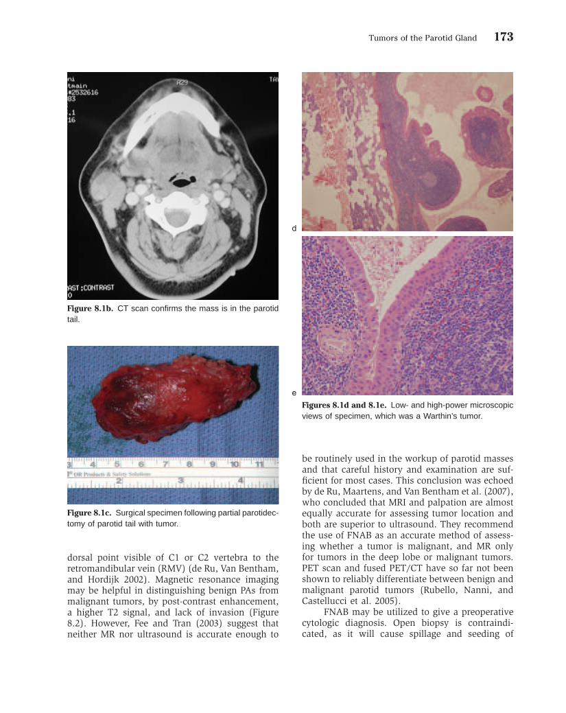
176 Tumors of the Parotid Gland
adenoma (53%) and intra-parotid lymph node (36%). Malignant salivary gland tumors with the highest false negative rate were acinic cell carci-noma (49%), low-grade mucoepidermoid carci-noma (43%), and adenoid cystic carcinoma (33%). It was felt the data confi rmed the diffi culty inher-ent in FNAB of salivary glands (Hughes, Volk, and Wilbur 2005). A paper from the Memorial Sloan Kettering Cancer Center concluded that an FNAB result positive for a malignant neoplastic process is generally predictive of the fi nal histologic diag-nosis, whereas the predictive value of a negative FNAB is low (Cohen, Patel, and Lin et al. 2004).
Surgical Management
The basic surgical procedure is the superfi cial parotidectomy in which the superfi cial lobe of the parotid is removed, preserving the facial nerve unless it is directly infi ltrated by the tumor. The author’s usual incision is the modifi ed Blair or “lazy S.” The skin fl ap is elevated in a plane through the subcutaneous fat superfi cial to the parotid capsule (Figure 8.4). Recently the use of a face lift incision has been advocated to improve esthetic results of the scar (Honig 2005; Menin-gaud, Bertolus, and Bertrand 2006). These authors have also combined face lift incisions with a sepa-rate SMAS (superfi cial musculoaponeurotic system) dissection to eliminate hollowing and reduce Frey’s syndrome. Concerns regarding access to anteriorly sited tumors when using a face lift approach for
parotidectomy do not appear to be borne out in anatomic studies (Nouraei, Al-Yaghchi, and Ahmed et al. 2006) (Figures 8.5 and 8.6).
Once the skin fl ap is elevated, the sterno-cleidomastoid muscle (SCM) is identifi ed with the overlying greater auricular nerve, whose branch to the earlobe may be preserved if it does not com-promise tumor resection (Figure 8.4e). The ante-rior border of the SCM is dissected free of the posterior parotid gland, which is retracted anteri-orly. Deeper dissection at the superior end of the SCM will allow identifi cation of the posterior belly of the digastric muscle. The facial nerve trunk lies 4 mm superior to the digastric and at the same depth and is an important landmark. Next, atten-tion is turned to the preauricular region with sharp and blunt dissection down the cartilage of the external auditory meatus to the bony portion of the meatus. A strip of parotid tissue remains, which separates the cervical from the preauricular dissection, and this tissue is carefully dissected away to the depth of the digastric muscle. Some troublesome bleeding has to be controlled with bipolar diathermy under direct vision superfi cial to where the facial nerve will be identifi ed. The facial nerve trunk can be confi rmed with a nerve stimu-lator and the nerve branches are dissected out peripherally to mobilize and remove the superfi cial parotid. It is usually best to dissect either the frontal or mandibular branches fi rst, depending on the site of the tumor, and then proceed stepwise inferiorly or superiorly dissecting the branches in order and staying superfi cial to the nerves.
If the tumor directly overlies the facial nerve trunk, making it impossible to access safely, then the peripheral branches can be identifi ed and fol-lowed backward as a retrograde parotidectomy, although this is more tedious. The mandibular branch of the facial nerve, where it crosses the anterior facial vein or the buccal branch with its close relationship to the parotid duct (Pogrel, Schmidt, and Ammar 1996), can be found initially. Despite a 66% incidence of weakness 1 week post-parotidectomy, normal facial nerve function was present in 99% of 136 retrograde parotidectomies in one series (O’Regan et al. 2007).
In tumors of the deep lobe it is usually neces-sary to undertake a total parotidectomy. The super-fi cial parotidectomy is performed, preserving the facial nerve and dissecting the superfi cial lobe from superiorly so that it remains attached to the deep lobe inferiorly and at the tail. Most deep lobe
Figure 8.3e. Histopathology confi rms diagnosis of pleo-morphic adenoma. Note marked pseudocapsule of collag-enous tissue. The patient is disease free 10+ years post-surgery.

177
a
b
Figures 8.4a and 8.4b. Patient with pleomorphic adenoma of the left parotid gland preoperatively.
Figure 8.4c. CT scan of left parotid showing superfi cial lobe tumor.
Figure 8.4d. Modifi ed Blair incision.

178
Figure 8.4f. The parotidectomy has been commenced from inferiorly and the superfi cial lobe is being retracted superiorly. The arrow indicates the deep surface of the tumor, which was adjacent to the nerve bifurcation (seen inferior to the tumor) and has no normal parotid tissue covering the tumor capsule.
Figure 8.4g. The surgical specimen is seen with the tumor superiorly with no good surrounding “cuff” of tissue.
Figure 8.4h. The surgical site post-parotidectomy with complete dissection of the facial nerve.
Figure 8.4i. A free abdominal fat graft is placed to reduce “hollowing,” which was a concern of the patient.
Figure 8.4e. Skin fl ap raised, the instrument indicates the sensory branch of the greater auricular nerve to the ear, which was preserved in this case.

179
j kFigures 8.4j and 8.4k. Six months postoperatively.
aFigures 8.5a and 8.5b. Preoperative facial views of patient with left parotid pleomorphic adenoma. Patient requests bilateral face lift at the same time as parotidectomy.
b

180
d e
Figure 8.5c. Surgical access through face lift incision running into occipital hair line. Arrow shows facial nerve trunk being dissected.
Figures 8.5d and 8.5e. Six months postoperatively. Notice absence of neck incision. (Aesthetic portions of this case undertaken by Dr. A. Pazoki, DDS, MD.)

181
Figure 8.6a. Preauricular portion of omega face lift inci-sion for parotidectomy for benign cystic lesion in the retro-mandibular portion of the superfi cial lobe of the left parotid gland.
Figure 8.6b. Post-auricular portion of omega incision runs in the post-auricular sulcus.
Figure 8.6c. A partial parotidectomy of the retroauricular portion of the superfi cial lobe is almost completed. The specimen is pedicled to the remains of the parotid tail.
Figure 8.6d. Close-up of the dissection. Arrow points to the mandibulo-cervical trunk, which was the only branch of the facial nerve that was dissected, as it crosses super-fi cial to the retromandibular vein.
Figure 8.6e. The patient 3 months postoperatively with no visible scar.

182 Tumors of the Parotid Gland
tumors will be retromandibular and lie inferior to the trunk of the nerve. The space inferiorly is larger and by gentle retraction of the nerve trunk and blunt dissection around the tumor it can usually be delivered into the neck. In larger tumors the neoplasm may be impacted between the man-dible and the mastoid with no means of mobilizing it without either dislocating the mandible forward or a subsigmoid or “C” osteotomy to give more space. As contemporary surgery has evolved, more emphasis has been placed on reducing morbidity. Deep lobe tumors may be removed without remov-ing the superfi cial lobe but leaving it attached anteriorly and then replacing it after excising the deep lobe tumor (Coleela et al. 2007). This tech-nique preserves facial contour and 84% of glan-dular function compared to the contralateral parotid.
In those tumors with parapharyngeal exten-sion, blind fi nger enucleation may lead to capsular rupture or cause brisk hemorrhage. In order to visualize and safely remove these tumors an oste-otomy of the mandible with or without lip split is utilized (Kolokythas, Fernandes, and Ord 2007) (Figures 8.7 and 8.8).
There is currently a controversy among sur-geons regarding superfi cial parotidectomy or extra-capsular dissection. This important topic will be discussed below in the section on pleomorphic adenomas.
Figure 8.7a. MR shows parapharyngeal pleomorphic adenoma.
Figure 8.7b. Standard lip split incision for mandibulotomy.
Figure 8.7c. Mandible is retracted out of the fi eld and the pleomorphic adenoma is dissected preserving the overly-ing lingual nerve.

183
Figure 8.8a. Deep lobe parotid tumor with parapharyngeal extension presenting as a palatal mass.
b c
Figures 8.8b and 8.8c. Axial and coronal CT scans show the tumor in the lateral pharyngeal space.

Figure 8.8d. The mandible is accessed via a cervical inci-sion from mastoid to chin without lip split. Subsigmoid osteotomy cut marked with saw through buccal cortex only (arrows) and plate has been applied prior to completing the osteotomy.
Figure 8.8e. Osteotomy marked with a saw through the buccal cortex (long arrow) anterior to the mental nerve (short arrow). Two miniplates applied prior to completing the osteotomy.
Figure 8.8f. The plates are removed, the double osteotomy is completed, and the osteotomized hemimandible is retracted upward and rotated to expose the lateral pharyngeal space, and the tumor is being delivered under direct vision.

185
Figure 8.8g. The fi nal specimen, which was PA, seen in rela-tion to the mandible.
Figure 8.8h. The post-resection tumor bed. The plates are reapplied to reconstruct the original position of the mandible and occlusion.

186 Tumors of the Parotid Gland
BENIGN TUMORS
Pleomorphic AdenomasThe PA is the commonest benign salivary gland tumor and the commonest salivary tumor overall, although it is comparatively rare in young chil-dren. It is slow growing and can reach giant pro-portions if neglected, and there is a 2–4% malignant change. PAs will recur if the tumor is inadequately removed. Although PAs have a pseudo-capsule of compressed fi brous tissue, the buds and pseudo-podia from the tumor involve the capsule so that simple enucleation will leave tumor remnants and lead to multifocal recurrence. The concept of whether the capsule is incomplete and whether pseudopodia of the tumor involves the parotid tissue is currently being questioned, and with it the need for complete superfi cial parotidectomy. Although parotidectomy is designed to remove PAs with a cuff or margin of normal tissue to prevent recurrence, the tumor’s proximity to the facial nerve frequently means that the dissection at some points leaves no tissue around the capsule. In a recent histologic analysis of the capsular form in PAs, 81% showed capsular exposure following parotidectomy or submandibular gland excision (Webb and Eveson 2001) (Figure 8.4f). This paper also showed 57% bosselations, 33% enveloping of the capsule, with 42% microinvasion and 12% “tumor buds” in the capsule; and large >25 mm hypocellular tumors had thinner capsules, possibly easier to rupture at surgery. The article suggested that a minimum of 1 mm of normal tissue around PAs was required as a margin. However, in an article reviewing 475 PAs of the superfi cial lobe of the parotid, 380 treated by extracapsular dissection and 95 by superfi cial parotidectomy, there was no difference in recurrence rate or permanent facial nerve palsy (McGurk et al. 1996). These surgeons postulated that tumor buds or micro-invasion into the capsule had little signifi cance and that extra-capsular dissection could be done safely. In 1999 a series of 59 partial parotidectomies with selective nerve dissection for benign and low-grade malig-nant tumors reported a zero incidence of perma-nent facial nerve paralysis or paresis and zero recurrence (Witt 1999). Although Witt in a later paper confi rmed that capsular exposure occurred in virtually all types of parotid surgery, and could fi nd no difference in recurrence, capsular rupture, tumor-facial nerve interface, and permanent facial palsy between total parotidectomy, superfi cial
parotidectomy, and extracapsular dissection, he recommended against minimum margin resection in extracapsular dissection (Witt 2002). Further evidence for extracapsular dissection is provided by a series of 83 cases in which the overall recur-rence rate was 6%, but 17.6% when the tumor itself was at the margins; however, cases with margins of <1 mm had a recurrence of only 1.8% (Ghosh et al. 2003). Ghosh et al. also reported that intraoperative capsular rupture and microscopic invasion of the capsule had no infl uence on recur-rence, suggesting that a fraction of a millimeter of normal tissue was an adequate margin and that only tumors that actually involved the margin were at risk for recurrence. These authors recom-mend that preservation of vital structures is a more important consideration than preserving a cuff of normal tissue.
In contrast, Piekarski, Nejc, and Szymczak et al. (2004) found a recurrence rate of 8.2% and an unacceptable rate of complications with extracap-sular dissection and did not recommend the tech-nique as too “technically demanding.” In a separate publication with 213 patients who were operated for pleomorphic adenoma of the parotid, 5 of 9 primary tumors (56%) that recurred were found to have pseudopodia extending outside the capsule on histologic review. This was statistically higher than the examined cases that did not recur (8%), and the authors concluded that pseudopodia extending outside the capsule was a signifi cant risk for recurrence (Henriksson et al. 1998). Interest-ingly, in the same study only 2 of 28 cases that ruptured during surgical removal recurred (7.1%), which was not signifi cantly different than the 4.1% recurrence rate for the tumors that had no rupture. A further cautionary note is raised by the histologic analysis of Zbaren and Stauffer (2007), in which 160 of 218 (73%) of PAs were found to have adverse capsular characteristics, 33% with an incomplete capsule and 13% with satellite nodules. These were most frequently seen in the stroma rich myxoid subtype. Similar fi ndings with stroma rich PAs showing 71% of focal absence of a capsule and 33% of satellite nodules have been reported with recommendations against local dissection (Stennert et al. 2004.)
It does not appear that extracapsular dissec-tion is just a “euphemism for enucleation” as some have claimed, as recurrence rates are comparable to parotidectomy and most papers show lower morbidity. The exact margin required for complete

Tumors of the Parotid Gland 187
removal of PA remains controversial. A criticism of extracapsular dissection has been that even if this technique is suitable for a presumed benign PA, what should the surgeon do if the fi nal histo-pathologic diagnosis turns out to be malignant? In a review of 662 clinically benign parotid tumors, 503 treated by extracapsular dissection and 159 by superfi cial parotidectomy, 5% were malignant and there was no difference in 5- or 10-year survival or recurrence rates between the malignant tumors in the two surgical groups, although morbidity was signifi cantly lower in the extracapsular dissection group (McGurk, Thomas, and Renehan 2003).
aFigures 8.9a, 8.9b, and 8.9c. MR images of large cystic benign tumor in the parotid tail.
b
c
A superfi cial lobe parotid tumor clinically benign and diagnosed as a PA on FNAB may also be treated with a limited superfi cial parotidectomy (without complete dissection of the facial nerve) and may not require a complete superfi cial paroti-dectomy for cure (O’Brien 2003). This is probably most commonly undertaken for tumors in the parotid tail (Figure 8.9).
Deep lobe PAs are usually larger and fre-quently will have less surrounding parotid tissue, especially deeply, where they abut the prevertebral muscles of the neck. However, the inability to obtain a surrounding cuff of parotid does not seem

188 Tumors of the Parotid Gland
Figure 8.9d. Following partial parotidectomy the two arrows point to the dissected cervico-mandibular branch of the facial nerve, the parotid tail tumor having been resected inferior to this nerve branch. The upper branches of the facial nerve have not been dissected in this case.
Figure 8.9e. The long arrow points to the resected parotid tail region, while the shorter arrow points to the remaining superfi cial parotid lobe preserved intact.
Figure 8.9f. Surgical specimen.
to lead to increased recurrence. Harney, Murphy, and Hone et al. (2003) found that the capsules of deep lobe tumors were signifi cantly thicker and that there was less extracapsular extension of tumor in the deep lobe tumors (58% vs. 79%), which may explain this phenomenon.
If the capsule of the tumor is ruptured during surgery, then recurrence is not inevitable and perhaps liberal irrigation with sterile water fol-lowed by normal saline may be tumoricidal (Webb and Eveson 2001). When recurrence occurs it is frequently multinodular (Figure 8.10) and requires
more radical en bloc surgery with excision of the previous scar, muscle, overlying skin, and facial nerve if they are involved. Maxwell, Hall, and Freeman (2004) in a retrospective study of 35 patients treated with surgery alone found a locore-gional control of 77% with a malignant transfor-mation of 5.7%. In a separate study of 42 cases of multinodular recurrence (6 with prior radiation), there were 2 patients with malignant transforma-tion who died of distant metastases. Twelve patients had subtotal parotidectomy, 25 total parotidectomy, 5 subtotal petrosectomy; 14 had facial nerve resection. Seven patients of 36 who were followed developed further recurrences (19.4%), all of whom had only undergone subtotal parotidectomy (Leonetti et al. 2005). In a further series of 33 patients, 73% multifocal, 9% with malignant transformation, treated surgically, 6 (18%) recurred at an average of 9 years, and 23% of patients with initial enucleation and 14% with initial superfi cial parotidectomy had permanent partial facial nerve injury (Zbaren Tschumi et al. 2006). Renehan, Gleave, and McGurk (1996) reviewed 144 cases of recurrent PAs and suggested a role for radiation in multi-nodular cases. A recent paper of 34 cases of

Tumors of the Parotid Gland 189
recurrent PAs with radiation therapy post–gross resection shows a 20-year actuarial control rate of 94% (Chen et al. 2006)
Warthin’s TumorThis is the second commonest benign tumor of the parotid. If it is diagnosed when small and asymp-tomatic it may not require treatment in an old or infi rmed patient. There is a 12% incidence of mul-tiple ipsilateral or bilateral tumors. There appears to be a link to heavy smoking and bilateral War-thin’s tumors (Klussman, Wittekindt, and Preuss et al. 2006). Eight percent of these tumors occur in extra-parotid cervical lymph nodes and may be found at the time of parotidectomy or serendipi-dously in neck dissection specimens. Treatment is as for PA. Warthin’s tumors have a tendency to occur in the parotid tail, where the majority of parotid lymph nodes occur, so partial parotidec-tomy is often all that is required.
MALIGNANT TUMORS
Principles of Management of Parotid CarcinomaThere is no universally agreed method for manag-ing parotid cancer; however, prognosis and man-agement are related to two variables: the histologic classifi cation/grade of the tumor and the staging. In reviewing 2,465 patients with carcinoma of parotid and submandibular glands, Wahlberg, Anderson, and Bjorklund et al. (2002) found 10-year survival of 88% for acinic cell carcinoma, 80% for mucoepidermoid carcinoma (MEC), and 74% for adenoid cystic carcinoma (ACC), but only 55% for adenocarcinoma unspecifi ed and 44% for undifferentiated carcinoma. It should be noted that 5-year survival fi gures for ACC will give an artifi -cially high value, as late local recurrence and distant metastasis continue over a 20+ year period (Chen, Garcia, Bucci et al. 2006; Lima, Tavares, Dias et al. 2005; Longuet, Nallet, Guedon et al. 2001). Harbo, Bungaard, and Pederson et al. (2002) also found acinic cell carcinoma to have the best 10-year survival, but in their Cox hazard regres-sion analysis found T stage, N stage, M stage, and histologic differentiation to be signifi cant in pre-dicting prognosis and recommended the use of both staging and histologic diagnosis to assess prognosis. In other series signifi cant factors include extraglandular extension, aggressive histology,
Figure 8.10a. Two large nodules of recurrent pleomorphic adenoma exist in the left parotid gland.
Figure 8.10b. Two smaller nodules are seen in the parotid tail.

190 Tumors of the Parotid Gland
and nodal disease (Bhattacharyya and Fried 2005), histologic grade, T stage, N stage, and facial nerve dysfunction (Lima et al. 2005), and N stage and perineural involvement (Hocwald, Korkmaz, and Yoo et al. 2001).
The only other predictor of adverse prognosis reported in several series was advancing age (Bhattacharyya and Fried 2005; Lima et al. 2005; Kirkbridge, Liu, and O’Sullivan et al. 2001).
The reported survival related to stage varies between authors, which may refl ect differences in therapy as well as different patterns of histopathol-ogy. Luukkaa, Klemi, and Leivo et al. (2005) found
Figure 8.11a. CT scan of large superfi cial lobe tumor in 22-year-old woman.
Figure 8.11b. Clinical appearance; the tumor has no signs of malignancy.
Figure 8.11c. Complete parotidectomy. The facial nerve was not involved.
Figure 8.11d. Histopathology of acinic cell carcinoma. The patient is alive and well 8+ years postoperatively.

Tumors of the Parotid Gland 191
5-year survival Stage I–IV 78%, 25%, 21%, and 23%, while Lima et al. (2005) found 10-year disease-specifi c survival Stage I–IV 97%, 81%, 56%, and 20%.
In considering management, Kaplan and Johns (1986) divide parotid cancers into 4 groups to recommend treatment. Group I T1-2 low-grade tumors are treated by parotidectomy with preser-vation of the facial nerve (Figures 8.11 and 8.12). Group II T1-2 high-grade are treated with paroti-dectomy plus fi rst echelon node removal and post-operative radiation therapy (RT) (Figure 8.13). Group III T3 tumors, any positive nodes and recur-rent tumor not in Group IV, are treated with radical parotidectomy with sacrifi ce of the facial nerve if necessary and radical neck dissection plus RT. Group IV includes T4 and tumors with signifi cant local extension; they are treated by radical paroti-
Figure 8.12a. Patient who had a “skin cyst” biopsied; it was histologically a low-grade mucoepidermoid carcinoma of the parotid. Note preauricular biopsy scar.
Figure 8.12b. Operative image shows Blair incision incor-porating 1 cm skin margin surrounding the biopsy.
Figure 8.12c. The level II nodes (fi rst echelon nodes) will be taken in continuity in this case.
Figure 8.12d. Histology shows a focus of mucoepider-moid carcinoma (arrow) in the biopsy scar between the skin and parotid, demonstrating the importance of excising “seeded” skin.

192 Tumors of the Parotid Gland
dectomy plus skin, muscle, and bone as indicated with radical neck dissection and postoperative RT (see chapter 11, Figure 11.16).
Controversy exists in the exact indications for RT, neck dissection, and facial nerve sacrifi ce. The majority of recent papers do show that RT is indi-cated for advanced parotid carcinoma and confers a survival benefi t (Bhattacharyya and Fried 2005) or longer disease-free survival (Hocwald, Korkmaz, and Yoo et al. 2001). However, there is a move toward suggesting RT for earlier stage disease. Zbaren, Nuyens, and Caversaccio et al. (2006) ret-rospectively analyzed T1-2 carcinomas with and without postoperative RT and found local recur-rence rates of 3% and 33%, respectively, and actu-arial and disease-free survival of 93% and 92% with and 83% and 70% without RT. In an earlier publication from the same unit RT was suggested
not just for high-grade tumors but low-grade T2-4 (Zbaren, Schupbach, and Nuyens et al. 2003). So perhaps RT is indicated for earlier stage disease than was previously recommended. The latest data regarding fast neutron therapy in the management of advanced salivary cancer with gross residual disease shows a 6-year local-regional control of 59%, and 100% with no evidence of gross residual disease (Douglas et al. 2003.) Benefi ts of chemo-therapy have not been clearly demonstrated for parotid cancer.
Similarly, although lymph node dissection was recommended for positive nodes and high-grade tumors, there is an increasing interest in the N0 neck. Occult metastasis rates of 22–45% led Stennert, Kisner, and Jungehuelsing et al. (2003) and Zbaren, Schupbach, and Nuyens et al. (2003) to recommend an elective neck dissection in the
Figure 8.12e. Mucicarmine stain confi rms intracellular mucus.
Figure 8.12f. Surgical specimen. Figure 8.12g. The patient is alive and disease-free 14 years postoperatively.

Tumors of the Parotid Gland 193
N0 neck for parotid cancer (Figure 8.14). Elective neck dissection for high-grade tumors and >T2 low-grade tumors should encompass levels I–III and upper V (Teymoortash and Werner 2002). In comparing elective neck dissection for the N0 neck against observation, Zbaren et al. (2005) found an
actuarial and disease-free survival of 80% and 86% for the elective neck dissection patients vs. 83% and 69% for the observation group in a ret-rospective study.
Regarding the facial nerve, Spiro and Spiro (2003) recommend preservation unless the nerve is adherent to/embedded in the tumor. They feel that close margins to the nerve can be treated suc-cessfully by RT. This view is supported by the work of Carinci, Farina, and Pelucchi et al. (2001), who found that sacrifi ce of the nerve was not always able to improve survival rate. In a series of 107 patients with parotid cancer, 91 had normal nerve function preoperatively and facial nerve preservation was possible in 79 patients. The 5-year disease-free rate and 5- and 10-year survival rates were 65%, 83%, and 54% in the preserved nerve cohort and 56%, 62%, and 42% in the patients with nerve sacrifi ce. The authors felt that
Figure 8.13a. MR of parotid tumor that radiologically was diagnosed as a pleomorphic adenoma, but fi ne needle aspiration biopsy showed adenoid cystic carcinoma.
Figure 8.13b. In view of the cytologic diagnosis, proposed treatment includes resection of overlying skin (which clini-cally was felt to be tethered) and level II cervical nodes.
Figure 8.13c. Surgical specimen shows parotid with skin. The arrows show where level II nodes and fat are in con-tinuity with the parotid tail. Final diagnosis was cellular pleomorphic adenoma with the FNAB diagnosis being a false positive.

194
Figure 8.14a. High-grade parotid malignancy with skin fi xation and N0 neck. Incision modifi ed to include skin exci-sion (arrow), and cervical incision extended to allow supra-omohyoid neck dissection.
Figure 8.14b. The skin fl aps developed with the skin over-lying the tumor left on the parotid gland.
Figure 8.14c. The selective neck dissection is complete with the specimen in continuity with the parotid gland. The facial nerve trunk has been exposed and the superfi cial parotidectomy is being performed.
Figure 8.14d. The surgical site following parotidectomy and selective neck dissection.
Figure 8.14e. The surgical specimen with the parotid superiorly and level I nodes pinned out with white pins.

Tumors of the Parotid Gland 195
preservation of the facial nerve by careful dissec-tion gave favorable oncologic results (Guntinas-Lichius, Klussman, and Schroeder et al. 2004). Finally, a disease-free survival in patients with normal, partially, and completely impaired facial nerve function preoperatively of 69%, 37%, and 13% despite the use of facial nerve sacrifi ce and postoperative RT indicates what a poor prognosis invasion of the nerve confers (Terhaard, Lubsen, and Tan et al. 2006).
In specifi c histologic tumor types variable results for different treatments have been reported. Mucoepidermoid carcinoma is the commonest sali-vary malignancy and most cases are fortunately low or intermediate grade. A series of 89 cases at the Mayo Clinic, 69 T1-2, 85 N0, and 83 low/inter-mediate grade, were treated by parotidectomy with “appropriate” neck dissection and only 7 had RT. Kaplan-Meier estimated cancer-specifi c survival rates at 5, 15, and 25 years were 98.9%, 97.4%, and 97.4% (Boahene, Olsen, and Lewis et al. 2004). Using a point grading system for histopathologic features in a series of 234 mucoepidermoid carcino-mas of the major salivary glands, cystic component <20%, 4 or more mitotic fi gures per 10 high power fi elds, neural involvement, necrosis, and anaplasia were found to have prognostic signifi cance for parotid MEC (Goode, Auclair, and Ellis 1998). Intermediate-grade MEC tends to behave more like low-grade MEC, while high-grade MEC behaves aggressively with local recurrence and regional and distant metastases in the majority of cases. Other low-grade tumors such as acinic cell carcinoma, epimyoepithelial carcinoma, and low-grade adeno-carcinoma all can be treated like low-grade MEC. Polymorphous low-grade adenocarcinoma is rare in the major glands, being seen mostly in the minor salivary glands of the oral cavity.
On the other hand, results for high-grade tumors such as primary squamous carcinoma of the parotid are poor; in one published series two-thirds were treated with radical surgery and RT and one-third with RT alone, but 5-year actuarial survival and disease-free survival was 31% and 33%, respectively (Lee, Kim, and Parks et al. 2001). Malignant change in PAs is most commonly seen as carcinoma ex-pleomorphic adenoma, and prognosis will depend on the histologic type of malignancy and whether the malignancy has spread outside the capsule. In carcinoma ex-pleo-morphic adenoma the use of postoperative RT improved 5-year local control from 49% to 75%
and improved survival in patients without cervical metastasis (Chen, Garcia, and Bucci et al. 2007). Two other forms of malignant PA occur, both rare: the “true” malignant mixed tumor or carci-nosarcoma where malignant change is seen in both the epithelial and myoepithelial component of the PA, and the benign metastasizing PA, which as its name suggests retains a benign histologic appearance despite the presence of metastases.
It is hard to interpret survival fi gures in some series, as ACC is very slow growing and 5-year survival is less meaningful in this neoplasm as survival continues to fall on 20-year follow-up. Thus in series with short follow-up ACC will erroneously be thought to have a good prognosis. Typical long-term survival fi gures are 84.3% 2 year, 75.9% 5 year, 50.49% 10 year, and 20.11% after 20 years (Issing, Hemmanouil, and Wilkens et al. 2002). The type of histologic appear-ance, solid vs. cylindrical, and the presence of perineural invasion are important prognostic factors. Even with documented lung metastases patients can live 5+ years, the average survival between the appearance of lung metastases and death being 32.3 months in one series (van der Waal et al. 2002). Wide fi eld adjuvant RT post–radical surgery is usually recommended for ACC.
The histologic grade of the tumor must be taken into account as well as TNM staging when interpreting survival results in reported series. Every parotid cancer will be unique and the deci-sion for what is the correct surgery will be made on an individual basis for each patient.
Summary
• 80% of parotid tumors are benign and most commonly pleomorphic adenomas.
• Less than one-third of malignant tumors will have obvious clinical signs of malignancy, for example, facial nerve palsy, ulceration, fi xa-tion, or lymphadenopathy.
• Routine use of CT or MR imaging does not appear justifi ed and should be used selec-tively for malignant neoplasms and deep lobe tumors.
• Preoperative open biopsy is contraindicated and FNAB is the modality of choice for pre-operative cytologic diagnosis.

196 Tumors of the Parotid Gland
• Although superfi cial parotidectomy remains the basic surgical procedure, there is currently much debate regarding the roles of partial parotidectomy and extracapsular dissection in the management of PA. The role of the capsule and the acceptable margin for PA remain undefi ned.
• Recurrent PAs will frequently require en bloc resection due to their infi ltrative and multi-nodular nature. Cure in this situation is prob-ably achieved in approximately two-thirds of cases.
• Management of malignant parotid tumors will depend on both the histologic diagnosis and the staging of the tumor.
• Radiation therapy may be more helpful in earlier stage disease and lower-grade tumors than previously advocated.
• Selective neck dissection for the N0 neck may be justifi ed in early stage disease given the high reported rate of occult nodes.
• The facial nerve should be preserved in parotid cancer unless it is directly infi ltrated by the tumor.
References
Ariyoshi Y, Shimahara M. 1998. Determining whether a parotid tumor is in the superfi cial or deep lobe using magnetic resonance imaging. J Oral Maxillofac Surg 56:23–27.
Bhattacharyya N, Fried MP. 2005. Determinants of survival in parotid gland carcinoma: A population based study. Am J Otolaryngol 26(1):39–44.
Boahene DK, Olsen KD, Lewis JE et al. 2004. Mucoepider-moid carcinoma of the parotid gland: The Mayo Clinic experience. Arch Otolaryngol Head Neck Surg 130(7):849–856.
Carinci F, Farina A, Pelucchi S et al. 2001. Parotid gland carcinoma: Surgical strategy based on local risk factors. J Craniofac Surg 12(5):434–437.
Chen AM, Garcia J, Bucci MK et al. 2007. The role of post-operative radiation therapy in carcinoma ex pleomorphic adenoma of the parotid gland. Int J Radiat Oncol Biol Phys 67(1):138–143.
Chen AM, Garcia J, Bucci MK, Quivey JM, Eisle DW. 2006. Recurrent pleomorphic adenoma of the parotid gland: Long term outcome of patients treated with radiation therapy. Int J Radiol Biol Phys 66(4):1031–1035.
Cohen EG, Patel SG, Lin O et al. 2004. Fine needle aspi-ration biopsy of salivary gland lesions in a selected patient population. Arch Otolaryngol Head Neck Surg 130(6):773–778.
Coleela C, Giudice A, Rambali PF, Cuccurullo V. 2007. Parotid function after selective deep lobe parotidectomy. Brit J Oral Maxillofacial Surg 45:108–111.
de Ru JA, Maartens SVL, Van Bentham PPG et al. 2007. Do magnetic resonance imaging and ultrasound add any-thing to the preoperative workup of parotid tumors? J Oral Maxillofac Surg 65:945–952.
de Ru JA, Van Bentham PPG, Hordijk GJ. 2002. The loca-tion of parotid gland tumors in relation to the facial nerve on magnetic resonance images and computed tomogra-phy scans. J Oral Maxillofac Surg 60:992–996.
Douglas JG, Koh WJ, Austin-Seymour M, Laramore GE. 2003. Treatment of salivary gland neoplasms with fast neutron radiotherapy. Arch Otolaryngol Head Neck Surg 129:944–948.
Fee WE Jr, Tran LE. 2003. Evaluation of a patient with a parotid tumor. Arch Otolaryngol Head Neck Surg 129:937–938.
Ghosh S, Panarese A, Bull PD, Lee JA. 2003. Marginally excised parotid pleomorphic adenomas: Risk factors for recurrence and management. A 12.5 year mean follow up study of histologically marginal excisions. Clin Oto-laryngol 28:262–266.
Goode RK, Auclair PL, Ellis GL. 1998. Mucoepidermoid carcinoma of the major salivary glands: Clinical and histopathologic analysis of 234 cases with evaluation of grading criteria. Cancer 82(7):1217–1224.
Guntinas-Lichius O, Klussman JP, Schroeder U et al. 2004. Primary parotid malignant surgery in patients with normal preoperative facial nerve function: Outcome and long-term postoperative facial nerve function. Laryngo-scope 114(5):949–956.
Harbo G, Bungaard T, Pedersen D et al. 2002. Prognostic indicators for malignant tumors of the parotid gland. Clin Otolaryngol Allied Sci 27(6):512–516.
Harney MS, Murphy C, Hone S et al. 2003. A histological comparison of deep and superfi cial lobe pleomor-phic adenomas of the parotid gland. Head Neck 25(8):649–653.
Henriksson G, Westrin KM, Carlsoo B, Silversward C. 1998. Recurrent primary pleomorphic adenomas of salivary gland origin: Intrasurgical rupture, histopathologic fea-tures, and pseudopodia. Cancer 82(4):617–620.
Hocwald E, Korkmaz H, Yoo GH et al. 2001. Prognostic factors in major salivary gland cancer. Laryngoscope 111(8):1434–1439.
Honig JF. 2005. Omega incision face lift approach and SMAS rotation advancement in parotidectomy for the prevention of contour defi ciency and conspicuous scars affecting the neck. Int J Oral Maxillofac Surg 34(6):612–618.
Hughes JH, Volk EE, Wilbur DC. 2005. Pitfalls in salivary gland fi ne-needle aspiration cytology: Lessons from the College of American Pathologists Interlaboratory Comparison Program in Nongynecologic Cytology. Arch Pathol Lab Med 129(1):26–31.

Tumors of the Parotid Gland 197
In der Maur CD, Klokman WJ, van Leeuwen FE et al. 2005. Increased risk of breast cancer development after diag-nosis of salivary gland cancer. European Journal of Cancer 41(9):1311–1315.
Issing PR, Hemmanouil I, Wilkens L et al. 2002. Long term results in adenoid cystic carcinoma. (Article in German.) Laryngorhinootologie 81(2):98–105.
Kaplan MJ, Johns ME. 1986. Salivary gland cancer. Clin Oncology 5:525–547.
Kirkbridge P, Liu FF, O’Sullivan B et al. 2001. Outcome of curative management of malignant tumors of the parotid gland. J Otolaryngol 30(5):271–279.
Klussman PJ, Wittekindt C, Preuss FS et al. 2006. High risk for bilateral Warthin’s tumors in heavy smokers—review of 185 cases. Acta Otolaryngol 126(11):1213–1217.
Kolokythas A, Fernandes RP, Ord RA. 2007. A non-lip-splitting-double mandibular osteotomy technique for resection of tumors in the parapharyngeal and pterygo-mandibular spaces. J Oral Maxillofac Surg 65(3):66–69.
Lamelas J, Terry JH, Alfonso AE. 1987. Warthin’s tumor: Multicentricity and increasing incidence in women. Am J Surg 154:347–351.
Lee S, Kim GE, Parks CS et al. 2001. Primary squamous cell carcinoma of the parotid gland. Am J Otolaryngol 22(6):400–406.
Leonetti JP, Marzo SJ, Petrzelli GJ, Herr B. 2005. Recurrent pleomorphic adenoma of the parotid gland. Otolaryngol Head Neck Surg 133(3):319–322.
Lima RA, Tavares MR, Dias FL, Kligerman J, Nascimento MF, Barbosa MM, Cernea CR, Soares JR, Santos IC, Salviano S. 2005. Clinical prognostic factors in malig-nant parotid gland tumors. Otolaryngol Head Neck Surg 133(5):702–708.
Longuet M, Nallet E, Guedon C et al. 2001. Diagnostic value of needle biopsy and frozen section histological examina-tion in the surgery of primary parotid tumors. Rev Laryngolo Otol Rhinol 122(1):51–55.
Lonn S, Alholm A, Christensen HC et al. 2006. Mobile phone use and risk of parotid gland tumor. Am J Epide-miol 164(7):637–643.
Luukkaa H, Klemi P, Leivo I et al. 2005. Salivary gland cancer in Finland 1991–96. Acta Otolaryngol 125(2):207–214.
Maxwell EL, Hall FT, Freeman JL. 2004. Recurrent pleo-morphic adenoma of the parotid gland. J Otolaryngol 33(3):181–184.
McGurk M, Renehan A, Gleave EN, Hancock BD. 1996. Clinical signifi cance of the tumor capsule in the treat-ment of parotid pleomorphic adenomas. Br J Surg 83(12):1747–1749.
McGurk M, Thomas BL, Renehan AG. 2003. Extracapsular dissection for clinically benign parotid lumps: Reduced morbidity without oncological compromise. Br J Cancer 89(9):1610–1613.
Meningaud JP, Bertolus C, Bertrand JC. 2006. Parotidec-tomy: Assessment of a surgical technique including face-lift incision and SMAS advancement. J Craniomaxillofac Surg 34(10):34–37.
Nouraei SA, Al-Yaghchi C, Ahmed J et al. 2006. An ana-tomical comparison of Blair and facelift incisions for parotid surgery. Clin Otolaryngol 31(6):531–534.
O’Brien CJ. 2003. Current management of benign parotid tumors—the role of limited superfi cial parotidectomy. Head Neck 25:946–952.
Ord RA. 1995. Surgical management of parotid tumors. Oral and Maxillofac Surg Clin of North America 7(3):529–564.
O’Regan BO, Bharadwaj G, Bhopal S, Cook V. 2007. Facial nerve morbidity after retrograde nerve dissection in parotid surgery for benign disease: A 10-year prospective observational study of 136 cases. Brit J Oral Maxillofac Surg 45(2):101–107.
Piekarski J, Nejc D, Szymczak W et al. 2004. Results of extracapsular dissection of pleomorphic adenoma of the parotid gland. J Oral Maxillofac Surg 62(10):1198–1202.
Pogrel MA, Schmidt B, Ammar A. 1996. The relationship of the buccal branch of the facial nerve to the parotid duct. J Oral Maxillofac Surg 54(1):71–73.
Renehan A, Gleave EN, McGurk M. 1996. An analysis of the treatment of 114 patients with recurrent pleomorphic adenoma of the parotid gland. Am J Surg 172:710–714.
Rubello D, Nanni C, Castellucci P et al. 2005. Does 18F-FDG PET/CT play a role in the differential diagnosis of parotid masses. Panminerva Med 47(3):187–189.
Saku T, Hayashi Y, Takahara O et al. 1997. Salivary tumors among atom bomb survivors, 1950–1987. Cancer 79(8):1465–1475.
Schroder U, Eckel HE, Rasche V et al. 2000. Value of fi ne needle puncture cytology in neoplasms of the parotid gland. HNO 48(6):421–429.
Spiro JD, Spiro RH. 2003. Cancer of the parotid gland: Role of 7th nerve preservation. World J Surg 27(7):863–867.
Stennert E, Kisner D, Jungehuelsing M et al. 2003. High incidence of lymph node metastasis in major salivary gland cancer. Arch Otolaryngol Head Neck Surg 129(7):720–723.
Stennert E, Wittekindt C, Klussman JP, Guntinas-Lichas O. 2004. New aspects in parotid surgery. Otolaryngol Pol 58(1):109–114.
Takahashi N, Okamoto K, Ohkubo M, Kawana M. 2005. High-resolution magnetic resonance of the extracranial facial nerve and parotid duct: Demonstration of the branches of the intraparotid facial nerve and its relation to parotid tumors by MRI with a surface coil. Clin Radiol-ogy 60(3):349–354.
Terhaard C, Lubsen H, Tan B et al. 2006. Facial nerve function in carcinoma of the parotid gland. Eur J Cancer 42(16):2744–2750.

198 Tumors of the Parotid Gland
Teymoortash A, Werner JA. 2002. Value of neck dissection in patients with cancer of the parotid gland and a clinical N0 neck. Onkologie 25(2):122–126.
van der Waal JE, Becking AG, Snow GB, van der Waal I. 2002. Distant metastases of adenoid cystic carcinoma of the salivary glands and the value of diagnostic examinations during follow up. Head Neck 24(8):779–783.
Wahlberg P, Anderson H, Bjorklund A et al. 2002. Carci-noma of the parotid and submandibular glands—a study of survival in 2,465 patients. Oral Oncol 38(7):706–713.
Webb AJ, Eveson JW. 2001. Pleomorphic adenomas of the major salivary glands: A study of the capsular form in relation to surgical management. Clin Otolaryngol 26:134–142.
Witt RL. 1999. Facial nerve function after partial superfi cial parotidectomy: An 11 year review 1987–1997. Otolaryn-gol Head Neck Surg 121(3):210–213.
Witt RL. 2002. The signifi cance of the margin in parotid surgery for pleomorphic adenoma. Laryngoscope 112(12):2141–2154.
Wong DS. 2001. Signs and symptoms of malignant parotid tumors: An objective assessment. J R Coll Surg Edinb 46(2):91–95.
Zbaren P, Nuyens M, Caversaccio M et al. 2006. Postopera-tive radiation for T1 and T2 primary parotid carcinoma: Is it useful? Otolaryngol Head Neck Surg 135(1):140–143.
Zbaren P, Schar C, Hotz MA et al. 2001. Value of fi ne-needle aspiration cytology of parotid gland masses. Laryngoscope 111(11 part 1):1989–2002.
Zbaren P, Schupbach J, Nuyens M et al. 2003. Carcinoma of the parotid gland. Am J Surg 186(1):57–62.
Zbaren P, Schupbach J, Nuyens M, Stauffer E. 2005. Elec-tive neck dissection versus observation in primary parotid cancer. Otolaryngol Head Neck Surg 132(3):387–391.
Zbaren P, Stauffer E. 2007. Pleomorphic adenoma of the parotid gland: Histopathologic analysis of the capsular characteristics of 218 tumors. Head Neck 29: 751–757.
Zbaren P, Tschumi I, Nuyens M, Stauffer E. 2006. Recur-rent pleomorphic adenoma of the parotid gland. Am J Surg 192(2):203–207.

Chapter 9
Tumors of the Submandibular and Sublingual Glands
mas (MECs), and adenoid cystic carcinomas (ACCs), PCNA, Ki-67, and p53 expression for PA and ACC in the submandibular gland was similar to that reported for tumors of the parotid gland and minor salivary glands. However, there was a higher expression of these markers in MEC of the submandibular gland (Alves, Pires, and DeAlem-eda et al. 2004). This may indicate that MEC of the submandibular gland is potentially more aggressive.
Approximately 10–15% of all salivary gland tumors will occur in the submandibular gland, and only 0.5–1% in the sublingual gland, so these tumors are very rare. In the submandibular gland approximately 50% of these tumors are benign. Series vary in their percentages, from 657 of 1,235 tumors (53%) benign (Auclair, Ellis, and Gnepp et al. 1991), 55% benign (Oudidi, El-Alami, and Bou-laich et al. 2006), which included nonepidermoid cancers, to 39.2% benign (Rapidis et al. 2004). Pleomorphic adenoma is the commonest benign tumor in the submandibular gland, while ACC predominates for malignant tumors. In examining malignant tumors, Bhattacharyya (2004) analyzed 370 cases from the Surveillance, Epidemiology, and End Results (SEER) database, fi nding ACC 42.2% and MEC 22.2%, while the Rapidis et al. (2004) literature review of 356 cases showed ACC 45.3%, adenocarcinoma 14.3%, MEC 12.9%, and carcinoma ex-pleomorphic adenoma 11.2%, and Auclair, Ellis, and Gnepp et al. (1991) found ACC 24% and MEC 19% of 578 cases.
Although sublingual gland tumors are extremely rare they are important to recognize, as they have an extremely high rate of malignancy. In a review of approximately 4,000 patients with salivary tumors collected over a 55-year period, only 18 (0.5%) had sublingual gland tumors, all of which were malignant (Spiro 1995). There are very few other large series of sublingual gland
OutlineIntroductionEpidemiology and EtiologyDiagnosis
Submandibular Gland TumorsSublingual Gland Tumors
ManagementSubmandibular Gland TumorsSublingual Gland Tumors
SummaryReferences
Introduction
This chapter will discuss the diagnosis and man-agement of epithelial derived tumors of the sub-mandibular and sublingual glands. These tumors are much less common than tumors of the parotid gland and a long series of cases from which to apply evidence-based medicine treatment proto-cols is lacking, particularly with regard to the sub-lingual gland neoplasms. Current approaches are highlighted, although the diversity of histologic types, paucity of cases, and lack of long-term follow-up results in many of these cases being treated empirically based on oncologic principles derived from other tumors and sites in the head and neck region.
Epidemiology and Etiology
The etiology of tumors of the submandibular and sublingual glands is the same as discussed in rela-tion to salivary gland tumors of the parotid gland (see chapter 8). At a molecular level in a study that examined PCNA, Ki-67, and p53 in pleomor-phic adenomas (PAs), mucoepidermoid carcino-
199

200 Tumors of the Submandibular and Sublingual Glands
tumors in the literature. Yu, Gao, and Wang et al. (2007) reported 30 cases collected over a 50-year period, all of which were malignant. In one of the author’s personal series (Ord) of 9 sublingual gland tumors collected over a 16-year period at the Uni-versity of Maryland, there were no benign tumors. Adenoid cystic carcinoma appears to be the com-monest histologic type, followed by MEC: ACC 50%, MEC 28% (Spiro 1995), ACC 56.7% (Yu, Gao, and Wang et al. 2007), and ACC 66%, MEC 33% (Perez, Pires, and Alves et al. 2005).
DiagnosisSUBMANDIBULAR GLAND TUMORS
Most submandibular gland tumors present with a slow-growing, painless mass inferior to the man-
dible (Figure 9.1). In a series of 87 submandibular gland carcinomas, 94% presented with a palpable mass and 39% with pain (Kaszuba, Zafero, and Rosenthal et al. 2007). As tumors of the gland are rare and infl ammatory swelling secondary to sialo-lithiasis is seen more often, they may not be ini-tially diagnosed and can present with late disease. In one series 50% of all referred patients with submandibular gland tumors had already had their submandibular gland removed on the presumption that the involved process was benign (Camilleri et al. 1998). The average tumor size in 370 cases of cancer of the submandibular gland was 2.9 cm (Bhattacharyya 2004). Infl ammatory disease, however, is often painful and usually character-ized by exacerbations and resolutions of the swell-ing in relation to eating. In a series of 258 submandibular gland excisions, 119 (46%) had sialolithiasis, 88 (34%) sialadenitis, and 51 (20%) tumors (Preuss, Klussmann, and Wittekindt et al. 2007).
Examination usually reveals a smooth, fi rm to hard mass in the submandibular triangle that is most commonly discrete and mobile. Fixation of the mass to the skin or underlying mylohyoid muscle is a sign of malignancy with advanced extracapsular infi ltration (Figure 9.2). Neural involvement of the mandibular branch of the facial nerve with ipsilateral lower lip palsy, the lingual nerve with ipsilateral anesthesia/paresthesia of the tongue, or the hypoglossal nerve with ipsilateral palsy of the tongue muscles are also signs of cancer. Associated hard cervical nodes due to regional metastasis may also be present in malig-nant tumors.
The differential diagnosis of a solitary mass in the submandibular triangle with no overt signs of malignancy will include lymphadenopa-thy, plunging ranula, vascular malformation, and branchial cysts. It may be diffi cult to differen-tiate a lymph node from the enlarged gland on clinical examination alone. If the mass is bimanu-ally palpable from within the fl oor of the mouth it is more likely to be a submandibular gland mass, and if it can be “rolled” over the lower border of the mandible on palpation it is a lymph node. The plunging ranula is usually soft-cystic in consis-tency but can become fi rm if chronically encysted. Vascular lesions are also soft and may “pit” on fi rm pressure or have thrills and murmurs. Bran-chial cysts lie more posterior and are partially
Figure 9.1. A 64-year-old man with a painless subman-dibular mass.

Tumors of the Submandibular and Sublingual Glands 201
beneath the anterior border of the sternocleido-mastoid muscle.
Imaging techniques to delineate submandibu-lar gland lesions include ultrasound, CT, and MR. As the submandibular gland is superfi cial in the neck high-resolution ultrasound can distinguish intraglandular from extraglandular masses and can differentiate benign tumors from those that are malignant (Alyas, Lewis, and Williams et al. 2005). CT scanning may be useful in detecting early corti-cal erosion of the mandible and identifying cervical nodes in malignant cases (Figure 9.3). In a study to identify whether a submandibular mass was intra- or extraglandular, the accuracy of contrast enhanced CT was 87%, CT sialography 85%, and MR 91% (Chikui, Shimizu, and Goto et al. 2004).
Figure 9.2a. Elderly lady with hard submandibular mass (adenoid cystic carcinoma on fi ne needle aspiration biopsy) who presented with a palsy of the marginal mandibular branch of the facial nerve.
Figure 9.2b. Lateral facial view shows skin fi xation and tethering.
Figure 9.2c. Histopathology of adenoid cystic carcinoma of submandibular gland.

202 Tumors of the Submandibular and Sublingual Glands
These authors did not fi nd displacement of the facial vein and its relationship to the mass a helpful guide.
Open biopsy of the submandibular gland mass is contraindicated for similar reasons that were discussed in relation to the parotid (see chapter 8). Fine needle aspiration biopsy (FNAB) is the method of choice for these tumors, one lit-erature review fi nding an overall accuracy of greater than 80% in skilled hands, which is com-parable with the accuracy of frozen section (Pogrel 1995).
SUBLINGUAL GLAND TUMORS
Tumors of the sublingual gland present as a mass in the fl oor of the mouth, usually painless and
a
b
Figures 9.3a and 9.3b. CT scans showing submandibular mass with differing regions of radiolucency and opacity. Histopathology showed pleomorphic adenoma.
Figure 9.4a. Adenocarcinoma of right sublingual gland.
Figure 9.4b. Polymorphous low-grade adenocarcinoma of the sublingual gland. Re-published with permission from Blanchaert RG, Ord RA, Kumar D. 1998. Polymorphous low-grade adenocarcinoma of the sublingual gland. Int J Oral Maxillofac Surg 27:115–117.
Figure 9.5. CT scan of large malignant sublingual gland tumor.

Tumors of the Submandibular and Sublingual Glands 203
slow-growing. They may be large enough to impair tongue movement with speech diffi culty or to prevent wearing a lower denture (Figure 9.4). Occasionally they may cause obstruction of Whar-ton’s duct either due to pressure or malignant infi ltration and present with a submandibular swelling. Virtually 100% of these tumors are malignant and involvement of the lingual nerve or hypoglossal nerve with ipsilateral anesthesia or weakness of the tongue may be seen. Examination by palpation reveals a fi rm to hard mass that may be tender and fi xed to the lingual periosteum. Infi ltration of the tongue muscles with slurring of speech or dysphagia can occur.
The only other entity on the differential diag-nosis is a ranula, which can resemble a cystic tumor.
Imaging is usually by CT or MR. CT scans will be more accurate for early cortical bone inva-sion (Figure 9.5). In MR imaging T1 weighted signal intensity of carcinomas in and near the sub-lingual gland is lower than the gland, whereas T2 weighted signal intensity of carcinomas exceeds that of the gland (Sumi et al. 1999).
In the sublingual gland histologic diagnosis is accomplished by incisional biopsy through the overlying oral mucosa.
ManagementSUBMANDIBULAR GLAND TUMORS
As in all salivary gland tumors surgery is the primary modality of treatment. When the diagno-sis is established preoperatively as benign PA by FNAB, then an extracapsular excision of the sub-mandibular gland is indicated (Figures 9.6 and 9.7). Pleomorphic adenomas should be treated in the same manner as for the parotid gland (see chapter 8). There is some evidence that the capsule of PAs in the submandibular gland is thinner than in the parotid (Webb and Eveson 2001), and it is important to maintain a margin of normal tissue around the tumor. If the entire gland and tumor is not removed but the PA merely enucleated, there is a higher risk of recurrence (Laskawi et al. 1995). In this dissection it is easy to maintain a little extra fat and connective tissue over areas where the PA may approach the surface of the gland (Figure 9.8). In a series of 15 PAs of the submandibular gland 20% were in the surface of the gland
Figure 9.6a. The incision for submandibular gland tumor removal lays approximately 1–2 fi nger-breadths below the lower border of the mandible and is placed in a natural skin crease.
Figure 9.6b. The submandibular gland is separated from its duct, which is indicated by the sharp scissors. The arrow points to the tumor in the hilum.

204 Tumors of the Submandibular and Sublingual Glands
(Laskawi et al. 1995) (Figure 9.9). The marginal mandibular branch of the facial nerve lies between the platysma superfi cially and the capsule of the submandibular gland (superfi cial layer of the deep cervical fascia) deeply and can be preserved either by dissecting it along its course and retracting it superiorly or by ligating and cutting the anterior
facial vein inferior to the nerve and using traction on the tied distal end of the vessel to retract the nerve out of the fi eld (the Hayes Martin maneu-ver). The incidence of transient palsy of the mar-ginal mandibular branch of the facial nerve is 7% in excising benign tumors and 21% in excising malignant tumors, with only 1 case (<1%) of per-manent palsy in this series (Preuss, Klussmann, and Wittekindt et al. 2007). The facial artery is sacrifi ced if it passes through the gland itself, but if not its numerous small branches including the submental branch can be clipped and the main vessel preserved.
In recurrent PA the disease will frequently be multinodular as in the parotid, and as 45% of these cases involve the subcutaneous tissue under the previous operative scar, excision of the scar with a margin of the surrounding skin is recommended as part of the en bloc excision (Laskawi et al. 1995).
Where a defi nite diagnosis of a benign tumor is not established preoperatively or when a low-grade malignant tumor is diagnosed, an en bloc resection of level I is safest. If the fi nal histologic diagnosis is benign no important structures have been sacrifi ced, only the gland and tumor plus fat with lymph nodes. If the tumor is a low-grade malignancy, then no further surgery is indicated. In the case of a high-grade tumor a selective or radical neck dissection can be completed at the same time (Figure 9.10).
Figure 9.6c. Surgical specimen with arrow indicating PA.
Figure 9.6d. Histopathology shows pleomorphic adenoma of submandibular gland.
Figure 9.7. Another case of submandibular pleomorphic adenoma dissected to the duct. The larger tumor at the hilum is indicated by the arrow.

205
Figure 9.8a. Axial CT shows tumor projecting beyond the gland surface.
Figure 9.8b. Coronal CT confi rms the surface involvement.
Figure 9.8c. High-power axial CT scan shows the tumor marked with a circle.
Figure 9.8d. Operative picture showing the tumor marked by palpation.
Figure 9.8e. Intraoperative view of extracapsular dissec-tion preserving soft tissue over the tumor surface.

206 Tumors of the Submandibular and Sublingual Glands
In undertaking the level I dissection the cervi-cal skin fl ap is lifted in a subplatysmal plane and the marginal mandibular branch of the facial nerve is preserved. The anterior belly of the digastric muscle is identifi ed and its fascia dissected free. The fascia is dissected off the mylohyoid muscle, freeing the fat and nodes from the digastric, mylo-hyoid, and inferior border of the mandible. The posterior edge of the mylohyoid muscle is retracted to identify the lingual nerve, which is preserved if it is uninvolved by cutting the branch to the gland and allowing the nerve to retract into the mouth. The duct is sectioned and the facial vein and artery tied off or dissected free as indicated to release the specimen. In low-grade N0 tumors a neck dissec-tion is usually not indicated, but the excision can be extended to encompass lymph node levels II and III as a supraomohyoid neck dissection. If lymph nodes are clinically involved, then a type I modifi ed radical neck dissection is required.
In high-grade tumors or advanced low-grade tumors with extracapsular infi ltration and involve-ment of the skin, muscle, or mandible extended resections with incorporation of these structures will be necessary to obtain clear margins. These resections will be dictated by the size and extent of the tumor. In N0 cases selective neck dissection levels I-III or I-IV will be used and modifi ed radical neck dissections for clinically positive necks. In the case of ACC, widespread infi ltrative growth beyond the palpable tumor makes obtaining clear margins challenging. The propensity for perineural inva-
sion with ACC will necessitate sacrifi ce of involved nerves, for example, lingual, hypoglossal, and facial, tracing the nerves proximally using frozen section guidance to determine clearance. Unfortu-nately, “skip” metastases can occur along the nerve and a negative frozen section is no guaran-tee of success. ACC is more prone to metastasize hematogenously than through lymphatics such that a selective neck dissection is usually suffi cient.
Postoperative radiation therapy is adminis-tered for high-grade tumors, positive margins, positive nodes, and perineural spread if re-resection is not possible. Chemotherapy has not been shown to improve survival in salivary gland cancer.
Prognosis will depend on the histologic grade and the stage of the tumor. Some authors (Ander-son, Thrkildsen, and Ockelman et al. 1991) have found a crude 10-year survival of 50% with 10% local recurrence and 39% of cases having metastasized at the time of diagnosis. In the series reported by Rapidis et al. (2004), 8 of 14 patients died during follow-up with a survival rate of 38.5%, but 11 of 14 of these patients presented with stage III or IV disease. Bhattacharyya (2004) analyzed 370 cases of submandibular gland cancer from the SEER database and reported a 59.7% 5-year survival; however, this fi gure is high, as 42.2% of his cases were ACC with a mean survival of 99 months. In the same series the patients with squamous cell carcinoma had a mean sur-vival of 52 months. Younger age, low-grade histol-ogy, and the use of radiation therapy were factors in improving survival. Weber, Byers, and Petit et al. (1990) found a 69% 5-year survival, with extracapsular infi ltration and lymph node metastases indicating a poor prognosis. Stages TI-TIVA had a case-specifi c 5-year survival of 88% compared to 55% for T4B, and 5-year survival of 86% for negative nodes compared to 30% for positive nodes.
Most published series have found a survival benefi t conferred by radiation therapy, with 75% of patients receiving adjuvant radiation in one study (Camilleri et al. 1998). Storey, Garden, and Morrison et al. (2001) report actuarial locoregional control of 88% at 5 and 10 years; however, the corresponding disease-free survival rates were 60% and 53% due to 36% of patients with locore-gional control developing distant metastases.
Figure 9.8f. Surgical specimen.

207
Figure 9.9a. Submandibular gland attached only by its duct. The pleomorphic adenoma is large and hangs beneath the gland attached only by the enveloping fascia of the capsule.
Figure 9.9b. Surgical specimen with arrows pointing to the large PA, which has no real attachment to the gland.
Figure 9.10a. 82-year-old man with pain and swelling of the right submandibular region.
Figure 9.10b. CT scan confi rms a submandibular gland mass diagnosed as malignant on fi ne needle aspiration biopsy.

208
c
dFigures 9.10c and 9.10d. Intraoperative fi gures showing level I excision combined with a selective neck dissection in view of high-grade cytology.
Figure 9.10e. Operative specimen.
f
g
Figures 9.10f and 9.10g. Hematoxylin and eosin stain (f) and Alcian Blue stain (g) confi rm a diagnosis of high-grade mucoepidermoid carcinoma.

Tumors of the Submandibular and Sublingual Glands 209
Nonetheless the median survival time for patients with locoregional control was 183 months com-pared to 19 months for those patients without locoregional control. In a similar study of adjuvant radiation therapy, cancer-specifi c survival was 79% and 57% at 5 and 10 years, with local control of 85% and 74%, respectively. Twenty percent of patients (all ACC) developed distant metastases (Sykes et al. 1999). A retrospective study of 87 patients that compared patients who received either initial enucleation of the gland (subcapsular dissection) with no evidence of residual primary or nodal disease followed by postoperative radia-tion therapy; or who had evidence of gross residual primary or nodal disease, grossly positive margins, or piecemeal removal following initial treatment who underwent defi nitive surgical resection fol-lowed by postoperative radiation therapy, found no difference in locoregional control, disease-specifi c survival, or overall survival (Kaszuba,
Zafero, and Rosenthal et al. 2007). This suggests patients without evidence of gross residual disease post-enucleation might be satisfactorily treated with radiation therapy without further surgery.
In a series of 22 patients with ACC of the submandibular gland, disease-free survival at 5 years of 57% and 10 years of 41% and overall survival of 70% and 37%, respectively, were found (Cohen, Damrose, and Huang et al. 2004). These authors concluded that early diagnosis, wide surgi-cal intervention, and postoperative radiotherapy were associated with a favorable prognosis, while not surprisingly large tumor size, positive surgical margins, perineural invasion, and local recurrence were negative prognostic factors.
In comparing submandibular gland cancers to parotid cancer a poorer overall prognosis was associated with submandibular gland tumors (Hocwald, Korkmaz, and Yoo et al. 2001). In addi-tion, the likelihood of developing distant metasta-sis is greater in the submandibular gland than the parotid (Schwenter et al. 2006); however, this may be due to the higher percentage of ACC. In one large series of 370 cases only 12 (3%) presented with distant metastases, but 24.9% were found to have positive regional nodes (Bhattacharyya 2004). Interestingly, in this retrospective review extrag-landular extension and nodal positivity did not affect survival. Goode, Auclair, and Ellis (1998) in a study of 234 cases of MEC of the major salivary glands stated that MECs with equal histopathologic grade had a better prognosis when their tumors were in the parotid gland rather than in the sub-mandibular gland. This fi nding was confi rmed in a Swedish study of 2,465 major salivary gland tumors (Wahlberg, Anderson, and Bjorklund et al. 2002). High-grade MEC is also more common in the submandibular gland, with 32 of 82 cases (39%) occurring in this gland (Bhattacharyya 2004).
SUBLINGUAL GLAND TUMORS
Virtually all sublingual gland tumors will be malig-nant and as for all salivary tumors, primary surgery is the treatment of choice. Prognosis will be deter-mined by the histologic grade and the stage of the tumor. In the very rare benign tumor or with small low-grade carcinomas a transoral wide local resec-tion may be successfully performed (Blanchaert, Ord, and Kumar 1998). This will be easier to undertake in edentulous patients, and Wharton’s
Figure 9.10h. One year postoperative view of the patient. He died 3 years postoperatively with distant metastases of the lung.

210 Tumors of the Submandibular and Sublingual Glands
duct will require a sialodochoplasty procedure (Figure 9.11). In most cases due to grade, tumor size, the presence of teeth, and involvement of mandibular periosteum/bone a wider access will be required. If the periosteum is uninvolved and can be safely peeled from the lingual bone a stan-dard “pull through” approach or a lip split with mandibulotomy can be used (Figure 9.12). The functional result of the lip split/mandibulotomy is better than the pull through (Devine, Rogers, and McNally et al. 2001). As both of these methods of access involve entering the neck, a supraomohyoid neck dissection is usually carried out in the N0 neck even for low-grade tumors (Figure 9.13).
When positive nodes are present, type I mod-ifi ed radical neck dissection is required. Both lingual and hypoglossal nerves can be involved by these tumors at an early stage, particularly the ACC. Sacrifi ce of the nerve with proximal tracing and frozen section guidance as described for the submandibular tumors may be needed. In tumors fi xed to periosteum or where minimal cortical erosion is present, an oblique marginal mandibular resection angling the cut to take a greater height of the lingual plate will be utilized. The marginal mandibular resection can be performed with the pull through or mandibulotomy approach. Where the medullary bone is invaded a segmental man-dibular resection with a composite en bloc resec-tion of the fl oor of the mouth is safest and will provide excellent access (Figure 9.14). In these
larger soft tissue resections a thin pliable fl ap such as the radial forearm fl ap probably gives the best results in maintaining tongue mobility. Where the mandible has been resected a fi bular fl ap (Rinaldo, Shaha, and Pellitteri et al. 2004) or deep circumfl ex iliac artery (DCIA) fl ap is appropriate.
Adjuvant radiation therapy is indicated for positive nodes, perineural invasion, extracapsular nodal spread, positive margins, and high-grade histology. Prognosis for these tumors is diffi cult to assess, as the literature is mostly composed of case reports and small series. Spiro (1995) reported only 3 of 18 patients (16.6%) dying of their tumor with a median follow-up of 74 months. However, Yu, Gao, and Wang et al. (2007) reported distant
Figure 9.11. Delivering sublingual gland with low-grade malignant tumor via an intraoral wide local excision. Re-published with permission from Blanchaert RG, Ord RA, Kumar D. 1998. Polymorphous low-grade adenocarcinoma of the sublingual gland. Int J Oral Maxillofac Surg 27:115–117.
Figure 9.12a. Adenoid cystic carcinoma of sublingual gland closely approximated to the mandible (seen via an intraoral mirror photograph).
Figure 9.12b. Following lip split and mandibulotomy the periosteum is found to be uninvolved and is stripped from the mandible, which is preserved.

Tumors of the Submandibular and Sublingual Glands 211
Figure 9.13a. CT scan shows low-grade mucoepidermoid carcinoma of the right sublingual gland.
Figure 9.13b. This tumor was accessed via a lip split inci-sion and mandibulotomy. Bilateral supraomohyoid neck dissections were undertaken, as can be seen in the surgi-cal specimen.
Figure 9.13c. Cosmetic result of lip split incision. Patient is alive and tumor-free 13 years postoperatively.
metastases and local recurrence as the main cause of death with local recurrence rates of 30% and distant metastases 26.7%. In this series 56.7% of tumors were stage III.
It is reasonable to conclude that although 5-year survival from submandibular and sublin-gual gland cancer is reasonable, the high percentage of ACC found in these glands leads to a continuing decrease in survival at 10 years and beyond due to late local recurrence and distant metastases.

212
Figure 9.14a. 46-year-old African American man with left cervical lymphadenopathy.
Figure 9.14b. Intraoral view of high-grade mucoepider-moid carcinoma fi xed to the right mandible.
Figure 9.14d. A modifi ed radical neck dissection and hemimandibulectomy with lip split was performed.
Figure 9.14c. CT scan confi rms necrotic node.

213
Figure 9.14e. Postoperative panoramic fi lm showing the reconstruction plate.
Figure 9.14f. Immediately post-chemoradiation.
Figure 9.14g. Mass in anterior mediastinum eroding sternum and manubrium.
Figure 9.14h. Close-up view of manubrial mass. The patient died shortly thereafter due to lung metastases. Re-published with permission from Ord RA. 2000. Salivary gland disease. In: Fonseca R (ed.), Oral and Max-illofacial Surgery, Volume 5, Surgical Pathology. Philadel-phia: W.B. Saunders Co., pp. 288–289 (Figures 10-21 a, b, c, d, e, f, g).

214 Tumors of the Submandibular and Sublingual Glands
Summary
• Submandibular gland tumors comprise only 10% of salivary tumors.
• Most submandibular swellings are infl amma-tory in etiology.
• 50% of submandibular tumors will be malignant.
• Open biopsy should not be used for subman-dibular gland tumors. FNAB is the preopera-tive diagnostic method of choice.
• Sublingual gland tumors comprise <1% of salivary tumors.
• 90% of sublingual gland tumors are malignant.
• ACC followed by MEC are the commonest cancers in both the submandibular and sub-lingual glands.
• Surgical management is based on both histo-logic diagnosis and stage.
ReferencesAlves FA, Pires FR, DeAlemeda OP et al. 2004. PCNA, Ki-67
and p53 expression in submandibular salivary gland tumors. Int J Oral Maxillofac Surg 33(6):593–597.
Alyas F, Lewis K, Williams M et al. 2005. Diseases of the submandibular gland as demonstrated using high resolu-tion ultrasound. Br J Radiol 78(928):362–369.
Anderson LJ, Thrkildsen MH, Ockelman HH et al. 1991. Malignant epithelial tumors in the minor salivary glands, the submandibular gland and the sublingual gland. Cancer 68:2431–2437.
Auclair PL, Ellis GL, Gnepp DR et al. 1991. Salivary gland neoplasms: General considerations. In: Ellis GL, Auclair PL, Gnepp DR (eds.), Surgical Pathology of the Salivary Glands. Philadelphia: W.B. Saunders, pp. 144–145.
Bhattacharyya N. 2004. Survival and prognosis for cancer of the submandibular gland. J Oral Maxillofac Surg 62(4):427–430.
Blanchaert RH, Ord RA, Kumar D. 1998. Polymorphous low-grade adenocarcinoma of the sublingual gland. Int J Oral Maxillofac Surg 27:115–117.
Camilleri IG, Malata CM, McLean NR, Kelly CG. 1998. Malignant tumors of the submandibular salivary gland: A 15 year review. Br J Plast Surg 51(3):181–185.
Chikui T, Shimizu M, Goto TK et al. 2004. Interpretation of the origin of a submandibular mass by CT and MR imaging. Oral Surg Oral Med Oral Pathol Radiol Endod 98(6):721–729.
Cohen AN, Damrose EJ, Huang RY et al. 2004. Adenoid cystic carcinoma of the submandibular gland: A 35 year review. Otolaryngol Head Neck Surg 131(6):994–1000.
Devine JC, Rogers SN, McNally D et al. 2001. A comparison of aesthetic, functional and patient subjective outcomes following lip-split mandibulotomy and mandibular lingual releasing access procedures. Int J Oral Maxillofac Surg 30(3):199–204.
Goode RK, Auclair PL, Ellis GL. 1998. Mucoepidermoid carcinoma of the major salivary glands: Clinical and histopathologic analysis of 234 cases with evaluation of grading criteria. Cancer 82(7):1217–1224.
Hocwald E, Korkmaz H, Yoo GH et al. 2001. Prognostic factors in major salivary gland cancer. Laryngoscope 111(8):1434–1439.
Kaszuba SM, Zafero ME, Rosenthal DI et al. 2007. Effects of initial treatment on disease outcome for patients with submandibular gland carcinoma. Arch Otolaryngol Head Neck Surg 133(6):546–550.
Laskawi R, Ellies M, Arglebe C, Schott A. 1995. Surgical management of benign tumors of the submandibular gland: A follow up study. J Oral Maxillofac Surg 53(5):506–508.
Oudidi A, El-Alami MN, Boulaich M et al. 2006. Primary submandibular gland tumors: Experience based on 68 cases. Rev Laryngol Otol Rhinol (Bord) 127(3):187–190.
Perez DE, Pires FR, Alves FA et al. 2005. Sublingual gland tumors: Clinicopathologic study of six cases. Oral Surg Oral Med Oral Pathol Radiol Endod 100(4):449–453.
Pogrel MA. 1995. The diagnosis and management of tumors of the submandibular and sublingual glands. Oral Maxil-lofac Clin North Amer 7(3):565–571.
Preuss SF, Klussmann JP, Wittekindt C et al. 2007. Sub-mandibular gland excision: 15 years of experience. J Oral Maxillofac Surg 65:953–957.
Rapidis AD, Stavrianos S, Lagogiannis G, Faratzis G. 2004. Tumors of the submandibular gland: Clinicopathologic analysis of 23 patients. J Oral Maxillofac Surg 62(10):1203–1208.
Rinaldo A, Shaha AR, Pellitteri PK et al. 2004. Management of malignant sublingual salivary gland tumors. Oral Oncol 40:2–5.
Schwenter I, Obrist P, Thumfart W, Sprinzi G. 2006. Distant metastasis of parotid tumors. Acta Otolaryngol 126(4):340–345.
Spiro RH. 1995. Treating tumors of the sublingual glands, including a useful technique for repair of the fl oor of mouth after resection. Am J Surg 170(5):457–460.
Storey MR, Garden AS, Morrison WH et al. 2001. Postopera-tive radiotherapy for malignant tumors of the submandibu-lar gland. Int J Radiat Oncol Phys 51(4):952–958.
Sumi M, Izumi M, Yonetsu K, Nakamura T. 1999. Sublin-gual gland: MR features of normal and diseased states. Am J Roentgenol 172(3):717–722.
Sykes AJ, Slevin NJ, Birzgalis AR, Gupta NK. 1999. Sub-mandibular gland carcinoma: An audit of local control and survival following adjuvant radiotherapy. Oral Oncol 35(2):187–190.

Tumors of the Submandibular and Sublingual Glands 215
Wahlberg P, Anderson H, Bjorklund A et al. 2002. Carci-noma of the parotid and submandibular glands—a study of survival in 2,465 patients. Oral Oncol 38(7):706–713.
Webb AJ, Eveson JW. 2001. Pleomorphic adenomas of the major salivary glands: A study of the capsular form in relation to surgical management. Clin Otolaryngol 26:134–142.
Weber RS, Byers RM, Petit B et al. 1990. Submandibular gland tumors. Arch Otolaryngol Head Neck Surg 116:1055–1060.
Yu T, Gao GH, Wang XY et al. 2007. A retrospective clini-copathologic study of 30 cases of sublingual gland malig-nant tumors (in Chinese). Hua Xi Kou Qiang Yi Xue Za Zhi 25(1):64–66.


Chapter 10
Tumors of the Minor Salivary Glands
explain the relative paucity of these tumors (Ansari 2007). The average annual incidence of salivary gland tumors per 100,000 population is 4.7 for benign tumors and 0.9 for malignant tumors (Ansari 2007). Both neoplastic and non-neoplastic entities are diagnosed in the salivary glands, including the minor salivary glands, thereby adding to the stimu-lating nature of the differential diagnosis, micro-scopic diagnosis, and treatment of minor salivary gland tumors. Data regarding the incidence of sali-vary gland tumors in general may be diffi cult to obtain. This is not only due to the rarity of these tumors but also to the previous non-routine nature of reporting of these diagnoses to hospital tumor registries and the occasional treatment of these lesions in offi ce settings (Melrose 1994). It has been estimated that minor salivary gland tumors account for only 2–5% of all head and neck tumors, with malignant minor salivary gland tumors accounting for only 2–4% of all head and neck cancers (MacIn-tosh 1995). In 1985 Regezi et al. reported on 238 minor salivary gland tumors among 72,282 (0.33%) total oral biopsy specimens diagnosed over a 19-year period (Regezi et al. 1985). Similarly, Rivera-Bastidas, Ocanto, and Acevedo reported 62 minor salivary gland tumors from a total of 9,000 oral biopsies (0.7%) during a 24-year period (Rivera-Bastidas, Ocanto, and Acevedo 1996). Another review of 40,000 head and neck tumors over a 40-year period of time revealed 196 (0.5%) minor salivary gland tumors. Approximately 10% of all salivary gland tumors arise in the minor glands (Ord 1994). Of these minor salivary gland tumors, 70% occur in the oral cavity, 25% in the nasal cavity/sinuses/nasopharynx, and 3% occur in the larynx (MacIntosh 1995). Of the oral minor salivary gland tumors, at least 50% have been diagnosed in the palate, according to most large series (Eveson and Cawson 1985; Spiro 1986). In addition to rela-tively low numbers of minor salivary gland tumor diagnoses, there are controversies regarding the
OutlineIntroductionEtiology of Minor Salivary Gland TumorsDiagnosis of Minor Salivary Gland TumorsTreatment of Minor Salivary Gland Tumors
General Principles of Surgery for Minor Salivary Gland Tumors
Surgical Treatment of Benign Minor Salivary Gland Tumors
Pleomorphic AdenomaCanalicular Adenoma
Surgical Treatment of Malignant Minor Salivary Gland Tumors
Mucoepidermoid CarcinomaAdenoid Cystic CarcinomaPolymorphous Low-Grade AdenocarcinomaAcinic Cell AdenocarcinomaEpithelial-Myoepithelial Carcinoma
Surgical Management of the Neck for Minor Salivary Gland Malignancies
The Role of Radiation Therapy in the Management of Minor Salivary Gland Malignancies
The Role of Chemotherapy in the Management of Minor Salivary Gland Malignancies
SummaryReferences
Introduction
The evaluation, diagnosis, and treatment of a patient with a mass occupying the territory of minor salivary gland tissue in the palate, buccal mucosa, or lips represent intellectually stimulating disci-plines. This statement is clearly derived from the relative paucity of lesions in these anatomic areas. Salivary gland tumors in general are quite rare, accounting for only 0.2–6.6% of all human tumors (Chidzonga, Lopez-Perez, and Portilla-Alvarez 1995). Both geographic and racial factors may
217

218 Tumors of the Minor Salivary Glands
precise microscopic diagnosis of these tumors. In Waldron’s review of 426 oral minor salivary gland tumors (Waldron, El-Mofty, and Gnepp 1988), each of these cases was reviewed by the three authors and complete concurrence of the microscopic diag-noses was reached in 346 cases (81.2%). In 49 cases, there were minor disagreements as to the diagnoses, mainly related to the subclassifi cation of the tumors. Signifi cant disagreement, regarding a benign vs. malignant diagnosis of the neoplasm, was noted in 21 cases (5%). Moreover, following the authors’ review, their diagnoses were com-pared to those of the contributing pathologists. There was complete agreement in 374 cases (87.8%). These statistics exemplify the complex nature of intraoral minor salivary gland tumors, thereby questioning the exact incidence of these neoplasms as a whole as well as specifi c diagnoses in particular.
Minor salivary gland tumors occur not only as benign and malignant entities but also as a spectrum of cell types within these glands (Carlson 1998). The frequency of benign versus malignant tumors occurring throughout the minor glands in the oral cavity is one feature that distinguishes these tumors from their counterparts in the major glands. One series reported 15% of parotid gland tumors and 37% of submandibular gland tumors to be malignant (Eveson and Cawson 1985). In general terms, published series record that approx-imately 20–70% of minor salivary gland tumors are malignant (Epker and Henny 1969; Ord 1994) (Table 10.1). It seems that the center that reports the incidence of benign vs. malignant minor sali-vary gland tumors of the oral cavity is the primary bias in these reports. For example, tertiary care referral centers with a cancer initiative may pref-erentially receive patients on referral with malig-nant diagnoses. Spiro’s report of his 35-year experience with salivary gland neoplasia at Memo-rial Sloan Kettering Cancer Center is a case in point. He reported on 2,807 patients, 607 of whom had minor salivary gland tumors. The frequency of malignant tumors in this report was 87%. The Armed Forces Institute of Pathology (AFIP) reported 2,945 cases of minor salivary gland tumors in 1991. By contrast, 49% of these cases were malig-nant. Numerous other series have reported similar fi gures, such that it has become reasonably well accepted that approximately 50% of minor salivary gland tumors of the oral cavity are benign and 50% are malignant.
Etiology of Minor Salivary Gland Tumors
Risk factors for salivary gland tumors have been studied extensively. Carcinoma of the major sali-vary glands, for example, has identifi ed a relation-ship with prior radiation therapy and previous skin cancer (Spitz, Tilley, and Batsakis et al. 1984). Another study reported 31 patients who had both a newly diagnosed salivary gland tumor and a history of radiation therapy to the head and neck region (Katz and Preston-Martin 1984). Radiation therapy had been administered with a range of 11–66 years prior to the development of the sali-vary gland tumors. No course of radiation therapy was administered for a malignant condition, but rather for acne, hypertrophied tonsils, keloids, and other benign conditions. As such, it is reasonable to assume that a low dose of radiation therapy was administered. Only 3 cases of minor salivary gland tumors were identifi ed among these 31 cases, including 2 adenoid cystic carcinomas and 1 muco-epidermoid carcinoma. One of the tumors was located in the palate and 2 were located in the cheek/retromolar region.
Benign and malignant salivary gland tumors have also been linked to exposure to ionizing radi-ation related to the atomic bombings in Hiroshima and Nagasaki during World War II. One hundred forty-fi ve salivary gland tumors have been studied in survivors of these bombings (Saku, Hayashi, and Takahara et al. 1997). One hundred nineteen major gland tumors (27 malignant tumors, 82 benign tumors, 10 undetermined tumors) and 26 minor gland tumors (14 malignant tumors, 12 benign tumors) were identifi ed. Among the 41 malignant tumors, the frequency of mucoepider-moid carcinoma was disproportionately high, and among the 94 benign tumors, the frequency of Warthin’s tumor was high.
The association between fi rst primary benign and malignant neoplasms of the salivary glands and the subsequent development of breast cancer has also been investigated (Abbey et al. 1984). This study identifi ed a four-fold to fi ve-fold increased risk of a second primary breast cancer subsequent to the fi rst salivary gland tumor. Of note is that all of the primary salivary gland tumors were of the major glands, and 3 of the 4 patients described had benign salivary gland tumors. While no association between minor salivary gland neo-

Tumors of the Minor Salivary Glands 219
plasia and breast cancer was established, the study nonetheless attempted to develop a relationship between the two. Moreover, the study investigated subsequent breast cancer in patients with a history of salivary gland neoplasia, rather than vice versa. As such, a risk factor for salivary gland tumor development would not be established in patients with breast cancer. In addition, minor salivary gland neoplasia was not represented in this cohort of patients. In the fi nal analysis, there is some evidence to identify risk factors for the develop-
ment of minor salivary gland tumors, yet not as much evidence as exists for the development of major salivary gland tumors.
Diagnosis of Minor Salivary Gland Tumors
The diagnosis of a minor salivary gland tumor begins with the establishment of a differential
Table 10.1. Incidence of minor salivary gland tumors
Authors Year Location Number of Cases Histology
Ansari 2007 Iran 18 Benign 11.1%Malignant 88.9%
AFIP (Auclair, Ellis, Gnepp et al.) 1991 USA 2,945 Benign 51%Malignant 49%
Chau, Radden 1986 Australia 98 Benign 62%Malignant 38%
Chidzonga, Lopez-Perez, Portilla-Alvarez 1995 Zimbabwe 282 Benign 80%Malignant 20%
Eveson, Cawson 1985 England 336 Benign 54%Malignant 46%
Isacsson, Shear 1983 Sweden 201 Benign 28%Malignant 72%
Ito, Ito, Vargas et al. 2005 Brazil 113 Benign 37%Malignant 63%
Jaber 2006 Libya 75 Benign 39%Malignant 61%
Lopes, Kowalski, Santos, Almeida 1999 Brazil 196 Benign 35%Malignant 65%
Potdar, Paymaster 1969 India 110 Benign 49%Malignant 51%
Regezi, Lloyd, Zarbo, McClatchey 1985 USA 238 Benign 65%Malignant 35%
Rivera-Bastidas, Ocanto, Acevedo 1996 Venezuela 62 Benign 55%Malignant 45%
Satko, Stanko, Longauerova 2000 Slovakia 31 Benign 48%Malignant 52%
Spiro 1986 USA 607 Benign 13%Malignant 87%
Stuteville, Corley 1967 USA 80 Benign 10%Malignant 90%
Toida, Shimokawa, Makita et al. 2005 Japan 82 Benign 67%Malignant 33%
Waldron, El-Mofty, Gnepp 1988 USA 426 Benign 58%Malignant 42%

220 Tumors of the Minor Salivary Glands
diagnosis. This differential diagnosis should be classifi ed categorically and in order of decreasing likelihood (Carlson 1998). The evaluation of a lesion of the palate, lip, or buccal mucosa might suggest infl ammatory, neoplastic, and non-neo-plastic entities. Ultimately, the differential diagno-sis is based on the patient’s history, physical examination, and the anatomic location of the pathologic entity under scrutiny. Generally speak-ing, benign and malignant minor salivary gland tumors present as painless, slowly enlarging intra-oral masses. When present, ulceration predicts a malignant diagnosis, although many minor sali-vary gland malignancies do not create ulceration of the oral mucosa. Pain is an ominous sign and is associated with perineural invasion, typically by adenoid cystic carcinoma. A painful enlargement of the minor salivary glands is malignant until proven otherwise. The presence or absence of pain therefore represents an important element of the patient’s history. Special imaging studies may be obtained prior to performing the biopsy, if required at all, or they may be obtained after the establish-ment of a diagnosis based on incisional biopsy. Experience shows, however, that their purpose is to anatomically delineate the extent of the tumor rather than to assist in the establishment of the diagnosis. The experienced salivary gland surgeon may detect nuances on imaging studies that favor various diagnoses on the differential diagnosis. While fi ne needle aspiration biopsy is an essential part of the diagnosis of parotid neoplasms, it has no practical role in the diagnosis of minor salivary gland neoplasms. Rather, an incisional biopsy should be routinely performed, for example, when planning treatment of palatal tumors due to the diverse nature of possible histopathologic diagno-ses, as well as the diverse nature of surgical treat-ment plans based on these diagnoses.
Other minor salivary gland tumor sites, such as the buccal mucosa and upper lip, require greater attention to the differential diagnosis in order to determine whether incisional or excisional biopsy best serves the needs of the patient. In many instances, the differential diagnosis may strongly support a benign neoplasm such that proceeding directly to excision is the most appropriate therapy. For example, a tumor in the upper lip that is freely moveable and associated with normal overlying mucosa indicates that an excisional biopsy may be performed in most instances due to the high likeli-hood of a benign tumor. A minor salivary gland
tumor of the buccal mucosa, similar to the palate, has a diverse number of possibilities on the dif-ferential diagnosis such that an incisional biopsy should be considered to establish the diagnosis (Table 10.2). There are instances, however, where a freely moveable buccal mucosal tumor may be excised without preceding incisional biopsy, similar to the upper lip tumor previously described.
Treatment of Minor Salivary Gland TumorsGENERAL PRINCIPLES OF SURGERY FOR MINOR SALIVARY GLAND TUMORS
The treatment of minor salivary gland tumors is distinctly surgical. The specifi c type of surgery is a function of the anatomic site of the tumor, the invasion of surrounding structures, and the histo-pathologic diagnosis, provided that an incisional biopsy has been performed. In general terms, a palatal minor salivary gland tumor requires an incisional biopsy so as to defi nitively establish the histopathologic diagnosis prior to the tumor surgery. This biopsy should be performed in the center of the mass so as to not seed the surround-ing normal tissue (Freedman and Jones 1994). The decision as to whether to perform an incisional biopsy of buccal mucosal and lip masses thought to represent minor salivary gland tumors rests on the surgeon’s intuition as to the benign vs. malig-nant nature of the mass. Smooth, freely moveable submucosal masses without fi xation to the overly-ing mucosa are likely benign and may be treated with excisional biopsies without fi rst performing an incisional biopsy due to the high likelihood of a benign process. By contrast, sizeable masses with mucosal fi xation in these areas should prob-ably be subjected to incisional biopsy so as to establish the diagnosis due to the concern for malignant disease. As with tumor surgery for other diagnoses, minor salivary gland tumor surgery requires a preoperative assessment of the anatomic barriers. Physical examination and imaging studies serve to delineate invasion of surrounding ana-tomic barriers by the tumor. In the palate, for example, it is important to determine whether the palatal bone has been invaded by the tumor. Benign tumors typically do not invade bone but may “cup it out.” In such situations, it is not nec-essary to resect bone. Malignant tumors of the

Tumors of the Minor Salivary Glands 221
palate display variable involvement of the palatal bone. Imaging studies, particularly coronal bone windows, must be obtained so as to assess the involvement of the anatomic barrier of palatal bone. Minor salivary gland tumors of the upper lip and buccal mucosa exhibit different behavior regarding their invasion of the anatomic barrier of the surrounding mucosa. In general terms, it is appropriate to preserve the mucosa surrounding a benign minor salivary gland tumor of these sites, while a malignant tumor surgery in these sites requires sacrifi ce of the surrounding mucosa (Table 10.3).
Treatment of minor salivary gland tumors becomes predicated on the histopathologic diagno-sis, which largely translates to the known biologic behavior of the neoplasm. Descriptive surgical terms may describe the sacrifi ce of surrounding soft and hard tissues as a matter of convenience (Carlson 1998). For example, surgical management of palatal tumors may be as straightforward as a
periosteal sacrifi cing, bone sparing wide local exci-sion with split thickness dissection of the soft palate. This specifi c surgical procedure is the main procedure performed for benign palatal tumors and also has a role to play in some low-grade malignancies (Carlson 1998). The bone-sparing, periosteally sacrifi cing wide local excision with full thickness sacrifi ce of the soft palate is reserved for deeply infi ltrative low-grade malignancies of the palate. The most aggressive surgery for palatal minor salivary gland tumors is the maxillectomy, specifi cally reserved for the highly aggressive minor salivary gland malignancies of the palate.
SURGICAL TREATMENT OF BENIGN MINOR SALIVARY GLAND TUMORS
The treatment of benign tumors of the minor sali-vary glands centers on the pleomorphic adenoma, with a brief discussion of the surgery for the cana-
Table 10.2. Incidence of benign and malignant minor salivary gland tumors at various sites.
Authors Year Palate Lip Buccal Mucosa
AFIP (Auclair, Ellis, Gnepp et al.) 1991 Benign 53%Malignant 47%
Benign 73%Malignant 27%
Benign 50%Malignant 50%
Chau, Radden 1986 Benign 67%Malignant 33%
Benign 77%Malignant 23%
Benign 64%Malignant 36%
Eveson, Cawson 1985 Benign 53%Malignant 47%
Benign 73%Malignant 27%
Benign 50%Malignant 50%
Isacsson, Shear 1983 Benign 78%Malignant 22%
Benign 71%Malignant 29%
Benign 89%Malignant 11%
Jaber 2006 Benign 58%Malignant 42%
Benign 46%Malignant 54%
Benign 46%Malignant 54%
Lopes, Kowalski, Santos, Almeida 1999 Benign 42%Malignant 58%
Benign 60%Malignant 40%
Benign 0%Malignant 100%
Potdar, Paymaster 1969 Benign 49%Malignant 51%
Benign 67%Malignant 33%
Benign 78%Malignant 22%
Regezi, Lloyd, Zarbo, McClatchey 1985 Benign 30%Malignant 70%
Benign 88%Malignant 12%
Benign 43%Malignant 57%
Rivera-Bastidas, Ocanto, Acevedo 1996 Benign 56%Malignant 44%
Benign 18%Malignant 82%
Benign 50%Malignant 50%
Spiro 1986 Benign 26%Malignant 84%
Benign 18%Malignant 82%
Stuteville, Corley 1967 Benign 4%Malignant 96%
Benign 33%Malignant 67%
Benign 27%Malignant 73%
Toida, Shimokawa, Makita et al. 2005 Benign 69%Malignant 31%
Benign 67%Malignant 33%
Benign 60%Malignant 40%
Waldron, El-Mofty, Gnepp 1988 Benign 58%Malignant 42%
Benign 75%Malignant 25%
Benign 54%Malignant 46%

222 Tumors of the Minor Salivary Glands
licular adenoma. The three most common minor salivary gland anatomic sites will be considered, including the palate, the lip, and the buccal mucosa.
Pleomorphic AdenomaThe terms “pleomorphic adenoma” and “mixed tumor” are equally satisfactory and interchange-able when describing this common minor salivary gland tumor. The designation “mixed” is based on the tumor’s mixtures of neoplastic elements such that each mixed tumor has unique features (Melrose 1994). It has also been pointed out that the designation refers to the tumor showing com-bined features of epithelioid and connective tissue-like growth (Waldron 1991). There is universal agreement that the pleomorphic adenoma is the most common salivary gland tumor. The Armed Forces Institute of Pathology data of 13,749 sali-vary gland tumors showed 6,880 cases of pleomor-phic adenoma, of which 4,359 were located in the parotid gland and 1,277 were located in minor salivary gland tissue (Auclair, Ellis, and Gnepp et al. 1991). The palate accounted for 711 of these 6,880 cases of pleomorphic adenoma (10.3%) and was the second most common site for this tumor in the AFIP data. The 711 cases in the palate rep-resent 56% of cases located in the minor salivary glands. Interestingly, the AFIP data subclassifi ed palatal pleomorphic adenomas into those occur-ring on the hard palate (118 cases) and those occurring in the soft palate (110 cases). There were 483 cases that were not specifi ed as to location in the palate. The subclassifi cation of specifi c ana-tomic location in the palate is of signifi cance when working up these cases and planning surgical treatment for these patients. Those pleomorphic
adenomas located primarily in the soft palate require investigation as to involvement of the para-pharyngeal space.
Treatment of the palatal pleomorphic adenoma is based on the realization that this tumor does not possess a capsule. This notwithstanding, the tumor does exhibit a “pseudocapsule” represented by a loose fi brillar network surrounding the tumor. In addition, the periosteum on the superior aspect of the tumor does serve as a very competent ana-tomic barrier such that palatal bone may be pre-served in this tumor surgery, even when the bone has been “cupped out” clinically and radiographi-cally. Under such circumstances, the pleomorphic adenoma does not invade bone histologically such that bone resection is not warranted. In fact, it is reasonable to proceed with surgery without obtain-ing CT scans preoperatively. A periosteally sacrifi c-ing wide local excision is performed, observing a 5–10 mm linear margin surrounding the clinically apparent tumor (Figure 10.1). While these tumors are submucosal in nature, the mucosa must be sacrifi ced with the tumor due to the close proxim-ity of the tumor and the overlying mucosa (Yih, Kratochvil, and Stewart 2005). The most appropri-ate linear margin of uninvolved soft tissue included at the periphery of the tumor seems to be a source of controversy (Carlson 1998; Ord 1994; Pogrel 1994). The soft palate musculature is dissected in a split thickness fashion so as to prevent an oral-nasal communication. A preoperatively fabricated palatal stent protects the exposed bone in the post-operative period until granulation tissue appears on the bone surface of the palate. There is no need to provide reconstruction of this exposed bone surface, as mucosalization ultimately occurs pre-dictably. Negative soft tissue margins in the speci-men predict a curative surgery without recurrence
Table 10.3 Management of the anatomic barriers in minor salivary gland tumor surgery
Histology Site
Palate Lip Buccal mucosa
Benign – Mucosal sacrifi cing– Periosteal sacrifi cing– Bone sparing
– Mucosal sparing– Muscle sparing– Skin sparing
– Mucosal sparing– Muscle sparing– Skin sparing
Malignant – Mucosal sacrifi cing– Periosteal sacrifi cing– Bone sacrifi cing (variable)
– Mucosal sacrifi cing– Muscle sacrifi cing– Skin sparing (variable)
– Mucosal sacrifi cing– Muscle sacrifi cing– Skin sparing (variable)

Figure 10.1a. The clinical appearance of a pleomorphic adenoma of the palate that has already undergone inci-sional biopsy.
Figure 10.1b. An incisional biopsy was performed and showed an acanthomatous variant of the pleomorphic adenoma.
Figure 10.1c. A periosteal sacrifi cing, bone-sparing wide local excision with split thickness sacrifi ce of the soft palate was performed with a 5–10 mm linear mucosal margin.
d
eFigures 10.1d and 10.1e. In so doing, the periosteum serves as the superior anatomic barrier on the specimen.
Figure 10.1f. The cut specimen shows the characteristic appearance of a pleomorphic adenoma.

224
Figure 10.1g. The histopathology of the tumor specimen shows the tumor approaching but well contained within the pseudocapsule.
Figure 10.1h. The remaining tissue bed is covered with a surgical stent and allowed to heal with tertiary intention. No tissue coverage of the palate is required.
Figure 10.1i. The tissue bed is noted at 3 months postoperatively.
Figure 10.1j. The tissue bed is noted at 12 months post-operatively. Effective mucosalization of the exposed bone surface of the hard palate and exposed muscle surface of the soft palate has occurred.

Tumors of the Minor Salivary Glands 225
of the tumor (Beckhardt, Weber, and Zane et al. 1995).
As previously mentioned, the pleomorphic adenoma that develops in the soft palate may be different from the pleomorphic adenoma of the hard palate, insofar as its anatomic progression is concerned. Tumors located on the hard palate will grow into the oral cavity (Figure 10.2), whereas
tumors of the soft palate (Figure 10.3) may descend into the parapharyngeal space (Carlson 1998). As such, when considering the surgical treatment for a pleomorphic adenoma of the soft palate, the surgeon should obtain CT scans preoperatively so as to determine possible involvement of the para-pharyngeal space. When dissection of the parapha-ryngeal space by the tumor is noted, a combined transoral/transcutaneous approach to tumor extir-pation is indicated. A mandibular osteotomy for
Figure 10.2. A large pleomorphic adenoma that is primar-ily located over the hard palate. As such, it is permitted to grow in an exophytic fashion, with cupping out of the palatal bone but no involvement of the parapharyngeal space. Reprinted with permission from Carlson ER. 1995. Salivary gland pathology—clinical perspectives and differ-ential diagnosis. In: The Comprehensive Management of Salivary Gland Pathology, Oral and Maxillofacial Surgery Clinics of North America 7. Philadelphia: W.B. Saunders, pp. 361–386.
Figure 10.3a. The clinical appearance of a pleomorphic adenoma that is located primarily in the soft palate.
b
cFigures 10.3b and 10.3c. Its chronic growth permitted entry into the parapharyngeal space, as noted on CT scans.

226
Figure 10.3d. Due to the relative inability to dissect this tumor bed entirely transorally, a decision was made to perform a combined transcutaneous and transoral approach to the tumor ablation with an Attia double osteotomy of the mandible. Wide transcutaneous access was accomplished for this tumor surgery.
Figure 10.3e. Dissection of the mandible was performed in a subperiosteal fashion, while maintaining as much peri-osteum and muscle as possible on the lateral surface of the mandible.
Figure 10.3f. Bone plates were placed on the mandible in preparation for the osteotomy. The plates were then removed and an Attia double osteotomy of the mandible was performed that involved a horizontal resection of the mandible superior to the mandibular foramen and a vertical resection of the mandible anterior to the mental foramen.
Figure 10.3g. Superior refl ection of the mandibular segment was then able to be accomplished.

227
Figure 10.3h. Refl ection of the medial surface of the medial pterygoid muscle permitted entry into the parapha-ryngeal space with identifi cation of the tumor.
Figure 10.3i. With the great vessels of the neck protected, the tumor ablation continued intraorally with development of the tumor dissection surrounding the pseudocapsule.
Figure 10.3j. The combination of transcutaneous access and transoral access permitted safe delivery of the specimen.
Figure 10.3k. Histopathology identifi ed a pleomorphic adenoma with tumor present in the pseudocapsule, but with negative margins.

228 Tumors of the Minor Salivary Glands
effective dissection of the tumor bed and protec-tion of the great vessels in the neck may be indicated.
Pleomorphic adenomas are known to occur in other minor salivary gland sites, including the lip, buccal mucosa, and tongue. Lip tumors accounted for 297 cases in the AFIP fi les, of which a majority occurred in the upper lip. Lower lip pleomorphic adenomas are very rare. The buccal mucosa accounted for 126 cases in the AFIP series. The surgery required for removal of pleomorphic adenomas in the lip and buccal mucosa involves an excision of the tumor and associated minor salivary gland tissue. The plane of dissection is “peri-pseudocapsular” in nature. This ensures an anatomic barrier of fascia surrounding the tumor. These tumor surgeries are curative as long as tumor spillage does not occur intraoperatively. Subtherapeutic ablation of these tumors in the form of an enucleation will certainly predispose the patient to persistent disease. Such recurrences are noted to be multifocal in nature as originally described in the major salivary glands (Foote and Frazell 1953).
Malignant pleomorphic adenomas of salivary gland origin are uncommon neoplasms. The broad heading, malignant mixed tumor, includes three different clinical and pathologic entities: carcinoma ex-pleomorphic adenoma, carcinosarcoma, and metastasizing pleomorphic adenoma. Carcinoma ex-pleomorphic adenoma, perhaps the most com-monly referenced malignant pleomorphic adenoma, is a pleomorphic adenoma in which a second neo-
plasm develops from the epithelial component that fulfi lls the criteria for malignancy. These features include invasiveness, destruction of normal tissues, cellular anaplasia, cellular pleomorphism, atypical mitoses, and abnormal architectural patterns (Wenig and Gnepp 1991). The AFIP data showed 326 cases of carcinoma ex-pleomorphic adenoma, which accounted for 2.4% of their 13,749 cases. A signifi cant majority of these were located in the parotid gland (64.4%); however, these malignan-cies occurred in the minor salivary glands, as well. The palate accounted for 36 of 57 cases in the minor glands, with the upper lip (6 cases), tongue (4 cases), and cheek (4 cases) also represented. A review of this tumor shows that preoperative dura-tion of a benign pleomorphic adenoma is the main determining factor regarding malignant transfor-mation. Specifi cally, the incidence of malignancy progressively increases from 1.6% for tumors present for less than 5 years to 9.4% for tumors present for periods longer than 15 years (Wenig and Gnepp 1991). The other predisposing condi-tion for the development of this malignancy is recurrence of a benign pleomorphic adenoma. This fact supports a curative approach to the pleomor-phic adenoma from the outset, with abandonment of the subtherapeutic enucleation of these tumors in the parotid gland or minor salivary gland tissues.
Figure 10.3l. Following delivery of the specimen, the plates are replaced on the mandible and closure occurred.
Figure 10.3m. The 6-month postoperative view of the palate is noted. This surgery was curative for this patient’s tumor. Reprinted with permission from Carlson ER, Schim-mele SR. 1998. The management of minor salivary gland tumors of the oral cavity. In: Surgical Management of Sali-vary Gland Disease, The Atlas of the Oral and Maxillofacial Surgery Clinics of North America 6. Philadelphia: W.B. Saunders, pp. 75–98.
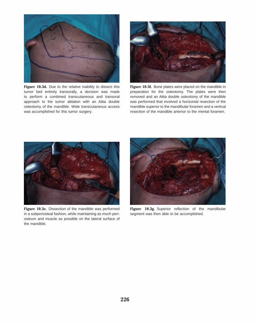
Tumors of the Minor Salivary Glands 229
The prognosis for this malignancy is generally con-sidered dismal, with 71% of patients exhibiting metastatic disease during the course of their disease.
Carcinosarcoma, also known as true malig-nant pleomorphic adenoma, is a tumor defi ned by histologic evidence of malignancy in both the epi-thelial and stromal elements of the tumor. These tumors are more rare than the carcinoma ex-pleo-morphic adenoma, accounting for only 8 cases in the AFIP registry, and none occurred in the minor salivary glands. Other cases presented in the litera-ture do identify the existence of this diagnosis in the minor salivary glands.
Metastasizing mixed tumor is a histologically benign pleomorphic adenoma, but located in distant sites. The pleomorphic adenomas are known to arise in major as well as minor salivary glands, and the metastatic foci have been identi-fi ed in the cervical lymph nodes, spine, and liver (Wenig and Gnepp 1991). Data on the interval from removal of the primary tumor to the identifi -cation of the fi rst metastasis is 1.5–51 years, with an average of 16.6 years.
Canalicular AdenomaThe canalicular adenoma is a benign tumor that has a signifi cant predilection for the upper lip (Figure 10.4). In the past, this tumor was
more commonly referred to as a “monomorphic adenoma.” Gardner recommended that the term monomorphic adenoma be used as a nosologic group of epithelial salivary gland tumors that are not pleomorphic adenomas (Gardner and Daley 1983). The canalicular adenoma and basal cell adenoma identify specifi c forms of monomorphic
Figure 10.4a. A freely moveable, indurated, submucosal mass of the upper lip in an elderly woman, highly sugges-tive of a canalicular adenoma.
Figure 10.4b. Based on this assumption, an incisional biopsy is not required. A pericapsular dissection and exci-sion of this mass was performed in association with sur-rounding minor salivary gland tissue, thereby allowing for delivery of the specimen.
Figure 10.4c. The histopathology of the specimen con-fi rms the clinical impression of canalicular adenoma. Reprinted with permission from Carlson ER, Schimmele SR. 1998. The management of minor salivary gland tumors of the oral cavity. In: Surgical Management of Salivary Gland Disease, The Atlas of the Oral and Maxillofacial Surgery Clinics of North America 6. Philadelphia: W.B. Saunders, pp. 75–98.

230 Tumors of the Minor Salivary Glands
Figure 10.5a. A freely moveable, indurated, submucosal mass of the buccal mucosa is noted in this patient.
b cFigures 10.5b and 10.5c. The CT scans show a well-circumscribed mass of this region.
adenomas (Daley, Gardner, and Smout 1984). The canalicular adenoma classically occurs in the upper lip in elderly women (Kratochvil 1991). In fact, canalicular adenomas typically affect an older pop-ulation compared to pleomorphic adenomas (Ord 1994). The canalicular adenoma is typically an asymptomatic, slow-growing, and freely moveable mass that uncommonly exceeds 2 cm in widest diameter. It may resemble mucoceles, which are uncommonly located in the upper lip. Of the 121 canalicular adenomas in the AFIP fi les, 89 of them occurred in the upper lip. The second most common site was the buccal mucosa (Auclair, Ellis, and Gnepp et al. 1991). The tumor is encapsulated such that an excision of the tumor in any anatomic site in a pericapsular fashion represents a curative surgery provided that tumor spillage does not occur (Figure 10.5). The canalicular adenoma is multifocal in 20% of cases (Ord 1994). If recur-rence is believed to have occurred, it might actu-ally represent a new primary tumor (Melrose 1994).

231
Figure 10.5d. A benign neoplastic process occupies a high position on the differential diagnosis such that a mucosal-sparing excision of the mass with transoral access is able to be performed without fi rst obtaining an incisional biopsy.
Figure 10.5e. A pericapsular dissection is performed.
Figure 10.5f. This dissection permits delivery of the speci-men. Stenson’s duct was intimately attached to the tumor and therefore sacrifi ced with the tumor.
g
h
Figures 10.5g and 10.5h. Histopathology identifi ed cana-licular adenoma (g) with an uninvolved capsule (h).
Figure 10.5i. The appearance of the site is noted to be well healed at 9 months postoperatively. Reprinted with permission from Carlson ER. 1995. Salivary gland pathol-ogy—clinical perspectives and differential diagnosis. In: The Comprehensive Management of Salivary Gland Pathology, Oral and Maxillofacial Surgery Clinics of North America 7. Philadelphia: W.B. Saunders, pp. 361–386.

232 Tumors of the Minor Salivary Glands
SURGICAL TREATMENT OF MALIGNANT MINOR SALIVARY GLAND TUMORS
The malignant diagnoses in the minor salivary glands are more diverse than their benign counter-parts. These malignant diagnoses may be low grade or high grade, and most represent histo-pathologic diagnostic challenges. As with the benign minor salivary gland tumors, surgery rep-resents the hallmark of therapy for malignant minor salivary gland tumors, and the principles of surgery have not changed signifi cantly over the past several decades (Bell et al. 2005). In addition to eradication of the primary malignancy, consid-eration should be given for neck dissection in very specifi c circumstances, as well as postoperative radiation therapy in this cohort of patients.
Mucoepidermoid CarcinomaThe mucoepidermoid carcinoma is the second most common tumor of the salivary glands overall, the most common salivary gland malignancy overall, and the most common minor salivary gland malignancy (Auclair and Ellis 1991). During the greater than 60 years since its fi rst description, this neoplasm has generated signifi cant debate regarding the possible existence of a benign variant, the optimal number of grades, and the proper treatment for certain minor salivary gland lesions. The term “mucoepidermoid tumor” was fi rst intro-duced by Stewart, Foote, and Becker in 1945 in their publication of 45 cases (Stewart, Foote, and Becker 1945). In this report, only two grades were utilized, including relatively favorable (benign) and highly unfavorable (malignant) tumors. The authors indicated that the adjective “benign” was rarely ever applicable in an absolute sense and as used in their report did not imply innocent behav-ior. It did indicate, however, that the authors had not observed metastasis from these tumors. The designation “malignant” indicated a histologic structure that was associated with the ability to produce regional lymph node and distant metasta-ses. This notwithstanding, the authors explicitly referred to and separated the benign and malignant tumors in their series of 45 cases in this report, of which there were 26 “benign” tumors and 19 “malignant” tumors. In 1953 this grading scheme was modifi ed to include three grades due to the development of metastases related to tumors previ-ously referred to as benign (Foote and Frazell
1953). These investigators accepted all of these tumors as malignant, and clinical and pathologic correlation suggested that separation into low-, intermediate-, and high-grade malignant subgroups might be useful, mainly due to histologically over-lapping qualities. The designation of intermediate grade was recognized as behaving more like the low-grade tumors than the high-grade tumors. Interestingly, despite the authors’ recognition that all of these tumors were malignant, the designa-tion “mucoepidermoid tumor” persisted through-out their paper. Subsequent studies were undertaken to more objectively determine if a benign variant existed. One such study investigated 23 mucoepi-dermoid carcinomas with a malignant course, such as evidence of local extension of tumor outside the capsule, local recurrences, histologically verifi ed metastases, or death due to the tumor (Eneroth et al. 1972). Fifteen patients showed local recur-rences, 13 showed histologically verifi ed metasta-ses, and 22 patients died of their disease. In 7 of the 23 cases the histology revealed highly or mod-erately differentiated structures, and in 3 of these cases the primary tumor as well as the lymph node metastases were highly differentiated. Six of the 23 patients had tumors in the palate, with 2 of these patients developing recurrences, 1 with lymph node metastases, and 5 of the patients died due to their disease. The authors concluded by stating that well-differentiated metastases in cases with a malignant course contradicted the existence of a benign variety of mucoepidermoid carcinoma, such that all of these neoplasms should be consid-ered cancers (Eneroth et al. 1972).
Of the 712 mucoepidermoid carcinomas occurring in the minor salivary glands in the AFIP registry, 305 (43%) of them were noted in the palate, 93 (13%) in the buccal mucosa, and 58 (8%) in the lip, with 37 specifi cally designated as the upper lip and 12 specifi cally designated as the lower lip (Auclair, Ellis, and Gnepp et al. 1991). While the AFIP data is generally recognized as being representative of the incidence of most sali-vary gland tumors, some authors have identifi ed the mucoepidermoid carcinoma to be more common in minor salivary gland sites than in major salivary gland sites (Plambeck, Friedrich, and Schmelzle 1996).
Histologic grading of mucoepidermoid carci-nomas is an important exercise. Histologic grade connotes biologic aggressiveness and prognosis and also provides the surgeon with important

Tumors of the Minor Salivary Glands 233
information with which to plan surgical treatment (Brandwein, Ivanov, and Wallace et al. 2001; Evans 1984). Mucoepidermoid carcinomas are composed of three cell types: mucous secreting, epidermoid, and intermediate. The intermediate cell is appropriately named because it is likely the progenitor of the two other cells (Batsakis and Luna 1990). Three grading schemes have found general acceptance among pathologists, and differ-ences in biologic behavior could be demonstrated as a function of grade, even though clinical stage has also been considered an important prognosti-cator. Indeed, Brandwein, Ivanov, and Wallace et al. (2001) found that only 5% of low-grade muco-epidermoid carcinomas of the major glands, and only 2.5% of low-grade mucoepidermoid carcino-mas of the minor glands, metastasized to regional lymph nodes or resulted in death. Spiro indicated that survival of patients with minor salivary gland carcinoma is signifi cantly infl uenced by the clinical stage and the histologic grade, but the applicability of grading to survival was limited to patients with mucoepidermoid carcinoma or adenocarcinoma in their study (Spiro, Thaler, and Hicks et al. 1991). They determined that staging was important in all patients regardless of the histologic diagnosis.
The mucoepidermoid carcinoma is the most common salivary gland malignancy in children (Auclair, Ellis, and Gnepp et al. 1991; Luna, Bat-sakis, and El-Naggar 1991; Ord 1994; Rogerson 1995). Although most of these tumors are noted in the parotid gland, the palate is the second most common site of involvement. Most appear to occur in teenagers, and the majority are low-grade or intermediate-grade histology. Mucoepidermoid carcinoma in children appears to follow a more favorable course with cure rates of 98–100% (Ord 1994).
Surgical treatment of the mucoepidermoid carcinoma of minor salivary gland origin is primar-ily a function of the anatomic site of the tumor and its histologic grade. Those arising in the palate are not only the most common but also the most vari-able insofar as surgical treatment is concerned. It is the histologic grade that is of utmost importance when determining treatment in the palate. Large series show that low-grade cancer is most common in this anatomic site (Pires et al. 2007). Incisional biopsy is clearly essential to establish the histopathologic diagnosis, as previously described. Computerized tomograms are essential in planning surgical treatment of palatal mucoepi-
dermoid carcinomas, as they assess the involve-ment of the underlying palatal bone. When the palatal bone does not appear to be involved by the cancer, a bone-sparing, periosteal sacrifi cing wide local excision with split thickness sacrifi ce of the soft palate musculature is the surgical treatment of choice (Figure 10.6). Similar to the surgery for the palatal pleomorphic adenoma, the periosteum serves as the anatomic barrier on the superior aspect of the tumor specimen, and tumor-free peri-
Figure 10.6a. A mass of the palate in a 45-year-old man.
Figure 10.6b. The extensive differential diagnosis, includ-ing benign and malignant entities, requires an incisional biopsy for diagnosis prior to performing defi nitive tumor surgery. The biopsy identifi es low-grade mucoepidermoid carcinoma.

234
Figure 10.6c. A periosteal sacrifi cing, bone-sparing wide local excision with split thickness sacrifi ce of the soft palate is planned with 1 cm mucosal linear margins.
Figure 10.6d. A sharp dissection is performed with a peri-osteal elevator between the periosteum on the superior aspect of the tumor specimen and the overlying palatal bone.
Figure 10.6e. The specimen is delivered and oriented for the pathologist with sutures. Histopathology shows low-grade mucoepidermoid carcinoma with negative margins.
Figure 10.6f. The association of the superior aspect of the tumor and the periosteum is noted histologically.
Tumors of the Minor Salivary Glands 235
osteal frozen and permanent sections should be obtained so as to confi rm this concept. When the periosteum has not been invaded by the cancer and all radial soft tissue margins are free of tumor, this surgery has a high frequency of cure.
The designation of an intermediate mucoepi-dermoid carcinoma of the palate may change the recommended surgical treatment of the tumor in this and other anatomic sites, with a more aggres-sive surgical procedure required for curative intent (Figure 10.7). This is particularly true if the desig-nation of intermediate grade is made by the pathol-ogist based on the worst microscopic pattern observed in the tumor. For example, a mucoepi-dermoid carcinoma that is predominantly low grade, but that shows a component of intermedi-ate-grade cancer, will likely be designated interme-diate grade. The behavior of such a tumor is likely to be low grade in nature. This scenario is different from a cancer that is designated intermediate grade that shows a predominantly intermediate-grade pattern with intermixed low-grade cancer. The surgeon may wish to offer more aggressive surgical therapy in the form of a partial maxillectomy for the mucoepidermoid carcinoma of the palate that is predominantly intermediate grade on micro-scopic sections. While rare, a high-grade mucoepidermoid carcinoma of the palate would require a partial maxillectomy, and prophylactic surgical treatment of the neck in the case of an N0 neck, or a therapeutic neck dissection in the case of an N+ neck. Postoperative radiation
therapy would also be administered in such circumstances.
Mucoepidermoid carcinoma of the buccal mucosa is the second most common minor salivary gland site affected. In contrast to benign neoplasms of this anatomic site, a mucosal-sacrifi cing tumor surgery is required, with attention to the sacrifi ce of surrounding submucosal anatomic barriers. The same is true of the lip (Figure 10.8).
Survival of patients with mucoepidermoid carcinomas of the minor salivary glands is clearly related to grade. Five-year survival rates have been estimated at 90% and 15-year survival rates have been estimated at 82% for low-grade mucoepider-moid carcinomas (Ord 1994).
Adenoid Cystic CarcinomaLike the mucoepidermoid carcinoma, the adenoid cystic carcinoma is a very diverse tumor with three histologic variants. These have been described morphologically rather than by grade as is the case with the mucoepidermoid carcinoma, and include the tubular, cribriform, and solid variants. The adenoid cystic carcinoma is characteristically
Figure 10.6g. The remaining tissue bed is temporarily covered with a palatal stent.
Figure 10.6h. Mucosalization of the exposed bone and soft palate musculature is noted at 9 months postopera-tively. This surgery provided curative care for this patient’s tumor. Reprinted with permission from Carlson ER, Schim-mele SR. 1998. The management of minor salivary gland tumors of the oral cavity. In: Surgical Management of Sali-vary Gland Disease, The Atlas of the Oral and Maxillofacial Surgery Clinics of North America 6. Philadelphia: W.B. Saunders, pp. 75–98.

236 Tumors of the Minor Salivary Glands
slow growing, with a high propensity for recurrent disease. It is highly infi ltrative, exhibits profound neurotropism, and is associated with a dismal long-term survival rate. This malignancy was fi rst described by Theodor Billroth in 1859 and referred to as “cylindroma” (Tomich 1991). In 1953 Foote and Frazell proposed the currently accepted nomenclature, adenoid cystic carcinoma. Of the 600 cases of adenoid cystic carcinoma in the AFIP fi les, 312 were noted in the major salivary glands, and 288 were noted in the minor salivary glands. The palate was the most common site affected in the minor salivary glands, followed by the tongue. Adenoid cystic carcinoma accounts for 8.3% of all
palatal salivary gland tumors and 17.7% of all malignant palatal salivary gland tumors in the AFIP series (Tomich 1991).
From a surgical standpoint, adenoid cystic carcinoma is probably the most challenging sali-vary gland tumor for the surgeon (Ord 1994). While straightforward to perform in most cases,
Figure 10.7a. A lesion of the right tuberosity region.
Figure 10.7b. The biopsy of the lesion from Figure 10.7a showed intermediate-grade mucoepidermoid carcinoma.
c
d
Figures 10.7c and 10.7d. Computerized tomograms iden-tifi ed an enhancing mass located lateral to the right tuberosity.

237
Figure 10.7e. Defi nitive tumor surgery involved a transoral partial maxillectomy and coronoidectomy en bloc.
Figure 10.7f. The cancer from Figure 10.7e.
Figure 10.7g. The resultant defect was obturated and allowed to contract signifi cantly over time.
h
i
j
Figures 10.7h, 10.7i, and 10.7j. Soft tissue reconstruction was accomplished with a buccal fat fl ap and advancement of the mucosa.

238 Tumors of the Minor Salivary Glands
radical resection is fraught with recurrences and ultimate distant metastases. This notwithstanding, palatal tumors should be managed with radical maxillectomy, observing 1–2 cm linear margins, and with resection of the greater palatine neuro-vascular bundle to foramen rotundum with frozen section guidance (Figure 10.9). The presence or absence of tumor in association with this nerve should be documented as far superior as possible. Cervical lymph node metastases are considered to be rare such that prophylactic neck dissection is not required in the patient with an N0 neck. Post-operative radiation therapy is generally considered advisable for all patients with adenoid cystic car-cinoma of the minor salivary glands, regardless of the adequacy of the resection (Dragovic 1995; Ord 1994; Triantafi llidou et al. 2006). The prognosis associated with adenoid cystic carcinoma of the minor salivary glands is inferior to that of the major salivary glands (Ampil and Misra 1987; Nas-cimento, Amaral, and Prado et al. 1986). In addi-tion, it has been found that the best prognosis for the adenoid cystic carcinoma is associated with the tubular variant, while the solid variant is associ-ated with the worst prognosis (Perzin, Gullane, and Clairmont 1978). It has also been pointed out that perineural invasion of major nerves and posi-tive margins at surgery, in addition to the solid variant of adenoid cystic carcinoma, are associated with increased treatment failures (Fordice et al. 1999). Typical survival statistics for adenoid cystic carcinoma in general include 60% for 5-year sur-
vival, 30% for 10-year survival, and 7% for sur-vival at 20 years (Ord 1994). It has been pointed out that adenoid cystic carcinoma of minor sali-vary gland sites has a worse prognosis, with a 0% survival at 20 years (Ord 1994). The presence of perineural spread has a signifi cant impact on sur-vival. Five-year survival rates of patients with peri-neural spread have been found to be 36.9%, while 5-year survival rates have been found to be 93.8% in patients without perineural spread (Ord 1994).
Polymorphous Low-Grade AdenocarcinomaIn 1983 two separate investigations reported on low-grade adenocarcinomas of minor salivary glands referred to as “terminal duct carcinoma” (Batsakis, Pinkston, and Luna et al. 1983) and “lobular carcinoma” (Freedman and Lumerman 1983). Terminal duct carcinoma was suggested to specify the histogenesis of the tumor, which was thought to be the progenitor cell of the terminal duct. Lobular carcinoma was suggested due to the morphology of the tumor resembling lobular car-cinoma of the breast. A review of these reports indicates that the authors were independently describing the same neoplasm (Wenig and Gnepp 1991). It is thought that, prior to this time, these neoplasms were classifi ed as either adenoid cystic carcinoma or adenocarcinoma (Regezi et al. 1991). High-power evaluation of polymorphous low-grade adenocarcinoma and adenoid cystic carcinoma may permit the distinction between the two malig-nancies, as adenoid cystic carcinoma shows ductal-type structures lined by multiple cells in thickness, while polymorphous low-grade carcinoma shows ductal-type structures more commonly lined by single cell layers (Figure 10.10). An Indian fi ling pattern is also seen in polymorphous low-grade adenocarcinoma. The common morphologic fea-tures of polymorphous low-grade adenocarcinoma and adenoid cystic carcinoma have led researchers to investigate methods of distinguishing these diagnoses (Beltran et al. 2006). In 1984 Evans and Batsakis described 14 cases of a distinctive minor salivary gland neoplasm that they named “poly-morphous low-grade adenocarcinoma.” This term emphasized the features of this neoplasm, includ-ing its cytologic uniformity and histologic diver-sity, variable growth patterns from solid to papillary to cribriform to fascicular, and relative lack of nuclear atypia (Evans and Batsakis 1984). Mitotic
Figure 10.7k. The reconstruction healed well as noted at 1 year postoperatively.

239
Figure 10.8a. An indurated upper lip mass that was fi xed to the surrounding mucosa.
Figure 10.8b. Due to the likely but equivocal malignant nature of the mass, incisional biopsy is essential for the establishment of the diagnosis prior to defi nitive surgical therapy. The histopathology of the incisional biopsy identi-fi ed intermediate-grade mucoepidermoid carcinoma.
Figure 10.8c. A wide local excision of the mass with oral mucosal sacrifi ce observing 1 cm linear margins was planned.
Figure 10.8d. A surgical plane was developed between the dermis of the upper lip and the musculature on the deep aspect of the tumor specimen.

240
Figure 10.8e. The specimen was delivered and oriented for the pathologist with sutures.
Figure 10.8f. Final histopathology identifi ed intermedi-ate-grade mucoepidermoid carcinoma with perineural invasion.
Figure 10.8g. The defect was reconstructed immediately with a full thickness skin graft.
Figure 10.8h. A prophylactic neck dissection was not per-formed as part of this cancer surgery due to the low concern for occult neck disease associated with this diagnosis. The patient underwent postoperative radiation therapy and the surgical site was noted to be well healed at 1 year postop-eratively without signs of recurrent disease. Reprinted with permission from Carlson ER. 1995. Salivary gland pathol-ogy—clinical perspectives and differential diagnosis. In: The Comprehensive Management of Salivary Gland Pathology, Oral and Maxillofacial Surgery Clinics of North America 7. Philadelphia: W.B. Saunders, pp. 361–386.

e
f
Figure 10.9a. A 52-year-old man with a 6-month history of a palatal mass.
Figure 10.9b. Incisional biopsy showed adenoid cystic carcinoma.
Figure 10.9c. Computerized tomograms identifi ed a soft tissue mass and minimal invasion of the palatal bone.
Figure 10.9d. A maxillectomy was planned for this patient observing 1 cm linear margins in bone and soft tissue.
Figures 10.9e and 10.9f. The bony cuts were created throughout the maxilla and the Smith Ramus Separator (Walter Lorenz Surgical, a Biomet Company, Jacksonville, Florida) (e) was utilized to separate the specimen from the remaining facial skeleton (f).

242
Figure 10.9g. The specimen was delivered and inspected from the palatal side so as to clinically confi rm the effi cacy of the resection.
Figure 10.9h. The specimen was also inspected from the nasal/sinus side so as to confi rm the adequacy of the resection.
Figure 10.9i. Frozen sections were obtained to micro-scopically examine the soft tissue margins as well as a segment of the greater palatine nerve in the superior aspect of the defect near foramen rotundum. All frozen sections were negative, thereby not requiring additional sampling of the nerve or mucosa. The fi nal decalcifi ed histopathology showed the tumor invading the maxillary bone.
Figure 10.9j. The patient underwent postoperative radia-tion therapy and showed no evidence of cancer at 1 year postoperatively. This view provides anatomic delineation of the eustachian tube in the defect.

Tumors of the Minor Salivary Glands 243
fi gures were infrequent and tumor necrosis was only seen in 1 case. Perineural invasion was and continues to be commonly noted in this malig-nancy. The tumors were distinctly unencapsulated and deeply infi ltrative of bone and surrounding soft tissues. Radical surgical procedures were required for tumor control, but no distant metas-tases were noted. The authors judged from their survey of adenocarcinomas of the major salivary glands that the polymorphous low-grade adenocar-
cinoma was at least primarily an oral neoplasm. Since this report, Batsakis and El-Naggar (1991) have subclassifi ed polymorphous low-grade ade-nocarcinomas (terminal duct adenocarcinomas) into papillary and nonpapillary forms. The papil-lary form was found to exhibit a more aggressive course with a higher rate of recurrence at the primary site, metastasis to cervical lymph nodes, and distant metastasis. The AFIP registry identifi ed 75 cases of this neoplasm, and all were located in the minor salivary glands (Wenig and Gnepp 1991). Forty-four of these cases were located in the palate (58.6%), with the upper lip and buccal mucosa showing 12 cases each. Involvement of other oral minor salivary gland sites has been reported; however, this is quite rare (de Diego et al. 1996; Kennedy et al. 1987).
Since its original description in minor salivary gland sites, cases of polymorphous low-grade ade-nocarcinoma have been reported in the parotid gland, such that this tumor cannot be stated to be exclusive to minor salivary gland tissue (Barak, Grobbel, and Rabaja 1998; Merchant, Cook, and Eveson 1996).
Treatment of polymorphous low-grade ade-nocarcinoma requires surgery with curative intent. The surgical procedure is based on the anatomic site. Surgical removal of these tumors in the palate requires a thorough assessment of the palatal bone with computerized tomograms. Bone involvement by this tumor is not an inherent property of this neoplasm but rather a function of chronicity of the tumor. Since these malignancies are not fast growing, many patients have long histories of their presence, such that palatal bone infi ltration by the tumor may occur over time. In addition, the characteristically deeply infi ltrative nature of these tumors into surrounding soft tissues, regardless of the chronicity of the tumor, is such that the soft palate typically requires full thickness sacrifi ce in most cases. These features are clearly a departure from the treatment of mucoepidermoid carcinoma of the palate, where grade is the main determining factor in planning surgical treatment. Once a biopsy diagnosis of polymorphous low-grade adenocarci-noma of the palate has been established, therefore, CT scans should be obtained to examine the quality of the palatal bone. If the bone is unaltered by the tumor, a bone-sparing, periosteal sacrifi cing wide local excision with full thickness sacrifi ce of the soft palate may be performed (Figure 10.11). Due to the tumor’s neurotropism, the greater palatine
a
b
Figures 10.10a and 10.10b. Subtle differences between the adenoid cystic carcinoma (a) and the polymorphous low-grade adenocarcinoma (b). The adenoid cystic carci-noma characteristically shows multiple cell layered ductal structures, while the polymorphous low-grade adenocarci-noma shows single cell layered ductal structures. Reprinted with permission from Carlson ER. 1995. Salivary gland pathology—clinical perspectives and differential diagnosis. In: The Comprehensive Management of Salivary Gland Pathology, Oral and Maxillofacial Surgery Clinics of North America 7. Philadelphia: W.B. Saunders, pp. 361–386.

Figure 10.11a. A 51-year-old man with a mass of the soft palate that had reportedly been present for only 2 months.
b
cFigures 10.11b and 10.11c. Incisional biopsy showed a microscopically cribriform tumor with obvious perineural invasion. A diagnosis of polymorphous low-grade adeno-carcinoma was made. Computerized tomograms did not reveal involvement of the palatal bone such that a perios-teal sacrifi cing, bone-sparing wide local excision with full thickness sacrifi ce of the soft palate was performed.
Figure 10.11d. A Dingman mouth gag (Walter Lorenz Sur-gical, a Biomet Company, Jacksonville, Florida) was uti-lized so as to provide acceptable retraction to perform this surgery.
Figure 10.11e. One-centimeter linear margins in mucosa were planned.
Figure 10.11f. The specimen is delivered.

Figure 10.11g. Final histopathology identifi ed a negative periosteal surface, thereby justifying the preservation of palatal bone.
Figure 10.11h. The greater palatine neurovascular bundle was clamped prior to delivery of the specimen so as to procure a 1 cm segment of nerve for frozen section analy-sis. The hemostat remained on the nerve stump while the frozen section was being evaluated, which was negative for cancer. If the nerve had been positive for cancer, the nerve would have been pulled down to procure additional frozen sections so as to clear the cancer.
i
j
k
Figures 10.11i, 10.11j, and 10.11k. The defect (i) was addressed with an immediate obturator that had been fabricated preoperatively (j) and was placed intraopera-tively (k).

246
Figure 10.11l. The exposed palatal bone is covered with immature granulation tissue at 1 month postoperatively.
Figure 10.11m. Immature granulation tissue underwent maturation by 3 months postoperatively as seen in this image.
Figure 10.11n. At 1 year postoperatively, the patient’s defect has demarcated well.
Figure 10.11o. A defi nitive obturator has been fabricated. Reprinted with permission from Carlson ER. 1995. Salivary gland pathology—clinical perspectives and differential diagnosis. In: The Comprehensive Management of Sali-vary Gland Pathology, Oral and Maxillofacial Surgery Clinics of North America 7. Philadelphia: W.B. Saunders, pp. 361–386.

Tumors of the Minor Salivary Glands 247
neurovascular bundle should be sampled for frozen sections superiorly. Since perineural spread is not characteristic of this tumor, it is unlikely to fi nd tumor tracking along the nerve, in contradistinc-tion to adenoid cystic carcinoma, where tumor may be found surrounding this nerve at foramen rotundum. An immediate surgical obturator is fab-ricated preoperatively for insertion at the time of
ablative surgery so as to permit the patient to begin taking a diet on the day of surgery. If the bone is eroded by the tumor, a traditional maxillectomy is necessary, also resulting in a full thickness sacri-fi ce of the soft palate (Figure 10.12). An immediate surgical obturator must also be fabricated preop-eratively for insertion at the time of surgery when a maxillectomy is planned for this diagnosis.
Figure 10.12a. A biopsy-proven polymorphous low-grade adenocarcinoma of the palate in a 53-year-old woman that had been present for several years, according to the patient.
Figure 10.12b. Computerized tomograms were obtained that identifi ed destruction of bone by the cancer.
Figure 10.12c. A maxillectomy was performed, observing 1 cm linear margins in bone and soft tissue.
Figure 10.12d. The maxillectomy specimen delivered.

248
Figure 10.12e. The histopathology confi rmed the involve-ment of the maxillary bone by the tumor.
Figure 10.12f. The large ablative defect.
Figure 10.12g. The large ablative defect was addressed with an immediate obturator.
Figure 10.12h. At 1 year postoperatively, the patient showed no signs of recurrent disease.

Tumors of the Minor Salivary Glands 249
The surgical treatment of polymorphous low-grade adenocarcinoma of the upper lip or buccal mucosa is similar to that of a mucoepidermoid carcinoma of these regions. The basic approach involves a mucosal-sacrifi cing wide local excision with attention to submucosal anatomic barriers being included on the specimen so as to ensure tumor-free margins (Figure 10.13).
The use of radiation therapy has been assessed in the management of polymorphous low-grade adenocarcinoma. In a clinicopathologic study of 164 cases of this malignancy, 17 patients under-went incisional, excisional, or wide local excision followed by radiation therapy (Castle, Thompson, and Frommelt et al. 1999). Adjuvant radiation therapy did not affect survival. Their study showed that patients who were treated with radiation therapy were more likely to have evidence of disease at last follow-up when compared with patients who did not have radiation therapy. Fur-thermore, there was no statistically signifi cant dif-ference in the overall patient outcome based on the type of initial treatment given or for any addi-tional treatment rendered, whether it was addi-tional surgery, radiation therapy, or chemotherapy. Based on this report and others (Crean et al. 1996), the treatment for polymorphous low-grade adeno-carcinoma of minor salivary glands remains surgi-
cal. It has been estimated that approximately 80% of patients survive their disease without evidence of tumor at periods from between several months to 25 years after removal (Wenig and Gnepp 1991). One case has been reported where death occurred as a result of this neoplasm with direct extension to vital structures of the head (Aberle, Abrams, and Bowe et al. 1985). In addition, while rare, metastasis to cervical lymph nodes (Kumar, Stiva-ros, and Barrett et al. 2004) and to distant organs (Hannen, Bulten, and Festen et al. 2000) has been reported from polymorphous low-grade adenocar-cinomas originating in the palate. These reports indicate that cervical lymph node involvement should be suspected in patients with papillary cystic change in the tumor, and that periodic chest X-ray examination should be performed postopera-tively when this variant of tumor is diagnosed.
Figure 10.13a. A biopsy proved polymorphous low-grade adenocarcinoma of the buccal mucosa in a 45-year-old woman.
Figure 10.13b. A very thick tumor was noted on biopsy. The superimposed optical micrometer shows the tumor to be about 9 mm in thickness.

250
c
Figures 10.13c and 10.13d. A mucosal-sacrifi cing wide local excision with 1 cm linear margins and isolation of Stenson’s duct was performed.
d
Figure 10.13e. The specimen was able to be delivered without tumor spillage.
Figure 10.13f. The defect was reconstructed with a split thickness skin graft and a sialodochoplasty of Stenson’s duct.

Tumors of the Minor Salivary Glands 251
Acinic Cell AdenocarcinomaAcinic cell adenocarcinoma is a very rare malig-nancy of the minor salivary glands. It has been estimated to represent approximately 2.5–3% of salivary gland tumors in general (Guimaraes et al. 1989; Spiro 1986) and about 4% of minor salivary gland tumors (Castellanos and Lally 1982). Indeed, acinic cell adenocarcinoma is not represented in many studies of minor salivary gland tumors (Chau and Radden 1986; Isacsson and Shear 1983; Jaber 2006), and other studies show only a very limited number of these cases (Lopes et al. 1999; Toida, Shimokawa, and Makita et al. 2005). The acinic cell adenocarcinoma behaves most similarly to the low-grade mucoepidermoid carcinoma (Ord 1994). In fact, like the low-grade mucoepidermoid carci-noma, the acinic cell adenocarcinoma was origi-nally purported to be a benign neoplasm (Ellis and Auclair 1991a). For the fi rst half of the twentieth century, these tumors were thought to be benign. In 1953, Buxton and his group were the fi rst to ascribe a malignant character to many of these tumors (Buxton, Maxwell, and French 1953). These were identifi ed as serous cell adenocarcinomas, after which time Foote and Frazell classifi ed these
tumors as acinic cell adenocarcinomas (Foote and Frazell 1953).
The AFIP registry shows 886 acinic cell ade-nocarcinomas, of which 753 were located in the major salivary glands (85%), and 133 (15%) in the minor salivary glands. The most common site of minor salivary gland involvement was the buccal mucosa, accounting for 43 cases (32%), followed by the lip (38 cases, 29%). Tumors in the upper lip were three times more common than tumors in the lower lip. The palate was the only other sig-nifi cant anatomic site to be affected by this tumor, and accounted for 22 cases (17%). A female pre-ponderance was noted, with a mean age of 44 years.
Surgery for acinic cell adenocarcinoma is per-formed in a similar fashion as that of low-grade mucoepidermoid carcinoma. Tumors of the buccal mucosa and upper lip are treated with mucosal-sacrifi cing wide local excisions, including 1 cm linear margins, with attention to the necessary sacrifi ce of surrounding anatomic barriers (Figure 10.14). Tumors of the palate can be treated with bone-sparing, periosteally sacrifi cing wide local excisions with split thickness sacrifi ce of the soft palate. Computerized tomograms may be obtained preoperatively to confi rm the lack of bone erosion. Recurrences and regional and distant metastases are rare when these malignancies are treated according to these recommendations. Five and 10-year survival rates are generally quite favorable and reported as 82% and 68%, respectively (Hickman, Cawson, and Duffy 1984).
Epithelial-Myoepithelial CarcinomaThe epithelial-myoepithelial carcinoma of minor salivary gland origin has been categorized as an intermediate-grade malignancy according to the AFIP classifi cation (Ellis and Auclair 1991b). Only 57 cases were identifi ed in their series, with 50 cases diagnosed in the major salivary glands (88%), and 7 cases (12%) in the minor salivary glands (Corio 1991). Of the 7 cases in the minor salivary glands, 4 were located in the palate, 1 in the tongue, and 2 cases were not specifi ed as to anatomic location. A mean age of 59 years was noted in these 57 cases. This tumor is known to be highly differentiated, yet it is malignant due to infi ltrative and destructive growth patterns, the presence of necrosis, perineural involvement, and metastases (Corio et al. 1982). Corio et al. pre-
Figure 10.13g. Acceptable healing is noted without tumor recurrence at 6 months postoperatively. Reprinted with permission from Carlson ER. 1995. Salivary gland pathol-ogy—clinical perspectives and differential diagnosis. In: The Comprehensive Management of Salivary Gland Pathology, Oral and Maxillofacial Surgery Clinics of North America 7. Philadelphia: W.B. Saunders, pp. 361–386.

252
a
b
Figures 10.14a and 10.14b. An acinic cell adenocarci-noma of the right buccal mucosa in a 52-year-old patient. The histopathology of the biopsy shows a typical blue dot tumor (b).
Figure 10.14c. A mucosal-sacrifi cing wide local excision observing 1 cm linear margins is performed.
Figure 10.14d. Excision of the specimen occurred without tumor spillage.
Figure 10.14e. The defect was reconstructed with mucosal fl aps so as to not distort the appearance of the upper lip.
Figure 10.14f. Acceptable healing without tumor recur-rence is noted at 1 year postoperatively.

Tumors of the Minor Salivary Glands 253
sented 16 cases of this neoplasm and found 12 cases to involve the parotid gland, 3 cases in the submandibular gland, and 1 case in the buccal mucosa (Corio et al. 1982).
Standardized recommendations for surgery for the epithelial-myoepithelial carcinoma are diffi cult to make due to the rare nature of this malignancy. Nonetheless, evaluation of involved anatomic barriers with physical examination and
CT scans generally permits an effective approach to eradication of these malignancies in various minor salivary gland sites (Figure 10.15). In such circumstances, the surgeon respects well-estab-lished principles of linear and anatomic barrier margins when operating on salivary gland tumors, while also relying on past experience with other low- and intermediate-grade minor salivary gland malignancies. In so doing, tumor-free margins can
Figure 10.15a. A mass of the right maxillary gingiva in a 12-year-old girl.
Figure 10.15b. Panoramic radiograph demonstrates tumor involvement of the bone between the fi rst premolar and canine teeth with divergence of the roots of these teeth.
c dFigures 10.15c and 10.15d. Computerized tomograms demonstrate a soft tissue mass with involvement of the maxillary bone.

254
Figure 10.15e. Incisional biopsy showed epithelial-myo-epithelial carcinoma.
Figure 10.15f. A partial maxillectomy observing 1 cm linear margins in bone and soft tissue was performed.
g hFigures 10.15g and 10.15h. The specimen was able to be removed without tumor spillage.

Figure 10.15i. Final histopathology showed destruction of bone by the cancer.
Figure 10.15j. The specimen radiograph demonstrated acceptable bone margins.
k
lFigures 10.15k and 10.15l. The resultant ablative defect of the maxilla (k) was reconstructed with an immediate obturator device (l).
Figure 10.15m. The use of the obturator permitted contracture of the defect as noted at 1 month postoperatively.

256
Figure 10.15n. By 3 months postoperatively, the defect had demarcated signifi cantly.
Figures 10.15o. An interim obturator was fabricated. Figure 10.15q. The patient functioned well with the defi ni-tive obturator, which permitted additional contracture of the maxillary defect.
Figures 10.15p. This interim obturator allowed for seal of the defect and function with teath.

Tumors of the Minor Salivary Glands 257
r
s
t
Figures 10.15r, 10.15s, and 10.15t. Soft tissue recon-struction was accomplished with a buccal fat fl ap and advancement of the buccal mucosa.
Figure 10.15u. The appearance of the healed fl ap is noted at 1 year postoperatively.
be obtained while performing surgery similar to that for a diagnosis of low- or intermediate-grade mucoepidermoid carcinoma. Recurrences have been reported (Corio 1991), but appropriate surgi-cal management of epithelial-myoepithelial carci-nomas of the minor salivary glands should be performed with curative intent. Quantitative sur-vival statistics are not published in the literature.
SURGICAL MANAGEMENT OF THE NECK FOR MINOR SALIVARY GLAND MALIGNANCIES
Surgical management of the neck is a controversial and intriguing concept for surgeons managing oral/head and neck malignant disease. At the core of this discipline is an assessment of occult disease in patients with clinically negative necks. To this end, there seems to be a consensus in the literature that occult neck disease is relatively uncommon related to minor salivary gland malignancies compared to squamous cell carcinoma of the oral/head and neck region. Moreover, it is also uncommon for patients with minor salivary gland malignancies to present with clinically palpable neck disease related to these tumors. Spiro found 53 patients presenting with cervical metastases among 378 patients (14%) with minor salivary gland malignancies (Spiro, Thaler, and Hicks et al. 1991). Another 26 patients (7%) developed subsequent cervical metastases for an overall rate of nodal involvement of 21%. Inter-

258 Tumors of the Minor Salivary Glands
estingly, 9 patients underwent an elective neck dissection, all of whom showed histologically con-fi rmed metastatic disease. The authors do not, however, discuss the incidence of occult and clini-cally apparent metastases as a function of anatomic site of the primary minor salivary gland malig-nancy. Sadeghi et al. identifi ed 9 patients present-ing with cervical metastases related to minor salivary gland malignancies, 5 of which were present in the tongue base (Sadeghi, Tran, and Mark et al. 1993). Beckhardt et al. found N+ necks in only 3% of their patients with malignant minor salivary gland tumors of the palate, while Chung identifi ed only 2 of 20 patients with malignant sali-vary gland tumors of the palate presenting with cervical metastases (Beckhardt, Weber, and Zane et al. 1995; Chung, Rahman, and Constable 1978). The latter three studies only discussed clinical staging of the neck without comments regarding their histology such that limited information is available regarding the true rate of metastasis to the cervical lymph nodes. In the fi nal analysis, it seems that the incidence of occult neck disease related to a minor salivary gland malignancy of the oral cavity is suffi ciently low to negate the need for elective neck dissection. Indications for neck dissection in these patients, therefore, are limited to patients who present with cervical metastases, those patients whose preoperative imaging studies document changes in the cervical lymph nodes consistent with metastatic disease, and those patients with high-grade malignancies, regardless of the clinical and radiographic imaging results.
THE ROLE OF RADIATION THERAPY IN THE MANAGEMENT OF MINOR SALIVARY GLAND MALIGNANCIES
It was once thought that salivary gland malignan-cies were radioresistant (Dragovic 1995). This pre-viously stated misconception can no longer be considered valid. As such, radiation therapy is indicated in the postoperative management of all high-grade malignant minor salivary gland tumors, as well as in patients with positive surgical margins, positive regional lymph nodes, and recurrent tumor (Dragovic 1995). This being the case, it is impor-tant to remember that surgery is the primary therapy for minor salivary gland malignancies. Shingaki et al.’s review of the role of radiation therapy in 44 patients with salivary gland cancers,
34 of whom were treated for minor salivary gland cancers, examined the results of surgery vs. surgery and postoperative radiation therapy in these patients (Shingaki et al. 1992). Interestingly, no patients experienced recurrent disease when nega-tive surgical margins were found in the specimen, regardless of whether surgery or surgery and post-operative radiation therapy was performed. All patients with positive surgical margins developed recurrent disease when surgery was the modality of treatment, and 8 of 15 patients (53%) with positive surgical margins developed recurrent disease when their salivary gland cancer was treated with a combination of surgery and postop-erative radiation therapy. While not broken down to major vs. minor salivary gland primary sites, these results do point to the signifi cant benefi t of obtaining negative margins in the resected specimen.
THE ROLE OF CHEMOTHERAPY IN THE MANAGEMENT OF MINOR SALIVARY GLAND MALIGNANCIES
There are few reports on the benefi t of systemic chemotherapy in the management of salivary gland cancers. Chemotherapy is generally reserved for the palliative management of advanced, non-resectable disease where radiation therapy has already been administered. Most patients for whom chemotherapy is considered will have diagnoses of mucoepidermoid carcinoma, adenoid cystic carci-noma, or high-grade adenocarcinoma (Laurie and Licitra 2006). The expression of c-kit in adenoid cystic carcinoma, overexpression of her-2 in muco-epidermoid carcinoma, overexpression of epithe-lial growth factor receptor in adenocarcinoma, and androgen receptor positivity in salivary duct carci-noma makes the use of imatinib, trastuzumab, cetuximab, and antiandrogen therapy at least theo-retically benefi cial (Laurie and Licitra 2006). While these agents may be of value in treating diffi cult cases of minor salivary gland malignancies, there is a need to conduct high-quality clinical trials in patients with these cancers.
Summary
• Approximately 10% of all salivary gland tumors arise in the minor glands.

Tumors of the Minor Salivary Glands 259
• Approximately 1% of head and neck tumors occur within the minor salivary glands, although some authors believe that this fi gure is somewhat higher.
• 70% of minor salivary gland tumors arise in the oral cavity.
• 50% of oral minor salivary gland tumors are found in the palate.
• 50% of oral minor salivary gland tumors are benign and 50% are malignant.
• Inconclusive evidence exists for cause and effect relationships with minor salivary gland tumors.
• An incisional biopsy is almost always indi-cated prior to defi nitive management of a palatal minor salivary gland tumor.
• An excisional biopsy of a buccal mucosal or upper lip minor salivary gland tumor may be acceptable without fi rst obtaining the histo-pathologic diagnosis, provided signs of benign disease exist.
• Benign tumors of the palate, buccal mucosa, and upper lip may be excised without special imaging studies.
• When present, ulceration predicts a malignant diagnosis, although many minor salivary gland malignancies do not create ulceration of the oral mucosa.
• Pain is associated with perineural invasion, most commonly seen with adenoid cystic carcinoma.
• Malignant tumors of the palate should undergo special imaging studies so as to determine involvement of the palatal bone.
• Malignant tumors of the buccal mucosa and upper lip may not require imaging prior to ablative surgery.
• Surgery represents the primary treatment of minor salivary gland tumors.
• Surgical removal of minor salivary gland tumors requires a scientifi c approach to the surrounding anatomic barriers.
• High cure rates are anticipated following removal of pleomorphic adenomas and cana-licular adenomas of minor salivary gland sites.
• High cure rates are anticipated following removal of low-grade mucoepidermoid carci-nomas and polymorphous low-grade adeno-carcinomas of minor salivary gland sites.
• Variable cure rates are associated with surgery for intermediate and high-grade mucoepider-
moid carcinomas and adenoid cystic carcino-mas of minor salivary glands.
References
Abbey LM, Schwab BH, Landau GC, Perkins ER. 1984. Incidence of second primary breast cancer among patients with a fi rst primary salivary gland tumor. Cancer 54:1439–1442.
Aberle AM, Abrams AM, Bowe R et al. 1985. Lobular (polymorphous low-grade) carcinoma of minor salivary glands: A clinicopathologic study of 20 cases. Oral Surg Oral Med Oral Pathol 60:387–395.
Ampil FL, Misra RP. 1987. Factors infl uencing survival of patients with adenoid cystic carcinoma of the salivary glands. J Oral Maxillofac Surg 45:1005–1010.
Ansari MH. 2007. Salivary gland tumors in an Iranian population: A retrospective study of 130 cases. J Oral Maxillofac Surg 65:2187–2194.
Auclair PL, Ellis GL. 1991. Mucoepidermoid carcinoma. In: Ellis GL, Auclair PL, Gnepp DR (eds.), Surgical Pathology of the Salivary Glands. Philadelphia: W.B. Saunders, pp. 269–298.
Auclair PL, Ellis GL, Gnepp DR et al. 1991. Salivary gland neoplasms: General considerations. In: Ellis GL, Auclair PL, Gnepp DR (eds.), Surgical Pathology of the Salivary Glands. Philadelphia: W.B. Saunders, pp. 135–164.
Barak AP, Grobbel M, Rabaja DR. 1998. Polymorphous low-grade adenocarcinoma of the parotid gland. Am J Otolaryngol 19:322–324.
Batsakis JG, El-Naggar AK. 1991. Terminal duct adenocar-cinomas of salivary tissues. Ann Otol Rhinol Laryngol 100:251–253.
Batsakis JG, Luna MA. 1990. Histopathologic grading of salivary gland neoplasms: I. Mucoepidermoid carcino-mas. Ann Otol Rhinol Laryngol 99:835–838.
Batsakis JG, Pinkston GR, Luna MA, et al. 1983. Adenocar-cinomas of the oral cavity: A clinicopathologic study of terminal duct carcinomas. J Laryngol Otol 97:825–835.
Beckhardt RN, Weber RS, Zane R et al. 1995. Minor salivary gland tumors of the palate: Clinical and patho-logic correlates of outcome. Laryngoscope 105:1155–1160.
Bell RB, Dierks EJ, Homer L, Potter BE. 2005. Management and outcome of patients with malignant salivary gland tumors. J Oral Maxillofac Surg 63:917–928.
Beltran D, Faquin WC, Gallagher G, August M. 2006. Selec-tive immunohistochemical comparison of polymorphous low-grade adenocarcinoma and adenoid cystic carci-noma. J Oral Maxillofac Surg 64:415–423.
Brandwein MS, Ivanov K, Wallace DI et al. 2001. Mucoepi-dermoid carcinoma: A clinicopathologic study of 80 patients with special reference to histological grading. Am J Surg Pathol 25:835–845.

260 Tumors of the Minor Salivary Glands
Buxton RW, Maxwell JH, French AJ. 1953. Surgical treat-ment of epithelial tumors of the parotid gland. Surg Gynecol Obstet 97:401–416.
Carlson ER. 1998. The management of minor salivary gland tumors of the oral cavity. Atlas Oral Maxillofac Surg Clin North Amer 6:75–98.
Castellanos JL, Lally ET. 1982. Acinic cell tumor of the minor salivary glands. J Oral Maxillofac Surg 40:428–431.
Castle JT, Thompson LDR, Frommelt RA et al. 1999. Poly-morphous low grade adenocarcinoma: A clinicopatho-logic study of 164 cases. Cancer 86:207–219.
Chau MNY, Radden BG. 1986. Intra-oral salivary gland neoplasms: A retrospective study of 98 cases. J Oral Pathol 15:339–342.
Chidzonga MM, Lopez-Perez VM, Portilla-Alvarez AL. 1995. Salivary gland tumours in Zimbabwe: Report of 282 cases. Int J Oral Maxillofac Surg 24:292–297.
Chung CK, Rahman SM, Constable WC. 1978. Malignant salivary gland tumors of the palate. Arch Otolaryngol 104:501–504.
Corio RL. 1991. Epithelial-myoepithelial carcinoma. In: Ellis GL, Auclair PL, Gnepp DR (eds.), Surgical Pathology of the Salivary Glands. Philadelphia: W.B. Saunders, pp. 412–421.
Corio RL, Sciubba JJ, Brannon RB, Batsakis JG. 1982. Epithelial-myoepithelial carcinoma of intercalated duct origin: A clinicopathologic and ultrastructural assess-ment of sixteen cases. Oral Surg Oral Med Oral Pathol 53:280–287.
Crean SJ, Bryant C, Bennett J, Harris M. 1996. Four cases of polymorphous low-grade adenocarcinoma. Int J Oral Maxillofac Surg 25:40–44.
Daley TD, Gardner GD, Smout MS. 1984. Canalicular adenoma: Not a basal cell adenoma. Oral Surg Oral Med Oral Pathol 57:181–188.
de Diego JI, Bernaldez R, Prim MP, Hardison D. 1996. Polymorphous low-grade adenocarcinoma of the tongue. J Laryngol Otol 10:700–703.
Dragovic J. 1995. The role of radiation therapy in the man-agement of salivary gland neoplasms. Oral Maxillofac Surg Clin North Amer 7:627–632.
Ellis GL, Auclair PL. 1991a. Acinic cell carcinoma. In: Ellis GL, Auclair PL, Gnepp DR (eds.), Surgical Pathology of the Salivary Glands. Philadelphia: W.B. Saunders, pp. 299–317.
Ellis GL, Auclair PL. 1991b. Classifi cation of salivary gland neoplasms. In: Ellis GL, Auclair PL, Gnepp DR (eds.), Surgical Pathology of the Salivary Glands. Philadelphia: W.B. Saunders, pp. 129–134.
Eneroth CM, Hjertman L, Moberger G, Soderberg G. 1972. Mucoepidermoid carcinomas of the salivary glands with special reference to the possible existence of a benign variety. Acta Otolaryng 73:68–74.
Epker BN, Henny FA. 1969. Clinical, histopathological and surgical aspects of intraoral minor salivary gland tumors: Review of 90 cases. J Oral Surg 27:792–804.
Evans HL. 1984. Mucoepidermoid carcinoma of salivary glands: A study of 69 cases with special attention to histologic grading. Am J Clin Pathol 81:696–701.
Evans HL, Batsakis JG. 1984. Polymorphous low-grade adenocarcinoma of minor salivary glands: A study of 14 cases of a distinctive neoplasm. Cancer 53:935–942.
Eveson JW, Cawson RA. 1985. Salivary gland tumors: A review of 2410 cases with particular reference to histo-logical types, site, age and sex distribution. J Pathol 146:51–58.
Foote FW, Frazell EL. 1953. Tumors of the major salivary glands. Cancer 6:1065–1133.
Fordice J, Kershaw C, El-Naggar A, Goepfert H. 1999. Adenoid cystic carcinoma of the head and neck: Predic-tors of morbidity and mortality. Arch Otolaryngol Head Neck Surg 125:149–152.
Freedman PD, Jones AC. 1994. A pathologist’s approach to tissue diagnosis. Oral Maxillofac Surg Clin North Amer 6:357–375.
Freedman PD, Lumerman H. 1983. Lobular carcinoma of intraoral minor salivary glands. Oral Surg Oral Med Oral Pathol 56:157–165.
Gardner DG, Daley TD. 1983. The use of the terms mono-morphic adenoma, basal cell adenoma, and canalicular adenoma as applied to salivary gland tumors. Oral Surg Oral Med Oral Pathol 56:608–615.
Guimaraes DS, Amaral AP, Prado LF, Nascimento AG. 1989. Acinic cell carcinoma of salivary glands: 16 cases with clinicopathologic correlation. J Oral Pathol Med 18:396–399.
Hannen EJM, Bulten J, Festen J et al. 2000. Polymorphous low grade adenocarcinoma with distant metastases and deletions on chromosome 6q23-qter and 1q23-qter: A case report. J Clin Pathol 53:942–945.
Hickman RE, Cawson RA, Duffy SW. 1984. The prognosis of specifi c types of salivary gland tumors. Cancer 54:1620–1624.
Isacsson G, Shear M. 1983. Intraoral salivary gland tumors: A retrospective study of 201 cases. J Oral Pathol 12:57–62.
Ito RA, Ito K, Vargas PA et al. 2005. Salivary gland tumors in a Brazilian population: A retrospective study of 496 cases. Int J Oral Maxillofac Surg 34:533–536.
Jaber MA. 2006. Intraoral minor salivary gland tumors: A review of 75 cases in a Libyan population. Int J Oral Maxillofac Surg 35:150–154.
Katz AD, Preston-Martin S. 1984. Salivary gland tumors and previous radiotherapy to the head or neck: Report of a clinical series. Am J Surg 147:345–348.
Kennedy KS, Healy KM, Taylor RE, Strom CG. 1987. Poly-morphous low-grade adenocarcinoma of the tongue. Laryngoscope 97:533–536.
Kratochvil FJ. 1991. Canalicular adenoma and basal cell adenoma. In: Ellis GL, Auclair PL, Gnepp DR (eds.), Surgical Pathology of the Salivary Glands. Philadelphia: W.B. Saunders, pp. 202–224.

Tumors of the Minor Salivary Glands 261
Kumar M, Stivaros N, Barrett AW et al. 2004. Polymor-phous low-grade adenocarcinoma—a rare and aggressive entity in adolescence. Br J Oral Maxillofac Surg 42:195–199.
Laurie SA, Licitra L. 2006. Systemic therapy in the palliative management of advanced salivary gland cancers. J Clin Oncol 24:2673–2678.
Lopes MA, Kowalski LP, Santos GC, Almeida OP. 1999. A clinicopathologic study of 196 intraoral minor salivary gland tumours. J Oral Pathol Med 28:264–267.
Luna MA, Batsakis JG, El-Naggar AD. 1991. Salivary gland tumors in children. An Otol Rhinol Laryngol 100:869–871.
MacIntosh RB. 1995. Minor salivary gland tumors: Types, incidence and management. Oral Maxillofac Surg Clin North Amer 7:573–589.
Melrose RJ. 1994. Clinicopathologic features of intraoral salivary gland tumors. Oral Maxillofac Surg Clin North Amer 6:479–497.
Merchant WJ, Cook MG, Eveson JW. 1996. Polymorphous low-grade adenocarcinoma of parotid gland. Br J Oral Maxillofac Surg 34:328–330.
Nascimento AG, Amaral ALP, Prado LAF et al. 1986. Adenoid cystic carcinoma of salivary glands: A study of 61 cases with clinicopathologic correlation. Cancer 57:312–319.
Ord RA. 1994. Management of intraoral salivary gland tumors. Oral Maxillofac Surg Clin North Amer 6:499–522.
Perzin KH, Gullane P, Clairmont AC. 1978. Adenoid cystic carcinomas arising in salivary glands: A correlation of histologic features and clinical course. Cancer 42:265–282.
Pires FR, Pringle GA, de Almeida OP, Chen SY. 2007. Intra-oral minor salivary gland tumors: A clinicopathological study of 546 cases. Oral Oncology 43:463–470.
Plambeck K, Friedrich RE, Schmelzle R. 1996. Mucoepider-moid carcinoma of salivary gland origin: Classifi cation, clinical-pathological correlation, treatment results and long-term follow-up in 55 patients. J Craniomaxillofac Surg 24:133–139.
Pogrel MA. 1994. The management of salivary gland tumors of the palate. J Oral Maxillofac Surg 52:454–459.
Potdar GG, Paymaster JC. 1969. Tumors of minor salivary glands. Oral Surg Oral Med Oral Pathol 28:310–319.
Regezi JA, Lloyd RV, Zarbo RJ, McClatchey KD. 1985. Minor salivary gland tumors: A histologic and immuno-histochemical study. Cancer 55:108–115.
Regezi JA, Zarbo RJ, Stewart JCB, Courtney RM. 1991. Polymorphous low-grade adenocarcinoma of minor salivary gland: A comparative histologic and immuno-histochemical study. Oral Surg Oral Med Oral Pathol 71:469–475.
Rivera-Bastidas H, Ocanto RA, Acevedo AM. 1996. Intra-oral minor salivary gland tumors: A retrospective study of 62 cases in a Venezuelan population. J Oral Pathol Med 25:1–4.
Rogerson KC. 1995. Salivary gland pathology in children. Oral Maxillofac Surg Clin North Am 7:591–598.
Sadeghi A, Tran LM, Mark R et al. 1993. Minor salivary gland tumors of the head and neck: Treatment strategies and prognosis. Am J Clin Oncol 16:3–8.
Saku T, Hayashi Y, Takahara O et al. 1997. Salivary gland tumors among atomic bomb survivors, 1950–1987. Cancer 79:1465–1475.
Satko I, Stanko P, Longauerova I. 2000. Salivary gland tumours treated in the stomatological clinics in Bratislava. J Craniomaxillofac Surg 28:56–61.
Shingaki S, Ohtake K, Nomura T, Nakajima T. 1992. The role of radiotherapy in the management of salivary gland carcinomas. J Craniomaxillofac Surg 20:220–224.
Spiro RH. 1986. Salivary neoplasms: Overview of a 35-year experience with 2,807 patients. Head & Neck Surg 8:177–184.
Spiro RH, Thaler HT, Hicks WF et al. 1991. The importance of clinical staging of minor salivary gland carcinoma. Am J Surg 162:330–336.
Spitz MR, Tilley BC, Batsakis JG et al. 1984. Risk factors for major salivary gland carcinoma: A case-comparison study. Cancer 54:1854–1859.
Stewart FW, Foote FW, Becker WF. 1945. Mucoepidermoid tumors of salivary glands. Ann Surg 122:820–844.
Stuteville OH, Corley RD. 1967. Surgical management of tumors of intraoral minor salivary glands: Report of eighty cases. Cancer 20:1578–1586.
Toida M, Shimokawa K, Makita H et al. 2005. Intraoral minor salivary gland tumors: A clinicopathological study of 82 cases. Int J Oral Maxillofac Surg 34:528–532.
Tomich CE. 1991. Adenoid cystic carcinoma. In: Ellis GL, Auclair PL, Gnepp DR (eds.), Surgical Pathology of the Salivary Glands. Philadelphia: W.B. Saunders, pp. 333–349.
Triantafi llidou K, Dimitrakopoulos J, Iordanidis F, Koufo-giannis D. 2006. Management of adenoid cystic carci-noma of minor salivary glands. J Oral Maxillofac Surg 64:1114–1120.
Waldron CA. 1991. Mixed tumor (pleomorphic adenoma) and myoepithelioma. In: Ellis GL, Auclair PL, Gnepp DR (eds.), Surgical Pathology of the Salivary Glands. Philadelphia: W.B. Saunders, pp. 165–186.
Waldron CA, El-Mofty SK, Gnepp DR. 1988. Tumors of the intraoral minor salivary glands: A demographic and his-tologic study of 426 cases. Oral Surg Oral Med Oral Pathol 66:323–333.
Wenig BM, Gnepp DR. 1991. Polymorphous low-grade adenocarcinoma of minor salivary glands. In: Ellis GL, Auclair PL, Gnepp DR (eds.), Surgical Pathology of the Salivary Glands. Philadelphia: W.B. Saunders, pp. 390–411.
Yih WY, Kratochvil FJ, Stewart JCB. 2005. Intraoral minor salivary gland neoplasms: Review of 213 cases. J Oral Maxillofac Surg 63:805–810.


Chapter 11
Non-salivary Tumors of the Salivary Glands
children, where they account for up to 35% of salivary tumors (Ord 2004). These tumors are most commonly seen under the age of 1 year and may be present at birth, where they may exhibit aggres-sive growth. Hemangioendotheliomas are more aggressive and rapidly growing and occur in the <6 month infant, while the older children tend to present with the slower-growing cavernous lesions (Figure 11.1). In the past surgical removal was advocated, but as the majority of tumors involute over time, and because of the morbidity of surgery in infants, this has largely been abandoned in favor of medical therapy. Previous papers have indicated that vascular malformations of the parotid respond poorly to medical therapy; however, this has been disproved by Greene, Rogers, and Mulliken (2004). These authors reviewed 100 consecutive children with a 4.5 : 1 female to male ratio with 59% ulcer-ating during the proliferating phase and 89% involving nearby structures. Seventy of the patients were treated medically, 67 primarily with cortico-steroids and 3 with interferon. Initially 56/67 of the patients treated with steroids showed regres-sion or stabilization, but 18 required further treat-ment with interferon. The overall response to steroids/alfa-2a or -2b interferon was 98%, and the authors concluded that parotid gland vascular tumors respond in the same way as hemangiomas elsewhere. Interestingly, 66% of the children required some form of reconstructive surgery during the involuted phase. In adults vascular lesions of the parotid are less common, but intra-muscular hemangiomas of the masseter muscle can be a diagnostic challenge (Figure 11.2).
Vascular lesions of the submandibular gland are seen rarely and like vascular lesions elsewhere will be treated depending on their fl ow character-istics and the vessel(s) affected (Figure 11.3).
LymphangiomasLymphangiomas may be capillary or cavernous (and associated with vascular malformations) or
OutlineIntroductionMesenchymal Tumors
Benign Mesenchymal TumorsHemangiomasLymphangiomasNeural TumorsLipomas
Malignant Mesenchymal TumorsSarcomas
Epithelial Non-salivary TumorsTumors of Salivary Gland Lymph Nodes
Primary Lymph Node TumorsLymphomas
Secondary Lymph Node TumorsRegional MetastasesDistant Metastases
SummaryReferences
Introduction
This chapter will review the non-salivary tumors that occur in the major salivary glands, having discussed the primary epithelial salivary gland tumors in chapters 8, 9, and 10. Essentially the chapter will be divided into primary benign and malignant mesenchymal tumors and metastatic lesions, primarily epithelial, in the lymph nodes or substance of the major glands. As the epidemiol-ogy and etiology of these tumors is extremely vari-able, they will be discussed in relation to individual tumors and groups of tumors.
Mesenchymal TumorsBENIGN MESENCHYMAL TUMORS
HemangiomasHemangiomas and hemangioendotheliomas are most commonly seen in the parotid gland and in
263

Figure 11.1a. A 3-year-old boy with prominent cavernous vascular neoplasm of the parotid gland.
Figure 11.1b. Operative sequence shows the neoplasm is mobilized forward after ligating feeding vessels and is being peeled off the main trunk of the facial nerve (arrow).
Figure 11.1c. The vascular neoplasm is removed in total with the superfi cial lobe of the parotid gland after complete facial nerve dissection.
Figure 11.2. The magnetic resonance scan shows the vascular malformation to be located intramuscularly in the left masseter muscle deep to the parotid.

265
a b
cFigures 11.3a, 11.3b, and 11.3c. Flow voids noted in the vascular malformation in the right submandibular region. At the time of surgery this was found to be cavernous and primarily venous.

266 Non-salivary Tumors of the Salivary Glands
be cystic in nature. They are common in the neck and seen more in the submandibular (37%) than parotid glands (31%) (Orvidas and Kasperbauer 2000). They may be prominent at birth as cystic hygromas and may pose a threat to the airway (Figure 11.4). In children they can increase con-siderably in size during upper respiratory tract infections. Some authors have documented the posterior triangle to be a more common site in the neck than the submandibular space, 54% versus 17%, respectively (Fageeh, Manoukian, and Tewfi k et al. 1997). In a series of 324 pediatric patients with salivary gland masses, 89 (27.5%) were lymphangiomas compared to 192 (59.2%) being hemangiomas (Bentz et al. 2000). Many of these lesions are treated surgically, but persistence and recurrence are problematic (Orvidas and Kasper-bauer 2000). Especially in the infi ltrating lesions, complete excision may be impossible and debulk-ing is performed. Surgery can be combined with sclerosing injections or these can be used as a
single modality. Sclerosing agents are most effec-tive in macrocystic lymphangiomas, and in 54 of these cases 49% had excellent results, 35% good, and 16% poor using sclerosant injection (Emran, Dubois, and Laberge et al. 2006).
Neural TumorsIn Siefert and Oehne’s (1986) review of 150 benign mesenchymal tumors of the salivary glands, 16% were neurogenic in origin distributed over the fourth to seventh decade. These were divided into neurilemmomas (neurinomas) in 12 of 27 cases, neurofi bromas in 12 of 27 cases, and neurofi bro-matosis in 3 of 27 cases. There was a predomi-nance of males, 75% for neurofi bromas but 65% females for neurilemmomas. Both MR and CT scan may be useful in imaging. In the parotid gland extension of the tumor in the gland and in the petrous bone is well defi ned by MR imaging, while CT scan shows bone erosion and relationship to the inner ear. A combination of CT and MR is recommended when surgical resection is planned (Martin et al. 1992).
Complete removal of these lesions, especially the plexiform neurofi broma, can be extremely diffi cult due to their infi ltrating nature and often increased vascularity (Figure 11.5). Although approximately one-third of neurilemmomas occur in the head and neck (Almeyda et al. 2004), they are comparatively rare in the salivary glands pub-lished as isolated case reports. However, as they may be mistaken for a malignant parotid tumor due to facial nerve dysfunction—for example, pro-gressive weakness, sudden facial paralysis, hemi-facial spasm and pain (Balle and Greisen 1984)—it is important to make the diagnosis to avoid inap-propriate radical surgery. Regarding intra-parotid neurofi bromas, a “conservative course of treat-ment with limited tumor excision and emphasis on retaining facial nerve function” is advocated (McGuirt, Johnson, and McGuirt 2003). Indeed, once the histologic diagnosis is made, because of the slow growth of the tumor and the unlikelihood of malignant change, conservative treatment of leaving the tumor in situ to preserve the nerve has been recommended (Fierke, Laskawi, and Kunze 2006).
LipomasApproximately 15–20% of lipomas occur in the head and neck region (Weiss and Goldblum 2001),
Figure 11.4. Lymphangioma involving both the parotid and submandibular glands in an 8-month-old infant.

Non-salivary Tumors of the Salivary Glands 267
Lipomas are comparatively rare in the oral cavity, but in one paper with 46 cases, 2 patients were classifi ed as having minor salivary gland lipomas (Fregnani, Pires, and Falzoni et al. 2003).
Salivary lipomas usually present as slow-growing painless masses, and their appearance on CT or MR is diagnostic (Figure 11.6). Surgical exci-sion is the treatment of choice, and although easy in classic lipoma, it can be challenging in the infi l-trating variety (Figure 11.7).
Recently a designation of sialolipoma has been proposed for lipomas containing glandular elements, for example, ductal or acinar tissue (Nagao, Sugano, and Ishida et al. 2001). In their series of 2,051 salivary tumors, 7 sialolipomas, 5 in the parotid, and 2 palatal, were reported. Exci-sion as for classic lipoma is curative. Since the initial report other cases both in major and minor glands have been published (Lin, Lin, and Chen et al. 2004; Michaelidis, Stefanopoulos, and Sam-baziotis et al. 2006).
Figure 11.5a. Massive plexiform neurofi broma involving the parotid gland and orbit.
Figure 11.5b. CT scan shows extensive soft tissue involvement.
and in reviewing 125 lipomas in the oral and max-illofacial region, 30 (24%) were parotid and 17 (13.6%) were submandibular in orgin (Furlong, Fanburg-Smith, and Childers 2004). In this series there was a 3 : 1 male to female gender ratio and a mean age of 51.9 years. Histologically almost half (62/125) were classic lipomas, while 59 were spindle cell/pleomorphic, 2 were fi brolipomas, and 2 chondroid lipomas. Spindle cell lipomas comprised the majority of parotid lipomas. In a review of 167 mesenchymal salivary gland tumors, Seifert and Oehne (1986) found lipomas comprised 22.5% of 150 benign tumors and 95% were in the parotid. Again 85% occurred in males. A report of 660 parotid neoplasms found only 8 patients had lipomatous tumors (1.3%), 5 with focal lipoma and 3 with diffuse lipomatosis (Ethunandan, Vura, and Umar et al. 2006). Only one tumor of 8 was in the deep lobe, but small series of parotid lipomas in the deep lobe have been reported (Gooskens and Mann 2006).

268
Figure 11.6a. Lipoma in the tail of the parotid.
Figure 11.6b. CT is diagnostic of lipoma.
Figure 11.6c. Intraoperative view of parotidectomy with parotid tail lipoma.
Figure 11.6d. Specimen with arrows showing lipoma.
Figure 11.6e. Histopathology confi rms the presence of lipoma.

Non-salivary Tumors of the Salivary Glands 269
Figure 11.7a. MR axial image of infi ltrating lipoma of right submandibular region extending between the cervi-cal muscles.
Figure 11.7b. Coronal MR image shows the lipoma extending medial to the right medial pterygoid muscle into the lateral pharyngeal space.
MALIGNANT MESENCHYMAL TUMORS
SarcomasSarcomas of the salivary glands are very rare and case reports of virtually all histologic types have been reported. In Siefert and Oehne’s 1986 review of 167 mesenchymal tumors of the salivary glands, only 17 were sarcomas (10%). In this series, 5 cases were malignant fi brous histiocytomas, 5 cases were malignant schwannomas, 4 cases were embryonal rhabdomyosarcoma, and single cases of myxoid liposarcoma, leiomyosarcoma, and malignant hemangioendothelioma were reviewed. In reviewing salivary masses in children, rhabdo-myosarcomas were the most common malignant mesenchymal tumor (7%) (Bentz et al. 2000), and in 137 children with rhabdomyosarcomas of the head and neck the parotid was the site for 6% of these tumors (Hicks and Flaitz 2002). Obviously treatment plans will be dictated by the individual sarcoma type, with initial chemotherapy for rhab-domyosarcoma in children followed by radiation therapy or surgery for residual disease. Rhabdo-myosarcoma of the salivary glands appears locally aggressive with a poor prognosis (BenJelloun,
Jouhadi, and Maazouzi et al. 2005). In malignant fi brous histiocytoma, clear surgical margins appear to be the most important prognostic factor (Sachse, August, and Alberty 2006). Angiosarcoma may affect the parotid as a primary or metastatic tumor, and in a series of 29 angiosarcomas of the oral and salivary gland region there were 4 primary parotid and 3 primary submandibular gland angiosarco-mas with a further 3 metastatic to the parotid (Fanburg-Smith, Furlong, and Childers 2003).
All of the metastatic patients died, but patients with primary salivary gland angiosarcoma appear to have a better prognosis than those with cutaneous or deep tissue angiosarcomas. Malignant neural sarcomas are treated with wide excision and facial nerve grafting or reanima-tion (McGuirt, Johnson, and McGuirt 2003). Other sarcomas of the salivary glands are rare; for example, Chadan et al. (2004) found only 11 reported cases of salivary gland liposarcoma in the literature.
Sarcomas can involve any of the major sali-vary glands although the parotid is most common, and due to its rarity, treatment is usually on an individual and empiric basis (Figure 11.8).

270
Figure 11.8a. Rapidly growing sublingual gland tumor diagnosed as synovial cell sarcoma on biopsy and immunohistochemistry.
Figure 11.8b. CT image reveals calcifi cation in the mass leading to an initial clinical diagnosis of a high-grade malig-nant carcinoma ex-pleomorphic adenoma.
Figure 11.8c. CT bone window shows calcifi cations throughout the mass.
Figure 11.8d. Bilateral selective neck dissections in con-tinuity with lip split to access the mandible.

Figure 11.8e. Midline mandibulotomy prior to excision of the fl oor of mouth and ventral tongue.
Figure 11.8f. Surgical specimen.
Figure 11.8g. Post-resection—the reconstruction will be a microvascular forearm fl ap.
Figure 11.8h. Four weeks post-surgery.
Figure 11.8i. Intraoral view showing the forearm fl ap reconstruction of the fl oor of the mouth.

272
Figure 11.9a. Elderly man with primary desmoid mela-noma of the parotid gland. A 2 cm margin is marked; the light blue staining of the skin around the lesion is from dye injection for sentinel node biopsy (patient had lymphoscin-tigraphy immediately preoperatively).
Figure 11.9c. The neck dissection and parotidectomy with preservation of the facial nerve is complete. The submental fl ap is pedicled on its vascular supply prior to being rotated into the defect.
Figure 11.9b. Markings for the proposed surgery involving a total parotidectomy with left supraomohyoid neck dissec-tion (unless sentinel nodes are found at levels IV or V). Reconstruction with a submental fl ap based on the sub-mental vessels.
Figure 11.9d. Three months postoperatively.

Non-salivary Tumors of the Salivary Glands 273
EPITHELIAL NON-SALIVARY TUMORS
The major salivary glands may be infi ltrated by squamous cell carcinoma from the overlying skin or be primarily involved by melanoma. Surgical resection with a margin of normal tissue preserving the facial nerve and utilizing neck dissection and adjuvant radiotherapy as indicated by the tumor stage is the appropriate treatment (Figure 11.9).
Tumors of Salivary Gland Lymph NodesPRIMARY LYMPH NODE TUMORS
LymphomasPrimary lymphoma of the salivary glands is rare. Eighty percent of lymphomas of the salivary glands are found in the parotid gland and 20% in the submandibular gland, with only case reports of sublingual and minor gland involvement (Eraso, Lorusso, and Palacios 2005) (Figure 11.10).
Other authors have found a higher incidence of submandibular involvement (39%) (Dunn, Kuo, and Shih et al. 2004). In 121 parotid tumors 8.3% were lymphomas (Shine, O’Leary, and Blake 2006), and in 51 submandibular tumors 14% were lym-phomas (Preuss, Klussman, and Wittekindt et al. 2007).
Patients with Sjogren’s syndrome, AIDS, and hepatitis C have an increased risk of developing salivary lymphomas. In a review of 463 cases of Sjogren’s syndrome, 27 patients had a diagnosis of lymphoma (5.8%) (Tonami, Matoba, and Kuginuki et al. 2003). In this series 26 of the 27 patients had non-Hodgkin’s lymphoma [including 6 mucosa-associated lymphoid tissue (MALT) lymphomas] and only 1 patient had Hodgkin’s lymphoma. At the initial presentation 14 (52%) of patients had extra-nodal disease, with 9 of 27 (33%) in the salivary glands. However, 21 patients (78%) had nodal involvement, mostly in the cervical nodes. Masaki and Sugai (2004) also reported a fi gure of 5% of Stage III Sjogren’s patients developing lymphomas that are thought to arise from lymphoepithelial lesions. The B cells in these lesions become acti-vated by interactions between CD40L and CD40 with progression from polyclonal lymphoprolifera-tion, to monoclonal lymphoproliferation, to MALT lymphoma, and fi nally to high-grade lymphoma as a multistep process. Other authors have highlighted
Figure 11.10a. Elderly lady with itchy facial and neck rash who complains of an intraoral swelling.
Figure 11.10b. A red fl eshy swelling of the sublingual gland is appreciated with retraction of the commissure by the patient’s fi nger and retraction of the tongue by the mirror. Biopsy showed a non-Hodgkin’s lymphoma of the sublingual gland.

274 Non-salivary Tumors of the Salivary Glands
the diffi culty in diagnosing true lymphoma from the other lymphoproliferative disorders occurring in Sjogren’s syndrome, although there is a forty-fold increased risk in developing B-cell lymphomas (Prochorec-Sobieszek and Wagner 2005). Clinical features associated with lymphoma include persis-tent major salivary enlargement (>2 months), per-sistent lymphadenopathy or splenomegaly, monoclonal gammopathy, and type II mixed cryoglobulinemia.
Hepatitis C is also associated with MALT lym-phomas of the salivary glands. In a series of 33 cases of primary salivary MALT lymphomas, 15 patients had a history of Sjogren’s syndrome (45.5%), 2 (6%) other autoimmune disease, and 7 (21%) hepatitis C infection (Ambrossetti, Zanotti, and Passaro et al. 2004). There is an increase in lymphoma in AIDS, however, although in 51% of patients in a study of 100 patients who died with AIDS without salivary gland symptoms who
Figure 11.11. A 52-year-old man with a left parotid mass and a fi rm level II node who has a salivary lymphoma as a presenting sign of previously undiagnosed AIDS.
showed histologic signs of parotid disease, only 1 case of lymphoma was found (Vargas, Mauad, and Bohm et al. 2003) (Figure 11.11).
Not all primary salivary lymphomas fall into the MALT group, and follicular lymphomas com-prise 30% and 22% of two recently published series (Kojima, Nakamura, and Ichimura et al. 2001; Nakamura, Ichimura, and Sato et al. 2006) (Figure 11.12). These lymphomas have a younger age of onset than MALT lymphomas, do not occur in patients with autoimmune disease, and appear relatively more common in the submandibular gland.
Most salivary lymphomas present as unilat-eral, painless masses, usually with a history of <4 months, and although CT scans show poorly
Figure 11.12. Non-Hodgkin’s lymphoma of the left parotid gland and submental nodes.

Non-salivary Tumors of the Salivary Glands 275
defi ned indistinct margins, there is no pathognomic sign for salivary lymphoma (Shine, O’Leary, and Blake 2006). The lesions may be multiple in the ipsilateral gland and associated lymphadenopathy can be noted. The use of FNAB in diagnosing salivary lymphoma has been questioned as inac-curate with high rates of false negative results.
Figure 11.13a. A 35-year-old lady with MALT lymphoma of Waldeyer’s ring controlled by chemotherapy. Now has bilateral parotid involvement that is not responding to medical therapy and she is concerned regarding her appearance.
Figure 11.13b. CT scan shows homogenous mass of MALT lymphoma in the anterior portion of the right parotid (arrow), which has slowly increased in size over a 3-year period.
Figure 11.13c. CT scan shows smaller mass of MALT lymphoma in the anterior left parotid gland.
Figure 11.13d. Proposed parotidectomy and excision of MALT lymphoma.
Zurrida, Alasio, and Tradati et al. (1993) were only able to identify 2 of 7 lymphomas (28.6%), and Hughes, Volk, and Wilbur (2005) found a 57% false negative rate in salivary lymphomas in reviewing the data from the College of American Pathologists Interlaboratory Comparison Program in Nongynecologic Cytology. In the absence of Sjogren’s syndrome or clinical suspicion of lym-phoma, these lesions are frequently diagnosed fol-lowing surgical removal.

276 Non-salivary Tumors of the Salivary Glands
Treatment is by chemotherapy, medical therapy, and radiation therapy depending on the histologic diagnosis and the clinical staging. MALT lymphomas of salivary glands appear to have a low-grade indolent course with 5-year overall sur-vival, case-specifi c survival, and progression-free survival of 85% (±8%), 94% (±6%), and 65% (±10%), respectively (Ambrossetti, Zanotti, and Passaro et al. 2004). These results were noted despite 42% of their patients being Stage IV, and local therapy was often adequate (Figure 11.13). Dunn, Kuo, and Shih et al. (2004) in 23 primary salivary lymphomas, 19 MALT, 3 diffuse large cell, and 1 follicular found overall 5-year survival of 94.7% and relapse-free survival of 51.4%. Only 2 patients died; MALT lymphoma in 1 patient trans-formed into diffuse large cell lymphoma and the patient died. Kojima, Nakamura, and Ichimura et al. (2001) noted that follicular lymphomas arising from salivary glands appeared to share some of the characteristics of MALT lymphoma with an indo-lent prognosis.
SECONDARY LYMPH NODE TUMORS
Regional MetastasesThe lymph nodes associated with the major glands may all become involved by regional metastases. In the parotid gland, skin cancer, particularly squa-mous cell carcinoma (SCC) and malignant mela-noma (MM) of the scalp, forehead, temple, upper lip, cheek, and ear is most common, although Merkel cell tumors, malignant syringomas, and other more unusual skin cancers can be seen (Figure 11.14).
The largest experience with these tumors is in Australia, where squamous cell carcinoma and melanoma of the facial skin is epidemic and meta-static cutaneous cancer is the commonest parotid malignancy (O’Brien, McNeil, and McMahon et al. 2002). Although less than 5% of patients with cutaneous SCC do metastasize to lymph nodes, certain features may give these tumors an increased risk of metastasizing. In a review of 266 patients, 61% having parotid lymph node involvement ± cervical involvement, tumor thickness >4–5 mm, and proximity to the parotid (temple/forehead, cheek, or ear) were high risks, and increasing tumor size and recurrence contributed to an increased risk (Veness, Palme, and Morgan 2006) (Figure 11.15). In 2002 O’Brien, McNeil, and
McMahon et al. suggested that the TNM system of designating all nodal metastases from cutaneous cancer N1 was limited and did not accurately delineate the extent of disease. They suggested separating disease in the parotid P1 <3 cm, P2 >3 cm and <6 cm, P3 >6 cm from neck disease: N0 no nodal disease, N1 a single node <3 cm, N2 multiple nodes or any node >3 cm for staging. In a multivariate analysis of 87 patients they found that increasing P stage, positive margins, and lack of adjuvant RT independently predicted for decreased local control in the parotid. Clinical and pathologic N stage both signifi cantly impacted sur-vival. They concluded that patients with positive nodes in both parotid and neck had the worst prognosis, and that prognosis was worse for nodal disease > N1 (Figure 11.16). A much smaller study from Israel showed a zero overall survival for patients with both parotid and cervical nodes posi-tive for metastatic cutaneous SCC (Barzilai et al. 2005). Using the separate staging system for parotid disease (P) and neck disease (N) proposed by
Figure 11.14. Malignant syringoma of forehead metastatic to the left parotid gland and fungating through preauricular skin.

277
Figure 11.15a. PET scan of elderly woman with left pre-auricular mass post-resection of squamous cell carcinoma of the cheek. Scan shows a hot spot (SUV 6.7).
Figure 11.15b. Fused PET/CT confi rms positive node in the parotid gland.
Figure 11.15c. Proposed surgery of superfi cial parotidec-tomy with extended Blair incision to allow for supraomohy-oid neck dissection.
Figure 11.15d. Incision allows wide access down to level III of the neck.

278 Non-salivary Tumors of the Salivary Glands
Figure 11.15e. Postoperatively the patient has no nerve weakness.
O’Brien, McNeil, and McMahon et al. (2002), two recent studies have been published. One series of 67 patients from New Zealand found again that the extent of parotid disease was an independent prog-nostic factor and that patients with both parotid and neck disease did worst, although interestingly adjuvant RT did not infl uence survival in their data (Ch’ng, Maitra, and Lea et al. 2006). The second paper was a retrospective multi-center trial from three Australian and three U.S. centers with 322 patients with metastatic cutaneous SCC to the parotid and/or neck. Results from this study show a signifi cantly worse 5-year survival for patients with advanced P stage, 69%, vs. 82% for early P stage; and 61% with parotid + neck disease vs. 79% for parotid alone. This study supported the adoption of the new staging system separating parotid and neck disease (Andruchow, Veness, and Morgan et al. 2006).
In terms of behavior of metastatic cancer to the parotid, Bron et al. (2003) reviewed 232 cases of which 54 were primary parotid cancers, 101 were metastatic cutaneous SCC, 69 MM, and 8 other metastatic cancers. Patients were treated with primary surgery sparing the facial nerve where indicated, with 54 therapeutic and 110 elec-tive neck dissections, and 78% of the patients had adjuvant RT. Five-year survival rates were 77% for primary cancers, 65% for metastatic SCC, 46% for MM, and 56% for other metastatic cancers. As expected, local failure was highest in metastatic SCC and distant failure in MM.
In treating patients with nodal involvement of the parotid, superfi cial parotidectomy with wide excision to obtain negative margins is indicated with facial nerve sacrifi ce if it is infi ltrated. When the neck is also involved level II is the commonest site, and in these cases a comprehensive neck dis-section is recommended (Figure 11.17). If the neck is clinically uninvolved (N0), and the primary cancer anterolateral to the parotid, then a suprao-mohyoid neck dissection including the external jugular nodes is indicated, and with a posterior primary level V should be dissected as well (Vauterin, Veness, and Morgan et al. 2006). Adju-vant RT is given in node positive necks for close margins or perineural invasion. In the case of MM where sentinel lymph nodes are identifi ed in the parotid by scintigraphy, intraparotid sentinel lymph node biopsy is a reliable, accurate, and safe pro-cedure (Loree, Tomljanovich, and Cheney et al. 2006).
In addition to regional metastasis from cuta-neous primary cancers, metastasis can occur from noncutaneous head and neck carcinomas. Metastasis to the parotid from other head and neck sites—for example, the oral cavity—is more common if the usual lymphatic drainage pattern has been disrupted by previous neck dis-section or radiation therapy (Ord, Ward-Booth, and Avery 1989). It is also possible for mucosal melanoma to metastasize to the parotid region (Figure 11.18).
There are fewer reports of involvement of the submandibular gland being directly involved by lymph node metastases, although it is routinely removed during neck dissection. The lymph nodes are not usually found within its capsule; however, Preuss, Klussman, and Wittekindt et al. (2007) found that in 24 malignant submandibular gland

279
a
Figures 11.16a and 11.16b. An elderly lady who had exci-sion of eyelids and orbital exenteration for advanced squa-mous cell carcinoma of the eyelids. She was lost to follow-up and now presents with massive disease in the parotid nodes (P3) and level II neck nodes (N2).
b
Figure 11.16c. CT scan shows huge parotid mass.
Figure 11.16d. Proposed surgery with radical parotidec-tomy and radical neck dissection.
Figure 11.16e. Post-resection the masseter muscle is sacri-fi ced along with the facial nerve and a total parotidectomy. The mandible was uninvolved by tumor and was preserved.

280
Figure 11.16f. Reconstruction is with a latissimus dorsi fl ap. The patient developed chest metastases 18 months post-surgery. Figures 11.16a, 11.16b, and 11.16f reprinted with permission from Ord RA. Local and regional fl ap reconstruction. In: Ward-Booth P, Schendel SA, Hausamen JE (eds.), Maxillofacial Surgery (2nd ed.). New York: Else-vier, 2007, pp. 643–665.
tumors, 30% were metastatic, 3 from the orophar-ynx, 2 from the nasopharynx, and 2 with unknown primaries.
In the sublingual gland metastatic spread from tongue cancer to sublingual nodes is not common and was fi rst reported in 3 cases in 1985 (Ozeki et al. 1985). In one study of 253 patients with 326 neck dissections, 5 cases of lingual lymph node metastases were found and in all of these cases bilateral cervical nodes were found (Woolgar 1999). Whether these may explain some cases of “local” recurrence with previous negative margins is unclear. Certainly these lingual nodes would be removed in composite or “commando” resections where the tongue primary is removed in continuity
with the neck dissection, but they may be left in cases where the primary cancer is resected from an intraoral approach and the neck dissection is performed separately. One study reports a 5-year actuarial survival of 80% for patients treated with incontinuity neck dissection compared to 63% for those with discontinuity dissection (Leemans et al. 1991). Whether some of this difference in survival is attributable to involvement of sublingual gland nodes is unknown.
Distant MetastasesThe major salivary glands can also be a site for distant metastatic disease, especially the parotid gland, although these cases are rare. In a literature review of over 800 patients with metastatic disease in the parotid, 80% were from cutaneous SCC or melanoma (as described above), while 66 were noncutaneous head and neck tumors and 87 from a distant primary site (Pisani et al. 1993). In their personal series of 38 patients, 10 had noncutaneous head and neck cancers, while 4 were from distant sites (2 renal and 2 lung). Nuyens et al. (2006) found 34 of 520 parotid tumors to be metastatic, 31 from cutaneous primaries, 2 from ductal breast cancer, and 1 from an extremity rhabdomyosar-coma. Although rare, these distant metastases can provide a diagnostic challenge, as the two com-monest primary sites appear to be lung and kidney. Small cell lung cancer is very diffi cult to differenti-ate from primary small cell carcinoma of the sali-vary glands, such that CT scan of the lungs is an essential part of the workup (Figure 11.19). Sali-vary glands are the second commonest head and neck site for primary small cell carcinomas (larynx being the most common), and these are aggressive tumors with an overall poor prognosis (Renner 2007). They are divided into neuroendocrine and ductal types, and according to cytokeratin 20 immu-noreactivity the ductal sites can be subdivided into pulmonary and Merkel types (Nagao, Gaffey, and Olsen et al. 2004). This study indicated that nega-tive immunostain for cytokeratin 20 could be a marker for poor prognosis and also that salivary gland small cell carcinoma may have a better prog-nosis than extrasalivary sites.
In a single case report, immunohistochemical study of estrogen receptors was used to identify a parotid tumor as a breast metastasis (Perez-Fidalgo, Chirivella, and Laforga et al. 2007). Regarding distant metastases from renal cell cancer the same

Figure 11.17a. Patient with nodular melanoma of poste-rior neck with palpable parotid lymph nodes.
Figure 11.17b. Post–total parotidectomy and radical neck dissection.
Figure 11.17c. Surgical specimen with 5 cm skin margins. Patient developed lung metastases within 6 months and died of disease. Figures 11.17a, 11.17b, and 11.17c reprinted with permission from Ord RA. Metastatic melanoma of the parotid lymph nodes. Int J Oral Maxillofac Surg 18:3, 1989.

282
c
Figure 11.18a. Patient with large lymph node swelling at level I and pigmented melanoma of lower lip.
Figure 11.18b. Intraoral exam shows deeply pigmented melanoma involving the buccal mucosa and extending to the retromolar region.
Figures 11.18c and 11.18d. CT scans show primary mel-anoma as thickening of the left cheek and lip with nodal involvement of the left parotid gland.
d

283
f
g
Figure 11.18e. CT scan at a more cephalad level now shows multiple positive nodes in the parotid gland.
Figures 11.18f and 11.18g. Hematoxylin and eosin stain of the tumor (f) and HMB-45 stain (g) confi rming the diag-nosis of melanoma.
Figure 11.19a. An 80-year-old woman with known small cell carcinoma of the lung presenting with metastatic mass in the left parotid gland.
Figure 11.19b. CT scan of the patient in Figure 11.19a.

284 Non-salivary Tumors of the Salivary Glands
problem is found. Most of these will be from clear cell renal carcinoma and mimic the salivary clear cell adenocarcinoma or clear cell variant of MEC, which are both primary salivary gland cancers. In a case report and review of the literature, Park and Hlivko (2002) were able to fi nd 25 cases of meta-static renal cell carcinoma to the parotid gland. In 14 of these cases (56%) the metastasis was the initial presenting sign of a previously undiagnosed renal carcinoma. None of the cases presented with facial paralysis and the authors were able to make the diagnosis in 3 of 6 cases with FNAB. In a small series of our own patients we were able to differ-entiate renal cell carcinoma from monomorphic clear cell salivary adenocarcinoma by immunohis-tochemistry and electron microscopic ultrastruc-tural differences (Rezende, Drachenberg, and Kumar et al. 1997).
Distant metastasis to salivary glands other than the parotid appears to be extremely unusual, although a unique case of bilateral submandibular gland metastases from breast carcinoma was pub-lished in 2001 (Cain, Goodland, and Denholm 2001).
Summary
• The commonest parotid tumors in children are hemangiomas and hemangioendotheliomas.
• Medical therapy with steriods or alpha/beta interferon is preferred in the treatment of vas-cular parotid lesions in children.
• A combination of debulking and sclerosing injections to macrocystic areas is used for management of salivary lymphangiomas.
• Parotid lymphomas are usually associated with Sjogren’s syndrome, hepatitis C, or AIDS.
• Most salivary lymphomas are MALT lympho-mas, which follow a fairly indolent course.
• The parotid lymph node bed may be the fi rst-echelon nodes for cutaneous cancers of the cheek, ear, scalp, forehead, and temple.
• Metastatic parotid nodes (P) and neck nodes (N) should be staged separately when involved by primary cutaneous cancers.
• Patients with both neck nodes and parotid nodes have the worst prognosis.
• Radiation therapy is given for close margins, perineural spread, and more than one positive node.
• When small cell carcinomas of the salivary glands are diagnosed, a full workup must be done to determine whether the tumor is a primary salivary gland cancer or a metastatic tumor from a distant site.
References
Almeyda R, Kothari P, Chau H, Cumberworth V. 2004. Submandibular neurilemmoma; a diagnostic dilemma. J Laryngol Otol 118(2):156–158.
Ambrossetti A, Zanotti R, Passaro C et al. 2004. Most cases of primary salivary mucosa-associated lymphoid tissue lymphoma are associated either with Sjogren’s syndrome or hepatitis C virus infection. Br J Haematol 126(1):43–49.
Andruchow JL, Veness MJ, Morgan GJ et al. 2006. Implica-tions for clinical staging of metastatic cutaneous carci-noma of the head and neck based on a multicenter study of treatment outcomes. Cancer 106(5):1078–1083.
Balle VH, Greisen O. 1984. Neurilemmomas of the facial nerve presenting as parotid tumors. Ann Otol Rhinol Laryngol 93:70–72.
Barzilai G, Greenberg E, Cohen-Kermen R, Doweck I. 2005. Pattern of regional metastases from cutaneous squamous cell carcinoma of the head and neck. Otolaryngol Head Neck Surg 132(6):852–856.
BenJelloun H, Jouhadi H, Maazouzi A et al. 2005. Rhabdo-myosarcoma of the salivary glands. Report of 3 cases. (Article in French.) Cancer Radiother 9(5):316–321.
Bentz BG, Hughes CA, Ludemann JP, Maddalozzo J. 2000. Masses of the salivary gland region in children. Arch Otolaryngol Head Neck Surg 126(12):1435–1439.
Bron LP, Traynor SJ, McNeil EB, O’Brien CJ. 2003. Primary and metastatic cancer of the parotid; comparison of clini-cal behavior in 232 cases. Laryngoscope 113(6):1070–1075.
Cain AJ, Goodland J, Denholm SW. 2001. Metachronous bilateral submandibular gland metastases from carci-noma of the breast. J Laryngol Otol 115(8):683–684.
Ch’ng S, Maitra A, Lea R et al. 2006. Parotid metastasis—an independent prognostic factor for head and neck cutane-ous squamous cell carcinoma. J Plast Reconstr Aesthet Surg 59(12):1288–1293.
Chadan VS, Fung EK, Woods CI, de la Roza G. 2004. Primary pleomorphic liposarcoma of the parotid gland: A case report and review of the literature. Am J Otolar-yngol 25:432–437.
Dunn P, Kuo TT, Shih LY et al. 2004. Primary salivary gland lymphoma: A clinicopathologic study of 23 cases in Taiwan. Acta Hematol 112(4):203–208.
Emran MA, Dubois J, Laberge L et al. 2006. Alcoholicsolution of zein (Ethibloc) sclerotherapy for treatment of lymphangiomas in children. J Pediatr Surg 41(5):975–979.

Non-salivary Tumors of the Salivary Glands 285
Eraso A, Lorusso G, Palacios E. 2005. Primary lymphoma of the parotid gland. ENT-Ear Nose Throat J 84(4):198–199.
Ethunandan M, Vura G, Umar T et al. 2006. Lipomatous lesions of the parotid gland. J Oral Maxillofac Surg 64(11):1583–1586.
Fageeh N, Manoukian J, Tewfi k T et al. 1997. Management of head and neck lymphatic malformations in children. J Otolaryngol 26(4):253–258.
Fanburg-Smith JC, Furlong JC, Childers EL. 2003. Oral and salivary gland angiosarcoma: A clinicopathologic study of 29 cases. Mod Pathol 16(3):263–271.
Fierke O, Laskawi R, Kunze E. 2006. Solitary intraparotid neurofi broma of the facial nerve. Symptomatology, biology and management. (Article in German.) HNO 54(10):772–777.
Fregnani ER, Pires FR, Falzoni R et al. 2003. Lipomas of the oral cavity: Clinical fi ndings, histological classifi ca-tion and proliferative activity of 46 cases. Int J Oral Maxillofac Surg 32(1):49–53.
Furlong MA, Fanburg-Smith JC, Childers EL. 2004. Lipomas of the oral and maxillofacial region: Site and subclassifi -cation of 125 cases. Oral Surg Oral Med Oral Pathol Oral Radiol Endod 98(4):441–450.
Gooskens I, Mann JJ. 2006. Lipoma of the deep lobe of the parotid gland: Report of 3 cases. ORL J Otorhinolaryngol Relat Spec 68(5):290–295, epub May 6, 2007.
Greene AK, Rogers GF, Mulliken JB. 2004. Management of parotid hemangiomas in 100 children. Plast Reconstr Surg 113(1):53–60.
Hicks J, Flaitz C. 2002. Rhabdomyosarcoma of the head and neck in children. Oral Oncol 38(5):450–459.
Hughes JH, Volk EE, Wilbur DC. 2005. Pitfalls in salivary gland fi ne-needle aspiration cytology: Lessons from the College of American Pathologists Interlaboratory Com-parison Program in Nongynecologic Cytology. Arch Pathol Lab Med 129(1):26–31.
Kojima M, Nakamura S, Ichimura K et al. 2001. Follicular lymphoma of the salivary gland: A clinicopathologic and molecular study of six cases. Int J Surg Pathol 94(4):287–293.
Leemans CR, Tiwari R, Nauta JJ, Snow GB. 1991. Discon-tinuous vs. in-continuity neck dissection in carcinoma of the oral cavity. Arch Otolaryngol Head Neck Surg 117(9):1003–1006.
Lin YJ, Lin LM, Chen YK et al. 2004. Sialolipoma of the fl oor of the mouth: A case report. Kaohsiung J Med Sci 20(8):410–414.
Loree TR, Tomljanovich PI, Cheney RT et al. 2006. Intrapa-rotid sentinel lymph node biopsy for head and neck melanoma. Laryngoscope 116(8):1461–1464.
Martin N, Sterkers O, Mompoint D, Nahum H. 1992. Facial nerve neuromas: MR imaging. Report of four cases. Neu-roradiology 34(1):62–67.
Masaki Y, Sugai S. 2004. Lymphoproliferative disorders in Sjogren’s syndrome. Autoimmun Rev 3(3):175–182.
McGuirt WF Sr, Johnson PE, McGuirt WT. 2003. Intra-parotid facial nerve neurofi bromas. Laryngoscope 113(10):82–84.
Michaelidis IG, Stefanopoulos PK, Sambaziotis D et al. 2006. Sialolipoma of the parotid gland. J Craniomaxillo-fac Surg 34(1):43–46, epub Dec. 15, 2005.
Nagao T, Gaffey TA, Olsen KD et al. 2004. Small cell carcinomas of the major salivary glands: Clinicopatho-logic study with emphasis on cytokeratin 20 immunore-activity and clinical outcomes. Am J Surg Pathol 28(6):762–770.
Nagao T, Sugano I, Ishida Y et al. 2001. Sialolipoma: A report of seven cases of a new variant of salivary gland lipoma. Histopathol 38:33–36.
Nakamura S, Ichimura K, Sato Y et al. 2006. Follicular lymphoma frequently originates in the salivary gland. Pathol Int 56(10):576–583.
Nuyens M, Schupbach J, Stauffer E, Zbaren P. 2006. Meta-static disease to the parotid gland. Otolaryngol Head Neck Surg 135(6):844–848.
O’Brien CJ, McNeil EB, McMahon JD et al. 2002. Signifi -cance of clinical stage, extent of surgery, and pathologic fi ndings in metastatic cutaneous squamous cell carci-noma of the parotid gland. Head Neck 24(5):417–422.
Ord RA. 2004. Salivary gland tumors in children. In: Kaban LB, Troulis MJ (eds.), Pediatric Oral and Maxillofacial Surgery. Philadelphia: W.B. Saunders, p. 202.
Ord RA, Ward-Booth RP, Avery BS. 1989. Parotid lymph node metastases from primary intraoral carcinomas. Int J Oral Maxillofac Surg 18:104–106.
Orvidas LJ, Kasperbauer JL. 2000. Pediatric lymphangio-mas of the head and neck. Ann Otol Rhinol Laryngol 109(4):411–421.
Ozeki S, Tashiro H, Okamamoto M, Matsushima T. 1985. Metatasis to the lingual lymph nodes in carcinoma of the tongue. J Maxillofac Surg 13(6):277–281.
Park YW, Hlivko TJ. 2002. Parotid gland metastasis from renal cell carcinoma. Laryngoscope 112(3):453–456.
Perez-Fidalgo JA, Chirivella I, Laforga J et al. 2007. Parotid gland metastasis of a breast cancer. Clin Transl Oncol 9(4):264–265.
Pisani P, Krengeli M, Ramponi A, Pia F. 1993. Parotid metastases: A review of the literature and case reports. (Article in Italian) Acta Otorhinolaryngol Ital 12 Suppl 37:1–28.
Preuss SF, Klussman JP, Wittekindt C et al. 2007. Subman-dibular gland excision: 15 years of experience. J Oral Maxillofac Surg 65(5):953–957.
Prochorec-Sobieszek M, Wagner T. 2005. Lymphoprolifera-tive disorders in Sjogren’s syndrome. (Article in Polish.) Otolaryngol Pol 59(4):559–564.
Renner G. 2007. Small cell carcinomas of the head and neck: A review. Semin Oncol 34(1):3–14.
Rezende RB, Drachenberg CB, Kumar D et al. 1997. Dif-ferential diagnosis between monomorphic adenocarci-

286 Non-salivary Tumors of the Salivary Glands
noma of the salivary glands and renal (clear) cell carcinoma. Am J Surg Pathol 23:1532–1538.
Sachse F, August C, Alberty J. 2006. Malignant fi brous histiocytoma in the parotid gland. Case series and litera-ture review. (Article in German.) HNO 54(2):116–120.
Seifert G, Oehne H. 1986. Mesenchymal (non-epithelial) salivary gland tumors. Analysis of 167 tumor cases of the salivary gland register. Laryngol Rhinol Otol (Stuttg) 65(9):485–491.
Shine NP, O’Leary G, Blake SP. 2006. Parotid lympho-mas—clinical and computed tomographic features. S Afr J Surg 44(2):62–64.
Tonami H, Matoba M, Kuginuki Y et al. 2003. Clinical and imaging fi ndings of lymphoma in patients with Sjogren’s syndrome. J Comput Assist Tomogr 27(4):517–524.
Vargas PA, Mauad T, Bohm GM et al. 2003. Parotid gland involvement in advanced AIDS. Oral Dis 9(2):55–61.
Vauterin TJ, Veness MJ, Morgan GJ et al. 2006. Patterns of lymph node spread of cutaneous squamous cell
carcinoma of the head and neck. Head Neck 28:785–791.
Veness MJ, Palme CE, Morgan GJ. 2006. High-risk cutane-ous squamous cell carcinoma of the head and neck: Results from 266 treated patients with metastatic lymph node disease. Cancer 106(11):2389–2396.
Weiss SW, Goldblum JR. 2001. In: Enzinger FM, Weiss SW (eds.), Soft Tissue Tumors (4th ed.). St. Louis: Mosby, p. 571.
Woolgar JA. 1999. Histological distribution of cervical lymph node metastases from intraoral/oropharyngeal squamous cell carcinomas. Br J Oral Maxillofac Surg 37(3):175–180.
Zbaren P, Schar C, Hotz MA et al. 2001. Value of fi ne-needle aspiration cytology of parotid gland masses. Laryngoscope 111(11 part 1):1989–2002.
Zurrida S, Alasio L, Tradati N et al. 1993. Fine needle aspiration of parotid masses. Cancer 72(8):2306–2311.

Chapter 12
Trauma and Injuries to the Salivary Glands
gland—will cause direct damage to the gland and possible related structures and may lead to the formation of an external salivary fi stula to the skin (Figure 12.1). When the substance of the gland is injured, suture of the parenchyma is recommended (Lewkowicz, Hasson, and Nablieli 2002). In addi-tion to direct closure of the parotid capsule, a pressure dressing for 48 hours is applied to reduce the chances of sialocele formation. In 51 cases of parotid complications following trauma, 15 (29.4%) developed parotid fi stula, treated by intravenous fl uids and nil by mouth, with faster healing of parenchymal injuries alone than when the ductal system was involved (Parkeh, Glezerson, and Stewart et al. 1989). Similarly, Ananthakrishnan and Parkash (1982) reported that their 3 cases of fi stula from the parotid gland parenchyma resolved without treatment, unlike the 14 fi stulae related to parotid ductal injury. In a study of 13 patients with traumatic parotid fi stulae, 54% resolved with con-servative management within 3 weeks and the remainder were cured by internal drainage with a catheter (Cant and Campbell 1991). Landau and Stewart (1985) advocated conservative manage-ment of post-traumatic parotid fi stulae and sialo-celes and found that parenchymal injuries alone resolved in 5 days, whereas ductal injuries took 14 days. Morestin (1917), in a series of 62 war injuries with parotid fi stula, 30 glandular and 32 ductal, reported good success with the creation of an intra-oral fi stula. In more extensive avulsive injuries with gross scarring, conservative treatment may be less successful (Figure 12.2), and established epi-thelialized fi stulae require excision with repair of the parotid capsule and closure.
The submandibular gland is less liable to be involved in the development of traumatic fi stulae, perhaps because it is protected by the mandible and its smaller size. Few cases of submandibular gland fi stulae have been reported. A case report of submandibular gland fi stula secondary to a gunshot wound was reported in 1995, where the authors also reviewed the literature and found only one
OutlineIntroductionPenetrating Injuries
Trauma to the GlandSalivary FistulaSialoceleNerve InjuryFrey’s SyndromeHollowing
Trauma to Salivary Gland DuctsTransectionStenosis of the Duct
Radiation InjuryExternal BeamRadioactive Iodine
BarotraumaSummaryReferences
Introduction
The salivary glands may be subjected to a number of injuries and insults. Trauma to the parotid is relatively rare, noted to be present in only 0.21% of patients in a trauma unit (Lewis and Knottenbelt 1991). Penetrating trauma may be truly uncon-trolled or accidental in nature; however, identical complications and injuries are seen after the inten-tional controlled trauma of surgery. This chapter will therefore deal with the complications of both salivary gland surgery and true traumatic injury. In addition, the injurious effects of radiation and barotrauma will be reviewed.
Penetrating InjuriesTRAUMA TO THE GLAND
Salivary FistulaPenetrating injury to the substance of a major gland—for example, the parotid or submandibular
287

288 Trauma and Injuries to the Salivary Glands
Figure 12.1a. Penetrating injury of the parotid after stab-bing with a pencil.
Figure 12.1b. Exploration via a modifi ed Blair incision to check for damage to the external carotid artery and facial nerve and to suture the parotid capsule.
Figure 12.1c. The length of the recovered pencil illustrates the depth of the wound. Patient treated by Mr. B.S. Avery, Consultant OMS, Middlesborough, England.
usually as a result of maxillary fracture with parotid fi stula into the maxillary antrum (Faussat, Ghiassi, and Princ 1993; Scher and Poe 1988). In a recent report of parotid fi stula into the maxillary antrum (and a very rare case of a sublingual gland fi stula to the skin), excellent results were achieved with botulinum toxin injection (Breuer, Ferrazzini, and Grossenberger 2006). Although the authors state that primary surgical repair should be carefully considered, they found the injection of botulinum toxin to be effective, to shorten fi stula closure time, and to be minimally invasive. The current management of fi stulae from the parotid gland parenchyma is therefore conservative, as cases that do not involve the duct will resolve. In recal-citrant cases botulinum toxin appears a good option. True fi stula post-surgery—for example, superfi cial parotidectomy—is not common but may occur through the surgical skin incision. Usually management with antisialogogues, nil by mouth, or botulinum toxin will lead to resolution. In a report of 3 cases post-parotidectomy treated with injection of botulinum toxin under electromyographic control, all resolved with no recurrence 14–21 months after therapy (Marchese-Ragona et al. 2006).
other case from 1976 (Singh and Shaha 1995). In their 1995 case the fi stula resolved without active treatment in 10 days.
Rarely internal parotid fi stulae can occur pre-senting as rhinorrhea or rhinorrhea related to food,

289
Figure 12.2a. Extensive avulsive injury from self-infl icted shotgun blast.
Figure 12.2b. Post-surgical reduction of facial fractures and wound closure.
Figure 12.2c. A rotation advancement fl ap is marked out for skin coverage following excision of the parotid fi stula.

290 Trauma and Injuries to the Salivary Glands
SialoceleA sialocele is formed by the extravasation of saliva into glandular or periglandular tissues due to dis-ruption of the parenchymal or ductal structures of the salivary gland. This is most commonly seen following trauma, and the usual sites are the sublingual gland (ranula) or minor salivary glands (mucocele). Ranulae and mucoceles were discussed in chapter 4, and this section will con-centrate largely on parotid sialoceles. Parotid sialo-celes are usually seen after penetrating trauma to the parotid region and will present as painless, cystic swellings that are gradually increasing in size. Aspiration of the sialocele with fl uid positive for amylase >10,000 units/liter will confi rm the diagnosis. Computerized tomograms will show a cystic mass with smooth margins and a density lower than the surrounding tissues. After 2 weeks there will be enhancing borders due to the devel-opment of a capsule (Cholankeril and Scioscia 1993) (Figure 12.3).
Traditional management has been conserva-tive, the same as for parotid parenchymal fi stulae (Cant and Campbell 1991; Landau and Stewart 1985; Parkeh, Glezerson, and Stewart et al. 1989), with resolution reported in approxi-mately the same time period as for fi stulae. Most sialoceles develop 8–14 days post-injury, and the development of a late capsulated sialocele is more diffi cult to treat. Literature reviews show that treatments proposed include multiple aspira-tions, pressure dressings, secondary duct repair if this is the etiology, creation of an intraoral fi stula, sectioning the auriculotemporal nerve, the use of antisialogogues (atropine, probanthine, glyco-pyrolate), duct ligation, and even radiation or parotidectomy (Canosa and Cohen 1999; Lewkow-icz, Hasson, and Nablieli 2002) (Figures 12.4 and 12.5).
In recent years the use of botulinum toxin has caused a paradigm shift in the way these injuries can be managed. In 1999 Ragona, Blotta, and Pastore et al. reported a case of post-traumatic parotid sialocele resistant to conservative therapy that was successfully cured using botulinum injec-tion. These authors used botulinum F due to its earlier and shorter effi cacy compared to botulinum A, and injected the gland with electromyographic control. Botulinum toxin works by causing a chem-ical denervation of the gland by blocking the cho-linergic neurotransmitter. Following this paper a
a
b
Figures 12.3a and 12.3b. Frontal and three-quarter views of patient with right parotid sialocele post-surgery.
report of 4 cases of recurrent post-parotidectomy sialoceles treated with botulinum A toxin injected subcutaneously with 100% success was published (Vargas, Galati, and Parnes 2000), as well as other

291
c
d eFigures 12.3c, 12.3d, and 12.3e. MR views of sialocele with enhancing capsule on Figure 12.3d.

292
Figure 12.4a. A 41-year-old male post-gunshot wound that entered in the left parotid region and traversed to the right parotid region with fracture of both left and right con-dyles. He developed an increasing sialocele in the right parotid gland.
Figure 12.4b. Panoramic fi lm shows the retained bullet in the right parotid gland (arrow).
Figure 12.4c. Modifi ed Blair incision and partial parotidec-tomy, with mosquito forceps indicating the bullet. The bullet was situated between the superior and inferior branches of the facial nerve, which was intact with no weakness. The capsule of the parotid gland was repaired and the sialocele resolved. Reprinted with permission from Blanchaert R, Ord RA. 1997. Management of late complications of penetrating injuries to the parotid gland. Pan American Journal of Trauma 6(1):52–57.
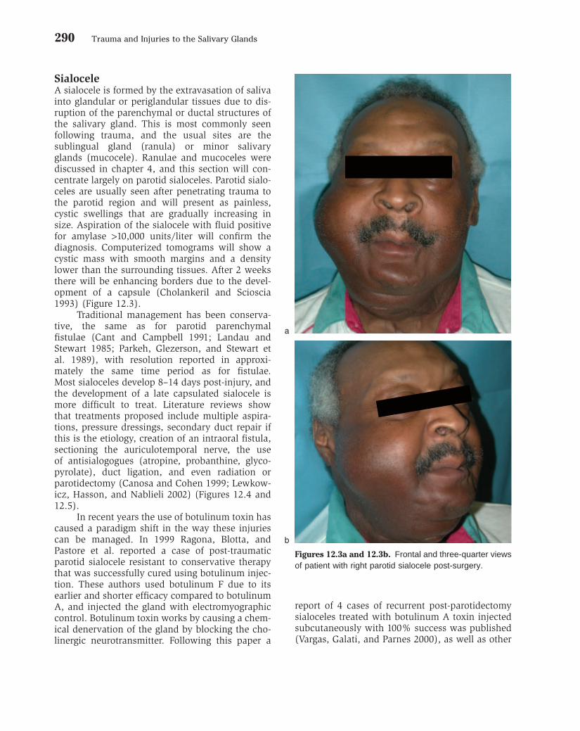
Trauma and Injuries to the Salivary Glands 293
Figure 12.5a. Plain fi lm of a 20-year-old man following a gunshot wound shows the bullet “fl oating” in parotid sialocele.
Figure 12.5b. Surgical exploration to remove the bullet revealed an abscess cavity where the bullet had lodged. Note draining pus. Reprinted with permission from Blanchaert R, Ord RA. 1997. Management of late compli-cations of penetrating injuries to the parotid gland. Pan American Journal of Trauma 6(1):52–57.
case reports (Chow and Kwok 2003). There is a single case of a submandibular sialocele treated with resolution using botulinum toxin A (Capac-cio, Cuccarini, and Benicchio et al. 2006).
Nerve InjuryThe facial nerve is at risk from penetrating injury to the facial region both in the parotid and in the distribution of its peripheral branches to the facial musculature. It is stated that damage to branches distal to a line drawn from the lateral canthus to the commissure does not require repair and may be managed expectantly. All patients with facial wounds should have a careful clinical examination of facial nerve function. Where this is not possible—for example, in the unconscious patient or the uncooperative infant (Figure 12.6)—the wound should be carefully explored at the time of surgery to exclude transaction of the branches of
Figure 12.6. Infant with laceration from broken glass. No facial nerve damage was present.

294 Trauma and Injuries to the Salivary Glands
the facial nerve. Primary repair soon after the injury with end to end anastomosis is the ideal scenario, as paralysis of the facial nerve is a dev-astating injury for the patient, and even when “successful” nerve repair has been carried out with satisfying results (based on House-Brackmann, Stennert, and May grading), patients experience a reduced quality of life (Guntinas-Lichius, Straesser, and Streppel 2007) (Figures 12.7 and 12.8). This section will discuss the management of the primary nerve injury and will not discuss the techniques for facial reanimation or static slings, which are beyond the scope of this text. The interested reader will fi nd many recent review articles addressing these topics (Guntinas-Lichius, Streppel, and Stennert 2006; Malik, Kelly, and Ahmed et al. 2005).
Classically the nerve is sutured under the microscope using 9-0 or 10-0 nylon sutures attempt-ing to coapt the nerve ends without tension (Figure
Figure 12.7a. Entrance wound for bullet below ear lobe.
Figure 12.7b. Bougie placed through track of bullet to demonstrate entrance and exit.
Figure 12.7c. Frontal and buccal facial nerve paralysis.
12.9). The suturing can be epineural or fasicular. In epineural suture less damage is caused to the neural bundles with less foreign body reaction in the fasicles due to the suture materials; however, fasicular suturing should allow better adaptation of the fasicles and trimming back the epineurium to prevent fi brous tissue in-growth. However, ana-tomic studies have shown the fasicular and con-nective tissue anatomy of the facial nerve to be complex, with the number of fasicles increasing in a proximo-distal way from the geniculate ganglion

a bFigures 12.8a and 12.8b. A 72-year-old man diagnosed with a stroke in the emergency room after falling through a plate glass window. The diagnosis was made due to the dense facial nerve palsy.
Figure 12.8c. After removing the dressings the deep pene-trating wound in the region of the facial nerve trunk is appreciated.

296 Trauma and Injuries to the Salivary Glands
with diminishing diameter (Figure 12.10). This variability in number of fasicles and structure along the extratemporal facial nerve constitutes a diffi culty in facial nerve repair (Captier, Canovas, Bonnel, Seignarbieux 2005). The use of tubes (e.g., collagen) to support the anastomosis and prevent connective tissue in-growth and also tissue glues (e.g., fi brin) to replace sutures and their foreign body reactions have been advocated. However, the tubes can themselves cause foreign body reactions and possible compression. Regard-ing the tissue glues, animal experiments appear to show sutures to be superior. In the rabbit model axonal growth was faster and greater with epineu-ral suture than fi brin adhesive (Junior, Valmaseda-Castellon, and Gay-Escoda 2004). Although the rate and amount of reduction in conduction veloc-ity was equivalent between the two methods, the authors concluded that epineural suture appears to
be the method of choice. In another study in the rat model looking at suture and the effects of plate-let rich plasma (PRP) and fi brin sealant, the best return of function for the facial nerve was again with suture (Farrag, Lehar, and Verhaegen et al. 2007). The authors did note a favorable neuro-trophic effect for the PRP but no benefi t for the fi brin sealant. As it is vital that the anastomosis be tension free, diffi culty is encountered when a gap between the nerve ends exists. In a cadaver study, Gardetto, Kovacs, and Piegger et al. (2002) showed that removal of the superfi cial part of the parotid gland could allow overlap of the cut branches of the facial nerve. They found it possible to bridge gaps of 15 mm in the temporo-zygomatic branches, 23 mm in the buccal-mandibular branches, and 17 mm in the nerve trunk. Following this experi-mental work the authors have reported successful clinical results on 3 patients, recommending the
edFigures 12.8d and 12.8e. One year post-repair of nerve trunk and microneural suture the patient still has some weakness of the upper lid and cannot “blow out” his cheek on the left side, but has good facial symmetry and function.

297
Figure 12.9a. Penetrating wound through ear and parotid gland.
Figure 12.9b. Exploration of wound reveals transection of the superior branch of the facial nerve. (Ruler from surgical marker makes a good background for microsuture if custom microsurgical background material is unavailable.)
Figure 12.9c. Post-microsurgical repair.
Figure 12.9d. Post-suturing of wounds. This case treated by Dr. J. Caccamese, Dept. OMS, University of Maryland.

298 Trauma and Injuries to the Salivary Glands
Figure 12.10a. Extensive penetrating wound anterior to the parotid gland, which involves the peripheral branches of the facial nerve.
Figure 12.10b. Nerve branches identifi ed for microneural repair.
Figure 12.10c. High-power view of completed repair.
technique for gaps up to 15 mm but cautioning against its use in the presence of infection or nerve defects (Piza-Katzer et al. 2004). In another approach the use of a rapid nerve expander (2 cm/30 minutes) was used to bridge gaps up to 3 cm (Ya, Gao, and Wang 2007). In 9 patients 5 achieved good results with EMG peak value of mimetic muscles 82–95% of the normal side, 3 cases were fair with EMG 60–90%, and 1 case poor with EMG 55%. Other surgical options in this situ-ation are the use of a tube as a conduit and graft-ing. In 7 patients with post-traumatic defects up to 3 cm the use of a bioabsorbable polyglycolic acid tube was reported as giving very good results in 1 case, good in 4, and fair in 2 (Navissano et al. 2005). The two commonest sites for donor nerves for the facial nerve are the greater auricular, which is adjacent to the surgical fi eld, and the sural nerve. Surgical principles for repair follow those for direct anastomosis. As expected, facial nerve function from cable nerve graft interposition is not as good as end to end anastomosis (Malik, Kelly, and Ahmed et al. 2005). If there is widespread destruc-tion of multiple branches from an injury such as a gunshot the entire superfi cial cervical plexus can be used to supply multiple grafts.
Frey’s SyndromeAlthough Frey’s syndrome is now most commonly seen in relation to parotid surgery, it was originally described after a shotgun injury to the parotid gland (Frey 1923); however, despite Frey’s land-mark paper, gustatory sweating was probably fi rst described by Baillarger in 1853 (Dulguerov, Marchal, and Gusin 1999). In the trauma situa-tion obviously the problem is management and treatment of the condition, whereas in the post-parotidectomy cases much work has been done on prevention.
Previously reported treatments of Frey’s syn-drome have included topical and systemic anticho-linergics, tympanic neurectomy, sectioning of the auriculotemporal or glossopharyngeal nerves, or interposition of fascia lata between the parotid bed and overlying skin. Currently the use of botulinum toxin is the most frequently reported therapeutic modality for Frey’s syndrome. A report of 33 patients with Frey’s syndrome treated by intra-cutaneous injection of 16–80 IU of botulinum toxin A showed all symptoms to resolve within a week (Eckardt and Kuettner 2003). In a prospective

299
d e
Figures 12.10d, 12.10e, 12.10f, and 12.10g. Post-repair. Note upper branch weakness persists. This case treated by Dr. J. Caccamese, Dept. OMS, University of Maryland.
f g

300 Trauma and Injuries to the Salivary Glands
Figure 12.11a. Lateral facial view of patient complaining of gustatory sweating (Frey’s syndrome).
Figure 12.11b. Bottle of iodine solution that is painted on the patient’s face and then covered in corn starch.
nonrandomized, nonblinded study of 11 patients treated with botulinum toxin A with follow-up 6–23 months, only 1 patient recurred and was suc-cessfully retreated (Kyrmizakis, Pangalos, and Papadakis et al. 2004). A prospective randomized trial to establish the ideal dosage and length of effect of botulinum toxin A was carried out on 20 patients divided into two groups receiving either 2 MU/cm2 or 3 MU/cm2. In the 3 MU/cm2 group a single injection resulted in nearly complete absence of gustatory sweating during the 12 month follow-up period. In the 2 MU/cm2 group 44% of the total skin areas were still sweating and required a second injection, and the authors concluded that 3 MU/cm2 is the recommended dose (Nolte, Goll-mitzer, and Loeffelbein et al. 2004). Some authors have cautioned that the effect of botulinum toxin
in Frey’s syndrome is often temporary and further injections may be necessary depending upon the initial dose and the length of time followed (Ferraro et al. 2005).
In regards to parotidectomy patients, proba-bly 100% will have gustatory sweating if tested with starch and iodine (Laage-Hellman 1957) (Figure 12.11). However, few patients clinically notice this problem and most do not wish for treat-ment, so that this condition will be underestimated in clinical reports. Frey’s syndrome is rarely reported after submandibular gland removal. Berini-Aytes and Gay-Escoda (1992) reviewed 206 submandibular gland excisions and found only 1 case of Frey’s syndrome, while Teague, Akhtar, and Phillips (1998) reviewed the literature and could fi nd 7 reported cases since 1934.

Trauma and Injuries to the Salivary Glands 301
The techniques described for prevention of Frey’s syndrome depend upon placing a barrier between the parotid bed and skin to prevent the growth of the secretory parasympathetic nerves from the parotid into the sweat glands, causing a paradoxical innervation. Acellular dermis has been used with success but with a higher complication rate (Govindaraj, Cohen, and Genden et al. 2001). Temporoparietal fl aps and the superfi cial musculo-aponeurotic system (SMAS) have been used with good outcomes in 146 parotidectomy patients (Cesteleyn et al. 2002). In reviewing 160 patients followed from 5 to 22 years treated with an inter-positional SMAS layer at the time of parotidectomy and tested with starch/iodine during follow-up, no cases of Frey’s were encountered (Bonanno et al. 2000). Other reported barriers have been the use
of parotid gland fascia (Zumeng, Zhi, Gang et al. 2006) and sternocleidomastoid muscle fl aps, with mixed results (Filho et al. 2004; Kerawala, McAlo-ney, and Stassen 2002).
It would appear that Frey’s syndrome is pre-ventable in most cases, and perhaps the use of the SMAS layer is the most convenient for the surgeon.
HollowingThis is a complication seen in patients following parotidectomy rather than trauma and can be managed in a variety of ways. Reconstruction at the time of surgery to prevent the defect occurring is preferable to secondary reconstruction, when scarring and the superfi cial position of the facial nerve post-parotidectomy increase the risk of nerve damage. Various techniques have been used, some of which have been discussed in this chapter in relation to Frey’s syndrome and in chapter 8. Tech-niques include the use of layered acellular dermis, free fat grafts (Figure 8.4), use of the SMAS layer, temporalis, and sternocleidomastoid fl aps, as well as microvascular free fl aps including fascial forearm fl aps for larger defects. Choice of tech-nique will depend on the size of the defect, the surgeon’s own experience, and the wishes of the patient.
TRAUMA TO SALIVARY GLAND DUCTS
TransectionAs has already been discussed above in the sec-tions on fi stulae and sialoceles related to parenchy-mal trauma, conservative management is usually satisfactory except in those cases where the injury involves partial or complete transection of the duct. Under these circumstances most papers have indicated that resolution is less certain and takes longer, with active management frequently required. There are studies that support conserva-tive measures in duct injuries and in one report, of 19 patients with duct injury confi rmed by meth-ylene blue dye injection who were treated non-operatively, 9 patients (47%) healed without complications. Although 7 patients (36.8%) devel-oped salivary fi stulae and 4 (21%) sialoceles, these were described as short-term and resolved without the need for surgery (Lewis and Knottenbelt 1991). However, in most cases current management is
Figure 12.11c. While the patient eats an apple the corn-starch is discolored blue-black, indicative of gustatory sweating.

302 Trauma and Injuries to the Salivary Glands
Figure 12.12. Surface markings of the parotid duct are shown by a line drawn from the tragus of the ear to bisect a line drawn from the alar base to the commissure. The middle third of this line (arrow) is surface marking of the parotid duct.
directed toward primary repair and the clinician must therefore have a high level of suspicion for injuries involving the region of the parotid duct. The anatomic surface markings of the duct are illustrated in Figure 12.12. Confi rmatory evidence for transaction is obtained by cannulating the distal portion of the duct through Stenson’s papilla and observing the catheter in the wound (Figure 12.13), or by injecting saline or a small (1 cc) amount of methylene blue through Stenson’s papilla. Identi-fi cation of the proximal end may be diffi cult, as it can retract into the gland substance. Milking the gland to obtain salivary fl ow is helpful in these circumstances, and the anesthesiologist must be cautioned preoperatively against the use of anti-parasympathetic agents. If the proximal and distal ends of the duct are identifi ed and can be coapted, then microsurgical repair can be carried out (Hallock 1992) (Figure 12.14).
The use of stents (usually indwelling cathe-ters) for 10–14 days to prevent stenosis is advo-cated by some and appears a reasonable hypothesis, although no long-term studies of these injuries
with and without stenting have been published. A technique of using a 4 F Foley embolectomy cath-eter for identifi cation of the transection and then leaving it in place as a stent has been described (Etoz, Tuncel, and Ozcan 2006). When the proxi-mal and distal ends of the duct cannot be coapted due to tissue loss, repair using a vein graft has been reported (Heymans et al. 1999). Steinberg and Herréra (2005) recommended the use of sia-lography postoperatively to assess the result of duct repair, stating that this technique may not always be practical or possible in the acute setting. However, we have used sialography intraopera-tively (Figure 12.13).
When the injury is too proximal, the wound is avulsive, or the duct cannot be identifi ed, then the clinician can either create an intraoral fi stula or ligate the proximal duct. A controlled fi stula can be created by suturing the proximal duct through the buccinator into the oral cavity if enough length is present, or by placing a catheter or drain from the area of the wound into the mouth and leaving it to fi stulize (Figure 12.15). Although tying off the proximal duct to cause eventual atrophy has been proposed (Van Sickels 1981), in our experience this is unpredictable and even with the use of pres-sure and antisialogogues these patients can have considerable swelling and pain. Chemical denerva-tion using botulinum toxin A may help to achieve a good outcome in these circumstances (Arnaud et al. 2006).
In the case of the submandibular duct, tran-section is usually iatrogenic as a result of surgery on the sublingual gland, sialolithotomy from Wharton’s duct, or resection of fl oor of the mouth cancer. In this case sialodochoplasty with reposi-tioning of the duct posteriorly is all that is required. One of the authors formerly used a catheter as a stent (Ord and Lee 1996) following reposition of Wharton’s duct in fl oor of the mouth cancer; however, now the duct lumen is identifi ed and one blade of a sharp iris scissors is inserted and a verti-cal cut through one wall of the duct carried out. The duct is now “fi sh-tailed” and sutured to a newly created hole in the oral mucosa with 6-0 nylon sutures. Stenosis and stricture have not been a problem with this technique.

303
Figure 12.13a. Patient with cheek laceration that was pri-marily sutured and that now has developed sialocele due to missed duct injury.
Figure 12.13b. Wound reopened for re-exploration; the duct is discovered to be transected. Vessel loop around distal end of duct. Lacrimal probe passed from intraoral through Stenson’s duct into the wound (short arrow).
Figure 12.13c. Stenson’s duct is approximated after fi nding the proximal end of the duct by milking the gland. The duct is cannulated and contrast dye injected for intra-operative sialogram.
Figure 12.13d. Fluoroscopic image of intraoperative sialo-gram with repaired duct (arrow). Case treated by Dr. D. Coletti, Dept. OMS, University of Maryland.

304
Figure 12.14a. Extensive slashing wound, with duct iden-tifi ed and repaired.
Figure 12.14b. High power of duct repair.
Figure 12.14c. Final skin repair. This case treated by Dr. J. Caccamese, Dept. OMS, University of Maryland.

Trauma and Injuries to the Salivary Glands 305
Figure 12.15a. Gunshot wound entering at right parotid region with exit at left infraorbital region as indicated by suction tubing.
Figure 12.15b. The parotid duct is identifi ed and dissected from the wound to be cannulated as shown.
Figure 12.15c. The duct is diverted intraorally via the cannula. Note powder burn at entrance wound.
Figure 12.15d. Final repair. This case treated by Dr. J. Caccamese, Dept. OMS, University of Maryland.
Stenosis of the DuctWhen ductal injuries are not surgically repaired immediately, complications such as fi stulae and sialoceles may arise and their management has been discussed. If the duct has not been surgically repaired by 72 hours, conservative or medical therapy is recommended (Arnaud et al. 2006). In the long term, stricture of the duct may occur (Figure 12.16), although most strictures are sec-
ondary to infl ammatory or infective conditions. In cases of intraductal salivary gland obstruction, 22.6% of 642 cases were due to strictures, which were more common in females (Ngu, Brown, and Whaites et al. 2007). When this occurs at the distal end of Stenson’s duct, excision and diversion of the duct into the oral cavity may be feasible. When the main duct is involved with strictures, sialoen-doscopy may be useful to dilate the strictures using

306 Trauma and Injuries to the Salivary Glands
b c
Figure 12.16a. Patient with soft tissue scarring from pen-etrating wounds caused by a road traffi c accident 10 years previously; now has a 7-year history of parotid and cheek swelling.
Figures 12.16b and 12.16c. Sialogram shows stricture of duct with proximal dilatation of Stenson’s duct and secondary ducts and a large cystic swelling distal to the stricture.
saline pressure, balloon dilatation, or the minifor-ceps grasper and even the insertion of a stent to the duct lumen (Nahliel, Bar, and Shacham et al. 2004). Simple balloon angioplasty was successful in 7 of 9 patients, and 5 of these patients remained asymptomatic on follow-up (Salerno, Lo Casto, and Comparetto et al. 2007). If this is unsuccessful and the patient continues to have recurrent swell-ing and sialadenitis, denervation with botulinum toxin or even parotidectomy may be required.
In the submandibular gland the stricture can be excised and sialodochoplasty performed as described above.
Radiation InjuryEXTERNAL BEAM
Radiotherapy is commonly given for patients with head and neck cancer; however, the injurious effect that this treatment modality has on the sali-vary glands leading to profound xerostomia, which may be permanent, is well known. The serous cells (found in the parotid gland) are extremely sensi-tive to apoptotic death following even moderate doses of radiation. Indeed, permanent loss of sali-

Trauma and Injuries to the Salivary Glands 307
vary function is seen after doses larger than approximately 3,500 cGy with little in the way of measurable parotid saliva, and 5% of patients will demonstrate a sialadenitis with gland swelling and raised amylase within 12 hours of their fi rst treat-ment (Parsons 1994). However, although it is known that damage to the salivary glands will increase with radiation dose and volume of gland irradiated, there is no universal agreement over the dose required to produce xerostomia. Someya, Sakata, and Nagakura et al. (2003) found gradual recovery of function over time with doses of <5,000 cGy, while no signifi cant recovery was seen in patients who had >5,800 cGy. The minor sali-vary/sublingual glands do not seem to play much of a role in the development of xerostomia, which seems to depend mainly on the mean dose to both the parotid and submandibular glands (Jellema, Doornaert, and Slotman et al. 2005). These authors also found that the stickiness of saliva post-radiation depended mainly on the mean dose to the submandibular glands.
The exact pathogenesis and mechanism of injury to the salivary glands as a result of radiation therapy is also controversial, with no universal agreement as to cause. Based on animal studies in the rat model, a mechanism of delayed serous cell death due to sublethal DNA damage, which results in death during a reproductive phase due to highly redox-reactive metal ions (e.g., iron, copper) asso-ciated with secretion granules has been proposed (Nagler 2002, 2003). Another study showed signifi -cant increase in cytotoxic T-cells in irradiated sub-mandibular glands, suggesting cell-mediated mechanisms may be responsible for the sialadenitis with subsequent acinar cell destruction/atrophy (Teymoortash, Simolka, and Schrader et al. 2005).
Obviously once established the effects of radi-ation damage are diffi cult to treat or reverse, so much effort has been aimed at prevention. Impor-tant advances in the delivery of radiation therapy using 3-D conformal planning and intensity-modulated radiation therapy (IMRT), combined with drugs such as growth factors, cholinergic ago-nists, and cytoprotective agents (Amifostine), are currently the preferred modalities of prevention (Garden, Lewin, and Chambers 2006).
It has been shown with conventional radia-tion therapy that the ability to spare the contralat-eral major salivary glands or to spare the parotid by positioning of the portals can signifi cantly increase salivary fl ow and reduce xerostomia (Beer
et al. 2002; Malouf, Aragon, and Henson et al. 2003). The sophistication of 3-D conformal plan-ning and IMRT allows the radiotherapist to give more radiation to the tumor target with increased sparing of normal tissue. In one study only 12% of patients developed xerostomia following IMRT for head and neck cancer, and there were no locoregional recurrences with a median follow-up of 24 months (Saarilahti, Kouri, and Collan et al. 2005). Jen, Shih, and Lin et al. (2005) compared 108 patients treated with conventional RT to 72 Gy treated with 3-D conformal RT, fi nding 3-D confor-mal RT delivered a higher dose to the tumor with better local control in T4 patients and improved survival with signifi cantly better parotid function. IMRT has also been used to spare the submandibu-lar glands to prevent radiation-induced xerostomia (Saarilahti, Kouri, and Collan et al. 2006). The ability to use 3-D conformal RT and IMRT to spare the opposite parotid by excluding the contralateral level II nodes from the fi eld was not shown to be associated with any locoregional recurrence, and no recurrence occurred in the spared area (Bussels, Maes, and Hermans et al. 2004).
A number of drugs have been investigated for preventing radiation damage. A phase III prospec-tive randomized trial of Amifostine (Ethyol) with 315 patients showed signifi cant reduction in grade 2 or > xerostomia and chronic xerostomia with no effect on locoregional control, disease-free survival, or overall survival. In this study, however, 53% of patients experienced nausea and/or vomiting (Brizel, Wasserman, and Henke et al. 2000). A follow-up study to review results of this study after 2 years found the signifi cant decrease in grade 2 or > xerostomia had been maintained, as well as an increase in the proportion of patients with mean-ingful unstimulated saliva and reduced mouth dryness. There was no compromise of locoregional control, progression-free, or disease-free survival (Wasseman, Brizel, and Henke et al. 2005). In this study the Amifostine was given intravenously, and a recent phase II study has shown a similar radio-protective benefi t for Amifostine given subcutane-ously as a simpler alternative (Anne, Machtay, and Rosenthal et al. 2007). Another approach has been to use pilocarpine, which has been used to treat xerostomia during radiotherapy as a chemopreven-tive agent. A randomized, double-blind, placebo-controlled trial of pilocarpine on 60 patients, only 39 of whom were evaluable, indicated that pilocar-pine used with radiotherapy could lead to signifi -

308 Trauma and Injuries to the Salivary Glands
cant diminution in subsequent xerostomia (Haddad and Karimi 2002). Another randomized trial with 66 patients also concluded that patients with stimu-lated glands from pilocarpine during radiation had less decrease in salivary fl ow, which reduced radia-tion side effects (Nyarady, Nemeth, and Ban et al. 2006). However, the Radiation Therapy Oncology Group (RTOG) study 97-09, which was a phase III trial with 245 patients, showed that although there was a signifi cantly increased unstimulated salivary fl ow in the pilocarpine group, there was no differ-ence in parotid stimulated salivary fl ow, in the amelioration of mucositis, or in the quality of life between the two groups (Scarantino, LeVeque, and Swann et al. 2006). Other novel approaches to the problem have been the use of gene therapy, which has yielded promising results in animal models (Cotrim, Mineshiba, and Sugito et al. 2006; Thula et al. 2005).
Finally, a surgical approach to prevention of xerostomia has been the transfer of the sub-mandibular glands into the submental triangle out of the radiation fi eld prior to the commencement of radiation therapy. In a phase II trial of patients who had primary surgery for oro-pharyngeal cancer followed by adjuvant RT, with or without subman-dibular gland transfer 24 of 51 patients were evalu-ated for swallowing. The cohort with preservation of 1 gland (13 patients) had signifi cantly increased saliva and swallowing function (Rieger, Seikaly, and Jha et al. 2005). Similar results were reported in a small series of patients undergoing chemora-diation (Al-Qahtani et al. 2006). Regarding long-term results in 26 patients followed for 2 years, normal amounts of saliva were reported in 83% (Seikaly, Jha, and Harris et al. 2004).
RADIOACTIVE IODINE
Radioactive iodine is used in the treatment of thyroid cancer but is also concentrated in the sali-vary glands, particularly the parotid, and may cause sialadenitis that is immediate or begins a few months after treatment (Mandel and Mandel 2003). In a prospective study of 76 patients receiving radioactive iodine, 20 (26%) developed salivary gland toxicity, 11 developed toxicity within 48 hours, and 9 not until 3 months post-therapy. A total of 16 patients had chronic toxicity, typically xerostomia, at 12 months (Hyer et al. 2007). In seeking to quantify salivary gland dysfunction using scintigraphy in 50 patients, 46% and 42%
were found to have decreased maximum secretion and uptake ratio, respectively (Raza et al. 2006). The damage was seen more in the parotid and was dependent on the radioiodine dose. The damage and symptoms may be permanent (Mandel and Mandel 1999). The damage is most likely related to an oxidation injury indicated by an increase in prostaglandin levels (Wolfram, Palumbo, and Chehne et al. 2004).
Current management is symptomatic as for sialadenitis and xerostomia from other causes. Animal studies using the rabbit model indicate that Amifostine can signifi cantly reduce radioiodine-induced parenchymal damage (Kutta, Kampen, and Sagowski et al. 2005). The use of sialoendo-scopic treatment in this condition for patients with partial duct stenosis has also been reported (Kim, Han, and Lee et al. 2007).
Barotrauma
Air can be forced in a retrograde fashion into the parotid duct by a rise in intraoral pressure and cause parotid emphysema or pneumoparotid. This condition may occur in glass blowers and musi-cians who play woodwind or brass instruments. It is usually a benign condition but can cause recur-rent sialadenitis or even progress to subcutaneous emphysema. The condition has also been reported in conjunction with the use of an air syringe during routine dentistry (Takenoshite, Kawano, and Oka 1991), secondary to coughing with chronic obstruc-tive airways disease (Cook and Layton 1993), and self-induced in children and adults (Goguen et al. 1995; Gudlaugsson, Geirsson, and Benediktsdottir 1998). The condition can be diagnosed by palpa-tion of emphysema in the parotid and the escape of frothy saliva from the duct. Sialography and CT scans have been used for diagnosis (Gudlaugsson, Geirsson, and Benediktsdottir 1998; Maehara, Ikeda, and Ohmura et al. 2005). In one case with subcutaneous emphysema extending to the medi-astinum parotid duct ligation was used for cure (Han and Isaacson 2004).
Summary (Figures 12.17 and 12.18)
• Most parenchymal penetrating injuries of the parotid gland will resolve with conservative treatment.

Trauma and Injuries to the Salivary Glands 309
Penetrating parotid gland injury
Parenchymal injury with Stenson’s duct injury
Approximation of ductSegments without tension?
Parenchymal injury only
Yes No
PrimaryClosure ofStenson’s duct
Proximalinjury
Distal injury
Ligate ductReconstructionwith implantationin oral cavity
Delayed sialocele orSalivary fistula development
Aspiration of salivaCompression dressingsAnticholinergic medicationsBotox
Facial nerve transection
Able to reapproximate without tension?
Yes
Primary closure
No
Superficialparotidectomy
Able to reapproximatewithout tension
Yes
No
Interpositional nerve graft orNerve expander orAlloplastic conduit
Debridement andClosure of wound
Figure 12.17. Algorithm for management of penetrating trauma to the parotid gland.
• Parotid fi stulae and sialoceles due to paren-chymal injury will also resolve with conserva-tive therapy.
• Chemical denervation of the gland with botu-linum A toxin injected subcutaneously appears to be a safe way of treating fi stulae and sialoceles.
• It is important to recognize the possibility of duct injury early, as immediate repair is indicated.
• Microneural repair of facial nerve injuries pri-marily without tension is the ideal manage-ment for a transected facial nerve or its branches.
• At present the use of tissue glues for nerve repair does not appear to improve results.
• Most cases of Frey’s syndrome following parotidectomy are subclinical.
• Botulinum toxin is useful in the treatment of Frey’s.
• A variety of barrier techniques to prevent Frey’s syndrome have been described with the use of the SMAS layer appearing to give good results.
• IMRT and 3-D conformal planning show great promise in preventing radiation damage to the salivary glands.
• Amifostine and pilocarpine used during radia-tion may act as radioprotectants.

310
Blunt trauma to parotid gland
Salivary gland injury Facial nerveinjury
Delayed sialoceleor salivary fistula development
Aspiration of salivaCompression dressingsAnticholinergic medicationsBotox
Partialweakness
Paralysis
Observation Nerve conduction study
Recovery
Yes
No
Consider surgicalexploration
Figure 12.18. Algorithm for management of blunt trauma to the parotid gland.

Trauma and Injuries to the Salivary Glands 311
ReferencesAl-Qahtani K, Hier MP, Sultanum K, Black MJ. 2006. The
role of submandibular salivary gland transfer in prevent-ing xerostomia in the chemoradiotherapy patient. Oral Surg Oral Med Oral Pathol Oral Radiol Endod 101(6):753–756.
Ananthakrishnan N, Parkash S. 1982. Parotid fi stulas: A review. Br J Surg 69:641–644.
Anne PR, Machtay M, Rosenthal DI et al. 2007. A phase II trial of subcutaneous amifostine and radiation therapy in patients with head and neck cancer. Int J Radiat Oncol Biol Phys 67(2):445–452.
Arnaud S, Batifol D, Goudot P, Yachouh J. 2006. Nonsurgi-cal management of traumatic injuries of the parotid gland using type a botulinum toxin. Plast Reconstr Surg 117(7):2426–2430.
Beer KT, Zehnder D, Lussi A, Greiner RH. 2002. Sparing of contralateral major salivary glands has a signifi cant effect on oral health in patients treated with radical radio-therapy of head and neck tumors. Strahlenther Onkol 178(12):722–726.
Berini-Aytes L, Gay-Escoda C. 1992. Morbidity associated with removal of the submandibular gland. J Craniomaxil-lofac Surg 20(5):216–219.
Bonanno PC, Palaia D, Rosenberg M, Casson P. 2000. Pro-phylaxis against Frey’s syndrome in parotid surgery. Ann Plast Surg 44(5):498–501.
Breuer T, Ferrazzini A, Grossenberger R. 2006. Botulinum toxin A as a treatment of traumatic salivary gland fi stu-las. (Article in German.) HNO 54(4):385–390.
Brizel DM, Wasserman TH, Henke M et al. 2000. Phase III randomized trial of amifostine as a radioprotector in head and neck cancer. J Clin Oncol 18(19):3339–3345.
Bussels B, Maes A, Hermans R et al. 2004. Recurrences after conformal parotid sparing radiotherapy for head and neck cancer. Radiother Oncol 72(2):119–127.
Canosa A, Cohen MA. 1999. Post-traumatic parotid sialo-cele: Report of two cases. J Oral Maxillofac Surg 57(6):742–745.
Cant PJ, Campbell JA. 1991. Management of traumatic parotid sialoceles and fi stulae: A prospective study. Aust N Z J Surg 61(10):742–743.
Capaccio P, Cuccarini V, Benicchio V et al. 2006. Treatment of iatrogenic submandibular sialocele with botulinum toxin Case report. Br J Oral Maxillofac Surg, epub March 31, 2006.
Captier G, Canovas F, Bonnel F, Seignarbieux F. 2005. Organization and microscopic anatomy of the adult human facial nerve: Anatomical and histological basis for surgery. Plast Reconstr Surg 115:1457–1465.
Cesteleyn L, Helman J, King S, Van de Vyvere G. 2002. Temporoparietal fascia fl aps and superfi cial musculoa-poneurotic system plication in parotid surgery reduces Frey’s syndrome. J Oral Maxillofac Surg 60(11):1284–1297.
Cholankeril JV, Scioscia PA. 1993. Post-traumatic sialoceles and mucoceles of the salivary glands. Clin Imaging 17(1):41–45.
Chow TL, Kwok SP. 2003. Use of botulinum type A in a case of persistent parotid sialocele. Hong Kong Med J 9(4):293–294.
Cook JN, Layton SA. 1993. Bilateral parotid swelling associ-ated with chronic obstructive pulmonary disease. A case of pneumoparotid. Oral Surg Oral Med Oral Pathol 76(2):157–158.
Cotrim AP, Mineshiba F, Sugito T et al. 2006. Salivary gland gene therapy 2006. Dent Clin North Amer 50(2):157–173.
Dulguerov P, Marchal F, Gusin C. 1999. Frey syndrome before Frey: The correct history. Laryngoscope 109(9):1471–1473.
Eckardt A, Kuettner C. 2003. Treatment of gustatory sweat-ing (Frey’s syndrome) with botulinum toxin A. Head Neck 25(8):624–628.
Etoz A, Tuncel U, Ozcan M. 2006. Parotid duct repair by use of an embolectomy catheter with a microvascular clamp. Plast Reconstr Surg 117(10):330–331.
Farrag TY, Lehar M, Verhaegen P et al. 2007. Effect of platelet rich plasma and fi brin sealant on facial nerve regeneration in a rat model. Laryngoscope 117(1):157–165.
Faussat JM, Ghiassi B, Princ G. 1993. Rhinnorrhea of parotid origin. Apropos of a case. Rev Stomatol Chir Maxillofac 94(6):363–365.
Ferraro G, Altieri A, Grella E, D’Andrea F. 2005. Botulinum toxin: 28 patients affected by Frey’s syndrome treated with intradermal injections. Plast Reconstr Surg 115(1):344–345.
Filho WQ, Dedivitis RA, Rapoport A, Guimaraes AV. 2004. Sternocleidomastoid muscle fl ap in preventing Frey’s syndrome following parotidectomy. World J Surg 28(4):361–364.
Frey L. 1923. Le Syndrome du nerf auriculo-temporal. Rev Neurol 2:97.
Garden AS, Lewin JS, Chambers MS. 2006. How to reduce radiation-related toxicity in patients with cancer of the head and neck. Curr Oncol Rep 8(2):140–145.
Gardetto A, Kovacs P, Piegger J et al. 2002. Direct coapta-tion of extensive facial nerve defects after removal of the superfi cial part of the parotid gland: An anatomic study. Head Neck 24(12):1047–1053.
Goguen LA, April MM, Karmody CS, Carter BL. 1995. Self induced pneumoparotitis. Arch Otolaryngol Head Neck Surg 121(12):1426–1429.
Govindaraj S, Cohen M, Genden EM et al. 2001. The use of acellular dermis in the prevention of Frey’s syndrome. Laryngoscope 111(11 Pt 1):1993–1998.
Gudlaugsson O, Geirsson AJ, Benediktsdottir K. 1998. Pneumoparotitis: A new diagnostic technique and a case report. Ann Otol Rhinol Laryngol 107(4):356–358.

312 Trauma and Injuries to the Salivary Glands
Guntinas-Lichius O, Straesser A, Streppel M. 2007. Quality of life after facial nerve repair. Laryngoscope 117(3):421–426.
Guntinas-Lichius O, Streppel M, Stennert E. 2006. Postop-erative functional evaluation of different reanimation techniques for facial nerve repair. Am J Surg 191(1):61–67.
Haddad P, Karimi M. 2002. A randomized, double-blind, placebo-controlled trial of concomitant pilocarpine with head and neck irradiation for prevention of radiation-induced xerostomia. Radiother Oncol 64(1):29–32.
Hallock GG. 1992. Microsurgical repair of the parotid duct. Microsurgery 13(5):243–246.
Han S, Isaacson G. 2004. Recurrent pneumoparotid: Cause and treatment. Otolaryngol Head Neck Surg 131(5):758–761.
Heymans O, Nelissen X, Medot M, Fissette J. 1999. Micro-surgical repair of Stenson’s duct using an interposition vein graft. J Reconstr Microsurg 15(2):105–107.
Hyer S, Kong A, Pratt B, Harmer C. 2007. Salivary gland toxicity after radioiodine therapy for thyroid cancer. Clin Oncol (R Coll Radiol) 19(1):83–86.
Jellema AP, Doornaert P, Slotman BJ et al. 2005. Does radiation dose to the salivary glands and oral cavity predict patient-rated xerostomia and sticky saliva in head and neck cancer patients treated with curative radio-therapy? Radiother Oncol 77(2):164–171.
Jen YM, Shih R, Lin YS et al. 2005. Parotid gland-sparing 3-dimensional conformal radiotherapy results in less severe dry mouth in nasopharyngeal cancer patients: A dosimetric and clinical comparison with conventional radiotherapy. Radiother Oncol 75(2):204–209.
Junior EDP, Valmaseda-Castellon E, Gay-Escoda C. 2004. Facial nerve repair with epineural suture and anastomo-sis using fi brin adhesive: An experimental study in the rabbit. J Oral Maxillofac Surg 62(12):1524–1529.
Kerawala CJ, McAloney N, Stassen LF. 2002. Prospective randomized trial of the benefi ts of a sternocleidomastoid fl ap after superfi cial parotidectomy. Br J Oral Maxillofac Surg 40(6):468–472.
Kim JW, Han GS, Lee SH et al. 2007. Sialoendoscopic treat-ment for radioiodine induced sialadenitis. Laryngoscope 117(1):133–136.
Kutta H, Kampen U, Sagowski C et al. 2005. Amifostine is a potent radioprotector of salivary glands in radioiodine therapy. Structural and ultrastructural fi ndings. Strahlen-ther Onkol 181(4):237–245.
Kyrmizakis DE, Pangalos A, Papadakis CE et al. 2004. The use of botulinum toxin type A in the treatment of Frey and crocodile tears syndromes. J Oral Maxillofac Surg 62(7):840–844.
Laage-Hellman J-E. 1957. Gustatory sweating and fl ushing after conservative parotidectomy. Acta Otolaryngol (Stokh) 48:234–252.
Landau R, Stewart M. 1985. Conservative management of post-traumatic parotid fi stulae and sialoceles: A prospec-tive study. Br J Surg 72:42–44.
Lewis G, Knottenbelt JD. 1991. Parotid duct injury: Is imme-diate surgical repair necessary? Injury 22:407–409.
Lewkowicz AA, Hasson O, Nablieli O. 2002. Traumatic injuries to the parotid gland and duct. J Oral Maxillofac Surg 60(6):676–680.
Maehara M, Ikeda K, Ohmura N et al. 2005. Multislice computed tomography of pneumoparotid: A case report. Radiat Med 23(2):147–150.
Malik TH, Kelly G, Ahmed A et al. 2005. A comparison of surgical techniques used in dynamic reanimation of the paralyzed face. Otol Neurotol 26(2):284–291.
Malouf JG, Aragon C, Henson BS et al. 2003. Infl uence of parotid-sparing radiotherapy on xerostomia in head and neck cancer patients. Cancer Detect Prev 27(4):305–310.
Mandel SJ, Mandel L. 1999. Persistent sialadenitis after radioactive iodine therapy: Report of two cases. J Oral Maxillofac Surg 57(6):738–741.
Mandel SJ, Mandel L. 2003. Radioactive iodine and the salivary glands. Thyroid 13(3):266–271.
Marchese-Ragona R, Marioni G, Restivo DA, Staffi eri A. 2006. The role of botulinum toxin in post-parotidectomy fi stula. A technical note. Am J Otolaryngol 27(3):221–224.
Morestin M. 1917. Contribution a l’etude du traitement des fi stules salivaires consecutives aux blessures de guerre. Bull Mém Soc Chir Paris 43:845.
Nagler RM. 2002. The enigmatic mechanism of irradiation induced damage to the major salivary glands. Oral Dis 8(3):141–146.
Nagler RM. 2003. Effects of head and neck radiotherapy on major salivary glands—animal studies and human impli-cations. In Vivo 17(4):369–375.
Nahliel O, Bar T, Shacham R et al. 2004. Management of chronic recurrent parotitis: Current therapy. J Oral Maxil-lofac Surg 62(9):1150–1155.
Navissano M, Malan F, Carnino R, Battiston B. 2005. Neu-rotube for facial nerve repair. Microneurosurgery 25(4):268–271.
Ngu, Brown JE, Whaites EJ et al. 2007. Salivary duct stric-tures: Nature and incidence in benign salivary obstruc-tion. Dentomaxillofac Radiol 36(2):63–67.
Nolte D, Gollmitzer I, Loeffelbein DJ et al. 2004. Botulinum toxin for treatment of gustatory sweating. A prospective randomized study. (Article in German.) Mund Kiefer Gesichtschir 8(6):369–375.
Nyarady Z, Nemeth A, Ban A et al. 2006. A randomized study to assess the effectiveness of orally administered pilocarpine during and after radiotherapy of head and neck cancer. Anticancer Res 26(2B):1557–1562.
Ord RA, Lee VA. 1996. Submandibular duct repositioning after excision of mouth cancer. J Oral Maxillofac Surg 54:1075–1078.
Parkeh D, Glezerson G, Stewart M et al. 1989. Post-trau-matic parotid fi stulae and sialoceles. A prospective study of conservative management in 51 cases. Ann Surg 209(1):105–111.

Trauma and Injuries to the Salivary Glands 313
Parsons JT. 1994. The effect of radiation on normal tissues of the head and neck. In: Million RR, Cassisi NJ (eds.), Management of Head and Neck Cancer: A Multidisci-plinary Approach. Philadelphia: Lippincott, pp. 247–250.
Piza-Katzer H, Balough B, Muzika-Herczeg E, Gardetto A. 2004. Secondary end to end repair of extensive facial nerve defects: Surgical technique and postoperative func-tional results. Head Neck 26(9):770–777.
Ragona RM, Blotta P, Pastore A et al. 1999. Management of parotid sialocele with botulinum toxin. Laryngoscope 109(8):1344–1346.
Raza H, Khan AU, Hameed A, Khan A. 2006. Quantitative evaluation of salivary gland dysfunction after radioiodine therapy using salivary gland scintigraphy. Nucl Med Commun 27(6):495–499.
Rieger J, Seikaly H, Jha N et al. 2005. Submandibular gland transfer for prevention of xerostomia after radiation therapy: Swallowing outcomes. Arch Otolaryngol Head Neck Surg 131(2):140–145.
Saarilahti K, Kouri M, Collan J et al. 2005. Intensity modu-lated radiotherapy for head and neck cancer: Evidence for preserved salivary gland function. Radiother Oncol 74(3):251–258.
Saarilahti K, Kouri M, Collan J et al. 2006. Sparing of the submandibular glands by intensity modulated radiother-apy in the treatment of head and neck cancer. Radiother Oncol 78(3):270–275.
Salerno S, Lo Casto A, Comparetto A et al. 2007. Sialo-dochoplasty in the treatment of salivary duct stricture in chronic sialadenitis: Technique and results. Radiol Med (Torino) 112(1):138–144.
Scarantino C, LeVeque F, Swann RS et al. 2006. Effect of pilocarpine during radiation therapy: Results of RTOG 97–09, a phase III randomized study in head and neck cancer patients. J Support Oncol 4(5):252–258.
Scher N, Poe DS. 1988. Post-traumatic prandial rhinorrhea. J Oral Maxillofac Surg 46(1):63–64.
Seikaly H, Jha N, Harris JR et al. 2004. Long term outcomes of submandibular gland transfer for prevention of post radiation xerostomia. Arch Otolaryngol Head Neck Surg 130(8):956–961.
Singh B, Shaha A. 1995. Traumatic submandibular salivary gland fi stula. J Oral Maxillofac Surg 53(3):338–339.
Someya M, Sakata K, Nagakura H et al. 2003. The changes in irradiated salivary gland function of patients with head and neck tumors treated with radiotherapy. Jpn J Clin Oncol 33(7):336–340.
Steinberg JM and Herréra AF. 2005. Management of parotid duct injuries. Oral Surg Oral Med Oral Pathol Oral Radiol Endodont 99(2):136–141.
Takenoshite Y, Kawano Y, Oka M. 1991. Pneumoparotitis an unusual occurrence of parotid gland swelling during dental treatment. Report of a case with a review of the literature. J Craniomaxillofac Surg 19(8):362–365.
Teague A, Akhtar S, Phillips J. 1998. Frey’s syndrome fol-lowing submandibular gland excision: An unusual post operative complication. ORL J Otorhinolaryngol Relat Spec 60(6):346–348.
Teymoortash A, Simolka N, Schrader C et al. 2005. Lym-phocyte subsets in irradiation-induced sialadenitis of the submandibular gland. Histopathology 47(5):493–500.
Thula TT, Schultz G, Tran-Soy-Tay R, Batich C. 2005. Effects of EGF and bFGF on irradiated parotid glands. Ann Biomed Eng 33(5):685–695.
Van Sickels JE. 1981. Parotid duct injuries. Oral Surg Oral Med Oral Pathol 52(4):364–367.
Vargas H, Galati LT, Parnes SM. 2000. A pilot study evalu-ating the treatment of post parotidectomy sialoceles with botulinum toxin type A. Arch Otolaryngol Head Neck Surg 126(3):421–424.
Wasseman TH, Brizel DM, Henke M et al. 2005. Infl uence of intravenous amifostine on xerostomia, tumor control, and survival after radiotherapy for head-and-neck cancer: A 2-year follow up of a prospective, randomized phase III trial. Int J Radiat Oncol Biol Phys 15(4):985–990.
Wolfram RM, Palumbo B, Chehne F, et al. 2004. (iso) Prostaglandins in saliva indicate oxidation injury after radioiodine therapy. Rev Esp Med Nucl 23(30):183–188.
Ya Z, Gao Z, Wang J. 2007. Primary clinical study on using end to end neurorrhaphy following rapid nerve expan-sion to repair facial nerve defect. (Article in Chinese.) Zhongguo Xiu Fu Chong Jian Wai Ke Za Zhi 21(1):23–25.
Zumeg Y, Zhi G, Gang Z et al. 2006. Modifi ed superfi cial parotidectomy: preserving both the greater auvicular nerve and the parotid gland fascia. Arch Otolaryngol Head Neck Surg 135(3): 458–62.

Chapter 13
Miscellaneous Pathologic Processes of the Salivary Glands
(Mandel 2006) and recurrent herpes labialis (Heath, McCleod, and Pearce 2006) have been described as presenting signs of salivary gland aplasia. The con-dition may occur as part of a recognized syndrome, associated with other congenital anomalies, or as an isolated phenomena. Aplasia of the lacrimal and salivary glands (ALSG) presenting with irritable eyes and xerostomia is an autosomal dominant condition that appears to be related to mutations in FGF10 (Entesarium, Dalqvist, and Shashi et al. 2007). In lacrimo-auriculo-dento-digital syndrome (LADD), agenesis of salivary glands as well as lac-rimal glands can be seen and is an autosomal domi-nant condition with variable expressivity (Inan, Yilmaz, and Demir et al. 2006). A case of subman-dibular agenesis with parotid gland hypoplasia in association with ectodermal dysplasia is reported (Singh and Warnakulasuriya 2004). In addition, aplasia in association with hypoplasia of the thyroid (D’Ascanio et al. 2006) and accessory parotid tissue (Antoniades et al. 2006) are described. Manage-ment of these cases is symptomatic and directed toward the xerostomia and other oral health care issues.
DUCT ATRESIA
Duct atresia is rare and in a 2001 review (Hof-frichter, Obeid, and Soliday) only 8 previous cases of submandibular duct atresia were found, with 6 unilateral and 2 bilateral. The condition usually presents in babies or infants as a “ranula” and is thought to be due to failure of the duct to penetrate the oral mucosa during development. The diagno-sis can be made by the presence of dilated Whar-ton’s duct(s) on CT scan. Management is by sialodochoplasty to create a new duct orifi ce.
ABERRANT GLANDS
Accessory glands are ectopic in position but possess a duct that usually opens into another main duct, for example, the accessory parotid gland, whereas
OutlineIntroductionHereditary and Congenital Conditions
AplasiaDuct AtresiaAberrant GlandsPolycystic Disease of the Salivary GlandsFirst Branchial Cleft Cysts, Fistulae, and SinusesCystic Fibrosis
SalivaSaliva as a Diagnostic FluidDroolingSaliva in the Management of Xerophthalmia
Ischemic/Degenerative ChangesNecrotizing SialometaplasiaAge Changes in Salivary Glands
SummaryReferences
Introduction
This chapter will review a heterogenous group of salivary diseases that are not covered in other sec-tions of this book. Hereditary and developmental conditions of the glands are rare, such that the most common branchial arch anomalies will be emphasized. Under the heading of saliva most clinical emphasis will be given to the treatment of drooling. This condition is not uncommon and there are a wide variety of treatment approaches that will be discussed.
Hereditary and Congenital ConditionsAPLASIA
Aplasia of one or all of the major salivary glands is a rare condition that may present with severe xero-stomia, rampant caries, candidiasis, pharyngitis, and laryngitis. In addition, “dental chipping”
315

316 Miscellaneous Pathologic Processes of the Salivary Glands
aberrant glands have no duct system. Some of these aberrant glands can form fi stulae and secrete while the patient is eating; others do not secrete but form choristomas. The commonest sites for these aberrant glands are the lateral neck, pharynx, and middle ear (Enoz and Suoglu 2006), presum-ably from their proximity to the fi rst two branchial arches during development. These aberrant glands may be involved in neoplastic change and may account for the central salivary tumors of the jaws (usually the mandible).
POLYCYSTIC DISEASE OF THE SALIVARY GLANDS
This is a rare disease that may be a hereditary condition, as familial cases have been reported
(Smyth, Ward-Booth, and High 1993). It is thought to be due to a developmental abnormality of the intercalated duct system. Seifert, Thomsen, and Donath (1981) reviewed 5,739 cases of salivary gland disease and found 360 cases of cystic disease, of which 2 patients were classifi ed with dysgenetic polycystic parotid disease. Although it is usually bilateral, unilateral cases have been described (Seifert, Thomsen, and Donath 1981) (Figure 13.1). It is said to be always seen in females; however, a case of the condition in the submandibular glands in a male patient has been reported (Garcia, Martini, and Caces et al. 1998). Histologically the gland is replaced with multiple cysts that may contain spheroliths or microliths. There is a marked absence of infl ammatory change. Parotidectomy may be carried out for aesthetic reasons.
a b
Figures 13.1a and 13.1b. Middle-aged woman with right parotid swelling for “many” years. Patient is concerned regarding her appearance, as she has no symptoms.

Miscellaneous Pathologic Processes of the Salivary Glands 317
c d
e fFigures 13.1c, 13.1d, 13.1e, and 13.1f. MR fi lms show multiple cysts within the gland. At the time of surgery multiple microliths were seen.
FIRST BRANCHIAL CLEFT CYSTS, FISTULAE, AND SINUSES
Anomalies of the fi rst brachial arch are intimately associated with the parotid gland and the periauric-ular structures. They are less common than second branchial arch anomalies. In a survey of 183 patients
with branchial cleft cysts and fi stulae, 148 patients (80.8%) had branchial cysts of which 35 (23.6%) arose from the fi rst arch and 35 (23.6%) had fi stulae of which 11 (31.4%) arose from the fi rst arch (Agaton-Bonilla and Gay-Escoda 1996). The usual fi gure for the incidence of fi rst branchial arch anom-alies is 10% (Olsen, Maragos, and Weiland 1980).

318 Miscellaneous Pathologic Processes of the Salivary Glands
Although Work (1972) classifi ed type I cystic lesions containing only squamous epithelium and type II lesions, which contained squamous epithe-lium with adnexal skin structures plus cartilage, the presence of infection may make it impossible to classify these lesions using these criteria. Olsen, Maragos, and Weiland (1980) simplifi ed this clas-sifi cation, dividing the type II anomaly into cysts, fi stulae, and sinuses. Cysts are tracts with no opening, sinuses are a tract with a single opening usually from the external auditory canal, and fi s-tulae are tracts with two openings usually from the external auditory meatus to the anterior neck above the hyoid bone. In their series of 39 cases, Triglia, Nichollas, and Ducroz et al. (1998) found 20 (51%) sinuses, 11 (28%) fi stulae, and 8 (21%) cysts. Similarly, in the series of 10 patients by Solares, Chan, and Koltal (2003), 5 (50%) were sinuses, 3 (30%) fi stulae, and 2 (20%) cysts.
Presentation is usually with recurrent infec-tion, with discharge of pus or abscess in the ante-rior neck, a chronic purulent discharge from the ear, or an infected swelling of the parotid region (Figure 13.2). The usual age of presentation is between birth and 20 years, with most cases diag-nosed at age 2.5 years.
Unfortunately the infection is often not rec-ognized as a manifestation of a fi rst branchial arch abnormality and is treated with drainage or inad-equate limited exploration, which will complicate subsequent surgery. In the series of Triglia, Nichol-las, and Ducroz et al. (1998), 44% of patients had undergone prior surgery, while 65% of patients had incomplete surgery before referral in another
Figure 13.2. Twenty-year-old girl with recurrent localized infection of the parotid and a periparotid sinus.
Figure 13.3a. Operative photograph of patient with a dis-crete mass thought to be a parotid tumor. (Patient’s ear at lower right of image.) While dissecting down the external auditory meatus a fi stulous tract to the cartilage was identi-fi ed and the clamp points to a bead of pus from the fi stu-lous tract.
paper (Martinez et al. 2007). As the fi stulae and sinuses communicate with the external auditory canal, and their relationship to the facial nerve is variable, a wide parotidectomy exposure with dis-section of the nerve is essential for complete removal. In fi stulae to the auditory meatus removal of the cartilage surrounding the fi stulous tract is recommended (Figure 13.3). If the fi stula or sinus tract is not completely removed the lesion will recur, and although the recurrence rate is small,
Figure 13.3b. The fi stula was removed with a rim of the cartilage from the ear canal and a superfi cial parotidec-tomy carried out to remove the branchial cyst (arrow).

Miscellaneous Pathologic Processes of the Salivary Glands 319
c d
Figures 13.3c and 13.3d. The parotidectomy specimen shows the cyst deep in the parotid, but it was lying superfi cial to the facial nerve.
Figure 13.3e. Following superfi cial parotidectomy (the ear lobe is sutured up for surgical retraction).
of 10 lesions were deep to the nerve and 1 lay between the branches. Larger cysts deep to the nerve may be diffi cult to remove, as the nerve may be adherent to them (Figure 13.5), and dissection can be slow and tedious.
It is very important to recognize the fi rst arch abnormality, as its complexity and anatomical variety necessitates wide exposure through a parotidectomy incision and dissection of the facial nerve to minimize the chances of subsequent facial nerve damage.
3–5% (Stulner et al. 2001), this may increase in patients with previous infection or inadequate surgery. Branchial cysts will usually appear as parotid masses and are usually clinically diagnosed as cystic parotid tumors (Figure 13.4).
The fi rst branchial lesions are usually super-fi cial to the nerve, and Triglia, Nichollas, and Ducroz et al. (1998) reviewed 73 cases including their 39 and found that 63% were superfi cial, 29% deep, and 8% between the nerve. In the Solares, Chan, and Koltal (2003) small series, however, 7

320
a b
c d
Figures 13.4a and 13.4b. CT scans of large branchial cyst in the parotid gland.
Figures 13.4c and 13.4d. Histology of the branchial (lymphoepithelial cyst). The proteinaceous cyst contents are superior and the arrow points to the squamous epithelial lining and its associated lymphoid follicles.

321
Figure 13.5a. A 27-year-old man with a large cystic lesion in his right parotid. The FNAB showed benign disease.
Figure 13.5b. Initial superfi cial parotidectomy (superfi cial lobe retracted by an Allis clamp) reveals the cyst lying deep to the cervico-mandibular branch of the facial nerve (arrow).
Figure 13.5c. The cervico-mandibular branch is carefully dis-sected off the cyst capsule and retracted toward the ear.
Figure 13.5d. Parotidectomy specimen with deep lobe branchial cyst.
Figure 13.5e. The cervico-mandibular trunk (arrow) is stretched over the defect that the deep lobe branchial cyst occupied.

322 Miscellaneous Pathologic Processes of the Salivary Glands
CYSTIC FIBROSIS
The composition of saliva is changed in cystic fi brosis and the formation of viscous mucus may lead to cystic dilations of the ducts and acini, espe-cially in the sublingual gland. The calcium concen-tration in saliva is also raised and microliths of calcium complexes with the viscous mucus can be seen.
SalivaSALIVA AS A DIAGNOSTIC FLUID
In many ways saliva represents an ideal fl uid for diagnostic analysis, being readily available and not requiring invasive techniques. Currently there is much interest in developing technologies to use saliva to diagnose, monitor progress, and assess treatment and recurrence of oral cancer. However, its mucous nature has made it diffi cult to analyze. In addition there is evidence to suggest that the methods of processing saliva prior to analysis may have a signifi cant effect on the results obtained for proteins in proteome analysis (Ohshiro, Rosenthal, and Koomen et al. 2007). In seeking to analyze the proteome of saliva, researchers are hoping to fi nd specifi c diagnostic biomarkers and develop tech-niques to discriminate between these biomarkers using proteomic and genomic technologies (Wong 2006). Several different research groups have examined varying aspects of salivary composition and have demonstrated signifi cant differences between saliva in healthy subjects and those with oral cancer. Studies have examined biochemical and immunological parameters (Shpitzer et al. 2007), salivary endothelin levels (Pickering, Jordan, and Schmidt 2007), and reactive nitrogen species and antioxidant profi le (Bahar, Feimesser, and Shpitzer et al. 2007). Using genomic analysis, four mRNAs (OAZ, SAT, IL8, IL1b) were identifi ed that collectively had a discriminatory power of 91% sensitivity and specifi city for detecting oral cancer (Zimmerman, Park, and Wong 2007). None-theless, despite these promising initial results this technique remains a research tool at the present time.
DROOLING
The term “drooling” is often used synonymously with sialorrhea; however, virtually all patients who
drool do not have an increase in the amount of saliva they produce. Patients with Parkinson’s disease with a reduced saliva production can often suffer from drooling. Drooling is the result of a lack of coordinated swallowing with pooling of saliva in the anterior fl oor of the mouth with subsequent drooling as exemplifi ed in conditions such as cere-bral palsy and amyotrophic lateral sclerosis. This condition can have a severe impact on the patient’s quality of life. Many different methods of managing this condition have been proposed and were sum-marized in an excellent review by Meningaud et al. in 2006. These patients benefi t from a multidisci-plinary team approach and both medical and surgi-cal treatments are used in their management. Medical therapy includes oral motor therapy, oro-facial regulation therapy, and behavioral modifi ca-tion via biofeedback. In an analysis of studies from 1970 to 2005 of behavioral treatments of drooling only 17 articles with 57 patients met the inclusion criteria. The evidence base found 15 studies that used a single participant design and 2 that used an experimental comparison group design. Some studies were poorly designed and methodological fl aws were identifi ed. Conclusions were that it was not possible to assess the effi cacy of behavioral therapy and that further research is needed (Van der Burg et al. 2007). Drug therapy may be by the use of anticholinergics given orally or by botulinum toxin injections. Certain conditions such as glau-coma preclude the use of these oral drugs, and side effects are not uncommon. In a systematic review of the literature only 7 papers were found to meet the inclusion criteria and the authors concluded that there was some evidence benztropine, glyco-pyrrolate, and bezhexol hydrochloride were effec-tive in children with drooling (Jongerius, van Tiel, and van Limbeek et al. 2003). Botulinum toxin A has been injected into the parotid glands solely or with the submandibular glands with good results. However, the effect is only temporary. In a double-blind placebo-controlled study on 20 patients with Parkinsonism, botulinum toxin A injection into the parotid and submandibular glands was found to be an effective and safe treatment for drooling (Mancini, Zangaglia, and Cristina et al. 2003). In another prospective, double-blind, placebo-con-trolled trial of different doses of botulinum toxin A (18.75, 37.5, and 75 MU per parotid), the primary end point was achieved with the highest dose of 75 MU without side effects (Lipp, Trottenberg, and Schink et al. 2003). Similarly, in a controlled trial

Miscellaneous Pathologic Processes of the Salivary Glands 323
of botulinum against scopolamine in children with cerebral palsy and drooling, botulinum toxin was found to have a signifi cant effect (Jongerius, van den Hoogen, and van Limbeek et al. 2004). Although botulinum showed fewer and less severe side effects than transdermal scopolamine, general anesthesia was required for the injections. Despite the fact that treatment by botulinum toxin A can improve drool-ing for up to 6 months, there is currently no data in the literature for optimum or maximum dosage, frequency of injections, and duration of action (Lal and Hotaling 2006).
Radiation therapy has been used to reduce saliva production but is obviously contraindicated in children due to its effects on growth and the possibility of radiation-induced sarcoma. Even in adults its long-term side effects preclude its use. An exception may be in patients such as those with poor life expectancy, including those with amyo-trophic lateral sclerosis, where radiation may be of benefi t in reducing salivary production.
Surgery encompasses both sectioning of secretory nerves and also operations on the glands and ducts. Sectioning of Jacobson’s nerve in the middle ear has fallen out of favor and sectioning of the chorda tympani will cause loss of taste. Many different surgical techniques have been described since Wilkie’s classic paper advocating excision of the submandibular glands combined with posterior positioning of the parotid ducts (Wilkie 1967). These methods have included duct ligation, duct repositioning, and gland excision of one or more of the major glands. At the present time excellent permanent results have been reported with submandibular duct repositioning and sublingual gland excision. Although subman-dibular gland excision with parotid duct ligation has been reported as 87% successful (Manrique, do Brasil Ode, and Ramos 2007), it does give rise to temporary parotid edema that can be signifi cant, and Greensmith et al. (2005) reported that bilateral submandibular duct repositioning gland with sub-lingual gland excision was superior to this tech-nique. However, some authors have questioned the need for excision of the sublingual glands. In a study to assess submandibular duct reposition alone against duct reposition and sublingual gland removal, a 3% postoperative hemorrhage and 12% of parents expressing concerns of pain were found for the duct reposition only procedure, while 13.7% hemorrhage and 36% concern over postop-erative pain were found in the group with sublin-
gual gland excision (Glynn and O’Dwyer 2007). As both procedures were equally effective in control-ling drooling, the authors state that they no longer carry out sublingual gland excision.
Due to the number of different causes of drooling and the multiple treatment choices avail-able, these patients are best assessed by a multi-disciplinary team. As in many other aspects of pediatrics and medicine, simple non-invasive methods of management are attempted fi rst before suggesting surgical management.
SALIVA IN THE MANAGEMENT OF XEROPHTHALMIA
Although dry eyes may occur in relation to dry mouth in conditions such as Sjogren’s syndrome, keratoconjunctivitis sicca can occur in isolation. Isolated keratoconjunctivitis sicca is not an uncom-mon condition and currently there is no satisfac-tory treatment. Transfer of the submandibular gland duct into the lacrimal basin was fi rst under-taken in 1986 (Murube-Del-Castillo 1986). The largest series of cases with micro-anastomosis of the submandibular gland vessels to the temporal vessels in the temporal fossa and insertion of Wharton’s duct into the upper eyelid is 38 (Yu, Zhu, and Mao et al. 2004). In this series only 5 cases failed, 8 cases had epiphora, which required reduction of the size of the submandibular gland, and 2 cases had ductal reconstruction secondary to blockage. The authors stress the use of scintig-raphy preoperatively to assess the salivary gland function and rule out a Sjogren’s disease, and also postoperatively to assess revascularization and function. Paniello (2007) reported success in 6 of 7 transfers (86%). Four of 5 patients had kerato-conjunctivitis sicca secondary to Stevens-Johnson syndrome.
Ischemic/Degenerative ChangesNECROTIZING SIALOMETAPLASIA
Necrotizing sialometaplasia can be seen in any of the salivary glands but is most commonly diag-nosed in the minor salivary glands of the palate (Figure 13.6).
It is thought to be secondary to local ischemia with secondary necrosis of the gland and may be secondary to trauma or surgery but is usually

324 Miscellaneous Pathologic Processes of the Salivary Glands
spontaneous. Initially there is swelling quickly fol-lowed by ulceration, which may be deep down to the bone. Healing may take 2–3 months. Biopsy may be necessary to distinguish this lesion from a malignancy and the histology may be misinter-preted. There is lobular necrosis of the salivary gland with squamous metaplasia of the ducts, and this can be misdiagnosed as mucoepidermoid car-cinoma or squamous cell carcinoma. In addition the epithelium adjacent to the ulcer can display pseudo-epitheliomatous hyperplasia, which can also be mistaken for squamous cell carcinoma. If the patient keeps the lesion clean with mouth-washes, healing will occur and recurrence is not seen. Biopsy will often be required to rule out malignancy and histologic interpretation by an experienced pathologist is essential.
AGE CHANGES IN SALIVARY GLANDS
Generalized acinar atrophy can occur in the major salivary glands with age. Frequently the glandular tissue is replaced with fat. In addition, oncocytic metaplasia increases in older patients. Oncocytes are large granular eosinophilic cells. Their granular cytoplasm appears to be secondary to numerous mitochondria. A diffuse oncocytosis of the salivary glands can occur. These changes are not clinically relevant, although oncocytes can give rise to an oncocytoma, which is usually benign but may occasionally be a malignant type. Oncocytomas are of interest as they are similar to Warthin’s tumor in appearing as “hot” spots on technetium scans.
Summary
• Fistulae and sinuses above the hyoid bone in the periparotid region should be suspected of being fi rst arch anomalies.
• In managing fi rst branchial arch anomalies, wide exposure with complete dissection of the facial nerve is mandatory because of the complex and unpredictable relationship of the sinuses, fi stulae, and cysts to the nerve.
• In managing drooling a multidisciplinary team is optimum.
• Surgery for drooling is used when other less invasive therapies have been tried and failed. Posterior repositioning of the submandibular ducts with or without sublingual gland exci-sion appears to give good results.
• Necrotizing sialometaplasia should be consid-ered in the diagnosis of ulcerative palatal lesions and may be mistaken clinically and histologically for a malignancy.
References
Agaton-Bonilla FC, Gay-Escoda C. 1996. Diagnosis and treatment of branchial cleft cysts and fi stulae: A retro-spective study of 183 patients. Int J Oral Maxillofac Surg 25:449–452.
Antoniades DZ, Markopoulos AK, Deligianni E, Andreadis D. 2006. Bilateral aplasia of the parotid glands correlated with accessory parotid tissue. J Laryngol Otol 120(4):327–329.
Bahar G, Feimesser R, Shpitzer T et al. 2007. Salivary analysis in oral cancer patients: DNA and protein oxida-
Figure 13.6a. Necrotizing sialometaplasia of palate with rolled edge and granular base clinically resembling squa-mous cell carcinoma.
Figure 13.6b. Necrotizing sialometaplasia at a later stage with exposed palatal bone.

Miscellaneous Pathologic Processes of the Salivary Glands 325
tion, reactive nitrogen species and anti oxidant profi le. Cancer 109(1):54–59.
D’Ascanio L, Cavuto C, Martinelli M, Salvinelli F. 2006. Radiological evaluation of major salivary gland agenesis: A case report. Minerva Stomatol 55(4):223–228.
Enoz M, Suoglu Y. 2006. Salivary gland choristoma of the middle ear. Laryngoscope 116(6):1033–1034.
Entesarium M, Dalqvist J, Shashi V et al. 2007. FGF10 mis-sense mutations in aplasia of major salivary gland agen-esis: A case report. Minerva Stomatol 55(4):379–382.
Garcia S, Martini F, Caces F et al. 1998. Polycystic disease of the salivary glands: Report of an attack on the sub-mandibular glands. (Article in French.) Ann Pathol 18(1):58–60.
Glynn F, O’Dwyer TP. 2007. Does the addition of sublin-gual gland excision to submandibular duct relocation give better overall results in drooling control? Clin Otolaryngol 32(2):103–107.
Greensmith AL et al. 2005. Prospective analysis of the outcome of surgical management of drooling in the pedi-atric population: A 10 year experience. Plast Reconstr Surg 116(5):1233–1242.
Heath N, McCleod I, Pearce R. 2006. Major salivary gland agenesis in a young child: Consequences for oral health. Int J Pediatr Dent 16(6):431–434.
Hoffrichter MS, Obeid G, Soliday JT. 2001. Bilateral sub-mandibular duct atresia: Case report. J Oral Maxillofac Surg 59:445–447.
Inan UU, Yilmaz MD, Demir Y et al. 2006. Characteristics of lacrimo-auriculo-dento-digital (LADD) syndrome: Case report of a family and literature review. Int J Pediatr Otorhinolaryngol 70(7):1307–1314.
Jongerius PH, van Tiel P, van Limbeek J et al. 2003. A systematic review for evidence of effi cacy of anticholin-ergic drugs to treat drooling. Arch Dis Child 88:911–914.
Jongerius PH, van den Hoogen FJ, van Limbeek J et al. 2004. Effect of botulinum toxin in the treatment of drooling: A controlled clinical trial. Pediatrics 114(3):620–627.
Lal D, Hotaling J. 2006. Drooling. Curr Opin Otolaryngol Head Neck Surg 14(6):381–386.
Lipp A, Trottenberg T, Schink T et al. 2003. A randomized trial of botulinum toxin A for treatment of drooling. Neurology 61(9):1279–1281.
Mancini F, Zangaglia R, Cristina S et al. 2003. Double-blind, placebo-controlled study to evaluate the effi cacy and safety of botulinum toxin type A in the treatment of drooling in Parkinsonism. Mov Disord 18(6):685–688.
Mandel L. 2006. An unusual pattern of dental damage with salivary gland aplasia. J Am Dent Assoc 137(7):984–989.
Manrique D, do Brasil Ode O, Ramos H. 2007. Drooling: Analysis and evaluation of 31 children who underwent bilateral submandibular gland excision and parotid duct
ligation. Rev Bras Otorhinolaryngol (Eng ed.) 73(1):40–44.
Martinez DP, Majumdar S, Bateman N, Bull PD. 2007. Presentation of fi rst branchial cleft anomalies: The Shef-fi eld experience. J Laryngol Otol 121(5):455–459.
Meningaud JP, Pitak-Arnnop P, Chikhani L, Bertrand JC. 2006. Drooling of saliva: A review of the etiology and management options. Oral Surg Oral Med Oral Pathol Oral Radiol Endod 101(1):48–57.
Murube-Del-Castillo J. 1986. Transplantation of salivary gland to the lacrimal basin. Scand J Rheumatol Suppl 61:264–267.
Ohshiro K, Rosenthal DI, Koomen JM et al. 2007. Pre-analytic saliva processing affects proteomic results and biomarker screening of head and neck squamous carcinoma. Int J Oncol 30(3):743–749.
Olsen KD, Maragos NE, Weiland LH. 1980. First branchial cleft anomalies. Laryngoscope 90:423–435.
Paniello RC. 2007. Submandibular gland transfer for severe xerophthalmia. Laryngoscope 117(1):40–44.
Pickering V, Jordan RC, Schmidt BL. 2007. Elevated sali-vary endothelin levels in oral cancer patients—a pilot study. Oral Oncol 43(1):37–41.
Seifert G, Thomsen ST, Donath K. 1981. Bilateral dysge-netic polycystic parotid glands: Morphological analysis and differential diagnosis of a rare disease of the salivary glands. Virchows Archives (Pathol Anat) 390:273–288.
Shpitzer T, Bahar G, Feinmesser R, Nagler RM. 2007. A comprehensive salivary analysis for oral cancer diagno-sis. J Cancer Res Clin Oncol, epub May 4, 2007, ahead of print.
Singh P, Warnakulasuriya S. 2004. Aplasia of submandibu-lar glands associated with ectodermal dysplasia. J Oral Pathol Med 33(10):634–636.
Smyth AG, Ward-Booth RP, High AS. 1993. Polycystic disease of the parotid glands: Two familial cases. Br J Oral Maxillofac Surg 31(1):38–40.
Solares CA, Chan J, Koltal PJ. 2003. Anatomical variations of the facial nerve in fi rst branchial cleft anomalies. Arch Otolaryngol Head Neck Surg 129(3):351–355.
Stulner C, Chambers PA, Telfer MR, Corrigan AM. 2001. Management of fi rst branchial cleft anomalies: Report of two cases. Br J Oral Maxillofac Surg 39(1):30–33.
Triglia J-M, Nichollas R, Ducroz V et al. 1998. First bran-chial cleft anomalies: A study of 39 cases and a review of the literature. Arch Otolaryngol Head Neck Surg 124(3):291–295.
Van der Burg JJ, Didden R, Jogerius PH, Rotteveel JJ. 2007. A descriptive analysis of studies on behavioral treatment of drooling (1975–2005). Dev Med Child Neurol 49(5):390–394.
Wilkie TF. 1967. The problem of drooling in cerebral palsy: A surgical approach. Can J Surg 10:60–67.
Wong DT. 2006. Salivary diagnostics powered by nano-technologies proteomics and genomics. J Am Dent Assoc 137(3):313–321.

326 Miscellaneous Pathologic Processes of the Salivary Glands
Work WP. 1972. Newer concepts of the fi rst branchial cleft defects. Laryngoscope 106:137–143.
Yu GY, Zhu ZH, Mao C et al. 2004. Microvascular subman-dibular gland autologous transfer in severe cases of kera-
toconjunctivitis sicca. Int J Oral Maxillofac Surg 33(3):235–239.
Zimmerman BG, Park NJ, Wong DT. 2007. Genomic targets in saliva. Ann N Y Acad Sci 1098:184–191.

Index
differentiating from polymorphous low-grade adenocarcinoma, 238, 243, 243
facial nerve palsy, 153, 201gene expression profi ling, 159–160histopathology, 242major salivary glands, 236maxillectomy, 238, 241minor salivary glands, 56, 56, 235, 236, 241–243MRI, 193palate, 236perineural spread, 206, 238prognosis, 238radiation therapy, 238sublingual gland, 56, 56, 210, 211
mandibulotomy, 210, 210, 211submandibular gland, 56, 56, 199
histopathology, 201survival rates, 189–191, 195, 206, 209, 238tongue, 236
Adenomyoepithelioma, 157Adenosquamous carcinoma, 158Adrenergic nerves, salivation and, 17AIDS. See HIV/AIDSAlcoholism, and sialadenosis, 144Aldosterone, 17Alpha particles, 32Amifostine, 307, 308, 309Amylase, 3, 15Amyotrophic lateral sclerosis, 322, 323Anaplastic small cell carcinoma, 157Androgen receptor, 258Aneurysms, 23, 46Angiography, 46Angiosarcoma, parotid gland, 269Angiotensin converting enzyme (ACE), 142Antiandrogen therapy, 258Antibiotics, prophylactic, 72Anticholinergics
for drooling, 322for Frey’s syndrome, 298
Antihistamines, 73Antioxidants, saliva, 322Antisialogogues
and acute bacterial parotitis, 73to treat fi stulae, 288
Page references in italics refer to fi gures.Page references in bold refer to tables.
Aberrant glands, 315–316Absolute effect measures, 150Accessory glands, 315–316Accessory parotid gland, 22, 315
occurrence, 40sialadenitis, 47, 48
Accessory salivary tissue, submandibular space, 44Acellular dermis, 301Achalasia, and sialadenosis, 144, 145, 146Acinar cells
age-related atrophy, 324lymphohistiocyte-mediated destruction, 134radiation injury, 307sialadenosis, 146, 146
Acinar ducts, dilated, 51Acini, 15, 16Acinic cell adenocarcinoma, lip, 251, 252Acinic cell carcinoma, 153, 190
gene expression profi ling, 160grading, 195survival rates, 189–191
Actin myofi laments, 15Acute bacterial parotitis, 67, 68, 75, 76
algorithm, 74community acquired, 72–73diagnosis, 73diabetes mellitus and, 73hospital acquired, 72–73, 74parotid duct, 71, 73and postoperative hydration, 71–72Stenson’s duct, 71treatment, 73–74, 74
Acute bacterial submandibular sialadenitis (ABSS), 82
Adenitis, mycobacterial, 112, 113Adenocarcinomas, 153–156
not otherwise specifi ed, 154sublingual gland, 202
Adenoid cystic carcinoma, 153ADC values, 29biomarkers, 160, 199diagnostic imaging, 56, 56, 57
327

328 Index
AP-2γ, 159Aplasia, 315Aplasia of the lacrimal and salivary glands (ALSG), 315Apoptosis-related genes, 160Apparent diffusion coeffi cient (ADC), 28–29Armed Forces Institute of Pathology (AFIP), 150Arteriovenous fi stulae (AVFs), 23, 46–47Arthritis, in sarcoidosis, 141, 147Articular protuberance, 4Artifacts
beam hardening, 23–24CT scanning, 23–24motion, 23, 24streak, 20susceptibility, 25–26, 29–30vascular pulsation, 29–30
Atheromas, calcifi ed, 116Attia double osteotomy, mandible, 226–227Auriculotemporal nerve, 5, 7–8, 10Australia, skin cancer in, 276Autoantibodies, 131Autoimmune diseases, associated with Sjogren’s
syndrome, 132, 147
Babies, ranula, 315“Bag of worms,” 59–60Balloon dilatation, 306Barotrauma, 308Bartonella henselae, 80, 81; See also Cat scratch diseaseBasal cell adenocarcinoma, 154Basal cell adenoma, 151–152Basaloid salivary carcinoma, 154B-cell lymphomas, in Sjogren’s syndrome, 273–274Beam hardening artifact, 23–24Behavioral modifi cation, 322Benign lymphoepithelial lesion
histopathology, 140HIV-associated, 101–106lymphoma in, 133, 158–159malignant change, 134and Mikulicz’s disease, 134, 135in Sjogren’s syndrome, 133, 158
Benign mesenchymal tumors, 263–268. See also under individual tumor
Benign pleomorphic adenomamalignant transformation, 228recurrence, 228
Benzhexol hydrochloride, 322Benztropine, 322Beta-blockers, 73Beta particles, 32Biglycan, 160Billroth, Theodore, 236Biofeedback, 322Biomarkers
adenoid cystic carcinoma, 160, 199
mucoepidermoid carcinoma, 160, 199pleomorphic adenoma, 199salivary, 322salivary duct cancer, 160tissue metalloproteinase inhibitors as, 160tumor, 150
candidate genes, 160Bird’s beak deformity, 145Blair incision, 191Blood, Hounsfi eld units, 22Blue dot tumor, 252B-lymphocytes, in benign lymphoepithelial lesion,
158Bone marrow, MRI T1 image, 25Bone windows, 23Botulinum toxins (A/B/F)
to close fi stulae, 288, 290, 293in duct repair, 302for Frey’s syndrome, 298, 300, 309for sialoceles, 290, 293, 309to treat drooling, 322–323
Brainedema
MRI FLAIR image, 28MRI STIR image, 27
glucose metabolism, 33lesions, MR spectroscopy, 29metal deposits, 27MRI T2 image, 25
Brain tumors, markers, 30Branchial cleft cysts, 107, 317, 318, 319, 319, 320,
321histology, 320imaging, 52–53parotidectomy, 318, 318–319parotid gland, 320, 321physical examination, 200–201superfi cial parotidectomy, 321superfi cial to facial nerve, 319, 321type I, 52–53, 52, 107, 318type II, 52–53, 52, 107, 318
Branchial cleft fi stulae, 317Breast cancer, and salivary gland tumors, 171, 218–
219Breath, shortness of, 140–141Brown adipose tissue (BAT), 33Buccal gland, 15Buccal mucosa
lesion evaluation, 220mucoceles, 93sialolithiasis, 126
Buccal mucosa tumorsacinic cell adenocarcinoma, 251, 252canalicular adenoma, 230–231epithelial-myoepithelial carcinoma, 253excisional vs. incisional biopsy, 220

Index 329
mucoepidermoid carcinoma, 232, 235pleomorphic adenoma, 228polymorphous low-grade adenocarcinoma, 154, 243
Bulimia, and sialadenosis, 144
Calculi. See SialolithsCanalicular adenoma, 152, 229–230, 230, 231
histopathology, 229Cancer
biomarkers, candidate genes, 160progression, gene expression profi ling, 159–160, 167
Candidiasis, 315Cannon ball lesions, 22Carcinoma ex lymphoepithelial lesion, 158Carcinoma ex mixed tumor, 156Carcinoma ex monomorphic adenoma, 154Carcinoma ex-pleomorphic adenoma, 156, 195, 228Carcinomas, undifferentiated, 157Carcinosarcoma
incidence, 228, 229malignant change, 195
Carotid artery, calcifi ed atheromas, 116Carotid space, 38Casein kinase 1 gene, 159Cat scratch disease
multiple parotid masses, 51submandibular gland, 80, 81
CD4/CD8 ratio, sarcoidosis, 144CD8 lymphocytes, 87CD40, 273CD40L, 273Cell adhesion genes, 160Cell phone use, 171Cell proliferation genes, 160Central mucoepidermoid carcinoma, 152Cephalosporin, 73Cerebral palsy, 322Cerebrospinal fl uid
MRI FLAIR image, 28MRI image, 25MRI STIR image, 27
Cervical lymphadenopathy, 212adjacent to parotid tail, 55CT scan, 38with sarcoidosis, 51
Cervical lymph nodesmycobacterial infection, 85and parotid tumors, 172polymorphous low-grade adenocarcinoma metastases,
249Cervical sympathetic ganglion, superior, 10Cervico-mandibular trunk, branchial cysts, 321Cetuximab, 258Cheek
carcinoma ex-pleomorphic adenoma, 228squamous cell carcinoma, 277
Cheek biting, 73Chemotherapy, minor salivary gland tumors,
258Chest
CT scan, 21, 22pain, sarcoidosis, 140–141
Chewing, diffi culty in, 132Children
cystic lesions, 92drooling, 322hemangiomas, 263, 264lymphangiomas, 45, 266mucoepidermoid carcinoma, 233parotid gland, 40
Choline, 30Cholinergic agonists, 307Cholinergic nerves, salivation and, 17Chondroid lipomas, 267Chorda tympani, sectioning of, 323Chronic bacterial parotitis
etiology, 76treatment, 78, 78–79, 80
Chronic recurrent submandibular sialadenitis, 82–83, 83, 84, 85
Chronic sclerosing sialadenitis, 48c-kit, 258Classifi cation
aim of, 151, 167classifi er performance, 150
Clear cell adenoma, 157Clear cell carcinoma, 154–155, 157Clear cell renal carcinoma, 284Clindamycin, 73Clinical stage, cancer. See Staging“Cluster of grapes,” 53Collagen sialadenitis, 88Complement fi xation test, viral mumps,
87Computed tomography (CT)
advanced, 24artifacts, 23–24comparison to MRI, 20contrast media, 20lymphangioma, 45parotid gland, 40parotid tumors, 172–173, 174technique, 20–24
Computed tomography angiography (CTA), 23–24, 23
Computed tomography perfusion, 24Congenital anomalies, fi rst branchial cleft cyst, imaging,
52–53, 52Conjunctiva, Sjogren’s syndrome and, 132Connective tissue disorders
ADC values, 29Sjogren’s syndrome and, 131, 147

330 Index
Contrast mediaCT, 20FDG, 33, 37, 40, 60gadolinium, 24, 27–28, 28, 40gallium, 32, 60iodine, 20iron oxide, 31
Cornea, Sjogren’s syndrome and, 132Coronal bone windows, 221Coxsackie A and B viruses, 87Costeotomy, 182Cranial nerve 7. See Facial nerveCreatine, 30Crohn’s disease, 144Cross-validation, 150CT. See Computed tomographyCTA. See Computed tomography angiographyCylindroma, 153, 236Cystadenocarcinoma, 155Cystadenoma, 152Cystic fi brosis, 322Cystic hygroma, 45–46, 49, 266, 266Cystic lesions, classifi cation and nomenclature, 92Cystic pleomorphic adenoma, MR T1 and T2 images, 174Cyst of Blandin and Nuhn’s gland, 99, 99Cysts, on MRI, 25Cytokeratin 14 gene, 160Cytokeratin 20 immunoreactivity, 280Cytokines, 131Cytoprotective agents, 307Cytotoxic T-cells, and radiation injury, 307
D13S824E gene, 160Deep circumfl ex iliac artery fl ap, 208Dehydration, acute bacterial parotitis, 73Dental amalgam, 23Dental caries, 134, 315Dental chipping, 315Dental work, 70Deoxyglucose metabolism, 33; See also 18F-deoxyglucose
(FDG)Dermatomyositis, 88Dermoid cysts, 46, 172Diabetes mellitus
acute bacterial parotitis, 73sialadenitis, 70sialadenosis, 146
Diazepam, to reduce BAT uptake, 33Diffuse infi ltrative lymphocytosis syndrome (DILS), 88,
87–88Diffusion weighted images (DWI), 28–29, 63Digastric muscle
anatomy, 9, 12, 37, 38, 40facial nerve and, 7, 176
Digastric triangle, 11Digestive enzymes, salivary, 3
Dingman mouth gag, 244“Dirty fat,” 47Distant metastases
lymph node tumors, 280–283parotid gland tumors, 280, 283sublingual gland tumors, 210–211, 213
DNA modifi cation genes, 160Donor nerves, 298Doppler ultrasonography, 31, 32, 32, 46Drooling
in children, 322treatment, 322-323
Ductal papilloma, 152Duct atresia, 315Duct injuries
botulinum toxins for, 302repair, 302, 304, 305transections, 301, 302, 303–304
Duct ligation, for drooling, 323Duct of Bartholin, 44Duct repositioning, for drooling, 323Ducts of Rivinus, 44, 96DWI. See Diffusion weighted images (DWI)Dynamic contrast enhanced MRI, 30, 63Dynamic multi-slice contrast-enhanced CT, 24Dysphagia, 152
Eardrainage from, 152numbness, 172
Earring lesion, 38Ectodermal dysplasia, 315Eikenella corrodens, 72Elderly, dehydration in, 67Electrolytes, 15, 17Embryonal rhabdomyosarcoma, 269Endothelin, 322Enteric cytopathic human orphan (ECHO) virus,
87Ephedrine, effect on BAT uptake, 33Epidermoid cysts, 46Epimyoepithelial carcinoma, grading, 195Epimyoepithelial islands, 101, 134, 140Epineural suture, 294, 296Epithelial growth factor receptor, 258Epithelial-myoepithelial carcinoma, 157, 253, 253–257
buccal mucosa, 253palate, 251parotid gland, 157, 253submandibular gland, 253
Epithelial non-salivary tumors, 273Epsilon gene, 159Epstein-Barr virus, 87, 158Erythema nodosum, 140, 147Escherichia coli, acute bacterial parotitis, 72Eskimo populations, tumors of, 158

Index 331
Estrogen receptors, 280Ethmoid sinus, tumor staging, 165–166Ethyol, 307External acoustic meatus, 4, 5, 37, 38External carotid artery, 8–9, 9, 39Extracapsular excision, submandibular gland tumors,
203–204, 205, 206Extracorporeal sonographically-controlled lithotripsy, 120,
122Extrapulmonary oat cell carcinoma, 157–158Eyelids, metastatic squamous cell carcinoma, 279Eyes, dry, 131, 132
Face lift incision, 176, 179–180, 181, 289Facial nerve, 5
anastomotic arcades, 7branching patterns, 8, 9dissection
submandibular gland excision, 202superfi cial parotidectomy, 7
parotid gland, 5, 6, 9, 37, 39perineural spread along, 62predicting position, 172–173proximity to branchial cleft cysts, 319, 321and retromandibular vein, 9submandibular gland, 12–13surgical landmarks, 6, 7
Facial nerve injury, 293–298epineural suture, 294fasicular suture, 294following stroke, 295gunshot wound, 294repair, 294–298, 309
Facial nerve palsy, 51, 143, 147, 152, 153adenoid cystic carcinoma, 153, 201carcinoma ex-pleomorphic adenoma, 156lymphoepithelial carcinoma, 158mucoepidermoid carcinoma, 152neural tumors, 266parotid tumors, 172primary squamous cell carcinoma, 157sebaceous adenocarcinoma, 155
Facial nerve schwannomas, 58Facial nerve trunk, branches, dissection of, 176, 178, 181,
182Facial skin, numbness, 172Facial trauma, sialadenitis associated with, 70Facial vein/artery
parotid gland, 5, 6submandibular gland, 12, 13, 13, 14
Fasicular suturing, 294Fast neutron therapy, 192Fat
MRI image, 26MRI STIR image, 27
Fat graft, to reduce hollowing, 178
18F-deoxyglucose (FDG), 33FDG PET
neural tumors, 58–59normal parotid, 40
FDG PET/CT, 37lymphoma staging and follow-up, 60
18F-Fluorodopamine PET/CT, 33FGF10, 315Fibrin adhesive, 296Fibrolipomas, 267Fibronectin, 72Fibronectin 1 gene, 160Fibrosarcoma, 159Fine needle aspiration biopsy
image-guided, 31parotid tumors, 171, 173–174, 176salivary glands, false positive rates, 174–176submandibular gland tumors, 202
First branchial arch, 317First branchial cleft cysts, 107, 317, 318, 319, 319, 320,
321histology, 320imaging, 52–53parotidectomy, 318, 318–319parotid gland, 320, 321physical examination, 200–201superfi cial parotidectomy, 321superfi cial to facial nerve, 319, 321type I, 52–53, 52, 107, 318type II, 52–53, 52, 107, 318
Fistulae, 287, 288, 288, 289botulinum toxins for, 288, 290, 293from duct injuries, 301parotid gland, 287, 288, 309
FLAIR. See Fluid attenuation inversion recovery (FLAIR)
Flaps, to prevent hollowing, 301Flip angle, 27Flow voids, 46, 265Fluid attenuation inversion recovery (FLAIR), 28FNAB. See Fine needle aspiration biopsyFocus score, salivary gland biopsies, 138, 147Foley embolectomy catheter, 302Follicular lymphomas, 274Frey’s syndrome, 176, 298–301, 309Frizzled-7 gene, 159Functional magnetic resonance imaging (fMRI), 29–30Fusobacterium spp., 72, 88
Gadolinium contrast-enhanced MRI, 24, 27–28, 28normal parotid, 40
67Gallium, 3267Ga-citrate scintigraphy, staging and follow-up for
lymphomas, 60Gamma rays, 32Gardner’s syndrome, 115

332 Index
Gene expression profi lingacinic cell carcinoma, 160adenoid cystic carcinoma, 159–160salivary gland tumors, 199
Genomics, tumor profi ling, 150Giant ranula, 49Gingiva, epithelial-myoepithelial carcinoma, 253–257Glass blowers, 308Glaucoma, 322Glucose metabolism, elevated, 33Glycerophosphocholine, 30Glycogen-rich adenoma (adenocarcinoma), 157Glycopyrrolate, 322Gout, 110Gradient recalled echo imaging (GRE), 26–27, 27Grading
defi ned, 161malignant parotid tumors, 189–191, 195mucoepidermoid carcinoma, 153, 195, 232–233salivary gland tumors, 160submandibular gland tumors, 206, 209
Granuloma, sarcoid, 144, 144GRE. See Gradient recalled echo imagingGreater auricular nerve, 5, 6, 10
dissection of, 176, 177Greater palatine neurovascular bundle, 245, 247Growth factors, 307Gunshot wound
duct injury, 305facial nerve injury, 294parotid sialocele, 292, 293
Gustatory sweating, 298, 300
HAART. See Highly active antiretroviral therapyHaemophilus infl uenzae, 72, 88Hard palate. See also Palate
adenoid cystic carcinoma, 56, 57periosteal sacrifi cing, 223pleomorphic adenoma, 222, 223–224, 225, 225–228polymorphous low-grade adenocarcinoma, 154
Hayes Martin maneuver, 204Hazard ratios, 150HCC-1 gene, 160Heerfordt’s syndrome, 143, 147Hemangioendotheliomas, 263Hemangiomas, 263
in children, 263, 264diagnostic imaging, 46–47, 46parotid gland, 263phleboliths in, 47, 112, 114, 116submandibular gland, 263, 265
Hemangiopericytoma, 159Hemimandibulectomy, 212Hepatitis, mumps, 87Hepatitis C, risk of MALT lymphomas, 273her-2, 258
Herpes labialis, 315Highly active antiretroviral therapy (HAART), 105, 106Hilar lymphadenopathy, 141, 147Hiroshima, 171, 220HIV/AIDS
and acute bacterial parotitis, 74-associated benign lymphoepithelial lesion, 101–106-associated parotid cysts, 100–101, 102, 103, 104, 105,
105, 106-associated salivary gland disease, 87–88-lymphoepithelial lesions, diagnostic imaging, 49mumps associated with, 87risk of lymphomas, 273, 274sialadenitis associated with, 70
Hodgkin’s lymphoma, 273Hollowing, 178, 301Hounsfi eld unit (H), 21–22, 23Human immunodefi ciency virus. See HIV/AIDSHydration, post-operative, 71Hyoid bone, 12Hypoglossal nerve, sublingual gland tumors, 203, 210
Imatinib, 258Immunoglobulin A, 3, 15, 17Immunoglobulin J chain gene, 160Incisional biopsy
minor salivary gland tumors, 137–138, 220parotid gland, 138, 139, 140
sialadenosis, 146, 146Incisions
Blair, 191face lift, 176, 179–180, 181, 289lip split, 182, 184, 185, 210, 210, 211, 212modifi ed Blair, 176, 177Weber-Ferguson, 136
Incontinence medications, 73Indian fi ling pattern, 238Infants, ranula, 315Infl uenza virus, 87Insulin-like growth factor binding protein 5, 160Intensity-modulated radiation therapy, 307, 309Intercalated ducts, 15, 316Interferons, 131Interventional sialoendoscopy, 121–122, 121Intracorporeal endoscopically-guided lithotripsy, 120, 121Intraductal papilloma, 152Intraparotid masses, 172Intraparotid sentinel lymph node biopsy, 278Inuit population, tumors of, 158Inverted duct papilloma, 152Iodine, 20123I-MIBG SPECT, 33Iron oxide contrast media, 31
Jacobson’s nerve, sectioning of, 323Jugular vein, 5

Index 333
Kallikrein, 15Keratin genes, 158Keratoconjunctivitis sicca, 131, 132, 147, 323Ki-67, 199KIAA0074 gene, 160Kidney, metastases from, 280, 283Klebsiella spp., 72, 88Kuttner tumor, 48
Labial biopsy, sarcoidosis, 144Labial gland, 15Lacrimal gland
enlargement, 134Sjogren’s syndrome, 51
Lacrimal probesalivary duct, sialadenitis, 70, 71Stenson’s duct, 124
Lacrimo-ariculo-dento-digital (LADD) syndrome, 315
Lactoferrin, 3, 15Large B-cell intraparotid lymphoma, 60Large cell undifferentiated carcinoma, 158Laryngitis, 315Larynx, tumors of, 217Laser capture microdissection, 159Lasertripsy, 121Latissimus dorsi fl ap, 280Lazy S incision, 176, 177Leiomyosarcoma, 269Light sensitivity, 132Likelihood ratios, 150Linear discriminate analysis, 30Lingual arteries, 13Lingual gland
anterior, 15posterior, 15
Lingual nervesubmandibular gland/duct, 13, 14sublingual gland tumors, 203, 210
Linnaeus, Carolus, 149Lip
cystic lesions, 92lesion evaluation, 220lower
melanoma, 282–283mucocele, 93mucoepidermoid carcinoma, 152, 232sarcoidosis, 143
sialolithiasis, 126tumors
acinic cell adenocarcinoma, 251, 252excisional vs. incisional biopsy, 220mucoepidermoid carcinoma, 239–240pleomorphic adenoma, 228staging, 162–165
upper
canalicular adenoma, 229carcinoma ex-pleomorphic adenoma, 228mucoepidermoid carcinoma, 232polymorphous low-grade adenocarcinoma, 154, 243,
249, 249–251Lipomas
CT scan, 21diagnostic imaging, 21, 57, 58infi ltrating, 267, 268parotid gland, 57, 58, 266–267parotid tail, 268submandibular gland, 267
Lip split incision, mandibulotomy, 182, 184, 185, 210, 210, 211, 212
Lithotripsyextracorporeal sonographically controlled, 120, 122intracorporeal endoscopically guided, 120, 121intraparotid stones, 124
Lobular carcinoma, 238Lobules, salivary gland, 15Lofgren’s syndrome, 141, 147Loop diuretics, 73Lorazepam, to reduce BAT uptake, 33Lower lip. See Lip, lowerLow-grade adenocarcinoma, grading, 195Low-grade papillary adenocarcinoma of the palate, 155Lung, metastases from, 280, 283Lung window settings, 20–21, 22, 23Lymphadenopathy
CT, 20ultrasonography, 31
Lymphangioma, 263, 266, 266and aneurysms, 46children, 45, 266diagnostic imaging, 45–46differential diagnosis, 45–46sclerosing agents, 266submandibular gland, 45
Lymph nodescalcifi ed, 112, 113parotid, 9physical examination, 200tuberculous adenitis, 112, 113ultrasonography, 31, 31uptake of iron oxide, 31
Lymph node tumorsprimary lymphomas, 273–276secondary
distant metastases, 280–283regional metastases, 276–280
Lymphocytes, grading, 138Lymphoepithelial carcinoma, 158Lymphoepithelial cysts
HIV-associated, 100–101, 102, 103, 104, 105, 105, 106parotid gland, 49
Lymphohistiocyte-mediated acinar destruction, 134

334 Index
Lymphoma, 273–276ADC values, 29in benign lymphoepithelial lesions, 133, 158–159clinical features, 274diagnostic imaging, 60–61, 60, 61etiology, 273–275multiple parotid masses, 51parotid tail, 60patient history, 274–275primary, 60secondary, 61in Sjogren’s syndrome, 60, 133, 138, 147treatment, 276
Lysozymesalivary, 3, 15tears, 132
Magnetic resonance angiography (MRA), 46Magnetic resonance imaging (MRI), 24–25
comparison to CT, 20cysts, 25diffusion weighted images (DWI), 28–29, 63dynamic contrast-enhanced, 30, 63fl uid attenuation inversion recovery (FLAIR), 28fMRI, 29-30gadolinium-enhanced, 24, 27–28, 28
normal parotid, 40gradient recalled echo imaging (GRE), 26–27, 27limitations, 63lymphangioma, 45magnetic resonance spectroscopy (MRS), 29–30, 63parotid tumors, 172–173, 174proton density images (PD), 26safety features, 24short tau inversion recovery (STIR), 27, 27spin-echo T1, 25, 26
gadolinium-enhanced, 27–28, 28spin-echo T2, 25–26, 26
gadolinium-enhanced, 27–28tissue characteristics, 26
Magnetic resonance microscopy, 31, 63Magnetic resonance sialography, 30, 50, 63Magnetic resonance spectroscopy (MRS), 29–30, 63Magnetic resonance virtual endoscopy, 30–31Malignant basal cell adenoma, 154Malignant epithelial tumors, most common, 153Malignant fi brous histiocytoma, 159, 269Malignant hemangioendothelioma, 269Malignant melanoma, regional metastases, 276Malignant mesenchymal salivary gland tumors, 159Malignant mixed tumor, 156, 195, 228Malignant papillary cystadenoma, 155Malignant schwannomas, 159, 269Malignant secondary neoplasms, 159Malignant syringoma, 276Malnutrition, acute bacterial parotitis, 73
MALT lymphoma, 134, 158–159, 273, 275diagnostic imaging, 60–61, 60, 61survival rates, 276
Mandible, 4, 38Attia double osteotomy, 226–227osteoma, 112, 115
Mandibular condyle, 4Mandibular fossa, 4Mandibulostylohyoid ligament, 6, 6Mandibulotomy
adenoid cystic carcinoma, sublingual gland, 210, 210, 211
parotid tumors, 182, 182, 184, 185Marsupialization, ranula, 96Masseter muscle, 5, 6
intramuscular hemangiomas, 263, 264Masticator space, 38Mastoid process, 4Maxillary artery, 8Maxillary sinus, adenoid cystic carcinoma, 56, 57Maxillectomy
adenoid cystic carcinoma, 238, 241partial, epithelial-myoepithelial carcinoma, 254–257polymorphous low-grade adenocarcinoma, 247–248radical, 238transoral partial, mucoepidermoid carcinoma, 237
Measles-mumps-rubella (MMR) vaccine, 87Mechanoreceptors, 17Melanoma
desmoplastic subtype, 62histopathology, 283lower lip, 282–283metastases, 61–62, 159nodular, 281
Merkel cell tumors, metastases, 61, 276Mesenchymal neoplasms, 159Metal deposits, brain, 27Metallic implants, 23–24Metalloproteinases, 131Metastases, 159
diagnostic imaging, 61–62distant. See Distant metastasesregional. See Regional metastasesskip, 204
Metastasizing mixed tumor, 156, 228, 229Metastasizing pleomorphic adenoma, 228, 229Methicillin resistant S. aureus (MRSA), acute bacterial
parotitis, 72, 75, 883H-1-Methionine, 33Microarray analysis, salivary gland tumor, 159–160Microliths, 316Middle ear, aberrant glands, 316Mikulicz’s disease, 134, 135Minor salivary glands, 15
anatomy, for diagnostic imaging, 44incisional biopsy, 137–138

Index 335
mucous escape reaction, 92necrotizing sialometaplasia, 323–324, 324saliva production, 3sarcoidosis, biopsy, 144sialolithiasis, 110, 126, 127, 128Sjogren’s syndrome, 134, 136, 137
Minor salivary gland tumorsacinic cell adenocarcinoma, 251, 252adenoid cystic carcinoma, 56, 56, 235, 236, 241–
243anatomic barriers for surgery, 220–221, 222benign vs. malignant, 218, 219, 221canalicular adenoma, 229–230, 230, 231carcinoma ex-pleomorphic adenoma, 228cervical metastases, 258chemotherapy, 258clear cell carcinoma, 154–155differential diagnosis, 219–220epithelial-myoepithelial carcinoma, 157, 251, 253,
253–257etiology, 218–219imaging, 220incidence, 217–218, 219, 221incisional biopsy, 137–138, 220mucoepidermoid carcinoma, 55–56, 232–235neck dissection, 257–258patient history, 220pleomorphic adenoma, 222–228polymorphous low-grade adenocarcinoma, 154, 238,
243, 244–246, 247, 247–248, 249, 250–251primary lymphomas, 273radiation therapy, 258sites of, 221staging, 162–165
Mixed tumor. See Pleomorphic adenomaModifi ed Blair incision, 176, 177Monomorphic adenoma, 151–152, 229Monomorphic clear cell tumor, 157Motion artifacts, 23, 24Mouth
dry, 131ranula, 92, 93, 93, 94
MRI. See Magnetic resonance imagingMRS. See Magnetic resonance spectroscopyMucocele, 91, 93, 107, 290Mucoepidermoid carcinoma, 152–153, 232–235
ADC values, 29biomarkers, 160, 199buccal mucosa, 232, 235children, 233clear cell variant, 284diagnostic imaging, 55–56, 55, 56epidermoid cell, 233fi xed to mandible, 212gene expression profi ling, 160grading, 153, 195, 232–233
histopathology, 208, 233, 234, 239, 240intermediate cell, 233lip, 152, 232, 239–240minor salivary glands, 55–56, 232–235mucous secreting cell, 233neck dissection, 235palate, 232, 233, 233–235, 235, 236–238, 238parotid gland, 55–56, 55, 56, 152, 233with perineural invasion, 240radiation therapy, 235sublingual gland, 211submandibular gland, 199survival rates, 189–191treatment, 233
Mucopapillary cysts, 99–100Mucosa-associated lymphoid tissue (MALT) lymphoma.
See MALT lymphomaMucosal melanoma, parotid metastases, 282–283Mucous escape reaction, 91–92
clinical features, 92–99diagnostic imaging, 49treatment, 96–99
Mucous retention cysts, 91, 99–100, 107Mucous retention reaction, 91
cyst of Blandin and Nuhn’s gland, 99, 99Mucus-producing adenopapillary carcinoma, 155Multiple-row detector CT (MDCT), 20Multiple sclerosis, MRI DWI, 28Multiplicative effect measures, 150Mumps, 47, 71
complications, 87viral, 87
Mural nodules, 49Musicians, barotrauma, 308Mycobacterial disease
adenitis, 112, 113nontuberculous, 85, 86tuberculous, 48, 51, 85
Mycobacterium avium-intracellulare, 85, 86Mycobacterium Kansasii, 85Mycobacterium scrofulaceum, 85Mycobacterium tuberculosis, 85Mylohyoid boutonniere, 44Mylohyoid line, 12Mylohyoid muscle, discontinuities, 94–95Myocarditis, mumps, 87Myoepithelial carcinoma, 158Myoepithelial cells, 15, 134Myoepithelial islands, benign lymphoepithelial lesion,
158Myoepithelioma, 152Myxoid liposarcoma, 269
Nagasaki, 171, 218Nasal cartilage, sarcoidosis, 142Nasal cavity tumors, 165, 217

336 Index
Nasopharynxmetastases from, 280tumors of, 217
National Cancer Institute (NCI), 150Neck
aberrant glands, 316CT scan, 20, 21
soft tissue window, 22Neck dissection, 206
metastatic squamous cell carcinoma, 278minor salivary gland tumors, 257–258mucoepidermoid carcinoma, 235radical, nodular melanoma, 281supraomohyoid, 209, 210, 211type 1 modifi ed radical, 206, 210, 212
Necrotizing sialometaplasia, minor salivary glands, 323–324, 324
Nerve expander, 298Nerve grafts, 298Nerves, donor, 298Neural network analysis, 30Neural tumors, 266, 267Neurilemmomas, 266Neurofi broma, 58–59, 266Neurofi bromatosis, 266Neurogenic tumors, diagnostic imaging, 58–60Nicotine, effect on BAT uptake, 33Non-Hodgkin’s lymphoma, 273, 274
parotid gland, 158, 274sublingual gland, 273
Nutritional disturbances, and sialadenosis, 144, 146
Obturator, 255defi nitive, 246, 256immediate, 245, 248
Omega face lift incision, 176, 181parotidectomy, 179–180shotgun wound, 289
Oncocytes, 54–55Oncocytoma, 55, 152, 155, 324Oncocytosis, 324Oral cancer, 217
salivary biomarkers, 322tumor staging, 162–165
Oral motor therapy, 322Oral mucosa, 44Orofacial regulation therapy, 322Oropharynx, metastases from, 280Orthodontic brackets, 70Osteoma, mandible, 112, 115Otic ganglion, 10
p53, 197Palatal gland, 15Palatal stent, 222, 235
Palate. See also Hard palate; Soft palatemucoceles, 93necrotizing sialometaplasia, 323–324, 324tumors, 217
adenoid cystic carcinoma, 236bony involvement, 220–221, 225carcinoma ex-pleomorphic adenoma, 228cupping out, 220, 225epithelial-myoepithelial carcinoma, 251incisional biopsy, 220lesion evaluation, 220mucoepidermoid carcinoma, 232, 233, 233–235, 235,
236–238, 238pleomorphic adenoma, 222, 223–224, 225, 225–228polymorphous low-grade adenocarcinoma, 154, 243
“Panda sign,” 51Panoramic radiographs, sialolithiasis, 69Papillary adenocarcinoma, 155Papillary cystadenoma lymphomatosum. See Warthin’s
tumorPapillotomy, 118Parainfl uenza virus, 87Paramyxovirus
acute sialadenitis, 47mumps, 87
Paranasal sinuses, tumor staging, 165Parapharyngeal space
giant ranula, 49growth of soft palate tumors into, 222, 225pleomorphic adenoma, 182, 183
Parasympathetic innervationsalivary glands, 11submandibular gland, 13–14
Parasympathetic secretomotor nerve supply, parotid, 10Parkinson’s disease, reduced saliva production, 322Parotid cysts, 172
HIV-associated, 100–101, 102, 103, 104, 105, 105, 106Parotid duct, 5, 5, 6, 10, 22, 40
acute bacterial parotitis, 71, 73injury to, 70, 73lacrimal probe, 124ligation, for drooling, 323obstruction, 305repair, 302, 305sialoendoscopy, 77sialoliths, 11, 123strictures, 124surface markings, 10
Parotidectomy, 124benign lymphoepithelial lesions, 105for chronic bacterial parotitis, 78–79face lift approach, 174, 177–178, 179fi rst branchial cleft anomalies, 318, 318–319for Frey’s syndrome, 300–301hollowing, 301mycobacterial disease, 86

Index 337
nodular melanoma, 281pleomorphic adenoma, 175–176primary desmoid melanoma, 272
Parotid emphysema, 308Parotid gland
acini, 15acute bacterial parotitis. See Acute bacterial parotitisanatomy, 4–6, 4, 5, 37–40associated structures, 5, 12auriculotemporal nerve, 7–8blunt trauma, algorithm, 310bony features related to, 4, 4borders of, 5–6branchial cleft cysts, 320, 321capsule, 4–5chronic infl ammatory changes, 48CT
adjacent structures, 38density, 23, 38normal scan, 36, 40
deep surface, 6embryology, 3–4external carotid artery, 8–9, 9facial nerve, 6–7, 37, 39fascia, 6fi stulae, 287, 288, 309gadolinium contrast, 40HIV infection and, 101hypoplasia, 315imaging, 37–40incisional biopsy, 138, 139, 140infectious process, 70lymph nodes, 9
metastases, 61–62squamous cell carcinoma metastases, 276, 277, 278
lymphoepithelial cysts, 49metastases to, 159milking, 71, 73MRI
STIR image, 27, 38T1 image, 38
mucous retention cysts, 99–100, 100, 107mumps, 87mycobacterial disease, 85, 86nerve supply to, 10pediatric, 40penetrating injuries, algorithm, 309PET, 36radioiodine damage, 308retromandibular vein, 8, 9saliva composition, 110saliva production, 3sarcoidosis, 51, 142, 143sialadenitis, 67, 70, 88sialadenosis, 144–146, 147
biopsy, 146, 146
sialocele, 290–293, 290, 309gunshot wound, 292, 293
sialolithiasis, 110–111, 122, 123, 124sialolithotomy, 124, 125, 126sialoliths, CT scan, 50, 50, 125Sjogren’s syndrome, 132, 133, 135standardized uptake value (SUV), 35superfi cial surface, 6traumatic fi stulae, 287, 288ultrasound of, 31, 32, 40vascular lesions, 46
Parotid gland tumorsacinic cell carcinoma, 153angiosarcoma, 269deep lobe, 182, 183, 187, 188diagnosis
differential, 172fi ne needle aspiration biopsy, 173–174, 176history, 171–172imaging, 172physical examination, 172
distant metastases, 280, 283epidemiology, 171epithelial-myoepithelial carcinoma, 157, 253etiology, 171incidence, 44large cell undifferentiated carcinoma, 158lipomas, 57, 58, 266–267lymphoepithelial carcinoma, 158malignant
grading and prognosis, 189–191, 195survival rates, 189treatment groups, 191–195
mandibulotomy, 182, 182, 184, 185metastatic melanoma, 61, 62metastatic squamous cell carcinoma, 62MRI, 172–173, 174mucoepidermoid carcinoma, 55–56, 55, 56, 152,
233neoplastic process, 70neurofi bromas, 266neurogenic, 58–60non-Hodgkin’s lymphoma, 158, 274oncocytoma, 55, 155pleomorphic adenoma, 53, 177–178, 186–189
biopsy/parotidectomy, 175–176polymorphous low-grade adenocarcinoma, 243primary desmoid melanoma, 272primary lymphomas, 273primary squamous cell carcinoma, 156–157
survival rates, 195regional metastases, 276sebaceous adenocarcinoma, 155superfi cial lobe, 177, 187treatment
extracapsular dissection, 187

338 Index
Parotid gland tumors (Continued)facial nerve sacrifi ce/preservation, 192–195mandibulotomy, 182, 182, 184, 185neck dissection, 192–195radiation therapy, 192retrograde parotidectomy, 176superfi cial parotidectomy, 176, 177–179, 182total parotidectomy, 176, 179–180, 181, 182
vascular tumors, 263, 264Warthin’s tumor, 151, 189
Parotid salivafi bronectin in, 72radiation sensitivity, 307
Parotid space, 38Parotid tail, 38
adjacent cervical lymphadenopathy, 55Parotid tail tumors, 172, 172, 173
lipomas, 268lymphoma, 60pleomorphic adenoma, 187, 187, 188, 189Warthin’s, 172, 189
Parotitis, 143, 147postoperative, 72viral, 47
Partial parotidectomy, parotid sialocele from gunshot wound, 292
PCNA, 199PD. See Proton density imagesPenetrating injuries
duct stenosis, 305, 306duct transection, 301, 302, 303–304fi stulae, 287, 288, 288, 289Frey’s syndrome, 298, 300, 301hollowing, 301nerve injury, 293–298parotid gland, algorithm, 309sialocele, 290–293
Penicillin, 73Perineural spread, 62
adenoid cystic carcinoma, 204, 238gene expression profi ling, 159mucoepidermoid carcinoma, 240
Pes anserinus, 7PET. See Positron emission tomographyPetrosal nerve, 10Pharyngeal constrictor muscle, 6Pharyngeal space, 38Pharyngitis, 315Pharynx, aberrant glands, 316Phleboliths, 46, 47, 112, 114, 116Phosphatidylcholine, 30Phosphocholine, 30Phosphocreatine, 30Pilocarpine, 308, 309Plasma cells, grading, 138Platelet rich plasma (PRP), 296
Platysma muscle, 5Pleomorphic adenoma, 151, 167
acanthomatous variant, 223ADC maps, 29biomarkers, 199buccal mucosa, 228choline to creatine ratio, 30cut specimen, 223diagnostic imaging, 53–54, 53, 54dissection, 204enhancement curve, 30extracapsular dissection vs. parotidectomy, 186–187histopathology, 224incidence, 222lip, 228malignant, 195, 228minor salivary glands, 222–228MRI T1 and T2 images, 174palate, 222, 223–224, 225, 225–228parapharyngeal space, 182, 183parotid gland, 53, 177–178, 186–189
biopsy/parotidectomy, 175–176postoperative tissue bed, 224pseudocapsule, 186, 222, 224recurrence rate, 186–187, 188, 189sites of, 222stroma rich, 186submandibular gland, 199, 202, 207–208
histopathology, 202tongue, 228
Plexiform neurofi broma, 59, 266, 267Plunging ranula, 49, 95, 95
physical examination, 200sublingual gland excision, 96–97, 98
Pneumoballistic energy, 121Pneumoparotid, 308Polycystic disease, 316, 316, 317Polyglycolic acid tube, 298Polymorphous low-grade adenocarcinoma, 154
buccal mucosa, 154, 243differentiating from adenoid cystic carcinoma, 238,
243, 243histopathology, 245, 246maxillectomy, 247–248metastases, 249minor salivary glands, 154, 238, 243, 244–246, 247,
247–248, 249, 250–251nonpapillary form, 243palate, 154, 243papillary form, 243parotid, 243perineural spread, 243radiation therapy, 249survival rate, 249treatment, 243, 249, 249–251upper lip, 243, 249, 249–251

Index 339
Polymyositis, 88Positron, 32Positron emission tomography (PET)
acquisition times, 33annihilation event, 32artifacts, 33radionuclides, 33standardized uptake value (SUV), 34–36, 37
Positron emission tomography/computed tomography (PET/CT), 37
benefi ts, 63false negatives, 37limitations, 63radionuclides, 33
Prevotella spp., 72, 88Primary desmoid melanoma, parotid gland, 272Primary lymphomas, 273Primary mucinous adenocarcinoma, 156Primary salivary gland tumors, 159Primary squamous cell carcinoma, 156–157, 195Propanolol, effect on FDG uptake, 33Prostate carcinoma tumor antigen, 160Protein defi ciency, and sialadenosis, 144Proteins, 17Proteomics, tumor profi ling, 150Proteus spp., 72, 88Proton density images (PD), 26Proton distribution, and MRI, 24Pseudoaneurysms, 46Pseudomonas spp., acute bacterial parotitis, 72, 88Pseudopodia, and recurrence rate, 186Pterygoid muscles, 6
Radiation exposurechronic sialadenitis due to, 48risk of mucoepidermoid carcinoma, 152risk of primary squamous cell carcinoma, 157risk of salivary gland tumors, 218
Radiation injurypreventing, 307–308xerostomia, 306–307
Radiation therapyadenoid cystic carcinoma, 238for drooling, 323metastatic squamous cell carcinoma, 278minor salivary gland tumors, 258mucoepidermoid carcinoma, 235polymorphous low-grade adenocarcinoma, 249and skin cancer risk, 218sublingual gland tumors, 210submandibular gland tumors, 206, 209wide fi eld adjuvant, 195
Radiation Therapy Oncology Group (RTOG), 308Radioactive iodine, 308Radiofrequency pulse, MRI, 24Radionuclide imaging (RNI), 32, 33
Ranula, 91, 93–99, 107, 290appearance, 93babies, 315giant, 49marsupialization, 96mouth, 92, 93, 93, 94oral and cervical mucous escape reactions, 93–94plunging. See Plunging ranularecurrence rate, 96and scar tissue formation, 94, 95simple, 49treatment, 95–99
Reactive nitrogen species, saliva, 322Reactive oncocytoid cysts, 99–100Recurrent juvenile parotitis, 80Regional metastases, 276–280Renal cell carcinoma, 284Reserpine, effect on FDG uptake, 33Respiratory symptoms, sarcoidosis, 140–141, 141, 147Reticuloendothelial system, uptake of iron oxide, 31Retrograde infection
acute bacterial parotitis, 73and sialadenitis, 72
Retrograde parotidectomy, 176Retromandibular vein, 8, 9, 22
in facial nerve positioning, 173Rhabdomyosarcoma, 159Rheumatoid arthritis, 131Rhinorrhea, 288Ribonucleoprotein particles, 131RNI. See Radionuclide imagingRose Bengal stain, 132Rose Hanger skin test, 80
Salivaantioxidant profi le, 322calcium in, 322composition, 3, 110in cystic fi brosis, 322defi ciencies, 3as diagnostic fl uid, 322endothelin, 322FDG in, 35function, 3production
in Parkinson’s disease, 322rate, 3
proteins in, 17reactive nitrogen species, 322secretory pressure, 15viscosity, 110
Salivary carcinosarcoma, 156Salivary duct
cannulation of, 70severance of, 91–92, 107traumatic injuries, 301, 302, 303–304

340 Index
Salivary duct carcinoma, 155–156, 157biomarkers, 160gene expression profi ling, 160primary, enhancement curve, 30
Salivary glandsage changes, 324apparent diffusion coeffi cient (ADC) values, 28–29biopsy
complications of, 138Sjogren’s syndrome, 137–138, 138, 139
CT density, 23fi ne needle aspiration biopsy, false positive rates,
174–176histology, 15, 16imaging choices, 62–63incisional biopsy, sialadenosis, 146infections
classifi cation of, 69risk factors for, 69
lymphohistiocyte-mediated acinar destruction, 134
minor. See Minor salivary glandsparasympathetic innervation, 11physical examination, 70standardized uptake value (SUV), 34–36, 37systemic disease, 131, 147; See also individual
diseasevascular pathology, 23
Salivary gland tumorscellular classifi cation, 150, 151, 167gene expression profi ling, 199grading, 160histological variability, 150microscopic diagnosis, 218occurrence, 171staging, 160–166standardized uptake value (SUV) and, 37
Salivary scintigraphy, 37Salivary sludge, 70–71, 71Salivary stasis, 110Salivation, control of, 17Salivatory nucleus, inferior, 10“Salt and pepper” appearance, 51Sarcoidosis
clinical features, 140–141, 140, 141, 147diagnostic imaging, 48, 51differentiating from Sjogren’s syndrome, 142histopathology, 144, 144labial biopsy, 144lip, 143minor salivary gland biopsy, 144parotid involvement, 51, 142, 143submandibular gland, 143type II, 141
Scanning electron microscopy (SEM), sialoliths, 109Schirmer test, 132
Schwann cells, 17Schwannomas
diagnostic imaging, 58–60, 58, 59malignant, 159, 269
Scientifi c classifi cation, 149Scleroderma, 88Sclerosing agents, lymphangiomas, 266Scopolamine, 323Scrofula, 112, 113Sebaceous adenocarcinoma, 155Sebaceous adenoma, 152Sebaceous cysts, 172Sebaceous lymphadenocarcinoma, 155Sebaceous lymphadenoma, 152Second branchial arch anomalies, 53, 317Secretory droplets, 15Sensory innervation, submandibular gland, 14Serous cells, radiation sensitivity, 306–307Serous demilunes, 15Shock wave lithotomy, 117, 120, 121Shock wave lithotripsy, parotid sialoliths, 124Short tau inversion recovery (STIR), 27, 27Shotgun wound
Frey’s syndrome, 298omega face lift incision, 289
Sialadenectomy, 83–85Sialadenitis
accessory parotid gland, 47, 48acute, diagnostic imaging, 47chronic
diagnostic imaging, 48–49recurrent, 82–83, 83, 84, 85sclerosing, 48
diabetes mellitus and, 70Gram-negative, 72Gram-positive, 72histopathology, 79, 85occlusal radiograph, 71panoramic radiograph, 71, 74parotid gland, 67, 70, 88patient history, 67, 70physical examination, 70from radioiodine, 308risk factors, 70, 72submandibular gland, 67, 69, 88
chronic recurrent, 82–83, 83, 84, 85Sialadenoma papilliferum, 152Sialadenosis
classifi cation, 145clinical features, 146diagnosis, 146diabetes mellitus and, 144, 146imaging, 50parotid gland, 144–146, 147
Sialoblastoma, 152Sialocele, 290–293

Index 341
aspiration, 290botulinum toxins for, 290, 293, 309due to duct injury, 301, 303parotid, 290–293, 290, 309
Sialodochoplasty, 116, 118, 306with duct repositioning, 302Wharton’s duct tumor, 208
Sialoendoscopy, 76, 77, 78, 80interventional, 121–122, 121strictures, 305–306
Sialogram, 303Sialography, 20Sialolipoma, 267Sialolithiasis
buccal mucosa, 126clinical features, 110–111diagnostic imaging, 48, 50, 50, 51differential diagnosis, 112, 113–115, 116lip, 126minor salivary glands, 110, 126, 127, 128panoramic radiographs, 69parotid gland, 110–111
algorithm, 122extraglandular duct, 122, 123intraglandular duct, 122, 124lithotripsy, 124parotidectomy, 124shock wave lithotripsy, 124sialolithotomy, 124, 125, 126transoral sialolithotomy, 123, 124treatment, 122–126
pathophysiology, 109–110sublingual gland, 48, 110–111submandibular gland, 50, 50, 51
algorithm, 117gland excision, 117, 119, 120interventional sialoendoscopy, 121–122, 121shockwave lithotomy, 117, 120, 121sialodochoplasty, 116, 118transoral sialolithotomy, 116, 118treatment, 116–122
Sialolithotomyparotid gland, 124, 125, 126submandibular gland, 116, 118
Sialoliths, 47CT scan, 21formation of, 109parotid gland, 50, 50, 125submandibular gland, 50, 51, 69, 111, 112
Sialorrhea, 322–323Sialosis. See SialadenosisSjogren’s syndrome, 47
B-cell lymphomas, 273–274benign lymphoepithelial lesions in, 101, 131, 158chronic infl ammatory changes, 48clinical features, 132–134
diagnostic imaging, 51differentiating from DILS, 88differentiating from sarcoidosis, 144extraglandular manifestations, 131, 132histopathology, 140, 140keratoconjunctivitis sicca, 131, 132, 147lymphoma in, 60, 133, 138, 147minor salivary glands, 134, 136, 137parotid gland lesions, 132, 133, 135primary, 51, 131–132salivary gland biopsy, 137–138, 138, 139secondary, 51, 132submandibular gland, 132, 134, 136, 137
SKAP 55 protein, 160Skin cancer, regional metastases, 276Skip metastases, 204Skull
CT scan, 21MRI STIR image, 27MRI T1 image, 25
Slit lamp examination, 132Small cell lung cancer, 280, 283Small cell undifferentiated carcinoma, 157–158SMAS. See Superfi cial musculo-aponeurotic systemSmoking, and Warthin’s tumor, 171, 189Soft palate. See also Palate
pleomorphic adenoma, 222, 223–224, 225, 225–228
polymorphous low-grade adenocarcinoma, 154, 243, 244–246
Soft tissue windows, 22, 23Soluble (S) antibodies, 87SON DNA-binding protein, 160Sour ball candies
for acute bacterial parotitis, 73for acute bacterial submandibular sialadenitis (ABSS),
82Sour gum test, 120SOX4, 159SPECT imaging, 32, 33, 63Spheroliths, 316Spindle cell/pleomorphic lipomas, 267Squamous cell carcinoma
metastases, 61–62, 159metastatic cutaneous, 276, 277, 278, 279, 280primary. See Primary squamous cell carcinomaregional metastases, 276standardized uptake value (SUV) and, 37
SS-A/Ro, 131SS-B/La, 131Staging
defi ned, 161malignant parotid tumors, 195minor salivary gland tumors, 162–165nasal cavity tumors, 165parameters, 161, 167

342 Index
Staging (Continued)salivary gland tumors, 161–166submandibular gland tumors, 206, 209
Standardized uptake value (SUV), salivary glands, 34–36, 37
Staphylococcus aureus, acute bacterial parotitis, 72, 88Staphylococcus spp.
acute sialadenitis, 47chronic bacterial parotitis, 76
Stenson’s (Stensen’s) duct, 5, 5, 6, 10, 22, 40acute bacterial parotitis, 71, 73injury to, 70, 73lacrimal probe, 124ligation, for drooling, 323obstruction, 305repair, 302, 305sialoendoscopy, 77sialolith in, 111, 123strictures, 124surface markings, 10
Stenson’s papilla, 122, 123, 302Stents, in duct repair, 302Sternocleidomastoid muscle, 5, 38
dissection of, 176, 177Stevens-Johnson syndrome, 323STIR. See Short tau inversion recovery (STIR)Streak artifacts, 20Streptococcus spp.
acute bacterial parotitis, 72, 88acute sialadenitis, 47chronic bacterial parotitis, 76
Striated ducts, 15, 17Stroke
facial nerve injury, 295MRI DWI, 28
Styloid process, 4, 6Stylomandibular ligament, 6, 6Stylomandibular tunnel, 38Sublingual ducts, 14Sublingual gland
acini, 15anatomy, 14, 43–44blood supply, 14CT scan, 36embryology, 14excision, 96–97, 98
complications, 96for drooling, 323
HIV infection and, 101imaging, 43–44innervation, 14lymphatic drainage, 14, 45MRI T1 image, 43mucous escape phenomenon, 49PET scan, 36ranula, 95
saliva production, 3sialolithiasis, 48, 110–111standardized uptake value (SUV), 35
Sublingual gland tumorsadenocarcinoma, 200adenoid cystic carcinoma, 56, 56, 210, 211diagnosis, 202–203distant metastases, 210–211, 213hypoglossal nerve, 203, 210imaging, 203lingual nerve, 203, 210malignancy rate, 199–200mucoepidermoid carcinoma, 211non-Hodgkin’s lymphoma, 273occurrence, 199primary lymphomas, 273prognosis, 210, 211radiation therapy, 210synovial cell sarcoma, 270treatment, 209–211, 211, 212, 213
Sublingual space, 40Submandibular duct, 13, 40, 43, 73
atresia, 315dilated, 315injury to, 96obstruction, 82, 203physical characteristics, 110physical examination, 116repair, 302, 306repositioning, 323and sublingual gland excision, 96–97transoral sialolithotomy with sialodochoplasty, 116,
118tumors, 209–210
sialodochoplasty, 210Submandibular fossa, 13Submandibular ganglion, 13, 14Submandibular gland, 6
acini, 15agenesis, 315anatomy, 11–14, 40–43angle of the genu, 110blood supply, 13cat scratch disease, 81CT
density, 23, 41, 43normal, 36
deep dissection, 13deep lobe, 12, 13embryology, 11excision, 117, 119, 120facial nerve, 12–13FDG uptake, 43HIV infection and, 101imaging, 40–43lingual nerve, 13, 14

Index 343
lymphangioma, 45lymphatic drainage, 13, 45mouth, relationship to fl oor of, 41MRI T1 image, 41, 42MRI T2 image, 42mucous retention cyst, 99mylohyoid muscle, relationship to, 41parasympathetic innervation, 13–14PET scan, 36, 42physical examination, 200ranula, 95saliva
composition, 110production, 3
sarcoidosis, 143sensory innervation, 14sialadenitis, 67, 69, 88
acute bacterial/viral, 47, 48, 82chronic recurrent, 82–83, 83, 84, 85
sialolithiasisalgorithm, 117predisposition to, 110treatment, 116–122
sialolithotomy, 116, 118sialoliths, 50, 51, 69, 111, 112
occurrence, 110–111Sjogren’s syndrome, 132, 134, 136, 137standardized uptake value (SUV), 35superfi cial dissection, 12superfi cial lobe, 11–12, 12sympathetic innervation, 14traumatic fi stulae, 287–288ultrasound of, 31
Submandibular gland tumorsadenoid cystic carcinoma, 55, 56, 199, 201diagnosis
differential, 200fi ne needle aspiration biopsy, 202history, 200imaging, 201–202physical examination, 200
epithelial-myoepithelial carcinoma, 253extracapsular excision, 203–204, 205, 206grading, 206, 209incidence, 44lipomas, 267lymphangioma, 45lymph node metastases, 278, 280lymphoepithelial carcinoma, 158metastases, 209, 278, 280mucoepidermoid carcinoma, 199occurrence, 199pleomorphic adenoma, 199, 202, 207–208
histopathology, 204primary lymphomas, 273prognosis, 206, 209
radiation therapy, 206, 209staging, 206, 209vascular tumors, 263, 265
Submandibular space, 40accessory salivary tissue, 44giant ranula, 49
Subsigmoid osteotomy, 182Superfi cial musculo-aponeurotic system (SMAS), 4
dissection, 176layer to prevent Frey’s syndrome, 301, 309mobilization of, 5
Superfi cial parotidectomy, 176, 177–179, 182branchial cleft cysts, 321dissected facial nerve, 7metastatic squamous cell carcinoma, 277, 278
Superfi cial temporal vessels, 5Superparamagnetic iron oxide particles, 31Supraomohyoid neck dissection, 208, 209Surveillance, Epidemiology and End Results (SEER)
database, 199, 206Susceptibility artifact, 25–26, 29–30Swallowing, diffi culty in, 132, 322Sympathetic innervation, submandibular gland, 14Synovial cell sarcoma, sublingual gland, 270Systemic lupus erythematosus, 88
“Target sign,” 60Taste fi bers, 17Tears
break-up time, 132fl ow, 132lysozyme, 132
99mTc-pertechnetate imaging, 37, 63Warthin’s tumor, 55
Temporomandibular joint, 5Temporoparietal fl aps, 301Tenascin-C, 160Terminal duct carcinoma, 238201TlCl, 333-D conformal planning, 307, 309Thyroid cancer, 308Thyroiditis, mumps, 87Time vs. signal intensity curves (TIC), pleomorphic
adenomas, 53TIMP. See Tissue inhibitors of matrix metalloproteinaseTissue glues, 296, 309Tissue inhibitors of matrix metalloproteinase (TIMP),
131, 160Tissues, characteristics on MRI, 26TNM staging, 155; See also StagingTobacco smoke, and Warthin’s tumor, 171, 189Tongue
adenoid cystic carcinoma, 236cancer, metastases, 280carcinoma ex-pleomorphic adenoma, 228epithelial-myoepithelial carcinoma, 251

344 Index
Tongue (Continued)impaired movement, 201mucoceles, 93pleomorphic adenoma, 228serous minor salivary glands, 44
Tonsils, hypertrophy, 49Toothbrush trauma, 73Total parotidectomy, 176, 179–180, 181, 182Transcription factor II-f, 160Transit time, CT perfusion, 24Transoral partial maxillectomy, mucoepidermoid
carcinoma, 237Transoral sialolithotomy
parotid sialoliths, 123, 124submandibular sialoliths, 116, 118
Trastuzumab, 258Trismus, 152True malignant pleomorphic adenoma, incidence,
229Trumpet blower’s syndrome, 70, 73Tuberculosis, 48, 51; See also Mycobacterial diseaseTubular solid adenoma, 157Tumor classifi cation, 150, 151, 167Tumors
molecular profi ling, 150, 159–160, 199recurrent, choline to creatine ratio, 30residual/recurrent, ADC values, 29
Tympanic neurectomy, 298Tympanic plate, 4
Ultrasonography, 31limitations, 63lymphangioma, 45parotid gland, 40parotid tumors, 172–173, 174technique, 32vascular lesions, 46
Undifferentiated carcinomas, 157Undifferentiated carcinoma with lymphoid stroma, 158Upper lip. See Lip, upperU.S. Public Health Service (USPHS), 150Utrecht line, 172–173Uveitis, 143, 147Uveoparotid fever, 143, 147
Validation, independent, 150Vascularity, Doppler ultrasonography, 32Vascular lesions
hemangioma, 46–47lymphangioma, 45–46
MRA, 46physical examination, 200ultrasound of, 46
Vascular pulsation artifact, 29–30Vein grafts, 302Venous anomalies, and lymphangiomas, 46Viral (V) antibodies, 87von Hippel Lindau gene, 160Von Recklinghausen’s disease, 58
Warthin’s tumor, 151, 189ADC values, 29choline to creatine ratio, 30diagnostic imaging, 54–55, 54enhancement curve, 30etiology, 171histopathology, 173parotid tail, 17299mTc-pertechnetate uptake, 55time to peak, 24
Water, secretion of, 17Weber-Ferguson incision, 136Wegener’s granulomatosis, 140, 141Wharton’s duct, 13, 40, 43, 73
atresia, 315dilated, 315injury to, 96obstruction, 82, 201physical characteristics, 110physical examination, 116repair, 302, 306repositioning, 323and sublingual gland excision, 96–97transoral sialolithotomy with sialodochoplasty, 116,
118tumors, 207–208
sialodochoplasty, 208Wide fi eld adjuvant radiation therapy, 195Window widths (WW), 22Wnt/β-catenin signaling pathway, 159
Xerophthalmia, 323Xerostomia, 129
aplasia, 315preventative drugs, 307–308radiation injury, 306–307Sjogren’s syndrome and, 130surgical approach to prevention, 308
X-ray diffraction analysis (EDX), sialoliths, 109X-rays, fan beam, 20