Salivary Fluoride Concentration Following the Application of ...
Upload
khangminh22Category
view
1download
0
This electronic thesis or dissertation has been
downloaded from the King’s Research Portal at
https://kclpure.kcl.ac.uk/portal/
Take down policy
If you believe that this document breaches copyright please contact [email protected] providing
details, and we will remove access to the work immediately and investigate your claim.
END USER LICENCE AGREEMENT
Unless another licence is stated on the immediately following page this work is licensed
under a Creative Commons Attribution-NonCommercial-NoDerivatives 4.0 International
licence. https://creativecommons.org/licenses/by-nc-nd/4.0/
You are free to copy, distribute and transmit the work
Under the following conditions:
Attribution: You must attribute the work in the manner specified by the author (but not in anyway that suggests that they endorse you or your use of the work).
Non Commercial: You may not use this work for commercial purposes.
No Derivative Works - You may not alter, transform, or build upon this work.
Any of these conditions can be waived if you receive permission from the author. Your fair dealings and
other rights are in no way affected by the above.
The copyright of this thesis rests with the author and no quotation from it or information derived from it
may be published without proper acknowledgement.
Biomarkers of salivary gland disease in Sjögren’s syndrome
Jazzar, Ahoud Abdulaziz A
Awarding institution:King's College London
Download date: 27. Aug. 2022

1
Biomarkers of salivary gland disease in Sjögren’s syndrome
Thesis submitted for the degree of Doctor of Philosophy
By
Ahoud Abdulaziz Jazzar
Mucosal and Salivary Biology Division
Dental Institute King’s College London
2017
Supervisors: Professor Gordon Proctor, Dr Pepe Shirlaw and Dr Guy Carpenter

2
Table of contents Table of contents .................................................................................................................. 2
Abstract................................................................................................................................. 3
List of awards and presentations ........................................................................................... 5
List of tables .......................................................................................................................... 6
List of figures ........................................................................................................................ 9
Acknowledgment ................................................................................................................. 13
Abbreviations ...................................................................................................................... 14
Chapter 1: Introduction ........................................................................................................ 16
Chapter 2: Materials and Methods ...................................................................................... 71
Chapter 3: Prognostic value of clinical parameters in Sjögren’s syndrome .......................... 86
Chapter 4: Salivary proteomic biomarkers in Sjögren’s syndrome ..................................... 128
Chapter 5: Salivary cytokines in Sjögren’s syndrome ........................................................ 171
Chapter 6: General discussion and future work ................................................................. 229
Appendices ....................................................................................................................... 241
References ....................................................................................................................... 280

3
Abstract
Sjögren’s syndrome (SS) is a systemic, chronic, autoimmune inflammatory disease that
affects the exocrine glands. The absence of early diagnostic markers contributes to delays in
its diagnosis. Furthermore, SS patients have an increased incidence of lymphoma and there
is a need for biomarkers to identify its development. To enable a better understanding of
disease activity and progression, the following aims were undertaken; 1. Investigate the
potential use of the salivary gland assessment tests (whole flow rates (WFR), parotid flow
rates (PFR), clinical oral dryness score (CODS) and ultrasound score (USS)) in discriminating
SS and non- SS sicca patients as well as to differentiate between the different subgroups of
SS. 2. Determine the diagnostic accuracy of USS. 3. Complete a longitudinal study on dry
mouth patients at 5 and 10-year time-points. 4. Identify salivary markers of disease activity
and progression in SS; MALT-L risk and MALT-L subgroups.
Methodology: Clinical parameters (WFR, PFR, CODS and USS) of 244 patients involved in
the cross- sectional study were recorded for SS of different subgroups as well as for disease
controls and ROC curves were constructed to determine an optimal cut- off USS. Five-year
follow- up of 80 patients was done while only 16 were involved in the 10-year follow- up.
Salivary (parotid) proteomic analysis was done followed with initial candidate biomarker
selection (S100A8/A9) and verification via immunoassay (ELISA) involving SS of different
subgroups, disease and healthy controls (n=83). Salivary (parotid) cytokine array analysis was
done followed by further screening of cytokines via multiplex bead- based assay on 76
samples and verification of the cytokines with significantly altered levels via a more sensitive
multiplex bead- based performance assay on 82 samples.
Results: 1. All parameters (WFR, PFR, USS and CODS) of the overall SS group showed a
significant difference when compared to the disease control group. This was attributed mainly
to the advanced subgroups of SS (SS at risk and MALT-L groups) (p<0.0001 for all
parameters). 2. USS would be an ideal non-invasive test to differentiate and monitor SS

4
patients in general; an optimal cut- off of 4 yielded 81% sensitivity and 94% specificity and
odds ratio of 70.5% with a negative predictive value of 66% while the positive predictive value
was 97%. 3. The follow- up of patients over 5 and 10 years has proven that in patients with a
longer disease duration SS is a slowly progressing disease where all the parameters remained
relatively stable, although some individuals may display flare ups and remissions as in any
other rheumatic diseases.
S100A8/A9 ELISA analysis of parotid saliva showed significant differences between the
overall SS group and both disease and healthy controls (p=0.001 and 0.031 respectively). SS
at risk of MALT-L and those with MALT-L also demonstrated increased levels when compared
to healthy controls (p=0.019 and 0.014).
IL-1α, -4, -6, and MCP-1 were significantly different in the overall SS group when compared
to the disease control group but only IL-6 was increased when compared to the healthy control
group. Further verification via more sensitive assays revealed that IL-6 and IL -4 continued to
show significance compared to the disease control group and IL-6 compared to the healthy
group.
Conclusion: the results of the above-mentioned studies suggest that several clinical
parameters can aid in differentiating between SS and non- SS sicca especially the subgroups
of SS (MALT-L risk and MALT-L sub groups) with attention to USS as a valuable diagnostic
tool. Salivary biomarkers showed differences between SS and the other groups as well.

5
List of awards and presentations
• Best oral presentation at the Plenary session of Post graduate research day (PGR) at
King’s College London, March 2015.
• Poster presentation at the International Association of Dental Research/ Pan-
European Regional (IADR/PER) Congress in Dubrovnik, Croatia, September 2014.
• Poster presentation at the British Society of Dental Research (BSODR) Annual
Conference in City Hall, Cardiff, September 2015.
• Poster presentation at the 9th Saudi Student Conference (SSC) in Birmingham,
February 2016.
• Poster presentation Biennial Congress of the European Association of Oral Medicine
(EAOM) September 2016 (presented by Dr Pepe Shirlaw).
• Abstract “Parotid salivary biomarkers of Sjögren’s syndrome” Oral Diseases 2016
volume 22, Issue Supplement S2, Pages 1-54.

6
List of tables
Table 1-1 Clinical features of xerostomia ............................................................................ 22
Table 1-2 Accompanying autoimmune diseases in SS ........................................................ 25
Table 1-3 Possible conditions leading to: ............................................................................ 36
Table 1-4 Exclusion criteria according to the AECG and ACR criteria ................................. 37
Table 1-5 Cytokines and their functions .............................................................................. 44
Table 1-6 Chemokines and their target cells ....................................................................... 46
Table 1-7 Risk factors associated with lymphoma development in SS ................................ 61
Table 3-1 Demographics and clinical characteristics of the SS patients and disease control
subjects included in the cross-sectional analysis (n=244) ................................................... 95
Table 3-2 Spearman correlation of the clinical parameters of the cross- sectional study ... 100
Table 3-3 ROC curve of ultrasound score in identifying patients with SS subgroups ......... 107
Table 3-4 Summary of chi square results on SS subgroups .............................................. 108
Table 3-5 SPSS analysis of the clinical parameters in the 10- year follow- up analysis ..... 111
Table 3-6 Demographics and clinical characteristics of the SS patients and disease control
subjects included in the 5-year longitudinal analysis (n=80) .............................................. 112
Table 3-7 Demographics and clinical characteristics of the SS patients and one disease
control patient included in the 10-year longitudinal analysis (n=16) ................................... 113
Table 4-1 Demographics of patients included in the salivary proteomic analysis ............... 133

7
Table 4-2 Final proteins with their fold change ratio and fold change when SS patients at risk
of developing MALT-L are compared to SS patients ......................................................... 139
Table 4-3 Demographics and clinical characteristics of the SS patients and control subjects
included in ELISA .............................................................................................................. 142
Table 4-4 Spearman correlation between flow rate and total protein concentration of parotid
and whole mouth saliva ..................................................................................................... 147
Table 4-5 Additional clinical characteristics of SS patients ................................................ 147
Table 4-6 Levels of S100A8/A9 in saliva from right and left side parotids .......................... 152
Table 4-7 Intra-assay precision ......................................................................................... 154
Table 4-8 Correlation between S100A8/A9 levels and different clinical parameters .......... 156
Table 4-9 Studies of salivary proteome in SS including our results ................................... 162
Table 4-10 Summary of salivary levels of calprotectin in literature .................................... 168
Table 5-1 Characteristics of patients included in the proteome profiler .............................. 177
Table 5-2 Summary of the first rank cytokines-proteins ordered by fold change and p-value
when compared to controls ............................................................................................... 181
Table 5-3 Summary of the first rank cytokines-proteins ordered by fold change and p-value
when compared to SS ....................................................................................................... 182
Table 5-4 Demographic and clinical characteristics of the SS patients and control subjects
included in Luminex screening assays .............................................................................. 185
Table 5-5 Salivary levels (parotid) of cytokines measured in healthy and disease subjects via
Luminex screening immunobead assays........................................................................... 189
Table 5-6 Summary of correlations between parotid and whole mouth salivary levels of
cytokines measured by performance assays ..................................................................... 206
Table 5-7 Clinical parameters in relation to cytokine levels in parotid and whole mouth saliva
......................................................................................................................................... 207

8
Table 5-8 ROC curve analysis on cytokines in parotid saliva ............................................ 208
Table 5-9 ROC curve analysis on cytokines in whole mouth saliva ................................... 209
Table 5-10 Cytokine levels measured in parotid saliva of SS patients by other studies ..... 221
Table 5-11 Cytokine levels measured in whole mouth saliva of SS patients by other studies
......................................................................................................................................... 222

9
List of figures
Figure 1-1 Histopathological lesion in minor salivary glands of a patient with SS. ............... 18
Figure 1-2 Extra- and intra oral manifestations. ................................................................... 24
Figure 1-3 Extra- glandular manifestation (cutaneous vasculitis) ......................................... 24
Figure 1-4 Difficultly in diagnosing Sjögren’s syndrome ..................................................... 26
Figure 1-5 Overview of pathogenesis of SS ........................................................................ 49
Figure 1-6 MALT- lymphoma of the parotid gland ............................................................... 54
Figure 1-7 Cellular and molecular pathways implicated in lymphomagenesis in SS ............ 58
Figure 1-8 Main functions of saliva and its components ...................................................... 65
Figure 2-1 The Lashley cup for the collection of parotid saliva ............................................ 76
Figure 2-2 Enzyme linked immunoassay (ELISA) principle. ................................................ 80
Figure 2-3 Membrane-based multiplex assay principle ........................................................ 81
Figure 2-4 Luminex bead- based immunoassay principle. .................................................. 84
Figure 3-1 Study identification and selection flow chart. ...................................................... 94
Figure 3-2 Whole mouth saliva flow rates of different groups (cross-sectional) (A) SS groups
are combined (B) SS subgroups. n= (244) ........................................................................ 101
Figure 3-3 Parotid saliva flow rates of different groups (cross- sectional) (A) SS groups are
combined (B) SS subgroups. n= (244) .............................................................................. 102
Figure 3-4 Ultrasound scores of different groups (cross- sectional) (A) SS groups are
combined (B) SS subgroups. n= (244) .............................................................................. 103
Figure 3-5 Clinical oral dryness score (CODS) of different groups (cross-sectional) (A) SS
groups are combined (B) SS subgroups. n= (244) ............................................................ 104

10
Figure 3-6 Ultrasound score of SS patients at low and high risk of developing mucosa
associated lymphoid tissue lymphoma (MALT-L) .............................................................. 105
Figure 3-7 ROC curve of ultrasound score in identifying patients with SS ......................... 106
Figure 4-1Schematic representation work flow of the protein identification for parotid saliva
samples based on 1D gel electrophoresis, trypsin digestion, LC-MS/MS and database
searching .......................................................................................................................... 131
Figure 4-2 Representative two SDS-PAGE gels of parotid saliva samples from healthy
controls and Sjögren’s syndrome patients. ........................................................................ 133
Figure 4-3 Schematic presentation of LC-MS/MS database searching of both runs .......... 134
Figure 4-4 Gene ontology (GO) functional categories of the proteins identified by the network
analysis ............................................................................................................................. 135
Figure 4-5 Scaffold list of identified proteins and their peptide count of the second run ..... 137
Figure 4-6 Final proteins selected using additional fold change (log2) cut-off of 2 or more
when compared to controls. .............................................................................................. 138
Figure 4-7 Overall schematic presentation of LC-MS/MS database searching and analysis
of both runs ....................................................................................................................... 140
Figure 4-8 Whole mouth saliva flow rates of different groups (A) SS groups are combined (B)
SS subgroups. (n=56) ....................................................................................................... 143
Figure 4-9 Parotid saliva flow rates of different groups (A)SS groups are combined (B) SS
subgroups. (n=83) ............................................................................................................. 144
Figure 4-10 Total whole protein concentration of different groups (A) SS groups are
combined (B) SS subgroups. (n=55) ................................................................................. 145
Figure 4-11 Total parotid protein concentration of different groups (A) SS groups are
combined (B) SS subgroups. (n=82) ................................................................................. 146
Figure 4-12 Concentration of S100A8/A9 in parotid saliva from different groups (A) SS
groups are combined (B) SS subgroups. (n=83). .............................................................. 149
Figure 4-13 TMB substrate colour development of parotid saliva assay ............................ 150

11
Figure 4-14 Concentration of S100A8/A9 in whole saliva from different groups (A) SS groups
are combined (B) SS subgroups. (n=56) ........................................................................... 151
Figure 4-15 Observed concentration S100A8/A9 in spiked saliva samples (n=3) .............. 153
Figure 4-16 Spearman rank correlations of S100A8/A9 levels from parotid and whole saliva
of all subjects (n=56) ......................................................................................................... 155
Figure 5-1 Schematic presentation of the work flow for cytokine analyses ........................ 175
Figure 5-2 Represents an example of one sample per membrane of cytokine antibody array
(n=4) ................................................................................................................................. 177
Figure 5-3 Work flow of the different type analyses generated on data obtained from the
proteome profiler. .............................................................................................................. 180
Figure 5-4 Volcano plot representing the significance (−log10 (p-value)) and the fold change
of the first rank cytokines/proteins (45/102) identified in all diseased groups when compared
to controls (A) and to SS (B). ............................................................................................ 183
Figure 5-5 Volcano plot representing the significance (−log10 (p-value)) and the fold change
of the second rank cytokines/proteins (37/102) identified in all diseased groups when
compared to controls (A) and to SS (B). ............................................................................ 184
Figure 5-6 Concentration of cytokines in parotid saliva from different groups when SS groups
are combined (A) and subgrouped (B) measured via screening assays. (n=76) ................ 187
Figure 5-7 Concentration of cytokines in parotid saliva from different groups when SS groups
are combined (A) and subgrouped (B) measured via screening assays. (n=76) ................ 188
Figure 5-8 Concentration of IL-1α in parotid saliva from different groups when SS groups are
combined (A) and when subgrouped (B) measured via performance assays. (n=82) ........ 192
Figure 5-9 Concentration of IL-1β in parotid saliva from different groups when SS groups are
combined (A) and when subgrouped (B) measured via performance assays. (n=82) ........ 193
Figure 5-10 Concentration of IL-4 in parotid saliva from different groups when SS groups are
combined (A) and when subgrouped (B) measured via performance assays. (n=82) ........ 194
Figure 5-11 Concentration of IL-6 in parotid saliva from different groups when SS groups are
combined (A) and when subgrouped (B) measured via performance assays. (n=82) ........ 195

12
Figure 5-12 Concentration of IL-8 in parotid saliva from different groups when SS groups are
combined (A) and when subgrouped (B) measured via performance assays. (n=82) ........ 196
Figure 5-13 Concentration of MCP-1 in parotid saliva from different groups when SS groups
are combined (A) and when subgrouped (B) measured via performance assays. (n=82) .. 197
Figure 5-14 Concentration of IL-1α in whole mouth saliva from different groups when SS
groups are combined (A) and when subgrouped (B) measured via performance assays.
(n=57) ............................................................................................................................... 200
Figure 5-15 Concentration of IL-1β in whole mouth saliva from different groups when SS
groups are combined (A) and when subgrouped (B) measured via performance assays.
(n=57) ............................................................................................................................... 201
Figure 5-16 Concentration of IL-4 in whole mouth saliva from different groups when SS
groups are combined (A) and when subgrouped (B) measured via performance assays.
(n=57) ............................................................................................................................... 202
Figure 5-17 Concentration of IL-6 in whole mouth saliva from different groups when SS
groups are combined (A) and when subgrouped (B) measured via performance assays.
(n=57) ............................................................................................................................... 203
Figure 5-18 Concentration of IL-8 in whole mouth saliva from different groups when SS
groups are combined (A) and when subgrouped (B) measured via performance assays.
(n=57) ............................................................................................................................... 204
Figure 5-19 Concentration of MCP-1 in whole mouth saliva from different groups when SS
groups are combined (A) and when subgrouped (B) measured via performance assays.
(n=57) ............................................................................................................................... 205
Figure 5-20 Schematic presentation of the work flow done in this study ............................ 210

13
Acknowledgment
My sincere appreciation goes to Allah who gave me the strength, health and determination to
carry on through difficult times and accomplish my dream academically. Many thanks to the
Saudi Ministry of Higher Education for funding my studies in the UK.
I would like to express my extreme thankfulness and respect to my first supervisor, Professor
Gordon Procter, for his guidance, support and inspiration throughout the project. I could have
never wished for a better supervisor who is caring, kind and patient with his students. I would
like to thank my second supervisor, Dr. Guy Carpenter for his guidance. I am very much
grateful to my supervisor, Dr. Pepe Shirlaw for her invaluable time, constant support,
dedication and advice during clinics and data collection. Professor Stephen Challacombe, for
his continuous advice, suggestions and discussions throughout the thesis. Special thanks to
the Oral Medicine Team (consultants, nurses and receptionists) for their help and cooperation
in providing samples, notes and bookings as well. I would like to extend my gratitude to Dr
Jacqueline Brown and her team at the radiology department for scoring the patients when
needed. Thank you to all of the patients who kindly participated in my studies. Many thanks to
my colleagues in the department and my fellow PhD students for their friendship and advice
along the way, Mukesh Mistry, Durdana Rahman and Steven Gilbert for their lab support and
a special thanks to David Moyes and Jonathan Richardson for their valuable ideas and
suggestions regarding the multiplex tests. Also, I would like to sincerely thank Steven Lynham
at the Institute of Psychiatry mass spectrometry department for his help in proteomics
experiments and for his valuable information and discussions during the proteomic analysis.
My greatest gratitude goes to my great family especially my beloved parents and sisters for
their support and help and for being there whenever I needed them. To my husband Raed,
my beloved boys Jameel and Abdulaziz, you have given me much happiness and kept me
hoping for the best, this PhD would have been impossible to complete without your help and
support I could have not wished for any better family.

14
Abbreviations
ACR American college of rheumatology
AECG American-European Consensus Group
AID Autoimmune disease
AIDS Acquired Immunodeficiency Syndrome
ANA Antinuclear antibody
Anti-M3R Anti-muscarinic type 3 receptors
APRIL A proliferation-inducing ligand
BAFF B cell activating factor
BCA Bicinchoninic Acid assay
BCA-1 (CXCL13) B cell-attracting chemokine
CMV Cytomegalovirus
CODS Clinical Oral Dryness Score
DCs Dendritic cells
DCT Disease control
DEMS Dry eyes and mouth syndrome
DIH Drug induced hyposalivation
DLBCL Diffuse Large B-cell lymphoma
EBV Epsteine Barr virus
EC Epithelial cells
ELISA Enzyme linked immunosorbent assay
EPR Electronic patient records
ESSPRI EULAR Sjögren’s syndrome patient reported index
EULAR European League Against Rheumatism
FLS Focal lymphocytic sialadenitis
FLT3L Fms-like tyrosine kinase 3 ligand
FNA Fine needle aspiration
GC Germinal centres
GCF Gingival crevicular fluid
HCV Hepatitis C virus
HHV-6 Human herpes virus type 6
HHV-8 Human herpes virus type 8
HIV Human immunodeficiency virus
HRP Horse radish peroxidase
HTLV-1 Human T lymphotropic virus type I
IFN Interferon
IP-10 (CXCL10) IFN-gamma-inducible protein
KCS Keratoconjunctivitis sicca
LC-MS/MS Liquid chromatography tandem mass spectrometry
LED Light-emitting diodes
LSG Labial Salivary Gland Biopsy
MALT-L Mucosa associated lymphoid tissue lymphoma
mDC Myeloid dendritic cells
MESA Myoepithelial sialadenitis
MIP-3-β (CCL19) Macrophage inflammatory protein-3-beta

15
MRI Magnetic resonance imaging
MZLs Marginal zone B- cell lymphoma
NHL Non-Hodgkin lymphoma
NS Not otherwise specific
OS Overall survival rate
OSS Ocular staining score
PBC Primary biliary cirrhosis
pDC Plasmacytoid dendritic cells
PFR Stimulated parotid flow rate
RA Rheumtoid arthritis
REAL The Revised European- American Lymphoma
RF Rheumatoid factor
ROC Receiver operator characteristic
SDF-1 (CXCL12) Stromal cell-derived factor- 1
SDS-PAGE Sodium Dodecyl Sulphate Polyacrylamide gel electrophoresis
SGUS Salivary gland ultrasonography
SICCA Sjögren’s International Collaborative Clinical Alliance
SIR Standardised incidence ratio
SLC (CCL21) Secondary lymphoid-tissue chemokine
SLE Systemic lupus erythematosus
SNOX Sialadenitis, nodal osteoarthritis & xerostomia
SPS Stimulated parotid saliva
SS Sögren's syndrome
Th cell T helper cell
TIC Total ion current
TLR Toll like receptor
USS Ultrasound score
UWMS Unstimulated whole mouth saliva
VAS Visual analogue scale
WFR Unstimulated whole flow rate
WHO World Health Organization

16
Chapter 1: Introduction 1 Introduction .................................................................................................................. 18
Background........................................................................................................... 19
Historical view of Sjögren’s syndrome ............................................................ 19
Epidemiology ................................................................................................. 19
Clinical features .................................................................................................... 20
Exocrine glandular manifestations: ................................................................ 20
Extra-glandular manifestations ....................................................................... 23
Accompanying autoimmune disease .............................................................. 25
Diagnosis of Sjögren’s syndrome .......................................................................... 26
Diagnostic tests ............................................................................................. 27
Ocular tests ............................................................................................ 27
Oral tests ................................................................................................ 27
Oral and ocular symptoms scores ........................................................... 28
Histopathology examination .................................................................... 29
Serological findings ................................................................................. 30
Radiological tests .................................................................................... 31
Classification criteria ...................................................................................... 33
Disease that may underlie dry mouth, dry eyes or parotid gland enlargement 36
Sialadenitis, nodal osteoarthritis and xerostomia (SNOX) as different disease
entity………………. ...................................................................................................... 38
Pathogenesis ........................................................................................................ 39
Aetiological factors ......................................................................................... 39
Immunopathology .......................................................................................... 40
Cellular pathways ................................................................................... 40
Cytokine production ................................................................................ 42
Prognosis of Sjögren’s syndrome .......................................................................... 50
Development of mucosa associated lymphoid tissue lymphoma (MALT-L) in
Sjögren’s syndrome ......................................................................................................... 51
Background ................................................................................................... 51
Historical perspective of MALT-L ............................................................ 51
Types ...................................................................................................... 52
Epidemiology .......................................................................................... 52
Pathological features and Histology ............................................................... 53
Lymphomagenesis of MALT-L ....................................................................... 55
Possible mechanisms underlying the development of lymphomas .......... 56
T cell dysregulation .............................................................................. 56
Abnormal B cell biology ....................................................................... 56

17
Cytokines ............................................................................................. 57
Viruses ................................................................................................ 57
Cellular and molecular pathways summary ............................................. 58
Predictive risk factors ..................................................................................... 59
Outcome and management ................................................................................... 62
Saliva .................................................................................................................... 63
Saliva composition ......................................................................................... 63
Salivary gland physiology .............................................................................. 64
Salivary flow .................................................................................................. 65
Functions of saliva ......................................................................................... 65
Saliva as a diagnostic tool.............................................................................. 66
Thesis aims........................................................................................................... 69

18
1 Introduction Sjögren’s syndrome (SS) (also known as autoimmune exocrinopathy and autoimmune
epitheliitis) is a systemic, chronic, inflammatory, autoimmune disease that affects the exocrine
glands, particularly the salivary and the lacrimal glands. The pathogenesis of the disease
remains not fully understood and to date no universally effective therapy is available. Clinically,
dry mouth (stomatitis sicca or xerostomia), dry eyes (keratoconjunctivitis sicca (KCS) or
xerophthalmia) and fatigue with /without inflammatory arthritis are among the most common
manifestations. Extra glandular manifestations can occur in SS along with glandular
manifestations which may affect the patients’ quality of life. Occurrence of mucosa associated
lymphoid tissue lymphoma (MALT-L) is one of the most serious manifestations of SS.
Histologically SS, is characterised by focal lymphocytic infiltration of the salivary and lacrimal
gland with concomitant destruction of the glandular tissue as shown in figure 1-1. The disease
can occur by itself or with an accompanying autoimmune disease (AID), most commonly
rheumatoid arthritis (RA) and systemic lupus erythematosus (SLE) (Fox, 2005).
Figure 1-1 Histopathological lesion in minor salivary glands of a patient with SS.
The hallmark of SS is periductal lymphocyte infiltration of the salivary glands
Focus Score= Number of lymphocytic aggregates (≥50 cells) per 4 mm2 ≥1(Chisholm and
Mason, 1968).

19
Background
Historical view of Sjögren’s syndrome
The history of Sjögren’s (Mikulicz, Gougerot's, Houwer, and Sicca amongst others) syndrome
goes back to more than 100 years ago; a German physician named Leber, first described one
of its components (Filamentary keratitis) in 1882. Between that year and 1927, several
clinicians [Hadden in 1888, Fischer in 1889, Gougerot in 1925, Houwer in 1927 and others]
reported similar cases with similar features. Mikulicz in 1892, reported a case of bilateral
parotid and lacrimal enlargements with round cell infiltrations. The term Mikulicz’s syndrome
could cover many different entities thus it did not provide sufficient prognostic or therapeutic
information. However, the term is still used intermittently to describe the histological
appearance of focal lymphocytic infiltrates in salivary-gland biopsy samples. In 1933, a full
description was made by a Swedish ophthalmologist named Henrik Sjögren in his doctoral
thesis in which he presented clinical and pathological findings of 19 patients (reached 80 in
1951) complaining of dry mouth, dry eyes and arthritis, the term keratoconjunctivis sicca was
first introduced by him as well. Ten years later (1943), Hamilton translated it into English
under the name of ‘a new concept of keratoconjunctivitis sicca’ (Bunim, 1961, Fox, 2005).
Epidemiology
Incidence and prevalence rates of SS vary widely around the world depending on the
classification criteria used (Qin et al., 2015). Few global studies have been conducted,
describing the epidemiology of SS and the regional difference are largely not known. The use
of different sample sizes and criteria across these studies probably lead to the variation in the
reported incidence and prevalence rates (being higher in earlier studies than in newer studies),
which causes difficulty in interpretation and comparison of their results. In Europe, Bolstad
and Skarstein, (2016) reported a prevalence of ~0.04 % from an oral perspective when the
American European Consensus group (AECG) criteria were only used (Vitali et al., 2002).
However, when different criteria were combined from different countries, it was found to be
from 0.03 to 2.7 (95%CI 1- 4.5) (Patel and Shahane, 2014). In the United States, prevalence

20
of 0.02-0.1 was reported (Maciel et al., 2016). Another study reported a prevalence range of
0.2‐3.0% which was generated from previous studies with a yearly incidence of 3.9 per
100.000 reported by other 2 studies . The female-to-male ratio of SS is 9:1 SS with an onset
that typically occurs in the fourth to sixth decades of life (Reksten and Jonsson, 2014).
Clinical features
The clinical manifestations of SS may vary and the onset is often insidious, it can be non-
specific during the early stages of the disease which might be overlooked or mistaken;
accordingly, diagnosis may therefore be delayed for many years. The main clinical
characteristic features of primary SS are the sicca symptoms which are progressive dryness
of the eyes (KCS) and dryness of the mouth (xerostomia). Both of which are part of the
exocrine glandular features (lacrimal and salivary glands) (Kassan and Moutsopoulos, 2004).
Exocrine glandular manifestations:
Ocular manifestations: include dryness of the eye; sensation of gritty (sandy), itchy or sore
eyes which can appear as normal or red eyes, discharge from the eyes, eye fatigue, inability
to cry, eyelid adherent in morning and increased sensitivity to light, all of which affecting the
person’s ability to watch and read. Superficial corneal erosions may occur and may become
as severe as filamentary keratitis, which is caused by the adherence of mucus filaments to the
eroded (damaged) areas of the eyes. In addition, patients may develop conjunctivitis via
Staphylococcus aureus infection and rarely manifest lacrimal gland enlargement (Kassan and
Moutsopoulos, 2004).
Oral manifestations: xerostomia (dry mouth) is the main symptom and although this
subjective complaint of xerostomia does not necessarily correlate with objective measures of
hyposalivation, it is rather correlated with compositional change of saliva (Billings et al., 2016).
Fox et al., (2008) reported xerostomia in 90% of their patients and if dry mouth is truly present
it may lead to numerous complications summarized in table 1-1; extra- oral findings include
cracked lip and angular cheilitis and intra- oral signs that range from subtle to obvious. Vissink

21
et al., (2003) reported a strong correlation between tear and saliva quality and secretion rate.
It is well known that xerostomia has a huge effect on the person’s quality of life in terms of its
physical, psychological, and social aspects. Patients might not complain of dryness but of
unpleasant taste and mouth soreness which might obscure the diagnosis for a period of time.
Salivary gland enlargement might occur alongside as well; which might be associated with the
increase risk of lymphoma development (Kassan and Moutsopoulos, 2004)(figure 1-2).
Other oral lesions of autoimmune aetiology (e.g. lichen planus and pemphigus vulgaris) might
occur in 12% of SS patients. These disorders often cause ulceration and irritation of the oral
tissues ranging from mild to severe (Likar‐Manookin et al., 2013).
Additional exocrine glands: other glands are affected as well and appear as respiratory,
vaginal and skin reduced glandular-secretions; nose, throat, tracheal dryness resulting in
nasal crusting, epistaxis, recurrent sinusitis, dry cough and dyspnoea. Vaginal dryness might
cause dyspareunia and pruritus whilst skin dryness may result in skin rash or burning and
ulcerations (Kassan and Moutsopoulos, 2004).

22
Table 1-1 Clinical features of xerostomia
Modified from Kassan and Moutsopoulos,(2004) and Mathews et al., (2008).
Oral consequences of xerostomia
Dry sore mucosa (stringy saliva) Gross accumulation of plaque
Difficult swallowing and speaking Increased frequency of caries(cervical)
Reduce denture retention Liability to Candidal infection
Burning fissured/lobulated tongue Gingivitis
Dry cracked sore lips Traumatic oral lesions
Ascending sialadenitis Mucosal atrophy
Salivary gland enlargement Halitosis

23
Extra-glandular manifestations
Typical but rarely diagnostic, extra-glandular manifestations can occur due to the typical
lymphocytic infiltration around the epithelium of target organs and because of the circulating
autoantibodies. They develop slowly with variable outcome, and in some cases the on-going
pathologic process may lead to severe organ damage and end-stage organ failure (Kassan
and Moutsopoulos, 2004).
The prevalence of these manifestations varied greatly between the studies with a range of
>10- 50% of SS patients. They include non -specific features such as fever, fatigue, malaise,
lymphadenopathy and musculoskeletal involvement such as arthralgias, myalgias and
myopathy. Some of the visceral manifestations include the following: pulmonary
(bronchitis/bronchiolitis), renal (e.g. interstitial nephritis), gastroenterologic (e.g. oesophageal
dysmotility), cardiovascular (e.g. pericarditis) and neurologic (e.g. peripheral sensory
neuropathy and central nervous involvement; acute and chronic myelopathies) (Kassan and
Moutsopoulos, 2004). Among them the most common symptoms include numbness and
tingling of the extremities, Raynaud’s phenomenon, malaise, lymphoadenopahy and
gastrointestinal disturbances also fatigue was reported amongst the most common complaints
(Vissink et al., 2012).
Furthermore, cutaneous vasculitis is another skin manifestation that is considered as one of
the most common extra-glandular features manifesting as palpable or non-palpable purpura.
It is associated with the presence of hypergammaglobulinemia and related to a benign B-cell
proliferation. On the other hand, cryoglobulinemic type of cutaneous vasculitis might occur, it
is a systemic immune complex-mediated vasculitis with complement activation and can be
used as a predictor of lymphoma; as it is considered as a prelymphomatous condition in SS
(Quartuccio et al., 2015) (figure 1-3).

24
Figure 1-2 Extra- and intra oral manifestations.
(A) Angular cheilitis (B) Parotid gland swelling (C) lobulated tongue (D) Cervical
caries.
Figure 1-3 Extra- glandular manifestation (cutaneous vasculitis)
(A) (B)
(D) (C)
(A) (B)

25
Accompanying autoimmune disease
SS can be associated with organ specific autoimmune disease such as primary biliary
cirrhosis, autoimmune thyroid or gastric disease. Furthermore, SS can develop secondary to
another connective tissue autoimmune condition. In some patients, SS may precede the other
disease by many years. It is sometimes difficult to determine whether a clinical manifestation
is solely a consequence of SS or is due to other autoimmune disease. The combination does
not modify the prognosis or outcome of the other autoimmune condition. About 60% of SS
patients have the disease secondary to another autoimmune disorder such as RA, SLE or
systemic sclerosis. On the other hand, almost 25% of patients with RA or SLE have histologic
evidence of SS (Kassan and Moutsopoulos, 2004). Conversely, the distinction between
primary and secondary forms of SS, which is usually based on an early detection of the
disease, may be now out-dated. It has become known that some individuals with one
autoimmune disease have increased susceptibility to develop another. Thus, it seems of little
use and may cause confusion to distinguish one autoimmune disease as secondary to
another. Consequently, the diagnosis of SS should be given to all who satisfy the classification
criteria while also diagnosing any organ-specific or multiorgan autoimmune disease occurring
with it, without distinguishing SS as primary or secondary (Shiboski et al., 2012). Table 1-2
summarises those accompanying autoimmune diseases.
Table 1-2 Accompanying autoimmune diseases in SS
Adapted from Lazarus and Isenberg (2005)
Additional autoimmune diseases found in SS
Hypothyroidism
Graves disease/thyrotoxicosis
Scleroderma
Pulmonary fibrosis
Chronic active hepatitis
Primary biliary cirrhosis
Systemic lupus erythematosus
Myositis
Renal tubular acidosis
Glomerulonephritis
Idiopathic thrombocytopenia
Coeliac disease
Pernicious anaemia
Rheumatoid arthritis

26
Diagnosis of Sjögren’s syndrome
Sjögren’s syndrome is one of the most clinically diverse autoimmune diseases. Owing to its
heterogeneous presentation, clinical management of SS remains a great challenge.
Therefore, there is a great need to diagnose it with the ability to assess the disease activity
accurately (Patel and Shahane, 2014). Early diagnosis of SS will prevent or ensure a timely
treatment of many complications of the disease (Kassan and Moutsopoulos, 2004). It requires
different areas of speciality practice. Due to the complex nature of the disease involving
multiple sites, there is no single gold standard test for establishing the diagnosis of SS (Vitali
et al., 2002). At present, the diagnosis usually depends on the combination of clinical and
laboratory findings based on an expert opinion which will be discussed in the following section.
Difficulty in diagnosing SS is explained in figure 1-4. The main differential diagnosis and
exclusion criteria are summarized in tables 1-3 and 1-4.
Figure 1-4 Difficultly in diagnosing Sjögren’s syndrome

27
Diagnostic tests
Objective assessment of the ocular and oral features of SS is important to develop an accurate
diagnosis:
Ocular tests
Ocular tests are performed to evaluate the tear volume (e.g. Schirmer’s test), stability of tear
film (e.g. tear break-up time) and the integrity of the ocular surface where selected semi-
quantitative methods to assess the severity of KCS using vital dyes are used. Some of the
modified tests lack validation and is not easily accessible also has low specificity, thus they
are not accepted universally. However, Schirmer’s test and Van Bijsterveld’s ocular dye score
(or any other dye) are part of the AECG criteria while ocular staining score (OSS) is part of
the American college of rheumatology (ACR) criteria (Hernandez-Molina and Sanchez-
Hernandez, 2013).
Oral tests
Oral tests are conducted to analyse the salivary gland function through sialometry and
sialochemistry. The latter has an advantage over sialometry, which has the potential of
showing these alterations in an early stage of the disease. It has been suggested as a
diagnostic tool and the recent advances in mainly mass spectrometry systems have allowed
several studies to identify several promising proteins and mRNA candidate biomarkers for SS
(Al-Tarawneh et al., 2011). However, these markers need validation through further research
(Hernandez-Molina and Sanchez-Hernandez, 2013).
According to Aframian et al., (2013) some of these sialochemistry studies showed different
conflicting results due to different saliva collection methods, which warrants the need for a
standardized collection protocol. Among these studies, it was found that in patients with SS,
some proteins such as proline rich enzymes and carbonic anhydrase were down- regulated
while the inflammatory proteins such as β-2 microglobulin and immunoglobulin (Ig) κ light
chain were up- regulated some of which were validated pre-clinically (Hu et al., 2010).

28
Furthermore, it was reported that Anti- Ro60 and Ro52 autoantibodies can be detected in the
saliva of SS patients with high specificity, suggesting a practical alternative to serum and
compatible with point-of-care testing in patients with SS (Ching et al., 2011). Therefore, more
evidence that supports using saliva biochemistry tests as a diagnostic tool is needed.
On the other hand, sialometry is easily performed and provides an objective assessment of
the flow rate. Different sialometric tests were used; wafer test, Saxon test, oral Schirmer test,
candy weight loss test, the palatal and specific glandular saliva flow, the capsaicin-stimulated
salivary flow and the more traditional whole saliva flow with or without stimulus. The
unstimulated whole flow rate test (WFR) is the only sialometric test that was included in the
AECG criteria while none has been included in the ACR criteria, which found that WFR was
unreliable diagnostic measure (Hernandez-Molina and Sanchez-Hernandez, 2013). It should
be noted that glandular saliva can be more informative regarding disease progression and
diagnosis (Pijpe et al., 2007, Vissink et al., 2012).
The Challacombe scale was developed from research conducted at King’s College London
Dental Institute, the Clinical Oral Dryness Score (CODS) uses a simple numeric system which
enables the clinician to quantify the severity of the xerostomia and to decide if the condition
needs treatment or not. This scale has been distributed to all general dental practitioners
(Challacombe et al., 2015) (Appendix 1). Noting that earlier, Navazesh et al., (1992) has
identified a set of 4 clinical parameters defining hyposalivation.
Oral and ocular symptoms scores
Oral and ocular scores are subjective measurements that can be determined using
questionnaires and indices; they can be either non-specific (e.g. Ocular surface disease index)
or disease- specific; (e.g. Sicca symptoms inventory). The subjective assessment of the AECG
criteria included two sets of questions regarding ocular and oral symptoms and the EULAR
Sjögren’s syndrome patient reported index (ESSPRI) was used (Hernandez-Molina and
Sanchez-Hernandez, 2013).

29
Histopathology examination
Histopathological examination of labial glands removed during a lower lip biopsy is a widely
accepted diagnostic method where a focal lymphocytic sialadenitis (FLS) with a focus score
of 1 or more is considered diagnostic for SS. Furthermore, the morphology contributes to the
prediction of non-Hodgkin’s lymphoma (NHL) development if there are lymphoid germinal
centres present in the glands. However, it is an area of controversy, some studies have
suggested excluding biopsies if patients were fulfilling enough criteria; clinical sicca
symptoms/signs and positive serology. While others reported that biopsies had the highest
predictive value which depends on the criteria being used (Vissink et al., 2012).
Shoboski et al., (2012) demonstrated that biopsy provides information about the extent and
nature of the disease process. It has been criticized for being invasive and difficult to apply in
all settings. However, being applied on nearly 1400 participants as part of their investigations
would suggest otherwise. It is a minimally invasive 15-minute procedure, when it is skilfully
and conservatively performed. Moreover, (Kapsogeorgou et al., 2013) found no change in
repeated biopsies obtained over median period of 55 months indicating development of
lymphoma in some patients with severe serological parameters. In addition, Carubbi et al.,
(2014) have identified an association between different histopathological patterns obtained via
labial gland biopsies and different cytokine expression and clinical subsets.
It is worth noting that limitations of this procedure include difficulty in asking the patients to
repeat the biopsy frequently for an assessment of the disease progression, possible
permanent dysaesthesia of the lip, negative focus scores in patients fulfilling the diagnostic
criteria for SS, variability of the histological interpretation and being inconclusive on occasions
(Colella et al., 2010). Moreover, the reliability of such procedure was recently tested by Costa
et al., (2015) demonstrated that the focus scores might be overestimated despite having a
good reliability due to differences in assessment methods, and even with their importance for
SS prognosis, germinal centres are often inadequately assessed.

30
To overcome these limitations parotid gland biopsy was suggested as an alternative way to
diagnose SS, predict and confirm lymphoma development with several advantages;
applicability of repeated biopsies of the same parotid gland, the results can be related with
other diagnostic results derived from the same gland (e.g. sialometry, sialochemistry and
sialography) and less sensory loss of skin (Vissink et al., 2012). Furthermore, measuring the
effect of therapeutic intervention can be accomplished using sequential parotid biopsies. Pijpe
et al., (2009) have demonstrated a histopathologic proof of glandular inflammation reduction.
The importance of standardisation of laboratory techniques such as the cutting procedure via
multiple cutting levels and an area of at least 4 mm2, noting the presence of non-specific
sialadenitis, ectopic germinal centres and fibrosis have been reported (Fisher et al., 2015).
Serological findings
Serological findings are used to diagnose SS and to assess its activity and progression as
well as for prediction of lymphoma development, recognition of extra- glandular manifestation
and evaluation of effectiveness of treatment. Hypergammaglobulinemia, several
autoantibodies against non- organ specific antigen [rheumatoid factor (RF), antinuclear
antibodies (ANA), extractable nuclear antigen (ENA) as SSA/Ro and SSB/La, rheumatoid
arthritis-associated nuclear antigen] are common findings (Aframian et al., 2013).
Anti-SSA ⁄ Ro (52 and 60 kDa) and anti-SSB ⁄ La (48 kDa) were the most characteristic
autoantibodies of SS (30–60%) and is accepted as a criterion of the AECG criteria (Vitali et
al., 2002, Aframian et al., 2013). While RF and ANA levels were suggested by the ACR as a
criterion that can substitute for ENA (Shiboski et al., 2012). In a most recent review by Ferro
et al., (2016) it was demonstrated that others were reconsidering the addition of positive anti-
SSB ⁄ La as SS diagnostic criterion where only 2 % of patients in a global cohort has positive
anti-SSB ⁄ La and negative Anti-SSA ⁄ Ro. In addition, a number of other autoantibodies has
been suggested as potential markers for SS diagnosis and severity, but they are more related
to a specific extra-glandular manifestation of the disease (e.g. increased levels of carbonic
anhydrase II Abs in patients with SS and distal renal tubular acidosis). Among them, two were

31
likely to have potential diagnostic value according to a review done by Aframian et al., (2013);
Anti-muscarinic type 3 receptors (anti-M3R) Abs and α- fodrin Abs. Some reported a
correlation between these Abs and disease severity and progression while others did not.
They suggested screening for anti α- fodrin Abs with the routinely used anti-SSA⁄ Ro and anti-
SSB ⁄ La might be useful, particularly in those patients who are negative for anti-SSA⁄ Ro and
anti-SSB ⁄ La. Further research is needed to determine their diagnostic value (Aframian et al.,
2013). Moreover, increased levels of IgG hypergammaglobulinemia, decreased complement
levels C3 and cryoglobulinemia were amongst the predictive factors of lymphoma
development and those aforementioned factors as well as anaemia, lymphopenia,
thrombocytopenia and presence of monoclonal proteins were associated with extra-glandular
manifestation such as splenomegaly, adenopathy and purpura. Regarding treatment effect, it
was reported that RF levels were decreased following intervention as well as for B circulating
cells. However, T regulatory cells have shown controversial results (Vissink et al., 2012).
Radiological tests
Imaging of the salivary glands includes conventionally X-ray sialography, scintigraphy, salivary
gland ultrasonography (SGUS), computed tomography and magnetic resonance imaging
(MRI). The first 2 tests are part of the AECG criteria (Vitali et al., 2002). When using an imaging
test, a person should consider the cost, the invasiveness, the ionising radiation dose, the
clinical usefulness, the applicability and more importantly the diagnostic accuracy. Sialography
for diagnosing SS significantly depends on the skills of the observer, indicating that it is not
generally applicable as a diagnostic tool in SS and requires specific expertise with a fair (inter-
observer) agreement between trained and expert observers (Kalk et al., 2002b). Moreover,
other conditions might mimic SS showing similar glandular appearance even in normal
subjects (Kalk et al., 1999). Also it can be considered as an invasive technique; with ionizing
radiation exposure, pain and swelling following the procedure, but a study by Kalk et al.,
(2001b) has shown that it has good patient acceptance, low morbidity and that it was not as
invasive as it was often thought. Also, given its high sensitivity and specificity in diagnosing

32
SS as well as its useful monitoring potential, sialography remains the best performing imaging
tool, more accurate than US and MRI in SS diagnosis. On the other hand, scintigraphy was
poor compared to sialography, MRI and US with no standardised quantitative indices despite
being as part of the AECG criteria (Vissink et al., 2012). SGUS or ultrasound score (USS)
may offer a promising alternative as a valuable tool for detecting salivary gland abnormalities
in SS; it is a feasible, easily accessible, inexpensive and non-irradiating imaging tool. Again,
it requires an experienced clinician to interpret the images of SGUS thus it might not be easily
applicable. It has been suggested to improve the diagnostic value of American-European
Consensus Group (AECG) and American college of rheumatology (ACR) criteria. And it has
been suggested as a first line of diagnosis by Salaffi et al.,(2008), showing a good correlation
with sialography by Shimizu et al., (2006) and replacing scintigraphy (Milic et al., 2012).
However, its reliability is still not clear yet and one of its limitations is difficulty of identifying the
early stages of SS; inability to penetrate deep tissue as the sialography does. In order to
enhance both the diagnostic and the monitoring potential of SGUS, it will require
standardization and validation by comparing the SGUS scores of parotid and submandibular
gland tissue with their flow rates in future studies (Vissink et al., 2012, Delli et al., 2015,
Jousse-Joulin et al., 2016). A simplified scoring system (Appendix 2) was modified at King’s
College London Dental Institute radiology department (Brown, 2010). It was used in the first
study (chapter 3), based on a system proposed by Hočevar et al., (2005).
Furthermore, other aspects of SS have been assessed, for example: -
• Fatigue is measured via various scales and indices among them the Visual analogue
scale (VAS) is one of the most frequently used tools.
• Quality of life is evaluated via generic and some organ- specific measures.
• Disease status incorporates measurements of the activity of the disease (e.g. SS
Disease Activity Index) and measurements of organ damage (e.g. SS Disease
Damage Index) (Hernandez-Molina and Sanchez-Hernandez, 2013).

33
Classification criteria
Classification criteria have been developed for trial purposes mainly in clinical studies to
guarantee the standardization of diagnosis, and to enable the analysis and comparisons of
results of patients between institutions. These classification criteria aim for 100% sensitivity
and specificity. However, classification criteria are often high in specificity but low in sensitivity.
Classification criteria are being used for diagnosing SS. The criteria may not be reliable in the
early stages of the disease where the characteristic features have not yet manifested. Thus,
a certain proportion of patients may be misclassified. The heterogeneity of signs and
symptoms has led to the development of multiple classification criteria.
Over the years, several sets of criteria have been proposed worldwide. 11 classification criteria
have been reported since 1965; none have been accepted or validated by the ACR or the
European League Against Rheumatism (EULAR). The most accepted criteria, which have
been used and referred to, is the AECG criteria. Diagnosis is based on the concurrent
presence of various signs and symptoms of the disease as established by 6 diagnostic
standards: oral symptoms, ocular symptoms, evidence of oral signs, evidence of ocular
dryness, evidence of salivary gland involvement, positive Anti-Ro/La autoantibodies and a
positive gland biopsy (Appendix 3) (Aframian et al., 2013). The AECG criteria yielded a
sensitivity of 96.1% and a specificity of 94.2% in primary SS diagnosis and in secondary SS
had a sensitivity and specificity of 97.2% and 90.2% respectively. The criteria became broadly
used and referred to, ever since (Vitali et al., 2002).
The ACR and EULAR criticized it for including subjective measures which can exclude
asymptomatic patients, physiologic measures that lack specificity, and for alternating objective
tests that differ in sensitivity and specificity. To address these issues and to develop a new
classification, the on-going Sjögren’s International Collaborative Clinical Alliance (SICCA) was
established. Their classification was provisionally approved by the ACR.

34
According to the SICCA (ACR) criteria proposed in 2012, the diagnosis of SS requires at least
2 of the following 3 findings:
• Positive serum anti-SSA and/or anti-SSB antibodies or positive rheumatoid factor (RF)
and antinuclear antibody (ANA) titre of at least 1:320.
• Ocular staining score (OSS) of at least 3.
• Presence of focal lymphocytic sialadenitis with a focus score of at least 1 focus/4 mm2
in labial salivary gland biopsy samples (Shiboski et al., 2012).
The level of agreement between the preliminary ACR criteria and the AECG criteria was high
when all objective tests were included to define the AECG criteria (88%). However, it was low
when subjective tests were allowed to replace the objective tests (Shiboski et al., 2012).
According to Bowman and Fox, (2014), the high level of statistical agreement was due to the
fact that recruitments to SICCA study included patients from support groups who possibly
originally diagnosed by the AECG criteria. Researchers emphasized the need for establishing
new classification criteria to support etiologic, genetic and therapeutic research trials. These
criteria should be easily performed, be clear, have higher sensitivity/specificity and consider
the potentially serious adverse effects and comorbidities of the new immunomodulating
agents. They should rely on well- established conventional objective tests that are strongly
associated with the different features of the disease and to be able to alternate between those
tests only if they were comparable diagnostically. In addition, it was suggested that it was
preferable for the new classification criteria to be acknowledged by the (ACR) and (EULAR).
This was indeed recently developed and will be discussed later.
Vitali et al., (2013) reported some critical points about these criteria; firstly, that these new
ACR criteria were not validated for secondary SS. Nevertheless, the new criteria were
proposed as a valid tool to classify secondary SS according the SICCA study. Secondly,
patients who fulfilled the AECG criteria might be misclassified due to the exclusion of
subjective symptoms. Furthermore, the alternative use of ANA and RF instead of anti Ro and

35
anti La, which can be seen in another autoimmune disease and this warrants further studies.
On the other hand, Vitali et al., (2002) suggested that the new ACR criteria which involve only
3 might be favoured over the more complex 6 domains (including 13 items) in AECG criteria.
However, they explained that the AECG criteria are readily accessible and easy to use. The
main criticism as mentioned before is alternating tests, which are not diagnostically equivalent.
It has been suggested that there should be a blend of the ACR and the AECG criteria to
include both sets with different classifications options which then can be accepted by the ACR
and EULAR (Bowman and Fox, 2014). Furthermore, several amendments to AECG criteria
have been suggested; such as adding salivary gland ultrasonography (SGUS) to the AECG
criteria (Cornec et al., 2013). Even though SGUS is a promising addition to AECG criteria not
only for classification but also for diagnosis and monitoring the disease activity and
progression nonetheless its role in the ACR classification criteria requires more research and
should be tested in further studies as mentioned earlier (Bootsma et al., 2013).
At the ACR 2015 annual conference, C. Shiboski presented a new classification criteria which
might replace the provisional ACR and AECG criteria and getting close to get accepted by the
EULAR. This new criteria demonstrated great concordance with the AECG criteria (Ferro et
al., 2016). The final classification criteria are based on the weighted sum of five items and a
cut- off of 4 points would be considered SS:
• Positive serum anti-SSA antibodies (3 points)
• Ocular staining score (OSS) ≥ 5 or Van Bijsterveld score of ≥4 (1 point)
• Schirmer’s test ≤5 in 5 min (1 point)
• Salivary flow rate ≤0.1 ml/min (1 point)
• Presence of focal lymphocytic sialadenitis with a focus score of at least 1 focus/4
mm2 in labial salivary gland biopsy samples (3 points) (Shiboski et al., 2017).

36
Disease that may underlie dry mouth, dry eyes or parotid gland enlargement
Table 1-3 Possible conditions leading to:
Dry mouth Dry Eyes Bilateral Parotid Gland Enlargement
Viral infections Drugs* Radiation
Inflammation: (chronic blepharitis or conjunctivitis, pemphigoid,or Stevens-Johnson syndrome)
Viral infections: (Mumps, Influenza, HCV, HIV)
Amyloidosis Amyloidosis Amyloidosis
Sarcoidosis Sarcoidosis Sarcoidosis
Trauma
Blink abnormality Metabolic diseases: Diabetes mellitus Chronic pancreatitis Hepatic cirrhosis Hyperlipoproteinemias Endocrine disorders:
Acromegaly
Gonadal hypofunction
SS
Psychogenic
Eyelid scarring
Diabetes mellitus Toxicity (burns or drugs)
SS Trauma to the eyes
Tumors
Neurologic conditions Corneal anaesthesia Hypovitaminosis A SS
*antihypertensive, parasympatholytic, and psychotherapeutic agents. Modified from Kassan and Moutsopoulos, (2004).

37
Table 1-4 Exclusion criteria according to the AECG and ACR criteria
AECG; American-European Consensus Group (Vitali et al., 2002)
ACR; American College Rheumatology (Shiboski et al., 2012)
Exclusion criteria
(AECG) (ACR)
Sarcoidosis
Graft versus host disease
Hepatitis C infection (HCV)
Acquired Immunodeficiency Syndrome (AIDS)
Head and neck radiation
Pre-existing lymphoma
Anticholinergic drugs
(since a time shorter than 4-fold the half-life of drug)
Sarcoidosis
Graft versus host disease
Hepatitis C infection (HCV)
(AIDS)
Head and neck radiation
Amyloidosis
IgG4-related disease

38
Sialadenitis, nodal osteoarthritis and xerostomia (SNOX) as different disease
entity
In a study by Kassimos et al., (1997), patients from an Oral medicine clinic setting were
diagnosed provisionally with SS but proven not to satisfy the criteria upon examination and
following their investigations (serology, histopathology and flows). The distinguishing features
were; diffuse non-specific sialadenitis in their biopsies, seronegative anti-SS-A, anti-SS-B and
Rheumatoid factor results with the presence of nodal osteoarthritis (NOA). As far as for the
flow rate was concerned, whole mouth flow rates were reduced compared to normal range
similarly to other SS patients (who satisfied the criteria). However, parotid flow rates were only
reduced in some of the SNOX patients. SNOX patients were not on any known xerogenic
drugs and fewer showed any ocular dysfunction (proven by Schirmer’s test only).
The other part of the study established SNOX as a disease entity by studying age and sex-
matched controls within a Rheumatology outpatient setting showing a statistically significant
increase in the number of patients with NOA and xerostomia. They also confirmed the
presence of fewer cases of ocular dysfunction. NOA is characterised by polyarticular
osteoarthritis which is mainly inflammatory. Twenty-five per cent of NOA patients had SNOX
features suggesting it as a distinct disease entity. Erosive osteoarthritis was linked to SS.
However, it is clearly that SS patients can also have osteoarthritis but SNOX can be
distinguished by the other features mentioned earlier. Another study has reported a positive
correlation between the reduced flow rate and reduced mucosal wetness and with increased
CODS seen in different dry mouth patients (including SNOX) (Osailan et al., 2011). A similar
group of patients was described by another study and they termed these patients as having
dry eyes and mouth syndrome (DEMS) (Price and Venables, 2002). SNOX was chosen as a
disease control group for the following chapters in order to verify its presence among dry
mouth patients while comparing it to SS patients.

39
Pathogenesis
Investigators have considered SS as an immune mediated salivary gland dysfunction prior to
glandular destruction. Tissue loss due to apoptosis, fibrosis and atrophy of the salivary glands
would signify the classical model of gland hypofunction in SS. A non-apoptotic model was
introduced in which atrophy is a result of glandular hypofunction not a cause of it and this
explains the presence of normal acinar cells in SS patients with glandular hypofunction
(Dawson et al., 2006). It is characterized by both T- cell lymphocytic infiltration of the exocrine
glands and B-cell lymphocyte hyperactivity. However, the mechanism by which this action
takes place is still unknown. Furthermore whether this hyperactivity is a primary cause or
secondary effect in SS is not known either (Jonsson et al., 2011).
Generally, the development of SS may be divided into three stages:
• Activation of autoimmunity by different aetiological factors.
• Initiation of the immune system (innate and acquired) and an increase in the chronic
autoimmune response.
• Tissue damage (Pers et al., 2012).
Aetiological factors
Although the pathogenesis of SS is still unclear, the following factors have been suggested:
Environmental factors: it has been postulated that viral infections such as cytomegalovirus
(CMV), Epsteine Barr virus (EBV), retroviral elements, human herpes virus type 6 (HHV6),
human T lymphotropic virus type 1 (HTLV-1), human herpes virus type 8 (HHV-8) and recently,
coxsackie viral sequences, play a role as triggering factors, still controversial results were
found between studies (Tzioufas et al., 2012).
It is worth noting that infections with certain viruses as hepatitis C virus (HCV) and human
immunodeficiency virus (HIV) might lead to inflammation of the salivary glands mimicking that
of SS without the production of autoantibodies (anti-SSA/SSB).

40
The genetic predisposition: it varies among ethnic groups in SS and studies have found
higher incidence of SS in family members and a higher prevalence of serological autoimmune
abnormalities than controls. As with other autoimmune diseases a strong association to
specific major histocompatibility complex (MHC) alleles has been demonstrated as well as the
presence of genetic polymorphism (Tzioufas et al., 2012). Studies on the role of epigenetics
have been extensively growing; it was found that in SS patients a global DNA methylation is
reduced in the epithelial cells of salivary gland also microRNA expression profile is altered in
SS patients compared to controls (Cornec et al., 2014).
Hormonal factors: the strong female predominance observed in SS, suggests an influence
of sex hormones but their role in the pathogenesis of SS remains unknown, however it seems
that estrogenic deficiency predisposes to SS, thus might explain the development of the
disease during the peri-menopausal period of life of females (Tzioufas et al., 2012).
Immunopathology
Cellular pathways
It is assumed that infection as a trigger (most commonly viral) and other factors (mentioned
above) caused the disorganisation of epithelial cells (ECs). Once triggered damaged or/and
dead, EC will have a significant role in the pathophysiology as a regulators of the inflammatory
process (aberrant homing of auto reactive B-cells and T- cells), it was speculated to be
involved in the initial steps of the pathogenesis hence the name autoimmune epitheliatis was
suggested as an etiological name of the disease (Tzioufas et al., 2012).
These triggered EC activate the HLA-independent innate immune system whereby they
release triggering auto-antigens (e.g. SS-A protein and SS-B) to Toll-like receptors (TLR),
which recognize pathogen-associated patterns (present in several microorganisms and
apoptotic products) and they act as antigen presenting cells together with the adjacent T- cells
and begin producing pro-inflammatory cytokines that up-regulate chemokines, and adhesion
molecules production in the gland. These changes will direct lymphoid migration into the gland
where the HLA-dependent acquired immunity takes place which involves lymphoid cell

41
recruitment, homing, activation, differentiation and proliferation, as well as the expansion and
organization of lymphoid infiltrate. (The following graph explains it in full detail)
After migration of the lymphocytes and other cells such as dendritic cells (DCs) into the gland
in response to chemokines and adhesion to specific adhesion molecules, they interact with
each other and with the ECs. It is noteworthy that DCs have gained more interest in recent
years, having a potential role in the pathogenesis. Immune complexes will be formed and will
bind to the plasmocytoid (p)DCs through TLR, activating it and this will lead to production of
Interferon (IFN)-α which is type-I IFN cytokine . This powerful modulator will further activate
the epithelial cells and other immune cells (e.g. further homing and activation of lymphocytes).
Studies have found an increased expression of IFN-regulated genes in SS patients (glands
and bloods) compared to controls (Pers et al., 2012). This is so called the interferon signature
that links both innate and acquired immunities. This was confirmed later by the presence of
DCs in salivary glands as part of the inflammatory foci (Jonsson et al., 2011).
Then B lymphocyte migration, interaction with T cells and activation in the gland occurs under
the influence of Th cells and cytokine B-cell activating factor (BAFF) that is produced in
abundance by myeloid (m)DC which is modulated by the IFN-α thus causing aberrant B-cell
maturation leading to the emergence of self-reactive B-cells serving as an antigen-presenting
cells to T cells, and locally produce autoantibodies and rheumatoid factors. The on-going
antigen stimulation can promote clonal expansion of B cells in the glands and blood, which
might become malignant later. The presence of Ro/SS-A antibodies is correlated with longer
periods of SS, greater damage of the glands and extra-glandular manifestation as mentioned
previously (Maslinska et al., 2015). Some of the other signs and indicators of B-cell
hyperactivity are presence of germinal centre-like structures in the glands, elevated glandular
levels of B- cell associated cytokines (IL-6/ IL-21, BAFF/ a proliferation-inducing ligand
(APRIL)) and chemokines (stromal cell- derived factor- 1 (SDF-1 or CXCL12), and B cell-
attracting chemokine 1 (BCA-1 or CXCL13) as well as in saliva, tears and serum. Elevated

42
levels of soluble (s)CD27 in tissue while reduced number of memory B-cells in peripheral blood
was reported amongst the indicators of B-cell hyperactivity (Kroese et al., 2014).
Analysis of infiltrates of salivary glands revealed majority of T cells surrounding the ductal
epithelial cells with 80% CD4 cells and 10% CD8 (cytotoxic) while the remaining are B cells,
DCs, macrophages and plasma cells. Their presence varies according to the severity of the
disease. In mild lesions, the T cells are the majority while B cells are in advanced lesions. As
the lesion increases in severity, the number of macrophages increases while the DCs
decrease. The T cells are decreased in advanced lesions, but the cells that are affected are
the CD4 while CD8 remain stable (Pers et al., 2012).
Cytokine production
Cytokines are produced by a broad range of cells and a given cytokine can be produced by
more than one cell type with a pleiotropic effect, an example of some cells is provided:
Activated T lymphocytes (CD4 mainly) with high expression of HLA class II molecules and
with the presence of co-stimulatory molecules, will differentiate into T helper (Th) 1 that
produces IL-2, -3, IFN-γ and TNF-α promoting cell mediated response and to Th2 that produce
IL-4, IL-5, IL-6, IL-10 and IL-13 which is engaged in humoral response. Th1/Th2 cross regulate
(balance) each other by their cytokines secretions (Pers et al., 2012). Conflicting studies
describing the role of regulatory T cells in the pathogenesis of SS in animals and patients were
reported. Several studies have indicated that even though Th1 responses are thought to
predominate in SS autoimmune lesions, evidence suggest that Th2 are the leading cytokines
in mild lesions and re-emerging later in advanced lesions. In addition, with the discovery of
IL23, identification of a new subset of Th cells which is Th 17 (secrets IL-17, IL-22 and other
pro-inflammatory cytokines) in patients’ salivary glands and bloods was done indicating a high
correlation with SS pathogenesis and warranting further investigation. Activated B- cells may
differentiate to produce similar Th1 and Th2 cytokines as well; Be1 and Be2. These cytokines
maybe classified to pro-inflammatory, immunosuppressive cytokines and hematopoitic growth
factors in addition to BAFF production (Pers et al., 2012).

43
Macrophages are one of the important components of SS histological lesions; they produce
IL-18 that was found associated with higher risk of lymphoma development. While IL-12 was
found associated with lower risk of lymphoma development, both of them are produced by DC
cells as well (Manoussakis et al., 2007). Other cells include epithelial cells as mentioned
previously and various stromal cells.
When dividing the cytokines into Th1 Th2 or any other cells one must bear in mind that
although mentioned above as being produced from and exhibiting a certain function, they
might be produced from other sources with different function; an example of which is IL-6 pro-
inflammatory cytokine produced by different cells as monocytes/macrophages and DCs, and
by T cells in chronic inflammatory conditions enhancing lymphocytes B growth and maturation.
Also, it seems to be important for resolving acute inflammation, by inducing mechanisms that
will shift innate to adaptive responses (anti-inflammatory). Lastly, IL-6 has also been shown to
be involved in Th17 cell activation (Sverremark-Ekstrom, 2010). This necessitates reporting
the cytokine sources and functions with caution. Also, it must be not forgotten that chemokines
are the motivating force of the lymphocyte recruitment. Initially pro-inflammatory chemokines
such as CXCL10 (IP-10; IFN-gamma-inducible protein) are involved, while at advance stages
the homeostatic chemokines CXCL12, CXCL13, macrophage inflammatory protein-3- β (MIP-
3- β); (CCL19) and secondary lymphoid-tissue chemokine (SLC); (CCL21) play an important
role in inflammatory infiltrate maintenance and progression (Bombardieri and Pitzalis, 2012,
Kroese et al., 2014, Barone et al., 2008). Tables 1-5 and 1-6 summarise most of the important
cytokines/chemokines with their functions adapted from (Murphy and Weaver, 2016).

44
Table 1-5 Cytokines and their functions
Family
Cytokines Producer cells Function
Interferons IFN-α
IFN-β
IFN-γ
Leukocytes, Dendritic cell
Fibroblasts
T cells, NK cells, neutrophils,
intraepithelial lymphocytes
Antiviral, Increased MHC class 1
expression
Antiviral, Increased MHC class 1
expression
Macrophages activation, increased
expression of MHC molecules and antigen
processing components, Ig class switching
and supress Th17 Th2
Interleukins IL-1 α
IL-1β
IL-2
IL-3
IL-4
Il-5
IL-6
IL-10
lL-12
IL-13
Macrophages and epithelial
cells
Macrophages and epithelial
cells
T cells
T cells, thymic epithelial and
stromal cells
T cells and Mast cells
T cells and Mast cells
T and B cells macrophages
and endothelial cells
T and B cells macrophages
and dendritic cells
Macrophages and dendritic
cells
T cells
T cell and macrophages activation and
fever
T cell and macrophages activation and
fever
T cell proliferation and differentiation, T reg
maintenance and function (T cell growth
factor)
Synergistic action in early hematopoisis
B cell activation, IgE switch, induce
differentiation into Th2 cells
Eosinophil growth and differentiation
T and B cell growth and differentiation,
acute phase protein production and fever
Potent suppressor of macrophages function
(cytokine synthesis inhibitory factor)
Activates NK cells, induces CD4 T cell
differentiation into Th1 like cells (NK- cell
stimulatory factor)
B cell growth and differentiation, inhibit
macrophage inflammatory cytokine
production and Th1 cell induces
allergy/asthma

45
MHC; major histocompatibility complex, TNF; tumour necrosis factor, APRIL; a proliferation-
inducing ligand, BAFF; B-cell activating factor, NK; natural killer cells, T fh; follicular T helper
cells.
Adapted from Murphy and Weaver (2016)
Family
Cytokines Producer cells Function
Interleukins IL-17A
IL-18
IL-21
IL-22
IL-23
IL-33
Th 17, CD8 T cells, NK cells
and neutrophils
Activated macrophages and
kupffer cells
Th2 cells, T cell and primarily
T fh cells
NK cells TH17 and Th22 cells
neutrophils
Dendritic and macrophages
High endothelial venules,
smooth muscle and epithelial
cells
Induce cytokine and antimicrobial peptide
production by epithelia, endothelia and
fibroblast (pro-inflammatory)
Induce IFN- γ production by T cells and NK
cells and promotes Th1 induction
Germinal centre maintained, induce
proliferation of T, B and NK cells
Induce cytokine and antimicrobial peptide
production, induce liver acute phase
proteins, pro-inflammatory agents and;
epithelial barrier
Induce proliferation of Th17 memory T cells
increase IFN- γ production
Induces T h2 cytokines (IL-4, -5 and -13)
TNF TNF-α
APRIL
BAFF
Macrophages, NK and T cells
Activated T cells
B cells pDC
Promotes inflammations, and endothelial
activation
B cell proliferation
B cell proliferation
Table 1-5 (Continued)

46
Table 1-6 Chemokines and their target cells
Chemokines
Name Target cells
CXCL8
CXCL10
CXCL11
CXCL12
CCL2
CCL3
CCL4
CCL5
IL-8
IP-10
I-TAC
SDF-1α/β
MCP-1
MIP-1α
MIP-1β
RANTS
Neutrophil, basophil CD8 subset and endothelial cell
Activated T cells (Th1> Th2), NK cells B and endothelial cells
Activated T cells (Th1> Th2), NK cells B and endothelial cells
CD34 bone marrow cell, thymocyte, monocyte/macrophages, naïve
activated T cell, B cell, neutrophil, plasmocytoid, immature and mature
dendritic cell
T cells (Th1> Th2), monocyte, basophils immature dendritic cell and NK
cell
monocyte/macrophages, T cells (Th1> Th2), NK cell, basophils, immature
dendritic cell, Eosinophil, neutrophil, astrocyte, fibroblast and osteoclast
monocyte/macrophages, T cells (Th1> Th2), NK cell, basophils, immature
dendritic cell, Eosinophil, B cell
monocyte/macrophages, T cells (memory T cell> T cell; Th1> Th2), NK
cell, basophils, immature dendritic cell, Eosinophil

47
NK; natural killer cells, IP-10; IFN-gamma-inducible protein 10, I-TAC; Interferon-inducible T-cell
alpha chemoattractant, SDF-1; stromal cell-derived factor- 1, MCP-1; monocyte chemoattractant
protein-1, MIP-1 α; macrophage inflammatory protein 1-alpha, RANTS; regulated on activation,
normal T cell expressed and secreted, SLC; secondary lymphoid-tissue chemokine.
Adapted from Murphy and Weaver (2016).
Chemokines
Name Target cells
CCL7
CCL19
CCL20
CCL21
MCP-3
MIP-3β
MIP-3α
SLC
T cells (Th1> Th2), monocytes eosinophil, basophils, immature dendritic
cell and NK cells
Naïve T cells, mature dendritic cell and B cells
T cells (memory T cells, Th17 cells) blood mononuclear cells, immature
dendritic cells, activated B cells NKT cells GALT development
Naïve T cells, B cells, thymocytes NK cells and mature dendritic cell and
Table 1-6 (Continued)

48
Cytokines in general contribute to the pathogenesis of SS in various ways; play a vital role in
the initiation and continuation of inflammation in the secretory glands, may have a direct effect
on epithelial cells, linked to systemic complications of SS and may play a role in
lymphomagenesis as well. While the pathogenesis of SS remains unclear, evidence suggests
that cytokines play a central role in SS;
• Elevated mRNA expressions of inflammatory cytokines have been consistently shown
in affected tissues (minor and major glands).
• Cellular studies have verified that cytokine-induced apoptosis of secretory epithelial
cells leading to secretory dysfunction.
• Studies on cytokine gene animal models have stated that both Th-1and Th-2 cytokines
are important for SS development.
• Studies reporting over expression of cytokines in serum, saliva and even tear of SS
patients compared to controls (Roescher et al., 2009).
Roescher et al., (2009) provided a substantial review on the commonly identified cytokines in
SS, some of the studies are summarised in (Appendix 20-24) while a summary is provided in
(Appendix 25) as part of the review of the literature which is used as part of the methodology
applied for chapter 5 with the addition of recently reported studies.
The aetiology and pathogenesis of SS are still far from being understood. Despite continued
extensive studies, several issues are yet to be addressed. Given the important role of
cytokines in SS, they were investigated in (chapter 5). Obviously, more studies are needed to
understand the complexity of such disease. An overview of the pathogenesis of SS is present
in figure 1-5.

49
Figure 1-5 Overview of pathogenesis of SS
Modified from Fox, (2005) and Dawson et al., (2006).
HLA-DR-independent
(Innate) immune system
Alteration of glandular
vascular endothelium
(chemokines/receptors)
Infiltration of gland by lymphocytes
of HLA-dependent (acquired)
immune system
Activation of lymphocytes within
the gland leading to production of
cytokines, autoantibodies and
metalloproteinases-->apoptosis
and cell destruction
Hyposecretion due to glandular
destruction
Disruption of the glandular
secretory process as a result of
interaction between B, T cells,
macrophages and acinar cells
Early hyposecretion due to
glandular disruption (cytokine
effect)
Apoptosis (cell destruction)
Non- apoptotic
model of glandular
hypofunction
in SS
Classical (apoptotic)
model of glandular
hypofunction
in SS
Environmental, hormonal and
genetic factors
lead to activation of glandular
cells

50
Prognosis of Sjögren’s syndrome
In most cases SS has a good prognosis in respect to mortality as it is a chronic slowly
progressing disease characterised by sicca symptoms with a generalised pain and fatigue.
Some patients may exhibit extra-glandular manifestation which might be life threatening and
may exhibit some of the considered risk factors of lymphoma development which is one of the
most serious manifestations in SS. Median time for development of lymphoma after the
diagnosis of SS has been reported to be about 7.5 years and a median age of 58 (Voulgarelis
et al., 2012). While more recently a median age of 55 with a median time of 5.3 years from
time of diagnosis was reported (Papageorgiou et al., 2015b).
The suggested three stages of SS progression are:
• Initial glandular infiltration of CD4+ Th cells and then B cells with the presence of serum
autoantibodies.
• Infiltration of lymphocytes into the extra-glandular tissue and the development of
pseudolymphoma and hypergammaglobulinaemia.
• Progression to B cell lymphoma, possibly attended by hypogammaglobulinaemia and
immunodeficiency; hence, it has been suggested that SS is a lymphoproliferative
disorder (Moriyama et al., 2012).
The following section will be summarizing the important aspects of lymphoma development in
SS.

51
Development of mucosa associated lymphoid tissue lymphoma
(MALT-L) in Sjögren’s syndrome
Background
Historical perspective of MALT-L
The association between SS and the subsequent development of malignant lymphoma was
first described in 1963 (Bunim and talal, 1963). 20 years later, the concept of mucosa
associated lymphoid tissue lymphoma was introduced by Isaacson and Wright, (1983) where
they described 2 cases of gastrointestinal lymphoma with a histology resembling that of MALT
rather than lymph nodes, a year later a description of lymphoma in extra-intestinal sites with
a similar histology was reported (Isaacson and Wright, 1984).
Following on from this, MALT lymphomas became recognised as a distinct subtype of NHL;
several classifications have been proposed, first recognition of MALT lymphoma as a discrete
entity was in 1994 by The Revised European- American Lymphoma (REAL) classification, it
was classified under the group of marginal zone B- cell lymphoma (MZLs) (Harris et al., 1994).
Three subtypes of MZLs were identified in the more recent criteria by the World Health
Organisation (WHO) for lymphoma, (established in 2001 and updated in 2008); nodal, extra-
nodal (MALT-L) and splenic type. This classification has shown to be more precise than any
of the previous classifications and it has since become used by most pathologists and
clinicians worldwide (Jaffe, 2009).

52
Types
Generally, MALT-L can develop nearly in every organ and it is divided to either gastric (most
common) or non- gastric MALT-L; head and neck region is the second most common site
where tonsil and salivary gland are commonly involved amongst others. Salivary gland MALT-
L is almost always associated with SS; if there is a prolonged antigen-driven stimulation
triggering subsequent lymphoid proliferation a malignant clone can emerge and a MALT-L can
develop. MALT-L are low grade lymphomas and can occur in any of the salivary glands, but
the parotid gland is most commonly involved in addition to other MALT sites that might be
involved. According to the WHO classification the official name used to be extra-nodal
marginal zone B- cell lymphoma, the only modification done later in 2008 was removing the
term B-cell (Cohen et al., 2006). There were other types of lymphomas that have been
reported with SS, but MALT-L followed with diffuse large B-cell lymphoma (DLBCL) are by far
the most common types associated with SS (Anderson et al., 2009).
Epidemiology
Today, it has been known that patients with certain autoimmune and inflammatory disorders
such as SS have an increased risk of developing malignant lymphoma. The risk varies
between studies due to the heterogeneity of the sample sizes. Initially the risk was
overestimated. Subsequently the standardised incidence ratio (SIR) was estimated at 20 and
15 according to Zintzaras et al., (2005) and Theander et al., (2006) respectively. While in a
more recent study a lower ratio was estimated (SIR =7) (Weng et al., 2012). Furthermore, a
study by Fallah et al., (2014) reported a standardised incidence ratio of 4.9. According to
Nishishinyaetal., (2015), the risk was 4, 10 and 18% during the first 5, 15 and 20 years
respectively. Parotid gland is the most commonly affected site displaying a 1000 fold increased
risk (Zintzaras et al., 2005). It has usually a good prognosis with a 90.9 % overall survival rate
(OS) (Papageorgiou et al., 2015b). 81% OS was reported in another study but lifelong follow-
up is needed due to a high recurrence rate that reaches up to 35% over five years according
to Anacak et al., (2012).

53
Pathological features and Histology
Clinically, salivary MALT lymphomas are localised (as an enlargement of the gland often
unilateral and hard) and clinically it would be difficult to differentiate it from benign hyperplasia.
No B symptoms are observed with rare involvement of the bone marrow. They exhibit a very
indolent course with an extended progression time and a generally excellent prognosis, even
without treatment. Some cases develop disseminated disease, usually to other MALT sites
(ocular adnexa, stomach) or to lymph nodes. A few may transform to aggressive diffuse large
B- cell lymphoma (Nocturne and Mariette, 2015). Para-neoplastic manifestations can include;
vasculitis and neuropathy amongst others which might be extra-glandular manifestation of SS
and used as predictive factors for lymphoma development as well which makes it difficult to
differentiate between these 2 causes, and due to its indolent nature it is usually difficult to
detect them (until later stages) (Risselada et al., 2013).
Histologically, most salivary gland MALT lymphomas occur on a background of myoepithelial
sialadenitis (MESA); benign lymphoepithelial lesion. It is a key feature consisting of atrophic
acinar tissue infiltrated by small lymphocytes and plasma cells, often with reactive lymphoid
follicles. Typically, there are numerous epimyoepithelial islands; which are often seen in major
glands and might be seen in minor glands but not often and might be difficult to recognize
because the glands are destroyed. The lymphoid tissue part consists of ectopic germinal
centres while the epithelial islands consist of basal epithelial cells and modified myoepithelial
cells with few ductal cells (figure 1-6). More advanced MALT lymphomas are characterised
by a damaging, proliferations of neoplastic marginal zone B cells, ‘‘cavitating’’ lymphoepithelial
lesions with a gradual replacement of reactive follicles. The neoplastic lymphoid cells are a
distinctive and constant; these are centrocyte-like cells, in some cases and they might
resemble monocytoid cells. Furthermore, scattered transformed blasts (large cells) and
plasma cells are present as well (Bacon et al., 2007). It is difficult to differentiate between
reactive follicle and neoplastic cells; but larger infiltrate, clear evidence of cytological atypia
and monotypic lymphoid population are more indicative of lymphoma.

54
Figure 1-6 MALT- lymphoma of the parotid gland
Arrow indicates the germinal centres while asterisks indicate lymphoepithelial lesion.
Adapted from Delli et al.,(2016).

55
Lymphomagenesis of MALT-L
Emerging data connecting autoimmune disease to lymphoma development has been
described (Mandal et al., 2014, Baecklund et al., 2013). Yet the exact mechanism is still far
from being understood. The following is a short description of the transition from SS to
lymphoma.
Continuing from the pathogenesis of SS and as mentioned previous; B- cell hyperactivity
appears to be a hallmark of SS with transition of MESA to lymphoma in a multi-stage manner;
with continuous stimulation and increased focal stimulation, ectopic, dysregulated germinal
centres (GC) are likely to occur which are representing the lymphoepithelial lesions. The auto-
reactive B- cells in these sites are associated with follicular DC networks resulting in the
formation of memory B- cells at these sites. With sustained survival of B- cells and continuous
production of autoantibodies, it would promote the expansion of unusual B-cell clones and
produce the outgrowth of monoclonal aggregates of B cells. Increased B- cells clonal
expansion in salivary glands of SS patients compared to controls was discussed in a review
(Kroese et al., 2014). Neoplastic transformations of these clones are implicated for lymphoma
development. It is still unclear what has driven the B- cells into clonal proliferation. Both ectopic
GC and clonal expansion are associated with the risk of lymphoma development though the
latter showed no absolute correlation (Jonsson et al., 2007). Yet there is a debate in the
literature whether monoclonality can reliably diagnose MALT-L and differentiate it from MESA
as it is difficult to differentiate between these entities histologically (Vega et al., 2005)
(summary is provided in figure 1-7).

56
Possible mechanisms underlying the development of lymphomas
The following section is based on a review done by Dong et al., (2013). As it is an autoimmune
disease with characteristic infiltrations of T and B lymphocytes, the following mechanisms or
causes were suggested demonstrating the importance of interactions among T cells, B cells
and epithelial cells:-
T cell dysregulation
T cells can:
• Stimulate B cells via the CD40-CD40L interaction with the action of various cytokines
and chemokines.
• Stimulate the production of BAFF; may contribute to the lymphoma development.
• Allow clonal B lymphoid cells to escape immunological surveillance and elimination by
inhibiting the protective T cell polyclonal lymphocytic infiltration into mucosal and
exocrine tissues through regulatory T cells.
• Regulate and participate in the autoimmune pathogenic process (T follicular helper)
cells.
Proliferation and differentiation of T cells are regulated by regulatory B cells which in turn if
become dysregulated will affect the T cells dysregulating them as well. All of which may
enhance the tendency toward lymphoma development.
Abnormal B cell biology
B cell accumulation is seen mainly in the later stages of SS supported by the following: -
• Reduction of circulating CD 27+ memory B cells while increased in inflamed tissue.
• Disturbed distributions of peripheral blood B cells classification which may enhance
lymphoma development.
• Over expression of both BAFF and APRIL contributing to sustained survival of auto-
reactive B cells which consequently lead to lymphoma development.
• Aberrant genetic mutations will generate B cells with defective apoptosis favoring
lymphoma development.

57
• Stimulation of RF-producing clones via immunoglobulins which are important in the
development and expansion of MESA-associated clones as well.
• Expression of hyper-mutated immunoglobulins with RF properties.
• Clonally expanded B cells in salivary glands may be at high risk of lymphoma
development.
Cytokines
• Both Th1 and Th2 cytokines are implicated in SS development with shift toward the
Th1 in the later stages then re-emergenace of Th2 in lymphoma development.
• BAFF over expression is controlled by type I IFN leading to lymphoma development.
• Increased Levels of Fms-like tyrosine kinase 3 ligand (FLT3L) which were correlated
with advanced lesions and lymphoma development.
• Expression of chemokines CXCL13 and CCL21 in lymphoepeithelial lesions while
CXCL12 in lymphoma lesions indicating its importance.
• Involvement of IL-6 in rearrangement in immunoglobulin V region genes during B-cell
proliferation.
Refer to Appendices 20-25 for the relevant references.
Viruses
Several viruses have been linked to SS as mentioned before and it was hypothesized that
they are not only implicated in initiating the immune response but act as a sustained antigen
leading to sustained B cell proliferation and consequently to lymphoma development. One
study found no relation between lymphoma development and EBV, CMV, HIV and H. Pylori
infection even though they were linked with other MALT lymphomas (Solans-Laque et al.,
2011). Some studies have found an association with HCV, a lymphotropic virus that mainly
affects B cells. However, this association remains uncertain. Furthermore, another link to
Human herpes virus infection was reported in a review.
Moreover, several oncogenic events, translocations and genetic polymorphisms have been
reported to facilitate lymphoma development (Dong et al., 2013).

58
Cellular and molecular pathways summary
Figure 1-7 Cellular and molecular pathways implicated in lymphomagenesis in SS
Under certain conditions with the involvement of several factors, salivary gland epithelial
cells are believed to start activated and deregulated initiating the inflammatory process
and recruiting the lymphocytic infiltrate; with the production of pro-inflammatory cytokines
they are further activated. Accordingly, apoptotic or/and necrotic epithelial cells will
develop due to the immune attack releasing autoantigens or non- apoptotic cell will
release exosomes containing these autoantigens complexes. These complexes will
activate the plasmacytoid dendritic cells (pDC) through TLR producing INF-α which in
turn facilitate the production of BAFF by the myeloid dendritic cells (mDC). Over
production of BAFF will lead to aberrant B cell maturation with the production of
autoreactive B cells that produce autoantibodies and rheumatoid factors and this long-
lasting activation of innate and adaptive immunity will lead to immunologic exhaustion
whereby the polyclonal expansions is switched to oligoclonal then finally to monoclonal
cells with surface immunoglobulins displaying RF-like activity and a malignant clone may
emerge that might ultimately lead to lymphoma development.
BAFF; B-cell activating factor, BAFFR; BAFF receptor, IFN-α; interferon-α.
Adapted from Routsias et al.,(2013).

59
Predictive risk factors
Despite efforts to identify risk factors for lymphoma development, neither the onset of its
development can be identified efficiently nor which patient will develop lymphoma. Some
predictors have been repeatedly reported by a number of studies with no clear explanation as
to why they are linked to lymphoma development, however they share the same biological
rationale, being a multistep disease that has antigen- driven stimulation, and so it will transform
polyclonal B cells to neoplastic cells through a benign B cell clone. According to a recent
review by Papageorgiou et al., (2015a) and a meta-analysis (18 published studies) by
Nishishinya et al., (2015), a number of local and systemic disease related risk factors have
been stated, some were well established while others were recently introduced table 1-7.
Among the clinical predictors are parotid gland enlargement and skin vasculitides (specifically
cryoglobulinemic type). Although the former failed to reach significance in some articles while
the latter showed significance in few studies (Nishishinya et al., 2015). Nevertheless they
were the most commonly reported predictors (Papageorgiou et al., 2015a).
Lymphadenopathy was reported as significant predictive marker and lesser commonly stated
predictors were splenomegaly, neuropathy and glomerulonephritis (Papageorgiou et al.,
2015a, Nishishinya et al., 2015).
Regarding the serological predictors, the role of CD4 T cell lymphocytopenia has been
confirmed as predictors and linked to lymphoma development in a number of studies, as well
as a lower CD4/CD8 ratio. Few have regarded neutropenia as an independent risk factor.
Several studies have reported mixed monoclonal cryoglobulinemia as a strong indicator of
lymphoma, and one of them has associated it with advanced stages of MESA. While another
study correlated MALT-L development with lower complement C4 levels which is another
strong predictor stated by others as well, whereas C3 levels was found associated in some
studies (n=3) but not in others (n=4). Hypergammaglobulinemia, β2 microglobulin, anti- Ro
/anti- La, and serum-free light chains levels have been linked with high disease activity but not
lymphoma (Papageorgiou et al., 2015a, Nishishinya et al., 2015). Male-sex, anaemia, positive

60
ANAs and RF were not associated with lymphoma development (Nishishinya et al., 2015). A
recent study demonstrated the link between peripheral neuropathy and lymphoma with low
vitamin D levels. In addition, the role of soluble biomarkers like (s) CD22, (s) CD27 are yet to
be investigated. However disturbance in the peripheral B cell subpopulations was linked to
lymphoma with reduction of memory B cells in the peripheral blood and increased (s) CD27
(Papageorgiou et al., 2015a). Furthermore, high serum BAFF levels and FLT3 ligand levels
were associated with lymphoma development and high disease activity (Papageorgiou et al.,
2015a, Nishishinya et al., 2015)
With regards to histological predictors a lymphocytic focus score of 3 or more was correlated
with lymphoma development. Also, the detection of ectopic GC-like structures was reported.
Although they are still ill defined, and little is known about the mechanisms initiating their
formation and potential subsequent lymphomagenesis, they are still considered one of the
strongest risk factors as they are not linked to lymphoma only but with the disease activity as
well. (Papageorgiou et al., 2015a). Given their importance, there is a need for consensus
guidelines to standardise the evaluation of ectopic lymphoid infiltrates and GCs in salivary
gland tissue of patients with SS (Delli et al., 2016). Regarding the evidence of monoclonality
in tissues controversial studies were reported (Vega et al., 2005). Certain chemokines and
cytokines were linked to lymphoma development as mentioned previously; CXCL12
expression was found in malignant lesions whereas CXCL13 and CCL21 were found in the
reactive follicles (Barone et al., 2008). Expression of IL-12 and IL-18 were negatively and
positively correlated with lymphoma development (Manoussakis et al., 2007). Also, several
genetic factors were linked to lymphoma development such as the p53 mutation, BAFF and
A20 genetic variations (Papageorgiou et al., 2015a). Furthermore, we have considered USS
scores with the presence of hypoechoic foci as one of the risk factors as well and this will be
discussed in chapter 3.

61
Table 1-7 Risk factors associated with lymphoma development in SS
BAFF; B-cell activating factor, FLT3 ligand; Fms-related tyrosine kinase 3 ligand. * commonly reported. Modified from Papageorgiou et al., (2015a) and Nishishinyaet al., (2015).
Risk factors
Clinical Recurrent or constant swelling of parotid glands *
Glomerulonephritis
Peripheral neuropathy
Skin vasculitis or palpable purpura*
Lymphadenopathy*
Splenomegaly
Serological/ haematological
Low complement factor C4 *
Mixed monoclonal cryoglobulineamia* Vitamin D serum levels Neutropenia CD4 T cell lymphocytopenia Serum BAFF levels Serum FLT3 ligand levels Peripheral B cell subpopulation disturbance
Histological
High focus score ≥3 Germinal centre-like structures in minor salivary gland biopsies/ AID expression CXCL12 expression IL-18/IL-12 expression, dendritic cells infiltration rate
Genetic
A20 genetic variation BAFF genetic variation P53 mutations

62
Outcome and management
MALT- lymphoma prognosis is closely related to its diagnosis, the histological type of lesion
and the stage of it; better prognosis for low grade lymphomas in comparison to high grade.
No standardised protocols for lymphoma screening and follow up of patients with SS have
been described and no ideal therapeutic strategy is defined yet. Treatment for each patient
should be tailored depending on the severity of the disease. Currently, SS treatment mainly
relies on symptomatic relief; analngesics, tears and saliva substitutes, sialogogues such as
pilocarpine and cevimeline. Immunosuppressant and systemic steroids are prescribed for
patients with extra-glandular manifestations however, their efficacy is yet to be determined.
Among the B cell targeted therapy; rituximab was the most studied, though its efficacy is not
enough to allow prescribing it in a large population (Cornec et al., 2014).
SS patients with known risk factors for lymphoma development (table 1-7) should be closely
monitored. Patients with localised lymphoma can be managed by local radiotherapy or surgery
and in the past the latter was preferred mostly. In addition, a more conservative watch and
wait strategy for those localised lymphomas has been used by some centres since it has
shown that treated and untreated patients have comparable favourable survival rates (87-
95%) (Pollard et al., 2011). Advanced stage lymphoma may be treated with combination
chemotherapy. A number of new promising drug candidates were introduced for the treatment
of SS and lymphoma and some biological treatments have proven ineffective to control the
disease. Furthermore no treatment will prevent its recurrence thus periodic local and systemic
evaluation are needed during follow-up visits (Jonsson et al., 2012, Nocturne and Mariette,
2015).

63
Saliva
Saliva is produced by salivary glands that exhibit similar functional anatomy across all gland
types with myoepithelial cells covering the acinar cells and a ductal system allowing saliva to
pass to the oral cavity. The secretory end pieces or acini are composed of either major mucous
(mucin) or serous (non-mucin) secreting acinar cells or a mixture of the two (muco-serous)
(Tandler, 1993, Proctor, 2016).
Saliva composition
Saliva in humans is a dynamic fluid. It retains several important functions for maintaining oral
health and homeostasis. The average composition of a sample of whole saliva is usually 99%
water and less than 1% solids (macromolecules) (inorganic and organic components). The
relative contribution of each gland is approximately 65%, 23%, 8%, 4% for submandibular,
parotid, minor salivary glands and sublingual glands respectively in case of unstimulated
saliva. While parotid glands contribute the most in case of stimulated saliva. Parotid glands
are the source of serous saliva while the rest contains mix serous and mucous being the
source of mucins. Also, it has non- glandular components from oropharyngeal mucosae. It
may contain gingival crevicular fluid (GCF), food debris and blood derived compounds such
as plasmatic proteins, erythrocytes and leukocytes (Chiappin et al., 2007). The inorganic
components are ions (Na+, Cl-, K+, Mg2+, Ca 2+, HCO3-, HPO3
-) present at different
concentrations than in plasma. Furthermore secretions from different glands have different
ionic levels (Chiappin et al., 2007). The organic components include: -
a) Non- protein components which include DNA, RNA and small amounts of uric acid,
billirubin, amino acids, cholesterol, fatty acids and glucose (Chiappin et al., 2007).
b) Proteins: such as amylase, cystatins, calgranulin, mucins and others, there are 2340
proteins identified (salivary or systemic origin) (Aframian et al., 2013). Protein secretion
depends on the stimulation condition of the salivary gland with most proteins secreted by
acinar cells. Among those for example are mucins (Muc5b and Muc7 gene products) secreted
by the submandibular and sublingual glands as well as most minor glands but are not secreted

64
by the parotid and von Ebner’s glands (Carpenter, 2013). The concentration of salivary
components is flow rate dependent noting that they may originate entirely from the salivary
glands or may be derived from the blood by passive diffusion or active transport. In the case
of salivary cytokines, concentrations were reported to be higher in whole mouth saliva than in
major glands having GCF as a primary source (Ruhl et al., 2004). However, the main sources
in glandular tissue would be infiltrating cells, epithelial cells, endothelial cells amongst others.
Salivary gland physiology
Normal secretion of saliva requires an intact acinar and ductal anatomical structure with
normal functioning acinar and ductal cells, intact neural stimulation to these cells as well as
an intact vascular supply. Any factors interfering with these structures might lead to
hyposalivation. Salivary secretion starts with an initial secretion of isotonic fluid by acinar cells
which is then modified by the ductal system producing a hypotonic fluid which enters the oral
cavity. Major salivary glands receive both parasympathetic and sympathetic nerve supplies,
where the secretion is mainly controlled by the parasympathetic stimulation and to a lesser
degree by sympathetic stimulation which differs in its intensity between the glands. It was
reported that the parotid glands are supplied with fewer sympathetic nerves than the
submandibular glands, and the labial minor glands are thought to lack a sympathetic secretory
innervation. The salivary reflex is influenced by impulses from the higher centres of the central
nervous system acting on the salivary centres in the medulla. The blood supply to the parotid
gland is from branches of the external carotid artery (superficial temporal and maxillary
arteries) and the venous drainage is via the retromandibular veins. Submental and sublingual
arteries supply both submandibular and sublingual glands and the facial artery supplies the
submandibular gland while the venous drainage is via anterior facial vein. The location of
minor salivary glands in the oral cavity region itself determines the blood supply and venous
drainage, reviewed by Proctor (2016).

65
Salivary flow
The average salivary flow varies between 0.5-1.5L per day at a rate of 0.4mL/min and
approximately 50% is unstimulated flow, while the stimulated saliva contributes around 40%
of the average daily production. The accepted normal range of unstimulated salivary flow is
0.1- 0.5 mL/min. Stimulated flow rate is almost 2 mL/min (Sreebny and Vissink, 2010).
Unstimulated parotid saliva range is 0.03-0.05 mL/min while the submandibular is 0.04-0.06
mL/min. The stimulated glandular saliva usually is 3 times the unstimulated secretion that can
reach up to 7 mL/min for parotid saliva and less than 0.2 mL/min is considered hyposalivation
(Mandel, 1980). Nevertheless, general agreement about these cut-off values does not exist.
Several factors can influence flow rate and composition (e.g. circadian rhythms, degree of
hydration, body position and others) (Edgar et al., 2004) (Dawes, 1987).
Functions of saliva
Figure 1-8 Main functions of saliva and its components
(Amerongen and Veerman, 2002)

66
Saliva as a diagnostic tool
Saliva is gaining attention as an attractive alternative diagnostic tool, and is increasing in
popularity. Collection of saliva is a non-invasive, safe, feasible, it doesn’t require special
training unless it is collected from a specific gland, and is a relatively easy process (Aframian
et al., 2013). When collecting saliva samples, one should consider the effect of variability of
flow rate for example intra- variation measurement error on repetition of 25% (Vissink et al.,
2012). Also, have a standardised collection time and site, while unstimulated saliva collection
is the preferable method but low volumes might be obtained especially with SS patients. A
collection device is the best method for glandular specific saliva such as a parotid saliva
sample. Sample storage conditions should also be optimised (on ice packs and frozen as soon
as collected), samples should be handled consistently e.g. centrifugation and mucins effect
on cytokines (Salimetrics)(discussed elsewhere).
Saliva can contribute significantly to disease screening, intervention evaluation, recurrence
prediction, risk assessment, and other prognostic outcome measures. Its potential use as a
diagnostic fluid depends on the ability to detect and stability of a named substance. For
example, cytokines and chemokines might be inhibited differently by different types of saliva
(being higher in whole mouth saliva than a specific gland sample). Also, it seems that the
detection of immunoglobulins are not affected by the inhibitory effect of saliva and that might
be explained by their volume difference (150 vs. 8-25 kDa) (Wozniak et al., 2002).
In the past decade, advances in mass spectrometric technologies led us to a new era in
biomarker discovery. Salivary proteomics can enhance the sensitivity and specificity of
disease detection, as the proteomics technologies continue to mature and expand allowing a
possible identification of minute details; lower volume proteins such as cytokines. Detection of
salivary biomarkers and the on-going progress of diagnostic technologies have addressed its
diagnostic value for clinical uses.
Compared to blood, saliva may express more sensitive and specific markers for certain local
oral diseases. Also, many blood components are reflected in saliva, which indicates that saliva

67
may offer a promising diagnostic tool for systemic disease. These components, if successfully
found and validated they may serve as biomarkers, thus a non- invasive tool for diagnosis will
be developed (Zhang et al., 2013). However, with respect to salivary composition, there is a
wide range of variation, low concentration of diagnostic components and the possibility of
blood contamination. This makes it difficult to develop standardized analytical techniques and
this has prevented salivary diagnostics from being clinically practical, making blood sampling
preferable. Also, one must bear in mind when analysing whole mouth saliva that it is
influenced by the proportion of parotid to submandibular secretions as well as the effect of
the non-salivary elements. In studies quantitating antigens, immunoglobulins, or any proteins
which are not produced by the salivary glands, extreme care must be taken in examining
whole saliva to eliminate gingival fluid especially if a whole saliva sample is collected with a
masticatory stimulus (Mandel, 1980).
Saliva has been used mainly in dentistry as a tool for caries risk assessment (buffering
capacity and bacterial count), periodontal disease assessment and for investigating systemic
diseases which influence the oral cavity e.g. Behçet's disease and oral cancer. Some of the
proposed diagnostic applications of saliva were; monitoring steroid hormone levels, detection
of drug abuse and monitoring of some medications. It has been particularly useful for
qualitative diagnosis (presence or absence of markers) such as viral infection detection (HIV,
HCV and EBV diagnosis). Several studies have tried to find a link to oral, breast and pancreatic
cancer using saliva; other studies have tried to use saliva for early detection of diabetes
mellitus (Chiappin et al., 2007). In the case of SS and dry mouth in general, saliva has been
used a part of routine diagnostic investigation, sialochemistry and proteomics have been done
for SS. Cytokines linked with the inflammation in the gland could serve as saliva biomarkers
in the diagnosis of SS and salivary gland lymphoma (Katsiougiannis and Wong, 2016). (This
will be discussed further in the relevant chapters).

68
When probing to identify disease-associated proteins, investigators tend to embrace a
discovery approach (unravelling the whole proteome) and/or a targeted approach, whereby a
selected number of proteins are further validated in a clinical setting. Each approach has its
own merits and drawbacks. However, if proven beneficial, a saliva test could be a valuable
supplement to established diagnostic methods. Due to the potential advantages of salivary
analysis, more studies are needed. Consequently, we are likely to see it being used more
often as a diagnostic tool and more people will be involved in the process of diagnosing a
variety of disorders. What is becoming increasingly clear is that composition of saliva is much
more complex and dynamic than earlier perceived.

69
Thesis aims
To enable a better understanding of disease activity and progression the following aims were
undertaken:
1. Investigate the potential use of the salivary gland assessment tests in discriminating
SS and non- SS sicca patients as well as to differentiate between the different
subgroups of SS.
2. Determine the diagnostic accuracy of USS.
3. Complete a longitudinal study on dry mouth patients at 5 and 10-year time-points.
In chapter 3 records and assessment of the clinical parameters of salivary gland function was
done. Some are commonly used in diagnosing Sjögren’s syndrome patients such as whole
and parotid flow rates while clinical oral dryness score is used as a supplementary test
enabling clinicians to visualise the extent of hyposalivation. In addition, the ultrasound score
has gained a considerable attention in the literature as an ideal non- invasive test thus
determining its diagnostic accuracy was of great importance. The ability of the clinical
parameters to stratify different subgroups of SS (SS at risk and SS with MALT-L) was looked
at and their long-term outcome was considered. This is of particular relevance as salivary
gland dysfunction is one of the main clinical features of SS, with its subsequent complications
and its well-known effect on the person’s quality of life thus monitoring an actual oral dryness
is of significant importance. Furthermore, it was intriguing to compare between different
subgroups of SS to identify whether these parameters can detect the differences between
them with a special interest in ultrasound scores.
4. Identify salivary markers of disease activity and progression in SS; MALT-L risk and
MALT-L subgroups.
In chapter 4 the proteomic profile of representative parotid saliva samples was determined
and this was followed by a candidate marker selection and verification via ELISA where even
matched whole mouth saliva was compared as well. The different subgroups will be included

70
together with SNOX and age, sex- matched healthy controls thus providing a new insight into
the potential use of the candidate protein as a biomarker.
In chapter 5 cytokines were studied due to their significant role in several aspects of SS
pathogenesis; their relevance in literature was recognized and gradual screening of cytokines
was performed with a purpose of identification of their salivary levels via semi-quantitative
tests. Then a more sensitive test (performance assay) was followed to further confirm their
presence and their salivary levels. The same patients’ groups (from chapter 4) were included
to assess their ability in differentiating between these groups and their levels in whole mouth
saliva were determined as well and compared. Finally, whether a relationship exists between
selected salivary components (chapter 4 and 5 and the clinical parameters of salivary gland
function (chapter 3) was determined.

71
Chapter 2: Materials and Methods Clinical .................................................................................................................. 72
Ethical approval ............................................................................................. 72
Study group ................................................................................................... 72
Exclusion criteria ..................................................................................... 73
Data collection ............................................................................................... 74
Saliva collection ............................................................................................. 76
Laboratory analyses .............................................................................................. 77
Total protein concentration ............................................................................. 77
Sample preparation and Gel Electrophoresis ................................................. 77
Coomassie Brilliant Blue, total protein stain ................................................... 78
Liquid chromatography tandem mass spectrometry (LC-MS/MS) and database
searching ..................................................................................................................... 78
Enzyme linked immunosorbent assay (ELISA) ............................................... 79
Proteome Profiler Array ................................................................................. 81
Luminex® xMAP Assays ................................................................................ 82
Magnetic Screening Luminex Assay ....................................................... 83
Magnetic Performance Luminex Assay ................................................... 84
Data analysis ................................................................................................. 85

72
2 Materials and methods
Clinical
Ethical approval
The protocol for this study was reviewed and approved by the National Research Ethics
Service (NRES) Committee London-Brent since October 2011, reference number:
11/LO/1121. All the participants signed an informed written consent form.
Study group
Patients with symptomatic dry mouth, attending clinics at Guy’s and St. Thomas’s Hospital
Oral Medicine department (GSTT NHS Foundation Trust), were placed in the following
diagnostic groups:
• SS with or without another autoimmune disease (AID).
• Sialadenitis, nodal osteoarthritis & xerostomia (SNOX).
• Drug induced hyposalivation (DIH).
• Not otherwise specified (NS) for patients who did not fit any of the above groups.
SS classification was based on the most widely accepted criteria that was proposed by the
American-European Consensus Group in 2002 (Vitali et al., 2002). Early SS was not
commonly included while patients with confirmed or suspected lymphoma attending Oral
Medicine clinics and lymphoma clinics (Department of clinical oncology) were sub grouped
accordingly to:
• SS at risk of developing MALT lymphoma; further categorised into high and low
risk patients.
• SS /confirmed MALT lymphoma with no treatment
• SS /confirmed MALT lymphoma post-treatment
The diagnosis of lymphoma was histologically proven in all cases either following excision of
the parotids, via fine needle aspiration (FNA), parotid or labial gland biopsy.

73
Healthy age and sex-matched controls were recruited from Oral Medicine, Oral Surgery,
Undergraduate Care Planning clinics and amongst staff. Their salivary samples were used for
chapters 4 and 5.
For chapter 3 all disease control sicca patients mentioned above were included while only
SNOX was selected for further salivary analysis in order to verify its presence among dry
mouth patients while comparing it to SS patients with a purpose of not adding variable groups
in the disease control patients and focusing on the variability within the SS subgroups.
Furthermore, considering the cost of including other samples it was decided to include only
one group which was not previously been tested.
Exclusion criteria
• Salivary gland disease secondary to Hepatitis, HIV infection or other disease (Vitali et
al., 2002).
• Infected gland with pus discharge which might interfere with analysis results.
• Patients with a provisional diagnosis of SS, who did not have their biopsies done and/or
their autoantibodies levels measured were excluded.
It was made clear to all subjects that participation will be anonymised. Participants were given
an outline of the project in an information sheet.

74
Data collection
Electronic patient records (EPR) and case files of patients were reviewed. Patient’s
demographics, dates of initial and last visits as well as their diagnosis were recorded. Clinical
parameters including: Clinical Oral Dryness Score (CODS), Ultrasound Scores (USS),
Salivary Flow Rates (parotid and whole mouth), labial Salivary Gland Biopsy results (LSG),
and autoantibody levels (ANAs, ENAs and Rheumatoid factor) were all recorded and the
information was entered into organized data sheets.
CODS also referred to as The Challacombe Scale enabling clinicians to visually identify and
semi-quantify the amount of dryness with a score of 1-10 where 1-3 indicate mild dryness, 4-
6 moderate and 7-10 indicate severe dryness (Challacombe et al., 2015) (Appendix 1).
To evaluate salivary gland changes on ultrasound imaging, Dr Jacqueline Brown Consultant
Radiologist and calibrated team at Guy’s and St. Thomas’s Hospital, developed a simplified
scoring system (Brown, 2010). It is based on a system proposed by Hočevar et al., (2005).
The radiologists were calibrated and worked to an agreed pictorially-based and verbally
descriptive scoring template. The score of 1 is the least involved while 9 is the severely
involved. Patients with a score of >4 and bilateral glandular involvement were considered SS
patients (Appendix 2).
Focal lymphocytic sialadenitis (FLS) is the characteristic pattern of inflammatory infiltrate
seen in the glandular biopsies of SS patients and it is scored according to Chisholm and Mason
Score. Focus Score= Number of lymphocytic aggregates (≥50 cells) per 4 mm2 ≥1 (Chisholm
and Mason, 1968).
Data were recorded for the cross- sectional study where the 4 main parameters (whole and
parotid flow rates), CODS and USS were compared between SS subgroups (SS, SS at risk of
developing MALT-L and those with MALT-L) as well as the disease control group (DCT).
Concurrently correlation between those parameters was done while correlating focus score
and USS only. For the longitudinal study, 80 patients were followed over a period of 5 years

75
and the same 4 parameters were compared at baseline and 5-year time points. Sixteen
patients were followed over 10 years and again their parameters were compared.
Simultaneously, Clinical and laboratory predictors of lymphoma described earlier by Nocturne
and Mariette, (2015) were recorded when found. For this study, patients were considered at
risk of developing lymphoma if they had at least three of these factors with a tendency towards
a higher risk with the presence of parotid gland swelling, purpura, low serum C4 complement,
cryoglobulinemia, high focus score and germinal centre in their biopsy. In addition, higher
ultrasound scores with the presence of the hypo-echoic foci were also considered as one of
the predictive factors and their role in predicting lymphoma will be studied.
Furthermore, data regarding patients with confirmed lymphoma was recorded as well and it
included: type and site of the lymphoma and type of treatment received.
To address the realistic issue of missing data potentially emerging from the retrospective
nature of this study, frequent communication (whenever applicable) was made with the
patients themselves, to gather more information and to invite them to participate and their
samples were collected and used when needed.

76
Saliva collection
As a part of the routine clinical assessment only unstimulated whole mouth saliva (UWMS)
and stimulated parotid saliva (SPS) were collected. The former was collected by asking
subjects to passively drool into a 20 mL universal sterile plastic tube for 10 minutes. While
SPS was collected into another universal tube for 10 minutes. The collection was done using
a Lashley cup (Lashley, Dec 1916) (figure 2-1) and stimulation of secretion with a 2% Citric
acid solution. The tubes were pre-weighed and then weighed again after collection in order to
calculate the salivary flow rates (detailed clinical protocol is found in Appendix 4 & 5).
Samples were immediately placed on ice and transferred to the lab where they were
processed by centrifugation at 10.000 rpm for 10 minutes (whole saliva). Parotid saliva was
left without centrifugation at first, but subsequent experiments (e.g. Luminex assays) identified
the importance of centrifugation for at least 5 minutes. Samples were split into a number of
aliquots and stored at -80 °C until required. None of the aliquots were used more than once
per assay. (Start date for collection August 2013. End date March 2016).
Figure 2-1 The Lashley cup for the collection of parotid saliva
(A) The cup consists of inner and outer circular chambers (rings). (B) The inner ring is placed
over the orifice of Stensen’s duct and connected to a plastic tube for the collection of saliva.
(C) The outer ring is used for suction and is connected to a syringe which is used to create a
negative pressure that maintains the cup in place without movement. Modified from
http://www.medscape.com/viewarticle/719259_9
(B)
(A)
(C)

77
Laboratory analyses
Total protein concentration
The total protein concentration of samples (parotid and whole saliva) was determined using
Bicinchoninic Acid assay (BCA assay) (Thermo Scientific, Rockford, Illinois, USA), according
to manufacturer’s instructions. A standard curve of absorbance vs. protein concentration was
generated using Bovine serum albumin which was serially diluted. Samples were diluted 1:10
in ultra-high-quality water (UHQ.H2O) and both standards and samples were run in duplicate
and absorbance at 450 nm was measured using a Dynex Revelation® 4.24
spectrophotometer. The final total protein concentrations of the samples were determined
considering the dilution factor and expressed as mg/mL.
Sample preparation and Gel Electrophoresis
Samples were analysed for protein content using Sodium Dodecyl Sulphate Polyacrylamide
gel electrophoresis (SDS-PAGE). Precast NuPAGE® Novex® 4-12% Bis-Tris Protein gels
cassettes (Invitrogen) placed in XCell vertical electrophoresis unit (Invitrogen), were used to
separate proteins under reducing conditions according to molecular weight. Samples were
prepared as recommended by addition of NuPAGE® lithium dodecyle sulphate sample buffer
(LDS, Invitrogen), 10% 0.5M dithiothreitol (DTT,x 10 stock) followed by heating at 100 oC for
three minutes. A final sample volume of 20 µl was prepared. SDS- Running buffer was
prepared y diluting it with 25 mL of NuPAGE® MES SDS running buffer and 475 mL UHQ.H2O.
Ten µL of the prepared sample and 5 µL of SeeBlue® Plus Pre-stained marker (Invitrogen)
were loaded into the wells. Electrophoresis was performed at 125 mA and 200 volts constant
for 35 minutes. This was followed by gel staining.

78
Coomassie Brilliant Blue, total protein stain
Separated protein bands were visualised by incubating gels with ready-made Coomassie
Brilliant Blue R 250 (sigma) diluted in acetic acid (10 mL of stain with 15 mL of acetic acid) for
30 minutes, with gentle agitation followed by distaining of background with 10% (v/v) acetic
acid in UHQ.H2O overnight. Gels were scanned on a transmission bed scanner (Amersham,
Biosciences) where their digital records were taken.
Liquid chromatography tandem mass spectrometry (LC-MS/MS) and database
searching
Following SDS- PAGE of parotid saliva samples collected from age and sex match patients
and controls, the sample lanes were divided into four sections prior to reduction, alkylation
and digestion with trypsin (Roche Diagnostics GmbH, Mannheim, Germany) at 37 oC for two
hours then overnight at room temperature. Digests (peptides) were extracted from the gel
pieces and dried to completion by speed vac (Thermo Fisher Scientific). The lyophilised
peptide mixture was resuspended in 50 mM ammonium bicarbonate (Sigma Chemicals) prior
to loading onto a nano-LCII chromatography system (Thermo Fisher Scientific). Peptides were
separated by reversed-phase chromatography, firstly by trapping column (C18-A1, 5mm, 2cm
packing) then by analytical column (C18-A2, 3mm, 10cm packing; both Thermo Fisher
Scientifc) at a flow rate of 300 nl/min using a three-step gradient (5-40% Acetonitrile
(ACN)/0.1% formic acid (FA); 40%-95% ACN/0.1% FA; 95%-5% ACN/0.1% FA) for 120
minutes.
Peptides were ionised by electrospray ionisation using an Orbitrap Velos Pro mass
spectrometer (Thermo Fisher Scientific) operating under Xcalibur v2.2. The instrument was
programmed to acquire in automated data-dependent switching mode, selecting precursor
ions based on their intensity, for sequencing by collision-induced dissociation using a Top 20
method. The MS/MS analyses were conducted using collision energy profiles that were
chosen based on the mass-to-charge (m/z) and the charge state of the peptide. Raw mass
spectrometry data were processed into peak lists using Proteome Discoverer v1.4 (thermos
Fisher Scientific).

79
All LC-MS/MS data were searched using the MASCOT database search algorithm (Matrix
Science, London, UK) against the human portion of the Uniprot protein database
(uniprot_sprot_130220_database) to obtain peptide and protein identifications. MASCOT
results were imported into Scaffold v2.2.06 (Proteome Software, Portland, OR, USA) and
viewed.
Enzyme linked immunosorbent assay (ELISA)
Commercially available pre-coated plates (Quantikine ELISA Kit, R&D Systems, Minneapolis,
MN), (cat. no. DS8900) for S100A8/S100A9 Heterodimer (calprotectin) were used for both
whole (n=56) and parotid saliva (n=83). The samples were thawed and diluted at an optimised
dilution factor of (1/500) (serial dilution D1=1:50, D2=1:10) for whole saliva and (1/200) (serial
dilution D1=1:10, D2=1:20) for parotid saliva. Standards and samples were loaded in
duplicate. Incubations were done at room temperature on a horizontal orbital microplate
shaker. All materials were supplied with the kit and the manufacturer’s instructions were
followed.
Generally, the samples and standards were added to the plate and any analyte present will
bind to the antibody (capture antibody). The plate was then washed to remove unbound
proteins. A second horse radish peroxidase (HRP)-labelled antibody (detection antibody) was
added, incubated and washed to remove any unbound materials. This was followed by the
addition of Tetramethylbenzidine (TMB) substrate solution and a blue colour changes in
proportion to the amount of analyte present in the sample (figure 2-2). Colour development
was stopped and measured at 540nm with a 450nm reference absorbance (Dynex
Revelation® 4.24; see Appendix 6 for detailed protocols).
The accuracy of the results was tested separately. To check for matrix interference the
samples were spiked with a known concentration equivalent to the first standard (40ng/mL)
(1:2). Three samples were spiked, 1 whole saliva sample and 2 parotid saliva samples (all
positive with a concentration comparable to the highest standard). The samples were tested
in duplicates in 4 dilutions (neat spiked sample, 1:2, 1:4, 1:8).

80
A four-point standard curve was generated with the same dilutions and compared to spiked
samples. Optical density was measured at 540 nm and 450 nm as mentioned previously.
Recovery and linearity were then calculated.
Intra-assay precision was assessed using 3 samples of known concentration each tested 5
times on one plate. Inter-assay precision was assessed using 2 samples tested in 6 separate
plates. Samples with high readings or zero values were retested in order to confirm their
results.
Figure 2-2 Enzyme linked immunoassay (ELISA) principle.
https://www.rndsystems.com/products/quantikine-colorimetric-sandwich-elisa-assay-principle

81
Proteome Profiler Array
The Human XL Cytokine Array (R&D Systems, Minneapolis, MN), (cat. no. ARY022) is a
membrane-based sandwich immunoassay that enables the relative detection of 102 cytokines
and chemokines simultaneously. The kit was used according to the manufacturer's protocol.
All steps were carried out in a 4-well multi dish provided in the kit and the incubations were
performed on a rocking platform shaker. In brief, all reagents and parotid saliva samples
collected from subjects (four samples per kit) (n=12) were brought to room temperature. A
suggested 100 μL of saliva (range 50-200 μL) was diluted with the array buffer (supplied with
the kit) to a final volume of 1.5 mL. Then added to the membranes and incubated overnight at
4°C. These nitrocellulose membranes (used as a substrate rather than a plate in a traditional
ELISA) are spotted with capture and control antibodies in duplicate, each pair of these spots
represents a different analyte. The membranes were washed to remove unbound material
followed by incubation with a cocktail of biotinylated detection antibodies. Streptavidin-horse
radish peroxidase (HRP) and chemiluminescent detection reagents were then added, and a
signal was produced at each capture spot equivalent to the amount of cytokine bound (figure
2-3). The chemiluminescence of the membrane was measured using the ChemiDoc (Bio-Rad,
Hemel Hempstead, UK) then quantified by band densitometry using ImageJ software
(downloaded from National Institute of Health, https://imagej.nih.gov/ij/). (Detailed steps are
found in Appendix 7).
Figure 2-3 Membrane-based multiplex assay principle
http://whitesci.co.za/brand/rnd-systems/proteome-profiler-antibody-arrays/

82
Luminex® xMAP Assays
Bead- based multiplexed immunoassays (R&D Systems, Minneapolis, MN) allow the detection
of multiple analytes in one assay and the main advantage of Luminex technology lies within
its significant reductions in time and cost compared with ELISA, especially when many
immunological markers needed to be measured and sample volumes are limited (Arellano-
Garcia et al., 2008).
The principle of the assay is similar to ELISA: after the addition of samples and standards to
the mixture of colour coded beads which are pre-coated with specific capture antibodies, the
analyte of interest was bound to them. Biotinylated detection antibody cocktail was added and
binds to the captured analytes, this is followed by the addition of Phycoerythrin (PE)-
conjugated streptavidin which binds to the detection antibody cocktail. The assay was run on
a 96-well plate format, followed by reading on a Luminex ® 100/200TM instrument (Bio-
Plex®200). A magnet captures and holds the magnetic beads in a monolayer, while two
spectrally separate light-emitting diodes (LEDs) lighten the beads. One LED recognizes the
analyte that is being detected and, the second LED controls the amount of the PE-derived
signal (figure 2-4). Images of the wells were taken with a CCD camera. Samples were
assayed for analytes according to established protocols and incorporating standard curves,
blank, controls (if present). The assays were completed in a minimum of 4 hours depending
on the viscosity of the samples. Once readings were completed the results were exported to
Excel spreadsheet. Account was taken of outlying sample results; present sample
fluorescence –background (blank), standard deviation, coefficient of variance (%CV), recovery
of analyte in sample, dilution, bead Count (minimum of 37) and sampling errors. Magnetic
beads were selected and the screening assays were used at first in order to have more
flexibility in creating the panel with a broader selection of analytes which can be up to 100
analytes simultaneously. Performance assays were performed subsequently on selected
analytes for maximum assay precision and developing more accurate results and it can be up
to 22 analytes per panel.

83
Magnetic Screening Luminex Assay
Twenty cytokines were selected and their levels were measured in parotid saliva of 76 subjects
(detailed description of the groups is provided in the relevant chapter). The cytokines assayed
included: IFN-γ, TNF-α, IL-1α, -1β, -2, -3, -4, -5, -6, -8, -10, -12p70, -17A, -22, -23, -33, IP10,
MCP-1, MCP-3 and ST2.
The selection was based partially on the results obtained earlier from the proteome profiler
analysis where substantial differences were observed between the groups. The remaining
were cytokines in saliva (mainly whole) that were selected after conducting a thorough review
of the literature which involved searching through the database for cytokines, SS and
lymphoma (this will be addressed in chapter 5). Commercial kits for luminex assays optimized
for detection of cytokines in serum, plasma and cell culture supernates were used following
the manufacturer’s instructions. Sample preparation and optimization was tested at first on a
trial kit with a single panel with just two analytes (IL-17A and -23) where the minimum required
dilution was determined by analysing multiple representative samples from each group at
different dilutions (neat, 1:2, 1:4 and 1:8). Neat samples yielded the highest readings within
the assay range thus no dilution was required. Also, the effect of centrifugation was assessed
where it was noticed that lower bead count or bead aggregation occurred as a result of the
viscous nature of the uncentrifuged samples. This was eliminated in most of the samples after
centrifugation for few minutes. The analytes were provided in two customised panels (kits).
One included IL-3 and ST2 (R&D Systems, Minneapolis, MN) (cat. no. LXSAHM-3), while the
rest were included in the other panel (R&D Systems, Minneapolis, MN) (cat. no. LXSAHM-
19). Standards and neat samples were added in duplicates and the steps were followed as
described in the protocol (Appendix 8). Initially the standard curve was extended to <1pg/mL
to further assess the sensitivity and the assay range. Points were excluded from the standard
curve if the CV<25% and accuracy outside of 75-125% were not obtained, starting from the
lowest standard. Samples still showing bead aggregation or bead count less than 37 were
excluded (Staples et al., 2013).

84
Magnetic Performance Luminex Assay
Selected cytokines (IL-1α, -1β, -4, -6, -8 and MCP1) were measured in parotid saliva samples
(n= 82) and whole saliva samples (n=57). The assays depend on panel-optimized diluents
that provide maximum performance.
Again, a trial kit with a single panel (IL17, -23) was used in order to verify the minimum
required volume of reagents to produce accurate results with ≥37 beads. Parotid samples that
showed high levels of certain cytokines in the screening test were diluted (1:2), while whole
mouth saliva were diluted (1:4) according to previous collaborative experiments by colleagues
(S.Alsahaf thesis, KCL, 2015). A Single customised panel (cat. no. FCSTM03) was used to
measure all of the six cytokines, using a similar protocol to the screening assay (Appendix
8).
Figure 2-4 Luminex bead- based immunoassay principle.
modified from https://www.rndsystems.com/resources/technical/luminex-bead-based-assay-principle

85
Data analysis
The results were exported to Windows 7 Excel 2007 spreadsheet. The statistical analyses
were carried out via IBM SPSS v21 and GraphPad Prism® 6 (GraphPad Software Inc.,
California, USA) was used for graphical presentation. All analyses were done on data obtained
until April of 2016. All data were tested for normality (using Shapiro-Wilk test) to establish the
type of analysis (parametric/nonparametric). For the cross-sectional data, Mann–Whitney U
test and kruskal-Wallis test followed by Dunn’s post hoc test were used for non- parametric
data while t- test and one-way ANOVA followed by Scheffe post hoc test were used for the
normally distributed data. For the longitudinal study, the same tests were applied to determine
the differences between the time points of the groups. Statistical significance was defined as
a p-value of less than 0.05. For diagnostic evaluation, receiver operating characteristic (ROC)
analysis was applied. The predictive power of USS was tested using chi square test.
Spearman's rank correlation coefficient was used to assess the associations. Regarding the
data obtained from the proteomic analysis, assessment was done using a cut- off of a
logarithmic transformed fold change ratio of ≥2. Data acquired from the proteome profiler
arrays was tested using two methods which were the fold change cut-off of ≥2 and one- way
ANOVA followed by Tukey HSD post hoc test. The data were plotted as fold change (log2 fold
change ratio) and significance (-log10 p-value) in a volcano plot. A kruskal-Wallis test followed
by Dunn’s post hoc test were used to determine the differences between groups regarding all
the Luminex results (screening and performance) and ELISA data. The sample size
calculation for Luminex and ELISA assays was based on one-way Analysis of Variance
(ANOVA) for comparing the average amount of a particular protein/cytokine present in the
salivary sample between 5 different groups of participants. A study with 80% power and an
effect size of 0.42 will require a total sample of 75 (15 per group) for comparing the protein
levels at 5% level of significant using a two-tailed test. The power calculation was carried out
using G-power version 3.1.5 software. Mean and SEM will be usually expressed in text and
median and IQR will be expressed in figures unless otherwise stated.

86
Chapter 3: Prognostic value of clinical parameters in Sjögren’s
syndrome Introduction ........................................................................................................... 87
Aims and objectives .............................................................................................. 90
Materials and methods .......................................................................................... 91
Study group ................................................................................................... 91
Data collection ............................................................................................... 92
Data analyses ................................................................................................ 93
Results .................................................................................................................. 94
Cross- sectional study (Study group) ............................................................. 94
SNOX as part of the disease control group ............................................. 96
SS sub group .......................................................................................... 96
MALT-L risk sub group ............................................................................ 97
MALT-L sub group .................................................................................. 98
Clinical parameters outcome ................................................................... 99
WFR .................................................................................................... 99
PFR ..................................................................................................... 99
USS ..................................................................................................... 99
CODS ................................................................................................ 100
Associations ......................................................................................... 100
Diagnostic accuracy of USS .................................................................. 106
Receiver operator characteristic (ROC) curve .................................... 106
Positive and negative predictive values (PPV and NPV) .................... 108
Association with focus scores ............................................................ 109
Measurement of agreement ............................................................... 109
Longitudinal study (Study group).................................................................. 110
Five-year follow- up .............................................................................. 110
Ten-year follow- up ............................................................................... 111
Discussion .......................................................................................................... 114
Cross- sectional analysis ............................................................................. 114
Clinical parameters ............................................................................... 116
Associations ......................................................................................... 118
Diagnostic accuracy of USS .................................................................. 119
Follow- up studies ........................................................................................ 123
Five-year follow- up .............................................................................. 123
Ten- year follow- up .............................................................................. 125
Other considerations in follow-up studies .............................................. 126

87
3 Prognostic value of clinical parameters in Sjögren’s syndrome
Introduction
Other than mucosa associated lymphoid tissue lymphoma (MALT-L) development, the course
of Sjögren’s syndrome (SS) is generally assumed to be characterized by mild differences in
disease symptoms with very slow deterioration. It is a heterogeneous multi-systemic disease
diagnosed by certain criteria requiring multiple clinical and laboratory parameters (Vitali et al.,
2002, Shiboski et al., 2012). The prognostic value of these parameters has been reported
worldwide where different studies have investigated the long-term outcome of some of them
in SS patients such as ocular disease parameters, serological parameters and clinical
parameters; in Denmark (Kriegbaum et al., 1989), the Netherlands (Kruize et al., 1996)
(Kruize et al., 1997), Japan (Cui et al., 1997), Poland (Chwalinska-Sadowska et al., 1998),
Greece (Skopouli et al., 2000), Italy (Botsios et al., 2001), Finland (Pertovaara et al., 1999,
Pertovaara et al., 2001), China (Lin et al., 2010), USA (Malladi et al., 2012) and in Hungary
(Horvath et al., 2014).
Other studies have assessed autoantibody patterns and followed SS patients with lung
disease (Linstow et al., 1990, Kelly et al., 1991, Davidson et al., 1999, Davidson et al., 2000,
Pertovaara et al., 2004, Mandl et al., 2012). Also, the SS damage index was used to evaluate
the long-term outcome in the form of growing damage in SS patients where they have reported
2 fold increase in total score damage in 10 years in 55% of patients. Unfortunately, they did
not report the oral domain as it was not reliably recorded (Krylova and Isenberg, 2010). Clinical
complications were addressed by another longer outcome study, demonstrating higher
malignancy rate and additional autoimmune and haematological disorders over a period of 25
years (Abrol et al., 2014) .
Although the previously mentioned studies have focused more on other clinical and serological
parameters, there are number of studies that have assessed the long-term course of salivary
gland function (oral parameters) mainly in terms of salivary flow rates such as Jonsson et al.,
(1993) which was the first study to report the impact of progression of salivary gland

88
inflammation on salivary flow rate. Theander et al., (2005) have assessed the oral signs as
part of the outcome measures and were the first to demonstrate a predictive value of
laboratory tests for exocrine function in SS. Furthermore, salivary flow rate follow- up was
reported by different studies (Gannot et al., 2000, Haga, 2002, Pijpe et al., 2007, Haldorsen
et al., 2008). Very slow deterioration was mainly observed in studies with patients with a longer
disease duration.
Salivary gland dysfunction is one of the main clinical features of SS, manifested by xerostomia
with its subsequent complications and its well-known effect on the person’s quality of life,
though it should be noted that the complaint of xerostomia might not be correlated with actual
hyposalivation. Thus, monitoring an actual oral dryness is of significant importance. Indeed,
salivary gland assessments are routinely used as part of the objective diagnostic criteria (oral
parameters) of SS specifically unstimulated whole flow rate (WFR) with a cut- off of 0.1 ml/min
(Vitali et al., 2002). In addition, individual glandular flow rate tests may demonstrate
consecutive involvement of a particular gland providing more information regarding the
disease progression and variances of different stages of SS. Even though
submandibular/sublingual flow rates might be useful in the early phases of SS reported by
others such as Pijpe et al.,(2007), parotid flow rate (PFR) might be more informative in patients
with longer disease duration where parotid glands are involved especially in SS patients at
risk of or already having developed lymphoma (MALT-L).
Additionally, evaluation of salivary gland changes can be done using ultrasonography, despite
not being part of the diagnostic criteria, it has proven its value as a diagnostic instrument in
SS in a lot of studies. A simplified ultrasound scoring system was developed (Brown, 2010). It
is based on a system proposed by Hočevar et al., (2005) and it was used in this study. In
spite of being not informative in early phases of SS it might be beneficial in later stages for the
same reason mentioned above.

89
One of the methods of assessing oral dryness is The Challacombe scale or Clinical Oral
Dryness Score (CODS) which uses a simple numeric system enabling a clinician to semi-
quantify the severity of the xerostomia and to decide if the condition needs treatment or not.
Also, it might provide a means of monitoring the progress of oral dryness over time
(Challacombe et al., 2015).
Loss of salivary gland function can be noticeable in early onset of SS and progression of this
functional loss seems to be even of a lower extent after a longer period of disease duration.
Mostly cross-sectional and only a few longitudinal studies on the prognosis of salivary gland
function in SS have been performed. This study has assessed salivary gland parameters and
clinical data in SS patients with a focus on whole and parotid flow rates, CODS and USS;
cross- sectional associations between each other and USS with histopathology were
undertaken for the following group: disease control (DCT) dry mouth group which involves;
not otherwise specified (NS), sialadenitis, nodal osteoarthritis & xerostomia (SNOX) and drug
induced hyposalivation (DIH). For SS groups, they were SS with or without other autoimmune
disease, SS at risk of developing MALT-L and those who developed MALT-L. Moreover those
4 oral parameters were followed in a cohort of patients over 5 and 10 years and their
progression was noted. Description of the classical predictors of lymphoma in SS patients was
done as well as detailed records of MALT-L patients.

90
Aims and objectives
Salivary gland assessments are commonly used in diagnosing Sjögren’s syndrome (SS)
patients and it is of great significance to evaluate their ability to stratify different subgroups of
SS and to consider their long-term outcome. Our hypothesis was that measurements of
salivary gland involvement; whole mouth and parotid flow rates, clinical oral dryness and
ultrasound scores can differentiate between SS subgroups and other dry mouth patients. The
secondary hypothesis was that SS does show on-going salivary function loss over time.
To enable better understanding of the disease activity and progress the following aims were
pursued:
1. Unravel whether the measurements of salivary gland involvement can help classify SS
patients into the different subgroups (SS, SS at risk of developing MALT-L and SS with
MALT-L) and whether they can differentiate them from other dry mouth patients (NS,
SNOX and DIH).
2. Establish whether there is an overall association between the above-mentioned
parameters.
3. Correlate the ultrasound scores with diagnostic focus scores as well as measuring their
levels of agreement.
4. Evaluate the diagnostic accuracy of the ultrasound scores.
Also in this study, a cohort of patients was followed- up and the following specific aim was
undertaken:
5. Complete a longitudinal study on dry mouth patients at 5 and 10-year time-points to
investigate deterioration in salivary function over time.

91
Materials and methods
Study group
Patients with symptomatic dry mouth attending Guy’s and St. Thomas’s Hospital Oral
Medicine department (GSTT NHS Foundation Trust) were included. Patients who fulfilled the
American-European Consensus Group (AECG) criteria (Vitali et al., 2002) were included as
SS patients while others were considered as disease control subjects according to their
relevant findings; drug induced hyposalivation (DIH), Sialadenitis, nodal osteoarthritis and
xerostomia (SNOX) and Not otherwise specified (NS). Dry mouth patients were selected from
the patients’ records where few patients were considered as newly diagnosed patients. Further
subgrouping of SS patients involved presence of another autoimmune disease (e.g.
rheumatoid arthritis, systemic lupus erythematosus, scleroderma and primary biliary cirrhosis).
Patients with lymphoma (MALT-L) were also recorded whenever found. SS patients were
considered at risk of developing MALT-L if they had at least three of any of the commonly
reported risk factors (Nocturne and Mariette, 2015), the markers of severe SS (e.g., parotid
enlargement, hypocomplementemia, palpable purpura, cryoglobulinemia, high focus score,
germinal centre in their biopsy and previous lymphoma) are associated with a more
pronounced risk of NHL, hence they were subgrouped as high risk patients while the low risk
group were associated with other generalised inflammatory factors such as raised β2
microglobulin levels, lymphopenia and hypergammaglobulinemia . In addition, higher
ultrasound scores with the presence of the hypo-echoic foci were also considered as one of
the predictive factors.

92
Data collection
Electronic patient records (EPR) of 366 patients during the years of 2005 to early 2016 were
initially examined; medical notes of patients were used to follow subjects who attended for
earlier durations. All patients had been or are currently being followed at clinics. Clinical
parameters; whole and parotid flow rates, ultrasound and dry mouth scores (USS and CODS)
were recorded for all the patients when present. Date from first visit and last visits were logged
in order to subgroup the patients into their relevant categories; follow- up of 3 years or less
was considered for the cross- sectional study while 10± 2 and 5± 1 year follow-up for the
longitudinal study, also patients’ data at baseline was included in the cross- sectional analysis.
Furthermore, patients who were due for a 5 or 10-year follow- up visit, who appeared to satisfy
the MALT-L risk diagnosis or with confirmed MALT-L were invited to clinics for this study;
altogether, 129 patients consented to a new clinical and laboratory investigation (samples
were used for the subsequent chapters 4 and 5). These investigations included whole and
parotid flow rates recordings while measurement performed by (AJ) and some of the clinical
staff so as for CODS and USS was performed by specialist radiologist. Patients with full
records at baseline and follow-up were considered for the longitudinal analysis while those
with one complete set of data at one time were for cross- sectional analysis; those with
incomplete data of the 4 parameters at a single time or still with a provisional diagnosis were
excluded. Cross- sectional comparisons at baseline were done between the different groups
of patients before proceeding with the long-term comparisons.

93
Data analyses
For the cross- sectional analysis, comparison between 2 groups (disease controls vs. overall
SS groups) was done via Mann–Whitney U tests then Kruskal-Wallis test followed by Dunn’s
post- hoc tests to determine the differences between the multiple groups (SS subdivisions) for
parameters that were not normally distributed (PFR, WFR and USS). CODS were normally
distributed thus one- way ANOVA followed by Scheffe post hoc test were used for multiple
comparisons and independent sample t- tests for comparing 2 groups (disease controls vs. al
SS groups). Spearman's rank correlation coefficient (Spearman's rho) was used to determine
the association of these 4 parameters. Discriminant validity was assessed via Receiver
Operator Characteristic (ROC) curves to determine the ability of USS to differentiate between
SS subgroups and non- SS group. Negative and positive predictive values as well as odds
ratios were calculated from contingency tables. Cohen’s kappa coefficient was used to
measure the level of agreement between biopsy focus scores and USS while Spearman's
rank correlation coefficient (Spearman's rho) was used to assess their relationship. For the
longitudinal study, the same tests were applied to determine the differences between the time
points of each parameter of the groups. The median and quartiles were used as estimates of
central tendency and dispersion of non -parametric data while means and standard error of
mean SEM were used to demonstrate the parametric data.

94
Results
Cross- sectional study (Study group)
Out of 366 patients, 122 were excluded due to insufficient data. The remaining 244 patients
were included in a cross- sectional analysis and of them 80 patients were followed over a 5-
year period while only 16 could be followed over a 10- year period with complete records of
all 4 parameters. The remaining patients (n=148) were either considered recently diagnosed
patients, had ≤3 years follow up (n= 117) or were patients with only one complete set of
records at a time point (baseline) bot not at follow-up (n=31). The 244 patients consisted of
non- SS patients (n=70) (25 DIH, 20 NS, and 25 SNOX) and SS patients (n=174; 119 without
and 55 with another autoimmune disease). Thirty of the SS patients were at risk of MALT-L
and 26 had MALT-L. A summary of the different diagnostic groups for the cross- sectional
study is shown in table 3-1. A schematic work flow of data collection is shown in figure 3-1.
Figure 3-1 Study identification and selection flow chart.
Records of 366 patients have been documented
122 excluded (incomplete data)
80 for 5 year follow-up
244 for the cross- sectional study
Among them 30 patients were found to be at risk of MALT-L and 26 have already developed it
16 for 10 year follow-up
148 recently diagnosed
WFR; unstimulated whole flow rate, PFR; stimulated parotid flow rate, USS; ultrasound score,
CODS; clinical oral dryness score.
WFR,
PFR,
USS
and CODS

95
Table 3-1 Demographics and clinical characteristics of the SS patients and disease control subjects included in the cross-sectional
analysis (n=244)
Disease controls (n=70) SS groups (n=174)
Diagnosis
SNOX
DIH
NS
SS SS at risk
SS/ MALT-L
Overall SS
Number (%)
25 (36) 25 (36) 20 (28) 118 (68) 30 (17) 26 (15) 174 (100)
SS SS+
80 (68) 38 (32)
22 (73) 8 (27)
17 (65) 9 (35)
119 (68) 55 (32)
Age, mean (SEM)
63.1 (1.9)
59 (2)
51 (3.4)
52 (1.1)
48.1 (2)
51.7 (2.8)
51.3 (0.9)
Gender, n (%) F M
23 (92) 2 (8)
21 (84) 4 (16)
18 (90) 2 (10)
109 (92) 9 (8)
30 (100) 0
26 (100) 0
165 (95) 9 (5)
Anti SSA/SSB positive, n (%)
0
0
0
78 (67) 34 only SSA
28 (93) 3 only SSA
23 (88) 1 only SSA
129 (74)
ANA positive, n (%)
1 (4)
4 (16)
3 (15)
64 (50)
24 (80)
20 (77)
108 (64)
Rf positive, n (%)
3 (12)
5 (20)
4 (20)
82 (69)
29(97)
24 (92)
135 (78)
Biopsy positive, n (%)
0 [non-specific] 6 ND
0 [normal] 11 ND
0 [normal] 4 ND
105 (89) 12 [non-specific] 1 [normal]
29 (97) 1 [non-specific]
25 (96) 1 [non-specific]
159 (95)
SNOX; sialadenitis, nodal osteoarthritis & xerostomia, DIH; drug induced hyposalivation, NS; not otherwise specified, SS; Sjogren’s syndrome, MALT-L; mucosa associated lymphoid tissue lymphoma, SS at risk; SS at risk of developing MALT-L, SS+; SS with other autoimmune disease, RF; Rheumatoid factor biopsy positive; positive for focal sialadenitis, [non- specific sialadenitis], ND; not done.

96
SNOX as part of the disease control group
Of the 70 disease control patients 7 were followed over 5 year while one only was followed
over 10 years, 25 patients (36%) appear to fulfil the description of SNOX (17 in the recently
diagnosed group and 7 in the 5- year follow up while 1 is in the 10- year); none were on
xerostomic drugs and 19 (76%) of them had a diagnosis of non- specific sialadenitis according
to their biopsy results while the other 6 patients have not had biopsies yet but they had the
rest of the features of osteoarthritis, xerostomia, negative anti SSA/SSB. Three (12%) were
positive for Rheumatoid factor. There was a female predominance (23 vs. 2) and they were of
an older age group (62 vs. 51). Although they complained of xerostomia but their WFR (0.14
±0.03 ml/min; mean ±SEM) were not below the accepted cut-off nor were their parotid flow
rates (0.3± 0.08 ml/min). Their CODS were 4 ±0.5. Only 5 patients had an average USS of 3
and 4 patients with an average of 4 while the remaining patients had scores of zero. These
patients are clearly distinguishable from SS patients and almost half of them were chosen for
the laboratory analysis in the next chapters.
SS sub group
Of the 118, 45 were followed over 5 years and 6 over 10 years. SS patients included in this
study, 80 were diagnosed as having SS and 38 as having SS with other autoimmune disease.
Sixty-seven % were ENA positive while 50% were ANA positive, 69 % RF positive and 89%
biopsy positive having a focus score of one or more while 12 patients had evidence of non-
specific sialadenitis and one patient showed normal gland histology. Forty-three parotid saliva
samples (and whole if present) were collected and half of them were used for the following
chapters. Interestingly some parotid saliva samples (n=7) were mucus like secretions of
patients who had longer SS duration (3 in the 10-year follow –up group, 3 in the 5- year group
while one was in the recently diagnosed group).

97
MALT-L risk sub group
From the cohort of 30 patients at risk of developing MALT-L, 13 were recently diagnosed (≤ 3
years or new patients), 13 patients were in the 5-year follow- up category and 4 patients were
in the 10- year category and all of them are still current patients. Demographics and clinical
characteristics for each group are shown in tables 3-1, -2 and -3. Further subgrouping has
revealed that patients that were classified as low risk of developing lymphoma (n=7) were
mostly in the recently diagnosed group. 5/13 patients (38%) vs. 2/13 patients (15%) in the 5-
year category and none in the 10 year. This group had at least three of the following;
consistently raised β2 microglobulin levels, lymphopenia and hypergammaglobulinemia or
raised ultrasound scores. Twenty-three patients were classified as high risk; 8/13 patients
(62%) of the newly diagnosed group, 11/13 (85%) of the 5-year group and all 4 (100%) of the
10-year group. Of the 23, 14 patients (61%) had raised β2 microglobulin levels, 15 (65%) had
either consistent or recurrent parotid swelling. Fifteen (65%) had hypergammaglobulinemia,
15 (65%) with high focus score of 4 and above and germinal centres were present in 4 of
them. Fourteen (61%) had lymphopenia, 12 (52%) had high ultra sound scores with hypo-
echoic foci (scores of 8 and 9) while another 10 (43%) patients had scores of 5-7 with the
presence of the foci, 10 (43%) with hypocomplementemia, 5 (22%) with lymphadenopathy, 4
(17%) with vasculitis, 2 (10%) with cryoglobulinemia, 2 (10%) with polyclonal expansion and
1 (4%) with glomerulonephritis. One patient had a chest lymphoma and another 3 had gastric,
Hodgkin’s and plasmacytic lymphoma respectively. One patient had a confirmation of MALT-
L during the period of follow-up. In addition, other types of cancer were noted in some of the
patients (e.g., melanoma, thymoma, ovarian and breast cancer). Twenty-seven parotid saliva
samples (and whole saliva if present) were collected while 3 were not collected and 18
samples were used for the subsequent studies (chapters 4 and 5) while 9 samples were not
used; mucus like secretions (plugs) which were difficult to analyse. Fourteen (52%) patients
were either treated with steroids or hydroxychloroquine by time of sample collection.

98
MALT-L sub group
From the cohort of 26 patients that developed MALT-L, 6 were recently diagnosed (≤ 3 years
or new patients), 15 patients were in the 5- year follow- up category and 5 patients were in the
10-year category. Demographics and clinical characteristics for each group are found in tables
3-1, 3-2 and 3-3. All patients are still currently seen at clinics while one patient died in the
course of the disease due to interstitial lung disease. Six patients (23%) had bilateral parotid
involvement. Ten (38%) were in left parotids and one of them had mediastinal MALT-L as well,
while the right side was noted in 5 patients (19%) and one of them had gastric MALT-L as
well. Left submandibular gland involvement was found in one patient (4%) and another 2 (8%)
were confirmed via buccal and labial biopsy respectively. Interestingly one patient had nasal
involvement while another was at risk group but confirmed upon biopsy of the parotid. Four of
them (15%) had monoclonal expansion. 17 patients (65%) were SS patients only while the
rest 9 (35%) were SS with other autoimmune disease. One patient had a recurrent lymphoma
following partial paridectomy 4 years later.
An attempt was made to collect 24 parotid saliva samples (and whole mouth saliva if present)
Two patients did not produce any saliva (parotid or whole) while six samples were collected
with sufficient volume but the volumes of the remaining samples were too low to calculate a
flow rate (used only for subsequent chapters 4 and 5). Eight of these parotid saliva samples
were mucus (plug) like secretions. Treatment was commenced following MALT-L diagnosis
and it was surgical excision (partial or full) (mostly done in the past) for 13/26 patients (50%)
followed by chemotherapy or radiotherapy in some cases. For the rest of the other 13 patients
a watch and wait policy (observation) was adopted for 6 (23%) patients. Three patients (13%)
had radiotherapy (4 Gy in two fractions) (low intensity radiation) while three had chemotherapy
(10 mg chlorambucil cycles). One patient had rituximab treatment.

99
Clinical parameters outcome
WFR
The overall median (Q1-Q3) unstimulated whole flow rate for the SS group 0.04 (0- 0.16) was
significantly reduced when compared to the disease control group 0.12 (0.1- 0.24) (p <0.0001).
Subgrouping of SS has revealed reduction of flow rates of the MALT-L risk group 0.01 (0- 0.1)
and MALT-L group 0.002 (0- 0.06) when compared to control group (p <0.0001 for both) as
well as when they were compared to SS subgroup 0.08 (0- 0.24) (p=0.039 and 0.049
respectively). No differences were detected between the SS subgroup and disease control
group (figure 3-2).
PFR
Median (Q1-Q3) stimulated parotid flow rate of the overall SS group 0.14 (0- 0.3) was
statistically lower than the disease control group 0.28 (0.16- 0.44) (p <0.0001) while when it
was subgrouped; at risk 0.01 (0- 0.25) and MALT- L 0 (0-0.13) groups were reduced when
compared with disease control group (p=0.005 and <0.0001correspondingly) whereas only
MALT-L group was reduced compared to the SS subgroup 0. 22(0- 0.33) (p=0.022). SS sub
group was not reduced in comparison to the disease control group (figure 3-3).
USS
Median (Q1-Q3) ultrasound score of the overall SS group 5 (4-7) was significantly higher than
the disease control group 0 (0-0) (p <0.0001) while subgrouping SS has shown an increased
means of all the SS subgroups; SS 5 (3-5), at risk 6.5 (5-8) and MALT-L 7.5 (6-9) when
compared to disease control (p <0.0001 in all). In addition, both at risk and MALT-L were
increased when compared to SS subgroup (p= 0.001 and p <0.0001 respectively) (figure 3-
4).

100
CODS
Median (Q1-Q3) dry mouth score of the overall SS group 5 (3-7) was higher than the disease
control group 3 (1-5) (p< 0.0001) as were the subgroups; SS 5 (2-7) at risk 5 (3-7) and MALT-
L 7.5 (5-8) (p=0.013, 0.005 and <0.0001 respectively). Amongst the subgroups, MALT-L was
higher than SS subgroup (p=0.006) (figure 3-5).
All parameters (WFR, PFR, USS and CODS) showed no differences between SS with and
without another autoimmune disease. In addition, no differences were detected between
patients at high and low risk of developing MALT-L except for USS where patients at high risk
had an increased median score (Q1-Q3) of 7 (6-9) compared to the low risk group 5 (3-6)
(p=0.029) (figure 3-6).
Associations
All the SS disease measures (clinical parameters) correlated well with each other in cross-
sectional analyses at baseline (n=244), notably whole flow rate with parotid flow rate and dry
mouth score. Ultrasound scores showed a reasonable correlation with all the measures as
shown in table 3-2.
Table 3-2 Spearman correlation of the clinical parameters of the cross- sectional
study
Spearman's rho
WFR PFR USS CODS
WFR 1
PFR .604** 1
USS -.39** -.37** 1
CODS -.59** -.503** .39** 1
WFR; whole flow rate, PFR; parotid flow rate, USS; ultrasound score, CODS; clinical
oral dryness score. ** p<0.0001**. Correlation is significant at the 0.01 level (2-tailed)

101
Figure 3-2 Whole mouth saliva flow rates of different groups (cross-sectional) (A) SS
groups are combined (B) SS subgroups. n= (244)
DCT; disease controls which are not otherwise specified (NS), sialadenitis, nodal
osteoarthritis & xerostomia (SNOX) and drug induced hyposalivation (DIH), SS; Sjögren’s
syndrome, MALT-L; mucosa associated lymphoid tissue lymphoma, SS at risk; SS at risk of
MALT-L. Data are reported as median ± (IQR) and expressed as mL/min.
(A) Mann–Whitney U test (B) Kruskal-Wallis test followed by Dunn’s post hoc test.
(A)
(B)

102
Figure 3-3 Parotid saliva flow rates of different groups (cross- sectional) (A) SS
groups are combined (B) SS subgroups. n= (244)
DCT; disease controls which are not otherwise specified (NS), sialadenitis, nodal
osteoarthritis & xerostomia (SNOX) and drug induced hyposalivation (DIH), SS; Sjögren’s
syndrome, MALT-L; mucosa associated lymphoid tissue lymphoma, SS at risk; SS at risk of
MALT-L. Data are reported as median ± (IQR) and expressed as mL/min.
(A) Mann–Whitney U test (B) Kruskal-Wallis test followed by Dunn’s post hoc test.
(A)
(B)

103
Figure 3-4 Ultrasound scores of different groups (cross- sectional) (A) SS groups are
combined (B) SS subgroups. n= (244)
DCT; disease controls which are not otherwise specified (NS), sialadenitis, nodal
osteoarthritis & xerostomia (SNOX) and drug induced hyposalivation (DIH), SS; Sjögren’s
syndrome, MALT-L; mucosa associated lymphoid tissue lymphoma, SS at risk; SS at risk of
MALT-L. Data are reported as median ± (IQR).
(A) Mann–Whitney U test (B) Kruskal-Wallis test followed by Dunn’s post hoc test.
(A)
(B)

104
Figure 3-5 Clinical oral dryness score (CODS) of different groups (cross-sectional) (A)
SS groups are combined (B) SS subgroups. n= (244)
DCT; disease controls which are not otherwise specified (NS), sialadenitis, nodal
osteoarthritis & xerostomia (SNOX) and drug induced hyposalivation (DIH), SS; Sjögren’s
syndrome, MALT-L; mucosa associated lymphoid tissue lymphoma, SS at risk; SS at risk of
MALT-L. Data are reported as mean ± (SEM).
(A)Independent t- test (B) One- way ANOVA followed by Scheffe post hoc test.
(A)
(B)

105
Figure 3-6 Ultrasound score of SS patients at low and high risk of developing mucosa
associated lymphoid tissue lymphoma (MALT-L)
Classified as igh risk patients (n=23) and low risk patients (n=7). Data are reported as
median ± (IQR). (Mann–Whitney U test).

106
Diagnostic accuracy of USS
Receiver operator characteristic (ROC) curve
From the optimal cut off points that were computed based on optimum separation via ROC
curves, USS ≥4 was selected and it displayed 81.03% sensitivity (95% CI: 74.41- 86.57) and
94.29% specificity (95% CI: 86.01- 98.42). Area under the curve (AUC) was 0.92 (95%CI; 0.89
to 0.96) and p<0.0001 with a likelihood ratio of 7.543 (figure 3-7). Further points based on the
maximum sum of sensitivity and specificity were selected and ROC curves were tested further
on the subgroups of SS summarised in table 3-3.
Figure 3-7 ROC curve of ultrasound score in identifying patients with SS
Overall SS (n=174) and non-SS (disease controls) (n=70) total (n=244).

107
Table 3-3 ROC curve of ultrasound score in identifying patients with SS subgroups
Optimal cut- off scores of ultrasound scores, DCT; disease controls, SS; Sjögren’s syndrome, SS/MALT-L; SS with mucosa associated lymphoid tissue lymphoma, SS at risk; at risk of MALT-L, AUC; area under the curve.
Roc curve
Ultrasound
Cut- off score
Sensitivity (%) (95%CI)
Specificity (%) (95%CI)
AUC P value
DCT vs. SS 4 74.58 (65.74- 82.14)
94.29 (86.01- 98.42)
0.89
<0.0001
DCT vs. SS at risk 5 83.33 (65.28- 94.36)
98.57 (92.3- 99.96)
0.97
<0.0001
DCT vs. SS/MALT-L 5 84 (65.35- 93.6)
98.57 (92.3- 99.96)
0.99
<0.0001
SS vs. SS at risk 6 70 (50.6- 85.27)
74.58 (65.74- 82.14)
0.76
<0.0001
SS vs. SS/MALT-L 6 84 (63.92- 95.46)
74.58 (65.74- 82.14)
0.86
<0.0001
At risk vs. SS/MALT-L
7
68 (46.5- 85.05)
50 (31.3- 68.7)
0.6 NS

108
Positive and negative predictive values (PPV and NPV)
The above selected cut- offs were used to generate a 2X2 contingency tables in order to
estimate the positive predictive value (PPV), negative predictive value (NPV) and odds ratio
where chi square tests were used. An example of the comparison between the disease
controls and overall SS groups is provided whilst the rest are summarised in table 3-4.
X2 analysis of 244 patients
Data analysed Overall Sjögren’s syndrome Disease controls Total
USS + (> OR =4) (True positives) 141 (False positives) 4 145
USS - (<4) (False negatives) 33 (True negatives) 66 99
Total 174 70 244
(Chi square=10.84; p<0.0001) PPV=97.24% (95%CI; 93.12- 98.92)
Odds ratio= 70.5% (95%CI; 24.16- 187.3) NPV=66.67% (95%CI; 56.91- 75.18)
Table 3-4 Summary of chi square results on SS subgroups
DCT; disease controls, SS; Sjögren’s syndrome, MALT-L; mucosa associated lymphoid tissue lymphoma, at risk; at risk of MALT-L, NPV; negative predictive value, PPV; positive predictive value, NS; not significant.
SS subgroups
Ultrasound
Cut- off score
PPV (%) (95%CI)
NPV (%) (95%CI)
Odds ratio (%)
P value
DCT vs. SS 4 95.6 (89.24- 98.28)
68.04 (58.23- 76.48)
46.31
<0.0001
DCT vs. SS at risk 5 96.15 (81.11- 99.86)
93.24 (85.14- 97.08)
345
<0.0001
DCT vs. SS/MALT-L 5 95.45 (78.2- 99.77)
94.52 (86.74- 97.85)
362 <0.0001
SS vs. SS at risk 6 40.38 (28.16- 53.93)
90.63 (83.13- 94.99)
6.54
<0.0001
SS vs. SS/MALT-L 6 40.38 (28.16- 53.93)
95.6 (89.24- 98.28)
14.73
<0.0001
At risk vs. SS/MALT-L 7 46.88 (30.87-63.55)
34.78 (18.8-55.11)
0.47 NS

109
Association with focus scores
The pathology results of labial gland biopsies from a total of 62/174 SS patients were recorded;
there were 43 patients in the SS subgroup with a mean score ± SEM of 3.7± 0.04, 11 MALT-
L risk patients with a mean score of 5.2± 0.6 and 8 MALT-L patients with a mean score of 6.6±
0.6. The scores for 57/70 disease control patients were scored 0 as their pathology results
were either normal gland or non- specific sialadenitis. Biopsies were not done for the rest of
controls while the remaining SS patients were done but not scored. A significant positive
correlation was noted between the USS and focus score of the overall group (spearman r=
0.7, n=119, p <0.0001) and a considerable correlation in the SS groups (spearman r=0.3,
n=62, p <0.0001).
Measurement of agreement
An ultrasound score of ≥4 was considered positive for SS patients and was used to sort them
accordingly to positive and negative disease groups. The focus score of ≥ 1 per 4 mm2 was
considered positive for SS and patients were sorted as well. Cohen's kappa (κ) was performed
in order to determine if there was agreement between the two methods, there was a good
agreement, κ = 0.748 ± (SEM) 0.061, p < .0001. The overall agreement was calculated as
87.3% and the USS results had a PPV of 88.1% and a NPV of 89.6% of the biopsy results;
out of the 60 patients with negative biopsy results, 8 were positive for ultrasound while out of
the 59 patients with positive biopsy results, 7 were negative for ultrasound.

110
Longitudinal study (Study group)
Eighty patients were followed over 5 years (7 non- SS patients and 73 SS patients; 45 SS, 13
SS at risk and 15 already developed MALT-L) while only 16 SS patients were followed for 10
years (6 SS, 4 at risk and 5 with MALT-L) and only one of them was a SNOX patient. Detailed
demographics and clinical characteristics are found in tables 3-6 and 3-7.
Five-year follow- up
The mean follow-up period (mean± SEM) was 5.5 ± 1.6 years with a range from 4 to 7 years
for the 80 patients. During the follow-up period, WFR levels were found to not change between
all of the groups of patients (DCT vs. overall SS) (p=0.33) and (DCT vs. SS subgroups)
(p=0.5), as well as for the PFR values (DCT vs. overall SS) (p=0.87) and (DCT vs. SS
subgroups) (p=0.8).
No statistically significant changes were seen between the mean values of dry mouth scores
(CODS) at baseline and follow-up between all the tested groups (DCT vs. overall SS) (p=0.54)
and (DCT vs. SS subgroups) (p=0.57).
Mean values of USS did not change at baseline and follow- up period between all of the tested
groups (DCT vs. overall SS) (0.78) and (DCT vs. SS subgroups) (p=0.5). We observed no
differences between SS patients with and without other autoimmune disease, or between
MALT-L risk patients (low vs. high) regarding all the parameters (Mann–Whitney U test or
Independent t-test).

111
Ten-year follow- up
Sixteen patients were included with a mean follow-up period (mean± SEM) of 11.3 ± 2.4 years
and a range from 8 to 13 years. Only one disease control patient (SNOX) was included. WFR
of the overall SS remained at their already very low level starting with a very low reading and
continued having zero reading at later timepoints afterwards. PFR of the overall SS remained
the same during the follow- ups. While USS of the overall SS showed only a slight non-
significant deterioration during the second follow - up. CODS for the overall SS groups was
increased insignificantly during the second follow-up and remained stable afterwards. None of
the above-mentioned parameters showed a significant change (table 3-5).
Table 3-5 SPSS analysis of the clinical parameters in the 10- year follow- up analysis
WFR; whole flow rate, PFR; parotid flow rate, USS; ultrasound score, DMS=CODS; clinical oral dryness
score. The first four groups (comparisons) were done when SS was combined while the second 4 groups
were done when SS was subgrouped.

112
Table 3-6 Demographics and clinical characteristics of the SS patients and disease control subjects included in the 5-year
longitudinal analysis (n=80)
Disease controls (n=7) SS patients (n=73)
Diagnosis SNOX
Overall SS (n=73)
SS (n=45)
SS at risk (n=13)
SS/ MALT-L (n=15)
Number (%)
6 SNOX (86) 1 DIH (14)
SS SS+
44 (60) 29 (40)
28 (62) 17 (38)
10 (77) 03(23)
6 (60) 9 (40)
Age, mean (SEM) 58.3 (4.4) 51.4 (1.3) 50.7 (1.6) 51.2 (2.6) 52.9 (3.9) Gender, n (%)
F M
23 (92) 02 (8)
68 (93) 05 (7)
40 (89) 05 (11)
13 (100) 00
15 (100) 00
Median (Q1-Q3) Median (Q1-Q3) Median (Q1-Q3) Median (Q1-Q3) Median (Q1-Q3)
WFR at baseline 0.2 (0.09- 0.42) 0.03 (0- 0.11) 0.07 (0- 0.16) 0 (0- 0.1) 0 (0- 0.03) WFR at follow-up 0.1 (0.05- 0.15) 0 (0- 0.14) 0.05 (0- 0.16) 0 (0) 0 (0)
PFR at baseline 0.4 (0.34- 0.5) 0.12 (0- 0.3) 0.2 (0- 0.5) 0 (0- 0.3) 0 (0- 0.1) PFR at follow-up
0.4 (0.15- 0.6)
0.1 (0- 0.3)
0.2 (0- 0.5)
0 (0- 0.2)
0 (0- 0.13)
Mean (SEM) Mean (SEM) Mean (SEM) Mean (SEM) Mean (SEM)
USS at baseline 0.7 (0.7) 5 (3.5) 4.6(0.3) 6.6 (0.5) 7.5(0.4)
USS at follow-up
0.6 (0.6) 5 (1) 4.8 (0.2) 6.3 (0.4) 6.9(0.5)
CODS at baseline 3 (1.2) 5.4 (0.3) 4.7 (0.4) 5.9 (0.7) 7.2(0.5)
CODS at follow-up 2.6 (1.1) 6 (0.3) 5.5 (0.4) 6.7 (0.5) 7.4 (0.5)
SNOX; sialadenitis, nodal osteoarthritis & xerostomia, DIH; drug induced hyposalivation, SS; Sjogren’s syndrome, +; with another autoimmune disease,
MALT-L; mucosa associated lymphoid tissue lymphoma. WFR; whole flow rate, PFR; parotid flow rate, USS ultrasound score, DMS; dry mouth score
^ (ml/min), Q1; first quartile, Q3; third quartile. Data are reported as median± (IQR) or mean± (SEM).
^
^

113
Table 3-7 Demographics and clinical characteristics of the SS patients and one disease control patient included in the 10-year
longitudinal analysis (n=16)
Disease controls (n=1) SS patients (n=15)
Diagnosis
SNOX
Overall SS (n=15)
SS (n=6)
SS at risk (n=4)
SS/ MALT-L (n=5)
Number (%)
1 SS 12 (80) 5(83) 4 (100) 3 (60)
SS+ 3 (20)
1(17) 0 2 (40)
Age, mean (SEM)
At first visit 64 44.9 (3.4) 53 (3.2) 37.5 (6.7) 41.2 (6.4)
At last visit 88 60.1 (2.8) 66.5 (1.3) 53 (7.5)
58.2 (5.2)
Median (IQR) Median (Q1-Q3) Median (Q1-Q3) Median (Q1-Q3) Median (Q1-Q3)
WFR at baseline 0 0.05 (0- 0.12) 0.2 (0.03- 0.4) 0 (0- 0.1) 0 (0- 0.1)
WFR at follow-up 1 0 0 (0- 0.04) 0.03 (0- 0.06) 0 (0- 0.2) 0.05 (0- 0.06)
WFR at follow-up 2 0 0 (0- 0.04) 0 (0- 0.04) 0 (0- 0.1) 0 (0- 0.01)
PFR at baseline 0 0 (0- 0.3) 0.11 (0- 0.3) 0 (0- 0.11) 0.13 (0- 0.4)
PFR at follow-up 1 0
0 (0- 0.1) 0 (0- 0.2) 0 (0- 0.12) 0 (0)
PFR at follow-up 2 0 0 (0- 0.14) 0 (0- 0.3) 0 (0- 0.4) 0 (0- 0.1)
USS at baseline 0 7 (5- 8) 4.5 (3- 5.25) 7 (6.25- 8.5) 8 (7- 8.5)
USS at follow-up 1 0
6 (5- 8)
5 (3.25- 6.25) 7 (5.25- 9.5) 7 (5- 8.5)
USS at follow-up 2 0 6 (5- 7) 5 (3.75- 6) 6.5 (6- 8.5) 7 (5.5- 8.5)
CODS at baseline 4 5 (2- 8) 5 (3.5- 6) 6 (2.5- 8) 7 (1- 8.5)
CODS at follow-up 1 10 7 (5- 8) 7 (4.75- 8) 8 (5.75- 8.75) 5(4- 8.5)
CODS at follow-up 2 5 6 (5- 8) 7 (3.75- 9.25) 7 (5.5- 7.75) 6 (4.5- 7.5)
SNOX; sialadenitis, nodal osteoarthritis & xerostomia, SS; Sjogren’s syndrome, MALT-L; mucosa associated lymphoid tissue lymphoma,
+; with another autoimmune disease, WFR; whole flow rate, PFR; parotid flow rate, USS ultrasound score, CODS; clinical oral dryness
score. ^ (ml/min). Q1; first quartile, Q3; third quartile. Data are reported as median± IQR. (Kruskal-Wallis test followed by Dunn’s post
hoc test).
^
^

114
Discussion
In addressing the aims of this study, we hoped to gain answers to a number of questions. Can
a marker differentiate between the different subgroups of SS and show a difference to other
disease control groups? Can ultrasound scores predict development of MALT-L?
Furthermore, is there a clinical parameter (marker) that is able to monitor the progression of
Sjögren’s syndrome over time and does SS show loss of glandular function over time?
Cross- sectional analysis
Xeostomia is one of the main clinical manifestations seen in SS patients. However, this
subjective complaint may not be necessarily reflecting an actual hyposalivation and loss of
salivary gland function (Billings et al., 2016). The loss of glandular function can be measured
via overall or specific glandular flow rates, (CODS) or implied from glandular structure (USS).
Histology would be the most suited test demonstrating the actual loss of normal tissue that
results in functional loss but other than the difficulty of obtaining repeated samples (ethically)
we have mentioned previously that functional loss might not be a result of tissue loss and
patients with normal glands might manifest signs of functional loss. Thus, it was decided to
select applicable tests that can be repeated easily while correlating selected measures with
histology.
SNOX patients were enrolled in this study and the subsequent studies (chapters 4 and 5) as
a non-SS sicca (disease) control group because the disease is often confused with SS and
shares some symptoms; fatigue, dryness complains and signs; inflammation of the salivary
glands (non-specific vs. focal in SS groups). SNOX was little tested by other studies since its
first description by Kassimos et al., (1997) and the following Price and Venables, (2002), it
was interesting to follow this disease group further.
Regarding the MALT-L risk patients, a number of risk factors have been documented from
large cohort studies (Papageorgiou et al., 2015a, Nishishinya et al., 2015). However, a clear

115
explanation as to why these factors should facilitate lymphoma development has not been
clarified yet, besides they might be as a result of an advanced disease rather than a possible
risk factors. For this study, in order to have more stringent selection with specified patients
who are considered at risk, a group of at least 3 factors was chosen as a selection criterion,
and patients were further sub grouped to high or low risk depending on the severity of the
manifestations (factors). In chapter 1 subsection 1.6.4 the commonly reported factors were
linked to high risk patients and those less reported (including the USS which is not commonly
reported by others) were linked to low risk patients. Patients in this study demonstrated at
least 4risk factors.
The MALT-L patients group included in this study was confirmed via biopsies of either parotid
gland in most of the patients or minor salivary glands in 2 patients, while one patient had
submandibular lymphoma. They were included for further verification where comparisons
between them and MALT-risk groups as well as SS patients can be done efficiently.

116
Clinical parameters
The cross-sectional analysis part of this study has revealed a significant reduction of WFR in
the overall SS group when compared to the disease control group. This is mostly attributed to
the subgroups of MALT-L risk and MALT-L as they were the ones showing the greatest
reduction when compared to the disease controls as well as to the SS subgroup (figure 3-2).
Whole flow rate could not differentiate between SS subgroups and the other forms of dryness
and this is supported by a number of studies (Billings et al., 2016, van den Berg et al., 2007,
Osailan et al., 2012, Ohyama et al., 2015) although the last study demonstrated higher values
but showed a similar trend . Normal healthy subjects were not included for this study due to a
difficulty in finding healthy subjects data but a small group (n=20) were collected and included
for the subsequent studies (chapters 4 and 5) and their flow rates were evaluated and
compared to the rest of the present groups (disease controls and SS subgroups). Most studies
have reported that reduced whole flow rates were frequently found in SS patients compared
to healthy controls (Mandel and Baurmash, 1976, Skopouli et al., 1989, Sreebny and Zhu,
1996, Carpenter et al., 2000, Kalk et al., 2002a, Vissink et al., 2003, Helenius et al., 2005,
Billings et al., 2016). In fact, it is one of AECG criteria (Vitali et al., 2002).
Similar findings were found regarding the PFR where the mean flow rate was reduced in
comparison to the disease controls yet again it was mainly because of the MALT-L risk and
MALT-L subgroups since no differences were found between the SS subgroup and the
disease control and only the MALT-L group was reduced compared to the SS subgroup
(figure 3-3). Again other similar findings were reported previously with similar values in
disease control and SS subgroup (Kalk et al., 2001a, Kalk et al., 2002c, van den Berg et al.,
2007, Osailan et al., 2012). Once more the differences between SS and other groups involved
comparisons with healthy controls more frequently. This supported our findings from the next
chapter (chapter 4) where we have included healthy controls as well. In a way, it also
supported the findings from this chapter since a lot of our patients were considered to be at
later stages of SS (with the inclusion of all of the at risk and MALT-L groups) showing reduced

117
parotid flow rates. Also, this was supported by others where they have reported reduced SS
flows (Stuchell et al., 1984 , Kalk et al., 2001a, Kalk et al., 2002c, Pedersen et al., 2005).
Concerning CODS, the mean value of the overall SS group was greater than the disease
control group and each of the subgroups was higher than the disease control while MALT-L
was increased compared to the SS subgroup (figure 3-5). Similarly, Osailanet al., (2012)
reported an increased CODS of SS patients when compared to SNOX and NOS while DIH
was insignificantly lower than the SS scores (Challacombe et al., 2015).
The USS mean values of the overall SS group and the SS subgroups were significantly
increased when compared to the disease control group. Furthermore, both MALT-L risk and
MALT-L were higher than SS subgroup. USS was the only parameter that showed a difference
between high and low MALT-L risk patients (figures 3-4 and 3-6).

118
Associations
Cross- sectional association (table 3-2) has identified a strong negative correlation between
both salivary flow rates (whole and parotid) and CODS most notable with the whole flow rate.
A study has identified a similar finding supporting the present results. CODS was the highest
in the group of patients demonstrating the lowest flows (irrespective of their diagnosis)
(Osailan et al., 2011, Osailan et al., 2012, Challacombe et al., 2015).
Both WFR and PFR showed a strong correlation (r=0.6, p<0.0001) in concordance to Kalk et
al., (2002a) and Vissink et al., (2003) (r=0.75, p<0.001). Although studies reporting the
involvement of parotid glands in later stages of SS suggest that there is no correlation between
whole mouth and parotid flow rates and that the reduction of whole saliva is attributed to the
significant decrease in the submandibular/sublingual flows (Vissink et al., 1993, Kalk et al.,
2001a, Kalk et al., 2002c, van den Berg et al., 2007, Pijpe et al., 2007) but patients involved
in this study are considered at their later stages specially those who are at risk or developed
lymphoma and this was evident when the correlation was done for the subgroups individually
where it was (r=0.5, p<0.0001) for SS while it was (r=0.9, p<0.0001) for MALT-L. Regarding
the USS, again a significant negative correlation was found with salivary flow rates and a
positive correlation with the CODS so a more severe case (reflected by low flows and high
CODS) will tend to have a high USS, although the association is considered moderate and it
will be discussed further in the following section.
It is well known that measurement of salivary flow (Jorkjend et al., 2004, Burlage et al., 2005)
and presumably CODS may show variations in the same individual and between other
individuals although this has not been verified in this study but it should be noted for further
investigation. Such variation needs to be studied in USS as well; inter- and intra- individual
and observer variability should be considered.

119
Diagnostic accuracy of USS
The sensitivity of the USS ≥ 4 yielded 81.03% with a specificity of 94.3% and odds ratio of
70.5% in this study (figure 3-7). Two systematic reviews reported comparable results, one
reported a lower sensitivity yet comparable specificity values. Delli et al., (2015) stated a
pooled sensitivity of 69% (95%CI: 0.67–0.71), specificity of 92% (95%CI: 0.91–0.93), and
diagnostic odds ratio of 33.89 (95%CI: 20.75–55.35) in their recent meta- analysis. Also, in
their systematic review, Jousse-Joulin et al., (2016) have reported up to date studies
evaluating the usefulness of ultrasound in which a number of them showed good sensitivity
and specificity in 4 out of 11 studies with a range of (45.8 to 91.6%) sensitivity and (73 to
98.1%) specificity. Some studies has suggested adding USS to the objective measures of the
AECG criteria as it showed an increased sensitivity up to 87% while retaining its specificity
(Cornec et al., 2013). Others such as Bootsma et al, (2013) have recommended correlating it
with flow rates of parotid and submandibular glands. Although the latter was not done in our
study we can comment on the ultrasound score relation to the parotid flow rates. In our results
both whole and parotid flow rate were correlated with USS (spearman r= -0.4, p value <0.0001)
confirming its importance as a diagnostic test (table 3-2). The same paper also pointed out
the potential of USS in identifying MALT-L, and this was done in our study where its ability to
differentiate between different subgroups of SS as well as between other non- SS sicca
patients was tested via roc curve analysis establishing the optimal cut off score that provided
the maximum sum of sensitivity and specificity followed by calculating the negative and
positive predictive values via contingency tables (table 3-4). The diagnostic odds ratio was
calculated to measure the effectiveness of ultrasound as a diagnostic test enhancing its
reliability which is also confirmed in our study via the significant correlation between the
histology focus scores and ultrasound scores of the overall subjects (spearman r= 0.7, p value
<0.0001) and a considerable association in the SS groups (spearman r=0.3, p value <0.0001)
thus suggesting a uniform disease process and supporting their other suggestion of using
ultrasound as a first measure (Bootsma et al., 2013). Therefore, theoretically a positive result
of an ultrasound (score of 4 or more) complemented by other tests such as ocular, serology

120
and flows would replace the need for performing a biopsy unless otherwise indicated, for
example; in case of MALT-L suspicion.
The use of USS as a diagnostic test was confirmed by Astorri et al., (2016) reporting an
agreement of 91% (κ=0.82) showing that USS had a positive predictive value of 85% and a
negative predicative value of 96% of the histology results. In contrast to our results the level
of agreement was lower showing 87.3% concordance between USS and focus score with a
slightly higher (88.1%) positive predictive value but lower negative predictive value (89.6%) of
the biopsy results.
However, one must bear in mind that although focus score is considered a gold standard test,
it does not mean that it has 100% sensitivity and specificity as it might misclassify some
patients introducing false positive and false negatives. Thus, the predictive values in the
present study were calculated based on a given confirmatory diagnosis and there was a lower
negative predictive value (66%) while the positive predictive value was higher (97%) than the
values calculated in relation to histology results. So, although the number of false negatives
was high (thus the lower NPV) we should note that some patients possibly in earlier stages of
SS with score of 3 or less were considered as negatives but on the other hand it limited the
number of false positives making it therefore a simple test to exclude patients without SS.
We report lower USS performance when differentiating between SS subgroups (table 3-3);
sensitivity (range 70- 84%), specificity (range 74-98%), PPV (range 40-95%) and NPV (range
68-95%) being lower especially in the later subgroups with higher cut- offs that provided the
maximum sum of sensitivity and specificity in each comparison. The lowest positive predictive
values of 40% in case of cut- off ≥6 to differentiate between SS and SS at risk or with MALT-
L are due to the presence of a high number of false positives which could for example be a
patient with low risk showing no hypo-echoic foci shown by USS or in case of patient with
MALT-L post- surgery who is scored accordingly with a lower USS but on the other hand the
high NPV makes it difficult to include false negatives. USS could not differentiate between
the MALT-L risk and MALT-L. Despite having high specificity values in discriminating SS from

121
other forms of dryness (NOS, SNOX and DIH), which generally presumed that they do not
have that much of an effect on the structure of the glandular parenchyma. In this study 70
DCT patients were given an USS value and only 9 gave a score greater than zero while the
rest were zero scored; only 4 of the 9 patients had scores of 4 or 5 making them false positives
while the rest of the 9 patients had an average score of 3 as opposed to the advanced
glandular involvement in SS. It should be considered that other conditions might cause a
similar ultrasound picture and this needs further attention. Furthermore, it should be noted that
the above- mentioned studies have reported different USS scoring systems, thus Jousse-
Joulin et al., (2016) have highlighted the importance of developing a consensual scoring
system with consistent procedures and appropriate training in achieving reproducible results.
Moreover, similar to other researchers they have suggested that USS can be an early
diagnostic test in SS diagnosis as it usually involves all of the glands opposing to sialogram
that only gives information regarding a single gland. Although we have successfully attempted
to correlate histology with ultrasound, one must not forget that the former deals with minor
glands while the latter is related to major glands and it would be more appropriate to correlate
it with major gland histology or vice versa (ultrasound of minor glands correlation with the
focus scores) and that warrants further investigation.
Additionally, although it is not our main focus in this study but it is worth stating that the mean
focus scores of the SS subgroups differed significantly from the control group being mostly 0
in the latter while the SS subgroups were showing a gradual increase in their focus score
being 3.7, 5 and 6 in SS, at risk and MALT-L respectively.
The correlation between USS and flow rates indicates the association between the severity of
sialadenitis seen as increased USS and functional incapacity of salivary glands, although it
should be noted that it was a moderate correlation even if it was statistically significant.

122
From the cross-sectional study the following conclusions can be drawn; regarding whole and
parotid flow rates, the overall SS group showed a significant difference when compared to the
non- SS sicca disease controls. It could be due to the significant difference observed in both
MALT-L risk and MALT-L groups since SS subgroup did not differ from the disease control
group. As for the CODS score and the ultrasound scores differences can be seen between
the overall SS as well as the SS subgroup when compared to DCT. The two advanced groups
(at risk and MALT-L) showed a significant difference when compared to SS subgroup as well,
indicating the necessity of identifying and monitoring them.
USS would be the ideal non-invasive test to differentiate and monitor SS patients in general
as proven in this study (81% sensitivity and 94% specificity) and supported by previous studies
replacing sialography/ scintigraphy and showing a good correlation as well as agreement with
focus scores, thus providing more evidence on its reliability. An optimum cut- off ultrasound
score of 4 could differentiate SS from non- SS patients while higher scores are used for the
later subgroups with a reduction in both sensitivity and specificity. However, this can be used
to prioritise biopsy for patients showing evidence of SS in their ultrasound.

123
Follow- up studies
The longitudinal follow-up part of this study (either 5 or 10 year) has shown that SS patients
did not change significantly regarding WFR, PFR, USS and CODS indicating a stable and
rather a mild course of SS disease where the duration has not influenced the salivary gland
function (tables 3-5 and 3-7). There are many possible factors that might affect the exocrine
function as mentioned before in chapter 1 subsection 1.4.1 but in this present study we have
not focused on them since our primary outcome was investigating the effect of time on salivary
function.
Five-year follow- up
Few studies have investigated the long-term course of SS and similar findings were reported
by some of them as well; 39 SS patients were followed prospectively by Jonsson et al., (1993)
over a period of 3.25 years (39 months) where histology focus scores and WFR (stimulated)
were re-evaluated. They have reported no statistically significant changes in the salivary flow
values while their focus scores were increased.
Theander et al., (2005) reported non- significant deterioration of WFR in their cohort of SS
patients (n=58) over 5 years (prospective follow- up), stating that 40% of their patients were
either 0 or 0.1 mL/ 15 min at baseline thus having this low volume will not become any further
reduced. This supported our results; 40% had 0 WFR and 32% had reduced WFR
(<0.1mL/min) and this was due to the inclusion of SS at risk and MALT-L groups. Furthermore,
in the present study, patients with 0 WFR remained the same while those with reduced WFR
became 0 and only some of them (n=6) were improved showing WFR >0.1 mL/min. The rest
of the patients who had normal flow rates at baseline remained stable except for reduction in
3 patients. According to Theander et al., (2005) 7% of the patients had improved while 33%
had worsened. Despite those similarities, our means were lower than their reported means
(0.8 mL/min at baseline and became 0.7 mL/ min) reported by them while ours was 0.1 mL/min
and became 0.07 mL/min, this is due to the fact that in our cohort, patients at risk or with
MALT-L were included as well. They have used a visual analogue scale (VAS) for oral dryness

124
and reported deterioration in some of the patients and improvement in others (Theander et al.,
2005). Whereas CODS in our cohort showed a non- significant reduction where 22% of
patients with low CODS became worse while the rest did not (few were improved and some
were stable). Most of the patients with high CODS remained stable as well.
Pijpe et al., (2007) followed 60 SS patients prospectively recording patients’ whole and
glandular saliva flow rates at baseline and follow- up along with sialochemistry analysis.
Similarly, unstimulated WFR (as well as unstimulated glandular flow rates) remained at their
rather low readings while glandular saliva (stimulated PFR) was correlated with disease
duration; decreased flows at follow- up, thus indicating that there is a loss of stimulated
glandular function over time which contrasted our findings. Pijpe et al., (2007) found that this
reduction is even more prominent in submandibular/sublingual flow rates especially in the
early phases of SS. Furthermore, they reported no difference between SS patients with or
without another autoimmune disease, nor between the VAS of oral dryness at baseline and
follow- up which are similar findings to ours. Another follow- up study was done on SS patients
(n=43) and reported similar findings (change in flows at later visit) (Bouma et al., 2003). A
possible explanation for the controversy regarding the PFR is the inclusion of SS patients with
an early onset disease in their study vs. late stages in ours.
Yet again, (Haldorsen et al., 2008) reported similar findings supporting our results; a median
5- year retrospective follow- up of 141 patients has revealed a non- significant change in the
median WFR stating a reduction in 29.8% of patients and increase in 31.9%.
In our study, out of the 73 patients 29 had an initial PFR of 0 which didn’t change in most of
them (5 only were improved), 6 patients had flows of ≤ 0.2 mL/min, 4 became worse, one did
not change and one was improved. While in patients with ≥ 0.2 mL/min, 15 showed worsening
of their flows and only 3 were improved while the rest did not change.

125
Ten- year follow- up
Regarding the 10-year follow- up, similar findings were found; these preliminary results
indicate no change in salivary function over time and they are in accordance with the results
of a Japanese study which investigated signs and symptoms of 43 SS patients reporting no
progression of sicca symptoms over 10-20 years period (Cui et al., 1997).
Relative stability of SS patient’s signs and symptoms as well as for their serological markers
was stated over a 5 to 10-year period - up, reporting the development of extra- glandular
manifestations in some of their patients (Gannot et al., 2000). The stimulated PFR of most of
the patients remained the same with some elevation. Although the results might be misleading
as some of the patients did not show salivary function on their initial visit and thus it cannot
become any worse. However, other patients’ flow rates did not change overtime as seen in
the previous studies as well as in ours indicating that a compromised salivary function can be
relatively stable over years.
Haga et al., (2002) have studied the association between low WFR and different clinical and
immunological markers including SS duration (mean 13.5 years) reporting no significant
correlation between them. This was explained by the inclusion of a relatively high number of
patients with long disease duration. However it was noted that the WFR of almost half of his
patients was > 1.5 ml/15 min (Pijpe et al., 2003).

126
Other considerations in follow-up studies
Finding records of a 5- year follow- up of SNOX patients was challenging while 10-year follow-
up was impossible (one patient) since they were normally discharged following confirmation
of their diagnosis or followed for few years. When they were re-invited, we had to consider
their age which hindered them from coming, thus it was difficult to confirm the stability of this
disease over the 10-year period. However, no change was noted over 5 years among all the
measured parameters confirming the stability and low progressing nature of it. The one patient
included in the 10-year study showed no salivary flows despite showing normal glands on the
ultrasound and her CODS was considered moderate at her first and last and severe in
between visits.
Other points worth mentioning are:
1. Some patients were recorded since their date of experiencing dryness symptoms not
per their diagnosis which was confirmed a few years later following their initial visit
while some were referred following their diagnosis from other hospitals.
2. Regarding SS patients with MALT-L the data suggests that a larger group of patients
were in the 5- year category. However, many of the others who were recently
diagnosed (or seen), would have already attended different clinics (rheumatology
clinics for example) and been diagnosed by others earlier and then referred to Oral
Medicine clinics later.
3. The medical records of number of patients were up to 20 years old but USS were only
introduced in 2007 which limited our search up to only 10 years and only 16 patients
with full records were included. In addition, digital ultrasound images were not found
in order to score them retrospectively (only few were done when found).
4. Despite the high variability of the groups it can be concluded that there is no significant
decline in exocrine gland function over time regarding WFR, PFR, USS and CODS
while individual patients may display flare ups and remissions as in any other

127
rheumatic diseases. The small number of patients involved in the 10-year follow- up
study precludes definitive conclusions but it was in accordance with other studies.
5. The subjective complaint of xerostomia is not always associated with an actual
decreased flow as demonstrated in this chapter especially in the disease control group
and SS subgroup where the median whole flow rate was 0.1 mL/min and almost 0.1
mL/min respectively while median parotid flow rate was 0.3 mL/min and 0.2 mL/min in
the cross- sectional part of this study. Xerostomia might be associated with
compositional changes of saliva (Carpenter et al., 2000, Kalk et al., 2001a, Kalk et al.,
2002c, Helenius et al., 2005, Pedersen et al., 2005, Billings et al., 2016). Thus, the
next 2 chapters we will be focusing on the compositional changes of saliva stating their
relevant studies as well.
128
Chapter 4: Salivary proteomic biomarkers in Sjögren’s
syndrome Introduction ......................................................................................................... 129
Aims and objectives ............................................................................................ 130
Materials and methods ........................................................................................ 131
Study group ................................................................................................. 131
Saliva sample collection ............................................................................... 131
Laboratory analyses..................................................................................... 132
Liquid chromatography tandem mass spectrometry (LC-MS/MS) ......... 132
SDS-gel electrophoresis and total protein stain ................................. 132
LC-MS/MS and database searching .................................................. 132
ELISA ................................................................................................... 132
Statistical analysis ....................................................................................... 132
Results ................................................................................................................ 133
Proteomic analysis ....................................................................................... 133
Study group .......................................................................................... 133
LC-MS/MS database searching ............................................................ 134
Preclinical validation by immunoassay (ELISA) ............................................ 141
Study group .......................................................................................... 141
Salivary flow rates and the total protein concentration .......................... 141
Association of the flow rates and total protein concentration ................. 147
Salivary S100A8/A9 levels .................................................................... 147
Parotid saliva ..................................................................................... 148
Whole mouth saliva ........................................................................... 148
Sensitivity of S100A8/ A9 immunoassay ............................................ 153
Assay reproducibility .......................................................................... 154
Associations of S100A8/A9 levels ......................................................... 155
Whole and parotid saliva levels ......................................................... 155
In relation to clinical parameters ........................................................ 156
Receiver operator characteristic (ROC) curve of S100A8/A9 levels ...... 157
Discussion .......................................................................................................... 158
Proteomic analysis ....................................................................................... 158
Candidate biomarker selection .............................................................. 163
Preclinical validation of selected protein ....................................................... 165
Salivary S100A8/A9 levels and their associations ................................. 165
Receiver operator characteristic (ROC) curve ....................................... 169
Salivary flow rates and the total protein concentration .......................... 169

129
4 Salivary proteomic biomarkers in Sjögren’s syndrome
Introduction
The identification of the protein profile of body fluids, such as saliva, is considered one of the
promising strategies for the discovery of new biomarkers. There is a well-recognized need for
the identification and validation of biomarkers to possibly be used in diagnosis, prognostic
assessments, as a research tool and probably to identify novel targets for therapeutic
intervention. Saliva can be non-invasively collected, thus providing an attractive alternative
approach that harbours a wide range of components. Changes in these components are
assumed to reflect the pathogenesis of Sjögren’s syndrome (SS), because salivary glands are
the major site of autoimmune destruction, saliva may represent the most appropriate body
fluid for biomarkers identification.
In addition, there is a 1000-fold increased risk of lymphoma development in parotid glands
from SS patients compared to other NHL subtypes (Ekstrom Smedby et al., 2008). This makes
parotid saliva a valuable substrate that might be able to differentiate between the different
subgroups of SS patients (SS, SS at risk of developing lymphoma and those with lymphoma).
Several proteomic studies have shown differential protein expression in the saliva of SS
patients and healthy control subjects (Fleissig et al., 2009, Ryu et al., 2006, Hu et al., 2007b,
Ambatipudi et al., 2012, Deutsch et al., 2015, Giusti et al., 2007, Baldini et al., 2011b). Some
studies have validated their candidate markers (Hu et al., 2010). While few have performed
proteomic analysis of samples from lymphoma patients (Baldini et al., 2011a, Hu et al., 2009).
In this study, proteomic analysis of parotid saliva samples from healthy subjects, SS patients
and SS patients at risk of developing lymphoma was done. This was followed by selecting a
candidate marker and validating it via ELISA on a larger group of samples including disease
control (SNOX) patients and SS patients who have developed lymphoma. The marker was
further tested on whole mouth saliva and differences between both types of saliva were
compared.

130
Aims and objectives
It is of great importance to assess whether saliva proteomic biomarkers can be used for the
stratification of patients with Sjögren’s syndrome (SS).
In this study, it was hypothesized that SS related proteins exist in human saliva and could be
used to discriminate SS patients from healthy control subjects. Furthermore, identified salivary
biomarkers could be useful in identifying a subgroup of SS who may progress to lymphoma.
To address this, the following aims were under taken:
1. Analyse newly collected saliva samples from SS patients to determine salivary
biomarkers of disease activity.
2. Analyse newly collected saliva samples from SS patients who are at risk of developing
lymphoma to determine salivary biomarkers associated with development of
lymphoma.
Selection of a candidate protein for initial biomarker verification is followed and evaluation of
its predictive value for the detection of SS, SS at risk of developing MALT-L and MALT-L was
commenced.
The specific aims were as follows:
3. Investigate the potential use of the candidate protein as a diagnostic biomarker in
discriminating SS with different subgroups.
a) Compare the levels of this protein in SS patients to levels in healthy and diseased
control patients (SNOX).
b) Establish whether there is an association between the candidate biomarker with the
overall salivary gland function.

131
Materials and methods
Study group
Six, age- and sex- matched subjects (2 healthy controls, 2 SS and 2 SS at risk of developing
MALT-L) were included in the proteomic analysis.
For further analysis via ELISA, their samples were then added to a larger group of subjects
with a total number of 83. Diagnosis breakdown for subjects was healthy controls, diseased
controls (SNOX), SS groups combined and separated (SS, SS at risk of developing MALT-L
and those with MALT-L).
Saliva sample collection
Stimulated parotid saliva (n=83) and unstimulated whole mouth saliva samples (n=56) were
collected and stored as outlined in chapter 2, subsection 2.1.4. A minimum of 1 hour fasting
preceded all sample collections. Their flows and total protein concentration were measured
as described in (Appendix 4 & 5) and chapter 2, subsection 2.2.1.
Figure 4-1Schematic representation work flow of the protein identification for parotid
saliva samples based on 1D gel electrophoresis, trypsin digestion, LC-MS/MS and
database searching
LC-MS/MS equipment
Trypsin digestion
Mascot, Uniprot database and
Scaffold (version 2.2.06), Proteome
Software
SDS-PAGE gel (first run)
1-CT
2-SS
3-SS/MALT-L risk
1 2 3

132
Laboratory analyses
Liquid chromatography tandem mass spectrometry (LC-MS/MS)
SDS-gel electrophoresis and total protein stain
Two sets of parotid saliva samples (gels) (n=3 each) were sent for proteomic analysis
separately in different occasions (different runs), they were prepared under reducing
conditions and subjected to gel electrophoresis then stained as described earlier in chapter 2,
subsections 2.2.2 and 2.2.3.
LC-MS/MS and database searching
The two sets of gels (each with 3 bands representing the different groups) were excised to
four sections and treated as described in chapter 2, subsection 2.2.4. The section with the
high abundant protein amylase was taken and analysed at the end, to enhance the detection
of the low abundant proteins (figure 4-1). All LC-MS/MS data were searched using the
MASCOT database search algorithm as mentioned previously. The results were then
analysed separately and introduced as the first proteomic analysis (run) and the second run.
ELISA
Commercially available pre-coated plates (Quantikine ELISA Kit, R&D Systems, Minneapolis,
MN), (cat. no. DS8900) for S100A8/S100A9 Heterodimer were used for both parotid saliva
(n=83) and whole saliva (n=57). Detailed protocols are found in chapter 2, subsection 2.2.5
and (Appendix 6).
Statistical analysis
All results were exported to Windows® Excel 2007 spread sheet. Proteomics data were
compared using fold change ratios and fold change while the statistical analyses for ELISA
data, were carried out via SPSS. GraphPad Prism® 6 (GraphPad Software Inc., California,
USA) was used for the graphical presentation.

133
Results
Proteomic analysis
Figure 4-2 Representative two SDS-PAGE gels of parotid saliva samples from healthy
controls and Sjögren’s syndrome patients.
1; healthy controls, 2; SS, 3; SS at risk of developing mucosa associated lymphoid tissue
lymphoma (MALT-L), all sent for proteomic analysis.
Study group
Table 4-1 Demographics of patients included in the salivary proteomic analysis
*Age difference is due to sample availability by time of collection.
Parotid saliva analysis First run Second run
Age*
42.3 ± 2.5 51.7 ±1.7
Gender
F F
Diagnosis
1 CT
1 SS
1 SS at risk
1 CT
1 SS
1 SS at risk
Count
3 3
1 2 3
1 2 3
First run
Second run

134
LC-MS/MS database searching
A total of 1324 proteins were identified overall from the first run (at lowest stringency; when
parameters (filters) were set to their minimum values), while 818 proteins were detected from
the second run. These proteins were filtered with these parameters; minimum number of 3
peptides, meeting or exceeding protein threshold values for 95% confidence level and 80%
peptide threshold, resulting in 161 proteins and 144 proteins respectively for each run. The
identified proteins were sorted into functional categories based on their annotations in the
Uniprot database and Gene Ontology searching (figure 4-4). Both were further filtered by
choosing the immune system process category, which resulted in 53 proteins in the first run
and 45 in the second as marked on (figure 4-4). The relative expression levels (fold change
ratio) of these proteins were compared between control samples and SS at risk of developing
MALT-L samples at first then between control samples against SS samples. A secondary
comparison of proteins was done after logarithmic transformation of the raw ratios (log2). A
final comparison between both runs was performed following their individual analysis where
differences between both disease samples were identified. Work flow is summarised in (figure
4-3).
1st run 1324 proteins
2nd run 818 proteins
3 peptides minimum 95%protein threshold 80%peptide threshold
161 proteins
144 proteins
Immune system process 53
proteins
45 proteins
Figure 4-3 Schematic presentation of LC-MS/MS database searching of both runs
Fold change ratio Then log2 ratio Common
proteins
Fold change ratio Then log2 ratio

135
Figure 4-4 Gene ontology (GO) functional categories of the proteins identified by the
network analysis
(A) The first run where the immune system process proteins were selected (n=53)
(B) The second run where the immune system proteins were selected (n=45)
(A)
(B)

136
Starting with an arbitrary fold change ratio cut- off of ≥2, the proteins were further reduced to
45 and 17 proteins in the 1st and 2nd run correspondingly. An example of the Scaffold list of
identified proteins of the second run and their peptide count is shown in (figure 4-5). After
comparing both runs, the preliminary list of common proteins of interest were selected (n=11)
as shown on (figure 4-5). Appendix 9 & 10 demonstrate these proteins with their total ion
currents (TIC) and their fold change ratio between sample groups calculated by TIC for the
first and second runs. In addition, 6 of them showed up-regulation in SS samples when
compared to controls in both runs.
For the first run, some proteins were present only in the SS at risk of developing MALT
lymphoma patient but neither in the healthy control nor the SS patients (e.g. Matrilysin).
Proteins not identified in healthy control but present in both diseased patients were 14-3-3
protein zeta/delta, Thioredoxin and Protein S100-A9. The remaining proteins had an increased
fold change in both diseased patients compared to the healthy control. Among them, Annexin
A1, Actin cytoplasmic 2, Ig γ-1 chain C region and S100-A8 showed the highest fold changes.
Similarly, the same eleven proteins were expressed differently between the samples in the
second run. Some proteins were not present in the control sample but present in both samples
from the disease groups (e.g. 14-3-3 protein zeta/delta and Matrilysin. The rest again had high
fold change values in samples from disease groups.
The list of proteins was further narrowed; the ratios between SS at risk of MALT-L to healthy
controls were transformed to log2 values to have more stringent selection with a cut- off set to
2. Likewise, the differences between the control samples and SS samples were detected. The
final selection was done based on having ≥2 fold increase in both diseased samples (SS and
SS at risk of MALT-L) as well. Thus, four proteins; Actin cytoplasmic 2, Ig γ-1chain C region,
S100-A8, and S100- A9 were finally selected (figure 4-6). One of which was not present in
healthy sample of the first run. A final comparison was done between both disease samples
of each run as shown in table 4-2.

137
Figure 4-5 Scaffold list of identified proteins and their peptide count of the second run
SS at risk of lymphoma development was compared to healthy control using arbitrary fold change ratio cut- off of ≥2, the proteins were selected as determined
by the blue line (n=17). Further to comparison with the first run, the common proteins were identified as marked on this graph; Yellow starred are the proteins
not present in the control sample but present in both disease samples. Blue starred are the proteins increased fold change in the diseased samples compared
to the control sample

138
(A)
(B)
Figure 4-6 Final proteins selected using additional fold change (log2) cut-off of 2 or more
when compared to controls.
(A) Three proteins in the first run, protein S100-A9 was not detected in the control sample.
(B) Four proteins in the second run.
SS; Sjögren’s syndrome, MALT-L risk; SS patients at risk of developing mucosa associated
lymphoid tissue lymphoma.

139
SS; Sjögren’s syndrome, MALT-L risk; SS patients at risk of developing mucosa associated lymphoid tissue lymphoma.
First run Second run
MALT-L risk/ SS Fold change
ratio
Fold change
Log2
Fold change ratio Fold change
Log2
Actin, cytoplasmic 2 2.4 1.3 1 0
Ig γ-1 chain C region
5 2.3 1.004 0.01
Protein S100-A8
2.4 1.3 9.6 3.3
Protein S100-A9
2.1 1.1 12.1 3.6
Table 4-2 Final proteins with their fold change ratio and fold change when SS patients at
risk of developing MALT-L are compared to SS patients

140
1st run 1324 proteins
161 proteins 144
proteins
53 proteins
45 proteins
2nd run 818 proteins
3 peptides minimum 95%protein threshold 80%peptide threshold
Immune system process
≥ 2* 45 proteins
Common proteins
17 proteins
≥ 2 Fold change (log2 of ratio)
4 proteins in both SS/CT and
MALT-L risk/CT
*Fold change ratio
Figure 4-7 Overall schematic presentation of LC-MS/MS database searching and analysis of
both runs
11 In MALT-L risk/CT
and 6 of them In SS/CT
2 proteins in MALT-L risk/SS
*Fold change ratio

141
Preclinical validation by immunoassay (ELISA)
Study group
A total of 83 subjects’ parotid saliva samples were included for this study, summary of patients’
demographics and clinical data is shown in table 4-3. As part of their routine clinical assessment,
parotid and whole mouth saliva flow rates were collected and recorded.
Salivary flow rates and the total protein concentration
Whole mouth saliva (WMS) was collected from 56/83 (68%), insufficient samples were mostly from the
patients group; SS 11/19 (57%), SS at risk of developing cancer 8/18 (44%), SS who developed the
cancer (SS/MALT-L) 7/14 (50%) and SNOX 12/14 (85%). Total protein concentrations were assayed
for all, except for one WMS sample and one parotid saliva sample.
Whole flow rate (WFR) was significantly lower in SS patients (overall cohort) compared to controls
(p<0.0001). While it was lower in SS patients at risk of lymphoma development and those who had
already developed it (p=0.001and p=0.01, respectively) in comparison to controls (figure 4-8).
Stimulated parotid flow rate (PFR) was significantly lower in SS/ MALT-L patients compared to healthy
controls (p=0.024) even though the overall SS group did not differ significantly (figure 4-9). Total protein
concentration for whole saliva was higher in the overall SS patients compared to the disease control
group (p=0.016) and it was higher for only the patients who developed the lymphoma in comparison to
the disease controls (p=0.026). On the other hand, the total parotid protein concentration did not differ
significantly between the groups (figures 4-10 and 4-11).
A negative correlation was found between whole mouth saliva flow rate and total protein concentration
(Spearman r=-0.34, p=0.012) while the parotid flow rate and total protein concentration were not
correlated. No association was found between whole mouth saliva and parotid flow rates nor between
their total protein concentrations (table 4-4).

142
Table 4-3 Demographics and clinical characteristics of the SS patients and control subjects included in ELISA
Controls Patients
Diagnosis Healthy controls
(CT)
Disease controls (SNOX)
SS SS at risk SS/ MALT-L Overall SS Statistically significant
Total (n= 86) Median (Q1-Q3)
n Median (Q1-Q3)
n Median (Q1-Q3)
n Median (Q1-Q3)
n Median (Q1-Q3)
n Median (Q1-Q3)
n Groups p value
Age (years) 50.7±2.6 19 62.7±2.6 15 51.5 ±2.7 20 51.9± 3 18 53.7± 2.6 14 52.3±1.6 52
sex F 18 15 19 18 14 51
M 1 0 1 0 0 1
Whole saliva
WFR (n=56)
(ml/min)
0.31 (0.12- 0.54)
18
0.11
(0.08- 0.3)
12
0.11
(0.01- 0.24)
11
0.005*
(0- 0.12)
8
0.04*
(0- 0.17)
7
0.08*
(0- 0.2)
26
CT---Overall SS CT---at risk CT---SS/MALT-L
<0.0001 0.001 0.01
Total protein (n=55)
(mg/ml)
1.3
(0.94-1.6)
18
1.04
(0.64- 1.6)
12
1.3
(0.91- 2.9)
11
3.2
(1.66- 3.5)
8
3.2*
(2.1- 7.2)
6
2.4*
(1.14- 3.5)
25
SNOX---Overall SS SNOX---SS/MALT-L
0.016 0.026
Parotid saliva
PFR (n=83)
(ml/min) 0.33
(0.2- 0.45)
18
0.29
(0.13- 0.44)
14
0.23
(0.2- 0.52)
19
0.2 (0.11- 0.47)
18
0.08*
(0- 0.25)
14
0.21 (0.085- 0.44)
51
CT---SS/MALT-L
0.024
Total protein (n=82)
(mg/ml) 0.87
(0.46- 1.7)
18
0.88
(0.48-1.4)
14
1.3
(0.68- 2.5)
19
1.02
(0.7- 1.3)
18
1.3
(0.92- 1.5)
13
1.2
(0.73- 1.5)
50
NS
CT; healthy controls, SNOX; sialadenitis, nodular osteoarthritis and xerostomia, SS; Sjogren’s syndrome, MALT-L; mucosa associated lymphoid tissue lymphoma, at risk of developing MALT-L, WFR; whole flow rate, PFR; parotid flow rate. Data are reported as Median and Q1; first quartile, Q3; third quartile. (Kruskal-Wallis test followed by Dunn’s post hoc test).

143
Figure 4-8 Whole mouth saliva flow rates of different groups (A) SS groups are combined
(B) SS subgroups. (n=56)
CT; healthy controls, SNOX; sialadenitis, nodular osteoarthritis and xerostomia, SS;
Sjogren’s syndrome, MALT-L; mucosa associated lymphoid tissue lymphoma, at risk; at risk
of MALT-L. Data are reported as median ± (IQR) and expressed as mL/min.
(Kruskal-Wallis test followed by Dunn’s post hoc test).
(A)
(B)

144
Figure 4-9 Parotid saliva flow rates of different groups (A)SS groups are combined (B)
SS subgroups. (n=83)
CT; healthy controls, SNOX; sialadenitis, nodular osteoarthritis and xerostomia, SS;
Sjogren’s syndrome, MALT-L; mucosa associated lymphoid tissue lymphoma, at risk; at risk
of MALT-L. Data are reported as median ± (IQR) and expressed as mL/min.
(Kruskal-Wallis test followed by Dunn’s post hoc test).
(A)
(B)

145
Figure 4-10 Total whole protein concentration of different groups (A) SS groups are combined
(B) SS subgroups. (n=55)
CT; healthy controls, SNOX; sialadenitis, nodular osteoarthritis and xerostomia, SS;
Sjogren’s syndrome, MALT-L; mucosa associated lymphoid tissue lymphoma, at risk; at risk
of MALT-L. Data are reported as median ± (IQR) and expressed as mg/mL.
(Kruskal-Wallis test followed by Dunn’s post hoc test).
(A)
(B)

146
Figure 4-11 Total parotid protein concentration of different groups (A) SS groups are
combined (B) SS subgroups. (n=82)
CT; healthy controls, SNOX; sialadenitis, nodular osteoarthritis and xerostomia, SS;
Sjogren’s syndrome, MALT-L; mucosa associated lymphoid tissue lymphoma, at risk; at risk
of MALT-L. Data are reported as median ± (IQR) and expressed as mg/mL.
(Kruskal-Wallis test followed by Dunn’s post hoc test).
(A)
(B)

147
Association of the flow rates and total protein concentration
Table 4-4 Spearman correlation between flow rate and total protein concentration of
parotid and whole mouth saliva
Salivary S100A8/A9 levels
Table 4-5 Additional clinical characteristics of SS patients
n*; overall number of group, SS; Sjogren’s syndrome, MALT-L; mucosa associated lymphoid
tissue lymphoma, RA; Rheumatoid arthritis, SLE; systemic lupus erythematous, PBC;
primary biliary cirrhosis.
Spearman's rho
WFR PFR TWPC TPPC
WFR 1
PFR 0.1 1
TWPC -.34* -0.06 1
TPPC -0.02 -0.03 0.07 1
SS
subgroups
n/n* (%)
SS
n/n* (%)
SS at risk of
MALT-L
n/n* (%)
SS with
MALT-L
n/n* (%)
Other autoimmune
disease
19/51 (37) 7/19 (37)
2 RA
5 SLE
8/18 (44)
2 RA
5 SLE
1PBC
4/14 (29)
1 RA
3 SLE
Treated with
immunosuppressive
agents
38/51 (75)
-
-
-
At high risk
-
-
11/18 (61)
-
WFR; whole flow rate, PFR; parotid flow rate, TWPC; total whole protein concentration,
TPPC; total parotid protein concentration. *P <0.05

148
Parotid saliva
The median concentration (Q1-Q3) of S100A8/A9 in parotid saliva from overall SS patients
was 743.1 (91- 3526) ng/mL while the healthy and disease controls were 31.9 (0- 273.2) and
208.9(0- 265.3) ng/mL respectively (figure 4-12 (A)). Significant differences were found in
S100A8/A9 levels of the overall SS patients group compared to healthy and diseased controls
groups (p=0.001 and 0.031) correspondingly.
When comparing the subgroups of SS patients; median concentration of S100A8/A9 in SS
patients was 506 (84.6- 1031) whereas it was 666.8 (131.2- 5642) and 937.4 (90- 4566) for
SS patients at risk and those who already developed the cancer respectively. Both later groups
showed a significant difference when compared to the healthy control group; (p=0.014) for the
patients who already developed the cancer and (p=0.019) for those who are at risk of
developing it while insignificant increase was noted in SS subgroup (figure 4-12 (B)). Visible
differences were noted during the ELISA (figure 4-13).
Whole mouth saliva
Although higher median level was detected in the overall SS group 16628 (3165- 20184), it
was not statistically different from the healthy and disease groups 8457(687.3- 17154) and
3857 (2418- 8697) (figure 4-14 (A)).
However, SS subgroups did show significant higher levels; median concentration of patients
who already developed lymphoma 22545 (16032- 28096) was higher than the healthy control
group (p=0.046), SNOX group (p=0.009) and even the SS group 5244 (1550- 17225)
(p=0.049). SS patients who are at risk of developing lymphoma had high levels 18841 (4034-
19872) but not statistically significant (figure 4-14 (B)).
All of the variables were statistically analysed using Kruskal-Wallis One-Way Analysis Of
Variance (ANOVA) followed by post-hoc Dunn’s test for multiple comparisons. An example of
the SPSS analysis on parotid saliva is available in Appendix 11 &12.

149
Figure 4-12 Concentration of S100A8/A9 in parotid saliva from different groups (A)
SS groups are combined (B) SS subgroups. (n=83).
CT; healthy controls, SNOX; sialadenitis, nodular osteoarthritis and xerostomia, SS;
Sjogren’s syndrome, MALT-L; mucosa associated lymphoid tissue lymphoma, at risk; at
risk of MALT-L, -/+ with or without, AID; autoimmune disease.
Data are reported as median ± (IQR) and expressed as ng/mL.
(Kruskal-Wallis test followed by Dunn’s post hoc test).
CT
SN
OX
SS
0
2 0 0 0
4 0 0 0
6 0 0 0
8 0 0 0
1 0 0 0 0
G ro u p s
Co
nc
en
tra
tio
n (
ng
/m
L)
( p = 0 .0 0 1 )
(p = 0 .0 3 1 )
- A ID
+ A ID(A)
(B)
co
ntr
ol
SN
OX
SS
at
r isk
MA
LT
-L
0
2 0 0 0
4 0 0 0
6 0 0 0
8 0 0 0
1 0 0 0 0
G ro u p s
Co
nc
en
tra
tio
n (
ng
/m
L)
( p = 0 .0 1 9 )
(p = 0 .0 1 4 )H ig h r isk
L o w r isk

150
Figure 4-13 TMB substrate colour development of parotid saliva assay
(A) Before stop solution
(B) After stop solution
Obvious colour intensification is noticed towards the SS groups (lanes 7-12) specially the
MALT-L group (lanes 11, 12).
(A)
(B)

151
Figure 4-14 Concentration of S100A8/A9 in whole saliva from different groups (A) SS
groups are combined (B) SS subgroups. (n=56)
CT; healthy controls, SNOX; sialadenitis, nodular osteoarthritis and xerostomia, SS;
Sjogren’s syndrome, MALT-L; mucosa associated lymphoid tissue lymphoma, at risk; at risk
of MALT-L, -/+ with or without, AID; autoimmune disease.
Data are reported as median ± (IQR) and expressed as ng/mL.
(Kruskal-Wallis test followed by Dunn’s post hoc test).
CT
SN
OX
SS
0
1 0 0 0 0
2 0 0 0 0
3 0 0 0 0
4 0 0 0 0
G ro u p s
Co
nc
en
tra
tio
n (
ng
/m
L)
- A ID
+ A ID
(A)
(B)
co
ntr
ol
SN
OX
SS
at
r isk
MA
LT
-L
0
1 0 0 0 0
2 0 0 0 0
3 0 0 0 0
4 0 0 0 0
G ro u p s
Co
nc
en
tra
tio
n (
ng
/m
L) ( p = 0 .0 0 9 )
(p = 0 .0 4 9 )
(p = 0 .0 4 6 )
H ig h r isk
L o w r isk

152
In both parotid and whole saliva, there was no statistically significant difference detected
between patients with (red) or without (blue) another autoimmune disease (AID) (figures 4-
12 and 4-14 (A)) nor between treated/ untreated patients as well (refer to Appendix 13). No
differences were detected between patients at high (orange) and low risk (green) of developing
lymphoma (figures 4-12 and 4-14 (B)) (Mann–Whitney U test).
Twenty- one samples (potential outliers) were repeated due to their questionable values; as
zero (9 samples; 8 parotid samples and 1 whole sample) or high values (12 samples; 5 whole
and 7 parotid samples), out of the 9 samples 5 remained zero while 4 gave a reading although
not high, while all of the high readings gave similar values upon repetition.
The mean concentration of S100A8/A9 was compared between the right and left parotid
glands of the same patient (n=2). Patients at risk of developing the cancer were selected
where one gland shown with swelling while the other did not. Higher levels were detected in
the swollen side for both patients (table 4-6). The higher levels were re-assayed as mentioned
earlier and they gave similar results, therefore their means were taken and compared.
Table 4-6 Levels of S100A8/A9 in saliva from right and left side parotids
Concentration (ng/mL)
Mean of
repeated
patient1
not
repeated
patient2
Swollen parotid gland
9093.3 1103
Normal parotid gland
3550.3 467.6

153
Sensitivity of S100A8/ A9 immunoassay
The accuracy of the ELISA was tested for whole and parotid saliva by spiking samples with a
known concentration of S100A8/A9. A four-point standard curve was generated with 4
dilutions (neat, 1:2, 1:4, 1:8) and compared to them (refer to chapter 2, subsection 2.2.5). The
spiking test was done to check whether any salivary components such as mucins were
inhibiting or interfering with the analyte detection. The results showed that the technique
remained sensitive with the presence of saliva as there was no difference between the
samples and the standard curve (all three lines representing the various conditions overlap)
(figure 4-15). The mean recovery (accuracy) of the three samples was 107.7% ranged
between 80.5 and 142.9%. The linearity was reported by the manufacturer (spiked whole
mouth saliva) (n=4) with a range of 92-115% for dilutions 1:2, 1:4, 1:8 and 1:16.
Figure 4-15 Observed concentration S100A8/A9 in spiked saliva samples (n=3)
Whole and 2 parotid saliva samples spiked 1:2 with highest calibrator and serially diluted.

154
Assay reproducibility
The reproducibility (intra-assay precision) of the ELISA was determined as the mean
coefficient of variation (CV) of 5 replicates of 3 samples (positive parotid saliva) in one assay
table 4-7. While the inter-assay precision was 8.2 % using two samples tested on 6 separate
plates, these were comparable to the values reported by the manufacturer (2.7- 4.5% for the
intra assay CV and 3.2- 5.8% for the inter assay CV).
Table 4-7 Intra-assay precision
Sample1 Sample2 Sample3 Overall
mean
Measurements (n)
5 5 5 -
Mean (ng/ml)
3550.3 7268.9 2034.4 4284.5
Standard deviation
124.6 313.9 59.6 166.03
CV (%)
3.5 4.3 2.9 3.9

155
Associations of S100A8/A9 levels
Whole and parotid saliva levels
There was a positive correlation between the salivary levels of S100A8/A9 in parotid and
whole saliva of the overall subjects group (n=56) (spearman r=0.268, 0=0.046) (figure 4-16).
Correlation was found when SS subgroups were only included (n=26) (Spearman r=0.496,
p=0.01). No association was found regarding the control group (n=18) (Spearman r=0.106,
p=0.67).
Figure 4-16 Spearman rank correlations of S100A8/A9 levels from parotid and whole
saliva of all subjects (n=56)
P=0.046

156
In relation to clinical parameters
Correlation was examined between S100A8/A9 levels and 3 clinical parameters which are
whole and parotid saliva flow rates and ultrasound scores. No correlation was noted between
the parameters except for the parotid flow rate and calprotectin levels in parotid saliva as
shown in table 4-8.
Table 4-8 Correlation between S100A8/A9 levels and different clinical parameters
PFR; parotid flow rate, WFR; whole flow rate, USS; ultrasound score. *P <0.05.
Spearman's rho
N
Correlation Coefficient (r)
P value
Parotid
PFR vs. S100A8/A9 levels 83 -0.256* 0.02
USS vs. S100A8/A9 levels 51 0.23 0.09
Whole
WFR vs. S100A8/A9 levels 56 -0.138 0.309
USS vs. S100A8/A9 levels 26 0.14 0.495

157
Receiver operator characteristic (ROC) curve of S100A8/A9 levels
ROC curves were constructed and showed that S100A8/A9 in parotid saliva was able to
differentiate between control subjects and the overall SS patients with 82.35 % sensitivity
(95%CI; 69.13- 91.6) and 56.25% specificity (95%CI; 37.66- 73.64) at a cut-off salivary
concentration of ≥ 80.6 ng/ml (AUC=0.75 (95%CI; 0.65-0.85), p=0.0001). While in whole saliva
the cut- off was ≥ 50766 ng/ml with 73.08% sensitivity (95%CI; 52.21- 88.43) and 50%
specificity (95%CI; 31.3- 68.7) (AUC=0.73, (95%CI; 0.53- 0.82), p=0.02).
When comparing SS subgroups vs. controls only whole saliva levels could differentiate
between SS with MALT-L and controls with a cut- off of ≥15748 and 85.71 % sensitivity
(95%CI; 42.13% to 99.64%) and 80% specificity (95%CI; 61.43- 92.29) (AUC=0.89, (95%CI;
0.75- 1.026), p=0.002).
Whereas whole mouth saliva levels cut- off of ≥15449 ng/ml was able to differentiate between
SS patients and SS patients who developed MALT-L with a 85.71 % sensitivity (95%CI; 42.13-
99.64) and 72.73% specificity (95%CI; 39.03- 93.98) (AUC=0.78, (95%CI; 0.74- 1.05).
p=0.006).

158
Discussion
Proteomic analysis
This study examined whether differentially expressed parotid salivary proteins might serve as
biomarkers of disease activity and be used for the stratification of patients with SS. The
proteomics data generated by LC-MS/MS analysis suggested that a range of proteins
including Actin cytoplasmic 2, Ig γ-1 chain C region, S100-A8, and S100- A9 have
considerable potential as biomarkers and two of these proteins were selected for further
validation via ELISA (figure 4-6) .
Ideally three patients per group should have been selected and analysed by LC-MS/MS at the
same time for a proper comparison utilizing sound statistical methods (p- values). This was
not achieved due to number of factors. Most importantly was the limited number of samples
at the beginning of the study since MALT-L and MALT-L risk patients were initially difficult to
identify with certainty. Secondly, since proteomics requires significant resources it was
decided to use proteomic analysis as a guide to plan for a further validation experiment utilizing
high throughput immunoassay to be done on a larger group of patients.
Two sets of samples each with 3 samples; 1 healthy, 1 SS and 1 SS at risk of MALT-L were
analysed on different occasions due to their availability by time of analysis and their results
were therefore not combined due to potential technical variations between runs.
Fold change ratio was calculated based on the total ion current in order to have unbiased
values reflecting what has been actually measured using the mass spectrometer rather than
using the peptide count.
Comparing SS at risk of developing MALT-L to healthy control sample has identified 45 over
expressed proteins in the first run and 17 proteins in the second run. 22 proteins were found
up-regulated when SS sample was compared to healthy control in the first run while 15 were
identified in the second run (figures 4-5 and 4-7).

159
Attention should be taken in interpreting the results that some proteins were only seen in one
group (either SS or SS at risk of MALT-L) but not in healthy control group, this might be due
under sampling of low abundance proteins by MS.
There were no proteins with a fold change ratio of ≤0.5 of the first run when sample of SS
patient at risk of MALT-L was compared to SS sample or to control sample but only 5 proteins
had a decreased fold change when SS sample was compared to healthy control. On the other
hand, the second run has revealed 11 down-regulated proteins when SS was compared to
healthy and 17 were identified when patient at risk of MALT-L was compared to healthy control
sample while 19 proteins had a decreased fold change ratio when samples from at risk patient
was compared to SS sample.
It is important to note that due to the small number of patient samples this comparison is
relative and does have possible errors thus data can't be normalised. The list of proteins had
to be subjected to a more stringent criterion using an additional comparison method through
logging the protein ratios which further reduced the proteins from eleven to four (Actin
cytoplasmic 2, Ig γ-1 chain C region, S100-A8, and S100- A9) (figure 4-6).
This final selection has an increased fold change of at least 2 in both SS and MALT-L risk
patients when compared to control samples in both runs. Only S100-A9 was not detected in
control sample but was present in both diseased samples of the first run. Actin and Ig γ-1
chain C region had an increased fold change in the first run while protein S100-A8 and A9 had
a higher fold change in the second run when SS patients at risk of developing lymphoma was
compared to SS patients (table 4-2).
Due to differences in proteomics analysis techniques and variations in saliva collection
methods, it might be difficult to compare results from different studies. As a general rule, whole
mouth saliva (WMS) and parotid saliva (PS), can lead to different proteomics profile results;
thus, different biomarkers are expressed. This does not seem to be the case for at least SS
reported in a review by an American group comparing 5 studies 3 of which used WMS while
2 used PS and reporting similar biomarker findings among them (Al-Tarawneh et al., 2011).
However, it is reasonable to say that whole mouth saliva will be more informative combining

160
different sources of secretion. On the other hand, the levels of many analytes are higher in the
general circulation than in saliva and if blood was to leak into saliva, it will affect the actual
levels of salivary analytes.
Also, stimulating parotid saliva may be required to obtain sufficient amounts of saliva
(uncontaminated with blood), in cases where salivary flow is diminished such as SS thus
providing more specific information regarding the gland itself (Al-Tarawneh et al., 2011).
Table 4-9 summarises more relevant information on those 5 proteomic studies of saliva
together with our results. All the studies have compared SS to healthy controls. Moreover, one
of these studies has included non-SS sicca syndrome and SS patients with other autoimmune
diseases (Baldini et al., 2011b). Another study has comprised dry mouth patients (Ryu et al.,
2006).
A recent study by Deutsch et al., (2015) analysing pooled WMS saliva from SS and healthy
control patients. Amongst them Ig γ-1chain C region and S100-A8 were reported with an
increased fold change ratio which corresponds well to our findings.
In another study conducted using comparable methods to those applied in the present study,
pooled parotid saliva samples from 5 SS subjects and 5 healthy controls were analysed using
LC-MS/MS. Among the identified proteins, Actin cytoplasmic 2, S100-A8, and S100- A9 were
present supporting our results (Ambatipudi et al., 2012).
With regards to proteomic analysis on SS/MALT-L patients, none have investigated subjects
at risk of developing MALT-L, but in a case report by Baldini and colleagues, a correlation was
reported between the salivary proteomic profile and the clinical course of the disease in a
patient with SS and B-cell lymphoma (MALT-L) for the first time. Changes in the levels of
several types of immunoglobulin levels including Ig alpha-1 chain C region were detected
(Baldini et al., 2011a).
In another study conducted by Hu and colleagues, parotid samples from SS patients and SS
with MALT-L where analysed and compared to non-SS control subjects. They have found that
many of the up-regulated proteins in SS/MALT-L and SS patients were related to immune
responses and actin was amongst them (Hu et al., 2009).

161
Since the first proteomic study on parotid saliva of SS patients in 1999 by Beeley and Khoo
(1999), saliva proteomics remained one of the most promising approaches to human disease
biomarker identification with definite advantages in terms of non-invasiveness. It is important
to note that stability of a normal saliva proteome is unknown and the characterization of it is
of great importance. Furthermore, standardization of sample handling and collection is still
required together with combining different proteomic analyses to achieve more reliable results
and a significant effort, especially for validating salivary biomarkers, is still needed. Also, it
would be of interest to perform an integrated proteomic analysis of the glandular tissue and
compare it to saliva proteome results.
Finally, it is worth noting that this study is small thus the results should be regarded as
preliminary and would be considered as a proof of concept thus it was followed by a larger
(ELISA) study. And it would be of great significance to conduct the same comparison on larger
group of patients using the same proteomic analysis to check for the reproducibility of the
results. However, it is important to mention that most of the preliminary proteins have been
associated with SS in one way or another in the literature (table 4-9).

162
Table 4-9 Studies of salivary proteome in SS including our results
Modified from Katsiougiannis and Wong, (2016) and Al-Tarawneh etal., (2011).
Study references Baldini
et al., 2011b
Ryu etal., 2006
Giusti et al., 2007
Hu et al., 2007
Fleissig etal., 2009
Study results
Samples UWMS SPS UWMS SWMS SPS SSMLS
UWMS SPS
Groups 19SS 21CT 10 nonSS 25SecSS
41SS 15 DM 5 CT
12 SS 12CT
10SS 10CT
16SS 8CT
2SS 2CT 2SS/MALT-L risk
Methods 2DE, MALDI-TOF
2D-DIGE, SELDI- TOF
2DE, MALDI-TOF
2DE, MALDI-TOF, LC-MS⁄MS
ESI- MS ⁄MS
SDS-PAGE, LC-MS/MS
Identified proteins
Actin,
cytoplasmic
2
✓ 1
✓ 1 ✓ 2 ✓ 1 ✓
Ig γ-1 chain C
region
✓ ✓ ✓
Protein
S100-A8
✓ 3 ✓ ✓
Protein
S100-A9 ✓ ✓ ✓ ✓ ✓ ✓
UWMS; unstimulated whole mouth saliva, SPS; stimulated parotid saliva, SWSF; stimulated whole
mouth saliva, SSMLS; stimulated submandibular and sublingual saliva, SS; Sjögren’s syndrome,
CT; healthy controls, non SS; sicca patients not SS, SecSS; SS patients with other autoimmune
disease, DM; dry mouth patients, 2D DIGE; two-dimensional difference gel electrophoresis, SELDI-
TOF; surface enhanced laser desorption ⁄ ionization time-of-flight mass spectrometry, 2DE, two-
dimensional gel electrophoresis, MALDI-TOF; matrix-assisted laser desorption ⁄ ionization time-of-
flight mass spectrometry, HPLC; high-performance liquid chromatography, ESI; electrospray
ionization, LC-MS; liquid chromatography-mass spectrometry, MS/MS; Tandem mass
spectrometry.
1. Alpha and Beta Actins
2. Alpha Actin
3. Down regulated

163
Candidate biomarker selection
Choosing the candidate protein was challenging as all of them have been previously reported
in SS literature (table 4-9) and all of them are up-regulated in both disease groups (figure 4-
6). In exploring proteins individually, the literature is found to be rich with studies correlating
some if not all with malignancy as well. So, it is justifiable to write a brief description of each
protein with a great emphasis on the selected ones and the reason behind selecting them.
The first protein is Actin cytoplasmic 2 or Actin γ 1 (42 kDa) which is one of the most
condensed forms of protein ubiquitously expressed in all eukaryotic cells. It is a key
cytoskeletal protein involved in various types of cell motility, and maintenance of the
cytoskeleton. Previous studies have described the presence of the other isoforms of Actin
(Alpha and Beta). However, actin, beta and gamma co-exist in most cell types thus
comparison was done across the studies (table 4-9). De Jong et al., (2010) identified Actin
as a promising salivary biomarker in differentiating pre- malignant and malignant oral lesions
and has supported their results by a number of studies. In the present study patients who are
at risk of developing MALT-L were compared to SS patients. There was a discrepancy
between each run where in the first run there was a of 2.4 fold change ratio in SS at risk of
MALT-L development compared to SS sample while in the second run was 1 (table 4-2), thus
this protein was excluded despite its great potential. In further work, it may be appropriate to
investigate actin levels in further patients at risk of developing MALT-L.
The second protein is Ig γ-1 chain C region or IgG1 (36 kDa) which is the constant region of
one of the four subclasses of immunoglobulin (Ig). It is the most abundant class of
immunoglobulin found in the plasma but only 5-15% of salivary Ig is made up of mainly IgG
and IgM which are to some extent, derived from gingival crevicular fluid (GCF) or from plasma
leakage (Chiappin et al., 2007). However, >70% of IgG in saliva is thought to be of local origin
(Edgar et al., 2004). Other than the aforementioned studies (table 4-9), a study by Donadio et
al., (2013) has reported an up-regulation of IgG and Protein S100-A9 in benign tumour of
parotid glands linking it to autoimmunity and inflammation, thus supporting our findings. Also,

164
it was attributed to B cell activation in periphery according to Katsiougiannis and Wong, (2016).
However, the fold change ratio (1.004) was below the cut- off when SS at risk of MALT-L was
compared to SS sample in the second run as shown in table 4-2 and it was omitted as well.
The last two proteins which are pro-inflammatory members of the Alarmin family, they are
myeloid-related, calcium binding proteins; S100-A9 (Calgranulin B) (11 kDa) and S100-A8
(Calgranulin A) (13 kDa) which are predominantly found as S100A8/A9 heterodimer having a
broad range of intracellular and extracellular functions; e.g. playing a prominent role in the
regulation of inflammation (chronic and acute) inducing leukocyte chemotaxis and adhesion.
Originally, S100A8/A9 was discovered as an immunogenic protein expressed by neutrophils
with potent anti-microbial properties referred to by its former name calprotectin in 1990
(Fagerhol et al., 1990). It has been reported as being up-regulated in many cancers including
melanoma, gastric cancer, breast cancer and colorectal cancer (Bresnick et al., 2015, Salama
et al., 2008). Furthermore, changes in S100A9 protein expression levels are known in
literature to be linked with autoimmune and inflammatory diseases such as arthritis,
rheumatoid arthritis, systemic sclerosis and systemic lupus erythematosus (Foell and Roth,
2004).
S100A8 and S100A9 were differently expressed in SS patients in the present study as well as
other studies as shown in table 4-9.
Both proteins were increased in diseased samples when compared to controls in both runs
and the fold change ratio was 2.4 and 9.6 for S100A8 while it was 2.1 and 12.1 for S100A9 in
the first and second runs respectively, when samples of patients at risk of developing
lymphoma were compared to SS samples seen in table 4-2. Therefore, it was decided to
choose the heterodimer of both for further assessment via ELISA.

165
Preclinical validation of selected protein
Can one of the candidate proteins identified in the proteomics study be selected for a large
cohort of subjects and might it be able to discriminate between different groups of SS
subjects? Moreover, are there associations between the candidate proteins and the overall
impairment of salivary gland function?
Immunoassays, such ELISA or western blotting, are the most frequently used methods for
validation of saliva biomarkers and if properly used, ELISA is a sensitive, accurate and rapid
detection method. It is especially effective when large numbers of samples must be assayed
thus it was selected.
Salivary S100A8/A9 levels and their associations
The ELISA analysis indeed supported the proteomics findings, showing similar expression
profile trends being higher in parotid saliva of SS groups compared to healthy controls in
addition to the newly added disease controls (figure 4-12). Although the levels in whole mouth
saliva were higher (6.8 times) than parotid saliva for SS patients which might be expected as
it is released from other sources, but they were not high enough to be significantly different
than the healthy and disease controls (figure 4-14) (whole saliva is 38 times higher in healthy
controls and 29 higher in disease controls than parotid saliva).
A possible explanation is that despite being strict in selecting age- matched healthy controls
who were not on any xerostomic medication, it might be that influences on oral health status
(e.g. caries, periodontal disease, fungal infection and ulcers) affected S100A8/A9 levels in
whole mouth saliva. Furthermore, it is extremely difficult to collect a true unstimulated whole
saliva sample without oral musculature movement (shedding cells) which may contribute to
the higher readings. Also, it is worth noting that almost half of the SS subjects didn’t produce
whole mouth saliva but if collection had been possible the assayed values might have been
different.

166
In general, our results were comparable to previously published work by Sweet et al., (2001),
reporting higher levels of S100A8/A9 (calprotectin) in whole saliva of SS patients in
comparison to healthy controls. Our results showed higher values but not significant enough
as explained previously. Interestingly, Sweet and colleagues have reported no correlation
between parotid and whole saliva levels attributed to not being of a salivary gland origin and
this is supporting our findings regarding the healthy control group, where no correlation was
found between parotid and whole saliva levels. On the other hand, it contrasted with findings
in SS patients suggesting that it might be of salivary gland origin or neighbouring tissue since
they are the major site of inflammation in this group (weak correlation).
It is important to note that there was a wide range of calprotectin levels (200-18000 ng/mL) in
whole saliva reported by this group (Sweet et al., 2001). It is similar to our findings but the
parotid saliva levels that they found in SS patients were not found. Levels in parotid saliva of
healthy controls were comparable to ours and to another study by Muller et al., (1993) where
they compared parotid saliva levels of calprotectin in controls and HIV patients. They have
reported that it might be derived from squamous epithelial cells lining the large parotid ducts
also from stromal granulocyte and macrophages.
Cuida and co-workers studied calprotectin levels in LSG of SS patients, as well as for the
salivary levels (stimulated whole mouth saliva) in SS and healthy subjects, they reported no
differences between control and SS levels (Cuida et al., 1993). Their other study reported a
positive correlation between plasma and whole mouth levels of SS patients despite being
higher in the latter. Also, there was a weak negative correlation with the flow rate and this was
comparable to our parotid saliva results where a weak negative correlation was found between
parotid flow rate and salivary parotid levels whereas our whole mouth results showed no
correlation between flows and S100A8/A9 levels (Brun et al., 1994).
Furthermore, they analysed levels of calprotectin in different oral fluids of healthy subjects.
They have showed greater levels in whole mouth saliva than parotid saliva and mucosal
transudate had even higher levels than both types of saliva. Their reported parotid values were

167
ten- fold higher than our findings and the aforementioned studies which could be due to
different collection methods since we stimulated the flow while they analyse unstimulated
saliva which can have higher protein concentrations (Cuida et al., 1995). In their later study
they have added the comparison of parotid and whole saliva levels in SS patients and controls
and reported a positive correlation between parotid levels and the focus score of SS patients
(Cuida et al., 1997). Furthermore, it should be noted that in the present study none of the flow
rates correlated with USS. Salivary levels of the above-mentioned studies are summarised in
table 4-10 together with our results.
Subgrouping of SS has revealed significant differences in parotid S100A8/A9 levels of SS
patients who developed and are at risk of developing MALT-L in comparison to healthy
controls (figure 4-12). While S100A8/A9 levels in whole mouth saliva were significantly
elevated in SS patients who developed lymphoma when compared to both disease and
healthy controls as well as to SS group (figure 4-14). This confirms the important link of
S100A8/A9 to cancer as an amplifier of not only inflammation but associated to tumour
development which has been previously reported (Salama et al., 2008, Bresnick et al., 2015).
Interestingly it was noted that when comparing left and right glands of 2 patients at risk of
developing MALT-L, that the swollen glands showed increased level of S100A8/A9 compared
to the contralateral unswollen gland.

168
Table 4-10 Summary of salivary levels of calprotectin in literature
Study references saliva Mean CT
levels(ng/mL) Mean SS levels(ng/mL)
Significant
Muller 1992
sParotid
207 *
-
-
Cuida et al.1993
sWhole
27100
29000
No
Brun et al.1994
sWhole
27100
23624
No
Cuida et al. 1995 uParotid 3200 - -
sWhole 22000 - - Cuida et al. 1997 sParotid 2700 5500 No
sWhole 15200 36300 No
Sweet et al. 2001 sParotid 300 Not written Not found
uWhole 2000 14000 Yes
our results sParotid 251.4 1968.8 Yes
uWhole 9545.2 13401 No
*median, uWhole; unstimulated whole saliva, sWhole; stimulated whole saliva, sParotid; stimulated parotid saliva, uParotid; unstimulated parotid saliva.

169
Receiver operator characteristic (ROC) curve
The diagnostic accuracy of the S100A8/A9 ELISA test in differentiating SS group from the
control subjects has identified rather a fair cut-off levels in both whole and parotid saliva
demonstrating better accuracy of parotid saliva vs. whole saliva; 82 % sensitivity vs. 73% and
56% specificity vs. 50%.
While a good diagnostic accuracy was found in differentiating the SS subgroups via whole
saliva levels which could differentiate between SS with developed MALT-L and controls as
well as SS subgroup having slightly higher sensitivity and specificity when compared to
controls (85% sensitivity and 80% specificity).
These readings (cut-offs) might be of significance since the sensitivity and the reproducibility
of the actual test (ELISA) was further done demonstrating an acceptable recovery readings.
Salivary flow rates and the total protein concentration
A diminished mean flow of WFR was evident in the overall SS patients particularly those who
are at risk or already developed MALT-L (figure 4-8), specifically where almost half of the
patients could not provide a sample of WMS, evidence from the literature was mentioned
previously in chapter 3. On the other hand parotid saliva flow rates did not differ between
control groups and the overall SS group as well as the SS subgroup (figure 4-9), and again
this has been reported by others (Fox et al., 1987, Thorn et al., 1989, Skopouli et al., 1989,
Atkinson et al., 1990, Atkinson, 1993, Carpenter et al., 2000, van den Berg et al., 2007) but
not by other studies such as Stuchell et al., (1984), Kalk et al. (2001a), (2002c) and Pedersen
et al., (2005). Although the underlying mechanism is not yet understood, a possible
explanation for it is the involvement of the submandibular and sublingual glands in SS at first
as mentioned previously. However, lymphoma patients did have lower parotid flows compared
to the controls and patients at risk of MALT-L had lower but not statistically significant,
confirming the involvement of parotid glands at this later stage of SS.

170
It is worth noting that the original cohort of parotid samples was even larger and included
mucous like secretions (mucus plugs), obtained mostly from the SS patients who were at risk
or had already developed lymphoma and in some SS patients with longer disease duration.
These samples were excluded since the mucus interfered with ELISA. Furthermore, in chapter
3 a significant difference was found between overall SS patients and disease control patients
and this is largely due to the inclusion of more patients in the previous study especially at risk
patients and those who developed MALT-L.
Regarding the total protein concentration it was noted that there was higher values in whole
mouth saliva of SS compared to controls (figure 4-10) which is supported by other studies
(Fox et al., 1987, van der Reijden et al., 1996, Thorn et al., 1989, Hernandez-Molina et al.,
2011). On the other hand, parotid total protein concentration did not change between the
groups (figure 4-11), which is in agreement with the findings of others (Carpenter et al., 2000,
Kalk et al., 2001a) but contrast with Hu et al., (2007a) who found a higher concentrations in
SS group. A negative correlation was observed between flow and total protein concentration
of whole saliva, while the parotid parameters did not correlate (table 4-4), in agreement with
Carpenter et al., (2000).
In conclusion, this study supports previous reports indicating the value of saliva as a diagnostic
tool of disease activity and progression in Sjögren’s syndrome. The preliminary results of
parotid saliva proteomic analysis indicate a list of candidate markers of SS and SS at risk of
lymphoma. Salivary levels of S100A8/A9 tend to reflect more local inflammatory activity
showing the greatest difference between groups in parotid saliva. Furthermore, the changes
in sialochemical analysis (total protein concentration) and sialometric analysis (flow rate) seen
with SS patients were indicated by previous studies. Further studies are needed to better
clarify the relationship of salivary S100A8/A9 with the focus score and possibly tear fluid to
better understand SS glandular disease activity. Analyses of S100-A8 and -A9 individually
would also be of great of interest.

171
Chapter 5: Salivary cytokines in Sjögren’s syndrome Introduction ......................................................................................................... 172
Aims and objectives ............................................................................................ 174
Materials and methods ........................................................................................ 175
Study group ................................................................................................. 175
Saliva sample collection ............................................................................... 175
Laboratory analyses..................................................................................... 176
Proteome ProfilerTM Human XL Cytokine Array ..................................... 176
Luminex® xMAP Assays ....................................................................... 176
Magnetic Screening Luminex Assay .................................................. 176
Magnetic Performance Luminex Assay .............................................. 176
Statistical analysis ....................................................................................... 176
Results ................................................................................................................ 177
Proteome ProfilerTM Human XL Cytokine Array ............................................ 177
Study group .......................................................................................... 177
Luminex Screening Immunobead assay ...................................................... 185
Study group .......................................................................................... 185
Cytokine levels: ..................................................................................... 186
Luminex Performance Immunobead Assay .................................................. 190
Study group .......................................................................................... 190
Cytokine levels in parotid saliva ............................................................ 190
Cytokine levels in whole mouth saliva ................................................... 198
Associations of cytokines levels ............................................................ 206
Whole and parotid saliva levels ......................................................... 206
Associations in relation to clinical parameters .................................... 207
Receiver operator characteristic (ROC) curve of cytokine levels ........... 208
Discussion .......................................................................................................... 211
Screening of cytokines ................................................................................. 211
Cytokine antibody array ........................................................................ 211
Review of literature ............................................................................... 213
Luminex Screening Immunobead assay ............................................... 214
Quantification of cytokine levels in SS.......................................................... 216
Parotid saliva ........................................................................................ 216
Whole mouth saliva .............................................................................. 217
Associations ......................................................................................... 224
ROC analysis ........................................................................................ 225
Problems encountered with methods ........................................................... 228

172
5 Salivary cytokines in Sjögren’s syndrome
Introduction
Cytokines are small, soluble peptides or proteins that are produced by a variety of cells that
participate in the immune response, including T cells, B cells, macrophages and others . They
are powerful immune regulators constituting the communication network of the immune
system. Any alteration in cytokine expression may lead to inflammation, allergy, immune
deficiency or autoimmunity. Exocrine glands are assumed to be the target of lymphocytic
hyperactivity in primary SS. Therefore, we found it of significance to study the salivary glands’
secretions to better understand the disease activity. Saliva in particular would be of interest,
since collection of this body fluid is a non-invasive procedure. Cytokine environment provides
important visions into pathological processes and can potentially serve as biomarkers for
Sjögren’s syndrome (Zhang and An, 2007, Roescher et al., 2009). Furthermore they play
important roles in B-cell activation, proliferation and apoptosis and thus may be etiologically
related to risk of B-cell non-Hodgkin lymphoma (Dong et al., 2013). (See chapter one
subsection 1.4.2.2 and 1.6.3.1).
Several investigators have measured specific cytokine concentrations in the saliva in SS
showing that alterations in salivary cytokine profiles may be useful for both its diagnosis and
progression (Ohyama et al., 2015, Moriyama et al., 2012, Hernandez-Molina et al., 2011, Fox
et al., 1994, Kang et al., 2011, Rhodus et al., 1998, Nguyen et al., 2008). Therefore, in this
chapter we have focused on cytokine levels in saliva and reviewed the literature regarding
cytokines in some of the other biological samples of SS patients in general.
Due to the heterogeneous nature of SS, it has been suggested that no single marker is likely
to be sufficient for the prediction of a disease state and a panel of biomarkers should be
identified. This could be accomplished by utilizing proteomic approaches on saliva samples
that have the capacity of profiling multiple biomarkers. While cytokines detection was
challenging via the aforementioned proteomic analysis (described in chapter 4) owing to their
low abundance, the proteome profiler human cytokine array test (membrane-based sandwich

173
immunoassay) and luminex multiplexed assay (bead-based assays) provided the alternative
solutions; enabling the identification of the relative expression of multiple cytokines in a single
test. These antibody- based methods allow greater sensitivity of detection. Cytokine antibody
arrays that bind and detect proteins from samples (semi-quantitatively) and multiplex antibody
based assay using bead- based systems that allow a more sensitive simultaneous
quantification of multiple proteins. These are particularly useful for cytokines and growth
factors which are low abundant proteins.
In this study, cytokine profiles of parotid samples from healthy, SS, SS at risk and those who
already developed lymphoma were explored via a cytokine antibody array. Bead- based
assays were then used for measuring the levels of selected cytokines in a larger group of
subjects with the addition of disease control patients (SNOX). Furthermore, the list of cytokines
was narrowed and measured via more sensitive performance bead-based assays; where
whole mouth saliva was included as well and compared.

174
Aims and objectives
Given the important role of cytokines in Sjögren’s syndrome (SS), the hypothesis behind the
study described in this chapter was:
Cytokines are present in saliva and can differentiate between SS, healthy controls and other
forms of dryness (SNOX), further they can distinguish different subgroups of SS. Thus, the
aims were as follow:
1. Identify parotid salivary cytokine biomarkers for SS using a cytokine antibody array
approach to determine whether they are involved in SS, SS at risk or already
developed lymphoma via comparison to healthy control samples.
2. Use the selected cytokines and compare their levels in SS to healthy and disease
controls (SNOX) via bead- based multiplex antibody assays.
3. Compare their levels in SS groups of different subgroups; SS, SS at risk and those
who already developed lymphoma via bead- based assay.
Also in this study, the group of selected cytokines were further narrowed and tested via more
sensitive assays and the specific aims were under taken:
1. Verify the above-mentioned aims to identify saliva indicators which are associated with
the different subgroups of SS.
2. Assess whole mouth salivary cytokines in relation to the above-mentioned groups of
patients and compare to levels in parotid saliva.
3. Establish whether there is an association between the candidate cytokines with the
overall salivary gland function.

175
Materials and methods
Study group
Twelve age and sex matched subjects from 4 groups (healthy controls (n=3), SS (n=3), SS at
risk of developing MALT-L (n=3) and SS who already developed it (n=3)) were included in the
proteome profiler analysis.
For further analysis via multiplex immunobead- assays (Luminex, R & D Systems,
Minneapolis, MN), samples were included to a total number of 76 for initial screening assays
then incorporated additional samples reaching to 82 samples for performance assay and 57
whole mouth saliva samples. For both assays a fifth group was added which is the disease
control (SNOX) group.
Saliva sample collection
Stimulated parotid saliva samples (n=76 for screening) and (n=82 for performance) were
collected and stored as outlined in chapter 2, subsection 2.1.4 and Appendix 4& 5.
Unstimulated whole mouth saliva samples were collected as well (n=57) and analysed using
the performance assay only. All samples were kept on ice and stored until required. Their
flows and total protein concentration were measured as described in chapter 2, subsection
2.2.1.
The Bio-Plex® 200 system
Luminex ® 100/200TM instrument
Human XL Cytokine array analysis (Proteome profiler)
102 analytes
Magnetic Screening
Luminex Assay 20 analytes Magnetic Performance
Luminex Assay
6 analytes
Figure 5-1 Schematic presentation of the work flow for cytokine analyses
Involving proteome profiler followed with 2 types of Luminex assays (screening then
performance) (R and D systems, Minneapolis MN).

176
Laboratory analyses
Proteome ProfilerTM Human XL Cytokine Array
Three sets (kits) of parotid saliva samples (4 samples each) were analysed; their cytokine
levels were determined semi-quantitatively with a multi-cytokine (n=102) membrane array (R
& D Systems, Minneapolis, MN). The kit (cat. no. ARY022) was used according to
manufacturer’s instructions (further details are found in chapter 2 subsection 2.2.6 and
Appendix 7).
Luminex® xMAP Assays
Magnetic Screening Luminex Assay
Two multiplex bead- based kits (R&D Systems, Minneapolis, MN) were used to measure the
concentrations of a total of 20 cytokines in parotid saliva. The first kit (cat. no. LXSAHM-3)
was used to measure IL-3, and ST2. While the second kit (cat. no. LXSAHM-19) measured
IFN-γ, TNF-α, IL-1α, -1β, -2, -4, -5, -6, -10, -12p70, -17A, -22, -23 and -33) and chemokines
(MCP-1, MCP-3, IP10 and IL8). Samples were thawed directly on the day of analysis and the
assays were performed as previously described (chapter 2 subsection 2.2.7.1 and Appendix
8).
Magnetic Performance Luminex Assay
Commercial customised panel (R&D Systems, Minneapolis, MN) (cat. no. FCSTM03) was
used to measure the levels of selected six cytokines (IL-1α, -1β, -4, -6, -8 and MCP1) in parotid
and whole mouth saliva samples, using a similar protocol to the screening assay. (Refer to
chapter 2 subsection 2.2.7.2 and Appendix 8).
Statistical analysis
All statistics were evaluated using SPSS, and GraphPad Prism® 6 (GraphPad Software Inc.,
California, USA) was used for the graphical presentation, where all the Data were transferred
to Windows® Excel 2007 spread sheet.

177
Results
Proteome ProfilerTM Human XL Cytokine Array
Study group
Table 5-1 Characteristics of patients included in the proteome profiler
The mean densities of spots on the membranes were quantified by ImageJ software, an
example of one kit with 4 membranes is shown in (figure 5-2) and difference between the
respective cytokine levels is represented as fold change and p-values which is seen in later
graphs and tables.
Groups Controls SS SS at risk SS/ MALT-L
Age 51.7±7 53.3±7.4 50±8.6 52.7±11.8
Gender F F F F
Count 3 3 3 3
(A)
(D)
(B)
Figure 5-2 Represents an example of one sample per membrane of cytokine
antibody array (n=4)
(A) Healthy control
(B) Sjögren’s syndrome (SS)
(C) SS at risk of developing mucosa associated lymphoid tissue lymphoma (MALT-L)
(D) SS developed MALT-L.
Examples of over-expressed cytokines/proteins are seen; (1) IL-10, (2) trefoil factor 3
(TFF3), (3) Vitamin D Binding protein, (4) TNF-α.
4
1
2
3
(C)

178
Based on the cytokine array profiling, cytokines (proteins) were ranked at first according to
their relative expression in samples; first ranked proteins were defined as being identified in
all 12 samples (45/102 cytokines); second ranked proteins were identified in 11 samples
(37/102) and the third ranked proteins were identified in 10 or less samples (20/102).
Then these three ranks were classified individually by fold change with an arbitrary cut-off of
1.5, starting with comparisons between control samples to all of the disease samples (SS, SS
at risk and those with MALT-L). For the first ranked proteins, 3 were up-regulated when SS
was compared to healthy while 4 were down- regulated. Eight proteins were up-regulated
when SS at risk was compared to control samples. Twenty-one proteins were up-regulated
when SS with MALT-L was compared to the healthy control group.
Comparisons of SS to the rest of the disease samples have revealed; 14 up-regulated proteins
when SS at risk was compared to SS while one was down-regulated. Twenty-six proteins were
up-regulated when patients with MALT-L were compared to SS and one was down- regulated.
Fourteen proteins were up- regulated, when SS patients with MALT-L were compared to those
who are at risk of developing it.
For the second ranked proteins, in SS to controls comparisons there were 2 up-regulated
proteins and 5 down-regulated. There were 13 and 19 up-regulated proteins when SS at risk
and SS who already developed the cancer were compared to controls respectively. Twenty-
eight proteins were up-regulated when SS at risk and confirmed MALT-L were compared to
SS group each. Nine up-regulated while 3 down-regulated proteins were identified when the
SS group with MALT-L was compared to the SS at risk group.
In the third raked proteins, 5 proteins were down-regulated when SS was compared to control
subjects while 3 and 7 up-regulated proteins were identified when SS at risk and SS with
MALT-L were compared to controls correspondingly. Comparisons of SS at risk and SS with
MALT-L to SS group have identified 16 and 14 up-regulated proteins respectively, while none
were found when SS with lymphoma was compared to SS at risk group.

179
To increase the stringency of results, these proteins were further ranked by p-value (one- way
ANOVA followed by Tukey HSD post hoc test); 2 proteins (ST2 and vitamin D BP) and 3
proteins (ST2, Endoglin and IL-22) were significantly different when SS at risk was compared
to control and SS groups individually, while one protein (CD30) was statistically different when
SS with MALT-L was compared to SS patients from the first ranked proteins.
From the second ranked proteins; one protein (MCP-1) was significantly different when
comparison between SS at risk and control groups was done, while 4 proteins (MCP-1, -3, IL-
10 and IL-32α/β/γ) were different when SS at risk was compared to SS patients. None of the
third ranked proteins were statistically different between the groups. Summary of the results
is found in (figure 5-3).
An example of the first ranked (45) cytokines/proteins ordered by fold change and p-value is
shown in tables 5-2 &5-3 when compared to controls then when compared to SS. The second
and third protein summaries are found in Appendix 14 to 17.
Data were plotted as fold change (log2 fold change ratio) and significance (-log10 p-value) in a
volcano plot for the first and second rank proteins (figures 5-4 and 5-5) while the third rank
proteins were not plotted as none showed any significant differences.

180
Figure 5-3 Work flow of the different type analyses generated on data obtained from the proteome profiler.
CT; healthy controls, SS; Sjögren’s syndrome, MALT-L risk; SS at risk of developing MALT-L, MALT-L;
mucosa associated lymphoid tissue lymphoma. * a common protein.
CT compared to disease which are SS, SS at risk and with MALT-L.
SS compared to disease which are SS at risk and with MALT-L.

181
Table 5-2 Summary of the first rank cytokines-proteins ordered by fold change and p-
value when compared to controls
Fold change in the first group (A) is ≥ 1.5 in all disease groups. In the second group (B) it is increased in 2 of the disease groups. While the third group (C) fold change is increased in one group only. NS; not significant, CT; healthy controls, SS; Sjögren’s syndrome, MALT-L; mucosa associated lymphoid tissue lymphoma. IL-1 receptor-like 1(ST2) and Vitamin D BP were significantly different when compared to controls (one- way ANOVA followed by Tukey HSD post hoc test). Blue up-regulated red down- regulated.
First ranked proteins were defined as being identified in all 12 samples
SS vs. CT MALT-L risk vs. CT MALT-L vs. CT
Analytes (22/45)
Fold change
p- value
Fold change
p- value
Fold change
p- value
MIG
1.9
NS
1.6
NS
5.5
NS
ST2
>-1.5, <1.5
NS
1.8
0.005*
1.6
NS
VEGF >-1.5, <1.5 NS 2 NS 3.9 NS
Resistin >-1.5, <1.5 NS 1.9 NS 6.4 NS
MMP-9p >-1.5, <1.5 NS 1.8 NS 5.6 NS
IL-18 Bpa >-1.5, <1.5 NS 1.6 NS 2.9 NS
IL-33 >-1.5, <1.5 NS 1.6 NS 1.7 NS
IP-10 3.7 NS >-1.5, <1.5 NS 4.7 NS
IL-16 2 NS >-1.5, <1.5 NS 2.5 NS
Vitamin D BP
>-1.5, <1.5
NS
>-1.5, <1.5
0.04*
4.6
NS
RBP4 >-1.5, <1.5 NS >-1.5, <1.5 NS 5.4 NS
uPAR >-1.5, <1.5 NS >-1.5, <1.5 NS 3.7 NS
IL-19 >-1.5, <1.5 NS >-1.5, <1.5 NS 3.5 NS
ENA-78 >-1.5, <1.5 NS >-1.5, <1.5 NS 3.4 NS
Endoglin >-1.5, <1.5 NS >-1.5, <1.5 NS 2.4 NS
GDF-15 >-1.5, <1.5 NS >-1.5, <1.5 NS 2.1 NS
Fas Ligand >-1.5, <1.5 NS >-1.5, <1.5 NS 1.7 NS
Complement factor D -1.9 NS >-1.5, <1.5 NS 1.6 NS
HGF -2.2 NS >-1.5, <1.5 NS 1.6 NS
EMMPRIN -1.5 NS >-1.5, <1.5 NS 1.5 NS
IL-4 -3.3 NS >-1.5, <1.5 NS 1.6 NS
Serpin
>-1.5, <1.5 NS 1.9 NS >-1.5, <1.5 NS
(A)
(B)
)
(C)
)

182
Table 5-3 Summary of the first rank cytokines-proteins ordered by fold change and p-
value when compared to SS
Fold change in the first group (A) is ≥ 1.5 in all disease groups. In the second group (B) it is increased
in 2 of the disease groups. While the third group (C) fold change is increased in one group only. NS;
not significant, CT; healthy controls, SS; Sjögren’s syndrome, MALT-L; mucosa associated lymphoid
tissue lymphoma. Endoglin, IL-1 receptor-like 1(ST2), CD30 and IL-22 were significantly different
when compared to SS (one- way ANOVA followed by Tukey HSD post hoc test). Blue up-regulated
red down- regulated. First ranked proteins were defined as being identified in all 12 samples
MALT-L risk vs. SS MALT-L vs. SS MALT-L vs. at risk
Analytes (29/45)
Fold change
p- value
Fold change
p- value
Fold change
p- value
ENA-78
1.5
NS
4
NS
2.6
NS
GDF-15 1.6 NS 3.4 NS 1.8 NS
IL-19 2.3 NS 4.8 NS 2.6 NS
Endoglin 1.6 0.02* 3.3 NS 1.7 NS
IL-4
3.5
NS
4.9
NS
>-1.5, <1.5
NS
Fas Ligand 2 NS 3.1 NS >-1.5, <1.5 NS
HGF 1.6 NS 2.6 NS >-1.5, <1.5 NS
IL-33 2.5 NS 2.6 NS >-1.5, <1.5 NS
IL-22 3.5 0.03* 2.8 NS >-1.5, <1.5 NS
ST2 2 0.007* 1.9 NS >-1.5, <1.5 NS
IL-6 2.3 NS 2.2 NS >-1.5, <1.5 NS
IL-11 2 NS 1.5 NS >-1.5, <1.5 NS
TfR 2.5 NS 2.4 NS >-1.5, <1.5 NS
EMMPRIN >-1.5, <1.5 NS 3 NS 1.6 NS
Resistin >-1.5, <1.5 NS 5.8 NS 4.4 NS
RBP4 >-1.5, <1.5 NS 4.6 NS 4.5 NS
uPAR >-1.5, <1.5 NS 4.6 NS 3.4 NS
MMP-9 >-1.5, <1.5 NS 4.2 NS 3.8 NS
Vitamin D BP >-1.5, <1.5 NS 4.1 NS 3.4 NS
MIG >-1.5, <1.5 NS 3.6 NS 3.9 NS
VEGF >-1.5, <1.5 NS 3 NS 1.9 NS
CD40 ligand
1.5
NS
>-1.5, <1.5
NS
>-1.5, <1.5
NS
CD30 >-1.5, <1.5 NS 2 0.05* >-1.5, <1.5 NS
IL-18 Bpa >-1.5, <1.5 NS 2.7 NS >-1.5, <1.5 NS
Complement factor D >-1.5, <1.5 NS 2.6 NS >-1.5, <1.5 NS
Thrombospondin-1 >-1.5, <1.5 NS 2 NS >-1.5, <1.5 NS
GM-CSF >-1.5, <1.5 NS 1.5 NS >-1.5, <1.5 NS
IL-16 >-1.5, <1.5 NS -1.7 NS 1.5 NS
IP-10 -2.8 NS >-1.5, <1.5 NS 3.8 NS
(B)
)
(A)
)
(C)
)

183
Figure 5-4 Volcano plot representing the significance (−log10 (p-value)) and the fold
change of the first rank cytokines/proteins (45/102) identified in all diseased groups
when compared to controls (A) and to SS (B).
CT; healthy controls, SS; Sjögren’s syndrome, MALT-L; mucosa associated lymphoid tissue
lymphoma.
First ranked proteins were defined as being identified in all 12 samples
0.001
0.010
0.100
1.000
-4 -2 0 2 4 6 8
Sig
nif
ica
nc
e
Fold change
SS/CT
MALT-L risk/CT
MALT-L/CT
.001
.010
.100
1.000
-4 -2 0 2 4 6 8
MALT-L risk/SS
MALT-L/SS
MALT-L/ MALT-L risk
(B)
(A)

184
Figure 5-5 Volcano plot representing the significance (−log10 (p-value)) and the fold
change of the second rank cytokines/proteins (37/102) identified in all diseased
groups when compared to controls (A) and to SS (B).
CT; healthy controls, SS; Sjögren’s syndrome, MALT-L; mucosa associated lymphoid tissue
lymphoma.
Second ranked proteins were identified in 11 samples of 12
.010
.100
1.000
-4 -2 0 2 4 6 8
Sig
nif
ica
nc
e
Fold change
SS/CT
MALT-L risk/CT
MALT-L/CT
.010
.100
1.000
-4 -2 0 2 4 6
Sig
nif
ica
nc
e
Fold change
MALT-L risk/SS
MALT-L/SS
MALT-L/MALT-L risk
(A)
(B)

185
Luminex Screening Immunobead assay
Study group
Seventy- six subjects were included for this study and their parotid samples were analyzed,
demographics and diagnosis break down are summarised in table 5-4. Salivary levels of 20
cytokines were compared among SS patients and control subjects. The cytokines (n=20)
assayed included: IFN-γ, TNF-α, IL-1α, -1β, -2, -3, -4, -5, -6, -8, -10, -12p70, -17A, -22, -23, -
33, IP10, MCP-1, MCP-3 and ST2.
Table 5-4 Demographic and clinical characteristics of the SS patients and control
subjects included in Luminex screening assays
Groups Age sex SPFR (ml/min)
CT (n=17) 50.2±2.9 1M 16F 0.34±0.04 SNOX (n=10) 63.4±1.8 0M 10F 0.34±0.09 Overall SS (n=49) 52.5±1.7 1M 48F 0.28±0.03 SS (n=20) 51.5±2.7 1M 19F 0.36±0.06 SS at risk (n=18) 51.9±3.01 0M 18F 0.26±0.05 SS/ MALT-L (n=11) 55.2±3.2 0M 11F 0.15±0.05*
CT; healthy controls, SNOX; sialadenitis, nodular osteoarthritis and xerostomia, SS; Sjögren’s syndrome, MALT-L; mucosa associated lymphoid tissue lymphoma, SPFR; stimulated parotid flow rate. Data are reported as mean±SEM. * SS/MALT-L is significanlty reduced compared to healthy controls (Kruskal-Wallis test followed by Dunn’s post hoc test).

186
Cytokine levels:
Values for cytokines in many samples were too low and outside of the standard curve. Out of
the 20 cytokines, 3 were not detectable in parotid saliva of all the subjects; IL-5, -10, and -17,
while IL-12p70 was detected in only 2 patients (SS and SS at risk) whereas it was not detected
in the rest of subjects. Few were not detected in some subgroups. On the other hand, levels
of IP10 in all the samples exceeded the dynamic range and required further dilution. Levels of
IL-8 and MCP-1 in few patients were above the detection limit which also needed further
dilution.
IL-1α level was found to be statistically higher in the overall SS group compared to the
diseased control group (p=0.002). Subgrouping revealed increased levels of IL-1α in SS
patients at risk and those who already developed lymphoma compared to SNOX patients
(p=0.046 and 0.003 respectively) (figure 5-6). Increases in IL-4, -6 and MCP-1 levels in overall
SS group relative to SNOX group was identified (p=0.018, >0.0001 and 0.011). IL-6 was
statistically higher in overall SS group in comparison to healthy controls as well (p=0.009).
Interestingly levels of MCP-1 were lower in SNOX patients compared to healthy controls
(p=0.027). While IL-8 was not significantly different in overall SS group compared to controls
(figures 5-6 and 5-7).
Furthermore, levels of MCP-1 were higher (p=0.024) in SS patients who developed MALT-L
relative to SNOX group while levels of IL-8 were higher in SS patients at risk of developing
lymphoma compared to SNOX (P=0.012) and even SS group (p=0.009) when SS patients
were subgrouped. Levels of IL-6 was elevated in the MALT-L group compared to healthy
(p=0.028) and disease (p=0.001) controls. While the levels in SS and MALT-L risk groups
were higher than the disease control group (p=0.024 and 0.004 correspondingly). On the other
hand, IL-4 levels were high in the MALT-L group but it was not significant (p=0.051) (figures
5-6 and 5-7). The other cytokines showed no significant differences between groups.
Moreover, almost all positive samples were very close to the low sensitivity point of the kit.
Salivary levels of all the cytokines are summarised in table 5-5.

187
Figure 5-6 Concentration of cytokines in parotid saliva from different groups when SS
groups are combined (A) and subgrouped (B) measured via screening assays. (n=76)
CT; healthy controls, SNOX; sialadenitis, nodular osteoarthritis and xerostomia, SS;
Sjögren’s syndrome, MALT-L; mucosa associated lymphoid tissue lymphoma. Data are
represented as median± interquartile range (IQR) and expressed as pg/ml. *P <0.05,
**P<0.01(Kruskal-Wallis test followed by Dunn post hoc test).
(A)
(B)

188
Figure 5-7 Concentration of cytokines in parotid saliva from different groups when SS
groups are combined (A) and subgrouped (B) measured via screening assays. (n=76)
CT; healthy controls, SNOX; sialadenitis, nodular osteoarthritis and xerostomia, SS;
Sjögren’s syndrome, MALT-L; mucosa associated lymphoid tissue lymphoma. Data are
represented as median± interquartile range (IQR) and expressed as pg/ml. *P <0.05,
**P<0.01(Kruskal-Wallis test followed by Dunn post hoc test).
(A)
(B)

189
Table 5-5 Salivary levels (parotid) of cytokines measured in healthy and disease subjects via Luminex screening immunobead assays
Cytokines
Healthy controls Disease controls SS SS at risk SS and MALT-L Overall SS
(CT) (n=17) (SNOX) (n=10) (n=20) (n=18) (n=11) (n=49)
Median (Q1-Q3) Median (Q1-Q3) Median (Q1-Q3) Median (Q1-Q3) Median (Q1-Q3) Median (Q1-Q3)
1 IL-1α 16.9 (12.02- 29.5) 0 (0-12.8) 17.8 (2- 34.5) 17.7 (12.3- 32.3) 24.3 (12.7- 47.7) 18.7(12.2 -35)** 2 IL-4 30.1 (18.9- 69.11) 0 (0- 25.2) 31.63 (4.318- 45.82) 29.1 (21.8- 48.07) 39.2 (29- 74.1) 32.02 (21.52- 53.7) * 3 IL-6 1.9 (1- 4.1) 1.1 (0.1- 2.3) 4.1 (1.9- 24.9) 6.9 (2.2- 16.4) 9.6 (5.7- 14.4) 5.2 (2.3- 14.8) ** 4 IL-8 124.1 (64.64- 195.8) 69.9 (43.93- 98.08) 72.78 (26.7- 101.4) 183.3 (74.46- 357.9) ** 117.4 (70.6- 293.2) 103.9(62.82- 253.7)
5 MCP-1 417.2 (229.4- 667.5) * 161.9 (51.94- 302.9) 376.8 (140.6- 805.9) 519.6 (123.3- 787.9) 890.2 (229.1- 1301) * 470.9(174.8- 874.5)
6 IL-1β 0 0 0 (0- 19.8) 0 (0- 18.9) 0 (0- 19.1) 0 (0- 18.4) 7 IFN-γ 0 (0- 3.8) 0 0 0 0 0 8 TNF-α 0 (0- 16.7) 0 0 (0- 17.8) 0 (0- 15.3) 14.2 (0- 19.4) 0- (0- 17.8) 9 IL-2 77.6 (40.9- 96.5) 58.5 (38.2- 89.9) 56.1 (47.6- 107.3) 84.51 (47-124.6) 82.13 (42.8- 123.1) 72.4 (46.1- 121.6) 10 IL-3 0 0 0 ND 0 0 11 IL-22 0 (0- 20.3) 0 (0- 5.3) 0 (0- 21.9) 0 (0- 4.6) 0 0 12 IL-23 0 (0- 482) 0 (0- 265.3) 0 (0- 491.2) 263.7 (0- 357.3) 332.2 (0- 561.5) 252.9 (0- 441.3) 13 IL-33 0 0 0 ND 0 0 14 MCP-3 0 (0- 68.3) 0 0 (0- 64.2) 0 (0- 65.6) 0 (0- 11) 0 (0- 110.3)
15 ST2 0 0 0 0 0 0
16 IL-5 ND ND ND ND ND ND 17 IL-10 ND ND ND ND ND ND
18 IL-12p70 ND ND ND+ ND+ ND ND
19 IL-17A ND ND ND ND ND ND
29 IP-10 ^ ^ ^ ^ ^ ^
CT; healthy controls, SNOX; sialadenitis, nodular osteoarthritis and xerostomia, SS; Sjogren’s syndrome, MALT-L; mucosa associated lymphoid tissue lymphoma,
ND; not detected, ND+; one sample detected, ^; needed further dilution. Cytokines were either showing differences between the groups (A), not showing differences
(B) or not detected in samples (C). Q1; first quartile, Q3; third quartile.
*P <0.05, **P<0.01(Kruskal-Wallis test followed by Dunn post hoc test).
(B)
(A)
(C)

190
Luminex Performance Immunobead Assay
Study group
This study included 82 subjects, where parotid samples in addition to whole mouth saliva
samples (n=57) were analysed. Demographics and clinical characteristics are found in table 4-
2 in chapter 4. The numbers of samples for each group differ slightly owing to low sample
volume or poor performance of assay. Analytes that showed significant differences between the
groups according to the screening assay were selected for this assay; (IL-1α, -4, -6, -8 and
MCP1).
Cytokine levels in parotid saliva
The median (pg/ml) levels (Q1- Q3) of IL-1α of the groups were as follows; the overall SS group
was 5.3 (2.1- 17.38) while the disease and healthy controls were 2.5 (1.8- 6.8) and 6.4 (1.6- 14)
respectively. The subgroups means were 5.3 (2.31- 15.05), 6 (1.73- 33.42) and 5.1 (1.7- 60) for
SS, SS at risk and SS who developed MALT-L separately. Despite the difference in their means,
there was no significant difference between the groups as shown in (figure 5-8).
On the other hand, the mean level of IL-1β was significantly higher (p=0.041) in the overall SS
group (1.5 (0.045- 16.69)) when compared to the healthy control group (0.1 (0- 1.2)) but not the
disease controls (0.3 (0- 2.6)). Subgrouping SS patients did not show any significant differences
between them (1.04 (0.19- 10.48), 3.4(0.12- 18.35) and 5.6 (0- 17.84)) for SS, SS at risk and
those who developed MALT-L (figure 5-9).
IL-4 levels of the overall SS group were significantly higher (p=0.027) than the disease control
group (17.7 (10.7- 25.8) vs. 12 (6.1-15), while they were not significantly higher than the healthy
controls (13 (9.5- 16.3). No statistically significant differences were noted across the groups
(p=0.053), when SS was subgrouped (17.7 (9.7-26.8), 18.7 (11.6- 23.4) and 14.5 (11.9-34.2)
for SS, SS at risk and SS with MALT-L (figure 5-10).

191
The levels of IL-6 showed a statistically significant difference between the overall SS group (2.5
(1.43-13.19) in relation to both healthy (1.6 (0.58- 1.95)) and disease controls (1.4 (1.3- 1.9))
(p=0.12 and 0.022 respectively). When SS was subgrouped the means for SS, SS at risk and
those who developed MALT-L were 2.54 (1.42- 11.39), 4.92 (1.38- 20.15) and 2.5 (1.37- 16.54),
yet again no differences were detected among the groups (pairwise) despite the overall
significant difference across the 5 groups (p values were corrected via post hoc test shifting
them from being significant) (figure 5-11) and Appendix 18.
Mean IL-8 and MCP-1 levels of overall SS groups (109.9 (62.51- 373.8) and 355.2 (138.7-1025))
were not significantly different than healthy controls (58.02 (35.83- 151.5) and 286 (128.7-
615.1)) or the disease controls (58.11 (43.46- 150.9) and 157.4 (106.8- 296.1)) (p=0.06 and
p=0.08 respectively). Neither were the subgroups different where the means for SS, SS at risk
and SS with MALT-L were 101.4 (49.76- 206.6), 196.9 (75.35- 387.9) and 93.26 (28.92- 615.6)
for IL-8 (p=0.12 across the groups) and 332.6 (138- 1264), 444.4 (234.2- 936.9) and 361.4
(108.6- 1673) for MCP-1 (p=0.2) (figures 5-12 and 5-13).
No difference was detected between SS subject groups according to the presence or absence
of another autoimmune (marked in blue and red) nor SS patients at high or low risk of developing
lymphoma (marked in orange and green) as shown in all figures.

192
Figure 5-8 Concentration of IL-1α in parotid saliva from different groups when SS groups
are combined (A) and when subgrouped (B) measured via performance assays. (n=82)
Healthy controls (n=18), SNOX; sialadenitis, nodular osteoarthritis and xerostomia (n=15), SS;
Sjögren’s syndrome (n=19), at risk of (n=17) or with developed MALT-L (mucosa associated
lymphoid tissue lymphoma) (n=13), -/+ with or without an accompanying AID (autoimmune
disease). Data are reported as median ± (IQR) and expressed as pg/ml.
(Kruskal-Wallis test followed by Dunn’s post hoc test).
(A)
(B)

193
Figure 5-9 Concentration of IL-1β in parotid saliva from different groups when SS groups
are combined (A) and when subgrouped (B) measured via performance assays. (n=82)
Healthy controls (n=18), SNOX; sialadenitis, nodular osteoarthritis and xerostomia (n=15), SS;
Sjögren’s syndrome (n=19), at risk of (n=17) or with developed MALT-L (mucosa associated
lymphoid tissue lymphoma) (n=13), -/+ with or without an accompanying AID (autoimmune
disease). Data are reported as median ± (IQR) and expressed as pg/ml.
(Kruskal-Wallis test followed by Dunn’s post hoc test).
(A)
(B)

194
Figure 5-10 Concentration of IL-4 in parotid saliva from different groups when SS groups
are combined (A) and when subgrouped (B) measured via performance assays. (n=82)
Healthy controls (n=18), SNOX; sialadenitis, nodular osteoarthritis and xerostomia (n=15), SS;
Sjögren’s syndrome (n=19), at risk of (n=17) or with developed MALT-L (mucosa associated
lymphoid tissue lymphoma) (n=13), -/+ with or without an accompanying AID (autoimmune
disease). Data are reported as median ± (IQR) and expressed as pg/ml.
(Kruskal-Wallis test followed by Dunn’s post hoc test).
(A)
(B)

195
Figure 5-11 Concentration of IL-6 in parotid saliva from different groups when SS groups
are combined (A) and when subgrouped (B) measured via performance assays. (n=82)
Healthy controls (n=18), SNOX; sialadenitis, nodular osteoarthritis and xerostomia (n=15), SS;
Sjögren’s syndrome (n=19), at risk of (n=17) or with developed MALT-L (mucosa associated
lymphoid tissue lymphoma) (n=13), -/+ with or without an accompanying AID (autoimmune
disease). Data are reported as median ± (IQR) and expressed as pg/ml.
(Kruskal-Wallis test followed by Dunn’s post hoc test).
(A)
(B)

196
Figure 5-12 Concentration of IL-8 in parotid saliva from different groups when SS groups
are combined (A) and when subgrouped (B) measured via performance assays. (n=82)
Healthy controls (n=18), SNOX; sialadenitis, nodular osteoarthritis and xerostomia (n=15), SS;
Sjögren’s syndrome (n=19), at risk of (n=17) or with developed MALT-L (mucosa associated
lymphoid tissue lymphoma) (n=13), -/+ with or without an accompanying AID (autoimmune
disease). Data are reported as median ± (IQR) and expressed as pg/ml.
(Kruskal-Wallis test followed by Dunn’s post hoc test).
(A)
(B)

197
Figure 5-13 Concentration of MCP-1 in parotid saliva from different groups when SS groups
are combined (A) and when subgrouped (B) measured via performance assays. (n=82)
Healthy controls (n=18), SNOX; sialadenitis, nodular osteoarthritis and xerostomia (n=15), SS;
Sjögren’s syndrome (n=19), at risk of (n=17) or with developed MALT-L (mucosa associated
lymphoid tissue lymphoma) (n=13), -/+ with or without an accompanying AID (autoimmune
disease). Data are reported as median ± (IQR) and expressed as pg/ml.
(Kruskal-Wallis test followed by Dunn’s post hoc test).
(A)
(B)

198
Cytokine levels in whole mouth saliva
The concentration (median (Q1-Q3) of IL-1α was significantly higher in the overall SS group
(951.6 (500.7- 1714)) when compared to healthy controls (476 (116- 786)) (p=0.02) but it was
not significantly different when compared to SNOX (568 (244- 1036)). Only SS with MALT-L
had significantly higher levels (1708 (951.6- 1720)) (p=0.03) while SS (774.5 (341.1- 1680)) and
SS at risk (777.5 (481.7- 1861)) were not significantly different despite having high levels (figure
5-14).
On the other hand, no significant differences detected between the concentration of IL-1β of the
overall SS group (859.2 (324- 2169)) healthy (409 (164- 720.5)) and disease controls (516 (351-
1097)) (p=0.07 across the groups). While subgrouping revealed higher levels in SS with MALT-
L group (2086 (732- 3614)) when compared to healthy controls (p=0.04) but the other subgroups
of SS (394.1 (272.7- 1043)) and SS at risk (1127 (753.9- 2683)) did not show any significant
differences (figure 5-15).
There was a significant difference in IL-4 levels between overall SS group (50.92 (36.9- 62.11))
and healthy (36.5 (29.75- 41.75)) as well as the disease control groups (33 (27- 43.5)) (p=0.009
and 0.04 respectively). There was an overall significant difference across the subgroups
(p=0.02) but not between them where SS, SS at risk and who developed lymphoma were (46
(35.78- 57.39), 61.2 (36.6- 73.8) and 52.24 (37- 63.6)) (figure 5-16).
IL-6 concentration was statistically higher in overall SS group (31.2 (15.57- 64.01)) than the
healthy controls (10 (5.75- 14.5)) (p=0.003) but slightly higher than the disease control (23 (4.5-
44)). Once more, SS group who developed MALT-L shows elevated levels (40.4 (15.81- 113.4))
compared to healthy controls while SS (31.22 (12.02- 51.18)) and SS at risk (27.8 (14.84-
63.78)) did not (figure 5-17).
Levels of IL-8 in whole saliva of overall SS patients (1930 (1057- 4878)) was significantly
elevated (p=0.002) compared to healthy controls (880 (515.8- 1133)) and not significantly
different than the disease controls (1361 (517.5- 2334)). While subgrouping has revealed

199
significant difference between healthy controls and SS at risk of developing lymphoma (4826
(1164- 5242)) as well as those who developed MALT-L (2366 (1911- 5091)) (p=0.022 and 0.012
separately). SS had slightly high levels (1226 (1021- 2295)) (figure 5-18).
MCP-1 concentration was statistically elevated in overall SS group (545.8 (289.6- 1036)) in
comparison to both healthy and disease controls (278 (163.5- 442.5) and 270 (86- 357))
(p=0.049 and 0.014 correspondingly). Although significant change across the groups was
detected (p=0.037) there was no significant differences detected between the subgroups SS
(480.4 (322.4- 766.4)) SS at risk (1153 (253- 1791)) and SS who developed MALT-L (533
(297.3- 864.7)) (figure 5-19).
No statistically significant differences were found between patients with or without another
autoimmune disease or between patients at high or low risk of developing lymphoma as marked
on all the graphs.
Schematic presentation of the work flow done in this chapter is shown in figure 5-20.

200
Figure 5-14 Concentration of IL-1α in whole mouth saliva from different groups when SS groups
are combined (A) and when subgrouped (B) measured via performance assays. (n=57)
WMS; whole mouth saliva, Healthy controls (n=18), SNOX; sialadenitis, nodular osteoarthritis
and xerostomia (n=13), SS; Sjögren’s syndrome (n=12), at risk of (n=7) or with developed
MALT-L (mucosa associated lymphoid tissue lymphoma) (n=7), -/+ with or without an
accompanying AID (autoimmune disease). Data are reported as median ± (IQR) and
expressed as pg/ml.
(Kruskal-Wallis test followed by Dunn’s post hoc test).
(A)
(B)

201
Figure 5-15 Concentration of IL-1β in whole mouth saliva from different groups when SS groups
are combined (A) and when subgrouped (B) measured via performance assays. (n=57)
WMS; whole mouth saliva,Healthy controls (n=18), SNOX; sialadenitis, nodular osteoarthritis
and xerostomia (n=13), SS; Sjögren’s syndrome (n=12), at risk of (n=7) or with developed
MALT-L (mucosa associated lymphoid tissue lymphoma) (n=7), -/+ with or without an
accompanying AID (autoimmune disease). Data are reported as median ± (IQR) and
expressed as pg/ml.
(Kruskal-Wallis test followed by Dunn’s post hoc test).
(B)
(A)

202
(B)
(A)
Figure 5-16 Concentration of IL-4 in whole mouth saliva from different groups when SS groups are
combined (A) and when subgrouped (B) measured via performance assays. (n=57)
WMS; whole mouth saliva,Healthy controls (n=18), SNOX; sialadenitis, nodular osteoarthritis
and xerostomia (n=13), SS; Sjögren’s syndrome (n=12), at risk of (n=7) or with developed
MALT-L (mucosa associated lymphoid tissue lymphoma) (n=7), -/+ with or without an
accompanying AID (autoimmune disease). Data are reported as median ± (IQR) and
expressed as pg/ml.
(Kruskal-Wallis test followed by Dunn’s post hoc test).
(A)

203
Figure 5-17 Concentration of IL-6 in whole mouth saliva from different groups when SS groups
are combined (A) and when subgrouped (B) measured via performance assays. (n=57)
WMS; whole mouth saliva,Healthy controls (n=18), SNOX; sialadenitis, nodular osteoarthritis
and xerostomia (n=13), SS; Sjögren’s syndrome (n=12), at risk of (n=7) or with developed
MALT-L (mucosa associated lymphoid tissue lymphoma) (n=7), -/+ with or without an
accompanying AID (autoimmune disease). Data are reported as median ± (IQR) and
expressed as pg/ml.
(Kruskal-Wallis test followed by Dunn’s post hoc test).
(B)
(A)

204
Figure 5-18 Concentration of IL-8 in whole mouth saliva from different groups when SS groups
are combined (A) and when subgrouped (B) measured via performance assays. (n=57)
WMS; whole mouth saliva,Healthy controls (n=18), SNOX; sialadenitis, nodular osteoarthritis
and xerostomia (n=13), SS; Sjögren’s syndrome (n=12), at risk of (n=7) or with developed
MALT-L (mucosa associated lymphoid tissue lymphoma) (n=7), -/+ with or without an
accompanying AID (autoimmune disease). Data are reported as median ± (IQR) and
expressed as pg/ml.
(Kruskal-Wallis test followed by Dunn’s post hoc test).
(B)
(A)

205
Figure 5-19 Concentration of MCP-1 in whole mouth saliva from different groups when SS groups
are combined (A) and when subgrouped (B) measured via performance assays. (n=57)
WMS; whole mouth saliva,
(B)
Healthy controls (n=18), SNOX; sialadenitis, nodular osteoarthritis and xerostomia (n=13), SS; Sjögren’s syndrome (n=12), at risk of (n=7) or with developed MALT-L (mucosa associated lymphoid tissue lymphoma) (n=7), -/+ with or without an accompanying AID (autoimmune disease). Data are reported as median ± (IQR) and expressed as pg/ml. (Kruskal-Wallis test followed by Dunn’s post hoc test).
(A)

206
Associations of cytokines levels
Whole and parotid saliva levels
There was a positive correlation between parotid and whole mouth salivary levels of IL-1α, -4, -
6 and MCP-1 of the subjects group (n=57), while there was no correlation between the salivary
levels of the other cytokines (IL-1β and IL-8). A correlation was noted as well when SS
subgroups were only included (n=26) for just IL- 4, -6 and MCP-1. On the other hand, no
association was found in control group (n=18) except for IL-1α, results are summarised in table
5-6.
Table 5-6 Summary of correlations between parotid and whole mouth salivary levels of
cytokines measured by performance assays
Spearman's rho
cytokines
Correlation P value
Correlation
P value
Correlation P value Coefficient
(r) Coefficient
(r) Coefficient
(r)
Whole parotid & levels
All subjects (n=57)
SS patients (n=26)
Healthy controls (n=18)
IL-1α
0.273*
0.042 0.232 0.254 0.489* 0.046
IL-1β
0.213
0.122 0.288 0.284 0.402 0.122
IL-4
0..279* 0.035 0.411* 0.037 0.044 0.861
IL-6
0.484**
0.000 0.502** 0.009 0.43 0.075
IL-8
0.157
0.244 0.12 0.559 0.284 0.254
MCP-1
0.213* 0.012 0.458* 0.018 0.022 0.93

207
Associations in relation to clinical parameters
Cytokine levels were associated with the flow rates and ultrasound scores (USS). A negative
correlation was found between all of the cytokines concentrations and flow rates (parotid and
whole saliva) and it was statistically significant between IL-1α, -4, -8 and MCP-1 concentrations
in parotid saliva and the parotid flow rate as well as between IL-4, -6 and MCP-1 levels in whole
mouth saliva and whole flow rate. However, no correlation was found between cytokine levels
in whole mouth saliva and USS but there was a positive correlation between parotid levels of
only IL-6 and -8 and USS. A summary of findings is shown in table 5-7.
Table 5-7 Clinical parameters in relation to cytokine levels in parotid and whole mouth
saliva
PFR; parotid flow rate, WFR; whole flow rate, USS; ultrasound score. *P <0.05, **P<0.01.
Spearman's rho PFR USS
Parotid levels
Correlation Coefficient
P value
Correlation Coefficient
P value
IL-1α
r= -0.32**,
p=0.003
r=0.09,
p=0.57
IL-1β
r= -0.19,
p=0.1
r=0.25,
p=0.1
IL-4
r= -0.24*,
p=0.034
r=0.07,
p=0.36
IL-6
r= -0.132,
p=0.24
r=0.3*,
p=0.049
IL-8
r= -0.22*,
p=0.046
r=0.35*,
p=0.02
MCP-1
r= -0.26*,
p=0.01
r=0.11,
p=0.48
Whole mouth levels
WFR USS
IL-1α
r= -0.25,
p=0.07
r=0.14,
p=0.52
IL-1β
r= -0.18,
p=0.2
r=0.32,
p=0.14
IL-4
r= -0.45**,
p=000
r=0.1,
p=0.35
IL-6
r= -0.38**,
p=0.004
r=0.2,
p=0.34
IL-8
r= -0.23,
p=0.09
r=0.29,
p=0.17
MCP-1
r= -0.43**,
p=0.001
r=0.12,
p=0.59

208
Receiver operator characteristic (ROC) curve of cytokine levels
ROC curves were constructed for the data (CT vs. overall SS), (CT vs. each subgroup
individually) and (SS vs. the other 2 groups individually and grouped). Overall almost all of the
cytokines (4/6) in parotid saliva can differentiate between the control group and SS groups but
none were significant enough to differentiate between the SS group and the SS subjects who
are at risk or developed lymphoma. The same finding was found regarding cytokine levels in
whole mouth saliva where all of the cytokines where able to differentiate between CT groups
and SS groups but none differentiated the SS subgroups from each other. A summary of their
cut-offs and sensitivity, specificity is found in tables 5-8 and 5-9.
Table 5-8 ROC curve analysis on cytokines in parotid saliva
CT; healthy controls, SS; Sjögren’s syndrome, SS/MALT-L; SS with mucosa associated lymphoid tissue lymphoma, SS at risk; at risk of MALT-L, AUC; area under the curve.
Salivary biomarkers in parotid saliva
Cut- off
Sensitivity (%)
Specificity (%)
AUC (%)
P value
IL-1β
CT vs. overall SS
> 0.735 60.42 65.63 67 0.01
CT vs. SS at risk
> 0.95 58.82 68.75 68 0.04
IL-4
CT vs. overall SS
> 14.15 65.31 66.67 70 0.002
CT vs. SS/ MALT-L
> 14.69 53.85 66.67 69 0.05
IL-6
CT vs. overall SS
> 1.60 65.31 63.64 74 0.0003
CT vs. SS at risk
> 2.51 52.94 90.91 67 0.05
CT vs. SS/MALT-L
> 2.8 53.85 93.94 81 0.001
IL-8
CT vs. overall SS
> 88.64 65.31 60.61 65 0.02
CT vs. SS/ MALT-L
> 88.6 69.23 60.61 72 0.02

209
Table 5-9 ROC curve analysis on cytokines in whole mouth saliva
DCT; disease controls, SS; Sjögren’s syndrome, SS/MALT-L; SS with mucosa associated lymphoid tissue lymphoma, SS at risk; at risk of MALT-L, AUC; area under the curve.
Salivary biomarkers in whole mouth saliva
IL-1α Cut- off
Sensitivity (%)
Specificity (%)
AUC (%)
P value
CT vs. overall SS
> 624 73.08 60 70 0.003
CT vs. SS/ MALT-L
> 858.6 100 80 90 0.0009
IL-1β
CT vs. overall SS
> 552.6 64 60 65 0.05
CT vs. SS at risk
> 720.5 85.71 66.67 78 0.02
CT vs. SS/ MALT-L
> 844.5 85.71 80 86 0.003
IL-4
CT vs. overall SS
> 37.25 73.08 64.52 75 0.001
CT vs. SS at risk
> 38.65 71.43 70.97 83 0.006
CT vs. SS/ MALT-L
> 42.2 85.71 74.19 83 0.007
IL-6
CT vs. overall SS
> 14.4 80.77 64.52 74 0.001
CT vs. SS at risk
> 18.74 71.43 67.74 77 0.02
CT vs. SS/ MALT-L
> 27.49 100 77.42 85 0.004
IL-8
CT vs. overall SS
> 1376 69.23 70.97 75 0.001
CT vs. SS at risk
> 1995 85.71 80.65 91 0.0009
CT vs. SS/ MALT-L
> 1843 57.14 80.65 76 0.03
MCP-1
CT vs. overall SS
> 312.8 73.08 64.52 74 0.002
CT vs. SS at risk
> 316. 71.43 64.52 77 0.02
CT vs. SS/ MALT-L
> 389. 85.71 80.65 82 0.009

210
Figure 5-20 Schematic presentation of the work flow done in this study

211
Discussion
This study investigated whether an individual’s Sjögren’s syndrome (SS) phenotype could be
defined on the basis of salivary cytokine biomarkers, whether it is related to risk of developing
lymphoma and whether these cytokines are associated with parameters of salivary gland
function. The approach taken was to screen saliva samples for a range of cytokines using
different high throughput immunoassays and to try and quantify few via more sensitive assay.
Screening of cytokines
Cytokine antibody array
A human cytokine array was utilized to evaluate various cytokines (102) providing guidance for
the subsequent analyses, to the best of our knowledge, this is the first study on the discovery
of candidate cytokine markers in parotid saliva of SS patients at different subgroups using the
proteome profiler approach.
It is worth noting that another study by Hu et al., (2011) have utilized the same approach;
although the microarrays were designed to detect salivary autoantibodies rather than cytokines;
however, the authors followed the assay by validation using ELISA similarly to the present study
(multiplex assay).
The data suggested a range of cytokines were up- regulated using a cut- off of 1.5 fold change,
when SS, SS at risk and those who developed lymphoma were compared to each other and to
healthy controls. Cytokines were easily visualised and tracked when ranked, and in order to
narrow the selected cytokines, further comparisons via ANOVA and post-hoc tests were done
to increase the power of analysis (tables 5-2 and 5-3). Results were prioritized, and from those
assigned highest priority candidate cytokines biomarkers were selected for further validation;
accordingly, ST2, vitamin D BP, CD30, Endoglin, IL-10, -22, - 32α/β/γ, MCP-1 and -3 were the
final selection (figures 5-4 and 5-5).
Bearing in mind the qualitative nature of this test where false positives and false negatives are
easily included/excluded and the relatively limited number of samples (n=3 per group), the final

212
selection was viewed with caution. Reviewing the literature supported most of the findings but
since the targets were primarily cytokines/chemokines; Endoglin (CD105 or TGF-β receptor),
CD30 and vitamin D BP were excluded (refer to Appendix 19 for the relevance of these
cytokines in published literature). The final selection was ST2, MCP-1, -3, IL-10, -22 and -
32α/β/γ.
The next phase was to analyse those markers via a more quantitative test that has the capacity
of measuring multiple markers at the same time. Bead- based assays (screening Luminex
assays) were chosen and 15 cytokines were included after reviewing the literature and a total
of 21 cytokines were selected.
Unfortunately, IL-32α/β/γ was not available commercially as part of the analytes supplied with
the Luminex kits, thus it was excluded (leaving 20 cytokines for analysis). However, it is worth
noting that this pro-inflammatory cytokine was linked to SS through its over-expression in the
cellular infiltrate of minor salivary glands of SS patients in comparison to healthy controls (Sfriso
et al., 2007). Also, it was correlated to degree of inflammation (Murray-Brown et al., 2013).
Interestingly Suppression of Tumorigenicity 2 (ST2) or (interleukin IL- receptor like 1) which is a
Th2 related, cardiac biomarker and a specific ligand of IL-33 was found significantly increased
when SS and control samples were compared to SS at risk of developing lymphoma (p=0.007
and 0.005 correspondingly). The ST2- IL-33 axis has a role in SS pathogenesis and it has been
reported in a number of studies; Awada et al., (2014) found increased levels of both IL-33 and
ST2 in SS sera when compared to controls and up-regulated IL-33 expression in salivary glands
of SS patients while ST2 expression was down-regulated. Similar findings regarding the serum
levels were reported by Zhao et al., (2013) where even higher levels were observed in SS
patients with interstitial lung disease and correlated with Rheumatoid factor and anti-SSB levels.
Another study by Jung et al., (2015) has noted similar findings to the first study where serum
levels and salivary gland expression of IL-33 and ST2 were higher in SS patients reporting a
reduced expression in advanced inflammatory cases and some associations to clinical
parameters. A more recent study has confirmed higher serum levels and a positive correlation

213
with disease activity (Margiotta et al., 2016). In addition, several studies have linked IL-33 and
ST2 to cancer according to a recent review (De la Fuente et al., 2015). Taken together, the
studies reported indicate the necessity of exploring the levels of IL-33 and ST2 in saliva as well,
thus they were selected for the next assay. While the relevance of the rest of the cytokines (IL-
10 and -22) and chemokines (MCP-1 and -3) will be discussed further in detail in the following
sections.
Review of literature
Exploring Sjögrens Syndrome literature has identified a wealth of research suggesting several
cytokines as potential biomarkers in SS activity and progression. Cytokines were frequently
measured in serum and salivary gland biopsies which might be beyond the scope of this study
but worth mentioning. The selection of additional cytokines for multiplex bead assays was based
mainly on their relevance in saliva from other studies whilst stating their relevance in some of
the other biological samples as well. Different methods were used to measure their levels, such
as ELISA and multiplexed assays. (Summary of the identified cytokines in different samples of
SS patients is found in Appendix 20 to 25.
In regards to cytokines in saliva several studies were found, it was noticed that BAFF, TNF-α,
IFN-γ, IL-1α, -1β, -2, -4, -5, -6, -8, -10, -12p70, -17A, -23, MCP-1, MIP-1α, MIP-1β, RANTES,
TARC, MDC, MIG, IP-10, SDF-1α, and BCA-1 were the most studied cytokines/chemokines.
From these published studies, IFN-γ, TNF-α, IL-1α, -1β, -2, -3, -4, -5, -6, -8, 12p70, -17A, -23
and IP10 were selected. In addition to the analytes selected according to the cytokine antibody
array results; IL-10, -22, -33, MCP-1, MCP-3 and ST2 were added. Their levels in relation to
other studies will be explained fully in the bead-assay section.

214
Luminex Screening Immunobead assay
Several studies have used multiplex immunobead-based assays for detection of salivary
cytokine biomarkers in different diseases such as oral cancer (Arellano-Garcia et al., 2008),
periodontitis (Teles et al., 2009), alveolar bone loss (Ng et al., 2007), peri-implantitis (Fonseca
et al., 2014), systemic lupus erythematosus (Marques et al., 2016), rheumatoid arthritis
(Mirrielees et al., 2010), in even healthy individuals (Khan, 2012) and many others.
Multiplex bead-based assays were selected to measure salivary cytokines because they are
fast and convenient and can measure many analytes in small volumes which is very useful for
some patients with diminished saliva volume (such as MALT-L risk and MALT-L patients).
Furthermore, it is based on the ELISA principle, and multiplexing itself does not affect the
sensitivity of assays (dupont et al., 2005).
The results obtained from this study (screening of 20 cytokines) have revealed that several
cytokines were below the lower detection limit of the assay (IL-5, -10, -17, and IL-12p70) and
only one was above the higher limit of detection (IP-10), while the majority were detected at the
lower end of the standard curve (table 5-5). Only 5 cytokines (figures 5-6 and 5-7) have shown
differences between the groups; IL-1α, -4, -6 and MCP-1 were increased in the overall SS group
compared to the SNOX group where most of the readings were extremely low and even levels
of MCP-1 in this group were lower than healthy controls. Difference in levels of IL-1α, -6, -8 and
MCP-1 were detected between SS subgroups and SNOX as well. A possible explanation for the
lower levels of cytokines in the SNOX group might be the presence of mucins or substances
that interfered with the detection of these cytokines via screening assays. Another more
reasonable explanation is that these assays were not sensitive enough to detect them and this
was confirmed by the later performance assays which extended the dynamic range of the
cytokine assayed. Unfortunately, IL-10 was not detected in any sample while ST2 and IL-33
were almost not detected in most of the samples according to the screening assay which
contradicted the results acquired from the cytokine array. Unlike the cytokine array, no

215
differences were detected in levels of IL-22 and MCP-3 and only MCP-1 continued to show
significant differences between the groups.
Compared to the literature it was found that according to Bertorello et al., (2004), salivary levels
of IL-10 in parotid saliva were increased in most of their SS patients (11/14) which contrasted
our results obtained from our screening assays where IL-10 was not detected in all of the
samples; this might be due to the semi-quantitate nature of our test (screening assays) versus
more sensitive test (ELISA) used by their group. IFN-γ was increased in some patients (4/14),
which is similar to our finding where we could detect it in some samples as well; healthy controls
(4/17), SS patients (4/49) and none from the SNOX group. It would be worth re-testing IL-10
and IFN-γ by performance assays to ascertain these results.
On the other hand, Bertorello et al., (2004) study has reported the absence of IL-2 and -4 from
parotid saliva which was different to our findings where we have detected IL-2 levels and they
were not showing significant differences between the groups. In addition, differences in IL-4
levels were detected in parotid saliva by screening then confirmed by the following performance
assay favouring our results. And equivalent levels of IL-6 in parotid saliva were noticed when
comparing our results obtained from the following performance assays to a study done by
Grisius et al., (1997). Fox et al., (1994) have reported elevated levels of their measured
cytokines in parotid saliva of SS patients when compared to healthy controls. Some of them
supported our findings while some did not (e.g. reporting the absence of IL-4 and -5, while it
agreed with our findings for IL-5 being below the detection limit but it contrasted IL-4. In addition,
IP-10 levels were found to be higher in the SS group in comparison to healthy controls
(Hernandez-Molina et al., 2011).

216
Quantification of cytokine levels in SS
Parotid saliva
To assess the accuracy of the results obtained from the Luminex screening assays more
sensitive Luminex performance assays were performed on a narrower selection of cytokines
(IL-1α, -4, -6, -8 and MCP1) which were chosen based on the significant differences detected
between the groups according to the screening assays IL-1β was selected to evaluate the
precision of the results (figures 5-8 to 5-13). Data acquired from the performance assays has
confirmed the differences seen in parotid saliva levels of IL-4 and -6 between the overall SS
group and SNOX and only IL-6 levels of overall SS group were elevated in comparison to
healthy controls, which is similar to screening assay results. Though that was not the same for
IL-1α, -8 and MCP-1 where insignificant higher levels were found. Surprisingly IL-1β levels have
shown significant differences between overall SS and healthy controls even though it did not
before via screening assays which might question the readings of other cytokines and might
indicate the importance of re-analysing all the cytokines via performance assays specially (ST2,
IL-10, -22, -33 and MCP-3) which were significant according to the cytokine array but not via
Lumniex screening assay. None of the subgroups were showing significant differences but
higher levels were found in all of the SS groups in comparison to controls.
It was noted that data was not as spread out as in screening assays with more comparable
medians of healthy and disease groups being little higher in the healthy group (a possible
explanation for the higher levels was mentioned in chapter 4). Correlation between screening
and performance assays was not claimed by the company and was not advisable thus it was
not presented (data not shown).

217
Whole mouth saliva
Given their detectable levels in parotid saliva, it was tempting to determine their levels in whole
mouth saliva, thus it was done and the results have highlighted that the analysis of whole saliva
can be more informative than the single gland-derived saliva. When the levels of those cytokines
were measured in whole saliva via performance assays, higher levels of all cytokines were
observed and this was explained previously in chapter 4. All analytes (IL-1α, -4, -6, 8 and MCP-
1) were significantly higher in overall SS group compared to healthy controls while only IL-4 and
MCP-1 were higher compared to SNOX as well. IL-1β was insignificantly higher in overall SS
group when compared to both control groups. Subgrouping has revealed some significant
differences in IL-1α, -1β, -6 and -8 levels between SS patients who developed lymphoma and
healthy controls and only IL-8 levels were significantly increased in SS patients who are at risk
of developing lymphoma when compared to healthy controls (figures 5-14 to 5-19). Some
potential outliers in parotid saliva data were re-analysed while those of whole mouth saliva were
not.
Taken together, the data obtained from the above-mentioned assays have shown no statistically
significant differences between SS subgroups although elevated levels of most of the cytokines
(in parotid and whole mouth saliva) was noted in the patients who are at risk or have developed
lymphoma when compared to SS group. Increasing the sample size per group will be more
informative in further studies. The low levels of cytokines in parotid saliva relate to the fact that
this site is relatively free from bacteria and it might not be the major contributor of these
cytokines. Whilst most of the cytokines have previously been measured in other biological
tissue/fluids including whole mouth saliva of SS patients the present study is the first to measure
them in parotid saliva of SS patients at different subgroups and compare with the SNOX disease
control group.
Comparing our results to the literature has identified comparable mean levels of IL-6 in whole
mouth saliva obtained from our assays to those reported by Rhodus et al., (1998) for both SS
and healthy control groups which was higher in the SS group. Furthermore, they have found

218
significant differences between SS and control groups regarding IL-2 levels, while we did not
measure it in whole mouth saliva. On the other hand, higher IL-6 readings than ours were
reported by Tishler et al., (1999) for SS group but comparable to control group. Yet again it was
significantly higher in the SS group. A similar finding was reported by Nguyen et al., (2008); IL-
6 was 3 fold increased more in SS group having again equivalent readings to ours. Another
study by Streckfus et al., (2001) have found elevated levels of salivary IL-2 and -6 in SS patients
than the control and SS patients with another autoimmune disease. Comparison between our
results to theirs was not applicable due to differences in units (pg/ml vs pg/mg). In addition Boras
et al., (2004) have found elevated salivary IL-6 levels in SS when compared to controls while
serum levels did not differ.
Median levels of MCP-1 in whole mouth saliva of SS patients were higher than healthy controls
which reinforced by the results of a study by Hernandez-Molina et al., (2011), the medians of
both groups were comparable to our values being slightly higher in their group as seen on table
5-11. No differences were reported between SS groups with or without autoimmune disease
and this supported the findings of the present study. Furthermore, in contrast to the present
study, Lee et al., (2010) have reported insignificant higher MCP-1 levels in SS group compared
to healthy and non-SS sicca controls whilst IL-8 was significantly different supporting our results.
A Japanese group conducted two studies that explored the levels of multiple cytokines in SS
(Moriyama et al., 2012). They have found that levels of IL-1β, -4, -6 and -8 were higher in SS
when compared to controls; this was similar to the present results except for IL-1β which was
insignificantly higher in the SS group. More recently, the same group found that IL-10 and -4
were higher in patients with strong focus scores and included 2 more groups (radiation induced
xerostomia and xerostomia associated with neuropsychiatric disorders and or drugs) reporting
higher levels of all of the measured cytokines (Ohyama et al., 2015). Again IL-4 was elevated
in patients with high focus scores together with IL-5, -1β and -12p70 per the previous study.
Kang et al., (2011) have reported higher levels of all of their measured cytokines when SS was
compared to sicca and control groups. Interestingly the above two studies have found higher

219
levels of IL-17 in SS compared to controls but their mean readings differed; ~1.3 pg/ml in Kang
et al., (2011) vs. 420 pg/ml in Ohyama et al., (2015) for SS group. On the other hand Nguyen
et al., (2008) have found no differences between groups demonstrating low levels of IL-17 (~2.5
pg/ml) in whole mouth saliva. It is worth noting that IL-17 levels were not detected in parotid
saliva when screening assays were done in the present study and IL-17 levels were extremely
low and almost undetectable in parotid and whole mouth saliva of a few samples when the
performance assay (trial kit) was used (data not shown). It would be interesting to measure their
levels in whole mouth saliva of a larger group and compare them with other studies.
Interestingly a previous study compared both types of saliva in healthy individuals and found
similar cytokine levels in both while some such as IL-1α were higher in whole mouth saliva being
a pro-inflammatory cytokine that is produced by epithelial cells in which is in agreement with our
results (Wozniak et al., 2002).
The above-mentioned studies are summarised in tables 5-10 and 5-11, that show a list of
reports indicating the readings of cytokines; those studies included 3 studies on parotid saliva
and 8 studies on whole mouth saliva together with results obtained from the present study for
detailed comparisons. Levels of salivary cytokines stated in those studies have varied widely,
and it would seem that much of this apparent discrepancy might be due to differences in saliva
handling and processing. It has been suggested that mucins or related components in saliva
may interfere with cytokine detection (Ng, et al. 2007, Wozniak, et al. 2002). This might explain
the absence of readings for some uncentrifuged samples in the present study. The effect of
centrifugation was viewed where it was noted that better readings with sufficient number of
beads were obtained when samples where centrifuged (data not shown), this was found not
only in whole mouth saliva but even in parotid saliva.

220
The role of the measured cytokines in SS pathogenesis was mentioned in chapter 1where we
have demonstrated the apparent activation of the different classes (Th1/Th2) exerting their
effect at different stages of the disease; Th1 being dominant response (initiation phase) as
reported by the previous studies while Th2 is involved in disease progression during the early
stages of inflammation, provoking B cell activation and autoantibody production, which may
diminish during more advanced phases, but possibly re-emerge in the event of lymphoma
development. Furthermore, chemokines play important roles in the induction of local
inflammation. It is worth mentioning that although the Th1/Th2 concept is useful in
understanding cytokine networks, its direct application in an autoimmune disease such SS is
difficult.

221
Table 5-10 Cytokine levels measured in parotid saliva of SS patients by other studies
ELISA; enzyme-linked immunosorbent assay, Gp; group, CT; healthy controls, SS; Sjögren’s syndrome, PBS, primary biliary cirrhosis, SNOX;
sialadenitis, nodular osteoarthritis and xerostomia, ND; not detected, * statistically significantly different.
Study
Assay
Mean Subjects IL-1α IL-1β IL-4 IL-6 IL-8 MCP-1
Fox et al 1994
ELISA Mean ± SEM pg/ml CT (n=11) 35±18 ----- ND 14±7 ----- -----
SS (n=9) 255± 42* ----- ND 289± 33* ----- -----
Grisius et al. 1997
ELISA
Mean Pg/ml
CT
(n=14)
-----
-----
-----
0.8
----- -----
PBC (n=31) ----- ----- ----- 1.07 ----- -----
SS (n=15) ----- ----- ----- 16.21* ----- -----
Bertorello et al. 2004
ELISA
------- Pg/ml
CT
(n=26)
-----
-----
ND
-----
-----
-----
SS (n=14) ----- ----- ND ----- ----- -----
Study results
Luminex bead-based assay
Mean ± SEM
Pg/ml
CT
(n=18)
5± 1.2
3.3±1.8
12.1± 1.3
1.4± 0.2
112.2± 28.2
418.9± 84.4
SNOX (n=15) 12.4± 5.2 3.4± 2 11.6±1.2 1.5± 0.2 96.8± 19.4 231.3± 47.8
SS (n=49) 27.5± 9.4 19.6± 7.2* 21.2± 2.4* 13.8± 3.4* 944.4± 584.4 879.6± 190.4

222
Table 5-11 Cytokine levels measured in whole mouth saliva of SS patients by other studies
Study
Assay
Mean or Median Subjects IL-1α IL-1β IL-4 IL-6 IL-8 MCP-1
Rhodus et al. 1998
ELISA
Mean ± SEM pg/ml
CT (n=10) -----
-----
----- ----- 11.6± 2.8 ----- -----
SS (n=10) ----- ----- 41.4± 7.1* ----- -----
Tishler et al. 1999
ELISA
Mean ± SEM pg/ml
CT (n=19) ----- ----- ----- 12.6± 6.8 ----- -----
SS
(n=36) ----- ----- ----- 200.5± 43.6* -----
-----
Nguyen et al. 2008
Luminex assay
Mean pg/ml CT (n=19) ----- ----- ----- ~ 10 ----- -----
SS (n=21) ----- ----- ----- ~ 30* ----- -----
Lee et al. 2010
Luminex bead-based assay
Median pg/ml CT (n=25) ----- ----- ----- ----- ~ 250 ~ 500 sicca (n=30) ----- ----- ----- ----- ~ 400 ~ 400 SS (n=30) ----- ----- ----- ----- ~ 700* ~ 1500
kang et al. 2011
Luminex bead-based assay
Mean ± SEM pg/ml
CT (n=25) ----- ----- ~ 0.1 ~12± 1.5 ----- -----
sicca (n=30) ----- ----- ~12± 5* ~14± 1 ----- -----
SS (n=30) ----- ----- ~5± 2* ~26± 7* ----- -----
Hernandez-Molina et al. 2011
Luminex bead-based assay
Median (min-max)
pg/ml
CT (n=32) ----- ----- ----- ----- ----- 376.8(14.7-3502) pre- (n=14) ----- ----- ----- ----- ----- 1015(203-10000)* SS (n=44) ----- ----- ----- ----- ----- 738.8(3.2-13773)* SS+ (n=30) ----- ----- ----- ----- ----- 1068(15.9-10802)*
SAD (n=49) ----- ----- ----- ----- ----- 784(60.4-14277)*
Moriyama et al. 2012
CBA
Mean ± SD pg/ml
CT (n=15) ----- ~ 50± 50 ~ 10± 0.1 ~ 40± 5 ~ 80± 50 -----
SS (n=36) ----- ~ 340± 40* ~70± 40* ~280± 10* ~480± 50* -----
Ohyama et al. 2015
CBA
Mean ± SD pg/ml
CT (n=36) ----- ~370± 60 ~10± 20 ~60± 15 ~480± 40
-----
RX (n=22) ----- ~100± 40 ~2± 2 ~45± 10 ~260± 40 -----
XND (n=30) ----- ~240± 40 ~1± 1 ~40± 5 ~500± 100 -----
SS (n=90) ----- ~1040± 80 ~55± 25 ~185± 40 ~1120± 20 -----

223
Study Assay Mean or Median Subjects IL-1α IL-1β IL-4 IL-6 IL-8 MCP-1 Study results
Luminex bead-based assay
Mean ±
SEM
pg/ml
CT
(n=18) 528.9± 117.5 462.6± 87.6 39± 3.1 16.2± 4.7 1108.9± 221.1 408.4± 100.1
SNOX (n=12) 633.4± 134 759.3±180.3 34.9± 2.2 25.4± 6.6 1307.3± 263.7 250.7± 39.6
SS (n=25) 1154.6± 164 1445.2± 314.2 50.8± 3.2 53.4± 12.8 2696.7± 378.7 810.9± 151.9
Median
CT (n=18) ----------------------------------------------------------------------------
----------------------------------------------------------------------------
----------------------------------------------------------------------------
880 278
SNOX (n=12) 1353 270.3
SS (n=25) 1930 545
Table 5-11(Continued)
ELISA; enzyme-linked immunosorbent assay, Gp; group, CT; healthy controls, SS; Sjögren’s syndrome, SS+; SS with other autoimmune disease,
pre; preclinical SS, SAD; systemic autoimmune disease, RX, radiation induced xerostomia, XND; xerostomia associated with neuropsychiatric
disorders and or drugs, * statistically significantly different. All cytokines in the present study were significantly different in SS groups.
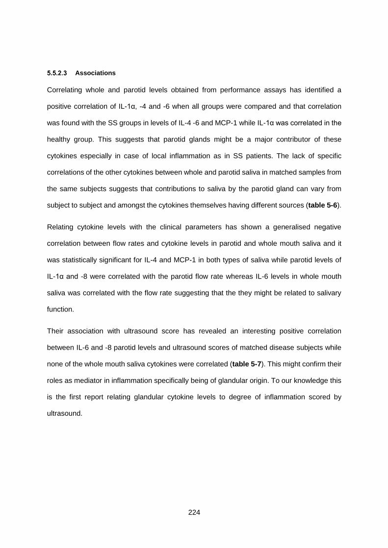
224
Associations
Correlating whole and parotid levels obtained from performance assays has identified a
positive correlation of IL-1α, -4 and -6 when all groups were compared and that correlation
was found with the SS groups in levels of IL-4 -6 and MCP-1 while IL-1α was correlated in the
healthy group. This suggests that parotid glands might be a major contributor of these
cytokines especially in case of local inflammation as in SS patients. The lack of specific
correlations of the other cytokines between whole and parotid saliva in matched samples from
the same subjects suggests that contributions to saliva by the parotid gland can vary from
subject to subject and amongst the cytokines themselves having different sources (table 5-6).
Relating cytokine levels with the clinical parameters has shown a generalised negative
correlation between flow rates and cytokine levels in parotid and whole mouth saliva and it
was statistically significant for IL-4 and MCP-1 in both types of saliva while parotid levels of
IL-1α and -8 were correlated with the parotid flow rate whereas IL-6 levels in whole mouth
saliva was correlated with the flow rate suggesting that the they might be related to salivary
function.
Their association with ultrasound score has revealed an interesting positive correlation
between IL-6 and -8 parotid levels and ultrasound scores of matched disease subjects while
none of the whole mouth saliva cytokines were correlated (table 5-7). This might confirm their
roles as mediator in inflammation specifically being of glandular origin. To our knowledge this
is the first report relating glandular cytokine levels to degree of inflammation scored by
ultrasound.

225
ROC analysis
To know the possibility of using cytokines for monitoring SS, Roc curve analysis was done
and has demonstrated that the diagnostic accuracy of cytokine levels in both parotid and whole
mouth saliva had rather a generalised poor-fair ability in differentiating control subjects and
SS (overall and subgroups). However, IL-6 levels of parotid saliva had a good ability to
differentiate between controls and SS patients who developed MALT-L with 93.9% specificity
and a low sensitivity of 53% even a higher cut off in whole mouth saliva levels with a good
ability to differentiate between the same groups (CT vs. SS/MALT-L).
IL-1β, IL-1α, IL-4, IL-8 and MCP-1 have shown a similar good accuracy in differentiating
between the same groups, while only IL-4 and IL-8 could differentiate between controls and
SS at risk of developing MALT-L being good for IL-4 and even excellent for IL-8. Cytokine
levels could not differentiate between control groups and SS neither between SS subgroup
and those who are at risk or developed MALT-L (tables 5-8 and 5-9).
Despite having performed the test for diagnostic accuracy, the readings (cut- offs) should be
viewed with caution since the validity and accuracy has not been determined yet.

226
Cytokine array system and bead-based assays might become powerful tools to extensively
describe the SS cytokine profile and to assess the role of these proteins in the pathogenesis
of the disease. Reliable quantification of a cytokine is crucially dependent on its abundance
which is true for sensitive performance assay where different concentrations were noted
between different types of samples. On the other hand, an important downside of bead-based
technologies such as Luminex is that by having predefined proteins of interest, researchers
may introduce a major selection bias into their study but this was only partially true for our
study where panels were customised based on selected cytokines and following reviewing the
literature (Maecker et al., 2012).
A lot of research has been published in the last decade linking SS with cytokines and
chemokines in human body fluids or in tissues. Although the mechanism(s) by which these
disease indicators come to exist in saliva has not been explained fully, our findings indicate
that saliva may represent a significant source of discriminatory biomarkers for local, systemic
inflammation in SS. However, it is important to note that lack of specificity of these molecules
for a given disease; being pleiotropic, exhibiting a very similar pattern in different clinical
circumstances and their rapidly changing levels are the major limitations of cytokine
measurement in practical clinical setting. Nonetheless these cytokines would be very useful
in preliminary stratification of SS patients for clinical trials for example allowing the exploration
of an immune, an inflammatory reaction or even a precancerous and cancerous involvement
where it takes place such as in parotid glands in SS patients. Furthermore, it ought to enhance
the design of special, individualized, cytokine targeted therapy for these patients (Bienvenu et
al., 2000). It would be interesting to explore the cytokine levels of the other salivary glandular
origins for comparisons in further studies. In addition, correlating the salivary and serum levels
would be of great importance.

227
Furthermore, tests using ELISAs where each cytokine is measured individually could be
undertaken in future studies. However it is worth noting that some studies have shown a good
association between the two techniques (multiplex and ELISAs), at least for some cytokines
(dupont et al., 2005, Dossus et al., 2009). While some have not (Liu et al., 2005). Similarly to
inconsistencies existing between ELISA kits from different companies due to their differences
in antibody pairs (dupont et al., 2005); a low association between these two techniques may
be due to the different antibodies used as well. Even though the possibility of using ELISA
assays would have identified additional associations between SS and cytokines measured via
screening assays, in particular those with levels below the limit of detection of the assay, i.e.,
IL-5, -10, -17, and IL-12p70, it should be noted that if high-sensitivity assays (performance)
were used for these cytokines they might have been detected and this warrants further
investigation. Still it is not clear that current ELISA assays would have been better able to
measure the very low levels of these cytokines than the multiplex assay used, a comparison
between both assays would be of great benefit.

228
Problems encountered with methods
It was planned to evaluate the levels of the rest of the cytokines/chemokines found in the
literature with the intention of focusing on BAFF, SDF-1α, and BCA-1 due to their relevance
in MALT-L via Luminex screening assays. However, the results obtained from the screening
assays needed further investigation thus it was decided to proceed with the performance
assays instead of doing more screening assays and it would be of great importance to study
the rest of the cytokines in parotid saliva in future studies.
An attempt to extrapolate the standard curve for Luminex screening assays to involve
cytokines with readings below the lower limit of detection according to the manufacture’s
recommended dynamic range appeared to have overestimated the differences between the
groups thus it was omitted and the usual protocol (standard curve) was followed. Another
attempt to evaluate the accuracy of the results obtained from the Luminex screening assays
via spike and recovery tests was unsuccessful (data not shown). It is assumed that some
precision is sacrificed in exchange for more flexibility in panel creation with a broader selection
of analytes (20 cytokines). Despite that it is argued that the sample values generated between
different lots of screening assays will be consistent to each other and would therefore be a
useful tool for longer term studies.
Upon selecting the cytokines for the final Luminex performance assays, IP-10 required an
additional panel which was not possible due to sample volume restriction thus it would be of
great importance to measure it in future studies.
Our preliminary trials (data not shown) where some mucus plugs (from parotid glands) were
tested have shown no results. Those samples were analysed in different condition; either
diluted and centrifuged with ceramic beads or filtered, yet again no results were obtained, a
possible explanation for some samples was due to their small volume thus they became over
diluted or fixed on filter papers as for other samples it might be that they needed even more
treatment. Therefore, it would be most interesting to find a way to treat these samples and
successfully analyse them in future studies.

229
Chapter 6: General discussion and future work General discussion ............................................................................................. 230
Clinical markers ........................................................................................... 230
Sialochemical biomarkers ............................................................................ 232
Future work ......................................................................................................... 237
Conclusion .......................................................................................................... 240

230
6 General discussion and future work
General discussion
In Sjögren’s syndrome, functional assessments such as salivary flow and ultrasound are
amongst the inter-related factors which describe glandular hypofunction. Because it
represents an ideal milieu for diagnosing a disease that affects the salivary glands, there is a
great interest towards using saliva itself as a diagnostic fluid for SS. Parotid saliva is of great
importance in cases of longer disease duration and those with or at risk of MALT-L. Although
some of these clinical parameters and salivary biomarkers have been investigated before,
researches have focused on one clinical aspect at a time and involved whole saliva rather
than individual glandular saliva, this thesis has explored these clinical parameters and
biomarkers and comparisons were done between whole and parotid gland saliva. The novelty
of this thesis relies on analysing different SS subgroups and comparing them to a SNOX
disease control group which has not commonly been reported by other studies. MALT-L is one
of the most serious manifestations of SS thus identifying/predicting it is of great significance
and this was examined throughout the thesis where patients who were defined as being at
risk of developing lymphoma and those who had already developed it were compared to SS
patients.
The overall aim of this thesis was to enable a better understanding of the disease activity and
progression via identifying different biomarkers whether they were clinical or laboratory/
sialochemical biomarkers.
Clinical markers
Chapter 3 investigated the potential use of the salivary gland assessment tests (WFR, PFR,
CODS and USS) in discriminating SS and non- SS sicca patients as well as to differentiate
between the different subgroups of SS and determine whether there is an overall association
between them. All parameters of the overall SS group showed a significant difference when
compared to disease controls attributed mainly to the advanced subgroups of SS (SS at risk
and MALT-L groups) which showed even differences when compared to the SS subgroup

231
mainly the MALT-L group in all parameters while at risk group was showing a difference in
USS and WFR but not the others. No differences were detected between SS subgroup and
disease control group regarding WFR and PFR which is in agreement with other studies (van
den Berg et al., 2007, Osailan et al., 2012, Ohyama et al., 2015, Billings et al., 2016).
Differences were detected between SS subgroup and disease controls regarding CODS
similarly to Osailan et al., (2012) and USS as well and the latter is a novel finding. All of the
parameters showed no differences between at risk and MALT-L groups. There was a strong
negative correlation observed between CODS and flow rates and a positive correlation with
USS suggesting the reliability of using CODS as a sensitive tool that can be easily done and
this was supported by another study (Osailan et al., 2012). The positive correlation between
WFR and PFR was found which is a similar finding to Vissink et al., (2003) and Kalk et al.,
(2002a), while it contrasted other studies (Kalk et al., 2001a, Kalk et al., 2002c, van den Berg
et al., 2007). These studies have found no relation between flow rates attributed to the
involvement of parotid glands in the later stages but in our study a lot of the patients were
considered at their later stages and the correlation was stronger in case of the advanced
stages of SS. A moderate negative correlation was noted between both flow rates and USS
reflecting an association between the output of the gland and its clinical picture. The diagnostic
accuracy of USS and its relation to focus score of biopsy results was investigated. It can be
concluded that USS would be the ideal non-invasive test to discriminate and monitor SS
patients in general (showing high sensitivity and specificity) replacing sialography and
scintigraphy with a good correlation and agreement with focus score in concordance to others
(Cornec et al., 2013, Delli et al., 2015, Jousse-Joulin et al., 2016, Astorri et al., 2016). The
present study suggested a score of 4 was found to be an optimum cut- off that could
differentiate SS from non- SS patients while higher scores usually reflecting advanced stages.
A longitudinal study on dry mouth patients at 5 and 10-year time-points was completed to
investigate worsening in salivary function over time. In patients with a longer disease duration
it SS is a slowly progressing disease and this was proven in this follow- up study where all of
the parameters remained relatively the same over the period of 5 years although some

232
individuals may display flare ups and remissions as in other rheumatic diseases which is in
agreement with other studies (Bouma et al., 2003 , Theander et al., 2005, Pijpe et al., 2007,
Haldorsen et al., 2008). The small number of patients involved in the 10-year follow- up study
precludes definitive conclusions but the result was in agreement with other studies showing
no significant differences between time 0 and 10 years later (Gannot et al., 2000, Haga, 2002).
Although these clinical markers might be able to differentiate between different SS subgroups
at one point (cross-sectional), the lack of change in clinical parameters in the longitudinal study
suggests that these clinical biomarkers may give limited utility in identifying patients at risk or
having MALT-lymphoma.
Sialochemical biomarkers
Initially a proteomics analysis of saliva was performed since this approach can identify novel
potential protein biomarkers which might differentiate SS patients from those who are at risk
of developing MALT-L and discriminating both disease groups from healthy controls. The
proteomics data generated by LC-MS/MS analysis of both runs suggested that a range of
proteins were up-regulated and down-regulated when different subgroups of SS were
compared to healthy controls; focusing on the logged transformed cut- offs and the up-
regulated proteins in both disease groups (SS and SS at risk) when compared with healthy
controls, Actin cytoplasmic 2, Ig γ-1 chain C region, S100-A8, and S100- A9 were selected
having considerable potential as biomarkers as reported previously (Al-Tarawneh et al., 2011,
Katsiougiannis and Wong, 2016). Further selection of candidate biomarker was based on
having cut- off of ≥ 2 when SS patients were compared to SS patients at risk in both runs, thus
S100-A8, and S100- A9 were the final selection and S100A8/A9 heterodimer was chosen.
Candidate biomarker was verified by ELISA; stratification of different stage of SS (SS, at risk
and MALT-L subgroups) as well as differentiating them from healthy and disease control
(SNOX) groups. S100A8/A9 ELISA analysis of parotid saliva supported the proteomics
findings showing significant differences between overall SS group and both disease and
healthy controls. Although subgrouping SS has revealed that only SS patients at risk and those

233
who already developed MALT-L were increased when compared to healthy controls only but
not disease controls while SS subgroup has shown no significant difference. S100A8/A9
ELISA analysis of whole mouth saliva has revealed a no significant difference in the overall
SS group when compared to both healthy and disease control groups showing similar values
to Muller et al., (1993) and Sweet et al., (2001). Similar trends were found by another group
who reported values that were 10 fold higher than ours which might be due to different sample
collection and handling (Cuida et al., 1993, Brun et al., 1994, Cuida et al., 1995, Cuida et al.,
1997). Only SS who already developed MALT-L subgroup has shown a difference when
compared to both healthy and disease control groups as well as to SS sub group suggesting
its important link to cancer development (Salama et al., 2008, Bresnick et al., 2015).
Investigation of the association between S100A8/A9 and overall salivary gland function
revealed a weak negative correlation between PFR and S100A8/A9 parotid levels. However,
no association was found between WFR and S100A8/A9 levels in whole saliva which may be
due to other contributions to levels in WMS. USS showed no correlation to both saliva (parotid
and whole) levels. A strong correlation was found between parotid and whole saliva levels of
S100A8/A9 in SS patients suggesting a salivary gland origin or neighbouring tissue source
since the glands are the major site of inflammation in this group. On the other hand, controls
showed no correlation. Proteomics is less effective in identifying cytokine biomarkers which
tend to be present at lower levels in saliva and other biofluids. Therefore, potential parotid
salivary cytokine biomarkers were examined using a cytokine antibody array approach to
identify SS sub groups (SS, SS at risk and already developed MALT-L) and to differentiate
them from healthy controls. Using a cut- off of 1.5 fold change and p- value, a range of
cytokines were up- regulated when SS, SS at risk and those who developed lymphoma were
compared to each other and to healthy controls (9 cytokines/proteins and 5 cytokines were
included). Further screening of the selected cytokines as well as other cytokines (relevant to
literature) was performed and levels were compared in SS subgroups to healthy and disease
controls (SNOX) via bead- based multiplex antibody assays. Most of the cytokines were at
the lower end of detection showing no differences between the groups but IL-1α, -4, -6 and

234
MCP-1 showed differences between the overall SS group and SNOX while only IL-6 was
increased in comparison to healthy controls as well. The cytokines have shown differences
with controls when SS was subgrouped as well as for IL-8. No significant difference was found
between all of SS subgroups, a tendency toward increased levels of salivary cytokines was
noted in MALT-L sub group. Verification of the previously significant cytokines while adding
one more cytokine (IL-1β) from the non-significant group (total 6 cytokines) using more
sensitive assays (bead- based performance) on a slightly larger number of samples yet similar
groups (healthy, disease controls and all three SS subgroups) was done with comparisons
between parotid levels and whole mouth levels. Some cytokines such as IL-6, -4 continued
to show significant difference compared to SNOX and IL-6 compared to healthy group as well.
Interestingly IL-1β showed significant increase of the overall SS group. Subgroups of SS
showed no differences when compared to other control groups or between each other,
however, a tendency toward increased levels was observed in MALT-L subgroup. All cytokine
levels in whole mouth saliva were significantly increased in the overall SS group compared to
healthy control this time while only 2 cytokines were increased in comparison to SNOX (IL-4
and MCP-1). Subgrouping of SS has revealed several differences especially in the MALT-L
subgroup. Despite using different techniques a number of studies reported similar findings
(Fox et al., 1994, Grisius et al., 1997, Tishler et al., 1999, Bertorello et al., 2004, Nguyen et
al., 2008, Lee et al., 2010, Kang et al., 2011, Moriyama et al., 2012, Ohyama et al., 2015 ).
The association between those 6 cytokines with overall salivary gland function as well as
between its parotid and whole saliva levels was investigated and generalised negative
correlation between flow rates and cytokine levels in parotid and whole mouth saliva was
observed. A positive correlation between ultrasound scores and IL-6 and -8 levels in parotid
saliva was noted while none of the whole mouth saliva cytokines were correlated suggesting
that parotid cytokines were mediators in inflammation specifically of glandular origin. Some
parotid and whole salivary cytokines levels correlated while others did not suggesting that
contributions to saliva by the parotid gland can vary from subject to subject and that glands
might be a major contributor of correlating cytokines especially in case of local inflammation

235
as in SS patients ,which might be linked with disease activity in different subjects. It is worth
noting that levels of cytokines and S100-A8/A9 were detected in patients with previous
lymphoma, in most cases in complete remission. This increase in saliva level could be, at least
in part, not related to the activity of the lymphoma, but could be constitutional and persist after
the lymphoma was cured which could be genetically linked. The diagnostic accuracy for each
test mentioned in all of the chapters was done establishing a test threshold for disease
detection by providing test sensitivity and specificity. Therefore, in this study we used Receiver
Operating Characteristic (ROC) curve in order to determine the best cut-off point for a given
biomarker values to generate an optimum cut-off that provides a maximum sensitivity and
specificity. The differences between the levels of different markers measured in parotid saliva
could not be attributed to an increased protein concentration in patients with deficient saliva,
because their protein concentrations were similar in all groups (figure 4-11, chapter 4).
Although protein concentration of whole mouth saliva did differ between the groups (especially
SS with MALT-L) (figure 4-11) a comparison of protein S100A8/A9 levels between the groups
was done after adjusting the values to ng/mg and showed no major differences between those
obtained via ng/ml. Cytokine values were not adjusted thus their levels for MALT-L group might
be related to the high total protein concentrations of saliva and possibly reduced fluid
secretion. The biomarker studies have a limitation since patients may have been under
immunosuppressor therapy. This notwithstanding, the studies showed that no difference was
found between treated and untreated patients but it might be due to the insufficient power of
the study to determine an effect of drugs in this rather small and heterogeneous group of
patients. However, the difference between treated and untreated patients was not verified
regarding the clinical parameters (chapter 3). Studies that have been carried in this thesis
were the first that have compared the levels of salivary cytokines and S100-A8/A9 in different
SS subgroups; at risk and those who developed MALT-L in SS patients as well as in SNOX
patients, that contribute to a better understanding of disease activity and progression. The
applicability of sialometry and sialochemistry as diagnostic instruments varies in different

236
clinical SS subgroups. While the former might be informative in case of advanced disease,
the latter is often useful in the early phases of the disease.
In the present studies, we have focused on the advanced subgroups of SS indicating that both
sialometry and sialochemistry are useful tools; proteomic analysis, cytokine array system and
screening bead-based assays are becoming powerful tools to extensively describe the SS
protein/cytokine profile assessing their role in SS pathogenesis. While ELISA remains one of
the gold standard tests in analysing biological samples, multiplex assays might offer several
advantages as well exhibiting a good correlation with ELISA. One of the more captivating
aspects in the search of salivary biomarkers is information achieved from oral saliva and
immunologic factors. Therefore, we found it of importance to study the salivary glands and
their secretions to better understand the inflamed processes involved. Although the
mechanism by which these disease markers come to exist in saliva has not been clarified fully,
our findings suggest that it may represent a significant source of discriminatory biomarkers for
Sjögren’s syndrome. Yet further investigations should be addressed.

237
Future work
This thesis has shed light on the potential use of multiple biomarkers in differentiating SS
subgroups. It can be taken further by focusing on the patients at risk group in order to
understand the mechanisms behind the observed compositional changes. Furthermore, an
assessment of a possible relationship between individual salivary components such as the
SA100A8/A9 and candida count might provide a further insight as to whether this biomarker
is of significant functional relevance.
Moreover, the results of this thesis give rise to further areas that need investigation:
1. Further analysis of the variations in the parameters (WFR, PFR, CODS and USS)
assessed in this thesis are necessary and the inter- and intra- individual and observer
variability should be considered in order to estimate whether differences observed are
the result of individual variations of the examination technique or real individual
variations.
2. The diagnostic accuracy of USS should be investigated on a larger cohort of patients
and further its correlation with histology especially major salivary gland biopsy
assessed in order to further reinforce results obtained from the present study indicating
its usefulness as a simple non-invasive test. Different USS scoring systems should be
compared in order to develop a consensus with consistent procedures.
3. Further follow- up longitudinal studies of greater than 10 years on larger group of
patients are required in order to establish whether there is functional loss over time.
4. Further studies of more SNOX patients compared with SS are required in order to
further verify it as a distinct disease entity.
5. Considering the presence of other disease modifying factors such as the effect of
treatment on the outcome measures; differences between treated and untreated
patients should be studied fully. Among the most used treatments; steroids,
immunosuppressants and hydroxyquinoline would be included. Patients would be
categorised according to the duration of treatment. Standardization of sample

238
handling, collection and processing is still required for further studies and special
attention to the effect of centrifugation would be of great importance.
6. Further analysis with different proteomic methods would achieve more reliable results
especially for validating salivary biomarkers an example of which would be Tandem-
mass-tagging or labelling methods. An integrated proteomic analysis of the glandular
tissue or another exocrine secretion such as tears should be performed and compared
with saliva to investigate the differences between their pathological involvements.
7. The relationship of salivary S100A8/A9 with the focus score and possibly tear fluid
S100A8/A9 levels should be clarified to better understand SS glandular disease
activity as well as analysing other glandular salivary S100A8/A9 levels (e.g.
submandibular/sublingual glands) in order to identify the possible analyte origins and
to compare the differences between the glandular secretions. Individual levels of S100-
A8 and -A9 should be analysed in order to determine their relative abundance in
relation to the heterodimer of both.
8. Performing cytokine array tests on whole mouth saliva with comparison to results
obtained from the multiplex bead- based assays would give some insight about the
reliability of these tests as first-choice screening tests. Re-testing cytokine levels
(showing low levels or 0 values) which were tested via screening bead- based assays
via more sensitive bead-based assays to ascertain their levels. Testing the accuracy
and reproducibility of bead- based assays is required via spike and recovery tests in
order to verify the applicability of using these tests for further research. Special interest
in measuring BAFF, SDF-1α, and BCA-1 levels in parotid saliva would be of great
importance since they were frequently linked to advanced subgroups of SS. Re-
measuring chemokine levels especially CXCL10 (above the detection limit) would
provide further insight to the extent of chemokine involvement. Considering other
salivary/exocrine sources of cytokines will further aid in understanding of their
pathogenic roles. Re-analysing the same tests specifically ELISA and bead-based

239
performance tests could be performed on a larger cohort of patients in order to improve
the reliability of these tests and demonstrate their utility.
9. It may well be that a multifactorial model including salivary biomarkers and clinical
parameters may have better accuracy than the result of a salivary test alone in order
to relate the pathologic process with the clinical picture of this disease where saliva
would be the most logical sample for correlation.
10. Important consideration would be proper testing and handling of mucoid plugs since it
was found in a considerable number of patients specially those who are with longer
disease duration, at risk or already developed MALT-L, these plugs would provide
valuable information once analysed since they are not normally secreted by parotid
glands.

240
Conclusion
In conclusion, it is known that SS is a slowly progressing disease and the present follow- up
study has shown that all of the clinical parameters remained relatively the same over a period
of time in patients with longer disease duration. The data implicate a greater role of these
clinical parameters in differentiating SS subgroups from other dry mouth patients especially
the advanced SS subgroups (at risk and who developed MALT-l). Furthermore, it can be
concluded that USS would be the ideal non-invasive test to discriminate and monitor SS
patients in general especially in the advanced subgroups.
The aim of developing salivary diagnostics remains an important goal which offers a number
of potential applications. The present study evaluated the use of some potential salivary
biomarkers (cytokines and SA100A8/A9). This study supports previous reports indicating the
value of saliva as a diagnostic tool in Sjögren’s syndrome. Salivary levels of S100A8/A9 reflect
more local inflammatory activity showing the greatest difference between groups in parotid
saliva. Salivary cytokine levels were not clear as with the S100A8/A9 levels, no pattern could
be drawn from the changes in cytokine levels. However, levels of IL-6 and -4 in parotid saliva
continued to show significant differences using different immune-based assays. The levels of
cytokines tend to show more prominent differences in whole mouth saliva due to the
contribution from their other sources. Further work is required particularly to improve the
sensitivity of their diagnostic applications and to explore further combining these results with
the clinical parameters that are part of the routine clinical assessment of SS.

241
Appendices
Appendix 1: The Challacombe Scale ............................................................................. 242
Appendix 2: Sjögren's Ultrasound Score template ......................................................... 243
Appendix 3: American-European Consensus Group (AECG) criteria ............................. 244
Appendix 4: Unstimulated Whole Saliva Flow Rate and Collection ................................ 246
Appendix 5: Stimulated Parotid Flow Rate and Collection ............................................ 247
Appendix 6: Human S100A8/S100A9 Heterodimer Quantikine ELISA protocol: ............ 248
Appendix 7: Proteome Profiler Human XL Cytokine Array protocol: .............................. 249
Appendix 8: Luminex Human Magnetic Assay protocols: .............................................. 250
Appendix 9: Preliminary proteins identified by LC-MS/MS proteomics analysis and
selected per fold change ratio (first run) ......................................................................... 251
Appendix 10: Preliminary proteins identified by LC-MS/MS proteomics analysis and
selected per fold change ratio (second run) ................................................................... 252
Appendix 11: Statistical (SPSS) analysis of S100A8/A9 (Calprotectin, CalpP) levels in
parotid saliva (overall SS and control groups) ................................................................ 253
Appendix 12: Statistical (SPSS) analysis of S100A8/A9 (Calprotectin, CalpP) levels in
parotid saliva (SS sub groups and control groups) ........................................................ 256
Appendix 13: S100A8/A9 levels in (A) parotid and (B) whole mouth saliva of SS treated
and untreated groups .................................................................................................... 260
Appendix 14: Summary of the second ranked cytokines-proteins ordered by fold change
and p-value when compared to controls (proteome profiler) .......................................... 261
Appendix 15: Summary of the second ranked cytokines-proteins ordered by fold change
and p-value when compared to SS (proteome profiler) .................................................. 262
Appendix 16: Summary of the third ranked cytokines-proteins ordered by fold change and
p-value when compared to controls (proteome profiler) ................................................. 264
Appendix 17: Summary of the third ranked cytokines-proteins ordered by fold change and
p-value when compared to SS (proteome profiler) ......................................................... 265
Appendix 18: Statistical SPSS analysis of IL-6 levels in parotid saliva (SS sub groups) 266
Appendix 19: Analytes excluded from further validation via Luminex screening assay . 269
Appendix 20: Cytokines identified previously in serum of SS subjects using different
methods ........................................................................................................................ 270
Appendix 21: Cytokines identified previously in serum and salivary glands of SS subjects
using different methods ................................................................................................. 272
Appendix 22: Cytokines identified previously in salivary glands of SS subjects using
different methods ........................................................................................................... 273
Appendix 23: Cytokines identified previously in saliva of SS subjects using different
methods ........................................................................................................................ 275
Appendix 24: Cytokines identified previously in parotid saliva of SS subjects using different
methods ........................................................................................................................ 277
Appendix 25: Summary of the review of literature on cytokines ..................................... 278

242
Appendix 1: The Challacombe Scale
Clinical oral dryness scale to derive CODS assessed as mild, moderate or severe (http://www.dentalhealth.org/uploads/download/resourcefiles/download_68_1_The%20Challacombe%20Scale.pdf )

243
Appendix 2: Sjögren's Ultrasound Score template

244
Appendix 3: American-European Consensus Group (AECG) criteria
Revised international classification criteria for Sjögren’s syndrome I. Ocular symptoms: a positive response to at least one of the following questions:
• Have you had daily, persistent, troublesome dry eyes for more than 3 months?
• Do you have a recurrent sensation of sand or gravel in the eyes?
• Do you use tear substitutes more than 3 times a day?
II. Oral symptoms: a positive response to at least one of the following questions:
• Have you had a daily feeling of dry mouth for more than 3 months?
• Have you had recurrently or persistently swollen salivary glands as an adult?
• Do you frequently drink liquids to aid in swallowing dry food?
III. Ocular signs: that is, objective evidence of ocular involvement defined as a positive result for at least one of the following two tests:
• Schirmer’s I test, performed without anaesthesia (<5 mm in 5 minutes)
• Rose bengal score or other ocular dye score (>4 according to van Bijsterveld’s scoring system)
IV. Histopathology: In minor salivary glands (obtained through normal-appearing mucosa) focal lymphocytic sialoadenitis, evaluated by an expert
• histopathologist, with a focus score >1, defined as a number of lymphocytic foci (which are adjacent to normal-appearing mucous acini and contain more than 50 lymphocytes) per 4 mm2 of glandular tissue
V. Salivary gland involvement: objective evidence of salivary gland involvement defined by a positive result for at least one of the following diagnostic tests:
• Unstimulated whole salivary flow (<1.5 ml in 15 minutes)
• Parotid sialography showing the presence of diffuse sialectasias (punctate, cavitary or destructive pattern), without evidence of obstruction in the major ducts
• Salivary scintigraphy showing delayed uptake, reduced concentration and/or delayed excretion of tracer.
VI. Autoantibodies: presence in the serum of the following autoantibodies:
• Antibodies to Ro(SSA) or La(SSB) antigens, or both

245
Appendix 3 (continue)
(Vitali et al., 2002)
Revised rules for classification For primary SS In patients without any potentially associated disease, primary SS may be defined as follows: a. The presence of any 4 of the 6 items is indicative of primary SS, as long as either item IV (Histopathology) or VI (Serology) is positive b. The presence of any 3 of the 4 objective criteria items (that is, items III, IV, V, VI) c. The classification tree procedure represents a valid alternative method for classification, although it should be more properly used in clinical-epidemiological survey
For secondary SS In patients with a potentially associated disease (for instance, another well-defined connective tissue disease), the presence of item I or item II plus any 2 from among items III, IV, and V may be considered as indicative of secondary SS

246
Appendix 4: Unstimulated Whole Saliva Flow Rate and Collection
Materials:
Balance
Pre-weighed and labelled 20 ml tubes and lids
Clock or timer
1. The unstimulated whole saliva collection procedure should be done before the stimulated
parotid collection procedure.
2. During the collection period, the subject shall be seated straight up with eyes open and head tilted slightly forward.
3. The subject will be instructed to minimize oro-facial movements to minimize influence on
salivary flow (the subject should not swallow and should not speak during the collection process).
4. Then the patient allows the saliva to accumulate in the floor of the mouth for 60 seconds
without swallowing.
5. The patient empties the entire accumulated saliva into the pre-weighed container for 10 minutes.
6. The collection vial is weighed both before and after collection, and both pre- and post-
collection weights are recorded.
7. Calculate flow rate on a ml/minute basis.
For detailed information please follow the link provided below
(https://sicca-online.ucsf.edu/documents/Oral-Saliva-SOP.pdf)

247
Appendix 5: Stimulated Parotid Flow Rate and Collection
Materials:
Pre-weighed and labelled 20 ml tubes and lids
2% Citric acid solution (2 g in 100 ml sterile water, kept refrigerated) (2 ml)
Parotid saliva collector (lashley cup)
Tygon tubing (10.2 cm for the collecting tube from the inner circle of the parotid saliva collector, and 30.5-40.6 cm to connect the parotid collector to syringe)
10 ml syringe
Medium Binder Clip (clamp)
Ice and container
Timer or clock
Balance
1. The orifice of the parotid duct is located on the buccal mucosa opposite the upper second molar tooth; dry the area with gauze for better vision.
2. The parotid collector is placed on the mucosa so that the inner ring surrounds the duct orifice.
3. The collector is held on the mucosa by suction from the outer ring, created by pulling back on the syringe and allowing the pressure to come to equilibrium.
a) Syringe can then be rested on the patient’s shoulder. b) A medium binder clip is attached to the tygon tubing going from the collector to the
syringe to “lock-in” the air in the tubing. The suction created should be sufficient to hold the cup in place without occluding the inner chamber of the parotid collector with tissue (i.e., not too much suction).
4. Saliva from the parotid gland then flows passively into the inner ring and through the attached tubing into pre-weighed tube.
5. 2% citric acid solution is applied to the posterior lateral surfaces of the tongue, bilaterally, using 2 drops every 30 seconds to stimulate secretion.
6. Flow may not begin for a minute or two after stimulation has been applied.
7. A maximum of 5 minutes is allowed for saliva to appear in the clear portion of the tubing.
8. If saliva flow is observed during this 5-minute period, an additional 10 minutes is allowed for the saliva to reach the end of the tubing and the 10-minute collection period begins when saliva begins to exit the Tygon tube.
9. Calculate flow rate on a ml/minute basis.
For detailed information please follow the link provided below
(https://sicca-online.ucsf.edu/documents/Oral-Saliva-SOP.pdf)

248
Appendix 6: Human S100A8/S100A9 Heterodimer Quantikine ELISA
protocol:
(cat. no. DS8900)
All reagents and samples were brought to room temperature and prepared as directed in
chapter 2 subsection 2.2.5.
Fifty μL of Assay Diluent RD1-34 was added to each well. Then 50 μL of Standard and sample
is added in duplicates and incubated for 2 hours at room temperature on a horizontal orbital
microplate shaker (0.12" orbit) set at 500 ± 50 rpm.
After washing (four times), 200 μL of Human S100A8/S100A9 Heterodimer Conjugate is
added and incubated for another 2 hours at room temperature on the shaker.
Subsequently another wash was done and 200 μL of Substrate Solution is added and
incubated for 30 minutes at room temperature on the benchtop protecting it from light.
The colour is stopped by the addition of 50 μL of Stop Solution to each well.
The optical density is determined using a microplate reader set to 540 nm at first and
subtracted from reading set at 450. This subtraction will correct for optical imperfections in the
plate.

249
Appendix 7: Proteome Profiler Human XL Cytokine Array protocol:
(cat. no. ARY022)
All reagents and samples were brought to room temperature and prepared as directed in chapter 2 subsection 2.2.6. A summary of the protocol is provided below
The membranes are ready for the Chemidoc followed by ImageJ for measurement.
Pipet 2 mL of Array Buffer 6 into each well of the 4-Well Multi-dish. (Array Buffer 6 serves as a block buffer)
Place each membrane in a separate well. (The number on the membrane should be facing upward)
Remove membranes and place into individual containers (plates) with 20 mL of 1X Wash Buffer (Rinse the 4-well multi-dish with distilled water and dry)
Wash each membrane with 1X Wash Buffer for 10 minutes on a rocking platform shaker (three times)
Add 30 μL of Detection Antibody Cocktail to 1.5 mL of 1X Array Buffer 4/6 for each array Pipette 1.5 mL per well of diluted Detection Antibody Cocktail into the 4-Well Multi-dish
Remove membranes from their washing plates and put back in the dish
Remove membranes and repeat the wash step Pipette 2 mL of 1X Streptavidin-HRP into each well of the 4-Well Multi-dish.
Remove membranes and repeat the wash step Place membranes on the bottom sheet of plastic protector
Pipette 1 mL of the prepared Chemi Reagent Mix evenly onto each membrane Cover with the top sheet of plastic protector
Squeeze out the reagent mix Leave membranes on bottom sheet
Cover with plastic wrap on top and around the back Smooth out air bubbles
Aspirate Array Buffer 6 from the wells and add the diluted samples.
Incubate for one hour on a rocking platform shaker
Incubate overnight at 2-8 °C on a rocking platform shaker
Incubate for one hour on a rocking platform shaker
Incubate for 30 minutes at room temperature on a shaker
Incubate for one minute.

250
Appendix 8: Luminex Human Magnetic Assay protocols:
Screening Human Magnetic Assay protocol:
(Premixed multi-analyte kit) (cat. no. LXSAHM)
Microparicle Cocktail (beads) preparation is done by vial centrifugation, vortexing then diluting
(250 μL of the cocktail + 2.5 mL Diluent RD2-1). Biotein antibody cocktail is treated similarly.
Streptavidin-PE is diluted with wash buffer (110 μL+ 2.7 mL).
After sample /standards /reagents preparation the beads are re-suspeneded before adding
them to the wells (25 μL). Samples and standards are then added (25 μL) and incubated on
shaker at room temperature for two hours. The wells are washed using magnetic device
intended to accommodate the plate (three times). Diluted antibody cocktail is then added (25
μL) and incubated for one hour on the shaker. Wash step is repeated and followed by the
addition of diluted streptavidin-PE (25 μL) with an incubation period of 30 minutes. Wash step
is repeated and the beads are re-suspended by adding 100 μL of wash buffer to each well
and incubated for two minutes on the shaker. The plate is now ready to read.
Performance Human Magnetic Assay protocol:
(Cytokine kit A) (cat. no. FCSTM03)
In general, it is the same as the screening assay with different volumes, diluents (more
sensitive) and incubation time. Bead preparation is mentioned before; the dilution is (12.5 μL
from each microparticle vial + 2.5 mL microparticle diluent). Biotein antibody cocktail is
prepared likewise with its relevant diluent. Streptavidin-PE is diluted with wash buffer (30 μL+
3 mL). The first incubation period of beads and samples/standards is three hours. The
following steps are the same as before.
.

251
Appendix 9: Preliminary proteins identified by LC-MS/MS proteomics analysis
and selected per fold change ratio (first run)
CT; healthy control, SS; Sjögren’s syndrome, SS at risk of lymphoma of developing mucosa‐associated
lymphoid tissue. Proteins were present in all groups but up-regulated in disease sample, not present in
control sample or not present in control and SS samples. ND; not detected, *could not be calculated.
Highlighted are the common up-regulated proteins when SS was compared to control (n=6). Underlined
are the final proteins from which the selected proteins were further analysed via ELISA.
Total ion current
(TIC)
Fold change ratio
NO Selected proteins CT SS SS at risk
SS/CT SS at risk/
CT
SS at risk/
SS
1 Annexin A1 15860.7 73230 1513363.2 4.6 95.4 20.7
2 Actin, cytoplasmic 2 93043.9 1580633.9 3826055.5 17 41.1 2.4
3 Ig γ-1 chain C region 335437.5 2676308.9 13318238.8 8 39.7 5
4 Protein S100-A8 26224.7 299185.7 712901.1 11.4 27.2 2.4
5 Lactotransferrin 18742385.1 7150834.6 69818117 0.4 3.7 9.8
6 Thrombospondin-1 112670 723090 2951800 6.4 26 4.1
7 Thioredoxin ND 221771.9 139336.5 * * 0.6
8 14-3-3 protein
zeta/delta
ND 392907.5 384322 * * 1
9 Protein S100-A9 ND 616106.3 1301008.8 * * 2.1
10 Golgi apparatus
protein 1
ND ND 156208.9 * * *
11 Matrilysin ND ND 141751.1 * * *

252
Appendix 10: Preliminary proteins identified by LC-MS/MS proteomics analysis
and selected per fold change ratio (second run)
CT; healthy control, SS; Sjögren’s syndrome, SS at risk of developing lymphoma of mucosa‐associated
lymphoid tissue. Proteins were present in all groups but up-regulated in disease sample, not present in
control sample or not present in control and SS samples. ND; not detected, *could not be calculated.
Highlighted are the common up-regulated proteins when SS was compared to control (n=6). Underlined
are the final proteins from which the selected proteins were further analysed via ELISA.
Total ion current
(TIC)
Fold change ratio
NO Selected proteins CT SS SS at risk
SS/CT SS at risk /
CT
SS at risk/
SS
1 Annexin A1 10026 9689.43 1180842.4 1 117. 8 121.9
2 Actin cytoplasmic 2 210492.7 878628 850867.97 4.2 4.04 1
3 Ig γ-1 chain C
region
211505.2 7948508.2 7981574.9 37.6 37.7 1.004
4 Protein S100-A8 14250.3 119945.89 1149673.8 8.4 80. 7 9.6
5 Lactotransferrin 12328407.3 90109906.93 47019126.6 7.3 3.8 0.5
6 Protein S100-A9 89449.2 363782 4414986.4 4.07 49.4 12.1
7 Thioredoxin 43669.5 58756.5 511520.5 1.3 11.7 8.7
8 14-3-3 protein
zeta/delta
ND 11870.75 518438.4 * * 43.7
9 Thrombospondin-1 ND 1572522.32 462161 * * 0.3
10 Golgi apparatus
protein
ND 89355.6 38873.2 * * 0.4
11 Matrilysin ND 268531.1 85661.4 * * 0.3

253
Appendix 11: Statistical (SPSS) analysis of S100A8/A9 (Calprotectin,
CalpP) levels in parotid saliva (overall SS and control groups)
Case Processing Summary
GP11
Cases
Valid Missing Total
N Percent N Percent N Percent
CalpP CT 18 100.0% 0 0.0% 18 100.0%
SNOX 14 100.0% 0 0.0% 14 100.0%
SS 51 100.0% 0 0.0% 51 100.0%
Tests of Normality
GP11
Kolmogorov-Smirnova Shapiro-Wilk
Statistic df Sig. Statistic df Sig.
CalpP CT .347 18 .000 .585 18 .000
SNOX .321 14 .000 .661 14 .000
SS .303 51 .000 .741 51 .000

254
Descriptives
GP11 Statistic Std. Error
CalpP CT Mean 251.3780 117.22698
95% Confidence Interval for Mean
Lower Bound 4.0507
Upper Bound 498.7053
5% Trimmed Mean 174.4082
Median 31.8955
Variance 247358.954
Std. Deviation 497.35194
Minimum 0.00
Maximum 1888.21
Range 1888.21
Interquartile Range 273.19
Skewness 2.600 .536
Kurtosis 6.937 1.038
SNOX Mean 204.2639 74.24008
95% Confidence Interval for Mean
Lower Bound 43.8780
Upper Bound 364.6499
5% Trimmed Mean 167.1028
Median 208.9451
Variance 77162.253
Std. Deviation 277.78094
Minimum 0.00
Maximum 1077.43
Range 1077.43
Interquartile Range 265.30
Skewness 2.605 .597
Kurtosis 8.292 1.154
SS Mean 1968.8121 371.62781
95% Confidence Interval for Mean
Lower Bound 1222.3757
Upper Bound 2715.2485
5% Trimmed Mean 1721.5018
Median 743.1429
Variance ##########
Std. Deviation 2653.95340
Minimum 0.00
Maximum 8948.86
Range 8948.86
Interquartile Range 3435.00
Skewness 1.337 .333
Kurtosis .416 .656

255
GP; groups

256
Appendix 12: Statistical (SPSS) analysis of S100A8/A9 (Calprotectin,
CalpP) levels in parotid saliva (SS sub groups and control groups)
Case Processing Summary
GP22
Cases
Valid Missing Total
N Percent N Percent N Percent
CalpP CT 18 100.0% 0 0.0% 18 100.0%
SNOX 14 100.0% 0 0.0% 14 100.0%
SS 19 100.0% 0 0.0% 19 100.0%
AT RISK 18 100.0% 0 0.0% 18 100.0%
5.00 14 100.0% 0 0.0% 14 100.0%
Tests of Normality
GP22
Kolmogorov-Smirnova Shapiro-Wilk
Statistic df Sig. Statistic df Sig.
CalpP CT .347 18 .000 .585 18 .000
SNOX .321 14 .000 .661 14 .000
SS .318 19 .000 .647 19 .000
AT RISK .290 18 .000 .764 18 .000
5.00 .262 14 .010 .825 14 .010
a. Lilliefors Significance Correction

257
Descriptives
GP22 Statistic Std. Error
CalpP CT Mean 251.3780 117.22698
95% Confidence Interval for Mean
Lower Bound 4.0507
Upper Bound 498.7053
5% Trimmed Mean 174.4082
Median 31.8955
Variance 247358.954
Std. Deviation 497.35194
Minimum 0.00
Maximum 1888.21
Range 1888.21
Interquartile Range 273.19
Skewness 2.600 .536
Kurtosis 6.937 1.038
SNOX Mean 204.2639 74.24008
95% Confidence Interval for Mean
Lower Bound 43.8780
Upper Bound 364.6499
5% Trimmed Mean 167.1028
Median 208.9451
Variance 77162.253
Std. Deviation 277.78094
Minimum 0.00
Maximum 1077.43
Range 1077.43
Interquartile Range 265.30
Skewness 2.605 .597
Kurtosis 8.292 1.154

258
SS Mean 1163.8734 431.36932
95% Confidence Interval for Mean
Lower Bound 257.6001
Upper Bound 2070.1467
5% Trimmed Mean 871.7872
Median 506.0000
Variance 3535510.254
Std. Deviation 1880.29526
Minimum 0.00
Maximum 7585.30
Range 7585.30
Interquartile Range 946.86
Skewness 2.611 .524
Kurtosis 7.466 1.014
AT RISK Mean 2432.5382 728.53053
95% Confidence Interval for Mean
Lower Bound 895.4732
Upper Bound 3969.6033
5% Trimmed Mean 2205.6615
Median 666.8104
Variance 9553621.187
Std. Deviation 3090.89327
Minimum 0.00
Maximum 8948.86
Range 8948.86
Interquartile Range 5511.19
Skewness .989 .536
Kurtosis -.654 1.038
MALT Mean 2465.0097 764.94043
95% Confidence Interval for Mean
Lower Bound 812.4564
Upper Bound 4117.5631
5% Trimmed Mean 2279.3600
Median 937.4286
Variance 8191873.956
Std. Deviation 2862.14499
Minimum 0.00
Maximum 8271.71
Range 8271.71
Interquartile Range 4476.00

259
GP; groups

260
Appendix 13: S100A8/A9 levels in (A) parotid and (B) whole mouth saliva
of SS treated and untreated groups
No significant differences were detected between treated (whole/parotid) (n=19/38) and
untreated (whole/parotid) (n=7/13) patients (Mann–Whitney U test).
un
treate
d
treate
d
-5 0 0 0
0
5 0 0 0
1 0 0 0 0
S S g ro u p s
Co
nc
en
tra
tio
n (
ng
/m
L)
(A)
(B)

261
Appendix 14: Summary of the second ranked cytokines-proteins ordered by
fold change and p-value when compared to controls (proteome profiler)
Fold change in the first group (A) is ≥ 1.5 in all disease groups. In the second group (B) it is increased in 2 of the disease groups. While the third group (C) fold change is increased in one group only. NS; not significant, CT; healthy controls, SS; Sjögren’s syndrome, MALT-L; mucosa associated lymphoid tissue lymphoma. MCP-1 is significantly different when compared to controls (one- way ANOVA followed by Tukey HSD post hoc test). Second ranked proteins were identified in 11 samples of 12
SS vs. CT MALT-L risk vs. CT MALT-L vs. CT
Analytes (23/37)
Fold change
p- value
Fold change
p- value
Fold change
P- value
MIP-3α
2.4
NS
1.8
NS
6.4
NS
TFF3
2.9
NS
2.9
NS
3.5
NS
TNF-alpha
>-1.5, <1.5
NS
1.7
NS
3.1
NS
Pentraxin-3 >-1.5, <1.5 NS 1.6 NS 2.7 NS
SDF-1α >-1.5, <1.5 NS 1.6 NS 2.1 NS
TGF-α >-1.5, <1.5 NS 1.6 NS 1.6 NS
MIP-3β >-1.5, <1.5 NS 1.6 NS 5.5 NS
IL-2 >-1.5, <1.5 NS 1.5 NS 1.6 NS
I-TAC >-1.5, <1.5 NS 2 NS 4.6 NS
IL-27
-1.6
NS
1.7
NS
>-1.5, <1.5
NS
IL-32α/β/γ -2 NS 1.7 NS >-1.5, <1.5 NS
MCP-1 >-1.5, <1.5 NS 2.1 0.04* >-1.5, <1.5 NS
IL-23 >-1.5, <1.5 NS 2.1 NS >-1.5, <1.5 NS
IL-24 >-1.5, <1.5 NS >-1.5, <1.5 NS 1.5 NS
GRO-α >-1.5, <1.5 NS >-1.5, <1.5 NS 4.2 NS
Chitinase 3-like 1 >-1.5, <1.5 NS >-1.5, <1.5 NS 3.6 NS
MIP-1α/MIP-1β >-1.5, <1.5 NS >-1.5, <1.5 NS 2.3 NS
FGF basic >-1.5, <1.5 NS >-1.5, <1.5 NS 1.7 NS
BAFF >-1.5, <1.5 NS >-1.5, <1.5 NS 1.5 NS C-Reactive protein >-1.5, <1.5 NS >-1.5, <1.5 NS 2.9 NS
G-CSF -2 NS >-1.5, <1.5 NS 1.8 NS
Complement C5/C5a -1.9 NS >-1.5, <1.5 NS 2.3 NS
ICAM-1
-1.6 NS >-1.5, <1.5 NS 3.4 NS
(A)
(B)
(C)

262
Appendix 15: Summary of the second ranked cytokines-proteins ordered by
fold change and p-value when compared to SS (proteome profiler)
MALT-L risk vs. SS
MALT-L vs. SS
MALT-L vs. MALT-L risk
Analytes (34/37)
Fold change
p- value
Fold change
p- value
Fold change P- value
ICAM-1
2.7
NS
5.1
NS
2.4
NS
I-TAC 2 NS 4.6 NS 2.6 NS
MIP-3β 1.5 NS 5.4 NS 4 NS
C-Reactive protein 2.4 NS 4 NS 1.6 NS
Complement C5/C5a
2 NS 4.1 NS 2.1 NS
IL-27
3.3
NS
1.7
NS
-1.6
NS
IL-32α/β/γ 3.6 0.02* 2 NS -1.7 NS
MIP-1α/MIP-1β 2.6 NS 3.6 NS >-1.5, <1.5 NS
Dkk-1 3 NS 3.3 NS >-1.5, <1.5 NS
BDNF 2.5 NS 3.3 NS >-1.5, <1.5 NS
Angiopoietin-2 2.8 NS 3.1 NS >-1.5, <1.5 NS
TNF-alpha 1.5 NS 3 NS >-1.5, <1.5 NS
FGF basic 2.2 NS 2.5 NS >-1.5, <1.5 NS
IL-15 2.2 NS 2.5 NS >-1.5, <1.5 NS
IL-34 2 NS 2.2 NS >-1.5, <1.5 NS
BAFF 1.5 NS 2 NS >-1.5, <1.5 NS
IL-24 2.6 NS 2.8 NS >-1.5, <1.5 NS
G-CSF 3.4 NS 3.9 NS >-1.5, <1.5 NS
TARC 2.3 NS 2.1 NS >-1.5, <1.5 NS
Cripto-1 2.9 NS 2.6 NS >-1.5, <1.5 NS
TGF-α 2.3 NS 2.3 NS >-1.5, <1.5 NS
IL-12 p70 2.9 NS 2 NS >-1.5, <1.5 NS
GRO-α >-1.5, <1.5 NS 5.6 NS 4.3 NS
MIP-3α >-1.5, <1.5 NS 4 NS 4.6 NS
SDF-1α >-1.5, <1.5 NS 1.6 NS 2.1 NS
Chitinase 3-like 1
>-1.5, <1.5 NS 4.2 NS 3.5 NS
(A)
(B)

263
Appendix 15 (Continued)
Fold change in the first group (A) is ≥ 1.5 in all disease groups. In the second group (B) it is
increased in 2 of the disease groups. While the third group (C) fold change is increased in one
group only. NS; not significant, CT; healthy controls, SS; Sjögren’s syndrome, MALT-L; mucosa
associated lymphoid tissue lymphoma. IL-32α/β/γ, MCP-1, IL-10 and MCP-3 are significantly
different when compared to SS (one- way ANOVA followed by Tukey HSD post hoc test).
Second ranked proteins were identified in 11 samples of 12
MALT-L risk vs. SS
MALT-L vs. SS MALT-L vs. MALT-L risk
Analytes Fold change
p- value
Fold change p- value
Fold change
P- value
MCP-1
2.2
0.03*
>-1.5, <1.5
NS
-2.2
NS
IL-10 2.2 0.04* >-1.5, <1.5 NS >-1.5, <1.5 NS
IL-13 2.1 NS >-1.5, <1.5 NS >-1.5, <1.5 NS
MCP-3 2.1 0.05* >-1.5, <1.5 NS >-1.5, <1.5 NS
RANTES 1.9 NS >-1.5, <1.5 NS >-1.5, <1.5 NS
PDGF-AB/BB 1.8 NS >-1.5, <1.5 NS >-1.5, <1.5 NS
Pentraxin-3 >-1.5, <1.5 NS 2.5 NS >-1.5, <1.5 NS
IL-2
>-1.5, <1.5 NS 1.5 NS >-1.5, <1.5 NS
(C)

264
Appendix 16: Summary of the third ranked cytokines-proteins ordered by
fold change and p-value when compared to controls (proteome profiler)
Fold change in the first group (A) is ≥ 1.5 in 2 of the disease groups. While the second group (B) fold
change is increased in one group only. NS; not significant, CT; healthy controls, SS; Sjögren’s
syndrome, MALT-L; mucosa associated lymphoid tissue lymphoma. No significant differences
detected when compared to controls (one- way ANOVA followed by Tukey HSD post hoc test).
Third ranked proteins were identified in 10 samples or less of 12
SS vs. CT MALT-L risk vs. CT MALT-L vs. CT
Analytes (8/20)
Fold
change
p-
value
Fold
change
p- value
Fold
change
p-
value
IL-3
-1.5
NS
1.8
NS
2.4
NS
MIF
>-1.5, <1.5
NS
1.8
NS
2.4
NS
Angiopoietin-1
>-1.5, <1.5
NS
>-1.5, <1.5
NS
2.4
NS
IL-5 -3.2 NS >-1.5, <1.5 NS 2 NS
IGFBP-2 -1.6 NS >-1.5, <1.5 NS 4.7 NS
IL-8 -1.6 NS >-1.5, <1.5 NS 4.9 NS
Aggrecan >-1.5, <1.5 NS >-1.5, <1.5 NS 1.9 NS
FGF-7
-1.6
NS
1.8
NS
>-1.5, <1.5
NS
(A)
(B)

265
Appendix 17: Summary of the third ranked cytokines-proteins ordered by
fold change and p-value when compared to SS (proteome profiler)
MALT-L risk vs. SS MALT-L vs. SS MALT-L vs. MALT-L risk
Analytes
(16/20)
Fold
change
p-
value
Fold
change
p-
value
Fold
change
P-
value
M-CSF
2.2
NS
2.8
NS
>-1.5, <1.5
NS
MIF 3.2 NS 3.8 NS >-1.5, <1.5 NS
IL-3 3.3 NS 3.9 NS >-1.5, <1.5 NS
IL-5 4.1 NS 5.1 NS >-1.5, <1.5 NS
IGFBP-3 1.8 NS 1.6 NS >-1.5, <1.5 NS
IL-1β 1.9 NS 1.6 NS >-1.5, <1.5 NS
PF4 2.6 NS 1.7 NS >-1.5, <1.5 NS
RAGE 2.7 NS 2.2 NS >-1.5, <1.5 NS
IL-31 3.4 NS 2.7 NS >-1.5, <1.5 NS
FGF-7 3.3 NS 2.8 NS >-1.5, <1.5 NS
IL-1α 3.8 NS 2.9 NS >-1.5, <1.5 NS
Angiopoietin-1 1.5 NS 3 NS >-1.5, <1.5 NS
Aggrecan 1.9 NS 3.2 NS >-1.5, <1.5 NS
Growth hormone
5.6
NS
5.1
NS
>-1.5, <1.5
NS
Leptin
1.8
NS
>-1.5,<1.5
NS
>-1.5, <1.5
NS
IFN-γ 1.9 NS >-1.5,<1.5 NS >-1.5, <1.5 NS
Fold change in the first group (A) is ≥ 1.5 in 2 of the disease groups. While the second group (B)
fold change is increased in one group only. NS; not significant, CT; healthy controls, SS; Sjögren’s
syndrome, MALT-L; mucosa associated lymphoid tissue lymphoma. No significant differences
detected when compared to SS (one- way ANOVA followed by Tukey HSD post hoc test).
Third ranked proteins were identified in 10 samples or less of 12
(A)
(B)

266
Appendix 18: Statistical SPSS analysis of IL-6 levels in parotid saliva (SS
sub groups)
Case Processing Summary
Group2
Cases
Valid Missing Total
N Percent N Percent N Percent
IL6P CT 18 100.0% 0 0.0% 18 100.0%
SNOX 15 100.0% 0 0.0% 15 100.0%
SS 19 100.0% 0 0.0% 19 100.0%
AT RISK
17 100.0% 0 0.0% 17 100.0%
MALT 13 100.0% 0 0.0% 13 100.0%
Descriptives
Group2 Statistic Std. Error
IL6P CT Mean 1.4233 .19230
95% Confidence Interval for Mean
Lower Bound 1.0176
Upper Bound 1.8290
5% Trimmed Mean 1.4104
Median 1.5900
Variance .666
Std. Deviation .81584
Minimum .16
Maximum 2.92
Range 2.76
Interquartile Range 1.40
Skewness -.031 .536
Kurtosis -.706 1.038
SNOX Mean 1.4893 .19165
95% Confidence Interval for Mean
Lower Bound 1.0783
Upper Bound 1.9004
5% Trimmed Mean 1.4715
Median 1.4000
Variance .551
Std. Deviation .74227
Minimum .34
Maximum 2.96

267
Range 2.62
Interquartile Range .62
Skewness .199 .580
Kurtosis .000 1.121
SS Mean 18.2832 7.54027
95% Confidence Interval for Mean
Lower Bound 2.4416
Upper Bound 34.1247
5% Trimmed Mean 14.1246
Median 2.5400
Variance 1080.259
Std. Deviation 32.86729
Minimum .63
Maximum 110.79
Range 110.16
Interquartile Range 9.97
Skewness 2.084 .524
Kurtosis 3.367 1.014
AT RISK
Mean 11.4635 3.84078
95% Confidence Interval for Mean
Lower Bound 3.3214
Upper Bound 19.6056
5% Trimmed Mean 9.3989
Median 4.9200
Variance 250.776
Std. Deviation 15.83592
Minimum .42
Maximum 59.67
Range 59.25
Interquartile Range 18.77
Skewness 2.067 .550
Kurtosis 4.592 1.063
MALT Mean 10.2454 4.19193
95% Confidence Interval for Mean
Lower Bound 1.1120
Upper Bound 19.3788
5% Trimmed Mean 8.5776
Median 2.5400
Variance 228.440
Std. Deviation 15.11422

268
Minimum .51
Maximum 50.00
Range 49.49
Interquartile Range 15.17
Skewness 1.929 .616
Kurtosis 3.266 1.191

269
Appendix 19: Analytes excluded from further validation via Luminex
screening assay
Biomarker
Study
Disease
Sample
Method
CD30 (Tumour marker) (1ST rank)
(Bassig et al., 2015) (Gerli et al., 1999, Ichikawa et al., 1998)
NHL(B- cell subtypes) SS
Serum Serum
ELISA
Vitamine D BP (1ST rank)
(Fleissig et al., 2009)
SS
WMS*
ESI-MS ⁄MS
ENDOGLIN (1ST rank)
(Schimming and Marme, 2002)
Oral squamous cell carcinoma
Tissue
Immunohisto-chemistry
SS; Sjögren’s syndrome, NHL; Non-Hodgkin lymphoma, PS; parotid saliva, WMS; whole mouth saliva, GCF; gingival crevicular fluid, ESI; electrospray ionization, MS/MS; Tandem mass spectrometry, 2DE, two-dimensional gel electrophoresis * down regulated, + the same level as healthy controls

270
Appendix 20: Cytokines identified previously in serum of SS subjects
using different methods
Biomarkers
Study
Disease
Sample
Method
Finding
IL-2, srIL-2, IL-6 and IL-10 (Garcic-Carrasco et al., 2001)
SS Serum ELISA All increased
IL-10 (Anaya et al., 2002)
SS Serum ELISA Increased
BAFF (Mariette et al., 2003)
SS Serum ELISA
Increased
IL-1β, IL-2, IL-4, IL-5, IL-6, IL-8, IL-10, IL-12p40, IL-13, IL-15, IL-17, IFN-γ, TNF-α, EGF, VEGF, FGF, G-CSF, GM-CSF, MCP-1, Eotaxin, MIP-1α, MIP-1β, RANTES, TNF-RI and TNF-RII
(Szodoray et al., 2004)
SS Serum 25-plex,bead- based assay
IL-1β, IL-6, IL-8, IL-12 p40, IL-15, TNF- α, EGF, MIP-1 β, MCP-1, Eotaxin, and RANTES were increased
IL-1α, IL-1β, IL-2, IL-4, IL-5, IL-6, IL-8, IL-10, IL-12p40, IL-13, IL-15, IL-17, IFN-γ, IFN-α, TNF-α, GM-CSF, MCP-1, Eotaxin, MIP-1α and MIP-1β BAFF
(Szodoray et al., 2005)
SS± GC Serum 20-plex bead- based assay ELISA
IL-1β, IL-2, IL-6, IL-15, IFN- γ and MIP-1 β were increased in both SS± GC IL-4, IL-10, GM-CSF, IFN- α, MIP-1α, Eotaxin and BAFF were increased in SS +GC
BAFF and APRIL (Jonsson et al., 2005)
SS± GC Serum ELISA Increased
BAFF
(Pers et al., 2005b)
SS Serum ELISA Increased
BAFF and β2 microglobulin
(Gottenberg et al., 2005)
SS Serum ELISA Both were not associated BAFF was associated with ENA but not with systemic manifestation
IL-4, IL-10, and IFN-γ (Szodoray et
al., 2008)
SS Serum ELISA IL-4 not different to CT IL-10 decreased while IFN-γ increased
IL-1β , IL-1Ra, IL-2, IL-2R, IL-
4, IL-5, IL-6, IL-7, IL-8, IL-10,
IL-12p40/p70, IL-13, IL-15,
IL-17, IFN-α, IFN-γ, TNF-α,
MCP-1, MIP-1α, MIP-1β,
RANTES, Eotaxin, MIG,
GM-CSF and IP-10
(Reksten et
al., 2009)
SS± GC
and ± FS
Serum 25-plex, bead-
based assay
IL-1beta, IL-4, IL-12p40,
IL-15, IL-17, IL-1RA,
MIP-1α, MIP-1β,
Eotaxin and IFN-α were
increased in SS+GC

271
SS; Sjögren’s syndrome, NHL, non- Hodgkin’s lymphoma, TTT; rituximab treatment, GC; germinal
centers, FS; focus score, ELISA; enzyme-linked immunosorbent assay, ENA; extractable nuclear
antibodies (anti-SSA/SSB)
Appendix 20 (Continued)
Biomarkers Study Disease
Sample Method Finding
FLT3LG (Tobon et al., 2010)
SS Serum ELISA
Increased
IL-22 (Lavoie et al., 2011)
SS Serum
Luminex bead- based assay
Increased
FLT3LG (Tobon et al., 2013)
SS±NHL Serum ELISA Increased levels were associated with history of lymphoma
BAFF and APRIL (Pollard et al., 2013b)
SS and TTT Serum ELISA Both increased in SS BAFF increased after TTT
IL-1β , IL-1Ra, IL-2, IL-2R, IL-4, IL-5, IL-6, IL-7, IL-8, IL-10,IL12p40/p70, IL-13, IL-15, IL-17, IFN-α, IFN-γ, TNF-α, MCP-1, MIP-1α, MIP-1β, RANTES, Eotaxin, MIG, GM-CSF and IP-10
(Pollard et al., 2013a)
SS and
TTT
Serum 25-plex, bead-
based assay
Nearly all of the cytokines were increased in SS except for IL-8, IFN-γ and RANTES
TTT affected GM-CSF, IL-1Ra, IL-6, IL-10, IFN-α, TNF-α, MIP-1β and MIG levels
BAFF (Quartuccio et al., 2013)
SS± NHL Serum ELISA
Increased levels were associated with history and at risk of lymphoma and higher disease activity
BAFF (Gottenberg et al., 2013)
SS+NHL Serum ELISA
Increased levels associated with higher disease activity scores and history of lymphoma
IL-17 (Alunno et al., 2015)
SS Serum ELISA
Detected in some samples
Eotaxin and BCA-1 (Nocturne
et al.,
2015)
SS± NHL Serum Multiplex assay BCA-1 increased in NHL
and associated with
higher disease activity
scores
BAFF and IL-17A (Deng et al., 2016)
SS Serum ELISA Both increased

272
Appendix 21: Cytokines identified previously in serum and salivary
glands of SS subjects using different methods
SS; Sjögren’s syndrome, NHL; non- Hodgkin’s lymphoma, MSG; minor salivary glands, SMG*; submaxillary glands, PBMCs; peripheral blood mononuclear cells, PGx; parotid glands, ELISA; enzyme-linked immunosorbent assay, IHC; immunohistochemistry, PCR; polymerase chain reaction.
Biomarkers Study Disease
Sample Method Finding
BAFF (Groom et al., 2002)
SS SS model
Serum MSG SMG*
ELISA IHC IHC/ flow cytometry
Increased Increased expression BAFF (Tg) mice developed SS
IL-18
(Bombardieri et al., 2004)
SS
Serum MSG
ELISA Double IHC
Increased Increased expression
BAFF
(Lavie et al., 2008)
SS
PBMCs
ELISA PCR
Increased No difference in BAFF mRNA levels
IL-23 IL-2, IL-4 ,IL-6, IL-12, IL-17,IFN-γ and TGF-β1 IL-6, IL-17, IL-23 and TGF-β1
(Katsifis et al., 2009)
SS Plasma Plasma MSG
ELISA Luminex bead-based assay IHC / PCR
Increased All increased except IFN-γ mRNA and protein levels of IL-6, -17A and -23 were increased
IL-17 producing cells IL-6, IL-17 and IL-23 IL-17, IL-23 and TLRs
(Kwok et al., 2012)
SS PBMCs PBMCs MSG
Flow cytometry ELISA IHC/ Flow cytometry/PCR
Increased Increased mRNA and protein levels of IL-17A and -23 were increased
IL-17 IL-17 producing cells
(Fei et al., 2014)
SS Serum PBMCs MSG
ELISA Flow cytometry/ PCR IHC
Increased Increased cells and mRNA levels Increased expression
IL-18, IL-22 and their receptors
(Ciccia et al., 2015)
SS±NHL PBMCs MSG PGx
Flow cytometry/ PCR IHC /Flow cytometry
Aberrant expression of IL-22R1 linked to IL-18

273
Appendix 22: Cytokines identified previously in salivary glands of SS
subjects using different methods
Biomarkers
Study
Disease
Sample
Method
Finding
IL-1α, IL-1β, IL-4, IL-6 TNF-α, TNF-β and IFN- γ
(Oxholm et al., 1992)
SS MSG
IHC IL-1β, IL-6 TNF-α and IFN- γ were over-expressed in SS IL-1α, IL-4 and TNF-β were not detected
IL-2, IL-6, IL-10 and TGF- β
(Ogawa et al., 1995)
SS MSG
IHC / PCR ELISA
mRNA levels of all cytokines are expressed in mod-severe lesions IL6 and -10 were detected
IL-1β, IL-2, IL-3, IL-4, IL-6, IL-6R, IL-9, IL-10, IL-12, TNF-α, IFN-γ and TGF-β
(De Vita et al., 1995)
SS+MESA SS±NHL
PGx PCR mRNA levels of all cytokines were expressed in all groups IL-4 absent in MESA but present in some of SS with NHL
IFN- γ, IL-2, IL-4 and IL-5
(Konttinen et al., 1999)
SS MSG IHC PCR
IFN- γ, IL-2 were expressed in both in SS and CT mRNA levels of IFN- γ, IL-2 and IL-4 were expressed in SS and CT
IL-2, IL-4, IL-10, IL-12, IL-18, TGF- β, TNF-α, IFN- γ
(Kolkowski et al., 1999)
SS MSG IHC / PCR mRNA levels of Th1 cytokines were expressed IL-4 was absent IL-10 and TGF-β were expressed in majority
SDF-1 and BCA-1 (Amft et al., 2001)
SS±GC MSG IHC BCA-1 was expressed in SS not CT SDF-1 was expressed in both
IP-10, MIG and SDF-1 (Ogawa et al., 2002)
SS MSG IHC / PCR-ELISA
mRNA levels of IP-10 and MIG were increased
SDF-1, BCA-1 and SLC (Salomonsson et al., 2003)
SS±GC MSG IHC No association with GC
I-TAC (Ogawa et al., 2004)
SS MSG IHC / PCR-ELISA
Expressed in the presence of IN-γ
BAFF (Lavie et al., 2004) SS MSG IHC / PCR Strong expression in SS
confirmed by PCR
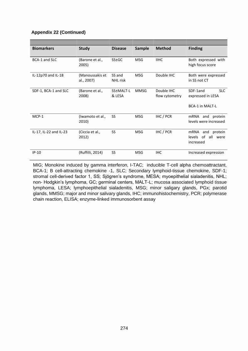
274
Appendix 22 (Continued)
Biomarkers
Study
Disease
Sample
Method
Finding
BCA-1 and SLC (Barone et al., 2005)
SS±GC MSG IIHC Both expressed with high focus score
IL-12p70 and IL-18 (Manoussakis et al., 2007)
SS and NHL risk
MSG Double IHC
Both were expressed in SS not CT
SDF-1, BCA-1 and SLC (Barone et al., 2008)
SS±MALT-L & LESA
MMSG Double IHC flow cytometry
SDF-1and SLC expressed in LESA BCA-1 in MALT-L
MCP-1 (Iwamoto et al., 2010)
SS MSG IHC / PCR mRNA and protein levels were increased
IL-17, IL-22 and IL-23 (Ciccia et al., 2012)
SS MSG IHC / PCR mRNA and protein levels of all were increased
IP-10 (Ruffilli, 2014)
SS MSG IHC Increased expression
MIG; Monokine induced by gamma interferon, I-TAC; inducible T-cell alpha chemoattractant,
BCA-1; B cell-attracting chemokine -1, SLC; Secondary lymphoid-tissue chemokine, SDF-1;
stromal cell-derived factor 1, SS; Sjögren’s syndrome, MESA; myoepithelial sialadenitis, NHL;
non- Hodgkin’s lymphoma, GC; germinal centers, MALT-L; mucosa associated lymphoid tissue
lymphoma, LESA; lymphoepithelial sialadenitis, MSG; minor saligary glands, PGx; parotid
glands, MMSG; major and minor salivary glands, IHC; immunohistochemistry, PCR; polymerase
chain reaction, ELISA; enzyme-linked immunosorbent assay

275
Appendix 23: Cytokines identified previously in saliva of SS subjects
using different methods
Biomarkers
Study
Disease
Sample
Method
Findings
IL-2 and IL-6 (Rhodus et al., 1998)
SS and TTT
Saliva ELISA Both increased but decreased after TTT
IL-6 (Tishler et al., 1999)
SS Saliva Serum
ELISA IL-6 increased No differences
IFN-γ, IL-2 and IL-6 (Streckfus et al.,
2001)
SS and
TTT
Saliva ELISA IL-2 and -6
increased but
decreased after TTT
IL-6 and bFGF (Boras et al.,
2004)
SS Saliva
Serum
ELISA Both increased
No difference
BAFF (Pers et al., 2005a)
SS Saliva Serum
ELISA ELISA
Increased Increased
75 analytes 82 analytes
(Delaleu et al., 2008)
SS model Saliva Serum
75-plex, bead- based assay (MAP) 82-plex, bead- based assay (MAP)
34 detected in saliva 38 detected in serum
IL-17 and IL-23 IL-17 IL-17 and IL-23 IL-17 and IL-6
(Nguyen et al., 2008)
SS model SS
SMG Saliva Serum MSG Saliva Serum
IHC /PCR Bead- based assay (mouse) IHC Bead- based assay (human)
Both expressed in later stages even mRNA levels low levels detected Detected in early stages Both were expressed IL-6 only increased Both did not differ from CT
MCP-1, MIP-1α, MIP-1β, IL-8 and IP-10
(Lee et al., 2010) SS Saliva Luminex bead- based assay
MIP-1α, MIP-1β, IL-8 and IP-10 were increased
IL-1, IL-4, IL-6, IL-10, IL-12p40, IL-17, IFN-γ and TNF-α
(Kang et al., 2011)
SS Saliva
Luminex bead- based assay
All were increased

276
Appendix 23 (Continued)
SS; Sjögren’s syndrome, TTT; pilocarpine treatment, SMG; submandibular glands, MSG; minor
salivary glands, PBMCs; peripheral blood mononuclear cells, ELISA; enzyme-linked
immunosorbent assay, IHC; immunohistochemistry, PCR; polymerase chain reaction, CBA;
cytometric bead assay, MAP; multianalyte profile, DFA; discriminant function analysis, GO; gene
ontology- based network analysis.
Biomarkers Study Disease Sample
Method
Finding
SDF_1α, BCA-1, IP-10, MCP-1, MIP-1α and RANTS
(Hernandez-Molina et al., 2011)
SS Saliva Luminex bead- based assay
IP-10 and MCP-1 were increased
IL-1β, IL-2, IL-4, IL-5, IL-6, IL-10, IL-12, TNF-α, IFN-γ, RANTES, IL-8, MIG and IP-10 (TARC, MDC and MIP-1α) IL-2, IFN-γ, IL-4, IL-5, IL 10, IL-12, IL-17 and TGF-β, IP-10, RANTS, TARC, MDC and MIP-1α
(Moriyama et al., 2012)
SS Saliva MSG PBMCs
CBA (ELISA) IHC / PCR PCR
IL-1 β, IL-8, IL-4, IL-6, IL-10 and MDC were increased. IL-4, IL-10 and MDC where higher with hicher focus scores mRNA levels of IL-2, IFN- γ, IL-4, IL-10, IL-17, IP-10, MIP-1α, RANTES, TARC and MDC were increased mRNA levels of IP-10 was increased
IL-1β, IL-2, IL-4, IL-5, IL-6, IL-10, IL-12p70, IL-17A, IFN-γ, and TNF-α
(Ohyama et al., 2015)
SS Saliva CBA All were increased
187 analytes (Delaleu et al., 2015)
SS Saliva 187-plex, bead-based assay (MAP)DFA and GO- analysis
52 were increased in SS 4- and 6- plex signature included IL-4 and IL-5

277
Appendix 24: Cytokines identified previously in parotid saliva of SS
subjects using different methods
Biomarkers
Study
Disease
Sample
Method
Finding
IL-1α, IL-2, IL-4, IL-5, IL-6, IL-10, IFN-γ, and TNF-α
(Fox et al., 1994)
SS (P) saliva MSG
ELISA PCR
All were increased in SS except IL-4 and -5 were not detected mRNA levels of IL-2, -10 and IFN-γ were detected in SS but not IL-4 and -5. TNF-α was detected in SS and normal IL-1α and -6 were detected in some
IL-6 (Grisius et al., 1997)
SS (P) saliva Serum
ELISA IL-6 increased IL-6 increased
IL-10, IL-2, IL-4 and IFN-γ (Bertorello et al., 2004)
SS (P) saliva ELISA IL-10 increased in most IFN- increased in some IL-2 and -4 are absent
SS; Sjögren’s syndrome, (P); parotid saliva, MSG; minor salivary glands, ELISA; enzyme-linked
immunosorbent assay, PCR; polymerase chain reaction

278
Appendix 25: Summary of the review of literature on cytokines
The findings of the above- listed studies for chapter 5 can be summarised as follows; in
serum, levels of IL-22, -10 were increased in SS patients with a direct correlation with some
of the clinical parameters, e.g. autoantibody levels (Anaya et al., 2002, Lavoie et al., 2011).
Levels of IL-6 were increased in SS patients when compared to controls (Grisius et al., 1997).
While another study found no differences between IL-6 levels of SS and control groups (Tishler
et al., 1999). IL-17 and linked cytokines (e.g. IL-6) were found to be up-regulated in SS in
comparison to controls (Katsifis et al., 2009, Kwok et al., 2012). In addition to IL-17 (Fei et al.,
2014). BAFF was also found highly elevated in SS patients, both of which correlated with
different SS phenotypes (Deng et al., 2016). BAFF and APRIL were elevated in SS patients
(Pollard et al., 2013b). On the other hand, Alunno et al., (2015) reported detectable levels of
IL-17 in 15/50 SS patients and that it was associated with longer disease duration which is a
similar finding to Nguyen et al., (2008) where they have analysed levels of IL-17 and -23 in
sera, saliva and glands of animal models and SS patients. Serum levels of IL-18 in SS were
higher than controls but similar to patients with rheumatoid arthritis (Bombardieri et al., 2004).
Also, serum levels of several cytokines were measured by multiplex assays in SS patients
identifying potential targets (Pollard et al., 2013a, Szodoray et al., 2005, Szodoray et al., 2004,
Reksten et al., 2009, Moriyama et al., 2012). In minor salivary glands, protein and mRNA
levels of IL-6, -17, 22, -23 and MCP-1 were increased in SS patients (Ciccia et al., 2012,
Iwamoto et al., 2010, Katsifis et al., 2009, Kwok et al., 2012, Fei et al., 2014) In addition IL-18
was expressed in SS patient’s glands but not in chronic sialadenitis glands (Bombardieri et
al., 2004). Th1 cytokines were identified in salivary glands of SS patients while IL-4 was
absent and IL-10 was present in the majority of samples (Kolkowski et al., 1999). Similar
findings were reported, demonstrating cytokine expression in healthy control samples as well
(Konttinen et al., 1999). Expression of some chemokines was reported by Ogawa et al., (2004)
and (2002) also the same group has looked at other cytokines as well (Ogawa et al., 1995).

279
Turning now to the non-Hodgkin lymphoma (NHL) literature, it can be seen that a considerable
number of studies were done looking at the cytokine/chemokine levels in SS patients with
lymphoma, correlating their levels with the predictive markers of lymphoma, where serum and
salivary glands were their main targets;
In salivary glands of SS patients with non-Hodgkin lymphoma (NHL), there was an aberrant
expression of IL-22R1 which is IL-18 dependent (Ciccia et al., 2015). Several cytokines were
detected in all SS stages (SS, SS with myoepithelial sialadenitis (MESA) and SS with NHL)
but expression levels of IL-4 were lacking from patients with MESA while present in some
salivary glands of SS and SS with NHL (De Vita et al., 1995). Some cytokines were increased
more in mod-severe lesion (Ogawa et al., 1995). IL-18 was positively correlated with other
predictive markers while IL-12 was negatively correlated (Manoussakis et al., 2007). A number
of chemokines (BCA-1 and SLC) were expressed in glands being higher with high focus
scores (germinal centres) (Barone et al., 2005, Amft et al., 2001). Also BCA-1 expression was
increased in salivary glands of SS patients with MALT-L (Barone et al., 2008). However,
Salomonsson et al., (2003) found no correlation between those cytokines and the formation
of germinal centres.
In serum, higher Fms-related tyrosine kinase 3 ligand (FLT3LG) levels were found in SS group
in comparison to controls and they were associated with a history of lymphoma as well as with
other predictive markers (Tobon et al., 2010, Tobon et al., 2013). Several studies have focused
on BAFF levels in SS, correlating them with serological levels (Pers et al., 2005b, Groom et
al., 2002, Mariette et al., 2003, Lavie et al., 2008), with certain predictors of lymphoma
development, with higher disease activity and with history of lymphoma (Gottenberg et al.,
2013, Quartuccio et al., 2013). In addition to BAFF, serum levels of APRIL were found
increased in SS in relation to controls and both were positively correlated with focus score
(Jonsson et al., 2005). Nocturne et al., (2015) reported elevated levels of BCA-1 in SS patients
with NHL. The levels of several cytokines in SS patients with and without germinal centres
were explored (Reksten et al., 2009, Szodoray et al., 2005).
280
References ABROL, E., GONZALEZ-PULIDO, C., PRAENA-FERNANDEZ, J. M. & ISENBERG, D. A. 2014. A
retrospective study of long-term outcomes in 152 patients with primary Sjogren's syndrome: 25-year experience. Clin Med (Lond), 14, 157-64.
AFRAMIAN, D., KONTTINEN, Y., CARROZZO, M. & TZIOUFAS, A. 2013. Urban legends series: Sjögren’s syndrome. Oral diseases, 19, 46-58.
AL-TARAWNEH, S. K., BORDER, M. B., DIBBLE, C. F. & BENCHARIT, S. 2011. Defining salivary biomarkers using mass spectrometry-based proteomics: a systematic review. OMICS, 15, 353-61.
ALUNNO, A., BISTONI, O., CATERBI, S., BARTOLONI, E., CAFARO, G. & GERLI, R. 2015. Serum interleukin-17 in primary Sjogren's syndrome: association with disease duration and parotid gland swelling. Clin Exp Rheumatol, 33, 129.
AMBATIPUDI, K. S., SWATKOSKI, S., MORESCO, J. J., TU, P. G., COCA, A., ANOLIK, J. H., GUCEK, M., SANZ, I., YATES, J. R., 3RD & MELVIN, J. E. 2012. Quantitative proteomics of parotid saliva in primary Sjogren's syndrome. Proteomics, 12, 3113-20.
AMERONGEN, A. V. & VEERMAN, E. C. 2002. Saliva--the defender of the oral cavity. Oral Dis, 8, 12-22.
AMFT, N., CURNOW, S. J., SCHEEL-TOELLNER, D., DEVADAS, A., OATES, J., CROCKER, J., HAMBURGER, J., AINSWORTH, J., MATHEWS, J., SALMON, M., BOWMAN, S. J. & BUCKLEY, C. D. 2001. Ectopic expression of the B cell-attracting chemokine BCA-1 (CXCL13) on endothelial cells and within lymphoid follicles contributes to the establishment of germinal center-like structures in Sjogren's syndrome. Arthritis Rheum, 44, 2633-41.
ANACAK, Y., MILLER, R. C., CONSTANTINOU, N., MAMUSA, A. M., EPELBAUM, R., LI, Y., CALDUCH, A. L., KOWALCZYK, A., WEBER, D. C., KADISH, S. P., BESE, N., POORTMANS, P., KAMER, S. & OZSAHIN, M. 2012. Primary mucosa-associated lymphoid tissue lymphoma of the salivary glands: a multicenter Rare Cancer Network study. Int J Radiat Oncol Biol Phys, 82, 315-20.
ANAYA, J. M., CORREA, P. A., HERRERA, M., ESKDALE, J. & GALLAGHER, G. 2002. Interleukin 10 (IL-10) influences autoimmune response in primary Sjogren's syndrome and is linked to IL-10 gene polymorphism. J Rheumatol, 29, 1874-6.
ANDERSON, L. A., GADALLA, S., MORTON, L. M., LANDGREN, O., PFEIFFER, R., WARREN, J. L., BERNDT, S. I., RICKER, W., PARSONS, R. & ENGELS, E. A. 2009. Population-based study of autoimmune conditions and the risk of specific lymphoid malignancies. Int J Cancer, 125, 398-405.
ARELLANO-GARCIA, M. E., HU, S., WANG, J., HENSON, B., ZHOU, H., CHIA, D. & WONG, D. T. 2008. Multiplexed immunobead-based assay for detection of oral cancer protein biomarkers in saliva. Oral Diseases, 14, 705-712.
ASTORRI, E., SUTCLIFFE, N., RICHARDS, P. S., SUCHAK, K., PITZALIS, C., BOMBARDIERI, M. & TAPPUNI, A. R. 2016. Ultrasound of the salivary glands is a strong predictor of labial gland biopsy histopathology in patients with sicca symptoms. J Oral Pathol Med, 45, 450-4.
ATKINSON, J. C. 1993. The role of salivary measurements in the diagnosis of salivary autoimmune diseases. Ann N Y Acad Sci, 694, 238-51.
ATKINSON, J. C., TRAVIS, W. D., PILLEMER, S. R., BERMUDEZ, D., WOLFF, A. & FOX, P. C. 1990. Major salivary gland function in primary Sjogren's syndrome and its relationship to clinical features. J Rheumatol, 17, 318-22.
AWADA, A., NICAISE, C., ENA, S., SCHANDENE, L., RASSCHAERT, J., POPESCU, I., GANGJI, V. & SOYFOO, M. S. 2014. Potential involvement of the IL-33-ST2 axis in the pathogenesis of primary Sjogren's syndrome. Ann Rheum Dis, 73, 1259-63.
BACON, C. M., DU, M. Q. & DOGAN, A. 2007. Mucosa-associated lymphoid tissue (MALT) lymphoma: a practical guide for pathologists. J Clin Pathol, 60, 361-72.

281
BAECKLUND, E., SMEDBY, K. E., SUTTON, L.-A., ASKLING, J. & ROSENQUIST, R. Lymphoma development in patients with autoimmune and inflammatory disorders–What are the driving forces? Seminars in cancer biology, 2013. Elsevier.
BALDINI, C., GIUSTI, L., CIREGIA, F., DA VALLE, Y., GIACOMELLI, C., DONADIO, E., FERRO, F., GALIMBERTI, S., DONATI, V. & BAZZICHI, L. 2011a. Correspondence between salivary proteomic pattern and clinical course in primary Sjögren syndrome and non-Hodgkin's lymphoma: a case report. J Transl Med, 9, 188.
BALDINI, C., GIUSTI, L., CIREGIA, F., DA VALLE, Y., GIACOMELLI, C., DONADIO, E., SERNISSI, F., BAZZICHI, L., GIANNACCINI, G., BOMBARDIERI, S. & LUCACCHINI, A. 2011b. Proteomic analysis of saliva: a unique tool to distinguish primary Sjogren's syndrome from secondary Sjogren's syndrome and other sicca syndromes. Arthritis Res Ther, 13, R194.
BARONE, F., BOMBARDIERI, M., MANZO, A., BLADES, M. C., MORGAN, P. R., CHALLACOMBE, S. J., VALESINI, G. & PITZALIS, C. 2005. Association of CXCL13 and CCL21 expression with the progressive organization of lymphoid-like structures in Sjogren's syndrome. Arthritis Rheum, 52, 1773-84.
BARONE, F., BOMBARDIERI, M., ROSADO, M. M., MORGAN, P. R., CHALLACOMBE, S. J., DE VITA, S., CARSETTI, R., SPENCER, J., VALESINI, G. & PITZALIS, C. 2008. CXCL13, CCL21, and CXCL12 expression in salivary glands of patients with Sjogren's syndrome and MALT lymphoma: association with reactive and malignant areas of lymphoid organization. J Immunol, 180, 5130-40.
BASSIG, B. A., SHU, X. O., KOH, W. P., GAO, Y. T., PURDUE, M. P., BUTLER, L. M., ADAMS-HADUCH, J., XIANG, Y. B., KEMP, T. J., WANG, R., PINTO, L. A., ZHENG, T., JI, B. T., HOSGOOD, H. D., HU, W., YANG, G., ZHANG, H., CHOW, W. H., KIM, C., SEOW, W. J., ZHENG, W., YUAN, J. M., LAN, Q. & ROTHMAN, N. 2015. Soluble levels of CD27 and CD30 are associated with risk of non-Hodgkin lymphoma in three Chinese prospective cohorts. Int J Cancer, 137, 2688-95.
BEELEY, J. A. & KHOO, K. S. 1999. Salivary proteins in rheumatoid arthritis and Sjogren's syndrome: one-dimensional and two-dimensional electrophoretic studies. Electrophoresis, 20, 1652-60.
BERTORELLO, R., CORDONE, M. P., CONTINI, P., ROSSI, P., INDIVERI, F., PUPPO, F. & CORDONE, G. 2004. Increased levels of interleukin-10 in saliva of Sjogren's syndrome patients. Correlation with disease activity. Clin Exp Med, 4, 148-51.
BIENVENU, J., MONNERET, G., FABIEN, N. & REVILLARD, J. P. 2000. The clinical usefulness of the measurement of cytokines. Clin Chem Lab Med, 38, 267-85.
BILLINGS, M., DYE, B. A., IAFOLLA, T., BAER, A. N., GRISIUS, M. & ALEVIZOS, I. 2016. Significance and Implications of Patient-reported Xerostomia in Sjogren's Syndrome: Findings From the National Institutes of Health Cohort. EBioMedicine.
BOLSTAD, A. I. & SKARSTEIN, K. 2016. Epidemiology of Sjogren's Syndrome-from an Oral Perspective. Curr Oral Health Rep, 3, 328-336.
BOMBARDIERI, M., BARONE, F., PITTONI, V., ALESSANDRI, C., CONIGLIARO, P., BLADES, M. C., PRIORI, R., MCINNES, I. B., VALESINI, G. & PITZALIS, C. 2004. Increased circulating levels and salivary gland expression of interleukin-18 in patients with Sjogren's syndrome: relationship with autoantibody production and lymphoid organization of the periductal inflammatory infiltrate. Arthritis Res Ther, 6, R447-56.
BOMBARDIERI, M. & PITZALIS, C. 2012. Ectopic lymphoid neogenesis and lymphoid chemokines in Sjogren's syndrome: at the interplay between chronic inflammation, autoimmunity and lymphomagenesis. Curr Pharm Biotechnol, 13, 1989-96.
BOOTSMA, H., SPIJKERVET, F. K., KROESE, F. G. & VISSINK, A. 2013. Toward new classification criteria for Sjogren's syndrome? Arthritis Rheum, 65, 21-3.
BORAS, V. V., CIKES, N., LUKAC, J., CEKIC-ARAMBASIN, A., VIRAG, M. & BOSNJAK, A. 2004. The significance of salivary and serum interleukin 6 and basic fibroblast growth factor levels in patients with Sjogren's syndrome. Coll Antropol, 28 Suppl 2, 305-9.

282
BOTSIOS, C., OSTUNI, P., SFRISO, P., GRAVA, C., ANDRETTA, M. & TODESCO, S. 2001. [Primary Sjogren's syndrome. A longitudinal study of 68 patients]. Recenti Prog Med, 92, 741-6.
BOUMA, I., PIJPE, J. & VISSINK, A. 2003. [Sjogren' syndrome: a progressive disease]. Ned Tijdschr Tandheelkd, 110, 316-20.
BOWMAN, S. J. & FOX, R. I. 2014. Classification criteria for Sjogren's syndrome: nothing ever stands still! Annals of the rheumatic diseases, 73, 1-2.
BRESNICK, A. R., WEBER, D. J. & ZIMMER, D. B. 2015. S100 proteins in cancer. Nat Rev Cancer, 15, 96-109.
BROWN, J. 2010. PLENARY ABSTRACT: Imaging in the patient with hyposalivation. Oral Diseases, 16, 503-504.
BRUN, J. G., CUIDA, M., JACOBSEN, H., KLOSTER, R., JOHANNESEN, A. C., HOYERAAL, H. M. & JONSSON, R. 1994. Sjogren's syndrome in inflammatory rheumatic diseases: analysis of the leukocyte protein calprotectin in plasma and saliva. Scand J Rheumatol, 23, 114-8.
BUNIM, J. & TALAL, N. 1963. The association of malignant lymphoma with Sjögren’s syndrome Trans Assoc Am Pysicians, 76, 45-55.
BUNIM, J. J. 1961. Heberden Oration A Broader Spectrum of Sjøgren's Syndrome and Its Pathogenetic Implications. Annals of the rheumatic diseases, 20, 1-10.
BURLAGE, F. R., PIJPE, J., COPPES, R. P., HEMELS, M. E., MEERTENS, H., CANRINUS, A. & VISSINK, A. 2005. Variability of flow rate when collecting stimulated human parotid saliva. Eur J Oral Sci, 113, 386-90.
CARPENTER, G. H. 2013. The secretion, components, and properties of saliva. Annu Rev Food Sci Technol, 4, 267-76.
CARPENTER, G. H., PROCTOR, G. B., PANKHURST, C. L., O'DONOHUE, J., SCOTT, D. & HUNNABLE, M. P. 2000. Sialochemical markers of salivary gland involvement with Sjogren's syndrome secondary to rheumatoid arthritis and primary biliary cirrhosis. J Oral Pathol Med, 29, 452-9.
CARUBBI, F., ALUNNO, A., CIPRIANI, P., DI BENEDETTO, P., RUSCITTI, P., BERARDICURTI, O., BARTOLONI, E., BISTONI, O., CATERBI, S., CICCIA, F., TRIOLO, G., GERLI, R. & GIACOMELLI, R. 2014. Is minor salivary gland biopsy more than a diagnostic tool in primary Sjogrens syndrome? Association between clinical, histopathological, and molecular features: a retrospective study. Semin Arthritis Rheum, 44, 314-24.
CHALLACOMBE, S. J., OSAILAN, S. M. & PROCTOR, G. B. 2015. Clinical Scoring Scales for Assessment of Dry Mouth. In: CARPENTER, G. (ed.) Dry Mouth: A Clinical Guide on Causes, Effects and Treatments. Berlin, Heidelberg: Springer Berlin Heidelberg.
CHIAPPIN, S., ANTONELLI, G., GATTI, R. & DE PALO, E. F. 2007. Saliva specimen: a new laboratory tool for diagnostic and basic investigation. Clinica Chimica Acta, 383, 30-40.
CHING, K. H., BURBELO, P. D., GONZALEZ-BEGNE, M., ROBERTS, M. E., COCA, A., SANZ, I. & IADAROLA, M. J. 2011. Salivary anti-Ro60 and anti-Ro52 antibody profiles to diagnose Sjögren’s syndrome. Journal of dental research, 90, 445-449.
CHISHOLM, D. M. & MASON, D. K. 1968. Labial salivary gland biopsy in Sjögren's disease. Journal of Clinical Pathology, 21, 656-660.
CHWALINSKA-SADOWSKA, H., ZAJACZEK-GRABOWSKA, A., BIERNAT-KALUZA, E., SZYMANSKA-CYGLER, B., FERDYNUS-CHROMY, J. & SZYMCZAK, E. 1998. Long-term follow-up of 50 patients with primary Sjogren's syndrome. Reumatologia (Warsaw), 36, 135-143.
CICCIA, F., GUGGINO, G., RIZZO, A., BOMBARDIERI, M., RAIMONDO, S., CARUBBI, F., CANNIZZARO, A., SIRECI, G., DIELI, F., CAMPISI, G., GIACOMELLI, R., CIPRIANI, P., DE LEO, G., ALESSANDRO, R. & TRIOLO, G. 2015. Interleukin (IL)-22 receptor 1 is over-expressed in primary Sjogren's syndrome and Sjogren-associated non-Hodgkin lymphomas and is regulated by IL-18. Clin Exp Immunol, 181, 219-29.
CICCIA, F., GUGGINO, G., RIZZO, A., FERRANTE, A., RAIMONDO, S., GIARDINA, A., DIELI, F., CAMPISI, G., ALESSANDRO, R. & TRIOLO, G. 2012. Potential involvement of IL-22 and IL-22-producing

283
cells in the inflamed salivary glands of patients with Sjogren's syndrome. Ann Rheum Dis, 71, 295-301.
COHEN, S. M., PETRYK, M., VARMA, M., KOZUCH, P. S., AMES, E. D. & GROSSBARD, M. L. 2006. Non-Hodgkin's lymphoma of mucosa-associated lymphoid tissue. Oncologist, 11, 1100-17.
COLELLA, G., CANNAVALE, R., VICIDOMINI, A. & ITRO, A. 2010. Salivary gland biopsy: a comprehensive review of techniques and related complications. Rheumatology, 49, 2117-2121.
CORNEC, D., JAMIN, C. & PERS, J. O. 2014. Sjogren's syndrome: where do we stand, and where shall we go? J Autoimmun, 51, 109-14.
CORNEC, D., JOUSSE‐JOULIN, S., PERS, J. O., MARHADOUR, T., COCHENER, B., BOISRAMÉ‐GASTRIN, S., NOWAK, E., YOUINOU, P., SARAUX, A. & DEVAUCHELLE‐PENSEC, V. 2013. Contribution of salivary gland ultrasonography to the diagnosis of Sjögren's syndrome: Toward new diagnostic criteria? Arthritis & Rheumatism, 65, 216-225.
COSTA, S., QUINTIN-ROUE, I., LESOURD, A., JOUSSE-JOULIN, S., BERTHELOT, J. M., HACHULLA, E., HATRON, P. Y., GOEB, V., VITTECOQ, O., PERS, J. O., MARCORELLES, P., NOWAK, E., SARAUX, A. & DEVAUCHELLE-PENSEC, V. 2015. Reliability of histopathological salivary gland biopsy assessment in Sjogren's syndrome: a multicentre cohort study. Rheumatology (Oxford), 54, 1056-64.
CUI, G., SUGAI, S., OGAWA, Y., TAKESHITA, S., MASAKI, Y. & FUKUTOKU, M. 1997. [Long-term follow-up of 43 patients with Sjogren's syndrome]. Ryumachi, 37, 770-80.
CUIDA, M., BRUN, J., JACOHSEN, H., RYGH, T., JOHANNESEN, A. & JONSSON, R. Calprotectin in chronically inflamed salivary glands and saliva. Proceedings of the IVth International Symposium on Sjogren’s syndrome, August 11-13 1993 Tokyo.
CUIDA, M., BRUN, J. G., TYNNING, T. & JONSSON, R. 1995. Calprotectin levels in oral fluids: the importance of collection site. Eur J Oral Sci, 103, 8-10.
CUIDA, M., HALSE, A. K., JOHANNESSEN, A. C., TYNNING, T. & JONSSON, R. 1997. Indicators of salivary gland inflammation in primary Sjogren's syndrome. Eur J Oral Sci, 105, 228-33.
DAVIDSON, B. K., KELLY, C. A. & GRIFFITHS, I. D. 1999. Primary Sjogren's syndrome in the North East of England: a long-term follow-up study. Rheumatology (Oxford), 38, 245-53.
DAVIDSON, B. K. S., KELLY, C. A. & GRIFFITHS, I. D. 2000. Ten year follow up of pulmonary function in patients with primary Sjögren's syndrome. Annals of the Rheumatic Diseases, 59, 709-712.
DAWES, C. 1987. Physiological factors affecting salivary flow rate, oral sugar clearance, and the sensation of dry mouth in man. J Dent Res, 66 Spec No, 648-53.
DAWSON, L., FOX, P. C. & SMITH, P. M. 2006. Sjögrens syndrome—the non-apoptotic model of glandular hypofunction. Rheumatology, 45, 792-798.
DE JONG, E. P., XIE, H., ONSONGO, G., STONE, M. D., CHEN, X. B., KOOREN, J. A., REFSLAND, E. W., GRIFFIN, R. J., ONDREY, F. G., WU, B., LE, C. T., RHODUS, N. L., CARLIS, J. V. & GRIFFIN, T. J. 2010. Quantitative proteomics reveals myosin and actin as promising saliva biomarkers for distinguishing pre-malignant and malignant oral lesions. PLoS One, 5, e11148.
DE LA FUENTE, M., MACDONALD, T. T. & HERMOSO, M. A. 2015. The IL-33/ST2 axis: Role in health and disease. Cytokine Growth Factor Rev, 26, 615-23.
DE VITA, S., DOLCETTI, R., FERRACCIOLI, G., PIVETTA, B., DE RE, V., GLOGHINI, A., D'AGOSTO, A., BARTOLI, E., CARBONE, A. & BOIOCCHI, M. 1995. Local cytokine expression in the progression toward B cell malignancy in Sjogren's syndrome. J Rheumatol, 22, 1674-80.
DELALEU, N., IMMERVOLL, H., CORNELIUS, J. & JONSSON, R. 2008. Biomarker profiles in serum and saliva of experimental Sjogren's syndrome: associations with specific autoimmune manifestations. Arthritis Res Ther, 10, R22.
DELALEU, N., MYDEL, P., KWEE, I., BRUN, J. G., JONSSON, M. V. & JONSSON, R. 2015. High fidelity between saliva proteomics and the biologic state of salivary glands defines biomarker signatures for primary Sjogren's syndrome. Arthritis Rheumatol, 67, 1084-95.

284
DELLI, K., DIJKSTRA, P. U., STEL, A. J., BOOTSMA, H., VISSINK, A. & SPIJKERVET, F. K. 2015. Diagnostic properties of ultrasound of major salivary glands in Sjogren's syndrome: a meta-analysis. Oral Dis, 21, 792-800.
DELLI, K., HAACKE, E. A., IHRLER, S., VAN DER VEGT, B., VISSINK, A., BOOTSMA, H., SPIJKERVET, F. K. & KROESE, F. G. 2016. Need for consensus guidelines to standardise the assessment of germinal centres and other histopathological parameters in salivary gland tissue of patients with primary Sjogren's syndrome. Ann Rheum Dis, 75, e32.
DENG, F., CHEN, J., ZHENG, J., CHEN, Y., HUANG, R., YIN, J., GAO, X., LIN, Q., HUANG, C., GAO, Y., YU, X. & LIU, Z. 2016. Association of BAFF and IL-17A with subphenotypes of primary Sjogren's syndrome. Int J Rheum Dis, 19, 715-20.
DEUTSCH, O., KRIEF, G., KONTTINEN, Y. T., ZAKS, B., WONG, D. T., AFRAMIAN, D. J. & PALMON, A. 2015. Identification of Sjogren's syndrome oral fluid biomarker candidates following high-abundance protein depletion. Rheumatology (Oxford), 54, 884-90.
DONADIO, E., GIUSTI, L., SECCIA, V., CIREGIA, F., DA VALLE, Y., DALLAN, I., VENTRONI, T., GIANNACCINI, G., SELLARI-FRANCESCHINI, S. & LUCACCHINI, A. 2013. New insight into benign tumours of major salivary glands by proteomic approach. PLoS One, 8, e71874.
DONG, L., CHEN, Y., MASAKI, Y., OKAZAKI, T. & UMEHARA, H. 2013. Possible Mechanisms of Lymphoma Development in Sjogren's Syndrome. Curr Immunol Rev, 9, 13-22.
DOSSUS, L., BECKER, S., ACHAINTRE, D., KAAKS, R. & RINALDI, S. 2009. Validity of multiplex-based assays for cytokine measurements in serum and plasma from "non-diseased" subjects: comparison with ELISA. J Immunol Methods, 350, 125-32.
DUPONT, N. C., WANG, K., WADHWA, P. D., CULHANE, J. F. & NELSON, E. L. 2005. Validation and comparison of luminex multiplex cytokine analysis kits with ELISA: determinations of a panel of nine cytokines in clinical sample culture supernatants. J Reprod Immunol, 66, 175-91.
EDGAR, W., O'MULLANE, D. & DAWES, C. 2004. Saliva and oral health, British Dental Association London.
EKSTROM SMEDBY, K., VAJDIC, C. M., FALSTER, M., ENGELS, E. A., MARTINEZ-MAZA, O., TURNER, J., HJALGRIM, H., VINEIS, P., SENIORI COSTANTINI, A., BRACCI, P. M., HOLLY, E. A., WILLETT, E., SPINELLI, J. J., LA VECCHIA, C., ZHENG, T., BECKER, N., DE SANJOSE, S., CHIU, B. C., DAL MASO, L., COCCO, P., MAYNADIE, M., FORETOVA, L., STAINES, A., BRENNAN, P., DAVIS, S., SEVERSON, R., CERHAN, J. R., BREEN, E. C., BIRMANN, B., GRULICH, A. E. & COZEN, W. 2008. Autoimmune disorders and risk of non-Hodgkin lymphoma subtypes: a pooled analysis within the InterLymph Consortium. Blood, 111, 4029-38.
FAGERHOL, M. K., ANDERSSON, K. B., NAESS-ANDRESEN, C.-F., BRANDTZAEG, P. & DALE, I. 1990. Calprotectin (the L1 leukocyte protein) In: Smith V L, Dedman J R, editors. Stimulus response coupling: the role of intracellular calcium-binding proteins. Boca Raton, Fla: CRC Press, Inc, pp., 187–210.
FALLAH, M., LIU, X., JI, J., FORSTI, A., SUNDQUIST, K. & HEMMINKI, K. 2014. Autoimmune diseases associated with non-Hodgkin lymphoma: a nationwide cohort study. Ann Oncol, 25, 2025-30.
FEI, Y., ZHANG, W., LIN, D., WU, C., LI, M., ZHAO, Y., ZENG, X. & ZHANG, F. 2014. Clinical parameter and Th17 related to lymphocytes infiltrating degree of labial salivary gland in primary Sjogren's syndrome. Clin Rheumatol, 33, 523-9.
FERRO, F., VAGELLI, R., BRUNI, C., CAFARO, G., MARCUCCI, E., BARTOLONI, E. & BALDINI, C. 2016. One year in review 2016: Sjogren's syndrome. Clin Exp Rheumatol, 34, 161-71.
FISHER, B. A., BROWN, R. M., BOWMAN, S. J. & BARONE, F. 2015. A review of salivary gland histopathology in primary Sjogren's syndrome with a focus on its potential as a clinical trials biomarker. Ann Rheum Dis, 74, 1645-50.
FLEISSIG, Y., DEUTSCH, O., REICHENBERG, E., REDLICH, M., ZAKS, B., PALMON, A. & AFRAMIAN, D. J. 2009. Different proteomic protein patterns in saliva of Sjogren's syndrome patients. Oral Dis, 15, 61-8.

285
FOELL, D. & ROTH, J. 2004. Proinflammatory S100 proteins in arthritis and autoimmune disease. Arthritis Rheum, 50, 3762-71.
FONSECA, F. J., MORAES JUNIOR, M., LOURENCO, E. J., TELES DDE, M. & FIGUEREDO, C. M. 2014. Cytokines expression in saliva and peri-implant crevicular fluid of patients with peri-implant disease. Clin Oral Implants Res, 25, e68-72.
FOX, P. C., BOWMAN, S. J., SEGAL, B., VIVINO, F. B., MURUKUTLA, N., CHOUEIRI, K., OGALE, S. & MCLEAN, L. 2008. Oral involvement in primary Sjögren syndrome. The Journal of the American Dental Association, 139, 1592-1601.
FOX, P. C., SARRAS, A. K., BOWERS, M. R., DROSOS, A. A. & MOUTSOPOULOS, H. M. 1987. Oral and sialochemical findings in patients with autoimmune rheumatic disease. Clin Exp Rheumatol, 5, 123-6.
FOX, R. I. 2005. Sjögren's syndrome. The Lancet, 366, 321-331. FOX, R. I., KANG, H. I., ANDO, D., ABRAMS, J. & PISA, E. 1994. Cytokine mRNA expression in salivary
gland biopsies of Sjogren's syndrome. J Immunol, 152, 5532-9. GANNOT, G., LANCASTER, H. E. & FOX, P. C. 2000. Clinical course of primary Sjogren's syndrome:
salivary, oral, and serologic aspects. J Rheumatol, 27, 1905-9. GARCIC-CARRASCO, M., FONT, J., FILELLA, X., CERVERA, R., RAMOS-CASALS, M., SISO, A., AYMAMI,
A., BALLESTA, A. M. & INGELMO, M. 2001. Circulating levels of Th1/Th2 cytokines in patients with primary Sjogren's syndrome: correlation with clinical and immunological features. Clin Exp Rheumatol, 19, 411-5.
GERLI, R., CALIGARIS-CAPPIO, F., BISTONI, O., BERTERO, M. T., GIACOMELLI, R. & FALINI, B. 1999. Soluble CD30 in primary Sjogren's syndrome. Clin Exp Rheumatol, 17, 389-90.
GIUSTI, L., BALDINI, C., BAZZICHI, L., BOMBARDIERI, S. & LUCACCHINI, A. 2007. Proteomic diagnosis of Sjogren's syndrome. Expert Rev Proteomics, 4, 757-67.
GOTTENBERG, J. E., BUSSON, M., COHEN-SOLAL, J., LAVIE, F., ABBED, K., KIMBERLY, R. P., SIBILIA, J. & MARIETTE, X. 2005. Correlation of serum B lymphocyte stimulator and beta2 microglobulin with autoantibody secretion and systemic involvement in primary Sjogren's syndrome. Ann Rheum Dis, 64, 1050-5.
GOTTENBERG, J. E., SEROR, R., MICELI-RICHARD, C., BENESSIANO, J., DEVAUCHELLE-PENSEC, V., DIEUDE, P., DUBOST, J. J., FAUCHAIS, A. L., GOEB, V., HACHULLA, E., HATRON, P. Y., LARROCHE, C., LE GUERN, V., MOREL, J., PERDRIGER, A., PUECHAL, X., RIST, S., SARAUX, A., SENE, D., SIBILIA, J., VITTECOQ, O., NOCTURNE, G., RAVAUD, P. & MARIETTE, X. 2013. Serum Levels of Beta2-Microglobulin and Free Light Chains of Immunoglobulins Are Associated with Systemic Disease Activity in Primary Sjogren's Syndrome. Data at Enrollment in the Prospective ASSESS Cohort. PLoS ONE, 8.
GRISIUS, M. M., BERMUDEZ, D. K. & FOX, P. C. 1997. Salivary and serum interleukin 6 in primary Sjogren's syndrome. J Rheumatol, 24, 1089-91.
GROOM, J., KALLED, S. L., CUTLER, A. H., OLSON, C., WOODCOCK, S. A., SCHNEIDER, P., TSCHOPP, J., CACHERO, T. G., BATTEN, M., WHEWAY, J., MAURI, D., CAVILL, D., GORDON, T. P., MACKAY, C. R. & MACKAY, F. 2002. Association of BAFF/BLyS overexpression and altered B cell differentiation with Sjogren's syndrome. J Clin Invest, 109, 59-68.
HAGA, H. J. 2002. Clinical and immunological factors associated with low lacrimal and salivary flow rate in patients with primary Sjogren's syndrome. J Rheumatol, 29, 305-8.
HALDORSEN, K., MOEN, K., JACOBSEN, H., JONSSON, R. & BRUN, J. G. 2008. Exocrine function in primary Sjogren syndrome: natural course and prognostic factors. Ann Rheum Dis, 67, 949-54.
HARRIS, N. L., JAFFE, E. S., STEIN, H., BANKS, P. M., CHAN, J. K., CLEARY, M. L., DELSOL, G., DE WOLF-PEETERS, C., FALINI, B., GATTER, K. C. & ET AL. 1994. A revised European-American classification of lymphoid neoplasms: a proposal from the International Lymphoma Study Group. Blood, 84, 1361-92.

286
HELENIUS, L. M., MEURMAN, J. H., HELENIUS, I., KARI, K., HIETANEN, J., SUURONEN, R., HALLIKAINEN, D., KAUTIAINEN, H., LEIRISALO-REPO, M. & LINDQVIST, C. 2005. Oral and salivary parameters in patients with rheumatic diseases. Acta Odontol Scand, 63, 284-93.
HERNANDEZ-MOLINA, G., MICHEL-PEREGRINA, M., HERNANDEZ-RAMIREZ, D. F., SANCHEZ-GUERRERO, J. & LLORENTE, L. 2011. Chemokine saliva levels in patients with primary Sjogren's syndrome, associated Sjogren's syndrome, pre-clinical Sjogren's syndrome and systemic autoimmune diseases. Rheumatology (Oxford), 50, 1288-92.
HERNANDEZ-MOLINA, G. & SANCHEZ-HERNANDEZ, T. 2013. Clinimetric methods in Sjogren's syndrome. Semin Arthritis Rheum, 42, 627-39.
HOČEVAR, A., AMBROŽIČ, A., ROZMAN, B., KVEDER, T. & TOMŠIČ, M. 2005. Ultrasonographic changes of major salivary glands in primary Sjögren's syndrome. Diagnostic value of a novel scoring system. Rheumatology, 44, 768-772.
HORVATH, I. F., SZANTO, A., PAPP, G. & ZEHER, M. 2014. Clinical course, prognosis, and cause of death in primary Sjogren's syndrome. J Immunol Res, 2014, 647507.
HU, S., GAO, K., POLLARD, R., ARELLANO-GARCIA, M., ZHOU, H., ZHANG, L., ELASHOFF, D., KALLENBERG, C. G., VISSINK, A. & WONG, D. T. 2010. Preclinical validation of salivary biomarkers for primary Sjogren's syndrome. Arthritis Care Res (Hoboken), 62, 1633-8.
HU, S., VISSINK, A., ARELLANO, M., ROOZENDAAL, C., ZHOU, H., KALLENBERG, C. G. & WONG, D. T. 2011. Identification of autoantibody biomarkers for primary Sjogren's syndrome using protein microarrays. Proteomics, 11, 1499-507.
HU, S., WANG, J., MEIJER, J., IEONG, S., XIE, Y., YU, T., ZHOU, H., HENRY, S., VISSINK, A., PIJPE, J., KALLENBERG, C., ELASHOFF, D., LOO, J. A. & WONG, D. T. 2007a. Salivary proteomic and genomic biomarkers for primary Sjogren's syndrome. Arthritis Rheum, 56, 3588-600.
HU, S., YU, T., XIE, Y., YANG, Y., LI, Y., ZHOU, X., TSUNG, S., LOO, R. R., LOO, J. R. & WONG, D. T. 2007b. Discovery of oral fluid biomarkers for human oral cancer by mass spectrometry. Cancer Genomics Proteomics, 4, 55-64.
HU, S., ZHOU, M., JIANG, J., WANG, J., ELASHOFF, D., GORR, S., MICHIE, S. A., SPIJKERVET, F. K., BOOTSMA, H., KALLENBERG, C. G., VISSINK, A., HORVATH, S. & WONG, D. T. 2009. Systems biology analysis of Sjogren's syndrome and mucosa-associated lymphoid tissue lymphoma in parotid glands. Arthritis Rheum, 60, 81-92.
ICHIKAWA, Y., YOSHIDA, M., YAMADA, C., HORIKI, T., HOSHINA, Y. & UCHIYAMA, M. 1998. Circulating soluble CD30 levels in primary Sjogren's syndrome, SLE and rheumatoid arthritis. Clin Exp Rheumatol, 16, 759-60.
ISAACSON, P. & WRIGHT, D. H. 1983. Malignant lymphoma of mucosa‐associated lymphoid tissue. A distinctive type of B‐cell lymphoma. Cancer, 52, 1410-1416.
ISAACSON, P. & WRIGHT, D. H. 1984. Extranodal malignant lymphoma arising from mucosa-associated lymphoid tissue. Cancer, 53, 2515-24.
IWAMOTO, N., KAWAKAMI, A., ARIMA, K., NAKAMURA, H., KAWASHIRI, S. Y., TAMAI, M., KITA, J., OKADA, A., KOGA, T., KAMACHI, M., YAMASAKI, S., ICHINOSE, K., IDA, H., ORIGUCHI, T. & EGUCHI, K. 2010. Regulation of disease susceptibility and mononuclear cell infiltration into the labial salivary glands of Sjogren's syndrome by monocyte chemotactic protein-1. Rheumatology (Oxford), 49, 1472-8.
JAFFE, E. S. 2009. The 2008 WHO classification of lymphomas: implications for clinical practice and translational research. Hematology Am Soc Hematol Educ Program, 523-31.
JONSSON, M. V., SZODORAY, P., JELLESTAD, S., JONSSON, R. & SKARSTEIN, K. 2005. Association between circulating levels of the novel TNF family members APRIL and BAFF and lymphoid organization in primary Sjogren's syndrome. J Clin Immunol, 25, 189-201.
JONSSON, M. V., THEANDER, E. & JONSSON, R. 2012. Predictors for the development of non-Hodgkin lymphoma in primary Sjögren's syndrome. La Presse Médicale, 41, e511-e516.
JONSSON, R., KRONELD, U., BACKMAN, K., MAGNUSSON, B. & TARKOWSKI, A. 1993. Progression of sialadenitis in Sjogren's syndrome. Br J Rheumatol, 32, 578-81.

287
JONSSON, R., NGINAMAU, E., SZYSZKO, E. & BROKSTAD, K. A. 2007. Role of B cells in Sjogren's syndrome--from benign lymphoproliferation to overt malignancy. Front Biosci, 12, 2159-70.
JONSSON, R., VOGELSANG, P., VOLCHENKOV, R., ESPINOSA, A., WAHREN-HERLENIUS, M. & APPEL, S. 2011. The complexity of Sjögren's syndrome: novel aspects on pathogenesis. Immunology letters, 141, 1-9.
JORKJEND, L., JOHANSSON, A., JOHANSSON, A. K. & BERGENHOLTZ, A. 2004. Resting and stimulated whole salivary flow rates in Sjogren's syndrome patients over time: a diagnostic aid for subsidized dental care? Acta Odontol Scand, 62, 264-8.
JOUSSE-JOULIN, S., MILIC, V., JONSSON, M. V., PLAGOU, A., THEANDER, E., LUCIANO, N., RACHELE, P., BALDINI, C., BOOTSMA, H., VISSINK, A., HOCEVAR, A., DE VITA, S., TZIOUFAS, A. G., ALAVI, Z., BOWMAN, S. J. & DEVAUCHELLE-PENSEC, V. 2016. Is salivary gland ultrasonography a useful tool in Sjogren's syndrome? A systematic review. Rheumatology (Oxford), 55, 789-800.
JUNG, S. M., LEE, J., BAEK, S. Y., LEE, J. H., LEE, J., PARK, K. S., PARK, S. H., KIM, H. Y. & KWOK, S. K. 2015. The Interleukin 33/ST2 axis in patients with primary Sjogren syndrome: expression in serum and salivary glands, and the clinical association. J Rheumatol, 42, 264-71.
KALK, W. W., MANSOUR, K., VISSINK, A., SPIJKERVET, F. K., BOOTSMA, H., KALLENBERG, C. G., ROODENBURG, J. L. & NIEUW AMERONGEN, A. V. 2002a. Oral and ocular manifestations in Sjogren's syndrome. J Rheumatol, 29, 924-30.
KALK, W. W., VISSINK, A., SPIJKERVET, F. K. & BOOTSMA, H. 1999. Primary sialoangiectasia--a diagnostic pitfall in Sjogren's syndrome: case report. Oral Surg Oral Med Oral Pathol Oral Radiol Endod, 87, 568-71.
KALK, W. W., VISSINK, A., SPIJKERVET, F. K., BOOTSMA, H., KALLENBERG, C. G. & NIEUW AMERONGEN, A. V. 2001a. Sialometry and sialochemistry: diagnostic tools for Sjogren's syndrome. Ann Rheum Dis, 60, 1110-6.
KALK, W. W., VISSINK, A., SPIJKERVET, F. K., BOOTSMA, H., KALLENBERG, C. G. & ROODENBURG, J. L. 2002b. Parotid sialography for diagnosing Sjogren syndrome. Oral Surg Oral Med Oral Pathol Oral Radiol Endod, 94, 131-7.
KALK, W. W., VISSINK, A., SPIJKERVET, F. K., MOLLER, J. M. & ROODENBURG, J. L. 2001b. Morbidity from parotid sialography. Oral Surg Oral Med Oral Pathol Oral Radiol Endod, 92, 572-5.
KALK, W. W., VISSINK, A., STEGENGA, B., BOOTSMA, H., NIEUW AMERONGEN, A. V. & KALLENBERG, C. G. 2002c. Sialometry and sialochemistry: a non-invasive approach for diagnosing Sjogren's syndrome. Ann Rheum Dis, 61, 137-44.
KANG, E. H., LEE, Y. J., HYON, J. Y., YUN, P. Y. & SONG, Y. W. 2011. Salivary cytokine profiles in primary Sjogren's syndrome differ from those in non-Sjogren sicca in terms of TNF-alpha levels and Th-1/Th-2 ratios. Clin Exp Rheumatol, 29, 970-6.
KAPSOGEORGOU, E. K., CHRISTODOULOU, M. I., PANAGIOTAKOS, D. B., PAIKOS, S., TASSIDOU, A., TZIOUFAS, A. G. & MOUTSOPOULOS, H. M. 2013. Minor salivary gland inflammatory lesions in Sjogren syndrome: do they evolve? J Rheumatol, 40, 1566-71.
KASSAN, S. S. & MOUTSOPOULOS, H. M. 2004. Clinical manifestations and early diagnosis of Sjogren syndrome. Archives of internal medicine, 164, 1275.
KASSIMOS, D. G., SHIRLAW, P. J., CHOY, E. H., HOCKEY, K., MORGAN, P. R., CHALLACOMBE, S. J. & PANAYI, G. S. 1997. Chronic sialadenitis in patients with nodal osteoarthritis. Br J Rheumatol, 36, 1312-7.
KATSIFIS, G. E., REKKA, S., MOUTSOPOULOS, N. M., PILLEMER, S. & WAHL, S. M. 2009. Systemic and local interleukin-17 and linked cytokines associated with Sjogren's syndrome immunopathogenesis. Am J Pathol, 175, 1167-77.
KATSIOUGIANNIS, S. & WONG, D. T. 2016. The Proteomics of Saliva in Sjogren's Syndrome. Rheum Dis Clin North Am, 42, 449-56.

288
KELLY, C. A., FOSTER, H., PAL, B., GARDINER, P., MALCOLM, A. J., CHARLES, P., BLAIR, G. S., HOWE, J., DICK, W. C. & GRIFFITHS, I. D. 1991. Primary Sjogren's syndrome in north east England--a longitudinal study. Br J Rheumatol, 30, 437-42.
KHAN, A. 2012. Detection and quantitation of forty eight cytokines, chemokines, growth factors and nine acute phase proteins in healthy human plasma, saliva and urine. J Proteomics, 75, 4802-19.
KOLKOWSKI, E. C., RETH, P., PELUSA, F., BOSCH, J., PUJOL-BORRELL, R., COLL, J. & JARAQUEMADA, D. 1999. Th1 predominance and perforin expression in minor salivary glands from patients with primary Sjogren's syndrome. J Autoimmun, 13, 155-62.
KONTTINEN, Y. T., KEMPPINEN, P., KOSKI, H., LI, T. F., JUMPPANEN, M., HIETANEN, J., SANTAVIRTA, S., SALO, T., LARSSON, A., HAKALA, M. & SORSA, T. 1999. T(H)1 cytokines are produced in labial salivary glands in Sjogren's syndrome, but also in healthy individuals. Scand J Rheumatol, 28, 106-12.
KRIEGBAUM, N. J., LINSTOW, M. V., OXHOLM, P. & PRAUSE, J. U. 1989. A Follow-Up Study of the Progress of Keratoconjunctivitis Sicca and Response to Treatment in Primary Sjögren's Syndrome. Scandinavian Journal of Rheumatology, 18, 193-196.
KROESE, F. G., ABDULAHAD, W. H., HAACKE, E., BOS, N. A., VISSINK, A. & BOOTSMA, H. 2014. B-cell hyperactivity in primary Sjogren's syndrome. Expert Rev Clin Immunol, 10, 483-99.
KRUIZE, A. A., HENÉ, R. J., VAN DER HEIDE, A., BODEUTSCH, C., DE WILDE, P. C. M., VAN BIJSTERVELD, O. P., DE JONG, J., FELTKAMP, T. E. W., KATER, L. & BIJLSMA, J. W. J. 1996. Long-term followup of patients with Sjögren's syndrome. Arthritis & Rheumatism, 39, 297-303.
KRUIZE, A. A., VAN BIJSTERVELD, O. P., HENE, R. J., DE WILDE, P. C., FELTKAMP, T. E., KATER, L. & BIJLSMA, J. W. 1997. Long-term course of tear gland function in patients with keratoconjunctivitis sicca and Sjogren's syndrome. Br J Ophthalmol, 81, 435-8.
KRYLOVA, L. & ISENBERG, D. 2010. Assessment of patients with primary Sjogren's syndrome--outcome over 10 years using the Sjogren's Syndrome Damage Index. Rheumatology (Oxford), 49, 1559-62.
KWOK, S. K., CHO, M. L., HER, Y. M., OH, H. J., PARK, M. K., LEE, S. Y., WOO, Y. J., JU, J. H., PARK, K. S., KIM, H. Y. & PARK, S. H. 2012. TLR2 ligation induces the production of IL-23/IL-17 via IL-6, STAT3 and NF-kB pathway in patients with primary Sjogren's syndrome. Arthritis Res Ther, 14, R64.
LASHLEY, K. S. Dec 1916. Journal of Experimental Psychology, Vol 1, 461-493. LAVIE, F., MICELI-RICHARD, C., ITTAH, M., SELLAM, J., GOTTENBERG, J. E. & MARIETTE, X. 2008. B-cell
activating factor of the tumour necrosis factor family expression in blood monocytes and T cells from patients with primary Sjogren's syndrome. Scand J Immunol, 67, 185-92.
LAVIE, F., MICELI-RICHARD, C., QUILLARD, J., ROUX, S., LECLERC, P. & MARIETTE, X. 2004. Expression of BAFF (BLyS) in T cells infiltrating labial salivary glands from patients with Sjogren's syndrome. J Pathol, 202, 496-502.
LAVOIE, T. N., STEWART, C. M., BERG, K. M., LI, Y. & NGUYEN, C. Q. 2011. Expression of interleukin-22 in Sjogren's syndrome: significant correlation with disease parameters. Scand J Immunol, 74, 377-82.
LAZARUS, M. N. & ISENBERG, D. A. 2005. Development of additional autoimmune diseases in a population of patients with primary Sjögren’s syndrome. Annals of the Rheumatic Diseases, 64, 1062-1064.
LEE, Y. J., SCOFIELD, R. H., HYON, J. Y., YUN, P. Y., LEE, H. J., LEE, E. Y., LEE, E. B. & SONG, Y. W. 2010. Salivary chemokine levels in patients with primary Sjogren's syndrome. Rheumatology (Oxford), 49, 1747-52.
LIKAR‐MANOOKIN, K., STEWART, C., AL‐HASHIMI, I., CURTIS, W., BERG, K., CHERIAN, K., LOCKHART, P. & BRENNAN, M. 2013. Prevalence of oral lesions of autoimmune etiology in patients with primary Sjogren's syndrome. Oral diseases, 19, 598-603.

289
LIN, D. F., YAN, S. M., ZHAO, Y., ZHANG, W., LI, M. T., ZENG, X. F., ZHANG, F. C. & DONG, Y. 2010. Clinical and prognostic characteristics of 573 cases of primary Sjogren's syndrome. Chin Med J (Engl), 123, 3252-7.
LINSTOW, M., KRIEGBAUM, N. J., BACKER, V., ULRIK, C. & OXHOLM, P. 1990. A follow-up study of pulmonary function in patients with primary Sjogren's syndrome. Rheumatol Int, 10, 47-9.
LIU, M. Y., XYDAKIS, A. M., HOOGEVEEN, R. C., JONES, P. H., SMITH, E. O., NELSON, K. W. & BALLANTYNE, C. M. 2005. Multiplexed analysis of biomarkers related to obesity and the metabolic syndrome in human plasma, using the Luminex-100 system. Clin Chem, 51, 1102-9.
MACIEL, G., CROWSON, C. S., MATTESON, E. L. & CORNEC, D. 2016. Prevalence of Primary Sjogren's Syndrome in a Population-Based Cohort in the United States. Arthritis Care Res (Hoboken).
MAECKER, H. T., LINDSTROM, T. M., ROBINSON, W. H., UTZ, P. J., HALE, M., BOYD, S. D., SHEN-ORR, S. S. & FATHMAN, C. G. 2012. New tools for classification and monitoring of autoimmune diseases. Nat Rev Rheumatol, 8, 317-28.
MALLADI, A. S., SACK, K. E., SHIBOSKI, S. C., SHIBOSKI, C. H., BAER, A. N., BANUSHREE, R., DONG, Y., HELIN, P., KIRKHAM, B. W., LI, M., SUGAI, S., UMEHARA, H., VIVINO, F. B., VOLLENWEIDER, C. F., ZHANG, W., ZHAO, Y., GREENSPAN, J. S., DANIELS, T. E. & CRISWELL, L. A. 2012. Primary Sjogren's syndrome as a systemic disease: a study of participants enrolled in an international Sjogren's syndrome registry. Arthritis Care Res (Hoboken), 64, 911-8.
MANDAL, S., PILE, K., CHACKO, R. T. & DANDA, D. 2014. Malignancy and autoimmunity: causally or casually related? Int J Rheum Dis, 17, 601-5.
MANDEL, I. D. 1980. Sialochemistry in diseases and clinical situations affecting salivary glands. Crit Rev Clin Lab Sci, 12, 321-66.
MANDEL, I. D. & BAURMASH, H. 1976. Sialochemistry in Sjogren's syndrome. Oral Surg Oral Med Oral Pathol, 41, 182-7.
MANDL, T., DIAZ, S., EKBERG, O., HESSELSTRAND, R., PIITULAINEN, E., WOLLMER, P. & THEANDER, E. 2012. Frequent development of chronic obstructive pulmonary disease in primary SS--results of a longitudinal follow-up. Rheumatology (Oxford), 51, 941-6.
MANOUSSAKIS, M. N., BOIU, S., KORKOLOPOULOU, P., KAPSOGEORGOU, E. K., KAVANTZAS, N., ZIAKAS, P., PATSOURIS, E. & MOUTSOPOULOS, H. M. 2007. Rates of infiltration by macrophages and dendritic cells and expression of interleukin-18 and interleukin-12 in the chronic inflammatory lesions of Sjogren's syndrome: correlation with certain features of immune hyperactivity and factors associated with high risk of lymphoma development. Arthritis Rheum, 56, 3977-88.
MARGIOTTA, D. P., NAVARINI, L., VADACCA, M., LO VULLO, M., PIGNATARO, F., BASTA, F. & AFELTRA, A. 2016. The IL33/ST2 axis in Sjogren syndrome in relation to disease activity. Eur Rev Med Pharmacol Sci, 20, 1295-9.
MARIETTE, X., ROUX, S., ZHANG, J., BENGOUFA, D., LAVIE, F., ZHOU, T. & KIMBERLY, R. 2003. The level of BLyS (BAFF) correlates with the titre of autoantibodies in human Sjogren's syndrome. Ann Rheum Dis, 62, 168-71.
MARQUES, C. P., VICTOR, E. C., FRANCO, M. M., FERNANDES, J. M., MAOR, Y., DE ANDRADE, M. S., RODRIGUES, V. P. & BENATTI, B. B. 2016. Salivary levels of inflammatory cytokines and their association to periodontal disease in systemic lupus erythematosus patients. A case-control study. Cytokine, 85, 165-70.
MASLINSKA, M., PRZYGODZKA, M., KWIATKOWSKA, B. & SIKORSKA-SIUDEK, K. 2015. Sjogren's syndrome: still not fully understood disease. Rheumatol Int, 35, 233-41.
MATHEWS, S. A., KURIEN, B. T. & SCOFIELD, R. H. 2008. Oral manifestations of Sjogren's syndrome. J Dent Res, 87, 308-18.
MILIC, V., PETROVIC, R., BORICIC, I., RADUNOVIC, G., MARINKOVIC-ERIC, J., JEREMIC, P. & DAMJANOV, N. 2012. Ultrasonography of major salivary glands could be an alternative tool

290
to sialoscintigraphy in the American-European classification criteria for primary Sjogren's syndrome. Rheumatology (Oxford), 51, 1081-5.
MIRRIELEES, J., CROFFORD, L. J., LIN, Y., KRYSCIO, R. J., DAWSON, D. R., 3RD, EBERSOLE, J. L. & MILLER, C. S. 2010. Rheumatoid arthritis and salivary biomarkers of periodontal disease. J Clin Periodontol, 37, 1068-74.
MORIYAMA, M., HAYASHIDA, J. N., TOYOSHIMA, T., OHYAMA, Y., SHINOZAKI, S., TANAKA, A., MAEHARA, T. & NAKAMURA, S. 2012. Cytokine/chemokine profiles contribute to understanding the pathogenesis and diagnosis of primary Sjogren's syndrome. Clin Exp Immunol, 169, 17-26.
MULLER, F., FROLAND, S. S., BRANDTZAEG, P. & FAGERHOL, M. K. 1993. Oral candidiasis is associated with low levels of parotid calprotectin in individuals with infection due to human immunodeficiency virus. Clin Infect Dis, 16, 301-2.
MURPHY, K. & WEAVER, C. 2016. Janeway's Immunobiology, New York and London, Garland Science MURRAY-BROWN, W., PICARELLI, G., VARTOUKIAN, S., SUTCLIFFE, N., FORTUNE, F., VALESINI, G.,
PITZALIS, C., TAPPUNI, A., PRIORI, R. & BOMBARDIERI, M. 2013. AB0222 Monocyte-derived IL-32 expression correlates with the level of inflammation in salivary glands of sjÖgren’s syndrome. Annals of the Rheumatic Diseases, 71, 650.
NAVAZESH, M., CHRISTENSEN, C. & BRIGHTMAN, V. 1992. Clinical criteria for the diagnosis of salivary gland hypofunction. J Dent Res, 71, 1363-9.
NG, P. Y., DONLEY, M., HAUSMANN, E., HUTSON, A. D., ROSSOMANDO, E. F. & SCANNAPIECO, F. A. 2007. Candidate salivary biomarkers associated with alveolar bone loss: cross-sectional and in vitro studies. FEMS Immunol Med Microbiol, 49, 252-60.
NGUYEN, C. Q., HU, M. H., LI, Y., STEWART, C. & PECK, A. B. 2008. Salivary gland tissue expression of interleukin-23 and interleukin-17 in Sjogren's syndrome: findings in humans and mice. Arthritis Rheum, 58, 734-43.
NISHISHINYA, M. B., PEREDA, C. A., MUNOZ-FERNANDEZ, S., PEGO-REIGOSA, J. M., RUA-FIGUEROA, I., ANDREU, J. L., FERNANDEZ-CASTRO, M., ROSAS, J. & LOZA SANTAMARIA, E. 2015. Identification of lymphoma predictors in patients with primary Sjogren's syndrome: a systematic literature review and meta-analysis. Rheumatol Int, 35, 17-26.
NOCTURNE, G. & MARIETTE, X. 2015. Sjogren Syndrome-associated lymphomas: an update on pathogenesis and management. Br J Haematol, 168, 317-27.
NOCTURNE, G., SEROR, R., FOGEL, O., BELKHIR, R., BOUDAOUD, S., SARAUX, A., LARROCHE, C., LE GUERN, V., GOTTENBERG, J. E. & MARIETTE, X. 2015. CXCL13 and CCL11 Serum Levels and Lymphoma and Disease Activity in Primary Sjogren's Syndrome. Arthritis Rheumatol, 67, 3226-33.
OGAWA, N., DANG, H., LAZARIDIS, K., MCGUFF, H. S., AUFDEMORTE, T. B. & TALAL, N. 1995. Analysis of transforming growth factor beta and other cytokines in autoimmune exocrinopathy (Sjogren's syndrome). J Interferon Cytokine Res, 15, 759-67.
OGAWA, N., KAWANAMI, T., SHIMOYAMA, K., PING, L. & SUGAI, S. 2004. Expression of interferon-inducible T cell alpha chemoattractant (CXCL11) in the salivary glands of patients with Sjogren's syndrome. Clin Immunol, 112, 235-8.
OGAWA, N., PING, L., ZHENJUN, L., TAKADA, Y. & SUGAI, S. 2002. Involvement of the interferon-gamma-induced T cell-attracting chemokines, interferon-gamma-inducible 10-kd protein (CXCL10) and monokine induced by interferon-gamma (CXCL9), in the salivary gland lesions of patients with Sjogren's syndrome. Arthritis Rheum, 46, 2730-41.
OHYAMA, K., MORIYAMA, M., HAYASHIDA, J. N., TANAKA, A., MAEHARA, T., IEDA, S., FURUKAWA, S., OHTA, M., IMABAYASHI, Y. & NAKAMURA, S. 2015. Saliva as a potential tool for diagnosis of dry mouth including Sjogren's syndrome. Oral Dis, 21, 224-31.
OSAILAN, S., PRAMANIK, R., SHIRODARIA, S., CHALLACOMBE, S. J. & PROCTOR, G. B. 2011. Investigating the relationship between hyposalivation and mucosal wetness. Oral Dis, 17, 109-14.

291
OSAILAN, S. M., PRAMANIK, R., SHIRLAW, P., PROCTOR, G. B. & CHALLACOMBE, S. J. 2012. Clinical assessment of oral dryness: development of a scoring system related to salivary flow and mucosal wetness. Oral Surgery, Oral Medicine, Oral Pathology and Oral Radiology, 114, 597-603.
OXHOLM, P., DANIELS, T. E. & BENDTZEN, K. 1992. Cytokine expression in labial salivary glands from patients with primary Sjogren's syndrome. Autoimmunity, 12, 185-91.
PAPAGEORGIOU, A., VOULGARELIS, M. & TZIOUFAS, A. G. 2015a. Clinical picture, outcome and predictive factors of lymphoma in Sjgren syndrome. Autoimmun Rev, 14, 641-9.
PAPAGEORGIOU, A., ZIOGAS, D. C., MAVRAGANI, C. P., ZINTZARAS, E., TZIOUFAS, A. G., MOUTSOPOULOS, H. M. & VOULGARELIS, M. 2015b. Predicting the outcome of Sjogren's syndrome-associated non-hodgkin's lymphoma patients. PLoS One, 10, e0116189.
PATEL, R. & SHAHANE, A. 2014. The epidemiology of Sjogren's syndrome. Clin Epidemiol, 6, 247-55. PEDERSEN, A. M., BARDOW, A. & NAUNTOFTE, B. 2005. Salivary changes and dental caries as
potential oral markers of autoimmune salivary gland dysfunction in primary Sjogren's syndrome. BMC Clin Pathol, 5, 4.
PERS, J.-O., LAHIRI, A., TOBÓN, G. J. & YOUINOU, P. 2012. Pathophysiological cytokine network in primary Sjögren's syndrome. La Presse Medicale, 41, e467-e474.
PERS, J. O., D'ARBONNEAU, F., DEVAUCHELLE-PENSEC, V., SARAUX, A., PENNEC, Y. L. & YOUINOU, P. 2005a. Is periodontal disease mediated by salivary BAFF in Sjogren's syndrome? Arthritis Rheum, 52, 2411-4.
PERS, J. O., DARIDON, C., DEVAUCHELLE, V., JOUSSE, S., SARAUX, A., JAMIN, C. & YOUINOU, P. 2005b. BAFF overexpression is associated with autoantibody production in autoimmune diseases. Ann N Y Acad Sci, 1050, 34-9.
PERTOVAARA, M., KORPELA, M., SAARELAINEN, S., LAITINEN, J., JARVENPAA, R., LAIPPALA, P. & LAHDENSUO, A. 2004. Long-term follow-up study of pulmonary findings in patients with primary Sjogren's syndrome. Scand J Rheumatol, 33, 343-8.
PERTOVAARA, M., KORPELA, M., UUSITALO, H., PUKANDER, J., MIETTINEN, A., HELIN, H. & PASTERNACK, A. 1999. Clinical follow up study of 87 patients with sicca symptoms (dryness of eyes or mouth, or both). Ann Rheum Dis, 58, 423-7.
PERTOVAARA, M., PUKKALA, E., LAIPPALA, P., MIETTINEN, A. & PASTERNACK, A. 2001. A longitudinal cohort study of Finnish patients with primary Sjogren's syndrome: clinical, immunological, and epidemiological aspects. Ann Rheum Dis, 60, 467-72.
PIJPE, J., KALK, W. W., BOOTSMA, H., SPIJKERVET, F. K., KALLENBERG, C. G. & VISSINK, A. 2007. Progression of salivary gland dysfunction in patients with Sjogren's syndrome. Ann Rheum Dis, 66, 107-12.
PIJPE, J., KALK, W. W. & VISSINK, A. 2003. Clinical and immunological factors associated with low lacrimal and salivary flow rate in patients with primary Sjogren's syndrome. J Rheumatol, 30, 206-7; author reply 207.
PIJPE, J., MEIJER, J. M., BOOTSMA, H., VAN DER WAL, J. E., SPIJKERVET, F. K., KALLENBERG, C. G., VISSINK, A. & IHRLER, S. 2009. Clinical and histologic evidence of salivary gland restoration supports the efficacy of rituximab treatment in Sjogren's syndrome. Arthritis Rheum, 60, 3251-6.
POLLARD, R. P., ABDULAHAD, W. H., BOOTSMA, H., MEINERS, P. M., SPIJKERVET, F. K., HUITEMA, M. G., BURGERHOF, J. G., VISSINK, A. & KROESE, F. G. 2013a. Predominantly proinflammatory cytokines decrease after B cell depletion therapy in patients with primary Sjogren's syndrome. Ann Rheum Dis, 72, 2048-50.
POLLARD, R. P., ABDULAHAD, W. H., VISSINK, A., HAMZA, N., BURGERHOF, J. G., MEIJER, J. M., VISSER, A., HUITEMA, M. G., SPIJKERVET, F. K., KALLENBERG, C. G., BOOTSMA, H. & KROESE, F. G. 2013b. Serum levels of BAFF, but not APRIL, are increased after rituximab treatment in patients with primary Sjogren's syndrome: data from a placebo-controlled clinical trial. Ann Rheum Dis, 72, 146-8.

292
POLLARD, R. P., PIJPE, J., BOOTSMA, H., SPIJKERVET, F. K., KLUIN, P. M., ROODENBURG, J. L., KALLENBERG, C. G., VISSINK, A. & VAN IMHOFF, G. W. 2011. Treatment of mucosa-associated lymphoid tissue lymphoma in Sjogren's syndrome: a retrospective clinical study. J Rheumatol, 38, 2198-208.
PRICE, E. J. & VENABLES, P. J. 2002. Dry eyes and mouth syndrome--a subgroup of patients presenting with sicca symptoms. Rheumatology (Oxford), 41, 416-22.
PROCTOR, G. B. 2016. The physiology of salivary secretion. Periodontol 2000, 70, 11-25. QIN, B., WANG, J., YANG, Z., YANG, M., MA, N., HUANG, F. & ZHONG, R. 2015. Epidemiology of
primary Sjogren's syndrome: a systematic review and meta-analysis. Ann Rheum Dis, 74, 1983-9.
QUARTUCCIO, L., ISOLA, M., BALDINI, C., PRIORI, R., BARTOLONI, E., CARUBBI, F., GREGORACI, G., GANDOLFO, S., SALVIN, S., LUCIANO, N., MINNITI, A., ALUNNO, A., GIACOMELLI, R., GERLI, R., VALESINI, G., BOMBARDIERI, S. & DE VITA, S. 2015. Clinical and biological differences between cryoglobulinaemic and hypergammaglobulinaemic purpura in primary Sjogren's syndrome: results of a large multicentre study. Scand J Rheumatol, 44, 36-41.
QUARTUCCIO, L., SALVIN, S., FABRIS, M., MASET, M., PONTARINI, E., ISOLA, M. & DE VITA, S. 2013. BLyS upregulation in Sjogren's syndrome associated with lymphoproliferative disorders, higher ESSDAI score and B-cell clonal expansion in the salivary glands. Rheumatology (Oxford), 52, 276-81.
REKSTEN, T. R. & JONSSON, M. V. 2014. Sjogren's syndrome: an update on epidemiology and current insights on pathophysiology. Oral Maxillofac Surg Clin North Am, 26, 1-12.
REKSTEN, T. R., JONSSON, M. V., SZYSZKO, E. A., BRUN, J. G., JONSSON, R. & BROKSTAD, K. A. 2009. Cytokine and autoantibody profiling related to histopathological features in primary Sjogren's syndrome. Rheumatology (Oxford), 48, 1102-6.
RHODUS, N., DAHMER, L., LINDEMANN, K., RUDNEY, J., MATHUR, A. & BEREUTER, J. 1998. s-IgA and cytokine levels in whole saliva of Sjogren's syndrome patients before and after oral pilocarpine hydrochloride administration: a pilot study. Clin Oral Investig, 2, 191-6.
RISSELADA, A. P., KRUIZE, A. A. & BIJLSMA, J. W. Clinical features distinguishing lymphoma development in primary Sjögren's syndrome—A retrospective cohort study. Seminars in arthritis and rheumatism, 2013. Elsevier, 171-177.
ROESCHER, N., TAK, P. P. & ILLEI, G. G. 2009. Cytokines in Sjogren's syndrome. Oral Dis, 15, 519-26. ROUTSIAS, J. G., GOULES, J. D., CHARALAMPAKIS, G., TZIMA, S., PAPAGEORGIOU, A. & VOULGARELIS,
M. Malignant lymphoma in primary Sjögren’s syndrome: An update on the pathogenesis and treatment. Seminars in arthritis and rheumatism, 2013. Elsevier, 178-186.
RUFFILLI, I. 2014. Sjogren's syndrome and chemokines. Clin Ter, 165, e464-9. RUHL, S., HAMBERGER, S., BETZ, R., SUKKAR, T., SCHMALZ, G., SEYMOUR, R. A., HILLER, K. A. &
THOMASON, J. M. 2004. Salivary proteins and cytokines in drug-induced gingival overgrowth. J Dent Res, 83, 322-6.
RYU, O., ATKINSON, J., HOEHN, G., ILLEI, G. & HART, T. 2006. Identification of parotid salivary biomarkers in Sjögren's syndrome by surface-enhanced laser desorption/ionization time-of-flight mass spectrometry and two-dimensional difference gel electrophoresis. Rheumatology, 45, 1077-1086.
SALAFFI, F., CAROTTI, M., IAGNOCCO, A., LUCCIOLI, F., RAMONDA, R., SABATINI, E., DE NICOLA, M., MAGGI, M., PRIORI, R. & VALESINI, G. 2008. Ultrasonography of salivary glands in primary Sjögren's syndrome: a comparison with contrast sialography and scintigraphy. Rheumatology, 47, 1244-1249.
SALAMA, I., MALONE, P. S., MIHAIMEED, F. & JONES, J. L. 2008. A review of the S100 proteins in cancer. Eur J Surg Oncol, 34, 357-64.
SALIMETRICS. Collecting and Handling Saliva Samples for Cytokine Analysis [Online]. Available: www.salimetrics.com/article/collecting-for-cytokine-analysis.

293
SALOMONSSON, S., JONSSON, M. V., SKARSTEIN, K., BROKSTAD, K. A., HJELMSTROM, P., WAHREN-HERLENIUS, M. & JONSSON, R. 2003. Cellular basis of ectopic germinal center formation and autoantibody production in the target organ of patients with Sjogren's syndrome. Arthritis Rheum, 48, 3187-201.
SCHIMMING, R. & MARME, D. 2002. Endoglin (CD105) expression in squamous cell carcinoma of the oral cavity. Head Neck, 24, 151-6.
SFRISO, P., OLIVIERO, F., CALABRESE, F., LUNARDI, F., BOTSIOS, C., ANDRETTA, M., FIOCCO, U., DINARELLO, C. & PUNZI, L. 2007. IL-32 EXPRESSION IN MINOR SALIVARY GLANDS OF PATIENTS WITH SJÖGREN'S SYNDROME. Ann Rheum Dis, 66 (Suppl II) :69.
SHIBOSKI, C. H., SHIBOSKI, S. C., SEROR, R., CRISWELL, L. A., LABETOULLE, M., LIETMAN, T. M., RASMUSSEN, A., SCOFIELD, H., VITALI, C., BOWMAN, S. J. & MARIETTE, X. 2017. 2016 American College of Rheumatology/European League Against Rheumatism classification criteria for primary Sjogren's syndrome: A consensus and data-driven methodology involving three international patient cohorts. Ann Rheum Dis, 76, 9-16.
SHIBOSKI, S., SHIBOSKI, C., CRISWELL, L., BAER, A., CHALLACOMBE, S., LANFRANCHI, H., SCHIØDT, M., UMEHARA, H., VIVINO, F. & ZHAO, Y. 2012. American College of Rheumatology classification criteria for Sjögren's syndrome: A data‐driven, expert consensus approach in the Sjögren's International Collaborative Clinical Alliance Cohort. Arthritis care & research, 64, 475-487.
SHIMIZU, M., OKAMURA, K., YOSHIURA, K., OHYAMA, Y., NAKAMURA, S. & KINUKAWA, N. 2006. Sonographic diagnostic criteria for screening Sjogren's syndrome. Oral Surg Oral Med Oral Pathol Oral Radiol Endod, 102, 85-93.
SKOPOULI, F. N., DAFNI, U., IOANNIDIS, J. P. & MOUTSOPOULOS, H. M. 2000. Clinical evolution, and morbidity and mortality of primary Sjogren's syndrome. Semin Arthritis Rheum, 29, 296-304.
SKOPOULI, F. N., SIOUNA-FATOUROU, H. I., ZICIADIS, C. & MOUTSOPOULOS, H. M. 1989. Evaluation of unstimulated whole saliva flow rate and stimulated parotid flow as confirmatory tests for xerostomia. Clin Exp Rheumatol, 7, 127-9.
SOLANS-LAQUE, R., LOPEZ-HERNANDEZ, A., BOSCH-GIL, J. A., PALACIOS, A., CAMPILLO, M. & VILARDELL-TARRES, M. 2011. Risk, predictors, and clinical characteristics of lymphoma development in primary Sjogren's syndrome. Semin Arthritis Rheum, 41, 415-23.
SREEBNY, L. M. & VISSINK, A. 2010. Dry Mouth, The Malevolent Symptom: A Clinical Guide, Wiley. SREEBNY, L. M. & ZHU, W. X. 1996. The use of whole saliva in the differential diagnosis of Sjogren's
syndrome. Adv Dent Res, 10, 17-24. STAPLES, E., INGRAM, R. J. M., ATHERTON, J. C. & ROBINSON, K. 2013. Optimising the quantification
of cytokines present at low concentrations in small human mucosal tissue samples using Luminex assays. Journal of Immunological Methods, 394, 1-9.
STRECKFUS, C., BIGLER, L., NAVAZESH, M. & AL-HASHIMI, I. 2001. Cytokine concentrations in stimulated whole saliva among patients with primary Sjogren's syndrome, secondary Sjogren's syndrome, and patients with primary Sjogren's syndrome receiving varying doses of interferon for symptomatic treatment of the condition: a preliminary study. Clin Oral Investig, 5, 133-5.
STUCHELL, R. N., MANDEL, I. D. & BAURMASH, H. 1984. Clinical utilization of sialochemistry in Sjogren's syndrome. J Oral Pathol, 13, 303-9.
SVERREMARK-EKSTROM, E. 2010. Division of cytokines into TH1/TH2: a word of caution. Reprod Sci, 17, 318-9.
SWEET, S. P., DENBURY, A. N. & CHALLACOMBE, S. J. 2001. Salivary calprotectin levels are raised in patients with oral candidiasis or Sjogren's syndrome but decreased by HIV infection. Oral Microbiol Immunol, 16, 119-23.
SZODORAY, P., ALEX, P., BRUN, J. G., CENTOLA, M. & JONSSON, R. 2004. Circulating cytokines in primary Sjogren's syndrome determined by a multiplex cytokine array system. Scand J Immunol, 59, 592-9.

294
SZODORAY, P., ALEX, P., JONSSON, M. V., KNOWLTON, N., DOZMOROV, I., NAKKEN, B., DELALEU, N., JONSSON, R. & CENTOLA, M. 2005. Distinct profiles of Sjogren's syndrome patients with ectopic salivary gland germinal centers revealed by serum cytokines and BAFF. Clin Immunol, 117, 168-76.
SZODORAY, P., GAL, I., BARATH, S., ALEKSZA, M., HORVATH, I. F., GERGELY, P., JR., SZEGEDI, G., NAKKEN, B. & ZEHER, M. 2008. Immunological alterations in newly diagnosed primary Sjogren's syndrome characterized by skewed peripheral T-cell subsets and inflammatory cytokines. Scand J Rheumatol, 37, 205-12.
TANDLER, B. 1993. Introduction to mammalian salivary glands. Microsc Res Tech, 26, 1-4. TELES, R. P., LIKHARI, V., SOCRANSKY, S. S. & HAFFAJEE, A. D. 2009. Salivary cytokine levels in
subjects with chronic periodontitis and in periodontally healthy individuals: a cross-sectional study. J Periodontal Res, 44, 411-7.
THEANDER, E., ANDERSSON, S. I., MANTHORPE, R. & JACOBSSON, L. T. 2005. Proposed core set of outcome measures in patients with primary Sjogren's syndrome: 5 year follow up. J Rheumatol, 32, 1495-502.
THEANDER, E., HENRIKSSON, G., LJUNGBERG, O., MANDL, T., MANTHORPE, R. & JACOBSSON, L. T. 2006. Lymphoma and other malignancies in primary Sjogren's syndrome: a cohort study on cancer incidence and lymphoma predictors. Ann Rheum Dis, 65, 796-803.
THORN, J. J., PRAUSE, J. U. & OXHOLM, P. 1989. Sialochemistry in Sjogren's syndrome: a review. J Oral Pathol Med, 18, 457-68.
TISHLER, M., YARON, I., SHIRAZI, I., YOSSIPOV, Y. & YARON, M. 1999. Increased salivary interleukin-6 levels in patients with primary Sjogren's syndrome. Rheumatol Int, 18, 125-7.
TOBON, G. J., RENAUDINEAU, Y., HILLION, S., CORNEC, D., DEVAUCHELLE-PENSEC, V., YOUINOU, P. & PERS, J. O. 2010. The Fms-like tyrosine kinase 3 ligand, a mediator of B cell survival, is also a marker of lymphoma in primary Sjogren's syndrome. Arthritis Rheum, 62, 3447-56.
TOBON, G. J., SARAUX, A., GOTTENBERG, J. E., QUARTUCCIO, L., FABRIS, M., SEROR, R., DEVAUCHELLE-PENSEC, V., MOREL, J., RIST, S., MARIETTE, X., DE VITA, S., YOUINOU, P. & PERS, J. O. 2013. Role of Fms-like tyrosine kinase 3 ligand as a potential biologic marker of lymphoma in primary Sjogren's syndrome. Arthritis Rheum, 65, 3218-27.
TZIOUFAS, A. G., KAPSOGEORGOU, E. K. & MOUTSOPOULOS, H. M. 2012. Pathogenesis of Sjogren's syndrome: what we know and what we should learn. J Autoimmun, 39, 4-8.
VAN DEN BERG, I., PIJPE, J. & VISSINK, A. 2007. Salivary gland parameters and clinical data related to the underlying disorder in patients with persisting xerostomia. Eur J Oral Sci, 115, 97-102.
VAN DER REIJDEN, W. A., VAN DER KWAAK, J. S., VEERMAN, E. C. & NIEUW AMERONGEN, A. V. 1996. Analysis of the concentration and output of whole salivary constituents in patients with Sjogren's syndrome. Eur J Oral Sci, 104, 335-40.
VEGA, F., LIN, P. & MEDEIROS, L. J. 2005. Extranodal lymphomas of the head and neck. Ann Diagn Pathol, 9, 340-50.
VISSINK, A., BOOTSMA, H., SPIJKERVET, F. K., HU, S., WONG, D. T. & KALLENBERG, C. G. 2012. Current and future challenges in primary Sjogren's syndrome. Curr Pharm Biotechnol, 13, 2026-45.
VISSINK, A., KALK, W. W., MANSOUR, K., SPIJKERVET, F. K., BOOTSMA, H., ROODENBURG, J. L., KALLENBERG, C. G. & NIEUW AMERONGEN, A. V. 2003. Comparison of lacrimal and salivary gland involvement in Sjogren's syndrome. Arch Otolaryngol Head Neck Surg, 129, 966-71.
VISSINK, A., PANDERS, A. K., NAUTA, J. M., LIGEON, E. E., NIKKELS, P. G. & KALLENBERG, C. G. 1993. Applicability of saliva as a diagnostic fluid in Sjogren's syndrome. Ann N Y Acad Sci, 694, 325-9.
VITALI, C., BOMBARDIERI, S., JONSSON, R., MOUTSOPOULOS, H., ALEXANDER, E., CARSONS, S., DANIELS, T., FOX, P., FOX, R. & KASSAN, S. 2002. Classification criteria for Sjögren's syndrome: a revised version of the European criteria proposed by the American-European Consensus Group. Annals of the rheumatic diseases, 61, 554-558.

295
VITALI, C., BOOTSMA, H., BOWMAN, S. J., DORNER, T., GOTTENBERG, J.-E., MARIETTE, X., RAMOS-CASALS, M., RAVAUD, P., SEROR, R. & THEANDER, E. 2013. Classification criteria for Sjögren's syndrome: we actually need to definitively resolve the long debate on the issue. Annals of the rheumatic diseases, 72, 476-478.
VOULGARELIS, M., ZIAKAS, P. D., PAPAGEORGIOU, A., BAIMPA, E., TZIOUFAS, A. G. & MOUTSOPOULOS, H. M. 2012. Prognosis and outcome of non-Hodgkin lymphoma in primary Sjogren syndrome. Medicine (Baltimore), 91, 1-9.
WENG, M. Y., HUANG, Y. T., LIU, M. F. & LU, T. H. 2012. Incidence of cancer in a nationwide population cohort of 7852 patients with primary Sjogren's syndrome in Taiwan. Ann Rheum Dis, 71, 524-7.
WOZNIAK, K. L., ARRIBAS, A., LEIGH, J. E. & FIDEL, P. L., JR. 2002. Inhibitory effects of whole and parotid saliva on immunomodulators. Oral Microbiol Immunol, 17, 100-7.
ZHANG, A., SUN, H., WANG, P. & WANG, X. 2013. Salivary proteomics in biomedical research. Clin Chim Acta, 415, 261-5.
ZHANG, J. M. & AN, J. 2007. Cytokines, inflammation, and pain. Int Anesthesiol Clin, 45, 27-37. ZHAO, L., YAO, L., YUAN, L., XIA, L., SHEN, H. & LU, J. 2013. Potential contribution of interleukin-33 to
the development of interstitial lung disease in patients with primary Sjogren's Syndrome. Cytokine, 64, 22-4.
ZINTZARAS, E., VOULGARELIS, M. & MOUTSOPOULOS, H. M. 2005. The risk of lymphoma development in autoimmune diseases: A meta-analysis. Archives of Internal Medicine, 165, 2337-2344.
Copyright © 2022 FDOKUMEN