Bacterial profiles of saliva in relation to diet, lifestyle factors, and socioeconomic status

Experimental Parasitology 119 (2008) 391–397
Contents lists available at ScienceDirect
Experimental Parasitology
journal homepage: www.elsevier .com/ locate/yexpr
Dermacentor variabilis: Regulation of fibroblast migration by tick salivary glandextract and saliva
Carolyn Kramer, Zachary Nahmias, Derek D. Norman, Tara A. Mulvihill, Lewis B. Coons, Judith A. Cole *
Department of Biology, The University of Memphis, Memphis, 201 Life Sciences Building, 3774 Walker Avenue, TN 38152, USA
a r t i c l e i n f o a b s t r a c t
Article history:Received 13 December 2007Received in revised form 2 April 2008Accepted 3 April 2008Available online 14 April 2008
Index Descriptors and Abbreviations:Dermacentor variabilisTick feeding lesionFibroblastsCell migrationSignal transductionERK, extracellular signal-regulated kinaseJNK, Jun amino-terminal kinaseMAPK, mitogen-activated protein kinasep38 MAPK, p38 mitogen-activated proteinkinaseSGx, salivary gland extractPDGF, platelet-derived growth factorEGF, epidermal growth factorTGFa, transforming growth factor aHB-EGF, heparin binding EGF-like growthfactorOK, opossum kidneyPACE, phosphoantibody cell-based ELISAVEGF, vascular endothelial growth factorFGF, fibroblast growth factorKGF, keratinocyte growth factorEDTA, ethylenediaminetetraacetic acidDMEM, Dulbecco’s modified Eagle’s mediumDMEM/F12, Dulbecco’s modified Eagle’smedium/F12 mediumPBS, phosphate-buffered salineFBS, fetal bovine serumELISA, enzyme-linked immunosorbent assayHBSS, Hank’s balanced salt solutionMOPS, 3-N(morpholino)propanesulfonicacidDMSO, dimethylsulfoxideMTS, 3-(4,5-dimethylthiazol-2-yl)-5-(3-carboxymethoxyphenyl)-2-(4-sulfophenyl)-2H-tetrazoliumFAK, focal adhesion kinase
0014-4894/$ - see front matter � 2008 Elsevier Inc. Adoi:10.1016/j.exppara.2008.04.005
* Corresponding author. Fax: +1 901 678 4457.E-mail address: [email protected] (J.A. Cole).
We examined the effects of tick SGx and saliva on basal- and platelet-derived growth factor (PDGF)-stim-ulated cell migration and extracellular signal-regulated kinase (ERK) signaling in fibroblasts. Repair ofinjured monolayers was delayed by SGx pretreatment and was not associated with reductions in cellnumber. In migration assays, SGx suppressed both basal- and PDGF-stimulated fibroblast movement.Furthermore, SGx and saliva reduced PDGF-stimulated ERK activity. Thus, the delayed repair of mono-layer injuries resulted from SGx inhibiting fibroblast migratory responses to chemotactic signals. SGx alsosuppressed injury- and growth factor-induced ERK activation in renal epithelial OK cells. Our data sug-gest that maintenance of the tick feeding lesion results, in part, from suppressing ERK signaling and fibro-blast migration, events playing integral roles in the wound healing response. The effects of SGx on cellsnot involved in wound healing suggest that a constituent(s) in tick saliva has global effects on the ERKsignaling pathway.
� 2008 Elsevier Inc. All rights reserved.
ll rights reserved.
1. Introduction
Worldwide, ticks are among the most common vectors of ani-mal and human diseases (Singh and Girschick, 2003). They also
392 C. Kramer et al. / Experimental Parasitology 119 (2008) 391–397
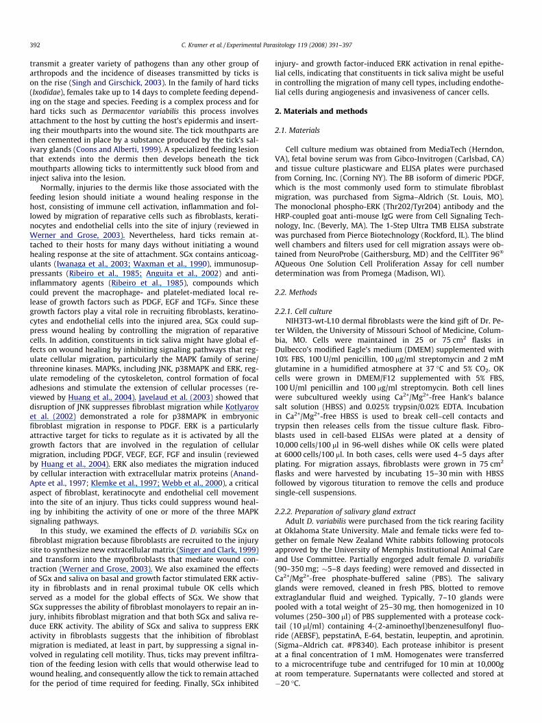
transmit a greater variety of pathogens than any other group ofarthropods and the incidence of diseases transmitted by ticks ison the rise (Singh and Girschick, 2003). In the family of hard ticks(Ixodidae), females take up to 14 days to complete feeding depend-ing on the stage and species. Feeding is a complex process and forhard ticks such as Dermacentor variabilis this process involvesattachment to the host by cutting the host’s epidermis and insert-ing their mouthparts into the wound site. The tick mouthparts arethen cemented in place by a substance produced by the tick’s sal-ivary glands (Coons and Alberti, 1999). A specialized feeding lesionthat extends into the dermis then develops beneath the tickmouthparts allowing ticks to intermittently suck blood from andinject saliva into the lesion.
Normally, injuries to the dermis like those associated with thefeeding lesion should initiate a wound healing response in thehost, consisting of immune cell activation, inflammation and fol-lowed by migration of reparative cells such as fibroblasts, kerati-nocytes and endothelial cells into the site of injury (reviewed inWerner and Grose, 2003). Nevertheless, hard ticks remain at-tached to their hosts for many days without initiating a woundhealing response at the site of attachment. SGx contains anticoag-ulants (Iwanaga et al., 2003; Waxman et al., 1990), immunosup-pressants (Ribeiro et al., 1985; Anguita et al., 2002) and anti-inflammatory agents (Ribeiro et al., 1985), compounds whichcould prevent the macrophage- and platelet-mediated local re-lease of growth factors such as PDGF, EGF and TGFa. Since thesegrowth factors play a vital role in recruiting fibroblasts, keratino-cytes and endothelial cells into the injured area, SGx could sup-press wound healing by controlling the migration of reparativecells. In addition, constituents in tick saliva might have global ef-fects on wound healing by inhibiting signaling pathways that reg-ulate cellular migration, particularly the MAPK family of serine/threonine kinases. MAPKs, including JNK, p38MAPK and ERK, reg-ulate remodeling of the cytoskeleton, control formation of focaladhesions and stimulate the extension of cellular processes (re-viewed by Huang et al., 2004). Javelaud et al. (2003) showed thatdisruption of JNK suppresses fibroblast migration while Kotlyarovet al. (2002) demonstrated a role for p38MAPK in embryonicfibroblast migration in response to PDGF. ERK is a particularlyattractive target for ticks to regulate as it is activated by all thegrowth factors that are involved in the regulation of cellularmigration, including PDGF, VEGF, EGF, FGF and insulin (reviewedby Huang et al., 2004). ERK also mediates the migration inducedby cellular interaction with extracellular matrix proteins (Anand-Apte et al., 1997; Klemke et al., 1997; Webb et al., 2000), a criticalaspect of fibroblast, keratinocyte and endothelial cell movementinto the site of an injury. Thus ticks could suppress wound heal-ing by inhibiting the activity of one or more of the three MAPKsignaling pathways.
In this study, we examined the effects of D. variabilis SGx onfibroblast migration because fibroblasts are recruited to the injurysite to synthesize new extracellular matrix (Singer and Clark, 1999)and transform into the myofibroblasts that mediate wound con-traction (Werner and Grose, 2003). We also examined the effectsof SGx and saliva on basal and growth factor stimulated ERK activ-ity in fibroblasts and in renal proximal tubule OK cells whichserved as a model for the global effects of SGx. We show thatSGx suppresses the ability of fibroblast monolayers to repair an in-jury, inhibits fibroblast migration and that both SGx and saliva re-duce ERK activity. The ability of SGx and saliva to suppress ERKactivity in fibroblasts suggests that the inhibition of fibroblastmigration is mediated, at least in part, by suppressing a signal in-volved in regulating cell motility. Thus, ticks may prevent infiltra-tion of the feeding lesion with cells that would otherwise lead towound healing, and consequently allow the tick to remain attachedfor the period of time required for feeding. Finally, SGx inhibited
injury- and growth factor-induced ERK activation in renal epithe-lial cells, indicating that constituents in tick saliva might be usefulin controlling the migration of many cell types, including endothe-lial cells during angiogenesis and invasiveness of cancer cells.
2. Materials and methods
2.1. Materials
Cell culture medium was obtained from MediaTech (Herndon,VA), fetal bovine serum was from Gibco-Invitrogen (Carlsbad, CA)and tissue culture plasticware and ELISA plates were purchasedfrom Corning, Inc. (Corning NY). The BB isoform of dimeric PDGF,which is the most commonly used form to stimulate fibroblastmigration, was purchased from Sigma–Aldrich (St. Louis, MO).The monoclonal phospho-ERK (Thr202/Tyr204) antibody and theHRP-coupled goat anti-mouse IgG were from Cell Signaling Tech-nology, Inc. (Beverly, MA). The 1-Step Ultra TMB ELISA substratewas purchased from Pierce Biotechnology (Rockford, IL). The blindwell chambers and filters used for cell migration assays were ob-tained from NeuroProbe (Gaithersburg, MD) and the CellTiter 96�
AQueous One Solution Cell Proliferation Assay for cell numberdetermination was from Promega (Madison, WI).
2.2. Methods
2.2.1. Cell cultureNIH3T3-wt-L10 dermal fibroblasts were the kind gift of Dr. Pe-
ter Wilden, the University of Missouri School of Medicine, Colum-bia, MO. Cells were maintained in 25 or 75 cm2 flasks inDulbecco’s modified Eagle’s medium (DMEM) supplemented with10% FBS, 100 U/ml penicillin, 100 lg/ml streptomycin and 2 mMglutamine in a humidified atmosphere at 37 �C and 5% CO2. OKcells were grown in DMEM/F12 supplemented with 5% FBS,100 U/ml penicillin and 100 lg/ml streptomycin. Both cell lineswere subcultured weekly using Ca2+/Mg2+-free Hank’s balancesalt solution (HBSS) and 0.025% trypsin/0.02% EDTA. Incubationin Ca2+/Mg2+-free HBSS is used to break cell–cell contacts andtrypsin then releases cells from the tissue culture flask. Fibro-blasts used in cell-based ELISAs were plated at a density of10,000 cells/100 ll in 96-well dishes while OK cells were platedat 6000 cells/100 ll. In both cases, cells were used 4–5 days afterplating. For migration assays, fibroblasts were grown in 75 cm2
flasks and were harvested by incubating 15–30 min with HBSSfollowed by vigorous tituration to remove the cells and producesingle-cell suspensions.
2.2.2. Preparation of salivary gland extractAdult D. variabilis were purchased from the tick rearing facility
at Oklahoma State University. Male and female ticks were fed to-gether on female New Zealand White rabbits following protocolsapproved by the University of Memphis Institutional Animal Careand Use Committee. Partially engorged adult female D. variabilis(90–350 mg; �5–8 days feeding) were removed and dissected inCa2+/Mg2+-free phosphate-buffered saline (PBS). The salivaryglands were removed, cleaned in fresh PBS, blotted to removeextraglandular fluid and weighed. Typically, 7–10 glands werepooled with a total weight of 25–30 mg, then homogenized in 10volumes (250–300 ll) of PBS supplemented with a protease cock-tail (10 ll/ml) containing 4-(2-aminoethyl)benzenesulfonyl fluo-ride (AEBSF), pepstatinA, E-64, bestatin, leupeptin, and aprotinin.(Sigma–Aldrich cat. #P8340). Each protease inhibitor is presentat a final concentration of 1 mM. Homogenates were transferredto a microcentrifuge tube and centrifuged for 10 min at 10,000gat room temperature. Supernatants were collected and stored at�20 �C.
C. Kramer et al. / Experimental Parasitology 119 (2008) 391–397 393
2.2.3. Collection of tick salivaPartially engorged females (90–350 mg; �5–8 days feeding)
were removed from the rabbit and attached to a slide (3–4 at atime) using double-sided adhesive tape. Each tick was injectedwith 10 ll of TS/MOPS (10 mM dopamine/theophylline in MOPSbuffered tick saline, pH 7.0, with 3% DMSO (Needham and Sauer,1979). If a tick failed to produce saliva by 5 min post-injection, itwas re-injected. If it failed to produce saliva within 5 min of thesecond injection, it was discarded. Ticks producing saliva weretreated as follows: saliva was collected using a 5 or 25 ll non-heparinized soda lime glass pipet. Once a tick had stopped pro-ducing saliva (�5 min with no additional production), it was in-jected with a second dose of TS/MOPS (10 ll). At the end of thesecond round of injection (5 min without additional production),the tick was injected for the third and final time. Routinely,�20% of the ticks do not respond to any stimulation and the aver-age production of saliva/tick is 4 ll/tick. Saliva was stored at�20 �C until needed.
2.2.4. Histological preparation of the tick feeding lesionRabbits were humanely sacrificed according to approved proto-
cols on record with the University of Memphis Institutional AnimalCare and Use Committee. Ticks in the rapid engorgement stage offeeding were fixed in situ with the feeding lesion in 4% paraformal-dehyde in PBS and excised using a surgical punch. Mouth partswere removed at this time to facilitate sectioning. Tissues weredehydrated in an ascending series of ethanols and embedded inparaffin. Sections (5 lm) were cut, stained with hematoxylin andeosin and examined with bright field optics using a Nikon Labo-phat microscope. Images were recorded with an RT Color Diagnos-tic Spot camera (0.76� lens) and transferred to Adobe PhotoshopTM
(Adobe Systems, Inc, San Jose, CA) where they were saved withoutfurther modification.
2.2.5. Monolayer injuryMonolayer scrape injuries were used to address the ability of
fibroblast monolayers to repair a wound. Cells grown to confluencyin six-well dishes were pretreated 30 min with increasing amountsof SGx and wounds were then made by scraping the monolayerwith a 100 ll sterile pipette tip using the technique described in El-lis et al. (2001). The initial width of the injury was viewed throughthe 10� objective of a phase contrast microscope and measured inarbitrary units using an ocular micrometer. Four and 24 h later, thewidth of the injury was re-measured and reported as the percentclosure of the injury.
2.2.6. Cell migration assayFibroblast chemotaxis (directed movement) and chemokinesis
(undirected increase in migratory behavior) were assessed usingfilter assays and single well chemotaxis (blind well) chambers.Confluent monolayers of fibroblasts were incubated for 15–20 min with Ca2+/Mg2+-free HBSS then removed from 75 cm2 flasksby tituration. Cells were resuspended in serum-free DMEM at aconcentration of 1 � 105 cells/ml. The lower chamber of the blindwell was loaded with serum-containing growth medium or PDGFas described in figure legends (see Fig. 3) and an 8 lm polycarbon-ate track-etch filter was placed between the lower and upperchambers. The upper chamber was then loaded with fibroblasts(100,000 cells/ml, 100 ll/chamber) treated with increasingamounts of SGx or vehicle (PBS). After a 4 h incubation at 37 �Cin a CO2 incubator, the chambers were opened and the filter wasremoved. Cells on the upper surface were removed with a cottonswab and the filter was fixed with 75% methanol, stained with Har-ris’ hematoxylin and mounted on a glass microscope slide. Cells onthe under surface of the filter were then counted in five high-power (40�) fields of a light microscope.
2.2.7. Cell number assayCell number was determined colorimetrically using the CellTit-
er 96� AQueous One Solution Cell Proliferation Assay, which em-ploys the tetrazolium compound MTS and an electron couplingreagent (phenazine ethosulfate). MTS is reduced into a solublecolored formazan product the absorbance of which is directlyproportional to the number of living cells in culture. Cells grownin 96-well tissue culture plates were treated as described in figurelegends followed by incubation for 2 h with MTS (20 ll/well).Plates were read on a Bio-Tek ELx808 Ultra Microplate Reader at490 nm, the data were normalized to basal absorption and re-ported as fold increase over basal.
2.2.8. Phosphoantibody cell-based ELISA (PACE)ERK activity was measured by a modification of the phosphoan-
tibody cell-based ELISA (PACE) described in Versteeg et al. (2000).Twenty-four hours prior to each experiment, cells grown in 96-well ELISA plates were changed to serum-free medium and drugswere added directly to the medium in each well. Cells were incu-bated for the times indicated in each figure and treatments wereterminated by adding ice-cold PBS to each well. Wells werewashed once and cells were fixed in 4% formaldehyde. After block-ing with 5% milk in PBS containing 0.1% Triton X-100 (PBST), cellswere incubated overnight at 4 �C with an activation-specific phos-pho-ERK (Thr202/Tyr204) antibody (1:4000). Plates were washedthree times with PBST, incubated 1 h with an HRP-coupled secondantibody and developed with 1-Step Ultra TMB ELISA substrate.Absorbance was read at 450 nm using a Bio-Tek ELx808 UltraMicroplate Reader and data were normalized to time-matched orvehicle controls.
2.2.9. Statistical analysesData are presented as means ± SEM of 3–6 experiments assayed
in duplicate or triplicate and performed over several passages ofcells. Statistical significance was determined by one-way ANOVAand Dunnett’s post-test for multiple comparisons using GraphPadPrism version 3.02 for Windows (GraphPad Software, San DiegoCA, http://www.graphpad.com. Differences in means were consid-ered significant at p 6 0.05.
3. Results
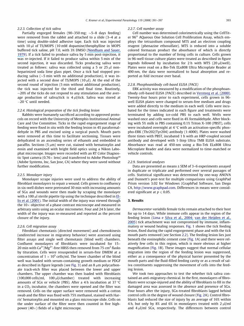
Dermacentor variabilis female ticks remain attached to their hostfor up to 14 days. While immune cells appear in the region of thefeeding lesion (Lima e Silva et al., 2004; van der Heijden et al.,2005) tick attachment was not compromised by immune, inflam-matory or wound healing responses. Fig. 1 shows the tick feedinglesion, fixed during the rapid engorgement phase and with the tickmouth parts removed (see Section 2.2). The feeding lesion lies justbeneath the eosinophilic cement cone (Fig. 1A) and there were rel-atively few cells in this region, which is more obvious at highermagnification (Fig. 1B). These images suggest that normal cellularmigration into the region of the feeding lesion was suppressed,either as a consequence of the physical barrier presented by themouth parts and the fluid-filled feeding cavity or as a result of sal-iva constituents suppressing the movement of cells into the feed-ing lesion.
We took two approaches to test the whether tick saliva con-tained an anti-migratory chemical. In the first, monolayers of fibro-blasts were scrape-injured and the ability of fibroblasts to fill in thedamaged area was assessed in the absence and presence of SGx.Four hours after the injury, SGx-treated fibroblasts lagged behindcontrol cells in the degree of wound closure (Fig. 2A). Control fibro-blasts had reduced the size of injury by an average of 16% within4 h, but only by 8% and 6% in monolayers treated with 2 ll/mland 4 ll/ml SGx, respectively. The differences between control

Fig. 1. Histological examination of the tick feeding lesion. (A) Low magnification ofthe tick feeding lesion showing cement cone (CC), the region around the mouth-parts (MP) and the feeding cavity (FC) has relatively few cells. (B) Higher magnif-ication of the feeding lesion. Scale bar equals 100 lm in both (A) and (B).
Fig. 2. Treatment with SGx suppresses closure on monolayer injuries without aff-ecting fibroblast number. SGx was diluted to 2 or 4 ll/ml in DMEM containing 0.1%BSA. Fibroblast monolayers were treated 30 min with SGx or PBS (0), then injured asdescribed in Section 2.2. The initial width of the injury was measured and then re-measured 4 and 24 h later. (A) Treatment with SGx slowed the closure of a mon-olayer injury at 4 h but there was no difference between control and SGx-treatedcells at 24 h. Data are means ± SEM, n = 4. *p 6 0.05 compared to control. (B) Thedelayed wound healing seen in SGx-treated cells was not the result of SGx-induceddecreases in cell number. Fibroblasts were treated with SGx for 30 min, injured asdescribed in Section 2.2, and cell number was determined colorimetrically.
Fig. 3. SGx suppresses serum- and PDGF-stimulated fibroblast migration. (A) Blindwell chemotaxis chamber. See Section 2.2 for a description of the assay. (B) SGxinhibits serum-induced fibroblast migration. Cells were treated 30 min with SGxdiluted to 2 or 4 ll/ml in DMEM containing 0.1% BSA or PBS (0) then loaded into theupper chamber of a blind well. After 4 h, the filters were processed as described inSection 2.2 and the number of cells migrating was determined by counting in 5–40� microscopic fields. Data are means ± SEM, n = 4. *p 6 0.05 compared to contr-ols. (B) SGx inhibits PDGF-stimulated fibroblast migration. Cells were pretreated for30 min with increasing volumes of SGx then exposed to a chemotactic signal pro-vided by PDGF (100 ng/ml) using blind well assays. Data are means ± SEM, n = 4.**p 6 0.001 compared to untreated control, #p 6 0.05 compared to PDGF withoutSGx.
394 C. Kramer et al. / Experimental Parasitology 119 (2008) 391–397
and SGx-treated monolayers were significant at 4 ll/ml. While theeffects of SGx were not huge, they reflect a single exposure to SGxwhile ticks salivate and suck blood continually during the entireattachment period. By 24 h post-injury, there were no significantdifferences between control and SGx-treated cells, possibly reflect-ing the loss of biological activity of the SGx. To determine if the dif-
ferences at 4 h were due to an SGx-mediated reduction in cellnumber, control and SGx-pretreated fibroblasts were scrape-in-jured and cell number estimated by incubating with the MTS tetra-zolium compound described in Section 2.2. There were nosignificant differences in labeling between control and SGx-treatedcells at 4 or 24 h (Fig. 2B), indicating that the inability to close thewound in SGx-treated cultures was most likely the result of sup-pressing fibroblast migration, rather than a reduction in cellnumber.
To directly measure the effect of SGx on fibroblast migration,we used blind well assays to assess the movement of fibroblastsfrom the upper to the lower surface of an 8 lm filter (Fig. 3A).Fibroblasts were pretreated for 30 min with increasing amountsof SGx then loaded into the upper chamber of a blind well wherethe lower chamber contained growth medium with 10% FBS. Aftera 4 h incubation, the migration rate in response to 10% FBS was2165 ± 678 cells/4 h (Fig. 3B). Pretreatment with 2 ll/ml SGx re-duced migration to 790 ± 96 cells/4 h while 4 ll/ml reduced migra-tion to 610 ± 143 cells/4 h. These data indicate that SGx dose-dependently reduced the migratory behavior of fibroblasts. Todetermine if migratory responses to a specific chemotactic–che-mokinetic signal were also compromised by SGx, medium contain-ing 0.1% BSA or PDGF (10 ng/ml) was loaded into the lowerchamber of a blind well, then control or SGx-treated fibroblastswere loaded into the upper chamber. Fig. 3B shows that migrationin the presence of 0.1% BSA averaged 416 ± 231 cells/4 h whilePDGF increased migration to 1373 ± 128 cells/4 h. Pretreating cellsfor 30 min with increasing volumes of SGx suppressed PDGF-stim-ulated cell migration by �40% at 2 ll/ml (791 ± 152 cells/4 h) andby �50% at 4 ll/ml (682 ± 82 cells/4 h). Although SGx appeared tostimulate migration in the absence of PDGF, these increases werenot statistically different from untreated controls.

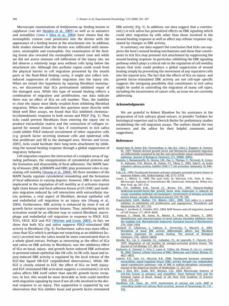
Fig. 5. Effect of SGx on injury- and EGF-induced ERK activation in OK cells. (A) SGxreduced both basal and injury-induced increases in ERK activity. ERK activity wasmeasured in uninjured cells or in cells 15 min after scrape injury. ERK activationwas determined by PACE assay as described in Section 2.2. Data are means ± SEM,n = 3. *,**p 6 0.05 and 0.001 compared to group control and #p 6 0.05 compared toinjury-induced ERK activation alone. (B) SGx inhibits EGF-stimulated ERK activationin OK cells. OK cells were pretreated 30 min with SGx, then incubated 5 min withEGF (100 ng/ml). ERK activity was then determined by PACE assay. Data are mea-ns ± SEM, n = 3. *p 6 0.05 compared to EGF alone.
C. Kramer et al. / Experimental Parasitology 119 (2008) 391–397 395
The serine/threonine kinase ERK plays an important role inmediating growth factor-induced cellular migration (Huang et al.,2004). Because PDGF stimulated fibroblast migration, it seemedpossible that SGx might alter ERK activation by this growth factor.We first determined the effects of PDGF on ERK activity in the fibro-blasts. PDGF caused dose dependent activation with 0.1 ng/mlincreasing ERK activity 26%, 1 ng/ml increasing activity 2.1-foldand 10 ng/ml increasing activity 3.0-fold (Fig. 4A). To determinethe effect of SGx on basal and PDGF-induced ERK activation, fibro-blasts were pretreated with increasing volumes of SGx or vehicle,then incubated with 1 ng/ml PDGF. SGx pretreatment did not signif-icantly increase basal ERK activity compared to the untreated con-trol. PDGF (1 ng/ml) increased ERK activity 2.2 ± 0.2-fold and SGxpretreatment reduced PDGF-stimulated ERK with 4 ll SGx/ml(which is equivalent to 0.8 lg of protein/ml) significantly reducingthe PDGF response by�40% (from 2.2 ± 0.2-fold to 1.5 ± 0.2 � basal)(Fig. 4B). Although SGx suppressed PDGF-stimulated ERK activity, itis possible these effects were related to cellular constituents ratherthan saliva. To address this possibility, we collected saliva as de-scribed in Section 2.2 and measured its effect on ERK activation byPDGF. Saliva has no effect on basal ERK activity but inhibitedPDGF-stimulated ERK activity (Fig. 4C). In fact, saliva was more effi-cacious than SGx with as little as 0.5 ll/ml (100 ng of protein) com-pletely blocking ERK activation by PDGF. The ability of SGx andsaliva to suppress PDGF-stimulated ERK activity suggests that theinhibition of fibroblast migration is mediated, at least in part, bysuppressing growth factor-stimulating ERK activation.
Based on the latter observations in fibroblasts, we wondered ifSGx would affect growth factor-dependent signaling in cells notparticipating in wound healing responses. To test this hypothesis,we used proximal tubule epithelial-like OK cells which expressEGF receptors and respond to injury and EGF with increases inERK activity (Cole, 1999; Lederer et al., 2000). Treating uninjuredOK cells with increasing amounts of SGx dose-dependently re-duced basal ERK activity with 10 ll/ml reducing basal activity by25% (Fig. 5A, left). Scrape-injuring OK cells elevated basal ERKactivity to 1.6 � basal (Fig. 5A, right) most likely the result of in-jury-induced release of the EGF-like growth factor HB-EGF fromthese cells (Sneddon et al., 2006). Injury-induced increases in OKcell ERK activity were inhibited by SGx with 10 ll/ml SGx signifi-cantly reducing ERK activity to near-basal levels. To directly dem-onstrate an effect of SGx on growth factor-stimulated ERK activity,we pretreated OK cells for 30 min with SGx and then incubated for5 min with 100 ng/ml EGF (Fig. 5B). EGF increased ERK activity3.7 ± 0.1-fold over basal, a response that was dose-dependentlyinhibited by SGx. These data indicate that the effects of SGx on
Fig. 4. PDGF-stimulated ERK activation is inhibited by SGx. (A) PDGF dose-dependently inactivity was measured by PACE assay. Data are means ± SEM, n = 3. (B) SGx pretreatmincreasing volumes of SGx then incubated for 5 min with 1 ng/ml PDGF. Data are meanalone. (C) Tick saliva inhibits ERK activation by PDGF. Cells were pretreated with saliva fo**p 6 0.001 compared to basal activity, #p 6 0.001 compared to PDGF alone.
ERK signaling are not limited to fibroblasts or other cells typicallypresent at the site of an injury. Instead, SGx may have global effectson ERK activity, an issue we are currently exploring.
4. Discussion
To complete feeding, hard ticks must remain attached to theirhost for up to 14 days. Normally, dermal injuries like those causedby tick attachment should initiate the host’s wound healing re-sponse. This includes the recruitment of immune cells (neutro-phils, monocytes and macrophages), macrophage-mediatedrelease of growth factors and cytokines and the recruitment ofkeratinocytes, dermal fibroblasts and endothelial cells to the injurysite (Singer and Clark, 1999; Werner and Grose, 2003). However,tick feeding lesions do not heal during the period of tick attach-ment, suggesting that ticks suppress part or all of the wound heal-ing response. A recent study showed that saliva from the hard tickIxodes scapularis inhibits endothelial cell proliferation and angio-genesis and thus may negatively impact angiogenesis-dependentwound healing and tissue repair (Francischetti et al., 2005). In thisstudy, we examined how controlling cell migration might contrib-ute to the ability of a tick to remain attached to its host for theduration of the feeding period.
creases ERK activity in fibroblasts. Cells were treated for 5 min with PDGF, then ERKent inhibits PDGF-stimulated ERK activity. Cells were pretreated for 30 min withs ± SEM, n = 3. **p 6 0.001 compared to basal activity, +p 6 0.05 compared to PDGF
r 30 min, then incubated with 10 ng/ml PDGF for 5 min. Data are means ± SEM, n = 5.
396 C. Kramer et al. / Experimental Parasitology 119 (2008) 391–397
Microscopic examinations of Amblyomma sp. feeding lesions incapybaras (van der Heijden et al., 2005) as well as in anteatersand armadillos (Lima e Silva et al., 2004) have shown that theeosinophilic cement cone penetrates into the dermis with theappearance of a feeding lesion at the attachment site. In addition,both studies showed that the dermis was infiltrated with mono-cytes, neutrophils and eosinophils. Our examination of the feed-ing lesion also revealed the eosinophilic cement cone and whilewe did not assess immune cell infiltration of the injury site, wedid observe a relatively large area without cells lying below theattachment site. Although this acellular region could result fromthe physical barrier to cell migration presented by the mouthparts or the fluid-filled feeding cavity, it might also reflect tick-induced suppression of cellular migration into the injury site.When we tested this hypothesis by injuring fibroblast monolay-ers, we discovered that SGx pretreatment inhibited repair ofthe damaged area. While this type of wound healing reflects acombination of migration and proliferation, our data indicatethere was no effect of SGx on cell number. Thus, the inabilityto close the injury most likely resulted from inhibiting fibroblastmigration. When we addressed this question more directly withblind well filter assays, we found that SGx inhibited chemotac-tic/chemokinetic response to both serum and PDGF (Fig. 3). Thusticks could prevent fibroblasts from entering the injury site toproduce extracellular matrix and the contraction of collagen re-quired for wound closure. In fact, if constituents in tick salivacould inhibit PDGF-induced recruitment of other reparative cells(e.g. growth factor secreting immune cells and epidermal cellsthat proliferate and fill in the damaged area; Werner and Grose,2003), ticks could facilitate their long-term attachment by inhib-iting the wound healing response through a global suppression ofmigratory behavior.
Cell migration requires the interaction of a complex array of sig-naling pathways, the reorganization of cytoskeletal proteins andthe formation and disassembly of focal adhesions. The MAPK fam-ily of kinases (JNK, p38MAPK and ERK) play prominent roles in reg-ulating cell motility (Huang et al., 2004). All three members of theMAPK family regulate cytoskeletal remodeling and the formationof focal adhesions to varying degrees; however, ERK is most oftenimplicated in the regulation of cell motility as it activates myosinlight chain kinase and focal adhesion kinase p125 (FAK) and medi-ates migration induced by cell interaction with extracellular ma-trix proteins, a vital aspect of fibroblast, dermal epithelial celland endothelial cell migration to an injury site (Huang et al.,2004). Furthermore, ERK activity is enhanced by most if not allgrowth factor receptor tyrosine kinases. Thus, interfering with itsactivation would be an efficient way to control fibroblast, macro-phage and endothelial cell migration in response to PDGF, EGF,TGFa, VEGF, KGF and FGF (Werner and Grose, 2003). In fact, weshow that both SGx and saliva suppress PDGF-stimulated ERKactivity in fibroblasts (Fig. 4). Furthermore, saliva was more effica-cious than SGx which is perhaps not surprising as an inhibitory fac-tor(s) secreted into the saliva would be more concentrated than ina whole gland extract. Perhaps as interesting as the effect of SGxand saliva on ERK activity in fibroblasts, was the inhibitory effectof SGx on basal, injury- and growth factor-induced ERK activationin proximal tubule epithelial-like OK cells. In OK cells, basal and in-jury-induced ERK activity is regulated by the local release of theEGF-like ligand HB-EGF (unpublished observations). While HB-EGF is closely related to EGF, the effect of SGx on both injury-and EGF-stimulated ERK activation suggests a constituent(s) in ticksaliva affects ERK itself rather than specific growth factor recep-tors. In fact, this would be more desirable as ticks could interferewith migration signaling by most if not all cells involved in the der-mal response to an injury. This supposition is supported by ourobservation that SGx inhibits basal and growth factor-stimulated
ERK activity (Fig. 5). In addition, our data suggest that a constitu-ent(s) in tick saliva has generalized effects on ERK signaling whichcould alter migration by cells other than those involved in thewound healing response as well as affect any cellular response reg-ulated by changes in ERK activity.
In summary, our data support the conclusion that ticks can sup-press the host’s wound healing mechanisms and show that constit-uents in tick SGx may promote tick attachment by suppressing thewound healing response. In particular, inhibiting the ERK signalingpathway which plays a critical role in the regulation of cell motilitymeans that ticks could produce a global suppression of woundhealing simply by preventing the movement of any reparative cellsinto the injured area. The fact that the effects of SGx on injury- andgrowth factor-stimulated ERK activity are not cell-type specificsuggests the intriguing possibility that constituents in tick salivamight be useful in controlling the migration of many cell types,including the invasiveness of cancer cells, an issue we are currentlyaddressing.
Acknowledgments
We are grateful to Robert Blaudow for his assistance in thepreparation of tick salivary gland extract, to Jennifer Tzefakes forhistological expertise and to Derrick Butler for preliminary studiesestablishing the cell migration assays. The authors thank the tworeviewers and the editor for their helpful comments andsuggestions.
References
Anand-Apte, B., Zetter, B.R., Viswanathan, A., Qiu, R.G., Chen, J., Ruggieri, R., Symons,M., 1997. Platelet-derived growth factor and fibronectin-stimulated migrationare differentially regulated by the Rac and extracellular signal-regulated kinasepathways. Journal of Biological Chemistry 272, 30688–30692.
Anguita, J., Ramamoorthi, N., Hovius, J.W., Das, S., Thomas, V., Persinski, R., Conze,D., Askenase, P.W., Rincon, M., Kantor, F.S., Fikrig, E., 2002. Salp15, an Ixodesscapularis salivary protein, inhibits CD4(+) T cell activation. Immunity 16, 849–859.
Cole, J.A., 1999. Parathyroid hormone activates mitogen-activated protein kinase inopossum kidney cells. Endocrinology 140, 5771–5779.
Coons, L., Alberti, G., 1999. The acari ticks. In: Harrison, F.W., Felix, R. (Eds.),Microscopic Anatomy of Invertebrates 8B, Chelicerate Arthropoda. Wiley-Liss,New York, pp. 267–514.
Ellis, P.D., Hadfield, K.M., Pascall, J.C., Brown, K.D., 2001. Heparin-bindingepidermal-growth-factor-like growth factor gene expression is induced byscrape-wounding epithelial cell monolayers: involvement of mitogen-activatedprotein kinase cascades. Biochemical Journal 354, 99–106.
Francischetti, I.M.B., Mather, T.N., Ribeiro, J.M.C., 2005. Tick saliva is a potentinhibitor of endothelial cell proliferation and angiogenesis. Thrombosis andHaemostasis 94, 167–174.
Huang, C., Jacobson, K., Schaller, M.D., 2004. MAP kinases and cell migration. Journalof Cell Science 117, 4619–4628.
Iwanaga, S., Okada, M., Isawa, H., Morita, A., Yuda, M., Chinzei, Y., 2003.Identification and characterization of novel salivary thrombin inhibitors fromthe ixodidae tick, Haemaphysalis longicornis. European Journal of Biochemistry270, 1926–1934.
Javelaud, D., Laboureau, J., Gabison, E., Verrecchia, F., Mauviel, A., 2003.Disruption of basal JNK activity differentially affects key fibroblastfunctions important for wound healing. The Journal of BiologicalChemistry 278, 24624–24628.
Klemke, R.L., Cai, S., Giannini, A.L., Gallagher, P.J., de Lanerolle, P., Cheresh, D.A.,1997. Regulation of cell motility by mitogen-activated protein kinase. TheJournal of Cell Biology 137, 481–492.
Kotlyarov, A., Yannoni, Y., Fritz, S., Laass, K., Telliez, J.B., Pitman, D., Lin, L.L., Gaestel,M., 2002. Distinct cellular functions of MK2. Molecular and Cellular Biology 22,4827–4835.
Lederer, E.D., Sohi, S.S., McLeish, K.R., 2000. Parathyroid hormone stimulatesextracellular signal-regulated kinase (ERK) activity through two independentsignal transduction pathways: role of ERK in sodium-phosphate cotransport.Journal of the American Society of Nephrology 11, 222–231.
Lima e Silva, M.F., Szabo, M.P., Bechara, G.H., 2004. Microscopic features oftick-bite lesions in anteaters and armadillos: Emas National Park and thePantanal region of Brazil. Annals of the New York Academy of Sciences1026, 235–241.
Needham, G.R., Sauer, J.R., 1979. Involvement of calcium and cyclic AMP incontrolling ixodid tick salivary fluid secretion. Journal of Parasitology 65, 531–542.

C. Kramer et al. / Experimental Parasitology 119 (2008) 391–397 397
Ribeiro, J.M., Makoul, G.T., Levine, J., Robinson, D.R., Spielman, A., 1985.Antihemostatic, antiinflammatory, and immunosuppressive properties of thesaliva of a tick, Ixodes dammini. Journal of Experimental Medicine 161, 332–344.
Singer, A.J., Clark, R.A., 1999. Cutaneous wound healing. The New England Journal ofMedicine 341, 738–746.
Singh, S.K., Girschick, H.J., 2003. Tick-host interactions and their immunologicalimplications in tick-borne diseases. Current Science 85, 1284–1298.
Sneddon, W.B., Yang, Y.A., Ba, J., Harinstein, L.A., Friedman, P.A., 2006. Extracellularsignal-regulated kinase activation by parathyroid hormone in distal tubulecells. American Journal of Physiology. Renal Physiology 292, F1028–F1034.
van der Heijden, K.M., Szabo, M.P., Egami, M.I., Pereira, M.C., Matushima, E.R., 2005.Histopathology of tick-bite lesions in naturally infested capybaras(Hydrochoerus hydrochaeris) in Brazil. Experimental & Applied Acarology 37,245–255.
Versteeg, H.H., Nijhuis, E., van den Brink, G.R., Evertzen, M., Pynaert, G.N., vanDeventer, S.J., Coffer, P.J., Peppelenbosch, M.P., 2000. A new phosphospecificcell-based ELISA for p42/p44 mitogen-activated protein kinase (MAPK), p38MAPK, protein kinase B and cAMP-response-element-binding protein. TheBiochemical Journal 350 (Pt 3), 717–722.
Waxman, L., Smith, D.E., Arcuri, K.E., Vlasuk, G.P., 1990. Tick anticoagulant peptide(TAP) is a novel inhibitor of blood coagulation factor Xa. Science 248, 593–596.
Webb, D.J., Nguyen, D.H., Gonias, S.L., 2000. Extracellular signal-regulated kinasefunctions in the urokinase receptor-dependent pathway by whichneutralization of low density lipoprotein receptor-related protein promotesfibrosarcoma cell migration and matrigel invasion. Journal of Cell Science 113(Pt 1), 123–134.
Werner, S., Grose, R., 2003. Regulation of wound healing by growth factors andcytokines. Physiological Reviews 83, 835–870.
Copyright © 2022 FDOKUMEN