Relationships between saliva and food bolus properties from model dairy products

This article appeared in a journal published by Elsevier. The attachedcopy is furnished to the author for internal non-commercial researchand education use, including for instruction at the authors institution
and sharing with colleagues.
Other uses, including reproduction and distribution, or selling orlicensing copies, or posting to personal, institutional or third party
websites are prohibited.
In most cases authors are permitted to post their version of thearticle (e.g. in Word or Tex form) to their personal website orinstitutional repository. Authors requiring further information
regarding Elsevier’s archiving and manuscript policies areencouraged to visit:
http://www.elsevier.com/copyright

Author's personal copy
Measuring cortisol in hair and saliva from dogs:coat color and pigment differences
A. Bennett, V. Hayssen*Department of Biology, Smith College, Northampton, MA, USA
Abstract
Cortisol concentrations are frequently measured from a variety of sources including blood, saliva, urine, and feces to quantifystress in dogs. However, a need still exists for less intrusive collection methods in domestic animals and for more efficient meansof measuring basal cortisol. The objectives of the present study were to minimize restraint for saliva sampling, to validate hairfor basal cortisol measurement in dogs, and to determine concentrations of cortisol within the hair shaft and in relation to haircolor. Using food luring, 79% of dogs required no restraint for saliva collection. Salivary and hair cortisol concentrations werepositively correlated (P � 0.001), thus validating hair as a medium for basal cortisol quantification. Black dogs had less cortisolthan nonblack dogs (P � 0.039) in hair, but not saliva. Across dogs, the average amount of cortisol did not differ between proximaland distal hair sections (P � 0.348). However, for 7 of the 9 dogs, more cortisol was present in the distal portions of the hair.We observed a difference in cortisol concentrations among hairs of different colors from individual dogs (P � 0.001). From thesame 7 � 7 cm ischiatic patch from the same dog, black (eumelanin) hairs were consistently lower in cortisol than yellow(pheomelanin) hairs, and cortisol concentrations of agouti hairs were intermediate. This is the first evidence that hair of differentcolors might sequester cortisol differently.© 2010 Elsevier Inc. All rights reserved.
Keywords: Stress; Glucocorticoid; Hypothalamic-pituitary-adrenal axis; Pheomelanin; Eumelanin; Canis
1. Introduction
Cortisol is at the center of an interconnected web ofphysiological, behavioral, and developmental functionsand responses. Cortisol has long been considered areliable physiological measure of the stress response inboth domestic mammals (cows [1] goats [2], guineapigs [3], horses [4], pigs [5], rats [6], and sheep [7]) andwild mammals (Antechinus [8], Macaca [9], Odocoil-eius [10], Peromyscus [11], Procavia [12], Puma [13],Saimiri [14], and Spermophilus [15]). In the short term,
stress is adaptive and helps individuals cope withemerging situations, but over the long term, stress ismaladaptive [16]. Overexposure to glucocorticoids ow-ing to malfunction or long-term (chronic) hyperactiva-tion of the stress system can damage the body, impairgrowth and development, and elicit endocrine, meta-bolic, autoimmune, or psychiatric disorders [17].
Cortisol is a key component of the stress response.Measuring cortisol usually requires invasive procedures,such as venous collection of blood, which may signifi-cantly disrupt behavior. Animal welfare concerns havecontributed to the development of noninvasive cortisolsampling methods, each with its own strengths and weak-nesses.
Cortisol concentrations are regularly assessedthrough blood [18,19], saliva [20–22], urine [23], and
* Corresponding author. Department of Biology, Smith College,Northampton, MA 01062, USA; Phone: (413) 585-3856; Fax: (413)585-3786.
E-mail address: [email protected] (V. Hayssen)
Available online at www.sciencedirect.com
Domestic Animal Endocrinology 39 (2010) 171–180www.domesticanimalendo.com
0739-7240/10/$ – see front matter © 2010 Elsevier Inc. All rights reserved.doi:10.1016/j.domaniend.2010.04.003

Author's personal copy
feces [24]. However, each sampling medium has con-straints. Blood and saliva provide instantaneous viewsof cortisol concentrations. The restraint required forblood collection can be stressful, thus raising cortisolconcentrations. Saliva sampling is generally “noninva-sive,” yet current methods employ mild to moderaterestraint. Saliva absorption materials can be flavoredwith beef stock to make them more palatable, but thismethod yields inconsistent cortisol results, and citric acidis sometimes added to increase salivation but is not welltolerated by dogs [25].
Urine and feces provide measures of cortisol over ashort time period, but using urine and feces has limi-tations. Both urine and fecal samples are less sensitiveto acute variability in cortisol concentrations than bloodor saliva [23,26,27] and assess an unknown or variableaccumulation of cortisol. Cortisol takes approximately3 h to reach maximum concentration in urine [26] andwould take longer to reach maximum concentration infeces. Urine samples can be difficult to collect, result-ing in incomplete data sets [23]. Corticosteroids maynot be uniformly distributed within fecal samples andmay be inversely related to fecal output [11]. Excretionpatterns are variable [27], and fecal samples collectedin the field risk cross-contamination.
Hair is the newest sampling medium for cortisolmeasurement. Hair is increasingly used to measure bothbasal and chronically elevated hormone concentrationsas compared to the instantaneous samples obtainedfrom blood and saliva or the short-term view providedby urine and feces [22,24,28]. Hair is of particularinterest because the hair follicle is a local source ofcortisol [29]. However, cortisol measured in the hairmay originate either directly from the hair follicle orsystemically from the hypothalamic-pituitary-adrenalaxis. Hair may also function as a storage area forcortisol. Because species differ in cortisol rhythms andsecretions as well as the stress response, hair samplingrequires validation for each species.
Little is known about how cortisol is stored orcirculates within the hair shaft. Cortisol appears todegrade over time in human hair, in which signifi-cantly lower cortisol concentrations are detected indistal hair segments [30]. Yet no difference wasfound between proximal and distal hair segments inrhesus monkeys [22].
Mammals have developmental, physiological, andbiochemical similarities with respect to glucocorti-coid and pigment production. The biochemistry ofpigment control involves melanocyte-stimulatinghormone (MSH, a derivative of pro-opiomelanocor-
tin) and melanocortin receptors. Similarly, the bio-chemistry of cortisol control involves adrenocortico-tropic hormone (ACTH, also a derivative of pro-opiomelanocortin) and melanocortin receptors. Thus,control of both pigment and cortisol involves the samefamilies of hormones and the same family of receptors.The product of the agouti gene (one of the major coatcolor genes) is an antagonist of MSH at its melanocor-tin receptor [31], but agouti may also interact with othermelanocortin receptors. Consequently, control of bothcoat color and cortisol may be influenced by the prod-uct of the agouti gene. Research on agouti and nona-gouti deer mice (Peromyscus) revealed a difference incorticosteroid concentrations with differences in coatcolor [11].
The predictions of our study were 4-fold: (1) thatfood luring would minimize the restraint required forsaliva collection; (2) that hair-cortisol concentrationswould correlate with concurrent salivary-cortisol con-centrations and thus validate hair sampling as a mediumfor measuring basal cortisol in dogs, Canis familiaris;(3) that cortisol concentrations would not vary signifi-cantly between proximal and distal segments of the hairshaft in dogs, as found in macaques but not humans;and (4) that cortisol and coat color would be correlatedin dogs, as observed in Peromyscus.
2. Materials and methods
The Smith College Institutional Animal Care andUse Committee approved this research protocol.
2.1. Dogs
The subjects were 48 dogs ranging in age from 9 moto 11 y whose owners volunteered them for the re-search. The group included 23 Labrador retrievers(LRs) and 25 German shepherd dogs (GSs), with 28females (6 intact, 22 spayed) and 20 males (3 intact, 17neutered). All subjects were pet dogs except for an 8y-old spayed female LR and a 6 y-old neutered GS,which were working guide dogs living in the home. Thedogs ranged in weight from 24 to 48 kg. Two of thefemale yellow LRs had recently weaned puppies.Household status was defined as whether a dog livedalone (single-dog) or with one or more other dogs(multidog). Fifteen of the dogs lived in single-doghouseholds, whereas 33 of the dogs lived in multidoghouseholds.
All dogs were sampled in their own homes, and nochanges to their normal routine or diet were required.Dogs in the home environment may offer a means to
172 A. Bennett, V. Hayssen / Domestic Animal Endocrinology 39 (2010) 171–180

Author's personal copy
measure “basal” cortisol values not obtainable in “chronicstress” environments such as kennels or shelters. Basalvalues reflect natural daily rhythms as well as everydaystressors encountered by pet dogs living in the home,including the daily activity of the family, changes in dietor weather, illnesses, trips to the veterinarian, socialinteractions with family members and/or other animals,exercise, and so on.
2.2. Hair and saliva sampling
All sampling occurred between May and October2008. Hair was collected using a shave and re-shavemethod: about 50 cm2 of old hair was removed from theischiatic region by shaving close to the skin with com-mercially available pet grooming clippers on day 1 foreach dog, then a sample of new hair (about 35 cm2) wascollected within the same region before complete re-growth (6 to 12 wk), being careful not to cut any oldhair. Hair samples were stored in aluminum foil andkept in a �20 °C freezer until time of assay. For eachdog, 5 representative hairs were chosen from the oldhair sample. Hair lengths were measured to the nearestmm and averaged for each dog. Hair re-growth wasrecorded as a percentage of the uncut hair length.
Saliva was collected between 1:00 and 6:30 PM onday 1 and approximately every 2 wk for 12 wk (a totalof 7 samples per dog). Between 0.06 mL and 1.50 mLof saliva was absorbed by swabbing inside a dog’scheeks and mouth with a 7-cm piece of cotton rope(Salimetrics, State College, PA, USA) while encourag-ing salivation by allowing the dog to sniff at treats inthe experimenter’s closed hand. Saliva was extractedfrom the cotton swab with a 5-mL needleless syringe(Fisher Scientific, Hampton, NH, USA) into 2-mLcryovials (Fisher Scientific), frozen at �20 °C to facil-itate the separation of particulate matter, and storeduntil time of assay (1 to 6 mo). To assess the ease of ournovel saliva collection method, we recorded the amountof restraint required for the initial saliva collection asfollows: no restraint (the dog participated voluntarily incollection), mild restraint (gently holding the dog’scollar), or medium restraint (holding the dog’s head oropening the dog’s mouth). In addition, saliva collectiontime was recorded for this initial sampling. None of thedogs had previous experience with saliva sampling.
2.3. Cortisol extraction from hair
Following Davenport et al. (2006) [22], hair sampleswere washed, dried, and ground into a powder. Approx-imately 250 mg of hair was washed twice in 5 mL ofisopropanol by gentle rotation for 3 min. Hair was dried
at room temperature for approximately 5 d, then groundto a fine powder with a Retsch ball mill (mixer millMM200; 10-mL stainless grinding jar; single 12-mmstainless steel grinding ball) for 5 min at 30 Hz.
Cortisol was extracted from the powdered hair. OnemL of methanol was added to 50 mg of powdered hairand incubated at room temperature with slow rotationfor 24 h. After spinning in a microcentrifuge for 30 sec,a 0.6-mL aliquot was taken from the top, and thisaliquot was dried using a vacuum centrifuge (SavantDNA Speed Vac NNA110). The dried extract wasreconstituted with either 0.2 or 0.4 mL of phosphatebuffer from the cortisol assay kit. We recommend useof 0.2 mL buffer, as this amount more often bringssamples into the range of the kit standards. The buffervolumes were accounted for in the final analysis.
2.4. Cortisol determination
Cortisol concentrations from saliva and reconsti-tuted hair were assessed using the Salimetrics EIA(enzyme immunoassay) kit for salivary cortisol (Sali-metrics, State College, PA, USA). This kit was chosenfor 2 reasons: (1) previous use for salivary cortisolassay in dogs [20,25]; and (2) previous use for cortisolassay in hair [22]. Whenever possible, all 7 salivasamples from each dog were run on a single plate.Samples were assayed in duplicate, and duplicate sam-ples with a coefficient of variance (CV) �10% werere-run until a CV �10% was achieved. If an insuffi-cient volume of saliva was available to run in duplicate,a single well was used (n � 7). If a sample ran outbefore achieving a CV �10%, then all results wereaveraged (n � 22). The mean intra- and interassay CVswere 6.1% and 14.6%, respectively. Intra- and interas-say CVs were calculated using cortisol concentrationsfrom the kit standards. The interassay CV for the 112samples that were run on more than 1 plate was 13.7%.In addition, 8 samples were serially diluted and com-pared with predicted dilution results to check for par-allelism and confirmed recovery of cortisol from doghair (a representative serial dilution is in Fig. 1).
2.5. Proximal and distal hair sections
To determine whether cortisol concentrations variedalong the length of the hair shaft, hundreds of individualhairs from 9 dogs (4 GSs, 5 LRs) were cut approximatelyin half into proximal and distal sections. These sampleswere then analyzed following the hair assay proceduresoutlined above. Statistical comparisons were made be-tween proximal and distal samples within subjects. As
173A. Bennett, V. Hayssen / Domestic Animal Endocrinology 39 (2010) 171–180

Author's personal copy
frequent hair washing might deplete cortisol more in thedistal section of the hair, we asked owners approximatelyhow often they bathed their dogs.
2.6. Cortisol and coat color
To investigate the relationship between cortisol andcoat color in dogs, we compared agouti (sable or blackand tan) and nonagouti (black) GSs. As hair grows,melanocytes can switch between the synthesis of the pig-ments eumelanin and pheomelanin in response to a para-crine signaling molecule, agouti protein [32]. The result-ant banded phenotype is known as agouti or wild-type. Incontrast, nonagouti hairs have only eumelanin and arethus completely black. German shepherd dogs are one of2 breeds in which a uniformly black coat color is alwayscaused by the recessive nonagouti genotype [33].
Melanistic (all black) animals may also result frommutations to other coat color genes, such as the uniqueK locus in dogs [34]. The KB allele causes a dominantinheritance of uniformly black coat color, as seen inLRs. To assess the effect of coat color regardless of thegenetic basis of that coat color, we included black(eumelanin) and yellow (pheomelanin) LRs and di-vided all dogs into groups of either black (eg, blackGSs and black LRs) or nonblack (eg, agouti GSs andyellow LRs), regardless of breed.
To determine whether cortisol differences werepresent at the hair level versus the coat-color level, weused only GSs. Many sable or black and tan GSs havenot only banded hairs, but also solid pheomelaninand/or solid eumelanin hairs. Individual hairs of agoutiGSs vary in the relative amounts of pheomelanin andeumelanin: some hairs are entirely black (all eumela-
nin), some hairs are entirely yellow (all pheomelanin),and some hairs are banded with both pigments in dif-ferent proportions (agouti). Hairs from each of 4 GSs wereseparated into 3 color categories: eumelanin, pheomela-nin, or agouti. Color categories were defined as follows:eumelanin hairs were 100% black; pheomelanin hairswere 100% yellow/red; and agouti hairs were hairs with aratio of eumelanin to pheomelanin that was no greaterthan 70:30 and no less than 30:70. The color categorieswere not pooled across dogs, and the statistical analysiscontrolled for individual dogs. Only hair from a singleshaving was used for each dog. Hair from 5 other GSswas sorted but did not yield enough hairs in the differ-ent color categories for analysis. Even for the 4 dogsthat had sufficient hair in different color categories, wewere not able to obtain the full volume of hair normallyused for assay for every color category. Adjustmentswere made in the grinding time in proportion to theamount of sample available.
2.7. Statistical analysis
Statistical analyses were conducted using Minitab,version 15 (Minitab Inc., State College, PA, USA;release date January 31, 2007). Two-sample t tests wereused for pairwise comparisons of categorical variables(eg, breed, sex, neuter status) with hair or salivarycortisol. Paired t tests were used to compare cortisolconcentrations in the 41 dogs with old and new hairsamples as well as cortisol concentrations in distalversus proximal hair segments of 9 dogs. Regressionanalysis was used for comparisons of continuous vari-ables (eg, age, weight) with hair or salivary cortisol.The data come from 2 sources: owner-derived data (eg,age, sex, weight) and experimenter-measured data (cor-tisol concentrations in hair and saliva). Unless other-wise indicated, results are presented as mean � SD. Foreach individual, new hair cortisol concentrations werecompared to the average cortisol concentration of con-current saliva samples. General linear models (GLM)compared cortisol with pigment type and with hairsegment (proximal vs distal). In each case, pigmenttype (agouti, black, yellow, 2 df) or hair segment (prox-imal, distal, 1 df) was one factor and individual dog (7df) was the second factor.
Although the generalized linear model (GLM) forthe amount of cortisol in the proximal versus distal hairsegment was not significant, we noticed that 7 of the 9dogs had more cortisol in the distal segment than theproximal segment of their hair. We used a binomial testto see whether this bias (7 of 9 dogs) was significant.Thus, the continuous data on cortisol amounts in prox-
Fig. 1. Parallelism for a single serially diluted hair sample comparedto predicted dilution results using a Salimetrics enzyme immunoassaykit for salivary cortisol. The curves do not differ significantly (P �0.121).
174 A. Bennett, V. Hayssen / Domestic Animal Endocrinology 39 (2010) 171–180
Author's personal copy
imal versus distal hair segments were assessed byGLM, but the count data on number of dogs with adirectional difference in cortisol in the segments wasassessed by the binomial test.
Analyses were done with and without cortisol out-liers, but most results are reported only for analyseswithout outliers. Outliers were excluded until box plotsrevealed no points above or below the whiskers. Cut-offs for outliers were as follows: mean salivary cortisol�0.318 �g/dL, old hair cortisol �27.1 pg/mg, and newhair cortisol �21.7 pg/mg. After removing outliers, thecortisol data were normally distributed (Anderson Dar-ling Normality tests). Correlations between hair andsalivary cortisol concentrations included only salivasamples that were concurrent with the entire period ofhair regrowth.
3. Results
Overall, cortisol concentrations were extremelyvariable among dogs and more variable in saliva than inhair. For the 315 saliva samples and 94 hair samples,coefficients of variability were 166% and 72%, respec-tively. Cortisol concentrations from new hair sampleshad the most extreme outliers. Without outliers, theadjusted mean cortisol concentrations were as follows:salivary cortisol for 45 dogs: 0.156 � 0.061 �g/dL(range � 0.070–0.318 �g/dL, CV � 39%); old haircortisol for 47 dogs: 12.63 � 5.45 pg/mg (range �4.56–27.09 pg/mg, CV � 43%); new hair cortisol for42 dogs: 10.88 � 3.85 pg/mg (range � 3.42–21.69pg/mg, CV � 35%). For the 41 dogs with cortisol datafor both old and new hair, cortisol concentrations wereslightly greater for old hair (11.96 vs. 10.69 pg/mg, P �0.049).
3.1. Salivary cortisol collection
Dogs readily accepted the new saliva samplingmethod, as illustrated by the low level of restraintneeded and the minimal collection time. In fact, of the48 dogs sampled, 79% required no restraint whatso-ever, whereas 13% needed mild restraint, and only 8%required medium restraint. Saliva collection time aver-aged 58 � 25 s (range � 30–120 s, n � 48). Afterremoving 2 outliers, collection time was shorter withless restraint (P � 0.01). Collection time averaged49.6 s (range � 30–100 s, n � 36) with no restraint,64.2 s (range � 55–90 s, n � 6) with mild restraint, and80.0 s (range � 30–120 s, n � 4) with medium re-straint.
3.2. Hair collection
The shave (old hair) and re-shave (new hair) collec-tion method provided adequate amounts of hair forcortisol analysis. Approximately 250 mg of hair isdesirable for cortisol analysis, but 150 mg is sufficient.The initial sampling provided a surplus of hair from alldogs. Of the 47 dogs available for re-shaving, only 2did not provide the desired amount (155.8 mg and241.9 mg), and one had not regrown a sufficient amountof hair by week 12 (all 3 samples were from LRs). Hairwas re-shaved at a mean time of 8.6 wk (range � 6–12,SD � 1.2) and at a mean of 82% regrowth (range �15–100%, SD � 15). Thus, the hair collection methodprovides enough hair for cortisol assay in these breedsafter approximately 9 wk of regrowth.
Old hair length differed significantly between breeds(t � 6.61, P � 0.001, df � 42). Mean hair length inGSs (4.3 cm, range � 1.6–6.8 cm) was about twicethat in LRs (2.3 cm, range � 0.9–3.8 cm).
3.3. Validating cortisol measurement in hair
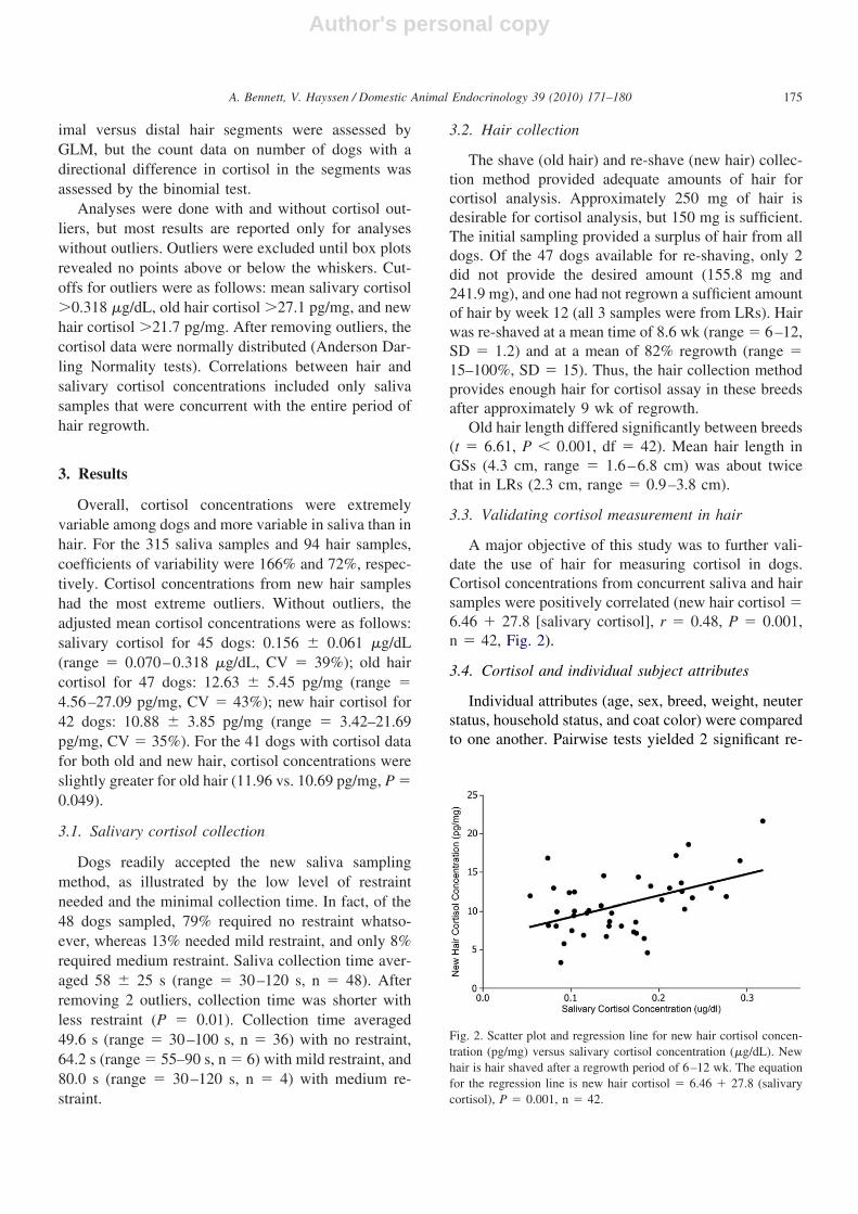
A major objective of this study was to further vali-date the use of hair for measuring cortisol in dogs.Cortisol concentrations from concurrent saliva and hairsamples were positively correlated (new hair cortisol �6.46 � 27.8 [salivary cortisol], r � 0.48, P � 0.001,n � 42, Fig. 2).
3.4. Cortisol and individual subject attributes
Individual attributes (age, sex, breed, weight, neuterstatus, household status, and coat color) were comparedto one another. Pairwise tests yielded 2 significant re-
Fig. 2. Scatter plot and regression line for new hair cortisol concen-tration (pg/mg) versus salivary cortisol concentration (�g/dL). Newhair is hair shaved after a regrowth period of 6–12 wk. The equationfor the regression line is new hair cortisol � 6.46 � 27.8 (salivarycortisol), P � 0.001, n � 42.
175A. Bennett, V. Hayssen / Domestic Animal Endocrinology 39 (2010) 171–180

Author's personal copy
lationships among individual subject attributes. First,females weighed 14% less on average (mean � 32.5 kg)than males (mean � 37.9 kg, t � �4.16, P � 0.001,df � 44). Second, on average, intact dogs were 2.4 yyounger (mean � 3.3 y) than neutered dogs (mean �5.7 y, t � �2.75, P � 0.014, df � 16).
Salivary and hair cortisol concentrations were alsoassessed versus individual attributes. Pairwise compar-isons revealed no significant differences with respect toindividual attributes and either salivary or new haircortisol, but there were 2 significant results for old haircortisol. First, dogs living alone had old hair cortisolconcentrations 36% lower (mean � 9.0 pg/mg) thandogs living in multidog households (mean � 14.2 pg/mg, t � �3.90, P � 0.001, df � 38) and second, blackdogs had old hair cortisol concentrations 24% lower(mean � 10.7 pg/mg) than nonblack dogs (mean �14.0 pg/mg, t � 2.13, P � 0.039, df � 39).
3.5. Cortisol in proximal and distal sections of thehair shaft
Although the amount of cortisol across dogs did notdiffer between proximal and distal hair segments(paired t test, P � 0.348, Fig. 3), the number of dogswith more cortisol in the distal segment was significant(7 of the 9 dogs, P � 0.002). One dog with high cortisolwas eliminated from the figure and the paired t test. Ifthis outlier were included, the significance of the pairedt test would be P � 0.211. No pattern with respect tobreed, sex, or coat color was evident.
A survey of dog owners revealed that bathing wasno more frequent than twice/mo. Dogs were washedbetween 0 to 24 times/y, with a mean of 3 baths/y. Onaverage, GSs were bathed 6 times more frequently(mean � 6 baths/y, range � 0–24) than LRs (mean �1 bath/y, range � 0–5). Bathing frequency was notcorrelated with the difference in cortisol in proximalversus distal hair segments (regression: P � 0.944).
3.6. Cortisol and hair color
Results of our comparison between agouti, eumela-nin, and pheomelanin hairs within individuals revealeda relationship between hair color and cortisol concen-tration (partial r � 0.47, P � 0.001). Eumelanin (black)hairs were consistently lower in cortisol than pheomela-nin (yellow) hairs. Cortisol concentrations of agoutihairs were intermediate (Fig. 4). One sample had aninsufficient mass of pheomelanin hairs for analysis.
4. Discussion
The objectives of the present study were to improvecortisol sampling methods, to validate hair relative tosaliva for measuring basal cortisol, to determine char-acteristics of cortisol within the hair shaft, and to assesscortisol with respect to coat and hair color.
The first objective of this research was to minimizethe restraint needed for saliva collection in dogs. Tra-ditional saliva collection, though considered nonstress-ful, still necessitates some restraint of the head andsome handling of the mouth, neither of which is vol-
Fig. 3. Hair cortisol concentrations in proximal (open bars) and distal(solid bars) sections of hair from 8 individual dogs did not differ(P � 0.348).
Fig. 4. Hair cortisol concentrations in different colored hairs (yellow,agouti, and black) from 4 individual dogs (A–D). Pheomelanin (yel-low, open bars) hairs were consistently higher in cortisol than eu-melanin (black, solid bars) hairs, with agouti (banded, striped bars)hairs intermediate (partial r � 0.47, P � 0.001). Pheomelanin hairsfrom dog D had an insufficient mass for analysis.
176 A. Bennett, V. Hayssen / Domestic Animal Endocrinology 39 (2010) 171–180
Author's personal copy
untarily accepted by most dogs. Using hidden treats, werarely had to restrain any of the subjects and never formore than 2 min. Not surprizingly, the less restraint weused, the less time was needed for saliva collection.Cortisol concentrations are unaffected by handling forup to 4 min [35], and all sampling took place wellwithin this time frame. Thus, saliva was collected withminimal restraint and completed quickly enough toavoid elevating the cortisol concentration.
Use of a hidden treat meant that salivation could beinduced without flavoring the collection material, thusavoiding unreliable results [25]. Food luring was anonstressful collection method, as demonstrated bythe dogs’ voluntary participation, and was also easyenough for owners to perform on their own. Saliva haslong been used as a valid measure of cortisol concen-trations, and this successful new food luring techniquemakes collecting saliva in the home environment evenmore convenient.
The second objective of this study was to establishthe validity of hair for measuring basal cortisol concen-trations in dogs. Basal concentrations of cortisol areresponsible for priming some of the organism’s homeo-static mechanisms for action [36]. Unlike many otherdomestic and wild species, dogs may display an epi-sodic pattern of cortisol secretion [37] and significantindividual circadian variability [38]. Though point sam-ples (saliva, blood) are collected within a narrow timeframe in this and most other cortisol studies, individualconcentrations may vary regardless of stress or time ofday. Hair sampling within the dog’s home provides ameans of measuring long-term or basal cortisol secre-tions that are less sensitive to individual circadian pat-terns, momentary stressors, or the chronic stress of shel-ters. Hair and salivary cortisol concentrations werepositively correlated in the home environment. This find-ing confirms hair as a valid medium to evaluate basalcortisol secretion in dogs. As cortisol concentrations in oldand new hair did not differ, a single shaving would besufficient for assessing cortisol concentrations and com-paring them across breeds, sexes, ages, and so on.
Hair has many advantages for cortisol measurement.Shaving is well tolerated by most domestic species.Although shaving may require some restraint or seda-tion of wild animals such as wolves, hair would be easyto collect during routine capture and release. However,relatively large volumes of hair are required for assay,thus small animals such as hamsters may not be goodcandidates for this sampling. Hair sampling allowscomparisons of baseline cortisol with individual traitssuch as temperament or social status [28]. In addition,
accumulation of hormones over precise time periodscan be measured with a timed shave and regrowthperiod, and thus, hair sampling obviates the need forrepeated blood sampling. Hair sampling could havebroad applications in kennels, shelters, laboratories, oranywhere that long-term cortisol concentrations need tobe assessed.
Hair cortisol concentrations obtained in this studywere higher than in previous work on dogs [24]. Wereport a mean first hair cortisol concentration of 12.6pg/mg and second hair cortisol concentration of 10.9pg/mg, whereas Accorsi et al [24] report a mean corti-sol concentration of 2.10 pg/mg. This difference maybe because powdering of hair resulted in a 3.5-foldincrease in cortisol recovery over the chopping methodused by Accorsi et al [22]. In addition, other differencesin assay methods—such as enzyme immunoassays ver-sus radioimmunoassay and differences in study subjectssuch as breed, size, and age—may account for thesediscrepancies.
Our study included 2 of the most popular breeds inthe United States, according to 2008 American KennelClub breed registration statistics [39]. Though these areboth large breeds, they are otherwise phenotypicallyvery different. Differences in initial hair length and ingrowth rates between the 2 breeds, as well as substan-tial individual variability, were consistent with otherstudies on dog hair regrowth [40,41]. Mean hair lengthin GSs was almost twice as long as in LRs. Ownersattest to the more frequent shedding in GSs than LRs,which would suggest differences in growth rates andhair growth stages between the 2 breeds. Both breeds inthis study required a similar amount of time to achievenearly complete hair regrowth despite the differences inoriginal hair length. The rapid regrowth observed in theischiatic (hip) region of dogs in our study is in agree-ment with previous work [40].
In terms of measuring cortisol in hair, the hair sam-pling area and regrowth time period outlined aboveprovide sufficient hair for cortisol assay. Although old-and new-growth hair did not differ in cortisol concen-trations in our study, the environments of the animalsalso did not change in any systematic manner. If onewere to use hair to measure the effect of a stressor, wewould recommend monitoring or marking the samplearea to avoid inadvertent collection of old-growth hairbecause of significant variation in hair growth ratesacross breeds and individuals.
Analysis of cortisol and individual subject attributesof age, sex, breed, weight, neuter status, and householdstatus yielded 1 significant relationship: dogs living
177A. Bennett, V. Hayssen / Domestic Animal Endocrinology 39 (2010) 171–180

Author's personal copy
alone had significantly lower basal cortisol (in old hairsamples) than did dogs living in multidog households.This finding is in agreement with reports of cortisolresponse in thunderstorm-phobic dogs to a simulatedthunderstorm: dogs living without other dogs had lowerbaseline cortisol concentrations but a more significantchange post-stressor than dogs living in multidoghouseholds [20]. Combined with our results, the higherbaseline cortisol seen in dogs from multidog house-holds suggests that living with other dogs is morestressful day to day than living without other dogs, butliving with other dogs may have a physiological pro-tective factor for dealing with certain stressors.
Although all 3 cortisol measurements (saliva, oldhair, and new hair) were significantly correlated, onlythe old hair measure revealed an effect of householdstatus. This finding may be owing to differences invariability among samples; new hair was the least vari-able after removing outliers for analysis, whereas oldhair was the most variable.
Previous investigations of cortisol concentrationsalso reported no significant relationship between corti-sol and sex or age [21,23]. Other studies of dogs alsofound no significant differences in cortisol concentra-tions among breeds [21], and no relationship betweencortisol concentration and neuter status [42]. Thus, ourresults confirm these studies and reveal no significanteffects of age, breed, weight, or neuter status on cortisolin either hair or saliva.
The third objective of this study was to determinewhether cortisol concentrations vary significantly alongthe length of the hair shaft in dogs. This objective wasachieved by comparing cortisol concentrations in prox-imal and distal sections of the hair shaft. Little is knownabout the possible leaching effects that washing andsunlight exposure may have on hair over time andthroughout the length of the hair shaft. No difference incortisol concentrations was evident between the proxi-mal and distal ends of rhesus macaque hair [22], but astudy on women by Kirschbaum et al [30] reported adecline in cortisol concentration throughout the lengthof hair from scalp to tip. We found no significantrelationship between cortisol concentration and posi-tion along the hair shaft in a species subjected to vari-able washing frequencies. However, more dogs (7 of 9)had larger concentrations of cortisol in the distal por-tions of their hair than in the hair closer to the skin, theopposite of what would be expected if cortisol wereleached from the hair as it aged.
The fourth and last objective was to assess the rela-tionship between cortisol and coat color or pigment.
We found differences in cortisol in relation to coatcolor and pigment. Across all subjects, black dogs hadless cortisol than nonblack dogs, suggesting a coatcolor-cortisol relationship. In addition, within an indi-vidual dog eumelanin (black) hairs had less cortisolthan pheomelanin (yellow) hairs, with agouti (banded)hairs intermediate.
The literature suggests 2 possible reasons for theblack coat color and individual black hairs having lesscortisol than yellow hairs. Given that glucocorticoidsare involved in stress-associated hair growth inhibition[43] as well as melanocyte development and differen-tiation [44,45], differences in cortisol in different pig-ment types or coat colors may be related to differentcontrol mechanisms. Second, hair may be a storagevehicle for cortisol. In general, yellow hair has lesspigment than black hair [46,47], thus yellow hair mayhave more room for glucocorticoids than black hair.
Interestingly, the pigment differences we found arewithin the same animal, not across breeds or coat col-ors. The differences between black, yellow, and agoutihairs within an individual dog do not translate to dif-ferences between breeds (LRs vs GSs) or coat colorswithin a breed (yellow vs black LRs, agouti vs blackGSs). We expected to find coat color differences withinGSs but not black and yellow LRs, because fecal glu-cocorticoid concentrations differed in agouti vs nona-gouti deer mice (animals that differ only at the agouticoat color locus and otherwise were genetically identi-cal [11]). Agouti and black GSs differ at the agoutilocus, although the animals also differ at other loci,unlike the laboratory deer mice. Also, agouti GSs, un-like agouti deer mice, have all-black hairs and all-yellow hairs, as well as banded agouti hairs. As hairfollicles have a local functional equivalent of the HPAaxis and synthesize cortisol [29,48], local production ofcortisol within hairs could be responsible for the dif-ferences between hairs with different pigment compo-sitions, but differences in cortisol production acrossindividuals may obscure differences across individualhairs. The genetic control that produces yellow, black,or agouti hairs differs in LRs vs GSs. Unfortunately, thebiochemical mechanism, which produces all-black orall-yellow hairs in addition to the agouti-striped hair allin the same GS, is not known.
In summary, (1) food luring minimizes the restraintrequired for saliva collection; (2) hair cortisol concen-trations do correlate with concurrent salivary cortisol,and thus hair is a valid medium for measuring basalcortisol in dogs; (3) distal hair segments more oftenhave higher cortisol than proximal segments in contrast
178 A. Bennett, V. Hayssen / Domestic Animal Endocrinology 39 (2010) 171–180
Author's personal copy
to both macaques and humans; and (4) within an indi-vidual dog, black hairs have less cortisol than yellowhairs, and across all dogs, black dogs have lower cor-tisol in their hair than nonblack dogs.
Acknowledgments
The authors are grateful to Jennifer Yoo for herassistance collecting samples, performing assays, andpresenting results; to Jerry Meyer for lending lab space,equipment, and expertise; to Karen Stonemetz for dem-onstrating the hair assay methods; to Rob Dorit andChristine White-Ziegler for lending lab space andequipment; to Paula Turini and Anna Symbor-Nagrab-ska for help with methodology; and to all the generousdog owners who volunteered their pets for our study.We are also thankful for funding from the B. ElizabethHorner Fund in the Biological Sciences and theBlakeslee Grant for Genetics Research at Smith Col-lege, which had no involvement in the research orpreparation of this article.
References
[1] Christison GI, Johnson HD. Cortisol turnover in heat-stressedcows. J Anim Sci 1972;35:1005–1010.
[2] Aoyama M, Negishi A, Abe A, Yokoyama R, Ichimaru T,Sugita S. Physiological and behavioural effects of an intracere-broventricular injection of corticotropin releasing hormone ingoats. Vet J 2008;177:116–123.
[3] Künzl C, Kaiser S, Meier E, Sachser N. Is a wild mammal keptand reared in captivity still a wild animal? Horm Behav 2003;43:187–196.
[4] Visser EK, Ellis AD, Van Reenen CG. The effect of twodifferent housing conditions on the welfare of young horsesstabled for the first time. Appl Anim Behav Sci 2008;114:521–533.
[5] Turner AI, Hemsworth PH, Tilbrook AJ. Susceptibility of re-production in female pigs to impairment by stress or elevationof cortisol. Domest Anim Endocrin 2005;29:398–410.
[6] Radahmadi M, Shadan F, Karimian SM, Sadr SS, Nasimi A.Effects of stress on exacerbation of diabetes mellitus, serumglucose and cortisol levels and body weight in rats. Pathophys-iology 2006;13:51–55.
[7] Smith RF, Dobson H. Hormonal interactions within the hypo-thalamus and pituitary with respect to stress and reproduction insheep. Domest Anim Endocrinol 2002;23:75–85.
[8] Bradley AJ, McDonald IR, Lee AK. Stress and mortality in asmall marsupial (Antechinus stuartii, Macleay). Gen Comp En-docr 1980;40:188–200.
[9] Davenport MD, Lutz CK, Tiefenbacher S, Novak MA, MeyerJS. A Rhesus monkey model of self-injury: Effects of relocationstress on behavior and neuroendocrine function. Biol Psychiatry2008;63:990–996.
[10] Millspaugh JJ, Washburn BE, Milanick MA, Beringer J, HansenLP, Meyer TM. Non-invasive techniques for stress assessmentin white-tailed deer. Wildlife Soc B 2002;30:899–907.
[11] Hayssen V, Harper JM, DeFina R. Fecal corticosteroids inagouti and non-agouti deer mice (Peromyscus maniculatus).Comp Biochem Phys A 2002;132:439–446.
[12] Koren L, Mokady O, Geffen E. Social status and cortisol levelsin singing rock hyraxes. Horm Behav 2008;54:212–216.
[13] Bonier F, Quigley H, Austad SN. A technique for non-inva-sively detecting stress response in cougars. Wildlife Soc B2004;32:711–717.
[14] Lyons DM, Ha CMG, Levine S. Social effects and circadianrhythms in squirrel monkey pituitary-adrenal activity. HormBehav 1995;29:177–190.
[15] Mateo JM. Inverted-U shape relationship between cortisol andlearning in ground squirrels. Neurobiol Learn Mem 2008;89:582–590.
[16] Nelson RJ. An Introduction to Behavioral Endocrinologyr, 3rded. Sunderland (MA): Sinaur Associates; 2005.
[17] Charmandari E, Tsigos C, Chrousos G. Endocrinology of thestress response. Annu Rev Physiol 2005;67:259–284.
[18] Vincent LC, Michell AR. Comparison of cortisol concentrationsin saliva and plasma of dogs. Res Vet Sci 1992;53:342–345.
[19] Beerda B, Schilder MBH, Bernadina W, Van Hooff JARAM,De Vries HW, Mol JA. Chronic stress in dogs subjected tosocial and spatial restriction. II. Hormonal and immunologicalresponses. Physiol Behav 1999;66:243–254.
[20] Dreschel NA, Granger DA. Physiological and behavioral reac-tivity to stress in thunderstorm-phobic dogs and their caregivers.Appl Anim Behav Sci 2005;95:153–168.
[21] Coppola CL, Grandin T, Enns RM. Human interaction andcortisol: Can human contact reduce stress for shelter dogs?Physiol Behav 2006;87:537–541.
[22] Davenport MD, Tiefenbacher S, Lutz CK, Novak MA, MeyerJS. Analysis of endogenous cortisol concentrations in the hair ofrhesus macaques. Gen Comp Endocrinol 2006;147:255–261.
[23] Stephen JM, Ledger RA. A longitudinal evaluation of urinarycortisol in kenneled dogs, Canis familiaris. Physiol Behav2006;87:911–916.
[24] Accorsi PA, Carloni E, Valsecchi P, Viggiani R, Gamberoni M,Tamanini C, Seren E. Cortisol determination in hair and faecesfrom domestic cats and dogs. Gen Comp Endocrinol 2008;155:398–402.
[25] Dreschel NA, Granger DA. Methods of collection for salivarycortisol measurement in dogs. Horm Behav 2009;55:163–168.
[26] Rooney NJ, Gaines SA, Bradshaw JWS. Behavioural and glu-cocorticoid responses of dogs (Canis familiaris) to kenneling:Investigating mitigation of stress by prior habituation. PhysiolBehav 2007;92:847–854.
[27] Buchanan KL, Goldsmith AR. Noninvasive endocrine data forbehavioral studies: The importance of validation. Anim Behav2004;67:183–185.
[28] Koren L, Mokady O, Karaskov T, Klein J, Korent G, Geffen E.A novel method using hair for determining hormonal levels inwildlife. Anim Behav 2002;63:403–406.
[29] Ito N, Ito T, Kromminga A, Bettermann A, Takigawa K, KeesF, Straub RH, Paus R. Human hair follicles display a functionalequivalent of the hypothalamic-pituitary-adrenal (HPA) axisand synthesize cortisol. FASEB J 2005;19:1332–1334.
[30] Kirschbaum C, Tietze A, Skoluda N, Dettenborn L. Hair as aretrospective calendar of cortisol production- Increased cortisolincorporation into hair in the third trimester of pregnancy.Psychoneuroendocrinol 2009;34:32–37.
[31] Lu D, Willard D, Patel IR, Kadwell S, Overton L, Kost T,Luther M, Chen W, Woychick RP, Wilkison WO, Cone RD.
179A. Bennett, V. Hayssen / Domestic Animal Endocrinology 39 (2010) 171–180
Author's personal copy
Agouti protein is an antagonist of the melanocyte-stimulatinghormone receptor. Nature 1994;371:799–802.
[32] Berryere TG, Kerns JA, Barsh GS, Schmutz SM. Association ofan Agouti allele with fawn or sable coat color in domestic dogs.Mamm Genome 2005;16:262–272.
[33] Schmutz SM, Berryere TG, Barta JL, Reddick KD, Schmutz JK.Agouti sequence polymorphisms in coyotes, wolves and dogssuggest hybridization. J Hered 2007;98:351–355.
[34] Kerns JA, Cargill EJ, Clark LA, Candille SI, Berryere TG,Olivier M, Lust G, Todhunter RJ, Schmutz SM, Murphy KE,Barsh GS. Linkage and segregation analysis of black and brin-dle coat color in domestic dogs. Genetics 2007;176:1679–1689.
[35] Kobelt AJ, Hemsworth PH, Barnett JL, Butler KL. Sources ofsampling variation in saliva cortisol in dogs. Res Vet Sci 2003;75:157–161.
[36] Munck A. Corticosteroids and stress. In: Fink G, editor-in-chief.Encyclopedia of Stress. Oxford (UK): Elsevier; 2007:613–619.
[37] Kemppainen RJ, Sartin JL. Evidence for episodic but not cir-cadian activity in plasma concentrations of adrenocorticotro-phin, cortisol, and thyroxine in dogs. J Endocrinol 1984;103:219–226.
[38] Castillo VA, Cabrera Blatter MF, Gómez NV, Sinatra V, Gal-lelli MF, Ghersevich MC. Diurnal ACTH and plasma cortisolvariations in healthy dogs and in those with pituitary-dependentCushing’s syndrome before and after treatment with retinoicacid. Res Vet Sci 2009;86:223–229.
[39] American Kennel Club. AKC dog registration statistics. http://www.akc.org/reg/dogreg_stats.cfm; 2010. Accessed June 2, 2010.
[40] Gunaratnam P, Wilkinson GT. A study of normal hair growth inthe dog. J Small Anim Pract 1983;24:445–453.
[41] Diaz SF, Torres SM, Dunstan RW, Lekcharoensuk C. An anal-ysis of canine hair re-growth after clipping for a surgical pro-cedure. Vet Dermatol 2004;15:25–30.
[42] Reimers TJ, Mummery LK, McCann JP, Cowan RG, Concan-non PW. Effects of age, sex, and body size on concentrations ofthyroxine, 3,5,3’-triiodothyronine and cortisol in serum of dogs.Biol Reprod 1984;31:148–154.
[43] Botchkarev VA. Stress and the hair follicle: Exploring theconnections. Am J Pathol 2003;162:709–712.
[44] Slominski A, Tobin DJ, Shibahara S, Wortsman J. Melaninpigmentation in mammalian skin and its hormonal regulation.Physiol Rev 2004;84:1155–1228.
[45] Roulin A, Almasi B, Rossi-Pedruzzi A, Ducrest A-L, Waka-matsu K, Miksik I, Blount JD, Jenni-Eiermann S, Jenni L.Corticosterone mediates the condition-dependent component ofmelanin-based coloration. Anim Behav 2008;75:1351–1358.
[46] Russell ES. A quantitative histological study of the pigmentfound in the coat color mutants of the house mouse. II.Estimates of the total volume of pigment. Genetics 1948;33:228 –236.
[47] Kaliss N. The morphogenesis of pigment in the hair follicle ofthe house mouse. J Morph 1942;70:209–219.
[48] Slominski A, Zbytek B, Semak I, Sweatman T, Wortsman J.CRH stimulates POMC activity and corticosterone productionin dermal fibroblasts. J Neuroimmunol 2005;162:97–102.
180 A. Bennett, V. Hayssen / Domestic Animal Endocrinology 39 (2010) 171–180
Copyright © 2022 FDOKUMEN