Cancer-associated noncoding mutations affect RNA G ...
11
1 SCIENTIFIC REPORTS | 7: 708 | DOI:10.1038/s41598-017-00739-y www.nature.com/scientificreports Cancer-associated noncoding mutations affect RNA G-quadruplex-mediated regulation of gene expression Mahdi Zeraati 1,2 , Aaron L. Moye 3 , Jason W. H. Wong 4 , Dilmi Perera 4 , Mark J. Cowley 1,2 , Daniel U. Christ 1,2 , Tracy M. Bryan 3 & Marcel E. Dinger 1,2 Cancer is a multifactorial disease driven by a combination of genetic and environmental factors. Many cancer driver mutations have been characterised in protein-coding regions of the genome. However, mutations in noncoding regions associated with cancer have been less investigated. G-quadruplex (G4) nucleic acids are four-stranded secondary structures formed in guanine-rich sequences and prevalent in the regulatory regions. In this study, we used published whole cancer genome sequence data to find mutations in cancer patients that overlap potential RNA G4-forming sequences in 5′ UTRs. Using RNAfold, we assessed the effect of these mutations on the thermodynamic stability of predicted RNA G4s in the context of full-length 5′ UTRs. Of the 217 identified mutations, we found that 33 are predicted to destabilise and 21 predicted to stabilise potential RNA G4s. We experimentally validated the effect of destabilising mutations in the 5′ UTRs of BCL2 and CXCL14 and one stabilising mutation in the 5′ UTR of TAOK2. These mutations resulted in an increase or a decrease in translation of these mRNAs, respectively. These findings suggest that mutations that modulate the G4 stability in the noncoding regions could act as cancer driver mutations, which present an opportunity for early cancer diagnosis using individual sequencing information. Substantial efforts have been made to characterise the cancer genome. e advent of next generation sequencing has enabled affordable and rapid large-scale sequencing of cancer genomes. e Cancer Genome Atlas (TCGA) 1 and the International Cancer Genome Consortium 2 are examples of large-scale cancer genomics projects that aim to catalogue cancer-associated mutations. One of the main goals of these projects is to differentiate cancer driver mutations, which contribute to cancer development, from non-functional passenger mutations and ulti- mately improve our understanding of how different types of cancers develop 3 . As a result of extensive efforts that have been directed towards detecting and characterising driver mutations in protein-coding regions, knowledge about abnormal proteins encoded by mutated cancer genes has grown substantially in recent years, such that new cancer therapeutics have been developed to target these anomalous proteins. Imatinib is an example of this type of therapeutic that inhibits the protein encoded by ABL, which is mutated in chronic myeloid leukaemia 4 . Noncoding somatic mutations, on the other hand, have not been examined as extensively as coding variations. Considering that less than 2% of the human genome encodes protein and that important regulatory regions including promoters and 5′ and 3′ untranslated regions (UTR) of mRNAs are located in the noncoding regions 5 , effective functional studies of mutations in the noncoding regions would provide a better understanding of cancer biology and development. Indeed, several studies have indicated that noncoding somatic mutations can alter gene expression in cancer 6–8 , prompting the hypothesis that these mutations affect regulatory motifs. Guanine (G)-rich DNA and RNA sequences can fold into non-canonical secondary structures called G-quadruplexes (G4s). e building block of a G4 is called a G-tetrad, which consists of four guanines in a square planar arrangement. Each guanine is connected to the two adjacent guanines by hydrogen bonds at the Watson 1 Garvan Institute of Medical Research, Sydney, NSW, 2010, Australia. 2 St Vincent’s Clinical School, Faculty of Medicine, University of New South Wales, Sydney, NSW, 2052, Australia. 3 Children’s Medical Research Institute, University of Sydney, Sydney, NSW, 2145, Australia. 4 Prince of Wales Clinical School, Faculty of Medicine, University of New South Wales, Sydney, NSW, 2052, Australia. Correspondence and requests for materials should be addressed to M.E.D. (email: [email protected]) Received: 5 January 2017 Accepted: 9 March 2017 Published: xx xx xxxx OPEN
-
Upload
khangminh22 -
Category
Documents
-
view
2 -
download
0
Transcript of Cancer-associated noncoding mutations affect RNA G ...

1Scientific RepoRts | 7: 708 | DOI:10.1038/s41598-017-00739-y
www.nature.com/scientificreports
Cancer-associated noncoding mutations affect RNA G-quadruplex-mediated regulation of gene expressionMahdi Zeraati1,2, Aaron L. Moye3, Jason W. H. Wong 4, Dilmi Perera4, Mark J. Cowley1,2, Daniel U. Christ1,2, Tracy M. Bryan 3 & Marcel E. Dinger 1,2
Cancer is a multifactorial disease driven by a combination of genetic and environmental factors. Many cancer driver mutations have been characterised in protein-coding regions of the genome. However, mutations in noncoding regions associated with cancer have been less investigated. G-quadruplex (G4) nucleic acids are four-stranded secondary structures formed in guanine-rich sequences and prevalent in the regulatory regions. In this study, we used published whole cancer genome sequence data to find mutations in cancer patients that overlap potential RNA G4-forming sequences in 5′ UTRs. Using RNAfold, we assessed the effect of these mutations on the thermodynamic stability of predicted RNA G4s in the context of full-length 5′ UTRs. Of the 217 identified mutations, we found that 33 are predicted to destabilise and 21 predicted to stabilise potential RNA G4s. We experimentally validated the effect of destabilising mutations in the 5′ UTRs of BCL2 and CXCL14 and one stabilising mutation in the 5′ UTR of TAOK2. These mutations resulted in an increase or a decrease in translation of these mRNAs, respectively. These findings suggest that mutations that modulate the G4 stability in the noncoding regions could act as cancer driver mutations, which present an opportunity for early cancer diagnosis using individual sequencing information.
Substantial efforts have been made to characterise the cancer genome. The advent of next generation sequencing has enabled affordable and rapid large-scale sequencing of cancer genomes. The Cancer Genome Atlas (TCGA)1 and the International Cancer Genome Consortium2 are examples of large-scale cancer genomics projects that aim to catalogue cancer-associated mutations. One of the main goals of these projects is to differentiate cancer driver mutations, which contribute to cancer development, from non-functional passenger mutations and ulti-mately improve our understanding of how different types of cancers develop3. As a result of extensive efforts that have been directed towards detecting and characterising driver mutations in protein-coding regions, knowledge about abnormal proteins encoded by mutated cancer genes has grown substantially in recent years, such that new cancer therapeutics have been developed to target these anomalous proteins. Imatinib is an example of this type of therapeutic that inhibits the protein encoded by ABL, which is mutated in chronic myeloid leukaemia4. Noncoding somatic mutations, on the other hand, have not been examined as extensively as coding variations. Considering that less than 2% of the human genome encodes protein and that important regulatory regions including promoters and 5′ and 3′ untranslated regions (UTR) of mRNAs are located in the noncoding regions5, effective functional studies of mutations in the noncoding regions would provide a better understanding of cancer biology and development. Indeed, several studies have indicated that noncoding somatic mutations can alter gene expression in cancer6–8, prompting the hypothesis that these mutations affect regulatory motifs.
Guanine (G)-rich DNA and RNA sequences can fold into non-canonical secondary structures called G-quadruplexes (G4s). The building block of a G4 is called a G-tetrad, which consists of four guanines in a square planar arrangement. Each guanine is connected to the two adjacent guanines by hydrogen bonds at the Watson
1Garvan Institute of Medical Research, Sydney, NSW, 2010, Australia. 2St Vincent’s Clinical School, Faculty of Medicine, University of New South Wales, Sydney, NSW, 2052, Australia. 3Children’s Medical Research Institute, University of Sydney, Sydney, NSW, 2145, Australia. 4Prince of Wales Clinical School, Faculty of Medicine, University of New South Wales, Sydney, NSW, 2052, Australia. Correspondence and requests for materials should be addressed to M.E.D. (email: [email protected])
Received: 5 January 2017
Accepted: 9 March 2017
Published: xx xx xxxx
OPEN

www.nature.com/scientificreports/
2Scientific RepoRts | 7: 708 | DOI:10.1038/s41598-017-00739-y
Crick and Hoogsteen edges. The G4 structure is formed by at least two stacked G-tetrads. A cation positioned at the center of each tetrad or between them further stabilises the G4 structure. Nucleotides that do not contribute to the G-tetrads form the loops9. Intramolecular G4 is assembled from guanines within one nucleic acid strand, whereas separate nucleic acid strands can form an intermolecular G4. Intramolecular DNA G4s can adopt various topologies10, whereas intramolecular RNA G4s predominantly fold into the parallel propeller topology11. Several different G4 structures have been studied in vitro. The results from these studies have enabled the development of algorithms that predict potential G4-forming sequences in the genome12–14. Using these algorithms, compu-tational studies have demonstrated that G4s are significantly enriched and conserved in regulatory elements, such as promoters and UTRs of mRNAs15. This observation suggests that G4s may have regulatory roles and be involved in biological processes.
A number of studies have demonstrated that G4s located in the 5′ UTRs modulate translation activity. Although it has been shown that G4 can augment translation16, most G4s that have been characterised to date act as repressors in the cap-dependent translation regulation of mRNA translation17–20, especially in case of proto-oncogenes, such as NRAS21. Given accumulating in vitro results that demonstrate 5′ UTR G4s have regu-latory functions and the increasing evidence that show G4 structures exist in vivo22, 23 and a number are altered in cancer tissues24, we hypothesized that mutations in cancer patients alter the stability of 5′ UTR G4s and conse-quently affect their regulatory function.
Here, we present an approach that permitted us to identify somatic mutations in cancer that change the sta-bility of 5′ UTR G4s. We have experimentally validated the effect of some of these mutations and showed that they can either destabilise RNA G4 structures or stabilise them. Furthermore, the mutations result in altered gene expression. Overall, our data suggest that mutations in regulatory elements such as 5′ UTR that alter G4s stability could act as cancer driver mutations.
ResultsSomatic mutations in cancer patients change the thermodynamic stability of RNA G4s in the 5′ UTR. Previous studies have demonstrated that RNA G4s act as regulatory motifs in the 5′ UTR of mRNAs. To determine the possible effects of mutations in cancer patients on the stability of G4s in this region, the procedure illustrated in Fig. 1 was followed. A dataset of potential G4-forming sequences in the human genome was con-structed. Quadparser algorithm13, which searches for the sequence pattern G3+N1–7G3+N1–7G3+N1–7G3+ across the genome, was employed to identify potential G4s. In this pattern, G refers to guanine and N can be any base including guanine. Concurrently, three publicly available databases, TCGA1, ICGC2 and Alexandrov et al.25, were used to obtain somatic mutation data in patients with different types of cancers. Comparing these two datasets, 217 overlaps were found between somatic mutations from cancer patients and potential G4-forming sequences in the 5′ UTRs (Supplementary Dataset). Considering that putative G4 sequences might fold into other secondary structures instead of G4 structures, it is possible that overlapping mutations do not affect RNA G4 structural stability. To address this possibility, a further screening step was performed that investigated the effect of each mutation on the predicted RNA G4 stability using RNAfold software26. In addition to the G4 sequence pattern (G2–7N1–15G2–7N1–15G2–7N1–15G2–7), RNAfold takes account of thermodynamic parameters and is therefore a more stringent method to predict G4 structures. To simulate a more biologically relevant condition, the effect of each mutation was examined in the context of the entire 5′ UTR.
We found 33 RNA G4 destabilising mutations among 217 overlaps between somatic mutations and predicted 5′ UTR G4s listed in the Supplementary Table 2. Based on the RNAfold predictions, these mutations remove one or more of the G-tetrads, increase the loop length or shift the RNA G4 position. A majority of them are transitions from guanine to adenine (Fig. 2A). On the other hand, 21 of the overlaps were found to be RNA G4 stabilising mutations (Supplementary Table 3). As predicted by RNAfold, in general, these mutations either increase the
Figure 1. Flowchart illustrating the procedure for identifying mutations that change predicted RNA G4 stability in the 5′ UTRs of genes mutated in cancer.
www.nature.com/scientificreports/
3Scientific RepoRts | 7: 708 | DOI:10.1038/s41598-017-00739-y
number of G-tetrads or shorten a loop length. Most of them are transversions from uracil to guanine (Fig. 2B). To evaluate the accuracy of the in silico results, one stabilising mutation in the 5′ UTR of TAOK2 (TAO Kinase 2), one destabilising mutation in the 5′ UTR of BCL2 (B-cell lymphoma 2) and two adjacent destabilising mutations in the 5′ UTR of CXCL14 (C-X-C motif ligand 14) were selected for experimental validation.
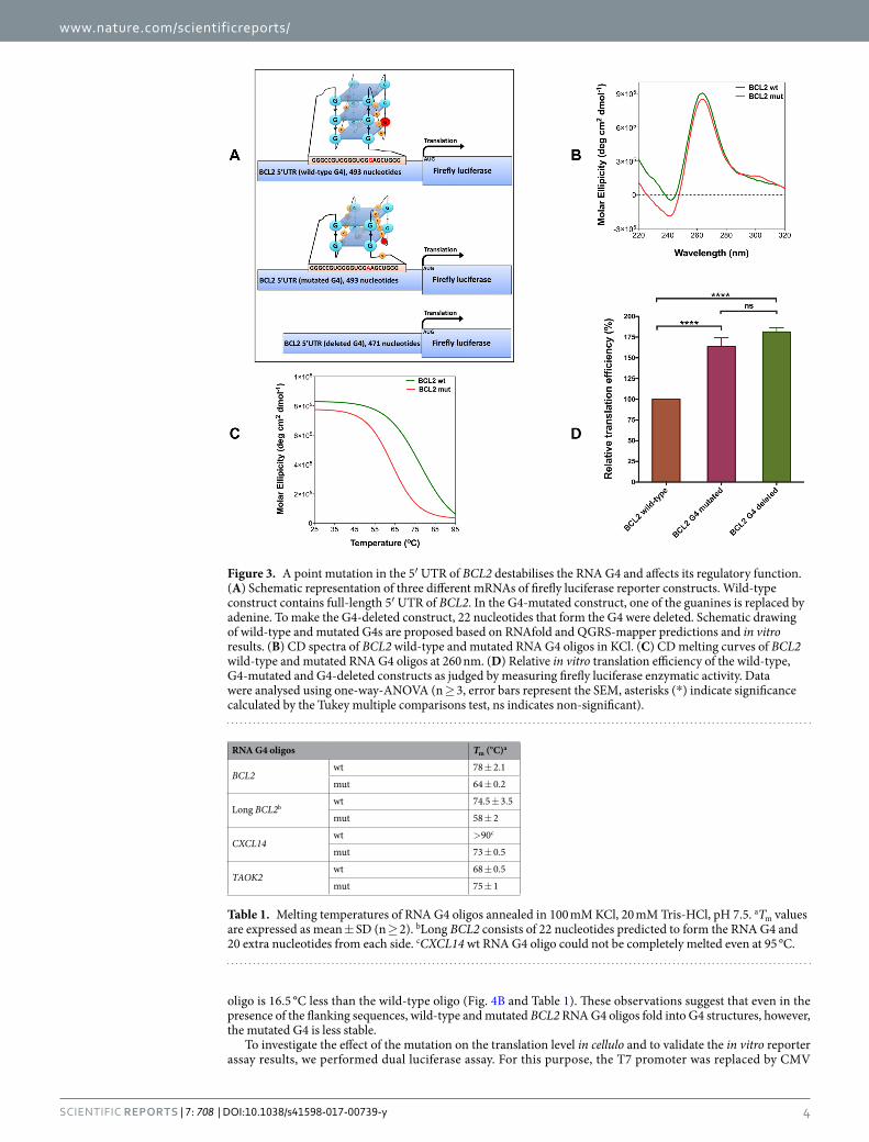
A point mutation destabilises the RNA G4 in the 5′ UTR of BCL2. It has been well demonstrated that the over-expression of BCL2 proto-oncogene contributes to the resistance of cancer cells to apoptosis27. Our bio-informatics analysis identified a guanine to adenine point mutation in a patient with malignant lymphoma, which was predicted to destabilise the RNA G4 in the 5′ UTR of BCL2 (Supplementary Table 2). It has been previously shown that the BCL2 RNA G4 is very stable near physiological ionic and pH conditions and acts as a translational repressor in the 5′ UTR of the BCL2 proto-oncogene28. To assess to what extent the mutation can destabilise the BCL2 RNA G4, in silico, in vitro and in cellulo evaluations were conducted. RNAfold and QGRS-Mapper14 both predict the same RNA G4 with three G-tetrads for the wild-type sequence. However, RNAfold does not predict any G4 structure for the mutated BCL2 G4 sequence, whereas QGRS-Mapper predicts a G4 structure with two G-tetrads (Fig. 3A; Supplementary Table 4).
CD spectroscopy is a well-established method to characterise the structure of G4s. A negative peak near 240 nm and a positive peak near 260 nm in the CD spectrum are characteristics of a parallel G429. CD spectra of wild-type and mutated BCL2 RNA G4 oligos were recorded in near physiological pH and potassium concentra-tion. Both wild-type and mutant oligos can fold into a parallel G4 conformation (Fig. 3B). It is noteworthy that the CD spectrum of the mutated RNA G4 oligo is in accordance with the QGRS-Mapper result, which predicts an RNA G4 containing two G-tetrads for this oligo. CD melting at 260 nm was performed to compare the stabil-ity of wild-type and mutated BCL2 RNA G4 oligos (Fig. 3C). Compared to the wild-type RNA G4 oligo, a 14 °C decrease in the Tm value of mutated oligo was observed (Table 1).
Luciferase reporter assays were performed to evaluate the impact of the identified BCL2 point mutation on this RNA G4’s function in vitro. Three constructs were generated by cloning full-length wild-type, G4-mutant and G4-deleted 5′ UTRs of BCL2 immediately downstream of a T7 promoter and upstream of the firefly luciferase gene (Fig. 3A). For each construct, 5′-capped RNA transcripts were generated in vitro using T7 RNA polymerase. Equal transcript amounts from each construct were subjected to in vitro translation using nuclease-treated rabbit reticulocyte lysate and translation efficiency was determined by measuring firefly luciferase catalytic activity. The deletion of RNA G4 sequence from the 5′ UTR resulted in the highest relative translation efficiency (Fig. 3D), which supports the notion that the RNA G4 in the 5′ UTR of BCL2 act as a translational repressor. Compared to the wild-type, a guanine to adenine point mutation in the 5′ UTR of BCL2 resulted in a significant increase in translation efficiency (P < 0.0001), which is consistent with destabilisation of the RNA G4 structure.
Although in silico analysis using RNAfold and in vitro luciferase reporter assay have been performed in the context of the entire 5′ UTR of BCL2, we repeated the biophysical studies using 62-mer oligos of the wild-type and mutated BCL2 RNA G4 that had 20 extra nucleotides from each side (Supplementary Table 1) to investigate if the flanking sequences can affect the G4 structure and stability. As recorded for the 22-mer wild-type and mutated oligos, CD spectroscopy showed a negative peak near 240 nm and a positive peak near 260 nm for both 62-mer wild-type and mutated oligos (Fig. 4A). CD melting of 62-mer oligos showed that the Tm value of the mutated
Figure 2. Pie chart demonstrating the frequency of different type of mutations in each of the RNA G4 (A) destabilising and (B) stabilising mutation groups.
www.nature.com/scientificreports/
4Scientific RepoRts | 7: 708 | DOI:10.1038/s41598-017-00739-y
oligo is 16.5 °C less than the wild-type oligo (Fig. 4B and Table 1). These observations suggest that even in the presence of the flanking sequences, wild-type and mutated BCL2 RNA G4 oligos fold into G4 structures, however, the mutated G4 is less stable.
To investigate the effect of the mutation on the translation level in cellulo and to validate the in vitro reporter assay results, we performed dual luciferase assay. For this purpose, the T7 promoter was replaced by CMV
Figure 3. A point mutation in the 5′ UTR of BCL2 destabilises the RNA G4 and affects its regulatory function. (A) Schematic representation of three different mRNAs of firefly luciferase reporter constructs. Wild-type construct contains full-length 5′ UTR of BCL2. In the G4-mutated construct, one of the guanines is replaced by adenine. To make the G4-deleted construct, 22 nucleotides that form the G4 were deleted. Schematic drawing of wild-type and mutated G4s are proposed based on RNAfold and QGRS-mapper predictions and in vitro results. (B) CD spectra of BCL2 wild-type and mutated RNA G4 oligos in KCl. (C) CD melting curves of BCL2 wild-type and mutated RNA G4 oligos at 260 nm. (D) Relative in vitro translation efficiency of the wild-type, G4-mutated and G4-deleted constructs as judged by measuring firefly luciferase enzymatic activity. Data were analysed using one-way-ANOVA (n ≥ 3, error bars represent the SEM, asterisks (*) indicate significance calculated by the Tukey multiple comparisons test, ns indicates non-significant).
RNA G4 oligos Tm (°C)a
BCL2wt 78 ± 2.1
mut 64 ± 0.2
Long BCL2bwt 74.5 ± 3.5
mut 58 ± 2
CXCL14wt >90c
mut 73 ± 0.5
TAOK2wt 68 ± 0.5
mut 75 ± 1
Table 1. Melting temperatures of RNA G4 oligos annealed in 100 mM KCl, 20 mM Tris-HCl, pH 7.5. aTm values are expressed as mean ± SD (n ≥ 2). bLong BCL2 consists of 22 nucleotides predicted to form the RNA G4 and 20 extra nucleotides from each side. cCXCL14 wt RNA G4 oligo could not be completely melted even at 95 °C.

www.nature.com/scientificreports/
5Scientific RepoRts | 7: 708 | DOI:10.1038/s41598-017-00739-y
promoter in the wild-type and G4-mutant constructs, which have been described earlier, and each of these con-structs co-transfected into MCF-7 cells together with pRL-TK as a normalizing vector and luciferase activity was measured. We observed ~75% higher translation level from G4-mutant construct (Fig. 4C) which is almost the same as in vitro translation results (Fig. 3D). In order to confirm that the observed difference in the gene expression level is due to the post-transcriptional regulation by the destabilised G4 in the 5′ UTR rather than the transcriptional regulation, we performed RT-qPCR on the total RNA extracted from the same samples used for the dual luciferase assay. As shown in Fig. 4C, there is no significant difference between the wild-type and G4-mutant constructs at the mRNA expression level which verifies post-transcriptional regulation is responsible for the difference observed in the protein expression level.
Two adjacent mutations destabilise the RNA G4 in the 5′ UTR of CXCL14. The length of loops within an RNA G4 are inversely proportional to its stability30. We identified several mutations predicted to desta-bilise RNA G4s by lengthening one of the loops (Supplementary Table 2). These mutations are located in a G-rich region, which more than four G-runs exist. Theoretically, just four of the G-runs with shorter loops contribute to the RNA G4 structure. When a destabilising mutation disrupts one of the G-runs involved in the G4 structure, the fifth G-run, which is a few nucleotides away from the destabilised G4, contributes to the G4 formation and one of the loops lengthens. As an example, two adjacent guanine to adenine mutations in a patient with melanoma can disrupt one of the five G-runs in the 5′ UTR of CXCL14 and destabilise the predicted RNA G4. CXCL14 is a chemokine, which mainly contributes to the regulation of immune cell migration31. RNAfold predicts a total disruption of the RNA G4 caused by these mutations in the context of a full-length 5′ UTR. However, RNALfold, which scans for locally stable secondary structures, and QGRS-Mapper both predict an RNA G4 with a longer loop and less stability (Fig. 5A; Supplementary Table 4).
To investigate whether predicted wild-type and mutated CXCL14 RNA G4s could fold into G4 structures in vitro, CD spectra of these oligos were recorded in near physiological pH and potassium concentration. The wild-type oligo was designed to consist of five G-runs, whereas in the G4-mutated oligo, two of the guanines were replaced by adenines in the second G-run (Fig. 5A; Supplementary Tables 1 and 4). The negative peak near 240 nm and the positive peak near 260 nm in the CD spectra of wild-type and G4-mutated oligos indicate that both oligos form parallel G4 structures (Fig. 5B). As the CXCL14 RNA G4 has not been characterized in vitro before, to exclude the possibility of a false positive G4 signature, the CD spectroscopy was repeated while the potassium in the buffer was replaced by lithium. It has been shown previously that lithium does not support G4 structure formation as strongly as potassium. As shown in Supplementary Figure 1, a significant difference is observed between the CD spectrum of wild-type CXCL14 RNA G4 in the presence of 100 mM of lithium com-pared with the same G4 in 100 mM of potassium. While CD spectroscopy results indicate both wild-type and mutated oligos can fold into parallel G4 structures, CD melting curves and Tm values of these two oligos show that the mutated oligo is less stable than the wild-type (Fig. 5C; Table 1). This might be because the mutations change the arrangement of G-runs and lengthen one of the loops, as predicted by RNALfold and QGRS-mapper (Fig. 5A; Supplementary Table 4).
To investigate the effect of these two mutations on the function of the CXCL14 RNA G4 in vitro, three differ-ent luciferase reporter constructs were generated (Fig. 5A) as described earlier for BCL2. Quantitation of lucif-erase catalytic activity showed that the translational efficiency for the G4-mutated construct is higher than the wild-type construct and lower than the G4-deleted construct (P < 0.0001), which supports an effect of the desta-bilised RNA G4 in de-repressing translation (Fig. 5D).
Figure 4. Further characterization of the RNA G4 destabilising mutation in the 5′ UTR of BCL2 (A) CD spectra of wild-type and mutated BCL2 oligomer which consist of 22 nucleotides predicted to form the G4 and 20 flanking nucleotides in KCl. (B) CD melting curves of wild-type and mutated BCL2 oligomer at 260 nm. (C) Gene expression level of wild-type and G4-mutated construct at the protein level as determined by dual luciferase assay and the mRNA level as determined by RT-qPCR. Data were analysed using multiple t-test (n ≥ 2, error bars represent the SEM, asterisks (*) indicate significance and ns indicates non-significant).

www.nature.com/scientificreports/
6Scientific RepoRts | 7: 708 | DOI:10.1038/s41598-017-00739-y
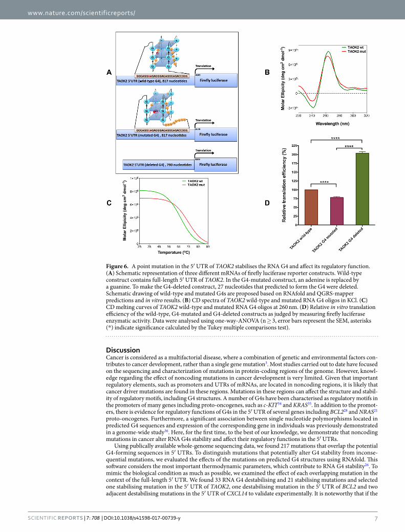
A point mutation stabilises the RNA G4 in the 5′ UTR of TAOK2. In addition to the destabilising mutations, our bioinformatics analyses predicted several mutations in the G-rich regions of 5′ UTRs that can stabilise potential RNA G4 structures (Supplementary Table 3). One of these mutations, which has been reported in a patient with colon cancer, overlaps a predicted RNA G4 in the 5′ UTR of TAOK2. This gene encodes a protein kinase, which involves in cell signalling pathways32 and the induction of apoptosis33. The mutation is an adenine to guanine located next to two adjacent guanines in the first loop. The mutation adds a G-run to the existing four G-runs and based on RNAfold and QGRS-Mapper predictions, it stabilises the RNA G4 by changing the arrange-ment of G-runs involved in the G4 structure formation and shortening one of the loops (Fig. 6A; Supplementary Table 4).
CD spectra of wild-type and G4-mutated TAOK2 oligos showed that they can fold into a parallel G4 confor-mations in near physiological pH and potassium concentration (Fig. 6B), whereas in lithium-containing buffer, the G4 structure signature is much weaker (Supplementary Figure 1). CD melting curves of these oligos (Fig. 6C) showed that the Tm value of the G4-mutated TAOK2 oligo is 7 °C higher than that of the wild-type oligo (Table 1), which further supports the increased stability of the RNA G4.
Three different luciferase reporter constructs were generated (Fig. 6A) as described earlier for BCL2 and CXCL14, to evaluate the G4 stabilising effect of this mutation in vitro. Compared to the wild-type G4 construct, the G4-mutated construct showed 25% lower translation efficiency (P < 0.0001), whereas the G4-deleted con-struct showed a significant increase in translation efficiency (P < 0.0001) (Fig. 6D). These results indicate that the mutated RNA G4 is more efficient in acting as a translational suppressor motif, which could be the consequence of its elevated thermodynamic stability.
Figure 5. Two adjacent mutations in the 5′ UTR of CXCL14 destabilise the RNA G4 and affect its regulatory function. (A) Schematic representation of three different mRNAs of firefly luciferase reporter constructs. Wild-type construct contains full-length 5′ UTR of CXCL14. In the G4-mutated construct, two guanines are replaced by two adenines. To make the G4-deleted construct, 23 nucleotides that predicted to form the RNA G4 were deleted. Schematic drawing of wild-type and mutated G4s are proposed based on RNAfold and QGRS-mapper predictions and in vitro results. (B) CD spectra of CXCL14 wild-type and mutated RNA G4 oligos in KCl. (C) CD melting curves of CXCL14 wild-type and mutated RNA G4 oligos at 260 nm. (D) Relative in vitro translation efficiency of the wild-type, G4-mutated and G4-deleted constructs as judged by measuring firefly luciferase enzymatic activity. Data were analysed using one-way-ANOVA (n ≥ 3, error bars represent the SEM, asterisks (*) indicate significance calculated by the Tukey multiple comparisons test).
www.nature.com/scientificreports/
7Scientific RepoRts | 7: 708 | DOI:10.1038/s41598-017-00739-y
DiscussionCancer is considered as a multifactorial disease, where a combination of genetic and environmental factors con-tributes to cancer development, rather than a single gene mutation3. Most studies carried out to date have focused on the sequencing and characterization of mutations in protein-coding regions of the genome. However, knowl-edge regarding the effect of noncoding mutations in cancer development is very limited. Given that important regulatory elements, such as promoters and UTRs of mRNAs, are located in noncoding regions, it is likely that cancer driver mutations are found in these regions. Mutations in these regions can affect the structure and stabil-ity of regulatory motifs, including G4 structures. A number of G4s have been characterised as regulatory motifs in the promoters of many genes including proto-oncogenes, such as c-KIT34 and KRAS35. In addition to the promot-ers, there is evidence for regulatory functions of G4s in the 5′ UTR of several genes including BCL228 and NRAS21 proto-oncogenes. Furthermore, a significant association between single nucleotide polymorphisms located in predicted G4 sequences and expression of the corresponding gene in individuals was previously demonstrated in a genome-wide study36. Here, for the first time, to the best of our knowledge, we demonstrate that noncoding mutations in cancer alter RNA G4s stability and affect their regulatory functions in the 5′ UTRs.
Using publically available whole-genome sequencing data, we found 217 mutations that overlap the potential G4-forming sequences in 5′ UTRs. To distinguish mutations that potentially alter G4 stability from inconse-quential mutations, we evaluated the effects of the mutations on predicted G4 structures using RNAfold. This software considers the most important thermodynamic parameters, which contribute to RNA G4 stability26. To mimic the biological condition as much as possible, we examined the effect of each overlapping mutation in the context of the full-length 5′ UTR. We found 33 RNA G4 destabilising and 21 stabilising mutations and selected one stabilising mutation in the 5′ UTR of TAOK2, one destabilising mutation in the 5′ UTR of BCL2 and two adjacent destabilising mutations in the 5′ UTR of CXCL14 to validate experimentally. It is noteworthy that if the
Figure 6. A point mutation in the 5′ UTR of TAOK2 stabilises the RNA G4 and affect its regulatory function. (A) Schematic representation of three different mRNAs of firefly luciferase reporter constructs. Wild-type construct contains full-length 5′ UTR of TAOK2. In the G4-mutated construct, an adenine is replaced by a guanine. To make the G4-deleted construct, 27 nucleotides that predicted to form the G4 were deleted. Schematic drawing of wild-type and mutated G4s are proposed based on RNAfold and QGRS-mapper predictions and in vitro results. (B) CD spectra of TAOK2 wild-type and mutated RNA G4 oligos in KCl. (C) CD melting curves of TAOK2 wild-type and mutated RNA G4 oligos at 260 nm. (D) Relative in vitro translation efficiency of the wild-type, G4-mutated and G4-deleted constructs as judged by measuring firefly luciferase enzymatic activity. Data were analysed using one-way-ANOVA (n ≥ 3, error bars represent the SEM, asterisks (*) indicate significance calculated by the Tukey multiple comparisons test).
www.nature.com/scientificreports/
8Scientific RepoRts | 7: 708 | DOI:10.1038/s41598-017-00739-y
RNAfold was used initially to find the overlapping mutations instead of the Quadparser algorithm, we might have overlooked the stabilising mutations.
BCL2 is a human proto-oncogene and belongs to the Bcl-2-family of proteins. These proteins are responsible for the physiological regulation of apoptosis, which is essential for development and tissue homeostasis27. Many examples exist where the expression of BCL2 is elevated in human malignancies, particularly lymphomas. Several mechanisms contribute to the overexpression of BCL2, including chromosomal translocations and loss of endog-enous microRNAs that repress BCL2 expression37. In addition, it has been demonstrated that cis-regulatory muta-tions contribute to the dysregulation of the BCL2 expression38. Balasubramanian et al. characterised a very stable RNA G4 in the 5′ UTR of BCL2, which was found to negatively regulate translation of the gene in vitro28. Here, we report a mutation from a patient with malignant lymphoma that overlaps this RNA G4. Comparison between CD melting curves and Tm values of the wild-type and mutated BCL2 RNA G4 show that the mutation destabilises this RNA G4 (Fig. 3C; Table 1). In order to rule out the possibility that the G4 flanking sequence might interfere with its formation and stability, we included 20 adjacent nucleotides from each side and repeated the biophysical study using 62-mer wild-type and mutated oligos. As shown in Fig. 4A, the CD spectra of both 62-mer wild-type and mutated oligos have the characteristic peaks for parallel propeller G4 structure. In addition, we observed that the difference between Tm values of the 62-mer wild-type and mutated oligos are almost the same as the 22-mer oligos (Table 1) denoting that the flanking sequence of BCL2 RNA G4 does not interfere with this G4 structure and stability. Using in vitro luciferase reporter assays, we show that this mutation significantly elevates the trans-lation efficiency (Fig. 3D). We verified this result by performing dual luciferase assay in MCF-7 cell line. We observed ~75% increase in the gene expression at the protein level for the BCL2 G4-mutant construct, whereas there is not a significant difference in the gene expression at the mRNA level between the BCL2 G4-mutant and wild-type constructs. Taken together, these results suggest the guanine to adenine point mutation in one of the G-tetrads of BCL2 RNA G4 does not completely disrupt the G4 structure, but significantly decreases its stability and affects its regulatory capability as a translational repressor in the 5′ UTR of the BCL2 proto-oncogene.
We also experimentally validated the effect of two adjacent guanine to adenine destabilising mutations in a patient with melanoma that overlap a predicted RNA G4 in the 5′ UTR of CXCL14. This gene belongs to the CXC subfamily of chemokines, which are involved in immunoregulatory and inflammatory processes. Tumor suppressing and promoting activities have been reported for CXCL14, depending on the type and the stage of can-cer39, 40. We showed that the mutations decrease the Tm value of the mutated RNA G4 oligo (Table 1) and increase the translation efficiency of the gene (Fig. 5D) probably because they rearrange the G4 structure by lengthening one of the loops and making the G4 structure less stable.
Contrary to mutations that destabilise G4 structures, some mutations in G-rich sequences can stabi-lise G4 structures and in turn affect their physiological function. Among 21 stabilising mutations listed in the Supplementary Table 3, we experimentally validated the effect of a mutation on a predicted G4 in the 5′ UTR of TAOK2. This gene is one of the TAO (thousand-and-one amino acids) protein kinases that involves in several different cellular processes including cell-signalling pathways32, 41, 42. TOAK2 has been suggested to be involved in the induction of apoptosis33. Here, we demonstrated that the predicted G4 sequence in the 5′ UTR of TAOK2 folds into a parallel quadruplex in vitro (Fig. 6B) and a mutation in a patient with colon cancer increases the Tm value of the mutated TAOK2 RNA G4 oligo (Table 1) and decreases the translation efficiency compared to the wild-type (Fig. 6D). The elevated stability in the mutated TAOK2 RNA G4 might be attributed to the reorganisa-tion of G-runs caused by the mutation. There are two successive guanines in the first loop of TAOK2 RNA G4. The adenine to guanine mutation next to these guanines together form a new G-run that shifts the last G-run outside the RNA G4. Therefore, the first loop is shortened and the RNA G4 becomes more stable.
The results presented in this paper are based on bioinformatics analyses and in vitro experiments that show somatic mutations in cancer patients can alter RNA G4 structure stability in the 5′ UTR of mRNAs and affect its regulatory function. This study provides evidence for the concept that these mutations might contribute to cancer development. Recently, Balasubramanian and colleagues developed a method for high-throughput sequencing of DNA G4s and identified more than 700,000 unique G4 structures in the genome, which is more than twice the number of G4s predicted by computational methods43. This dataset represents a good starting point for a deeper analysis of the effect of mutations on G4 structure and function. Analysing larger datasets might obviate the lack of recurrency among the G4 stabilising or destabilising mutations observed in this study. Finally, considering that G4 binding ligands can stabilise G4 structures, it will be important to investigate whether they can restore the stability of a destabilised G4. Such ligands may include small organic molecules or alternately protein ligands such as antibody fragments which can be generated by in vitro selection technology23, 44. Further understanding of the relationship of the pathogenicity of variants within G4 structures will improve the diagnostic potential of genome sequencing for both somatic and germline mutations and may form a basis for new therapeutic targets.
Material and MethodsThe sequences of all of the oligonucleotides used in the biophysical studies, primers and 5′ UTRs are provided in Supplementary Table 1.
Bioinformatics. Somatic mutation data from whole cancer genomic sequences were obtained from three publicly available data sources: TCGA, ICGC and Alexandrov et al.25. The single base substitution data from the ICGC were obtained from the ICGC data portal (release 16) and the FTP site provided by Alexandrov et al. These mutations were used directly for the analysis. For samples obtained from TCGA, mutations were called from BAM files obtained from CGHub using Strelka with default parameters45. The genomic coordinates of somatic variants are reports using NCBI build 37, aka hg19.
Quadparser algorithm13 was used to identify potential G4-forming sequences in the NCBI build 37 version of the human genome sequence. UCSC Table Browser46 was used to find the overlaps between identified mutations

www.nature.com/scientificreports/
9Scientific RepoRts | 7: 708 | DOI:10.1038/s41598-017-00739-y
and potential G4-forming sequences in the 5′ UTR regions. The effect of a mutation on the thermodynamic sta-bility of an RNA G4 was evaluated using RNAfold and RNALfold26 version 2.1.9 in the context of the full-length 5′ UTR while GU base paring was not allowed. In addition to RNAfold, the QGRS-Mapper14 online tool was used to investigate the effect of mutations on the RNA G4-forming sequences in the 5′ UTR of BCL2, CXCL14 and TAOK2.
Circular dichroism spectroscopy. Circular dichroism spectra were recorded as previously described47. Briefly, RNA oligos were annealed by heating at 90 °C for 10 minutes and slowly cooling down to room tempera-ture. CD spectra were recorded at 25 °C on an Aviv 215S circular dichroism spectrometer equipped with a Peltier temperature controller. RNA G4 samples of the desired sequence were prepared at 5 µM in the G4 folding buffer (100 mM KCl, 20 mM Tris HCl pH 7.5). KCl was replaced by LiCl in the control samples. Four scans were accu-mulated over the wavelength range 220–320 nm in a 0.1 cm path length cell at the standard sensitivity, data pitch 0.1 nm, continuous scanning mode, scanning speed 100 nm min−1, response 4 sec, and bandwidth 1 nm. Buffers alone were scanned and these spectra subtracted from the average scans for each sample. Tm analysis was recorded by CD at 260 nm and heated at 1 °C min−1 over the temperature range 25–95 °C. CD and melt curve spectra were collected in units of millidegrees, normalized to the total species concentrations and expressed as molar ellipticity units (deg × cm2 dmol−1). Each CD spectrum was smoothed by averaging ten neighbour points using Prism 6. Sigmoidal dose-response curves (variable slope) were fitted to the melt curve data using Prism 6.
Construction of plasmids. Overlap extension PCR was used to clone either T7 or CMV promoter upstream of multiple restriction sites of pGL3 basic plasmid (Promega). The sequences of the 5′ UTRs of BCL2, CXCL14 and TAOK2 were obtained from the UCSC Genome Browser48 based on the NCBI build 37 version of the human genome sequence. The synthetically synthesised full-length 5′ UTRs of each candidate was purchased from GenScript and inserted in the KpnI and HindIII restriction sites of T7 or CMV pGL3 plasmid. These constructs are considered as wild-types. G4-mutated and G4-deleted constructs were created based on the wild-type con-structs for each 5′ UTR. The NEBaseChangerTM online tool provided by New England BioLabs was used to design primers for making desired point mutations in the G4-forming sequences and also deleting the entire G4-forming sequences from the 5′ UTRs.
In vitro transcription. A gene fragment containing T7 promoter and the full-length 5′ UTR attached to the firefly luciferase gene was provided from each of the wild-types, G4-mutated and G4-deleted constructs by PCR amplification. The size of each fragment was examined on a 1% agarose gel followed by gel purification. An addi-tional step of PCR clean up was performed and RNase inhibitor (Ambion) at a final concentration of 1 U/μL was added to each sample. The PCR products were used as templates to produce 5′-capped transcripts in vitro using mMESSAGE mMACHINE T7 kit (Ambion). Transcripts were DNase treated and purified using the RNeasy Mini Kit (QIAGEN). The size and integrity of the transcripts were confirmed on a 1% agarose gel. The concentration of each set of transcripts was determined by NanoDrop.
In vitro translation and luciferase assay. Nuclease-treated rabbit reticulocyte lysate (Promega) was used to perform the cell-free translation. 150 ng/μL (final concentration) of the in vitro transcribed 5′-capped mRNA containing the full-length 5′ UTR attached to the firefly luciferase coding sequence was translated according to the manufacturer’s protocol. In order to measure the firefly luciferase activity, 5 μL of the translation reaction was added to 75 μL of luciferase assay reagent (Promega) at room temperature. The reaction was quickly mixed and luminescence was measured using CLARIO star microplate reader (BMG LABTECH).
Dual luciferase assay and RT-PCR. MCF-7 cells were seeded in 12-well plates (Corning) and incubated until they reached to 70–80% confluency. The cells were co-transfected with pRL-TK (Promega) normalizing vector, which expresses renilla, and the CMV pGL3 plasmids constructs, which prepared as described earlier and express firefly, with the ratio of 1 to 7 using Lipofectamine 3000 (Thermo Fisher Scientific) according to the manufacturer’s protocol. When cells reached to 90–95% confluency, renilla and firefly luciferase activities were measured using Dual-Luciferase Reporter Assay System (Promega) following the manufacturer’s protocol in the CLARIO star microplate reader (BMG LABTECH). Total RNA was extracted from the remaining cells using RNeasy mini kit (QIAGEN). 200 ng of each total RNA sample was reverse-transcribed using SuperScript III First-Strand Synthesis System (Thermo Fisher Scientific). The cDNA from each sample was used to perform RT-qPCR using LightCycler 480 SYBR Green I Master reagents (Roche) and a LightCycler 480 SYBR device (Roche). Primer sets are listed in the Supplementary Table 1. The relative mRNA expression level was calcu-lated using the following mathematical model49: Ratio = (E target)ΔCP
target(control-sample)/(E reference)ΔCP
reference(control-sample)
where firefly and renilla were considered as the target and reference genes, respectively. GAPDH expression in each sample was considered as the control.
References 1. Cancer Genome Atlas Research, N. et al. The Cancer Genome Atlas Pan-Cancer analysis project. Nat Genet 45, 1113–1120,
doi:10.1038/ng.2764 (2013). 2. International Cancer Genome, C. et al. International network of cancer genome projects. Nature 464, 993–998, doi:10.1038/
nature08987 (2010). 3. Stratton, M. R., Campbell, P. J. & Futreal, P. A. The cancer genome. Nature 458, 719–724, doi:10.1038/nature07943 (2009). 4. Druker, B. J. Translation of the Philadelphia chromosome into therapy for CML. Blood 112, 4808–4817, doi:10.1182/
blood-2008-07-077958 (2008). 5. Little, P. F. Structure and function of the human genome. Genome Res 15, 1759–1766, doi:10.1101/gr.4560905 (2005). 6. Weinhold, N., Jacobsen, A., Schultz, N., Sander, C. & Lee, W. Genome-wide analysis of noncoding regulatory mutations in cancer.
Nat Genet 46, 1160–1165, doi:10.1038/ng.3101 (2014).
www.nature.com/scientificreports/
1 0Scientific RepoRts | 7: 708 | DOI:10.1038/s41598-017-00739-y
7. Puente, X. S. et al. Non-coding recurrent mutations in chronic lymphocytic leukaemia. Nature 526, 519–524, doi:10.1038/nature14666 (2015).
8. Fredriksson, N. J., Ny, L., Nilsson, J. A. & Larsson, E. Systematic analysis of noncoding somatic mutations and gene expression alterations across 14 tumor types. Nat Genet 46, 1258–1263, doi:10.1038/ng.3141 (2014).
9. Bochman, M. L., Paeschke, K. & Zakian, V. A. DNA secondary structures: stability and function of G-quadruplex structures. Nat Rev Genet 13, 770–780, doi:10.1038/nrg3296 (2012).
10. Karsisiotis, A. I., O’Kane, C. & Webba da Silva, M. DNA quadruplex folding formalism–a tutorial on quadruplex topologies. Methods 64, 28–35, doi:10.1016/j.ymeth.2013.06.004 (2013).
11. Zhang, D. H. et al. Monomorphic RNA G-quadruplex and polymorphic DNA G-quadruplex structures responding to cellular environmental factors. Biochemistry 49, 4554–4563, doi:10.1021/bi1002822 (2010).
12. Todd, A. K., Johnston, M. & Neidle, S. Highly prevalent putative quadruplex sequence motifs in human DNA. Nucleic Acids Res 33, 2901–2907, doi:10.1093/nar/gki553 (2005).
13. Huppert, J. L. & Balasubramanian, S. Prevalence of quadruplexes in the human genome. Nucleic Acids Res 33, 2908–2916, doi:10.1093/nar/gki609 (2005).
14. Kikin, O., D’Antonio, L. & Bagga, P. S. QGRS Mapper: a web-based server for predicting G-quadruplexes in nucleotide sequences. Nucleic Acids Res 34, W676–682, doi:10.1093/nar/gkl253 (2006).
15. Maizels, N. & Gray, L. T. The G4 genome. PLoS Genet 9, e1003468, doi:10.1371/journal.pgen.1003468 (2013). 16. Agarwala, P., Pandey, S., Mapa, K. & Maiti, S. The G-quadruplex augments translation in the 5′ untranslated region of transforming
growth factor beta2. Biochemistry 52, 1528–1538, doi:10.1021/bi301365g (2013). 17. Bugaut, A. & Balasubramanian, S. 5′-UTR RNA G-quadruplexes: translation regulation and targeting. Nucleic Acids Res 40,
4727–4741, doi:10.1093/nar/gks068 (2012). 18. Beaudoin, J. D. & Perreault, J. P. 5′-UTR G-quadruplex structures acting as translational repressors. Nucleic Acids Res 38, 7022–7036,
doi:10.1093/nar/gkq557 (2010). 19. Arora, A. et al. Inhibition of translation in living eukaryotic cells by an RNA G-quadruplex motif. RNA 14, 1290–1296, doi:10.1261/
rna.1001708 (2008). 20. Morris, M. J. & Basu, S. An unusually stable G-quadruplex within the 5′-UTR of the MT3 matrix metalloproteinase mRNA represses
translation in eukaryotic cells. Biochemistry 48, 5313–5319, doi:10.1021/bi900498z (2009). 21. Kumari, S., Bugaut, A., Huppert, J. L. & Balasubramanian, S. An RNA G-quadruplex in the 5′ UTR of the NRAS proto-oncogene
modulates translation. Nat Chem Biol 3, 218–221, doi:10.1038/nchembio864 (2007). 22. Biffi, G., Tannahill, D., McCafferty, J. & Balasubramanian, S. Quantitative visualization of DNA G-quadruplex structures in human
cells. Nat Chem 5, 182–186, doi:10.1038/nchem.1548 (2013). 23. Biffi, G., Di Antonio, M., Tannahill, D. & Balasubramanian, S. Visualization and selective chemical targeting of RNA G-quadruplex
structures in the cytoplasm of human cells. Nat Chem 6, 75–80, doi:10.1038/nchem.1805 (2014). 24. Biffi, G., Tannahill, D., Miller, J., Howat, W. J. & Balasubramanian, S. Elevated levels of G-quadruplex formation in human stomach
and liver cancer tissues. PLoS One 9, e102711, doi:10.1371/journal.pone.0102711 (2014). 25. Alexandrov, L. B. et al. Signatures of mutational processes in human cancer. Nature 500, 415–421, doi:10.1038/nature12477 (2013). 26. Lorenz, R. et al. 2D meets 4G: G-quadruplexes in RNA secondary structure prediction. IEEE/ACM Trans Comput Biol Bioinform 10,
832–844, doi:10.1109/TCBB.2013.7 (2013). 27. Cory, S., Huang, D. C. & Adams, J. M. The Bcl-2 family: roles in cell survival and oncogenesis. Oncogene 22, 8590–8607, doi:10.1038/
sj.onc.1207102 (2003). 28. Shahid, R., Bugaut, A. & Balasubramanian, S. The BCL-2 5′ untranslated region contains an RNA G-quadruplex-forming motif that
modulates protein expression. Biochemistry 49, 8300–8306, doi:10.1021/bi100957h (2010). 29. Paramasivan, S., Rujan, I. & Bolton, P. H. Circular dichroism of quadruplex DNAs: applications to structure, cation effects and ligand
binding. Methods 43, 324–331, doi:10.1016/j.ymeth.2007.02.009 (2007). 30. Halder, K., Wieland, M. & Hartig, J. S. Predictable suppression of gene expression by 5′-UTR-based RNA quadruplexes. Nucleic
Acids Res 37, 6811–6817, doi:10.1093/nar/gkp696 (2009). 31. Lu, J., Chatterjee, M., Schmid, H., Beck, S. & Gawaz, M. CXCL14 as an emerging immune and inflammatory modulator. J Inflamm
(Lond) 13, 1, doi:10.1186/s12950-015-0109-9 (2016). 32. Chen, Z. & Cobb, M. H. Regulation of stress-responsive mitogen-activated protein (MAP) kinase pathways by TAO2. J Biol Chem
276, 16070–16075, doi:10.1074/jbc.M100681200 (2001). 33. Zihni, C. et al. Prostate-derived sterile 20-like kinase 1-alpha induces apoptosis. JNK- and caspase-dependent nuclear localization
is a requirement for membrane blebbing. J Biol Chem 282, 6484–6493, doi:10.1074/jbc.M608336200 (2007). 34. Fernando, H. et al. A conserved quadruplex motif located in a transcription activation site of the human c-kit oncogene. Biochemistry
45, 7854–7860, doi:10.1021/bi0601510 (2006). 35. Cogoi, S. & Xodo, L. E. G-quadruplex formation within the promoter of the KRAS proto-oncogene and its effect on transcription.
Nucleic Acids Res 34, 2536–2549, doi:10.1093/nar/gkl286 (2006). 36. Baral, A. et al. Quadruplex-single nucleotide polymorphisms (Quad-SNP) influence gene expression difference among individuals.
Nucleic Acids Res 40, 3800–3811, doi:10.1093/nar/gkr1258 (2012). 37. Yip, K. W. & Reed, J. C. Bcl-2 family proteins and cancer. Oncogene 27, 6398–6406, doi:10.1038/onc.2008.307 (2008). 38. Mathelier, A. et al. Cis-regulatory somatic mutations and gene-expression alteration in B-cell lymphomas. Genome Biol 16, 84,
doi:10.1186/s13059-015-0648-7 (2015). 39. Hara, T. & Tanegashima, K. Pleiotropic functions of the CXC-type chemokine CXCL14 in mammals. J Biochem 151, 469–476,
doi:10.1093/jb/mvs030 (2012). 40. Benarafa, C. & Wolf, M. CXCL14: the Swiss army knife chemokine. Oncotarget 6, 34065–34066, doi:10.18632/oncotarget.6040 (2015). 41. Chen, Z., Hutchison, M. & Cobb, M. H. Isolation of the protein kinase TAO2 and identification of its mitogen-activated protein kinase/
extracellular signal-regulated kinase kinase binding domain. J Biol Chem 274, 28803–28807, doi:10.1074/jbc.274.40.28803 (1999). 42. Coffey, E. T. Nuclear and cytosolic JNK signalling in neurons. Nat Rev Neurosci 15, 285–299, doi:10.1038/nrn3729 (2014). 43. Chambers, V. S. et al. High-throughput sequencing of DNA G-quadruplex structures in the human genome. Nat Biotechnol 33,
877–881, doi:10.1038/nbt.3295 (2015). 44. Lee, C. M., Iorno, N., Sierro, F. & Christ, D. Selection of human antibody fragments by phage display. Nat Protoc 2, 3001–3008,
doi:10.1038/nprot.2007.448 (2007). 45. Saunders, C. T. et al. Strelka: accurate somatic small-variant calling from sequenced tumor-normal sample pairs. Bioinformatics 28,
1811–1817, doi:10.1093/bioinformatics/bts271 (2012). 46. Karolchik, D. et al. The UCSC Table Browser data retrieval tool. Nucleic Acids Res 32, D493–496, doi:10.1093/nar/gkh103 (2004). 47. Moye, A. L. et al. Telomeric G-quadruplexes are a substrate and site of localization for human telomerase. Nat Commun 6, 7643,
doi:10.1038/ncomms8643 (2015). 48. Kent, W. J. et al. The human genome browser at UCSC. Genome Res 12, 996–1006, doi:10.1101/gr.229102. Article published online
before print in May 2002 (2002). 49. Pfaffl, M. W. A new mathematical model for relative quantification in real-time RT-PCR. Nucleic Acids Res 29, e45–45, doi:10.1093/
nar/29.9.e45 (2001).

www.nature.com/scientificreports/
1 1Scientific RepoRts | 7: 708 | DOI:10.1038/s41598-017-00739-y
AcknowledgementsM.Z. is supported by UIPA PhD scholarship from University of New South Wales; A.L.M. is supported by Kids Cancer Alliance PhD scholarship; J.W.H.W. is supported by an Australian Research Council Future Fellowship (FT130100096); M.J.C. is supported by a Cancer Institute NSW Early Career Fellowship (13/ECF/1-46); D.U.C. is supported by National Health Medical Research Council and the Australian Research Council; T.M.B. is supported by Cancer Council NSW (project grant RG11-07) and Cancer Institute NSW (11/CDF/3-05).
Author ContributionsM.Z., M.J.C. and M.E.D. conceived the project; M.Z. and M.E.D. designed the experiments; J.W.H.W. and D.P. performed the curation of the mutations. M.J.C. advised on the bioinformatics analysis; A.L.M. performed the biophysical studies; M.Z. conducted the bioinformatics analyses and the rest of the experiments; M.Z., D.U.C., T.M.B. and M.E.D. wrote the paper.
Additional InformationSupplementary information accompanies this paper at doi:10.1038/s41598-017-00739-yCompeting Interests: The authors declare that they have no competing interests.Publisher's note: Springer Nature remains neutral with regard to jurisdictional claims in published maps and institutional affiliations.
Open Access This article is licensed under a Creative Commons Attribution 4.0 International License, which permits use, sharing, adaptation, distribution and reproduction in any medium or
format, as long as you give appropriate credit to the original author(s) and the source, provide a link to the Cre-ative Commons license, and indicate if changes were made. The images or other third party material in this article are included in the article’s Creative Commons license, unless indicated otherwise in a credit line to the material. If material is not included in the article’s Creative Commons license and your intended use is not per-mitted by statutory regulation or exceeds the permitted use, you will need to obtain permission directly from the copyright holder. To view a copy of this license, visit http://creativecommons.org/licenses/by/4.0/. © The Author(s) 2017