ICOS ligation recruits the p50alpha PI3K regulatory subunit to the immunological synapse.
Upload
khangminh22Category
view
1download
0
Cell Death & Differentiation (2021) 28:2634–2650https://doi.org/10.1038/s41418-021-00774-3
ARTICLE
The long noncoding RNA Synage regulates synapse stability andneuronal function in the cerebellum
Fei Wang1● Qianqian Wang1
● Baowei Liu1● Lisheng Mei1 ● Sisi Ma2 ● Shujuan Wang3
● Ruoyu Wang1,4●
Yan Zhang5● Chaoshi Niu6
● Zhiqi Xiong7● Yong Zheng3
● Zhi Zhang 1● Juan Shi2 ● Xiaoyuan Song 8
Received: 2 July 2020 / Revised: 7 March 2021 / Accepted: 10 March 2021 / Published online: 24 March 2021© The Author(s), under exclusive licence to ADMC Associazione Differenziamento e Morte Cellulare 2021. This article is published with open access
AbstractThe brain is known to express many long noncoding RNAs (lncRNAs); however, whether and how these lncRNAs functionin modulating synaptic stability remains unclear. Here, we report a cerebellum highly expressed lncRNA, Synage, regulatingsynaptic stability via at least two mechanisms. One is through the function of Synage as a sponge for the microRNA miR-325-3p, to regulate expression of the known cerebellar synapse organizer Cbln1. The other function is to serve as a scaffoldfor organizing the assembly of the LRP1-HSP90AA1-PSD-95 complex in PF-PC synapses. Although somewhat divergent inits mature mRNA sequence, the locus encoding Synage is positioned adjacent to the Cbln1 loci in mouse, rhesus macaque,and human, and Synage is highly expressed in the cerebella of all three species. Synage deletion causes a full-spectrumcerebellar ablation phenotype that proceeds from cerebellar atrophy, through neuron loss, on to synapse density reduction,synaptic vesicle loss, and finally to a reduction in synaptic activity during cerebellar development; these deficits areaccompanied by motor dysfunction in adult mice, which can be rescued by AAV-mediated Synage overexpression frombirth. Thus, our study demonstrates roles for the lncRNA Synage in regulating synaptic stability and function duringcerebellar development.
Introduction
Cerebellar development is an important process for reg-ulating the onset of a variety of motor and non-motorbehaviors [1]. The cerebellar cortex is composed of threelayers: the molecular layer, the Purkinje cell (PC) layer, andthe granule cell (GC) layer, from outermost to innermost
These authors contributed equally: Fei Wang, Qianqian Wang,Baowei Liu
Edited by M. Bianchi
* Xiaoyuan [email protected]
1 Hefei National Laboratory for Physical Sciences at the Microscale,CAS Key Laboratory of Brain Function and Disease, School ofLife Sciences, Division of Life Sciences and Medicine, Universityof Science and Technology of China, Hefei, Anhui, China
2 National Laboratory of Medical Molecular Biology, Institute ofBasic Medical Sciences, CAMS and PUMC, Beijing, China
3 State Key Laboratory of Proteomics, Beijing Proteome ResearchCenter, National Center for Protein Sciences (Beijing), BeijingInstitute of Lifeomics, Beijing, China
4 Graduate School of Biomedical Sciences, University of Texas MDAnderson Cancer Center and UTHealth, Houston, TX, USA
5 Stroke Center & Department of Neurology, The First AffiliatedHospital of USTC, Division of Life Sciences and Medicine,
University of Science and Technology of China, Hefei, Anhui,China
6 Department of Neurosurgery, The First Affiliated Hospital ofUSTC, Division of Life Sciences and Medicine, University ofScience and Technology of China, Hefei, Anhui, China
7 Institute of Neuroscience, State Key Laboratory of Neuroscience,CAS Center for Excellence in Brain Science and IntelligenceTechnology, Chinese Academy of Sciences, Shanghai, China
8 MOE Key Laboratory for Membraneless Organelles and CellularDynamics, Hefei National Laboratory for Physical Sciences at theMicroscale, CAS Key Laboratory of Brain Function and Disease,School of Life Sciences, Division of Life Sciences and Medicine,University of Science and Technology of China, Hefei, China
Supplementary information The online version containssupplementary material available at https://doi.org/10.1038/s41418-021-00774-3.
1234
5678
90();,:
1234567890();,:
[2]. The GC layer consists of small and densely packedexcitatory granule neurons that make up the vast majority ofneurons in the cerebellum and the brain [3]. The PC layerharbors the largest GABAergic inhibitory PCs, as well asBergmann glial cells (BGCs), which are mainly locatedaround PCs [4, 5]. The formation of mature neurons andstabilized synapses during development is a prerequisite forproper nervous system functionality, which require synapticproteins. For instance, CBLN1, highly expressed in cere-bellar GCs, is a synaptic protein crucial for organization ofparallel fibers (PFs, axons of the GCs) and PCs [6, 7].Similarly, LRP1, a postsynaptic transmembrane protein,forms a complex with postsynaptic N-methyl-D-aspartatereceptors through PSD-95 (a postsynaptic density protein)to modulate synaptic transmission and synaptic plasticity[8–12]. However, much remains unclear about themechanism of synaptic stability.
Long noncoding RNAs (lncRNAs) are transcripts longerthan 200 nucleotides that do not translate into functionalproteins (except in some cases forming small, potentiallyfunctional peptides). Like many protein-coding mRNAs,many lncRNAs also exhibit strong specificity in their spatio-temporal expression [13]. Many lncRNAs function in neu-rodevelopment. For example, the nervous system-specificlncRNAs Evf2 and Pinky regulate neural development[14, 15]. The nuclear-enriched GM12371 lncRNA is a pro-lific transcriptional regulator and is critical for synapsefunction in hippocampal neurons [16]. Nevertheless, theneurobiological functions of the overwhelming majority oflncRNAs remain enigmatic compared with protein-codinggenes. Importantly, it remains unclear whether and howlncRNAs modulate synaptic stability. The Gm2694 lncRNA(alias AK082312) was originally found to have enrichedexpression in the mouse cerebellar cortex [17]. The Gm2694lncRNA (alias linc1582) was later found to be associatedwith neuroectoderm differentiation [18]. Recently, Gm2694(alias Trincr1) was documented to regulate FGF/ERK sig-naling and the self-renewal of embryonic stem cells [19].Although with all of these studies, whether and howGm2694 functions in the cerebellar synapse was mysterious.
Here, we studied this synapse-functional cerebellumlncRNA, particularly three isoforms of Gm2694, which wedesignated as Synage, and which is distributed in thecytoplasm and synapses of cerebellar cells. We showed thatSynage modulates the expression of the synaptic proteinCBLN1 in GCs and contributes to preventing cerebellaratrophy and motor defects. We also found that Synagemediates the assembly of the LRP1-HSP90AA1-PSD-95-Synage complex in PCs to regulate synaptic stability. Ourstudy presents long-term in vivo data demonstrating thedevelopmental functions of Synage lncRNA and reveals themechanisms underlying its regulation of synaptic stabilityand cerebellar development.
Materials and methods
Animal models
Wild-type (WT) BALB/C and C57BL/6 mice were purchasedfrom the Vital River Laboratories (Beijing, China) and housedin a humidity and temperature-controlled room under areverse 12-h dark–light cycle. Mice were provided with foodand water ad libitum under standard conditions. All proce-dures were in accordance with the guidelines of and approvedby the University of Science and Technology of China Ani-mal Resources Center and University Animal Care and UseCommittee (Permit Number: USTCACUC1801015).
The sgRNAs were designed to target exon 1 and the endof last exon of Synage, thus excising almost the entireSynage locus, while avoiding the shared promoter regionwith Cbln1, to produce Synage knockout (KO) mice usingthe CRISPR-Cas9 system. Subsequently, genomic DNAwas extracted and used to identify mouse genotypes withthree pairs of specific primers for polymerase chain reaction(PCR) amplification and sequencing. In this study, the WToffspring of Synage heterozygous KO mice were used as thecontrol group for Synage homozygous mice. BIOGLECompany provided F1 heterozygous KO mice. The sgRNA,shRNA, and genotyping primer sequences are listed inSupplementary Table S3.
Reverse transcription and quantitative PCR (RT-qPCR)
Total RNAs were extracted using TRIzol reagent (Ambion,15596) following the manufacturer’s instructions. RNase-free DNase (Promega, M6101) was used to remove residualDNA. Reverse transcription was performed using thecommercially available reverse transcription system (Pro-mega, A5001). The real-time qPCR experiments were per-formed on a Bio-Rad CFX96 qPCR system according to themanufacturer’s instructions (Vazyme, Q111-03). Geneexpression levels were calculated relative to the referencegene GAPDH. The RT-qPCR primer sequences are listed inSupplementary Table S3.
5′- and 3′-Rapid amplification of cDNA ends (RACE)
Both 5′- and 3′-RACE experiments were performed fol-lowing the manufacturer’s instructions (Invitrogen, 18374-058, 18373-019). Briefly, 5′ and 3′ gene-specific primers(GSPs) were designed to perform amplification of cDNAends using the RACE-Ready cDNA as a template. ThecDNA products were then inserted into a pRACE vector.Both PCR screening with GSPs and Sanger sequencingwere further performed to identify the full-length cDNA ofthe target gene.
The long noncoding RNA Synage regulates synapse stability and neuronal function in the cerebellum 2635

Western blot
Mouse tissue was dissected on ice. The whole cerebellumwas then transferred to a homogenizer and added to 500-μlRIPA buffer (50-mM Tris-HCl (pH 7.4), 150-mM NaCl,1-mM EDTA, 1% Triton X-100, 1% SDS, 1% sodium-deoxycholate, fresh proteinase inhibitor cocktail, PMSF)for every 20 mg of samples. After homogenization andconstant agitation at 4 °C for 2 h, each sample was cen-trifuged at 14,000 g for 10 min at 4 °C. The supernatantwas aspirated and placed in a fresh tube kept on ice. Thelysate was then diluted with 2X SDS loading buffer(125-mM Tris-HCl (pH 6.8), 4% SDS, 20% glycerol,0.2% bromophenol blue, 10% β-mercaptoethanol) andboiled at 100 °C for 5 min and stored at −80 °C. Thesubsequent Western blot protocol was performed asdescribed previously [20].
Co-immunoprecipitation (co-IP)
The whole homogenized mouse cerebellum was lysed in 1-ml NET-N buffer (20-mM Tris-HCl (pH 8.0), 125-mMNaCl, 1-mM EDTA, 0.5% NP-40, 10% glycerol, freshproteinase inhibitor cocktail, PMSF). The lysate was incu-bated at 4 °C for 1.5 h on a rotating platform and subse-quently treated with ultrasonication. The sonicated lysatewas cleared by centrifugation at 14,000 g for 10 min at 4 °C,then the supernatant was pre-cleared with Protein A/GMagnetic Beads (Thermo, 88803). Next, the supernatantwas incubated with specific antibodies (HSP90AA1 (Pro-teintech, 13171-1-AP), or LRP1 (Abcam, ab92544), PSD-95 (Abcam, ab2723), or control IgG (Cell Signaling Tech-nology, 5873/ 8726)) overnight on a rotating platform at 4 °C. Subsequently, Protein A/G Magnetic Beads were addedto the supernatant for 2 h at 4 °C under gentle rotation. Thebeads were pelleted and washed three times with ice-coldNET-N buffer. The sample was then boiled in 1X SDSloading buffer at 100 °C for 5 min before Western blotdetection.
Synage overexpression and co-IP
Cells (1 × 10⁶) were transfected with Synage overexpressionplasmid (V12-PLKO.1) and then cultured in a 6-cm dish.Forty-eight hours later, the overexpressed Synage cells werepelleted at 500 g for 5 min. After washing twice withphosphate-buffered saline (PBS), cell pellets were resus-pended in 0.7-ml NET-N buffer with fresh proteinaseinhibitor cocktail and PMSF, and incubated at 4 °C withrotation for 60 min. The lysate was isolated by centrifuga-tion at 14,000 g for 10 min. The co-IP protocol was per-formed as described above.
RNA immunoprecipitation (RIP)
The whole homogenized mouse cerebellum was cross-linked in 1% formaldehyde (Sangon Biotech, A501912-0500) for 10 min on a rotating platform. To stop the reac-tion, 0.125-M glycine was added for 10 min and the samplewas pelleted at 2000 g for 5 min. Then the cell pellet wasincubated in NET-N buffer for 1.5 h at 4 °C. The lysate wassonicated and centrifuged at 14,000 g for 10 min. Next, thesupernatant was divided into two samples and incubatedwith specific antibodies and control IgG overnight on arotating platform at 4 °C. Next, the supernatant was incu-bated with Protein A/G Magnetic Beads for 2 h at 4 °Cunder gentle rotation. Beads were recovered and washedthree times with NET-N buffer. The beads were resus-pended in proteinase K buffer (100-mM NaCl, 10-mM Tris-HCl (pH 7.0), 1-mM EDTA, 0.5% SDS, 20-μg/ml protei-nase K) for 20 min at 56 °C. The remaining beads were usedfor RNA extraction and RT-qPCR analysis to identifyinteracting RNA segments according to the manufacturer’sinstructions.
In vivo RNA pull-down–mass spectrometry (MS)
The in vivo RNA pull-down–MS assay was performed aspreviously described [21]. Briefly, the whole homogenizedmouse cerebellum was cross-linked in 1% formaldehyde for10 min, followed by 0.125-M glycine quenching for 5 min.The sample was pelleted at 2000 g for 5 min. Cross-linkedcell pellets were lysed in lysis buffer (50-mM Tris-HCl (pH7.0), 10-mM EDTA, 1% SDS, fresh proteinase inhibitorcocktail, PMSF, Murine RNase inhibitor) and solubilizedby sonication. The supernatant was incubated with bioti-nylated antisense oligo probes in hybridization buffer(750-mM NaCl, 1% SDS, 50-mM Tris-Cl pH 7.0, 1-mMEDTA, 15% formamide, fresh proteinase inhibitor cocktail,PMSF, Murine RNase inhibitor) at 37 °C overnight undergentle rotation. M-280 Streptavidin beads (Invitrogen,11206D) were added to the lysis buffer and incubated at37 °C for 30 min with rotation. The RNA-binding proteincomplex components were washed five times using washbuffer (2X SSC, 0.5% SDS, fresh PMSF). For proteinelution, beads were resuspended in protein elution buffer(7.5-mM, N-2-hydroxyethylpiperazine-N-2-ethanesulfonicacid (HEPES, pH 7.5), 15-mM EDTA, 0.15% SDS, 75-mMNaCl, 0.02% sodium-deoxycholate) at 25 °C for 20 min andat 65 °C for 10 min. Twenty-five percent total volume TCA(trichloroacetic acid) was added to the clean eluent, andmixed proteins were precipitated at 4 °C overnight. Subse-quently, proteins were pelleted at 16,000 g at 4 °C for30 min. The supernatant was removed, and the proteinpellet was washed with cold acetone and pelleted again at
2636 F. Wang et al.

16,000 g at 4 °C for 30 min. The pellet was left to air-dry for1 min and stored at −80 °C before MS.
Biotin-mmu-miR-325-3p pull-down
The procedure for miRNA pull-down was performed aspreviously described [22]. In brief, for biotin-labeledmiRNA pull-down experiments, 200-pmol biotin-labeledmmu-miR-325-3p (GenePharma) were transfected into 2 ×10⁶ HT-22 cells. After 24 h, the cells were lysed in lysisbuffer (20-mM Tris (pH 7.4), 100-mM KCl, 0.3% NP-40,5-mM MgCl2, fresh proteinase inhibitor cocktail, PMSF,Murine RNase inhibitor). Streptavidin magnetic beads(Invitrogen, 11206D) were added to the cell lysate andincubated at 4 °C for 4 h with rotation. The M-280 Strep-tavidin beads were washed three times using lysis buffer.RNA bound to the M-280 magnetic beads was isolatedusing TRIzol LS reagent (Invitrogen, 10296028) andquantified by RT-qPCR.
Electrophoretic mobility shift assays (EMSAs)
EMSAs were performed using the ChemiluminescentEMSA Kit according to the manufacturer’s protocol(Beyotime, GS009). Briefly, whole cerebellum tissue lysatewas prepared in cytoplasmic lysis buffer (1% Triton X-100,25-mM Tris-HCl (pH 7.4), 40-mM KCl) with fresh pro-teinase inhibitor cocktail, PMSF, and RNase inhibitors. Thecerebellum lysate was centrifuged at 14,000 g for 10 min,after which the supernatant was treated with M-280 Strep-tavidin beads (Invitrogen, 11206D) to pre-clear the cyto-plasmic extract (CE). Biotin-labeled Synage RNA probes(150 ng) were incubated with 14-μg CE. Unlabeled SynageRNA probes (7500 ng) were used as a competitor. Thereaction was separated by native 6% PAGE gels andtransferred onto nylon membranes. Biotin signals weredetected by chemiluminescence.
Immunofluorescence (IF)
The mice were anesthetized and intracardially perfused withPBS. The brain was dissected and fixed in 4% paraf-ormaldehyde (PFA) overnight and dehydrated with asucrose gradient. The frozen sagittal sections (thickness,8–10 μm) were washed three times with 1X PBS. Thesections were permeabilized by 0.1% Triton X-100 for 30min. Tissue sections were incubated in 3% bovine serumalbumin (BSA) for 1 h and then incubated with primaryantibodies overnight at 4 °C. Tissue sections were washedwith 1X PBS three times and subsequently incubated insecondary antibodies for 1–2 h at room temperature (RT).Tissues were washed with 1X PBS three times and incu-bated for 2 min with Hoechst 33342 for nuclear
counterstaining. The immunostained tissues were visualizedusing an FV1200 confocal microscope system (OLYMPUS,Japan). The cerebellum sections with the largest area ineach mouse were selected for cerebellar developmentalphenotype analysis. The following primary antibodies wereused: PSD-95 (Abcam, ab2723), LRP1 (Abcam, ab92544),HSP90AA1 [Alexa Fluor® 647] (Novus, NBP1-77682AF647), CBLN1 (Abcam, ab181379), Calbindin-D28k (Proteintech, 14479-1-AP, 66394-1-lg), Gdf10(Santa Cruz, sc-390046). The following secondary anti-bodies were used: goat anti-rabbit IgG H&L (Alexa Fluor®
488, Abcam, ab150077), Alexa Fluor594-conjugated goatanti-mouse IgG (H+ L) (Proteintech, SA00006-3), andgoat anti-rabbit IgG H&L, F (ab′) 2 Fragment (Alexa Fluor®
647 conjugate, Santa Cruz, 4414).
In vitro transcription
The cDNA of mouse Synage (n424059) was cloned intopcDNA3.1 vector. The plasmid was linearized to produce aDNA template, and full-length sense or antisense RNAswere transcribed in vitro using T7 RNA polymerase (Invi-trogen, 18033019) or MAXIscript SP6 Transcription Kit(Invitrogen, AM1330) in combination with biotinylatedNTPs (Roche, 11685597910). In vitro transcribed RNAswere further used for FISH assays in cerebellar sectionsdirectly as described below, or for EMSAs asdescribed above.
Fluorescence in situ hybridization (FISH)
All solutions were prepared using RNase-free reagents anddiethylpyrocarbonate-treated double deionized water.Fresh-frozen brain sections (10 μm) were fixed in 4% PFAfor 10 min and washed with 1X PBS three times. Sectionswere digested with 1 μg/ml of proteinase K buffer for 20min and incubated in acetylation solution for 10 min. Thesections were placed in the hybridization buffer (50% for-mamide, 5X SSC, 0.3-mg/ml tRNA) without biotinylatedprobes for 5 h at RT and incubated in the hybridizationsolution with corresponding RNA probes (200 ng/ml) for12–14 h at 70 °C. After hybridization, the slices werewashed once in 5X SSC at 70 °C and washed twice in 0.2XSSC for 30 min at 70 °C. Sections were further incubatedwith streptavidin antibody and other primary antibodies for2 h at RT. Tissue sections were washed with 1X PBS threetimes and subsequently incubated in secondary antibodiesfor 1 h at RT. Tissues were washed three times with 1X PBSand incubated with Hoechst 33342 for nuclear counter-staining, then visualized using an FV1200 confocal micro-scope system (OLYMPUS, Japan). The following primaryantibodies were used: Streptavidin, Alexa FluorTM 555Conjugate (Thermo Fisher, S21381), Anti-PSD-95 antibody
The long noncoding RNA Synage regulates synapse stability and neuronal function in the cerebellum 2637

(Abcam, ab2723), Anti-LRP1 antibody (Abcam, ab92544),and HSP90 alpha Antibody [Alexa Fluor® 647] (Novus,NBP1-77682AF647). Sections were then incubated in thefollowing secondary antibodies: goat anti-rabbit IgG H&L(Alexa Fluor® 488) (Abcam, ab150077, 1:1000) and AlexaFluor594-conjugated goat anti-mouse IgG (H+ L) (Pro-teintech, SA00006-3, 1:500). Biotin-labeled short specificprobes for Synage lncRNA were used in RNA-FISH assaysin C8-D1A cell line, lacZ probes were used as negativecontrols (NCs).
Transmission electron microscopy (TEM)
Mice were deeply anesthetized with 8% chloral hydrate, andthen perfused and prefixed via the heart with 1X PBS and4% PFA. The brain tissue was removed and immediatelyfixed in 4% PFA for 1–2 h at RT. The target sample (200-µm thick, 1 mm2) was obtained using a vibratome anddisposable biopsy punches (Robbins instruments, RBP-10).The sample was fixed with 2% PFA and 3% glutaraldehydein 0.1-M phosphate buffer and 1% osmium tetroxide in 0.1-M cacodylate buffer (pH 7.4). The sample was then dehy-drated using graded ethanol, followed by embedding withEPON 812. Ultra-thin sections (60-nm thick, 200 µm2) wereobtained and stained. Finally, electron micrographs weretaken at final magnification of ×11,000 and ×30,000. Allelectron micrographs were analyzed by ImageJ plugins.
Electrophysiology
Mice were deeply anesthetized with pentobarbital sodium(2% w/v, i.p.) and intracardially perfused with ~20-ml ice-cold oxygenated modified N-methyl-D-glucamine artificialcerebrospinal fluid (NMDG ACSF) that contained 93-mMN-methyl-D-glucamine (NMDG), 2.5-mM KCl, 1.2-mMNaH2PO4, 30-mM NaHCO3, 20-mM HEPES, 25-mM glu-cose, 2-mM thiourea, 5-mM Na-ascorbate, 3-mM Na-pyr-uvate, 0.5-mM CaCl2, 10-mM MgSO4, and 3-mMglutathione (GSH). The pH of the ACSF was 7.3–7.4, andosmolarity was 300–305 mOsm/kg. Coronal slices (250 µm)were sectioned at 0.18 mm/s on a VT1200s vibratingmicrotome (Leica, Germany). The brain slices were initiallyincubated in NMDG ACSF for 10–12 min at 33 °C, fol-lowed by HEPES ACSF that contained 92-mM NaCl,2.5-mM KCl, 1.2-mM NaH2PO4, 30-mM NaHCO3, 20-mMHEPES, 25-mM glucose, 2-mM thiourea, 5-mM Na-ascorbate, 3-mM Na-pyruvate, 2-mM CaCl2, 2-mMMgSO4, and 3-mM GSH (pH 7.3–7.4, osmolarity300–305 mOsm/kg) for at least 1 h at 28 °C.
The brain slices were transferred to a slice chamber(Warner Instruments, USA) for electrophysiologicalrecording and were continuously perfused with standardACSF that contained 124-mM NaCl, 2.4-mM CaCl2, 5-mM
KCl, 1.3-mM MgSO4, 26.2-mM NaHCO3, 1.2-mMKH2PO4, and 10-mM glucose (pH: 7.3–7.4, osmolarity:300–305 mOsm/kg) at 2.5–3 ml/min at 32 °C. The tem-perature of the ACSF was maintained by an in-line solutionheater (TC-344B, Warner Instruments, USA). Patch pipettes(3–5MΩ) were pulled from borosilicate glass capillaries(VitalSense Scientific Instruments Co., Ltd., Wuhan, China)with an outer diameter of 1.5 mm on a four-stage horizontalpuller (P1000, Sutter Instruments, USA). For recordingminiature inhibitory postsynaptic current (mIPSCs), thepipettes were filled with intracellular solution that contained145-mM CsCl, 10-mM EGTA, 10-mM HEPES, 2-mMMgCl2, 2-mM CaCl2, 2-mM Mg-ATP, and 5-mM QX-314.The osmolarity of the solution was adjusted to 285–290mOsm/kg, and the pH was adjusted to 7.2 with CsOH. 6,7-dinitroquinoxaline-2,3(1H,4H)-dione (10 μM) was added toeliminate excitatory components, and 1-μM tetrodotoxin(TTX) was added to the bath solution to eliminate sponta-neous action potentials. For recording miniature excitatorypostsynaptic current (mEPSCs), the pipettes were filled withan intracellular solution that contained 130-mM K-gluco-nate, 2-mM MgCl2, 5-mM KCl, 0.6-mM EGTA, 10-mMHEPES, 2-mM Mg-ATP, and 0.3-mM Na-GTP (osmolarity:290–300 mOsm/kg), and the pH was adjusted to 7.4 withKOH. To abolish the inhibitory synaptic transmission, 50-μM PTX and 1-μM TTX were added in the standard ACSF.All electrophysiological recordings were Bessel-filtered at2.8 KHz and sampled at 100 KHz. All electrophysiologicaldata were analyzed by pCLAMP software version 10.7(Axon Instruments, USA).
Virus injection
AAV-EGFP-control or AAV-EF1α-Synage virus(BrainVTA, China) was injected into the cerebellum ofrandomly assigned neonatal WT and Synage KO mice.Briefly, neonatal mice were anesthetized on ice for 4 minand placed into a stereotaxic frame. An injecting pipettecontaining 300–400 nl of the AAV was injected into thecerebellum, then the mice were placed on a heating paduntil they woke up. The whole procedure was completed in<20 min. The injected pups were then transferred to themother for care after they recovered normal movement.
Rotarod test
Rotarod tests were performed during the light phase using arotarod training system (XR1514, Xinruan, Shanghai,China). Before the training sessions, mice were placed inthe behavioral test room at least 30 min in advance in orderto adapt to the environment. Mice were then habituatedto stay on the stationary rod for 2 min. Subsequently,mice were placed on a rotarod apparatus that accelerates
2638 F. Wang et al.
4–40 rpm for 5 min. Most mice fell before 40 rpm. Micewere trained in three trials for 20-min intervals per day for 2consecutive days. The rotarod was cleaned between indi-vidual tests. Latency of falling and total running distancewere recorded automatically. The mice that did not moveduring the rotarod test were not analyzed.
Balance beam test
The balance beam tests were performed as previouslydescribed [23]. Briefly, the beam apparatus included 120-cm beams with a flat surface of 10 or 8-mm width placed ontwo brackets 50 cm above the table top. On training days,each mouse crossed the center 80 cm of a 10-mm beamthree times, and subsequently an 8-mm beam three times.Mice were trained in six trials in 10-min intervals per dayfor 2 consecutive days. On the test day, the time to cross thecenter 80 cm of each beam was measured and recorded. Thebeams were cleaned with 75% ethanol before each trial andbetween each mouse. The mice that did not move during thebalance beam test were not analyzed.
Quantification and statistical analysis
All experiments were independently repeated at least threetimes with similar results. All statistical analyses wereconducted using GraphPad software. To assess the statis-tical significance of a difference between two treatments, we
used unpaired, two-tailed Student’s t tests. We used two-way ANOVA followed by Tabular’s multiple comparisonsfor multiple groups. Statistical significances are shown as*P < 0.05, **P < 0.01, ***P < 0.001, and data are shown asthe mean ± SEM.
Results
Conserved Synage lncRNA is highly expressed in thecerebellum from mouse to human
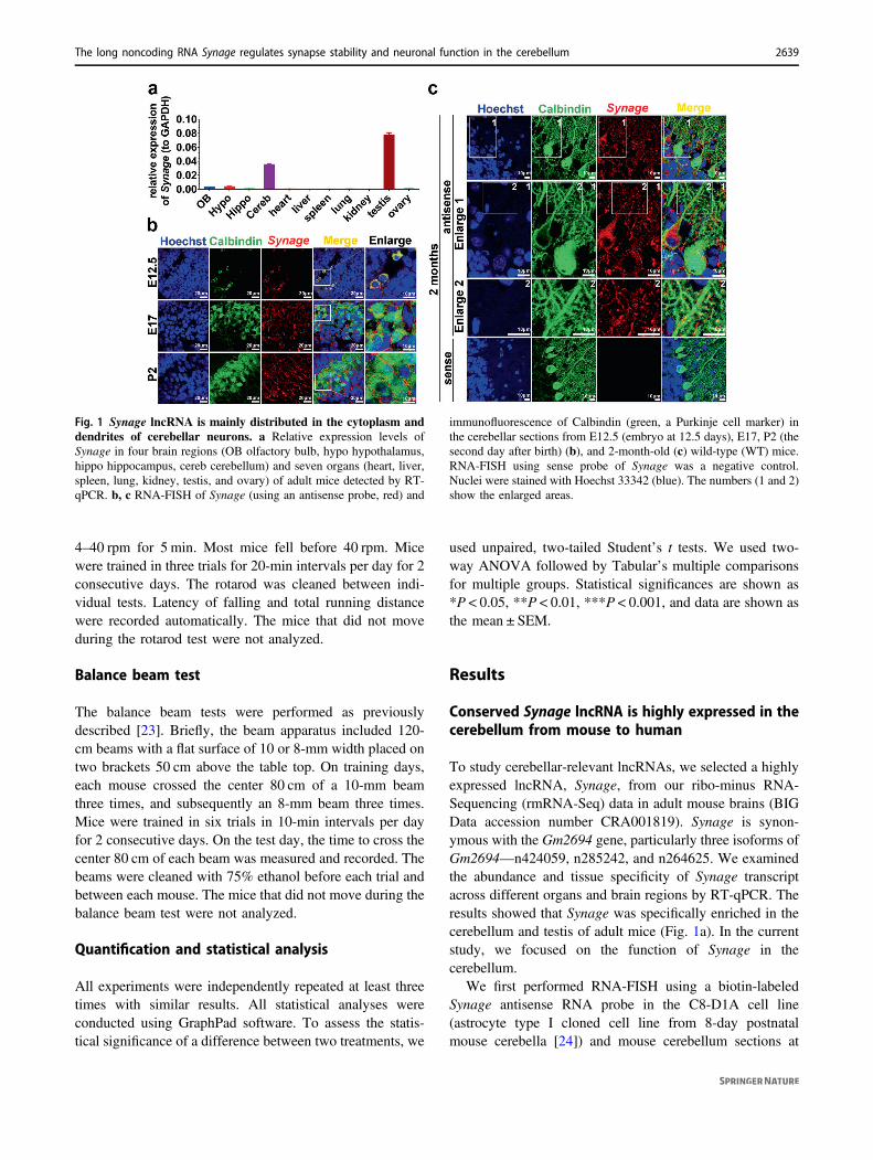
To study cerebellar-relevant lncRNAs, we selected a highlyexpressed lncRNA, Synage, from our ribo-minus RNA-Sequencing (rmRNA-Seq) data in adult mouse brains (BIGData accession number CRA001819). Synage is synon-ymous with the Gm2694 gene, particularly three isoforms ofGm2694—n424059, n285242, and n264625. We examinedthe abundance and tissue specificity of Synage transcriptacross different organs and brain regions by RT-qPCR. Theresults showed that Synage was specifically enriched in thecerebellum and testis of adult mice (Fig. 1a). In the currentstudy, we focused on the function of Synage in thecerebellum.
We first performed RNA-FISH using a biotin-labeledSynage antisense RNA probe in the C8-D1A cell line(astrocyte type I cloned cell line from 8-day postnatalmouse cerebella [24]) and mouse cerebellum sections at
Fig. 1 Synage lncRNA is mainly distributed in the cytoplasm anddendrites of cerebellar neurons. a Relative expression levels ofSynage in four brain regions (OB olfactory bulb, hypo hypothalamus,hippo hippocampus, cereb cerebellum) and seven organs (heart, liver,spleen, lung, kidney, testis, and ovary) of adult mice detected by RT-qPCR. b, c RNA-FISH of Synage (using an antisense probe, red) and
immunofluorescence of Calbindin (green, a Purkinje cell marker) inthe cerebellar sections from E12.5 (embryo at 12.5 days), E17, P2 (thesecond day after birth) (b), and 2-month-old (c) wild-type (WT) mice.RNA-FISH using sense probe of Synage was a negative control.Nuclei were stained with Hoechst 33342 (blue). The numbers (1 and 2)show the enlarged areas.
The long noncoding RNA Synage regulates synapse stability and neuronal function in the cerebellum 2639

several developmental stages, including E12.5, E17, P2,and 2 months. Synage was mainly distributed in the cyto-plasm of C8-D1A cerebellum cells (Fig. S1), and wasspecifically distributed in the Purkinje cell precursors(PCPs) at E12.5, while at E17, P2, and 2 months, it waslocalized primarily in cytoplasm and dendrites of cerebellarcells, including PCs (Fig. 1b, c).
Synage-homologous genes (LOC106995009 in rhesusmacaque, and RP11-491F9.1 in human) were conserved interms of their locations in the genomes of mouse, rhesusmacaque, and human (adjacent to the Cbln1 gene)(Fig. S2a–c). Similar to Synage lncRNA in mouse [17], itshomologous lncRNAs exhibited cerebellum-specificexpression in rhesus macaque [25] and in human tissuesaccording to a recent study [26] and GTEx project database(dbGaP Accession phs000424.v8.p2) (Fig. S2d). Takentogether, Synage lncRNA is conserved in its genomiclocation (adjacent to the Cbln1 gene) and in its distributionspecificity in the cerebellum among mouse, rhesus maca-que, and human.
According to our rmRNA-Seq in mouse brains(Fig. S3a), the expression level of n424059 (one of theSynage isoforms) was the highest, followed by n285242(one of the Synage isoforms) and TCONS_00072254(Fig. S3b). Although the expression trend of Synage iso-forms detected by RT-qPCR was not completely consistentwith that of rmRNA-Seq (Fig. S3c–g), n424059 andn285242 were relatively highly expressed in both rmRNA-Seq and RT-qPCR. In addition, the 5′- and 3′-RACEexperiments using n424059-specific primers (Fig. S4a–d)revealed that Synage is 663 nt in length, consistent with thelength of n424059 and with cDNA sequencing conducted inthis study (Fig. S4e). Therefore, our data support thatn424059 is a typical sequence for Synage RNA; we thusused the n424059 lncRNA sequence for our followingin vitro and in vivo overexpression experiments.
Synage KO mice show significant cerebellar atrophyand neuronal loss during cerebellar development
We generated Synage KO mice with sgRNAs targetingSynage exon 1 and the 3′ end of the last exon coupled withthe CRISPR-Cas9 system, ablating most Synage locus,while avoiding deletion of the putative promoter regionshared with Cbln1 (Fig. 2a). F1 mice that carried hetero-zygous (HT) alleles were confirmed by PCR-based geno-typing (Fig. S5a). Homozygous mice were obtained bycrossing these HT mice, and again confirmed by PCR(Fig. S5a) and sequencing (Fig. S5b). We used RT-qPCR(Fig. 2b) and FISH (Fig. S5c) to verify the absence of theSynage transcript in Synage KO mice compared with WTmice.
Between WT and Synage KO mice, there were no sig-nificant changes in the body appearance (Fig. S5d), bodyweight (Fig. S5e, f), or brain weight (Fig. S5g, h). However,the weight of cerebella relative to body weight was sig-nificantly decreased in both female and male KO mice(Figs. S5i–k and 2c). We performed IF staining for PCs(using Calbindin, a specific marker for PCs [27–29]) oncerebellar sections of 2-month-old mice. The number ofPCs in adult KO mice was significantly decreased comparedto those of both WT mice and HT mice, while it did notdiffer between WT and HT mice (Fig. S5l, m). The proteinexpression levels of NeuN (a neuronal marker) andGephyrin (an inhibitory postsynaptic marker) were alsosubstantially reduced in Synage KO cerebella compared toWT (Fig. S5n, o).
Cerebellar neurons are generated from two distinct neu-roepithelial zones: the ventricular zone (VZ) and the moredorsally located rhombic lip (RL) [30, 31], while PCs aredifferentiated/generated from the VZ at E10.5–E12.5 [32–34], before subsequently migrating dorsally to form a multi-cell-thick immature PC layer called the cerebellar plate at∼E14.5 [35]. We further sectioned cerebella sagittally andIF stained them for PCs [27–29] at six developmental stages(E12.5, E17.5, P4, P7, P14, and 2 months) [36]. The resultsshowed that the VZ became thinner and revealed RLinvagination in E12.5 Synage KO mice compared to the WTcontrols (Fig. 2d).
Given that the PCPs expand in concert with cerebellardevelopment, and considering that several PC clusters(PCCs) are known to aggregate with the PCs forming amultilayer below the external germinal layer by E17.5 [37],it was informative when we observed obviously reducednumbers of PCPs and of PCCs in E17.5 Synage KO micecompared to the WT mice (Fig. 2e). During differentiation,mouse PCs are in cluster stage around E18 to P3 [38], andare in dispersal situation at P3–P7. Premature PCs arborize(maturation process) at P12–P13; the PCs maturation endsat around P18–P20 [39], and the cerebellum undergoesdramatic increases in size and changes after birth [40]. Wefound that the number of PCs was reduced in Synage KOmice at P4 (Fig. 2f), P7 (Fig. 2g), P14 (Fig. 2h), and2 months (Fig. 2i).
In addition, we counted the numbers of major cerebellarneurons (PCs and GCs) and BGCs in the cerebellum at P10,P23, and 2 months, using Calbindin-positive cells to repre-sent PCs, Gdf10-stained cells to determine BGCs [41–43],and the signal intensity of Hoechst 33342 staining in the GClayer to estimate GCs (since the signals for GCs-specificmarker Pax6 had too much overlay with each other toaccurately quantify). The numbers of PCs and GCs, but notBGCs, significantly decreased at P10 (Fig. S6a–d), P23(Fig. S6a, e–g), and 2 months (Fig. S6a, h–j) in Synage KO
2640 F. Wang et al.

mice compared with WT mice. Overexpression of Synage inthe cerebellum rescued the number of PCs to the level ofWT mice at 3 weeks after injection by injecting AAV-EF1α-
Synage into the cerebella of newborn Synage KO mice(Fig. 3a–c). These results supported that Synage is necessaryfor cerebellar development and maturation.
Fig. 2 Synage knockout mice show cerebellar defects and neuronalloss during cerebellar development. a Schematic representation ofthe position of Synage, Cbln1, sgRNAs, and genotyping primers (Fforward primers, R reverse primers). b Relative expression levels ofSynage in the cerebella of WT and knockout (KO) adult mice detectedby RT-qPCR. c Gross morphology of representative brains from adultWT and KO male mice. d Hoechst 33342 staining of cerebella from
E12.5 WT and KO mice (WT, n= 9; KO, n= 6). e–i Immuno-fluorescence staining of the Purkinje cell marker protein (Calbindin,green) in the cerebella from E17.5 (e), P4 (f), P7 (g), P14 (h), and 2-month-old (i) WT and KO mice. WT (E17.5), n= 7; KO (E17.5), n=3; WT (P4, P7, P14, and 2 months), n= 3; KO (P4, P7, P14, and2 months), n= 3. Nuclei were stained with Hoechst 33342 (blue).
The long noncoding RNA Synage regulates synapse stability and neuronal function in the cerebellum 2641
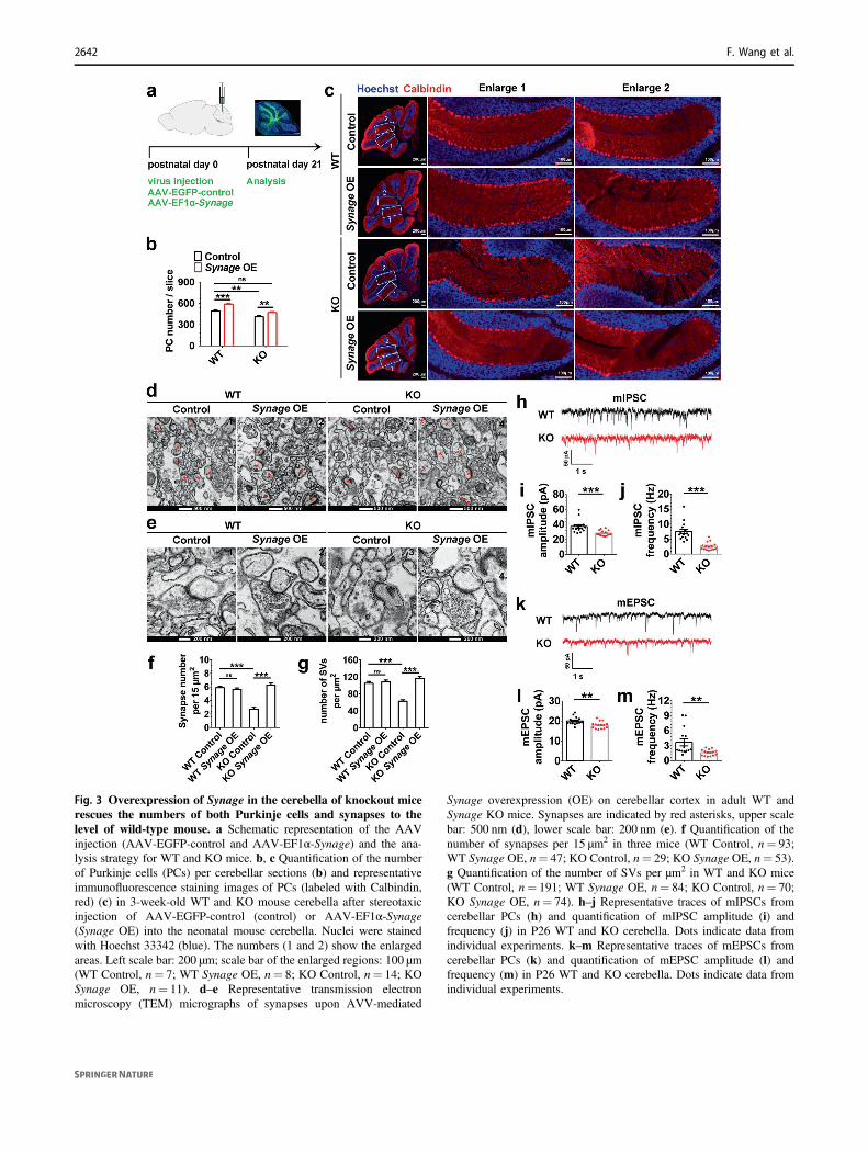
Fig. 3 Overexpression of Synage in the cerebella of knockout micerescues the numbers of both Purkinje cells and synapses to thelevel of wild-type mouse. a Schematic representation of the AAVinjection (AAV-EGFP-control and AAV-EF1α-Synage) and the ana-lysis strategy for WT and KO mice. b, c Quantification of the numberof Purkinje cells (PCs) per cerebellar sections (b) and representativeimmunofluorescence staining images of PCs (labeled with Calbindin,red) (c) in 3-week-old WT and KO mouse cerebella after stereotaxicinjection of AAV-EGFP-control (control) or AAV-EF1α-Synage(Synage OE) into the neonatal mouse cerebella. Nuclei were stainedwith Hoechst 33342 (blue). The numbers (1 and 2) show the enlargedareas. Left scale bar: 200 μm; scale bar of the enlarged regions: 100 μm(WT Control, n= 7; WT Synage OE, n= 8; KO Control, n= 14; KOSynage OE, n= 11). d–e Representative transmission electronmicroscopy (TEM) micrographs of synapses upon AVV-mediated
Synage overexpression (OE) on cerebellar cortex in adult WT andSynage KO mice. Synapses are indicated by red asterisks, upper scalebar: 500 nm (d), lower scale bar: 200 nm (e). f Quantification of thenumber of synapses per 15 μm2 in three mice (WT Control, n= 93;WT Synage OE, n= 47; KO Control, n= 29; KO Synage OE, n= 53).g Quantification of the number of SVs per μm2 in WT and KO mice(WT Control, n= 191; WT Synage OE, n= 84; KO Control, n= 70;KO Synage OE, n= 74). h–j Representative traces of mIPSCs fromcerebellar PCs (h) and quantification of mIPSC amplitude (i) andfrequency (j) in P26 WT and KO cerebella. Dots indicate data fromindividual experiments. k–m Representative traces of mEPSCs fromcerebellar PCs (k) and quantification of mEPSC amplitude (l) andfrequency (m) in P26 WT and KO cerebella. Dots indicate data fromindividual experiments.
2642 F. Wang et al.

Synage deletion leads to severe morphological andfunctional defects in synapses
Since synapses can form between neurons and are essentialto neuronal function, the loss of neurons (PCs and GCs)upon Synage KO led us to investigate if there was also anablation of cerebellar synapses after Synage KO. We per-formed TEM analyses on cerebellar cortex slides in adultWT and Synage KO mice to observe the numbers ofsynapses and synaptic vesicles (SVs). We found: (1) sig-nificantly reduced numbers of both synapses and SVs inpresynaptic terminals in the cerebellar cortex of KO com-pared to WT mice (Fig. 3d–g). (2) Cerebella overexpressingSynage in adult WT mice showed no significant change inthe number of synapses or SVs compared to WT mice(Fig. 3d–g). (3) Complementation of Synage expression inSynage KO mice rescued the numbers of both synapses andSVs (which reached the levels detected for WT mice)(Fig. 3d–g). The reduction in synapse density in Synage KOmice further suggests an additional deficit in synaptogenesisand/or in synapse maintenance. Moreover, the reduction inthe number of SVs also shows a deficit in the structure andfunction of individual synapses.
The severe morphological defects in the cerebellarsynapses of Synage KO mice suggested that synaptic con-nectivity and function are potentially adversely affected. Wethus evaluated the excitatory and inhibitory synapses of PCsby measuring mIPSCs and mEPSCs from PCs on cerebellarsagittal slices using a whole-cell patch-clamp. Both ampli-tude and frequency of mIPSCs were reduced in Synage KOmice (Fig. 3h–j), as were mEPSCs (Fig. 3k–m). Theseresults indicate that synaptic connectivity and function inPCs in the cerebella of Synage KO mice are severelycompromised.
Aberrant cerebellar morphology often leads to motorbehavior defects [44]. The rotarod test and the balance beamtest, well-established methods to evaluate motor coordina-tion in rodents [23, 45], showed that motor abilities andmotor-dependent learning and memory were severelyimpaired in Synage KO mice (Fig. S7a, b). Taken together,our findings of the decrease in cerebellar neurons andsynapses and the defects in neuronal synaptic function inSynage KO mice all strongly suggest that the severe mor-phological and functional defects in neurons and synapsesare responsible for the observed motor dysfunction ofSynage KO mice.
Synage lncRNA maintains stability and function ofcerebellar synapses partially by regulating Cbln1mRNA
Cbln1, a cerebellum highly expressed and synapse-relatedglycoprotein-coding gene, is upstream of Synage and is
transcribed in the opposite direction (Fig. S2a). CBLN1protein is secreted from cerebellar GCs to act as a criticalsynapse organizer between PFs and PCs [6, 7]. Since manylncRNAs regulate their neighboring protein-coding genes,we asked whether Synage lncRNA also modulates Cbln1expression. C8-D1A cells have poor transfection efficiency,which prevented us from performing in vitro experiments inthis cell line. After screening for many cell lines, we foundthat two isoforms of Synage (n285242, n264625) wererobustly expressed in the HT-22 cell line (Fig. S8a), whichis a mouse hippocampal neuronal cell line that has highertransfection efficiency than the C8-D1A cell line [46, 47].Therefore, we designed two shRNAs specifically targetingSynage, and transfected them into the HT-22 cell line toknockdown Synage (Fig. S8b). The results indicated that theexpression levels of both Cbln1 mRNA and protein weresignificantly reduced upon Synage knockdown in the HT-22cell line (Fig. S8b–d).
To further test the effect of Synage knockdown on Cbln1in vivo, we injected the Synage shRNAs into the cerebellaof adult WT mice. Two weeks later, we again observed thatCbln1 expression was diminished in the cerebellum com-pared with its expression in the olfactory bulb (Fig. S8e),Cbln1 mRNA and protein levels had a similar decrease inSynage KO mouse cerebellum (Figs. 4a and S8f, g). Wealso found that overexpression of Synage in its KO mousesignificantly increased the CBLN1 expression comparedwith KO control group (Figs. 4b and S8h). Thus, bothin vivo and in vitro Synage knockdown as well as in vivoSynage KO experiments demonstrated that Synage regulatesthe expression of Cbln1. Given the important role of Cbln1in the regulation of cerebellar synaptic function, theseresults suggest that the Synage lncRNA may be alsoinvolved in regulating cerebellar synaptic functions.
Given their spatial proximity in the genome, we askedwhether Synage KO inhibiting Cbln1 expression is due toinadvertent excision of some potential regulatory elementsupstream of the Cbln1 gene. We thus performed a nuclearrun on assay [48–50] on WT and KO mice cerebella, andfound that Synage deletion did not affect the number ofnascent Cbln1 transcripts (Fig. 4c), although the total Cbln1mRNA and protein levels were significantly decreased inthe cerebella of 2-month-old Synage KO mice (Figs. 4aand S8f, g). These data suggest that Synage regulates Cbln1expression at the mRNA and/or protein levels.
Synage deletion exerted a strong influence on both themRNA and protein levels of Cbln1; however, we did notdetect the CBLN1 protein in our in vivo RNA pull-down–MS experiment as a potential Synage-associatingprotein. Considering our finding that Synage is localized inthe cytoplasm of cerebellar cells, we explored the possibilitythat Synage may function as a ceRNA by competing withmiRNAs [51, 52]. Specifically, we predicted the shared
The long noncoding RNA Synage regulates synapse stability and neuronal function in the cerebellum 2643
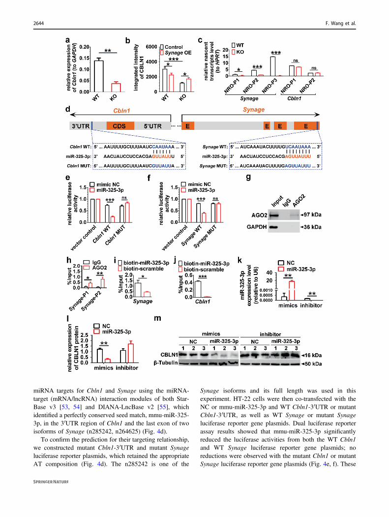
miRNA targets for Cbln1 and Synage using the miRNA-target (mRNA/lncRNA) interaction modules of both Star-Base v3 [53, 54] and DIANA-LncBase v2 [55], whichidentified a perfectly conserved seed match, mmu-miR-325-3p, in the 3′UTR region of Cbln1 and the last exon of twoisoforms of Synage (n285242, n264625) (Fig. 4d).
To confirm the prediction for their targeting relationship,we constructed mutant Cbln1-3′UTR and mutant Synageluciferase reporter plasmids, which retained the appropriateAT composition (Fig. 4d). The n285242 is one of the
Synage isoforms and its full length was used in thisexperiment. HT-22 cells were then co-transfected with theNC or mmu-miR-325-3p and WT Cbln1-3′UTR or mutantCbln1-3′UTR, as well as WT Synage or mutant Synageluciferase reporter gene plasmids. Dual luciferase reporterassay results showed that mmu-miR-325-3p significantlyreduced the luciferase activities from both the WT Cbln1and WT Synage luciferase reporter gene plasmids; noreductions were observed with the mutant Cbln1 or mutantSynage luciferase reporter gene plasmids (Fig. 4e, f). These
2644 F. Wang et al.

results together indicated that mmu-miR-325-3p directlybinds to the 3′UTR of Cbln1 mRNA and to the last exon ofSynage.
MicroRNAs silence gene expression by repressingtranslation and promoting target mRNA degradation [56].To determine whether Synage regulates Cbln1 as a ceRNAby competing for the above identified shared miRNA(mmu-miR-325-3p) in the AGO2-miRNA pathway, wemade use of the published AGO2 CLIP-Seq data frommouse cortex tissue (GSE73058) to identify AGO2-boundRNAs [57]. We found that AGO2 had multiple binding siteslocated in the Cbln1 and Synage (Supplementary Table S1).We performed an AGO2-RIP-qPCR experiment usingmouse cerebellum tissue, which showed that n285242 andn264625 (two isoforms of Synage) were pulled down in theAGO2 complex (Fig. 4g, h). Conversely, a biotin-labeledmmu-miR-325-3p RNA was able to pull-down both Synage(by a primer pair that targeting all of the three isoforms) andCbln1 transcripts (Fig. 4i, j). We next determined the effectsof mmu-miR-325-3p on Synage and Cbln1 expression, bydetecting the respective decrease or increase in CBLN1protein in response to the mmu-miR-325-3p mimics orinhibitor in the transfected HT-22 cell line (Fig. 4k–m).Together, our data indicate that Synage (particularly,
isoforms of n285242 and n264625) acts as a sponge formmu-miR-325-3p to regulate Cbln1 mRNA expression,which leads to the change of the CBLN1 protein levels.
Cbln1−/− mice showed cerebellar ataxia and impairedperformance accompanied by a significant reduction in thenumber of PF-PC synapses, as well as severe impairment tosynaptic function (Supplementary Table S2) [6, 58–62].Synage KO mice exhibited phenotypes consistent with thesereports, including synapse reduction and dysfunction, aswell as motor defects, but otherwise showed more severeimpairment than the phenotypes of Cbln1−/− mice, includ-ing decreased SVs, obvious neuronal loss, decreased cere-bellar weight, and reduced fertility (SupplementaryTable S2). On the other hand, Cbln1 heterozygotes showedmore than half of CBLN1 protein loss in the cerebellum butdisplayed no markedly impaired performance on theaccelerating rotarod test [6]. CbIn1 heterozygous micemight have developed some compensatory mechanisms,while Synage KO- or knockdown-induced 50% Cbln1downregulation may be sufficient to manifest the phenotypeof synapses loss. In addition, Cbln1 was also found tomediate specific aspects of fear conditioning and spatialmemory differentially and regulate both motor and non-motor functions [63]. We thus speculated that both Synageand Cbln1 likely have additional functions that are inde-pendent of one another. Synage probably modulates cere-bellar development and function through other mechanismsin addition to regulation of Cbln1 expression.
Synage lncRNA modulates cerebellar synapses byorchestrating assembly of synaptic LRP1-HSP90AA1-PSD-95 complex
In our in vivo RNA pull-down–MS experiments, LRP1 andHSP90AA1 were the two strongest candidates identified byMS in the cerebellum (Fig. 5a, b). We conducted LRP1-RIPand HSP90AA1-RIP assays (Fig. 5c, d), and Synage wasdetected in both LRP1-RIP and HSP90AA1-RIP samples(Fig. 5e, f), confirming that Synage bound both LRP1 andHSP90AA1 and might form a complex. LRP1 is known tointeract with PSD-95, through which it modulates synapticfunction [9, 64]. We noted a similarity of the neuronal loss inmice induced by LRP1 deletion and the Synage KO (Sup-plementary Table S2). We thus speculated that PSD-95 mayalso be in the complex of LRP1-HSP90AA1-Synage. RNA-FISH in combination with IF using different fluorophores tolabel Synage, LRP1, PSD-95, and HSP90AA1, showed co-localization of Synage with all three proteins in PCs of WTcerebella, and the co-localization was significantly reduced inPCs of Synage KO cerebella (Fig. 5g, h).
We also examined the influence of Synage on LRP1-HSP90AA1-PSD-95 interactions by performing co-IP withmouse cerebellum tissue (Fig. 5i–k) and found that the
Fig. 4 Synage lncRNA regulates Cbln1 mRNA through the AGO2-miR-325-3p pathway. a Relative expression level of Cbln1 mRNA inadult WT and KO mouse cerebella. b Relative quantification of inte-grated intensity of CBLN1 protein in the cerebellar cortex of 3-week-old WT and KO mice detected by immunofluorescence staining afterstereotaxic injection of AAV-control or AAV-Synage (Synage OE)into the neonatal mouse cerebellum. c Relative nascent transcriptlevels of both Synage and Cbln1 using multiple pairs of primers,normalized to housekeeping gene HPRT (hypoxanthine guaninephosphoribosyl transferase). NRO nuclear run on, P primer pairs. dSchematic representation of the predicted binding sites of miR-325-3pon Synage and Cbln1 (CDS coding sequence, UTR untranslatedregion, E exon). e Relative luciferase activity of vector control and WTCbln1-3′UTR and mutant Cbln1-3′UTR luciferase reporter geneplasmids upon co-transfection of mimic negative control (NC) or miR-325-3p in the HT-22 cell line. f Relative luciferase activity of vectorcontrol and WT Synage and mutant Synage luciferase reporter geneplasmids upon co-transfection of mimic NC or miR-325-3p in the HT-22 cell line. Data are presented as the relative ratio of firefly luciferaseactivity to Renilla luciferase activity. gWestern blot analysis of AGO2protein immunoprecipitation by AGO2 antibody in AGO2-RNAimmunoprecipitation. h The amount of Synage lncRNA binding toAGO2 or IgG was quantified as a percentage of input in IP by RT-qPCR (Synage-P1, a primer pair specifically targeting n264625;Synage-P2, a primer pair targeting both n264625 and n285242). i, jThe amount of Synage lncRNA (i) and Cbln1 mRNA (j) binding tomiR-325-3p was quantified as a percentage of input in miR-325-3ppull-down assays by RT-qPCR. k–m Relative expression levels ofmiR-325-3p with in vitro transfection of miR-325-3p mimics andinhibitor in the HT-22 cell line, normalized to U6 (k). Quantification(l) and representative images of Western blots (m) for CBLN1 within vitro transfection of miR-325-3p mimics and inhibitor in the HT-22cell line.
The long noncoding RNA Synage regulates synapse stability and neuronal function in the cerebellum 2645

interactions among all three of these proteins were reducedin Synage KO mice compared with WT controls (Fig. 5l–n).Although IF experiments revealed a decrease in the protein
level of LRP1 in the cerebellar cortex of KO mice comparedto WT mice (Figs. 5g, h and S9a), the total protein levels ofthe LRP1, HSP90AA1, and PSD-95 did not differ when
2646 F. Wang et al.

examined by Western blots of cerebella samples of adultWT versus Synage KO mice (Fig. S9b, c). These datasuggest that Synage functions in synapse stability not byreducing protein levels per se, but rather by somehowregulating Synage-dependent assembly of the LRP1-HSP90AA1-PSD-95 complex in the cerebellar cortex.
We further investigated the possibility of interactionsbetween Synage and each of these proteins (LRP1, PSD-95,and HSP90AA1) using biotin-labeled n424059 (one of theSynage isoforms) sense probes in the Neuro-2a cell line byEMSAs (Fig. 5o). The Neuro-2a cell line is a fast-growingmouse neuroblastoma cell line [65, 66], and n424059 is notexpressed in this cell line, so that we could exclude theinterference from endogenous n424059 (Fig. S10). TheEMSAs data revealed that LRP1 and PSD-95 could each bindto Synage (Fig. 5o). Furthermore, overexpression of Synagein the HT-22 cell line significantly increased the interactionbetween LRP1 and HSP90AA1 compared with the controls,as shown by RT-qPCR and co-IP experiments (Fig. 5p, q).
In addition, we knocked down each protein (LRP1,HSP90AA1, and PSD-95) by three shRNAs specificallytargeting their mRNAs respectively in the HT-22 cell line(Fig. S11a–c). PSD-95 protein was dramatically reducedupon knockdown of LRP1 or HSP90AA1 in the HT-22 cellline (Fig. S11a, b, d, e). Furthermore, the LRP1 protein wassignificantly upregulated upon knockdown of HSP90AA1 orPSD-95 (Fig. S11b, c, e, f). These results demonstrated thatknocking down LRP1 or HSP90AA1 or PSD-95 impactedthe levels of the other two proteins comprising the
LRP1-HSP90AA1-PSD-95 complex. We also found that,after LRP1 knockdown, overexpression of Synage in theHT-22 cell line significantly increased the extent of theLRP1-HSP90AA1 interaction as compared to the randomshRNA control (Fig. S11g, h), indicating that LRP1 deple-tion inhibits the interactions between LRP1-HSP90AA1 andSynage. Together, these results demonstrate the function ofSynage as a key organizer of the LRP1-HSP90AA1-PSD-95complex in PCs, to maintain the stability and function ofthese proteins in cerebellar synapses.
Discussion
lncRNAs are abundant in the brain. In the present work, wediscovered that a cerebellum highly expressed lncRNA,Synage, mainly distributed in the cytoplasm and extendinginto neurites and synapses, regulate cerebellar synapticstability and cerebellar development starting at the E12.5embryo stage. Synage KO leads to a dramatic decrease incerebellar neuron number, synapse density, and synapticstability in mice cerebella. At the molecular level, weshowed that Synage regulates synaptic stability and functionby Synage-dependent assembly of the LRP1-HSP90AA1-PSD-95 complex in the cerebellar cortex (Fig. 6). BothHSP90AA1 and LRP1 have been reported to exhibit anti-apoptotic effects in neurons by directly binding and/oractivating Akt kinase [67, 68]. We thus speculate that theinteraction of Synage with HSP90AA1 and LRP1 mightincrease Akt activation and thus protect PC and GC neuronsfrom apoptosis in the WT mice. In addition, Synage appearsto be multitasking in that it also functions as a ceRNA toadsorb a Cbln1-targeted miRNA to upregulate Cbln1expression in GCs (Fig. 6). Synage KO mice were moreseverely impaired than the Cbln1−/− mice (SupplementaryTable S2), consistent with our data showing that Synageexerts its function via at least two mechanisms, first byregulating the expression level of Cbln1 (in GCs), andsecond by orchestrating the formation of the synapticLRP1-HSP90AA1-PSD-95 complex (in PCs).
The synapses between climbing fibers and PCs, as wellas those between PFs and PCs, are known to form in thecerebellar cortex after birth [69–71]. Our data, however,showed that the loss of PCs occurred during embryonicdevelopment of the Synage KO cerebellum. Accordingly,our data support that the observed decrease in PCs numberin the Synage KO mice during embryonic developmentclearly has longer term impacts that manifest later asreductions in synapse density and in synaptic activity afterbirth. Although we cannot rule out the possibility that thereduced PCs number may also affect synaptic formation ormaintenance, our data indicate that altered synaptic forma-tion (or maintenance) after birth is impacted by the
Fig. 5 Synage lncRNA acts as an organizer to scaffold the synapticLRP1-HSP90AA1-PSD-95 complex. a RT-qPCR analysis of in vivoRNA pull-down showing retrieval of Synage lncRNA with Synage-specific probes in the adult mouse cerebella. LacZ probes were thenegative controls. Synage_p1 and Synage_p2 represent different pri-mers targeting Synage (all three isoforms). b List of top Synage-binding proteins detected in the adult mouse cerebella by in vivo RNApull-down–mass spectrometry. c–f Western blots assessing LRP1 andHSP90AA1 immunoprecipitation (IP) by anti-LRP1 (c) or anti-HSP90AA1 (d) antibody in RNA immunoprecipitation (RIP). Rela-tive enrichment of Synage binding to LRP1 (e) or HSP90AA1 (f) orIgG in IP was quantified as a percentage of input by RT-qPCR. p1 andp2 represent different primers targeting Synage (all three isoforms). g,h Co-localization analysis of Synage with PSD-95 and LRP1 (g) aswell as with HSP90AA1 and LRP1 (h) by RNA-FISH of Synage andimmunofluorescence staining of these proteins in the 2-month-old WTand KO mouse cerebella. i–n Western blots and relative quantificationof the interactions among LRP1 (i, l), HSP90AA1 (j, m), and PSD-95(k, n) as determined by co-IP analysis in the adult WT and KO mousecerebella. o RNA-electrophoretic mobility shift assays (EMSAs) forverification of the direct interactions between Synage and its bindingproteins in the Neuro-2a cell line. p, q RT-qPCR analysis of theoverexpression level of Synage (sense) in the HT-22 cell line (p),Western blots assessing LRP1 and HSP90AA1 co-immunoprecipitation by anti-LRP1 antibody after Synage over-expression in the HT-22 cell line, Synage antisense RNA was used as anegative control (q).
The long noncoding RNA Synage regulates synapse stability and neuronal function in the cerebellum 2647

destruction of Synage-binding protein complexes in theSynage KO cerebellum.
In summary, this study demonstrates that cerebellumhighly expressed Synage lncRNA regulates the formation ofthe LRP1-HSP90AA1-PSD-95 complex in PCs and Cbln1expression in GCs. These interacting components are locatedin synapses where they perform unique roles, thereby dif-ferentially affecting synaptic stability and functionality,which in turn affects the growth of neurons, the developmentand function of the cerebellum, and greatly contributes tomotor function during development. Since the genomiclocalization and the cerebellar distribution of Synage areconserved from mice to humans, targeting of the LRP1-HSP90AA1-PSD-95-Synage complex or Synage-Cbln1interactions may protect against developmental defects inhumans and in model organisms; insights into their neuro-protective mechanisms can potentially yield pharmaceuticalsdesigned to extend neurological health and synaptic function,and to mitigate cerebellar neurodevelopmental disorders.
Data availability
The accession number for the ribo-minus RNA-Seq datareported in this paper is CRA001819 and is publiclyaccessible at https://bigd.big.ac.cn/gsa.
Acknowledgements We thank Dr Wei Xiong (USTC) for providingthe mouse brain stereotactic injection instrument and the experimentalplatform for feeding and behavior tests of KO mice and for helpfulsuggestions. We thank Dr Jiangning Zhou (USTC), Dr Aihui Tang(USTC), and Dr Shouhong Guang (USTC) for constructive sugges-tions. We also appreciate Dr Jiangning Zhou (USTC) for providing thebalance beam instrument and Neuro-2a and HT-22 cell lines. We thank
Dr Yong Shen (USTC) for providing antibodies for Western blots(anti-NeuN, -Gephyrin and -PSD-95 antibodies). We thank DrXiangting Wang (USTC) for providing the C8-D1A cell line and forconstructive suggestions. We also appreciate Chunying Yin (USTC)for TEM technical support.
Author contributions XS supervised the project; XS and FW con-ceived and designed the study; young and old brain samples forrmRNA-Seq were provided and collected by SM and JS; FW per-formed bioinformatic analyses with the help of RW, discoveredSynage lncRNA. and predicted its function in vitro and in vivo; FWand QW carried out phenotype analyses, section staining, and behaviortests; FW and BL performed the molecular mechanism experiments;SW and YZ performed mass spectrometry detection. LM and FWconducted the electrophysiological experiment with supervision fromZZ; YZ and ZX worked collaboratively with XS to supervise theproject; CN provided human cerebellum sample; FW analyzed the dataand produced the figures; FW and XS wrote the paper. QW and BLedited the manuscript. All authors reviewed the manuscript andapprove of its contents.
Funding This work was funded by the National Natural ScienceFoundation of China (91540107 to XS) and the State Key Laboratoryof Neuroscience, Shanghai Institutes for Biological Sciences, ChineseAcademy of Sciences (SKLN-201805 to XS), the Natural ScienceFoundation of Anhui Province (2008085QC160 to FW), and theScience and Technology Project grant from Anhui Province(1604a0802069 to CN).
Compliance with ethical standards
Conflict of interest The authors declare no competing interests.
Ethics approval All animal experiments were carried out in accor-dance with the guidelines of and approved by the University ofScience and Technology of China (USTC) Animal Resources Centerand University Animal Care and Use Committee (permit number:USTCACUC1801015).
Fig. 6 A schematic diagram of the mechanism of the SynagelncRNA in the regulation of synaptic and neuronal function in theWT and Synage KO mouse cerebella. Synage lncRNA regulatesstability and function of cerebellar synapses via at least two mechan-isms. One is through the function of Synage as a sponge for mmu-miR-
325-3p to regulate Cbln1 mRNA expression, which leads to thechange of the CBLN1 protein levels. The other function is to serve as ascaffold for orchestrating the assembly of synaptic LRP1-HSP90AA1-PSD-95 complex.
2648 F. Wang et al.

Publisher’s note Springer Nature remains neutral with regard tojurisdictional claims in published maps and institutional affiliations.
Open Access This article is licensed under a Creative CommonsAttribution 4.0 International License, which permits use, sharing,adaptation, distribution and reproduction in any medium or format, aslong as you give appropriate credit to the original author(s) and thesource, provide a link to the Creative Commons license, and indicate ifchanges were made. The images or other third party material in thisarticle are included in the article’s Creative Commons license, unlessindicated otherwise in a credit line to the material. If material is notincluded in the article’s Creative Commons license and your intendeduse is not permitted by statutory regulation or exceeds the permitteduse, you will need to obtain permission directly from the copyrightholder. To view a copy of this license, visit http://creativecommons.org/licenses/by/4.0/.
References
1. Sathyanesan A, Zhou J, Scafidi J, Heck DH, Sillitoe RV, Gallo V.Emerging connections between cerebellar development, behaviourand complex brain disorders. Nat Rev Neurosci. 2019;20:298–313.
2. Manto M, De Zeeuw CI. Diversity and complexity of roles ofgranule cells in the cerebellar cortex. Editor Cerebellum.2012;11:1–4.
3. Marzban H, Del Bigio MR, Alizadeh J, Ghavami S, ZachariahRM, Rastegar M. Cellular commitment in the developing cere-bellum. Front Cell Neurosci. 2014;8:450.
4. Ito M. Historical review of the significance of the cerebellum andthe role of Purkinje cells in motor learning. Ann N Y Acad Sci.2002;978:273–88.
5. Heiney SA, Kim J, Augustine GJ, Medina JF. Precise control ofmovement kinematics by optogenetic inhibition of Purkinje cellactivity. J Neurosci. 2014;34:2321.
6. Hirai H, Pang Z, Bao D, Miyazaki T, Li L, Miura E, et al. Cbln1 isessential for synaptic integrity and plasticity in the cerebellum.Nat Neurosci. 2005;8:1534–41.
7. Ito-Ishida A, Okabe S, Yuzaki M. The role of Cbln1 on Purkinjecell synapse formation. Neurosci Res. 2014;83:64–8.
8. Eugenin EA, King JE, Nath A, Calderon TM, Zukin RS, BennettMV, et al. HIV-tat induces formation of an LRP-PSD-95-NMDAR-nNOS complex that promotes apoptosis in neurons andastrocytes. Proc Natl Acad Sci USA. 2007;104:3438–43.
9. Maier W, Bednorz M, Meister S, Roebroek A, Weggen S, SchmittU, et al. LRP1 is critical for the surface distribution and inter-nalization of the NR2B NMDA receptor subtype. Mol Neurode-gener. 2013;8:25.
10. May P, Rohlmann A, Bock HH, Zurhove K, Marth JD, Schom-burg ED, et al. Neuronal LRP1 functionally associates withpostsynaptic proteins and is required for normal motor function inmice. Mol Cell Biol. 2004;24:8872–83.
11. Liu Q, Trotter J, Zhang J, Peters MM, Cheng H, Bao J, et al.Neuronal LRP1 knockout in adult mice leads to impaired brainlipid metabolism and progressive, age-dependent synapse loss andneurodegeneration. J Neurosci. 2010;30:17068–78.
12. Mantuano E, Lam MS, Shibayama M, Campana WM, Gonias SL.The NMDA receptor functions independently and as an LRP1 co-receptor to promote Schwann cell survival and migration. J CellSci. 2015;128:3478–88.
13. Goff LA, Groff AF, Sauvageau M, Trayes-Gibson Z, Sanchez-Gomez DB, Morse M, et al. Spatiotemporal expression andtranscriptional perturbations by long noncoding RNAs in themouse brain. Proc Natl Acad Sci USA. 2015;112:6855–62.
14. Bond AM, VanGompel MJW, Sametsky EA, Clark MF, SavageJC, Disterhoft JF, et al. Balanced gene regulation by an embryonicbrain non-coding RNA is critical for GABA circuitry in adulthippocampus. Nat Neurosci. 2009;12:1020–7.
15. Ramos AD, Andersen RE, Liu SJ, Nowakowski TJ, Hong SJ,Gertz C, et al. The long noncoding RNA Pnky regulates neuronaldifferentiation of embryonic and postnatal neural stem cells. CellStem Cell. 2015;16:439–47.
16. Raveendra BL, Swarnkar S, Avchalumov Y, Liu X-A, Grinman E,Badal K, et al. Long noncoding RNA GM12371 acts as a tran-scriptional regulator of synapse function. Proc Natl Acad Sci.2018;115:E10197.
17. Mercer TR, Dinger ME, Sunkin SM, Mehler MF, Mattick JS.Specific expression of long noncoding RNAs in the mouse brain.Proc Natl Acad Sci USA. 2008;105:716–21.
18. Guttman M, Donaghey J, Carey BW, Garber M, Grenier JK,Munson G, et al. lincRNAs act in the circuitry controlling plur-ipotency and differentiation. Nature. 2011;477:295–300.
19. Li YP, Duan FF, Zhao YT, Gu KL, Liao LQ, Su HB, et al. ATRIM71 binding long noncoding RNA Trincr1 represses FGF/ERKsignaling in embryonic stem cells. Nat Commun. 2019;10:1368.
20. Mahmood T, Yang PC. Western blot: technique, theory, andtrouble shooting. N Am J Med Sci. 2012;4:429–34.
21. Chu C, Zhang QC, da Rocha ST, Flynn RA, Bharadwaj M,Calabrese JM, et al. Systematic discovery of Xist RNA bindingproteins. Cell. 2015;161:404–16.
22. Lal A, Thomas MP, Altschuler G, Navarro F, O’Day E, Li XL,et al. Capture of microRNA–bound mRNAs identifies the tumorsuppressor miR-34a as a regulator of growth factor signaling.PLoS Genet. 2011;7:e1002363.
23. Luong TN, Carlisle HJ, Southwell A, Patterson PH. Assessmentof motor balance and coordination in mice using the balancebeam. J Vis Exp: JoVE. 2011;49:2376.
24. Alliot F, Pessac B. Astrocytic cell clones derived from establishedcultures of 8-day postnatal mouse cerebella. Brain Res. 1984;306:283–91.
25. Liu S, Wang Z, Chen D, Zhang B, Tian RR, Wu J, et al. Anno-tation and cluster analysis of spatiotemporal- and sex-relatedlncRNA expression in rhesus macaque brain. Genome Res.2017;27:1608–20.
26. Zhang XQ, Wang ZL, Poon MW, Yang JH. Spatial-temporaltranscriptional dynamics of long non-coding RNAs in humanbrain. Hum Mol Genet. 2017;26:3202–11.
27. Ito-Ishida A, Kakegawa W, Kohda K, Miura E, Okabe S, YuzakiM. Cbln1 downregulates the formation and function of inhibitorysynapses in mouse cerebellar Purkinje cells. Eur J Neurosci.2014;39:1268–80.
28. Zhang B, Chen LY, Liu X, Maxeiner S, Lee SJ, Gokce O, et al.Neuroligins sculpt cerebellar Purkinje-cell circuits by differentialcontrol of distinct classes of synapses. Neuron. 2015;87:781–96.
29. Sergaki MC, Ibanez CF. GFRalpha1 regulates purkinje cellmigration by counteracting NCAM function. Cell Rep.2017;18:367–79.
30. Hatten ME, Alder J, Zimmerman K, Heintz N. Genes involved incerebellar cell specification and differentiation. Curr Opin Neu-robiol. 1997;7:40–7.
31. Yamada M, Seto Y, Taya S, Owa T, Inoue YU, Inoue T, et al.Specification of spatial identities of cerebellar neuron progenitorsby ptf1a and atoh1 for proper production of GABAergic andglutamatergic neurons. J Neurosci. 2014;34:4786–800.
32. Minaki Y, Nakatani T, Mizuhara E, Inoue T, Ono Y. Identificationof a novel transcriptional corepressor, Corl2, as a cerebellar Pur-kinje cell-selective marker. Gene Expr Patterns. 2008;8:418–23.
33. Hashimoto M, Mikoshiba K. Neuronal birthdate-specific genetransfer with adenoviral vectors. J Neurosci. 2004;24:286–96.
The long noncoding RNA Synage regulates synapse stability and neuronal function in the cerebellum 2649
34. Hashimoto M, Mikoshiba K. Mediolateral compartmentalizationof the cerebellum is determined on the “birth date” of Purkinjecells. J Neurosci. 2003;23:11342–51.
35. Miyata T, Ono Y, Okamoto M, Masaoka M, Sakakibara A,Kawaguchi A, et al. Migration, early axonogenesis, and Reelin-dependent layer-forming behavior of early/posterior-born Purkinjecells in the developing mouse lateral cerebellum. Neural Dev.2010;5:23.
36. Yuasa S, Kawamura K, Ono K, Yamakuni T, Takahashi Y.Development and migration of Purkinje cells in the mouse cere-bellar primordium. Anat Embryol. 1991;184:195–212.
37. Fujita H, Morita N, Furuichi T, Sugihara I. Clustered fine com-partmentalization of the mouse embryonic cerebellar cortex and itsrearrangement into the postnatal striped configuration. J Neurosci.2012;32:15688–703.
38. Rahimi-Balaei M, Bergen H, Kong J, Marzban H. Neuronalmigration during development of the cerebellum. Front CellNeurosci. 2018;12:484.
39. Wang VY, Zoghbi HY. Genetic regulation of cerebellar devel-opment. Nat Rev Neurosci. 2001;2:484–91.
40. Goldowitz D, Hamre K. The cells and molecules that make acerebellum. Trends Neurosci. 1998;21:375–82.
41. Mecklenburg N, Martinez-Lopez JE, Moreno-Bravo JA,Perez-Balaguer A, Puelles E, Martinez S. Growth anddifferentiation factor 10 (Gdf10) is involved in Bergmann glialcell development under Shh regulation. Glia. 2014;62:1713–23.
42. Koirala S, Corfas G. Identification of novel glial genes by single-cell transcriptional profiling of Bergmann glial cells from mousecerebellum. PLoS ONE. 2010;5:e9198.
43. Martinez S, Andreu A, Mecklenburg N, Echevarria D. Cellularand molecular basis of cerebellar development. Front Neuroanat.2013;7:18.
44. Sillitoe RV, Joyner AL. Morphology, molecular codes, and cir-cuitry produce the three-dimensional complexity of the cere-bellum. Annu Rev Cell Dev Biol. 2007;23:549–77.
45. Shiotsuki H, Yoshimi K, Shimo Y, Funayama M, Takamatsu Y,Ikeda K, et al. A rotarod test for evaluation of motor skill learning.J Neurosci Methods. 2010;189:180–5.
46. He M, Liu J, Cheng S, Xing Y, Suo WZ. Differentiation renderssusceptibility to excitotoxicity in HT22 neurons. Neural RegenRes. 2013;8:1297–306.
47. Herrera F, Martin V, Garcia-Santos G, Rodriguez-Blanco J,Antolin I, Rodriguez C. Melatonin prevents glutamate-inducedoxytosis in the HT22 mouse hippocampal cell line through anantioxidant effect specifically targeting mitochondria. J Neu-rochem. 2007;100:736–46.
48. Roberts TC, Hart JR, Kaikkonen MU, Weinberg MS, Vogt PK,Morris KV. Quantification of nascent transcription by bromour-idine immunocapture nuclear run-on RT-qPCR. Nat Protoc.2015;10:1198–211.
49. Patrone G, Puppo F, Cusano R, Scaranari M, Ceccherini I, PulitiA, et al. Nuclear run-on assay using biotin labeling, magnetic beadcapture and analysis by fluorescence-based RT-PCR. Biotechni-ques 2000;29:1012–4, 6–7.
50. Smale ST. Nuclear run-on assay. Cold Spring Harb Protoc.2009;2009:pdb.prot5329.
51. Zhang Y, Liu Y, Xu X. Knockdown of LncRNA-UCA1 sup-presses chemoresistance of pediatric AML by inhibiting glyco-lysis through the microRNA-125a/hexokinase 2 pathway. J CellBiochem. 2018;119:6296–308.
52. Salmena L, Poliseno L, Tay Y, Kats L, Pandolfi PP. A ceRNAhypothesis: the Rosetta Stone of a hidden RNA language? Cell2011;146:353–8.
53. Yang JH, Li JH, Shao P, Zhou H, Chen YQ, Qu LH. starBase: adatabase for exploring microRNA-mRNA interaction maps fromArgonaute CLIP-Seq and Degradome-Seq data. Nucleic AcidsRes. 2011;39:D202–9.
54. Li JH, Liu S, Zhou H, Qu LH, Yang JH. starBase v2.0: decodingmiRNA-ceRNA, miRNA-ncRNA and protein-RNA interactionnetworks from large-scale CLIP-Seq data. Nucleic Acids Res.2014;42:D92–7.
55. Paraskevopoulou MD, Vlachos IS, Karagkouni D, Georgakilas G,Kanellos I, Vergoulis T, et al. DIANA-LncBase v2: indexingmicroRNA targets on non-coding transcripts. Nucleic Acids Res.2016;44:D231–8.
56. Jonas S, Izaurralde E. Towards a molecular understanding ofmicroRNA-mediated gene silencing. Nat Rev Genet. 2015;16:421–33.
57. Moore MJ, Scheel TK, Luna JM, Park CY, Fak JJ, Nishiuchi E,et al. miRNA-target chimeras reveal miRNA 3′-end pairing as amajor determinant of Argonaute target specificity. Nat Commun.2015;6:8864.
58. Yuzaki M. Two classes of secreted synaptic organizers in thecentral nervous system. Annu Rev Physiol. 2018;80:243–62.
59. Takeuchi E, Ito-Ishida A, Yuzaki M, Yanagihara D. Improvementof cerebellar ataxic gait by injecting Cbln1 into the cerebellum ofcbln1-null mice. Sci Rep. 2018;8:6184.
60. Rong Y, Wei P, Parris J, Guo H, Pattarini R, Correia K, et al.Comparison of Cbln1 and Cbln2 functions using transgenic andknockout mice. J Neurochem. 2012;120:528–40.
61. Uemura T, Lee SJ, Yasumura M, Takeuchi T, Yoshida T, Ra M,et al. Trans-synaptic interaction of GluRdelta2 and Neurexinthrough Cbln1 mediates synapse formation in the cerebellum. Cell2010;141:1068–79.
62. Ito-Ishida A, Miura E, Emi K, Matsuda K, Iijima T, Kondo T,et al. Cbln1 regulates rapid formation and maintenance of exci-tatory synapses in mature cerebellar Purkinje cells in vitro andin vivo. J Neurosci. 2008;28:5920–30.
63. Otsuka S, Konno K, Abe M, Motohashi J, Kohda K, Sakimura K,et al. Roles of Cbln1 in non-motor functions of mice. J Neurosci.2016;36:11801–16.
64. Nakajima C, Kulik A, Frotscher M, Herz J, Schafer M, Bock HH,et al. Low density lipoprotein receptor-related protein 1 (LRP1)modulates N-methyl-D-aspartate (NMDA) receptor-dependentintracellular signaling and NMDA-induced regulation of post-synaptic protein complexes. J Biol Chem. 2013;288:21909–23.
65. Salto R, Vilchez JD, Giron MD, Cabrera E, Campos N, ManzanoM, et al. beta-Hydroxy-beta-methylbutyrate (HMB) promotesneurite outgrowth in Neuro2a cells. PLoS ONE 2015;10:e0135614.
66. Olmsted JB, Carlson K, Klebe R, Ruddle F, Rosenbaum J. Iso-lation of microtubule protein from cultured mouse neuroblastomacells. Proc Natl Acad Sci USA. 1970;65:129–36.
67. Sato S, Fujita N, Tsuruo T. Modulation of Akt kinase activity bybinding to Hsp90. Proc Natl Acad Sci USA. 2000;97:10832–7.
68. Fuentealba RA, Liu Q, Kanekiyo T, Zhang J, Bu G. Low densitylipoprotein receptor-related protein 1 promotes anti-apoptoticsignaling in neurons by activating Akt survival pathway. J BiolChem. 2009;284:34045–53.
69. Hashimoto K, Kano M. Functional differentiation of multipleclimbing fiber inputs during synapse elimination in the developingcerebellum. Neuron 2003;38:785–96.
70. Hashimoto K, Kano M. Synapse elimination in the developingcerebellum. Cell Mol Life Sci. 2013;70:4667–80.
71. Kano M, Watanabe T. Developmental synapse remodeling in thecerebellum and visual thalamus. F1000Res. 2019;8:F1000 FacultyRev-1191.
2650 F. Wang et al.
Copyright © 2022 FDOKUMEN